Acute Reactogenicity after Intramuscular Immunization with ...
Upload
independentCategory
view
0download
0
Vaccine 24 (2006) 5559–5570
Development of novel fusogenic vesosomesfor transcutaneous immunization
Vivek Mishra, Sunil Mahor, Amit Rawat, Praveen Dubey,Prem N. Gupta, Paramjit Singh, Suresh P. Vyas ∗
Drug Delivery Research Laboratory, Department of Pharmaceutical Sciences,Dr. Harisingh Gour Vishwavidyalaya, Sagar, Madhya Pradesh 470003, India
Received 7 August 2005; received in revised form 28 March 2006; accepted 21 April 2006Available online 4 May 2006
Abstract
Transcutaneous immunization (TCI) is a novel vaccination strategy based on the application of antigen together with an adjuvant ontohydrated bare skin. This simple and non-invasive immunization procedure elicits systemic and cell mediated immune responses and therefore,ivwsnmrp(Ftid©
K
1
ayoTvi
0d
t provides a viable and cost-effective strategy for disease prevention. In the present study, novel fusogenic vesicular carrier constructs, i.e.esosomes were developed and evaluated for topical delivery of vaccines using tetanus toxoid (TTx) as a model antigen. Prepared vesosomesere characterized for size, shape, entrapment efficiency and zeta potential. The prepared novel systems were examined for in process antigen
tability and long-term storage stability studies. In vitro skin permeation and fluorescence microscopy study were also preformed for preparedovel vesicular systems for the evaluation of skin penetration efficiency. The immune stimulating activity of these vesicles was studied byeasuring the serum anti-tetanus toxoid IgG titer and isotype ratio IgG2a/IgG1 following topical immunization in three different protocols and
esults were compared with the alum adsorbed tetanus toxoid given intramuscularly and topically administered plain tetanus toxoid solution,lain liposomes and cationic fusogenic liposomes. Serum IgG titers after three consecutive topical administrations were significantly better
*P < 0.05) than single administration of TTx antigen with vesosomal systems, suggesting an effective stimulation of serum immune response.urthermore, notable serum anti-TTx antibody titers also occurred in animals primed with alum adsorbed TTx and subsequently boosted with
opical administration of novel vesosomal systems. In each immunization studies, the vesosomal systems could elicit combined Th1 and Th2mmune responses following topical administration. These results suggest that the investigated vesosomal systems can be effective as topicalelivery of vaccines.
2006 Elsevier Ltd. All rights reserved.
eywords: Topical immunization; Tetanus toxoid; Vaccine delivery; Fusogenic vesicles; Vesosomes
. Introduction
Traditionally, the skin immune system has been perceiveds an environment for immunopathology, but in the recentears, the potential for exploitation of the skin for purposesf vaccination has received a great deal of attention [1,2].ranscutaneous immunization (TCI), topical application ofaccine formulation on the skin, provides access to the skinmmune system which is dominated by densely distributed
∗ Corresponding author. Tel.: +91 7582 265525; fax: +91 7582 2265525.E-mail address: [email protected] (S.P. Vyas).
and potent antigen presenting cells (APC), Langerhan’s cellsthat can be manipulated by adjuvants to orchestrate spe-cific, robust immune responses. This exploitation of normalimmune defense mechanisms for the purposes of immuno-protection or immunomodulation can be achieved by theapplication of a simple patch, gel or solution to the skin.
Initial skepticism regarding transcutaneous immunizationand the revelation that large molecules in simple solutioncould in fact penetrate the skin [3,4]. This approach com-bines the advantages of needle-free delivery while targetingthe immunologically rich milieu of the skin. Vaccinationthrough the skin may be particularly advantageous as the
264-410X/$ – see front matter © 2006 Elsevier Ltd. All rights reserved.oi:10.1016/j.vaccine.2006.04.030
5560 V. Mishra et al. / Vaccine 24 (2006) 5559–5570
immunocompetent Langerhan’s cells are found in abundancealong the transdermal penetration pathways and these cellsare aligned specifically along the minute pores through whichpathogens are likely to invade the body. Langerhan’s cells arefound in close proximity to the stratum corneum and repre-sent a network of immune cells that underlie 25% of the skin’stotal surface area [4,5]. Epidermal Langerhan’s cells play avital role in antigen presentation to CD4+ T cells [6]. Thesecells bind cutaneously encountered antigen and then processit. Along with processed antigen, Langerhan’s cells migratefrom the epidermis into lymphatic vessels and finally intoregional lymph nodes. Differentiation of Langerhan’s cellsinto dendritic cells occurs during this process and the den-dritic cells offer the antigen to naı̈ve CD4+ T cells that haveentered the lymph nodes through the high endothelial venules[7,8].
Recently, we have described of novel vesicular carrierbased topical delivery of antigens to the skin that has bothpractical and immunologic merits [9,10]. Fusogenic lipo-some is a class of novel carrier can potentially facilitatethe intracellular delivery of encapsulated drugs/proteins byfusing with target cell. A variety of approaches can beenvisioned for constructing fusogenic liposomes. Examplesinclude the inclusion of lipids that are able to form non-bilayer phases, such as dioleoyl phophatidylethanolamine(DOPE), which can promote destabilization of the bilayer,itlpfftdigdpiasp
cmaicpepppl(t
2. Materials and methods
2.1. Chemicals and immunological reagents
Tetanus toxoid (TTx) was received from the Serum Insti-tute of India, Pune as a gift sample. Phosphatidylcholine,dioleoyl phosphatidylethanolamine (DOPE), dioleoyltrimethyl ammonium propane (DOTAP), dipalmitoylphosphatidylcholine (DPPC) Sephadex G-200, tetramethylbenzidine (TMB), FITC-dextran, horseradish peroxidase(HRP) labelled goat anti-rat IgG, IgG2a and IgG1 antibodieswere purchased from Sigma (Sigma, St. Louis, USA). Gelelectrophoresis kit was purchased from the Genei (GeneiPvt. Ltd., Bangalore, India) and used for SDS–PAGE studies.
2.2. Preparation of fusogenic vesosomes
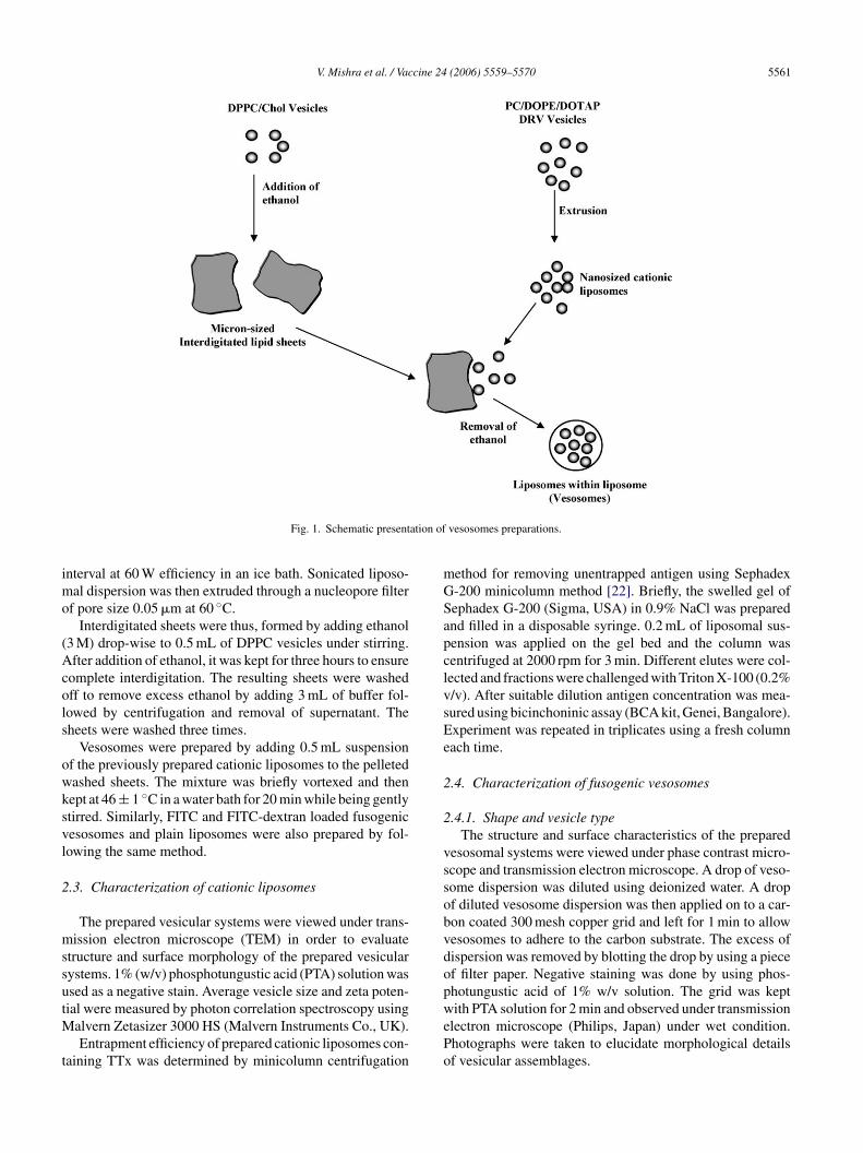
Fusogenic vesosomes were prepared by following thetwo steps. Firstly, cationic liposomes were prepared usingthe cationic lipid DOTAP and then prepared vesicles wereentrapped within outer vesicular systems. Schematic presen-tation of fusogenic vesosomes preparation is shown in Fig. 1.
2.2.1. Preparation of cationic liposomesTTx antigen loaded cationic liposomes were pre-
pared using the lipid composition of PC, DOPE andDdoatNvt(i(twwpfilpSls
2
mtmdhs
nducing fusion events [11,12]. Furthermore, alterations inhe lipid composition can render liposomes pH sensitive,eading to an enhanced fusogenic tendencies in low pH com-artments such as endosomes [13,14]. Alternatively, efficientusogenic liposomes can also be achieved by incorporatingusogenic proteins into liposomes [15,16]. The feasibility ofhis approach has been demonstrated for the delivery of theiptheria toxin A subunit using liposomes produced fromnfluenza virus envelops. Fusogenic peptides can be conju-ated to the liposomes and may also promote intracellularelivery [17]. Baraka et al. have demonstrated the fusogenicroperties of nonphospholipid liposomes (NPL) contain-ng dioxyethylene acyl ethers, single-tailed, nonphospholipidmphiphiles as principal membrane constituent. These lipo-omes can fuse with phasphatidylcholine liposomes at neutralH [18].
The present study deals with an aim to develop andharacterize novel fusogenic vesosomes, i.e. multicompart-ental aggregate of tethered vesicles encapsulated withinlarge bilayer, which can deliver the antigens effectively
n order to produce an effective immune response via topi-al administration. Prepared fusogenic vesosomes were pre-ared and characterized for size, shape, zeta potential andntrapment efficiency. In process and storage stability ofrepared systems were also determined. The efficacy of therepared novel vesicular constructs was determined and com-ared for their immunization potential with conventionaliposomes following topical administration. Tetanus toxoidTTx) was selected as model antigen for topical immuniza-ion.
OTAP in a molar ratio of 2:1:0.5, respectively followingehydration–rehydration (DRVs) method as reported previ-usly [19,20]. The lipids were dissolved in chloroform andthin film was casted on the inner wall of the round bot-
om flask using a rotary vacuum evaporator (York India Ltd.,ew Delhi). Prepared lipid film was desiccated for 1 h underacuum. The lipid film was then hydrated with 5 mL of dis-illed water and vortexed gently. Dispersion was sonicatedSoniweld, India) for ten minutes followed by two minutesnterval at 60% efficiency. Aqueous solution of TTx antigen60 Lf/mL) with 1% mannitol (cryoprotectant) was added tohis sonicated lipid dispersion and the liposomal suspensionas freeze dried at −40 ◦C and lyophilized. The dried powderas rehydrated with 2 mL of PBS and vortexed gently. Pre-ared cationic liposomes were extruded through a membranelter (Polycarbonate, 400 nm pore size; Millipore, USA) fol-
owed by another membrane filter (100 nm pore size; Milli-ore, USA) using an extruder (Liposofast, Avestin, Canada).imilarly FITC and FITC-dextran loaded cationic fusogenic
iposomes were also prepared by DRVs method using theame lipid ratio.
.2.2. Preparation of interdigitated lipid bilayersInterdigitated lipid bilayers were prepared using 97.5:2.5
olar ratio of dipalmitoyl phosphatidylcholine and choles-erol (DPPC and Chol) as described by Ahl et al. with slight
odifications [21]. Lipid mixture was evaporated to completeryness to form a thin film of lipids. The lipid film was thenydrated with PBS (pH 7.4) and obtained dispersion of lipo-omes was sonicated (Soniweld, India) for 20 min with 2 min
V. Mishra et al. / Vaccine 24 (2006) 5559–5570 5561
Fig. 1. Schematic presentation of vesosomes preparations.
interval at 60 W efficiency in an ice bath. Sonicated liposo-mal dispersion was then extruded through a nucleopore filterof pore size 0.05 �m at 60 ◦C.
Interdigitated sheets were thus, formed by adding ethanol(3 M) drop-wise to 0.5 mL of DPPC vesicles under stirring.After addition of ethanol, it was kept for three hours to ensurecomplete interdigitation. The resulting sheets were washedoff to remove excess ethanol by adding 3 mL of buffer fol-lowed by centrifugation and removal of supernatant. Thesheets were washed three times.
Vesosomes were prepared by adding 0.5 mL suspensionof the previously prepared cationic liposomes to the pelletedwashed sheets. The mixture was briefly vortexed and thenkept at 46 ± 1 ◦C in a water bath for 20 min while being gentlystirred. Similarly, FITC and FITC-dextran loaded fusogenicvesosomes and plain liposomes were also prepared by fol-lowing the same method.
2.3. Characterization of cationic liposomes
The prepared vesicular systems were viewed under trans-mission electron microscope (TEM) in order to evaluatestructure and surface morphology of the prepared vesicularsystems. 1% (w/v) phosphotungustic acid (PTA) solution wasused as a negative stain. Average vesicle size and zeta poten-tial were measured by photon correlation spectroscopy usingM
t
method for removing unentrapped antigen using SephadexG-200 minicolumn method [22]. Briefly, the swelled gel ofSephadex G-200 (Sigma, USA) in 0.9% NaCl was preparedand filled in a disposable syringe. 0.2 mL of liposomal sus-pension was applied on the gel bed and the column wascentrifuged at 2000 rpm for 3 min. Different elutes were col-lected and fractions were challenged with Triton X-100 (0.2%v/v). After suitable dilution antigen concentration was mea-sured using bicinchoninic assay (BCA kit, Genei, Bangalore).Experiment was repeated in triplicates using a fresh columneach time.
2.4. Characterization of fusogenic vesosomes
2.4.1. Shape and vesicle typeThe structure and surface characteristics of the prepared
vesosomal systems were viewed under phase contrast micro-scope and transmission electron microscope. A drop of veso-some dispersion was diluted using deionized water. A dropof diluted vesosome dispersion was then applied on to a car-bon coated 300 mesh copper grid and left for 1 min to allowvesosomes to adhere to the carbon substrate. The excess ofdispersion was removed by blotting the drop by using a pieceof filter paper. Negative staining was done by using phos-photungustic acid of 1% w/v solution. The grid was keptwith PTA solution for 2 min and observed under transmissionePo
alvern Zetasizer 3000 HS (Malvern Instruments Co., UK).Entrapment efficiency of prepared cationic liposomes con-
aining TTx was determined by minicolumn centrifugation
lectron microscope (Philips, Japan) under wet condition.hotographs were taken to elucidate morphological detailsf vesicular assemblages.

5562 V. Mishra et al. / Vaccine 24 (2006) 5559–5570
2.4.2. Percent entrapment efficiencyThe unentrapped antigen was removed following the mini-
column centrifugation method using sephadex G-200 mini-column method [22]. The unentrapped antigen was estimatedby BCA method and entrapment efficiency was determinedby reducing the amount of unentrapped antigen from usedantigen.
2.5. In process stability
SDS–PAGE experiment was carried out to analyze theintegrity of TTx antigen under non-denaturing condition.Protein bands were detected by coomassie blue staining.SDS–PAGE experiment was performed in the presence ofstrong anionic reducing agent, i.e. sodium dodecyl sulphates(SDS). In presence of strong reducing agent and heat, pro-teins get dissociated before they were applied on the gel [23].In brief, SDS–PAGE experiments were performed on a 5%stacking gel and 8% resolving gel following the standard pro-tocol. The gel was dried and the samples were applied on tothe gel for electrophoresis. The samples were heated to 95 ◦Cfor 5 min prior to their applications.
2.6. Storage stability
Storage stability studies are of important concern intTesihl2lng
2
ctaaouTwcft5caw
ume of fresh receptor solution. The samples from the receptormedium were analyzed by the BCA method.
2.8. In vitro cell fusion studies
Murine macrophage cell line J774 A.1 (National Centerfor Cell Science, Pune) was maintained in RPMI (GIBCOBRL) supplemented with 10% Fetal calf serum (Himedia,India) in 5% CO2 at 37 ± 1 ◦C. 5 × 10⁶ cells/well were platedin a 24 well plate and incubated overnight. After incubationmedia was washed, cell monolayers were washed thrice with50 mM PBS (pH 7.4). Two hundred micro liters suspensionsof liposomal formulations loaded with FITC were added toeach well and plates were incubated again for 30 min. Afterincubation, formulation suspensions were withdrawn; cellswere washed thrice with 50 mM PBS (pH 7.4) and observedunder a fluorescent microscope (Nikon TE2000-U, Japan).
The treated cells were also dissociated from plates bytreating with 0.25% trypsin and were analyzed by FACScal-ibur flow cytometer (Becton Dickinson, Mansfield, MA). Toinhibit endocytosis, J774 A.1 cells were treated with 5 �g/mLof cytochalasin B or 0.2 �g/mL cytochalasin D (Sigma, St.Louis, USA). Following 1-h incubation, liposomes, cationicfusogenic liposomes and fusogenic vesosomes (300 parti-cles/cell) were added and the cells were incubated for 30 min.Uptake efficiency was measured by flow cytometry.
2
2
oAwopMotwbtwd(sfc
vgftwto
he development of pharmaceutically acceptable product.hough immunological products are stored only in refrig-rated conditions, however, it was anticipated that encap-ulation of antigen in vesicles and subsequent coating mayncrease the stability by preventing antigen from externalostile challenges. Stability studies of TTx loaded formu-ations were carried out after storing them at 4 ± 1 ◦C and8 ± 1 ◦C for 30 days. After an interval of 30 days the formu-ations were evaluated for changes in vesicle size and shape,umber of vesicles per cubic mm and residual intact anti-en.
.7. In vitro skin permeation experiment
The permeation of tetanus toxoid-loaded vesosomes,ationic fusogenic liposomes, plain liposomes, plain tetanusoxoid solution, and a physical mixture of tetanus toxoidnd vesosomes through the skin were determined by usinglocally fabricated Franz-diffusion cell as described previ-
usly [24]. Prior to use, the skin was observed for any damagesing microscopy after staining with haematoxylin and eosin.he 6–12 weeks old male albino rat (Wistar strain) skinas mounted on the receptor compartment with the stratum
orneum side facing upward into the donor compartment. Theormulation containing 40 �g tetanus toxoid was applied onhe skin in the donor compartment. The receptor medium wasmL 0.2 M phosphate-buffered saline (pH 6.5). The receptorompartment was maintained at 37 ◦C with magnetic stirringt 500 rpm. At appropriate intervals 200 �L receptor mediumas withdrawn and immediately replaced with an equal vol-
.9. Immunization studies
.9.1. Animals, skin immunization and inoculationSix to twelve week old male albino rats (Wistar strain)
f average weight 150–200 g were used for present study.nimals were housed in groups (five animals in each group)ith free access to food and water. The studies were carriedut according to the guidelines of the Council for the Pur-ose of Control and Supervision of Experiments on Animals,inistry of Social Justice and Empowerment, Government
f India. Rats were carefully shaved to avoid nicking onhe dorsum and rested for 48 h. The rats were anaesthetizedith ketamine (110 mg/kg body wt.) and xylazine (11 mg/kgody wt.) for 0.5–2 h during the immunization procedureo prevent grooming. The shaved area was swabbed gentlyith cotton pad saturated with alcohol. This removes oil andry epidermal cells. Then 250 �l of immunizing formulation5 Lf/animal TTx equivalent dose) was placed on the shavedkin over a 2 cm2 area and left to dry. No adverse effectsrom the shaving, anesthesia, immunization, or washing pro-edures were generally observed.
Three different protocols were followed to assess the inivo performance of developed systems. In first protocol, sin-le primary topical immunization was done on day 0th andollowed by booster dosing on day 28th via topical adminis-ration. In second protocol, animals were primarily inoculatedith the topically administered vesosomal formulations on
hree consecutive days and followed by topical booster dosen day 28th. In third protocol, rats were intramuscularly
V. Mishra et al. / Vaccine 24 (2006) 5559–5570 5563
primed on day 0th with the alum adsorbed TTx and fol-lowed by boosting with topically administered vesosomalformulations on day 28th. In all three protocols the resultswere compared against intramuscularly administered alumadsorbed TTx antigen followed by booster dose on day 28th.
2.9.2. Collection of fluidsPreimmune samples of serum were obtained on day 0
before immunization. Subsequent to immunization, collec-tions were made at week 2, 4, 6 and 8. Serum was obtained bycentrifugation of blood samples collected from retro-orbitalplexus of rats under ether anesthesia.
2.10. Antibody detection
Antibody responses in the serum of immunized animalswere determined using a microplate ELISA. Microtiter plates(Nunc-Immuno Plate® Fb 96 Mexisorp, NUNC) were coatedwith 100 �l/well of 10 �g/mL TTx antigen in PBS (pH 7.4)and incubated overnight at 4 ◦C. The plates were thoroughlywashed with PBS-Tween 20 (PBS-T, 0.05%, v/v). The serumsamples were serially diluted 1:1 with PBS and 100 �l of eachsample was added to each well of coated ELISA plates. Theplates were incubated for 1 h at room temperature and washedthree times with PBS-T. 100 �l of peroxidase labelled goatanti-rat IgG, IgG2a or IgG1 was added to each well. Theppdodraa
2
ecFsstafl
2
uaSd*
w
test by using GraphPad Prism software (version 4.01) anddata were considered statistically significant if *P < 0.05.
3. Results and discussion
Liposomes are artificial lipid vesicles that represent apromising carrier system for delivery of drugs, biologicallyactive molecules and antigens. Lipid composition, liposomesize and specific targeting devices can be chosen such thatspecific cells and/or tissue could selectively be reached.Novel fusogenic vesicular constructs ‘liposomes within lipo-some’ (vesosomes) were prepared and characterized for theirpotential in delivery of TTx via topical route. The devel-oped novel fusogenic vesosomes composed of inner cationicliposomes encapsulated in outer liposomal bilayer. Thesefusogenic vesicular delivery systems that have the poten-tial to microinject entrapped bioactive(s) directly into thecytoplasm of target cells as a result of a fusion event at theplasma membrane. The fusion mediated delivery of encap-sulated antigens to the cytosol of antigen presenting cellsis anticipated to cause induction of class I restricted anti-gen presentation, in addition to presentation along with classII MHC molecules due to extracellular antigen encounteredafter the release of bioactive in the vicinity of antigen pre-senting cells. It was reported earlier that highly charged andscr3<tslTtfi
3v
arp(vopcovsbpD
lates were covered and after incubation for 1 h at room tem-erature washing was repeated. 100 �l of tetramethyl benzi-ine solution was added to each well followed by additionf 50 �l of H2SO4 after 90 min. After 15 min the intensity ofeveloped color was measured at 450 nm using a microplateeader (Labsystems, Finland). End point titers were expresseds the log10 of the reciprocal of the last dilution, which gaven optical density at 450 nm above the OD of controls.
.11. Fluorescence microscopy
Macromolecular fluorescent marker, i.e. FITC-dextranncapsulated different vesicular carriers, i.e. vesosomes,ationic fusogenic liposomes, plain liposomes and plainITC-dextran solution was applied topically to the shavedkin of albino rats. After 4 h the rats were killed and thekin removed. Microtomy was performed and ribbons of sec-ions (thickness 6 �m) were fixed onto glass slides using egglbumin as the fixative. The sections were viewed under auorescence microscope (Leica wild MP 582, Switzerland).
.12. Statistical analysis
Statistical analysis was conducted on antibody titerssing logarithmically transformed data and standard devi-tion (S.D.) was calculated. Values are given as mean ± S.D.tudent’s t-test was performed to compare mean values ofifferent groups. Statistical significance was designated asP < 0.05. Comparative analysis between the various groupas performed by one-way analysis of variance (ANOVA)
ize >10 �m are unable to reach deep into hair follicle due tohemical environment present in hair follicle. It has also beeneported that microparticles with diameters ranging from
to 7 �m selectively penetrate follicular ducts, and those3 �m are distributed randomly into hair follicles and stra-
um corneum [25]. Taking these facts into consideration, nanoized cationic fusogenic liposomes were encapsulated withiniposome to make them suitable in respect of size and charge.hese vesicles within vesicle systems were optimized for
heir in vitro parameters to ascertain reproducibility and tond out their potential as antigen carriers.
.1. Preparation and characterization of fusogenicesosomes
In the present study, tetanus toxoid was used as a modelntigen and quantitatively incorporated by dehydration–ehydrations method into liposome composed of phos-hatidylcholine (PC) and dioleoylphosphatidylethanolamineDOPE) supplemented with cationic lipids DOTAP. A wideariety of molecules (peptides, interleukins, antigens andther proteins) can be quantitatively entrapped into aqueoushase of liposomes by DRVs procedure in the absence of soni-ation, detergents or organic solvents. The procedure consistsf mixing ‘empty liposomes’, preferably small unilamellaresicles (produced by sonication, microfluidization or extru-ion) with a solution of the antigen, dehydration of mixturey freeze drying and subsequent rehydration of the formedowder. This leads to the formation of tetanus toxoid loadedOTAP based cationic liposomes. The cationic liposomes

5564 V. Mishra et al. / Vaccine 24 (2006) 5559–5570
Table 1Typical characteristic of vesicular preparations
Formulation parameters Cationic liposomes (nm) Vesosomes (�m)
Average vesicle size 104 ± 4.2 3.7 ± 0.8Zeta potentials (mV) 32.4 ± 4.2 18.84 ± 1.34% Entrapped antigen 39.1 ± 1.2 32.8 ± 1.7
were prepared using PC:DOPE:DOTAP lipids in molar ratioof 2:1:0.5. The liposomal formulations were extruded soas to get a homogeneous population and in order to getthe size that makes them suitable for further encapsulationinto another vesicular systems. Vesosomes were preparedinto interdigitated phospholipid sheets. The interdigitatedsheets were prepared using DPPC and cholesterol at 97.5:2.5molar ratio and these prepared sheets were mixed with pre-formed cationic liposomes to form vesosomes. The averagevesicle size and entrapment efficiency of cationic liposomeswas found to be 104 ± 4.2 nm and 39.1 ± 1.2%, respectivelywhereas in case of vesosomal systems, the average vesiclesize and entrapment efficiency was found to be 3.7 ± 0.8 �mand 32.8 ± 1.7%, respectively. Table 1 presents a summaryof some major characteristics of cationic and fusogenic veso-somes.
Cationic liposomes have strong positively charge with zetapotential of 32.4 ± 4.2 mV. This surfacial charge could beaccounted for the smaller size of cationic liposomes and repelthem each other sufficiently during dehydration–rehydrationsteps so as to interfere with the progress of membrane adhe-sion and eventually fusion especially during dehydrationphase of the procedure thus, resulting vesicles of smaller size[19]. The possible reason of loss in percent antigen encap-sulation in the preparation of vesosomal systems may bethe evaporation of solvents when the lipid phases separat-itdomwl
3
ocalt
3
cd
The vesosomal systems were stored in tightly closed glassbottles at 4 ± 1 ◦C and ambient temperature 28 ± 1 ◦C for 30days and compared with conventional liposomes. Averagevesicle size was found to increase on storage for 30 days.The increase in vesicle size was more in the case of for-mulations stored at 28 ± 1 ◦C than those stored at 4 ± 1 ◦C(Table 2). In contrast, percent vesicle number and percentresidual antigen amount was higher at elevated tempera-ture than the refrigerated storage condition. In comparisonto conventional liposomes the vesosomal systems were morestable under refrigerated condition than the room temperature(28 ± 1 ◦C).
3.4. In vitro skin permeation studies
In-vitro skin permeation experiment was performed usingFranz-diffusion cell and permeation behavior was comparedwith the cationic fusogenic liposomes, conventional lipo-somes and plain TTx solution. The experiment was carriedout for a period of 48 h and withdrawn samples were analyzedfor protein content using BCA method. After 48 h, percentcumulative permeation of TTx was 16.5 ± 0.16, 11.67 ± 1.1and 10.38 ± 0.91 in case of vesosomes cationic fusogenicliposomes and liposomes, respectively (Fig. 4). The slowrelease of the entrapped antigen could be due to the largemolecular size of the antigen with poor diffusivity throughvrafpctbbtsTcAtdettitevPtoaatt
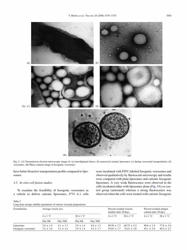
ng the internal aqueous from external aqueous compartmenthins to a bilayer membrane, thus, the percent encapsulationecreased to a final value of ∼32–36%. Structural studiesn the shape of the prepared vesosomal system using trans-ission electron microscopy have indicated that the systemsere almost spherical and surface was smooth and unilamel-
ar in nature (Fig. 2).
.2. SDS–PAGE electrophoresis
The in process antigen stability was determined in termsf its integrity during the preparation of formulations. Fig. 3learly indicates a visible band of pure as well as extractedntigen at the same loci. This in turns reveals that the formu-ation procedures used do not irreversibly aggregate or cleavehe antigen.
.3. Storage stability studies
Storage stability of formulations based on vesicular drugarriers is of a great concern as it is the major restraint in theevelopment of clinically acceptable marketed preparations.
esicular membrane. Furthermore, small amount of antigenelease might be attributed to the release of surface associateds well as outer aqueous compartmental antigen. Vesosomalormulation showed enhanced permeation profile as com-ared to conventional liposomes, possibly due to cationicharge of vesicular systems. In the liposomes, the constitu-ive component cholesterol affects profoundly their mem-rane properties. Incorporation of cholesterol in gel stateilayer can induce a continuous and permanent transitiono an ordered liquid crystalline state. This may attributed tolow release characteristics through vesosomal formulations.here are two types of interactions between the skin and vesi-les that may facilitate transdermal biomolecule delivery: (1)dsorption and fusion of biomolecule loaded vesicles onto
he surface of skin lead to high thermodynamic activity gra-ient of the biomolecule-stratum corneum surface. (2) Theffect of vesicle on stratum corneum may change the bioac-ive permeation kinetics due to an impaired barrier function ofhe stratum corneum [24,26]. Phospholipids have high affin-ty for biological membranes. Mixing of the phospholipid ofhe carrier system with the skin lipid of the intercellular lay-rs may also contribute to the permeability of the skin to lipidesicles [27,28]. The presence of unsaturated fatty acid soyaC and their packing nature may change the fluidity of stra-
um corneum lipid structure and facilitated the permeationf bioactive [29]. Topically applied lipid vesicles affect char-cteristics and integrity of the skin permeability barrier. Inddition, they may extract the lipid from the skin or disrupthe order within and between the corneocyte upon binding tohe keratin filament. This study is revealing that vesosomes
V. Mishra et al. / Vaccine 24 (2006) 5559–5570 5565
Fig. 2. (A) Transmission electron microscopic image of: (a) interdigitated sheets; (b) nanosized cationic liposomes; (c) during vasosomal encapsulation; (d)vesosomes. (B) Phase contrast image of fusogenic vesosomes.
have better bioactive transportation profile compared to lipo-somes.
3.5. In vitro cell fusion studies
To examine the feasibility of fusogenic vesosomes asa vehicle to deliver cationic liposomes, J774 A.1 cells
were incubated with FITC-labeled fusogenic vesosomes andobserved qualitatively by fluorescent microscopy and resultswere compared with plain liposomes and cationic fusogenicliposomes. A very weak fluorescence were observed in thecells incubated either with liposomes alone (Fig. 5A) or con-trol group (untreated) whereas a strong fluorescence wasobserved when the cells were treated with cationic fusogenic
Table 2Long-term storage stability parameters of various vesicular preparations
Formulations Average vesicle size Percent residual vesiclesnumber after 30 days
Percent residual antigencontent after 30 days
4 ± 1 ◦C 28 ± 1 ◦C 4 ± 1 ◦C 28 ± 1 ◦C 4 ± 1 ◦C 28 ± 1 ◦C
Day 0th Day 30th Day 0th Day 30th
Liposomes 3.9 ± 1.4 4.1 ± 1.7 3.9 ± 1.4 4.6 ± 1.3 89.58 ± 7.3 68.75 ± 5.2 90.6 ± 1.9 77.8 ± 3.6Fusogenic vesosomes 3.2 ± 1.6 3.3 ± 1.4 3.9 ± 1.4 3.5 ± 1.7 93.03 ± 7.7 74.41 ± 5.8 92.1 ± 2.9 85.4 ± 2.7
5566 V. Mishra et al. / Vaccine 24 (2006) 5559–5570
Fig. 3. SDS–PAGE analysis of tetanus toxoid in various formulations; lane1: standard TTx antigen; lane 2: Alum adsorbed TTx antigen; lane 3: TTxantigen in plain lipsomoes; lane 4: TTx antigen in cationic liposomes; lane5: TTx antigen in fusogenic vesosomes.
liposomes and fusogenic vesosomes as shown in Fig. 5B andC, respectively. This is a predictable observation becausecationic fusogenic liposomes and vesosomes fuse with thecell membrane and deliver it content to the cytoplasm. Theseresults suggested that Fusogenic vesosomes could deliver theencapsulated cationic liposomes into the cells in an effectivemanner.
3.6. Endocytosis-dependent cytoplasmic delivery byfusogenic vesosomes
As mentioned above, fusogenic vesosomes can efficientlydeliver the encapsulated contents to the cytoplasm thus, wenext tried to test delivery of vesosomal systems either by
Fig. 4. Percentage cumulative permeation of TTx antigen through rat skin.Vesosomes, Cationic fusogenic liposomes, Liposomes, plain tetanus toxoidand a physical mixture of Vesosomes and TTx were used for in vitro skinpermeation using Franz-diffusion cell. Data represent the mean ± S.D. ofmean of three independent experiments.
membrane fusion, or by endocytosis. To address this, cellswere treated with an endocytosis inhibitor, cytochalasin Bfollowed by the addition of vesosomes or cationic fusogenicliposomes, or plain liposomes. While cytochalasin B treat-ment resulted in a marked reduction of liposomal and veso-somal uptake, no significant inhibition of cationic fusogenicliposomes uptake was observed when cells were incubated(Fig. 6). Similar results were obtained when cells were treated
F deliveryl mes (Cw agnific
ig. 5. Fluorescent microscopy of fusogenic vesosomes mediated effectiveabeled plain liposomes (A), or cationic fusogenic liposomes (B) or vesosoere visualized by fluorescent microscopy. Bars indicate 100 �m at 400× m
of FITC labeled vesicular carriers. J774 A.1 cells were treated with FITC) or control (without any treatment, D) for 30 min. After staining, the cellsation.
V. Mishra et al. / Vaccine 24 (2006) 5559–5570 5567
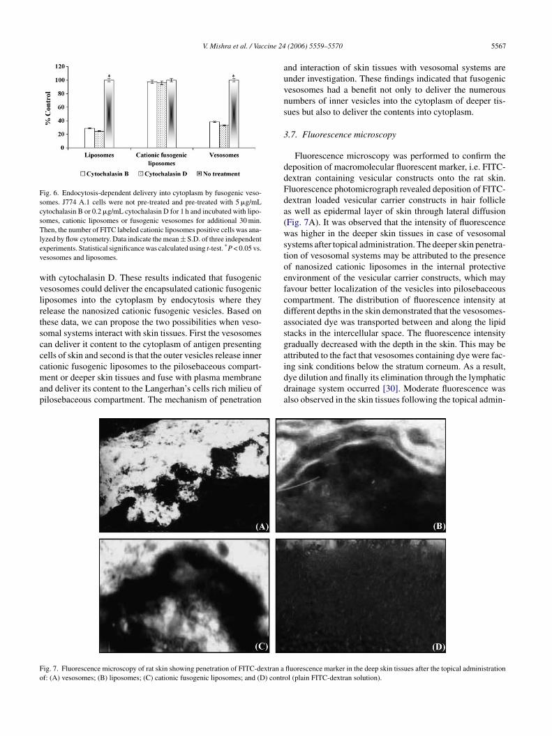
Fig. 6. Endocytosis-dependent delivery into cytoplasm by fusogenic veso-somes. J774 A.1 cells were not pre-treated and pre-treated with 5 �g/mLcytochalasin B or 0.2 �g/mL cytochalasin D for 1 h and incubated with lipo-somes, cationic liposomes or fusogenic vesosomes for additional 30 min.Then, the number of FITC labeled cationic liposomes positive cells was ana-lyzed by flow cytometry. Data indicate the mean ± S.D. of three independentexperiments. Statistical significance was calculated using t-test. *P < 0.05 vs.vesosomes and liposomes.
with cytochalasin D. These results indicated that fusogenicvesosomes could deliver the encapsulated cationic fusogenicliposomes into the cytoplasm by endocytosis where theyrelease the nanosized cationic fusogenic vesicles. Based onthese data, we can propose the two possibilities when veso-somal systems interact with skin tissues. First the vesosomescan deliver it content to the cytoplasm of antigen presentingcells of skin and second is that the outer vesicles release innercationic fusogenic liposomes to the pilosebaceous compart-ment or deeper skin tissues and fuse with plasma membraneand deliver its content to the Langerhan’s cells rich milieu ofpilosebaceous compartment. The mechanism of penetration
and interaction of skin tissues with vesosomal systems areunder investigation. These findings indicated that fusogenicvesosomes had a benefit not only to deliver the numerousnumbers of inner vesicles into the cytoplasm of deeper tis-sues but also to deliver the contents into cytoplasm.
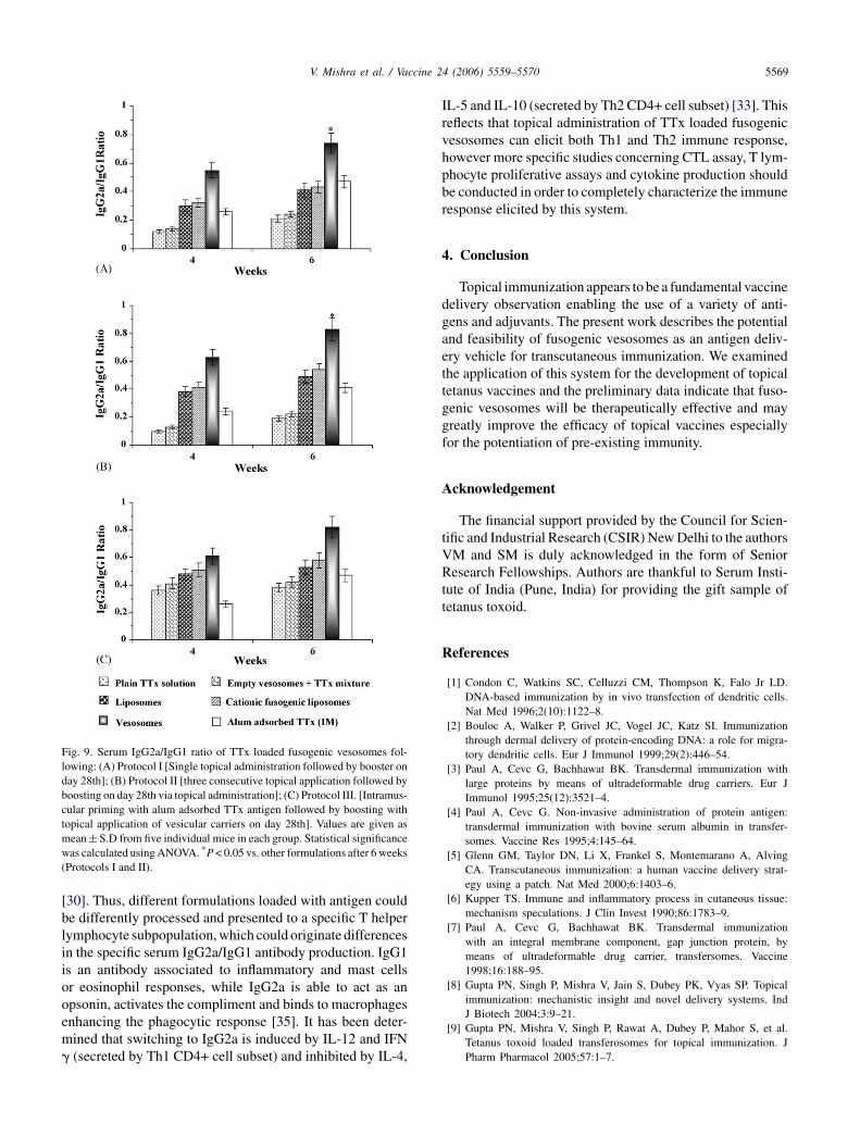
3.7. Fluorescence microscopy
Fluorescence microscopy was performed to confirm thedeposition of macromolecular fluorescent marker, i.e. FITC-dextran containing vesicular constructs onto the rat skin.Fluorescence photomicrograph revealed deposition of FITC-dextran loaded vesicular carrier constructs in hair follicleas well as epidermal layer of skin through lateral diffusion(Fig. 7A). It was observed that the intensity of fluorescencewas higher in the deeper skin tissues in case of vesosomalsystems after topical administration. The deeper skin penetra-tion of vesosomal systems may be attributed to the presenceof nanosized cationic liposomes in the internal protectiveenvironment of the vesicular carrier constructs, which mayfavour better localization of the vesicles into pilosebaceouscompartment. The distribution of fluorescence intensity atdifferent depths in the skin demonstrated that the vesosomes-associated dye was transported between and along the lipidstacks in the intercellular space. The fluorescence intensitygradually decreased with the depth in the skin. This may beaidda
F xtran ao ) contr
ig. 7. Fluorescence microscopy of rat skin showing penetration of FITC-def: (A) vesosomes; (B) liposomes; (C) cationic fusogenic liposomes; and (D
ttributed to the fact that vesosomes containing dye were fac-ng sink conditions below the stratum corneum. As a result,ye dilution and finally its elimination through the lymphaticrainage system occurred [30]. Moderate fluorescence waslso observed in the skin tissues following the topical admin-
fluorescence marker in the deep skin tissues after the topical administrationol (plain FITC-dextran solution).

5568 V. Mishra et al. / Vaccine 24 (2006) 5559–5570
istration of cationic fusogenic liposomes and conventionalliposomes as compared to vesosomal systems (Fig. 7B andC). Plain FITC-dextran solution was not found to penetrateinto deeper skin layers; the fluorescence in this case wasmainly confined to the superficial skin layers (Fig. 7D).
3.8. Serum anti-TTx antibodies
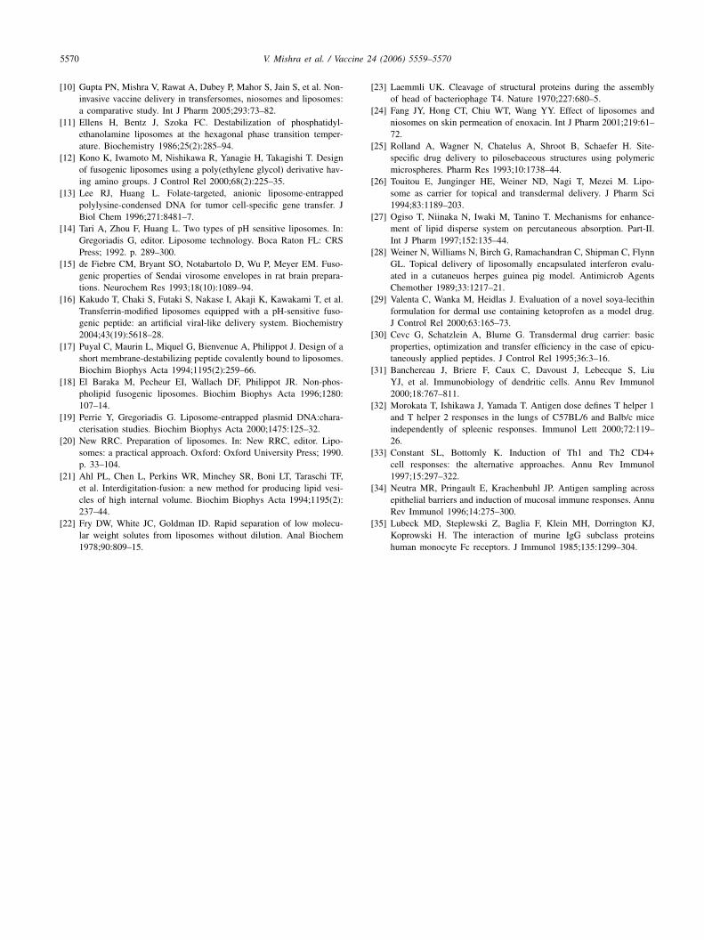
The level of anti-TTx antibodies (IgG) was determined forall experimental groups after 2, 4, 6 and 8 weeks. Fig. 8 showsserum IgG response obtained with three protocols, singleinoculum (protocol I), three inoculations in consecutive days(protocol II) and intramuscularly primed alum adsorbed anti-gen (protocol III), respectively. Booster immunization wasperformed for all groups after 4 weeks. Results suggest thattopical immunization with vesosomal systems significantlyincreased IgG levels (P < 0.05) compared to conventionalliposomal formulations administered topically. It has beenobserved that alum adsorbed TTx produced a higher IgG titer,when administered intramuscularly than the vesicular sys-tems in the single inoculum protocol (Fig. 8A). At the otherextreme, when three doses of TTx were applied topically(protocol II) to produce primary immune response, almostall formulations exhibited an increase in IgG response thanthe single inoculation (Fig. 8B). In a primed-boost immuniza-tion protocol (protocol III), the serum IgG titers of rats weresarglvmalo
3
corpbmssrcscps
s
Fig. 8. Serum anti-TTx antibody titer following: (A) Protocol I [Single top-ical administration followed by booster on day 28th]; (B) Protocol II [threeconsecutive topical application followed by boosting on day 28th via topicaladministration]; (C) Protocol III. [Intramuscular priming with alum adsorbedTTx antigen followed by boosting with topical application of vesicular car-riers on day 28th]. Anti-TTx antibody titers were expressed as mean log10
values ± S.D. of mean from five individual mice in each group. Statisti-cal significance was calculated using ANOVA. *P < 0.05 vs. other topicallyapplied formulations except Alum adsorbed TTx (IM) (Protocol II) and*P < 0.05 vs. liposomes at week 8 (Protocol III).
T cells, will favor the isotype and magnitude of the B cellantibody response [32,33]. Neutra et al. reported differentbody distribution of dendritic cells on the epithelial barriersat different sites of the body [34]. Dendritic cells are con-sidered as initiators and modulators of the immune responseand are capable to process the antigen by both the majorhistocompatibility complex (MHC) class I and MHC class IIpathways, depending upon the nature of the observed antigen
ignificantly higher (*P < 0.05) with the vesosomal systemss compared to other topical administered boosted groups ofats and almost comparable with alum adsorbed TTx boostedroups of rats (Fig. 8C). Furthermore, release of encapsu-ated cationic vesicles within hair follicle and fusion of theseesicles with immune responsive cells (e.g. LCs, epider-al T cells) may be possible reason for better and effective
ntigen presentation. Apart from fusion, cationic fusogeniciposomes may release encapsulated antigen in the vicinityf these cells.
.9. IgG2a/IgG1 ratio
Fig. 9 shows that formulations are capable of eliciting aombined serum IgG2a/IgG1 response, with predominancef the IgG1 response. Results are consistent with thoseeported by Banchereau et al. [31]. They suggested thatarticulate antigens can be processed and presented eithery MHC class I or MHC class II by dendritic cells andacrophages, which stimulates Th1 and Th2 lymphocyte
ubpopulations, while soluble antigens are exclusively pre-ented by class II MHC, stimulating the Th2 response. Thiseflects that topical administration of TTx loaded vesosomesan elicit both Th1 and Th2 immune response, however moretudies concerning T lymphocyte proliferative assays andytokine production should be conducted in order to com-letely characterize the immune response elicited by thisystem.
It has been well established that the nature of antigen pre-enting cell, which presents the antigen to the specific native
V. Mishra et al. / Vaccine 24 (2006) 5559–5570 5569
Fig. 9. Serum IgG2a/IgG1 ratio of TTx loaded fusogenic vesosomes fol-lowing: (A) Protocol I [Single topical administration followed by booster onday 28th]; (B) Protocol II [three consecutive topical application followed byboosting on day 28th via topical administration]; (C) Protocol III. [Intramus-cular priming with alum adsorbed TTx antigen followed by boosting withtopical application of vesicular carriers on day 28th]. Values are given asmean ± S.D from five individual mice in each group. Statistical significancewas calculated using ANOVA. *P < 0.05 vs. other formulations after 6 weeks(Protocols I and II).
[30]. Thus, different formulations loaded with antigen couldbe differently processed and presented to a specific T helperlymphocyte subpopulation, which could originate differencesin the specific serum IgG2a/IgG1 antibody production. IgG1is an antibody associated to inflammatory and mast cellsor eosinophil responses, while IgG2a is able to act as anopsonin, activates the compliment and binds to macrophagesenhancing the phagocytic response [35]. It has been deter-mined that switching to IgG2a is induced by IL-12 and IFN� (secreted by Th1 CD4+ cell subset) and inhibited by IL-4,
IL-5 and IL-10 (secreted by Th2 CD4+ cell subset) [33]. Thisreflects that topical administration of TTx loaded fusogenicvesosomes can elicit both Th1 and Th2 immune response,however more specific studies concerning CTL assay, T lym-phocyte proliferative assays and cytokine production shouldbe conducted in order to completely characterize the immuneresponse elicited by this system.
4. Conclusion
Topical immunization appears to be a fundamental vaccinedelivery observation enabling the use of a variety of anti-gens and adjuvants. The present work describes the potentialand feasibility of fusogenic vesosomes as an antigen deliv-ery vehicle for transcutaneous immunization. We examinedthe application of this system for the development of topicaltetanus vaccines and the preliminary data indicate that fuso-genic vesosomes will be therapeutically effective and maygreatly improve the efficacy of topical vaccines especiallyfor the potentiation of pre-existing immunity.
Acknowledgement
The financial support provided by the Council for Scien-tific and Industrial Research (CSIR) New Delhi to the authorsVRtt
R
M and SM is duly acknowledged in the form of Senioresearch Fellowships. Authors are thankful to Serum Insti-
ute of India (Pune, India) for providing the gift sample ofetanus toxoid.
eferences
[1] Condon C, Watkins SC, Celluzzi CM, Thompson K, Falo Jr LD.DNA-based immunization by in vivo transfection of dendritic cells.Nat Med 1996;2(10):1122–8.
[2] Bouloc A, Walker P, Grivel JC, Vogel JC, Katz SI. Immunizationthrough dermal delivery of protein-encoding DNA: a role for migra-tory dendritic cells. Eur J Immunol 1999;29(2):446–54.
[3] Paul A, Cevc G, Bachhawat BK. Transdermal immunization withlarge proteins by means of ultradeformable drug carriers. Eur JImmunol 1995;25(12):3521–4.
[4] Paul A, Cevc G. Non-invasive administration of protein antigen:transdermal immunization with bovine serum albumin in transfer-somes. Vaccine Res 1995;4:145–64.
[5] Glenn GM, Taylor DN, Li X, Frankel S, Montemarano A, AlvingCA. Transcutaneous immunization: a human vaccine delivery strat-egy using a patch. Nat Med 2000;6:1403–6.
[6] Kupper TS. Immune and inflammatory process in cutaneous tissue:mechanism speculations. J Clin Invest 1990;86:1783–9.
[7] Paul A, Cevc G, Bachhawat BK. Transdermal immunizationwith an integral membrane component, gap junction protein, bymeans of ultradeformable drug carrier, transfersomes. Vaccine1998;16:188–95.
[8] Gupta PN, Singh P, Mishra V, Jain S, Dubey PK, Vyas SP. Topicalimmunization: mechanistic insight and novel delivery systems. IndJ Biotech 2004;3:9–21.
[9] Gupta PN, Mishra V, Singh P, Rawat A, Dubey P, Mahor S, et al.Tetanus toxoid loaded transferosomes for topical immunization. JPharm Pharmacol 2005;57:1–7.
5570 V. Mishra et al. / Vaccine 24 (2006) 5559–5570
[10] Gupta PN, Mishra V, Rawat A, Dubey P, Mahor S, Jain S, et al. Non-invasive vaccine delivery in transfersomes, niosomes and liposomes:a comparative study. Int J Pharm 2005;293:73–82.
[11] Ellens H, Bentz J, Szoka FC. Destabilization of phosphatidyl-ethanolamine liposomes at the hexagonal phase transition temper-ature. Biochemistry 1986;25(2):285–94.
[12] Kono K, Iwamoto M, Nishikawa R, Yanagie H, Takagishi T. Designof fusogenic liposomes using a poly(ethylene glycol) derivative hav-ing amino groups. J Control Rel 2000;68(2):225–35.
[13] Lee RJ, Huang L. Folate-targeted, anionic liposome-entrappedpolylysine-condensed DNA for tumor cell-specific gene transfer. JBiol Chem 1996;271:8481–7.
[14] Tari A, Zhou F, Huang L. Two types of pH sensitive liposomes. In:Gregoriadis G, editor. Liposome technology. Boca Raton FL: CRSPress; 1992. p. 289–300.
[15] de Fiebre CM, Bryant SO, Notabartolo D, Wu P, Meyer EM. Fuso-genic properties of Sendai virosome envelopes in rat brain prepara-tions. Neurochem Res 1993;18(10):1089–94.
[16] Kakudo T, Chaki S, Futaki S, Nakase I, Akaji K, Kawakami T, et al.Transferrin-modified liposomes equipped with a pH-sensitive fuso-genic peptide: an artificial viral-like delivery system. Biochemistry2004;43(19):5618–28.
[17] Puyal C, Maurin L, Miquel G, Bienvenue A, Philippot J. Design of ashort membrane-destabilizing peptide covalently bound to liposomes.Biochim Biophys Acta 1994;1195(2):259–66.
[18] El Baraka M, Pecheur EI, Wallach DF, Philippot JR. Non-phos-pholipid fusogenic liposomes. Biochim Biophys Acta 1996;1280:107–14.
[19] Perrie Y, Gregoriadis G. Liposome-entrapped plasmid DNA:chara-cterisation studies. Biochim Biophys Acta 2000;1475:125–32.
[20] New RRC. Preparation of liposomes. In: New RRC, editor. Lipo-
[
[
[23] Laemmli UK. Cleavage of structural proteins during the assemblyof head of bacteriophage T4. Nature 1970;227:680–5.
[24] Fang JY, Hong CT, Chiu WT, Wang YY. Effect of liposomes andniosomes on skin permeation of enoxacin. Int J Pharm 2001;219:61–72.
[25] Rolland A, Wagner N, Chatelus A, Shroot B, Schaefer H. Site-specific drug delivery to pilosebaceous structures using polymericmicrospheres. Pharm Res 1993;10:1738–44.
[26] Touitou E, Junginger HE, Weiner ND, Nagi T, Mezei M. Lipo-some as carrier for topical and transdermal delivery. J Pharm Sci1994;83:1189–203.
[27] Ogiso T, Niinaka N, Iwaki M, Tanino T. Mechanisms for enhance-ment of lipid disperse system on percutaneous absorption. Part-II.Int J Pharm 1997;152:135–44.
[28] Weiner N, Williams N, Birch G, Ramachandran C, Shipman C, FlynnGL. Topical delivery of liposomally encapsulated interferon evalu-ated in a cutaneuos herpes guinea pig model. Antimicrob AgentsChemother 1989;33:1217–21.
[29] Valenta C, Wanka M, Heidlas J. Evaluation of a novel soya-lecithinformulation for dermal use containing ketoprofen as a model drug.J Control Rel 2000;63:165–73.
[30] Cevc G, Schatzlein A, Blume G. Transdermal drug carrier: basicproperties, optimization and transfer efficiency in the case of epicu-taneously applied peptides. J Control Rel 1995;36:3–16.
[31] Banchereau J, Briere F, Caux C, Davoust J, Lebecque S, LiuYJ, et al. Immunobiology of dendritic cells. Annu Rev Immunol2000;18:767–811.
[32] Morokata T, Ishikawa J, Yamada T. Antigen dose defines T helper 1and T helper 2 responses in the lungs of C57BL/6 and Balb/c miceindependently of spleenic responses. Immunol Lett 2000;72:119–26.
[
[
[
somes: a practical approach. Oxford: Oxford University Press; 1990.p. 33–104.
21] Ahl PL, Chen L, Perkins WR, Minchey SR, Boni LT, Taraschi TF,et al. Interdigitation-fusion: a new method for producing lipid vesi-cles of high internal volume. Biochim Biophys Acta 1994;1195(2):237–44.
22] Fry DW, White JC, Goldman ID. Rapid separation of low molecu-lar weight solutes from liposomes without dilution. Anal Biochem1978;90:809–15.
33] Constant SL, Bottomly K. Induction of Th1 and Th2 CD4+cell responses: the alternative approaches. Annu Rev Immunol1997;15:297–322.
34] Neutra MR, Pringault E, Krachenbuhl JP. Antigen sampling acrossepithelial barriers and induction of mucosal immune responses. AnnuRev Immunol 1996;14:275–300.
35] Lubeck MD, Steplewski Z, Baglia F, Klein MH, Dorrington KJ,Koprowski H. The interaction of murine IgG subclass proteinshuman monocyte Fc receptors. J Immunol 1985;135:1299–304.
Copyright © 2022 FDOKUMEN


![Poly[di(carboxylatophenoxy)phosphazene] is a potent adjuvant for intradermal immunization](https://static.fdokumen.com/doc/165x107/6335c6c4a1ced1126c0af097/polydicarboxylatophenoxyphosphazene-is-a-potent-adjuvant-for-intradermal-immunization.jpg)