Chitosan and O-carboxymethyl chitosan modified Fe3O4 for hyperthermic treatment
Ca
CAa
b
a
ARR2AA
KCSA
1
blemceb
ccn
nd
0d
Vaccine 28 (2010) 2607–2614
Contents lists available at ScienceDirect
Vaccine
journa l homepage: www.e lsev ier .com/ locate /vacc ine
hitosan-based nanoparticles for improving immunizationgainst hepatitis B infection
ecilia Pregoa, Patrizia Paolicelli a, Belen Díazb, Sara Vicentea,lejandro Sáncheza, África González-Fernándezb, María José Alonsoa,∗
Nanobiofar Group, Department of Pharmacy and Pharmaceutical Technology, University of Santiago de Compostela, 15782 Santiago de Compostela, SpainImmunology Area, Faculty of Biology, University of Vigo, Campus Lagoas Marcosende, 36310 Vigo, Pontevedra, Spain
r t i c l e i n f o
rticle history:eceived 2 April 2009eceived in revised form1 December 2009ccepted 11 January 2010vailable online 20 January 2010
eywords:hitosan-based nanoparticlesubunit antigensdjuvant
a b s t r a c t
The design of effective vaccine delivery vehicles is opening up new possibilities for making immunizationmore equitable, safe and efficient. In this work, we purpose polysaccharidic-based nanoparticles as deliv-ery structures for virus-like particle antigens, using recombinant hepatitis B surface antigen (rHBsAg) asa model.
Polysaccharidic-based nanoparticles were prepared using a very mild ionic gelation technique, bycross-linking the polysaccharide chitosan (CS) with a counter ion. The resulting nanoparticles couldbe easily isolated with a size in the nanometric range (160–200 nm) and positive surface charge (+6to +10 mV). More importantly, CS-based nanoparticles allowed the efficient association of the antigen(>60%) while maintaining the antigenic epitope intact, as determined by ELISA and Western blot. Theentrapped antigen was further released in vitro from the nanoparticles in a sustained manner withoutcompromising its antigenicity. In addition, loaded CS-based nanoparticles were stable, and protected the
associated antigen during storage, either as an aqueous suspension under different temperature condi-tions (+4 ◦C and −20 ◦C), or as a dried form after freeze-drying the nanoparticles. Finally, immunizationstudies showed the induction of important seroprotection rates after intramuscular administration ofthe nanoparticles, indicating their adjuvant capacity. In fact, CS-based nanoparticles were able to induceanti-HBsAg IgG levels up to 5500 mIU/ml, values 9-fold the conventional alum-adsorbed vaccine.In conclusion, we report here a polysaccharidic nanocarrier which exhibits a number of in vitro and ina pro
vivo features that make it. Introduction
Vaccination is considered one of the greatest achievements ofiomedical science and public health. Routine immunization had
ed to the control and significant reduction of many infectious dis-ases. However, the benefit of immunization is severely limited toost of the population in need [1]. The important disparity in vac-
ination coverage between industrialized and developing countriesntails millions of deaths from preventable diseases, which coulde averted by improving immunization technologies.
The development of strategies to reduce the inequities in vac-ination from preventable infectious diseases represents a grandhallenge in global health. Indeed, there is a commitment fromumerous philanthropic associations from the public and private
∗ Corresponding author at: Department of Pharmacy and Pharmaceutical Tech-ology, School of Pharmacy, University of Santiago de Compostela, 15782 Santiagoe Compostela, Spain. Tel.: +34 981 563100; fax: +34 981 547148.
E-mail address: [email protected] (M.J. Alonso).
264-410X/$ – see front matter © 2010 Elsevier Ltd. All rights reserved.oi:10.1016/j.vaccine.2010.01.011
mising adjuvant for vaccine delivery of subunit antigens.© 2010 Elsevier Ltd. All rights reserved.
sectors, such as the Global Alliance for Vaccines and Immunization(GAVI) and the Bill & Melinda Gates Foundation, among others,to bring technologies which could solve the problems related tothe vaccination failure in developing countries [2]. Within thisframe, the technological approaches are expected to be focused onimproving the vaccine thermostability, decreasing the number ofshots and, when possible, avoiding injections with a needle-freevaccination approach [3].
First of all, to ensure the optimal efficacy of vaccines, particularattention is needed during their storage, distribution or handling. Inthe case of alum-adsorbed vaccines, a loss of potency occurs whenfrozen due to a destruction of the particular gel structure of theadjuvant [4], and after exposure to elevated temperatures due tochanges on the properties of the solution such as ionic strength andpH, among others. So far, strategies addressed to obtain a stable-
dried formulation while maintaining the immunogenicity, werebased on the use of spray-freeze-drying or spray coating techniques[5,6].Secondly, to induce a full protective antibody response againstthe target disease, a multiple-dose vaccination schedule is usu-

2 ine 28
acpwhstitlespcftwr
wbbAbisvtdCttlC(te
wftithtteipStgdud
2
2
otPh
608 C. Prego et al. / Vacc
lly required. As a consequence, a reduction in the immunizationompliance with the subsequent breakdown in the schedule takeslace. Thus, the development of single-shot vaccination approachesould improve the immunization efficacy, and additionally, wouldelp reduce the waste disposal associated to the needles andyringes. Different delivery systems have been proposed to solvehe mentioned drawback. Polyester-based microparticles are undernvestigation since early nineties, showing that the administra-ion of single doses of PLGA microspheres led to the induction ofong-lasting immune responses, similar to those achieved for sev-ral doses of conventional vaccines [7–9]. Interesting alternativetrategies are based on the oil-in-water emulsion MF59®, which hasroved to be an efficacious and safe adjuvant for the influenza vac-ine, although it remains to be seen whether this approach worksor other vaccines [10]; or the adjuvant MPL, a TLR ligand deriva-ive from the lipid A molecule found in gram-negative bacteria,hich induces a potent immune response even in patients with
enal insufficiency [11].Nevertheless, the major impact on immunization coverage,
hich would also decrease the risk of transmission of blood-orne pathogens and reduce the waste disposal, would take placey the development of a needle-free vaccination approach [12].mong the non-parenteral routes of administration, mucosal mem-ranes, which possess associated lymphoid tissue with all of the
mmunocompetent cells required for the induction of antigen-pecific immune responses, are emerging as attractive routes foraccination [13,14]. In particular, the nasal cavity has been inves-igated as a promising site for vaccine administration. Differentelivery systems have been explored by the nasal route, includingS-based nanoparticles incorporating soluble antigens, like diph-heria and tetanus toxoids, and plasmid DNA, which have showno be efficient for intranasal vaccination, inducing important andong-lasting immune responses [15,16]. The interesting features ofS nanoparticles for transmucosal vaccine administration include:i) the mild conditions required for their preparation [17,18]; (ii)he easy-scalable procedure; and (iii) their great capability for thentrapment of therapeutic and antigenic proteins [17–21].
Bearing in mind this information, the objective of the presentork was the development of a nanoscale vaccine delivery system
or subunit antigens, using the rHBsAg as a model. The selection ofhis antigen was made within the frame of the Grand Challengesn Global Health (Bill and Melinda Gates Foundation) because ofhe recognized need of improving immunization coverage againstepatitis B in developing countries. It is important to mention thathis antigen is a virus-like particle [22] composed of a 24 kDa pro-ein monomer in a non-glycosilated and glycosilated form, partiallymbedded in a lipidic membrane forming a 22 nm particle size bytself [23,24]. To achieve this goal, we firstly associated this com-lex antigen to CS nanoparticles without altering its antigenicity.econdly, we evaluated the ability of CS nanoparticles to releasehe encapsulated antigen in its active form, and then, we investi-ated the stability of these nanocarriers, either in solution or in ary state. In the final section, we carried out in vivo studies to eval-ate the potential of these polysaccharidic nanoparticles as vaccineelivery systems.
. Materials and methods
.1. Materials
Ultrapure CS hydrochloride salt, having a molecular weightf around 125 kDa and an acetylation degree of 14% (Pro-asan UP Cl 113), was purchased from Novamatrix (Norway).entasodium tripolyphosphate (TPP) and poloxamer 188, withydrophilia–lipophilia balance of 29 (Pluronic F68®), were
(2010) 2607–2614
obtained from Sigma–Aldrich (Madrid, Spain). Chitosanase-RD,an N-acetylglucosaminohydrolase from Bacillus sp. PI-7S with anactivity of 0.15–0.35 U/mg, was purchased from United States Bio-logical (MA, USA).
Recombinant hepatitis B surface antigen was kindly donated byShantha Biotechnics Limited (Hyderabad, India).
Enzyme linked immunosorbent assay (ELISA) kit (Murex HBsAgVersion 3) was obtained from Abbott Diagnostics Division (Madrid,Spain). This sandwich assay uses a mixture of monoclonal antibod-ies that specifically detect the “a” determinant epitopes of HBsAg.
Antibodies for Western-blot detection, chicken polyclonal anti-body to hepatitis B virus surface antigen and rabbit polyclonal tochicken conjugated with horseradish peroxidase were purchasedfrom Abcam pcl (Cambridge, UK).
The absolute amounts of anti-HBsAg IgG in serum sampleswere analyzed by ELISA using Maxisorb microtiter wells (Nunc,Denmark). Bovine serum albumin (BSA) and ABTS (2,2′-azino-bis 3-ethylbenzthiazoline-6-sulphonic acid) were purchased bySigma–Aldrich (Madrid, Spain), rabbit IgG directed against HBsAg(bioelisa anti-HBS, Biokit, Barcelona, Spain), and horseradish per-oxidase conjugated goat anti-rabbit IgG (SourthernBiotech, USA).
2.2. Preparation of CS-based nanoparticles
CS-based nanoparticles were prepared in very mild conditionstaking the advantages of the ionotropic gelation of the cationicpolysaccharide in the presence of a counter ion [17,18]. Morespecifically, nanoparticle formation occurred immediately uponthe addition of a fixed volume of the ionic cross-linker solution,TPP (1.2 ml), to a fixed volume of CS solution containing polox-amer (3 ml) under magnetic stirring at room temperature. Theagitation was maintained during 15 min in order to allow the stabi-lization of the system. Several process variables were investigated:CS concentration (1 and 2 mg/ml), TPP concentration (0.42, 0.5, and0.625 mg/ml), and the incorporation or not of poloxamer (1%, w/v).
The preparation of CS-based nanoparticles loaded with rHBsAgwas performed after dissolving the antigen (0.160 mg/ml) togetherwith the cross-linking agent (TPP) prior to nanoparticle formation.The theoretical loading of this complex macromolecule was 2.5%(w/w) with respect to the total amount of CS used for nanoparticlepreparation.
The obtained nanoparticles were centrifuged at 16,000 × g for40 min on a 10 �l glycerol bed (Universal 32 R, Hettich Zentrifugen,Germany) for their isolation. Following centrifugation, CS-basednanoparticles were resuspended in ultrapure water for furtherstudies.
2.3. Physico-chemical characterization of CS-based nanoparticles
The hydrodynamic diameter, polydispersity index and zetapotential of the nanoparticles were determined by photoncorrelation spectroscopy (PCS) and laser-Doppler anemometry(Zetasizer®, NanoZS, Malvern Instruments, Malvern, UK), after dilu-tion of the samples with ultrapure water.
The morphology of the nanoparticles was examined by trans-mission electron microscopy (CM 12 Philips, Eindhoven, TheNetherlands) after negative staining of the nanoparticles with a 2%(w/v) phosphotungstic acid solution.
2.4. Loading capacity of CS-based nanoparticles
The ability of CS-based nanoparticles to entrap the complexantigen, rHBsAg, within their structure was directly determinedafter enzymatic degradation of loaded nanoparticles with chi-tosanase. This enzyme, an N-acetylglucosaminohydrolase, is able tohydrolyze CS into oligosaccharides, releasing the associated antigen

ine 28
fctitibt
vi5rCa
ttfilotwf
2n
toapiccwaslrtwetnasi
2n
oti
wwanrE
C. Prego et al. / Vacc
or further analysis. The degradation conditions and the analyti-al techniques were conveniently validated in order to assess theotal delivery of the associated antigen without compromising itsntegrity and avoiding interferences of the enzyme and degrada-ion products in the analytical techniques. Different controls werencluded: (i) plain rHBsAg, (ii) plain rHBsAg plus chitosanase, (iii)lank CS nanoparticles plus chitosanase, and (iv) plain rHBsAg inhe presence of blank CS nanoparticles and chitosanase.
Briefly, rHBsAg-loaded CS-based nanoparticles (45 �g), pre-iously centrifuged and resuspended in ultrapure water, werencubated with 0.11 U of chitosanase in a 50 mM acetate buffer pH.5 for 4 h at 37 ◦C. After this time, the amount of antigenically activeHBsAg released from the nanoparticles was determined by ELISA.alibration curves were made with degraded blank nanoparticles,nd all measurements were performed in triplicate.
Western-blot analysis of the rHBsAg associated was carried outo visualize the integrity of the antigen, providing complemen-ary information to ELISA. To accomplish this analysis, the protocolor nanoparticle degradation was modified in order to avoid anynterference of the enzyme. In this case, after nanoparticle iso-ation, 80 �g of loaded nanoparticles was incubated with 0.03 Uf chitosanase in acetate buffer pH 5.5 during 4 h at 37 ◦C. Then,he samples were centrifuged at 16,000 × g for 10 min, the antigenas collected, and concentrated in Microcon tubes (Millipore) for
urther visualization by Western blot.
.5. In vitro release studies of rHBsAg from CS-basedanoparticles
In vitro release studies of the antigen from CS-based nanopar-icles were performed with the goal of investigating the abilityf these delivery systems to release the associated rHBsAg in itsctive form. Therefore, the selection of the release medium waserformed taking into account the stability of the nanoparticles
n terms of physico-chemical characteristics (particle size, surfaceharge), which will ultimately play a role in the accurate quantifi-ation of the antigen by ELISA. Thus, rHBsAg-loaded nanoparticlesere incubated in water with the preservative thimerosal (5 �g/ml)
t 37 ◦C under moderate agitation. At different time-intervals, theuspension was centrifuged, the supernatant was removed to ana-yze the amount of rHBsAg released by ELISA, and the pellet wasesuspended with fresh medium. In spite of the mild conditions ofhe experiment, we found that centrifugation/resuspension cyclesere harmful for the nanoparticles. Consequently, we decided to
valuate the amount of rHBsAg that remained associated withinhe nanoparticles. To achieve this objective, after incubation of theanoparticles, an aliquot was collected, isolated by centrifugationnd degraded in the presence of chitosanase. The resulting free rHB-Ag content in each sample was analyzed by ELISA, and its epitopentegrity by Western blot.
.6. In vitro stability studies of rHBsAg within CS-basedanoparticles
In vitro stability studies were performed after storing the aque-us suspensions of rHBsAg-loaded CS-based nanoparticles withhimerosal at +4 ◦C. At varying time-intervals, nanoparticles weresolated, degraded and the rHBsAg content was evaluated by ELISA.
To deepen our knowledge on the stability, loaded nanoparticlesere submitted to a freeze–thaw cycle. Nanoparticle suspensions
ere frozen at −20 ◦C for 14 h, and then thawed at room temper-ture. After the process, the physico-chemical properties of theanoparticles were determined and the ability of the polysaccha-idic colloidal carrier to preserve the antigen was evaluated byLISA.
(2010) 2607–2614 2609
2.7. Freeze-drying rHBsAg-loaded CS-based nanoparticles
Preliminary experiments were conducted to determine the mostappropriate conditions to freeze-dry blank nanoparticles in orderto preserve their physico-chemical properties after reconstitutionof the resulting dry product. The main formulation factors involvedin the process were the type of cryoprotectant and the concentra-tion of the nanoparticles. Among the technological variables, thefreezing temperature and time played a role on the lyophilizationprocess. On the basis of these results, the parameters for freeze-drying rHBsAg-loaded CS-based nanoparticles were selected. Asuspension of 2 mg/ml of loaded nanoparticles was frozen at −80 ◦Covernight in the presence of different concentrations of the widelyavailable glucose (0, 1, 2.5 and 5%, w/v) as cryoprotectant. Then, thevials were placed in the lyophilizer, and the primary drying wascarried out for 40 h at −35 ◦C under high vacuum, whereas seconddrying step was maintained during 8 h, in which the temperaturegradually raised until +20 ◦C. After lyophilization, loaded nanopar-ticles were reconstituted with ultrapure water in order to analyzetheir particle size and surface charge. Additionally, rHBsAg-loadednanoparticles were degraded by chitosanase, and the free antigenwas concentrated using Microcon tubes for further visualization byWestern blot.
In a further step, the stability of rHBsAg-loaded nanoparti-cles in the freeze-dried form was investigated. To this end, vialscontaining rHBsAg-loaded CS-based nanoparticles in the pres-ence of 2.5% (w/v) glucose were lyophilized and stored in thefridge. Every month up to three, the lyophilized nanoparticleswere reconstituted with ultrapure water and analyzed in terms ofphysico-chemical properties and antigen integrity, prior nanopar-ticle degradation.
2.8. Determination of the structural integrity of rHBsAg
Western-blot analysis was only used as a qualitative tool toexamine the structure of the antigen, thus complementing thequantification performed by ELISA. To achieve this experiment,rHBsAg extracted from the nanoparticles was concentrated andloaded onto a 4% stacking gel. Samples were subjected to elec-trophoresis on a 12.5% separation gel at 200 V (Bio-Rad, USA) untilthe dye band reached the gel bottom. For Western-blot analysis,gels were electroblotted to a PVDF (polyvinylidene fluoride) mem-brane at 90 V for 90 min. Then, membranes were soaked for 1 h in ablocking solution containing 5% (w/v) of non-fat milk and incubatedovernight at 4 ◦C in the presence of chicken polyclonal antibodyto hepatitis B virus surface antigen. After the incubation process,the membranes were washed five times, and rabbit polyclonal tochicken conjugated with horseradish peroxidase was added, fol-lowed by incubation for an additional hour. The membranes wereexhaustively washed and the antibody–antigen complexes werevisualized by chemiluminiscence using the ECL Plus Western Blot-ting Detection Reagents (Amersham Biosciences, UK).
2.9. Immunization studies
The ability of polysaccharidic-based nanoparticles to induce aprotective immune response was assessed in 4 weeks old BALB/cfemale mice with an average weight of 20 g. Animals were housedin a 12-h light/12-h dark cycle with constant temperature envi-ronment of 22 ◦C and a standard diet and water were supplied adlibitum. The animals were kept conscious during the immunization
and subsequent sample collection. The mice were used accordingto the guidelines of Spanish regulations (Royal Decree 1201/2005)regarding the use of animals in scientific research.Groups of 10 mice were randomly assigned and immunized,either intramuscular or intranasally, with a prime and a boost dose,

2610 C. Prego et al. / Vaccine 28 (2010) 2607–2614
Table 1Physico-chemical properties of rHBsAg-loaded CS-based nanoparticles and their respective controls. The formulations differ in the amount of CS/TPP (4/1 and 5/1).
Formulation Size (nm) Polydispersity index � potential (mV) Association efficiency (%)
Blank CS NP 4/1 241.1 ± 17.8 0.117 ± 0.059 +30.0 ± 3.8 –0.0110.0250.040
n
2lauwwagsbta1g
oIEaaiPcbwPaiawt
2
eKa
rHBsAg-loaded CS NP 4/1 163.3 ± 4.5 0.107 ±Blank CS NP 5/1 230.8 ± 10.8 0.165 ±rHBsAg-loaded CS NP 5/1 197.3 ± 7.1 0.188 ±
.d.: not determined.
8 days distant. On one hand, intramuscular injections of rHBsAg-oaded CS-based nanoparticles and its control, alum-adsorbedntigen, were administered at a dose of 10 �g of rHBsAg to eval-ate their adjuvant capacity. The control, alum-adsorbed antigen,as prepared in the lab just before immunization. Briefly, rHBsAgas incubated with alum hydroxide gel (Sigma–Aldrich, Spain) atratio 3:1 under magnetic agitation at 4 ◦C for 30 min. The conju-ate was precipitated at 10,000 × g for 10 min and resuspended interile saline, being the efficacy of adsorption 98%, as determinedy ELISA. On the other hand, we assayed the intranasal administra-ion of CS-based nanoparticles at two different rHBsAg doses: 10nd 20 �g of encapsulated antigen, which correspond to 654 and308 �g of the nanoparticles, respectively. In addition, a controlroup received intranasal administration of blank nanoparticles.
Serum samples were collected from the mouse maxillary veinn indicated days beginning on day 15 post-immunization. SerumgG endpoint titers for rHBsAg were determined for each sample byLISA. For this purpose, maxisorb microtiter wells were coated withsolution of rHBsAg at 2 �g/ml in carbonate buffer pH 9.6 overnightt 4 ◦C. Wells were blocked with PBS–BSA 1.5% (w/v) for 1 h at 37 ◦Cn order to minimize non-specific interactions, and washed withBS–Tween 20 at 0.05%. Serum samples and the calibration curveontrols, which consisted of a solution of isotype IgG rabbit anti-ody against rHBsAg with a known concentration in mIU/ml [25],ere serially diluted into the wells and incubated for 2 h at 37 ◦C.
lates were washed and secondary antibodies (goat anti-rabbit IgGnd goat anti-mouse IgG, both conjugated with horseradish perox-dase) were added to each corresponding well and incubated for 1 ht 37 ◦C. Bound antibodies were revealed with ABTS and the titersere expressed in concentration (mIU/ml). All serum samples were
ested in duplicate.
.10. Statistical analysis
The statistical significance of the differences between IgG lev-ls was tested by the analysis of variance (ANOVA) followed by aruskal–Wallis test. Differences were considered to be significantt a level of p < 0.05.
Fig. 1. Morphology of rHBsAg-loaded CS-based nanopartic
+5.9 ± 0.6 n.d.+35.3 ± 0.6 –+10.6 ± 3.3 61.8 ± 3.4
3. Results and discussion
3.1. Formation and physico-chemical properties of CS-basednanoparticles containing rHBsAg
As a first step, and keeping in mind the reported informationregarding the mechanism of formation of CS nanoparticles [17,18],some preliminary experiments intended to select the most appro-priate formulation conditions were conducted (data not shown).Based on those results, we chose the following parameters: CS con-centration of 1 mg/ml, TPP concentration of 0.5 and 0.625 mg/ml,and the presence of poloxamer (1%, w/v). Therefore, we inves-tigated two different ratios CS/TPP (4/1 and 5/1) and one ratioCS/poloxamer (1/14). It is worthwhile to mention that the incorpo-ration of poloxamer was decided because of its potential utility tohelp maintain the stability of the antigen [26,27], and to contributeto the in vivo efficacy of the nanoparticles [28,29].
As shown in Table 1, the conditions assayed for the formationof nanoparticles by the ionotropic gelation of the polysaccha-ride chitosan led to a hydrodynamic diameter around 240 nmwith monomodal distribution and highly positive surface charge(+30 mV).
In a second step, and in order to achieve the entrapment ofrHBsAg into the nanoparticles, the antigen was included into thealkaline TPP solution at a fixed theoretical loading of 2.5% (w/w)with respect to the polysaccharide. The solution containing theantigen and the counter ion was dropped on the CS/poloxamer solu-tion under magnetic stirring, leading to the immediate formationof the nanoparticles. Results in Table 1 show that, after associa-tion of rHBsAg to the nanoparticles, the mean particle size slightlydecreased while the polydispersity index remained unmodified(0.1), and the surface charge was less positive as compared to theblank nanoparticles. With regard to the particle size, a possibleexplanation to this feature is related to the degree of reticulation
of CS. The free amino groups of the polysaccharide could interactwith the negatively charged antigen, increasing the cross-linkingof the polymer, and thus, decreasing the particle size. In relation tothe surface charge measurements, the reduction of the zeta poten-les visualized by transmission electron microscopy.

C. Prego et al. / Vaccine 28
F(
tbrtt
fpatscc
cnnoawttafaai
atctfwmaht(
ab
3
pdtwot
to be the major factor governing the release process [15–19].
ig. 2. Western-blot analysis of rHBsAg associated to CS-based nanoparticlesCS/TPP 5/1). Lane 1: plain rHBsAg; lane 2: rHBsAg associated to the nanoparticles.
ial achieved after loading rHBsAg within the nanoparticles coulde attributed to the effect of the negatively charged antigen thatemained close to the surface of CS nanoparticles, contributing tohe nanoparticle surface charge and thus, causing the decrease fromhe highly positive zeta potential values of the blank nanoparticles.
The visualization of loaded nanoparticles’ morphology was per-ormed in a dried state by TEM, after negative staining withhosphotungstic acid. TEM images revealed that the nanoparticlesre individual entities with spherical shape and homogenous dis-ribution. It is possible to observe that the particle size is slightlymaller as compared to the one obtained by PCS due to the differentonditions required for each technique, in a dried form in the firstase, and in aqueous suspension in the latest (Fig. 1).
The encapsulation efficiency of rHBsAg within the nanoparti-les was directly determined after enzymatic degradation of loadedanoparticles with chitosanase. As it is shown in Table 1, loadedanoparticles showed an important capacity for the incorporationf antigenically active rHBsAg (determined by ELISA), being thessociation efficiency higher than 60%. The antigen entrapmentas obtained after mixing the antigen (pI 6.85) in the TPP solu-
ion (pH 9.5), in which rHBsAg is negatively charged and, thus, ableo ionically interact with the positive amino groups of CS. Addition-lly, other interactions such as hydrogen bonding and hydrophobicorces could be involved in the association process. This high associ-tion efficiency correlates well with the values reported for solublentigens and other proteins, such as tetanus and diphtheria toxoids,nsulin and BSA [15–21].
The integrity of the antigen associated to the nanoparticles waslso analyzed by SDS-PAGE electrophoresis and Western blot. As inhe case of the ELISA technique, the degradation conditions (con-entration and type of chitosanase, temperature and incubationime) had to be conveniently adapted in order to avoid any inter-erence of the enzyme in the gel. After Coomassie Blue staining,e observed that the antigen entrapped within the nanoparticlesaintained its size (data not shown). In addition, Western-blot
nalysis illustrated that rHBsAg extracted from the nanoparticlesad the same pattern as the non-treated antigen, thus evidencinghe preservation of the antigen epitope during the loading processFig. 2).
Overall, this part of the work shows the efficient quantitativend qualitative association of a subunit antigen, rHBsAg, to CS-ased nanoparticles.
.2. In vitro release of rHBsAg from CS-based nanoparticles
In order to evaluate the release of the antigen from theolymeric nanoparticles, we have selected a mild medium thato not compromise the stability of the nanoparticles during
he study. Therefore, loaded nanoparticles were incubated inater containing thimerosal. At different time-points, a samplef antigen-containing nanoparticles was taken, digested with chi-osanase, and the resulting free antigen was assayed by ELISA
(2010) 2607–2614 2611
and Western blot. As it can be observed in Fig. 3A, rHBsAg wasreleased from the nanoparticles in a sustained manner for up to14 days. More specifically, within four days of incubation, a sig-nificant amount of rHBsAg was released from the nanoparticles(50%). This initial release could be attributed to the dissociation ofthe antigen located close to the surface of the nanoparticles. Afterthis period, a sustained release of the tighter antigen associatedto nanoparticles was achieved for 10 more days. It is important tounderline that the amount of rHBsAg that remained associated tothe nanoparticles during the course of the release study is antigeni-cally active, as it was determined by ELISA. Finally, the Western-blotanalysis during the in vitro release process, displayed that CS-basednanoparticles were able to maintain the integrity of the associatedrHBsAg at 4 and 7 days since no structural alterations of the antigenwere detected (Fig. 3B). The similar band intensity observed in theWestern-blot analysis for both samples, despite the less antigencontent within nanoparticles after 7 days of release, is due to thedifferent concentration of the antigen prior loading the gel to havea better visualization of the bands. After 14 days, most of the anti-gen was released from the nanocarriers, and thus, the amount ofantigen which remained entrapped within the nanoparticles couldnot be visualized by Western blot.
These findings correlate well with the typical in vitro releaseprofile observed for soluble antigens and macromolecules frompolysaccharidic-based nanoparticles [15,16,18] which is character-ized by an initial release, followed by a slow release for extendedperiods of time. However, the amount of drug release rate at eachtime-point is affected by the type of polymers forming part of thenanoparticle, i.e. the properties of CS and the presence of polox-amer, and the type of protein incorporated into the nanoparticles. Infact, the interaction between protein and polymer(s) is considered
Fig. 3. In vitro release studies of rHBsAg from CS-based nanoparticles after incuba-tion at 37◦C. (A) Release profile of rHBsAg. (B) Western-blot analysis of rHBsAg thatremain associated to CS-based nanoparticles after their incubation at 37 ◦C.

2 ine 28 (2010) 2607–2614
3n
aassatl(
ltwcstbc
3
otttpppeft
tcrst(yrs
iklepc(
of
3
otnseh
Fig. 4. Freeze-drying study of rHBsAg-loaded CS-based nanoparticles in the pres-ence of different amounts of cryoprotectant. (A) Particle size of the nanoparticles. (B)Surface charge of the nanoparticles. (C) Western blot of the rHBsAg recovered from
612 C. Prego et al. / Vacc
.3. In vitro stability studies of rHBsAg within CS-basedanoparticles
A very important issue when developing aqueous suspensions ofntigen-loaded nanoparticles is to ensure that the antigen remainsssociated to the nanoparticles in its active form. In the presenttudy, we determined the ability of CS-based nanoparticles to pre-erve the entrapped antigen by ELISA. The results showed that nontigen was released from the nanoparticles following storage inhe fridge (4 ◦C) for up to 14 days. The totality of the encapsu-ated antigen remained antigenically active as detected by ELISA93.93 ± 8.58).
In the next step, to deepen our knowledge of the in vitro stability,oaded nanoparticles were submitted to a freeze–thaw cycle. Afterhis process, the physico-chemical properties of the nanoparticlesere maintained in terms of hydrodynamic diameter and surface
harge. Nevertheless, the most important consideration from thistudy was related to the antigen stability. Either the composition,he organization of the polymers in the form of nanoparticles oroth were able to protect the associated antigen after a freeze–thawycle, as it was quantified by ELISA.
.4. Freeze-drying of rHBsAg-loaded CS-based nanoparticles
The combination of the lyophilization process and the nan-technology is an interesting alternative to widen the narrowhermostability range of the vaccines, a problem especially impor-ant in developing countries where the infrastructure to maintainhe “cold chain” is usually deficient. Being aware of the ability ofolysaccharidic nanoparticles to maintain their physico-chemicalroperties after their lyophilization [30,31], their capacity toreserve the activity of the entrapped antigen, as well as the prop-rties of poloxamers to stabilize proteins [26], we explored theeasibility of lyophilization for rHBsAg-loaded CS-based nanopar-icles.
Results evidenced that, after freeze-drying a fixed concentra-ion of loaded nanoparticles (2 mg/ml) in the presence of a variableoncentration of glucose of 0, 1, 2.5 and 5% (w/v), an adequateeconstitution of the formulation was achieved. Neither the particleize nor the zeta potential suffered any change upon freeze-dryinghe nanoparticles in the presence of 2.5 or 5% of cryoprotectantFig. 4A and B). More importantly, as noted by Western-blot anal-sis, the antigen entrapped within the freeze-dried nanoparticlesemained stable during the lyophilization process (Fig. 4C), pre-erving its epitope and size as compared to the bulk rHBsAg.
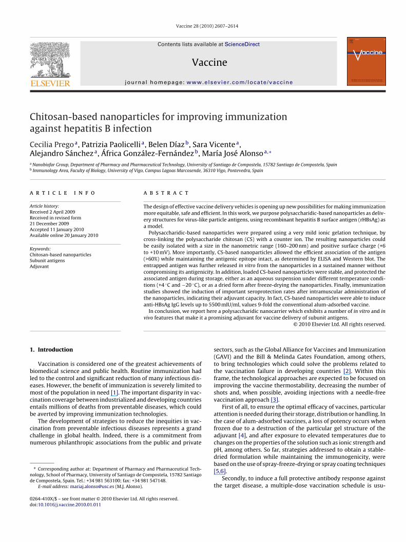
Having in mind these positive and encouraging results, stabil-ty studies of the lyophilized formulations were performed aftereeping them in the fridge up to 3 months. Lyophilized rHBsAg-oaded CS-based nanoparticles containing 2.5% of glucose wereasily redispersed in water while preserving the physico-chemicalroperties of the nanoparticles (Fig. 5A) and, more importantly, nohanges on the structural integrity of the antigen were appreciatedFig. 5B) for at least 3 months of storage (limit of the study).
Overall these results, we could conclude that the lyophilizationf rHBsAg-loaded CS-based nanoparticles may provide a useful toolor preserving the stability of the antigen.
.5. Immunization studies
As a final step of this work, and in order to evaluate the potentialf CS-based nanoparticles loaded with subunit antigens to induce
he generation of protective IgG titers, immunization studies afterasal and intramuscular administration were performed using rHB-Ag as a model (Fig. 6). The graphs represent the mean IgG valuesxpressed as concentration (mIU/ml) collected from a single study;owever CS-based nanoparticles were evaluated in two sets oflyophilized CS-based nanoparticles: Lane 1: plain rHBsAg; lane 2: rHBsAg recov-ered from lyophilized nanoparticles in the presence of 5% glucose; lane 3: rHBsAgrecovered from lyophilized nanoparticles in the presence of 2.5% glucose.
experiments made in different laboratories, obtaining comparableresults.
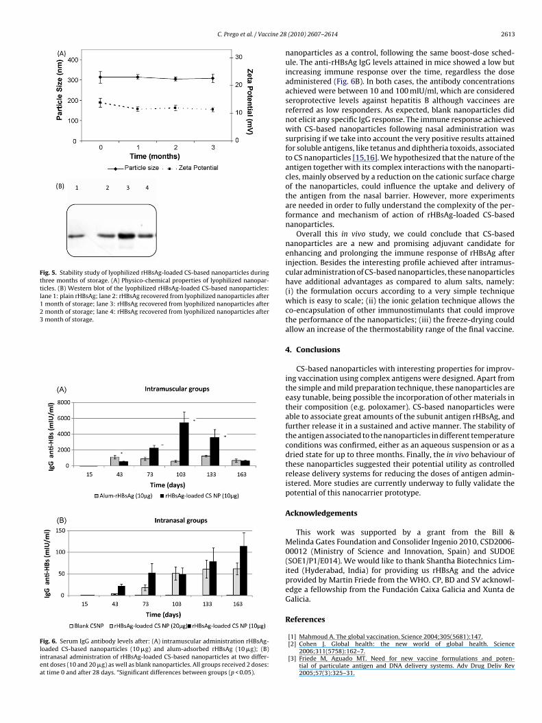
We evaluated the effectiveness of CS-based nanoparticles toinduce IgG titers as compared to alum-adsorbed rHBsAg, after intra-muscular administration to mice (two doses of 10 �g at days 0 and28). Loaded CS-based nanoparticles elicited an immune responseslightly delayed as compared to the alum. However, with thetime, the IgG concentration was much higher for the nanoparti-cles than the conventional adjuvant, achieving values higher than5400 mIU/ml, more than 9-fold as compared to the alum (Fig. 6A).This result highlighted the properties of CS-based nanoparticlesas an alternative adjuvant, as well as their ability to control thedelivery of the antigen and their features to preserve the immuno-genicity of rHBsAg after being formulated in the nanoparticles.Nevertheless, more studies are needed to elucidate the potential ofCS-based nanoparticles to reduce the number of doses as compared
to alum salts.On the other hand, we also investigated the potential of rHBsAg-loaded CS-based nanoparticles after intranasal immunization. Weassayed two different rHBsAg doses, 10 and 20 �g, as well as blank
C. Prego et al. / Vaccine 28
Fig. 5. Stability study of lyophilized rHBsAg-loaded CS-based nanoparticles duringthree months of storage. (A) Physico-chemical properties of lyophilized nanopar-ticles. (B) Western blot of the lyophilized rHBsAg-loaded CS-based nanoparticles:lane 1: plain rHBsAg; lane 2: rHBsAg recovered from lyophilized nanoparticles after1 month of storage; lane 3: rHBsAg recovered from lyophilized nanoparticles after2 month of storage; lane 4: rHBsAg recovered from lyophilized nanoparticles after3 month of storage.
Fig. 6. Serum IgG antibody levels after: (A) intramuscular administration rHBsAg-loaded CS-based nanoparticles (10 �g) and alum-adsorbed rHBsAg (10 �g); (B)intranasal administration of rHBsAg-loaded CS-based nanoparticles at two differ-ent doses (10 and 20 �g) as well as blank nanoparticles. All groups received 2 doses:at time 0 and after 28 days. *Significant differences between groups (p < 0.05).
(2010) 2607–2614 2613
nanoparticles as a control, following the same boost-dose sched-ule. The anti-rHBsAg IgG levels attained in mice showed a low butincreasing immune response over the time, regardless the doseadministered (Fig. 6B). In both cases, the antibody concentrationsachieved were between 10 and 100 mIU/ml, which are consideredseroprotective levels against hepatitis B although vaccinees arereferred as low responders. As expected, blank nanoparticles didnot elicit any specific IgG response. The immune response achievedwith CS-based nanoparticles following nasal administration wassurprising if we take into account the very positive results attainedfor soluble antigens, like tetanus and diphtheria toxoids, associatedto CS nanoparticles [15,16]. We hypothesized that the nature of theantigen together with its complex interactions with the nanoparti-cles, mainly observed by a reduction on the cationic surface chargeof the nanoparticles, could influence the uptake and delivery ofthe antigen from the nasal barrier. However, more experimentsare needed in order to fully understand the complexity of the per-formance and mechanism of action of rHBsAg-loaded CS-basednanoparticles.
Overall this in vivo study, we could conclude that CS-basednanoparticles are a new and promising adjuvant candidate forenhancing and prolonging the immune response of rHBsAg afterinjection. Besides the interesting profile achieved after intramus-cular administration of CS-based nanoparticles, these nanoparticleshave additional advantages as compared to alum salts, namely:(i) the formulation occurs according to a very simple techniquewhich is easy to scale; (ii) the ionic gelation technique allows theco-encapsulation of other immunostimulants that could improvethe performance of the nanoparticles; (iii) the freeze-drying couldallow an increase of the thermostability range of the final vaccine.
4. Conclusions
CS-based nanoparticles with interesting properties for improv-ing vaccination using complex antigens were designed. Apart fromthe simple and mild preparation technique, these nanoparticles areeasy tunable, being possible the incorporation of other materials intheir composition (e.g. poloxamer). CS-based nanoparticles wereable to associate great amounts of the subunit antigen rHBsAg, andfurther release it in a sustained and active manner. The stability ofthe antigen associated to the nanoparticles in different temperatureconditions was confirmed, either as an aqueous suspension or as adried state for up to three months. Finally, the in vivo behaviour ofthese nanoparticles suggested their potential utility as controlledrelease delivery systems for reducing the doses of antigen admin-istered. More studies are currently underway to fully validate thepotential of this nanocarrier prototype.
Acknowledgements
This work was supported by a grant from the Bill &Melinda Gates Foundation and Consolider Ingenio 2010, CSD2006-00012 (Ministry of Science and Innovation, Spain) and SUDOE(SOE1/P1/E014). We would like to thank Shantha Biotechnics Lim-ited (Hyderabad, India) for providing us rHBsAg and the adviceprovided by Martin Friede from the WHO. CP, BD and SV acknowl-edge a fellowship from the Fundación Caixa Galicia and Xunta deGalicia.
References
[1] Mahmoud A. The global vaccination. Science 2004;305(5681):147.[2] Cohen J. Global health: the new world of global health. Science
2006;311(5758):162–7.[3] Friede M, Aguado MT. Need for new vaccine formulations and poten-
tial of particulate antigen and DNA delivery systems. Adv Drug Deliv Rev2005;57(3):325–31.

2 ine 28
[
[
[
[
[
[
[
[
[
[
[
[
[
[[
[
[
[
[
[
[
614 C. Prego et al. / Vacc
[4] Zapata MI, Peck GE, White JL, Hem SL. Mechanism of freeze–thaw instabilityof aluminium hydroxycarbonate and magnesium hydroxide gels. J Pharm Sci1984;73(1):3–8.
[5] Maa YF, Zhao L, Payne LG, Chen D. Stabilization of alum-adjuvanted vac-cine dry powder formulations: mechanism and application. J Pharm Sci2003;92(2):319–32.
[6] Maa YF, Ameri M, Rigney R, Payne LG, Chen D. Spray-coating for biopharmaceu-tical powder formulations: beyond the conventional scale and its application.Pharm Res 2004;21(3):515–23.
[7] Alonso MJ, Cohen S, Park TG, Gupta RK, Siber GR, Langer R. Determinantsof release rate of tetanus vaccine from polyester microspheres. Pharm Res1993;10(7):945–53.
[8] Shi L, Caufield MJ, Chern RT, Wilson RA, Sanyal G, Volkin DB. Phar-maceutical and immunological evaluation of a single-shot hepatitis Bvaccine formulated with PLGA microspheres. J Pharm Sci 2002;91(4):1019–35.
[9] Feng L, Qi XR, Zhou XJ, Maitani Y, Wang SSC, Jiang Y, et al. Pharmaceuticaland immunological evaluation of a single-dose hepatitis B vaccine using PLGAmicrospheres. J Control Release 2006;112(1):35–42.
10] O’Hagan DT, Valiante NM. Recent advances in the discovery and delivery ofvaccine adjuvants. Nat Rev Drug Discov 2003;2(9):727–35.
11] Tong NK, Beran J, Kee SA, Miguel JL, Sánchez C, Bayas JM, et al. Immunogenic-ity and safety of an adjuvanted hepatitis B vaccine in pre-hemodialysis andhemodialysis patients. Kidney Int 2005;68(5):2298–303.
12] Giudice EL, Campbell JD. Needle-free vaccine delivery. Adv Drug Deliv Rev2006;58(1):68–89.
13] Neutra MR, Kozlowski PA. Mucosal vaccines: the promise and the challenge.Nat Rev Immunol 2006;6(2):148–58.
14] Kiyono H, Fukuyama S. Nalt-versus Peyer’s-Patch-mediated mucosal immu-nity. Nat Rev Immunol 2004;4(9):699–710.
15] Vila A, Sanchez A, Tobio M, Calvo P, Alonso MJ. Design of biodegradable particlesfor protein delivery. J Control Release 2002;78(1–3):15–24.
16] Vila A, Sanchez A, Janes KA, Behrens I, Kissel T, Vila Jato JL, et al. Low molecularweight chitosan nanoparticles as new carriers for nasal vaccine delivery. Eur JPharm Biopharm 2004;57(1):123–32.
17] Calvo P, Remunan-Lopez C, Vila Jato JL, Alonso MJ. Novel hydrophilic chitosan-polyethylene oxide nanoparticles as protein carriers. J Appl Polym Sci1997;63(1):125–32.
[
(2010) 2607–2614
18] Calvo P, Remunan-Lopez C, Vila Jato JL, Alonso MJ. Chitosan and chi-tosan/ethylene oxide-propylene oxide block copolymer nanoparticles as novelcarriers for proteins and vaccines. Pharm Res 1997;14(10):1431–6.
19] Fernandez-Urrusuno R, Calvo P, Remunan-Lopez C, Vila Jato JL, Alonso MJ.Enhancement of nasal absorption of insulin using chitosan nanoparticles.Pharm Res 1999;16(10):1576–81.
20] Janes KA, Calvo P, Alonso MJ. Polysaccharide colloidal particles as deliverysystems for macromolecules. Adv Drug Deliv Rev 2001;47(1):83–97.
21] Prego C, Torres D, Alonso MJ. The potential of chitosan for the oral administra-tion of peptides. Expert Opin Drug Deliv 2005;2(5):843–54.
22] Grgacic EV, Anderson DA. Virus-like particles: passport to immune recognition.Methods 2006;40(1):60–5.
23] Shouval D. Hepatitis B vaccines. J Hepatol 2003;39(1):S70–6.24] Gavilanes F, Gonzalez-Ros JM, Peterson DL. Structure of hepatitis B surface
antigen. J Biol Chem 1982;257(13):7770–7.25] Wang S, Liu X, Fisher K, Smith JG, Chen F, Tobery TW, et al. Enhanced type I
immune response to a hepatitis B DNA vaccine by formulation with calcium-or aluminium phosphate. Vaccine 2000;18(13):1227–35.
26] Katakam M, Banga AK. Use of poloxamer polymers to stabilize recombinanthuman growth hormone against various processing stresses. Pharm Dev Tech-nol 1997;2(2):143–9.
27] Kang ML, Jiang HL, Kang SG, Guo DD, Lee DY, Cho CS, et al. Pluronic F127enhances the effect as an adjuvant of chitosan microspheres in the intranasaldelivery of Bordetella bronchiseptica antigens containing dermonecrotoxin.Vaccine 2007;25(23):4602–10.
28] Coeshott CM, Smithson SL, Verderber E, Samaniego A, Blonder JM, Rosen-thal GJ, et al. Pluronic F127-based systemic vaccine delivery systems. Vaccine2004;22(19):2396–405.
29] Westerink MA, Smithson SL, Srivastava N, Blonder J, Coeshott C, RosenthalGJ. ProJuvant (Pluronic F127/chitosan) enhances the immune response tointranasally administered tetanus toxoid. Vaccine 2001;20(5–6):711–23.
30] Fernandez-Urrusuno R, Romani D, Calvo P, Vila-Jato JL, Alonso MJ. Development
of a freeze-dried formulation of insulin-loaded chitosan nanoparticles intendedfor nasal administration. STP Pharma Sci 1999;9:429–36.31] Cuna M, Alonso-Sande M, Remunan-Lopez C, Pivel JP, Alonso-Lebrero JL,Alonso MJ. Development of phosphorylated glucomannan-coated chitosannanoparticles as nanocarriers for protein delivery. J Nanosci Nanotechnol2006;6(9–10):2887–95.
Copyright © 2022 FDOKUMEN