Boron microlocalization in oral mucosal tissue: implications for boron neutron capture therapy
8
Boron neutron capture therapy (BNCT) is a targeted radio- therapeutic modality used for the treatment of brain tumours and melanoma in the USA, Europe and Japan. BNCT was extensively reviewed in two recent articles (Barth et al, 1999; Coderre and Morris, 1999). The targeting effectiveness of BNCT is dependent upon the preferential delivery of boron-10 ( 10 B) to the primary tumour and its metastatic spread. A beam of thermalized neutrons is used to ‘activate’ the 10 B, with the resultant production of highly localized high linear energy transfer (LET) radiation. This is due to the capture of thermal neutrons by 10 B with the resultant fission reaction ( 10 B + 1 n→ 11 B→ 7 Li + 4 He (α) + 2.79 MeV) giving rise to high energy charged particles with short trajectories in tissue (5– 9 μm). The microlocation and differential accumulation of 10 B will therefore have a critical bearing on the therapeutic outcome of BNCT, and also on the response of normal tissue in the irradiated volume. In the clinical situation, relatively high therapeutic ratios have been obtained enabling BNCT to be given as a single dose or a small number (maximum 4) of dose fractions (Hatanaka et al, 1994; Coderre et al, 1997). Mucositis is a commonly observed side-effect during conven- tional radiotherapy for cancer of the head and neck. While acute radiation damage of the oral mucosa is not generally associated with the current treatment of gliomas using BNCT, a number of patients involved in the American clinical studies are now presenting with transient acute effects. The problem relates to individuals with tumours at the front of the brain, in proximity to mucosal tissue (Coderre et al, 1997). This finding suggests that damage to the oral mucosa could be a potential complication in future BNCT clinical protocols involving larger irradiation field sizes than those in current use or multiple fields. The purpose of the present investigation was to evaluate the microdistribution of 10 B in the oral mucosa, and relate this to the biological effectiveness of the 10 B(n,α) 7 Li neutron capture reaction in this tissue. A CAMECA IMS-3f ion microscope, which is essentially a direct imaging secondary ion mass spectrometer (Benninghoven et al, 1987), was used to analyse cryopreserved Boron microlocalization in oral mucosal tissue: implications for boron neutron capture therapy GM Morris 1 , DR Smith 2 , H Patel 1 , S Chandra 2 , GH Morrison 2 , JW Hopewell 1 , M Rezvani 1 , PL Micca 3 and JA Coderre 3 1 Research Institute, University of Oxford, Churchill Hospital, Oxford, OX3 7LJ, UK; 2 Department of Chemistry and Chemical Biology, Baker Laboratory, Cornell University, Ithaca, NY 14853–1301, USA; 3 Medical Department, Brookhaven National Laboratory Upton, NY 11973, USA Summary Clinical studies of the treatment of glioma and cutaneous melanoma using boron neutron capture therapy (BNCT) are currently taking place in the USA, Europe and Japan. New BNCT clinical facilities are under construction in Finland, Sweden, England and California. The observation of transient acute effects in the oral mucosa of a number of glioma patients involved in the American clinical trials, suggests that radiation damage of the oral mucosa could be a potential complication in future BNCT clinical protocols, involving higher doses and larger irradiation field sizes. The present investigation is the first to use a high resolution surface analytical technique to relate the microdistribution of boron-10 ( 10 B) in the oral mucosa to the biological effectiveness of the 10 B(n,α) 7 Li neutron capture reaction in this tissue. The two boron delivery agents used clinically in Europe/Japan and the USA, borocaptate sodium (BSH) and p-boronophenylalanine (BPA), respectively, were evaluated using a rat ventral tongue model. 10 B concentrations in various regions of the tongue mucosa were estimated using ion microscopy. In the epithelium, levels of 10 B were appreciably lower after the administration of BSH than was the case after BPA. The epithelium:blood 10 B partition ratios were 0.2:1 and 1:1 for BSH and BPA respectively. The 10 B content of the lamina propria was higher than that measured in the epithelium for both BSH and BPA. The difference was most marked for BSH, where 10 B levels were a factor of six higher in the lamina propria than in the epithelium. The concentration of 10 B was also measured in blood vessel walls where relatively low levels of accumulation of BSH, as compared with BPA, was demonstrated in blood vessel endothelial cells and muscle. Vessel wall:blood 10 B partition ratios were 0.3:1 and 0.9:1 for BSH and BPA respectively. Evaluation of tongue mucosal response (ulceration) to BNC irradiation indicated a considerably reduced radiation sensitivity using BSH as the boron delivery agent relative to BPA. The compound biological effectiveness (CBE) factor for BSH was estimated at 0.29 ± 0.02. This compares with a previously published CBE factor for BPA of 4.87 ± 0.16. It was concluded that variations in the microdistribution profile of 10 B, using the two boron delivery agents, had a significant effect on the response of oral mucosa to BNC irradiation. From a clinical perspective, based on the findings of the present study, it is probable that potential radiation- induced oral mucositis will be restricted to BNCT protocols involving BPA. However, a thorough high resolution analysis of 10 B microdistribution in human oral mucosal tissue, using a technique such as ion microscopy, is a prerequisite for the use of experimentally derived CBE factors in clinical BNCT. © 2000 Cancer Research Campaign Keywords: borocaptate sodium; p-boronophenylalanine; rat ventral tongue mucosa; compound biological effectiveness factor; ion microscopy imaging 1764 Received 25 March 1999 Revised 28 January 2000 Accepted 17 February 2000 Correspondence to: GM Morris British Journal of Cancer (2000) 82(11), 1764–1771 © 2000 Cancer Research Campaign DOI: 10.1054/ bjoc.2000.1148, available online at http://www.idealibrary.com on
Transcript of Boron microlocalization in oral mucosal tissue: implications for boron neutron capture therapy
aivae
r
u
ea
ose or al,
n-ute
ated ofw toothat in
the
Boron microlocalization in oral mucosal tissue:implications for boron neutron capture therapy
GM Morris 1, DR Smith 2, H Patel 1, S Chandra 2, GH Morrison 2, JW Hopewell 1, M Rezvani 1, PL Micca 3 and JA Coderre 3
1Research Institute, University of Oxford, Churchill Hospital, Oxford, OX3 7LJ, UK; 2Department of Chemistry and Chemical Biology, Baker Laboratory,Cornell University, Ithaca, NY 14853–1301, USA; 3Medical Department, Brookhaven National Laboratory Upton, NY 11973, USA
Summary Clinical studies of the treatment of glioma and cutaneous melanoma using boron neutron capture therapy (BNCT) are currentlytaking place in the USA, Europe and Japan. New BNCT clinical facilities are under construction in Finland, Sweden, England and California.The observation of transient acute effects in the oral mucosa of a number of glioma patients involved in the American clinical trials, suggeststhat radiation damage of the oral mucosa could be a potential complication in future BNCT clinical protocols, involving higher doses and largerirradiation field sizes. The present investigation is the first to use a high resolution surface analytical technique to relate the microdistributionof boron-10 (10B) in the oral mucosa to the biological effectiveness of the 10B(n,α)7Li neutron capture reaction in this tissue. The two borondelivery agents used clinically in Europe/Japan and the USA, borocaptate sodium (BSH) and p-boronophenylalanine (BPA), respectively,were evaluated using a rat ventral tongue model. 10B concentrations in various regions of the tongue mucosa were estimated using ionmicroscopy. In the epithelium, levels of 10B were appreciably lower after the administration of BSH than was the case after BPA. Theepithelium:blood 10B partition ratios were 0.2:1 and 1:1 for BSH and BPA respectively. The 10B content of the lamina propria was higher thanthat measured in the epithelium for both BSH and BPA. The difference was most marked for BSH, where 10B levels were a factor of six higherin the lamina propria than in the epithelium. The concentration of 10B was also measured in blood vessel walls where relatively low levels ofaccumulation of BSH, as compared with BPA, was demonstrated in blood vessel endothelial cells and muscle. Vessel wall:blood 10B partitionratios were 0.3:1 and 0.9:1 for BSH and BPA respectively. Evaluation of tongue mucosal response (ulceration) to BNC irradiation indicated aconsiderably reduced radiation sensitivity using BSH as the boron delivery agent relative to BPA. The compound biological effectiveness(CBE) factor for BSH was estimated at 0.29 ± 0.02. This compares with a previously published CBE factor for BPA of 4.87 ± 0.16. It wasconcluded that variations in the microdistribution profile of 10B, using the two boron delivery agents, had a significant effect on the response oforal mucosa to BNC irradiation. From a clinical perspective, based on the findings of the present study, it is probable that potential radiation-induced oral mucositis will be restricted to BNCT protocols involving BPA. However, a thorough high resolution analysis of 10Bmicrodistribution in human oral mucosal tissue, using a technique such as ion microscopy, is a prerequisite for the use of experimentallyderived CBE factors in clinical BNCT. © 2000 Cancer Research Campaign
Keywords : borocaptate sodium; p-boronophenylalanine; rat ventral tongue mucosa; compound biological effectiveness factor;ion microscopy imaging
British Journal of Cancer (2000) 82(11), 1764–1771© 2000 Cancer Research CampaignDOI: 10.1054/ bjoc.2000.1148, available online at http://www.idealibrary.com on
Boron neutron capture therapy (BNCT) is a targeted radtherapeutic modality used for the treatment of brain tumours melanoma in the USA, Europe and Japan. BNCT was extensreviewed in two recent articles (Barth et al, 1999; Coderre Morris, 1999). The targeting effectiveness of BNCT is dependupon the preferential delivery of boron-10 (10B) to the primarytumour and its metastatic spread. A beam of thermalized neutis used to ‘activate’ the 10B, with the resultant production of highlylocalized high linear energy transfer (LET) radiation. This is dto the capture of thermal neutrons by 10B with the resultant fissionreaction (10B + 1n→ 11B→ 7Li + 4He (α) + 2.79 MeV) giving rise tohigh energy charged particles with short trajectories in tissue9 µm). The microlocation and differential accumulation of 10B willtherefore have a critical bearing on the therapeutic outcomBNCT, and also on the response of normal tissue in the irradi
e
seterrved
1764
Received 25 March 1999Revised 28 January 2000Accepted 17 February 2000
Correspondence to: GM Morris
io-ndelyndnt
ons
e
(5–
ofted
volume. In the clinical situation, relatively high therapeutic ratihave been obtained enabling BNCT to be given as a single dosa small number (maximum 4) of dose fractions (Hatanaka et1994; Coderre et al, 1997).
Mucositis is a commonly observed side-effect during convetional radiotherapy for cancer of the head and neck. While acradiation damage of the oral mucosa is not generally associwith the current treatment of gliomas using BNCT, a numberpatients involved in the American clinical studies are nopresenting with transient acute effects. The problem relatesindividuals with tumours at the front of the brain, in proximity tmucosal tissue (Coderre et al, 1997). This finding suggests damage to the oral mucosa could be a potential complicationfuture BNCT clinical protocols involving larger irradiation fieldsizes than those in current use or multiple fields.
The purpose of the present investigation was to evaluatemicrodistribution of 10B in the oral mucosa, and relate this to thbiological effectiveness of the 10B(n,α)7Li neutron capture reactionin this tissue. A CAMECA IMS-3f ion microscope, which iessentially a direct imaging secondary ion mass spectrom(Benninghoven et al, 1987), was used to analyse cryoprese

t
iga
hapa
w
p9rdaa
li
o
3e
o inn
c
nw
f rm
sasesa
thisen
theionss-edg a
eamfperomass-ed
eterandctedtion of
ec-leda
ts/of
in,erethe ofbra-
ith
ioner
lly
reasia. to
hert
to-
asssin. onndle
thector
Boron microlocalization in oral mucosa 1765
tissue sections of the rat ventral tongue. This same instrumenemployed in a previous study (Smith et al, 1996) to assess10Bmicrolocalization in a rat brain tumour model for BNCT. Iomicroscopy is a trace surface analytical technique with partsmillion (ppm) detection limits for 10B. In principal the CAMCAIMS-3f can produce images of all isotopes from H to U with hresolution. The optics of the ion microscope maintain the spintegrity of an analyte in a sample, producing images of isotodistributions which can be related to tissue morphology witresolution comparable to a conventional light microscope. A madvantage of using the CAMCA IMS-3f ion microscofor tissue microdistribution studies is the ability to observe quantify, in a single image, the concentration gradient of 10B inmultiple zones of tissue. The 10B distribution in the epithelium,lamina propria and muscle of the oral mucosa can be viesimultaneously within a 150µm diameter field of view.
The rat ventral tongue oral mucosa model, originally develoby Morris and Coderre (Coderre et al, 1999; Morris et al, 19was used in the present study. It is essentially a scaled up veof the standard murine ventral tongue model used in rabiological studies (e.g. Moses and Kummermehr, 1986; Dorr Kummermehr, 1990). The larger tongue area of the rat facilitthe jigging and shielding procedures required for local BNC irraation, thereby reducing the radiation burden to the head. Inpresent study two boron delivery agents, borocaptate sod(BSH) and boronophenylalanine (BPA), were used in the evation of 10B microdistribution in the tongue mucosa. The radbiological findings related to BSH-mediated BNC irradiation acompared and contrasted with a previous report (Coderre e1999) detailing the effects of BPA-mediated BNC irradiation the same model.
MATERIALS AND METHODS
All experimental procedures involved the use of male Fischerrats aged 12 weeks (260–290 g). The animal studies were reviand approved by the Brookhaven National Laboratory (BNInstitutional Animal Care and Use Committee. Rats were accmodated two to a cage in temperature controlled rooms andfree access to food and water. They were maintained controlled light/dark cycle, with lights on between 07:00 a18:00 h.
Disodium mercaptoundecahydro-close dodecaborate (borotate sodium) containing 55.1% (certified weight percentage)10Bwith 95.8% enrichment (Centronic Ltd., Croydon, UK) was usas the boron delivery agent. Vials of BSH powder, sealed unitrogen gas, were opened immediately prior to use. Dilution in sterile normal saline (10 mg BSH ml–1). Administration of theBSH solution was by intraperitoneal (i.p.) injection in a volume1.5 ml (55 mg kg–1; 30 mg 10B kg–1). BPA was given i.p. in afructose solution at a dose of 700 mg kg–1 (34 mg 10B kg–1) asdescribed previously (Coderre et al, 1994). Concentrations o10Bin solutions of BSH and BPA were determined using direct curplasma atomic emission spectroscopy (DCP-AES) and progamma analysis (Fairchild et al, 1986; Barth et al, 1991).
Boron biodistribution
Boron concentrations in blood and tongue were measured u(DCP-AES) and prompt gamma spectrometry.
© 2000 Cancer Research Campaign
was nper
htialpic ajorend
ed
ed9),sionio-ndtesdi-theiumua-o-ret al,n
44wedL)m-had ad
ap-
edderas
of
entpt
sing
The microdistribution of boron in the ventral tongue mucowas evaluated using ion microscopy. The ion microscopy analywere performed using a CAMECA IMS-3f ion microscope, direct imaging secondary ion mass spectrometer. The use ofinstrument for BNCT-related microlocalization studies has bedescribed previously (Smith et al, 1996). In the present studyion microscope was operated in the positive secondary imaging mode with the samples biased to +4500 V. A mafiltered O2
+ primary ion beam accelerated to 10 keV was focusand adjusted to a nominal beam current of 120 nA, producinstationary beam spot with a diameter of approximately 50µm atthe surface of the sample. For all analyses, the primary ion bwas raster scanned over 250 × 250µm square regions. The rate oerosion of the tissue specimens was approximately 20 Å second. Singly charged positive secondary ions, originating frthe surface of the sample, were collected as a global (non-mfiltered) image by the instrument’s immersion lens and focususing the 150µm transfer optics in conjunction with a 60µmcontrast aperture. The energy window of the mass spectromwas centred and set to a maximum value of 130 eV. Energy mass filtered secondary ion images were magnified and projeon a single microchannel plate/phosphor screen image detecassembly. The gain of the microchannel plate was set to 73%maximum. Images were recorded directly from the image dettion assembly using a Photometrics Ltd. CH220 charged-coupdevice (CCD) liquid-cooled camera head equipped with Thompson-CSF TH7882 CDA CCD camera digitized to 14 bipixel by a photometric camera controller. For each region analysis, sequential ion images from 39K, 23Na, 10B, 12C, 24Mg and40Ca were acquired with integration times of 0.2 s, 0.2 s, 2 m2 min, 30 s and 30 s respectively. The digitized ion images wanalysed using Alice™ 2.4.0 image processing software for Macintosh (Hayden Image Processing Group). Quantificationthe boron images was carried out using an ion microscopy calition approach for biological specimens (Ausserer et al, 1989;Smet al, 1996). Estimates of the 10B concentration in the ventraltongue mucosa were determined from pixel to pixel registratand ratio of the 10B and 12C images, assuming an 85% tissue watcontent.
Samples for ion microscopy imaging were cryogenicaprepared to preserve the in situ distribution of 10B and minimizeredistribution artefacts at the microscopic level. Two rats weadministered BSH at 30 min prior to anaesthesia and euthanTissue specimens were frozen in liquid isopentane cooled–150°C in liquid nitrogen. Frozen sections, 4-µm-thick, were cutperpendicular to the ventral surface of the tongue using a ReicHistoSTAT cryostat microtome operated at –20°C. Sections weretaken in series to correlate the tissue morphology (optical aufluorescence microscopy) with 10B distribution (ion microscopy).Sections prepared for optical imaging were mounted on glmicroscope slides and stained with haematoxylin and eoSections designated for ion microscopy imaging were mountedhigh purity indium/silicon substrates, freeze-dried for 24 h acoated with a 400 Å thick layer of Au/Pd alloy to minimize sampcharging during analysis.
Dosimetry
Boron neutron capture (BNC) irradiations were carried out on thermal beam port of the Brookhaven Medical Research Rea
British Journal of Cancer (2000) 82(11), 1764–1771

c
se
eupnr
ra
nTr2nn
-
e e o
t
a
onsing
ed%
s,Ing
e
nd
ueof
ralA.a-r toivetederells
blecle
sedn
eshe
m
1766 GM Morris et al
Table 1 Thermal beam exposure times, blood 10B content at the time of irradiation and physical absorbed doses forthe various treatment groups. Errors indicate ± s.e.m.
Thermal beam Blood 10B Thermal beam 10B(n, α)7Li
exposure time content dose component dose component Total dose(MW min) ( µg g)–1 (Gy) (Gy) (Gy)
7.5 43.6 ± 0.9 1.6 9.3 10.98.5 46.6 ± 3.0 1.9 11.2 13.19.5 41.5 ± 2.0 2.1 11.2 13.3
12.5 44.7 ± 2.3 2.7 15.9 18.614.0 44.3 ± 2.5 3.0 17.7 20.715.0 46.6 ± 4.2 3.3 19.8 23.116.5 43.3 ± 1.4 3.6 20.3 23.9
(BMRR). The dosimetry of the mixed radiation field, whiconsisted of thermal neutrons, fast neutrons, gamma rayscharged particles (α, 7Li, 1H, 14C) from the 10B(n,α)7Li and14N(n,p)14C neutron capture reactions were calculated uthermal neutron fluence data from bare and cadmium-covgold foils and wires (secured to and inserted in the tongues ofrats). A uniform distribution of 2.7% w/w nitrogen in tissue wassumed. Thermoluminescent dosimeters (TLD-700: HarsChemical, Solon, OH, USA) attached to the tongue, were usmeasure the gamma dose. The combined fast (>10 keV) neand gamma doses at the beam port were measured using tissue-equivalent (TE) plastic ionization chambers contaimethane-based tissue equivalent gas, and a graphite chambewith carbon dioxide gas. The fast neutron dose rate was calcuby subtraction of the gamma dose rate from the total doseMonte Carlo computation, normalized to the in-air neutdosimetry, was used to estimate the fast neutron dose rate level of the ventral tongue mucosa.
The thermal neutron fluence at the surface of the tongue5.46 × 109 cm–2 s–1 with the reactor running at 1 MW power, usia 10.2 cm thick collimator with a 20 mm diameter aperture. collimator was constructed of lithium carbonate (7.5%) dispein polyethylene. The physical dose rates (cGy/MW min) were (per µg10B g–1) for the 10B(n,α)7Li reaction; 9.9 for the fast neutrointeraction with hydrogen 1H(n,n)p; 7.0 for the nitrogen neutrocapture reaction (14N(n,p)14C); and 4.8 for the total gamma-racomponent (from the beam and induced in tissue by the 1H(n,γ)2Hreaction).
Irradiations
Anaesthesia during irradiation was maintained by the i.p. injecof ketamine (54 mg kg–1) and xylazine (9 mg kg–1). The anaesthetized rat was positioned in a body radiation shield (Joel e1990; Morris et al, 1994a) that ensured that the rat was secursupported during irradiation. The procedure for thermal beamBNC irradiation has been described in detail previously (Codet al, 1999). Briefly, the tongue was secured with tape to20-mm diameter collimator and positioned to ensure unifirradiation of the ventral surface.
BSH was administered by i.p. injection 30 min prior to thermneutron irradiation. A 0.5 ml blood sample was taken immediaprior to irradiation and the boron content determined using progamma analysis. On average, eight rats were irradiated pergroup. The various dose groups are listed in Table 1. Radi
British Journal of Cancer (2000) 82(11), 1764–1771
h and
ingred
deadashawd totronaireding filledlatedrate.ont the
wasghe
sed.84
y
tion
t al,lyandrretherm
alelymptdosetion
reactions in the tongue were scored daily, under light carbdioxide/oxygen anaesthesia. Dose response was quantified uulceration as the end point.
The dose–effect curve for the incidence of ulceration were fittusing probit analysis. The dose required to produce a 50incidence of ulceration (ED50) was calculated from this curve.Errors indicate the standard error of the mean (± s.e.m.).
RESULTS
Boron biodistribution and microlocalization
Blood 10B concentrations from BSH for the various dose groupobtained immediately prior to irradiation, are given in Table 1. an additional evaluation of boron biodistribution (six rats) usinDCP-AES, levels of 10B in blood, ventral tongue and dorsal tonguwere 38.9 ± 1.8, 21.1 ± 0.4 and 17.1 ± 0.6µg g–1 respectively.
The microdistribution of 10B in the ventral tongue mucosa wasevaluated using ion microscopy. The distributions of BSH aBPA were compared and 10B concentrations were determined inthe epithelium, lamina propria and contiguous ventral tongmuscle. Additionally, the boron accumulation characteristics BSH and BPA were compared in the walls of blood vessels.
An optical (autofluorescence) image of cryosectioned venttongue mucosa of a rat administered BPA is shown in Figure 1White arrows point to the smooth surface of the stratified squmous epithelium which is generally not cornified on the lowesurface of the tongue. The dotted lines in panel 1A were drawndelineate the boundaries of the epithelium (Ep), the connecttissue of the lamina propria (LP) and cross-sections of striamuscle (M). The basal layer of the epithelium lies along thboundary of the epithelium and the lamina propria. Cells which aremoved from the surface of the epithelium are replaced by cewhich move up from the basal layer. The basal layer is vulnerato radiation and damage manifests itself in this region. The cirsuperimposed in the optical image is scaled to 150µm in diameterand correlates with the region on the adjacent cryosection analywith the ion microscope (Figure 1 B–D). Dotted lines in the ioimages of 10B, 24Mg and 12C (Figure 1 B–D) aid in visualizing thehistology of the oral mucosa. The white arrows in the ion imagpoint to the smooth surface of the epithelium. The ratio of twhite pixel density in the 10B and 24Mg ion images (Figure 1 B,C)to the white pixel density of the 12C image (Figure 1D), is directlyproportional to the total concentration of boron and magnesiurespectively. The image of the tissue matrix, 12C, is used in the
© 2000 Cancer Research Campaign

Boron microlocalization in oral mucosa 1767
wht
pw
i
cn
lehth
en
o
loto
en
ofd,
2.
gnare- and oftri-eed
tra-
arisof
s.n ofple
Ep
LP
M 60 µm
A
Ep
LP
M
50 µm
B 10B
Ep
LP
M
50 µm
D 12C
Ep
LP
M
50 µm
C 24Mg
Figure 1 Ventral tongue mucosa of a rat administered BPA. (A) Autofluorescence-fluorescein isothiocyanate (FITC) photomicrograph of haematoxylin andeosin stained cryosection of ventral tongue mucosa. The circle is scaled to 150 µm in diameter and correlates with the region of the adjacent cryosection oftissue analysed with the ion microscope (see panels B–D). (B–D) Sequential ion images from a freeze-dried cryosection of tissue mounted on indium showingthe distribution of the isotopes 10B (B), 24Mg (C) and 12C (D). Images of 39K, 23Na and 40Ca are not shown. White arrows in (A–D) point to the outer surface of theepithelium. Black arrows in (B–D) point to regions of excessive brightness caused by the non-uniform response of the microchannel plate/phosphor screendetection assembly of the ion microscope. Dotted lines in (A–D) aid in visualization of the epithelium (Ep), lamina propria (LP) and muscle (M)
quantification scheme to reduce potential errors associated the non-uniform response of the microchannel plate/phospscreen detection assembly of the ion microscope (Ausserer e1989). Temporal degradation of the detection assembly is ressible for the excessive brightness seen in the upper left and loright quadrants of the 10B, 24Mg and 12C images (black arrows). Inthe absence of the 12C image, the white pixel intensity of theepithelium in Figure 1B,C would be incorrectly interpreted asgradient with the highest concentrations of boron and magnesin the upper left and lower right quadrants.
Not shown are images of total 39K, 23Na and 40Ca. Theseelements are imaged in sequence with 10B, 24Mg and 12C. Images of39K, 23Na, 24Mg and 40Ca are relevant in assessing the physiologistatus of the cryopreserved tissue. The ratio of the image inteties of 39K to 23Na from regions within the epithelium and muscwere 3:1, indicating that the native distribution of these higdiffusible species was not significantly altered. The ratio of image intensities of 39K to 23Na in the lamina propria falls off tonearly 1:1 which is consistent with an area rich in blood vessBlood has a relatively high sodium and low potassium conteThe 24Mg ion image (Figure 1C) shows a gradient of distributiconsistent with that observed for 39K and 23Na. The epithelium andmuscle had a high Mg content while the lamina propria had levels of Mg. As with K, the Mg content of blood is low relative the vessels and surrounding tissue.
Calcification (not shown) was limited to a thin layer at thventral surface of the tongue (white arrows in Figure 1 B,C) a
© 2000 Cancer Research Campaign
ithor al,on-
er
aum
alsi-
lye
ls.t.
n
w
d
some extracellular spaces, the latter possibly due to some formcalcium precipitate. Viable tissues were not excessively calcifieindicating minimal damage due to sample preparation.
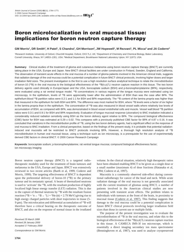
Sequential ion images of 24Mg and 10B from two regions of theventral tongue mucosa of a rat given BSH are shown in FigureThe distribution of 24Mg in the epithelium, lamina propria andmuscle was similar to that observed after BPA administration. Mwas high in the epithelium and muscle and low in the lamipropria. Assessment of the physiological status of the cryopserved tissue based on the estimated concentrations of K, Na,Ca (images not shown) was also consistent with the patterndistribution observed in the BPA-treated rats. However, the disbution pattern of 10B from BSH contrasts with that observed in thtongues of rats treated with BPA. There were significantly reducconcentrations of 10B in the epithelium and muscle relative to thelamina propria and blood. The estimated 10B concentrations in thevarious regions of the ventral tongue mucosa, after the administion of BSH and BPA are given in Table 2.
The values in Table 2 are reported as average 10B concentrationsfor the individual histological features and are not subcellulassignments. Although the CAMECA IMS-3f ion microscope operated to produce ion images with a nominal resolution ~0.5µm, the concentration gradient of 10B within the histologicalfeatures analysed was insufficient to resolve cellular boundarie
Blood 10B levels were measured using DCP-AES and not iomicroscopy. This is due to the fact that the blood componentsarteries and vessels are often lost during cryogenic sam
British Journal of Cancer (2000) 82(11), 1764–1771
1768 GM Morris et al
h
ae
l
toa
nee
dereris-
of
ofionlal
Figure 2 Ventral tongue mucosa of a rat administered BSH. Panels A, B and panels C, D are sequential ion images from different regions of analysis of afreeze-dried cryosection of tissue on indium. (A, C) Ion images showing the distribution of the isotope 24Mg. (B, D) Ion images showing the distribution of theisotope 10B. Images of 12C, 39K, 23Na and 40Ca are not shown. Dotted lines in panels A–D aid in visualization of the Ep, LP and M. For key to symbols refer toFigure 1
LP
50 µm
A 24Mg
M
LP
50 µm
C 24Mg
Ep
LP
50 µm
B 10B
BSH
M
50 µm
D 10B
BSH
Table 2 Measurement of 10B concentrations in ventral tongue mucosa using ion microscopy based direct imaging secondary ion massspectrometry (SIMS). Errors indicate ± s.e.m.
Boron Epithelium Lamina Muscle Blood Blood a
delivery propria vesselagent wall
µg g–1 µg g–1 µg g–1 µg g–1 µg g–1
BSH 6.1 ± 1.0 36.2 ± 4.2 6.6 ± 0.6 9.7 ± 1.3 36.6 ± 6.4b 5.8 ± 0.4
BPA 21.5 ± 0.5 31.1 ± 1.2 15.6 ± 1.7 19.2 ± 1.6 20.7 ± 0.7b21.6 ± 0.4
a Blood 10B concentration was measured using DCP-AES. bBasal cell layer 10B concentration.
preparation for ion microscopy. The 10B concentration in theepithelium of BSH-treated rats was a factor of ~3.5 lower than tfound in the epithelium of BPA-treated rats. 10B was distributeduniformly throughout the viable epithelium. Analysis of the baslayer, which is the location of the epithelial stem cells, indicatsimilar levels of 10B (P > 0.1, Student’s t-test) to that measured inthe entire viable epithelium (Table 2). The epithelium:blood 10Bpartition ratios were 0.2:1 and 1:1 for BSH and BPA respectiveIn other words, levels of 10B in the epithelium were a factor of 5lower than in the blood after BSH administration, but similar those in the blood after BPA administration. The laminpropria:blood 10B partition ratio was 1:1 after the administrationof BSH and is consistent with a relatively high density of bloovessels in this region. For BPA treated rats, the lamipropria:blood 10B partition ratio was 1.5:1. The reason for thhigher levels of 10B in the lamina propria, as compared with th
British Journal of Cancer (2000) 82(11), 1764–1771
at
ld
y.
da
blood, is not entirely clear. It may be related to the much broarange of 10B concentrations obtained in the lamina propria aftBPA (8–75µg g–1, 168 samples) as compared with BSH admintration. The concentration of 10B in muscle was a factor of ~1.3lower than that in the blood of rats given BPA, but were a factor~5 lower than the blood in rats given BSH. The quantity of 10B inblood vessel walls was relatively low (9.7 ± 1.3µg g–1) after BSHadministration but was considerably higher (19.2 ± 1.6µg g–1)after BPA administration (Table 2).
Radiation response
The mean 10B concentrations in the blood, measured at the timeirradiation, were used in the calculation of the physical radiatdose from the 10B(n,α)7Li reaction. This resulted in a potentiavariation in the total physical dose (which included the therm
© 2000 Cancer Research Campaign
Boron microlocalization in oral mucosa 1769
)sr
t ebea
er
i
n o
e
ohcti
lEe
ts ofs anth of thetheialtionwithith
ingtion
asabledels.ays,lcersand9 to
ionss
er the dere ofdels
t isn
s not
uesthishe
foreood.gly
100
90
80
70
60
50
40
30
20
10
0
0 2 4 6 8 10 12
Time after irradiation (days)
Tong
ue fi
elds
exh
ibiti
ng u
lcer
atio
n (%
)100
90
80
70
60
50
40
30
20
10
0
0 5 10 15 20 25Dose (Gy)
Tong
ue fi
elds
exh
ibiti
ng u
lcer
atio
n (%
)
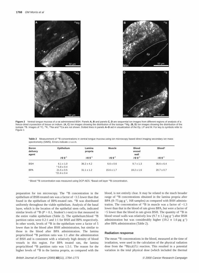
Figure 3 Time-related changes in the incidence of tongue ulceration afterthermal neutron irradiation in the presence of BSH. The doses shown arephysical radiation doses. ■ = 10.9 Gy; l = 13.1 Gy; ▲ = 13.3 Gy;▼ = 18.6 Gy; = 23.1 Gy; ◆ = 23.9 Gy
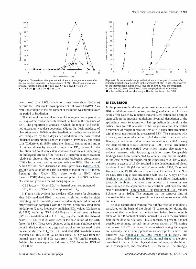
Figure 4 Dose-related change in the incidence of tongue ulceration afterirradiation with thermal neutrons in the presence of BSH. Dose–effect curvesfor the thermal beam-alone and X-rays are shown by way of comparison(Coderre et al, 1999). The doses shown are physical radiation doses■ = thermal beam-alone; ◆ = X-rays; ● = thermal beam plus BSH
beam dose) of ≤ 7.6%. Irradiation times were short (3–5 minbecause the BMR reactor was operated at full power (3 MW). Aresult, fluctuation in the 10B content of the blood was minimal ovethe period of irradiation.
Ulceration of the ventral surface of the tongue was apparen7–8 days after irradiation with thermal neutrons in the presencBSH. The proportion of animals in which the tongue field exhiited ulceration was dose dependent (Figure 3). Peak incidenculceration was at 8–9 days after irradiation. Healing was rapid was completed by 8–11 days after irradiation. The dose-relaincidence of ulceration is shown in Figure 4. Previously publishdata (Coderre et al, 1999) using the identical end point and stof rat are shown by way of comparison. ED50 values for theulceration end point were calculated from these curves. In definthe biological effects of the 10B(n,α)7Li neutron capture reactionrelative to photons, the term compound biological effectivene(CBE) factor was used as an alternative to RBE. The ratiobehind this has been discussed in detail previously (Morris et1994). Calculation of the CBE is similar to that of the RBE factEquating the X-ray ED50 dose with a BNC dose(beam + BSH) that gives the same end point of a 50% incidof ulceration produces the following equation:
CBE factor = [(X-ray ED50) – (thermal beam component ofED50 × RBE)]/10B(n,α)7Li component of ED50.
In Figure 4 it is evident that the dose–effect curve for ulceratiafter BSH-mediated BNC irradiation, is shifted to the far rigindicating that this modality has a considerably reduced biologieffectiveness as compared with the thermal beam-only irradiamodality or X-rays. Previously published ED50 values (Coderre etal, 1999) for X-ray (13.4 ± 0.2 Gy) and thermal neutron beam(BMRR) irradiation (4.2 ± 0.1 Gy), together with the thermabeam RBE (3.2 ± 0.1), were used in the calculation of the CBfactor for BSH. These were obtained using the same biological point in the identical strain, age and sex of rat to that used inpresent study. The ED50 for BSH mediated BNC irradiation wascalculated at 18.6 ± 1.0 Gy. Of this value, 2.8 Gy was from thethermal beam and 15.8 Gy was from the 10B(n,α)7Li reaction.Solving the above equation indicates a CBE factor for BSH0.29 ± 0.02.
© 2000 Cancer Research Campaign
a
by of- ofndtedd
ain
ng
ssalal,r.
nc
n,talon
ndthe
of
DISCUSSIONIn the present study, the end point used to evaluate the effecBNC irradiation on oral mucosa, was tongue ulceration. This iacute effect caused by radiation induced sterilization and deastem cells in the mucosal epithelium. Eventual denudation ofepithelium leads to ulceration. The epithelium is therefore critical area for 10B analysis in the tongue mucosa. The initoccurrence of tongue ulceration was at 7–8 days after irradiawith thermal neutrons in the presence of BSH. This compares a latency to tongue ulceration of 6–8 days after irradiation wX-rays, thermal beam – alone or in combination with BPA – usthe identical strain of rat (Coderre et al, 1999). For all irradiamodalities, the time period over which tongue ulceration wevident increased with increasing radiation dose. Comparfindings have been documented by others using murine moIn the case of ventral tongue, single exposures of 29 kV X-rat doses in excess of 15 Gy, resulted in the development of uby days 9 and 10 (Moses and Kummermehr, 1986; Dorr Kummermehr, 1990). Mucositis was evident in mouse lips at 10 days after single dose irradiation with 250 kV X-rays or 60Co(Parkins et al, 1983; Ang et al, 1984). In the clinic, fractionatprotocols involving irradiation over periods of 2 weeks or lehave resulted in the appearance of mucositis at 9–10 days aftstart of irradiation (Dutreix et al, 1971; Parkins et al, 1983; vanSchueren et al, 1983). This suggests that the turnover timmucosal epithelium is comparable in the various rodent moand man.
The dose contribution from the 10B(n,α)7Li reaction is routinelycalculated on the basis of the blood 10B concentration during thecourse of thermal/epithermal neutron irradiation. No accountaken of the 10B content of critical normal tissues in the irradiatiofield in the dose calculations. This is because, at present, it ipossible to estimate normal tissue concentrations of 10B duringthe course of BNC irradiation. Non-invasive imaging techniqare currently under development in an attempt to achieve objective (e.g. Kabalka et al, 1997; Imahori et al, 1998). Tphysical dose delivered to a specific normal tissue is theredescribed in terms of the physical dose delivered to the blAs a consequence, the calculated CBE factor will be stron
British Journal of Cancer (2000) 82(11), 1764–1771
1770 GM Morris et al
t
hti
re
efrs
r
ti
k
f
ehth.
o
yh
a-
5n o
SDas
fen
~2
f a
acellthe
eec-and
l
ll)
orme
tionile
s inn ofnted
Moretem
for
ensi-cedoffiumentd on
ch, ben.
imal
CP-
tudyrma-
t-ich
influenced by variations in the amount of 10B present in the targenormal tissue relative to the blood (i.e. the 10B partition ratio). Inthe present study, based solely on the 10B concentration of theblood, the CBE factor for BSH was estimated at ~0.3. Tcompares with a CBE estimate for BPA of ~4.9, using the idenstrain, sex and age of rat (Coderre et al, 1999).
RBE and CBE factors have been reported for skin, using mdesquamation (epithelial denudation) as the end point. For thebeams, RBE values in the range 2.7–3.9 have been docum(Yamamoto, 1961; Archambeau, 1989; Morris et al, 1994). A Cfactor of ~2.5 has been estimated for both hamster and humanusing BPA as the boron delivery agent (Hiratsuka et al, 19Fukuda et al, 1994). A higher CBE estimate for BPA of ~3.7 wreported using rat skin (Morris et al, 1994). The strain, age andwere identical to that employed in the present study. This suggthat the oral mucosa (CBE ~4.9) is more sensitive to BPA-mated BNC irradiation than the skin. With BSH the CBE factor tongue mucosa was much lower at ~0.3. This value is compawith analogous CBE estimates (~0.4–0.5) for rat and dog (Morris et al, 1994; Gavin et al, 1997).
Ion microscopy imaging studies of 10B in body tissues (Smithet al, 1996, 1997), has enabled the biodistribution of this elemto be analysed with considerably greater precision than was pously possible. Although other analytical techniques have butilized for boron biodistribution studies, such as quantitaneutron capture autoradiography (Gabel et al, 1987a) or the elec-tron microprobe (Feldman and Mayer, 1986), they lack eitherspatial resolution or the sensitivity necessary to determinemicrodistribution of this trace analyte. Ion microscopy providing new insights into the relationship between 10B biodistri-bution and the biological effectiveness of the high LET particproduced by the 10B(n,α)
7Li neutron capture reaction in vivo. Bulanalysis (DCP-AES) of whole tissue samples from the ventongue mucosa indicated appreciable uptake of 10B (~21µg g–1)after the administration of BSH. A similar finding was reported BPA administered at a comparable 10B concentration to BSH(Coderre et al, 1999), where the level of 10B in the ventral tonguewas estimated at ~23µg g–1. While this data indicated that thoverall concentrations of 10B in the tongue were similar for botdelivery agents, it provided no information with regard to biodistribution profile of the 10B in different regions of the tissueIon microscopy analysis revealed that the level of 10B in themucosal epithelium was very low after BSH administratiIndeed, the 10B content of the mucosal epithelium was ~3.5 timhigher in rats treated with BPA. In the case of BSH, the majoritthe 10B was located in the lamina propria. This differs from tfindings for BPA where the 10B content in the mucosal epitheliumwas closer to that in the lamina propria, the differentials beingfor BSH and 1:1.5 for BPA. Mucosal epithelium:blood 10B parti-tion ratios were ~0.2 (6.2:36.6) and ~1 (21.5:20.7) for BSH BPA respectively. In other words, 10B accumulation in the epithelium, relative to the blood, was 5 times higher with BPA than wBSH. The physical dose (the blood dose) that resulted in a incidence (ED50) of ventral tongue ulceration was ~3.0 Gy usiBPA (Coderre et al, 1999) and ~18.6 Gy using BSH. Whentotal radiation dose that was delivered directly to the mucepithelium was estimated from the ion microscopy-derived 10Bconcentrations, it was ~3.2 Gy and ~4.9 Gy for BPA and Brespectively. The CBE factor values calculated from these E50swere ~4.6 for BPA and ~2.2 for BSH. This indicates that a mfactor contributing to the difference in the CBE values (0.3 ver
British Journal of Cancer (2000) 82(11), 1764–1771
iscal
oistmalnted
BE skin91;as sexestsdi-orablekin
entevi-eenve
thetheis
les
tral
or
e
n.es ofe
1:6
nd
ith0%gthesal
H
jorus
4.9), estimated using blood 10B concentrations, was low uptake oBSH by the mucosal epithelium. However, the fact that evtaking into account the 10B levels in the epithelium in radiationdose calculations, the CBE factor for BPA was a factor of higher than for BSH suggests that the microdistribution of 10Bfrom these two compounds differs at the intracellular level.
The majority of the energy released after the capture othermal neutron by a 10B atom (10B + n → 11B → 7Li + α + 2.79MeV) is carried by the α and 7Li charged particles. Thus, as consequence, the pattern of energy deposition in critical organelles, such as the nucleus is strongly influenced by microdistribution of the 10B atoms. Monte Carlo simulations havdemonstrated that 10B located in the cell nucleus is more effectivthan 10B distributed in the cytoplasm, which is in turn more effetive than boron attached to the cell membrane (Kobayashi Kanda, 1982; Gabel et al, 1987b; Verrijik et al, 1994). Thus, themicrolocalization of 10B has a critical effect on the biologicaeffectiveness of the 10B(n,α)7Li reaction. Monte Carlo simulationsfor a cubic cell with a volume of 1150µm3 and a spherical nucleusof volume 230µm3 (representative of a basal epithelial ceindicate a sevenfold reduction in efficacy when the 10B is locatedexclusively extracellularly, and a fivefold increase in efficacy f10B is located within the nucleus, as compared with the saamount of 10B distributed homogeneously (Gabel et al, 1987b). Atthe present time, data is not available on the precise microlocaof 10B at the intracellular level for the mucosal epithelium. Whthe resolution (~0.5µm) of the ion microscope is sufficiently highto evaluate the subcellular localization of 10B, it is not possible atpresent to delineate the boundaries of individual epithelial celltissue sections. For this reason, the subcellular microdistributio10B was not evaluated in the current study. The data presedetails the average concentration of 10B in the various histologicalzones of the tongue mucosa, such as the mucosal epithelium. specific evaluation of the basal layer (the location of the scells) revealed no appreciable difference in 10B content relative tothe higher suprabasal layers in the epithelium. This was trueboth BSH and BPA.
CONCLUSIONS
The ventral tongue mucosa of the rat was considerably less stive to BSH as compared with BPA-mediated BNCT. This redusensitivity was directly related to the microdistribution profile 10B. At similar doses of 10B from BSH or BPA, the concentration othis element was a factor of ~3.5 lower in the mucosal epithelafter BSH as compared with BPA administration. At the prestime, due to practical considerations, CBE factors are estimatethe basis of the radiation dose from the 10B(n,α)7Li neutron capturereaction delivered to the blood. While this is a valid approaexperimentally derived CBE factors from animal models mustused in BNCT clinical dosimetry protocols with extreme cautioThe microdistribution profiles of 10B, for a given boron deliveryagent in a given tissue, must be comparable in the relevant anmodel and man for reliable extrapolation of the CBE factor.
Bulk analytical techniques, such as prompt gamma and DAES are routinely used for the intercomparison of 10B biodistribu-tion in human and animal tissues. The findings of the present sindicate that such an approach is inadequate, because no infotion is provided on 10B accumulation in critical target cell comparments. A major advance is offered by ion microscopy, whfacilitates the simultaneous analysis of 10B microdistribution and
© 2000 Cancer Research Campaign

Boron microlocalization in oral mucosa 1771
n
a
nr
o
gre
t
in,
e
,mn
fg
.
8
shie
hild
nd
ur
0)
ll
e
al
L,
concentration at high resolution in individual tissue compartmesuch as the mucosal epithelium.
ACKNOWLEDGEMENTS
This work was supported by the Cancer Research CampaAdditional support was provided by the Office of Biologicand Environmental Research, US Department of Energy, uncontract number DE-AC02-98CH10886 and DE-FG02-ER6113
REFERENCES
Ang KK, Landuyt W, Rijners A and Schueren and van der E (1984) Differences irepopulation kinetics in mouse skin during split course multiple fractions peday (MDF) or daily fractionated irradiations. Int J Radiat Oncol Biol Phys10:95–99
Archambeau JO (1989) A model to evaluate dose recovery from different radiatiIn: Clinical Aspects of Neutron Capture Therapy, pp. 9–20. Plenum Press: NewYork
Ausserer WA, Ling YC, Chandra S and Morrison GH (1989) Quantitative imaginof boron, calcium, magnesium, potassium and sodium distributions in cultucells with ion microscopy. Anal Chem61: 2690–2695
Barth RF, Adams DM, Soloway AH, Mechetner EB, Alam F and AnisuzzamanAKM (1991) Determination of boron in tissues and cells using direct currenplasma atomic emission spectroscopy. Anal Chem63: 890–893
Barth RF, Soloway AH, Goodman JH, Gahbauer RA, Gupta N, Blue TE, Yang Wand Tjarks W (1999) Boron neutron capture therapy of brain tumours: anemerging therapeutic modality. Neurosurgery44: 433–451
Benninghoven A, Rudenauer FG and Werner HW (1987) Instrumentation In: ElvPJ, Eincfordner JD and Kolthoff IM (Eds) Secondary ion mass spectrometrybasic concepts, instrumental aspects, applications and trends,pp. 594–606. John Wiley & Sons: New York
Coderre JA and Morris GM (1999) The radiation biology of boron neutron capturtherapy. Radiat Res151: 1–18
Coderre JA, Button TM, Micca PL, Fisher CD, Nawrocky MM and Liu HB (1994)Neutron capture therapy of the 9L rat gliosarcoma using the p-boronophenylalanine–fructose complex. Int J Radiat Oncol Biol Phys30:643–652
Coderre JA, Bergland R, Capala J, Chadha M, Chanana AD, Elowitz E, Joel DDLiu HB and Slatkin DN (1997) Boron neutron capture therapy for glioblastomultiform using p-boronophenylalanine and epithermal neutrons: trial desigand early clinical results. J Neuro-oncol33: 141–152
Coderre JA, Morris GM, Micca PL, Ma RM and Gordon CR (1999) The effects oboron neutron capture irradiation on oral mucosa: evaluation using a rat tonmodel. Radiat Res152: 113–118
Dorr W and Kummermehr J (1990) Accelerated repopulation of mouse tongueepithelium during fractionated irradiations or following single doses. RadiotherOncol17: 249–259
Dutreix J, Tubiana M, Wambersie A and Malaise E (1971) The influence of cellproliferation in tumours and normal tissues during fractionated radiotherapyEur J Cancer7: 205–213
Fairchild RG, Gabel D, Laster BH, Greenberg D, Kiszenick W and Micca PL (19Microanalytical techniques for boron analysis using the 10B(n,α)7Li reaction.Med Phys13: 50–56
Feldman LC and Mayer JW (1986) Radiative transitions and the electronmicroprobe. In: Fundamentals of Surface and Thin Film Analysis,pp. 243–247. North Holland: New York
© 2000 Cancer Research Campaign
ts,
ign.lder8.
n.
d
g
a
ue
6)
Fukuda H, Hiratsuka J, Honda C, Kobayashi T, Yoshino K, Karashima H, TakahaJ, Abe Y, Kanda K, Ichihashi M and Mishima Y (1994) Boron neutron capturtherapy of malignant melanoma using 10B-paraboronophenylalanine withspecial reference to evaluation of radiation dose and damage to the skin. RadiatRes138: 435–442
Gabel D, Holstein H, Laarsson B, Gille L, Ericson G, Sacker D, Som P and FaircRG (1987a) Quantitative neutron capture radiography for studying thebiodistribution of tumour-seeking boron-containing compounds. Cancer Res47: 5451–5454
Gabel D, Foster S and Fairchild RG (1987b) The Monte Carlo simulation of thebiological effect of the 10B(n,α)
7Li reaction in cells and tissue and itsimplications for boron neutron capture therapy. Radiat Res111: 14–25
Gavin PR, Kraft SL, Huiskamp R and Coderre JA (1997) A review: CNS effects anormal tissue tolerance in dogs. J Neuro-Oncol33: 71–80
Hatanaka H and Nakagawa Y (1994) Clinical results of long-surviving brain tumopatients who underwent boron neutron capture therapy. Int J Radiat Oncol BiolPhys28: 61–66
Hiratsuka J, Fukuda H, Kobayashi T, Karashima H, Yoshino K, Imajo Y andMishima Y (1991) The relative biological effectiveness of 10B neutron capturetherapy for early skin reactions in the hamster. Radiat Res128: 186–191
Imahori Y, Ucda Y, Ohmori T, Kusuki T, Ono K, Fujii R and Ido T (1998) Fluorine-18 labelled fluoroboronophenylalanine PET in patients with glioma. J NuclMed39: 325–333
Joel DD, Fairchild RG, Laissue JA, Saraf SK, Kalef-Ezra JA and Slatkin DN (199Boron neutron capture therapy for intracerebral rat gliosarcomas. Proc NatlAcad Sci USA87: 9808–9812
Kabalka GW, Smith GT, Dyke JP, Reid WS, Longford CPD, Roberts TG, ReddyNK, Buonocore E and Hubner KFC (1997) Evaluation of fluorine-18 BPAfructose for boron neutron capture treatment planning. J Nucl Med38:1762–1767
Kobayashi T and Kanda K (1982) Analytical calculation of boron-10 dosage in cenucleus for neutron capture therapy. Radiat Res91: 77–94
Morris GM, Coderre JA, Hopewell JW, Micca PL and Rezvani M (1994) Responsof rat skin to boron neutron capture therapy with p-boronophenylalanine orborocaptate sodium. Radiother Oncol39: 253–259
Morris GM, Coderre JA, Smith DR and Hopewell JW (1999) A rat model of oralmucosal response to boron neutron capture irradiation. In: Proceedings of theEighth International Symposium on Neutron Capture therapy for Cancer.Plenum Press: New York (in press).
Moses R and Kummermehr J (1986) Radiation response of the mouse tongueepithelium. Brit J Cancer53: Suppl VII, 12–15
Parkins C, Fowler JF and Shen Y (1983) A murine model of lip epidermal/mucosreactions to x-irradiation. Radiother Oncol1: 159–165
Smith DR, Chandra S, Coderre JA and Morrison GH (1996) Ion microscopyimaging of 10B from p-boronophenylalanine in a brain tumour model for boronneutron capture therapy. Cancer Res56: 4302–4306
Smith DR, Chandra S, Coderre JA, Barth RF, Yang W, Liu L, Wilson JL, Micca PNawrocky MM, Rotaru JH and Morrison GH (1997) Quantitative ionmicroscopy imaging of boron-10 in rat brain tumour models for BNCT. In:Advances in Neutron Capture Therapy. Vol. II, Chemistry and Biology, LarsonB, Crawford J and Weinreich R pp. (eds), 308–314. Elsevier Science:Amsterdam.
van der Schueren E, van der Bogaert W and Ang KK (1983) Radiotherapy withmultiple fractions per day. In: The Biological Basis of Radiotherapy, Steel GG,Adams G and Peckam M (eds), pp. 195–210. Elsevier Science: Amsterdam
Verrijk R, Huiskamp R, Begg AC, Wheeler FJ and Watkins PRD (1994) A comprehensive PC based computer model for microdosimetry of BNCT. Int J Radiat Biol65: 241–253
Yamamoto YL (1961) The biological effectiveness of thermal neutrons and of theheavy particles from the 10B(n,α)7Li reaction for the rabbits ear and itsutilisation for neutron capture therapy. Yokohoma Med Bull12: 4–22
British Journal of Cancer (2000) 82(11), 1764–1771