Metastatic malignant struma ovarii diagnosed during pregnancy
Synthetic Affibody Molecules: A Novel Class of Affinity Ligands
for Molecular Imaging of HER2-Expressing Malignant Tumors
Anna Orlova,1,2
Vladimir Tolmachev,1,2
Rikard Pehrson,1Malin Lindborg,
1
Thuy Tran,2Mattias Sandstrom,
3Fredrik Y. Nilsson,
1,2Anders Wennborg,
1
Lars Abrahmsen,1and Joachim Feldwisch
1,2
1Affibody AB, Bromma, Sweden; 2Department of Oncology, Radiology and Clinical Immunology, Rudbeck Laboratory, Uppsala University;and 3Department of Hospital Physics, Uppsala University Hospital, Uppsala, Sweden
Abstract
The Affibody molecule ZHER2:342-pep2, site-specifically andhomogeneously conjugated with a 1,4,7,10-tetra-azacylodo-decane-N,N ¶,N 00,N�-tetraacetic acid (DOTA) chelator, wasproduced in a single chemical process by peptide synthesis.DOTA-ZHER2:342-pep2 folds spontaneously and binds HER2 with65 pmol/L affinity. Efficient radiolabeling with >95% incorpo-ration of 111In was achieved within 30 min at low (roomtemperature) and high temperatures (up to 90�C). Tumoruptake of 111In-DOTA-ZHER2:342-pep2 was specific for HER2-positive xenografts. A high tumor uptake of 23% injectedactivity per gram tissue, a tumor-to-blood ratio of >7.5, andhigh-contrast gamma camera images were obtained already1 h after injection. Pretreatment with Herceptin did notinterfere with tumor targeting, whereas degradation of HER2using the heat shock protein 90 inhibitor 17-allylamino-geldanamycin before administration of 111In-DOTA-ZHER2:342-pep2obliterated the tumor image. The present results showthat radiolabeled synthetic DOTA-ZHER2:342-pep2 has the poten-tial to become a clinically useful radiopharmaceutical forin vivo molecular imaging of HER2-expressing carcinomas.[Cancer Res 2007;67(5):2178–86]
Introduction
The concept of personalized medicine involves both novelselective targeting therapeutics adapted for treatment of defineddisease stages and means to diagnose and stratify the patientpopulation that may respond to these treatments (1). Currentlyused noninvasive imaging techniques, such as anatomic imaging bycomputer tomography and metabolic imaging using 18F-fluoro-deoxyglucose (18F-FDG), do not provide molecular information ononcological target molecules. In contrast, tumor marker–targetedmolecular imaging can biochemically characterize the imagedstructures in the body, thereby adding new qualitative informationto these images not available today (e.g., for assessing theaggressiveness of cancer and for monitoring of targeted therapy;ref. 2). A targeting agent suitable for molecular imaging should beable to specifically target relevant pathologic structures in the body,while avoiding normal tissue (3, 4). In patients, it should quickly
find its target while unbound molecules should be rapidly excreted,thus facilitating high-contrast imaging and reducing the timerequired for the examination. For clinical development, site-specific, homogeneous, reproducible, and easy radiolabeling isdesirable. However, the most commonly used class of targetingagents (i.e., antibodies and various antibody derived fragments)are usually modified at multiple and randomly distributed sitesby using modification chemistries based on amine, thiol, ortyrosine-reactive reagents (5). Thus, the resulting targetingmolecules are heterogeneous preparations with various degreesof modification (6, 7).The clinical use of antibodies for molecular imaging is limited
due to their long biodistribution times, slow tumor penetration,and slow blood clearance. Improved imaging agents have beenobtained by using smaller antibody fragments that have fasterbiodistribution and more rapid blood and whole body clearance(8). However, even the mass of the smallest fragments (27-54 kDa)may not be small enough to allow for efficient extravasation, goodtissue penetration, and fast blood clearance (9). Pretargeting ofbispecific antibodies followed by administration of radiolabeledsmall peptides have shown high tumor signal intensities, improvedtumor-to-blood (T/B) ratios, and contrasts (10). However, pre-targeting is a multistep process whose practical clinical use maybe hampered by the prolonged treatment regimes of the 24 to48 h required before injection of the radiolabeled peptide. Shortpeptides, on the other hand, are small and have very rapid kinetics,but there is a limited repertoire of natural peptides to choose from(11, 12), and peptides derived from phage display libraries seldomhave the high affinity to be considered a general class of targetingmolecules (13).Affibody molecules are small non-immunoglobulin affinity
ligands based on a 58-amino-acid Z-domain scaffold, derived fromone of the IgG-binding domains of staphylococcal protein A (14).Randomization of 13 amino acid positions in the binding surfaceof this domain scaffold has been used for construction ofcombinatorial phagemid libraries, from which Affibody moleculesbinding desired target molecules can be selected by phage display(15, 16). Thus, the Affibody molecule with binding specificity forthe target human epidermal growth factor receptor 2 (HER2, alsoknown as Neu and ErbB2) described here differs from otherAffibody molecules (e.g., a Taq polymerase–specific Affibodymolecule; ref. 16), only in the 13 amino acids that form the targetbinding site.HER2 is overexpressed in a number of carcinomas and is
associated with shorter time to disease progression and decreasedoverall survival in breast cancer patients (17–19). Noninvasivedetection of HER2 expression by novel imaging radiopharmaceu-ticals could become an important complement to immunohisto-chemistry or fluorescence in situ hybridization, allowing the
Note: The phrase ‘‘Affibody molecule’’ is used in this publication instead of‘‘AffibodyR molecule.’’ Affibody is a trademark owned by Affibody AB. Affibody is atrademark registered in Sweden, Europe, and the United States and under trademarkapplication in Japan.Requests for reprints: Joachim Feldwisch, Affibody AB, Voltavagen 13, P.O. Box
20137, SE-16102 Bromma, Sweden. Phone: 46-859883852; Fax: 46-859883801; E-mail:[email protected].
I2007 American Association for Cancer Research.doi:10.1158/0008-5472.CAN-06-2887
Cancer Res 2007; 67: (5). March 1, 2007 2178 www.aacrjournals.org
Research Article
Research. on August 16, 2015. © 2007 American Association for Cancercancerres.aacrjournals.org Downloaded from
identification of HER2-positive metastases not amenable tobiopsy. One potential clinical application is the identification ofpatients where metastases from HER2-negative primary breasttumors are HER2-positive and thus may respond to trastuzumabtreatment (20). Furthermore, imaging of HER2 expression couldprovide a direct readout of the efficacy of pharmaceuticals aimedto interfere with HER2 overexpression, such as 17-allylamino-geldanamycin (17-AAG), which belongs to a new class of heatshock protein 90 (HSP90) inhibitors (7, 21). HSP90 is a molecularchaperone that is overexpressed in a large number of cancers,including breast cancer. This protein is crucial for folding of alarge number of client proteins involved in growth control, cellsurvival, and development processes, like receptors, kinases, andtranscription factors. Thus, inhibition of HSP90 leads to increasedproteasome-mediated degradation of client proteins, which inturn leads to inhibition of cell proliferation and finally toapoptosis (22).Our earlier work has indicated a large potential of Affibody
molecules for imaging of HER2-overexpressing tumors. The firstgeneration of HER2-specific Affibody molecules (His6-ZHER2:4) bindHER2 with a KD of 50 nmol/L (23). Dimerization of this Affibodymolecule (ZHER2:4)2 resulted in improved target binding affinity(KD f3 nmol/L), and radioiodination of recombinantly producedHis6-(ZHER2:4)2 allowed selective targeting and imaging of HER2-expressing xenografts in vivo (24). The second-generation HER2-specific Affibody molecule (His6-ZHER2:342) was obtained by affinitymaturation (25). His6-ZHER2:342 binds HER2 with a KD of 22 pmol/L,and radioiodination of the monomeric form resulted in good tumortargeting and imaging. Further improvement was obtained using111In-labeled benzyl-DTPA-His6-ZHER2:342 (26). However, all Affibodymolecules described thus far were produced by recombinantexpression in Escherichia coli , and radiolabeling was done usinglabeling chemistries relying on amine or thiol-reactive reagents.Thus, similar to antibodies or antibody fragments, these radio-labeled Affibody molecules are heterogeneous preparations withvarious degrees of modification.Here, we describe a synthetic Affibody molecule, which over-
comes the limitations of other HER2 imaging agents currently underdevelopment. DOTA-ZHER2:342-pep2 is made by peptide synthesis ina single chemical process. This Affibody molecule is site-specificallymodified at the NH2 terminus with the chelator 1,4,7,10-tetra-azacylododecane-N ,N ¶,N 00,N�-tetraacetic acid (DOTA), which can beefficiently labeled with the radiometal indium-111 (111In). Theproperties of this well-defined and homogenous radiopharmaceu-tical were evaluated in vivo in mice carrying xenografts derived fromhuman SKOV-3 ovarian cancer cells.
Materials and Methods
General. Reagents were purchased from the following commercialsources: 111In chloride, Tyco Healthcare (Solna, Sweden); silica gel–
impregnated glass fiber sheets for instant TLC (ITLC SG), Pall Life
Sciences, Inc. (Ann Arbor, MI); 50 mg/mL Ketalar, Pfizer (New York, NY);20 mg/mL Rompun, Bayer (Leverkusen, Germany); 5,000 IE/mL heparin,
Leo Pharma (Copenhagen, Denmark); Herceptin (trastuzumab), Roche
(Basel, Switzerland); and 17-AAG (17-allylamino-17-demethoxygeldanamy-
cin), Invivogen (San Diego, CA). All cell lines were obtained from theAmerican Type Culture Collection (LGC Promochem, Boras, Sweden) or the
European Collection of Animal Cell Cultures (Salisbury, United Kingdom).
Data on cellular uptake, biodistribution, and gamma camera images were
analyzed by unpaired, two-tailed t test using GraphPad Prism (version 4.00for Windows GraphPad Software, San Diego, CA) to determine any
significant differences (P < 0.05).
Animal model. The animal study was approved by the local EthicsCommittee for Animal Research. Female outbreed BALB/c nu/nu mice were
used in all experiments. Xenografts of SKOV-3 (ovary ascites adenocarci-
noma), MDA-MB-231, MCF7, BT474 (mammary gland adenocarcinomas),
and Ramos (B lymphocyte, Burkitt’s lymphoma) were implanted in the lefthind leg. The selection of cell lines was based on (a) the possibility and ease
to generate xenografts and (b) the HER2 expression levels. BT474 and
SKOV-3 were chosen as cell lines with high HER2 expression level (+4 to +5)
and MDA-MB-231 and MCF7 as cell lines with low expression level (+1;ref. 27). The Ramos cell line was chosen as a negative control. The
xenografts were allowed to develop up to 100 mm3 for biodistribution and
500 mm3 for imaging studies.
Peptide synthesis. The 58-amino-acid-long peptides with a DOTAchelator coupled to the NH2 terminus (DOTA-ZHER2:342-pep2 and DOTA-
Ztaq4:5) were made by standard Fmoc peptide synthesis by Innovagen
(Lund, Sweden) or Bachem (Bubendorf, Switzerland). DOTA-mono-NHS-tris(t-bu)ester (Macrocyclics, Dallas, TX) was used for NH2-terminal
modification of the peptide. The DOTA-peptides were obtained as
lyophilized white powder.
Surface plasmon resonance analysis. Binding of Affibody molecules to
HER2 was analyzed using a Biacore 2000 instrument. The recombinant
human ErbB2/Fc chimeric protein, consisting of the extracellular domain of
ErbB2 (HER2, Met1-Thr652) fused to the Fc region of human IgG1 (Pro100-
Lys330) with a COOH-terminal His6-tag (R&D Systems, Minneapolis, MN),
was immobilized (1,540 resonance units) onto a surface of a CM5 sensor
chip using amine-coupling chemistry. The extracellular domain of HER2
(HER2-ECD; ref. 28) was immobilized (1,090 resonance units) onto a second
surface of the chip to allow comparison with previously published results
(25). One surface on the chip was activated and deactivated for use as
reference cell. Affibody molecules diluted in HBS-EP buffer [10 mmol/L
HEPES, 150 mmol/L NaCl, 3 mmol/L EDTA, 0.005% surfactant P-20
(pH 7.4)] were used as analytes. Three analyte concentrations (6, 20, and
60 nmol/L) were injected in duplicates over the chip using a constant flow
rate of 50 AL/min. The total injection time was 3.5 min (association)
followed by a wash for 10 min (dissociation). The surface was regenerated
with one injection of 25 mmol/L HCl. The response measured in the
reference cell and the response from a HBS-EP buffer injection were sub-
tracted from the response measured in the cell with immobilized ErbB2/Fc
or HER2-ECD, respectively. For analysis of the binding kinetics, a 2-fold
dilution series of the analytes ranging from 6 to 0.19 nmol/L ( final
concentrations) were injected in duplicates over the chip using a constant
flow rate of 50 AL/min. The association phase was 5 min followed by a long
dissociation phase (60 min) to account for the slow off rate of the Affibody
molecules. The dissociation constant KD, the association rate constant
ka, and the dissociation rate constant kd were calculated using the 1:1
Langmuir binding model with mass transfer correction of the BIAevaluation
4.1 software (Biacore AB, Uppsala, Sweden). For the heating experiment,
DOTA-ZHER2:342-pep2 was incubated for 5 min at 90jC in a heating block,
allowed to cool to room temperature, and then injected over the chip.
Radiolabeling. DOTA-ZHER2:342-pep2 [25 AL, 2 mg/mL in 0.2 mol/Lammonium acetate buffer (pH 5.25)] was mixed with a predetermined
amount of 111In chloride. The mixture was incubated for 30 min or 1 h at
different temperatures (room temperature, 37jC, 50jC, and 90jC), and the
radiochemical purity was evaluated using ITLC eluted with 0.2 mol/L citricacid (pH 2). Radiolabeled Affibody molecules remained at the origin,
whereas free indium migrated with the solvent front. For biological
experiments, 111In-DOTA-ZHER2:342-pep2 was diluted with PBS. Labeling with177Lu was done as described above at 60jC for 30 min, yielding a peptidedenoted 177Lu-DOTA-ZHER2:342-pep2. Indirect radioiodination of the recom-
binant Affibody molecule His6-ZHER2:342 was done using the linker molecule
N-succinimidyl-p-(trimethylstannyl)benzoate, which is first labeled with 125I
followed by coupling of the resulting N-succinimidyl-p-iodobenzoate toamine groups of the protein (25, 29).
In vitro cell binding assay. Cultured SKOV-3 cells were incubated
for 1 h at 37jC with 111In-DOTA-ZHER2:342-pep2 using a 1:1 molar ratio of111In-DOTA-ZHER2:342-pep2 to HER2 receptor (1.2 � 106 receptors per cell;
ref. 30). For blocking experiments, a 100-fold excess of unlabeled Affibody
Synthetic Affibody Molecules for Molecular Imaging
www.aacrjournals.org 2179 Cancer Res 2007; 67: (5). March 1, 2007
Research. on August 16, 2015. © 2007 American Association for Cancercancerres.aacrjournals.org Downloaded from
molecule was added 5 min before the addition of 111In-DOTA-ZHER2:342-pep2.All assays were done in triplicates. After incubation, the medium was
collected, and the cells were washed six times with cold serum-free medium
followed by treatment with 0.5 mL trypsin-EDTA solution (0.25% trypsin,
0.02% EDTA in buffer; Flow, Irvine, United Kingdom) for 10 min at 37jC.When cells were detached, 0.5 mL complete medium was added to each
dish, and the cells were resuspended. The radioactivity associated with the
cells and culture media was measured in a gamma counter.
To study cellular retention of radioactivity after interrupted incubation,cultured SKOV-3 cells were incubated for 2 h with 111In-DOTA-ZHER2:342-pep2or 125I-labeled His6-ZHER2:342 as described above. The dishes were then
washed six times with cold serum-free culture medium; fresh complete
medium was added; and the cells were incubated at 37jC. At predeterminedtime points, incubation medium was collected from three culture dishes,
and cells were detached from culture dishes by trypsin treatment, as
described above. The radioactivity associated with the cells, and the culturemedium was measured. The fraction of the cell-associated radioactivity was
analyzed as a function of time.
Biodistribution in tumor-bearing mice. The mice with xenografts
were randomized into groups of four. Animals of the blocking group weres.c. injected with 375 to 500 Ag of unlabeled His6-ZHER2:342 1 h before
injection of the radiolabeled Affibody molecule. All mice were injected s.c.
with 100 AL (1 Ag, 100 kBq) of 111In-DOTA-ZHER2:342-pep2. After injection of a
lethal dose of Ketalar-Rompun solution, mice were sacrificed byexsanguinations via heart puncture at 1, 4, 12, 24, and 72 h after injection.
Animals from the blocking group were sacrificed 4 h after injection. The
organs were dissected and weighted, and their radioactivity content wasmeasured in a gamma counter. Radioactivity uptake was calculated as
percentage of injected activity per gram tissue (%IA/g).
Pretreatment of SKOV-3 xenografts with Herceptin was done by s.c.
injection of 1 mg trastuzumab in PBS 2 days before injection of 177Lu-DOTA-ZHER2:342-pep2. The animals from the Herceptin group were sacrificed
4 h after injection, and %IA/g was compared with animals from the group
not treated with Herceptin. Mice with BT474, MDA-MB-231, Ramos, or
MCF7 xenografts were s.c. injected with 100 AL (1 Ag, 100 kBq) 111In-DOTA-ZHER2:342-pep2. For every tumor model, one group was s.c. injected
with 375 Ag of unlabeled His6-ZHER2:342 1 h before injection of the
radiolabeled Affibody molecule. The mice were sacrificed 4 h after injection.Gamma camera studies. Animals with SKOV-3 xenografts were injected
in the tail vein with 3 MBq (3 Ag) of 111In-DOTA-ZHER2:342-pep2. The animals
were euthanized with a lethal dose of Ketalar/Rompun 1, 2, or 4 h after
injection. Imaging was done using a Millenium GE gamma camera equippedwith a MEGP collimator at the Department of Nuclear Medicine of Uppsala
University Hospital. The specificity of HER2-dependent imaging was
assessed by two different experiments. In the first experiment, HER2
receptors were preblocked by s.c. injection of 0.9 mg of unlabeled His6-ZHER2:342 in PBS 45 min before injection of 111In-DOTA-ZHER2:342-pep2. One
hour after injection of the radiolabeled Affibody molecule, the animals were
sacrificed and imaged as described above. In the second experiment, HER2
was degraded using the HSP90 inhibitor 17-AAG. The treated animalsreceived three injections of 50 mg/kg 17-AAG within 24 h with the last dose
3 h before injection of 111In-DOTA-ZHER2:342-pep2. Four hours after injection,
the animals were sacrificed and imaged as described above. Thescintigraphic results were evaluated visually and analyzed quantitatively
using the Hermes software (Nuclear Diagnostics, Stockholm, Sweden).
Quantitative analysis was done by drawing equal regions of interest (ROI)
over the tumor and the contralateral thigh. Tumor-to-nontumor ratios werecalculated based on average count per pixel in a ROI.
Results
Analysis and radiolabeling of DOTA-ZHER2:342-pep2. A series ofexperiments were done to confirm that the synthetic Affibodymolecule DOTA-ZHER2:342-pep2 folds into an active conformationwith retained HER2 target binding activity, including surfaceplasmon resonance binding assay using Biacore, establishment of
methods for labeling with 111In, and assays to assess the bindingactivity of 111In-labeled DOTA-ZHER2:342-pep2 to HER2-overexpress-ing SKOV-3 cells. The recombinantly produced proteins His6-ZHER2:342 and
125I-labeled His6-ZHER2:342 with known HER2-bindingactivity served as positive controls.The Biacore experiments showed that synthetic HER2-specific
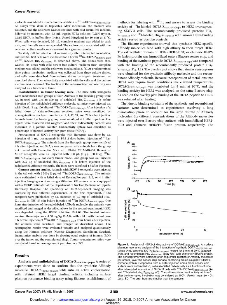
Affibody molecules bind with high affinity to their target HER2.The extracellular domain of HER2 (HER2-ECD) or chimeric HER2/Fc fusion protein was immobilized onto a Biacore sensor chip, andbinding of the synthetic peptide DOTA-ZHER2:342-pep2 was comparedwith the binding of the recombinantly produced protein His6-ZHER2:342 (Fig. 1A). The overlay plot shows that similar sensorgramswere obtained for the synthetic Affibody molecule and the recom-binant Affibody molecule. Because incorporation of metal ions intoDOTA may require harsh conditions like elevated temperatures,DOTA-ZHER2:342-pep2 was incubated for 5 min at 90jC, and thebinding activity for HER2 was analyzed on the same Biacore chip.As seen on the overlay plot, binding of the DOTA-peptide to HER2was retained after heating.The kinetic binding constants of the synthetic and recombinant
variants were determined in experiments involving a longdissociation phase to account for the slow off-rate of Affibodymolecules. Six different concentrations of the Affibody moleculeswere injected over Biacore chip surfaces with immobilized HER2-ECD and chimeric HER2/Fc fusion protein, respectively. The
Figure 1. Analysis of HER2-binding activity of DOTA-ZHER2:342-pep2. A, surfaceplasmon resonance analysis of the interaction of synthetic DOTA-ZHER2:342-pep2
(black line ), synthetic DOTA-ZHER2:342-pep2 heated for 5 min at 90jC (dashedline ), and recombinant His6-ZHER2:342 (gray line ) with chimeric HER2/Fc protein.The sensorgrams were obtained after sequential injection of Affibody molecules(20 nmol/L) over the sensor chip surface containing amine-coupled HER2/Fcchimeric protein. Responses from a buffer injection and a blank sensor chipsurface were subtracted. B, cell-associated radioactivity as a function of timeafter interrupted incubation of SKOV-3 cells with 111In-DOTA-ZHER2:342-pep2 (.)and 125I-labeled His6-ZHER2:342 (o). The cell-associated radioactivity at time 0after the interrupted incubation was considered as 100%. Points, mean (n = 3);bars, SD. The error bars are smaller than the symbols.
Cancer Research
Cancer Res 2007; 67: (5). March 1, 2007 2180 www.aacrjournals.org
Research. on August 16, 2015. © 2007 American Association for Cancercancerres.aacrjournals.org Downloaded from
binding affinity of the DOTA-ZHER2:342-pep2 was about thrice lowerthan the recombinant Affibody molecule His6-ZHER2:342. Thedissociation constants (KD = kd/ka) of DOTA-ZHER2:342-pep2 werecalculated to 65 versus 78 pmol/L for HER2-ECD or chimericHER2/Fc fusion protein, respectively, and 20 versus 29 pmol/L forHis6-ZHER2:342. For HER2-ECD, the association rate constants (ka)were determined as 4.0 � 106 M�1 s�1 for DOTA-ZHER2:342-pep2 and4.0 � 106 M�1 s�1 for His6-ZHER2:342, and the dissociation rateconstants (kd) were 2.6 � 10�4 s�1 for DOTA-ZHER2:342-pep2 and8.1 � 10�5 s�1 for His6-ZHER2:342.DOTA-ZHER2:342-pep2 was efficiently labeled with 111In at slightly
acidic conditions. The pH of the final reaction mixture was f5.The labeling efficiency was evaluated using ITLC, where radio-labeled Affibody molecules remained at the start line of the ITLCsheet, whereas free indium migrated with the solvent front. Underall conditions tested, the labeling efficiency was above 95% of 111Inincorporation, with 96% and 98% after incubation for 30 or 60 minat room temperature, respectively; 98% for both times at 37jC; 99%for both times at 50jC; and 99% for 30 min at 90jC.Binding specificity tests showed that binding of 111In-DOTA-
ZHER2:342-pep2 to living HER2-expressing SKOV-3 cells was receptormediated because saturation of receptors by preincubation withnon-labeled His6-ZHER2:342 significantly decreased binding of theradiolabeled Affibody molecule (P < 0.0001). The binding specificitywas preserved in the whole range of labeling temperatures, fromroom temperature to 90jC (data not shown). The antigen bindingcapacity (i.e., percentage of specifically cell-bound radioactivity)was 82 F 1% after labeling at room temperature and 85.7 F 0.5%after labeling at 90jC. Cellular retention of 111In after interruptedincubation of 111In-labeled DOTA-ZHER2:342-pep2 with SKOV-3 cellswas measured over time and compared with the retention of125I-labeled His6-ZHER2:342 (Fig. 1B). After an initial reduction, thecell-bound radioactivity remained constant at about 90% of theinitially bound activity if the cells were incubated with 111In-labeledDOTA-ZHER2:342-pep2. In contrast, cells incubated with 125I-labeledHis6-ZHER2:342 lost about 40% of the original cell associatedradioactivity during the first 4 h and additional 20% until 29 h afterthe interrupted incubation.Biodistribution of 111In-DOTA-ZHER2:342-pep2 in xenograft
bearing mice. The next set of experiments assessed the in vivotumor targeting activity and specificity of 111In-labeled DOTA-ZHER2:342-pep2 by analyzing the binding of this molecule to HER2-positive or HER2-negative tumor xenografts and the whole bodybiodistribution in SKOV-3 xenograft mice. The Taq polymerase–specific Affibody molecule 111In-DOTA-Ztaq4:5 served as negativecontrol. In one experiment, 177Lu-labeled DOTA-ZHER2:342-pep2 wasused to show that this peptide can be labeled also with otherradionuclides.Biodistribution studies of 111In-DOTA-ZHER2:342-pep2 were done
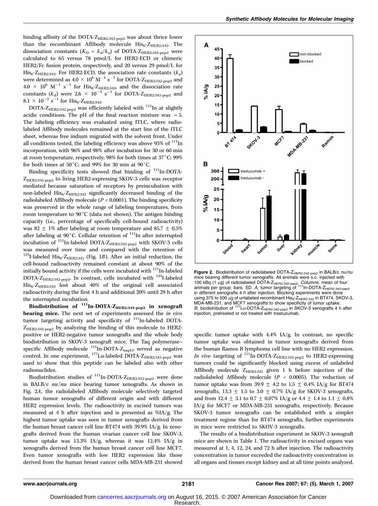
in BALB/c nu/nu mice bearing tumor xenografts. As shown inFig. 2A , the radiolabeled Affibody molecule selectively targetedhuman tumor xenografts of different origin and with differentHER2 expression levels. The radioactivity in excised tumors wasmeasured at 4 h after injection and is presented as %IA/g. Thehighest tumor uptake was seen in tumor xenografts derived fromthe human breast cancer cell line BT474 with 39.9% IA/g. In xeno-grafts derived from the human ovarian cancer cell line SKOV-3,tumor uptake was 13.3% IA/g, whereas it was 12.4% IA/g inxenografts derived from the human breast cancer cell line MCF7.Even tumor xenografts with low HER2 expression like thosederived from the human breast cancer cells MDA-MB-231 showed
specific tumor uptake with 4.4% IA/g. In contrast, no specifictumor uptake was obtained in tumor xenografts derived fromthe human Ramos B lymphoma cell line with no HER2 expression.In vivo targeting of 111In-DOTA-ZHER2:342-pep2 to HER2-expressingtumors could be significantly blocked using excess of unlabeledAffibody molecule ZHER2:342 given 1 h before injection of theradiolabeled Affibody molecule (P < 0.0005). The reduction oftumor uptake was from 39.9 F 4.2 to 1.5 F 0.4% IA/g for BT474xenografts, 13.3 F 1.5 to 3.0 F 0.7% IA/g for SKOV-3 xenografts,and from 12.4 F 3.1 to 0.7 F 0.07% IA/g or 4.4 F 1.4 to 1.1 F 0.8%IA/g for MCF7 or MDA-MB-231 xenografts, respectively. BecauseSKOV-3 tumor xenografts can be established with a simplertreatment regime than for BT474 xenografts, further experimentsin mice were restricted to SKOV-3 xenografts.The results of a biodistribution experiment in SKOV-3 xenograft
mice are shown in Table 1. The radioactivity in excised organs wasmeasured at 1, 4, 12, 24, and 72 h after injection. The radioactivityconcentration in tumor exceeded the radioactivity concentration inall organs and tissues except kidney and at all time points analyzed.
Figure 2. Biodistribution of radiolabeled DOTA-ZHER2:342-pep2 in BALB/c nu /numice bearing different tumor xenografts. All animals were s.c. injected with100 kBq (1 Ag) of radiolabeled DOTA-ZHER2:342-pep2. Columns, mean of fouranimals per group; bars, SD. A, tumor targeting of 111In-DOTA-ZHER2:342-pep2
in different xenografts 4 h after injection. Blocking experiments were doneusing 375 to 500 Ag of unlabeled recombinant His6-ZHER2:342 in BT474, SKOV-3,MDA-MB-231, and MCF7 xenografts to show specificity of tumor uptake.B, biodistribution of 177Lu-DOTA-ZHER2:342-pep2 in SKOV-3 xenografts 4 h afterinjection, pretreated or not treated with trastuzumab.
Synthetic Affibody Molecules for Molecular Imaging
www.aacrjournals.org 2181 Cancer Res 2007; 67: (5). March 1, 2007
Research. on August 16, 2015. © 2007 American Association for Cancercancerres.aacrjournals.org Downloaded from
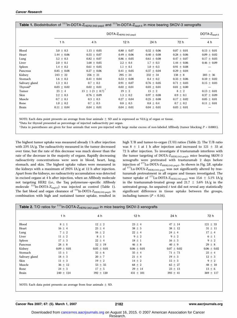
The highest tumor uptake was measured already 1 h after injectionwith 23% IA/g. The radioactivity measured in the tumor decreasedover time, but the rate of this decrease was much slower than therate of the decrease in the majority of organs. Rapidly decreasingradioactivity concentrations were seen in blood, heart, lung,stomach, and skin. The highest uptake values were measured inthe kidneys with a maximum of 295% IA/g at 12 h after injection.Apart from the kidneys, no radioactivity accumulation was detectedin excised organs at 4 h after injection, when an Affibody moleculenot targeting HER2 (i.e., the Taq polymerase–specific Affibodymolecule 111In-DOTA-Ztaq4:5) was injected as control (Table 1).The fast blood and organ clearance of 111In-DOTA-ZHER2:342-pep2, incombination with high and sustained tumor uptake, resulted in
high T/B and tumor-to-organ (T/O) ratios (Table 2). The T/B ratiowas 8 F 1 at 1 h after injection and increased to 121 F 33 at72 h after injection. To investigate if trastuzumab interferes withthe tumor targeting of DOTA-ZHER2:342-pep2, mice bearing SKOV-3xenografts were pretreated with trastuzumab 2 days beforeinjection of 177Lu-DOTA-ZHER2:342-pep2. As shown in Fig. 2B , uptakeof 177Lu-DOTA-ZHER2:342-pep2 was not significantly altered by tras-tuzumab pretreatment in all organs and tissues investigated. Thetumor uptake of 177Lu-DOTA-ZHER2:342-pep2 was 15.6 F 5.1% IA/gin the trastuzumab-treated group and 21.7 F 5.6% IA/g in theuntreated group. An unpaired t test did not reveal any statisticallysignificant difference in tissue uptake between the groups,including tumors (P = 0.16).
Table 1. Biodistribution of 111In-DOTA-ZHER2:342-pep2 and 111In-DOTA-Ztaq4:5 in mice bearing SKOV-3 xenografts
DOTA-ZHER2:342-pep2 DOTA-Ztaq4:5
1 h 4 h 12 h 24 h 72 h 4 h
Blood 3.0 F 0.3 1.15 F 0.05 0.80 F 0.07 0.32 F 0.06 0.07 F 0.01 0.13 F 0.01
Heart 1.44 F 0.06 0.55 F 0.07 0.49 F 0.06 0.40 F 0.08 0.28 F 0.06 0.09 F 0.02
Lung 3.2 F 0.3 0.82 F 0.07 0.86 F 0.05 0.61 F 0.08 0.47 F 0.07 0.17 F 0.03
Liver 2.0 F 0.1 1.68 F 0.05 2.2 F 0.4 1.7 F 0.3 1.44 F 0.06 0.46 F 0.09Spleen 1.4 F 0.2 0.61 F 0.05 1.1 F 0.1 1.0 F 0.3 0.91 F 0.08 —
Pancreas 0.85 F 0.08 0.27 F 0.06 0.41 F 0.05 0.37 F 0.04 0.29 F 0.04 —
Kidney 243 F 22 256 F 21 295 F 24 232 F 34 138 F 8 203 F 36Stomach 1.6 F 0.2 0.41 F 0.04 0.53 F 0.08 0.4 F 0.2 0.33 F 0.06 0.10 F 0.02
Salivary gland 1.3 F 0.1 0.7 F 0.2 0.91 F 0.07 0.76 F 0.05 0.71 F 0.03 0.15 F 0.03
Thyroid* 0.03 F 0.02 0.02 F 0.01 0.02 F 0.01 0.03 F 0.01 0.01 F 0.00 —
Tumor 23 F 4 13 F 1 (3 F 0.7)c
19 F 2 15 F 2 8 F 2 0.13 F 0.01Skin 2.2 F 0.3 0.76 F 0.09 1.3 F 0.2 0.97 F 0.08 0.9 F 0.1 0.37 F 0.09
Muscle 0.7 F 0.1 0.3 F 0.3 0.27 F 0.04 0.25 F 0.08 0.17 F 0.02 0.05 F 0.01
Bone 1.0 F 0.2 0.7 F 0.3 0.8 F 0.3 0.8 F 0.4 0.7 F 0.2 0.11 F 0.01
Brain 0.11 F 0.04 0.04 F 0.01 0.04 F 0.01 0.04 F 0.01 0.03 F 0.01 —
NOTE: Each data point presents an average from four animals F SD and is expressed as %IA/g of organ or tissue.
*Data for thyroid presented as percentage of injected radioactivity per organ.cData in parentheses are given for four animals that were pre-injected with large molar excess of non-labeled Affibody (tumor blocking P < 0.0001).
Table 2. T/O ratios for 111In-DOTA-ZHER2:342-pep2 in mice bearing SKOV-3 xenografts
1 h 4 h 12 h 24 h 72 h
Blood 8 F 1 12 F 2 23 F 4 47 F 14 121 F 33
Heart 16 F 4 25 F 4 38 F 5 38 F 12 31 F 11
Lung 7 F 2 16 F 2 22 F 4 24 F 4 17 F 4
Liver 11 F 2 8 F 1 9 F 2 9 F 2 6 F 1Spleen 17 F 3 22 F 4 18 F 1 16 F 3 9 F 2
Pancreas 28 F 8 52 F 19 46 F 8 40 F 9 29 F 8
Kidney 0.09 F 0.01 0.05 F 0.01 0.06 F 0.01 0.07 F 0.02 0.06 F 0.02Stomach 15 F 1 32 F 6 35 F 4 71 F 73 25 F 4
Salivary gland 18 F 3 20 F 7 21 F 4 19 F 3 12 F 3
Skin 11 F 3 19 F 2 14 F 2 15 F 3 9 F 2
Muscle 36 F 12 53 F 35 68 F 2 65 F 27 48 F 10Bone 24 F 3 17 F 5 29 F 14 23 F 13 13 F 6
Brain 240 F 124 392 F 120 431 F 101 393 F 41 369 F 117
NOTE: Each data point presents an average from four animals F SD.
Cancer Research
Cancer Res 2007; 67: (5). March 1, 2007 2182 www.aacrjournals.org
Research. on August 16, 2015. © 2007 American Association for Cancercancerres.aacrjournals.org Downloaded from
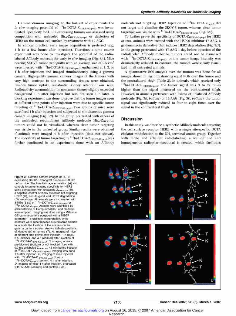
Gamma camera imaging. In the last set of experiments thein vivo imaging potential of 111In-DOTA-ZHER2:342-pep2 was inves-tigated. Specificity for HER2 expressing tumors was assessed usingcompetition with unlabeled His6-ZHER2:342-pep2 or depletion ofHER2 on the tumor cell surface by treatment with 17-AGG.In clinical practice, early image acquisition is preferred (e.g.,
1 h to a few hours after injection). Therefore, a time courseexperiment was done to investigate the potential of the radio-labeled Affibody molecule for early in vivo imaging (Fig. 3A). Micebearing SKOV3 tumor xenografts with an average size of 0.5 cm3
were injected with 111In-DOTA-ZHER2:342-pep2; euthanized at 1, 2, or4 h after injection; and imaged simultaneously using a gammacamera. High-quality gamma camera images of the tumors withvery high contrast to the surrounding tissues were obtained.Besides tumor uptake, substantial kidney retention was seen.Radioactivity accumulation in nontumor tissues slightly exceededbackground 1 h after injection but was not seen 1 h later. Ablocking experiment was done to prove that the tumor images seenat different time points after injection were due to specific tumortargeting of 111In-DOTA-ZHER2:342-pep2. Two groups of mice weresacrificed 1 h after injection and subjected to simultaneous gammacamera imaging (Fig. 3B). In the group pretreated with excess ofthe unlabeled, recombinant Affibody molecule His6-ZHER2:342tumors could not be visualized, whereas clear tumor targetingwas visible in the untreated group. Similar results were obtainedif animals were imaged 4 h after injection (data not shown).The specificity of tumor targeting by 111In-DOTA-ZHER2:342-pep2 wasfurther confirmed in an experiment done with an Affibody
molecule not targeting HER2. Injection of 111In-DOTA-Ztaq4:5 didnot target and visualize the SKOV-3 tumor, whereas clear tumortargeting was visible with 111In-DOTA-ZHER2:342-pep2 (Fig. 3C).To further prove the specificity of DOTA-ZHER2:342-pep2 for HER2
in vivo , animals were treated with the HSP90 inhibitor 17-AAG, ageldanamycin derivative that induces HER2 degradation (Fig. 3D).In the group pretreated with 17-AAG 1 day before injection of theradiolabeled Affibody molecule, tumors could not be visualizedwith 111In-DOTA-ZHER2:342-pep2, or the tumor image intensity wasdramatically reduced. In contrast, the tumors were clearly visual-ized in all untreated animals.A quantitative ROI analysis over the tumors was done for all
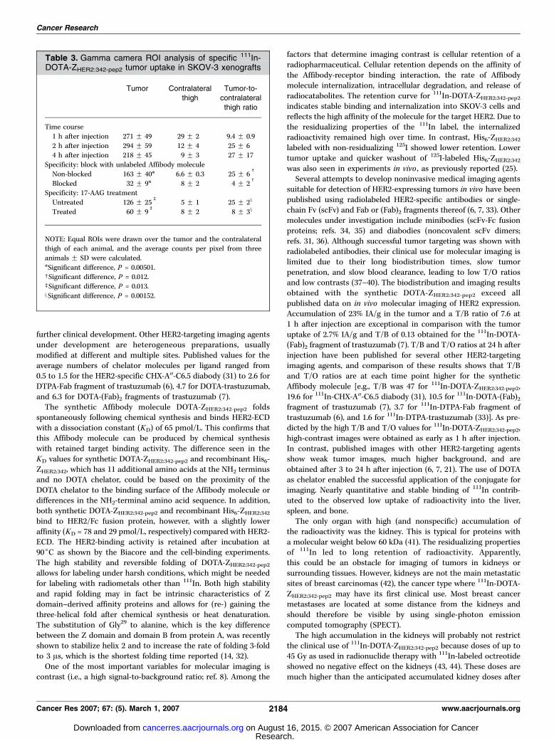
images shown in Fig. 3 by drawing equal ROIs over the tumor andthe contralateral thigh (Table 3). In animals, which received only111In-DOTA-ZHER2:342-pep2, the tumor signal was 9 to 27 timeshigher than the signal measured on the contralateral thigh.However, in animals pretreated with excess of unlabeled Affibodymolecule (Fig. 3B, bottom) or 17-AAG (Fig. 3D, bottom), the tumorsignal was significantly reduced to four to eight times over thesignal in the contralateral thigh.
Discussion
In this study, we describe a synthetic Affibody molecule targetingthe cell surface receptor HER2, with a single site-specific DOTAchelator modification at the NH2-terminal amino group. Togetherwith facile and efficient radiolabeling, a well-defined andhomogeneous radiopharmaceutical is created, which facilitates
Figure 3. Gamma camera images of HER2-expressing SKOV-3 xenograft tumors in BALB/cnu /nu mice. The time to image acquisition (A ) andcontrols to prove imaging specificity for HER2using competition with unlabeled ZHER2:342 (B),a negative control Affibody molecule not targetingHER2 (C ), and drug-induced HER2 degradation(D ) are shown. All animals were i.v. injected with3 MBq (3 Ag) of 111In-DOTA-ZHER2:342-pep2 or111In-DOTA-Ztaq4:5. Animals were sacrificed byadministration of Rompun/Ketalar, and bladderswere emptied. Imaging was done using a MilleniumGE gamma-camera equipped with a MEGPcollimator. To facilitate interpretation, whitecontours were superimposed around some animalsto indicate the location of the animals on thegamma camera screen. Arrows indicate positionsof kidneys (K ) or tumors (T ). A, imaging of miceat different time points after injection, 1 h (top ),2 h (middle ), and 4 h (bottom ) after injection of111In-DOTA-ZHER2:342-pep2. B, imaging of micepre-blocked (bottom ) or not blocked (top ) with0.9 mg unlabeled ZHER2:342 45 min before injectionof 111In-DOTA-ZHER2:342-pep2. Imaging was done1 h after injection. C, imaging of mice injectedwith 111In-DOTA-ZHER2:342-pep2 (top ) or111In-DOTA-Ztaq4:5 (bottom ) 4 h after injection.D, imaging of mice 4 h after injection, pretreatedwith 17-AAG (bottom ) and controls (top ).
Synthetic Affibody Molecules for Molecular Imaging
www.aacrjournals.org 2183 Cancer Res 2007; 67: (5). March 1, 2007
Research. on August 16, 2015. © 2007 American Association for Cancercancerres.aacrjournals.org Downloaded from
further clinical development. Other HER2-targeting imaging agentsunder development are heterogeneous preparations, usuallymodified at different and multiple sites. Published values for theaverage numbers of chelator molecules per ligand ranged from0.5 to 1.5 for the HER2-specific CHX-A00-C6.5 diabody (31) to 2.6 forDTPA-Fab fragment of trastuzumab (6), 4.7 for DOTA-trastuzumab,and 6.3 for DOTA-(Fab)2 fragments of trastuzumab (7).The synthetic Affibody molecule DOTA-ZHER2:342-pep2 folds
spontaneously following chemical synthesis and binds HER2-ECDwith a dissociation constant (KD) of 65 pmol/L. This confirms thatthis Affibody molecule can be produced by chemical synthesiswith retained target binding activity. The difference seen in theKD values for synthetic DOTA-ZHER2:342-pep2 and recombinant His6-ZHER2:342, which has 11 additional amino acids at the NH2 terminusand no DOTA chelator, could be based on the proximity of theDOTA chelator to the binding surface of the Affibody molecule ordifferences in the NH2-terminal amino acid sequence. In addition,both synthetic DOTA-ZHER2:342-pep2 and recombinant His6-ZHER2:342bind to HER2/Fc fusion protein, however, with a slightly loweraffinity (KD = 78 and 29 pmol/L, respectively) compared with HER2-ECD. The HER2-binding activity is retained after incubation at90jC as shown by the Biacore and the cell-binding experiments.The high stability and reversible folding of DOTA-ZHER2:342-pep2allows for labeling under harsh conditions, which might be neededfor labeling with radiometals other than 111In. Both high stabilityand rapid folding may in fact be intrinsic characteristics of Zdomain–derived affinity proteins and allows for (re-) gaining thethree-helical fold after chemical synthesis or heat denaturation.The substitution of Gly29 to alanine, which is the key differencebetween the Z domain and domain B from protein A, was recentlyshown to stabilize helix 2 and to increase the rate of folding 3-foldto 3 As, which is the shortest folding time reported (14, 32).One of the most important variables for molecular imaging is
contrast (i.e., a high signal-to-background ratio; ref. 8). Among the
factors that determine imaging contrast is cellular retention of aradiopharmaceutical. Cellular retention depends on the affinity ofthe Affibody-receptor binding interaction, the rate of Affibodymolecule internalization, intracellular degradation, and release ofradiocatabolites. The retention curve for 111In-DOTA-ZHER2:342-pep2indicates stable binding and internalization into SKOV-3 cells andreflects the high affinity of the molecule for the target HER2. Due tothe residualizing properties of the 111In label, the internalizedradioactivity remained high over time. In contrast, His6-ZHER2:342labeled with non-residualizing 125I showed lower retention. Lowertumor uptake and quicker washout of 125I-labeled His6-ZHER2:342was also seen in experiments in vivo , as previously reported (25).Several attempts to develop noninvasive medical imaging agents
suitable for detection of HER2-expressing tumors in vivo have beenpublished using radiolabeled HER2-specific antibodies or single-chain Fv (scFv) and Fab or (Fab)2 fragments thereof (6, 7, 33). Othermolecules under investigation include minibodies (scFv-Fc fusionproteins; refs. 34, 35) and diabodies (noncovalent scFv dimers;refs. 31, 36). Although successful tumor targeting was shown withradiolabeled antibodies, their clinical use for molecular imaging islimited due to their long biodistribution times, slow tumorpenetration, and slow blood clearance, leading to low T/O ratiosand low contrasts (37–40). The biodistribution and imaging resultsobtained with the synthetic DOTA-ZHER2:342-pep2 exceed allpublished data on in vivo molecular imaging of HER2 expression.Accumulation of 23% IA/g in the tumor and a T/B ratio of 7.6 at1 h after injection are exceptional in comparison with the tumoruptake of 2.7% IA/g and T/B of 0.13 obtained for the 111In-DOTA-(Fab)2 fragment of trastuzumab (7). T/B and T/O ratios at 24 h afterinjection have been published for several other HER2-targetingimaging agents, and comparison of these results shows that T/Band T/O ratios are at each time point higher for the syntheticAffibody molecule [e.g., T/B was 47 for 111In-DOTA-ZHER2:342-pep2,19.6 for 111In-CHX-A00-C6.5 diabody (31), 10.5 for 111In-DOTA-(Fab)2fragment of trastuzumab (7), 3.7 for 111In-DTPA-Fab fragment oftrastuzumab (6), and 1.6 for 111In-DTPA-trastuzumab (33)]. As pre-dicted by the high T/B and T/O values for 111In-DOTA-ZHER2:342-pep2,high-contrast images were obtained as early as 1 h after injection.In contrast, published images with other HER2-targeting agentsshow weak tumor images, much higher background, and areobtained after 3 to 24 h after injection (6, 7, 21). The use of DOTAas chelator enabled the successful application of the conjugate forimaging. Nearly quantitative and stable binding of 111In contrib-uted to the observed low uptake of radioactivity into the liver,spleen, and bone.The only organ with high (and nonspecific) accumulation of
the radioactivity was the kidney. This is typical for proteins witha molecular weight below 60 kDa (41). The residualizing propertiesof 111In led to long retention of radioactivity. Apparently,this could be an obstacle for imaging of tumors in kidneys orsurrounding tissues. However, kidneys are not the main metastaticsites of breast carcinomas (42), the cancer type where 111In-DOTA-ZHER2:342-pep2 may have its first clinical use. Most breast cancermetastases are located at some distance from the kidneys andshould therefore be visible by using single-photon emissioncomputed tomography (SPECT).The high accumulation in the kidneys will probably not restrict
the clinical use of 111In-DOTA-ZHER2:342-pep2 because doses of up to45 Gy as used in radionuclide therapy with 111In-labeled octreotideshowed no negative effect on the kidneys (43, 44). These doses aremuch higher than the anticipated accumulated kidney doses after
Table 3. Gamma camera ROI analysis of specific 111In-DOTA-ZHER2:342-pep2 tumor uptake in SKOV-3 xenografts
Tumor Contralateralthigh
Tumor-to-contralateral
thigh ratio
Time course
1 h after injection 271 F 49 29 F 2 9.4 F 0.9
2 h after injection 294 F 59 12 F 4 25 F 64 h after injection 218 F 45 9 F 3 27 F 17
Specificity: block with unlabeled Affibody molecule
Non-blocked 163 F 40* 6.6 F 0.3 25 F 6c
Blocked 32 F 9* 8 F 2 4 F 2c
Specificity: 17-AAG treatment
Untreated 126 F 25b
5 F 1 25 F 2x
Treated 60 F 9b
8 F 2 8 F 3x
NOTE: Equal ROIs were drawn over the tumor and the contralateral
thigh of each animal, and the average counts per pixel from three
animals F SD were calculated.
*Significant difference, P = 0.00501.cSignificant difference, P = 0.012.bSignificant difference, P = 0.013.xSignificant difference, P = 0.00152.
Cancer Research
Cancer Res 2007; 67: (5). March 1, 2007 2184 www.aacrjournals.org
Research. on August 16, 2015. © 2007 American Association for Cancercancerres.aacrjournals.org Downloaded from

multiple diagnostic imaging with 111In-DOTA-ZHER2:342-pep2. Anample number of publications describe ways to reduce the renaluptake of radiometal-labeled proteins and peptides. The use ofpositively charged amino acids (41, 45), colchicine (46), andGelofusine (47) showed positive effects. Further studies with thesemolecules will show if they can also be used to reduce renal uptakeof 111In-DOTA-ZHER2:342-pep2.Pretreatment of animals with trastuzumab did not interfere with
tumor targeting by 111In-DOTA-ZHER2:342-pep2. This result indicatesthat this radiopharmaceutical might also be used to determine theHER2 status of metastatic lesions in patients with ongoingHerceptin treatment. Thus, DOTA-ZHER2:342-pep2 could be used tofollow the effect of HER2-targeted therapy with Herceptin andpotentially also of other drugs currently under investigation ordevelopment, including new antibodies like Omnitarg (pertuzu-mab), a HER dimerization inhibitor (48), and small molecules, likethe HSP90 inhibitor 17-AAG (22) and the tyrosine kinase inhibitorlapatinib (49). Monitoring the presence of drug target andsubsequent effect of drug treatment on tumor HER2 levels wouldbe greatly facilitated by molecular imaging in patients using anoninvasive imaging agent. In the present study, we show that111In-DOTA-ZHER2:342-pep2 can be used to monitor changes in HER2expression level in animals treated with 17-AAG. Thus, molecularimaging using 111In-DOTA-ZHER2:342-pep2 has the potential to gobeyond localization of metastatic lesions in vivo by adding new
qualitative information not available today by conventionalimaging techniques.Molecules conjugated with a DOTA chelator can not only be
labeled with radiometals suitable for SPECT imaging, such as 111In,but also with positron emitters like 68Ga for positron emissiontomography (PET) imaging. Indeed, a method for 68Ga labeling ofDOTA-ZHER2:342-pep2 was recently established, thus broadening thepotential clinical applicability of synthetic DOTA-ZHER2:342-pep2 foreither SPECT or PET (50). Moreover, the present study shows stableattachment of 177Lu toDOTA-ZHER2:342-pep2, opening for the possibilityto use a 177Lu-labeled Affibodymolecule for locoregional treatment ofurinary bladder carcinomas, which often overexpress HER2 (19). Inconclusion, the results presented here show that synthetic DOTA-ZHER2:342-pep2 is one of the best available radiopharmaceuticals forin vivo molecular imaging of HER2-expressing carcinomas.
Acknowledgments
Received 8/3/2006; revised 12/6/2006; accepted 1/8/2007.Grant support: Swedish Cancer Society.The costs of publication of this article were defrayed in part by the payment of page
charges. This article must therefore be hereby marked advertisement in accordancewith 18 U.S.C. Section 1734 solely to indicate this fact.
We thank Lena Israelson for the cell culture work, Veronika Eriksson and staff ofthe animal facility of Rudbeck laboratory for technical assistance, and CharlesWidstrom (Department of Hospital Physics, Uppsala University Hospital) for help withthe gamma camera.
References1. Thrall JH. Personalized medicine. Radiology 2004;231:613–6.2. Wallace AM, Comstock C, Hoh CK, Vera DR. Breastimaging: a surgeon’s prospective. Nucl Med Biol 2005;32:781–92.3. Behr TM, Gotthardt M, Barth A, Behe M. Imagingtumors with peptide-based radioligands. Q J Nucl Med2001;45:189–200.
4. Britz-Cunningham SH, Adelstein SJ. Molecular target-ing with radionuclides: state of the science. J Nucl Med2003;44:1945–61.5. Wilbur DS. Radiohalogenation of proteins: an over-view of radionuclides, labeling methods and reagents forconjugate labeling. Bioconjug Chem 1992;3:433–70.6. Tang Y, Wang J, Scollard DA, et al. Imaging of HER2/neu -positive BT-474 human breast cancer xenografts inathymic mice using 111In-trastuzumab (Herceptin) Fabfragments. Nucl Med Biol 2005;32:51–8.7. Smith-Jones PM, Solit DB, Akhurst T, Afroze F, RosenN, Larson SM. Imaging the pharmacodynamics of HER2degradation in response to Hsp90 inhibitors. NatBiotechnol 2004;22:701–6.8. Wu AM, Senter PD. Arming antibodies: prospects andchallenges for immunoconjugates. Nat Biotechnol 2005;23:1137–46.9. Michel CC, Curry FE. Microvascular permeability.Physiol Rev 1999;79:703–61.10. Sharkey RM, Cardillo TM, Rossi EA, et al. Signalamplification in molecular imaging by pretargeting amultivalent, bispecific antibody. Nat Med 2005;11:1250–5.11. Heppler A, Froidevaux S, Eberle AN, Maecke H.Receptor targeting for tumor localization and therapywith radiopeptides. Curr Med Chem 2000;7:791–4.12. Reubi JC. Somatostatin and other peptide receptorsas tools for tumor diagnosis and treatment. Neuroen-docrinology 2004;80:51–6.13. Landon LA, Deutscher SL. Combinatorial discoveryof tumor targeting peptides using phage display. J CellBiochem 2003;90:509–17.14. Nilsson B, Moks T, Jansson B, et al. A synthetic IgG-binding domain based on staphylococcal protein A.Protein Eng 1987;1:107–13.
15. Nord K, Nilsson J, Nilsson B, Uhlen M, Nygren PA.A combinatorial library of an alpha-helical bacterialreceptor domain. Protein Eng 1995;8:601–8.16. Nord K, Gunneriusson E, Ringdahl J, Stahl S, UhlenM, Nygren PA. Binding proteins selected from combi-natorial libraries of an alpha-helical bacterial receptordomain. Nat Biotechnol 1997;15:772–7.17. Aunoble B, Sanches R, Didier E, Bignon YJ. Majoroncogenes and tumor suppressor genes involved inepithelial ovarian cancer. Int J Oncol 2000;16:567–76.18. Carlsson J, Nordgren H, Sjostrom J, et al. HER2expression in breast cancer primary tumours andcorresponding metastases. Original data and literature.Br J Cancer 2004;90:2344–8.19. Gardmark T, Wester K, De la Torre M, Carlsson J,Malmstrom PU. Analysis of HER2 expression in primaryurinary bladder carcinoma and corresponding metasta-ses. BJU Int 2005;95:982–6.20. Zidan J, Dashkovsky I, Stayerman C, Basher W,Cozacov C, Hadary A. Comparison of HER-2 over-expression in primary breast cancer and metastatic sitesand its effect on biological targeting therapy ofmetastatic disease. Br J Cancer 2005;93:552–6.21. Smith-Jones PM, Solit D, Afroze F, Rosen N, LarsonSM. Early tumor response to Hsp90 therapy using HER2PET: comparison with 18F-FDG PET. J Nucl Med 2006;47:793–6.22. Whitesell L, Lindquist SL. HSP90 and the chaperon-ing of cancer. Nat Rev Cancer 2005;5:761–72.23. Wikman M, Steffen AC, Gunneriusson E, et al.Selection and characterization of HER2/neu -bindingAffibody ligands. Protein Eng Des Sel 2004;17:455–62.24. Steffen AC, Orlova A, Wikman M, et al. Affibody-mediated tumour targeting of HER-2 expressing xeno-grafts in mice. Eur J Nucl Med Mol Imaging 2006;33:631–8.25. Orlova A, Magnusson M, Eriksson TL, et al. Tumorimaging using a picomolar affinity HER2 bindingAffibody molecule. Cancer Res 2006;66:4339–48.26. Tolmachev V, Nilsson F, Widstrom C, et al. 111In-benzyl-DTPA-ZHER2:342, an Affibody-based conjugate forin vivo imaging of HER2 expression in malignanttumors. J Nucl Med 2006;74:846–53.27. Yang D, Kuan CT, Payne J, et al. Recombinant
heregulin-Pseudomonas exotoxin fusion proteins: inter-actions with the heregulin receptors and antitumoractivity in vivo . Clin Cancer Res 1998;4:993–1004.28. Horak EM, Heitner T, Garrison JL, et al. Engineeringbispecific single-chain Fv molecules to alter signaling ofthe EGF receptor family. Proc Am Assoc Cancer Res2001;42:774.29. Steffen AC, Wikman M, Tolmachev V, et al. In vitrocharacterization of a bivalent anti-HER-2 Affibody withpotential for radionuclide-based diagnostics. CancerBiother Radiopharm 2005;20:239–48.30. Persson M, Tolmachev V, Andersson K, Gedda L,Sandstrom M, Carlsson J. [177Lu]pertuzumab: experi-mental studies on targeting of HER-2 positive tumourcells. Eur J Nucl Med Mol Imaging 2005;32:1457–62.31. Adams GP, Shaller CC, Dadachova E, et al. A singletreatment of yttrium-90-labeled CHX-A00-C6.5 diabodyinhibits the growth of established human tumorxenografts in immunodeficient mice. Cancer Res 2004;64:6200–6.32. Arora P, Oas TG, Myers JK. Fast and faster: a designedvariant of the B-domain of protein A folds in 3 microsec.Protein Sci 2004;13:847–53.33. Lub-de Hooge MN, Kosterink JG, Perik PJ, et al.Preclinical characterisation of 111In-DTPA-trastuzumab.Br J Pharmacol 2004;143:99–106.34. Olafsen T, Tan GJ, Cheung CW, et al. Characterizationof engineered anti-p185HER-2 (scFv-CH3)2 antibodyfragments (minibodies) for tumor targeting. ProteinEng Des Sel 2004;17:315–23.35. Olafsen T, Kenanova VE, Sundaresan G, et al.Optimizing radiolabeled engineered anti-p185HER2antibody fragments for in vivo imaging. Cancer Res2005;65:5907–16.36. Robinson MK, Doss M, Shaller C, et al. Quantitativeimmuno-positron emission tomography imaging ofHER2-positive tumor xenografts with an iodine-124labeled anti-HER2 diabody. Cancer Res 2005;65:1471–8.37. De Santes K, Slamon D, Anderson SK, et al. Radio-labeled antibody targeting of the HER-2/neu oncopro-tein. Cancer Res 1992;52:1916–23.38. Xu FJ, Yu YH, Bae DS, et al. Radioiodinated antibodytargeting of the HER-2/neu oncoprotein. Nucl Med Biol1997;24:451–9.
Synthetic Affibody Molecules for Molecular Imaging
www.aacrjournals.org 2185 Cancer Res 2007; 67: (5). March 1, 2007
Research. on August 16, 2015. © 2007 American Association for Cancercancerres.aacrjournals.org Downloaded from

39. Zalutsky MR, Xu FJ, Yu Y, et al. Radioiodinatedantibody targeting of the HER-2/neu oncoprotein:effects of labeling method on cellular processing andtissue distribution. Nucl Med Biol 1999;26:781–90.40. Garmestani K, Milenic DE, Plascjak PS, BrechbielMW. A new and convenient method for purification of86Y using a Sr(II) selective resin and comparison ofbiodistribution of 86Y and 111In labeled Herceptin. NuclMed Biol 2002;29:599–606.41. Behr TM, Sharkey RM, Sgouros G, et al. Overcomingthe nephrotoxicity of radiometal-labeled immunoconju-gates: improved cancer therapy administered to a nudemouse model in relation to the internal radiationdosimetry. Cancer 1997;80:2591–610.42. Weigelt B, Peterse JL, van’t Veer LJ. Breast cancermetastasis: markers and models. Nat Rev Cancer 2005;5:591–602.
43. Valkema R, De Jong M, Bakker WH, et al. Phase Istudy of peptide receptor radionuclide therapy with [In-DTPA]octreotide: the Rotterdam experience. Semin NuclMed 2002;32:110–22.44. Kwekkeboom DJ, Mueller-Brand J, Paganelli G, et al.Overview of results of peptide receptor radionuclidetherapy with 3 radiolabeled somatostatin analogs. J NuclMed 2005;46 Suppl 1:62–6S.45. Rolleman EJ, Valkema R, de Jong M, Kooij PP,Krenning EP. Safe and effective inhibition of renaluptake of radiolabelled octreotide by a combination oflysine and arginine. Eur J Nucl Med Mol Imaging 2003;30:9–15.46. Rolleman EJ, Krenning EP, Van Gameren A, BernardBF, De Jong M. Uptake of [111In-DTPA0]octreotide in therat kidney is inhibited by colchicine and not by fructose.J Nucl Med 2004;45:709–13.
47. van Eerd JE, Vegt E, Wetzels JF, et al. Gelatin-basedplasma expander effectively reduces renal uptake of 111In-octreotide in mice and rats. J Nucl Med 2006;47:528–33.48. Adams CW, Allison DE, Flagella K, et al. Humaniza-tion of a recombinant monoclonal antibody to producea therapeutic HER dimerization inhibitor, pertuzumab.Cancer Immunol Immunother 2006;55:717–27.49. Spector NL, Xia W, Burris Hr, et al. Study of thebiologic effects of lapatinib, a reversible inhibitor ofErbB1 and ErbB2 tyrosine kinases, on tumor growth andsurvival pathways in patients with advanced malignan-cies. J Clin Oncol 2005;23:2502–12.50. Baum RP, Orlova A, Tolmachev V, Feldwisch J.Receptor PET/CT and SPECT using an Affibodymolecule for targeting and molecular imaging of HER2positive cancer in animal xenografts and human breastcancer patients. J Nucl Med 2006;47:108P.
Cancer Research
Cancer Res 2007; 67: (5). March 1, 2007 2186 www.aacrjournals.org
Research. on August 16, 2015. © 2007 American Association for Cancercancerres.aacrjournals.org Downloaded from

2007;67:2178-2186. Cancer Res Anna Orlova, Vladimir Tolmachev, Rikard Pehrson, et al. Malignant TumorsLigands for Molecular Imaging of HER2-Expressing Synthetic Affibody Molecules: A Novel Class of Affinity
Updated version
http://cancerres.aacrjournals.org/content/67/5/2178
Access the most recent version of this article at:
Cited articles
http://cancerres.aacrjournals.org/content/67/5/2178.full.html#ref-list-1
This article cites 44 articles, 16 of which you can access for free at:
Citing articles
http://cancerres.aacrjournals.org/content/67/5/2178.full.html#related-urls
This article has been cited by 20 HighWire-hosted articles. Access the articles at:
E-mail alerts related to this article or journal.Sign up to receive free email-alerts
Subscriptions
Reprints and
To order reprints of this article or to subscribe to the journal, contact the AACR Publications
Permissions
To request permission to re-use all or part of this article, contact the AACR Publications
Research. on August 16, 2015. © 2007 American Association for Cancercancerres.aacrjournals.org Downloaded from
Copyright © 2022 FDOKUMEN