
Characteristics of electron beam evaporated nanocrystalline SnO 2 thin films annealed in air
Upload
independentCategory
view
1download
0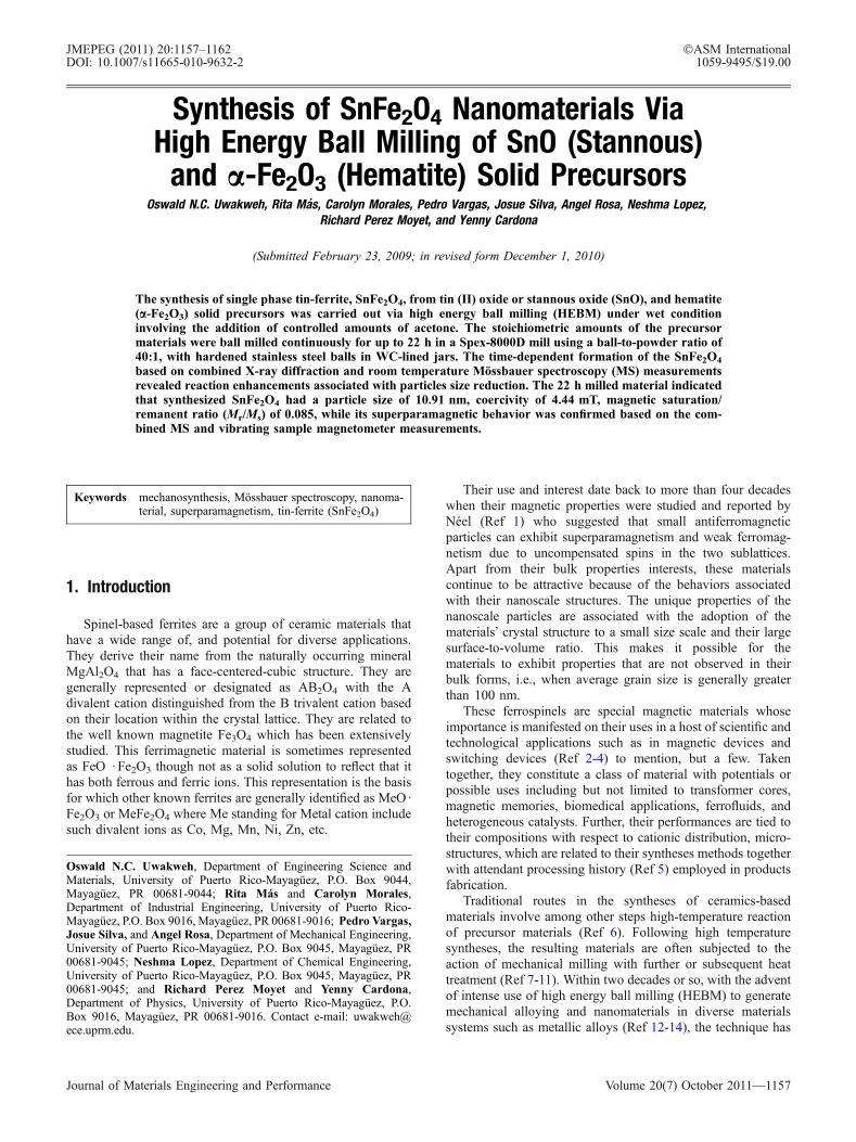
Synthesis of SnFe2O4 Nanomaterials ViaHigh Energy Ball Milling of SnO (Stannous)
and a-Fe2O3 (Hematite) Solid PrecursorsOswald N.C. Uwakweh, Rita Mas, Carolyn Morales, Pedro Vargas, Josue Silva, Angel Rosa, Neshma Lopez,
Richard Perez Moyet, and Yenny Cardona
(Submitted February 23, 2009; in revised form December 1, 2010)
The synthesis of single phase tin-ferrite, SnFe2O4, from tin (II) oxide or stannous oxide (SnO), and hematite(a-Fe2O3) solid precursors was carried out via high energy ball milling (HEBM) under wet conditioninvolving the addition of controlled amounts of acetone. The stoichiometric amounts of the precursormaterials were ball milled continuously for up to 22 h in a Spex-8000D mill using a ball-to-powder ratio of40:1, with hardened stainless steel balls in WC-lined jars. The time-dependent formation of the SnFe2O4
based on combined X-ray diffraction and room temperature Mossbauer spectroscopy (MS) measurementsrevealed reaction enhancements associated with particles size reduction. The 22 h milled material indicatedthat synthesized SnFe2O4 had a particle size of 10.91 nm, coercivity of 4.44 mT, magnetic saturation/remanent ratio (Mr/Ms) of 0.085, while its superparamagnetic behavior was confirmed based on the com-bined MS and vibrating sample magnetometer measurements.
Keywords mechanosynthesis, Mossbauer spectroscopy, nanoma-terial, superparamagnetism, tin-ferrite (SnFe2O4)
1. Introduction
Spinel-based ferrites are a group of ceramic materials thathave a wide range of, and potential for diverse applications.They derive their name from the naturally occurring mineralMgAl2O4 that has a face-centered-cubic structure. They aregenerally represented or designated as AB2O4 with the Adivalent cation distinguished from the B trivalent cation basedon their location within the crystal lattice. They are related tothe well known magnetite Fe3O4 which has been extensivelystudied. This ferrimagnetic material is sometimes representedas FeO Æ Fe2O3 though not as a solid solution to reflect that ithas both ferrous and ferric ions. This representation is the basisfor which other known ferrites are generally identified as MeO ÆFe2O3 or MeFe2O4 where Me standing for Metal cation includesuch divalent ions as Co, Mg, Mn, Ni, Zn, etc.
Their use and interest date back to more than four decadeswhen their magnetic properties were studied and reported byNeel (Ref 1) who suggested that small antiferromagneticparticles can exhibit superparamagnetism and weak ferromag-netism due to uncompensated spins in the two sublattices.Apart from their bulk properties interests, these materialscontinue to be attractive because of the behaviors associatedwith their nanoscale structures. The unique properties of thenanoscale particles are associated with the adoption of thematerials� crystal structure to a small size scale and their largesurface-to-volume ratio. This makes it possible for thematerials to exhibit properties that are not observed in theirbulk forms, i.e., when average grain size is generally greaterthan 100 nm.
These ferrospinels are special magnetic materials whoseimportance is manifested on their uses in a host of scientific andtechnological applications such as in magnetic devices andswitching devices (Ref 2-4) to mention, but a few. Takentogether, they constitute a class of material with potentials orpossible uses including but not limited to transformer cores,magnetic memories, biomedical applications, ferrofluids, andheterogeneous catalysts. Further, their performances are tied totheir compositions with respect to cationic distribution, micro-structures, which are related to their syntheses methods togetherwith attendant processing history (Ref 5) employed in productsfabrication.
Traditional routes in the syntheses of ceramics-basedmaterials involve among other steps high-temperature reactionof precursor materials (Ref 6). Following high temperaturesyntheses, the resulting materials are often subjected to theaction of mechanical milling with further or subsequent heattreatment (Ref 7-11). Within two decades or so, with the adventof intense use of high energy ball milling (HEBM) to generatemechanical alloying and nanomaterials in diverse materialssystems such as metallic alloys (Ref 12-14), the technique has
Oswald N.C. Uwakweh, Department of Engineering Science andMaterials, University of Puerto Rico-Mayaguez, P.O. Box 9044,Mayaguez, PR 00681-9044; Rita Mas and Carolyn Morales,Department of Industrial Engineering, University of Puerto Rico-Mayaguez, P.O. Box 9016, Mayaguez, PR 00681-9016; Pedro Vargas,Josue Silva, and Angel Rosa, Department of Mechanical Engineering,University of Puerto Rico-Mayaguez, P.O. Box 9045, Mayaguez, PR00681-9045; Neshma Lopez, Department of Chemical Engineering,University of Puerto Rico-Mayaguez, P.O. Box 9045, Mayaguez, PR00681-9045; and Richard Perez Moyet and Yenny Cardona,Department of Physics, University of Puerto Rico-Mayaguez, P.O.Box 9016, Mayaguez, PR 00681-9016. Contact e-mail: [email protected].
JMEPEG (2011) 20:1157–1162 �ASM InternationalDOI: 10.1007/s11665-010-9632-2 1059-9495/$19.00
Journal of Materials Engineering and Performance Volume 20(7) October 2011—1157
since evolved from its use in generating nanosized particles(Ref 15-19), to its use in the syntheses of ceramic basedmaterials in a process commonly referred to as mechanosyn-theses (Ref 20-23). Mechanochemical processing entailingsynthesis of MFe2O4 (M = Cu and Zn) spinel systems based ondifferent milling procedures showed that different startingmaterials can be used to obtain ferrospinels with varyingmagnetic properties even when the final material had nearlyidentical phases (Ref 24, 25). Since these materials can begenerated by substituting the Fe cations with other divalentmetal ions in order to generate MeFe2O4 with the spinelstructure from the parent Fe3O4, it is expected that possibility ofnewer ones can be investigated with appropriate cation doping.In this wise, the doping of Fe3O4 with Sn was reported byDrokin et al. (Ref 26) who investigated their photoconductivitybehavior in polycrystalline states (SnxFe3�xO4, with x = 0.1,0.2, 0.3, 0.4, and 1) while Berry and Helgason (Ref 27) studiedthe Sn doping of iron oxides with composition Sn0.1Fe2.9O4
using Mossbauer spectroscopic techniques.As is common in most ceramic syntheses, the Sn-doped
ferrites were mainly prepared via solid-state reactions whichentailed temperatures as high as 1300 �C over long-timeperiods with the realized polycrystalline materials having grainsin the micrometer (lm) ranges. Independent of the solid-statereaction technique, Liu et al. (Ref 28) reported that they wereable to successfully synthesize SnFe2O4 nanoparticles throughsolid-phase reaction in solution described as precipitationexchange. According to these authors, their synthesis methodhas an advantage over traditional solid-state reaction asrelatively low temperature (around 100 �C), because of theirsimple experimental setup resulting in well-crystallized mate-rial. Using FeCl3 ÆH2O, SnCl2 ÆH2O, and NH3 ÆH2O, as startingmaterials, they were able to synthesis SnFe2O4 with particlesize control ranging from 4 to 15 nm after undertaking an openair tube furnace thermal anneal in the temperature range of 85to 200 �C, respectively. Given that the overall interest innanosized materials is based on the fact that their size-dependent properties confers on them a lot of technologicalpremise, we undertook the synthesis of SnFe2O4 via HEBM-induced reaction of solid precursors at room temperatures. Inthis study, SnO oxide was mixed in stoichiometric amountswith a-Fe2O3 followed by the addition of controlled amounts ofacetone in what we describe as wet ball milling synthesis. Theresulting synthesized SnFe2O4 material was achieved bymonitoring the ball milling process as a function of time. Inorder to monitor the mechanosynthesis process, sample mate-rials were drawn intermittently as a function of time todetermine the progress of the synthesis process, and subjectedto X-ray diffraction (XRD), vibrating sample magnetometer(VSM), and Mossbauer spectroscopy (MS) measurements.
2. Experimental Procedure
Alfa Aesar supplied SnO, (tin(II) oxide) also known asstannous oxide, and iron(III) oxide (hematite), a-Fe2O3,powders having average grain size of 74 nm was ball milledwith hardened stainless steel balls measuring 10 mm indiameter, in a WC-lined steel jars. The ball-to-mass ratio(BPR) used was 40:1, while the milling operations were allcarried out under wet condition entailing the use of controlledamount of acetone (0.6 mL) that was added to the initial
mixture before subsequent ball milling using a Spex-8000Dmill. The size of the 17 hardened steel balls used was 10 mm.The milling times were varied from 5 to 22 h. Following ballmilling, samples were drawn from the milled powders for thedifferent characterization techniques used in this study.
A Joel 5410-LV scanning electron microscope operated at10 kV was to undertake a scanning electron microscopy (SEM)characterization of the materials. Thus, the morphologies of thematerials in their as-received and ball milled states wereobserved and compared.
The XRD was carried out with a Siemens D500 diffrac-tometer with a CuKa radiation source and a (b) Ni filter. Thescanning range used in the measurements was 5� to 95� in 2hby steps of 0.03� and operated with a current of 35 A and avoltage of 30 kV. The Power Cell program was used in thepeaks analyses in order to determine the average crystallite size,residual strain, and lattice parameter changes accompanying theball milling of the material.
The magnetic properties such as coercivity and saturationmagnetization were determined through measurements con-ducted with a Lakeshore 7400 Series VSM, with applied fieldup to 22000 gauss on powder samples weighing about 30 mg.
TheMossbauer spectroscopic measurements were carried outwith a WEBRES spectrometer (now SEECO Company) oper-ating at constant acceleration mode with a 50 mCi 57Co gammaray source in Rh matrix made by Ritverc GmbH. The velocityrange of the scan was selected in order to permit a thorough scanof the samples in order to reveal features associated with both theparamagnetic and magnetic portions of the recorded spectra. The1,024 point raw data were folded and analyzed usingWMOSS, apublic domain Mossbauer spectral analysis program available atwww.SEECo.us (Ref 29) which was formerly www.webres.com.The velocity calibration was carried out with known line posi-tions of room temperature a-Fe metal.
3. Results and Discussion
3.1 Scanning Electron Microscopy
Figure 1(a) showed the SEM image of the mixture of SnOand a-Fe2O3 after 5 h of ball milling during the synthesis ofSnFe2O4 material, while Fig. 1(b) and (c), showed the mor-phologies of the materials after 10, and 22 h respectively.Examination of Fig. 1(a) showed the material mixture existingin the form of clumps of particles having varied sizes. Thebigger clumps showed well-defined facets which were notobserved in the ball milled states after 10 and 22 h. Further, the22 h ball milled material exhibited a lot of particle agglomer-ation and interparticle pores, in addition to crystallite sizereduction as would be discussed further in the XRD section.
According to Anantharaman et al. (Ref 30), spinel ferriteshave a preference for crystallographic surface exposure foundto be the B(111) and D(110) planes based on the Knozinger andRatnaswamy (Ref 31) notations. These surfaces have octahe-dral sites as opposed to the tetrahedral ones. According to theseauthors, all the other crystallographic planes associated with theAB2O4 spinel structure have the A and B types sites on theirexposed surfaces except the D(110) and the B(111) typesurfaces. In other to avoid possible confusion over the use ofsimilar letters (B and D) for the surfaces and typical spinelstructure, the spinel structure would henceforth be designated
1158—Volume 20(7) October 2011 Journal of Materials Engineering and Performance
as MeFe2O4 where Me is the metal divalent cation in the spinelstructure. In addition to these studies, others as well were ingeneral agreement based on other spinel structured materials.For instance, Shelef and Yao (Ref 32) concluded that tetrahe-dral sites were not on the surface of spinel structured materialssuch as Co3O4 and were later backed by the studies of Beaufilsand Barbaux based on their differential neutron diffractionstudies on Co3O4 and MgAl2O4, that only the D(110) andB(111) planes are exposed on the surface (Ref 33-35). Themorphologies corresponding to the different states of ballmilling of this study suggested that the materials were inmetastable states since the well-defined facets were notobserved at the magnification used in this study. Further, asshown in Fig. 1(d), the EDAX spectrum of the material after22 h of ball milling showed that the material was homogenous,and was as well at 5 h of ball milling. While some contam-ination may occur during ball milling, the homogeneity of themilled materials is usually not compromised.
3.2 X-Ray Diffraction Measurements
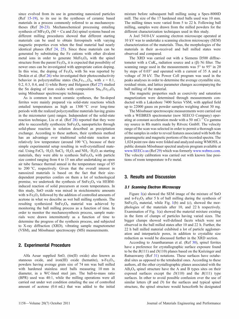
Powder XRD of the precursor materials, hematite (a-Fe2O3)and SnO in their as-received bulk forms were recorded andconstituted the basis for comparison with the milled states. Thiswas achieved by carrying out the XRD measurements on themanually mixed precursor materials as shown in Fig. 2(a) withcharacteristic peaks associated with the phases indicated withdifferent symbols. Their indexing was based on the knowledgeof the tetragonal crystal structure of SnO and the rhombohedralcrystal structure of the a-Fe2O3 materials. In general, the
intensities of the SnO peaks were much higher than those ofhematite. The diffraction spectra shown in the Fig. 2(b-e),corresponding to the different ball milled states starting from5 h in comparison to manual mixture spectra, indicatedcombinations of peaks loss of intensities, broadening, andshifts. These were evidence of both structural changes andprogression of the synthesis of the single phase SnFe2O4. Inaddition to the peaks broadening, there were reductions in
Fig. 1 SEM micrograph of (a) 5 h, (b) 10 h, and (c) 22 h ball milled SnO + a-Fe2O3 during the synthesis of SnFe2O4. (d) DAX spectrumshowing elemental analysis of the ball milled state corresponding to 22 h
25 30 35 40 45 50 55 60 65 70 75 80 85 90-300
-200
-100
0
100
200
300
400
500
600
700
800
900
(e)
(d)
(c)
(b)
a
$$$
*
Fe2O
3
(311
)
(306
)
(101
0)
(208
)
(202
)
(333
)
(200
)(400
)
22 h (e) 12 h (d) 10 h (c) 5 h (b) Manual Mixture (a)
Incr
easi
ng M
illin
g T
ime
(h)
2θ (Degrees)
Inte
nsity
(A
rbitr
ary
Uni
ts)
(222
)
* SnO$
*
$ ** (a)
Fig. 2 XRD patterns corresponding to the different ball milledstates, starting from the manual mixture of SnO and a-Fe2O3
Journal of Materials Engineering and Performance Volume 20(7) October 2011—1159
peaks intensities that accompanied the shifts of the peaks afterthe reaction between the precursors had commenced. Forinstance, within the first 5 h of ball milling, the peakscorresponding to (104), (110), and (113) (around 2h = 33�,35.5�, and 41�, respectively) for a-Fe2O3, while, (101), (110),and (002) (around 2h = 30�, 33�, and 37�, respectively) for theSnO, phases had reduced drastically in their intensities. Further,the peaks observed in the 2h� = 30�-35�, and 2h� = 50�-60�,ranges diminished and disappeared after 5 h of ball milling.Further, the emergence of the peaks corresponding to the newphase, SnFe2O4, manifested around 2h� = 35�-47� range forinstance, demonstrated the efficiency of the mechanosynthesisprocess. In addition to the gradual and progressive increase ofintensities of this peak, the same trend was observed for thepeaks in the 2h� = 39�-44� range. After 22 h of ball milling, theemergence of the peaks around 2h� = 68�, and 75� wasobserved as opposed to their absence in the 5, 10, and 12 h ofball milling under the stated wet ball milled conditions. Theindexing of these peaks was matched based on the knowledgeof spinel structured magnetite and known ferrospinels, includ-ing the comparison of the data reported by Lui et al. (Ref 28)for the SnFe2O4 synthesis via ion exchange solid-state precip-itation, and also according to Li et al. (Ref 36).
The analyses of the XRD measurements with the powder cellprogram revealed that the lattice parameter for the single phaseSnFe2O4 was 0.8434 nm for the 5 h ball milled state, while0.8543 nm for the 22 h ball milled state. In addition, based on theWilliamson-Hall method (Ref 37), the particle size and averageinternal strain were equally determined. This method is based onplotting the expression: bcos h= jk/D + gsinh (with the firstterm as the y-axis, and the third term on the x-axis. In theexpression, b, is the width of the peak corresponding to peakposition h, while D, k, and g, are the crystallite size (coherentscattering length), the wavelength of the radiation, and themicrostrain, respectively. The constant, j has a value that isapproximately, 0.9, but can be in the range of 0.80-1.39, fromwhich the crystallite size and strains are deduced from the slopeand intercepts of the plot. The particle size corresponding to the 5and 22 h ball milled states were 19.11 and 10.91 nm, respec-tively. The average internal strain was observed to be dependenton the milled state, with the values for the 5 and 22 h ball milledstates corresponding to 0.00190 and 0.00430 mm/mm, respec-tively. Taken altogether, the XRD measurements proved thatsynthesis of the desired SnFe2O4 single phase took placeprogressively with ball milling, thereby justifying the mechano-synthesis via ball milling route as a valid method for obtainingferrospinel materials. In other words, the simultaneous particlesize reduction of the precursor materials under the high-energyimpacts with the balls facilitated the chemical reaction synthesis.
3.3 Vibrating Sample Magnetometer
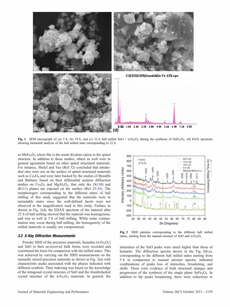
The VSM measurements corresponding to the differentstates of ball milling at room temperature were reported inFig. 3(a-d) to reflect their XRD homologues of Fig. 2(b-e),respectively. These plots showed that the hysteresis decreasedwith ball milling time during the mechanosynthesis of SnFe2O4
from the solid SnO and a-Fe2O3 precursor materials. The plotsshowed a consistent general trend associated with difficulty inthe attainment of saturation, with decreasing saturation as afunction of time. This can be explained from the fact that theparticle sizes were in the nanometer ranges, and progressiveformation of the single phase SnFe2O4 with time. The evolution
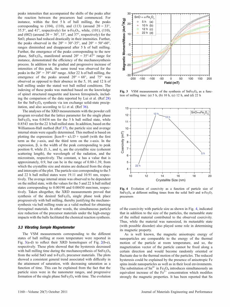
of the coercivity with particle size as shown in Fig. 4, indicatedthat in addition to the size of the particles, the metastable stateof the milled material contributed to the observed coercivity.Thus, while the material was nanosized, its metastable state(with possible disorder) also played some role in determiningits magnetic property.
As is well known, the magnetic anisotropic energy ofnanoparticles are comparable to the energy of the thermalmotion of the particle at room temperature, and so, themagnetization vector of the particle cannot be fixed along acertain direction and would become randomly oriented orfluctuate due to the thermal motion of the particles. The reducedhysteresis could be explained by the presence of anisotropic Fespins inside nanoparticles as well as in their local environments.The substitution of Sn4+ in Fe3O4 introduces simultaneously anequivalent increase of the Fe2+ concentration which modifiesstrongly the magnetic properties. It has been reported that Sn
-200 -150 -100 -50 0 50 100 150 200-30
-20
-10
0
10
20
30SnO + α-Fe
2O
3
5 h (a)(b)(c)(d)
10 h 12 h 22 h
Mag
net
ic M
om
ent/
Mas
s (A
m2 /K
g)
Field (mT)
Fig. 3 VSM measurements of the synthesis of SnFe2O4 as a func-tion of milling time: (a) 5 h, (b) 10 h, (c) 12 h, and (d) 22 h
10 12 14 16 18 20
4
5
6
7
8
9
10
11
12
13
5 h
10 h12 h
22 h
Coe
rciv
ity (
mT
)
Crystallite Size (nm)
SnO+αFe2O
3
Fig. 4 Evolution of coercivity as a function of particle size ofSnFe2O4 at different milling times from the solid SnO and a-Fe2O3
precursors
1160—Volume 20(7) October 2011 Journal of Materials Engineering and Performance
enters magnetite exclusively as Sn4+ into the octahedralsublattice according to Neel (Ref 38). The introduction ofone Sn4+ ions into the B-sublattice would cause the disappear-ance of two Fe3+ ions via (i) direct replacement, and(ii) valency change from three into twofold.
The magnetic saturation decreased progressively with the22 h ball milled material displaying the least value of17.753 A m2/kg and magnetic saturation and remanent ratio,Mr/Ms ratio of 0.0845. This is consistent with the fact thatSnFe2O4 phase formed progressively with ball milling time,together with simultaneous structural changes such as cationicredistributions. Our results were consistent with the results ofLiu and Li (Ref 39) who observed that the coercivity ofSnFe2O4 synthesized via solid reaction in solution decreasedwith particle size reduction at ambient temperature. Thoughthese authors attributed this behavior to the presence ofnoncolinear arrangements of Fe spins inside clusters and aswell as in their local environments which caused decreasedhysteresis, however, the drastic loss of coercivity in theirsample with particles size of 4 nm at 350 K when particleagglomeration should have taken place suggested that otherfactors could have contributed to the coercivity loss. Theoverall coercivity of the milled material was 6.272 mT after 5 hof ball milling, while 4.441 mT corresponding to the 22 h ballmilled state.
3.4 Mossbauer Spectroscopy Measurements
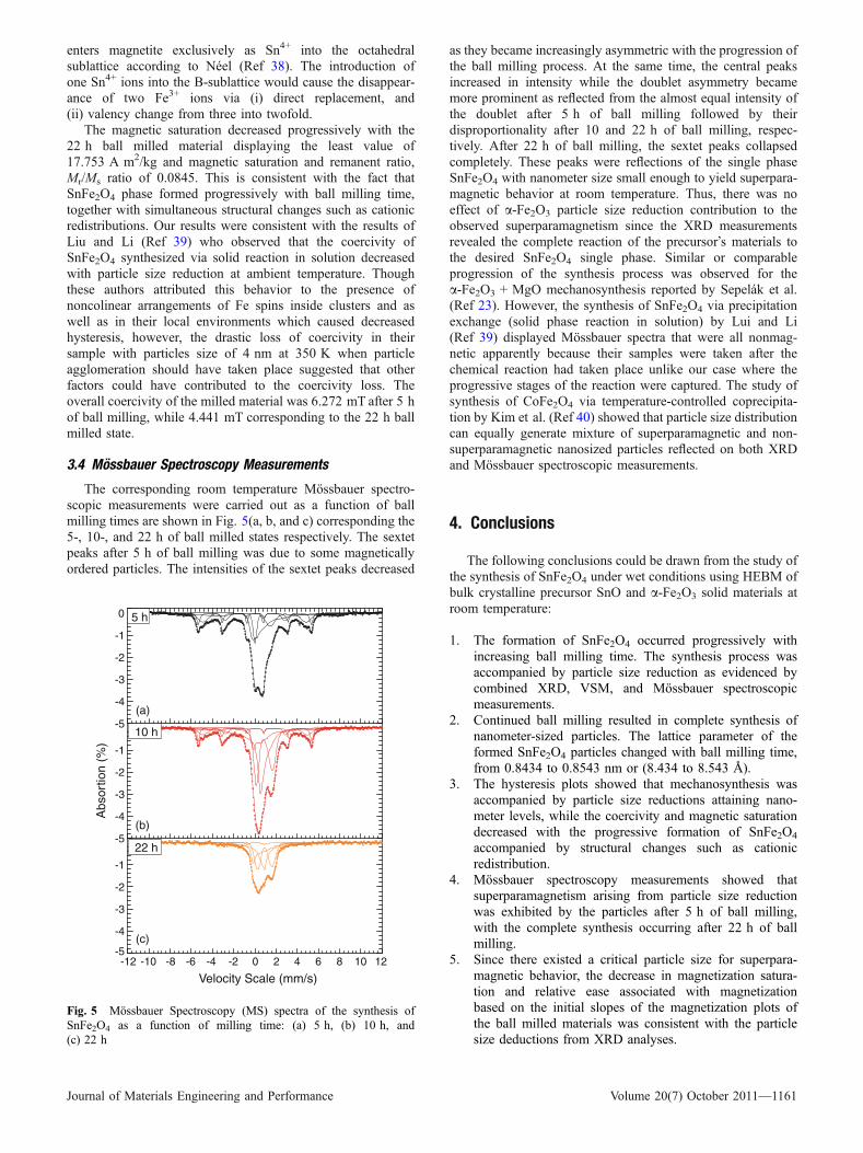
The corresponding room temperature Mossbauer spectro-scopic measurements were carried out as a function of ballmilling times are shown in Fig. 5(a, b, and c) corresponding the5-, 10-, and 22 h of ball milled states respectively. The sextetpeaks after 5 h of ball milling was due to some magneticallyordered particles. The intensities of the sextet peaks decreased
as they became increasingly asymmetric with the progression ofthe ball milling process. At the same time, the central peaksincreased in intensity while the doublet asymmetry becamemore prominent as reflected from the almost equal intensity ofthe doublet after 5 h of ball milling followed by theirdisproportionality after 10 and 22 h of ball milling, respec-tively. After 22 h of ball milling, the sextet peaks collapsedcompletely. These peaks were reflections of the single phaseSnFe2O4 with nanometer size small enough to yield superpara-magnetic behavior at room temperature. Thus, there was noeffect of a-Fe2O3 particle size reduction contribution to theobserved superparamagnetism since the XRD measurementsrevealed the complete reaction of the precursor�s materials tothe desired SnFe2O4 single phase. Similar or comparableprogression of the synthesis process was observed for thea-Fe2O3 + MgO mechanosynthesis reported by Sepelak et al.(Ref 23). However, the synthesis of SnFe2O4 via precipitationexchange (solid phase reaction in solution) by Lui and Li(Ref 39) displayed Mossbauer spectra that were all nonmag-netic apparently because their samples were taken after thechemical reaction had taken place unlike our case where theprogressive stages of the reaction were captured. The study ofsynthesis of CoFe2O4 via temperature-controlled coprecipita-tion by Kim et al. (Ref 40) showed that particle size distributioncan equally generate mixture of superparamagnetic and non-superparamagnetic nanosized particles reflected on both XRDand Mossbauer spectroscopic measurements.
4. Conclusions
The following conclusions could be drawn from the study ofthe synthesis of SnFe2O4 under wet conditions using HEBM ofbulk crystalline precursor SnO and a-Fe2O3 solid materials atroom temperature:
1. The formation of SnFe2O4 occurred progressively withincreasing ball milling time. The synthesis process wasaccompanied by particle size reduction as evidenced bycombined XRD, VSM, and Mossbauer spectroscopicmeasurements.
2. Continued ball milling resulted in complete synthesis ofnanometer-sized particles. The lattice parameter of theformed SnFe2O4 particles changed with ball milling time,from 0.8434 to 0.8543 nm or (8.434 to 8.543 A).
3. The hysteresis plots showed that mechanosynthesis wasaccompanied by particle size reductions attaining nano-meter levels, while the coercivity and magnetic saturationdecreased with the progressive formation of SnFe2O4
accompanied by structural changes such as cationicredistribution.
4. Mossbauer spectroscopy measurements showed thatsuperparamagnetism arising from particle size reductionwas exhibited by the particles after 5 h of ball milling,with the complete synthesis occurring after 22 h of ballmilling.
5. Since there existed a critical particle size for superpara-magnetic behavior, the decrease in magnetization satura-tion and relative ease associated with magnetizationbased on the initial slopes of the magnetization plots ofthe ball milled materials was consistent with the particlesize deductions from XRD analyses.
-12 -10 -8 -6 -4 -2 0 2 4 6 8 10 12-5
-4
-3
-2
-1
-5
-4
-3
-2
-1
-5(a)
(b)
(c)
-4
-3
-2
-1
0
Velocity Scale (mm/s)
22 h
Abs
ortio
n (%
)
10 h
5 h
Fig. 5 Mossbauer Spectroscopy (MS) spectra of the synthesis ofSnFe2O4 as a function of milling time: (a) 5 h, (b) 10 h, and(c) 22 h
Journal of Materials Engineering and Performance Volume 20(7) October 2011—1161
Acknowledgment
The support of NSF-DMR PREM at UPRM based on Grant No.0351449 for the authors is hereby acknowledged.
References
1. L. Neel, Superparamagnetisme de grains tres fins antiferromagnetiques,C. R. Acad Sci., 1961, 252, p 4075–4080 (Translated in Selected Worksof L. Neel (Kurti, N., ed.), New York, Gordon and Breach, 1988,p 107–110)
2. R.W. Chantrell and K. O�Grady, The Magnetic Properties of FineParticles, Applied Magnetism, C.D. Wright and G. Asti, Ed., KluwerAcademic Publishers, The Netherlands, 1994, p 113–164
3. T. Nakamura, T. Tsutaoka, and K. Hatakeyama, Frequency Dispersionof Permeability in Ferrite Composite Materials, J. Magnet. Magnet.Mater., 1994, 138(3), p 319–328
4. T. Tsutaoka, M. Ueshima, T. Tokunaga, T. Nakamura, andK. Hatakeyama, Frequency Dispersion and Temperature Variation ofComplex Permeability of Ni-Zn Ferrite Composite Materials, J. Appl.Phys., 1995, 78, p 3983–3991
5. T.M. Clark and B.J. Evans, Enhanced Magnetization and CationDistributions in Nanocrystalline ZnFe2O4: A Conversion ElectronMossbauer Spectroscopic Investigation, IEEE Trans. Magnet., 1997,33(5 Part 2), p 3745–3747
6. J.A. Jacobs and T.F. Kilduff, Engineering Materials Technology,Structures, Processing, Properties, and Selection, 5th ed., PearsonPrentice Hall, USA, 2005
7. H.M. Yang, X.C. Zhang, A.D. Tang, and G.Z. Qui, Cobalt FerriteNanoparticles Prepared by Coprecipitation/Mechanochemical Treat-ment, Chem. Lett., 2004, 33, p 826–827
8. G. Ennas, G. Marongiu, S. Marras, and G. Piccaluga, Mechanochem-ical Route for the Synthesis of Cobalt Ferrite-Silica and Iron-CobaltAlloy-Silica Nanocomposites, J. Nanopart Res., 2004, 6, p 99–105
9. S. Bid and S.K. Pradhan, Microstructure Characterization and PhaseTransformationKinetic studyofmechanosynthesizedNon-stoichiometricCdF2O4 by Rietveld�s Analysis, Jpn. J. Appl. Phys., 2004, 43, p 5455–5464
10. M. Mozaffari and J. Amighian, Preparation of Al-Substituted Ni FerritePowders Via Mechanochemical Processing, J. Magn. Mater., 2003,260, p 244–249
11. H.M. Widatallah and F.J. Berry, The Influence of Mechanica Millingand Subsequent Calcination on the Formation of Lithium Ferrites,J. Solid State Chem., 2002, 164, p 230–236
12. Z.T. Lui and O.N.C. Uwakweh, Ball Milling of Fe-Zn Intermetallics,J. Mater. Res., 1996, 11(7), p 1665–1672
13. Z. Lui and O.N.C. Uwakweh, Mossbauer Effect Study of MechanicallyAlloyed C and C1-Fe-Zn Intermediate Phases, Metall. Mater. Trans. A,1997, 28 A, p 743–747
14. O. Uwakweh, Z. Lui, A. Jordan, B. Chakoumakos, S. Spooner, andP. Maziasz, Neutron Diffraction and Phase Evolution of the Mechan-ically Alloyed Intermetallic Compound 1-FeZn, Metall. Mater. Trans.A, 2000, 31 A, p 2739–2745
15. C.C. Koch, Mechanical Milling and Alloying, Materials Science andTechnology, R.W. Cahn, P. Haasen, and E.J. Kramer, Ed., VCHPublishers, Weinheim, 1991, p 193–245
16. C. Suryanarayana, Mechanical Alloying, Progr. Mater. Sci., 2001, 46,p 1–184
17. J. Ding, W.M. Miao, P.G. McCormick, and R. Street, High MagneticPerformance in Mechanically Alloyed Co-Substituted Fe3O4, J. Appl.Phys. Lett., 1994, 65(24), p 3135–3136
18. J. Ding, P.G. McCormick, and R. Street, Formation of SpinelMn-Ferrrite During Mechanical Alloying, J. Magn. Magn. Mater.,1997, 171(3), p 309–314
19. N. Guigue-Millot, S. Begin-Colin, Y. Champion, M.J. Hytch, G. LeCaer, and P. Perriat, Control of Grain Size and Morphologies ofNanograined Ferrites by Adaptation of the Synthesis Route: Mechano-synthesis and Soft Chemistry, J. Solid State Chem., 2003, 170(1),p 30–38
20. V. Sepelak, U. Steinike, D.C. Uecker, S. Wissmann, and K.D. Becker,Structural Disorder in Mechanosynthesized Zinc Ferrite, J. Solid StateChem., 1998, 135(1), p 52–58
21. E. Avvakumov, M. Senna, and N. Kosova, Soft MechanochemicalSynthesis: A Basis for New Chemical Technologies, Kluwer AcademicPublishers, Boston, 2001
22. T. Verdier, N. Nachbaur, and M. Jean, Mechanosynthesis of ZincFerrite in Hardened Steel Vials: Influence of ZnO on the Appearance ofFe(II), J. Solid State Chem., 2005, 178, p 3243–3250
23. V. Sepelak, A. Feldhoff, P. Heitjans, F. Krumeich, D. Menzel,F.J. Litterst, I. Bergmann, and K.D. Becker, Nonequilibrium CationDistribution, Canted Spin Arrangement, and Enhanced Magnetizationin Nanosized MgFe2O4 Prepared by a One-Step MechanochemicalRoute, Chem. Mater., 2006, 18, p 3057–3067
24. F. Goya and H.R. Rechenberg, On the Magnetic Properties ofMechanosynthesized and Ball Milled Spinel Ferrites, Mater. Sci.Forum, 2002, 403, p 127–132
25. R.H. Kodama, S.A. Makhlouf, and A.E. Berkowitz, Finite Size Effectsin Antiferromagnetic NiO Nanoparticles, Phys. Rev. Lett., 1997, 79(7),p 1393–1396
26. N.A. Drokin, E. Yu Aksenova, and Yu.A. Mamalui, Photoconductivityin Tin-Doped Magnetite, Soviet Phys. Solid State, 1984, 26, p 1837–1838
27. F.J. Berry and O. Helgason, Mossbauer Spectroscopic Properties ofTin-Doped Iron Oxides, Hyperfine Interact., 2000, 126(1–4), p 269–275
28. F. Liu, T. Li, and H. Zheng, Structure and Magnetic Properties ofSnFe2O4 Nanoparticles, Phys. Lett. A, 2004, 323, p 305–309
29. Available at www.SEECO.us30. M.R. Anantharaman, S. Reijne, J.P. Jacobs, H.H. Brongersma,
R.H.H. Smits, and K. Seshan, Preferential Exposure of CertainCrystallographic Planes on the Surface of Spinel Ferrites: A Studyby LEIS on Polycrystalline Spinel Ferrite Surfaces, J. Mater. Sci.,1999, 34, p P4279–P4283
31. H. Knozinger and P. Ratnaswamy, Catalytic Aluminas: SurfaceModels and Characterization of Surface Sites, Catal. Rev., 1978,17(1), p 31–70
32. M. Shelef, M.A.Z. Wheeler, and H.C. Yao, Ion Scattering Spectra fromSpinel Surfaces, Surf. Sci., 1975, 47, p 697–703
33. H.C. Yao and M. Shelef, Nitric Oxide and Carbon MonoxideChemisorption on Cobalt-Containing Spinels, J. Phys. Chem., 1974,78(24), p 2490–2496
34. J.P. Beaufils and J. Barbaux, Determination, Par Diffraction Differen-tielle de Neutrons, des Faces Cristallines Exposees par des Supports deCatalyseurs en Poudre, J. Chim. Phys., 1981, 78, p 347–352
35. J.P. Beaufils and J. Barbaux, Study of Adsorption on Powders bySurface Differential Diffraction Measurements. Argon on Co3O4,J. Appl. Cryst., 1982, 15, p 301–307
36. F. Li, H. Wang, L. Wang, and J. Wang, Magnetic Properties ofZnFe2O4 Nanoparticles Produced by a Low-Temperature Solid StateReaction Method, J. Magn. Magn. Mater., 2007, 309(2), p 295–299
37. B.D. Culity and S.R. Stock, Elements of X-ray Diffraction, 3rd ed.,Prentice Hall, USA, 2001
38. L. Neel, Some Theoretical Aspects of Rock Magnetism, Adv. Phys.,1955, 4, p 191–243
39. F.X. Liu and T.Z. Li, Synthesis and Magnetic Properties of SnFe2O4
Nanoparticles, Mat. Lett., 2005, 59(2–3), p 194–19640. Y. Kim, II, D. Kim, and C.S. Lee, Synthesis and Characterization of
CoFe2O4 Magnetic Nanoparticles Prepared by Low Temperature-Controlled Coprecipitation Method, Physica B, 2003, 337(1–4),p 42–51
1162—Volume 20(7) October 2011 Journal of Materials Engineering and Performance
Copyright © 2022 FDOKUMEN