Synthesis of hexagonal lanthanum germanate apatites through site selective isovalent doping with...
14
1 Synthesis of hexagonal lanthanum germanate apatites through site selective isovalent doping with yttrium E. Kendrick and P.R. Slater * Chemistry, University of Surrey, Guildford, Surrey. GU2 7XH, UK * Correspondence to: Dr. P.R. Slater Materials Chemistry Group, Chemistry, SBMS, University of Surrey, Guildford, Surrey. GU2 7XH. UK Tel. +44 (0)1483 686847 Fax +44 (0)1483 686851 [email protected]
-
Upload
independent -
Category
Documents
-
view
1 -
download
0
Transcript of Synthesis of hexagonal lanthanum germanate apatites through site selective isovalent doping with...

1
Synthesis of hexagonal lanthanum germanate apatites through site selective isovalent
doping with yttrium
E. Kendrick and P.R. Slater*
Chemistry, University of Surrey, Guildford, Surrey. GU2 7XH, UK
*Correspondence to:
Dr. P.R. Slater
Materials Chemistry Group, Chemistry, SBMS, University of Surrey, Guildford,
Surrey. GU2 7XH. UK
Tel. +44 (0)1483 686847
Fax +44 (0)1483 686851

2
Abstract
Apatite-type lanthanum silicates/germanates have been attracting considerable interest as
a new class of oxide ion conductors, whose conductivity is mediated by oxide ion
interstititials. For the germanates, it has been shown that, depending on composition, the
cell can be either hexagonal or triclinic, with evidence for reduced low temperature
conductivities for the latter, attributed to increased defect trapping in this lower symmetry
cell. In this paper we show that site selective doping of Y into the triclinic apatite-type
oxide ion conductors, La9.33+z(GeO4)6O2+3z/2 (0.33≤z≤0.67) results in a hexagonal lattice
for the complete series with correspondingly enhanced low temperature conductivity.
Keywords: A Oxides, B. Chemical Synthesis, C. X-ray diffraction, D. Ionic
Conductivity.
3
Introduction
Apatite materials have attracted considerable interest for a range of applications,
including electrolytes for solid oxide fuel cells (SOFCs), bioceramics for bone implants,
and hazardous waste encapsulation materials. In terms of the former it is the Lanthanum
silicate and germanate materials, La9.33+z(Si/GeO4)6O2+3z/2 which have attracted
considerable attention due to their high oxide ion conductivities at elevated
temperatures [1-17]. In contrast to the traditional fluorite and perovskite-type oxide ion
conductors, where conductivity is mediated by oxide ion vacancies, the weight of
experimental and theoretical evidence for the apatite systems indicates that they conduct
via an interstitial mechanism. Consequently high conductivity is favoured by the
presence of oxygen interstitials, which can be either present as oxygen
hyperstoichiometry (z>0) or Frenkel defects [4,9,10,12,13].
So far, the majority of the work on these apatite systems have focused on the silicate
systems, which can be partly related to the additional complexities shown by the apatite
germanates. In particular, the germanates are complicated by the change in the crystal
symmetry from hexagonal to triclinic as the La content/oxygen content increases. In
this respect Leon-Reina et al. have reported the preparation of single phase samples of
La9.33+z(GeO4)6O2+3z/2 for 0.19≤z≤0.42, with samples in the range 0.19≤z≤0.27
possessing hexagonal symmetry, while samples with higher La content, 0.33≤z≤0.42,
exhibit a triclinic cell [6]. In terms of potential applications the triclinic cell is not ideal,
as it results in lower oxide ion conductivities at low temperatures, most likely due to
enhanced defect trapping in the lower symmetry cell.
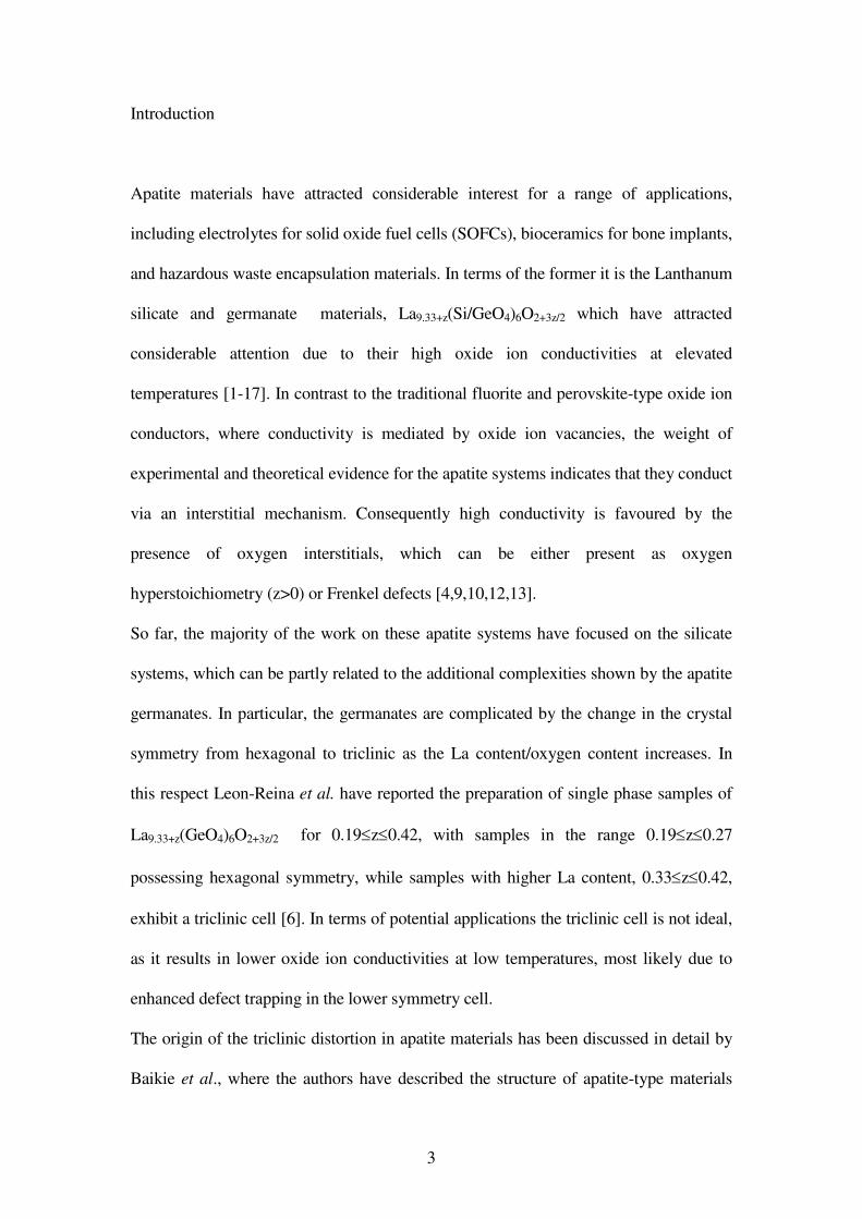
The origin of the triclinic distortion in apatite materials has been discussed in detail by
Baikie et al., where the authors have described the structure of apatite-type materials

4
(A10(MO4)6X2; A=alkaline earth, rare earth, Pb; M=Si, Ge, P, V; X=O, OH, Halides) in
terms of a “microporous” framework (A(1)4(MO4)6) composed of face sharing A(1)O6
trigonal meta-prismatic columns, that are corner connected to the MO4 tetrahedra
(figure 1) [17]. This framework allows some flexibility to accommodate the remaining
A(2)6X2 units. A triclinic apatite cell is obtained in systems such as the lanthanum
germanates due to the large size of the A(1)4(MO4)6 framework. This places extension
stress at the A cation site in the A(2)6X2 unit, and compression stress at the M site,
which are relieved by the MO4 tetrahedra tilting in the triclinic cell. The presence of
interstitial oxygen atoms also enhances triclinic distortion, most probably by
contributing to a further expansion of the A(1)4(MO4)6 framework, hence accounting
for the fact that samples with low oxygen excess (z≤0.27) are hexagonal, whereas
higher oxygen content samples are triclinic [6]
The ideal way to overcome this structural stress would be to increase the size of the
A(2) cations. Thus Ca10V6O24F2 is triclinic, while isovalent doping with the larger Pb
cation results in hexagonal symmetry [17]. However, such an isovalent doping strategy
is not possible for La9.33+z(GeO4)6O2+3z/2, as La is the largest lanthanide. Therefore, we
have investigated an alternative approach, namely the doping of a smaller rare earth, Y,
into La9.33+z(GeO4)6O2+3z/2, with the aim of selectively substituting for the La site in the
A(1)4(MO4)6 framework in order to relieve strain by reducing the size of this
framework. Atomistic modelling studies of the related apatite lanthanum silicates have
predicted a preference for Y substitution in the A(1)4(MO4)6 framework [18]. In this
paper we confirm that such site selective doping is successful, and does indeed lead to
the formation of hexagonal apatites even for high oxygen contents.
Experimental

5
Samples, La9.33+z-xYx(GeO4)6O2+3z/2 (0≤z≤0.67; x=1, 2, 3), were prepared from high
purity La2O3, Y2O3 and GeO2. The dried powders were intimately mixed in the correct
ratios and heated to 1100°C for 24 hours with an intermediate regrind. Phase purity was
established through X-ray powder diffraction (Panalytical X’Pert Pro diffractometer, Cu
Kα1 radiation). For preliminary structural studies to determine the location of the Y
dopant, data were collected over 12 hours in the range 10-120° (2θ). Rietveld analysis
was then performed using the GSAS suite of programs, with the fits compared for Y on
each of the La sites [19].
Pellets (1.6 cm diameter) for conductivity measurements were prepared as follows: the
powders were ball milled (350 rpm, Fritsch Pulverisette 7 Planetary Mill) for 1 hour
before pressing at 8000 kg cm-2
. The pressed pellets were then heated at 1300°C for 2
hours, leading to densities > 85% theoretical. Both sides of the pellet were coated with
Au paste and then heated to 700°C for 1 hour to ensure bonding to the pellet.
Conductivity measurements were made in air using AC impedance spectroscopy
(Hewlett Packard 4182A impedance analyser).
Results and discussion
X-ray diffraction indicated that single phase samples of La9.33+z-xYx(GeO4)6O2+3z/2 were
formed for 0.22≤z≤0.67 (y=1, 2, 3), representing a larger solid solution range compared
to that reported by Leon-Reina et al. for the undoped system (0.19≤z≤0.42) [6].
Contrary to samples without Y doping, where a hexagonal lattice is obtained for
0.19≤z≤0.27, with triclinic samples observed for higher values of z, all Y doped
compositions (0.22≤z≤0.67) were shown to be hexagonal (figure 2).

6
In order to determine the location of the Y dopant, preliminary refinement of X-ray
diffraction data was performed for a range of samples. In all cases the R factors were
lowest for Y located in the A cation site within the A(1)4(MO4)6 framework (e.g.
La8Y2(GeO4)6O3; Chi2 = 2.83 (Y on A(1) site), 4.67 (Y on A(2) site). Thus in
agreement with the predicted strategy, selective doping of Y in the A(1)4(MO4)6
framework stabilises the hexagonal lattice. Due to the general insensitivity of X-rays to
light atoms, such as oxygen, and the considerable structural distortion around the
interstitial site, further more detailed neutron diffraction studies are planned to
structurally characterise these systems fully, particularly to elucidate the oxygen
interstitial sites.
Refinement of the cell parameters shows that the Y doping leads to an overall reduction
in unit cell size as expected due to the smaller size of Y3+
compared to La3+
, with the
cell parameters obeying Vegard’s law, consistent with Y substitution on the same site
across the series. However, this contraction is not uniform in all directions, with the
main contraction along the c axis. This can be most clearly visualised by plotting the %
change along each of the unit cell axes with increasing Y content (figure 3). Further
increases in Y content are also possible, i.e. x>3, however peak broadening indicative
of a return to the triclinic cell is observed for these higher Y dopant levels.
High oxide ion conductivities were observed for all samples (0.025-0.040 Scm-1
at
800°C). Compared to triclinic La9.33+z(GeO4)6O2+3z/2 samples without Y doping, the
conductivities were enhanced at lower temperatures, while at higher temperatures they
are comparable (figure 4). The convergence of the data at higher temperatures (>700°C)
can be related to the fact that in this temperature range, even the undoped
La9.33+z(GeO4)6O2+3z/2 samples are hexagonal; prior reports showing that there is a
change from a triclinic to a hexagonal cell at high temperatures [7,16]. Thus the

7
enhanced lower temperature conductivity can be correlated with the more facile oxide
ion conduction within the higher symmetry hexagonal lattice.
Conclusions
In summary, we have shown that site selective doping of La9.33+z(GeO4)6O2+3z/2 with a
smaller rare earth, Y, leads to a stabilisation of the hexagonal lattice, even for high
oxygen contents. Furthermore this has the effect of enhancing the low temperature
conductivities.
References
1. S. Nakayama, H. Aono, Y. Sadaoka, Chem. Lett. (1995) 431.
2. S. Nakayama, M. Sakamoto, M. Higuchi, K. Kodaira, J. Mater. Sci. Lett. 19 (2000)
91.
3. S. Tao, J.T.S. Irvine, Mater. Res. Bull. 36 (2001) 1245.
4. J.E.H. Sansom, D. Richings, P.R. Slater, Solid State Ionics 139 (2001) 205.
5. H. Arikawa, H. Nishiguchi, T. Ishihara, Y. Takita, Solid State Ionics 136-137
(2000) 31.
6. L. Leon-Reina, M.E. Martin-Sedeno, E.R. Losilla, A. Caberza, M. Martinez-Lara, S.
Bruque, F.M.B. Marques, D.V. Sheptvakov, M.A.G. Aranda; Chem. Mater. 15 (2003)
2099.
7. E.J. Abram, C.A. Kirk, D.C. Sinclair, A.R. West; Solid State Ionics 176 (2005)
1941.
8. J.E.H. Sansom, L. Hildebrandt, P.R. Slater, Ionics 8 (2002) 155.
9. J.R. Tolchard, M.S. Islam, P.R. Slater; J. Mater. Chem. 13 (2003) 1956.
8
10. L. Leon-Reina, E.R. Losilla, M. Martinez-Lara, M.C. Martin-Sedeno, S. Bruque,
P.Nunez, D.V. Sheptyakov, M.A.G. Aranda; Chem. Mater. 17 (2005) 596.
11. V.V. Kharton, A.L. Shaula, M.V. Patrakeev, J.C. Waerenborgh, D.P. Rojas, N.P.
Vyshatko, E.V. Tsipis, A.A. Yaremchenko, F.M.B. Marques; J. Electrochem. Soc.
151 (2004) A1236.
12. J.E.H. Sansom, J.R. Tolchard, D. Apperley, M.S. Islam, P.R. Slater; J. Mater.
Chem. 16 (2006) 1410.
13. E. Kendrick, M.S. Islam, P.R. Slater; J. Mater. Chem. 2007,
DOI:10.1039/B704426G
14. Y. Masubuchi, M. Higuchi, S. Kikkawa, K. Kodaira, S. Nakayama; Solid State
Ionics 175 (2004) 357.
15. S. Celerier, C. Laberty-Robert, J.W. Long, K.A. Pettigrew, R.M. Stroud, D.R.
Rolison, F. Ansart, P. Stevens, Adv. Mater. 18 (2006) 615.
16. L. Leon-Reina, J.M. Porras-Vasquez, E.R. Losilla, M.A.G. Aranda; J. Solid State
Chem. 180 (2007) 1250.
17. T. Baikie, P.H.J. Mercier, M.M. Elcombe, J.Y. Kim, Y. Le Page, L.D. Mitchell,
T.J. White, P.S. Whitfield; Acta Crystallographica B 63 (2007) 251.
18. J. R. Tolchard, P. R. Slater and M. S. Islam; Adv. Funct. Mater. (2007) DOI
10.1012/adfm.200600789.
19. A.C. Larson, R.B. Von Dreele. Los Alamos National Laboratory, Report. No
LA-UR-86-748, 1987.

9
Figure Legends
Figure 1(a) Illustration of the “microporous” A(1)4(MO4)6 framework of the apatite
(A10(MO4)6X2) structure (tetrahedra=MO4; A(1) cations at centre of trigonal meta-
prisms). The remaining A(2)6X2 units occupy the cavities within this framework. (b)
Illustration of the structure as a whole (Large White Spheres= A2 cations, Small
White Spheres= X anions).
Figure 2. X-ray diffraction patterns for La9.83(GeO4)6O2.75 and La8Y2(GeO4)6O3.
Figure 3. Plot of % change in cell parameters and cell volume with increasing Y
content for La9.55-xYx(GeO4)6O2.33.
Figure 4. Conductivity plots for La9.83(GeO4)6O2.75 (�), La7.83Y2(GeO4)6O2.75 (�),
and La8Y2(GeO4)6O3 (�)
10
Figure 1a

11
Figure 1b

12
Figure 2

13
Figure 3
-4
-3
-2
-1
0
1
0 1 2 3
Y content (x)
% c
han
ge
a
c
Volume

14
Figure 4
-3
-2
-1
0
1
2
0.9 1 1.1 1.2 1.3 1.4 1.5 1.6
1000K/T
log
σσ σσT
/Scm
-1K