Electrical and magnetic properties of nanomaterials containing iron or cobalt nanoparticles
Upload
khangminh22Category
view
4download
0
Synthesis and Magnetic Properties of Cobalt Nickel Nanoparticles
Prepared by Chemical Reduction Methods
Jalpa Dipesh Patel
Thesis submitted to the University of London, University College London in partial fulfilment of the requirements for the degree of
Doctor of Philosophy In Chemistry
Supervised by:
Prof Ivan P. Parkin, Department of Chemistry, University College London
Prof Quentin A. Pankhurst, London Centre for Nanotechnology, University CollegeLondon
June 2007
-1-

UMI Number: U592381
All rights reserved
INFORMATION TO ALL USERS The quality of this reproduction is dependent upon the quality of the copy submitted.
In the unlikely event that the author did not send a complete manuscript and there are missing pages, these will be noted. Also, if material had to be removed,
a note will indicate the deletion.
Dissertation Publishing
UMI U592381Published by ProQuest LLC 2013. Copyright in the Dissertation held by the Author.
Microform Edition © ProQuest LLC.All rights reserved. This work is protected against
unauthorized copying under Title 17, United States Code.
ProQuest LLC 789 East Eisenhower Parkway
P.O. Box 1346 Ann Arbor, Ml 48106-1346

Declaration
I, Jalpa Dipesh Patel, confirm that the work presented in this thesis is my own. Where
information has been derived from other sources, I confirm that this has been indicated
in the thesis.
-2-

ABSTRACT
The purpose of this work was to prepare and characterise CoxNii.x/CoNiO core-shell
magnetic nanoparticles which showed magnetic exchange bias. The particles were
synthesised using a variety of stabilising surfactants and nucleating seeds, via the polyol
reduction method. The surfactants were used to coat nanoparticles of various diameters,
to prevent agglomeration and oxidation. A mixture of 1:1 oleic acid: oleylamine was
found to be the best stabilising agent for the particles as it protected against complete
oxidation whilst allowing a partial oxide shell to form. Phosphine-based surfactants
yielded particles with spherical morphologies. However, these particles were too small
to support oxide-shell growth, and oxidised fully to antiferromagnetic phases. The
nucleation of particles was probed using homogeneous and heterogeneous methods.
Homogeneous nucleation resulted in particles which had predominantly oxidised to the
core, and therefore did not yield pronounced exchange bias effects. Heterogeneous
nucleation was attempted using various seeding techniques and seed materials. Platinum
seeds were found to be the most effective in controlling the size of CoxNii_x
nanoparticles. They yielded larger particles with core-shell morphology. Following
optimisation of the synthesis conditions, a compositional series of CoxNii.x
nanoparticulate composite alloys were made. All samples were analysed using TEM to
determine the size and structure of the individual particles. A number of other
techniques including X-ray diffraction, X-ray photoelectron spectroscopy, Energy-
dispersive X-ray analysis, Electron energy loss spectroscopy and Thermogravimetric
magnetic analysis, were also used to fully characterise the phase, crystallinity,
composition and oxidation in individual particles. The magnetic properties of the
particles, made using the various reaction conditions, were measured using the SQUID
technique. Exchange bias has been observed in several of the alloyed samples in
additional to the cobalt particles made using the polyol technique. Nickel particles did
not display any characteristic exchange bias properties.

Acknowledgements
I would firstly like to thank my supervisors, Prof. Ivan Parkin and Prof. Quentin
Pankhurst, for their support, guidance and insight over the last few years. I also would
like to thank Dr Dorothy Farrell for her guidance which has been invaluable. Dorothy
also helped obtain the SQUID measurements that are reported in this thesis, training in
TEM analysis and particle synthesis. I thank Dr Ana Garcia Prieto for her effort and
insight into characterisation of the synthesised particles using XRD analysis. Dr Steve
Firth provided a great deal of assistance with technical aspects relating to the TEM and
in particular, helped with my understanding of the TGA technique. Robert Palgrave is
thanked for carrying out XPS measurements on the samples reported in this thesis, as is
Mrs Jillian Maxwell for elemental analysis. Kevin Reeves is also thanked for helping to
refresh my memory on the SEM and EDX analysis of nanoparticles. John Hill, Dave
Knapp, Dave Ladd, Joe Nolan and Phil Hayes are thanked for kindly taking the time to
help me with various technical problems I have had over the years. Their effort and
enthusiasm is much appreciated.
All members of the Parkin and Carmalt research groups are thanked for making
the lab an enjoyable place to work, and in that respect I would particularly like to thank
Robert Palgrave, Sobia Ashraf, Nicolas Boscher, Dr Jesus Gil-Tomas, Plaboni Hasan
and Uzma Qureshi, Kristopher Page, Stephen Potts, Naima Narband, Dr Clara
Piccirillo, Paulo Melgari who have been fun to work with. Dr Chris Blackman, Dr
Russell Binions, Dr Geoff Hyett and Dr Troy Manning are thanked for many useful
discussions over the past four years, and for general help with Schlenk line work,
synthetic methods and analysis. I would particularly like to thank Dr Siama Basharat,
Dr Ashti Rampaul and Dr Dina Solanki for being the great colleagues and friends that
they are. The advice they have given me over the past few years has been more
important than they probably realise.
There are always so many friends and family members to thank, however I
would like to begin by taking the opportunity to thank my paternal grandparents, Mr
Bhanubhai and Mrs Bhanuben Patel, for their love and encouragement over the years.
Their encouraging phone calls from India have always been valued, as has the advice
they have given me. I would also like to thank my maternal grandparents, Late Mr
-4-

Kanubhai Patel and Mrs Induben Patel for their support and encouragement over the
years. My great-uncle Bhagwati Patel is also thanked for the interest and enthusiasm he
has expressed when asking about the progress I have made in my studies.
My parents, Ranjit and Urvashi, have always been very supportive throughout
my studies, encouraging me to achieve the best I possibly can. I am indebted to them for
the sacrifices they have made to ensure that I had the opportunities they may not have
had themselves and know they are happy and proud to see me finally finish after all
these years of studying! I thank my brother, Pritesh, for being such fun and for just
generally being there for me over the years. Thanks bro.
I am especially grateful to Narendra and Harminda for their support and
encouragement over the past three years. I would also like to thank the rest of my
husband’s family (Pushpa Ba, aunties, uncles and cousins), you have all been
wonderfully supportive. In particular, Rajan Patel, Pina Patel, Asha Patel and Raul
Patel are thanked for making the last couple of years a lot of fun!
I would like to thank all my aunties, uncles and cousins, of which there are too
many to list! However, a special mention must go to Atin, Kirti, Nikita and Sandhya for
their support and also to Varsha Gallen and Maya Gallen for their friendship. There are
many friends I would like to thank, however, Khola Khan, Farkhanda Uddin, Alay
Patel, Pritesh Patel, Priya Patel and Jessica Patel, in particular, are thanked for their
support, encouragement and friendship. I would also like to thank the Munch Bunch
(Tan, Niketa, Manish, Pranali, Josh, Jo, Chirag, Nirav, Praveen, Upeksha, Ketan, Nilu)
for being the lovely people that they are, and for sharing my love of all things food
related, over the past few years. Dip’s friends (Chetan, Priya, Hursh, Nihar, Neel, Sagar,
Ravs, Rehan, Rooby and little Ehsan) who have also become my friends over the last 3
years, are thanked for being a great laugh and for encouraging me to finish.
Finally, I would like to thank my husband, Dipesh, for his love and friendship.
He has helped me through my various crises, making sure I never lost track of the
bigger picture and kept me motivated during the most challenging phases of my
research. I couldn’t have done it without you.

Table of Contents
Title page 1
Declaration 2
Abstract 3
Acknowledgements 4
Table of Contents 6
List of figures 11
List of tables 17
List of schemes 17
List of abbreviations 18
Chapter 1 Introduction 20
1.1 Background 21
1.2 Polyol reduction synthesis 22
1.3 Particle formation mechanism 23
1.3.1 Nucleation step 26
1.3.2 Growth step 26
1.4 Homogeneous nucleation 27
1.5 Heterogeneous nucleation 29
1.6 Surfactant contributions 31
1.7 Magnetism 33
1.7.1 Fundamentals of magnetism 33
1.7.2 Classes of magnetic materials 35
1.8 Hysteresis in magnetism 44
1.9 Exchange anisotropy 46
1.10 References 49
Chapter 2 Polyol synthesis of CoxNii.x nanoparticles 58
2.1 Introduction 59
2.2 Overview of reaction 60
2.3 Reaction schematic 60
2.4 Homogeneous nucleation 63
2.4.1 Introduction 63
-6-

2.4.2 Synthesis of CosoNiioparticles by homogeneous nucleation 63
2.4.3 Results 63
2.5 Heterogeneous nucleation 67
2.5.1 Introduction 67
2.5.1.1 Platinum seeds 67
2.5.1.1.1 Introduction 67
2.5.1.1.2 Synthesis o f Co8oNi2oparticles with platinum 6 8
seeds
2.5.1.1.3 Results 6 8
2.5.1.2 Use of premade nanoparticles as nuclei for further 74
growth
2.5.1.2.1 Introduction 74
2.5.1.2.2 Synthesis of CogoNi^oparticles with premade 74
nanoparticle seeds
2.5.1.2.3 Results 76
2.5.1.3 Other Noble metal seeds 79
2.6 Variations in stabilising surfactants 79
2.6.1 Introduction to variations in stabilising surfactant 79
2.6.2 Oleic Acid (OA) 82
2.6.2.1 Introduction 8 2
2.6.2.2 Synthesis o f Co8oNi2o particles using oleic acid 83
2.6.2.3 Results 83
2.6.3 Oleylamine (OY) 85
2.6.3.1 Introduction 85
2.6.3.2 Synthesis o f Co8oNi2o particles using oleylamine 85
2.6.3.3 Results 85
2.6.4 Oleic Acid/Oleylamine mixture (OA/OY) 88
2.6.4.1 Introduction 88
2.6.4.2 Synthesis o f Co8oNi2o particles using oleic acid / 89
oleylamine
2.6 4.3 Results 89
2.6.5 Trioctylphosphine oxide (TOPO) 92
2.6.5.1 Introduction 92
-7-

2.6.52 Synthesis o f Co8 ^ 2 0 particles using trioctylphosphine 92
oxide
2.6.53 Results 93
2.6.6 Tributylphosphine (TBP) 95
2.6.6.1 Introduction 95
2.6.6.2 Synthesis of CosoNi2o particles using tributylphosphine 96
2.6.6.3 Results 96
2.6.7 Summary of samples made with different surfactant stabilisers 98
2.7 Composition study 99
2.7.1 Introduction to composition series 99
2.7.2 Cobalt nanoparticles 99
2.7.2.1 Introduction 99
2.7.2.2 Synthesis of cobalt nanoparticles 100
2.7.2.3 Results 100
2.7.3 CogoNi2o nanoparticles 107
2.7.3.1 Introduction 107
2.7.3.2 Synthesis of CosoNix) nanoparticles 107
2.7.3.3 Results 108
2.7.4 Co^Ni^ nanoparticles 115
2.7.4.1 Introduction 115
2.7.4.2 Synthesis of C060Ni40 nanopa rticles 115
2.7.4.3 Results 116
2.7.5 CosoNiso nanoparticles 119
2.7.5.7 Introduction 119
2.7.5.2 Synthesis of CosoNiso nanoparticles 119
2.7.5.3 Results 119
2.7.6 Co4oNi6o nanoparticles 126
2.7.6.7 Introduction 126
2.7.6.2 Synthesis of C080NH20 nanoparticles 126
2.7.6.3 Results 126
2.7.7 Co2oNigo nanoparticles 128
2.7.7.7 Introduction 128
2.7.7.2 Synthesis of CosoNi20 nanoparticles 128
-8-

2.7.7.3 Results 128
2.7.8 Nickel nanoparticles 130
2.7.8.1 Introduction 130
2.7.8.2 Synthesis of CogoNiio nanoparticles 131
2.7.8.3 Results 131
2.7.9 Summary of samples made with variations in composition 134
2.8 Conclusions 136
2.9 References 138
Chapter 3 Magnetic properties of CoxNii.x nanoparticles 144
3.1 Introduction 145
3.2 Homogeneously nucleated particles 146
3.3 Heterogeneously nucleated particles 148
3.3.1 Platinum seeds 148
3.3.2 Premade particles as nuclei for further growth 153
3.3.3 Noble metal seeds 156
3.4 Surfactant stabilisers 158
3.5 Composition 160
3.5.1 Cobalt nanoparticles 160
3.5.2 CogoNi2o nanoparticles 166
3.5.3 Co6oNi4o nanoparticles 174
3.5.4 CosoNiso nanoparticles 176
3.5.5 Co4oNi«5o nanoparticles 179
3.5.6 Co2oNigo nanoparticles 180
3.5.7 Nickel nanoparticles 181
3.6 Summary 183
3.6.1 Summary table 187
3.7 Conclusions 187
3.8 References 189
Chapter 4 Conclusions and Future work 194
4.1 Conclusions 194
4.1.1 Synthesis of CoxNii_x nanoparticles 194
-9-

4.1.2 Magnetic properties of CoxNii.x nanoparticles 196
4.2 Future work 198
Chapter 5 Experimental 200
5.1 Introduction 200
5.1.1 Reactant materials 201
5.1.2 Reaction set-up 201
5.2 Synthesis of homogeneously nucleated Co8oNi2o particles 202
5.3 Synthesis of heterogeneously nucleated particles 203
5.3.1 Synthesis of Co8oNi2o particles using platinum seeds 203
5.3.2 Synthesis of CogoNi2oparticles using premade particles as seeds 204
5.3.3 Synthesis of Co8oNi2o particles using other noble metal seeds 205
5.4 Surfactant variations 205
5.4.1 Synthesis of Co8oNi2o particles with oleic acid 205
5.4.2 Synthesis of Co8oNi2o particles with oleylamine 206
5.4.3 Synthesis of Co8oNi2o particles with oleic acid/oleylamine mixture 206
(OA/OY)
5.4.4 Synthesis of CosoNi2o particles with trioctylphosphine oxide 207
(TOPO)
5.4.5 Synthesis of CosoNi2o particles with tributylphosphine (TBP) 208
5.5 Compositional variation 208
5.5.1 Synthesis of cobalt nanoparticles 209
5.5.1 Synthesis of CosoNi2onanoparticles 209
5.5.1 Synthesis of Co^Ni^ nanoparticles 210
5.5.1 Synthesis of CosoNiso nanoparticles 210
5.5.1 Synthesis of Co4oNi6onanoparticles 211
5.5.1 Synthesis of Co2oNisonanoparticles 211
5.5.1 Synthesis of nickel nanoparticles 212
5.6 Post-preparative procedures 212
5.7 Characterisation of nanoparticles 213
Appendix A 219
Appendix B 220
-10-
List of figures
Chapter
Figure 1
Figure 2
Figure 3
Figure 4
Figure 5
Figure 6
Figure 7
Figure 8
. Introduction
LaMer reaction scheme for the formation of monodispersed colloids 24
showing dissolved molecular precursor before and after nucleation
as a function of time.
Formation of metal nanoparticles via “salt reduction” method. The 25
particles shown in the diagram below are CogoNi2o particles made
using the polyol method of synthesis in this study.
Generic shapes of hysteresis loop for some of the different classes of 35
magnetic material, at room temperature; (a) ferromagnet (b)
paramagnet (c) superparamagnet.
Schematic representation of electron orbitals in a (a) diamagnet and; 37
spin arrangements in a (b) paramagnet (c) ferromagnet (d)
antiferromagnet (e) ferrimagnet (f) superparamagnet with an
externally applied field.
Diagrammatic representation of crystals in a ferromagnetic material; 38
(a) without (b) with a magnetic field applied. The spins within each
grain in (a) are initially randomly oriented. The spins then line up
parallel to the direction of an externally applied magnetic field.
The Ndel temperature transition (TN) illustrated by a peak shown in 40
a plot of temperature versus magnetic susceptibility, in a typical
AFM material.
(a) Single domain grain shown with magnetisation (M) and an 42
applied magnetic field (H). The grain’s easy axis, or anisotropy axis,
makes an angle (<I>) with the applied magnetic field 78 (b) Coercivity
as a function of mean particle volume.
Beating the superparamagnetic limit for small magnetic 43
nanoparticles, (a) Magnetic anisotropy energy is comparable to the
thermal energy (kT) (b) surface exchange bias between
ferromagnetic material and antiferromagnetic material provides an
additional anisotropy energy, which stabilises magnetisation in one
direction and hence prevents superparamagnetism.
-11-
Figure 9 The important features of a hysteresis loop. The saturation 45
magnetisation, Ms, remanent magnetisation, Mr and coercivity, He,
are shown.
Figure 10 Schematic displaying coercivity enhancement (Hc —► 2Hc) and loop 47
shift (Hex) due to FM-AFM exchange coupling, in a core-shell
nanoparticle or bilayer, after field-cooling in an applied magnetic
field.
Chapter 2. Polyol synthesis of CoxNii.x magnetic nanoparticles
Figure 11 Schematic diagram of the reactions discussed in this chapter (a) 61
Key of abbreviations and codes used in diagram (b) reaction
schematic linking the main themes in this chapter.
Figure 12 (a) Image of a typical area on the TEM grid of 7.6 nm 64
homogeneously CogoNi2o particles (b) log normal size distribution
curve of CogoNi2o particles.
Figure 13 (a) Low resolution TEM image of typical grid area for 69
heterogeneously nucleated Co8oNi2o particles with a mean diameter
of 12.6 nm (b) size distribution of the heterogeneously nucleated
Co8oNi2o particles.
Figure 14 Diffraction pattern of heterogeneously nucleated, 12.6 nm Co8oNi2o 71
nanoparticles.
Figure 15 (a) Particles used to seed subsequent growth of larger particles and 76
corresponding size distribution plot (b) Image of larger particles
which have approximately doubled in diameter with size
distribution plot.
Figure 16 Repulsion of neighbouring particles due to the steric bulk of the 80
surfactant.
Figure 17 UV-Vis spectra of synthesis of Cogo^o particles taken at (a) 120 81
°C (left) and (b) 150 °C (right) with different surfactants.
Figure 18 Size distribution of 7.1 nm CogoNi2o particles made with oleic acid 83
as sole surfactant displays single nucleation event.
Figure 19 Size distribution of 6 nm Co8oNi2o particles made with oleylamine 86
as sole surfactant stabiliser.
-12-
Figure 20
Figure 21
Figure 22
Figure 23
Figure 24
Figure 25
Figure 26
Figure 27
Figure 28
Figure 29
Figure 30
Figure 31
Figure 32
Figure 33
JR spectra of neat oleic acid, neat oleylamine and an OA/OY 1:1
surfactant mixture.
Size distribution of particles made with trioctylphosphine oxide as
surfactant (sample C -see Table 3), with seeds.
Size distribution of 3 nm CogoNi2o particles made with
tributylphosphine.
Image of heterogeneously synthesised 8.8 nm cobalt nanoparticles
made with OA/OY and Pt seeds (b) overall size distribution of the
8.8 nm cobalt particles.
Co 2p level surface etch for 8.8 nm cobalt nanoparticles.
Cobalt 2p level of 8.8 nm cobalt nanoparticles following 390
seconds of etching.
Top-down etching of a core-shell cobalt, or CoxNii_x nanoparticle,
via X-ray photoelectron spectroscopy. Argon ions bombard the
surface of the nanoparticle and etch away monolayers over time.
X-ray powder diffraction pattern of platinum seeded cobalt
nanoparticles coated with OA/OY, between 40 and 60 20 degrees.
Image of 12.0 nm mean diameter Co8oNi2o particles made with
platinum seeds and oleic acid/oleylamine surfactant, shows
particles of a variety of shapes. Particles are polydisperse but fit
well to a lognormal distribution.
XPS spectrum-surface etch of Co 2p core level at 0 seconds for
CogoNi20 nanoparticles.
Oxygen Is level, of CogoNi20 particles, after a surface etch.
EELS relative composition profiles of (a) cobalt (b) nickel and (c)
oxygen in a single CogoNi2o particle.
Curie temperature as a function of composition in the Co-Ni
system in a study by Gallagher et al}
Overall size distribution of the Co6oNi4o nanoparticles shows a
lognormal distribution, although the calculations of mean particle
size were made using the two peaks which represented bimodal
nucleation events.
90
94
96
101
102
103
104
105
108
109
110
112
114
116
-13-
Figure 34
Figure 35
Figure 36
Figure 37
Figure 38
Figure 39
Figure 40
Figure 41
Figure 42
Chapter 3.
Figure 43
XRD pattern of Co6oNi4o particles made with Pt seeds and coated 117
in an OA/OY surfactant mixture.
(a) CosoNiso particles made using acetate precursor with platinum 119
seeds (b) size distribution of the CosoNiso particles.
Size distribution of CosoNiso particles made with acetylacetonate 120
precursor salts.
CosoNiso particles made with acetylacetonate precursor, OA/OY 122
surfactant mixture and platinum seeds in dioctyl ether (a) Co 2p
level at 0 seconds of etching (b) Co 2p level at 1330 seconds of
etching.
(a) Particles made using acetate salts, with no exchange bias 128
interactions (b) size distribution of the particles shown in (a).
Size distribution of Co2oNigo sample made with acetylacetonate 129
salts.
(a) Image of nickel nanoparticles made with platinum seeds and 131
OA/OY surfactant coating (b) Size distribution of the nickel
nanoparticles.
TGA with an applied magnetic field of a nickel calibration 132
standard shows a Curie transition, characterised by a small
percentage weight increase (red trace). The blue trace shows the
DSC measurement for the nickel calibration standard and yields
the melting point.
Magnetic TGA study of particles with varied composition shows a 134
close correlation between the Curie transition found by Gallagher
et all (blue) versus the results for particles with different
compositions, measured in this study (pink).
Magnetic properties of CoxNii.x nanoparticles
Typical ZFC and FC behaviour in fine magnetic nanoparticles, in 144
this case illustrating a blocking temperature (Tb) of approximately
135 K, due to the peak in the ZFC curve.
1 Gallagher, P. K.; Blaine, R.; Charsley, E. L.; Koga, N.; Ozao, R.; Sato, H.; Sauerbrunn, S.; Schultze, D.; Yoshida, H., Magnetic temperature standards for TG. Journal o f Thermal Analysis and Calorimetry 2003, 72, (3), 1109-1116.

Figure 44 Hysteresis loops of oxidised homogeneously nucleated Co8oNi2o
nanoparticles, with a 7.6 nm mean diameter, in 0 kOe and 60 kOe 146
fields. The small shift in the loops between the zero-field and field-
cooled condition indicates a minimal exchange bias effect in the
antiferromagnetic material.
Figure 45 Room temperature hysteresis loop of superparamagnetic 12.6 nm 148
heterogeneously nucleated CogoNi2o particles (bimodal
distribution) (b) Ms versus temperature for the 12.6 nm Co8oNi2o
particles (c) ZFC/FC curves measured at 100 Oe for
heterogeneously nucleated Co8oNi2o particles.
Figure 46 The M-H loops of the heterogeneously nucleated, Co8oNi2o 149
nanoparticles with a mean diameter of 12.6 nm (bimodal
distribution), measured after field cooling from 280 K to the
labelled temperature.
Figure 47 Exchange-bias effect in 12.6 nm Co8oNi2o particles, with an
exchange field of 650 Oe, measured at 5 K.
Figure 48 Wasp-waisted hysteresis behaviour from the addition of hysteresis
loops with contrasting coercivities as suggested by Becker and
Roberts.2
Figure 49 M vs H loops measured at 5 K in 0 kOe and 60 kOe fields, for 7.4 153
nm Co8oNi2o particles seeded with 4.4 nm Co8oNi2o particles, in a
two-step synthetic process.
Figure 50 Possible structure of Co8oNi2o 7.4 nm particles seeded with 4.4 nm
Co8oNi2o particles, made via the Yamamuro method
Figure 51 M vs H curve of cobalt nanoparticles made with platinum seeds,
oleic acid surfactant and acetate salt. Slight penguin shape to the
loops indicates the different sizes of particle within the sample.
Figure 52 (a) ZFC/FC curve measured at 100 Oe (b) M-H plot of bimodal 162
cobalt nanoparticles at 10 K, with 0 kOe and 60 kOe fields (c) M
vs H curves at a range of temperatures.
150
151
155
160
Becker, J. J., Surface Effects on Hysteresis Loop Shapes in High-Coercive-Force Crystallized Amorphous-Alloys. leee Transactions on Magnetics 1982, 18, (6), 1451-1453.
-15-
Figure 53 Plot of %” versus temperature, to obtain the blocking temperatures 164
(Tb) of the core and shell of cobalt nanoparticles.
Figure 54
Figure 55
Figure 56
Figure 57
Figure 58
Figure 59
Figure 60
Figure 61
Chapter 5.
Figure 62
M vs H plot of 12.0 nm CogoNi2o nanoparticles made with 166
acetylacetonate salts, OA/OY surfactant mixture and platinum
seeds after field cooling, at 10 K in a 0 kOe and 60 kOe applied
field.
M versus H curve for 10.9 nm CogoNi2o particles with greater 167
exchange bias measured at 10 K after field cooling in a 0 kOe and
60 kOe applied field.
ZFC/FC curves for CogoNi2o 12 nm (blue) and 10.9 nm (red) 168
particles, showing that blocking temperature increases as the
particle sizes also increase.
(a) M-H loops in 0 kOe and 60 kOe after 10 K field-cooling from 173
280 K, for Co6oNi4o nanoparticles made with platinum seeds,
OA/OY mixture and acetylacetonate salts (b) ZFC/FC curves for
Co6oNi4o particles with Tb> 280 K.
Saturation magnetisation, Ms, of CogoN^o, CosoNiso, Co2oNigo and 176
Feo.i3[Co5oNi5o]o.87 versus the reciprocal of particles mean radius.
Hysteresis loops of CosoNiso after cooling from room temperature 176
in 0 kOe (or 60 kOe ) to 10 K.
Hysteresis loop of Co2oNigo particles, with a mean diameter of 16.2 179
nm, made using acetate salts, oleic acid and platinum seeds.
(a) Cobalt content versus coercivity and (b) Cobalt content versus 182
exchange field, of CoxNii_x nanoparticles, after field-cooling in a
60 kOe field at 10 K.
Experimental
Polyol reduction synthesis-typical set-up for synthesis of CoxNii_x 202
core-shell nanoparticles
-16-
List of tables
Table 1
Table 2
Table 3
Table 4
Table 5
Table 6
XRD theoretical peak positions can be assigned to the diffraction 71
pattern for the 12.6 nm Co8oNi2o nanoparticles.
The effect of surfactant used in polyol synthesis, on CogoNi2o 81
nanoparticle formation temperature.
Reaction parameters of TOPO syntheses of Co8oNi2o 93
nanoparticles, made with or without seeds. The amounts of each
reactant with respect to one another are shown.
Key magnetic properties such as; saturation magnetisation (Ms), 163
coercivity (He) and exchange field (Hex), ascertained from the
hysteresis loops shown in Figure 52 (b) and (c).
Effective anisotropy constants of 10.9 nm and 12.0 nm, CogoNi2o 169
nanoparticles at estimated blocking temperatures (Tb). The
equation for blocking temperature is rearranged to determine K
(Tb = KV/30 kb).
Effective anisotropy calculated using the coercivity and saturation 170
magnetisations of the CogoNi2o nanoparticles ((He Ms )/2 = Keff).
The exchange field of the 10.9 nm and 12.0 nm particles are also
shown.
List of schemes
Scheme 1 Schematic of particle growth in a two-step synthesis; [A] particles 75
made in stage one of the reaction. [B] Particles grown from
reaction of [A] with additional reactants which are heated to reflux
in the second stage results in particles.
Scheme 2 Synthesis of metallic nickel nanoparticles formed from a nickel- 87
oleylamine complex.
-17-
List of abbreviations
acac Acetylacetonate
acet Acetate
AFM Antiferromagnet
Ag Silver
Au Gold
CNT Classical nucleation theory
Co Cobalt
EDXA Energy dispersive x-ray analysis
EELS Electron energy-loss spectroscopy
eV Electron volts
EXAFS Extended X-ray absorption fine structure
FC Field-cooled
fee face-centred cubic
Fe Iron
FM Ferromagnet
FTIR Fourier transform infrared spectroscopy
H Magnetic field strength
Hc Coercivity
hep hexagonal close packed
HDA Hexadecylamine
He Exchange bias field
k Boltzmann’s constant
K Anisotropy constant
M Mass magnetisation
Mr Remanent magnetisation
Ms Saturation magnetisation
N Nitrogen
Ni Nickel
nm nanometer
0 Oxygen
OA Oleic Acid
-18-

Oe Oersted
OY Oleylamine
Pt Platinum
SQUID Superconducting quantum interference device
TBP Tributylphosphine
TEM Transmission electron microscopy
TGA Thermogravimetric analysis
TOPO Trioctylphosphine oxide
UV-Vis Ultraviolet-Visible Spectroscopy
XRD X-ray diffraction
ZFC Zero field-cooled
Tb Blocking temperature
Tc Curie temperature
Tn Ndel temperature
X Magnetic susceptibility
XPS X-ray photoelectron spectroscopy
-19-

Chapter 1 Introduction
1. Introduction
This thesis reports the synthesis and magnetic functional properties of core-shell
CoxNii_x/(CoNi)0 composite alloy nanoparticles, fabricated using the polyol method.
The aim was to produce alloyed metallic nanoparticles, so the magnetic properties of
the materials can be tuned by varying composition. In order to achieve this, the key
objectives are to gain an understanding of the synthetic process and examine the
effectiveness of specific reaction conditions, on the size, shape, structure and stability to
oxidation of the nanoparticles. In addition to this, the exchange bias magnetic
interactions between the ferromagnetic metallic “core” and antiferromagnetic “oxide”
shell will be probed.
This chapter begins by outlining the theory behind the polyol reduction
technique, and gives some background of nucleation theory and the variables that can
affect nanoparticle growth. This is followed by a brief overview of magnetism, with a
focus made upon the magnetic properties associated with core-shell nanoparticles, with
reference to the literature. Having investigated the background theory in this chapter,
Chapter 2 investigates the synthesis of CoxNii_x particles. The synthetic process is
-20-

Chapter 1 Introduction
examined with variable factors such as nucleation methods, surfactant stabilisers and
composition, investigated thoroughly. Following on from this, the magnetic properties
of the particles described in Chapter 2 are characterised and explained in Chapter 3.
This chapter addresses the exchange bias properties of the particles, and how they are
manifested in terms of the features on a hysteresis plot. Chapter 3 also links back to the
effects of specific reaction conditions on the magnetic properties of the particles.
Chapter 4 gives conclusions and suggestions for future work. Chapter 5 documents the
experimental methods used to fabricate the magnetic nanoparticles and the analytical
techniques used to characterise them.
1.1 Background
Magnetic nanoparticles with well-defined morphology and size are desirable due to
their use in a wide range of applications including ferrofluids1'4 and high-density
recording devices.5'9 Particles with magnetic properties have recently been employed in
efforts to combat cancer, utilising a technique known as magnetic “hyperthermia”. The
particles can be used as contrast agents for magnetic resonance imaging or therapeutic
agents for less destructive treatments, as an alternative to chemotherapy.10'12
Intensity of research in the field of magnetic nanoparticles is due to the
variation in physical properties, in comparison to that of bulk materials.13, 14 Recent
works have detailed progress in this field, with various procedures allowing relatively
high yields of magnetic materials, with a strong magnetic response and anisotropy
tailored to the memory-storage application.15
The focus of research in this particular field, thus far has been on FePt, CoPt and
cobalt nanoparticles. These materials have been highlighted as having the potential to be
of use in high-density memory storage devices.13,14,16 22 Some work has been carried
out on the synthesis of thin film CoxNii_x systems.23 Attempts to make individual
monodisperse CoxNii_x particles have yielded particles with large mean diameters of
several microns,24 or more recently as small as 10 nm particles.25 However, those
reactions have taken place in a basic solution, with the polyol acting as the solvent and
reducing agent. Synthesis of CoxNii.x particles, using a modified version of the polyol
method reported in this thesis, is aimed at tuning the magnetic properties for
-21-

Chapter 1 Introduction
development in memory-storage applications and is a novel direction on work already
carried out in this field.
1.2 Polyol reduction synthesis
The chemical reduction of transition metal salts in the presence of surfactant stabilisers,
to generate zero-valent metal colloids in aqueous or organic media, was first published
by Michael Faraday26 in 1857. Faraday produced the first fully characterised gold
nanocrystals, whilst endeavouring to investigate the optical properties of gold. Prior to
this, gold and a-Fe2C>3 nanoparticles had been produced by workers making coloured
panes for stained glass windows. But they had not been identified as nanoparticles, until
very recently.27 Following on from work conducted by Faraday, the first reproducible
method for metal colloid preparation was established by Turkevich et a/,28’ 29 also for
gold nanoparticle preparation.
The polyol reduction technique has been very successful in producing
monodisperse particles of a tuneable size, with good control of shape and useful
magnetic properties. It builds upon other synthetic methods that have been used in the
past to produce magnetic nanoparticles, by introducing various methods of control for
particle sizes, shapes and stability to oxidation. This in turn allows a better control of
the magnetic properties in core-shell ferromagnetic-antiferromagnetic particles. This
chemical reduction procedure is described as a wet-chemistry “bottom-up” method, and
relies upon the reduction of metallic salts. An analogous “bottom up” approach features
the controlled decomposition of organometallic compounds such as metal carbonyls.15,30
Polyols are mild reducing agents that have an ability to reduce ions of noble
metals, copper and more electropositive metals such as cobalt and nickel to the zero
valence state. Polyols cannot directly reduce iron (II) or iron (III) ions to a zero-
valence/metallic state. However, they are able to produce iron or iron-based particles
via the disproportionation of iron (II).31 Polyols in other instances have acted
simultaneously as a solvent, reducing agent and nanocrystal growth medium. In some
cases it has been known to act as a complexing agent for metallic cations.
-22-
Chapter 1 Introduction
Various finely divided metallic powders have been prepared by the reduction of
a suitable precursor using polyol reduction. Fine metallic particle formation proceeds in
a liquid medium with the following key steps: (i) initial dissolution of precursors; (ii)
chemical reduction of the dissolved species by the polyol; (iii) nucleation and growth of
nanoparticles from a supersaturated solution.
The main benefit of this procedure is the control it allows, so that monodisperse
metal particles with a defined shape, controlled size and narrow size distribution
(<20%) can be made. These high-quality particles will only be formed if the nucleation
and growth steps are separated from one another.
1.3 Particle formation mechanism
The earliest work reported, which endeavours to explain nanoparticle growth, was39research led by LaMer and Dinegar into the mechanism of formation of monodisperse
hydrosols. Their model can be used to explain the formation of nanoscaled particles or
larger amorphous spheres.
In order to precipitate a solid from solution, its solubility must be exceeded. In
ionic solutions, the products of the concentrations (activities) of the reactants must be
greater than solubility product of the resulting compound at a given temperature. In this
supersaturated condition, reactants precipitate from solution, forming atomic or
molecular clusters. These clusters are the seeds for further particle growth in a
homogeneously nucleated particle synthesis.
This supersaturation condition does not predetermine the properties of the
resultant precipitate, as the solid-phase formation proceeds through a series of stages,
each of which can affect the composition, size and shape of the final particles. However,
in ambient conditions, diffusional growth and aggregative growth pathways may lead to
“monodispersed” systems.
-23-

Chapter 1 Introduction
Figure 1. LaMer reaction scheme for the formation of monodispersed colloids,
showing dissolved molecular precursor before and after nucleation as a function of32time.
Critical limiting of supersaturation
Rapid self-nucleation Partial relief of supersaturation
Growth by diffusionTeo Solubility, Cs
cduCoCo
UIII
Time
Turkevich et al2% built upon this work and proposed a mechanism based on nucleation,
growth and coalescence of gold nanoparticles. A fast supersaturated-burst nucleation,
followed by slow growth without agglomeration, is the key to achieving monodisperse
particles with a narrow size distribution.33 Using modem data on kinetics and
thermodynamics, this model can be refined further.34 A schematic detailing the
preparation mechanism is detailed in Figure 2.
-24-
Chapter 1 «---------------------
Introduction
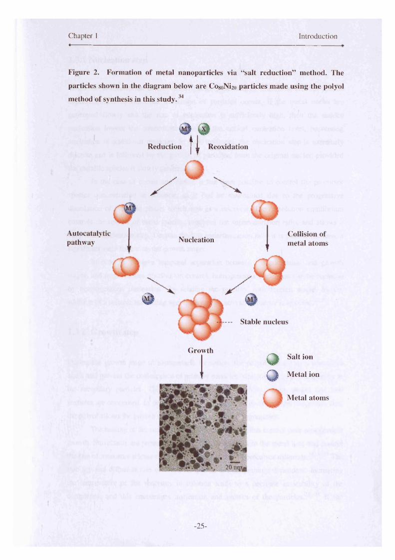
Figure 2. Formation of metal nanoparticles via “salt reduction” method. The
particles shown in the diagram below are CosoNi2o particles made using the polyol
method of synthesis in this study.34
© ®11Reduction 1 Reoxidation
Autocatalyticpathway Nucleation
©
Collision of metal atoms
Stable nucleus
GrowthO Salt ion
O Metal ion
Metal atoms
- 25 -

Chapter 1 Introduction
1.3.1 Nucleation step
When the concentration of the metal species generated by reduction, reaches critical
supersaturation, spontaneous nucleation of particles occurs. If the metal nuclei are
generated slowly and the rate of nucleation is sufficiently high, then the sudden
nucleation lowers the concentration below the critical nucleation level, preventing
nucleation of additional particles. In such instances, the nucleation step is extremely
discrete and is followed by the growth of particles, from the original nuclei, provided
the metallic species is slowly generated.
In the case of cobalt and nickel, it has been possible to control the precursor
species concentration in solution, as it can be maintained due to the progressive
dissolution of the solid phase, which acts as a reservoir. The dissolution equilibrium
controls the release of these species, regulates the supersaturation ratio, and allows a
very short nucleation step. Despite the low supersaturation ratio it is possible to have a
significant yield following the growth stage.
In order to achieve temporal separation between the nucleation and growth
stages, and improve size distribution control, homogeneous nucleation can be replaced
by heterogeneous nucleation. By seeding the reaction with foreign nuclei by the
addition of a suitable nucleating agent, the energy activation barrier is lowered.24,35
1.3.2 Growth step
During the growth stage in nanoparticle formation, the polyol can act as a protective
agent and prevent the coalescence of primary particles, which leads to polydispersity in
the secondary particles. This is particularly relevant where cobalt, nickel and iron
particles are concerned. In order to obtain metallic particles of uniform shape and size,
the polyol allows the growth stage to propagate without aggregation.
The heating of the solution is key in order to establish control over nanoparticle
growth. Surfactants are present in the solution to complex to the metal ions and control
the rate of monomer release during the decomposition of precursor materials.14,36 39 The
stability and diffusion rate of these complexes are temperature dependent. Increasing
the temperature of the reactants in solution leads to a decrease in stability of the
complexes, and this encourages nucleation and growth of the particles.24, 35 If the
-26-
Chapter 1 Introduction
temperature increase is too high, this can result in uncontrolled growth and hence lack
of control of particle sizes and distributions. Determining the correct temperature range
is also a key to producing monodisperse, spherical particles with a narrow size
distribution.40’41
1.4 Homogeneous nucleation
Modified forms of the LaMer32 method have been used to produce metallic
nanoparticles, particularly cobalt, using a hot coordinating solvent containing a
surfactant mixture that is injected with an organometallic precursor salt. 14 The reactions
are typically carried out in an inert atmosphere. The rapid injection of precursor leads to
a separation of the growth, and nucleation stages. Therefore, a temporally discrete
homogeneous nucleation occurs. The absence of seeds in this type of reaction is notably32 42important. This type of reaction arises from the self-nucleation of particles. ’
The high temperature of the solution decomposes the precursors, resulting in
supersaturation of reagent species in solution, which is relieved by the burst nucleation
that occurs. The rapid rate of addition raises the precursor concentration above the
nucleation threshold. At the point of nucleation, the concentration of these species dips
below that of the critical concentration required for nucleation, and therefore further
reagent species can only bind to the particle nuclei that have been formed.43 ,44
Thermal decomposition of cobalt carbonyl has been successfully used to
produce cobalt nanoparticles, via a homogeneous route, by many research groups.
Petit45 adopted this method when synthesising passivated cobalt nanocrystals.
Controlling the reaction parameters has an effect on the size and shape of the particles
in this type of reaction.15, 461 47 Many of the reactions that have previously been
attempted, have involved the rapid injection of an organometallic precursor into a hot48boiling coordinating solvent, to achieve homogeneous nucleation.
Puntes et al 49 modified this concept and developed a new synthetic procedure.
The group succeeded in producing 8 -cobalt nanoparticles that are single crystals and
defect free by using the rapid pyrolysis of the cobalt carbonyl precursor. Their study
introduced a new method of producing magnetic ferrofluids via homogeneous
nucleation. The size distribution of these particles was narrow (< 5%), due the discrete
nucleation event that took place. These particles however, did not show evidence of the
-27-

Chapter 1 Introduction
formation of an oxide layer which is not ideal for exchange bias properties. The
pyrolysis of carbonyls is a clean route, as elemental cobalt is the only non-volatile
product synthesised in the reaction: [Co2(CO)g] —► 2Co + 8 CO.50 The size distribution of
particles made using this synthesis was controlled by changes to the reaction
temperature, surfactant-type and concentration. 14,51,52
Dinega et al50 decomposed dicobalt octacarbonyl, to produce cobalt
nanoparticles in the presence of trioctylphosphine oxide (TOPO). They reported an
interesting result in the discovery of a new crystal structure of cobalt, which was
subsequently named epsilon-cobalt, and had a similar unit cell to (3-manganese (a high
temperature phase of manganese). This was a breakthrough in the area of cobalt
chemistry, as cobalt had only been known to have two crystalline phases; face-centred
cubic (fee) and hexagonal close-packed (hep) prior to this. The fee structure is
thermodynamically favourable at high temperatures (above 450 °C), whilst hep is
favoured at lower temperatures. Both phases coexist at room temperature, however, for
smaller particles the fee structure is preferred below room temperature.53
Puntes et al54 also reported the formation of e-cobalt through the pyrolysis of
the dicobalt octacarbonyl using TOPO, oleic acid and lauric acid as stabilising agents.
Particles produced, using thermal decomposition or pyrolysis techniques, have proven
to be monodisperse with a narrow size distribution. Disks, platelets and nanorods were
formed by controlling the reaction parameters, and reproducibility is well-established
using this method. 14,49,54155
The polyol method of synthesis is different to the thermal decomposition
methods adopted by others, and the formation of monodisperse self-assembled arrays of
particles is not as prevalent, due to issues with reproducibility. Homogeneous nucleation
via the polyol method has been attempted by Viau et al,24 where nucleation and growth
arises from the solution without the presence of external nuclei. In order to control the
particle shape and size, surfactants of varying steric bulk are used. The temperature of
the solution is monitored to allow for a short nucleation period and subsequent slow
growth. However, control of particle size and shape using the polyol method is much
more difficult due to the nature of the reaction.
The nucleation and growth steps are even harder to control in alloy particles,
where the underlying chemistry and decomposition temperatures of each salt are
different. Decomposition pathways tend to determine the success of a synthesis, due to
the mechanism and reaction kinetics of supersaturated solutions.40 Therefore,
-28-

Chapter 1 Introduction
homogeneous nucleation using the polyol synthetic technique is at present a challenging
process.
1.5 Heterogeneous nucleation
The synthesis of nanoparticles is challenging due to the numerous variables involved.
Size control is critical, when considering potential applications, the material may be
suited to. Control of the size distribution is also important in order to maintain
reproducibility, so that particles can be modelled in terms of their functional properties
and behaviour.
The use of seeds in synthetic procedures such as polyol synthesis is well
established.2 4 ,3 5 ,56 The seeds act as nuclei around which the growth of a nanoparticle
may occur by lowering the barrier to nucleation. Noble metal seeds form particles at
lower temperatures in comparison to transition metals, and act as preferential growth
sites for precursor atoms in solution.35 The high interfacial energy of transition metals in
comparison to the noble metals, means that the lower energy path created by the noble
metal nuclei is much more favoured.57 The concentration of seeds helps to determine
the particle size and influence size distribution of the nanoparticles. The more seeds
there are, the greater the number of nucleation sites, and hence a larger number of
smaller nanoparticles are formed. The inverse is true for a lower concentration of seeds,
where you would expect a smaller number of larger nanoparticles to be formed. 35 ,56
Classical nucleation theory (CNT) kinetically favours heterogeneous nucleation
over homogeneous, and this is evident in the equation describing nucleation rate of the
particles, equation (5) . 58 The spontaneous formation of small crystal nuclei occurs only
if their size exceeds a critical value, the so-called critical nucleus size, otherwise they
re-dissolve into their mother phase. The rate of nucleation is dependent upon the
probability function Pent, the probability of the critical nucleus forming spontaneously,
and on a kinetic factor (B), which measures the rate at which the critical nuclei then59grow.
-29-

Chapter 1 Introduction
The probability per particle of the formation of a critical nucleus, is
exponentially dependent upon the free energy AGcrit that is needed to form such a
nucleus:
Peril = exp(-AGCrit/kBT) (1)
Where T is the temperature and ke is the Boltzmann constant. Classical nucleation
theory also details the total Gibbs free-energy required, to form a spherical crystallite
with radius R as being
AG = 4/3 7cR3 ps A \l + 47tR2 y (2)
Where ps is the density of the solid, A|I (<0) is the difference between chemical
potential of the solid and liquid, and y is the solid-liquid interfacial free energy density.
The function AG passes through a maximum of R = 2y/(/?s[A|i]) and the height of the
nucleation barrier is:
AGcrit = (16n/3) YV (AlAul)2 (3)
The crystal nucleation rate per unit volume, J, is the product of PCdt and the kinetic
prefactor B:
J = B exp (-AGcrit/kfiT) (4)
The crystal nucleation expression for the nucleation rate then becomes:
J = B exp [-/ (16jc/3)y3/ (A[Aji])2 with 0</< 1 (5)
Where,/ describes the influence of foreign nuclei or seeds, on the nucleation barrier. In
the case of homogenous nucleation, / , may be counted as being equal to 1 and
therefore, has the largest possible energy barrier to nucleation.58 Equation (5) provides a
representation of crystal nucleation theory to date. It is by no means an absolute
-30-
Chapter 1 Introduction
explanation of the formation of crystallite spheres in solution, but is a good
approximation to make when analysing nucleation theory.
1.6 Surfactant contributions
Surfactants play an important role in nanoparticle synthesis. Metallic nanostructured
colloidal particles need a protective surfactant to prevent aggregation and oxidation.
There is a great difficulty associated with ensuring nanoparticles, once made, remain
separated from one another. This is due to attractive interparticle interactions which
arise from van der Waals forces. Nanocrystals remain stabilised if the repulsive force
provided by the surfactant is sufficient to prevent the intrinsic van der Waals forces
between the particles.15, 43, 60 Balancing the concentrations of surfactants in such
nanoparticle fluids is important as too much can cause coalescence and aggregation,
whereas too little causes particles to “precipitate out” of the solvent and complete
oxidation. The surfactants can also control interparticle distances in large-scale self
assembled arrays.61,62
Magnetic dipole interactions can also be evident in metal nanoparticle
suspensions, although this effect is negligible in dilute superparamagnetic particle
suspensions. Superparamagnetic nanoparticles form stable ferrofluids where the
particles remain suspended in the solvent.63' 65 Ferrofluids are attracted to magnets and
are displaced upon the application of a magnet.64' 66
Shape control by the addition of surfactants is well reported in the cases of CdS
and CdSe nanoparticles.67'69 A wide range of different shapes including rods, teardrops
and tetrapods have been synthesised with a controlled change in surfactant
concentration and composition.70 The effects of surfactant on cobalt nanoparticle shape
has been investigated with fervour, due to the interest in the magnetic memory storage
capabilities of arrays consisting of cobalt-based nanoparticles and the potential for high
magnetic anisotropy in non-spherical particles.71' 74 When particles are coated with a
close packed monolayer of organic surfactant, the surfactant has the ability control the
shape of nanoparticles.
When adjustments are made with the metal precursor-to-surfactant ratio, particle
size correspondingly changes. A greater amount of surfactant yields small particles,
whereas a decrease in surfactant concentration produces large particles. Bulkier
-31-

Chapter 1 Introduction
surfactants such as tributylphosphine and trioctylphosphine inhibit particle growth and
hence also form smaller particles. Therefore, steric bulk in surfactants is also an
important factor to consider when designing a synthesis. This was demonstrated by Sun
et alu in their work on the formation of monodisperse cobalt nanocrystals.
Surfactants complex with metal ions in solution, and the decomposition
temperature of these complexes determine the nucleation point of the particles.
Different surfactants, complex well with specific types of metal ion species e.g. nickel
and oleylamine.75 These complexes act as the monomer precursor species in the
reaction. The choice of surfactant used in a reaction is therefore an important issue to
consider. Many different types of surfactant including alkylphosphines, alkylphosphine
oxides and carboxylic acids have been used to control particle shapes. 15 The surfactants
produce a protective organic shell that mediates growth, stabilises the nanoparticles in
solution and limits the oxidation after synthesis. In many typical syntheses, two
surfactants are used concurrently, where the first binds tightly to the surface (e.g oleic
acid) promoting slow growth, and the second allows rapid growth by binding weakly to
the surface (e.g. trioctylphosphine and tributylphosphine)13, 43, 44 The surfactants work
together to tune the overall nanoparticle size.
-32-
Chapter 1 Introduction
1.7 Magnetism
Interest in magnetic nanomaterials is driven by the fundamental applications of devices
which harness the magnetic properties for use in modem technology. Greek writings
around 600 BC first described magnetic phenonema in Lodestone (magnetite, Fe30 4), a
natural non-metallic solid, which seemed to attract iron. The significance of this
discovery was not realised until much later, when it was used as the first technical
magnetic material for the formation of the first compass.76
1.7.1 Fundamentals of magnetism
The magnetisation (M) of a sample is defined as;
Where; N= number of atoms
<p> = average magnetic moment per site
However, in most magnetic systems, the magnetic moment is dependent upon the field
so magnetic susceptibility is used. The magnitude of the moment is dependent upon the
amount of material. The magnetic susceptibility, % is a measure of the magnetic
response or magnetisation, M, of a material in an applied magnetic field, H. The
susceptibility or magnetisation is an intrinsic property of the material.
The dimensionless volume magnetic susceptibility, is closely related to the molar
magnetic susceptibility, %m:
M = N < j l i > (i)
M = XH (2)
X m — X (3)
-33-
Chapter 1 Introduction
where Vm is the molar volume of the substance. The magnetic flux density, B, is related
to the applied field strength and the magnetisation by
B = |io(H + M) = |Jo(l+ x) H (4)
Where po, is the vacuum permeability and (1+ x) is the relative permeability. The
magnetic flux density can be thought of as the density of magnetic force lines
permeating the medium. Materials for which % is positive, are either paramagnetic or
ferromagnetic, and those for which % is negative are diamagnetic. The superconducting
quantum interference device (SQUID) is one of a few methods, which can be used to
measure magnetic susceptibility. The permanent magnetic moment can be determined
from susceptibility measurements by plotting % against 1/T.77

Chapter 1 Introduction
1.7.2 Classes of magnetic materials
In nature, there are six main types of magnetic material; (I) diamagnetic (II)
paramagnetic (III) ferromagnetic (IV) antiferromagnetic (V) ferrimagnetic (VI)
superparamagnetic. Additional classes include antiferrimagnets and spin glasses,
amongst several others. The schematic in Figure 3 illustrates the atomic magnetic
behaviour of some of these particular classes of magnetic material.7,78
Figure 3. Generic shapes of hysteresis loop for some of the different classes of
magnetic material, at room temperature; (a) ferromagnet (b) paramagnet (c)
superparamagnet.
(a) (b)
M
H
(c)M ----
----
H
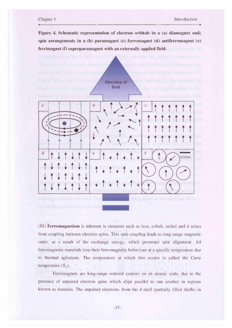
(I) Diamagnetism occurs in materials with no permanent magnetic dipole. Hence, they
have no magnetisation in a magnetic field. Materials that are conventionally considered
to be “non-magnetic” such as water, DNA, oil and many plastics are diamagnetic in
nature. In an applied field, a small negative magnetic moment (M) is induced on the
diamagnetic atoms, which is proportional to the applied field strength (H) . 7 ,79
Diamagnetism is independent of temperature and arises due the effect of applied
magnetic fields on the motion of the inner electrons of the atoms present. This is shown
-35-
Chapter 1 Introduction
in Figure 4(a). The electronic orbits surrounding the atomic nucleus are often
considered as if they are an electric current. In an applied magnetic field, the electronic
motions are modified and this leads to a change in the magnetic moment due to the
currents. This is called an “induced magnetic moment”. Lenz’s law of electromagnetic
induction states that currents induced by a magnetic field are in such a direction that
their magnetic fields tend to oppose the original inducing field. This means the induced78 80magnetisation is negative, as is the susceptibility. ’
(II) Paramagnetism occurs in atoms or molecules with a permanent magnetic dipole
moment, even in the absence of an applied field, where the widely separated
neighbouring atoms do not interact. This occurs in the presence of unpaired electrons in
atomic/molecular orbitals. In zero-applied magnetic fields, the moments are randomly
orientated and fluctuate due to thermal agitation, resulting in a zero net moment. When
an external field is applied, the spins align with the field, but this magnetisation effect is
weaker than that exhibited by a ferromagnetic material. The spins remain predominantly
oriented in a random manner, with some moments aligning with the field (II). However,
there is a small net magnetisation in the direction of the externally applied field. 78’ 80
Above their Curie temperature, ferromagnetic materials become paramagnetic
and their susceptibility depends on temperature.80Antiferromagnetic materials also
behave as paramagnets above their Neel temperature. Key points to note are that upon
the removal of a magnetic field, magnetisation will disappear. Also, at low temperatures
thermal fluctuations are obviously reduced, therefore paramagnetic materials are easier
to magnetise (align spins) than at higher temperatures.79’ 81
-36-
Chapter 1 Introduction
Figure 4. Schematic representation of electron orbitals in a (a) diamagnet and;
spin arrangements in a (b) paramagnet (c) ferromagnet (d) antiferromagnet (e)
ferrimagnet (f) superparamagnet with an externally applied field.
Direction of field
10n m
(III) Ferromagnetism is inherent in elements such as iron, cobalt, nickel and it arises
from coupling between electron spins. This spin coupling leads to long range magnetic
order, as a result of the exchange energy, which promotes spin alignment. All
ferromagnetic materials lose their ferromagnetic behaviour at a specific temperature due
to thermal agitations. The temperature at which this occurs is called the Curie
temperature (Tc).
Ferromagnets are long-range ordered systems on an atomic scale, due to the
presence of unpaired electron spins which align parallel to one another in regions
known as domains. The unpaired electrons, from the d shell (partially filled shells) in
-37-
Chapter 1 Introduction
transition metals, give rise to this spin coupling. There is a strong intrinsic magnetic
field within the magnetic domains, and this can exist when there is no external magnetic
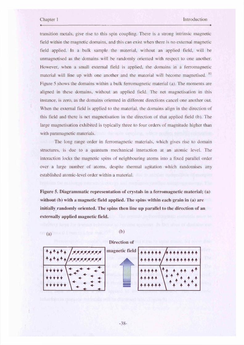
field applied. In a bulk sample the material, without an applied field, will be
unmagnetised as the domains will be randomly oriented with respect to one another.
However, when a small external field is applied, the domains in a ferromagnetic
material will line up with one another and the material will become magnetised. 81
Figure 5 shows the domains within a bulk ferromagnetic material (a). The moments are
aligned in these domains, without an applied field. The net magnetisation in this
instance, is zero, as the domains oriented in different directions cancel one another out.
When the external field is applied to the material, the domains align in the direction of
this field and there is net magnetisation in the direction of that applied field (b). The
large magnetisation exhibited is typically three to four orders of magnitude higher than
with paramagnetic materials.
The long range order in ferromagnetic materials, which gives rise to domain
structures, is due to a quantum mechanical interaction at an atomic level. The
interaction locks the magnetic spins of neighbouring atoms into a fixed parallel order
over a large number of atoms, despite thermal agitation which randomises any
established atomic-level order within a material.
Figure 5. Diagrammatic representation of crystals in a ferromagnetic material; (a)
without (b) with a magnetic field applied. The spins within each grain in (a) are
initially randomly oriented. The spins then line up parallel to the direction of an
externally applied magnetic field.
Direction of
magnetic field
AAk
+ + i + i \ ♦ *+ + i + + * \ ^ * t *
...A -t-L ***** w ♦ * * * * *
-38-
Chapter 1 Introduction
Weiss first gave an answer to the riddle of why a piece of iron, at room
temperature and well below its’ Curie temperature, does not yield a macroscopic total
moment in the absence of an externally applied field. Weiss proposed that a
macroscopic magnetic material will break into domains that align themselves in a
manner that will minimize the total effective moment of the material. 80,82
A magnetic field contains energy proportional to the field squared and its
volume. Therefore, the magnetostatic energy of a monodomain of parallel spins can be
decreased by breaking it into smaller, oppositely aligned domains. This decrease in
energy is beneficial, as it would further continue to divide into more and yet smaller
domains, were it not for the exchange energy that increases with declining size.
Exchange energy occurs at the boundary between oppositely aligned domains which, by
the ferromagnetic nature of the coupling, battles against the anti-alignment. Domain
formation is the balance between the spin coupling, which prefers parallel orientation
and magnetic dipole interactions which prefer antiparallel alignments. This is why
domains are formed at all, rather than whole material having aligned spins.
The opposition between the magnetostatic energy and what is known as a
domain wall, limits the division of the material to domains of a finite size. The
boundary between domains, the domain wall, is due to another competition of energies
between the exchange interaction energy and magnetocrystalline anisotropy energy. The
energy required to form a domain wall is larger than the energy cost of uniform
magnetization in very small particles. Therefore domain magnetism occurs in bulk
materials and is not applicable to the fine particles produced in this thesis, where single
domain behaviour is much more likely. The crystals in ferromagnetic materials must be
relatively large for domain behaviour to become apparent. In fact sizes of domains can
range from 0 .1 mm to a few mm.82 ,83
Ferromagnetic materials retain a magnetised state, to some extent, for some time
after exposure to an external imposed field due to the spin coupling described earlier.
This is the main difference between ferromagnetic and paramagnetic materials. The
ability of a material to remember its’ magnetic history is called “hysteresis.” The
application of a magnet on such materials means that when the applied field is removed,
the ferromagnetic material will not recoil back to it unmagnetised state without being
driven back to zero by a field applied in the opposite direction. This hysteretic
behaviour in magnetic materials will be discussed later (Figure 9).
-39-
Chapter 1 Introduction
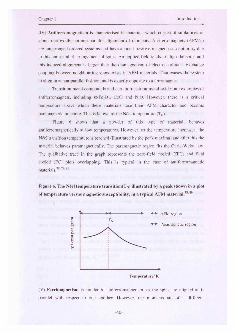
(IV) Antiferromagnetism is characterised in materials which consist of sublattices of
atoms that exhibit an anti-parallel alignment of moments. Antiferromagnets (AFM’s)
are long-ranged ordered systems and have a small positive magnetic susceptibility due
to this anti-parallel arrangement of spins. An applied field tends to align the spins and
this induced alignment is larger than the diamagnetism of electron orbitals. Exchange
coupling between neighbouring spins exists in AFM materials. That causes the system
to align in an antiparallel fashion, and is exactly opposite to a ferromagnet.
Transition metal compounds and certain transition metal oxides are examples of
antiferromagnets, including a-Fe2C>3, CoO and NiO. However, there is a critical
temperature above which these materials lose their AFM character and become
paramagnetic in nature. This is known as the Ndel temperature (T n).
Figure 6 shows that a powder of this type of material, behaves
antiferromagnetically at low temperatures. However, as the temperature increases, the
Neel transition temperature is reached (illustrated by the peak maxima) and after this the
material behaves paramagnetically. The paramagnetic region fits the Curie-Weiss law.
The qualitative trace in the graph represents the zero-field cooled (ZFC) and field
cooled (FC) plots overlapping. This is typical in the case of antiferromagnetic
Figure 6. The Neel temperature transition (T n) illustrated by a peak shown in a plot
of temperature versus magnetic susceptibility, in a typical AFM material. ’
materials.
<-+■ AFM regionE3 Tn
Paramagnetic region
Temperature/ K
(V) Ferrimagnetism is similar to antiferromagnetism, as the spins are aligned anti
parallel with respect to one another. However, the moments are of a different
Chapter 1 Introduction
magnitude, and therefore there is a net overall magnetisation, as they do not cancel each
other out. The magnetisation of this type of material is similar to a ferromagnet in terms
of its hysteretic behaviour.7 8 ,81
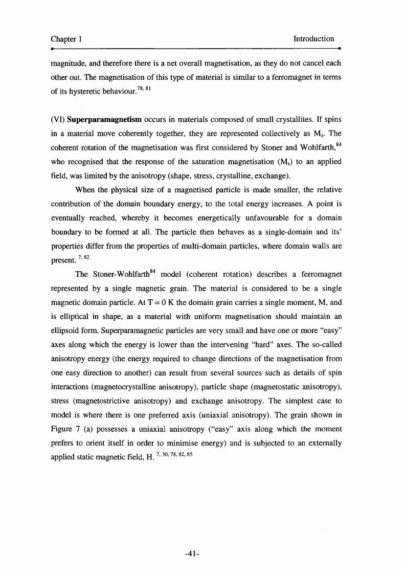
(VI) Superparamagnetism occurs in materials composed of small crystallites. If spins
in a material move coherently together, they are represented collectively as Ms. The0 4
coherent rotation of the magnetisation was first considered by Stoner and Wohlfarth,
who recognised that the response of the saturation magnetisation (Ms) to an applied
field, was limited by the anisotropy (shape, stress, crystalline, exchange).
When the physical size of a magnetised particle is made smaller, the relative
contribution of the domain boundary energy, to the total energy increases. A point is
eventually reached, whereby it becomes energetically unfavourable for a domain
boundary to be formed at all. The particle then behaves as a single-domain and its’
properties differ from the properties of multi-domain particles, where domain walls are+ 7, 82present.
The Stoner-Wohlfarth84 model (coherent rotation) describes a ferromagnet
represented by a single magnetic grain. The material is considered to be a single
magnetic domain particle. At T = 0 K the domain grain carries a single moment, M, and
is elliptical in shape, as a material with uniform magnetisation should maintain an
ellipsoid form. Superparamagnetic particles are very small and have one or more “easy”
axes along which the energy is lower than the intervening “hard” axes. The so-called
anisotropy energy (the energy required to change directions of the magnetisation from
one easy direction to another) can result from several sources such as details of spin
interactions (magnetocrystalline anisotropy), particle shape (magnetostatic anisotropy),
stress (magnetostrictive anisotropy) and exchange anisotropy. The simplest case to
model is where there is one preferred axis (uniaxial anisotropy). The grain shown in
Figure 7 (a) possesses a uniaxial anisotropy (“easy” axis along which the moment
prefers to orient itself in order to minimise energy) and is subjected to an externally
applied static magnetic field, H. 7> 30,78,82,85
-41-
Chapter 1 Introduction
Figure 7 (a) Single domain grain shown with magnetisation (M) and an applied
magnetic Held (H). The grain’s easy axis, or anisotropy axis, makes an angle
(<g>) with the applied magnetic field (b) Coercivity as a function of mean particle
volume.7
(a)
Anisotropy axis
Single domain Multidomain
Superparamagnetism,
ICritical diameter Particle Volume
Coherent rotation of a single domain occurs when the anisotropy is much greater than
the thermal energy (KV » kT), where particles of a critical radius cannot
thermodynamically support the formation of a domain wall. This requires magnetisation
reversal via the rotation of the magnetisation, in the direction of the applied field. The
preferred direction that the magnetic single domain grain takes, in the absence of an
-42-
Chapter 1 Introduction
applied field, is determined by the magnetic anisotropy (combination of all the
anisotropies described earlier). This orientation is depicted in Figure 7 (a) as the
anisotropy axis. 30
As the particle size decreases below the single-domain value, the spins become
increasingly affected by thermal fluctuations ( KV « kT) and the system behaves
superparamagnetically.85
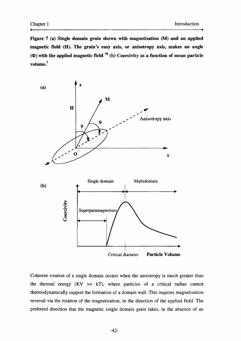
Figure 8. Beating the superparamagnetic limit for small magnetic nanoparticles,
(a) Magnetic anisotropy energy is comparable to the thermal energy (kT) (b)
surface exchange bias between ferromagnetic material and antiferromagnetic
material provides an additional anisotropy energy, which stabilises magnetisation
in one direction and hence prevents superparamagnetism.
(a) (b)
0 Orientation angle rc Orientation angle
Superparamagnetism is a time-dependent phenomenon. The anisotropy energy, KV,
may be thought of as the energy barrier required for keeping the magnetisation oriented
in a certain direction. The probability for overcoming this energy barrier via thermal
fluctuation is directly proportional to the Boltzmann factor exp(-KV/kT). When this
occurs the particles are superparamagnetic, as thermal fluctuations cause the spins to
randomly flip the magnetisation direction, between parallel and antiparallel
orientations.9 ,86 By introducing an attempt timescale, To, the probability for overcoming
the fluctuation can be quantified. The timescale for a successful jump of the KV barrier
is
-43-
Chapter 1 Introduction
^ ^ -KV/kTT = T0 e (l)
The value for Xo is about 10' 9 s. In a typical magnetic measurement, with a
magnetometer, the experimental time, x , would be between 10 to 100 s. If the
saturation magnetisation reverses at times shorter than the experimental timescales, the
system appears superparamagnetic. Therefore, using x ~ 100 s and Xo = 10' 9 s to
substitute into equation (1 ), we obtain for the critical volume,
Vsp=25kbT/K (2)
A particle with a volume smaller than this behaves superparamagnetically on the 100 s
experimental timescale. Equation (2) can be rearranged to give
Tb = KV/ 25kb (3)
where Tb is the blocking temperature. The free movement of spins is blocked below this
temperature due to the induced anisotropy in exchange biased materials. Above the
blocking temperature, kT kicks free the moment, so that the material appears82 8Ssuperparamagnetic. ’
A size limitation is apparent for observed superparamagnetism in particles, and
it varies between different materials. Superparamagnetic particles are single-domain as
shown in Figure 7 (b), where the relationship between particle volume and coercivity is
displayed. Superparamagnetic particles exhibit ferromagnetic behaviour below the
blocking temperature. Above the blocking temperature, they behave as paramagnets
with very large susceptibilities, because the thermal energy is sufficient to rotate the
particle moment. Monodomain particles, on the other hand, can have blocking
temperatures greater than room temperature e.g. typical sizes for single domain cobalt
and nickel particles are 70 and 55 nm respectively.85
1.8 Hysteresis in magnetism
A key feature of magnetic materials is their ability to exhibit a complex change in
-44-

Chapter 1 Introduction
magnetisation upon the application of a magnetic field. This is best explained by a
hysteresis loop. A great deal can be learnt about a magnetic material by analysing its
hysteresis loop. The plot displays the relationship between applied field (H) and
magnetisation (M). Hence, a loop is obtained by measuring the magnetic moment of a
sample, as the applied field is cycled between large positive and negative values. An
example of a hysteresis plot is shown in Figure 9.
Hysteresis is a widely recognised behaviour in ferromagnetic materials. When a
magnetic field is externally applied to a ferromagnet, the material will become
magnetised in the same direction as the applied field. When the field is removed, the
ferromagnet will retain this magnetisation, due to the spin coupling energy. It needs to
be driven back to zero by a field in the opposing direction. This irreversibility
characterises magnetic hysteresis.78,81,87
Figure 9. The important features of a hysteresis loop. The saturation
magnetisation, Ms, remanent magnetisation, Mr and coercivity, Hc, are shown. 87
Domains are aligned in a positive direction to the field.
Remanence-No field (has been switched off). The magnetic effect remains as the domains are still aligned.
No magnetisation
P Saturation magnetisation, Ms
*• Width of loop x Vi = coercivity, H,
^ Remanent magnetisation, Mr
Saturation magnetisation
Saturation in opposite direction
Domains are aligned in a negative direction to the field. The moments have flipped.
-45-

Chapter 1 Introduction
At high applied fields, the magnetisation approaches saturation point, or
saturation magnetisation, Ms. Then if the magnetic field is decreased to zero, the M
versus H curve does not follow the path of the initial curve but instead lags behind.
Increasing the field in the opposite direction drives M to negative saturation. The
magnetisation of a material is measured in emu/g. When H = 0, a residual magnetisation
remains, this is called the remanence, Mr. If the field is applied in a reverse direction (a
negative field), the magnetisation is forced to zero at a field magnitude known as the
coercivity, He. The coercivity is the half-width of the hysteresis loop, where the
magnetisation passes through zero in the positive and negative direction of field, and is
measured in Oersted. Hence the cardinal points along the hysteresis plot are Ms, Mr and
^ 78,85 Materials with a He > 100 Oe are called hard magnets. Soft magnetic materials
typically have a He < 10 Oe. However, the boundary between hard and soft is
ambiguous and highly debated. 82
1.9 Exchange anisotropy
Exchange anisotropy was first observed in fine cobalt nanoparticles enveloped with a
cobaltous oxide shell, by Meiklejohn and Bean,88 of the General Electric Research
Laboratory in 1957. This new kind of magnetic anisotropy (K) was given the name
“exchange anisotropy”. The effect is also known as “exchange bias” or “exchange
pinning” and also occurs in bilayers or multilayers of magnetic materials. 89 91 The
discovery led to the development of the first commercially viable magnetic hard disk
drive, produced in 1956, by IBM.
The phenomenon is attributed to the exchange interaction between adjacent
ferromagnetic and antiferromagnetic materials. The Curie temperature (T c) of the
ferromagnet (FM) must be larger than the Neel temperature of the antiferromagnet
(AFM). When a FM-AFM bilayer interface is cooled in an external magnetic field,
through the Ndel temperature (T n ) of the AFM, the AFM spins couple to the already
aligned FM spins. The interfacial AFM-FM spin layers are subsequently pinned
together, in parallel alignment, when a ferromagnet (FM)-antiferromagnetic (AFM)
bilayer interface is cooled in an external magnetic field, through the Neel temperature
(T n ) of the AFM. The Curie temperature (T c) of the FM must be larger than the Neel
temperature. As the AFM layer spins pass through their Ndel temperature, they couple
-46-
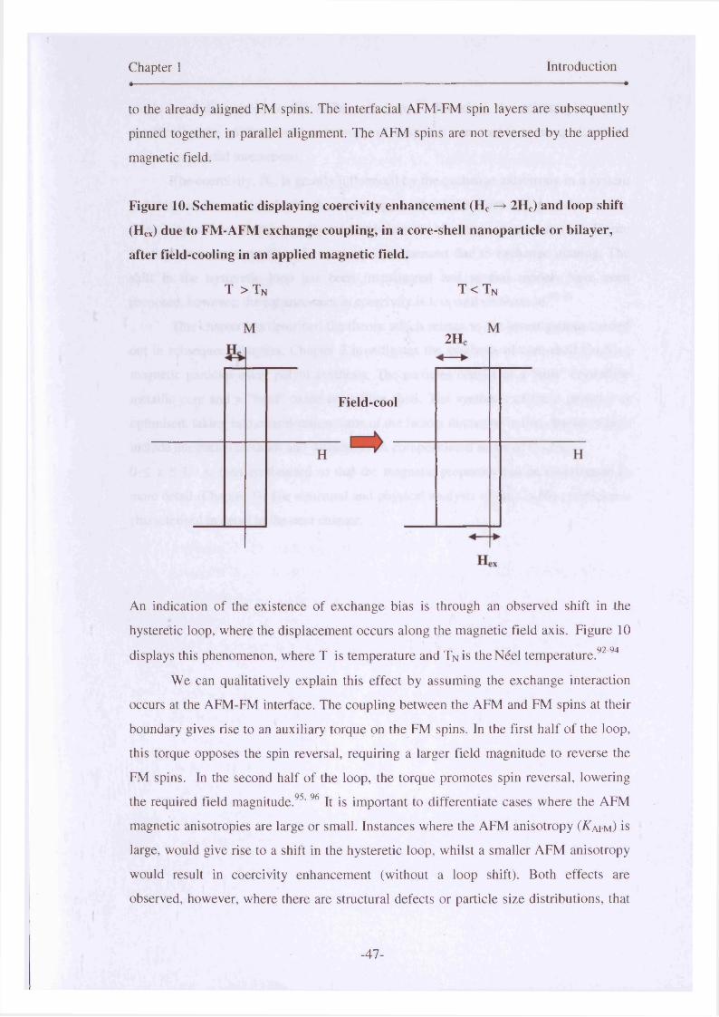
Chapter 1 Introduction«--------- — -— ----------------------------------------------------------------------------------------------------------------------------------------------------------------------------------------------------------------------------------•
to the already aligned FM spins. The interfacial AFM-FM spin layers are subsequently
pinned together, in parallel alignment. The AFM spins are not reversed by the applied
magnetic field.
Figure 10. Schematic displaying coercivity enhancement (Hc —> 2HC) and loop shift
(Hex) due to FM-AFM exchange coupling, in a core-shell nanoparticle or bilayer,
after field-cooling in an applied magnetic field.
T > T n T < T n
2H(
Field-cool
An indication of the existence of exchange bias is through an observed shift in the
hysteretic loop, where the displacement occurs along the magnetic field axis. Figure 10
displays this phenomenon, where T is temperature and Tn is the Neel temperature.92’94
We can qualitatively explain this effect by assuming the exchange interaction
occurs at the AFM-FM interface. The coupling between the AFM and FM spins at their
boundary gives rise to an auxiliary torque on the FM spins. In the first half of the loop,
this torque opposes the spin reversal, requiring a larger field magnitude to reverse the
FM spins. In the second half of the loop, the torque promotes spin reversal, lowering
the required field magnitude.95, 96 It is important to differentiate cases where the AFM
magnetic anisotropies are large or small. Instances where the AFM anisotropy (ATAfm) is
large, would give rise to a shift in the hysteretic loop, whilst a smaller AFM anisotropy
would result in coercivity enhancement (without a loop shift). Both effects are
observed, however, where there are structural defects or particle size distributions, that
-47-

Chapter 1 Introduction
yields local differences in the AFM anisotropy.97 Therefore the anisotropies, more
specifically, the unidirectional anisotropy of a system can enable us to qualitatively
explain interfacial interactions.
The coercivity, He, is greatly influenced by the exchange anisotropy in a system
and this can be correlated with the anisotropy in the AFM layer of a particle or film.
Coercivity enhancement is usually evident below the blocking temperature (T b). Figure
10 displays a representation of coercivity enhancement due to exchange pinning. The
shift in the hysteretic loop has been investigated and several models have beenQ O Q Q
proposed, however, the enhancement in coercivity is less well understood. ’
This chapter has described the theory which relates to the investigations carried
out in subsequent chapters. Chapter 2 investigates the synthesis of core-shell CoxNii_x
magnetic particles using polyol synthesis. The particles consist of a “soft” crystalline
metallic core and a “hard” oxide crystalline shell. The synthesis of these particles is
optimised, taking into consideration some of the factors discussed in this chapter, which
include nucleation methods and surfactants. A compositional series of CoxNii.x>
0 < x < 1 , is then synthesised so that the magnetic properties can be investigated in
more detail (Chapter 3). The structural and physical analysis of the CoxNii_x particles is
characterised in detail in the next chapter.
-48-

Chapter 1 Introduction
1.10 References
1. Lubbe, A. S.; Alexiou, C.; Bergemann, C., Clinical applications of magnetic
drug targeting. Journal o f Surgical Research 2001,95, (2), 200-206.
2. Nethe, A.; Scholz, T.; Stahlmann, H. D., Improving the efficiency of electric
machines using ferrofluids. Journal of Physics-Condensed Matter 2006, 18,
(38), S2985-S2998.
3. Scherer, C.; Neto, A. M. F., Ferrofluids: Properties and applications. Brazilian
Journal of Physics 2005, 35, (3A), 718-727.
4. Kim, E. H.; Lee, H. S.; Kwak, B. K.; Kim, B. K., Synthesis of ferrofluid with
magnetic nanoparticles by sonochemical method for MRI contrast agent.
Journal of Magnetism and Magnetic Materials 2005,289, 328-330.
5. Li, K. B.; Wu, Y. H.; Guo, Z. B.; Zheng, Y. K.; Han, G. C.; Qiu, J. J.; Luo, P.;
An, L. H.; Zhou, T. J., Exchange coupling and its applications in magnetic data
storage. Journal of Nanoscience and Nanotechnology 2007, 7, (1), 13-45.
6 . Sun, Y.; Salamon, M. B.; Gamier, K.; Averback, R. S., Memory effects in an
interacting magnetic nanoparticle system. Physical Review Letters 2003, 91,
(16).
7. Sugimoto, T., Monodispersed particles. 2001.
8 . Murdock, E. S., ROADMAP FOR 10 GBIT/IN2 MEDIA - CHALLENGES.
IEEE transactions on magnetics 1992,28, (5), 3078-3083.
9. Skumryev, V., Stoyanov,S., Zhang,Y., Hadjipanayis, G.,Givord, D., Nogues, J.„
Beating the superparamagnetic limit with exchange bias. Nature 2003,423, 850-
853
10. Pankhurst, Q. A., Nanomagnetic medical sensors and treatment methodologies.
Bt Technology Journal 2006,24, (3), 33-38.
11. Pankhurst, Q. A.; Connolly, J.; Jones, S. K.; Dobson, J., Applications of
magnetic nanoparticles in biomedicine. Journal of Physics D-Applied Physics
2003, 36, (13), R167-R181.
12. Tartaj, P., Nanomagnets-from fundamental physics to biomedicine. Current
Nanoscience 2006,2, (1), 43-53.
-49-

Chapter 1 Introduction
13. Sun, S. H.; Murray, C. B., Synthesis of monodisperse cobalt nanocrystals and
their assembly into magnetic superlattices (invited). Journal of Applied Physics
1999, 85, (8 ), 4325-4330.
14. Puntes, V. F.; Krishnan, K. M.; Alivisatos, A. P., Colloidal nanocrystal shape
and size control: The case of cobalt. Science 2001,291, (5511), 2115-2117.
15. Hyeon, T., Chemical synthesis of magnetic nanoparticles. Chemical
Communications 2003, (8 ), 927-934.
16. Shevchenko, E. V.; Talapin, D. V.; Schnablegger, H.; Komowski, A.; Festin, O.;
Svedlindh, P.; Haase, M.; Weller, H., Study of nucleation and growth in the
organometallic synthesis of magnetic alloy nanocrystals: The role of nucleation
rate in size control of CoPt3 nanocrystals. Journal of the American Chemical
Society 2003, 125, (30), 9090-9101.
17. Lu, M. H.; Song, T.; Zhou, T. J.; Wang, J. P.; Piramanayagam, S. N.; Ma, W.
W.; Gong, H., FePt and Fe nanocomposite by annealing self-assembled FePt
nanoparticles. Journal o f Applied Physics 2004, 95, (11), 6735-6737.
18. Mizuno, M.; Sasaki, Y.; Inoue, M.; Chinnasamy, C. N.; Jeyadevan, B.;
Hasegawa, D.; Ogawa, T.; Takahashi, M.; Tohji, K.; Sato, K.; Hisano, S.,
Structural and magnetic properties of monolayer film of CoPt nanoparticles
synthesized by polyol process. Journal o f Applied Physics 2005,97, (10).
19. Chantrell, R. W.; Weller, D.; Klemmer, T. J.; Sun, S.; Fullerton, E. E., Model of
the magnetic properties of FePt granular media. Journal of Applied Physics
2002,91, (1 0 ), 6 8 6 6 -6 8 6 8 .
20. Shevchenko, E.; Talapin, D.; Komowski, A.; Wiekhorst, F.; Kotzler, J.; Haase,
M.; Rogach, A.; Weller, H., Colloidal crystals of monodisperse FePt
nanoparticles grown by a three-layer technique of controlled oversaturation.
Advanced Materials 2002,14, (4), 287-290.
21. Sun, S. H., Recent advances in chemical synthesis, self-assembly, and
applications of FePt nanoparticles. Advanced Materials 2006, 18, (4), 393-403.
22. Tzitzios, V.; Niarchos, D.; Margariti, G.; Fidler, J.; Petridis, D., Synthesis of
CoPt nanoparticles by a modified polyol method: characterization and magnetic
properties. Nanotechnology 2005, 16, (2), 287-291.
23. Blackwood, D. J.; Li, Y. Y.; Chow, G. M., Polyol electroless and
electrodeposition of nanostructured Ni-Co films and powders. Journal o f the
Electrochemical Society 2002, 149, (3), D27-D34.
-50-

Chapter 1 Introduction
24. Viau, G.; FievetVincent, F.; Fievet, F., Nucleation and growth of bimetallic
CoNi and FeNi monodisperse particles prepared in polyols. Solid State Ionics
1996, 84, (3-4), 259-270.
25. Luna, C.; Morales, M. D.; Sema, C. J.; Vazquez, M., Exchange anisotropy in
Co80Ni20/oxide nanoparticles. Nanotechnology 2004,15, (4), S293-S297.
26. Faraday, M., Bakerian lecture- Experimental relations of gold (and other metals)
to light. Philosophical Transactions of the Royal Society of London 1857, 147,
145-181.
27. Fredrickx, P.; Schryvers, D.; Janssens, K., Nanoscale morphology of a piece of
ruby red Kunckel glass. Physics and Chemistry of Glasses 2002, 43, (4), 176-
183.
28. Turkevich, J.; Stevenson, P. C.; Hillier, J., A Study of the Nucleation and
Growth Processes in the Synthesis of Colloidal Gold. Discussions o f the
Faraday Society 1951, (11), 55-75.
29. Turkevich, J.; Stevenson, P. C.; Hillier, J., The Formation of Colloidal Gold.
Journal of Physical Chemistry 1953,57, (7), 670-673.
30. Willard, M. A.; Kurihara, L. K.; Carpenter, E. E.; Calvin, S.; Harris, V. G.,
Chemically prepared magnetic nanoparticles. International Materials Reviews
2004,49, (3-4), 125-170.
31. Viau, G.; FievetVincent, F.; Fievet, F., Monodisperse iron-based particles:
Precipitation in liquid polyols. Journal of Materials Chemistry 1996, 6 , (6 ),
1047-1053.
32. Lamer, V. K.; Dinegar, R. H., Theory, Production and Mechanism of Formation
of Monodispersed Hydrosols. Journal of the American Chemical Society 1950,
72,(11), 4847-4854.
33. Yamamuro, S. A., T.;Sumiyama, K.; Uchida, T., Kojima, I.„ Monodisperse
metallic iron nanoparticles synthesized from noncarbonyl complexes. Japanese
Journal of Applied Physics 2004,44, (No 7 A), 4458-4459.
34. Bonnemann, H.; Richards, R. M., Nanoscopic metal particles - Synthetic
methods and potential applications. European Journal of Inorganic Chemistry
2001, (10), 2455-2480.
35. Viau, G.; Toneguzzo, P.; Pierrard, A.; Acher, O.; Fievet-Vincent, F.; Fievet, F.,
Heterogeneous nucleation and growth of metal nanoparticles in polyols. Scripta
Materialia 2001,44, (8-9), 2263-2267.
-51-

Chapter 1 Introduction
36. Jana, N. R.; Chen, Y. F.; Peng, X. G., Size- and shape-controlled magnetic (Cr,
Mn, Fe, Co, Ni) oxide nanocrystals via a simple and general approach.
Chemistry of Materials 2004, 16, (20), 3931-3935.
37. Yu, W. W.; Wang, Y. A.; Peng, X. G., Formation and stability of size-, shape-,
and structure-controlled CdTe nanocrystals: Ligand effects on monomers and
nanocrystals. Chemistry of Materials 2003, 15, (22), 4300-4308.
38. Battaglia, D.; Peng, X. G., Formation of high quality InP and InAs nanocrystals
in a noncoordinating solvent. Nano Letters 2002, 2, (9), 1027-1030.
39. Peng, Z. A.; Peng, X. G., Nearly monodisperse and shape-controlled CdSe
nanocrystals via alternative routes: Nucleation and growth. Journal of the
American Chemical Society 2002, 124, (13), 3343-3353.
40. Yin, Y., Alivisatos A. P., , Colloidal nanocrystal synthesis and the organic-
inorganic interface. Nature 2005,437, 664-670.
41. Peng, X. G.; Thessing, J., Controlled synthesis of high quality semiconductor
nanocrystals. In Semiconductor Nanocrystals and Silicate Naoparticles, 2005;
Vol. 118, pp 79-119.
42. Zhu, Y. C.; Yang, Q.; Zheng, H. G.; Yu, W. C.; Qian, Y. T., Flower-like cobalt
nanocrystals by a complex precursor reaction route. Materials Chemistry and
Physics 2005,91, (2-3), 293-297.
43. Murray, C. B.; Kagan, C. R.; Bawendi, M. G., Synthesis and characterization of
monodisperse nanocrystals and close-packed nanocrystal assemblies. Annual
Review of Materials Science 2000, 30, 545-610.
44. Murray, C. B.; Sun, S. H.; Gaschler, W.; Doyle, H.; Betley, T. A.; Kagan, C. R.,
Colloidal synthesis of nanocrystals and nanocrystal superlattices. Ibm Journal of
Research and Development 2001,45, (1), 47-56.
45. Petit, C.; Taleb, A.; Pileni, M. P., Cobalt nanosized particles organized in a 2D
superlattice: Synthesis, characterization, and magnetic properties. Journal of
Physical Chemistry B 1999,103, (11), 1805-1810.
46. Green, M., Organometallic based strategies for metal nanocrystal synthesis.
Chemical Communications 2005, (24), 3002-3011.
47. Park, J.; An, K. J.; Hwang, Y. S.; Park, J. G.; Noh, H. J.; Kim, J. Y.; Park, J. H.;
Hwang, N. M.; Hyeon, T., Ultra-large-scale syntheses of monodisperse
nanocrystals. Nature Materials 2004, 3, (12), 891-895.
-52-
Chapter 1 Introduction
48. Alivisatos, A. P., Semiconductor clusters, nanocrystals, and quantum dots.
Science 1996,271, (5251), 933-937.
49. Puntes, V. F.; Krishnan, K. M., Synthesis, structural order and magnetic
behavior of self-assembled epsilon-Co nanocrystal arrays. Ieee Transactions on
Magnetics 2001, 37, (4), 2210-2212.
50. Dinega, D. P.; Bawendi, M. G., A solution-phase chemical approach to a new
crystal structure of cobalt. Angewandte Chemie-Intemational Edition 1999, 38,
(12), 1788-1791.
51. Gao, Y. H.; Bao, Y. P.; Beerman, M.; Yasuhara, A.; Shindo, D.; Krishnan, K.
M., Superstructures of self-assembled cobalt nanocrystals. Applied Physics
Letters 2004, 84, (17), 3361-3363.
52. Bao, Y. P.; Beerman, M.; Pakhomov, A. B.; Krishnan, K. M., Controlled
crystalline structure and surface stability of cobalt nanocrystals. Journal of
Physical Chemistry B 2005,109, (15), 7220-7222.
53. Kitakami, O.; Sato, H.; Shimada, Y.; Sato, F.; Tanaka, M., Size effect on the
crystal phase of cobalt fine particles. Physical Review B 1997, 56, (21), 13849-
13854.
54. Puntes, V. F.; Krishnan, K. M.; Alivisatos, P., Synthesis, self-assembly, and
magnetic behavior of a two-dimensional superlattice of single-crystal epsilon-Co
nanoparticles. Applied Physics Letters 2001, 78, (15), 2187-2189.
55. Puntes, V. F.; Zanchet, D.; Erdonmez, C. K.; Alivisatos, A. P., Synthesis of hcp-
Co nanodisks. Journal of the American Chemical Society 2002, 124, (43),
12874-12880.
56. Fievet, F.; Lagier, J. P.; Blin, B.; Beaudoin, B.; Figlarz, M., Homogeneous and
Heterogeneous Nucleations in the Polyol Process for the Preparation of Micron
and Sub-Micron Size Metal Particles. Solid State Ionics 1989, 32-3, 198-205.
57. Yong, K. T.; Sahoo, Y.; Swihart, M. T.; Prasad, P. N., Growth of CdSe quantum
rods and multipods seeded by noble-metal nanoparticles. Advanced Materials
2006, 18,(15), 1978-1982.
58. Liu, X. Y., Heterogeneous nucleation or homogeneous nucleation? Journal of
Chemical Physics 2000, 1 12, (22), 9949-9955.
59. Auer, S.; Frenkel, D., Prediction of absolute crystal-nucleation rate in hard-
sphere colloids. Nature 2001,409, (6823), 1020-1023.
-53-
Chapter 1 Introduction
60. Shevchenko, E. V.; Talapin, D. V.; Murray, C. B.; O'Brien, S., Structural
characterization of self-assembled multifunctional binary nanoparticle
superlattices. Journal o f the American Chemical Society 2006, 128, (11), 3620-
3637.
61. Shafi, K.; Gedanken, A.; Prozorov, R., Surfactant-assisted self-organization of
cobalt nanoparticles in a magnetic fluid. Advanced Materials 1998, 10, (8 ), 590-
593.
62. Gelbart, W. M.; Sear, R. P.; Heath, J. R.; Chaney, S., Array formation in nano
colloids: Theory and experiment in 2D. Faraday Discussions 1999, (112), 299-
307.
63. Chantrell, R. W.; Popplewell, J.; Charles, S. W., Measurements of Particle-Size
Distribution Parameters in Ferrofluids. leee Transactions on Magnetics 1978,
14, (5), 975-977.
64. Farrell, D.; Cheng, Y.; McCallum, R. W.; Sachan, M.; Majetich, S. A., Magnetic
interactions of iron nanoparticles in arrays and dilute dispersions. Journal of
Physical Chemistry B 2005,109, (28), 13409-13419.
65. Farrell, D.; Yamamuro, S.; Ijiri, Y.; Majetich, S. A., Magnetic interactions in Fe
nanoparticle arrays. In Nanopatteming-from Ultralarge-Scale Intergration to
Biotechnology, Merhari, L.; Gonsalves, K. E.; Dobisz, E. A.; Angelopoulos, M.;
Herr, D., Eds. 2002; Vol. 705, pp 303-314.
6 6 . Giersig, M.; Hilgendorff, M., Magnetic nanoparticle superstructures. European
Journal of Inorganic Chemistry 2005, (18), 3571 -3583.
67. Milliron, D. J.; Alivisatos, A. P.; Pitois, C.; Edder, C.; Frechet, J. M. J.,
Electroactive surfactant designed to mediate electron transfer between CdSe
nanocrystals and organic semiconductors. Advanced Materials 2003, 15, (1), 58-
61.
6 8 . Murray, C. B.; Norris, D. J.; Bawendi, M. G., Synthesis and Characterization of
Nearly Monodisperse Cde (E = S, Se, Te) Semiconductor Nanocrystallites.
Journal of the American Chemical Society 1993, 115, (19), 8706-8715.
69. Peng, X. G.; Manna, L.; Yang, W. D.; Wickham, J.; Scher, E.; Kadavanich, A.;
Alivisatos, A. P., Shape control of CdSe nanocrystals. Nature 2000,404, (6773),
59-61.
-54-
Chapter 1 Introduction
70. Manna, L.; Scher, E. C.; Alivisatos, A. P., Synthesis of soluble and processable
rod-, arrow-, teardrop-, and tetrapod-shaped CdSe nanocrystals. Journal o f the
American Chemical Society 2000, 122, (51), 12700-12706.
71. Shukla, N.; Svedberg, E. B.; Ell, J.; Roy, A. J., Surfactant effects on the shapes
of cobalt nanoparticles. Materials Letters 2006, 60, (16), 1950-1955.
72. Shevchenko, E. V.; Talapin, D. V.; Rogach, A. L.; Komowski, A.; Haase, M.;
Weller, H., Colloidal synthesis and self-assembly of COPt3 nanocrystals.
Journal of the American Chemical Society 2002, 124, (38), 11480-11485.
73. Su, Y. K.; Shen, C. M.; Yang, T. Z.; Yang, H. T.; Gao, H. J.; Li, H. L., The
dependence of Co nanoparticle sizes on the ratio of surfactants and the influence
of different crystal sizes on magnetic properties. Applied Physics a-Materials
Science & Processing 2005, 81, (3), 569-572.
74. Luna, C.; Morales, M. P.; Sema, C. J.; Vazquez, M., Effects of surfactants on
the particle morphology and self-organization of Co nanocrystals. Materials
Science & Engineering C-Biomimetic and Supramolecular Systems 2003, 23, (6 -
8 ), 1129-1132.
75. Park, J.; Kang, E.; Son, S. U.; Park, H. M.; Lee, M. K.; Kim, J.; Kim, K. W.;
Noh, H. J.; Park, J. H.; Bae, C. J.; Park, J. G.; Hyeon, T., Monodisperse
nanoparticles of Ni and NiO: Synthesis, characterization, self-assembled
superlattices, and catalytic applications in the Suzuki coupling reaction.
Advanced Materials 2005,17, (4), 429-434.
76. Darling, S. B.; Bader, S. D., A materials chemistry perspective on
nanomagnetism. Journal of Materials Chemistry 2005, 15, (39), 4189-4195.
77. Atkins, P., Physical Chemistry. Oxford: 1998.
78. Crangle, J., Solid State Magnetism. Van Nostrand Reinhold: 1991.
79. Ohanian, H., Physics. W. W. Norton & Company: 1989.
80. Chikazumi, S., Physics of Ferromagnetism. Oxford Science: 1997.
81. Chikazumi, S., Physics of Magnetism. Krieger: 1964.
82. Klabunde, K., Nanoscale Materials in Chemistry. Wiley: 2001.
83. Craik, D., J., Structure and properties of magnetic materials. Pion: 1971.
84. Stoner, E. C.; Wohlfarth, E. P., A Mechanism of Magnetic Hysteresis in
Heterogeneous Alloys. Philosophical Transactions of the Royal Society of
London Series a-Mathematical and Physical Sciences 1948, 240, (826), 599-
642.
-55-

Chapter 1 Introduction
85. LesliePelecky, D. L.; Rieke, R. D., Magnetic properties of nanostructured
materials. Chemistry of Materials 1996, 8, (8), 1770-1783.
86. Eisenmenger, J., Schuller, I. K.„ Magnetic nanostructures: overcoming thermal
fluctuations. In Nature, 2003, 2, 437-438.
87. Bertotti, G., Hysteresis in magnetism: for physicists, material scientists and
engineers. Academic Press: 1998.
88. Meiklejohn, W. H.; Bean, C. P., New Magnetic Anisotropy. Physical Review
1956, 102, (5), 1413-1414.
89. Kazakova, O.; Daly, B.; Holmes, J. D., Tunable magnetic properties of
metal/metal oxide nanoscale coaxial cables. Physical Review B 2006,74, (18).
90. Spray, J.; Nowak, U., Exchange bias in ferromagnetic/antiferromagnetic bilayers
with imperfect interfaces. Journal of Physics D-Applied Physics 2006, 39, (21),
4536-4539.
91. van Belle, F.; Lew, W. S.; Vaz, C. A. F.; Bland, J. A. C., Coercivity and
switching field - Engineered magnetic multilayers for 3-D patterned media. Ieee
Transactions on Magnetics 2006,42, (10), 2957-2959.
92. Nogues, J., Exchange bias in nanostructures. Physics reports 2005, 422, (3), 65-
117.
93. Nogues, J.; Schuller, I. K., Exchange bias. Journal of Magnetism and Magnetic
Materials 1999,192, (2), 203-232.
94. Nowak, U.; Misra, A.; Usadel, K. D., Modeling exchange bias microscopically.
Journal of Magnetism and Magnetic Materials 2002, 240, (1-3), 243-247.
95. Stamps, R. L., Mechanisms for exchange bias. Journal of Physics D-Applied
Physics 2000, 33, (23), R247-R268.
96. Kiwi, M., Exchange bias theory. Journal of Magnetism and Magnetic Materials
2001,234, (3), 584-595.
97. Stiles, M. D.; McMichael, R. D., Coercivity in exchange-bias bilayers. Physical
Review B 2001,6305, (6).
98. Takano, K.; Kodama, R. H.; Berkowitz, A. E.; Cao, W.; Thomas, G., Interfacial
uncompensated antiferromagnetic spins: Role in unidirectional anisotropy in
polycrystalline Ni81Fel9/CoO bilayers. Physical Review Letters 1997, 79, (6),
1130-1133.
-56-
Chapter 1 Introduction-«
99. Mauri, D.; Siegmann, H. C.; Bagus, P. S.; Kay, E., Simple-Model for Thin
Ferromagnetic-Films Exchange Coupled to an Antiferromagnetic Substrate.
Journal of Applied Physics 1987,62, (7), 3047-3049.

Chapter 2 Polyol synthesis of CoxNii_x magnetic nanoparticles
2. Polyol synthesis of CoxNii_x magnetic
nanoparticles
This chapter details the polyol synthesis of CoxNii_x/(CoNi)0 magnetic alloy
nanoparticles. The ideal structure of the particles is a metallic core (CoxNii_x)
surrounded by an oxide shell (CoO, NiO). The reaction conditions and post-synthesis
treatment were studied in order to attain particles of the correct composition and size.
All the particles synthesised in this chapter follow an approximate composition of
Co8oNi2o, apart from the section which investigates variations in alloy composition. This
is to allow an effective comparison between the different synthetic variables that are
tested.
A large number of synthetic procedures were studied-including homogeneous
nucleation, heterogeneous nucleation, use of noble metal seeds, use of surfactants and
heating rates in order to optimise the process. The ultimate aim being to make
monodisperse ca~ 20 nm sized CoxNii_x/(CoNi)0 core-shell nanoparticles, with an
oxide shell thickness of 2-5 nm.
-58-

Chapter 2 Polyol synthesis of CoxNii_x magnetic nanoparticles
2.1 Introduction
Polyols are mild reducing agents that have the ability to reduce metallic salts to finely-
divided metallic powders. Liquid diols are commonly used as a reducing agent. In this
study, 1,2-hexadecanediol was used as the reducing agent. It is an a-diol that is
analogous to traditional reducing agents used in polyol synthesis (ethylene glycol and
propylene glycol) . 1 , 2 Polyol reduction is a chemical solution synthesis that has been
used successfully to make magnetic nanoparticles with cobalt, nickel and iron as sole
components. Binary alloy nanoparticles have also been made using the polyol technique
and include FePt,3'7 CoPt8' 10 and CoPt3.u Further details about polyol synthesis and
particle formation mechanisms are given in Chapter 1.
The polyol process described in this chapter, addresses the numerous variables
that have an effect on the nature of the particle’s produced. Endeavours are made to
control the size and stability of particles by examining each of the factors in turn. The
factors investigated include: (I) nucleation methods; (II) surfactants and (III)
nanoparticle composition. Explanations are made for their specific effects on size and
structure of the particles. Control of these numerous factors presents a complex and
challenging task.
Although the syntheses of CoxNii_x particles have been reported previously in
the literature, in this work the polyol reaction has been modified further by using 1 ,2 -
hexadecanediol as the primary reducing agent, as opposed to ethylene glycol or 1 ,2 -
propanediol.1, 2 The most significant change implemented here, is the absence of a base
in the reaction to control pH, and the use of different metallic precursor salts
(acetylacetonate and acetate salts). Several well-established methods, for the synthesis
of metallic nanoparticles have been adapted and combined to create a novel route to
magnetic nanoparticles. 12' 14
The particles discussed in this thesis vary from between 4 and 30 nm in mean
diameter, and are close to monodisperse due to the presence of stabilising agents, with
size distributions that are in the region of <30 %.
-59-
Chapter 2 Polyol synthesis of CoxNii.x magnetic nanoparticles
2.2 Overview of reaction
The polyol reaction was carried out using standard Schlenk line procedures, in an
atmosphere of nitrogen. Full experimental details are given in Chapter 5. The precursors
used are soluble metallic salts (cobalt and nickel acetate or acetylacetonate), a mild
reducing agent (1,2-hexadecanediol), surfactant stabiliser (OA, OY, TOPO, TBP or
mixtures of these), a high boiling point solvent and where appropriate nucleating seeds.
The salts and surfactant are heated gradually to the nucleation temperature of the
particles. This temperature is dependent upon a number of factors such as the surfactant,
heat rate and nature of the starting material. Samples were analysed by a number of
techniques, and these are also highlighted in Chapter 5. Isolation, storage and
preparations of the particles for analysis are also explained in that chapter.
Overall reaction: Solventn 2
Metal salts + nucleating agent -----------------► Metal-metal oxide core-shell NP+ 1, 2 -hexadecanediol + A coated in surfactant.
surfactant
2.3 Reaction schematic
The following schematic (Figure 11) illustrates, in an integrated flow diagram, the
experimental investigations that were made and their relationships to each other. Some
of the investigations were more successful than others, and led to an evolution of the
reaction conditions and enable synthetic optimisation. This is indicated by the thick
lined black arrows that pass through the scheme. These arrows highlight the route that
was taken to achieve an optimised synthesis, so a nanoparticle series with varied
composition could be produced. The conditions necessary for effective nanoparticle
synthesis are tested and their specific effects on particle size, shape and structure are
evaluated throughout this chapter.
The schematic divides into three main investigatory paths. The principle
variables that have been tested include; (I) nucleation methods (II) surfactants and
(III) composition. These represent the three areas that were investigated in order to
-60-
Chapter 2 Polyol synthesis of COxNii.x magnetic nanoparticles
optimise the nanoparticle synthesis. The reactions that were carried out, under the
umbrella of these main variables are also shown in Figure 11.
The key explains the abbreviations that have been used throughout this chapter,
for the surfactants employed in the syntheses. An indication of the size and shape of the
particles synthesised in this study are also given in the schematic for certain samples. A
scale bar, located at the bottom right-hand comer of the page, indicates approximately,
the relative mean particle size of a particular sample.
Figure 11. Schematic diagram of the reactions discussed in this chapter (a) Key of
abbreviations and codes used in diagram (b) reaction schematic linking the main
themes in this chapter.
(a) Key:-
Oleic acid
Oleylamine
Trioctylphosphine oxide
Tributylphosphine
Illustration of particles shapes and their approximate
relative sizes.
Route taken to synthesise particles with varied composition.
OA
OY
TOPO
TBP
-61-
Chapter 2 Polyol synthesis of CoxNii_x magnetic nanoparticles
(b)
» : *. **
«
/ Synthesis of CoJNi,.* particles \
Homogeneous Nucleation
Sect 2.4
Heterogeneous nucleation
Sect 2.5
Co8oNi2o NP’s Sect 2.7.3
Co6oNi4o NP’s Sect 2.7.4
Platinumseeds
Sect 2.5.1.1
OA Sect 2.6.2
1IOA
Sect'OY2.6.4
I
NP seeds Sect 2.5.1.2
Other Noble metal seeds Sect 2.5.1.3
Surfactants Sect 2.6
TBP Sect 2.6.6
OY Sect 2.6.3
TOPO Sect 2.6.5
• •.• ♦
Co NP’sp ■ - ▼
Ni NP’sSect 2.7.2 Composition Sect 2.7.8
Sect 2.7
Co20Ni8o NP’s Sect 2.7.7
CosoNiso NP’s
Sect 2.7.5
Co4oNi6o NP’s Sect 2.7.6
Scale bar2 0 nm
Chapter 2 Polyol synthesis of CoxNii_x magnetic nanoparticles
2.4 Homogeneous nucleation
2.4.1 Introduction
This section describes the homogeneous nucleation of Co8oNi2o particles using the
polyol synthesis. Homogeneous nucleation involves the formation of nanoparticles
without the use of foreign nuclei to seed particle growth. The nucleation occurs from the
supersaturated solution, whereby the burst of nucleation relieves the saturation, and
growth of particles subsequently occurs. The growth and nucleation stages are hence
separated. Monodisperse particles are formed due to the temporally discrete nature of
the nucleation process.15,16
Metal atoms, made in homogeneous solutions are generally insoluble in the
liquid solvent, and gradually coalesce to form clusters called “embryos”. As additional
metal atoms are generated, the embryos reach a critical size and grow into nuclei which
separate themselves from the liquid and become solid particles. The number and size of
nuclei is dependent upon factors such as temperature, in addition to nature and
concentration of the surfactant. Further addition of metal atoms leads to growth of the
nuclei and hence the formation of nanosize particles.17
The modified polyol method adopted in this study allows the formation of an
oxide layer, and this is proven in similar works carried out by Jeyadevan et a/.18 The
growth of an oxide layer was assisted by annealing the sample. Here, shell growth
occurs by controlling the particle’s exposure to oxygen in air, post-synthesis. This is
described in Chapter 5 (Section 5.6).
2.4.2 Synthesis o f CosoNi2oparticles by homogeneous nucleation
Synthesis of Co8oNi2o particles was carried out by heating cobalt and nickel tetrahydrate
acetate salts, a mild reducing agent (1,2-hexadecanediol), and a surfactant stabiliser
(oleic acid) in dioctyl ether solvent. The reactant mixture was magnetically stirred in an
inert atmosphere (N2) to prevent complete oxidation of particles during the synthesis.
Further experimental detail is given in section 5.2 of chapter 5.
2.4.3 Results
TEM studies of the homogenously nucleated Co8oNi2o particles, made with oleic acid,
indicated a single nucleation event that was evidenced by the sizes of nanoparticles
-63-
Chapter 2 Polyol synthesis of CoxNii.x magnetic nanoparticles
shown in Figure 12 (a) and (b). The TEM image displays contrast variations across the
particles. This is due to differing orientations of the particles’ with respect to the
incident electron beam. The particles appear to be roughly spherical in shape. The
existence of multi-grained particles cannot be ruled out or confirmed, due to the dark
contrast of the particles making it difficult to elucidate individual grains.
Figure 12. (a) Image of a typical area on the TEM grid of 7.6 nm homogeneously
CosoNi2o particles (b) Lognormal size distribution curve of CosoNi2o particles.
(a) (b)
w m i « • —♦ 20 nm
•m. * J
# c-* J** * - •* * •
♦ 9• *
140-
j2 1 2 0 -
! 1 0 0 -
oc<l>8 0 -
6 0 -
I 4 0 -2 0 -
L. LL l1 — 1— i— 1— i— 1— i— 1— i6 8 10 12 14
Nanoparticle size/ Nm
The particles in Figure 12 (a) appear close in size, with slight deviations in shape.
However, size analysis of a large sample of particles indicates otherwise. The shape of
the overall size distribution fits a log normal curve, so the mean fitted diameter was
determined as 7.6 +/- 3.2 nm. By fitting the curve, the mean size of the particles was
much more accurate than a calculation of the overall mean diameter. The standard
deviation is large, due to the tail in the size distribution caused by the formation of
larger particles. The size distribution curve also indicates a large population of particles
with a mean diameter of between 7-9 nm. The tail in the graph could be eliminated with
longer heating, as some smaller particles would coalesce to form larger particles. 19 The
morphology of these particles is spherical-like, and this was confirmed by the majority
of the particles having an aspect ratio of above 0.80. Aspect ratio is a measure of how
circular a particle is, with a value of 1 indicating a perfect sphere.
The preparation of homogeneously nucleated bimetallic alloy particles can only
occur if the precursor salts are soluble in the polyol, in this case, 1 ,2 -hexadecanediol.
-64-
Chapter 2 Polyol synthesis of CoxNii.x magnetic nanoparticles
Previously, the size of nickel and cobalt particles produced using polyol methods with
homogeneous nucleation, ranged from 1-5. pm.2 However, the method used here has
shown that particles with a much smaller mean diameter can be formed. TEM analysis
has shown that with careful modifications to the reaction process, the particle size can
be controlled and refined to nanometer regime.
Elemental analysis of the dried out CogoNi2o particles, recovered by removal of
solvent in the antechamber of a glovebox, revealed that ca~ 2 0 .0 mass % was due to the
carbon and hydrogen in the oleic acid surfactant coating. The carbon value was 17.3
mass% and the hydrogen proportion was 2.7 mass%. Therefore, the amount of oxygen
in the surfactant was calculated to be 2.3 mass%, after considering the proportion of
oxygen in an oleic acid molecule, as a weight percentage. The total weight due to the
surfactant was 22.3 mass%. The remainder was attributed to the “core” metallic
elements and the oxygen in the oxide shell of the nanoparticles.
EDX analysis showed that it was likely a solid-solution had been formed, due to
the relative ratios between cobalt and nickel corresponding to the initial precursor molar
ratios for the analogous salts. There was no evidence of phase segregation. The different
areas across the stub that were analysed, all yielded elemental quantities that were very
close in percentage, with an approximate variation of 5.0 atom% from the average
which is quoted below. The average cobalt atomic percentage over the sample stub was
determined as 45.6 atom%, whilst nickel was 12.1 atom%. The amount of oxygen
present in the sample was calculated as being 42.3 atom%. The accuracy of EDX for
oxygen measurements is not as good as with the electron energy-loss spectroscopy
(EELS) technique, which has a better sensitivity for lighter elements. However, this
measurement served as a good first approximation. The oxygen detected in the sample
was due to the presence of CoO and NiO surrounding the metallic CoxNii_x core of the
particles. Due to the limitations of this EDX package, it was impossible to determine
elemental composition of individual nanoparticles, but as was mentioned earlier, EELS
was carried out on a select few samples to accurately quantify the presence of oxygen.
These are described later, in the composition section of this chapter.
X-ray diffraction was tried, but no diffraction pattern from a crystalline
component was observed, possibly due to the size of the particles. Although it is not
clear from the image in Figure 12, it is possible that the particles were formed of multi
grains which had coalesced during the synthesis process. The multi-grain structure in
-65-

Chapter 2 Polyol synthesis of CoxNii_x magnetic nanoparticles
the particles could have meant that the individual grains which make up the nanocrystal,
were not big enough to be detected by the diffractometer for these measurements.
The magnetic properties, are discussed in further detail, in chapter 3 (Section
3.2), and show this sample had a low magnetic moment. The larger particles in the
sample may display core-shell morphology, however the smaller particles are probably
completely oxidised. Therefore, any exchange bias effect that is seen, could be due to
the larger particles (above 10 nm). It would be envisaged to be a small effect, due to the
relatively low population of the larger particles, in comparison to the smaller particles
(6-9 nm) as shown in Figure 12 (b). Increasing the size of the particles would lead to a
more pronounced exchange bias effect. The oxide shell radius of the larger particles in
this sample is also probably quite small and this could also cause minimal exchange
bias in the sample.
In summary, the particles that have been synthesised using homogeneous
nucleation methods have produced variable results. Particles with a mean diameter of
7.6 nm were formed but there was a tail in the particle’s size distribution, at the larger
end of the size scale. This has led to the large standard deviation cited for the particles.
Heating for a longer period of time during the growth stage would possibly eliminate
the tail in the size distribution. 19 Oleic acid was the sole surfactant used in the synthesis
and did not seem to fully stabilise the particle against complete oxidation. Shape
analysis of the particles showed that the majority were nearly spheres, with an aspect
ratio of above 0.8. The particles did not remain stabilised in solution and responded
very slightly to a magnet. The particles deviated from the solvent towards the magnet
that was applied to the outside of the vial, housing the nanoparticulate solution.
Comparatively speaking, heterogeneous nucleation has been shown in the
literature to allow better size control, and the presence of noble metal clusters in the
centre of the particles also physically increases the overall mean diameter of the CoxNii- 2 20x particle. ’ The noble metal clusters themselves have a diameter of approximately 2-3
nm.20 This method of synthesis is discussed in the next section of the chapter and could
possibly improve upon what was observed for homogeneously nucleated CogoNi2o
particles described in this section.
-66-
Chapter 2 Polyol synthesis of CoxNii_x magnetic nanoparticles
2.5 Heterogeneous nucleation
2.5.1 Introduction to heterogeneous nucleation
Heterogeneous nucleation synthesis methods have been described in detail by Fievet
and co-workers,21 amongst others, as an excellent method of controlling particle size.
Particle formation, using this method, is induced by the presence of seeds which act as
nucleation sites for particle growth. The reactants then grow around the seed to form
nanoparticles. It is believed that the addition of seeds, leads to a definite separation
between the nucleation and growth steps. Control of the nucleation stage also means the
final mean diameter of a synthesised particle can be tuned.22 The presence of seed
nuclei, lowers the energy barrier to particle formation and subsequently the nucleation
rate. Therefore, heterogeneous nucleation can take place at lower supersaturation and
precursor concentrations, whilst the opposite is true of homogeneous nucleation.
Heterogeneous nucleation is kinetically, much more favourable, in comparison to
homogenous nucleation. 16
Following on from the studies carried out on homogeneous nucleation methods
(Section 2.4), this section details the preparation of CogoNi2o particles using a variety of
nucleating seeds and heterogeneous methods.
2.5.1.1 Platinum seeds
2.5.1.1.1 Introduction
Noble metal salts such as Pt and Ag, have successfully acted as preferential growth sites
for ferromagnetic nanoparticle formation, in previous works by Viau et al.22 They have
also contributed to helping achieve size control of the nanoparticles. A study has been
carried out to establish the effect of the nature of a nucleating agent and concentration
on the particle size and structure.20 A linear relationship was established, between the
mean diameter dm and the inverse of the cubic root of the molar ratio of seeding agent
over cobalt and nickel precursor concentration.
dm= 3V[Pt] / [Co + Ni]
-67-
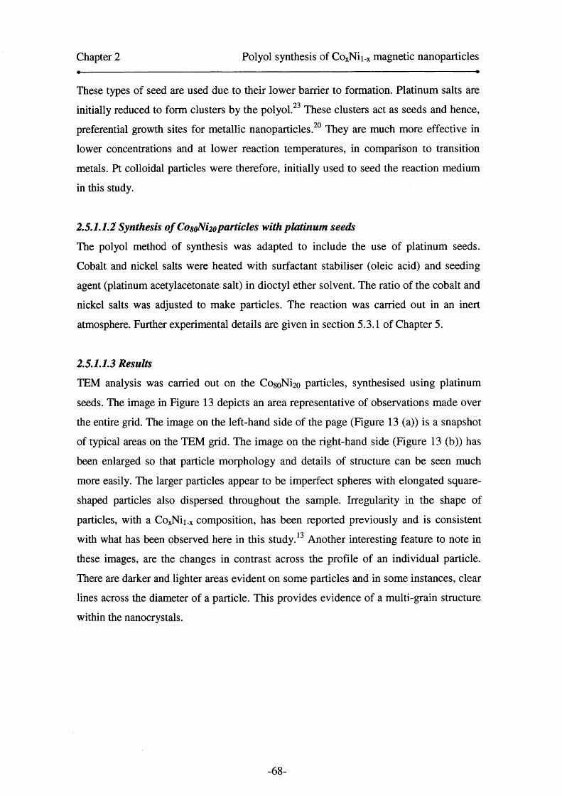
Chapter 2 Polyol synthesis of CoxNii_x magnetic nanoparticles
These types of seed are used due to their lower barrier to formation. Platinum salts are
initially reduced to form clusters by the polyol.23 These clusters act as seeds and hence,
preferential growth sites for metallic nanoparticles. They are much more effective in
lower concentrations and at lower reaction temperatures, in comparison to transition
metals. Pt colloidal particles were therefore, initially used to seed the reaction medium
in this study.
2.5.1.1.2 Synthesis o f CosoNi2oparticles with platinum seeds
The polyol method of synthesis was adapted to include the use of platinum seeds.
Cobalt and nickel salts were heated with surfactant stabiliser (oleic acid) and seeding
agent (platinum acetylacetonate salt) in dioctyl ether solvent. The ratio of the cobalt and
nickel salts was adjusted to make particles. The reaction was carried out in an inert
atmosphere. Further experimental details are given in section 5.3.1 of Chapter 5.
2.5.1.1.3 Results
TEM analysis was carried out on the CogoNi2o particles, synthesised using platinum
seeds. The image in Figure 13 depicts an area representative of observations made over
the entire grid. The image on the left-hand side of the page (Figure 13 (a)) is a snapshot
of typical areas on the TEM grid. The image on the right-hand side (Figure 13 (b)) has
been enlarged so that particle morphology and details of structure can be seen much
more easily. The larger particles appear to be imperfect spheres with elongated square
shaped particles also dispersed throughout the sample. Irregularity in the shape of
particles, with a CoxNii.x composition, has been reported previously and is consistent
with what has been observed here in this study. 13 Another interesting feature to note in
these images, are the changes in contrast across the profile of an individual particle.
There are darker and lighter areas evident on some particles and in some instances, clear
lines across the diameter of a particle. This provides evidence of a multi-grain structure
within the nanocrystals.
-68-

Chapter 2 Polyol synthesis of CoxNii_x magnetic nanoparticles
Figure 13. (a) Low resolution TEM image of typical grid area for heterogeneously
nucleated CosoNi2o particles with a mean diameter of 12.6 nm (b) size distribution
of the heterogeneously nucleated CogoNi2o particles.
lOOnm
Magnified area of
nanoparticles
— | [ |~~~[. | |10 11 12 13 14 15 16
Nanoparticle size (Nm)
The images in Figure 13, shows small satellite particles surrounding the larger 12 nm
particles. The satellite particles have not fully grown and are believed to be completely
oxidised to their cores. The size distribution graph in Figure 13(b) displays clearly that
two nucleation events have taken place, leading to the bimodality seen in this particular
sample. This is evidenced by the two peaks seen in the graph. The peaks were fit to
lognormal distributions, to obtain a mean diameter for each group of particles. The peak
to the left of graph yielded a mean diameter of 2.6 +/- 0.9 nm. The deviation from the
mean is large, at around 35%, but the accuracy of the TEM below 3 nm is not known.
The peak to the right of graph gave a mean of 12.6 +/-1.1 nm, which is representative of
what was observed across the TEM grid. The larger particles had a markedly narrower
size distribution (~9%). If a mean diameter had been obtained for the sample overall,
without taking into account the bimodality, a very large size distribution would be
apparent. Both small and large particles were monodisperse and well separated from
one another, due to the surfactant stabiliser.
Chapter 2 Polyol synthesis of COxNii_x magnetic nanoparticles
Oxidation of the smaller satellite particles described above was confirmed due to
magnetic studies, on 6 nm CogoNi2o particles that had been synthesised using the
heterogeneous nucleation method, earlier in this investigation. The particles were much
smaller than the particles shown in Figure 13, as a slower heat rate had been used during
their synthesis. The magnetic studies showed that the 6 nm CogoNi2o particles displayed
very low magnetic moments (6 emu g'1). The room temperature hysteresis loop also
resembled that of a polycrystalline antiferromagnet. This is characteristic in cobalt and
nickel particles that have oxidised completely.24 The small size of the particles must
have led to complete oxidation to the particle’s cores. The blocking temperature of the 6
nm CogoNi2o particles was also low, suggesting that a significant percentage of the
particles were fully oxidized and therefore did not support exchange biasing. Therefore,
in order to optimise the synthesis and establish control over the particle sizes, the heat
rate was adjusted so it was slow initially and then very fast after the solution
temperature had reached 160 °C to the reflux temperature of the solvent. This induced
fast nucleation and subsequent growth of the particles. The 12.6 nm particles shown in
Figure 13 were synthesised after the synthetic method of the 6 nm particles was
sufficiently adapted to allow the growth of larger particles.
Elemental combustion analysis of the bimodally distributed CogoNi2o particles
gave the percentage of sample weight that was due to surfactant stabiliser. Due to the
multiple washings and addition of extra surfactant that took place post-synthesis, before
the drying of the particles (section 5.6), this value varied from sample to sample by a
few percent. The 12.6 nm particles showed that 16.9 mass% was due to the surfactant.
Carbon corresponded to 13.6 mass% and hydrogen was 1.7 mass% respectively.
Oxygen from the surfactant was determined as 1.6 mass%.
EDX analysis showed that the precursor had transferred into the final alloy in
corresponding ratios. The five different spots that were examined showed consistency
and indicated a solid solution had probably been formed. The atomic percentages for
cobalt and nickel present were 51.2 atom% and 13.1 atom% respectively. This
corresponded to a composition of CogoNi2o for the particles. The oxygen component of
the sample was 35.7 atom%.
X-ray diffraction was carried out, and the diffraction pattern displayed in Figure
14 was obtained, for the heterogeneously nucleated Co8oNi2o 12.6 nm particles. The
crystalline phases cobalt (fee) and nickel (fee) were characterised, in addition to CoO
(fee) and NiO (fee). The strongest reflection is due to the {llljplanes of cobalt (fee),
-70-

Chapter 2 Polyol synthesis of CoxNii.x magnetic nanoparticles
nickel (fee) and cobalt (hep) phases at 20 -44.0°. There was some evidence that C0 2O3
crystallites may have been present, due to the peak at 2 0 =51.8° possessing a shoulder.
XPS results carried out on samples with a similar composition, which are described
later (Section 2.7.2), also displayed peaks characteristic of C02O3. However, this
particular oxide has not been included in the peak assignments of the diffraction pattern
(Figure 14) or in Table 1, as the peaks corresponding to Co20 3 are masked by the peaks
due to Co(fcc)/ Ni (fee) and the detector grid (20 = 57.4°).
Figure 14. Diffraction pattern of heterogeneously nucleated, 12.6 nm Co8oNi2o
nanoparticles.
.0x10 -
0 .9 -
0 .8 -
0 .7 -
0 .6 -
0 .5-I6030 35 40 45 50 55
2 0 / degrees
Table 1. XRD theoretical peak positions can be assigned to the diffraction pattern
for the 12.6 nm CosoNi2o nanoparticles.
Key20/ degrees (Peak
position)Crystallographic
Plane Description
■ 44.251.8
{111}{200} fee Co
• 44.4 {111}52 {200} fee Ni
n 36.4 {111}1—1
42.4 {200} fee CoO
a 37.2 {111}43.4 {200} fee NiO41.5 {100}
■ 44.3 {002} hep Co
The coloured boxes and spheres in Table 1 indicate the peak positions for the main
crystallographic planes of cobalt, cobalt (II) oxide, nickel and nickel (II) oxide in Figure
-71-

Chapter 2 Polyol synthesis of CoxNii.x magnetic nanoparticles
14. The theoretical values in Table 1 correspond well to the peaks shown in the
diffraction pattern in Figure 14. Due to background peaks from the substrate, only peaks
between 30 ° and 60 ° have been shown. This has been maintained throughout this
thesis, where it has been possible to obtain diffraction patterns.
The peaks for the {111} and {200} planes of cobalt (fee) and nickel (fee)
correspond with what was found in a study carried out by Luna et al? The peaks for the
possible hep phase of cobalt are also confirmed by the presence of {1 0 0 } and {0 0 2 }
reflections, at 20 =41.4° and 44.1° respectively. The reflections attributed to CoO and
NiO are due to the partial oxidation of the particles. Luna reported broad peaks with low
intensities, however here, the intensities of the peaks due to oxide appear to be much
more intense.2 The particles made by Luna’s group had a mean diameter of 15 nm
(measured from TEM) and a crystal size of 10 nm (calculated from {111} reflection of
XRD), with a monomodal distribution. The mean diameters in Luna’s study, correlate
well with the mean diameter of the larger particles in this bimodally distributed sample.
The crystallite size calculated from the {111} broadening of cobalt and nickel (fee) for
the pattern in Figure 14 is 5.7 nm .2 This is a very interesting result, as it is close to the
overall numerical mean diameter for the Co8oNi2o 12.6 nm particles (5.9 +/-0.9 nm), if
the size distribution curve in Figure 13 was fitted to a lognormal distribution and the
assumption that there is no bimodality in the particles’ size distribution. The result from
Scherrer analysis of the 12.6 nm Co8oNi2o particles, indicates one of two things; the
particles are either multi-grained nanocrystals or the diameter obtained is due to an
average over all the particles in the sample. The multi-grain structure in the particles is
also evidenced by the TEM images shown in Figure 13. Therefore, it is most likely that
the crystallite size obtained by Scherrer analysis is due to the multi-grain structure in the
particles. It is however, important to remember that the Scherrer equation was not
derived for samples with polydisperse crystallites. The equation does not take into
account variations in size of nanoparticles as it was designed for particles that are
approximately equal in diameter.25
In summary, CosoNi2o particles have been synthesised using heterogeneous
nucleation methods with platinum as the seed. The particles had a bimodal size
distribution and exhibited multi-grain structure in TEM studies, characterised by lines
across the surface of individual nanoparticles. The existence of multi-grains has been
seen previously in studies carried out into the synthesis of CosoNi2o particles, using a
similar yet slightly different synthetic method.2, 13 EDX showed that atomic ratios of
-72-

Chapter 2 Polyol synthesis of COxNii_x magnetic nanoparticles
cobalt and nickel in the particles correlated well with the ratios of precursor materials
initially used to make the particles. XRD identified the dominant phases in the sample
and also indicated that the oxide shell of the particles could have been composed of a
mixture of cobalt (II) and (III) oxide crystallites. Scherrer analysis correlated with the
multi-grain theory determined by TEM analysis of the particles. The nanoparticle
solution of these 1 2 .6 nm CogoNi2o particles was a stable ferrofluidic solution which
responded to a magnet. The black nanoparticle solution deviated towards a magnet,
although this response was much weaker in comparison to that of the dried
nanoparticulate powder obtained from the solution.
The synthesis of Co8oNi2o particles has been carried out, prior to this
investigation, but had required the use of a base solution (NaOH), to control the pH in
the reactant solution.2, 13, 1 8 ,22 The base in that particular reaction, did not allow the
precipitation of intermediate solid-phase alkoxides to occur, as these act as reservoirs
for the metal ions species and inhibit their subsequent growth into particles.22
The 12.6 nm particles formed in this study displayed similar structure to those
reported in the Luna study.2 Luna et al found that larger particles were polycrystals,
whilst smaller particles (<10 nm) were single crystals.2, 13 TEM analysis of particles
(Figure 13) in this investigation, have shown that the larger particles appeared to be
polycrystals or multi-grains, due to the lines and contrast gradients across the particles.
This confirms what was found in previous studies. Luna also reported a shoulder on the
peak of the fcc{lll} reflection for cobalt and nickel, attributed to a small fraction of
hep phase cobalt, in their 10 nm CogoNi2o particles.2 This was also observed in the 12.6
nm CogoNi2o particles synthesised in this study. The particle fraction with hep structure
is said to become more significant as the particle size decreases, as Luna observed a
progressive increase in the asymmetry of this fcc{ 1 1 1 }reflection.2
If a comparison is made between the homogeneous and heterogeneous methods
of synthesis, it is clear that the heterogeneous route appears to be more promising, as the
mean size of the particles is larger than the homogeneously nucleated samples. The
heterogeneously nucleated particles can therefore, support the growth of an oxide shell
coating around the metallic alloy core. The key to continuing use of the heterogeneous
method, is to manipulate the heat rate and other reaction variables to encourage a single
nucleation event. Heterogeneous nucleation encourages a more definitive step between
the nucleation and growth stages and therefore, the development of heterogeneous
-73-
Chapter 2 Polyol synthesis of CoxNii_x magnetic nanoparticles
synthesis methods could lead to monodisperse core-shell nanoparticles with a larger size
and narrower size distribution than the CoxNii_x particles made by other groups. 22
2.5.1.2 Use of premade nanoparticles as nuclei for further growth
2.5.1.2.1 Introduction
An alternative heterogeneous synthetic approach was attempted in order to try and
increase particle size further, and therefore improve exchange bias effects.
Heterogeneous nucleation methods already employed in this investigation (section
2.5.1.1), were adapted so that premade nanoparticles were used as seed nuclei. This was
a modification of a method adopted by Yamamuro et al for the synthesis of iron
nanoparticles.10
The Yamamuro method involves the preparation of nanoparticles in a two-stage
procedure, where the premade particles remain in the reaction vessel. Additional
reagents are then introduced into the vessel, and the reagents are heated up with the
nanoparticle seed solution. The monomers in the reactant solution grow around the pre
existing nanoparticle seeds and this leads to an increase in the size of the final particles.
This two-stage method was implemented by calculating the extra amount of reactants
required to increase the particle volume 8-fold, and hence double the particle diameter.
The main aim of utilising this type of heterogeneous method was to double the particle
size.
2.5.1.2.2 Synthesis o f CosoNi2oparticles with premade nanoparticle seeds
Using the same methodology adopted by Yamamuro,12 Co8oNi2o particles were also
fabricated in a two stage process. In the first stage of the synthesis cobalt and nickel
tetrahydrate acetate salts were dissolved in the solvent, with 1,2-hexadecanediol acting
as a reducing agent. Two different surfactants (OA and TBP) were used as stabilisers
and were introduced into the reaction flask at different stages of the reaction, due to
their individual steric influences over the nanoparticles’ rate of growth. OA was added
to the reaction mixture with the other precursors at the beginning of the reaction, whilst
TBP was injected into the flask at 160 °C when the nucleation and growth of the00particles began to occur. The solution was then heated rapidly to the reflux point. The
nanoparticles that were formed in the first stage were the nanoparticle seeds. The
-74-

Chapter 2 Polyol synthesis of CoxNii.x magnetic nanoparticles
primary nanoparticles act as seed nuclei for the subsequent growth of secondary larger
nanoparticles in the second stage. The two-stage process is pictorially depicted in
Scheme 1.
Scheme 1. Schematic of particle growth in a two-step synthesis; [A] particles made
in stage one of the reaction. [B] Particles grown from reaction of [A] with
additional reactants which are heated to reflux in the second stage results in
particles.
0 9 mmol of salts
0 9 mmol o f surfactant
CoO/NiOextra solvent ♦ reducing
agent
8 nm particles, grown from 4 nm particles, that are used as seeds for growth.
4 nm particles used to seed growth
An appropriate volume of seed solution was then kept in the flask to be used as
seed nuclei for the subsequent reaction, whilst the additional seed solution was extracted
from the flask to be subsequently analysed. In the second step, additional cobalt and
nickel salts, reducing agent, solvent and surfactant stabiliser were introduced to the
flask that contained a few millilitres of the previously made nanoparticle seed solution.
The reaction to grow larger particles followed the same profile as that used to
make seeds. The larger particles that were grown from the smaller nanoparticle seeds
appeared to respond to a magnet. Whereas the smaller nanoparticle seed solution, which
had been extracted from the flask prior to the second stage, did not show this magnetic
response.
-75-

Chapter 2 Polyol synthesis of CoxNii.x magnetic nanoparticles
2.5.1.2.3 Results
A TEM image of the particles used to seed the subsequent reaction is shown in Figure
15(a). It can clearly be seen that these seed particles have partially ordered into an array-
type formation. The packing seems to be hexagonal in the areas where the particles are
mostly self-organised. The streaking, due the solvent edge drying, can also be seen on
the right hand edge of the image. Detailed size distribution studies were carried out on
the particles in this sample, and an average particle size of 4.4 +/- 2.0 nm was
calculated. The size of seed particles was fit to a lognormal distribution. There was no
evidence of a contrast gradient across the particles; therefore these particles are
probably single crystals. The interparticle distance was measured from the centre of one
nanoparticle to the centre of its neighbouring particle and was 6.6 nm. This was only
carried out on the seed particles as hexagonal packing was evident.
Figure 15 (a) Particles used to seed subsequent growth of larger particles and
corresponding size distribution plot (b) Image of larger particles which have
approximately doubled in diameter with size distribution plot.
(a) (b)
12 0 -
10 0 -
<o| 8 0 -oog 6 0 - a>3CT® 4 0 -u.
2 0 -
0-
o 40-
3 0 -
5 6 7 8 9 10 11 12 13 14 15Nanoparticle size/ nm
12 3 4 5 6 7 8 9 10 11 12
Nanoparticle size /nm
-76-
Chapter 2 Polyol synthesis of CoxNii.x magnetic nanoparticles
The particles that were grown from the nanoparticle seeds of Figure 15(a)
appear to have almost doubled in size (Figure 15 (b)). The actual mean diameter
obtained for them was 7.4 +/- 3.9 nm. The plot was fit to a lognormal distribution
overall, although there appears to be slight bimodality with a reasonably large
population of particles at around 10-11 nm. Although this method led to a significant
increase in particle size, polydispersity was an issue. The size distribution of the initial
seeds solution was quite large and this has followed through into the final seeded
sample shown in 15(b).
Elemental analysis was carried out on the nanoparticle sample which had been
grown from the initial seeds. The proportion of the nanoparticulate powder attributed to
surfactant stabiliser, was found to be 23.8 weight%. The amount of carbon was found to
be 18.9 weight% and hydrogen was 2.8 weight%. Oxygen in the oleic acid and
tributylphosphine was 2.1 weight%. The remaining weight was assumed to be due to the
metallic nanoparticle alloy.
EDX results suggested the larger 7.4 nm CogoNi20 particles that were made after
seeding with smaller 4.4 nm CogoNi2o particles, had formed a solid solution as studies
into atomic concentrations showed consistency in composition at different areas around
the sample stub. The amount of oxygen present in the 7.6 nm Co8oNi2o particles was
calculated as 47.2 atom%. The atomic percentages of cobalt and nickel were 40.9
atom% and 11.9 atom% respectively. This correlated with a 4:1 precursor molar ratio,
between the cobalt and nickel salts.
XRD diffraction was tried and did not yield a crystalline diffraction pattern,
possibly due the small sizes of the particles in both samples. The amorphous peaks were
difficult to distinguish from the background signal of the instrument, due to a weak
signal from the samples.
In summary, CogoNi2o particles that have been synthesised using the polyol
route, have been used as seeds for further growth, to form particles with a diameter of
7.4 nm. The reaction process was a success, as the mean diameter of the particles had
almost doubled using this route. The larger seeded particles gave a small response to a
magnet. The Yamamuro seeding technique had previously only been used for the
synthesis of iron nanoparticles, therefore the work done here on a CoxNii_x system is
novel.12
Investigations carried out by Yamamuro showed that particle size could be
tuned by controlling the reaction temperature, as this is a key parameter for the control
-77-

Chapter 2 Polyol synthesis of CoxNii.x magnetic nanoparticles
of supersaturation. ’ His work showed an increase in particle size, from 5 nm seed
particles to 8 nm and 12.5 nm nanometer particles depending on the heat rates used, in
this two stage process. 10 The 4.4 nm Co8oNi2o seeds formed in this study were
comparable in size to the 5 nm iron nanoparticle seeds used by Yamamuro.12 After 75
minutes of heating the seed solution with additional reactants, 7.4 nm CogolN^o particles
were formed. This compared well with the 8 nm iron particles, previously produced by
Yamamuro with 145 minutes of heating. The key difference in both reactions was that
Yamamuro had used a mixture of oleic acid and oleylamine as surfactant for the iron
particles. In order to synthesise CosoNi2o particles, a mixture of oleic acid and
tributylphosphine surfactants was used.
The surfactant used plays a major role in the size control of particles. The steric
bulk of the tributylphosphine inhibited the particle growth, and hence the mean particle
diameter. Phosphine-based ligands are known to control nanoparticle sizes due to their
steric bulk.27 This was observed earlier in the investigation when TOPO and TBP were
solely used to make CoxNii_x particles. It is also possible that faster nucleation and
therefore particle growth occurs with the OA/TBP combination of surfactants, due to
the ease with which they complex to the metallic ion species. This could explain the
faster reaction time over which the Co8oNi2o particles were formed, 75 minutes
compared to 145 minutes, even though the reaction conditions were identical to those
adopted by Yamamuro for iron particle synthesis.
The primary concern about this technique was indeed the small size of the
particles formed after seeding, which was still quite small at around 8 nm and had led to
the weak magnetic response. Polydispersity was also an issue as the size distributions
that were determined were large. In order to solve this, careful manipulation of the
heating rates and hence reaction temperatures could lead to better control of particle size
and size distribution in the future. However, this would require intensive examination
due to the complex nature of the CoxNii_x system, in comparison to the iron nanoparticle
synthesis, and so is beyond the scope of this project due to time constraints. This
particular study of seeding particle growth with like-nanoparticle seeds, has indicated
that a one step procedure better for the CoxNii_x system. The Yamamuro technique was
therefore, not pursued for the synthesis of CoxNii_x particles, but had been successful in
its’ goal of increasing the mean particle size and in terms of being a novel method of
increasing particle size for the CoxNii_x system.
-78-

Chapter 2 Polyol synthesis of CoxNii.x magnetic nanoparticles
2.5.3 Other Noble metal seeds
Having investigated platinum seeds as nucleating agents for the synthesis, other
possible seeds, gold and silver, were also examined to probe their effect on the synthesis
of CoxNii_x nanoparticles. The results would serve as a comparison between the noble
metals, and their effectiveness as seeds. Previously, Viau et al 20, 22 had investigated
silver as a seed in polyol synthesis, however gold has not been investigated prior to this.
Investigations carried out where silver was a nucleating seed, showed that it was not as
good a nucleating agent as platinum. The size range of particle diameter achieved by
200 nm< dm <500 nm, even when the highest concentrations of silver nitrate
concentrations were used. 20 Recently, Luna et a l13 investigated Ag and Pt seeds for the
synthesis of CogoNi2o particles. The platinum seeds produced fine nanosized particles of
between 5 nm < dm< 150 nm and the silver seeds, in their study ranged from between
100 nm < dm< 540 nm.
In this study, the use of silver seeds yielded particles of 40 nm < dm< 100 nm
and gold seeds yielded 60 nm< dm< 150 nm. However, the particles were extremely
polydisperse with large size distributions. The particles had also aggregated, making it
difficult to visualise them, as they had coalesced together in clumps. Therefore, this
route was not pursued any further, due to the size and quality of particles that were
obtained. These seeds were not deemed to be effective at controlling particle size.
Magnetic measurements were also not carried out, as the extremely large size
and the aggregation put into question the magnetic properties of the particles, in terms
of exchange bias. Platinum seeds offered better size control for the CoxNii-x system and
yielded crystalline particles with narrow size distributions. Therefore, platinum seeds
were the most effective method of controlling particle sizes for this system.
2.6 Variations in stabilising surfactants
2.6.1 Introduction to variations in stabilising surfactant
A number of surfactants were tested to examine their effects on particle size, shape and
stability to oxidation. These properties are affected by how tightly the surfactants bind
to the metallic ions in the reactant solution and form a ligand-metallic ion complex.
These complexes act as the reaction precursor and so are important for the eventual
-79-
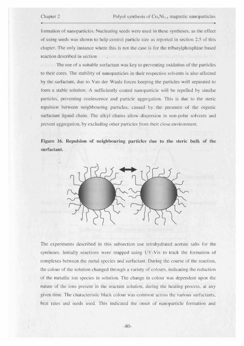
Chapter 2 Polyol synthesis of CoxNii_x magnetic nanoparticles
formation of nanoparticles. Nucleating seeds were used in these syntheses, as the effect
of using seeds was shown to help control particle size as reported in section 2.5 of this
chapter. The only instance where this is not the case is for the tributylphosphine based
reaction described in section
The use of a suitable surfactant was key to preventing oxidation of the particles
to their cores. The stability of nanoparticles in their respective solvents is also affected
by the surfactant, due to Van der Waals forces keeping the particles well separated to
form a stable solution. A sufficiently coated nanoparticle will be repelled by similar
particles, preventing coalescence and particle aggregation. This is due to the steric
repulsion between neighbouring particles, caused by the presence of the organic
surfactant ligand chain. The alkyl chains allow dispersion in non-polar solvents and
prevent aggregation, by excluding other particles from their close environment.
Figure 16. Repulsion of neighbouring particles due to the steric bulk of the
surfactant.
The experiments described in this subsection use tetrahydrated acetate salts for the
syntheses. Initially reactions were mapped using UV-Vis to track the formation of
complexes between the metal species and surfactant. During the course of the reaction,
the colour of the solution changed through a variety of colours, indicating the reduction
of the metallic ion species in solution. The change in colour was dependent upon the
nature of the ions present in the reactant solution, during the heating process, at any
given time. The characteristic black colour was common across the various surfactants,
heat rates and seeds used. This indicated the onset of nanoparticle formation and
-80-

Chapter 2 Polyol synthesis of CoxNii.x magnetic nanoparticles
occurred at temperatures between 150 °C-232 °C, depending on the reaction conditions
and precursors used for the synthesis.
In these reactions, nuclei formation is followed by the formation of metal-ion
surfactant complexes. This is evident through the characteristic colour changes in the
solution during synthesis. The bright coloured solutions result from the ions/complexes
of Co2+, Ni2+ and Co3+. The solutions subsequently darken, to a brown/black colour
upon the growth of particles, and hence the presence of absorption centres in the flask,
characterised by a flat-line in the UV-Vis spectra.
Figure 17. UV-Vis spectra of synthesis of Co8oNi2o particles taken at (a) 120 °C
(left) and (b) 150 °C (right) with different surfactants.
'33 2 .5 -
h£<
2 .0 -
8cce
1 .5 -
x>oC/3-O
1 .0 -
< 0 .5 -
OAHDA
2 .5 -'3D
2 .0 -Jb13< 1 .5 -0)§ 1 .0 -
——UwC/3N< 0 .5 -
1----------------- 1----------------- 1-------------------1------------------1------------------1
400 500 600 700 800 900Wavelength /nm
TOPOHDA
i--------1-------- 1--------1-------- r------- 1400 500 600 700 800 900
Wavelength / nm
Table 2. The effect of surfactant used in polyol synthesis, on Co8oNi2o nanoparticle
formation temperature.
Surfactant NP formation temperature/ ° COleic Acid 220Oleylamine 218
OA/OY 232Tributylphosphine 224Trioctylphosphine 204Hexadecylamine 150
Early investigations into the reaction profile were carried out using UV-Vis studies,
where aliquots of reaction solution were taken at specific temperature points during the
reaction, to track the formation of complexes and the subsequent growth of particles.
- 81 -
Chapter 2 Polyol synthesis of CoxNii.x magnetic nanoparticles
The point of nanoparticle formation was observed in each case, and is variable and
dependent upon the reaction conditions and surfactant used (see Table 2).
The UV-Vis spectrum in Figure 17, on the left panel, displayed a shift in the
largest peak when oleic acid (OA) was replaced with hexadecylamine (HDA). As the
temperature rises, the peak intensity of the HDA reaction decreases, with the peak
disappearing by 150 °C, as displayed in the right panel of Figure 17. This indicates
particle formation as the particles absorb light across the visible spectrum. This effect is
not seen until 220 °C in the samples made with OA. The spectrum on the right hand
panel also shows an absorption produced by the formation of a TOPO-metal ion
complex at around 150 °C. The peak position is similar to that of the OA reaction,
however, the peak becomes characteristically sharp at this temperature. For the oleic
acid reaction, the peak width remains unchanged after 120 °C, indicating that the metal
ion-oleate complex has been formed by this temperature. A rise in the low wavelength
absorption occurs between 170 °C and 200 °C for this sample.
This technique gave a lot of information about the effect of surfactants on
nanoparticle formation temperatures. It also allowed the synthesis to be optimised for
subsequent reactions, due to the detailed plotting of the effect of temperature on particle
growth, and hence the ideal conditions required to initiate particle formation. This
information was used to tailor the reaction to specific heating profiles, allow complex
formation and hence nucleation followed by particle growth. It also gave an indication
of the suitability of certain surfactants for nanoparticle growth and in particular, the type
of surfactant that suits a particular composition of particle e.g. Ni- amine stabilised
particles.
2.6.2 Oleic acid (OA)
2.6.2.1 Introduction
An investigation was carried out into the effect of using oleic acid as the sole surfactant
stabiliser, to coat Co8oNi2o core-shell nanoparticles. The reaction was similar to that
described in the platinum seeds section earlier (Section 2.5.1.1). The heating rate was
different in this reaction, in order to try and control the bimodality that was evident
before.
-82-
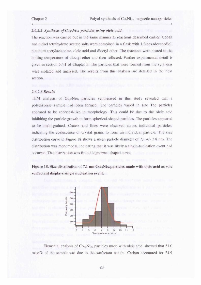
Chapter 2 Polyol synthesis of CoxNii_x magnetic nanoparticles
2.6.2.2 Synthesis of CogoNix) particles using oleic acidThe reaction was carried out in the same manner as reactions described earlier. Cobalt
and nickel tetrahydrate acetate salts were combined in a flask with 1,2-hexadecanediol,
platinum acetylacetonate, oleic acid and dioctyl ether. The reactants were heated to the
boiling temperature of dioctyl ether and then refluxed. Further experimental detail is
given in section 5.4.1 of Chapter 5. The particles that were formed from the synthesis
were isolated and analysed. The results from this analysis are detailed in the next
section.
2.6.2.3 ResultsTEM analysis of CogoNi2o particles synthesised in this study revealed that a
polydisperse sample had been formed. The particles varied in size The particles
appeared to be spherical-like in morphology. This could be due to the oleic acid
inhibiting the particle growth to form spherical-shaped particles. The particles appeared
to be multi-grained. Craters and lines were observed across individual particles,
indicating the coalescence of crystal grains to form an individual particle. The size
distribution curve in Figure 18 shows a mean particle diameter of 7.1 +/- 2.8 nm. The
distribution was monomodal, indicating that it was likely a single-nucleation event had
occurred. The distribution was fit to a lognormal shaped curve.
Figure 18. Size distribution of 7.1 nm CogoNi2o particles made with oleic acid as sole
surfactant displays single nucleation event.
3 4 5 6 7 8 9 10 11 12Nanoparticle size/ nm
Elemental analysis of CogoNi2o particles made with oleic acid, showed that 31.0
mass% of the sample was due to the surfactant weight. Carbon accounted for 24.9
-83-
Chapter 2 Polyol synthesis of CoxNii.x magnetic nanoparticles
mass%, whilst hydrogen was 3.2 mass% and oxygen was 3.0 mass%. EDX analysis of
the particles showed that 38.7 atom% was due to oxygen, whilst 47.3 atom% was due to
cobalt and 13.9 atom% due to nickel. The level of nickel was slightly high, however in
general terms the relative cobalt: nickel ratios corresponded. The amount of oxygen
observed was consistent with what has been seen with other samples in this study.
Powder XRD studies revealed an amorphous material with no sharp crystalline peaks.
The small size of the particles and hence the multigrain structure evident in TEM
studies, could be a contributory factor to the difficulty in obtaining a strong signal from
the sample, for the XRD technique. Fluorescence from the cobalt could have also
contributed.
The particles formed using oleic acid as sole surfactant, with seeds, were small
in size and displayed multi-grain structures. The amount of surfactant that remained
adsorbed to the surface of the particles, determined by elemental analysis, was quite
high. This is possibly due to oleic acid readily forming a complex with the metal ion
species or excess surfactant that may have adsorbed onto the nanoparticle surface
during the washing procedure. The mean size of the particles, and the distribution
indicated a single nucleation event. However, the small size of the particles means that
they have probably oxidised to their cores, and are primarily composed of
antiferromagnetic cobalt-nickel oxide crystallites. This was further confirmed by a
magnet being held up to the particles and producing no response. The complete
oxidation of the Co8oNi2o particles, coated in oleic acid, indicates that it does not work
well enough on its own to stabilise the particles.
Oleic acid forms a complex with Co and Ni ions very easily during the
nucleation stages of a polyol synthesis, and this complex acts as a precursor to the
formation of the particles. Wu et al 26 claimed that chemisorption occurs between the
carboxylate ion and the surface of cobalt nanoparticles through the two oxygen atoms
and this is characterised by two peaks in the IR spectra of oleic acid coated cobalt
nanoparticles. Wu also claimed the peaks are due to the asymmetric and symmetric
bands of the carboxylate ion. 26 However, this is cast into doubt as peaks have formed in
those same positions, in a spectrum taken for neat oleic acid in this study (Figure 20).
Thereby indicating that covalent bond formation between the surfactant and
nanoparticle is unlikely. This theory has also proven to be inconclusive in this
investigation, due to the oxidation of the CoxNii_x particles described earlier.
-84-
Chapter 2 Polyol synthesis of CoxNii.x magnetic nanoparticles
Oleic acid could be used in conjunction with another surfactant such as
oleylamine, for the synthesis of CoxNii.x particles. Sun et a l 7’ 28 successfully used an
oleic acid/oleylamine surfactant mixture to stabilise FePt nanoparticles. Therefore, the
next logical step in this study was to examine the effects of oleylamine as a sole
surfactant stabiliser for this system, followed by a study into the effects of using both
OA and OY as surfactant stabiliser for the CoxNii.x system.
2.6.3. Oleylamine (OY)
2.6.3.1. Introduction
Oleylamine has been used as a sole surfactant in the synthesis of CoO,29 Ni and NiO30
nanoparticles in previous studies. Therefore, the use of oleylamine as a stabiliser for
CogoNi2o alloy particles was attempted in this study. Oleylamine does not complex as
tightly to the metallic ion species, as with some of the phosphine-based ligands or oleic
acid. Therefore, oleylamine is generally used in conjunction with other surfactants, such
as oleic acid, to allow control of particle shape and size. 14 However, this particular study
is carried out in order to establish whether or not oleylamine can be used as a sole
surfactant stabiliser for this particular system.
2.6.3.2 Synthesis o f CosoNi20 particles using oleylamine
Particles of expected composition, CogoNi2o,were synthesised using the modified polyol
method which has been described earlier. Cobalt and nickel tetrahydrate acetates,
platinum acetylacetonate, 1 ,2 -hexadecanediol and oleylamine were dissolved in dioctyl
ether, and heated in an atmosphere of nitrogen, in a round-bottomed flask. The solution
was heated up to the reflux temperature of the solvent, and the resulting particles were
extracted post-synthesis. Further experimental detail in given in section 5.4.2 of Chapter
5.
2.6.3.3 Results
The particles formed from the synthesis were small in size, but were clumped together
in large aggregate superstructures, which made quantification of particle sizes difficult.
The oleylamine surfactant did not appear to stabilise the particles from agglomeration.
TEM determined the mean particle size as 6.0 +/- 4.0 nm, and the curve was fitted to a
-85-

Chapter 2 Polyol synthesis of CoxNii.x magnetic nanoparticles
Gaussian function, although it is slightly skewed at the higher end of the size scale.
There were also large clumps of aggregates in the sample. This could be due to an
excess of surfactant in this particular system. The particles formed here appeared to be
much smaller than those made with oleic acid. They also appear to be much more
irregular in shape. Particles of this size have in this investigation, been shown to oxidise
to the core. This has been shown through the magnetic properties of the sample.
Figure 19. Size distribution of 6 nm CosoNi2o particles made with oleylamine as sole
surfactant stabiliser.
3 4 5 6 7 8 9 10 11 12Nanoparticle size/ nm
Elemental analysis revealed that 18.3 weight% of the CogoNi2o particles,
synthesised with an oleylamine coating, was due to carbon, hydrogen and nitrogen. The
value is much lower than that shown for the sample with oleic acid as sole surfactant,
indicating that it is possible oleylamine is easily washed off, and is therefore not very
good at preventing oxidation. Studies carried out by Park et a l30 confirm this.
The particles also did not respond to a magnet at room temperature, therefore,
detailed magnetic measurements were not carried out on the sample because of the
likelihood of oxidation due to small size of the particles. Although, VSM indicated the
particles had oxidised due to the low magnetic moment and the hysteresis loop
resembling that of an antiferromagnet. EDX analysis also exhibited high oxygen levels
in the particles, which indicated they had oxidised.
Previous works by Zhang et al sought to produce CoO nanoparticles using
oleylamine as a stabiliser and solvent, with cobalt (acetylacetonate)2 as the precursor
salt. Zhang found that particles of between 4 -23 nm could be synthesised using this
- 86-

Chapter 2 Polyol synthesis of CoxNii.x magnetic nanoparticles
method.29 Although, the oleylamine was degassed prior to the synthesis and a
nitrogeneous atmosphere was kept during the synthesis. Zhang claimed that the
oxidation arose from the decomposition of [Co(acac)2], and was not due to the
surfactant, or oxygen infiltration into the reaction flask as the reaction was carried out
in an inert atmosphere.29 However, during their post-reaction treatment of the
synthesised particles, they exposed the sample to air, and dried the particles in a
standard oven overnight. For particles of those sizes, particularly the smaller ones, it is
likely that oxidation occurred post-synthesis, and not as they claim in the reaction
vessel.
Park et a l 30 investigated the synthesis of Ni and NiO particles using oleylamine
followed by TOPO. The particles were formed by the thermal decomposition of the Ni-
oleylamine complex. The complex was fabricated before heating the solution, and
introducing the bulkier phosphine surfactant. The equation in Scheme 2 shows the
reaction pathway for formation of the particles. The particles were protected from
aggregation and formed monodisperse single crystals. Upon exposure to air, Park et alOA
claimed that the particles oxidise. This is evidenced by XRD taken before and after
exposure to air for the 5 nm Ni particles. Prior to air exposure single peaks belonging to
the {111} and {220} planes of Ni (fee). After air exposure, the XRD pattern revealed
peaks due to a NiO (fee) structure. This indicated the susceptibility of nickel
nanoparticles to oxidation.
Scheme 2. Synthesis of metallic nickel nanoparticles formed from a nickel-
oleylamine complex.30
(Mevlamine
NH
OAOleylamine can complex with metallic ions at low temperatures. Park et al
claimed that this can occur at around 100 °C. This could explain the smaller mean
diameters obtained, as oleylamine complexes easily with the metallic ions, and these
complexes would also possibly decompose at lower temperatures to bring about the
-87-
Chapter 2 Polyol synthesis of CoxNii_x magnetic nanoparticles
formation of nanoparticles. The lower decomposition temperature would lead to smaller
particles as the nucleation step would be very short in such reactions, and the growth
step would not be sufficiently long enough to redress the balance of the short nucleation
stage.
Similar results have been found in this investigation with CoxNii_x particles of
various compositions, where the particle size is predominantly below 10 nm. This total
oxidation has been consistently observed in particles with a small mean diameter in this
study. The variations in surfactant for a particular metallic nanoparticle synthesis should
be specifically tailored, due to individual characteristics of each surfactant. Different
surfactants can complex with metallic ions with ease or difficulty, depending on factors
such as sterics and their individual affinity to a particular metallic ion species. Amine
based stabilisers such as oleylamine and hexadecylamine have been generally used for
Ni nanoparticle synthesis, however TOPO has also recently been used in conjunction
with these to form Ni particles.31"33
The Co8oNi2o alloy particles coated with oleylamine, in this study, oxidised
completely. This suggests that oleylamine is inefficient as a surfactant stabiliser, when
used solely in particular. Other stabilisers used in this study thus far, such as oleic acid,
have also to some extent shown they cannot sufficiently stabilise a particle from
oxidation if the size of the particles formed is very small (approximately clOnm). In the
case of oleylamine, for the CoxNij.x system, it also could not prevent particle
agglomeration.
Combinations of surfactant have recently been used in many investigations.9,34"
As was mentioned earlier, mixtures of surfactants, such as oleylamine with other oleic
acid have been investigated by other groups for the synthesis FePt nanoparticles.14,36
Therefore, the next plausible approach considered for this investigation was the
combination of oleic acid and oleylamine to stabilise the CoxNii_x particles much more
effectively.
2.6.4. Oleic Acid/Oleylamine mixture
2.6.4.1. Introduction
Following work carried out with oleic acid and oleylamine as sole surfactants, attempts
-88-

Chapter 2 Polyol synthesis of CoxNii.x magnetic nanoparticles
were made to synthesise particles using Co8oNi2o particles with both stabilising agents
together. Previous research on other metallic species, indicated that oleic acid and
oleylamine could be used together, to synthesise metallic nanoparticles.12 Sun et a t
used an OA/OY mixture to produce monodisperse FePt colloids with a modified polyol
method. However, the same principles have been applied to synthesise particles.
2.6.4.2 Synthesis o f CosoNi2o particles using oleic acid/ oleylamine
Cobalt and nickel acetylacetonate salts 1,2-hexadecanediol and a 1:1 mixture of OA/OY
were combined in a flask and heated to the boiling temperature of dioctyl ether.
Platinum seeds were not used in this particular reaction, as particles formed using oleic
acid and oleylamine surfactants on their own with seeds, had yielded small particles
(sections 2.6.2 and 2.6.3).Therefore, by removing seeds from the synthesis, larger
particles were expected to be formed.
2.6.4.3 Results
TEM analysis of CogoNi2o particles, coated with an OA/OY mixture showed that large
spherical and round comer square-shaped particles were formed. The particles were
well-separated from one another. However, the irregularity in shape and polydispersity
meant the particles could not pack to form arrays. The large size of the particles was due
to the lack of nucleating seeds. There were contrast differences across the particles and
also areas on the grid with excess surfactant. This made it difficult to observe individual
particles on occasion. The particles also appeared to be crystalline, and there were less
occurrences of multi-grain structured particles. The mean particle diameter was
determined to be 71.2 +/- 12.0 nm. A plot of the particle size versus frequency was
fitted to a lognormal distribution to obtain an accurate mean diameter value.
Elemental combustion analysis (CHN analysis) determined the mass of the
nanoparticulate sample, due to surfactant, was 12.7 weight%. This constituted 11.6
weight% carbon and 1.1 weight% hydrogen. Nitrogen was not picked up by the
analysis, perhaps due to the trace amount associated with one atom of nitrogen, in the
amide linkage.
EDX analysis of the CogoNi2o particles coated with an OA/OY mixture, yielded
an atomic percentage mass of 42.6 atom% for cobalt, whilst nickel was 11.7 atom%.
The value for nickel indicates that the sample maybe slightly nickel-rich due to it
nucleating before cobalt in the reaction solution. The amount of oxygen present, was
-89-

Chapter 2 Polyol synthesis of CoxNii_x magnetic nanoparticles• - ■■ ■ - - - »
determined to be 45.7 atom%.
The Co8oNi2o particles coated with OA/OY, responded to magnet very strongly
in solution, this was probably due to the size of the particles. Monodisperse particles of
a smaller size are ideal due to the need to overcome the superparamagnetic limit of the
particles and induce thermal switching.37 It is possible that the CogoNi2o particles made
here are single domain, even though they are quite large, as the single-domain sizes for
cobalt and nickel particles have previously been determined as 70 and 55 nmoo
respectively.
Both oleic acid and oleylamine surfactants were combined in 1:1 molar
amounts before the reaction, as described in the experimental methods section.
Evidence suggests that a weak acid and weak amine, when combined, react to release a
molecule of water and form an amide through the head groups. The surfactant tail
becomes double-sheathed which provides a stronger barrier to oxidation and
agglomeration. Infra-red spectroscopy corroborates this theory, when compared to the
spectra of neat oleic acid and oleylamine.
Figure 20. IR spectra of neat oleic acid (blue trace), neat oleylamine (green trace)
and an OA/OY (red trace) 1:1 surfactant mixture.O-H out-of-
Broad OH stretch C=0 stretch C -0 stretch p]ane bend
C-H stretches superimposed on O-H stretch C-O-H in plane
O-H in- ,plane bend^v. en **
C-NstretchN-H stretchc2i-
N-Hbend (scissoring) CH2
scissoringN-H wag
Aliphatic C-H stretch
OA OY OA/OY
Amide linkage
15004000 3500 3000 2500 2000 1000 500Wavenumber/ cm
- 90 -

Chapter 2 Polyol synthesis of CoxNii_x magnetic nanoparticles
The amide group is proven by the existence of a peak at 1558 cm' 1 in the red trace of
Figure 20. Oleic acid inhibits growth when coupled with oleylamine. It works well as a
stabiliser, as the amide that has formed complexes with the metal ions less tightly to the
nanoparticle surface and allows particle growth. However, it is not as weak as
oleylamine and therefore prevents oxidation somewhat compared to if both surfactants
are used on a sole basis. This allowed the growth of the particles to occur and also their
stability to oxidation.
In the literature, an OA/OY combination has previously been used to make FePtORnanoparticles. Sun et al carried out the thermal decomposition of Fe(CO)5 in a high-
temperature solution with 1,2-hexadecanediol as the reducing polyalcohol. Oleic acid
and oleylamine were used to produce hexagonally close-packed 3-D arrays of FesoPtso
particles, with an interparticle distance of ~4nm maintained throughout the array due to
the oleic acid/oleylamine mix. In that particular study, particle sizes were adjusted by
controlling the molecular ratio of the iron carbonyl to the Pt salt. The OA/OY mixture
also protected the particles from oxidation. Another similar procedure is described in
the literature as a route to iron oxide nanoparticles, and involved the high-temperature39reduction of iron (III) acetylacetonate in the presence of oleic acid and oleylamine.
Cobalt and nickel carbonyls were not used in this study due to their toxicity. The
Yamamuro12 method of synthesis was adapted to synthesise CogoNi2o particles with an
OA/OY surfactant mixture. As with other surfactant investigations, the reaction was
conducted without the use of nucleating seeds. The surfactant concentration was slightly
altered, compared to other syntheses carried out thus far in this study, so that the
Yamamuro method was followed exactly. This resulted in an approximate 10 %
decrease in the surfactant concentration per millilitre of solvent used, in comparison to
the concentration of surfactant in previous reactions, where a single surfactant had been
used. The effect on size was much more significant than would be expected for such a
small change in surfactant concentration. A particle size increase would be expected,
however, not a change from around 6 and 7 nm for oleylamine and oleic acid used on
their own, to -70 nm for Co8oNi2o particles made with a 1:1 combination of the two
surfactants. The main difference was the use of two surfactants together. The
homogeneous nucleation reaction described earlier also used oleic acid as sole
surfactant, with no seeds, and yielded particles with a mean diameter of 7.6 nm.
Whereas here the particles synthesised are much larger. Therefore, this method was not
pursued in its present form. The OA/OY seemed to stabilise and coat the particles well,
-91-
Chapter 2 Polyol synthesis of CoxNii.x magnetic nanoparticles
and it was felt that by adapting this method to include platinum seeds, which have also
shown promising results in terms of size control (see section 2.5.1.1), it would be
possible to make CoxNii.x particles of a controlled size and stability to oxidation.
Section 2.7 discusses the developments made to the synthesis in order to produce
particles with varied composition, and build upon the methods and results described
here.
2.6.5 Trioctylphosphine oxide
2.6.5.1 Introduction
TOPO has been used successfully for the synthesis of semiconductor nanocrystallites
(CdE where E= S, Se, Te), in important works carried out by Bawendi and co-
workers.40 TOPO was presented to be an effective capping agent of nanoparticles, due
to its selective binding characteristics. It has been found that TOPO selectively absorbs
to preferred crystallographic planes and hence controls the growth of specific crystal
faces.41,42
The Bawendi group 43 also applied this to the synthesis of cobalt nanocrystals.
Dicobalt octacarbonyl (DCOC) was subjected to thermolysis in toluene, yielding 20 nm
spheroidal cobalt particles with a size distribution of approximately 15 %. An increase
in the reaction temperature, together with a large increase in growth, indicated that the
TOPO-metal complex was weak and could be easily removed. The particles would then
be exposed to air and subsequently oxidise. In this investigation, several experiments
have been carried out with trioctylphosphine oxide as the sole surfactant stabiliser. The
effect of varying the concentration of TOPO and the use of heterogeneous and
homogeneous nucleation was probed.
2.6.5.2 Synthesis o f CosoNiioparticles using trioctylphosphine
The particle synthesis proceeded in the same manner as other syntheses described in this
chapter. The metal salts, surfactant, solvent and reducing agent were all heated to the
boiling temperature of the solvent. The subsequently formed nanoparticle solutions
were isolated and kept for analysis. Seeds were not used in the synthesis of sample A
(see Table 3), whereas samples B-D were heterogeneously nucleated and hence were
made with seeds. Table 3 details the range of experiments carried out with TOPO as
-92-

Chapter 2 Polyol synthesis of CoxNii_x magnetic nanoparticles
surface-active agent. However, experimental detail of how TOPO-coated particles were
formed is given in Chapter 5 (section 5.4.4).
2.6.5.3 Results
The formation of TOPO-coated CogoNi2o particles, without the presence of seeds was
investigated. This yielded a stable solution, and particles with a mean diameter of 5.9
+/- 1.7 nm (sample A-Table 3). The particles formed a stable solution, but did not
respond to a magnet, as they had probably oxidised.
The effect of platinum seeds on the nucleation of CogoNi2o particles with TOPO
was subsequently investigated. The concentration of the platinum seeds was varied, in
addition to the surfactant concentration, to establish their effects on particle size. The
molar amount of reactants was varied, over the series of experiments, yielding particles
of different sizes.
Table 3. Reaction parameters of TOPO syntheses of CosoNi2o nanoparticles, made
with or without seeds. The amounts of each reactant with respect to one another
are shown.
I Seeds/ Sample | mmol
Surfactant/mmol
Reactant salts /mmol Particle size (notes)
SampleA / 3.125 2 5.9 +/-1.7 nm particles formed
SampleB
0.0013 3.125 2 Particles are too small to get accurate size ~2-3 nm
SampleC
0.0013 6.25 2 Agglomerated particles mean an accurate size cannot be obtained
Sample 1 D
0.0025 6.25 2 Polydisperse but with small particles - 4.1 +/-2.6 nm
The concentration of seeds was changed, therefore, in theory less seeds meant
less nucleation sites, and so the particles would grow to a larger size when the reagents
were all consumed. The same was true of the amount of surfactant. Less surfactant
stabiliser can lead to larger particles, however, this depended upon the influence of
steric factors on particle growth. Both the seed and surfactant concentration were low in
Sample B and therefore, relatively large particles were expected. However, this was not
-93-

Chapter 2 Polyol synthesis of CoxNii.x magnetic nanoparticles
the case, and the particles that were formed were much smaller than particles made with
oleic acid earlier. So much so, that an accurate size could not be determined as it was
difficult to distinguish the particles from the noise generated by the carbon background,
with the size analysis software.
The results for the TOPO did not follow the expected pattern, and polydispersity
seemed to be intrinsic in any sample made with it as sole surfactant. It was thought that
TOPO would stabilise the particles well from oxidation and control the growth. Sample
D showed the best quality of particles. The particles sizes were fit to a Lorentzian
distribution curve and this yielded a mean diameter of 4.1 nm +/- 2.6 nm. The size
distribution of the particles was large at approximately 60 % and TEM analysis of the
particles revealed polydispersity. Sample B was expected to have the largest particles
due to the lower surfactant concentration. However, there was not enough surfactant to
stabilise the CogoNi2o particles, they had oxidised and did not respond to a magnet at
room temperature. The same was true of samples B, C and D. Therefore, the particles
were of a good shape and morphology, with an absence of multi-grains, but they were
not the right size to support the growth of an oxide shell layer.
The small size of the particles meant that it was also not possible to resolve an
XRD pattern. EDX measurements were not carried out as the size meant that the
particles had almost certainly oxidised fully. The non-responsiveness of these particles
to a magnet further confirmed this.
Figure 21. Size distribution of particles made with trioctylphosphine oxide as
surfactant (sample C -see Table 3), with seeds.
3 4 5 6 7 8 9 10Nanoparticle size / Nm
-94-
Chapter 2 Polyol synthesis of CoxNii_x magnetic nanoparticles
As Dinega 43 had observed in the synthesis of cobalt nanoparticles, TOPO does
not sufficiently stabilise the particles from oxidation. However, it does stabilise the
particles well enough to encourage separation and a spherical morphology.
The main drawback to using TOPO as sole surfactant was the small size of the
particles produced. This led to full oxidation of nanoparticles and yielded a low
magnetic moment. UV-Vis studies (Figure 17) had earlier indicated that the TOPO-
metal ion complex, which precludes particle formation occurs at around 150 °C, with
the point of nanoparticle formation at around 204 °C (Table 2). The metal ion-surfactant
complex was formed much earlier, for particles that had a larger mean diameter, in
comparison to TOPO (e.g oleic acid).
TOPO proved to be a slightly better stabilising agent than oleic acid or
oleylamine in terms of ensuring a stable solution was formed and the particles did not
precipitate out of their respective solvent. The TOPO-coated CogoNi2o particles also
redispersed well into non-polar solvents such as hexane and toluene. Samples made
with just OA or OY as the primary stabiliser had sometimes remained stable, but the
majority of the time precipitated out to the bottom of the sample vial. However, the
main issue that appeared to affect this type of synthesis was the small size of the
particles generated using TOPO. This led to the TOPO-coated particles oxidising
readily and did not respond to a magnetic field. The TOPO-route of synthesising
CoxNii_x particles was therefore not pursued as it was unsuitable for this particular
synthesis.
In order to improve upon these results, TBP was subsequently tested as a
stabiliser for the synthesis of CoxNii_x particles, as it is less bulky than TOPO. This
would perhaps inhibit the particle growth less and lead to the formation of particles with
a slightly larger mean diameter.44
2.6.6. Tributylphosphine
2.6.6.1 Introduction
When compared to TOPO, TBP provides less steric hindrance as it is less bulkier, and
therefore promotes the growth of larger particles45 TOPO is much bulkier, and
therefore slows the growth of the nanocrystal, which should result in smaller
nanoparticles overall. In order to improve upon the results shown in previous studies
-95-
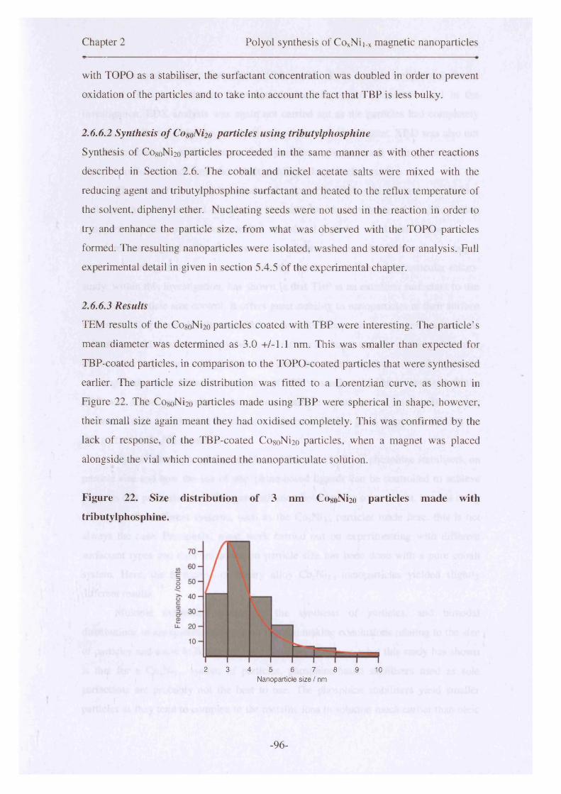
Chapter 2 Polyol synthesis of COxNii_x magnetic nanoparticles
with TOPO as a stabiliser, the surfactant concentration was doubled in order to prevent
oxidation of the particles and to take into account the fact that TBP is less bulky.
2.6.6.2 Synthesis o f CosoNi2o particles using tributylphosphine
Synthesis of Co8oNi2o particles proceeded in the same manner as with other reactions
described in Section 2.6. The cobalt and nickel acetate salts were mixed with the
reducing agent and tributylphosphine surfactant and heated to the reflux temperature of
the solvent, diphenyl ether. Nucleating seeds were not used in the reaction in order to
try and enhance the particle size, from what was observed with the TOPO particles
formed. The resulting nanoparticles were isolated, washed and stored for analysis. Full
experimental detail in given in section 5.4.5 of the experimental chapter.
2.6.6.3 Results
TEM results of the Co8oNi20 particles coated with TBP were interesting. The particle’s
mean diameter was determined as 3.0 +/-1.1 nm. This was smaller than expected for
TBP-coated particles, in comparison to the TOPO-coated particles that were synthesised
earlier. The particle size distribution was fitted to a Lorentzian curve, as shown in
Figure 22. The Co8oNi2o particles made using TBP were spherical in shape, however,
their small size again meant they had oxidised completely. This was confirmed by the
lack of response, of the TBP-coated Co8oNi2o particles, when a magnet was placed
alongside the vial which contained the nanoparticulate solution.
Figure 22. Size distribution of 3 nm CosoNi2o particles made with
tributylphosphine.
2 3 4 5 6 7 8 9 10Nanoparticle s iz e / nm
-96-

Chapter 2 Polyol synthesis of CoxNii_x magnetic nanoparticles
Elemental analysis showed that 18.9 mass% of the sample weight was due to
surfactant. This is similar to the value obtained for other surfactants used in the
investigation. EDX analysis was again not carried out as the particles had completely
oxidised, as illustrated by the powder’s non-response to the magnet. XRD was also not
possible on particles of this size, as the crystallites were too small to be picked up and
also due to fluorescence from the cobalt yielding a large background.
The size of the particles produced using tributylphosphine, were an obvious
indicator that this method could not be used for the synthesis of CoxNii_x alloy
nanoparticles. This method had yielded very small particles which oxidised and were
therefore non-magnetic. The absence of seeds in the reaction should have led to larger
particles forming, however this was evidently not the case. What this particular micro
study, within this investigation, has shown is that TBP is an excellent surfactant to use
in terms of particle size control. It offers great stability to nanoparticles at their surface
in terms of shape and also the size. This is good for this particular type of synthesis, as
it limits the additional anisotropy due to deviations in shape, and any exchange bias that
did occur in larger core-shell particles that could be made in the future, would be due to
exchange anisotropy. If the concentration of TBP was adjusted in future studies or if the
TBP was added post-synthesis to the nanoparticle solutions and used with another
surfactant, such as oleic acid for example, this could lead to good shape and stability for
this particular CoxNii.x system.
Hyeon44 has reported on the effect of the steric bulk, of phosphine stabilisers, on
particle size and how the use of phosphine-based ligands can be controlled to achieve
particles of a particular size. This generalisation holds true to some extent, but this study
shows that for different systems, such as the CoxNii_x particles made here, this is not
always the case. Previously, most work carried out on experimenting with different
surfactant types and their influence on particle size has been done with a pure cobalt
system. Here, the formation of binary alloy CoxNii.x nanoparticles yielded slightly
different results.
Multiple nucleation events in the synthesis of particles, and bimodal
distributions in the systems have meant that the making conclusions relating to the size
of particles and steric bulk has not been possible. However, what this study has shown
is that for a CoxNii.x system of particles, phosphine-based stabilisers used as sole
surfactants are probably not the best to use. The phosphine stabilisers yield smaller
particles as they tend to complex to the metallic ions in solution much earlier than oleic
-97-
Chapter 2 Polyol synthesis of CoxNii_x magnetic nanoparticles
acid for example, and this has led to inhibited particle growth and particle formation at
lower temperatures.
2.6.7 Summary of samples made with different surfactant stabilisers
The investigations carried out using the different surfactant stabilisers were very useful
in determining the optimum reaction conditions for this system. The synthesis of
CoxNii_x alloy nanoparticles has proven to be difficult, due to the varying relationships
of Co and Ni ions, with respect to the surfactants that have been studied. Oleic acid on
its own, did not seem to provide particle stability to both oxidation and nucleation. This
led in many instances, to bimodal distributions of particles. However, in some instances
where the growth of the particles was controlled and the particles that were formed were
large, oleic acid did stabilise the particles from oxidation (heterogeneous nucleation
with platinum seeds-section 2.5.1.1). The particles made with oleic acid were generally
polycrystalline, with multiple grains fusing together during the growth stage of particle
formation.
Oleylamine did not seem to stabilise CoxNii_x particles well and yielded multi
grain nanocrystals, which did not respond to a magnet due their complete oxidation.
Trioctylphosphine and tributylphosphine, used as sole-surfactants for the synthesis of
CogoNi2o particles, had spherical morphology and were single crystals. However, the
nanocrystals were very small (between 3-5.9 nm) and oxidation of such small particles
to their cores could not be prevented. Therefore, the use of these particular surfactants,
for this alloy system, was not pursued as they did not sufficiently stabilise the particles
from oxidation due to the small size of the particles formed using them.
A mixture of oleic acid and oleylamine was tested and this produced well-
formed spherical particles which responded to a magnet. A 1:1 mixture of both
components was used in the syntheses. The particles formed were very large at 70 nm in
mean diameter. Due to the size of the particles produced using this method, the need to
decrease the size was evident, as was the need to retain the quality and shape of
particles produced. The size needed to be decreased as exchange bias is an interfacial
effect, and with such large particles the effect was not evident.
It was hypothesised that by introducing heterogeneous nucleation methods,
which had proven successful in the initial stages of this synthetic optimisation study, it
-98-
Chapter 2 Polyol synthesis of CoxNii.x magnetic nanoparticles
would be possible to tune the size of the 70 nm CoxNii_x particles to ca~10-15 nm.
Platinum seeds had given the best results out of the various seeds and seeding
techniques that had been trialled (Section 2.5). Therefore, the synthetic route that was
deemed appropriate for synthesising CoxNii.x particles, which would display exchange
bias properties, were those that were heterogeneously nucleated with an OA/OY
surfactant mixture and were made of acetylacetonate precursor salts. This optimised
method was adopted for subsequent investigations into the synthesis of CoxNii.x alloy
magnetic particles, where x was varied to produce particles with different compositions.
2.7 Composition
2.7.1 Introduction to composition series
A series of synthetic experiments were carried out, where the composition of the
magnetic nanoparticles was varied. The aim of this study was to synthesise a
compositional series of CoxNii_x nanoparticles that had tuned magnetic properties.
Following investigations into the effects of an OA/OY mixture as a stabilising agent, an
effective method of making the particles was established (Section 2.6.4). Subtle
modifications were made to the design of the OA/OY synthesis described in section
2.6.4.2, in order to obtain nanoparticles of a smaller size (-10-15 nm), compared to
what was generated previously by using that particular surfactant mix (-70 nm). In
these studies, nucleating Pt seeds were also used to control the particle size.
2.7.2 Cobalt nanoparticles
2.7.2.1 Introduction
Cobalt nanoparticle synthesis has been famously documented in the literature, and has
been the basis of many magnetic studies.42, 46 49 Here, several methods were employed
to try and synthesise cobalt nanoparticles. Different reactant salts and surfactants were
used in order to try and optimise the synthesis. The use of metal tetrahydrate acetate
salts yielded particles that were too small and hence unstable to oxidation. Therefore,
acetylacetonate salts were used as precursors for the synthesis of CoxNij.x particles.
Cobalt particles were synthesised using methods that were highlighted earlier12 (Section
2.6.4).
-99-

Chapter 2 Polyol synthesis of CoxNii_x magnetic nanoparticles
2.7.2.2 Synthesis o f cobalt nanoparticles
Cobalt particles were synthesised by heating the acetylacetonate salt, reducing agent
and platinum seeds in dioctyl ether. The solution was heated and the particles that were
formed were isolated and kept for analysis. Further experimental detail is given in
Chapter 5. Acetylacetonate precursor salts were deemed to be better than acetate salts
due to there being less water molecules within the salt structure. Water molecules from
the salts had been found to affect the efficiency of the heating, during particle synthesis.
This had contributed to the lack of reproducibility and problems with reducing the salts
in reactions with tetrahydrate salt precursors.
2.7.2.3 Results
The cobalt particles made using the acetylacetonate salts were better in terms of size, in
comparison to cobalt particles made with acetate salts, as they were large enough to
support the growth of an oxide shell. TEM analysis of the pure cobalt nanoparticles
showed they had clumped together in some areas, due to excess surfactant. In the image
of the cobalt nanoparticles (Figure 23 (a), it is easy to distinguish between the carbon
background (light grey) and the darker spherical particles laid on top. The wispy darker
objects, encasing some of the particles, are due to excess surfactant. The particles are
spherical-like but exhibit contrast differences across their surfaces indicating facets and
multigrain crystals are present. The mean particle size that was determined has been
split into two values, due to the bimodality in the size distribution. If an overall average
was taken, this would be skewed, and not representative of the sizes of particles that
have been seen.
- 100-
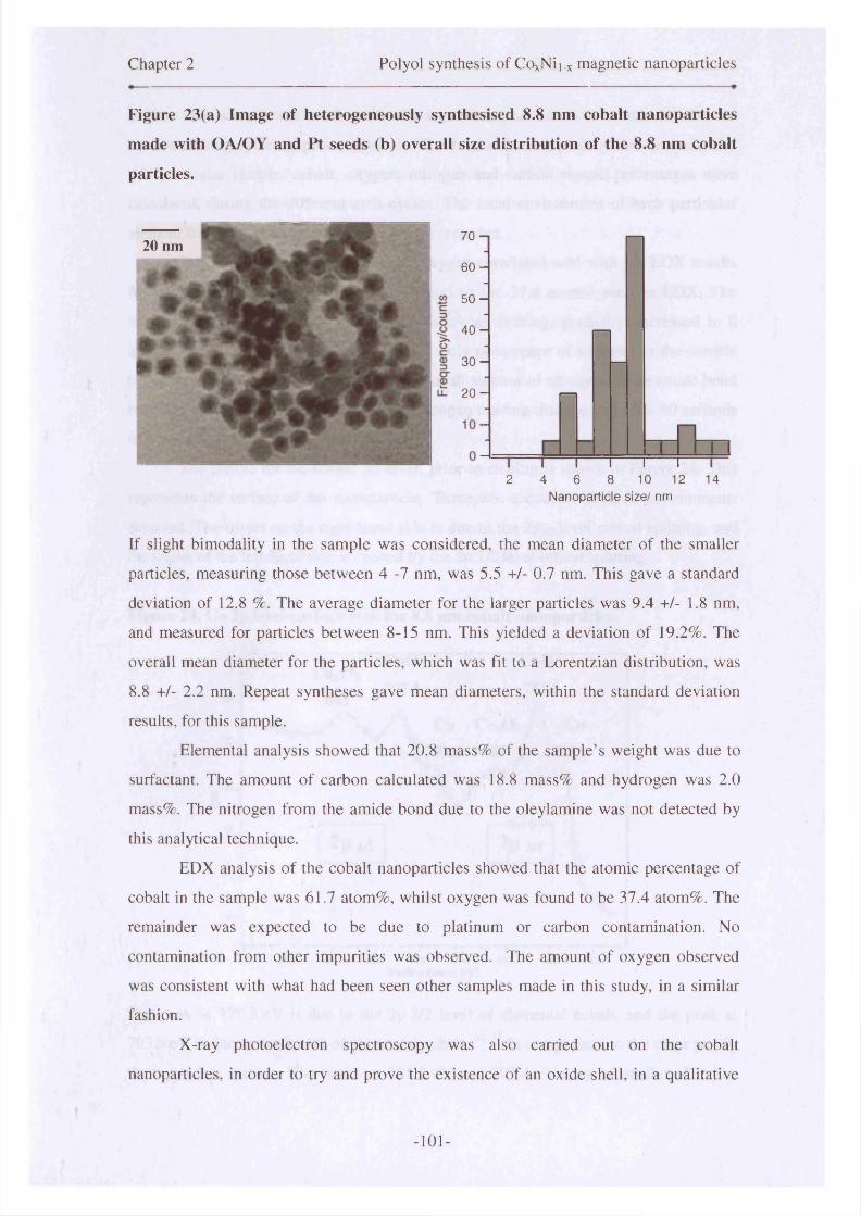
Chapter 2 Polyol synthesis of CoxNii.x magnetic nanoparticles
Figure 23(a) Image of heterogeneously synthesised 8 .8 nm cobalt nanoparticles
made with OA/OY and Pt seeds (b) overall size distribution of the 8 .8 nm cobalt
particles.
70 -i
6 0 -
5 0 -
4 0 -
3 0 -
2 0 -
2 4 6 8 10 12 14Nanoparticle size/ nm
If slight bimodality in the sample was considered, the mean diameter of the smaller
particles, measuring those between 4 -7 nm, was 5.5 +/- 0.7 nm. This gave a standard
deviation of 12.8 %. The average diameter for the larger particles was 9.4 +/- 1.8 nm,
and measured for particles between 8-15 nm. This yielded a deviation of 19.2%. The
overall mean diameter for the particles, which was fit to a Lorentzian distribution, was
8 .8 +/- 2.2 nm. Repeat syntheses gave mean diameters, within the standard deviation
results, for this sample.
Elemental analysis showed that 20.8 mass% of the sample’s weight was due to
surfactant. The amount of carbon calculated was 18.8 mass% and hydrogen was 2.0
mass%. The nitrogen from the amide bond due to the oleylamine was not detected by
this analytical technique.
EDX analysis of the cobalt nanoparticles showed that the atomic percentage of
cobalt in the sample was 61.7 atom%, whilst oxygen was found to be 37.4 atom%. The
remainder was expected to be due to platinum or carbon contamination. No
contamination from other impurities was observed. The amount of oxygen observed
was consistent with what had been seen other samples made in this study, in a similar
fashion.
X-ray photoelectron spectroscopy was also carried out on the cobalt
nanoparticles, in order to try and prove the existence of an oxide shell, in a qualitative
20 nm
-101-
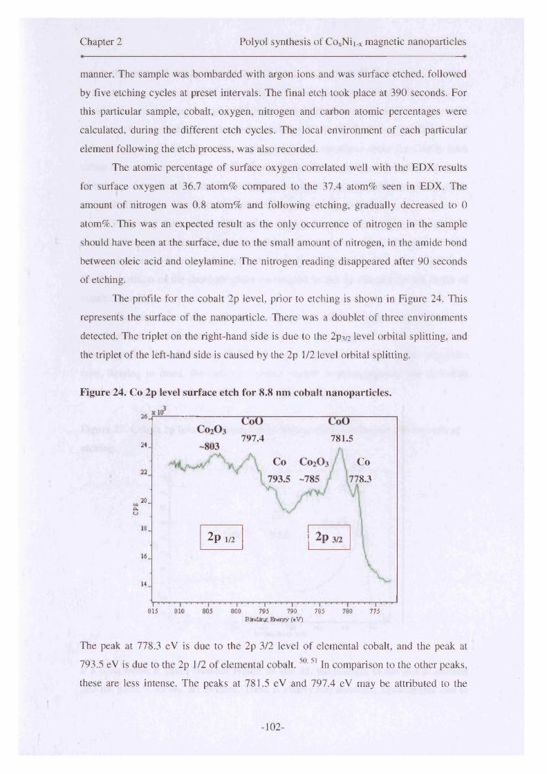
Chapter 2 Polyol synthesis of CoxNii.x magnetic nanoparticles
manner. The sample was bombarded with argon ions and was surface etched, followed
by five etching cycles at preset intervals. The final etch took place at 390 seconds. For
this particular sample, cobalt, oxygen, nitrogen and carbon atomic percentages were
calculated, during the different etch cycles. The local environment of each particular
element following the etch process, was also recorded.
The atomic percentage of surface oxygen correlated well with the EDX results
for surface oxygen at 36.7 atom% compared to the 37.4 atom% seen in EDX. The
amount of nitrogen was 0 . 8 atom% and following etching, gradually decreased to 0
atom%. This was an expected result as the only occurrence of nitrogen in the sample
should have been at the surface, due to the small amount of nitrogen, in the amide bond
between oleic acid and oleylamine. The nitrogen reading disappeared after 90 seconds
of etching.
The profile for the cobalt 2p level, prior to etching is shown in Figure 24. This
represents the surface of the nanoparticle. There was a doublet of three environments
detected. The triplet on the right-hand side is due to the 2 p3/2 level orbital splitting, and
the triplet of the left-hand side is caused by the 2 p 1 /2 level orbital splitting.
Figure 24. Co 2p level surface etch for 8 .8 nm cobalt nanoparticles.
2 6 ,
781.5797.424 _
Co778.3
20 _CS2ftu
18 _ 2p 1/216 _
14_
815 810 800 795 790 785 780 775Binding Bruergy feV)
The peak at 778.3 eV is due to the 2p 3/2 level of elemental cobalt, and the peak at
793.5 eV is due to the 2p 1/2 of elemental cobalt. 5 0 ,51 In comparison to the other peaks,
these are less intense. The peaks at 781.5 eV and 797.4 eV may be attributed to the
-102-
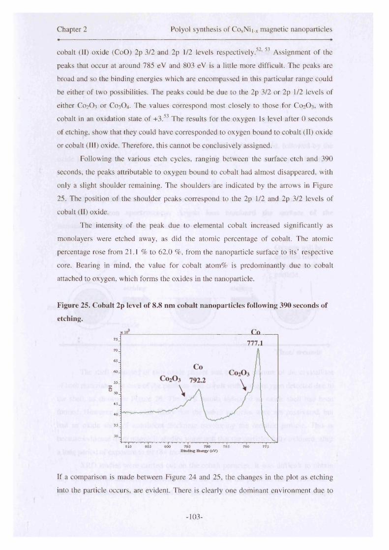
Chapter 2 Polyol synthesis of CoxNii_x magnetic nanoparticles
cobalt (II) oxide (CoO) 2p 3/2 and 2p 1/2 levels respectively .5 2 ,53 Assignment of the
peaks that occur at around 785 eV and 803 eV is a little more difficult. The peaks are
broad and so the binding energies which are encompassed in this particular range could
be either of two possibilities. The peaks could be due to the 2p 3/2 or 2p 1/2 levels of
either C0 2 O3 or C0 3 O4 . The values correspond most closely to those for C 0 2 O3, with
cobalt in an oxidation state of +3 .53 The results for the oxygen Is level after 0 seconds
of etching, show that they could have corresponded to oxygen bound to cobalt (II) oxide
or cobalt (III) oxide. Therefore, this cannot be conclusively assigned.
Following the various etch cycles, ranging between the surface etch and 390
seconds, the peaks attributable to oxygen bound to cobalt had almost disappeared, with
only a slight shoulder remaining. The shoulders are indicated by the arrows in Figure
25. The position of the shoulder peaks correspond to the 2p 1/2 and 2p 3/2 levels of
cobalt (II) oxide.
The intensity of the peak due to elemental cobalt increased significantly as
monolayers were etched away, as did the atomic percentage of cobalt. The atomic
percentage rose from 2 1 .1 % to 62.0 %, from the nanoparticle surface to its’ respective
core. Bearing in mind, the value for cobalt atom% is predominantly due to cobalt
attached to oxygen, which forms the oxides in the nanoparticle.
Figure 25. Cobalt 2p level of 8 .8 nm cobalt nanoparticles following 390 seconds of
etching.
Co777.1
7 0 .
65
60 _
55.
5 0 .
45 _
40 J
35.
30
810 805 800 785 780 775
If a comparison is made between Figure 24 and 25, the changes in the plot as etching
into the particle occurs, are evident. There is clearly one dominant environment due to
-103-
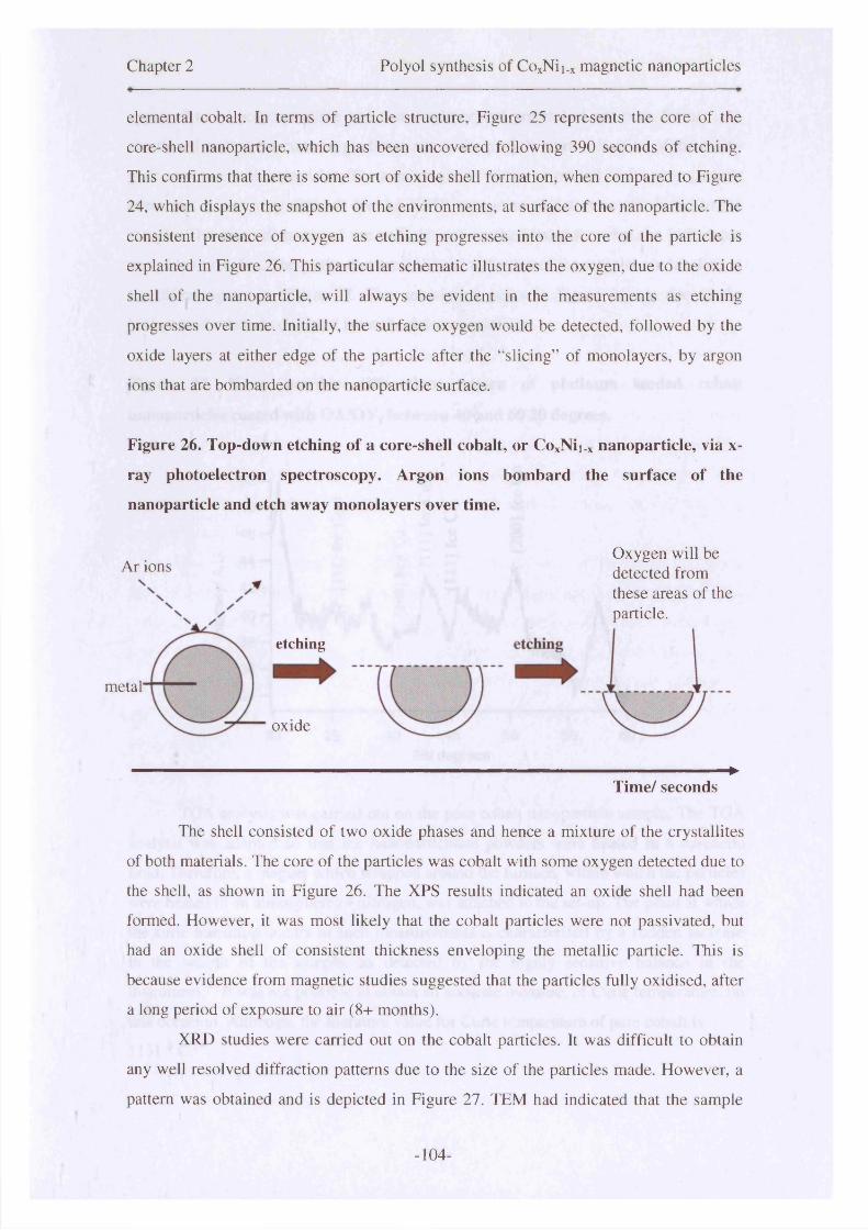
Chapter 2 Polyol synthesis of CoxNii.x magnetic nanoparticles
elemental cobalt. In terms of particle structure, Figure 25 represents the core of the
core-shell nanoparticle, which has been uncovered following 390 seconds of etching.
This confirms that there is some sort o f oxide shell formation, when compared to Figure
24, which displays the snapshot of the environments, at surface of the nanoparticle. The
consistent presence of oxygen as etching progresses into the core of the particle is
explained in Figure 26. This particular schematic illustrates the oxygen, due to the oxide
shell of the nanoparticle, will always be evident in the measurements as etching
progresses over time. Initially, the surface oxygen would be detected, followed by the
oxide layers at either edge of the particle after the “slicing” of monolayers, by argon
ions that are bombarded on the nanoparticle surface.
Figure 26. Top-down etching of a core-shell cobalt, or CoxNii.x nanoparticle, via x-
ray photoelectron spectroscopy. Argon ions bombard the surface of the
nanoparticle and etch away monolayers over time.
etching
oxide
►Time/ seconds
The shell consisted of two oxide phases and hence a mixture of the crystallites
of both materials. The core of the particles was cobalt with some oxygen detected due to
the shell, as shown in Figure 26. The XPS results indicated an oxide shell had been
formed. However, it was most likely that the cobalt particles were not passivated, but
had an oxide shell of consistent thickness enveloping the metallic particle. This is
because evidence from magnetic studies suggested that the particles fully oxidised, after
a long period of exposure to air (8 + months).
XRD studies were carried out on the cobalt particles. It was difficult to obtain
any well resolved diffraction patterns due to the size of the particles made. However, a
pattern was obtained and is depicted in Figure 27. TEM had indicated that the sample
metal'
Ar ionsOxygen will be detected from these areas of the particle.
-104-
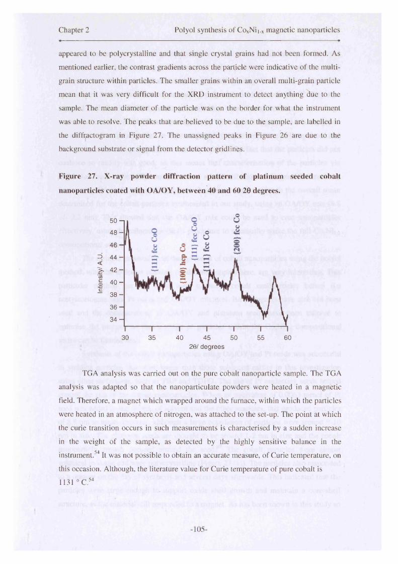
Chapter 2 Polyol synthesis of CoxNii.x magnetic nanoparticles
appeared to be polycrystalline and that single crystal grains had not been formed. As
mentioned earlier, the contrast gradients across the particle were indicative of the multi
grain structure within particles. The smaller grains within an overall multi-grain particle
mean that it was very difficult for the XRD instrument to detect anything due to the
sample. The mean diameter o f the particle was on the border for what the instrument
was able to resolve. The peaks that are believed to be due to the sample, are labelled in
the diffractogram in Figure 27. The unassigned peaks in Figure 26 are due to the
background substrate or signal from the detector gridlines.
Figure 27. X-ray powder diffraction pattern of platinum seeded cobalt
nanoparticles coated with OA/OY, between 40 and 60 20 degrees.
50 - |
4 8 -
3 44 -<> 4 2 -(Oo 40 -c
3 8 -
3 6 -
3 4 -
30 35 40 45 50 605520/ degrees
TGA analysis was carried out on the pure cobalt nanoparticle sample. The TGA
analysis was adapted so that the nanoparticulate powders were heated in a magnetic
field. Therefore, a magnet which wrapped around the furnace, within which the particles
were heated in an atmosphere o f nitrogen, was attached to the set-up. The point at which
the curie transition occurs in such measurements is characterised by a sudden increase
in the weight of the sample, as detected by the highly sensitive balance in the
instrument.54 It was not possible to obtain an accurate measure, of Curie temperature, on
this occasion. Although, the literature value for Curie temperature of pure cobalt is
1131 ° C .54
-105-

Chapter 2 Polyol synthesis of CoxNii_x magnetic nanoparticles
In comparison to other groups who have also made cobalt nanoparticles by a
variety of methods, this study has shown that it is possible to make magnetic cobalt
nanoparticles of a controlled size with an OA/OY coating. Luna et al reported the
synthesis of 8 nm cobalt nanocrystals using only oleic acid as surfactant. They also
reported that the individual cobalt particles that were formed coalesced into large
superstructures of around 100 nm in magnitude. This was not apparent in this study and
the amount of surfactant added to the nanoparticles meant they were well separated and
did not form these large superstructures of particles. The fact that the particles did not
coalesce so readily was good, as this meant that characterisation of the particles via
TEM was much easier. The mean size and size distribution of the particles made by
Luna using OA surfactant coating ( 8 +/- 2 nm) was very similar to the overall mean
determined for the cobalt particles synthesised in our study, using an OA/OY mix (8 .8
+/- 2.2 nm). This showed that the OA/OY mix could be used to coat nanoparticles
effectively, using the tailored synthesis procedure to eventually make the full CoxNii.x
compositional series.
The resulted garnered from the synthesis of cobalt nanoparticles using the polyol
method, with the different reaction conditions adopted here, are very interesting. This
particular method has not been used to make cobalt nanoparticles before (i.e
acetylacetonate salts, Pt seeds and OA/OY mixture). Base reagents have also not been
used and the concentrations of OA/OY and platinum seeds have been tailored to
optimise the process, so that a range of particles within the CoxNii.x compositional
series can be formed next.
Synthesis of the cobalt nanoparticles using OA/OY and Pt seeds was successful
in yielding particles that were larger than those produced earlier in this investigation
using other surfactants, such as TBP and TOPO. The use of Pt nucleating seeds helped
control the size of the cobalt nanoparticles. When an equivalent of 0.0063 mmol of Pt
seed was added to the 10 mL of solvent and the other reactants, this produced particles
of 8 .8 nm in mean diameter, although a large population of particles were around 9-10
nm in size. The platinum seeds are thought to be around 1-2 nm in size and the cobalt
nanoparticle growth occured around the seed.48 The cobalt particles were almost
spherical in shape and were polycrystalline (Figure 23 (a)). The particles also responded
to a magnet on the day of synthesis and several days afterwards. This indicated that the
particles were large enough to support oxide shell growth and maintain a core-shell
structure, as the material still responded to a magnet. As has been shown in this study so
-106-
Chapter 2 Polyol synthesis of CoxNii.x magnetic nanoparticles
far, where particles have been too small, they are fully oxidised and have not responded
to a magnet. The actual synthesis reaction was scaled up, to allow for the production of
more nanoparticles for analysis (chapter 5). Repeat syntheses were carried out so the
magnetic properties could be compared to those obtained from the cobalt nanoparticles
described in this section. The magnetic properties of these cobalt samples are discussed
in detail in chapter 3.
2.7.3 CogoNi2o nanoparticles
2.7.3.1 Introduction
The synthesis of CogoNi2o particles has been carried out in similar studies where the
preparation of bimetallic alloy nanoparticles via the polyol synthesis has been
examined. Luna et al 2’ 13 succeeded in making CogoNi2o particles with diameters
ranging from between 18 and 540 nm in mean diameter. They investigated the use of
silver and platinum, as seeds for growth, and determined that platinum seeds yielded
better size control. The particles made with this platinum nucleating seeds were at the
lower end of the size range described above. The size of the particles made in that
particular study, was dependent upon the concentration of the seeds in the reactant
medium. The higher the concentration of seeds, the more nucleating sites for the
reactants to grow around and therefore the smaller the nanoparticle mean diameters.2
The CogoNi2o particles made in this study were relatively small in comparison to
those made by Luna et al? The precursor salts used to make particles with a particular
composition, in that study were acetate salts. This was in comparison to the
acetylacetonate salts utilised in this investigation.
2.7.3.2 Synthesis of CosoNi20 nanoparticles
Particles of the expected composition, CogoNi2o, were made using the polyol procedure.
The ratios of cobalt and nickel salts were adjusted to produce alloy particles of a
particular composition (CoxNii.x). The metallic salts, nucleating seeds (Pt), reducing
agent, surfactant and solvent were all heated together in an atmosphere of nitrogen. The
particles that were formed were isolated and removed for analysis.
-107-

Chapter 2 Polyol synthesis of CoxNii_x magnetic nanoparticles
2.7.3.3 Results
TEM analysis of the heterogeneously nucleated Co8oNi2o nanoparticles, coated with
OA/OY, showed they had grown in a variety of shapes. There were particles that were
triangular, hexagonal and rhomboidal in shape, with apparent size variations. The mean
particle size was calculated as 12.0 +/-3.4 nm, and the distribution fitted well to a
lognormal curve. This can be seen in Figure 28, as can the range of shapes of particle
formed within this sample. The image also shows that the particles that have been laid
down onto the carbon film for TEM analysis have clumped together slightly, due to
excess surfactant. The particles have different contrasts across their surface indicated
the existence of multi-grains in the sample. A repeat synthesis of the CosoNi2o particles
yielded a mean diameter of 10.9 +/- 3.1 nm. The magnetic properties of both sets of the
12.0 nm and 10.9 nm particles are described in section 3.5.2 of Chapter 3. In this
chapter, the structural and physical properties of the 12.0 nm particles are described in
detail.
Figure 28. Image of 12.0 nm mean diameter CosoNi2o particles made with platinum
seeds and oleic acid/oleylamine surfactant, shows particles of a variety of shapes.
Particles are polydisperse but fit well to a lognormal distribution.
Elemental analysis of the 12.0 nm CogoNi2o particles showed that 16.2 % o f the
sample’s mass was due to surfactant weight. Carbon was 13.0 mass % whilst hydrogen
was 3.18 mass% of the sample’s weight. EDX analysis showed that the atomic
percentage shown by cobalt was 48.6 atom%. The amount due to nickel was 11.5
atom% and oxygen was 39.9 atom%.
-108-

Chapter 2 Polyol synthesis of CoxNii.x magnetic nanoparticles
XPS analysis gave interesting results and indicated the formation of a bimetallic
alloy. Figure 29 displays a doublet of triplets, therefore, cobalt has three predominant
environments. The surface layer shows a high concentration of oxygen, which would be
expected in a metal/metal oxide core-shell structure. The peak at 777.2 eV is due to the
Co 2p 3/2 level of cobalt bound to nickel (CoNi), as this occurs in this particular
region.55 This peak increases in intensity as the particle is etched into, as the atomic
concentration of CoNi increases and the metallic core of the core-shell particle is
progressively reached.
Figure 29. XPS spectrum-surface etch of Co 2p core level at 0 seconds for Co8oNi2o
nanoparticles.
CoO780.9CoO
796.9240
230 CoNi777.2
C 0 2 O 3
-784-789A W
CoNi K 79,2.8
2p 1/2
130
795 790 785 780805 800Binding Eheigy (eV)
The peak at 780.9 eV is due to cobalt (2+) bound to oxygen (CoO) .56 Whilst the peaks
between 784-789 eV were more difficult to characterise, due to their broad range. These
particular values were closest to those cited in the literature, for the 2p 3/2 level of
cobalt (3+) bound to oxygen (C0 2O3 ).53 This indicated the oxidation processes occurring
at the shell was not uniform. The peak due to C0 2O3 decreases in intensity and
disappears after the 90 second etch. The corresponding 2p 1/2 level peaks are present on
the left-hand side of Figure 29. Therefore, the peak at 792.8 eV is caused by cobalt (2p
Vi) alloyed to nickel (CoNi) and the peak at 796.9 eV is due to the cobalt (2p Vi) bound
-109-
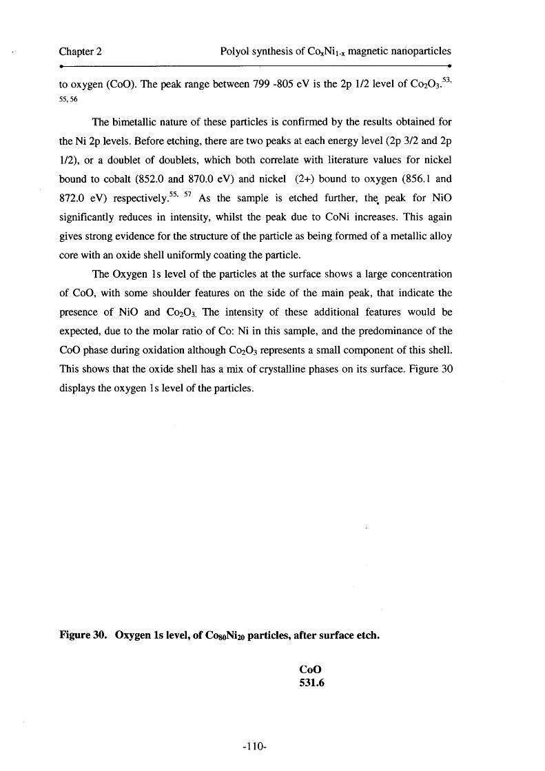
Chapter 2 Polyol synthesis of COxNii.x magnetic nanoparticles
to oxygen (CoO). The peak range between 799 -805 eV is the 2p 1/2 level of C0 2O3.53’55, 56
The bimetallic nature of these particles is confirmed by the results obtained for
the Ni 2p levels. Before etching, there are two peaks at each energy level (2p 3/2 and 2p
1/2 ), or a doublet of doublets, which both correlate with literature values for nickel
bound to cobalt (852.0 and 870.0 eV) and nickel (2+) bound to oxygen (856.1 and
872.0 eV) respectively.55, 57 As the sample is etched further, the# peak for NiO
significantly reduces in intensity, whilst the peak due to CoNi increases. This again
gives strong evidence for the structure of the particle as being formed of a metallic alloy
core with an oxide shell uniformly coating the particle.
The Oxygen 1 s level of the particles at the surface shows a large concentration
of CoO, with some shoulder features on the side of the main peak, that indicate the
presence of NiO and C0 2O3. The intensity of these additional features would be
expected, due to the molar ratio of Co: Ni in this sample, and the predominance of the
CoO phase during oxidation although C02O3 represents a small component of this shell.
This shows that the oxide shell has a mix of crystalline phases on its surface. Figure 30
displays the oxygen Is level of the particles.
Figure 30. Oxygen Is level, of CosoNi2o particles, after surface etch.
CoO531.6
-110-

Chapter 2 Polyol synthesis of CoxNii_x magnetic nanoparticles
x 10314 I
12 .
Shoulder due to C02O3
10 .
Shoulder due to NiO
542 540 538 536 534 532 530 528 526Binding Unogy (eV)
The quantitative measure of atomic concentration, showed oxygen levels
depleted with etching. The amount of cobalt and nickel, conversely increased. The ratio
between cobalt and nickel was consistent throughout the etching cycles. The relative
amount of nickel to cobalt was slightly elevated, as the metallic core of the particle was
penetrated. This was possibly due to the nucleation of nickel occurring before slightly
that of cobalt in the synthesis of the Co8oNi2o particles.
In comparison to the XPS data of the cobalt particles described in section 2.7.2,
the doping 20% nickel into these particles seems to have shifted the peaks slightly. This
indicates the probability of a solid solution having been formed and therefore the
particles are well mixed with each other. XRD measurements were also carried out but
the peaks from the diffraction pattern could not be distinguished from the background
signal from the detector and the substrate. This was due to the weak signal and
broadness of the peaks in the diffraction pattern.
TGA in a magnetic field was carried out to confirm the presence of surfactant
and determine the Curie transition (0C) temperature of the alloy sample. The sample was
heated in an alumina crucible over the temperature range of 0 °C- 1400 °C. The boiling
temperatures of oleic acid and oleylamine would lead to the expectation that all organic
surfactant would be volatilised by 360 °C. The mass loss at approximately 100°C was
due to the removal of adsorbed water. Any mass loss between 100-360 °C was due to
the decomposition of the surfactant. This resulted in a 17.4 % mass loss, which
correlates nicely with the results from elemental combustion analysis. A Curie transition
-111-
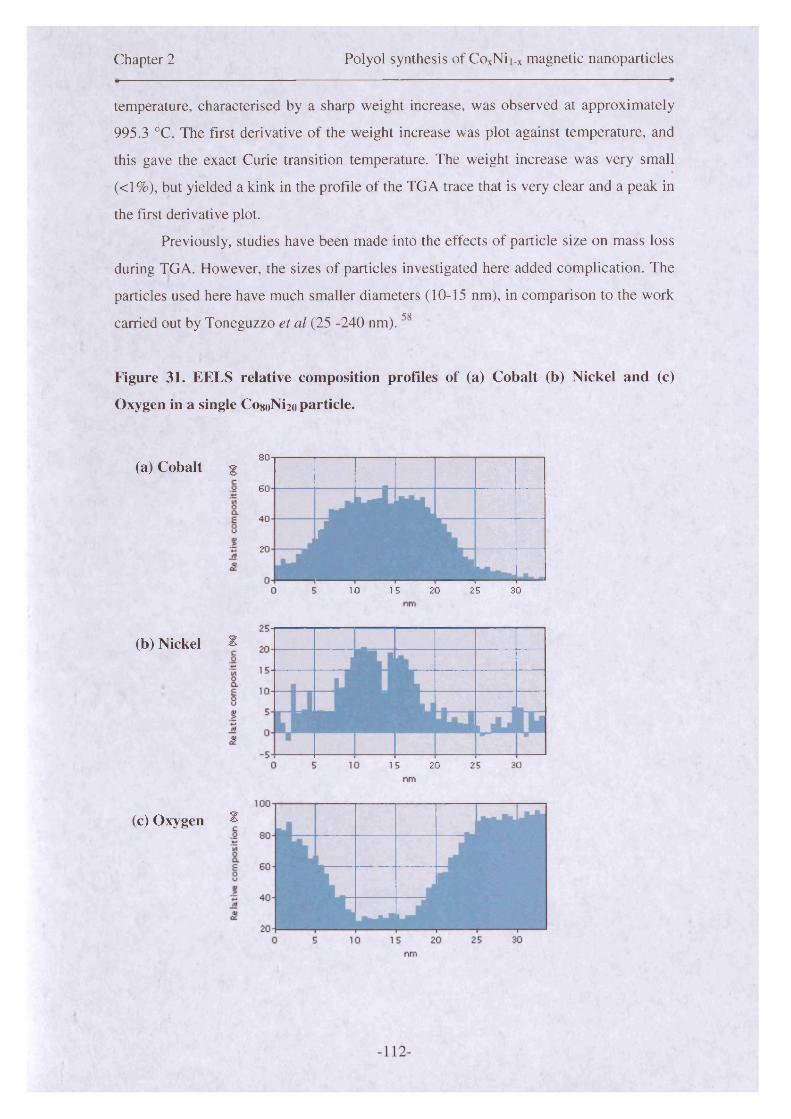
Chapter 2 Polyol synthesis of CoxNii_x magnetic nanoparticles
temperature, characterised by a sharp weight increase, was observed at approximately
995.3 °C. The first derivative of the weight increase was plot against temperature, and
this gave the exact Curie transition temperature. The weight increase was very small
(<1%), but yielded a kink in the profile of the TGA trace that is very clear and a peak in
the first derivative plot.
Previously, studies have been made into the effects of particle size on mass loss
during TGA. However, the sizes of particles investigated here added complication. The
particles used here have much smaller diameters (10-15 nm), in comparison to the workc o
carried out by Toneguzzo et al (25 -240 nm).
Figure 31. EELS relative composition profiles of (a) Cobalt (b) Nickel and (c)
Oxygen in a single Co8oNi2o particle.
(a) Cobalt g80
60
40
20
m .
- L _ _
10 15 20 25 30
(b) Nickel 2
15 20 25 30
(c) Oxygen f

Chapter 2 Polyol synthesis of CoxNii_x magnetic nanoparticles
Energy electron-loss spectroscopy (EELS) analysis was carried out on the
Co8oNi2o particles to verify the nature of alloying that had occurred and also to confirm
a core-shell structure had been formed. Further detail about this technique is given in
Chapter 5. The instrument measured the relative compositions of each element
simultaneously at 0.2 nm intervals across the diameter of a particle. A single line
profile, across an individual particle was analysed to ascertain the relative compositions
of the elements, as well as their distribution throughout a particle. The relative
compositions of the three main elemental components (Co, Ni and O) was mapped and
showed that the particles in this sample had nickel rich cores, and a mix of cobalt and
nickel in the proposed location of the oxide shells. The profiles are displayed in Figure
31. The relative ratios were approximately proportional to the precursor molar ratios,
employed at the start of a reaction. However, there were slight variations across the
particle for the core and shell. The oxygen levels spiked towards the outer edges of the
particle diameter indicating oxide shell formation. The oxygen levels shown in Figure
31 (c) display a huge trough, with the majority of oxygen located on the outer rims of
the particle. There is some oxygen detected when the beam passes through the centre of
a particle. Depending on where the electron beam transmits through the core-shell
particle, different counts are observed for each of the elements (Figure 31). If the beam
passes through the central region of the particle, contributions from the oxide of the top
and bottom surfaces of the particle are observed. The beam therefore, passes through
oxide as it enters and leaves the particle. At the edge of a core-shell particle, as the
beam transmits through the shell, oxygen counts would be expected to be quite large as
it would pass through more oxygen at that point. This is proven by the spikes, at either
edge of the particle, shown in Figure 31 (c). The core-shell structure of the particles is
very well supported by this data.
The relative proportions of cobalt to nickel are indicative of an approximate
Co8oNi2o composition. However if oxygen is excluded from calculations and a relative
ratio between cobalt and nickel is measured, the ratio between both elements is
approximately 7.5:2.5 which indicates a nickel-rich core, perhaps due to the nickel
nucleating first. The levels of cobalt are consistently slightly lower in comparison to
nickel, throughout the sample.
In summary, these results have shown that CosoNi2o alloy core-shell
nanoparticles have been successfully synthesised. The particles are core-shell in
structure with an oxide layer coating, composed of CoO, NiO and possibly some C0 2O3
-113-
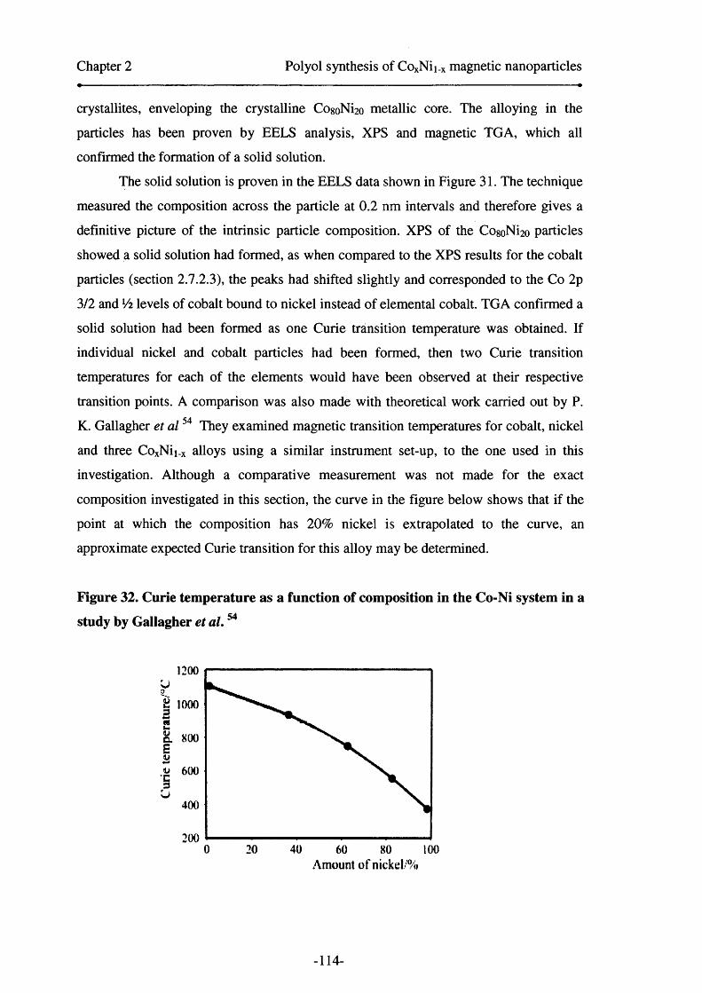
Chapter 2 Polyol synthesis of CoxNii_x magnetic nanoparticles
crystallites, enveloping the crystalline CogolNfoo metallic core. The alloying in the
particles has been proven by EELS analysis, XPS and magnetic TGA, which all
confirmed the formation of a solid solution.
The solid solution is proven in the EELS data shown in Figure 31. The technique
measured the composition across the particle at 0 . 2 nm intervals and therefore gives a
definitive picture of the intrinsic particle composition. XPS of the CogoNi^ particles
showed a solid solution had formed, as when compared to the XPS results for the cobalt
particles (section 2.1.23), the peaks had shifted slightly and corresponded to the Co 2p
3/2 and Vi levels of cobalt bound to nickel instead of elemental cobalt. TGA confirmed a
solid solution had been formed as one Curie transition temperature was obtained. If
individual nickel and cobalt particles had been formed, then two Curie transition
temperatures for each of the elements would have been observed at their respective
transition points. A comparison was also made with theoretical work carried out by P.
K. Gallagher et a l54 They examined magnetic transition temperatures for cobalt, nickel
and three CoxNii_x alloys using a similar instrument set-up, to the one used in this
investigation. Although a comparative measurement was not made for the exact
composition investigated in this section, the curve in the figure below shows that if the
point at which the composition has 2 0 % nickel is extrapolated to the curve, an
approximate expected Curie transition for this alloy may be determined.
Figure 32. Curie temperature as a function of composition in the Co-Ni system in a
study by Gallagher et a l.54
1200
800
600
400
2000 4020 10060 80
Amount o f nickel/%
-114-

Chapter 2 Polyol synthesis of CoxNii_x magnetic nanoparticles
The value determined from the graph above, by extrapolating the corresponding
point for a 20 % nickel alloy from the trend line, is approximately 1010 °C. There is a
difference of about 15 °C, in comparison the literature values described by Gallagher et
al.54 This is due to the calibration of the instrument at high temperatures. The accuracy
of the curve above approximately 1100 °C cannot be guaranteed, as there are no
calibration standards that can be melted at such a high temperature, with this particular
instrument. The value obtained for the Co8oNi2o sample, lies within the errors
determined for the calibration curve of the instrument. It is also important to consider
that the study carried out by Gallagher et al,54 used sheet form alloys and not powders,
that have been used in this study. The measurement conditions of that particular study
were mimicked for the purpose of these measurements. Differences in the set-up, as
well as those between the samples measured can cause variations in the Curie
temperature obtained.
The particles that were formed were relatively monodisperse and of a good size.
Multi-grains were evident in the structure of the particles due to polycrystallinity and
the particles were also non-spherical in shape. However, due to their size and the choice
of surfactant stabiliser, the particles remained stable to oxidation for several months and
responded well to a magnet at room temperature. The results from magnetic
measurements, of particles made with a Co8oNi2o composition, are shown in Chapter 3.
2.7.4 C0 6 0 N140 nanoparticles
2.7.4.1 Introduction
The synthesis of a compositional series of CoxNii_x alloy nanoparticles was continued,
with the fabrication of Co6oNi4o particles. The results obtained are described in the
following sections.
2.7.4.2 Synthesis o f Co6oNi4o nanoparticles
Particles of expected composition, Co6oNi4o, were synthesised using methods described
in detail in the experimental section. The cobalt, nickel and platinum salts were mixed
with OA/OY surfactant in dioctyl ether. These reactants were magnetically stirred and
heated until particle formation occurred. The synthesis of particles with this
composition was repeated once, as with all other samples where compositions have
-115-

Chapter 2 Polyol synthesis of CoxNii.x magnetic nanoparticles
been varied. The particle formation profile followed a similar path to other reactions,
(Section 2.7.2 and 2.7.3).
2.7.4.3 Results
TEM analysis showed that the Co<5oNi4o particles had a slight bimodality in their
distribution. Figure 33 shows the size distribution for the Q)6oNi4o particles and the
curve if the overall distribution was fit to a lognormal curve, which was entirely feasible
if the general shape of the distribution was considered. The larger particles had the
greater frequency count overall. Particle separation was also good in the sample, and in
cases where agglomeration did occur, it was due to the presence of excess surfactant
stabiliser. However, in order to make representative assumptions of the particles in
terms of the magnetic properties, calculations were made on the basis of a bimodal
distribution with two sets of particles of different sizes representing a bimodal
nucleation event. The mean particle size for the larger particles was fit to a Gaussian
distribution and was 10.8 +/-1.9 nm which represents a standard deviation of 17.5%.
The distribution of the smaller particles was also fit to a Gaussian curve and was 6.4 +/-
0.9 nm, giving a standard deviation of 14.1 % which may also be classified as narrow.
The particles are visibly much more spherical in shape, with less faceting being
apparent due to the lack of contrast or line features in some cases, across a particle. The
repeat synthesis showed a similar size and distribution for the particles. The presence of
excess surfactant contributed to the lack of self assembly and has caused localised
agglomeration.
-116-

Chapter 2 Polyol synthesis of CoxNii_x magnetic nanoparticles
Figure 33. Overall size distribution of the C060M 40 nanoparticles shows a
lognormal distribution, although the calculations of mean particle size were made
using the two peaks which represented bimodal nucleation events.
3 0 -
£ 25 -c8 2 0 -
>»0g 1 5 -cr1 1 0 -
5 -
64 8 10 12 14 16Nanoparticle/ Size
Elemental analysis o f the particles showed that 14.2 mass% was due to
surfactant stabiliser coating the particles, after the washing and cleaning procedures had
taken place. EDX analysis showed that the cobalt to nickel ratio was determined as
approximately 3:2, whilst oxygen was 36.4 atom%. If the results for cobalt and nickel
are normalised to 1 , and a relative percentage taken between these two elements, then it
is clear that the particles appear to be slightly nickel rich. This was consistent
throughout all the samples. XPS studies were not performed on this particular sample
due to information gained from analysing other alloys in this series.
XRD studies were carried on the C o ^ N i^ particles and the diffraction pattern
that was obtained is displayed in Figure 34. The reflections for this sample are much
stronger and therefore clearer, in comparison to those for CogoNi2o particles. The
structure on the peaks was indicative of the presence of a number of different phases,
with some reflections being masked by others. The peaks positions also corresponded
well to those reported by previous authors, and the traces that have been captured in this
investigation . 13 ,22 The fee phases of Co, Ni, CoO and NiO were apparent. The peaks at
approximately 31° and 57° are due to the substrate.
-117-
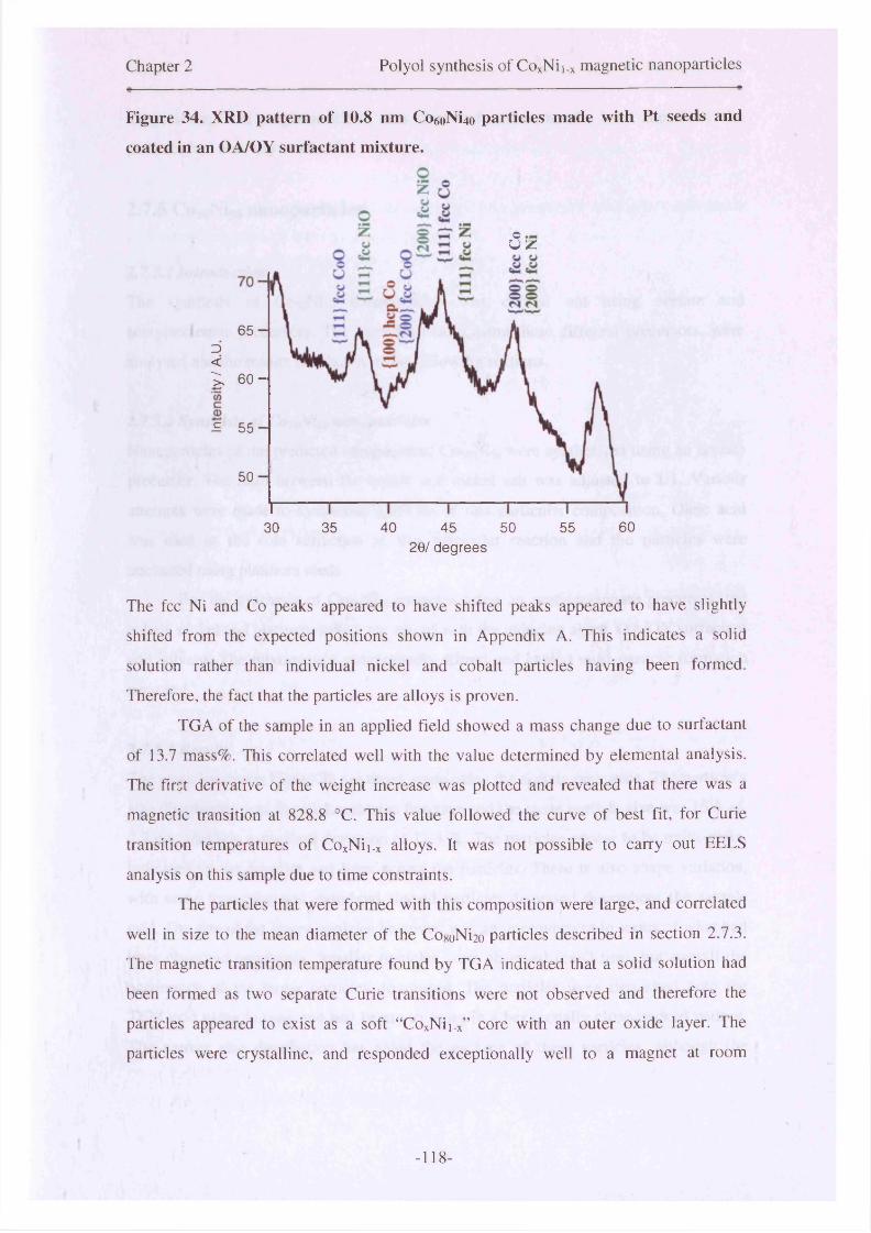
Chapter 2 Polyol synthesis of CoxNii_x magnetic nanoparticles
Figure 34. XRD pattern of 10.8 nm Co6oNi4o particles made with Pt seeds and
coated in an OA/OY surfactant mixture.
ou z
6 5 -Z><^ 6 0 -
£ 5 5 -
5 0 -
6035 40 5030 45 552 0 / degrees
The fee Ni and Co peaks appeared to have shifted peaks appeared to have slightly
shifted from the expected positions shown in Appendix A. This indicates a solid
solution rather than individual nickel and cobalt particles having been formed.
Therefore, the fact that the particles are alloys is proven.
TGA of the sample in an applied field showed a mass change due to surfactant
of 13.7 mass%. This correlated well with the value determined by elemental analysis.
The first derivative of the weight increase was plotted and revealed that there was a
magnetic transition at 828.8 °C. This value followed the curve of best fit, for Curie
transition temperatures of CoxNii_x alloys. It was not possible to carry out EELS
analysis on this sample due to time constraints.
The particles that were formed with this composition were large, and correlated
well in size to the mean diameter of the CogoNi^ particles described in section 2.7.3.
The magnetic transition temperature found by TGA indicated that a solid solution had
been formed as two separate Curie transitions were not observed and therefore the
particles appeared to exist as a soft “CoxNii_x” core with an outer oxide layer. The
particles were crystalline, and responded exceptionally well to a magnet at room
-118-

Chapter 2 Polyol synthesis of CoxNii.x magnetic nanoparticles
temperature. This magnetic effect was observed a few months later, therefore, the
particles had not fully oxidised.
2.7.5 Co5oNi50 nanoparticles
2.7.5.1 Introduction
The synthesis of CosoNiso nanoparticles was carried out using acetate and
acetylacetonate precursors. The particles made, using these different precursors, were
analysed and the results are shown in the following sections.
2.7.5.2 Synthesis of CosoNiso nanoparticles
Nanoparticles of the predicted composition, CosoNiso were synthesised using an acetate
precursor. The ratio between the cobalt and nickel salt was adjusted to 1:1. Various
attempts were made to synthesise particles of this particular composition. Oleic acid
was used as the sole surfactant in this particular reaction and the particles were
nucleated using platinum seeds.
For the synthesis of Co5oNi50 particles using an acetylacetonate precursor, the
cobalt, nickel and platinum salts were mixed with the reducing agent, OA/OY surfactant
and solvent. The mixture was magnetically stirred and heated until particle formation
occurred.
2.7.5.3 Results
The particles shown Figure 35 are those made using the acetate precursor. The particle’s
size distribution was fit to a Lorentzian function and the mean particle size was 15.6 +/-
2.7 nm, yielding a standard deviation of 17.3 %. The particles appear to be multi-grain,
indicated by the contrast and lines across the particles. There is also shape variation,
with some triangular and rhomboid shaped particles dispersed throughout the sample
grid. The size of the larger particles however, was an improvement in terms of what had
been observed previously. Smaller particles were observed (ca~3 nm), but overall the
occurrence of the larger particles dominated. The particles were deposited onto the
TEM grid using hexane and had begun to order in a hexagonally close-packed pattern.
The narrow size distribution has aided the packing of these particles, although the
-119-
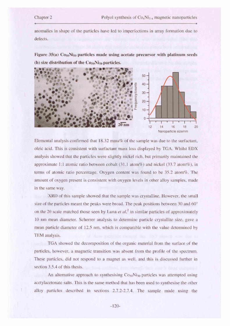
Chapter 2 Polyol synthesis of CoxNii.x magnetic nanoparticles
anomalies in shape of the particles have led to imperfections in array formation due to
defects.
Figure 35(a) CosoNiso particles made using acetate precursor with platinum seeds
(b) size distribution of the CosoNi5o particles.
Elemental analysis confirmed that 18.32 mass% of the sample was due to the surfactant,
oleic acid. This is consistent with surfactant mass loss displayed by TGA. Whilst EDX
analysis showed that the particles were slightly nickel rich, but primarily maintained the
approximate 1:1 atomic ratio between cobalt (31.1 atom%) and nickel (33.7 atom%), in
terms of atomic ratio percentage. Oxygen content was found to be 35.2 atom%. The
amount of oxygen present is consistent with oxygen levels in other alloy samples, made
in the same way.
XRD of this sample showed that the sample was crystalline. However, the small
size of the particles meant the peaks were broad. The peak positions between 30 and 60°
on the 20 scale matched those seen by Luna et al, in similar particles of approximately
10 nm mean diameter. Scherrer analysis to determine particle crystallite size, gave a
mean particle diameter of 12.5 nm, which is comparable with the value determined by
TEM analysis.
TGA showed the decomposition of the organic material from the surface of the
particles, however, a magnetic transition was absent from the profile of the spectrum.
These particles, did not respond to a magnet as well, and this is discussed further in
section 3.5.4 of this thesis.
An alternative approach to synthesising CosoNiso particles was attempted using
acetylacetonate salts. This is the same method that has been used to synthesise the other
alloy particles described in sections 2.1.2-2.1 A. The sample made using the
12 14 16 18 20I 1 I— 1— I 1— I 1— I
Nanoparticle size/nm
- 120-

Chapter 2 Polyol synthesis of CoxNii_x magnetic nanoparticles
acetylacetonate salts was also analysed by TEM. Images showed that particles had more
shape variations in comparison to the sample made with acetate salts. The size
distribution curve in Figure 36 shows that multiple nucleation events have occurred and
the particles mean diameter is therefore difficult to determine. The distribution from the
mean is great, due to the breadth in size of particles. The mean diameter for this sample,
if the curve is fitted to a Gaussian distribution is 12.6 +/- 9.6 nm. This assumes that
there was just a single nucleation event. It was not possible to fit the data to a particular
distribution due to the multiple nucleation events.
Figure 36. Size distribution of CosoNiso particles made with acetylacetonate
precursor salts.
30 -i
<2 25c
O 20
1 5 -
r i o -5 -
0 n r10 12 14 16 18
Nanoparticle size/ Nm
O“ I 120
The shape of the distribution could also be described as being multimodal, and therefore
the mean diameter for each of these modes should be calculated from the numerous
peaks. However, the general distribution of the curve overall will be used as nothing
meaningful can be gathered from splitting the data into several plots to obtain accurate
means for each of the nucleation events that have occurred.
Elemental analysis of these particles showed that 10.7 mass% was due to
surfactant stabiliser. This is a quite low compared to other samples that have had EA
carried out. This could possibly explain the poor magnetic properties of the particles,
which are discussed in Chapter 3. Due to poor coverage over the nanoparticle surface, it
is possible that these particles have oxidised, even though their size is large compared
other oxidised samples in this investigation.
EDX studies determined that 25.4 atom% was due to cobalt in the sample, whilst
30.8 atom% was due to nickel. The amount of oxygen present was determined to be
- 121-
Chapter 2 Polyol synthesis of CoxNii.x magnetic nanoparticles
43.8 atom% of the sample. The sample was therefore, slightly nickel rich and this is
consistent with what has been seen elsewhere with the other alloy particles discussed in
this chapter.
XPS results differ slightly to those discussed earlier for CogoNi2oin section 2.7.3,
when the relative atomic percentages of each element were measured during the etching
process. The quantitative elemental values for each of the main components are
different, due to change in relative ratio between Co and Ni. The surface layer of the
particles, displayed a lower level of nickel than cobalt, however, as etching proceeded
the nickel content superseded that of cobalt. After 90 seconds of etching, the levels of
cobalt: nickel, were approximately equal. The core of the particle, after 1330 seconds of
etching, showed cobalt content of 23.2 atom% versus 28.6 atom% for nickel. This is
consistent with what was observed via EDX measurements. These results corroborate
with the results from other alloys, with different compositions in this series. Nickel
appears to nucleate first and hence the cores are slightly nickel-rich. The nucleation
temperature of nickel is much lower than that of pure cobalt. However, the oxide phases
dominate in this particular sample in terms of atomic concentration. The plot in Figure
37 (a), displays a doublet of doublets, prior to etching. However, there was an indication
of a shoulder on the right-hand side of the main central peaks, of each of the doublets.
Therefore, there were three environments present. These are consistent with literature
values for CoO, C0 2O3 and CoNi. Following etching for 1330 seconds, the spectrum in
Figure 37 (b) shows the effect on the profile. The peaks corresponding to the 2p 3/2 and
2p 1/2 levels of CoO decrease significantly in intensity. Whilst the equivalent peaks for
C02O3 have disappeared. The peak indicating that cobalt and nickel have alloyed
becomes dominant in the spectrum. This proves that oxygen content decreases as the
particles are infiltrated by etching and the core is predominantly composed of a CoxNii_x
alloy, with some CoO inclusions.
- 122-

Chapter 2 Polyol synthesis of CoxNii_x magnetic nanoparticles
Figure 37. C050M 50 particles made with acetylacetonate precursor, OA/OY
surfactant mixture and platinum seeds in dioctyl ether (a) Co 2p level at 0 seconds
of etching (b) Co 2p level at 1330 seconds of etching.xlO2
190. CoO781.4CoO
797.6 CoNi/ " \ -794.9 C°2° ; / \ / 786.3
180
170
160
150
130.
1 2 0 .
815 810 805 800 795 790 785 780 775 770Binding Energy (eV)
xlO3
(b) CoNi776.8
40 .
CoNi791.6 CoO
CoO780.7
801.8
2 p 1/2
820 810 800 790 780 770Binding Energy (eV)
Quantitative analysis of the sample by XPS, showed that oxygen concentration
decreased from 37.7 atom% to 14.9 atom% during etching. This shows that there are
slight inclusions of oxygen in the core. The value for surface oxygen, correlates
approximately with that determined by EDX analysis. The oxygen peak at the surface
showed a predominant peak at 531.5 eV, consistent with a CoO Is level peak. The peak
-123-
Chapter 2 Polyol synthesis of CoxNii_x magnetic nanoparticles
displayed two shoulders on either side of the peak’s incline profile. These were due to
NiO at approximately 529 eV and C0 2 O3 at around 534 eV. The oxygen atomic
concentration significantly decreases during etching. The shape of the peak due to CoO
changes as etching takes place, and this indicates that the oxide phases that are
interdispersed throughout the sample are a mixture of CoO, NiO and C0 2 O3.
Nickel concentration relative to cobalt, was lower in the shell in comparison to
the core which had a higher concentration of nickel. This is consistent with the belief
that nickel nucleates first, followed by cobalt. The higher cobalt concentration at the
surface is due to the preferential oxidation of cobalt, evidenced by the existence of two
cobalt oxide phases in the shell. The relative ratios of cobalt: nickel equalised after
approximately 90 seconds of etching, after which, the nickel concentration was larger
than that of cobalt. This was an interesting result, considering that both precursors were
employed with equivalent molar ratios, and indicated the influence of the nucleation
processes in the formation of binary alloy core-shell particles.
The key information garnered from XPS analysis shows a strong similarity
between the CosoNiso sample and the CogoNi2o sample (section 2.7.3) in terms of
structural morphology. The quantification of atomic concentration, for each element
during etch cycles, helped build a picture of the way nucleation and growth of these
particles takes place. The data indicates the formation of a core/shell structured particles
with some smaller oxidised particles within the nanoparticle sample. This information
may aid to explain some of the anomalous magnetic properties in these alloys, such as a
low moment or no exchange bias in smaller particles which have oxidised to their cores.
XRD of the Co5oNi5o sample showed only an amorphous background. Therefore,
an estimate of particle size and elemental phase could not be made, other techniques
such as XPS were used to elucidate phase, whilst particle size was investigated earlier
by TEM analysis.
Thermogravimetric analysis in an external magnetic field was carried out on the
Co5oNi5o sample. The first derivative of the mass loss was plotted, and a magnetic Curie
transition (Tc) was determined as 786.1 °C. The value determined for this alloy is
consistent with the study carried out by Gallagher et al, which was discussed earlier (see
Figure 32). It is also within the error boundaries found by this investigation.
EELS analysis was also conducted on the CosoNiso sample, in order to clarify the
expected core-shell structure of particles and monitor the pattern of oxidation across an
individual particle.
-124-
Chapter 2 Polyol synthesis of CoxNii_x magnetic nanoparticles
The profiles of each element showed a distinct anomaly between the relative
concentrations of cobalt and nickel. As was seen in the XPS quantitative results, the
nickel concentration was lower than that of cobalt at the surface or shell. However, here
the nickel concentration remains low throughout the sample. The amount of nickel
atoms is skewed so there are more present on the left-hand side of the particle than the
right. The average composition of the particle, as determined by EDX, shows that the
ratio between cobalt and nickel remains as approximately 1 :1 , with the sample being
slightly nickel rich. However, here the results show that in this particular particle, the
amount of nickel is slightly lower than anticipated. If comparisons are made with other
particles, which have been analysed on this grid, then a different conclusion could be
drawn. This shows that there can be major differences in the composition between
individual particles at an atomic level. The oxygen level displays peaks at either end of
the particle, and a trough represents the metallic core of the particle. The fact that
oxygen remains throughout the particle has been proven earlier by XPS (section 2.7.3).
This technique may help to explain the problems with reproducibility experienced with
particles of different compositions, in terms of their physical and functional properties.
Particle sizes are slightly different between samples made following an identical
method. However, the reactions reported in this thesis have attained a certain level of
reproducibility, which allows assumptions to be made. This means that the particles that
have been formed, as repeats to an earlier synthesis, have maintained sizes within the
distribution of the initial sample. Although the actual mean particle sizes are different
due to the different nucleation processes.
In summary, the properties of Co5oNi5o particles, made with acetate and
acetylacetonate salts has been discussed. The acetate salt method yielded well formed
particles of a good shape and size, however, the particles had fully oxidised and did not
respond well to an external magnetic field. Multi-grains were also evident in the
particles made in this manner. The acetylacetonate salts method, which has been
adopted for other alloys in this compositional series, displayed interesting results.
Although the particles were smaller in mean diameter compared to the acetate salt
sample, they responded well to a magnet and therefore had not oxidised. The various
analytical techniques employed to characterise these particles, displayed strong
evidence for the existence of core-shell morphology and crystallinity in these particles.
XPS and EELS analysis, in particular showed that there was a high concentration of
oxide at the surface of the particles, with a marked increase in CoxNii_x alloy
-125-

Chapter 2 Polyol synthesis of CoxNii_x magnetic nanoparticles
concentration with etching into the particle. This was also observed in the Co8oNi2o
sample discussed earlier. Magnetic TGA analysis proved that the particles were formed
from a solid solution, however, EELS analysis showed that there are variations in
composition across individual particles and this should also be considered. The
information gained from analysis of this sample helped in the understanding of the
CoxNii.x system in general.
2.7.6 Co4oNi60 nanoparticles
2.7.6.1 Introduction
Particles of predicted composition, Co^Ni^ were synthesised via the polyol process.
The method used to synthesise the particles was identical to that used to make other
alloy particles in this compositional series.
2.7.6.2 Synthesis of Co4oNi6o nanoparticles
Cobalt and nickel precursor salts were used in a 2:3 ratio, respectively. These salts,
along with a platinum salt (nucleating agent), 1 ,2 -hexadecanediol (reducing agent) and
the OA/OY surfactant mixture were stirred and heated to the reflux temperature of the
solvent, dioctyl ether.
2.7.6.3 Results
TEM analysis of the particles showed that the particles were of a similar size to those
synthesised in this compositional series. The mean particle diameter for the larger
particles was determined to be 13.6 +/- 2.5 nm which is equivalent to a standard
deviation of 18.4 %. These particles can be described as having a narrow size
distribution. The bimodality of the overall size distribution of the particles in the
Co^Ni^ sample cannot be neglected, therefore the size distribution of the smaller set of
particles was also determined. The mean diameter was found to be 6.7 +/- 1.4 nm,
which corresponds to a deviation of 19.4 %, therefore the distribution of these particles
may also be described as narrow.
The nucleation of the CoxNii_x particles has led to the bimodality that has been
seen throughout this compositional series. The largest particles formed in this sample
(13.6 nm) appeared to be large enough to support oxide shell growth. The smaller
-126-

Chapter 2 Polyol synthesis of CoxNii_x magnetic nanoparticles
particles had possibly oxidised to their cores. Sangregorio et al 59 have described the
synthesis of CoxNii_x particles, via a sol-gel technique, which were embedded within a
silica matrix and were subsequently tested for their magnetic properties. The particle
synthesised in this particular compositional series increased in size as cobalt content
increased. Therefore, the blocking temperature for pure nickel particles (7 K) was much
lower than that for the pure cobalt particles (70 K). 59 In comparison, the synthesis of
particles using polyol methods in this study have shown that as nickel content increases,
the general particle size has also increased.
Elemental analysis of the sample showed that the percentage mass due to
surfactant was 13.8 mass%. This was correlated with the TGA data. EDX analysis of
samples showed that the composition of the particles was nickel rich, as has been seen
with some of the other alloys in this series. Cobalt was determined at 18.3 atom%, with
the amount of nickel found to be 45.7 atom%. Therefore, the Co/Ni ratio was 1: 2.5,
which corresponds to the composition that had slightly higher nickel levels, than had
been predicted prior to synthesis. This has been attributed to the nickel nucleating first
in the reaction process. Oxygen levels in the sample, at 36.0 atom%, were consistent
with what has been seen in other alloys samples.
XRD analysis of the sample yielded an amorphous background. The trace that
was obtained was a very weak pattern and could not be clearly distinguished against the
background signal. This was due to the multi-grain structure of the particles, as the
small particles within a whole particle were not large enough to be detected. Therefore,
assignment of the crystallographic planes was not possible.
TGA analysis of the Co4oNi6o particles, in an applied magnetic field, revealed
that approximately 13.2 mass% decrease could be assigned to the surfactant
decomposition from the surface of the nanoparticle. This corroborated well with the
mass loss determined via elemental analysis. The Curie transition of the particles was
found to be 719.9 °C. The transition was characterised by a small peak, in the profile of
the first derivative, from the weight loss measured by the instrument.
The results from studies carried out this sample, indicated that there were clear
trends in terms of the particle nucleation becoming apparent. The elevated levels of
nickel (+ 5-8 %) in the samples prove this. The size of the particles formed using this
adapted polyol method seem to be pretty consistent, although the multi-grain structure
in the particles, has led to problems in characterising them with XRD. The size
distribution in the particles indicates that some more work could be done on
-127-

Chapter 2 Polyol synthesis of CoxNii_x magnetic nanoparticles# ■■ . . . - . . . — #
investigating the nucleation process in further details, so that monomodal distributions
could be consistently achieved.
2.7.7 Co2oNi8o nanoparticles
2 .7.7.1 Introduction
Particles of the composition Co2oNigo, have been synthesised before in works by Viau et
al, using the polyol method. However, the particles made by Viau and co-workers
were larger. This section describes the controlled synthesis of Co2oNi8o nanoparticles
using a synthetic process that has evolved, so that core-shell particles can be formed.
2.7.7.2 Synthesis o f Co2oNiso nanoparticles
Particles were prepared by polyol synthesis methods. Cobalt and nickel salts were used
in a proportional molar ratio of 1:4 respectively. The metallic precursor salts, platinum
acetylacetonate (nucleating agent), 1,2-hexadecanediol (reducing agent) and OA/OY
surfactant mixture were all dissolved in dioctyl ether and heated under nitrogen and
stirring until particle formation occurred. A more detailed description of the synthesis is
given in section 5.5.6 of chapter 5.
2.7.7.3 Results
Initially, acetate salts were used as the starting material and resulted in large particles,
as with the CosoNiso particles described in section 2.7.4. TEM analysis showed that the
particles, made with acetate salts, appeared to attempt to self-assemble due to better
uniformity in shape.
Figure 38 (a) Particles made using acetate salts, with no exchange bias interactions
(b) size distribution of the particles shown in (a).
30nm
k
• • * v, ,v •• • • .**4 0 -
oo
o<D=3O"a>
lj-1 0 -
-128- 4 6 8 10 Nanopartide size/ nm
12 14

Chapter 2 Polyol synthesis of CoxNii.x magnetic nanoparticles
The Co2oNi8o particles are shown in Figure 38 (a) and indicated a mean particle diameter
of 9.1 +/- 3.1 nm which translates to a standard deviation of 34 %. The particle sizes
were fit to a lognormal curve and had a broad distribution. The particles clearly exhibit
multi-grain structure, due to the contrast and clear lines across the particles. There are
also more occurrences of spherical-like particles than the triangular and rhomboidal
shapes, in comparison to what has been seen in particles made using acetylacetonate
salts.
TEM analysis of the particles made using acetylacetonate salts, indicated a slight
bimodal distribution, as shown in Figure 38 (b). Due to the bimodality, in order to
accurately calculate the distribution of the particles, the dataset was split into two
groups of smaller and larger particles. The calculated mean for the larger particles was
13.0 +/- 3.0 nm (standard deviation 23 %), whilst the average particle diameter for the
smaller particles was 7.9 +/- 0.7 nm (standard deviation 8.9 %). The particle distribution
displayed two peaks with a large tail at the higher end of the particle size scale, and
hence was difficult to fit to a specific distribution. The two peaks for each set of
particle, were fitted to Lorentzian distributions.
Figure 39. Size distribution of Co2oNigo sample made with acetylacetonate salts.
3 0 -
w 25 —c8 2 ° -
o' 1£- c 15 -(D® 10 —
it5 - :oi
6 8 10 12 14 16 18 20 22Nanoparticle size/ nm
The mean for the larger particles is on average approximately 3 nm less in diameter than
the overall mean diameter of particles made with acetate salts. The sample made with
acetyalacetonate salts responded to a magnet, whereas the acetate salt sample did not.
The magnetic properties of these samples are discussed in chapter 3.
Elemental analysis studies showed that 11.6 mass% was due to surfactant mass.
EDX analysis of the particles showed that the sample had 9.5 atom% cobalt, 68.1
atom% nickel and oxygen 22.4 atom%. The ratio between cobalt and nickel in this case
-129-
Chapter 2 Polyol synthesis of CoxNii_x magnetic nanoparticles
is interesting as these results show that the nickel precursor has transferred better into
the final particle. The atomic percentages of Co/Ni correlate with a 12% and 8 8 % Co:
Ni ratio. Therefore the particles in this case have clearly shown the dominance of nickel
in the nucleation process. The oxygen value determined is lower than what has
previously been observed with the other alloys. This could be due to the way in which
nickel oxidises, as this will have the dominant oxide phase in the shell due to the higher
molar ratio of nickel precursor to its analogous cobalt precursor.
The particles appeared be crystalline in the TEM microscope. It was also not
possible to obtain TGA results for this particular sample, following several attempts.
The particles however, did respond to a magnet and warranted magnetic property
investigations due to their size. This is interesting, as it was the first particular
composition, in the series to display non-magnetic behaviour. The results from the
physical characterisation of the Co2oNigo particles could contribute to a predicted lack of
exchange anisotropy in the particles. The particle size displayed in this sample is quite
large, however, there is evidence of multiple nucleation events during the synthesis,
provided by the size distribution plot in Figure 39. A multi-grain structure is evident in
the samples as nanocrystal grains have coalesced to form these particles. The properties
of the nickel particles, discussed in the following section are interesting, as it will
confirm if the higher concentration of nickel in the particles has an effect on their
structure and magnetic properties.
2.7.8 Nickel nanoparticles
2.7.8.1 Introduction
Nickel nanoparticles were synthesised using the nickel acetylacetonate salt, an OA/OY
surfactant mixture and Pt seeds to control the size of the particles. Nickel has been
shown to bind well to oleylamine, and other amine based surfactants, in similar polyol
reactions. 3 0 ,31 ,60 ,61 Earlier investigations into a suitable surfactants showed that nickel
salts formed complexes with hexadecylamine and oleylamine at low temperatures.
However, upon reaching the reflux temperature the particles “crashed out” of solution
and became aggregated. By using oleic acid with oleylamine, in this study, the stability
of the nickel particles in terms of aggregation has been improved.
-130-
Chapter 2 Polyol synthesis of CoxNii_x magnetic nanoparticles
2.7.8.2 Synthesis o f nickel nanoparticles
Nickel nanoparticles were synthesised by heating nickel acetylacetonate, platinum
seeds, 1,2-hexadecanediol, OA/OY surfactant mixture in dioctyl ether. The solution was
heated and magnetically stirred in reflux conditions until particle formation occurred.
The resultant particles were isolated and stored for analysis using various
characterisation techniques.
2.7.8.3 Results
TEM analysis showed a mean particle diameter of 16.0 nm +/- 5.6 nm, which is a
standard deviation of 35 %. The distribution was fitted to a monomodal Lorentzian
function to obtain mean particle size. The particles had a broad size distribution, and
excess surfactant caused spherical superstructures of particles to form. The shape of the
individual particles, as can be seen in Figure 40(a), was highly irregular with triangular
and square shaped particles being formed. The particles were well separated apart from
in areas where there is excess surfactant. A repeat of this reaction led to particles with
mean diameters that were within the size distribution determined above.
Figure 40(a) Image of nickel nanoparticles made with platinum seeds and OA/OY
surfactant coating (b) Size distribution of the nickel nanoparticles.
10 12 14 16 18 20 22N anoparticle s iz e / nm
Elemental analysis of these particles showed that 10.3 mass% was due to the
surfactant weight. This is less than what has been seen previously for particles in this
-131-
Chapter 2 Polyol synthesis of COxNii_x magnetic nanoparticles
series. EDX showed that 76.3 atom% was due to nickel, whilst 23.7 atom% was due to
oxygen. The oxygen is quite low in comparison to samples which have some cobalt in
them. XRD showed only an amorphous background.
TGA analysis of the sample showed a Curie transition temperature of 348 °C.
This was consistent with the intrinsic error within the particular instrument and is close
to the Curie transition expressed by Gallagher et al, of 357.2 °C, for nickel.
Figure 41 displays the Curie transition of a nickel calibration standard and is
characterised by a weight increase in the TGA profile. The plot shows the very small
percentage change exhibited. This graph is indicative of the results obtained using the
TGA with an applied magnetic field. The first derivative of each sample’s TGA profile
was measured and the Curie temperature obtained from that particular plot. For alloy
samples measured over the large temperature range (0- 1400 °C), this effect was
characterised by a small deviation in the profile of the TGA trace, and a peak in the first
derivative plot. Figure 41 highlights the small weight increase that is observed.
Therefore, over this temperature range, the Curie transition for the CoxNii_x alloy
particles was just as small.
-132-

Chapter 2 Polyol synthesis of CoxNii.x magnetic nanoparticles
Figure 41. TGA with an applied magnetic field of a nickel calibration standard
shows a Curie transition, characterised by a small percentage weight increase (red
trace). The blue trace shows the DSC measurement for the nickel calibration
standard and yields the melting point.
100.0
99.8
99.6
Value: 357.0 °C
99.4
Mass change: 0.58 %
Melting point of nickel:
1464.2 °C99.0 Peak: 346.2 °C
1400 1600200 800 1000 1200400 600
The nickel particles in this compositional series displayed interesting results.
They did not seem to respond to a magnet, as with the Co2oNigo particles described in
2.7.7. This begins to indicate that as nickel content is increased through the series, the
magnetic effect of the particles at room temperature is lost. This could have
implications on the exchange bias properties of the particles, which are discussed in
Chapter 3. The size of the nickel particles was also, on average, larger than those with
some cobalt doped into them. They also seem to have nucleated much more easily and
this is indicated by the lower temperature at which the nickel particles form. The
solution turned instantaneously black at 220 °C, whereas, the particles with some cobalt
doped in have consistently shown higher formation temperatures of between 230-
240°C, with the OA/OY combination of surfactant. The XPS results of other alloys
within this compositional series have also indicated that the nucleation of nickel
progresses much more easily than that of alloy particles. This has been evidenced by the
-133-
Chapter 2 Polyol synthesis of COxNii_x magnetic nanoparticles
elevated levels of nickel in the core of the particles (following etching), which have
shown that nickel tends to nucleate first.
2.7.9 Summary of samples made with variations in composition
The main aim for this investigation was to produce particles with magnetic properties
that could be tuned according to the composition. A range of sample between Co<
CoxNii-x< Ni were synthesised using OA/OY as the stabilisers. The particles were also
heterogeneously nucleated using platinum seeds, so that the particle size was tuned to
be in the region of 8-20 nm. These particles therefore, were large enough to support
oxide shell growth, and therefore possibly exhibit exchange bias, as detailed in the
chapter 4.
Where CoxNii_x alloys were formed, the particles appeared to be slightly nickel-
rich, due nickel nucleating first in the reaction. This was indicated by EELS analysis
carried out on a select few samples. EELS studies have definitively proven the existence
of an oxide shell in the particles. XPS analysis indicated that the particles did appear to
be alloyed and also confirmed the core-shell structure, first exhibited in EELS analysis.
XPS also indicated the particles had nickel-rich cores.
-134-

Chapter 2 Polyol synthesis of CoxNii_x magnetic nanoparticles
Figure 42. Magnetic TGA study of particles with varied composition shows a close
correlation between the curie transition found by Gallagher et al (blue) versus the
results for particles with different compositions, measured in this study (pink).
1200 i -------------------------------------------------------------------------------------------------------------------------------------------------
1100 - °
1000 - •
? 900 -
3 •1 800 - #
§ ♦ • ---------------------------- I------------------- — 1g ♦ P.K Gallagher study«£ 600 -.g . • Our results
3 * ______________________________________
u 500 -
400 - O
300 -
2 0 0 : 1 1 1 1 1 1 1-------------------
0 10 20 30 40 50 60 70 80 90 100
% Cobalt
Thermogravimetric magnetic analysis was carried out on these samples, and this
technique showed that the Curie temperature o f the particles could be altered by varying
the composition. This proved that the particles were a solid-solution, and had formed
alloys. The technique showed that by increasing the nickel concentration, the Curie
temperature of the materials decreased. The magnetic properties o f the particles
synthesised in this chapter are investigated in detail in chapter 3. The structural
characterisation of particles in this chapter has provided a wealth of information, and
given detailed evidence of specific properties, which could impact upon the magnetic
properties observed in the particles.
TGA study of CoxNi,.x alloys
♦ •♦ P.K Gallagher study
• Our results
r
-135-

Chapter 2 Polyol synthesis of CoxNii_x magnetic nanoparticles
2.8 Conclusions
Particles of various sizes and compositions have been synthesised by altering the
reaction conditions and variables in this polyol-based synthesis. Initially, studies were
carried out to establish whether heterogeneous nucleation or homogenous nucleation
would be the better approach for particles synthesis, with regards to a CoxNii_x system.
Heterogeneous nucleation afforded particles that could be controlled in size and
therefore produced interesting magnetic properties.
Problems have occurred with this type of nucleation. Controlling the number of
nucleation events in the reaction mixture has been difficult, due to the number of
variable factors and the different nucleation rates of cobalt and nickel. In the alloy
systems investigated in this chapter, it seems that nickel nucleates first, and then cobalt.
This was proven by in depth EELS analysis that was carried out. EELS and EDX
showed a slightly higher concentration of nickel than expected in the alloyed samples.
Control of particle size and shape was performed in the next stage, where a
variety of surfactant stabilisers were investigated. Each surfactant interacted differently
within the reaction solution. It was found using UV-Vis studies, that metal-surfactant
complexes were formed, rather than covalent bonds, as indicated by other groups. The
overall best stabilising agent was proven to be a mixture of OA and OY. This was used
in subsequent reactions for particle synthesised with a varied composition.
A solid solution was formed when CoxNii_x particle synthesis was attempted.
This was evidenced by results from XPS analysis and magnetic TGA. The growth of an
oxide shell surrounding the metallic alloy core was proven by EELS analysis of a select
few samples. The particles were exposed to air in a controlled manner, and therefore,
oxide shell growth was permitted after the reaction had taken place. For the duration of
the synthesis, the particles were held in an inert atmosphere, so that controlled shell
growth would occur post-synthesis.
Various other synthetic techniques have been implemented to characterise the
particles, however size-effects have led to the problems with characterising some of the
particles via techniques such as XRD. Where it has been successful, particles have
shown that they were predominantly composed from the fee phase of cobalt and nickel,
with corresponding CoO and NiO fee crystallites, surrounding the metallic alloy core.
There have been indications of other oxide phases, such as C0 2O3 being formed, by
-136-
Chapter 2 Polyol synthesis of CoxNii_x magnetic nanoparticles
XRD and XPS. However, these have been dominated by other signals, and so have been
difficult to definitively assign.
The polyol method of synthesis has been effectively used to synthesise a variety
of particles with different sizes, shapes, nucleation methods and surfactant coatings. The
synthetic process was optimised so that a compositional'series of CoxNii_x particles
could be made and the magnetic functional properties could be investigated in further
detail. Chapter 3 details the way in which the CoxNii_x particles were characterised
magnetically. The particles are examined to see if the exchange bias effect is apparent
and how this relates to the nucleation and growth of the particles, which were described
in Chapter 2.
-137-

Chapter 2 Polyol synthesis of COxNii_x magnetic nanoparticles
2.9 References
1. Larcher, D.; Patrice, R., Preparation of metallic powders and alloys in polyol
media: A thermodynamic approach. Journal of Solid State Chemistry 2000,154,
(2), 405-411.
2. Luna, C.; Morales, M. D.; Sema, C. J.; Vazquez, M., Exchange anisotropy in
Co80Ni20/oxide nanoparticles. Nanotechnology 2004,15, (4), S293-S297.
3. Shevchenko, E.; Talapin, D.; Komowski, A.; Wiekhorst, F.; Kotzler, J.; Haase,
M.; Rogach, A.; Weller, H., Colloidal crystals of monodisperse FePt
nanoparticles grown by a three-layer technique of controlled oversaturation.
Advanced Materials 2002,14, (4), 287-290.
4. Jeyadevan, B.; Hobo, A.; Urakawa, K.; Chinnasamy, C. N.; Shinoda, K.; Tohji,
K., Towards direct synthesis of fct-FePt nanoparticles by chemical route.
Journal of Applied Physics 2003,93, (10), 7574-7576.
5. Jeyadevan, B.; Urakawa, K.; Hobo, A.; Chinnasamy, N.; Shinoda, K.; Tohji, K.;
Djayaprawira, D. D. J.; Tsunoda, M.; Takahashi, M., Direct synthesis of fct-
FePt nanoparticles by chemical route. Japanese Journal o f Applied Physics Part
2-Letters 2003,42, (4A), L350-L352.
6 . Lu, M. H.; Song, T.; Zhou, T. J.; Wang, J. P.; Piramanayagam, S. N.; Ma, W.
W.; Gong, H., FePt and Fe nanocomposite by annealing self-assembled FePt
nanoparticles. Journal o f Applied Physics 2004,95, (11), 6735-6737.
7. Sun, S. H.; Murray, C. B.; Weller, D.; Folks, L.; Moser, A., Monodisperse FePt
nanoparticles and ferromagnetic FePt nanocrystal superlattices. Science 2000,
287, (5460), 1989-1992.
8 . Mizuno, M.; Sasaki, Y.; Inoue, M.; Chinnasamy, C. N.; Jeyadevan, B.;
Hasegawa, D.; Ogawa, T.; Takahashi, M.; Tohji, K.; Sato, K.; Hisano, S.,
Stmctural and magnetic properties of monolayer film of CoPt nanoparticles
synthesized by polyol process. Journal of Applied Physics 2005,97, (10).
9. Tzitzios, V.; Niarchos, D.; Margariti, G.; Fidler, J.; Petridis, D., Synthesis of
CoPt nanoparticles by a modified polyol method: characterization and magnetic
properties. Nanotechnology 2005, 16, (2), 287-291.
-138-

Chapter 2 Polyol synthesis of CoxNii.x magnetic nanoparticles
10. Tzitzios, V.; Niarchos, D.; Gjoka, M.; Boukos, N.; Petridis, D., Synthesis and
characterization of 3D CoPt nanostructures. Journal of the American Chemical
Society 2005,127, (40), 13756-13757.
11. Shevchenko, E. V.; Talapin, D. V.; Rogach, A. L.; Komowski, A.; Haase, M.;
Weller, H., Colloidal synthesis and self-assembly of COPt3 nanocrystals.
Journal of the American Chemical Society 2002,124, (38), 11480-11485.
12. Yamamuro, S. A., T.;Sumiyama, K.; Uchida, T., Kojima, I.„ Monodisperse
metallic iron nanoparticles synthesized from noncarbonyl complexes. Japanese
Journal of Applied Physics 2004,44, (No 7A), 4458-4459.
13. Luna, C.; Morales, M. D.; Sema, C. J.; Vazquez, M., Multidomain to single
domain transition for uniform Co80Ni20 nanoparticles. Nanotechnology 2003,
14, (2), 268-272.
14. Sun, S. H., Compositionally controlled FePt nanoparticle materials. IEEE
transactions on magnetics 2001, 37, (4), 1239-1243.
15. Lamer, V. K.; Dinegar, R. H., Theory, Production and Mechanism of Formation
of Monodispersed Hydrosols. Journal of the American Chemical Society 1950,
72,(11), 4847-4854.
16. Liu, X. Y., Heterogeneous nucleation or homogeneous nucleation? Journal of
Chemical Physics 2000, 112, (22), 9949-9955.
17. Goia, D. V.; Matijevic, E., Preparation of monodispersed metal particles. New
Journal of Chemistry 1998,22, (11), 1203-1215.
18. Jeyadevan, B.; Chinnasamy, C. N.; Perales-Perez, O.; Iwasaki, Y.; Hobo, A.;
Shinoda, K.; Tohji, K.; Kasuya, A., Synthesis and magnetic properties of core
shell structured (NiCo)0(AFM)-NiCo(FM) magnetic nanoparticles. Ieee
Transactions on Magnetics 2002, 38, (5), 2595-2597.
19. Buhrman, R. A.; Granqvist, C. G., Size Distributions of Ultrafine Particles.
Bulletin of the American Physical Society 1976, 21, (3), 229-229.
20. Viau, G.; Toneguzzo, P.; Pierrard, A.; Acher, O.; Fievet-Vincent, F.; Fievet, F.,
Heterogeneous nucleation and growth of metal nanoparticles in polyols. Scripta
Materialia 2001,44, (8-9), 2263-2267.
21. Fievet, F.; Lagier, J. P.; Blin, B.; Beaudoin, B.; Figlarz, M., Homogeneous and
Heterogeneous Nucleations in the Polyol Process for the Preparation of Micron
and Sub-Micron Size Metal Particles. Solid State Ionics 1989,32-3, 198-205.
-139-
Chapter 2 Polyol synthesis of CoxNii_x magnetic nanoparticles
22. Viau, G.; FievetVincent, F.; Fievet, F., Nucleation and growth of bimetallic
CoNi and FeNi monodisperse particles prepared in polyols. Solid State Ionics
1996, 84, (3-4), 259-270.
23. Toneguzzo,. P.; Viau, G.; Acher, O.; Fievet-Vincent, F.; Fievet, F.,
Monodisperse ferromagnetic particles for microwave applications. Advanced
Materials 1998, 10, (13), 1032-1035.
24. Patel, J., Farrell, D., Pankhurst, Q.A.;Parkin, I. P.„ Particle size and oxidation in
CoNi nanoparticles. Mater Res Soc Symp Proc 2005, 877E S5 8 1-6.
25. Calvin, S. R., C. J.; Carpenter, E. E. ;Morrison, S. A., Stroud, R. M. ;Harris, V.
G.;, Estimating crystallite size in polydispersed samples using EXAFS. Physica
Scripta 2005, T115.744.
26. Wu, R.; Fu, F.; Su, M.; Aslam, M.; Wong, K. C.; Dravid, V. P., Interaction of
fatty acid monolayers with cobalt nanoparticles. Nano Letters 2004, 4, (2), 383-
386.
27. Su, Y. K.; Shen, C. M.; Yang, T. Z.; Yang, H. T.; Gao, H. J.; Li, H. L., The
dependence of Co nanoparticle sizes on the ratio of surfactants and the influence
of different crystal sizes on magnetic properties. Applied Physics a-Materials
Science & Processing 2005, 81, (3), 569-572.
28. Sun, S. H., Recent advances in chemical synthesis, self-assembly, and
applications of FePt nanoparticles. Advanced Materials 2006, 18, (4), 393-403.
29. Zhang, H. T.; Chen, X. H., Controlled synthesis and anomalous magnetic
properties of relatively monodisperse CoO nanocrystals. Nanotechnology 2005,
16, (10), 2288-2294.
30. Park, J.; Kang, E.; Son, S. U.; Park, H. M.; Lee, M. K.; Kim, J.; Kim, K. W.;
Noh, H. J.; Park, J. H.; Bae, C. J.; Park, J. G.; Hyeon, T., Monodisperse
nanoparticles of Ni and NiO: Synthesis, characterization, self-assembled
superlattices, and catalytic applications in the Suzuki coupling reaction.
Advanced Materials 2005, 17, (4), 429-434.
31. Cordente, N.; Respaud, M.; Senocq, F.; Casanove, M. J.; Amiens, C.; Chaudret,
B., Synthesis and magnetic properties of nickel nanorods. Nano Letters 2001, 1,
(10), 565-568.
32. Hou, Y. L.; Gao, S., Monodisperse nickel nanoparticles prepared from a
monosurfactant system and their magnetic properties. Journal o f Materials
Chemistry 2003,13, (7), 1510-1512.
-140-
Chapter 2 Polyol synthesis of CoxNii.x magnetic nanoparticles
33. Hou, Y.; Kondoh, H.; Ohta, T.; Gao, S., Size-controlled synthesis of nickel
nanoparticles. Applied Surface Science 2005,241, (1-2), 218-222.
34. Sun, S. H.; Murray, C. B., Synthesis of monodisperse cobalt nanocrystals and
their assembly into magnetic superlattices (invited). Journal of Applied Physics
1999, 85, (8 ), 4325-4330.
35. 2eng, H.; Sun, S. H.; Sandstrom, R. L.; Murray, C. B., Chemical ordering of
FePt nanoparticle self-assemblies by rapid thermal annealing. Journal of
Magnetism and Magnetic Materials 2003,266, (1-2), 227-232.
36. Chen, M.; Nikles, D. E.; Yin, H.; Wang, S.; Harrell, J. W.; Majetich, S. A.,
Patterning self-assembled FePt nanoparticles. Journal of Magnetism and
Magnetic Materials 2003, 266, (1-2), 8-11.
37. Willard, M. A.; Kurihara, L. K.; Carpenter, E. E.; Calvin, S.; Harris, V. G.,
Chemically prepared magnetic nanoparticles. International Materials Reviews
2004,49, (3-4), 125-170.
38. LesliePelecky, D. L.; Rieke, R. D., Magnetic properties of nanostructured
materials. Chemistry o f Materials 1996, 8 , (8 ), 1770-1783.
39. Vargas, J. M., Zysler, R.D., Tailoring the size in colloidal iron oxide magnetic
nanoparticles. Nanotechnology 2005,16,1474-1476
40. Murray, C. B.; Norris, D. J.; Bawendi, M. G., Synthesis and Characterization of
Nearly Monodisperse Cde (E = S, Se, Te) Semiconductor Nanocrystallites.
Journal of the American Chemical Society 1993,115, (19), 8706-8715.
41. Green, M., Organometallic based strategies for metal nanocrystal synthesis.
Chemical Communications 2005, (24), 3002-3011.
42. Puntes, V. F.; Zanchet, D.; Erdonmez, C. K.; Alivisatos, A. P., Synthesis of hcp-
Co nanodisks. Journal of the American Chemical Society 2002, 124, (43),
12874-12880.
43. Dinega, D. P.; Bawendi, M. G., A solution-phase chemical approach to a new
crystal structure of cobalt. Angewandte Chemie-International Edition 1999, 38,
(12), 1788-1791.
44. Hyeon, T., Chemical synthesis of magnetic nanoparticles. Chemical
Communications 2003, (8 ), 927-934.
45. Murray, C. B.; Sun, S. H.; Gaschler, W.; Doyle, H.; Betley, T. A.; Kagan, C. R.,
Colloidal synthesis of nanocrystals and nanocrystal superlattices. Ibm Journal of
Research and Development 2001,45, (1), 47-56.
-141-

Chapter 2 Polyol synthesis of CoxNii.x magnetic nanoparticles
46. Chakroune, N.; Viau, G.; Ricolleau, C.; Fievet-Vincent, F.; Fievet, F., Cobalt-
based anisotropic particles prepared by the polyol process. Journal of Materials
Chemistry 2003,13, (2), 312-318.
47. Gao, Y. H.; Bao, Y. P.; Beerman, M.; Yasuhara, A.; Shindo, D.; Krishnan, K.
M., Superstructures of self-assembled cobalt nanocrystals. Applied Physics
Letters 2004, 84, (17), 3361-3363.
48. Luna, C.; Morales, M. P.; Serna, C. J.; Vazquez, M., Effects of surfactants on
the particle morphology and self-organization of Co nanocrystals. Materials
Science & Engineering C-Biomimetic and Supramolecular Systems 2003, 23,
(6 -8 ), 1129-1132.
49. Murray, C. B.; Sun, S. H.; Doyle, H.; Betley, T., Monodisperse 3d transition-
metal (Co, Ni, Fe) nanoparticles and their assembly into nanoparticle
superlattices. Mrs Bulletin 2001, 26, (12), 985-991.
50. Andersson, S. L. T., Howe, R.F. , Journal of Physical Chemistry 1989, 93,
4913.
51. Bonnelle, J. P., Grimblot J., D'huysser A., J. Electron Spectrosc. Relat. Phenom.
1975,7, 151.
52. Oku, M., Hirokawa, K. , J. Electron Spectrosc. Relat. Phenom. 1976, 8 ,475.
53. Tan, B. J., Klabunde, K.J., Sherwood. P.M.A., Journal of American Chemical
Society 1991, 113, 855.
54. Gallagher, P. K.; Blaine, R.; Charsley, E. L.; Koga, N.; Ozao, R.; Sato, H.;
Sauerbrunn, S.; Schultze, D.; Yoshida, H., Magnetic temperature standards for
TG. Journal of Thermal Analysis and Calorimetry 2003,72, (3), 1109-1116.
55. Turner, N. H., Single, A .M ., Surf. Interface Anal. 1990, 15,215.
56. Okomoto, Y., Imanaka, T., Teranishi, S., J. Catal. 1980,65, 448.
57. Khawaja, E. E., Salim, M.A., Khan, M.A., Al-Adel, F.F., Khattak, G.D., and
Hussain, Z, J. Non-cryst. Solids 1989,110, 33.
58. Toneguzzo, P.; Viau, G.; Acher, O.; Guillet, F.; Bruneton, E.; Fie vet-Vincent,
F.; Fievet, F., CoNi and FeCoNi fine particles prepared by the polyol process:
Physico-chemical characterization and dynamic magnetic properties. Journal of
Materials Science 2000, 35, (15), 3767-3784.
59. Sangregorio, C.; Fernandez, C. d. J.; Battaglin, G.; De, G.; Gatteschi, D.; Mattei,
G.; Mazzoldi, P., Magnetic properties of Co-Ni alloy nanoparticles prepared by
-142-
Chapter 2 Polyol synthesis of CoxNii_x magnetic nanoparticles
the sol-gel technique. Journal of Magnetism and Magnetic Materials 2004, 272-
276, (Supplement 1), E1251-E1252.
60. Kodama, R. H.; Makhlouf, S. A.; Berkowitz, A. E., Finite size effects in
antiferromagnetic NiO nanoparticles. Physical Review Letters 1997, 79, (7),
1393-1396.
61. Seto, T.; Akinaga, H.; Takano, F.; Koga, K.; Orii, T.; Hirasawa, M., Magnetic
properties of monodispersed Ni/NiO core-shell nanoparticles. Journal of
Physical Chemistry B 2005, 109, (28), 13403-13405.
-143-

Chapter 3 Magnetic properties of CoxNii.x nanoparticles
3. Magnetic properties of CoxNii.x nanoparticles
This chapter describes the magnetic characterisation of the core-shell CoxNii_x/CoNiO
magnetic nanoparticles whose synthesis and structural characterisation, has been
described in Chapter 2. The aim was to produce particles with a large magnetic moment
and exchange bias effect, which arises in particles with core-shell morphology, where
the metallic core is ferromagnetic and the oxide shell is antiferromagnetic. Exchange
bias manifests itself with a pronounced shift in the hysteresis loop at low temperatures,
coupled with an enhancement in coercivity. Relationships between the physical
properties of the CoxNii_x nanoparticles, established in Chapter 2, and their magnetic
properties are discussed in this chapter.
-144-
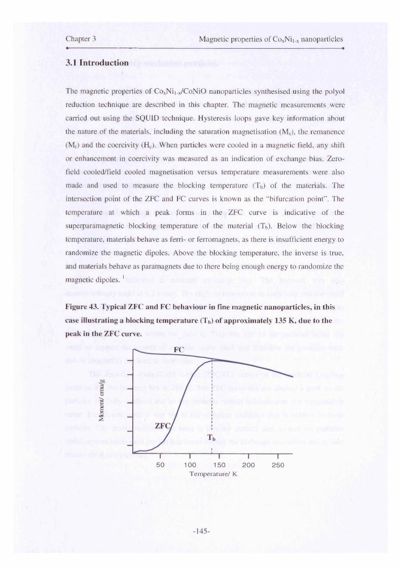
Chapter 3 Magnetic properties of CoxNii_x nanoparticles
3.1 Introduction
The magnetic properties of CoxNii_x/CoNiO nanoparticles synthesised using the polyol
reduction technique are described in this chapter. The magnetic measurements were
carried out using the SQUID technique. Hysteresis loops gave key information about
the nature of the materials, including the saturation magnetisation (Ms), the remanence
(Mr) and the coercivity (Hc). When particles were cooled in a magnetic field, any shift
or enhancement in coercivity was measured as an indication of exchange bias. Zero-
field cooled/field cooled magnetisation versus temperature measurements were also
made and used to measure the blocking temperature (Tb) of the materials. The
intersection point of the ZFC and FC curves is known as the “bifurcation point” . The
temperature at which a peak forms in the ZFC curve is indicative of the
superparamagnetic blocking temperature of the material (Tb). Below the blocking
temperature, materials behave as ferri- or ferromagnets, as there is insufficient energy to
randomize the magnetic dipoles. Above the blocking temperature, the inverse is true,
and materials behave as paramagnets due to there being enough energy to randomize the
magnetic dipoles. 1
Figure 43. Typical ZFC and FC behaviour in fine magnetic nanoparticles, in this
case illustrating a blocking temperature (Tb) of approximately 135 K, due to the peak in the ZFC curve.
FC
5 0 100 150 2 0 0 2 5 0Tem perature/ K
-145-

Chapter 3 Magnetic properties of CoxNii_x nanoparticles
3.2 Homogeneously nucleated particles
Experiments were carried out to synthesise Co8oNi2o nanoparticles that were
homogeneously nucleated. However, what was in fact initially made using the
homogeneous nucleation method, were oxidised Co8oNi2o nanoparticles, which had a
mean particle diameter of 7.6 nm (Section 2.4). The magnetic properties of the oxidised
form of these particles were measured, as it was important to characterise the
antiferromagnetic phase in the particles for future reference. The measurements also
served as an indicator of the typical properties of the oxidised form of these particles.
The low moment and slope of the curve indicate AFM behaviour due to the oxidation of
the said particles.
Figure 44 displays the two measured hysteresis loops for the homogeneously
nucleated oxidised CosoNi2o particles, field-cooled to 10 K in 0 kOe and 60 kOe fields.
The hysteresis loop that was measured at 10 K and in a zero-applied field gave a
coercivity of 920 Oe. The coercivity of the material after cooling in a 60 kOe field, at
the same temperature, was found to be 930 Oe. The shift of 110 Oe during the field-
cooling process indicated a minimal exchange bias. The moment was also
correspondingly small at 1.7 emu/g. The slight enhancement in coercivity and the small
loop shift indicated that some exchange-biasing had occurred, however the effect was
very small. Exchange-pinning in this instance was due to pinning in the
antiferromagnetic oxide within the particle. This was due to the particles being too
small to support the growth of a stable oxide shell, and therefore the particles were
almost completely oxidised to their cores.
The zero-field cooled/field cooled (ZFC/FC) curves of the oxidised Co8oNi2o
particles did not intersect below 280 K. The ZFC curve did not display a peak as the
particles are fully oxidised and so the particles remain unblocked in this temperature
range. Insufficient particle size led to the complete oxidation that is evident in these
particles. The results indicated the need to increase particle size, so that the particles
could support oxide shell growth and hence display the exchange bias effect that is seen
in core-shell nanoparticles.2
-146-
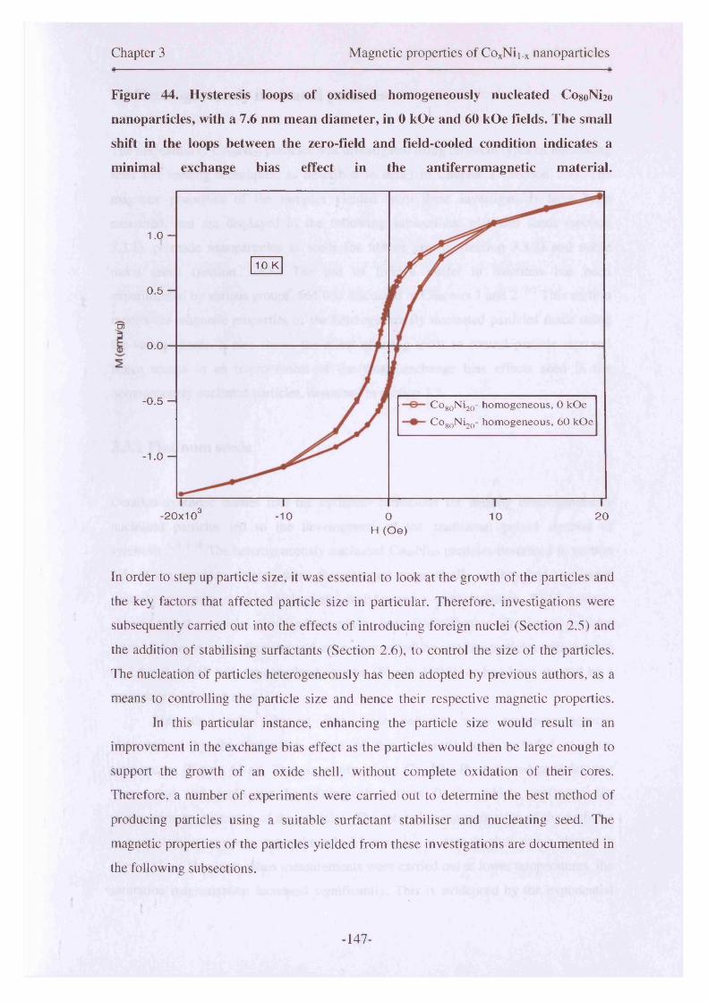
Chapter 3 Magnetic properties of CoxNii_x nanoparticles
Figure 44. Hysteresis loops of oxidised homogeneously nucleated CosoNi2o
nanoparticles, with a 7.6 nm mean diameter, in 0 kOe and 60 kOe fields. The small
shift in the loops between the zero-field and field-cooled condition indicates a
minimal exchange bias effect in the antiferromagnetic material.
1.0 -
10 K
0.5 -
.CD
0.0
-0.5 - Co80Ni20- homogeneous, 0 kOe COgoN^Q- homogeneous, 60 kOe
- 1.0 -
,3-20x10 -10 0 10 20H (Oe)
In order to step up particle size, it was essential to look at the growth of the particles and
the key factors that affected particle size in particular. Therefore, investigations were
subsequently carried out into the effects of introducing foreign nuclei (Section 2.5) and
the addition of stabilising surfactants (Section 2.6), to control the size of the particles.
The nucleation of particles heterogeneously has been adopted by previous authors, as a
means to controlling the particle size and hence their respective magnetic properties.
In this particular instance, enhancing the particle size would result in an
improvement in the exchange bias effect as the particles would then be large enough to
support the growth of an oxide shell, without complete oxidation of their cores.
Therefore, a number of experiments were carried out to determine the best method of
producing particles using a suitable surfactant stabiliser and nucleating seed. The
magnetic properties of the particles yielded from these investigations are documented in
the following subsections.
-147-
Chapter 3 Magnetic properties of CoxNii.x nanoparticles
3.3 Heterogeneously nucleated particles
The nucleation of CogoNi2o particles was investigated using different types of nucleating
seed and seeding techniques, as described in detail in Chapter 2 (Section 2.5). The
magnetic properties of the samples yielded from these investigations have been
measured, and are displayed in the following subsections; platinum seeds (section
3.3.1), premade nanoparticles as seeds for further growth (section 3.3.2) and noble
metal seeds (section 3.3.3). The use of foreign nuclei in reactions has been
experimented by various groups, and was discussed in Chapters 1 and 2 . 3 7 This section
reports the magnetic properties of the heterogeneously nucleated particles made using
the various seeds. It also shows the effect of using seeds to control particle size and
hence results in an improvement of the small exchange bias effects seen in the
homogenously nucleated particles, described in section 3.2.
3.3.1 Platinum seeds
Detailed synthetic studies into the optimum conditions for making heterogeneously
nucleated particles led to the development of the traditional polyol method of
synthesis.3 ,4 ’ 810 The heterogeneously nucleated Co8oNi2o particles described in section
2.5. of the previous chapter, were characterised magnetically as they had responded
strongly to a magnet upon drying, and had formed a stable solution. TEM analysis
showed that the particles displayed bimodality in their distribution. The larger particles
were 1 2 .6 nm in mean diameter and were surrounded by satellite particles with a mean
of 2.6 nm, which had not completely grown (Figure 13(b)), or had been created by a
secondary nucleation event.
Magnetic studies showed that these particles were superparamagnetic,
characterised by the distinctive shape of the hysteresis loop recorded at room
temperature (Figure 45(a)). The coercivity of the CogoNi2o Pt nucleated particles was
very small and could not be distinguished from 0 Oe, which confirmed the
superparamagnetic nature of the particles. The saturation magnetisation of the particles
at room temperature was approximately 11 emu g' 1 as seen in then curve shown in
Figure 45(a). However, when measurements were carried out at lower temperatures, the
saturation magnetisation increased significantly. This is evidenced by the exponential
-148-
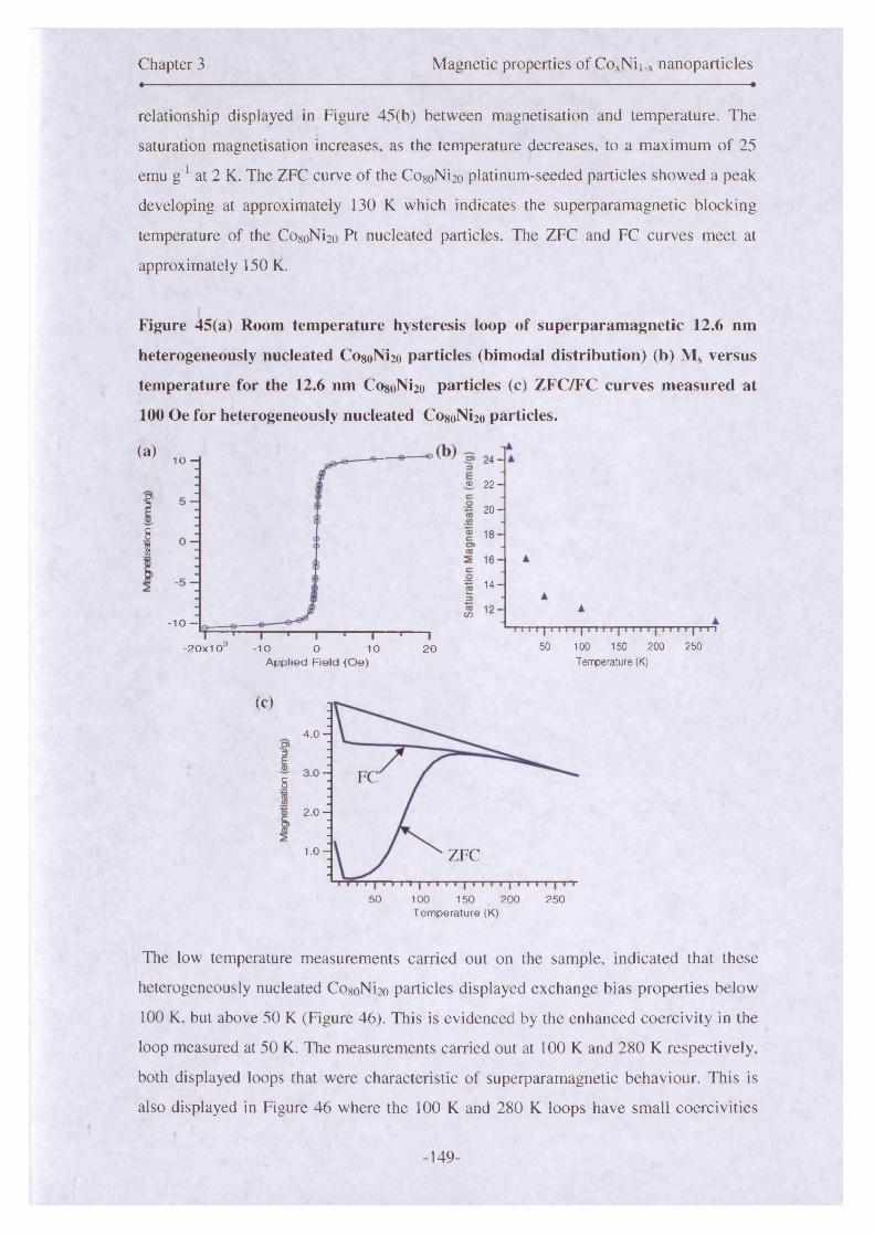
Chapter 3 Magnetic properties of CoxNii.x nanoparticles• •
relationship displayed in Figure 45(b) between magnetisation and temperature. The
saturation magnetisation increases, as the temperature decreases, to a maximum of 25
emu g ' 1 at 2 K. The ZFC curve of the CogoNi2o platinum-seeded particles showed a peak
developing at approximately 130 K which indicates the superparamagnetic blocking
temperature of the CogoNi2o Pt nucleated particles. The ZFC and FC curves meet at
approximately 150 K.
Figure 45(a) Room temperature hysteresis loop of superparamagnetic 12.6 nm
heterogeneously nucleated CogoNi2o particles (bimodal distribution) (b) Ms versus
temperature for the 12.6 nm CogoNi2o particles (c) ZFC/FC curves measured at
100 Oe for heterogeneously nucleated CogoNi2o particles.
(a) 10 -I
® . 2 2 -D)2 0 -
51
14-
cn- 1 0 -
-2 0 x 1 0 3 50 100 1 50 200 250-10 O 1 0 20A p p lie d F ie ld ( O e ) Temperature (K)
(C)
4 .0 -
FC5
2 .0 -
1.0 - ZFC
50 100 150 250200Tem perature (K)
The low temperature measurements carried out on the sample, indicated that these
heterogeneously nucleated CogoNi2o particles displayed exchange bias properties below
100 K, but above 50 K (Figure 46). This is evidenced by the enhanced coercivity in the
loop measured at 50 K. The measurements carried out at 100 K and 280 K respectively,
both displayed loops that were characteristic of superparamagnetic behaviour. This is
also displayed in Figure 46 where the 100 K and 280 K loops have small coercivities
-149-
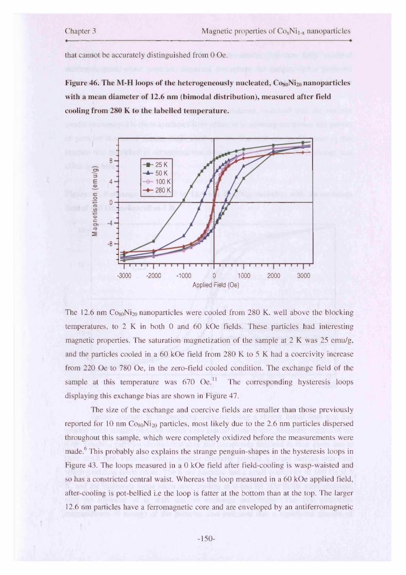
Chapter 3 Magnetic properties of CoxNii.x nanoparticlesi-------------------------------------------------------------------------------------------------------- «
that cannot be accurately distinguished from 0 Oe.
Figure 46. The M-H loops of the heterogeneously nucleated, CosoNi2o nanoparticles
with a mean diameter of 1 2 .6 nm (bimodal distribution), measured after field
cooling from 280 K to the labelled temperature.
csCO
CDecoCO
8- * - 2 5 K -A - 50 K- e - 100 K
280 K4
0
•4
■8
-3000 -2000 -1000 0 1000Applied Field (Oe)
2000 3000
The 12.6 nm Co8oNi2o nanoparticles were cooled from 280 K, well above the blocking
temperatures, to 2 K in both 0 and 60 kOe fields. These particles had interesting
magnetic properties. The saturation magnetization of the sample at 2 K was 25 emu/g,
and the particles cooled in a 60 kOe field from 280 K to 5 K had a coercivity increase
from 220 Oe to 780 Oe, in the zero-field cooled condition. The exchange field of the
sample at this temperature was 670 O e.11 The corresponding hysteresis loops
displaying this exchange bias are shown in Figure 47.
The size of the exchange and coercive fields are smaller than those previously
reported for 10 nm CogoNi2o particles, most likely due to the 2.6 nm particles dispersed
throughout this sample, which were completely oxidized before the measurements were
made.6 This probably also explains the strange penguin-shapes in the hysteresis loops in
Figure 43. The loops measured in a 0 kOe field after field-cooling is wasp-waisted and
so has a constricted central waist. Whereas the loop measured in a 60 kOe applied field,
after-cooling is pot-bellied i.e the loop is fatter at the bottom than at the top. The larger
12.6 nm particles have a ferromagnetic core and are enveloped by an antiferromagnetic
-150-
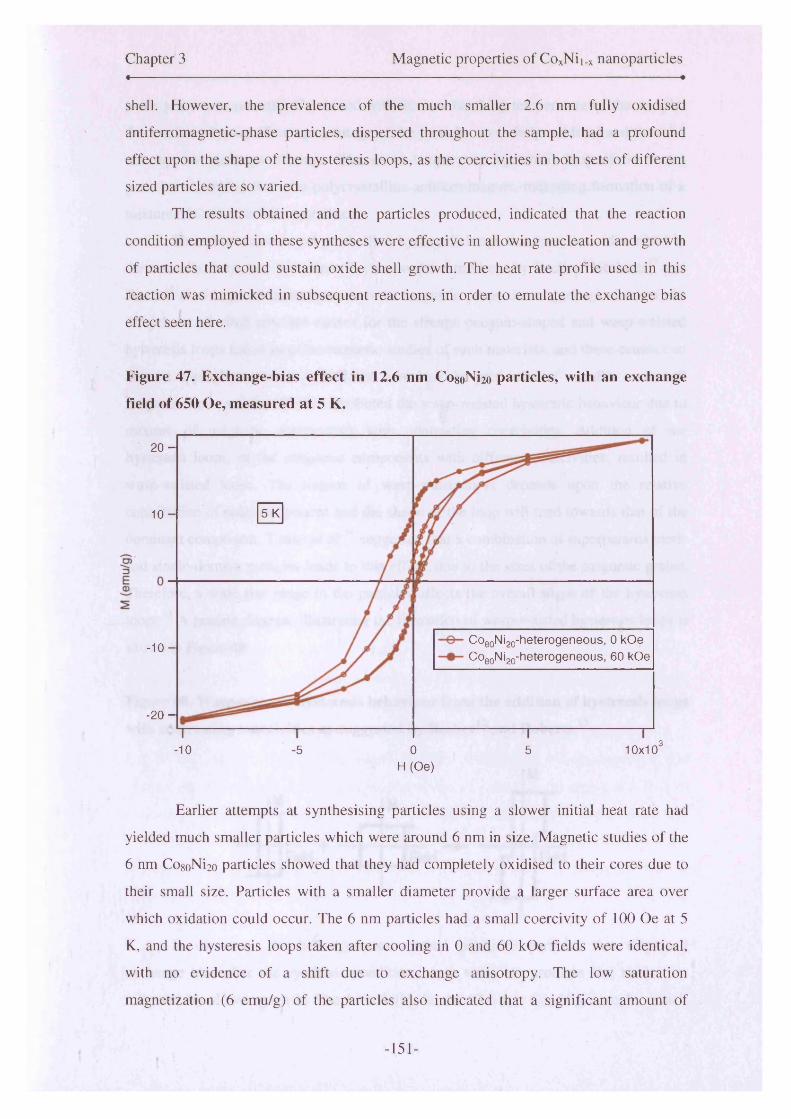
Chapter 3 Magnetic properties of CoxNii_x nanoparticles« ■ ■— ■ - , . »
shell. However, the prevalence of the much smaller 2.6 nm fully oxidised
antiferromagnetic-phase particles, dispersed throughout the sample, had a profound
effect upon the shape of the hysteresis loops, as the coercivities in both sets of different
sized particles are so varied.
The results obtained and the particles produced, indicated that the reaction
condition employed in these syntheses were effective in allowing nucleation and growth
of particles that could sustain oxide shell growth. The heat rate profile used in this
reaction was mimicked in subsequent reactions, in order to emulate the exchange bias
effect seen here.
Figure 47. Exchange-bias effect in 12.6 nm Co80Ni2o particles, with an exchange
field of 650 Oe, measured at 5 K.
2 0 -
5 K1 0 -
COeoNigQ-heterogeneous, 0 kOe Co80Ni20-heterogeneous, 60 kOe
- 1 0 -
- 2 0 -
,310x10'-10 ■5 0 5
H (Oe)
Earlier attempts at synthesising particles using a slower initial heat rate had
yielded much smaller particles which were around 6 nm in size. Magnetic studies of the
6 nm CogoNi2o particles showed that they had completely oxidised to their cores due to
their small size. Particles with a smaller diameter provide a larger surface area over
which oxidation could occur. The 6 nm particles had a small coercivity of 100 Oe at 5
K, and the hysteresis loops taken after cooling in 0 and 60 kOe fields were identical,
with no evidence of a shift due to exchange anisotropy. The low saturation
magnetization ( 6 emu/g) of the particles also indicated that a significant amount of
-151-

Chapter 3 Magnetic properties of CoxNii_x nanoparticles
oxide phase was present. Combined with the low blocking temperature of the sample,
this confirmed 3 significant percentage of the particles were fully oxidized and thus did
not support exchange biasing. The room temperature hysteresis loop of the 6 nm
particles resembled that of a polycrystalline antiferromagnet, indicating formation of a
mixture of CoO and NiO crystallites.
The wasp-waisted and pot-bellied hysteresis loops that have been described in
these results, have also been described in works carried out by Becker, 12 Roberts13 and
Tauxe14 et al in previous research into the magnetic properties of geological materials.
They have all cited possible causes for the strange penguin-shaped and wasp-waisted
hysteresis loops found in paleomagnetic studies of such materials, and these causes can
also be applied to studies carried out here into the properties of metallic core-shell
magnetic nanoparticles. Becker attributed the wasp-waisted hysteretic behaviour due to
mixture of magnetic components with contrasting coercivities. Addition of the
hysteresis loops, of the magnetic components with different coercivities, resulted in
wasp-waisted loops. The degree of wasp-waistedness depends upon the relative
contribution of each component and the shape of the loop will tend towards that of the
dominant component. Tauxe et a l 14 suggested that a combination of superparamagnetic
and single-domain particles leads to this effect, due to the sizes of the magnetic grains.
Therefore, a wide size range in the particles affects the overall shape of the hysteresis
loops. 15 A generic diagram illustrating the formation of wasp-waisted hysteresis loops is
shown in Figure 48.
Figure 48. Wasp-waisted hysteresis behaviour from the addition of hysteresis loops
with contrasting coercivities as suggested by Becker12 and Roberts.13
| M
Rdd +
In summary, the heterogeneous method produced particles that displayed
exchange bias after the synthesis conditions were varied to promote fast nucleation
followed by slower growth. The bimodality in the 12.6 nm CogoNi2o sample, which
M
M
Field
[7f Field
-152-

Chapter 3 Magnetic properties of CoxNii.x nanoparticles
displayed exchange bias, affected the shape of the hysteresis loop as the smaller
particles oxidised and this caused a penguin-shaped loop. This was due to a mixture of
superparamagnetic and single-domain particles within the sample.
Initial magnetic studies indicated that heterogeneous nucleation of nanoparticles
was a better route to synthesising particles with exchange bias, due to the control in
particle size that could be implemented, during synthesis. The use of oleic acid as a sole
surfactant has been shown to be successful where large particle growth has occurred.
However, the results shown by the 6 nm CogoNi2o particles have shown that oleic acid
did not sufficiently stabilise the smaller particles. The washing procedure post
synthesis, if not replaced with sufficient stabiliser, can induce premature oxidation of
the particles. The heterogeneous route of synthesis was therefore developed further in
this investigation, through the use of different seeding techniques and different types of
seeds (Section 2.5.1.2 and 2.5.1.3). The magnetic studies of these particular samples are
detailed in the following subsections.
3.3.2 Premade particles as nuclei for further growth
It had been previously shown by Yamamuro et a l 16 that particle size could be increased
using premade particles, that were reintroduced into the reaction set-up with additional
reactants, to improve particle size. The seed particles described in subsection 2.5.1.2
were small at around 4.4 nm in diameter, and displayed partial self assembly when
initially made and when additional reactants were added to promote the growth of larger
particles, the mean diameter of the resultant nanoparticles almost doubled. The particles
that were grown from these seeds were 7.4 nm in mean diameter. The hysteresis loop of
the 7.4 nm particles is shown in Figure 49. Low temperature measurements of this
sample were carried out at 5 K. The loop’s saturation point was 10 emu/g at 5 K. The
coercivity of the particles was 440 Oe in zero-field, whereas in a 60 kOe field this
increased to 520 Oe. This increase was not much greater than that determined for the
homogeneously nucleated particles described earlier (section 3.2), and therefore did not
improve upon the results garnered from heterogeneously nucleating the particle’s grown
with platinum seeds.
-153-

Chapter 3 Magnetic properties of CoxNii_x nanoparticles• — •
Figure 49. M vs H loops measured at 5 K in 0 kOe and 60 kOe fields, for 7.4 nm
CosoNi2o particles seeded with 4.4 nm CosoNi2o particles, in a two-step synthetic
process.
5 -
- 5 -
Particles made with COgoNi^ seeds, 0 kOe
Particles made with Co80Ni20 seeds, 60 kOe
-20x10 3 -1 0 0 10 2 0
A pp lied field (O e)
The ZFC/FC curves did not overlap below 280 K when the particles were measured at
100 Oe. The ZFC curve displayed a plateau at approximately 40 K and then rose to 130
K, however, there was no evidence o f a peak. This line of investigation was not
pursued, in terms of the synthesis, as the particles made using this method had almost
oxidised completely to their cores. This resulted in the low moment that is seen in
Figure 49 and hence the minimal exchange bias effect that can also be seen due to the
domination of the antiferromagnetic phase.
The polyol method of synthesis had been used in the synthesis of iron
nanoparticles by Y am am uro16 whereas here, the CoxNii_x system was experimented
with. These results are therefore novel, as this particular seeding technique had not been
used before to synthesise this type of particle. The particles did show evidence of
exchange bias, however, the reaction conditions would require further investigation to
eliminate oxidation of the smaller preformed seed particles, as this translated through
into the final 7.4 nm particles. Although nitrogen flow was maintained, the small seed
particles (4.4 nm) may have oxidised quickly when the additional reactants were
-154-

Chapter 3 Magnetic properties of CoxNii.x nanoparticles
introduced to the vessel, as the flask was opened up to the atmosphere. Although the
subsequent heating of the reaction, to grow larger particles occurred in a nitrogen
atmosphere, the seed particles could have partially oxidised. Particles of around this
size, in the previous investigations of homogeneously nucleated particles (section 2.4-
7.6 nm), oxidised easily and therefore yielded low moment, and small exchange bias
effects.
The results shown in this section have indicated it is possible to increase particle
size, by using a particle of identical composition to the final particle, as nuclei for
growth. With the addition of other reactants and controlled heating, the reactants grow
around the nuclei to form larger particles. The controlled heating tailors the reaction to
temperature so that the supersaturation of the solution is correspondingly controlled.
This was highly important in the synthesis of these particles.
The small exchange bias effect could be due to the structural morphology of the
particle. The seed particles that were synthesised were core-shell-surfactant structured17
with the non-magnetic surfactant, coating the antiferromagnetic shell, which enveloped
the metallic core. If these particles acted as a seed, then subsequent growth around
them, if the surfactant was not completely washed off, would again be of that structure.
There was the possibility that the surfactant coating the seed particle meant a core-shell-
-core1 - shell1 - surfactant1 structure was present. Exchange bias is an interfacial effect1 0 1 ft 90which occurs across the antiferromagnet shell and ferromagnetic core interface. ’ ’
Therefore, it is possible that the small effect seen in Figure 49, is due to the interaction
between core1 and shell1 seen in Figure 50.
-155-
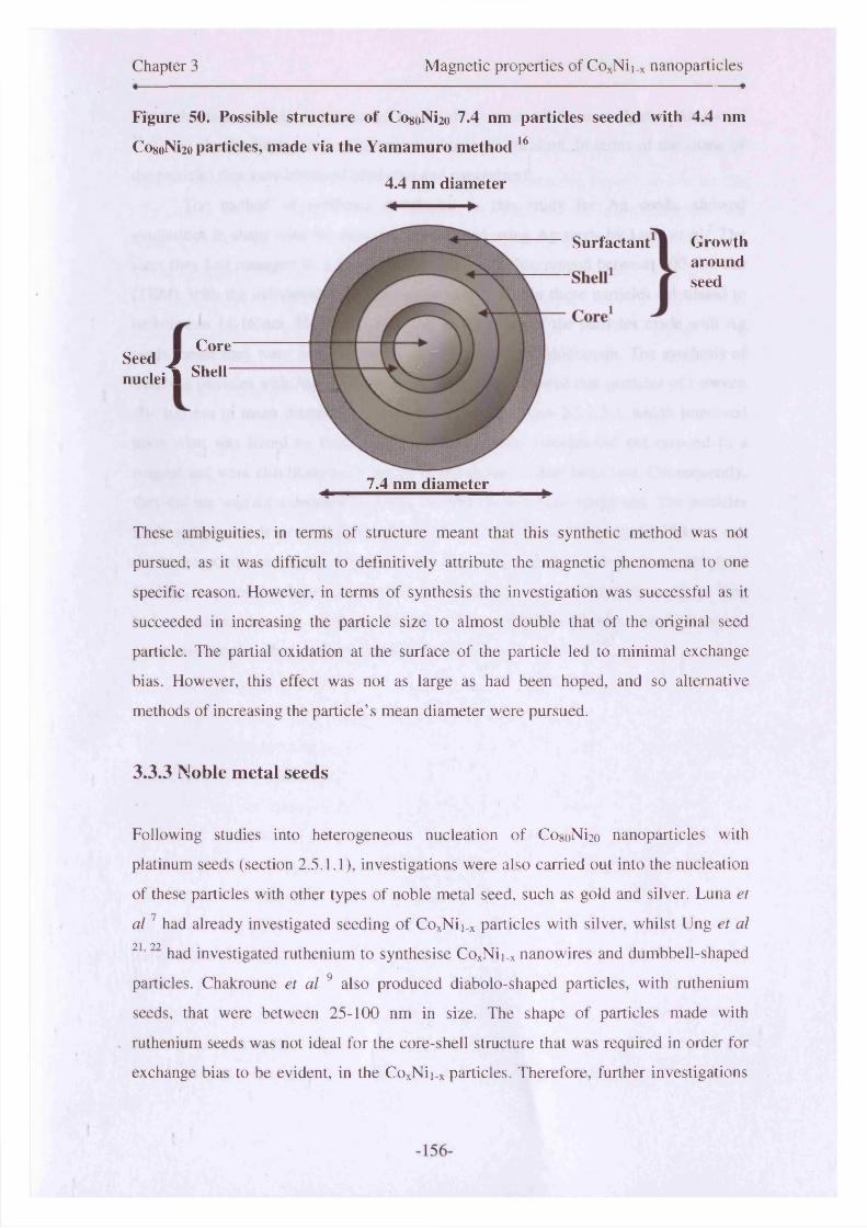
Chapter 3 Magnetic properties of CoxNii_x nanoparticles
Figure 50. Possible structure of CosoNi2o 7.4 nm particles seeded with 4.4 nm
Co«oNi2o particles, made via the Yamamuro method 16
4.4 nm diameter
_ Core Seed Jnuclei1 She"
Surfactant
w
%
7.4 nm diameter
Growtharoundseed
These ambiguities, in terms of structure meant that this synthetic method was not
pursued, as it was difficult to definitively attribute the magnetic phenomena to one
specific reason. However, in terms of synthesis the investigation was successful as it
succeeded in increasing the particle size to almost double that of the original seed
particle. The partial oxidation at the surface of the particle led to minimal exchange
bias. However, this effect was not as large as had been hoped, and so alternative
methods of increasing the particle’s mean diameter were pursued.
3.3.3 Noble metal seeds
Following studies into heterogeneous nucleation of Co8oNi2o nanoparticles with
platinum seeds (section 2.5.1.1), investigations were also carried out into the nucleation
of these particles with other types of noble metal seed, such as gold and silver. Luna et
a l 1 had already investigated seeding of CoxNii_x particles with silver, whilst Ung et al0 1 00
' had investigated ruthenium to synthesise CoxNii_x nanowires and dumbbell-shaped
particles. Chakroune et al 9 also produced diabolo-shaped particles, with ruthenium
seeds, that were between 25-100 nm in size. The shape of particles made with
ruthenium seeds was not ideal for the core-shell structure that was required in order for
exchange bias to be evident, in the CoxNii_x particles. Therefore, further investigations

Chapter 3 Magnetic properties of CoxNii_x nanoparticles
into ruthenium seeds were not carried out for the purpose of this investigation. Ung et al
21, 22 had clearly displayed the limitations of using ruthenium, in terms of the shape of
the particles that were obtained (diabolos and nanowires).
The method of synthesis developed in this study for Ag seeds, showed
similarities in shape with the particles synthesised using Ag seeds by Luna et al? The
sizes they had managed to achieve for CogoN^o particles, ranged between 200-540 nm
(TEM), with the individual crystallite sizes (XRD) within those particles calculated to
be between 14-16 nm. Therefore, the large overall size of the particles made with Ag
seeds meant they were polycrystalline and likely to be multidomain. The synthesis of
Co8oNi2o particles with Ag seeds, in our investigation, showed that particles of between
40- 100 nm in mean diameter could be fabricated (section 2.5.1.3 ), which improved
upon what was found by Luna et al. 1 However, the particles did not respond to a
magnet and were also likely to be polycrystalline due to their large size. Consequently,
they did not warrant a detailed investigation into the magnetic properties. The particles
made with Au seeds were also polycrystals due to their size of between 60-150 nm, and
did not respond to a magnet. Therefore, the use of Ag and Au seeds in the synthesis of
CoxNii_x was not developed any further and Pt seeds were subsequently used, as they
had controlled the size of the particles in this particular system, much better in
comparison to the other noble metals that were tested.
-157-

Chapter 3 Magnetic properties of CoxNii_x nanoparticles
3.4 Surfactant stabilisers
The investigations carried out into the most effective nucleation process to use for the
synthesis of CoxNii.x particles, indicated that heterogeneous nucleation was the best
route to follow. The seed that was found to be the best for this system was platinum.
The heterogeneously nucleated particles displayed the strongest exchange bias effects in
this study, when compared to the homogeneously nucleated particles described in
section 3.2. Another method of controlling particle size that has been investigated by
various groups, and in this study, was the use of stabilising surfactants to arrest or
promote particle growth. 1 7 ,2 3 ,24 The binding of the surfactant to form a complex with
the metallic ions and steric effects that led to the ability to tune the size of particles.
Particles formed using oleic acid displayed variable results. The homogeneously
nucleated particles coated with oleic acid, almost completely oxidised due to their small
size. Whereas the particles that had heterogeneously nucleated were much larger and
were therefore able to support oxide shell growth. This had a significant impact on the
magnetic properties, as the almost completely oxidised homogeneously nucleated
Co8oNi2o particles had a low moment, and therefore did not display exchange bias effect
due to the absence of a core-shell structure. The larger CogoNi2o particles that were
heterogeneously nucleated, did support oxide shell growth, and hence exchange bias
was observed. What this did show was the importance of balancing the nucleation, with
the appropriate type of surfactant for a particular sample.
When amine-based surfactants were used, the nickel particles were stabilised
well from agglomeration. However, this was not the case for samples with cobalt-doped
into them. The particles formed by oleylamine as sole surfactant were too small and had
completely oxidised, as they did not respond to a magnet. Therefore, they did not
warrant any further magnetic investigation. The same was true for TOPO and TBP. The
particles formed using these particular surfactants, on a sole-basis, were too small to
warrant detailed magnetic studies as they are predominantly oxidised to their cores. The
exception to this was oleic acid, which had shown evidence of core-shell textured
particles being formed. Therefore, some investigations were carried out on particles
made with oleic acid as sole surfactant due to the promising results achieved where
larger particles had been grown.
The use of an oleic acid/ oleylamine mixture had previously been developed for
-158-

Chapter 3 Magnetic properties of CoxNii_x nanoparticles
the stabilisation of iron nanoparticles by Sun et al.25 Studies into the suitability of
surfactants in this investigation, also indicated the possibility that the OA/OY
combination could successfully mediate particle growth and stabilise the particles from
oxidation. This was due to the strong interaction between the metallic ion species in
solution with OA, and the weaker interaction with OY. The use of these surfactants, in
addition to heterogeneously nucleating the particles, led to successful results. These are
described in detail within the compositional study subsection (section 3.5). The OA/OY
mixture was believed to have formed an amide, and hence a double-sheathed surfactant,
which protected the particles from agglomeration and full-oxidation. The particles were
also sufficiently large to warrant detailed magnetic investigations. The OA/OY
synthesised particles were partially oxidised and this gave rise to the interesting
properties described in the following sections.
-159-
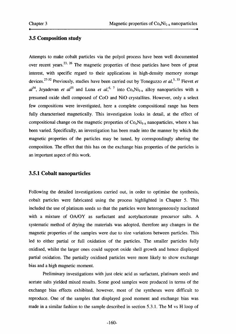
Chapter 3 Magnetic properties of CoxNii.x nanoparticles
3.5 Composition study
Attempts to make cobalt particles via the polyol process have been well documented
over recent years. ’ The magnetic properties of these particles have been of great
interest, with specific regard to their applications in high-density memory storage
devices.27' 32 Previously, studies have been carried out by Toneguzzo et al,5 , 33 Fievet et
al34, Jeyadevan et al35 and Luna et al,6, 1 into CoxNii_x alloy nanoparticles with a
presumed oxide shell composed of CoO and NiO crystallites. However, only a select
few compositions were investigated, here a complete compositional range has been
fully characterised magnetically. This investigation looks in detail, at the effect of
compositional change on the magnetic properties of CoxNii_x nanoparticles, where x has
been varied. Specifically, an investigation has been made into the manner by which the
magnetic properties of the particles may be tuned, by correspondingly altering the
composition. The effect that this has on the exchange bias properties of the particles is
an important aspect of this work.
3.5.1 Cobalt nanoparticles
Following the detailed investigations carried out, in order to optimise the synthesis,
cobalt particles were fabricated using the process highlighted in Chapter 5. This
included the use of platinum seeds so that the particles were heterogeneously nucleated
with a mixture of OA/OY as surfactant and acetylacetonate precursor salts. A
systematic method of drying the materials was adopted, therefore any changes in the
magnetic properties of the samples were due to size variations between particles. This
led to either partial or full oxidation of the particles. The smaller particles fully
oxidised, whilst the larger ones could support oxide shell growth and hence displayed
partial oxidation. The partially oxidised particles were more likely to show exchange
bias and a high magnetic moment.
Preliminary investigations with just oleic acid as surfactant, platinum seeds and
acetate salts yielded mixed results. Some good samples were produced in terms of the
exchange bias effects exhibited, however, most of the syntheses were difficult to
reproduce. One of the samples that displayed good moment and exchange bias was
made in a similar fashion to the sample described in section 5.3.1. The M vs H loop of
-160-
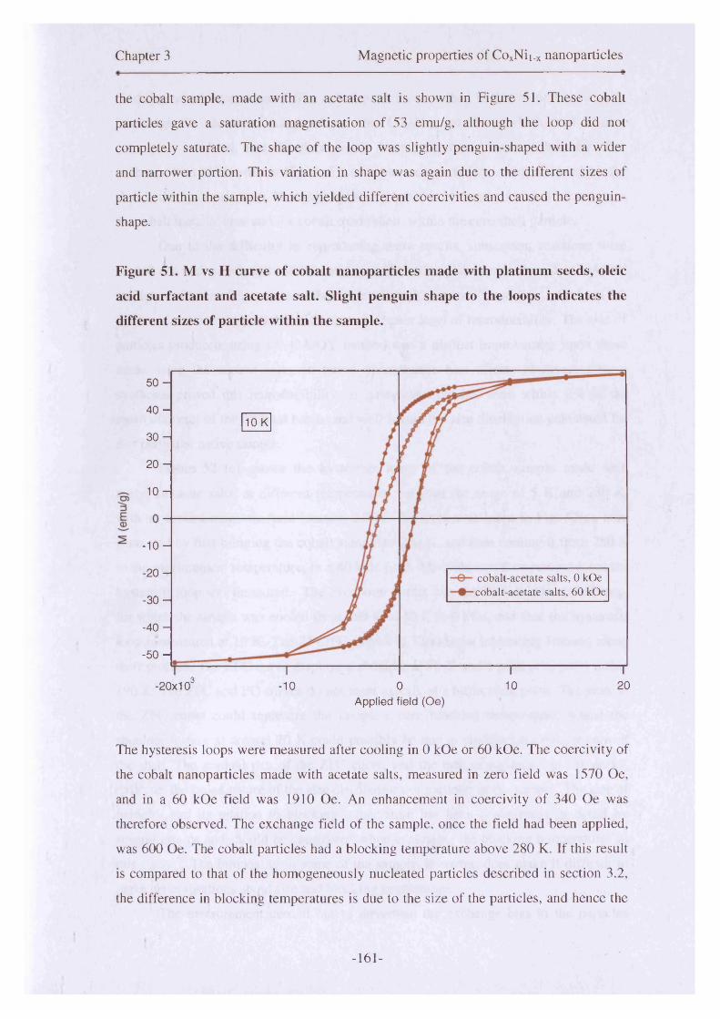
Chapter 3 Magnetic properties of CoxNii.x nanoparticles
the cobalt sample, made with an acetate salt is shown in Figure 51. These cobalt
particles gave a saturation magnetisation of 53 emu/g, although the loop did not
completely saturate. The shape of the loop was slightly penguin-shaped with a wider
and narrower portion. This variation in shape was again due to the different sizes of
particle within the sample, which yielded different coercivities and caused the penguin-
shape.
Figure 51. M vs H curve of cobalt nanoparticles made with platinum seeds, oleic
acid surfactant and acetate salt. Slight penguin shape to the loops indicates the
different sizes of particle within the sample.
5 0 -
4 0 -
3 0 -
2 0 -
1 0 -
- 1 0 -
- 2 0 -
- 3 0 -
- 4 0 -
- 5 0 -
10 K
cobalt-acetate salts, 0 kOe cobalt-acetate salts, 60 kOe
-20x103 -10 0 10 20Applied field (Oe)
The hysteresis loops were measured after cooling in 0 kOe or 60 kOe. The coercivity of
the cobalt nanoparticles made with acetate salts, measured in zero field was 1570 Oe,
and in a 60 kOe field was 1910 Oe. An enhancement in coercivity o f 340 Oe was
therefore observed. The exchange field of the sample, once the field had been applied,
was 600 Oe. The cobalt particles had a blocking temperature above 280 K. If this result
is compared to that of the homogeneously nucleated particles described in section 3.2,
the difference in blocking temperatures is due to the size of the particles, and hence the
-161-

Chapter 3 Magnetic properties of CoxNii.x nanoparticles
oxidation which has occurred. The cobalt particles described in this section have more
ferromagnetic character compared to the CogoNi2o particles which had almost
completely oxidised, and therefore would have little ferromagnetic character. The
exchange-bias effect seen in Figure 51, of cobalt nanoparticles, is characterised by the
shift in the hysteresis loop. This shift has arisen because of the interfacial effect between
the cobalt metallic core and the cobalt oxide shell, within the core-shell particle.
Due to the difficulty in reproducing these results, subsequent reactions were
carried out with cobalt and nickel acetylacetonate salts, platinum seeds and an OA/OY
surfactant mixture. This was an adaptation of the Yamamuro16 and Sun25 methods for
synthesis of iron nanoparticles and ensured a better level of reproducibility. The size of
particles produced, using the OA/OY method was a distinct improvement upon those
made using the acetate salts, in terms of exchange bias effects observed. Repeat
syntheses proved this reproducibility, as subsequent batches were within 2 % of the
mean diameter of the original batch, and well within the size distribution calculated for
that particular native sample.
Figure 52 (c) shows the hysteresis loops of the cobalt sample made with
acetylacetonate salts, at different temperatures between the range of 5 K and 280 K,
with an applied magnetic field (section 2.7.2). The hysteresis loops in Fig. 52(c) were
generated by first bringing the cobalt sample to 280 K, and then cooling it from 280 K
to the measurement temperature, in a 60 kOe field. The field was then removed and the
hysteresis loop was measured. The exception to this was the 10 K 0 kOe cooled loop,
for which the sample was cooled from 280 K to 10 K in 0 kOe, and then the hysteresis
loop is measured at 10 K. The ZFC/FC curves in 52(a) show interesting features along
their profiles. The ZFC curve displays a shoulder at 70 K and a peak at approximately
190 K. The ZFC and FC curves do not meet exactly at a bifurcation point. The peak in
the ZFC curve could represent the sample’s core blocking temperature, whilst the
shoulder feature at around 70 K could possibly be due to the blocking temperature of
the shell. The gradual rise of the ZFC curve, and the lack of definition in the peaks,
indicates the broad nature of the size distributions of particles in the sample. The size of
particles and its relation to blocking temperature has been investigated in detail by
several groups and should be considered when evaluating the blocking temperatures in
this study.36 The bimodality in some of the sample, however, does make it difficult to
make generalisations about size and blocking temperature.
The measurement carried out to determine the exchange bias in the particles
-162-
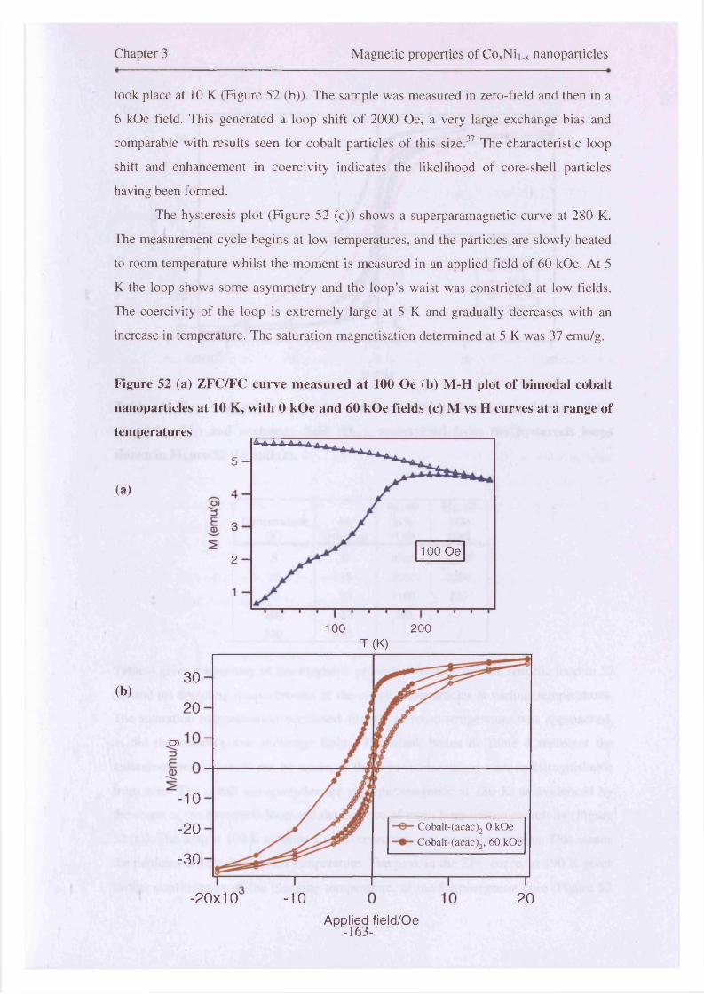
Chapter 3 Magnetic properties of CoxNii.x nanoparticles
took place at 10 K (Figure 52 (b)). The sample was measured in zero-field and then in a
6 kOe field. This generated a loop shift of 2000 Oe, a very large exchange bias and
comparable with results seen for cobalt particles of this size.37 The characteristic loop
shift and enhancement in coercivity indicates the likelihood of core-shell particles
having been formed.
The hysteresis plot (Figure 52 (c)) shows a superparamagnetic curve at 280 K.
The measurement cycle begins at low temperatures, and the particles are slowly heated
to room temperature whilst the moment is measured in an applied field of 60 kOe. At 5
K the loop shows some asymmetry and the loop’s waist was constricted at low fields.
The coercivity of the loop is extremely large at 5 K and gradually decreases with an
increase in temperature. The saturation magnetisation determined at 5 K was 37 emu/g.
Figure 52 (a) ZFC/FC curve measured at 100 Oe (b) M-H plot of bimodal cobalt
nanoparticles at 10 K, with 0 kOe and 60 kOe fields (c) M vs H curves at a range of
temperatures
(a)
(b)
5 -
100 Oe2 -
100 200T (K)
30
20
05
-10
Cobalt-(acac)2 0 kOe Cobalt-(acac)2, 60 kOe
-20
-30
-20x10 -10 20Applied field/Oe
-163-

Chapter 3 Magnetic properties of CoxNii.x nanoparticles «
2 0 -
fE0)
- 1 0 - 5 K 50 K 100 K 280 K
- 2 0 -
-3 0 -
.3 20-20x10' -10 0 10H (Oe)
Table 4. Key magnetic properties such as; saturation magnetisation (Ms),
coercivity (Hc) and exchange field (Hex)* ascertained from the hysteresis loops
shown in Figure 52 (b) and (c).
Tem perature(K)
Ms(em u / g)
Hc. 60 kOe (Oe)
Hex-60kOe(Oe)
5 37 2320 224010 35 2020 200050 33 1100 510100 32 380 /280 30 / /
Table 4 gives a summary o f the magnetic properties relating to each specific loop in 52
(b) and (c) depicting measurements of the cobalt nanoparticles at various temperatures.
The saturation magnetisation decreased slightly as room temperature was approached,
as did the coercive and exchange fields. The blank boxes in Table 4 represent the
measurements that could not be made, as those particular values were indistinguishable
from zero. The cobalt nanoparticles are superparamagnetic at 280 K, as evidenced by
the shape of the hysteresis loop and the absence of room-temperature coercivity (Figure
52 (c)). The loop at 100 K exhibits a small coercivity, but no exchange-bias. This means
the particles are blocked at this temperature. The peak in the ZFC curve, at 190 K gives
further confirmation of the blocking temperature, of the ferromagnetic core (Figure 52
-164-
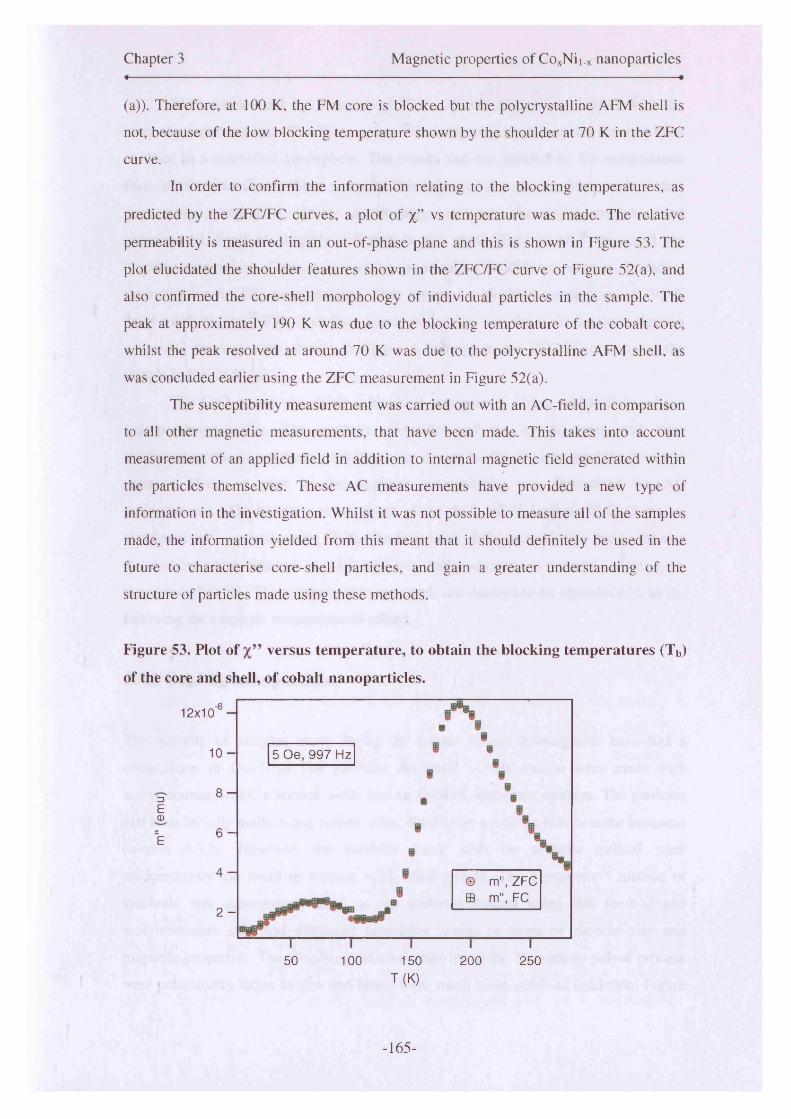
Chapter 3 Magnetic properties of CoxNii.x nanoparticlesa «
(a)). Therefore, at 100 K, the FM core is blocked but the polycrystalline AFM shell is
not, because of the low blocking temperature shown by the shoulder at 70 K in the ZFC
curve.
In order to confirm the information relating to the blocking temperatures, as
predicted by the ZFC/FC curves, a plot of %” vs temperature was made. The relative
permeability is measured in an out-of-phase plane and this is shown in Figure 53. The
plot elucidated the shoulder features shown in the ZFC/FC curve of Figure 52(a), and
also confirmed the core-shell morphology of individual particles in the sample. The
peak at approximately 190 K was due to the blocking temperature of the cobalt core,
whilst the peak resolved at around 70 K was due to the polycrystalline AFM shell, as
was concluded earlier using the ZFC measurement in Figure 52(a).
The susceptibility measurement was carried out with an AC-field, in comparison
to all other magnetic measurements, that have been made. This takes into account
measurement of an applied field in addition to internal magnetic field generated within
the particles themselves. These AC measurements have provided a new type of
information in the investigation. Whilst it was not possible to measure all of the samples
made, the information yielded from this meant that it should definitely be used in the
future to characterise core-shell particles, and gain a greater understanding of the
structure of particles made using these methods.
Figure 53. Plot of versus temperature, to obtain the blocking temperatures (Tb)
of the core and shell, of cobalt nanoparticles.
12x10
1 0 - 5 Oe, 997 Hz
8 -13ECD
6 -E
4 -© m", ZFC ffl m", FC
2 -
50 100 150 200 250T (K)
-165-

Chapter 3 Magnetic properties of CoxNii.x nanoparticles
Work carried out by Tracy et al 38 has shown distinct similarities with the
research carried out here. The Tracy group 38 investigated cobalt particles that had been
oxidised in a controlled atmosphere. The results that are referred to, for comparisons
sake, are the partially oxidised samples. It is expected that the particles made in this
investigation possess a similar morphology, although the drying methods and
experimental detail are slightly different in this case. Tracy et al 38 measured the
blocking temperature of cobalt, by looking for a peak in the ZFC temperature-dependent
magnetisation for DC measurements. They reported a blocking temperature of 195 K
for the cobalt core. This correlates nicely with what was found in this investigation
using AC measurements shown in Figure 53 alongside the ZFC/FC curves for the
sample in Figure 52 (a).
The CoO grains also have a blocking temperature above which they behave
superparamagnetically. The random orientation of the crystal axes and their
antiferromagnetic nature means that the grains cannot be directly measured for magnetic
moment. Hence, a peak in the in-phase susceptibility was determined via AC
measurements. The blocking temperature of the shell, which consists of CoO grains,
was found to be approximately 70 K. The broadness of the peaks is also due to the size
distribution, which although is not completely uncharacteristic in materials of this type,
is particularly wide. The synthesis was repeated, and deemed to be reproducible, as the
following the magnetic measurements tallied.
3.5.2 CogoNi2o nanoparticles
The majority of samples made during the course of this investigation have had a
composition of Co8oNi2o- The particles described in this section were made with
acetylacetonate salts, platinum seeds and an OA/OY surfactant mixture. The particles
that were initially made, using acetate salts, did display exchange bias in some instances
(section 3.3.1). However, the particles made with the acetates method were
predominantly too small to support oxide shell growth. The Yamamuro16 method of
synthesis was subsequently used as the particles formed using this method and
acetylacetonate salts had displayed promising results in terms of particle size and
magnetic properties. The CosoNi2o particles made using the Yamamuro polyol process
were consistently larger in size and hence were much more stable to oxidation. Figure
-166-
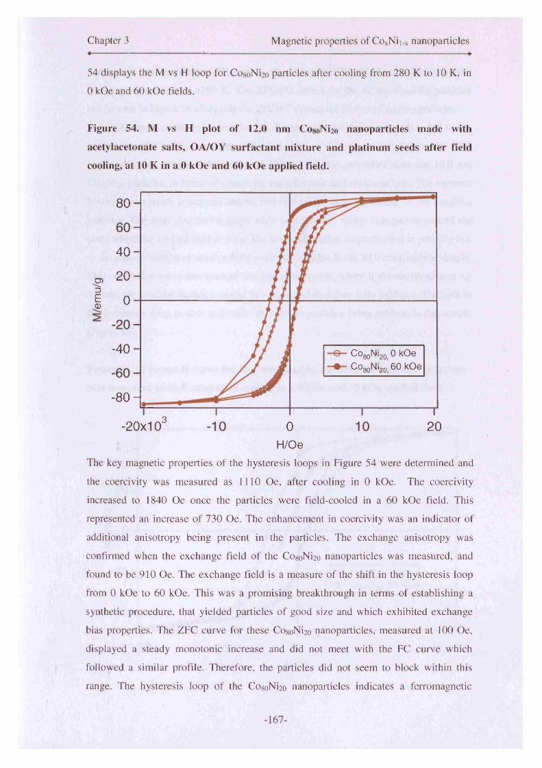
Chapter 3 Magnetic properties of CoxNii.x nanoparticles
54 displays the M vs H loop for CogoNi2o particles after cooling from 280 K to 10 K, in
0 kOe and 60 kOe fields.
Figure 54. M vs H plot of 12.0 nm Co8oNi2o nanoparticles made with
acetylacetonate salts, OA/OY surfactant mixture and platinum seeds after field
cooling, at 10 K in a 0 kOe and 60 kOe applied field.
8 0 -
4 0 -
0 2 0 -
1 o -
- 4 0 - Co80Ni20 0 kOe Co«nNLn 60 kOe
- 8 0 -
3-20x10 0 20H /O e
The key magnetic properties of the hysteresis loops in Figure 54 were determined and
the coercivity was measured as 1110 Oe, after cooling in 0 kOe. The coercivity
increased to 1840 Oe once the particles were field-cooled in a 60 kOe field. This
represented an increase of 730 Oe. The enhancement in coercivity was an indicator of
additional anisotropy being present in the particles. The exchange anisotropy was
confirmed when the exchange field of the Co8oNi2o nanoparticles was measured, and
found to be 910 Oe. The exchange field is a measure of the shift in the hysteresis loop
from 0 kOe to 60 kOe. This was a promising breakthrough in terms of establishing a
synthetic procedure, that yielded particles of good size and which exhibited exchange
bias properties. The ZFC curve for these Co8oNi2o nanoparticles, measured at 100 Oe,
displayed a steady monotonic increase and did not meet with the FC curve which
followed a similar profile. Therefore, the particles did not seem to block within this
range. The hysteresis loop of the Co8oNi2o nanoparticles indicates a ferromagnetic
-167-
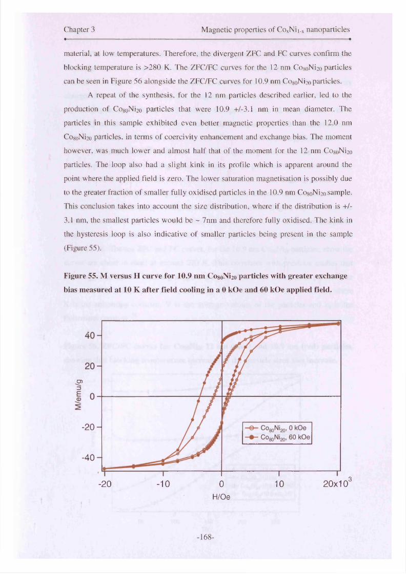
Chapter 3 Magnetic properties of CoxNii.x nanoparticles« *
material, at low temperatures. Therefore, the divergent ZFC and FC curves confirm the
blocking temperature is >280 K. The ZFC/FC curves for the 12 nm Co8oNi2o particles
can be seen in Figure 56 alongside the ZFC/FC curves for 10.9 nm CogoNi2o particles.
A repeat of the synthesis, for the 12 nm particles described earlier, led to the
production of CogoNi2o particles that were 10.9 +/-3.1 nm in mean diameter. The
particles in this sample exhibited even better magnetic properties than the 12.0 nm
CogoNi2o particles, in terms of coercivity enhancement and exchange bias. The moment
however, was much lower and almost half that of the moment for the 12 nm CogoNi2o
particles. The loop also had a slight kink in its profile which is apparent around the
point where the applied field is zero. The lower saturation magnetisation is possibly due
to the greater fraction of smaller fully oxidised particles in the 10.9 nm CogoNi2o sample.
This conclusion takes into account the size distribution, where if the distribution is +/-
3.1 nm, the smallest particles would be ~ 7nm and therefore fully oxidised. The kink in
the hysteresis loop is also indicative of smaller particles being present in the sample
(Figure 55).
Figure 55. M versus H curve for 10.9 nm CogoNi2o particles with greater exchange
bias measured at 10 K after field cooling in a 0 kOe and 60 kOe applied field.
O)
-20
-40
,320x10-20 -10 0 10
H/Oe
-168-
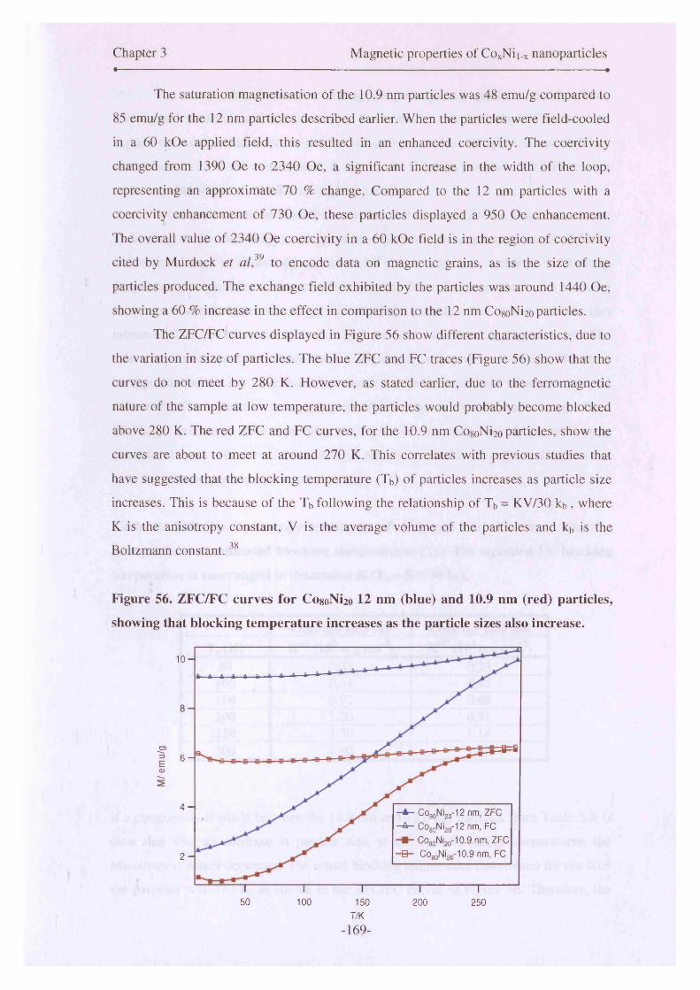
Chapter 3 Magnetic properties of CoxNii-x nanoparticles« — ——------------------------------------------------------------------------------------------- •
The saturation magnetisation of the 10.9 nm particles was 48 emu/g compared to
85 emu/g for the 12 nm particles described earlier. When the particles were field-cooled
in a 60 kOe applied field, this resulted in an enhanced coercivity. The coercivity
changed from 1390 Oe to 2340 Oe, a significant increase in the width of the loop,
representing an approximate 70 % change. Compared to the 12 nm particles with a
coercivity enhancement of 730 Oe, these particles displayed a 950 Oe enhancement.
The overall value of 2340 Oe coercivity in a 60 kOe field is in the region of coercivity
cited by Murdock et al,39 to encode data on magnetic grains, as is the size of the
particles produced. The exchange field exhibited by the particles was around 1440 Oe,
showing a 60 % increase in the effect in comparison to the 12 nm Co8oNi2o particles.
The ZFC/FC curves displayed in Figure 56 show different characteristics, due to
the variation in size of particles. The blue ZFC and FC traces (Figure 56) show that the
curves do not meet by 280 K. However, as stated earlier, due to the ferromagnetic
nature of the sample at low temperature, the particles would probably become blocked
above 280 K. The red ZFC and FC curves, for the 10.9 nm CogoNi2o particles, show the
curves are about to meet at around 270 K. This correlates with previous studies that
have suggested that the blocking temperature (Tb) of particles increases as particle size
increases. This is because of the Tb following the relationship of Tb = KV/30 kb, where
K is the anisotropy constant, V is the average volume of the particles and kb is the
Boltzmann constant.38
Figure 56. ZFC/FC curves for Co8oNi2o 12 nm (blue) and 10.9 nm (red) particles,
showing that blocking temperature increases as the particle sizes also increase.
1 0 -
#=ao>1 6- <D
4 - - A - COgoNijg-^ nm, ZFC
—A— Co80Ni20-12 nm, FC
COgoNi.,0-10.9 nm, ZFC
1Q 1 COnoNion‘10.9 nm, FC2 -
50 100 150 200 250T/K
-169-

Chapter 3 Magnetic properties of CoxNii_x nanoparticles
The relationship between blocking temperature and particle size has been
investigated in detail, and the correlation between the two is evident. This helps to
explain the relationship between the two CogoNi2o samples with particles of different
mean diameters and the convergence in the 12 nm CogoNi2o particle’s ZFC/ FC curves
occurring after that of the 10.9 nm CogoNi2o particles.
A comparison can be made between the 10.9 nm and 12.0 nm CogoNi2o particles
by looking at their anisotropy constants at estimated blocking temperatures. Table 3
shows the change in anisotropy constant (Keff) with respect to a change in blocking
temperature for particles with a constant volume. Table 5 shows that as the blocking
temperature increases, the anisotropy constant for a particle with constant volume, also
increases. This is shown for both the 10.9 nm and 12.0 nm CogoNi2o particles. The
anisotropy values quoted in Table 5 and 6 are actually effective anisotropies. This must
be stipulated before any conclusions are made on the basis of these values, as the total
anisotropy of nanoparticles is a component of the magnetocrystalline anisotropy ( from
CoxNii.x), exchange anisotropy (from alloy/oxide interface and shape anisotropy
(particle shapes). However, this exercise serves to model the expected values for
particles of these particular volumes.
Table 5. Effective anisotropy constants of 10.9 nm and 12.0 nm, Co8oNi2o
nanoparticles at estimated blocking temperatures (Tb). The equation for blocking
temperature is rearranged to determine K (Tb= KV/30 kb).
Co8oNi2o 10.9 nm Co8oNi2o 12.0 nmTb(K) Keff (105 erg cm 3) Keff (10s erg cm'3)
50 0.31 0.23100 0.61 0.46150 0.92 0.69200 1.20 0.91250 1.50 1.14300 1.80 1.37
If a comparison is made between the 10.9 nm and 12.0 nm particles from Table 5 it is
clear that with an increase in particle size, at the same blocking temperatures, the
anisotropy constant decreases. The actual blocking temperature determined for the 10.9
nm particles was 270 K, as shown in the ZFC/FC curves in Figure 56. Therefore, the
-170-

Chapter 3 Magnetic properties of CoxNii.x nanoparticles
anisotropy constant determined for the 10.9 nm Co8oNi2o particles, using the method
used for calculating K in Table 5 was 1.65 x 10⁵ erg cm'3. The blocking temperature for
the 12.0 nm particles cannot be given definitively, as the ZFC and FC curves for these
particles do not meet. However, an estimate of the blocking temperature can be made,
as it is known that it is definitely greater than 280 K. If the blocking temperature of
these particles is taken as 310 K, this would yield anisotropy of 1.42 x 10⁵ erg cm'3.
The moment of the particles also increases as particle volume increases.40
Effective anisotropy of the sample made in this study are calculated and shown in Table
4. Coercivity and saturation magnetisation are used to estimate Keff using the
relationship, (He Ms )/2 = Keff in this particular table.
Table 6. Effective anisotropy calculated using the coercivity and saturation
magnetisations of the CosoNho nanoparticles ((Hc Ms )/2 = Keff). The exchange field
of the 10.9 nm and 12.0 nm particles are also shown.
Co8oNi2o 10.9 nm Co8oNi2o 12.0 nm0 kOe Held 60 kOe field 0 kOe field 60 kOe field
Hc/Oe 1390 2340 1110 1840Mg/emu/g 48 48 85 85
Kefr/ 105 erg cm'3 0.33 0.56 0.47 0.78H^/Oe / 1440 / 910
The effective anisotropies calculated in Table 6 yielded interesting results. Table 6
shows the effects of oxidation of the smaller particles on the anisotropy and gives a
measure of the strength of the exchange anisotropy in the particles. This is shown by
comparing the effective anisotropies of the particles are field-cooling in 0 kOe or 60
kOe.
The smaller particles in the 10.9 +/-3.1 nm sample, as mentioned earlier, were
probably fully oxidised as they would have had a mean diameter of ~7 nm. If the size
distribution in the 12.0 +/- 3.4 nm sample is taken into consideration, it is likely that
some of the smaller particles will have oxidised, however less so than with the 10.9 nm
particles. This is reflected in the difference in effective anisotropies, between the values
in Table 5 and the values determined in Table 6, from the coercivities and saturation
magnetisations of both sets of particle. The 270 K blocking temperature elucidated from
-171-

Chapter 3 Magnetic properties of CoxNii_x nanoparticles
the ZFC/FC plots is quite high. Partial oxidation of metallic nanoparticles significantly
increases the blocking temperature as shown in the study by Tracy et a l 38 into cobalt
nanoparticles. The blocking temperature is also linearly proportional to the volume,
therefore the standard deviation obtained from TEM in terms of particle size can be
translated into a standard deviation of 65 K, in the blocking temperature. These factors
would have all contributed to the variations in blocking temperature.
The exchange fields cited in Table 6 are for the particles measured at 10 K after
field-cooling in a 60 kOe field. The exchange coupling in the 10.9 nm and 12.0 nm
CogoNi2o particles becomes much stronger as particle diameter decreases. This was also
observed in CogoNi2o particles of different sizes made by Luna et al 6 using a different
version of the polyol method.
Previous work carried out by Luna et al 6’7 and Viau et a l3> 4 had also focussed
upon the CogoNi2o composition. However, the objectives in those particular studies were
different to those expressed in this investigation. Luna et al 6’ 7 reported a saturation
magnetisation for 10 nm Co8oNi2o particles of approximately 45 emu/g. By comparison
the 10.9 nm particles made in this study yielded a 48 emu/g saturation point. This
compared well with the literature.6 The mean diameter for another Co8oNi2o sample of
particles, made in this study, was 12.0 +/- 3.4 nm. The magnetic moment for those
particles was determined to be 85 emu/g at 10 K. Therefore, the results achieved here
proved to be promising. This significantly larger moment was probably due to good
crystallinity of the particles. Uniformity of alloying in the Co8oNi2o nanoparticles may
have also contributed to the good magnetic moment exhibited. EELS studies carried out
on the sample also evidenced the relative compositions between cobalt and nickel being
approximately 4:1 in an individual core-shell nanoparticle. The larger particle diameter
could have also contributed to the greater moment, as this has been shown in the
literature to occur with various other metallic nanoparticles as particle size increases.
Metallic nanoparticle crystallinity has been investigated previously, and some
research groups have induced greater crystallinity in nanoparticles by annealing them,
so that the moment increases. The theoretical saturation magnetisation values for bulk
cobalt and bulk nickel are 161 emu/g 38 and 55 emu/g 41 respectively. Therefore, by
mixing the two elements, a magnetic moment between these values is likely. The size
and hence oxidation of the particles is critical to the properties which have been
described thus far, especially properties such as the moment. The broad size distribution
of the particles, formed in this particular study has contributed to the variations in
-172-

Chapter 3 Magnetic properties of CoxNii.x nanoparticles
moment, between samples of identical composition. Fractions of small oxidised
superparamagnetic particles dispersed throughout a sample of core-shell ferromagnetic
nanoparticles leads to a lower moment. This phenomenon also gives rise to the penguin
shaped hysteresis loops that have been observed. These arise due to the smaller particles
being superparamagnetic at 10 K.
A model proposing the ideal conditions, for future ultra-high-density recording
media was detailed in 1992, by Murdock et al 39 from the Hewlett Packard research
laboratories in California. At that time, they had offered an insight into routes towards
achieving 10 Gb/in in media. They had discussed the need for uniform particles with a
mean diameter of 8-10 nm or less, and a high He of approximately 2500 Oe was needed,
to be able to encode data in individual magnetic grains. More recently, authors have
sought to synthesise particles that self-organise into arrays and deliver an increase in
magnetic recording densities, of between 100 Gb/in2 and 1 Tb/ in2. 25,42 A coercivity, in
this particular region, was attained by the CogoNi2o nanoparticles with a diameter of 10.9
nm in this study by comparison.
Generally speaking, the majority of particles synthesised in this study have been
of the Co8oNi2o composition. Partly due to the fact that any similar work carried out in
this area, has usually incorporated the study of this particular composition. Therefore,
comparisons could be made between work carried out here, with other research work
undertaken in this field. The size of particles does have a major influence on the
magnetic properties, as has been seen in this study. It impacts upon the overall structure
of the particles in terms of whether or not they form an oxide shell or completely
oxidise, in addition to affecting properties such as the blocking temperature.
Composition can also affect the blocking temperature of particles, as shown in a
study by Sangregorio et al.43 They used the sol-gel technique to make CoxNii.x particles
and showed that increasing the nickel content in particles lowers the blocking
temperature significantly. They had found that by adding just 20 % nickel to the
composition of pure cobalt particles, the blocking temperature would be reduced to
approximately 90 K from around 225 K for pure cobalt. Although this has thus far not
been the case if a comparison is made between the cobalt particles described in section
3.5.1, when compared to the CogoNi2o described in this section, these variations can be
attributed to the inconsistencies in particle size between the different compositions. If
the sizes were identical, it would much easier to build a picture of such properties, in a
conclusive manner. This can be achieved by controlling the nucleation of the particles
-173-

Chapter 3 Magnetic properties of CoxNii.x nanoparticles
better, and would form the basis of any future investigations.
3.5.3 Co60Ni4o nanoparticles
Co6oNi4o particles, made using the polyol method of synthesis, also exhibited interesting
properties. There was slight bimodality exhibited in the distribution of the particles
made of this composition. The mean diameter determined for the larger particles was
10.8 +/-1.9 nm, with the smaller particles yielding a mean of 6.4 +/- 0.9 nm. These
particles yielded a saturation magnetisation of 38 emu/g.
Figure 57 (a) M-H loops in 0 kOe and 60 kOe after 10 K field-cooling from 280 K,
for Co6oNLm) nanoparticles made with platinum seeds, OA/OY mixture and
acetylacetonate salts (b) ZFC/FC curves for Co6oNi4o particles with Tb> 280 K.
30
1005
-10
-20
-30
.3 10 20-20x10' -10 0Applied field (Oe)
8 -
6 -
CD
CD 4 -
2 -
50 100 200 250150H/Oe
-174-
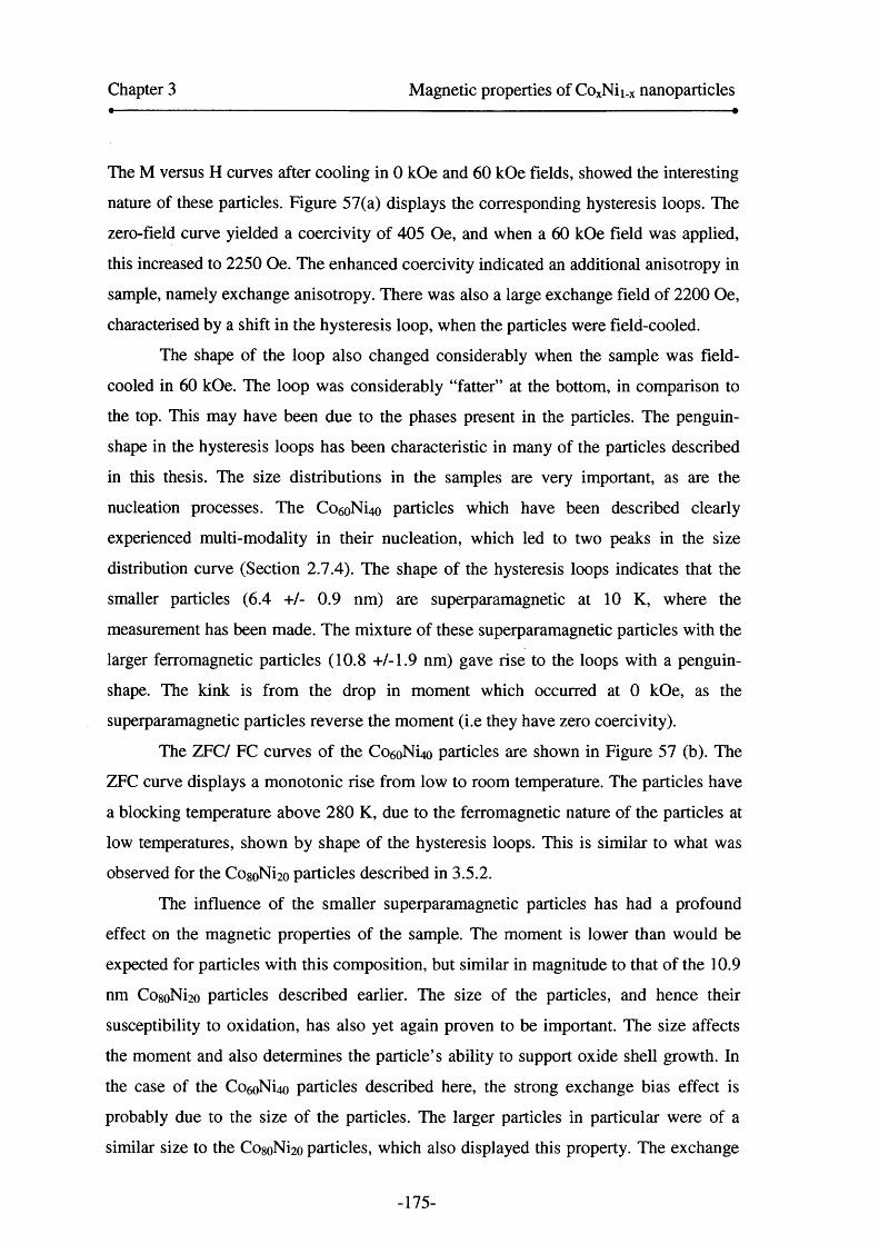
Chapter 3 Magnetic properties of CoxNii.x nanoparticles
The M versus H curves after cooling in 0 kOe and 60 kOe fields, showed the interesting
nature of these particles. Figure 57(a) displays the corresponding hysteresis loops. The
zero-field curve yielded a coercivity of 405 Oe, and when a 60 kOe field was applied,
this increased to 2250 Oe. The enhanced coercivity indicated an additional anisotropy in
sample, namely exchange anisotropy. There was also a large exchange field of 2200 Oe,
characterised by a shift in the hysteresis loop, when the particles were field-cooled.
The shape of the loop also changed considerably when the sample was field-
cooled in 60 kOe. The loop was considerably “fatter” at the bottom, in comparison to
the top. This may have been due to the phases present in the particles. The penguin-
shape in the hysteresis loops has been characteristic in many of the particles described
in this thesis. The size distributions in the samples are very important, as are the
nucleation processes. The Co6oNi4o particles which have been described clearly
experienced multi-modality in their nucleation, which led to two peaks in the size
distribution curve (Section 2.7.4). The shape of the hysteresis loops indicates that the
smaller particles (6.4 +/- 0.9 nm) are superparamagnetic at 10 K, where the
measurement has been made. The mixture of these superparamagnetic particles with the
larger ferromagnetic particles (10.8 +/-1.9 nm) gave rise to the loops with a penguin-
shape. The kink is from the drop in moment which occurred at 0 kOe, as the
superparamagnetic particles reverse the moment (i.e they have zero coercivity).
The ZFC/ FC curves of the Co6oNi4o particles are shown in Figure 57 (b). The
ZFC curve displays a monotonic rise from low to room temperature. The particles have
a blocking temperature above 280 K, due to the ferromagnetic nature of the particles at
low temperatures, shown by shape of the hysteresis loops. This is similar to what was
observed for the CogoNi2o particles described in 3.5.2.
The influence of the smaller superparamagnetic particles has had a profound
effect on the magnetic properties of the sample. The moment is lower than would be
expected for particles with this composition, but similar in magnitude to that of the 10.9
nm CogoNi2o particles described earlier. The size of the particles, and hence their
susceptibility to oxidation, has also yet again proven to be important. The size affects
the moment and also determines the particle’s ability to support oxide shell growth. In
the case of the Co6oNi4o particles described here, the strong exchange bias effect is
probably due to the size of the particles. The larger particles in particular were of a
similar size to the CosoNi2o particles, which also displayed this property. The exchange
-175-

Chapter 3 Magnetic properties of CoxNii.x nanoparticles
bias effect shown by the Co6oNi4o particles was stronger than that of the larger and
similar sized 12.0 nm and 10.9 nm Cosobfoo particles. Although 40 % nickel had been
doped in, the particles showed an exchange bias that was comparable to the exchange
field of the pure cobalt nanoparticles, made by similar methods and described in section
5.5.1 of Chapter 5. This was due to the size of the cobalt nanoparticles, overall, being
smaller than that of the Co6oNi4o particles described here. The probability of the larger
Co6oNi4Q particles sample supporting oxide shell growth was greater. Although some of
the larger particles in the cobalt sample could support the core-shell model, the majority
were susceptible to oxidation. These slight variations in size can influence a particle’s
oxidation, due to the amount of surface area exposed to air.
3.5.4 Co5oNi5o nanoparticles
The results from this particular composition of particles proved to be very interesting,
due to the 1: 1 mix of nickel and cobalt precursors, and hence the supposed effect this
would have on the magnetic effects of the particles. The bimodal distribution of particle
sizes had an influence on the moment due to the presence of smaller particles
throughout the sample. The slightly elevated levels of oxygen, as evidenced by EDX,
also indicate that smaller particles may have oxidised more so than in other samples.
The saturation magnetisation of the C050N150 particles synthesised in our study
was determined to be 57 emu/g using SQUID measurements. If compared to the curve
of best fit that was experimentally determined by Toneguzzo et al33 for saturation
magnetisation of C o5oNi5o particles (Figure 58), this value correlated with that
determined by their research group. The value obtained by carrying out this calculation
was approximately 58 emu/g. The curve of best fit for the Co5oNi5o (white squares trace)
was extrapolated to a reciprocal radius value of 0.159 nm'1. This value represented an
average diameter of 12.6 nm (radius = 6.3 nm) for the particles, which was calculated
through TEM analysis. The calculation served as an excellent first approximation, as the
values for moment were almost identical, despite the multimodality in size distribution
of the particles made in our study (section 2.7.5).
-176-
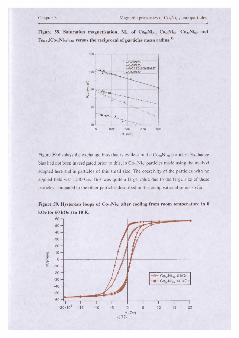
Chapter 3 Magnetic properties of CoxNii_x nanoparticles
Figure 58. Saturation magnetisation, Ms, of Co8oNi2o, CosoNi5o, CozoNigo and
Fe0.13tCo50Ni50J0.87 versus the reciprocal of particles mean radius.33
160
• Co80Ni20 0 Co50Ni50x FeO 13{Co50Ni50]0 87 a Co20Ni80135
I110
0 002 004 006 008R ' (nm ’)
Figure 59 displays the exchange bias that is evident in the CosoNiso particles. Exchange
bias had not been investigated prior to this, in CosoNiso particles made using the method
adopted here and in particles o f this small size. The coercivity of the particles with no
applied field was 1240 Oe. This was quite a large value due to the large size of these
particles, compared to the other particles described in this compositional series so far.
Figure 59. Hysteresis loops of CosoNiso after cooling from room temperature in 0
kOe (or 60 kOe) to 10 K.60 - i
5 0 -
40 -
3 0 -
20 -
D> 1 0 “3E 0 -<D
^ - 1 0 -
- 2 0 -
O COgoNigg, 0 kOe Co50Ni50, 60 kOe- 3 0 -
- 4 0 -
- 5 0 -
-60 - I.3 20-10 10 15-20x10 -15 •5 0 5
H (Oe) -177-

Chapter 3 Magnetic properties of CoxNii_x nanoparticles
In the literature, single domain ferromagnetic particles exhibit an increase in
coercivity as particle volume increases. This relationship continues to a maximum, after
which the coercive force decreases due to the particle volume being too great for it to be
single-domain, and multidomain particles become apparent. The coercivity decreases as
the particle volume is reduced, until the particle becomes superparamagnetic (Hc).40,44
These particles therefore exhibit single-domain behaviour.
The coercivity of the CosoNiso particles increased to 2280 Oe, when they were
field-cooled to 10 K in a 60 kOe field. The exchange field for these particles was 1290
Oe. The shift in the hysteresis loop, due to exchange bias, was slightly smaller than
what has been seen for other alloys in this series with a higher abundance of cobalt in
their compositions. This was a significant result, as the Co4oNi6o alloy particles that are
discussed in section 3.5.5, displayed a larger exchange bias. However, this may be again
due to the sizes of the particles formed. The particles formed with a composition of
Co4oNi6o are marginally larger, 13.6 nm compared with the 12.6 nm determined here,
for the CosoNiso particles.
The ZFC and FC curves for the Co5oNi5o particles, displayed a monotonic rise up
to 280 K, where a peak appeared to form. The ZFC and FC curves also began to
converge although a clear bifurcation point was not evident. The particles again showed
evidence of blocking around room temperature, due to the characteristic ferromagnetic
hysteresis loops at low temperature.
Toneguzzo and co-workers 5’ 33 have investigated the magnetic properties of
these CosoNiso particles in the past. Jeyadevan et al35 reported the synthesis of 40 and
100 nm CosoNiso particles which were supposedly core-shell in structure. The particles
made by the Jeyadevan group were annealed to induce oxidation and hence a core-shell
structure.
-178-

Chapter 3 Magnetic properties of CoxNii_x nanoparticles
3.5.5 Co4oNi60 nanoparticles
Co4oNi6o nanoparticles were made using methods described in Chapter 5 and displayed
a bimodal distribution. The particle’s mean diameters were 13.6 nm and 6.7 nm,
respectively, for each set of particles across the bimodally distributed sample. The
magnetic saturation point determined for these particles was 42 emu/g. A repeat
synthesis of the Co4oNi6o particles yielded a saturation magnetisation of 30 emu/g. The
second Co4oNi6o sample was much larger in size, with a mean diameter of 17.4 nm.
The 13.6 nm particles displayed a larger moment, but were much smaller in size.
This conflicts with the results that have been observed thus far. It is normally the case
that the larger the particles, the greater the saturation magnetisation will be .33 However,
in the scheme of things the difference between the saturations points is relatively small.
The difference could have arisen due to the contribution made by the oxide shell.
The shape of the loop was again penguin-shaped, with a thinner top portion and
a fatter bottom portion. The 13.6 nm particles displayed exchange bias and an enhanced
coercivity. The loop’s exchange field was 1590 Oe after field-cooling in a 60 kOe field
at 10 K. The coercivity increased from 1080 Oe in a 0 kOe field, to 2220 Oe in a 60
kOe field, after field-cooling. The second sample, with 17.4 nm sized particles also
exhibited exchange bias with an exchange field of 1760 Oe. This was an improvement
upon the exchange field shown by the 13.6 nm particles. The enhanced coercivity, after
the application of a magnetic field, for the 17.4 nm particles was an increase of 560 Oe
(0 kOe) to 1840 Oe (60 kOe).
The ZFC and FC curves, for the samples with different 13.6 and 17.4 nm mean
diameters respectively, were very similar. Both did not display blocking temperatures,
as the convergence of the ZFC and FC curves did not occur. The plots also both
illustrated the beginnings of peak formation in the ZFC curve, at around 280 K. These
results showed that exchange bias effect has slightly increased as the nickel
concentration in the particles has been increased. This result showed that the exchange
field of the particles were following a general trend of decreasing over the whole series,
although the results are variable and would need further tests to confirm this
relationship if the particles were all of the same size, and followed a monomodal
distribution.
-179-

Chapter 3 Magnetic properties of CoxNii_x nanoparticles» #
3.5.6 Co2oNi80 nanoparticles
As has been discussed in chapter 2 , Co2oNi8o particles were made with both acetate and
acetylacetonate salts. The particles that were synthesised using the acetate salts, were
large in size and hence it was believed they would be large enough to support a
passivating oxide layer. However, subsequent magnetic measurements that were carried
out, painted another picture altogether. Figure 60 displays the hysteresis measurement
for Co2oNigo particles made with acetate salts, oleic acid and platinum seeds.
Figure 60. Hysteresis loop of Co2oNiso particles, with a mean diameter of 16.2 nm,
made using acetate salts, oleic acid and platinum seeds after field-cooling to 10 K
in a 60 kOe field.
2 -
10 K
fEQ)
Co20Ni80-acetate salts, 60 kOe
-2 -
3-20x1 O' 0-10 10 20H (Oe)
The magnetic moment of the particles was extremely low (3 emu/g). The multi-grain
structure that was evident, in the particles, meant that it was highly possible that smaller
particles coalesced to form the larger particle, resulting in a polycrystal. This could have
contributed to the low moment and lack of exchange bias. The acetate salts method was
deemed to be ineffective, as was the sole use of oleic acid as a surfactant, early on
during this investigation. Therefore, later syntheses of the Co2oNigo particles, using
acetylacetonate salts, OA/OY surfactant mixture and platinum seeds, were implemented
-180-
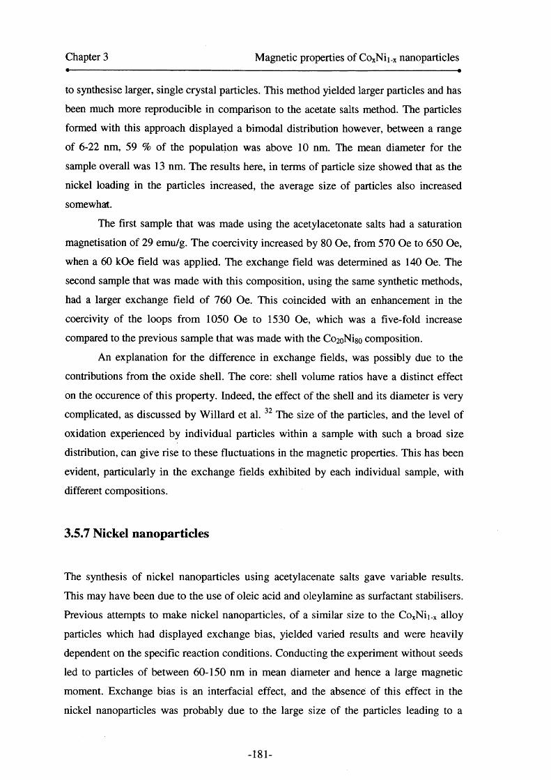
Chapter 3 Magnetic properties of CoxNii.x nanoparticles
to synthesise larger, single crystal particles. This method yielded larger particles and has
been much more reproducible in comparison to the acetate salts method. The particles
formed with this approach displayed a bimodal distribution however, between a range
of 6-22 nm, 59 % of the population was above 10 nm. The mean diameter for the
sample overall was 13 nm. The results here, in terms of particle size showed that as the
nickel loading in the particles increased, the average size of particles also increased
somewhat.
The first sample that was made using the acetylacetonate salts had a saturation
magnetisation of 29 emu/g. The coercivity increased by 80 Oe, from 570 Oe to 650 Oe,
when a 60 kOe field was applied. The exchange field was determined as 140 Oe. The
second sample that was made with this composition, using the same synthetic methods,
had a larger exchange field of 760 Oe. This coincided with an enhancement in the
coercivity of the loops from 1050 Oe to 1530 Oe, which was a five-fold increase
compared to the previous sample that was made with the Co2oNigo composition.
An explanation for the difference in exchange fields, was possibly due to the
contributions from the oxide shell. The core: shell volume ratios have a distinct effect
on the occurence of this property. Indeed, the effect of the shell and its diameter is very
complicated, as discussed by Willard et al. 32 The size of the particles, and the level of
oxidation experienced by individual particles within a sample with such a broad size
distribution, can give rise to these fluctuations in the magnetic properties. This has been
evident, particularly in the exchange fields exhibited by each individual sample, with
different compositions.
3.5.7 Nickel nanoparticles
The synthesis of nickel nanoparticles using acetylacenate salts gave variable results.
This may have been due to the use of oleic acid and oleylamine as surfactant stabilisers.
Previous attempts to make nickel nanoparticles, of a similar size to the CoxNii_x alloy
particles which had displayed exchange bias, yielded varied results and were heavily
dependent on the specific reaction conditions. Conducting the experiment without seeds
led to particles of between 60-150 nm in mean diameter and hence a large magnetic
moment. Exchange bias is an interfacial effect, and the absence of this effect in the
nickel nanoparticles was probably due to the large size of the particles leading to a
-181-
Chapter 3 Magnetic properties of CoxNii_x nanoparticles
relatively small percentage of atoms at the interface.
The nickel particles that were synthesised, following modifications to the
synthesis, displayed the largest mean diameter of all the particles that were made
throughout the study. The particles had a mean diameter of 16.1 +/2.8 nm. The structure
of the nickel particles varied somewhat in comparison to the pure cobalt and CoxNii.x
alloy particles that have been synthesised throughout this study. The particles appeared
to be single crystal and of a larger size (section 2.7.8). However, they did not respond to
a magnet at room temperature and this indicated they were likely to be fully oxidised,
The nickel nanoparticles made here, displayed very small coercivities in 0 kOe
and 60 kOe fields which could not be distinguished from zero, and did not display
exchange bias. This was also the case with the repeat syntheses that were carried out.
The saturation magnetisation was extremely low at 1.7 emu/g. The particles behave as
paramagnets, because the magnetic moment exhibited a linear change with respect to
the applied magnetic field in a hysteresis loop measured at 10 K. The form of the ZFC
and FC curves was a monotonic decrease. The nickel particles, therefore appeared to be
fully oxidised, maybe as a consequence of poor surface coating with the OA/OY
surfactant, in comparison to the cobalt containing samples.
The magnetic properties found in this study, correlated very well with results
from a similar study of Ni and NiO nanoparticles, carried out by Park et al.45 Thermal
decomposition of a nickel-oleylamine complex had previously been used by Park45, as a
method of synthesising Ni and NiO particles, with trioctylphosphine introduced in a
further surfactant addition, after nucleation has occurred. The laser ablation technique
has also been employed in the past, to make core-shell Ni/NiO nanoparticles from
between 5-20 nm in mean diameter, by Seto et a l46 Low moment in NiO nanoparticles
was observed in an investigation by Park et a l45 where particles of between 2-7 nm
exhibited moments of approximately the same order as those observed in this study.
In Park’s study, magnetic measurements carried out below 10 K yielded
blocking temperatures of 2 K and 5 K, for the respective 2 nm and 7 nm NiO particles.
In comparison, the ZFC/FC curves for the Ni particles made using polyol methods did
not display any signs of features such as a peak, although the measurements began at 10
K and so it would be interesting to measure hysteresis loops at 2 and 5 K to eliminate
the existence of any hysteresis.
-182-
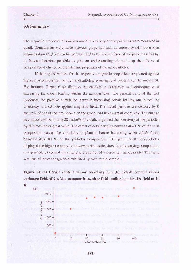
Chapter 3 Magnetic properties of CoxNii.x nanoparticles. . . — . . — — ♦
3.6 Summary
The magnetic properties of samples made in a variety of compositions were measured in
detail. Comparisons were made between properties such as coercivity (Hc), saturation
magnetisation (Ms) and exchange field (He) to the composition of the particles (CoxNii_
x). It was therefore possible to gain an understanding of, and map the effects of
compositional change on the intrinsic properties of the nanoparticles.
If the highest values, for the respective magnetic properties, are plotted against
the size or composition of the nanoparticles, some general patterns can be unearthed.
For instance, Figure 61(a) displays the changes in coercivity as a consequence of
increasing the cobalt loading within the nanoparticles. The general trend of the plot
evidences the positive correlation between increasing cobalt loading and hence the
coercivity in a 60 kOe applied magnetic field. The nickel particles are denoted by 0
molar % of cobalt content, shown on the graph, and have a small coercivity. The change
in composition by doping 20 molar% of cobalt, improved the coercivity of the particles
by 80 times the original value. The effect of cobalt doping between 40-60 % of the total
composition causes the coercivity to plateau, before increasing when cobalt forms
approximately 80 % of the particles composition. The pure cobalt nanoparticles
displayed the highest coercivity, however, the results show that by varying composition
it is possible to control the magnetic properties of a core-shell nanoparticle. The same
was true of the exchange field exhibited by each of the samples.
Figure 61 (a) Cobalt content versus coercivity and (b) Cobalt content versus
exchange field, of CoxNii.x nanoparticles, after field-cooling in a 60 kOe field at 10
K(a)
2 5 0 0 -
2000 -
-C 1 5 0 0 -
1000 -
5 0 0 -
0 20 4 0 60 80 100Cobalt content (%)
-183-
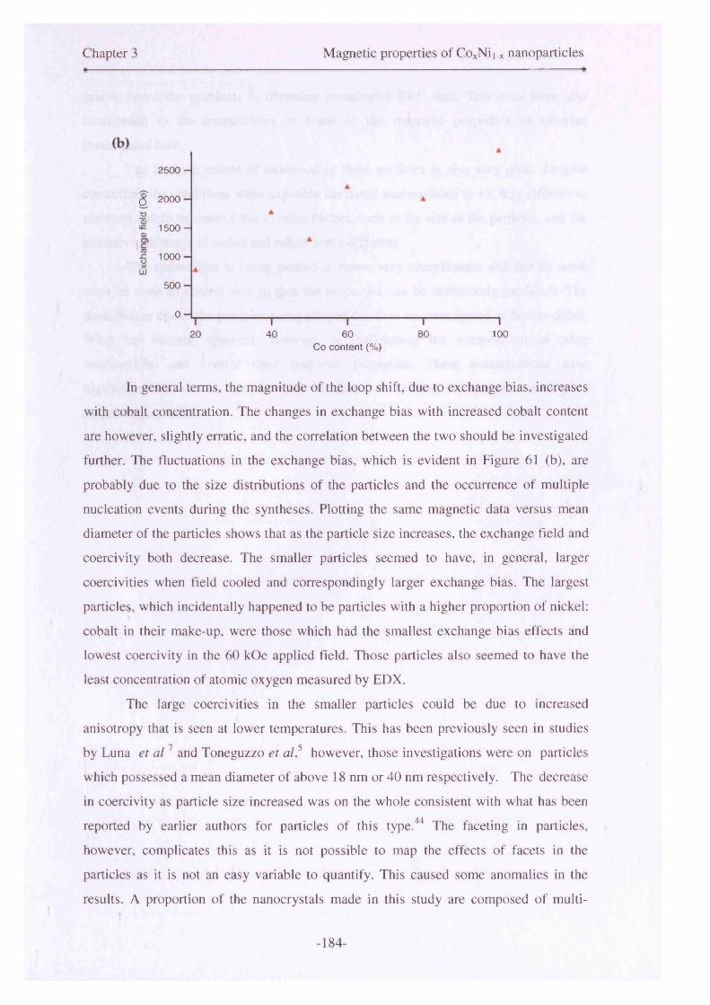
Chapter 3 Magnetic properties of CoxNii_x nanoparticles
2500 -
$ 2 0 0 0 -~o1 1500-o
1 1000-LD
500 -
0 -
20 40 60 80 100Co content (%)
In general terms, the magnitude of the loop shift, due to exchange bias, increases
with cobalt concentration. The changes in exchange bias with increased cobalt content
are however, slightly erratic, and the correlation between the two should be investigated
further. The fluctuations in the exchange bias, which is evident in Figure 61 (b), are
probably due to the size distributions of the particles and the occurrence of multiple
nucleation events during the syntheses. Plotting the same magnetic data versus mean
diameter of the particles shows that as the particle size increases, the exchange field and
coercivity both decrease. The smaller particles seemed to have, in general, larger
coercivities when field cooled and correspondingly larger exchange bias. The largest
particles, which incidentally happened to be particles with a higher proportion of nickel:
cobalt in their make-up, were those which had the smallest exchange bias effects and
lowest coercivity in the 60 kOe applied field. Those particles also seemed to have the
least concentration of atomic oxygen measured by EDX.
The large coercivities in the smaller particles could be due to increased
anisotropy that is seen at lower temperatures. This has been previously seen in studies
by Luna et a l7 and Toneguzzo et al,5 however, those investigations were on particles
which possessed a mean diameter of above 18 nm or 40 nm respectively. The decrease
in coercivity as particle size increased was on the whole consistent with what has been
reported by earlier authors for particles of this type.44 The faceting in particles,
however, complicates this as it is not possible to map the effects of facets in the
particles as it is not an easy variable to quantify. This caused some anomalies in the
results. A proportion of the nanocrystals made in this study are composed of multi-
-184-
Chapter 3 Magnetic properties of CoxNii_x nanoparticles« •
grains, hence the problems in obtaining meaningful XRD data. This must have also
contributed to the irregularities in some of the magnetic properties of samples
investigated here.
The intricate nature of oxidation in these particles is also very clear. Despite
controlling the conditions when exposing the metal nanoparticles to air, it is difficult to
maintain a definite control due to other factors, such as the size of the particles, and the
oxidative pathways of nickel and cobalt being different.
The system that is being probed is hence very complicated, and further work
must be done to control size so that the properties can be definitively modelled. The
contribution due to the particles being alloyed can then be investigated in further detail.
What has become apparent, however, is that tuning the composition of alloy
nanoparticles can control their magnetic properties. These investigations have
highlighted the various factors which contribute to creating nanoparticles with good
magnetic properties.
-185-

3.6.1 Summary table
Composition Salt Mg/emu/gHc/0kOe
He/60 kOe Hex/ Oe
KeR/105 erg cm'3 Tb/K
Description (ZFC/FC curves) Size/Nm Comments
Co Acet 53 1570 1910 600 0.51 >280 K / /
Co Acac 35 620 2020
"
2000 0.35 -280 K
shoulder in ZFC around 70K, broad peak from
-190K9.4 +/- 1.8 and
5.5 +/- 0.78.8 +/- 2.2
(overall) SB
Co Acac 23 1170 2860 2830 0.33 -270 K
shoulder in ZFC at -40K, and broad peak
at -240K 8.6 +/- 2.4 SB
CogoNi2o Acac » 1110 1840 910 0.78 >280 K
monotonic increase, peak not visible at
280K 12.0+/-3.4 M
CosoNi^ Acac 48 1390 2340 1440 0.56 >280 K
decrease between 10- 20K in ZFC. Curves haven’t met by 280K,
but looks like beginning of peak 10.9 +/-3.1 M
Co6oNi4o Acac 38 720 2250 2200 0.43 >280 K
monotonic increase, curves have not met by
280 K.10.8+/-1.9
| and 6.4 +/- 0.9 BM
Co-soNi o Acac 57 1240 2280 1290 0.65 >280 K
monotonic increase, 1 curves have not met by 1 280K, looks like ZFC 1 peak just beginning I 12.6+/-9.6 MM
Co4()Ni6o Acac 42 1080 2220 1590 0.47 >280 K
ZFC is maybe B beginning to reach peak I
at 280 K; ZFC/FC I 13.6 +/- 2.5 haven't met by 280 K | and 6.7 +/- 1.4 BM
-185-

Composition Salt Mj/emu/g Hc/OT Hc/6T Hex/OeK^/105 erg cm'3 Tb/K
Description (ZFC/FC curves) Size/ Nm Comments
Co4oNi6o Acac 30 560 1850 1760 0.28 >280 K
ZFC and FC haven't met at 280 K; ZFC is begun to reach peak
13.6+/- 2.5 and 6.7+/-1.4 BM
Co2(>Ni8o Acac 29 570 650 140 0.09 -250 K
shoulder in ZFC around 70K, broad peak from -190K
13.0 +/- 3.0 and 7.9 +/- 0.7 BM
Co2oNiso Acac 11 1050 1530 760 0.08 -250 K
ZFC peak -180 K, meets FC around 250
K / /Ni Acac 2 20 20 o | 0.00 / monotonic decrease 16.0 +/- 5.6 M
Key:-
Acac- Acetylacetonate salt
Acet- Acetate salt
SB-Slightly bimodal
M- Monomodal
BM- Bimodal
MM- Multimodal
M-Monomodal
/-TEM analysis failed
-186-
Chapter 3 Magnetic Properties of CoxNii.x nanoparticles
3.7 Conclusions
This chapter has described detailed investigations into the magnetic properties of
CoxNii.x, fully and partially oxidised nanoparticles. The partially oxidised particles have
indicated core-shell texture due to the evidence of an additional anisotropy which has
been induced in the particles. This anisotropy gave rise to shifts in the hysteresis plots
of many of the particles, when they were field-cooled in an applied magnetic field.
Hence, exchange bias was evident in particles that had been synthesised in a manner of
different ways. Exchange bias also caused an enhancement in the coercivity (half
width) of the respective hysteresis loops, when the sample in question was placed in a
magnetic field and cooled. These effects occur in metallic particles which are enveloped
in an oxide shell coating, and were evident in many of the samples that have been
discussed in this chapter.
The investigations showed that in general terms, the exchange bias effect
seemed to decrease as more nickel was doped into the particles. The coercivity of the
particles, with a low concentration of nickel, was also likely to be low in a 60 kOe field,
at 10 K. However, this effect showed evidence of a plateau between cobalt
concentrations of 40-80%. Further investigations would be required to verify this as size
effects seemed to play a major role in magnetic properties of the particles.
Size seemed to affect the blocking temperatures of the particles, where it was
possible to obtain them at all. With increases in particle size, the blocking temperature
also seemed to increase. The size of particles seemed to affect their susceptibility to
oxidation. Smaller particles seemed to tend towards full oxidation, whereas larger
particles could support oxide shell growth. The oxide shell cannot be definitively
claimed to be a “passivating oxide shell” as over time the samples lost their response to
a magnet. This effect was dependent on the sample, however, the particles that
displayed the largest magnitude exchange fields, tended to retain their responsiveness to
a magnet at room temperature for several months. The samples that displayed a weak
exchange bias effect, had oxidised much more quickly. Oxide inclusions within the
shell and gradual oxidation of the core, due to degradation or loss of the surfactant
could be the cause of this.
Interesting results were obtained through the use of AC susceptibility
measurements, and this method enabled the characterisation of the core and shell’s
-187-

Chapter 3 Magnetic Properties of CoxNii_x nanoparticles
respective blocking temperatures. This technique also helped to clarify ambiguous
structural features in the ZFC and FC curves of the cobalt sample. This would be an
interesting method of analysis to develop in the future, and use on similar particles to
elucidate the structural profiles that are evident on occasions, in ZFC and FC plots.
These results have shown improvements, in terms of the exchange bias effects,
previously reported for particles of this size and composition. Limitations with the
nucleation of the particles have presented difficulties in terms of modelling the particles
due to size. However, generic conclusions can be drawn in terms of the size effects. The
great advancement that has been made, through this investigation, is that it has been
proven that the magnetic properties can be tuned by varying the composition of the
particles. This was the key objective that was set out at the beginning of this thesis, and
the studies in this chapter have presented evidence that this can be achieved.
Investigations carried out in Chapter 2, resulted in an optimisation of this process. This
resulted in the advancements that have been documented in this chapter. More work
must be done to improve the synthesis, so that the magnetic properties of the particles
can be modelled, alongside the results achieved from the structural characterisation of
the particles described in Chapter 2. Processes and investigations that could be carried
out to achieve this in the future are highlighted in the next chapter.
-188-

Chapter 3 Magnetic Properties of CoxNii_x nanoparticles
3.8 References
1. Nogues, J., Exchange bias in nanostructures. Physics reports 2005, 422, (3), 65-
117.
2. Meiklejohn, W. H.; Bean, C. P., New Magnetic Anisotropy. Physical Review
1956, 102, (5), 1413-1414.
3. Viau, G.; Toneguzzo, P.; Pierrard, A.; Acher, O.; Fievet-Vincent, F.; Fievet, F.,
Heterogeneous nucleation and growth of metal nanoparticles in polyols. Scripta
Materialia 2001,44, (8-9), 2263-2267.
4. Viau, G.; Fie vet Vincent, F.; Fievet, F., Nucleation and growth of bimetallic
CoNi and FeNi monodisperse particles prepared in polyols. Solid State Ionics
1996, 84, (3-4), 259-270.
5. Toneguzzo, P.; Acher, O.; Viau, G.; Pierrard, A.; Fie vet-Vincent, F.; Fievet, F.;
Rosenman, I., Static and dynamic magnetic properties of fine CoNi and FeCoNi
particles synthesized by the polyol process. Ieee Transactions on Magnetics
1999, 35, (5), 3469-3471.
6. Luna, C.; Morales, M. D.; Serna, C. J.; Vazquez, M., Exchange anisotropy in
Co80Ni20/oxide nanoparticles. Nanotechnology 2004,15, (4), S293-S297.
7. Luna, C.; Morales, M. D.; Serna, C. J.; Vazquez, M., Multidomain to single
domain transition for uniform Co80Ni20 nanoparticles. Nanotechnology 2003,
14, (2), 268-272.
8. Viau, G.; Fie vet Vincent, F.; Fievet, F., Monodisperse iron-based particles:
Precipitation in liquid polyols. Journal of Materials Chemistry 1996, 6, (6),
1047-1053.
9. Chakroune, N.; Viau, G.; Ricolleau, C.; Fievet-Vincent, F.; Fievet, F., Cobalt-
based anisotropic particles prepared by the polyol process. Journal o f Materials
Chemistry 2003, 13, (2), 312-318.
10. Toneguzzo, P.; Viau, G.; Acher, O.; Fievet-Vincent, F.; Fievet, F.,
Monodisperse ferromagnetic particles for microwave applications. Advanced
Materials 1998, 10, (13), 1032-1035.
11. Patel, J., Farrell, D., Pankhurst, Q.A.;Parkin, I. P.„ Particle size and oxidation in
CoNi nanoparticles. Mater Res Soc Symp Proc 2005, 877E S5 8 1-6.
-189-
Chapter 3 Magnetic Properties of CoxNii_x nanoparticles
12. Becker, J. J., Surface Effects on Hysteresis Loop Shapes in High-Coercive-
Force Crystallized Amorphous-Alloys. Ieee Transactions on Magnetics 1982,
18, (6), 1451-1453.
13. Roberts, A. P.; Cui, Y. L.; Verosub, K. L., Wasp-Waisted Hysteresis Loops -
Mineral Magnetic Characteristics and Discrimination of Components in Mixed
Magnetic Systems. Journal o f Geophysical Research-Solid Earth 1995, 100,
(B9), 17909-17924.
14. Tauxe, L.; Mullender, T. A. T.; Pick, T., Potbellies, wasp-waists, and
superparamagnetism in magnetic hysteresis. Journal of Geophysical Research-
Solid Earth 1996,101, (Bl), 571-583.
15. Linford, N., Archaeological applications of naturally occurring nanomagnets. In
Fifth International Conference on Fine Particle Magnetism, Pankhurst, Q., Ed.
2005; Vol. 17, pp 127-144.
16. Yamamuro, S. A., T.;Sumiyama, K.; Uchida, T., Kojima, I.„ Monodisperse
metallic iron nanoparticles synthesized from noncarbonyl complexes. Japanese
Journal o f Applied Physics 2004,44, (No 7A), 4458-4459.
17. Giersig, M.; Hilgendorff, M., Magnetic nanoparticle superstructures. European
Journal o f Inorganic Chemistry 2005, (18), 3571-3583.
18. Stamps, R. L., Mechanisms for exchange bias. Journal o f Physics D-Applied
Physics 2000, 33, (23), R247-R268.
19. Nogues, J.; Schuller, I. K., Exchange bias. Journal o f Magnetism and Magnetic
Materials 1999, 192, (2), 203-232.
20. Nogues, J., Exchange bias in ferromagnetic nanoparticies embedded in an
antiferromagnetic matrix. International journal of nanotechnology 2005, 2, (1-
2), 23-42.
21. Ung, D.; Viau, G.; Ricolleau, C.; Warmont, F.; Gredin, P.; Fievet, F. F., CoNi
nanowires synthesized by heterogeneous nucleation in liquid polyol. Advanced
Materials 2005, 17, (3), 338-344.
22. Ung, D.; Viau, G.; Fievet-Vincent, F.; Herbst, F.; Richard, V.; Fievet, F.,
Magnetic nanoparticies with hybrid shape. Progress in Solid State Chemistry
2005, 33, (2-4), 137-145.
23. Luna, C.; Morales, M. P.; Sema, C. J.; Vazquez, M., Effects of surfactants on
the particle morphology and self-organization of Co nanocrystals. Materials
-190-
Chapter 3 Magnetic Properties of CoxNii_x nanoparticies
Science & Engineering C-Biomimetic and Supramolecular Systems 2003, 23,
(6-8), 1129-1132.
24. Bao, Y. P.; Beerman, M.; Pakhomov, A. B.; Krishnan, K. M., Controlled
crystalline structure and surface stability of cobalt nanocrystals. Journal of
Physical Chemistry B 2005, 109, (15), 7220-7222.
25. Sun, S. H.; Murray, C. B.; Weller, D.; Folks, L.; Moser, A., Monodisperse FePt
nanoparticies and ferromagnetic FePt nanocrystal superlattices. Science 2000,
287, (5460), 1989-1992.
26. Lu, A. H.; Li, W. C.; Matoussevitch, N.; Spliethoff, B.; Bonnemann, H.; Schuth,
F., Highly stable carbon-protected cobalt nanoparticies and graphite shells.
Chemical Communications 2005, (1), 98-100.
27. Bertotti, G., Hysteresis in magnetism: for physicists, material scientists and
engineers. 1998.
28. Chantrell, R. W.; Weller, D.; Klemmer, T. J.; Sun, S.; Fullerton, E. E., Model of
the magnetic properties of FePt granular media. Journal o f Applied Physics
2002, 91, (10), 6866-6868.
29. Darling, S. B.; Bader, S. D., A materials chemistry perspective on
nanomagnetism. Journal o f Materials Chemistry 2005, 15, (39), 4189-4195.
30. Murray, C. B.; Sun, S. H.; Gaschler, W.; Doyle, H.; Betley, T. A.; Kagan, C. R.,
Colloidal synthesis of nanocrystals and nanocrystal superlattices. Ibm Journal of
Research and Development 2001,45, (1), 47-56.
31. Skumryev, V., Stoyanov,S., Zhang,Y., Hadjipanayis, G.,Givord, D., Noguds, J.„
Beating the superparamagnetic limit with exchange bias. Nature 2003,423, 850-
853
32. Willard, M. A.; Kurihara, L. K.; Carpenter, E. E.; Calvin, S.; Harris, V. G.,
Chemically prepared magnetic nanoparticies. International Materials Reviews
2004,49, (3-4), 125-170.
33. Toneguzzo, P.; Viau, G.; Acher, O.; Guillet, F.; Bruneton, E.; Fievet-Vincent,
F.; Fievet, F., CoNi and FeCoNi fine particles prepared by the polyol process:
Physico-chemical characterization and dynamic magnetic properties. Journal of
Materials Science 2000, 35, (15), 3767-3784.
34. Fievet, F.; Lagier, J. P.; Blin, B.; Beaudoin, B.; Figlarz, M., Homogeneous and
Heterogeneous Nucleations in the Polyol Process for the Preparation of Micron
and Sub-Micron Size Metal Particles. Solid State Ionics 1989, 32-3, 198-205.
-191-
Chapter 3 Magnetic Properties of CoxNii_x nanoparticies
35. Jeyadevan, B.; Chinnasamy, C. N.; Perales-Perez, O.; Iwasaki, Y.; Hobo, A.;
Shinoda, K.; Tohji, K.; Kasuya, A., Synthesis and magnetic properties of core
shell structured (NiCo)0(AFM)-NiCo(FM) magnetic nanoparticies. leee
Transactions on Magnetics 2002, 38, (5), 2595-2597.
36. Park, J.; An, K. J.; Hwang, Y. S.; Park, J. G.; Noh, H. J.; Kim, J. Y.; Park, J. H.;
Hwang, N. M.; Hyeon, T., Ultra-large-scale syntheses of monodisperse
nanocrystals. Nature Materials 2004, 3, (12), 891-895.
37. Spasova, M.; Wiedwald, U.; Farle, M.; Radetic, T.; Dahmen, U.; Hilgendorff,
M.; Giersig, M., Temperature dependence of exchange anisotropy in
monodisperse cobalt nanoparticies with a cobalt oxide shell. Journal of
Magnetism and Magnetic Materials 2004,272-276, (Part 2), 1508-1509.
38. Tracy, J. B.; Weiss, D. N.; Dinega, D. P.; Bawendi, M. G., Exchange biasing
and magnetic properties of partially and fully oxidized colloidal cobalt
nanoparticies. Physical Review B 2005, 72, (6).
39. Murdock, E. S., Roadmap for 10 GBit/in2 media- Challenges. IEEE transactions
on magnetics 1992, 28, (5), 3078-3083.
40. Klabunde, K., Nanoscale Materials in Chemistry: Wiley: 2001.
41. Nayak, B. B.; Vitta, S.; Nigam, A. K.; Bahadur, D., Ni and Ni-nickel oxide
nanoparticies with different shapes and a core-shell structure. Thin Solid Films
2006,505,(1-2), 109-112.
42. Sun, S. H.; Murray, C. B., Synthesis of monodisperse cobalt nanocrystals and
their assembly into magnetic superlattices (invited). Journal of Applied Physics
1999, 85, (8), 4325-4330.
43. Sangregorio, C.; Fernandez, C. d. J.; Battaglin, G.; De, G.; Gatteschi, D.; Mattei,
G.; Mazzoldi, P., Magnetic properties of Co-Ni alloy nanoparticies prepared by
the sol-gel technique. Journal of Magnetism and Magnetic Materials 2004, 272-
276, (Supplement 1), E1251-E1252.
44. Sugimoto, T., Monodispersed particles. Elsevier: 2001.
45. Park, J.; Kang, E.; Son, S. U.; Park, H. M.; Lee, M. K.; Kim, J.; Kim, K. W.;
Noh, H. J.; Park, J. H.; Bae, C. J.; Park, J. G.; Hyeon, T., Monodisperse
nanoparticies of Ni and NiO: Synthesis, characterization, self-assembled
superlattices, and catalytic applications in the Suzuki coupling reaction.
Advanced Materials 2005, 17, (4), 429-434.
-192-
Chapter 3 Magnetic Properties of CoxNii.x nanoparticies
46. Sakiyama, K.; Koga, K.; Seto, T.; Hirasawa, M.; Orii, T., Formation of size-
selected Ni/NiO core-shell particles by pulsed laser ablation. Journal of
Physical Chemistry B 2004, 108, (2), 523-529.

Chapter 4 Conclusions and Future Work
4. Conclusions and Future Work
4.1 Conclusions
4.1.1 Synthesis of CoxNii.x nanoparticies
CoxNii_x/CoNiO core-shell nanoparticies have been synthesised using a modified
version of the polyol method. The particles varied in mean diameter from between 6-22
nm. A number of parameters were varied in order to optimise the synthesis, so that it
was possible to fabricate a compositional series of CoxNii_x/CoNiO alloy core-shell
nanoparticies.
The three reaction parameters that were investigated in detail were nucleation
methods, surfactants and composition. The particles were homogeneously nucleated,
and heterogeneously nucleated with platinum seeds, preformed nanoparticies and noble
metal seeds. The heterogeneous nucleation of particles using platinum seeds was the
most effective for controlling particle size in this system. A number of surfactants were
trialled, to test their ability to prevent agglomeration during particle growth, control
-194-

Chapter 4 Conclusions and Future Work
particle size and act as a barrier to prevent oxidation. Investigations into the binding of
the surfactant with the metallic ion species in solution was carried out using UV-Vis
studies. This indicated that the surfactants formed complexes with the metallic ion
species, and it was these complexes that acted as nuclei for subsequent particle growth.
The formation of these complexes was tracked and approximate nucleation and particle
formation temperatures were determined for a range of particles that were synthesised.
The surfactant, which best provided these elements of control and contributed to
the optimisation of the synthesis, was the 1:1 mixture of oleic acid and oleylamine. This
particular mix enabled the formation of particles that were pre-dominantly air-stable and
of a suitable size to warrant oxide shell growth, without oxidation occurring to the core
of the particles. The unusual feature of using this surfactant mixture was the shape of
the particles formed. Some were spherical, however, a large proportional of the particles
produced were triangular, square and rhomboidal in shape. The optimisation of the
synthetic process, in terms of nucleation and surfactant type was then completed, and it
was subsequently possible to vary the composition of the particles.
Characterisation of the particles, and therefore the effectiveness of the synthetic
process, was carried out using a number of techniques. Elemental analysis of the
particles indicated that between 10-25% of the particle’s mass was due to surfactant.
This value was dependent upon how well the surfactant coated the nanoparticle surface
and the ease with which it was washed away. EDX analysis showed that the molar
ratios of precursor material, translated through to the relative atomic ratios of elements
in the nanoparticulate powders.
The particles that were formed were characterised as being core-shell using the
EELS and XPS techniques. EELS displayed a high concentration of oxygen atoms at
the edges of individual particles that were measured. EELS also showed that the relative
ratios between cobalt and nickel varied across a single particle, with the cores being
slightly nickel-rich. This indicated that the nucleation of nickel occurred First during the
synthesis of the particles. There was also oxygen present across the diameter of the
particle, as an oxygen count was measured. The electron beam penetrated through the
top and bottom of the particle and yielded a summed measure of the oxide shell. UV-
Vis studies had also shown that the nucleation of nickel occurs slightly before that of
cobalt.
-195-

Chapter 4 Conclusions and Future Work
XPS studies revealed a higher concentration of oxygen atoms bounds to cobalt
and nickel at the surface of the nanoparticies. After a few etch cycles, the concentration
of oxygen dramatically decreased and the concentration of cobalt bound to nickel, in a
binary alloy was a dominant feature. The profiles of each of the samples that were
measured all indicated core-shell morphology to the particles. This analysis also
indicated evidence of nickel-rich cores, due to the marginally elevated concentration of
nickel atoms, following a few etch cycles. The ratio between the nickel and cobalt
atoms at various etching stages showed that this was more pronounced as the core of the
particle was approached. The core-shell texture in particles was a prerequisite for the
occurrence of exchange bias which is discussed in section 4.1.2.
XPS and XRD indicated that there could have been another oxide phase of
cobalt present at the surface of the particles (C0 2O3). XRD confirmed the dominant fee
phased for the cobalt, nickel, cobalt (II) oxide and nickel (II) oxide. In some samples, it
was also possible to calculate crystallite size using Scherrer analysis. This indicated that
in some samples, particles were composed of multigrains. TEM studies gave further
evidence for this, as the multigrain structure in the individual particles could be seen.
Finally, magnetic TGA studies showed that a solid solution and hence alloyed particles
had been formed, as it was also possible to tune the Curie temperature of the particles.
4.1.2 Magnetic properties of CoxNii.x nanoparticies
The magnetic properties of the CoxNii.x/CoNiO core-shell nanoparticies have also been
investigated in this thesis. Initially, room temperature hysteresis loops were measured of
the samples, and if a large moment was evident and the particles responded to a magnet,
then measurements were made at low temperatures using SQUID magnetometry. Only
the most promising samples had measurements taken to determine whether or not they
displayed the characteristic traits of exchange bias. Exchange anisotropy is
characterised by an enhancement in the coercivity from 0 kOe to 6 kOe at low
temperatures e.g 10 K. The measured hysteresis loop also shifts if exchange anisotropy
is induced from the particles.
Particles that were not sufficiently stabilised, displayed hysteresis loops
characteristic of polycrystalline antiferromagnets, as they oxidised fully to their cores.
This was the case with many of the particles synthesised using oleic acid, oleylamine,
-196-
Chapter 4 Conclusions and Future Work
trioctylphosphine and tributylphosphine as sole surfactants. The particles synthesised
using these surfactants were very small in mean diameter and so became fully oxidised
upon exposure to air. However, the use of oleic acid and oleylamine in tandem,
produced particles that were stabilised to oxidation and aggregation over prolonged
periods of time. The oleic acid and oleylamine formed an amide when heated together,
and this yielded a double-sheathed surfactant stabiliser. The use of this surfactant
mixture allowed the growth of larger particles, in comparison to what had been obtained
using the other sole surfactants. The particles formed using this method, were large
enough to support oxide shell growth. Hence, when magnetic measurements were made
of these particular particles they tended to display exchange bias effects.
When chemical composition of the CoxNii_x particles was varied, it was possible
to correlate some of the magnetic properties of the particles, with the composition.
Measurements made of the exchange fields in samples where the composition was
varied showed that as nickel-loading increased the exchange bias effect was dampened.
The largest exchange field determined for any sample in this study was for the pure
cobalt sample at around 2800 Oe. The addition of 20 % nickel decreases this slightly to
around 2000 Oe. However, the exchange field increases again, slightly for the sample
with 40 % nickel content. Therefore, detailed investigations into this particular range
would be interesting to carry out in the future. Large enhancements in coercivity were
also evident in these samples, which was characteristic of exchange anisotropy in the
particles.
Studies carried out into the blocking temperatures of the particles were less
conclusive. However, there were some encouraging results which suggested a
correlation between particle diameter and blocking temperature. Measurements on two
sets of Cog()Ni2o particles, with different mean diameters, showed that the smaller
particles blocked at a lower temperature than the larger particles. This suggests a
positive correlation between particle size and blocking temperature.
These results coupled with the physical and structural characterisation described
earlier, indicated the highly interesting properties of particles within the CoxNii.x
system. This study has shown that it is possible to tune the magnetic properties, and in
particular control the exchange bias in a systematic fashion, by altering the composition.
-197-
Chapter 4 Conclusions and Future Work
4.2 Future work
The key to any future work, which follows on from what has been achieved in this
study, is to establish more control over the size of the particles by establishing rigid
control over their nucleation. The process that was adopted for the synthesis of these
particles has presented many problems, and by possibly adapting the synthesis a little
further, the benefits could be immense. For example, if the heating of the reactant
solutions were controlled by using a temperature programmable heating mantle, the
effect of the heat rate on nucleation could be much more definitive.
Another improvement which could be made to the synthetic process is the
implementation of all reactions in a glove-box under an atmosphere of argon. This
would severely limit the possibility of oxidation to the core of the smaller particles. It
would also allow particles to be oxidised in a more controlled manner, so that
experiments could be carried out on the native, partially oxidised and fully oxidised
particles. This would enable a detailed picture to be built of the composite alloy
particles and their physical and functional magnetic properties at the various stages of
oxidation. Furthermore, the particles could be stored in their native solutions, in an inert
atmosphere to prolong their life and lengthen the time over which analysis can occur. If
reactions were carried out in such an atmosphere, it may even be possible to experiment
with nickel and cobalt carbonyls to synthesise alloy particles. The thermal
decomposition of carbonyls has been extensively researched, and the nucleation of
particles using this method could be investigated to compare with the polyol reduction
process reported in this thesis.
Studies into the local environment of the particles that have been produced,
using EXAFS would be very interesting, and would provide a great deal of valuable
information about the structure of the particles. HRTEM would also be beneficial for
routine observation work, as it would prove the presence of a core-shell structure. The
ability to resolve lattice planes using this technique could be correlated with XRD
studies, to characterise the phases present across an individual particle.
XPS studies gave very interesting results in this study and it would be
illuminating to build upon the work carried out here, and be able to quantify the depth
of the penetrating beam during the etching process. By determining this, it may be
possible to use this information, along with the particles’ mean diameter to determine
-198-

Chapter 4 Conclusions and Future Work
the oxide shell diameter, in addition to the information already garnered from this
technique.
The magnetic studies carried out in this investigation have yielded useful
information, and helped characterise the functional properties of core-shell CoxNii.
x/CoNiO nanoparticles. Preliminary AC susceptibility studies were carried out, and
given more time, it would have been interesting to analyse some of the other samples
using this technique. This technique helped to shed light on the profile of the ZFC and
FC curves of a specific sample, and could be used in the future to elucidate the structure
in such plots and in turn, characterise core-shell blocking temperatures in the particles.
The work described in this thesis has shown that the magnetic properties of
CoxNii.x/CoNiO nanoparticles can be tuned. This system has been interesting to
characterise, however, if some of the issues discussed in this section are addressed, then
the synthesis and hence the magnetic properties could be further refined in the future.
-199-

Chapter 5 Experimental
5. Experimental
5.1 Introduction
This chapter gives an outline of each of the individual synthetic processes developed
during the course of this investigation, in order to achieve the aims set out at the
beginning of this thesis. An overview of each of the various analytical techniques used
to characterise the particles is given in section 5.7 of this chapter.
Chapter 1 introduced the nucleation theory which forms the basis of polyol
synthesis, in addition to describing the significance of making magnetic nanoparticles
and their various properties. Chapter 2 discussed in great detail the main lines of
investigation that were probed during this study, which included homogenous
nucleation, heterogeneous nucleation, surfactants and compositional variation. The
evolution and developments of the reaction conditions are also described in detail in
Chapter 2. The main objective of the project was to synthesise magnetic nanoparticles
-200-

Chapter 5 Experimental
which displayed the exchange bias functional property. The characterisation of
magnetic properties, and the results obtained from these processes, is documented in
Chapter 3.
5.1.1 Reactant materials
The syntheses were carried out using standard airless Schlenk line techniques under N2.
Absolute ethanol (GPR), toluene (GPR) and hexane (Analar) were used as received, for
the cleaning and washing of nanoparticle solutions before the particles were isolated
and dried for analysis. Dioctyl ether (99.9%), dodecane, decalin, diphenyl ether, 1,2-
hexadecanediol, oleic acid (-90% technical grade), oleylamine (-70% technical grade),
trioctylphosphine oxide, tributylphosphine, cobalt (II) tetrahydrate acetate, nickel (II)
tetrahydrate acetate, cobalt (III) acetylacetonate, nickel (II) acetylacetonate and
platinum (II) acetylacetonate were purchased from Aldrich Chemical Company. These
commercially available reagents were also used as received and no further purification
was carried out to any of the reagents.
5.1.2 Reaction set-up
The synthesis of CoxNii.x particles was carried out using the polyol method. The
particles were made using Schlenk line techniques in an atmosphere of nitrogen, so that
oxygen was excluded during the synthesis and particle oxidation was limited. The
metallic salts, reducing agent, nucleating agent (if used), surfactants and solvent were
all added to a 250 mL two- or three-necked round-bottomed flask prior to the reaction
beginning. The flask and its contents were subsequently evacuated and flushed with
nitrogen in three cycles. The reactant solution was then heated using a heating mantle
with a magnetic stirring function. The particles were stirred, and the temperature of the
reactant solution was measured throughout the synthetic process, with a thermometer
attached to the set-up. A diagram of the apparatus required for a typical polyol synthesis
in this study is shown in Figure 62.
-201-
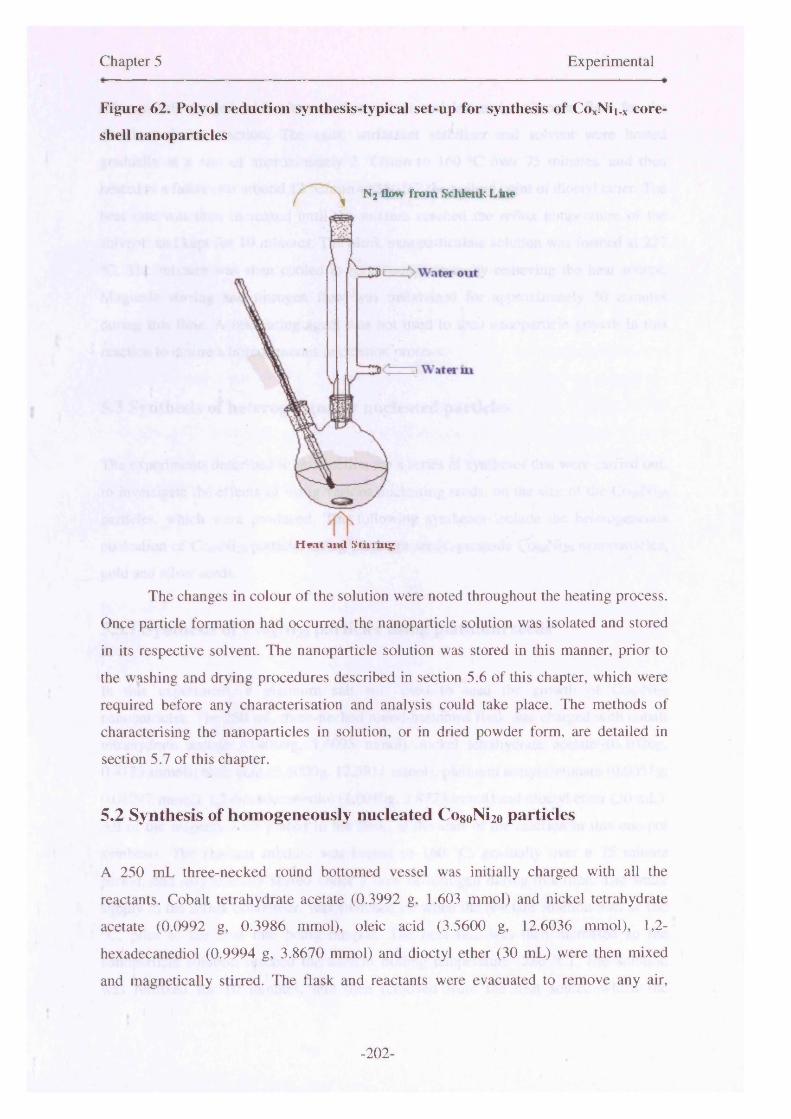
Chapter 5 Experimental
Figure 62. Polyol reduction synthesis-typical set-up for synthesis of CoxNii.x core
shell nanoparticles
The changes in colour of the solution were noted throughout the heating process.
Once particle formation had occurred, the nanoparticle solution was isolated and stored
in its respective solvent. The nanoparticle solution was stored in this manner, prior to
the washing and drying procedures described in section 5.6 of this chapter, which were
required before any characterisation and analysis could take place. The methods of
characterising the nanoparticles in solution, or in dried powder form, are detailed in
section 5.7 of this chapter.
5.2 Synthesis of homogeneously nucleated Co80Ni2o particles
A 250 mL three-necked round bottomed vessel was initially charged with all the
reactants. Cobalt tetrahydrate acetate (0.3992 g, 1.603 mmol) and nickel tetrahydrate
acetate (0.0992 g, 0.3986 mmol), oleic acid (3.5600 g, 12.6036 mmol), 1,2-
hexadecanediol (0.9994 g, 3.8670 mmol) and dioctyl ether (30 mL) were then mixed
and magnetically stirred. The flask and reactants were evacuated to remove any air,
Nj flow from Schlenk L me
H nc and Stinin?
-202-

Chapter 5 Experimental
subsequently purged with nitrogen three times and left under nitrogen flow for the
duration of the reaction. The salts, surfactant stabiliser and solvent were heated
gradually at a rate of approximately 2 °C/min to 160 °C over 75 minutes, and then
heated at a faster rate around 12 °C/min to 286 °C, the boiling point of dioctyl ether. The
heat rate was then increased until the mixture reached the reflux temperature of the
solvent, and kept for 10 minutes. The black nanoparticulate solution was formed at 227
°C. The mixture was then cooled to room temperature by removing the heat source.
Magnetic stirring and nitrogen flow was maintained for approximately 30 minutes
during this time. A nucleating agent was not used to seed nanoparticle growth in this
reaction to ensure a homogeneous nucleation process.
5.3 Synthesis of heterogeneously nucleated particles
The experiments described in this section are a series of syntheses that were carried out,
to investigate the effects of using various nucleating seeds, on the size of the Co8oNi2o
particles, which were produced. The following syntheses include the heterogeneous
nucleation of CosoNLo particles using platinum seeds, premade CosoNho nanoparticles,
gold and silver seeds.
5.3.1 Synthesis of CogoNi2o particles using platinum seeds
In this experiment, a platinum salt was used to seed the growth of CosoNho
nanoparticles. The 250 mL three-necked round-bottomed flask was charged with cobalt
tetrahydrate acetate (0.4009g, 1.6095 mmol), nickel tetrahydrate acetate (0.1026g,
0.4123 mmol), oleic acid (3.5000g, 12.3911 mmol), platinum acetylacetonate (0.005 lg,
0.01297 mmol), 1,2-hexadecanediol (l.OOlOg, 3.8773 mmol) and dioctyl ether (30 mL).
All of the reagents were placed in the flask, at the start of the reaction in this one-pot
synthesis. The reactant mixture was heated to 160 °C, gradually over a 75 minute
period, and magnetically stirred under a flow of nitrogen during this time. The water
supply to the reflux condenser, was switched on when the reactant solution was at 160
°C, prior to the heat rate being ramped. The heat rate was then increased so the
nanoparticle solution reached the solvent boiling temperature (286 °C). The solution
was refluxed for 1 0 minutes, and then removed from the heat source whilst the
-203-
Chapter 5 Experimental
dispersion was cooled to room temperature under nitrogen flow, with magnetic stirring.
The flask was then opened to air and the particles were washed with ethanol and then
toluene in 3 cycles (1 x 30 mL ethanol followed by 1 X 10 mL toluene). Finally, the
particles were redispersed in 20 mL hexane or toluene and the solutions stored in glass
screw cap vials.
The colour of the reaction solution changed from red to mauve, bright purple to
deep purple and then to a blackcurrant colour before particle formation was observed.
The solution changed from red to blackcurrant over a period of 25 minutes, from
between 38 °C and 120 °C. Prior to the solution turning red, the salts and reducing agent
remained undissolved between room temperature and 38°C. Nanoparticle formation
occurred at approximately 230 °C in this instance, and the solution was completely
black at around 242 °C. The observed colour changes were similar to those in the
homogeneously nucleated sample. This solution was stable upon application of a
magnet, and a few days later, the particles had not fallen out of solution. The black
liquid remained stable for a several months in air.
5.3.2 Synthesis of CogoNi2o particles using premade particles as seeds.
In the first step of this two-stage synthesis, cobalt tetrahydrate acetate (0.402lg, 1.6143
mmol), nickel tetrahydrate acetate (0.1016g, 0.4083 mmol), 1,2-hexadecanediol
(1.0030g, 3.8809mmol) and oleic acid (0.4494g, 1.5911 mmol) were dissolved in
diphenyl ether (30 mL). The entire reaction was carried out in an atmosphere of
nitrogen with magnetic stirring. The resulting solution was heated from room
temperature to approximately 150°C over 20 minutes. At this point tributylphosphine
(0.3048g, 1.5070 mmol) was injected into the hot solvent solution. The heat rate was
then increased and the solution’s temperature rapidly rose to the reflux point of diphenyl
ether (260 °C). The black nanoparticle seed solution was formed at approximately 228
°C. The temperature of the solution was maintained at this 260 °C for 90 minutes to
allow for the full crystallisation of the particles during reflux. The resulting black
colloidal solution was then cooled to room temperature. Co8oNi2o particles were
synthesised and used as a seed solution to grow larger CogoNi2o particles.
The second step of the growth process was then implemented. A calculated
volume, of the black nanoparticle solution was kept in the reaction flask (3 mL), whilst
-204-
Chapter 5 Experimental
the rest of the solution was removed for storage. This 3 mL aliquot of solution was used
to seed the growth of larger nanoparticles. Further reactants including cobalt
tetrahydrate acetate (0.1777g, 0.7134 mmol), nickel tetrahydrate acetate (0.0447g,
0.1796 mmol), 1,2-hexadecanediol (0.4600g, 1.9050 mmol), and oleic acid (0.1685g,
0.5965 mmol) were directly added to the 3 mL aliquot of solution that had remained in
the reaction flask with diphenyl ether (30 mL). The solution with the additional
reactants was then heated to about 150°C. At this point tributylphosphine (0.1464g,
0.7236 mmol) was injected into the flask. The solution was then heated to the reflux
temperature of the diphenyl ether (260 °C), and kept at that temperature for 30 minutes.
The formation of the larger particles in the second reaction occurred at approximately
238 °C.
5.3.3 Synthesis of Co8oNi2o particles using other noble metal seeds
Particles were synthesised using noble metal seeds, as described in section 5.3.1.
However, silver chloride (AgCl) and auric acid (HAuCL) were used as a replacement
for platinum acetylacetonate. The same synthetic procedure and molar ratios between
reactants were adopted, as with the platinum seeds experiments. The effect of these
seeds on the size of the CogoNi2o particles that were synthesised is described in Section
2.2.2. These experiments were not developed any further, as there was no improvement
to the size control of the C oso^o from this method.
5.4 Surfactant variations
The following sections detail syntheses of CogoNi2o nanoparticles, using a number of
surfactant stabilisers to control particle growth, prevent oxidation and also limit
aggregation.
5.4.1 Synthesis of CogoNi2o particles with oleic acid
Cobalt tetrahydrate acetate (0.4003g, 1.6071 mmol), nickel tetrahydrate acetate
(0.1013g, 0.4071 mmol), 1,2-hexadecanediol (1.0009g, 3.8729 mmol), oleic acid
-205-
Chapter 5 Experimental
(3.5308g, 12.5002 mmol), platinum acetylacetonate (0.0048g, 0.02204 mmol) and
dioctyl ether (30 mL) were added to the flask. The reactants were heated with magnetic
stirring in an atmosphere of nitrogen. The heat rate was initially slow, and the reactants
were heated to 160°C, over a period of 75 minutes. The solution’s colour changed from
red to deep purple during this period. The heat rate was then increased so that the
solution reached the reflux point of 286°C in 15 minutes. The black nanoparticulate
solution was recovered from the flask and stored in glass sample vials until the cleaning
procedure was implemented for analysis of the particles. Particle formation occurred at
220°C, the liquid was stable and particles were not drawn out of solution upon
application of a magnet, or over time due to gravity. The reaction proceeded in a similar
manner to that described earlier for platinum seeds (section 5.3.1). These reactions are
essentially the same in terms of molar quantities of salts and reactant used.
5.4.2 Synthesis of Co8oNi2o particles with oleylamine
CogoNi2o nanoparticles were synthesised using cobalt tetrahydrate acetate (0.4007 g,
1.6087 mmol), nickel tetrahydrate acetate (0.1003 g, 0.4031 mmol), 1,2-hexadecanediol
(1.0005 g, 3.8713 mmol), oleylamine (3.3436 g, 12.4999 mmol), platinum
acetylacetonate (0.0053 g, 0.0135 mmol) and dioctyl ether (30 mL) were stirred and
heated in an atmosphere of nitrogen. The solution was initially heated to 160°C over a
75 minute period, and then rapidly heated to 286°C, the boiling temperature of dioctyl
ether. Particle formation occurred at 218 °C and was characterised by the sudden
appearance of black particles around rim of the flask. The nanoparticle solution was
recovered and subsequently analysed using a variety of techniques.
5.4.3 Synthesis of CogoNi2o particles with oleic acid/oleylamine mixture
(OA/OY)
The method adopted for the OA/OY synthesis was different to other reactions where
surfactant has been altered. Reasons for adopting this new method are given in Chapter
2. Cobalt (acetylacetonate^ (0.2850 g, 0.8000 mmol), nickel (acetylacetonate)2 (0.0518
g, 0.2016 mmol), 1 ,2-hexadecanediol (1.2923 g, 5.0004 mmol), oleic acid (0.8474 g,
3.0001 mmol), oleylamine (0.8025 g, 3.0001 mmol), platinum acetylacetonate (0.0056
-206-
Chapter 5 Experimental
g, 0.0142 mmol) and dioctyl ether (20 mL) were heated and stirred under nitrogen flow
to 160 °C over 6 8 minutes. The heat rate was then increased, and the solvent reached
reflux temperature after 7 minutes. The solution was kept at 286 °C for 10 minutes, and
then was removed from the mantle, but left to stir in the inert atmosphere. Particle
formation was noted at 230 °C. Prior to this synthesis, the oleic acid and oleylamine
surfactant mixture was prepared by weighing 1 :1 molar quantities of both components
and then combining these with moderate heating and vigorous stirring. The quantities of
both surfactants were accurately weighted on a four figure balance. The density
difference of both surfactants when melted, meant that vigorous stirring was required to
ensure they were sufficiently mixed with one another and two separate layers of
surfactant did not form.
5.4.4 Synthesis of CogoNi2o particles with trioctylphosphine oxide
(TOPO)Several experiments were carried out in order to synthesise TOPO stabilised CogoNi2o
nanoparticles. The following synthesis is one of four described in Chapter 2 (section
2.6.5.1) and illustrates the manner by which those particular syntheses took place.
Cobalt tetrahydrate acetate (1.6 mmol), nickel tetrahydrate acetate (0.4 mmol), 1,2-
hexadecanediol (3.98 mmol), trioctylphosphine oxide, platinum acetylacetonate (molar
amounts in Table 3) and dioctyl ether (30 mL) were heated to 160°C over a 75 minute
period in an inert atmosphere with magnetic stirring. The solution was then heated to
286°C, the solvent boiling temperature. The heating profile adopted for these reactions
was identical to that of other syntheses where surfactant-type has been varied, apart
from the OA/OY synthesis described in section 5.4.3. Nanoparticle formation,
characterised by a change in colour of the reaction solution from deep purple to black,
occurred at between 204 - 209 °C for the various TOPO-coated Co8oNi2o particles that
were made. The reaction conditions of several nanoparticle syntheses that used TOPO
as sole surfactant, are documented in section 2.6.5.1 (Table 3), as are the results and
discussion relating to these.
-207-

Chapter 5 Experimental
5.4.5 Synthesis of CogoNiio particles with tributylphosphine (TBP)
Cobalt acetate tetrahydrate (0.3998 g, 1.6051 mmol), nickel tetrahydrate acetate (0.0999
g, 0.4015 mmol), 1,2-hexadecanediol (1.0018 g, 3.8763 mmol) and diphenyl ether (30
mL) were heated and stirred in an atmosphere of nitrogen. A nucleating agent was not
used in this particular reaction. The reaction mixture was initially heated from room
temperature to approximately 160 °C in 40 minutes. The colour of the solution changed
from red to a deep purple during this time, due to reduction of the respective metallic
ion species in solution. At this point, tributylphosphine (2.6240 g, 12.9695 mmol) was
injected into the reaction mixture. Upon the addition of TBP, the mixture darkened in
colour, so that the solution was dark brown with a purple hue at the meniscus. The
addition of the TBP also led to a puff of vapour being expelled in the flask. The heat
rate was then increased to a rapid rate of approximately 10 °C per minute. The reactant
mixture reached 260 °C in 10 minutes, and was then left to reflux at this temperature for
a further 10 minutes. The nanoparticle solution was subsequently cooled under nitrogen
flow, with magnetic stirring, following the removal of the vessel from the mantle.
Nanoparticle formation occurred at approximately 224 °C. On the day of synthesis,
there was a slight response of the solution to a magnet. The particles did not maintain
their stability in solution and settled to the bottom of the vial after a few days.
5.5 Compositional variation
Following detailed investigations into nucleation methods and suitable surfactants for
the CoxNii.x system, using the CogoNi2o formulation as a basis, synthesis of the other
alloys in this series was carried out. Experimental details are given in the following
subsections. The various studies that had been carried out into this CoxNii_x system,
indicated that it was best to nucleate the particles heterogeneously. The best surfactant
determined for these alloy nanoparticles was an OA/OY mixture. This particular
mixture had shown that it was effective at stabilising the particles against oxidation and
agglomeration, in addition to controlling their respective sizes. The syntheses described
in the following section put into practice this optimised synthetic process, in order to
fabricate particles within this compositional series.
-208-
Chapter 5 Experimental
5.5.1 Synthesis of cobalt nanoparticles
The synthesis of cobalt nanoparticles was carried out using similar methods to those
described earlier for the OA/OY mixture synthesis of CogoNi2o. Cobalt
(acetylacetonate>3 (0.3557 g, 0.9843 mmol), 1,2-hexadecanediol (1.2923 g, 5.0003
mmol), oleic acid (0.8474 g, 3.0001 mmol), oleylamine (0.8025 g, 3.0001 mmol),
platinum acetylacetonate (0.0058 g, 0.0147 mmol) and dioctyl ether (20 mL) was heated
to 162°C over 70 minutes. The heat rate was then increased and ramped to a reflux
temperature of 286°C in approximately 8 minutes. The solution was refluxed for 10
minutes at this temperature and then left to cool under nitrogen flow, with stirring.
Nanoparticle formation occurred at 226°C, characterised by a change in the colour of
the solution from deep olive green to black. Once cooled to room temperature, the
solution was tested with a magnet, to determine whether magnetic particles had formed.
The particles were attracted to the magnet very strongly and were drawn out of the
solvent. The particles remained magnetic for several months in air, suggesting oxide
shell growth had occurred and the particles were well passivated.
5.5.2 Synthesis of CogoNi2o nanoparticles
CogoNi2o nanoparticles were synthesised with cobalt (acetylacetonate).! (0.2858 g,
0.8022 mmol), nickel (acetylacetonate)2 (0.0518 g, 0.2082 mmol), 1,2-hexadecanediol
(1.2922 g, 5.0000 mmol), oleic acid (0.8474 g, 3.0000 mmol), oleylamine (0.8025 g,
3.0001 mmol), platinum acetylacetonate (0.0046 g, 0.0116 mmol) and dioctyl ether (20
mL). The reactant mixture was heated to 162°C over 70 minutes and then to the reflux
temperature of 286°C in 8 minutes. The mixture was then left to reflux for 10 minutes at
the solvent boiling temperature. The mixture was then removed from the heating mantle
and allowed to cool, with stirring and nitrogen flow, down to room temperature. The
solution turned black, indicating nanoparticle formation at approximately 230°C.The
particles were strongly attracted to a permanent magnet with some being drawn out of
solution. This effect remained even after several months of the nanoparticle solution
being left out in air.
-209-
Chapter 5 Experimental
5.5.3 Synthesis of Co6oNi4o nanoparticles
Cobalt (acetylacetonate)3 (0.2138 g ,0.6001 mmol) and nickel (acetylacetonate)2 (0.1028
g, 0.4001 mmol) were used in a 3:2 ratio for the synthesis of Co6oNi4o. Platinum
(acetylacetonate)2 (0.0050 g, 0.0127 mmol) was also used as the sample was
heterogeneously nucleated. Oleic acid (0.8474 g, 3.0000 mmol) and oleylamine (0.8025
g, 3.0001 mmol) made up an OA/OY 1:1 mixture which was the surfactant stabiliser
with dioctyl ether (20 mL) used as the solvent medium. The heating regime that had
been used in previous syntheses was maintained. The mixture was steadily heated up to
160°C over 100 minutes, and then rapidly heated to the solvent boiling temperature over
8 minutes. Nanoparticle formation occurred at 228°C, and was characterised by the
solution changing in colour from deep purple to black. The solution was initially variant
shades of green due to the colour of the precursor salts, and eventually deepened in
colour to purple followed by black. The black nanoparticle solution was isolated after
cooling under nitrogen and stirring. The resultant nanoparticle solution was tested with
a magnet. These particles also responded on the day of synthesis, and over subsequent
weeks.
5.5.4 Synthesis of CosoNiso nanoparticles
The molar precursor ratio of the cobalt and nickel salts in this reaction, were varied to
form the CosoNiso nanoparticulate alloy. Cobalt (acetylacetonate).! (0.1786 g, 0.5013
mmol) and nickel (acetylacetonate)2 (0.1290 g, 0.5021 mmol) were therefore used in a
1:1 ratio. The growth of the particles was seeded with platinum (acetylacetonate)2
(0.0048 g, 0.0122 mmol), with oleic acid/oleylamine used to stabilise the particles
(1.6508g, 6 mmol) and dioctyl ether (20 mL) used as solvent. The reaction mixture was
heated gradually to 160°C over 65 minutes, and then rapidly heated to 286 °C over 8
minutes. Nanoparticle formation occurred at 222°C. Repeat syntheses also yielded black
nanoparticulate solutions at around the same formation temperature. These particles did
respond to a magnet on the day of synthesis and fell out of solution.
-210-

Chapter 5 Experimental
5.5.5 Synthesis of Co4oNi6o nanoparticles
Nanoparticles of expected composition, Co4oNi6o, were synthesised using similar
methods to those described for the other samples in this compositional series. The molar
ratios of the salts were adjusted accordingly. Cobalt (acetylacetonate).! (0.1432 g, 0.4020
mmol) and nickel (acetylacetonate)2 (0.1530 g, 0.5955 mmol) were used in a 2:3 ratio.
Platinum (acetylacetonate)2 (0.0058 g, 0.0014 mmol) was used to seed particle growth
and an oleic acid/oleylamine mixture stabilised the particles (1.6506 g, 6 mmol).The
solvent used for these reactions was again dioctyl ether (20 mL). The reaction
proceeded by heating the reactants for 75 minutes until the solution temperature was
158°C. The heat rate was then increased so that the boiling point of the solvent was
attained in 8 minutes. The solution was heated for a further ten minutes at the reflux
temperature of 286°C. Nanoparticle formation occurred at 224°C, characterised by
change in solution colour from deep purple to black. The following day the
nanoparticles had crashed out of solution and also responded strongly to a magnet.
5.5.6 Synthesis of Co2oNiso nanoparticles
The molar ratios of the metallic precursors were altered so that Co2oNi8o nanoparticles
could be formed (1:4 ratio). Cobalt (acetylacetonate).! (0.0703 g, 0.1973 mmol), nickel
(acetylacetonate)2 (0.2055 g, 0.7998 mmol), 1,2-hexadecanediol (1.2922 g, 5.0000
mmol), oleic acid/oleylamine mixture (1.6502 g, 6 mmol), nucleating agent; platinum
acetylacetonate (0.0050 g, 0.0127 mmol) and dioctyl ether (20 mL) were used to make
the particles. The reactants were heated to 161 °C over a period of 50 minutes. The heat
rate was increased so that the reflux temperature of the solvent was achieved after 8
minutes. Nanoparticle formation occurred at around 220°C. The solution was refluxed
at this temperature for 10 minutes. Some vapour was evolved during this stage of the
reaction. The reaction vessel was removed from the mantle and the nanoparticulate
mixture was cooled with nitrogen flow. The nanoparticles in solution did not respond to
a magnet on the day of synthesis or at any time over subsequent weeks. The particles
had also fallen out of solution and did not remain as a stable liquid.
-211-
Chapter 5 Experimental
5.5.7 Synthesis of nickel nanoparticles
The nickel nanoparticles were made using similar methods to those described above for
other samples in this compositional series. Nickel (acetylacetonate)2 (0.2490 g, 0.9692
mmol), 1,2-hexadecanediol (1.2920 g, 4.9992 mmol), oleic acid/oleylamine (1.6497g, 6
mmol), platinum acetylacetonate (0.0050 g, 0.0127 mmol) and dioctyl ether (20 mL)
were weighed out and placed in the two-necked round bottomed flask. The reactants
were heated to 158°C in approximately 85 minutes, and then the heat was increased so
that the reflux point of 286°C was reached in 7 minutes. The solution was held at this
approximate temperature for a further 10 minutes. Nanoparticles were formed when the
solution was at a temperature of 220°C. This was evident by the change in the
appearance of the colour of the solution, which turned from a murky green/brown
colour to black, in an instant at that particular temperature. The solution was then
cooled, by maintaining the stirring condition, and removal of the flask from the heating
mantle. The particles that were formed did not respond to a magnet on the day of
synthesis, or few weeks later. These particles also did not form a stable nanoparticulate
solution.
5.6 Post-Preparative Procedures
After cooling the reaction mixture to room temperature with magnetic stirring and N2
flow, the following steps were performed in air. The crude solution of black CoxNii.x
nanoparticles was precipitated and separated via centrifugation. Ethanol (25 mL) was
added to 5 mL of the nanoparticle solution and shaken. The ethanol acted as a flocculent
which caused the particles to “crash out” of solution. The resultant precipitate was
isolated by centrifugation (~ 10,000 rpm). This precipitate was then re-dispersed in
toluene (10 mL) and centrifuged again. This cleaning cycle was carried out two times
additional times, however, the amount of ethanol used in subsequent washes was
reduced to 10 mL. This process ensured the washing and removal of excess stabilisers.
Each time toluene was introduced to the nanoparticle solution, two drops of the specific
surfactant used in the reaction was also added. This addition of surfactant ensured that
any excess stabiliser that was washed away from the nanoparticle coatings was
replaced, as this would result in aggregation of the particles. After the washings had
-212-

Chapter 5 Experimental
taken place, the resulting black precipitate which contained CoxNii.x nanocrystals, was
re-dispersed in various non-polar solvents in a variety of concentrations (toluene,
hexane etc).The nature of the solvent used for the particle’s storage was dependent upon
the subsequent particle analysis that had to take place.
The nanoparticles were isolated in a powder form, by performing the cleaning
cycles detailed above, and then in the final step, re-dispersing in acetone. The acetone-
particle solution was decanted from the centrifuge tubes into glass sample vials or
aluminium boats. These were then placed either in: (i) air to allow to air drying or (ii)
the antechamber port of a glove-box or (iii) in a dessicator under the flow of nitrogen
gas. The materials were left in this state for two days to allow the slow and controlled
growth of an oxide shell. After initial investigations into the oxidative processes using
magnetic studies detailed in later chapters. It was found that the best method of drying,
in order to encourage the growth of an oxide shell, was by using antechamber of the
glove-box. Therefore, for all of the particles discussed in this thesis, this particular
method was used to dry the particles. The resultant dry powders were stored in glass
screw-cap vials in air, or in their native liquid solutions also within the glass vials. The
samples were dried, as required, for particle analysis. This was because the particles
were less susceptible to oxidation in their native liquid form, in comparison to a
nanoparticulate powder, as the solvent and surfactant provided a barrier to surface
oxidation.
5.7 Characterisation of nanoparticles
The structural morphology, composition and magnetic properties of the CoxNii_x
nanoparticles were analyzed using transmission electron microscopy (TEM), elemental
analysis (EA), energy-dispersive X-ray analysis (EDX) and a superconducting
interference device (SQUID). Each sample described in this thesis was characterised
using a number of techniques. Some of the samples were characterised further because
of their possible magnetic functional properties. The additional techniques used in these
instances included: High-resolution transmission electron microscopy (HRTEM);
electron energy loss spectroscopy (EELS); thermogravimetric analysis (TGA), x-ray
photoelectron spectroscopy (XPS) and x-ray diffraction (XRD). Fourier transform
-213-

Chapter 5 Experimental
infrared spectroscopy (FTIR) was also used to characterise surfactant interactions with
the nanoparticles.
Samples were cleaned and prepared for TEM analysis by re-dispersion of the
nanoparticles into hexane. A droplet of the nanoparticle hexane dispersion was dropped
onto an amorphous carbon copper grid. All observational analysis was carried out using
a JEOL 100KV instrument. Electron film images were taken of different areas of the
grid to give an accurate representation of particle size and shape in a particular sample.
These were scanned to obtain digital images and the contrast adjusted to improve
particle visibility, in Adobe Photoshop1 software. This was necessary in order to be able
to view detail in the particle structure. Particle size distributions were calculated using
the Image J programme,2 which correlates pixel coverage of the nanoparticles with size,
using the magnification used in the image capture to calculate size. The particles are
identified by the difference in contrast between the individual nanoparticle and the
background. Analysis was made of at least 200 particles in a sample, in different areas
of the amorphous carbon grid. The samples were each viewed carefully and incidences
where particles had aligned themselves on top of one another lead to the software
counting them as a single particle. Therefore, these were not included for purposes of a
size distribution count. The software measured the particles’ major and minor axes, and
these were then averaged to obtain an approximate nanoparticle diameter. This
accounted for the imperfect shapes of the particles, so they could not be modelled to
that of a perfect sphere.
Igor Pro3 data analysis software was used to fit the data to appropriate
distribution functions, so that average particle sizes and standard deviations could be
obtained. Multi-peak fitting was implemented for samples that had a variable
distribution i.e bimodal distribution. This was carried out in order to determine
accurately the mean particle size in samples that had multiple nucleation events,
resulting in large populations of particles in two or three dominant size ranges.
Elemental microanalysis was carried out at UCL, on milligram quantities of
sample. The elements tested for were carbon, hydrogen and nitrogen as they were
present in the surfactants. The technique was used to quantify the amount of sample
mass that was due to surfactant stabiliser.
1 http://www.adobe.com/2 http://rsb.info.nih.gov/ij/3 http://www.wavemetrics.com/
-214-

Chapter 5 Experimental
EDX was carried out in conjunction with a scanning electron microscope
(SEM), as an additional analytical package to the SEM set-up. SEM and EDX
measurements were carried out on a Field Emission JEOL 630IF using the keV system.
The SEM was calibrated with standard reading from a cobalt calibration. The
calibration was performed frequently between a number of measurements. An elemental
study was carried out on samples to establish relative quantitative atomic ratios between
the components of the nanoparticles. EDX provided an average quantification of an area
of nanoparticles, which correlates with the electron beam size of the SEM. The beam
size was approximately several microns in diameter. A much more precise method of
analysis was carried out on a few samples using EELS which is described in detail on
the next page. However, this method was sufficient in obtaining a relative ratio between
the cobalt and nickel elements.
Samples were prepared in the following way: (i) a carbon background sticker
was placed onto an aluminium stub (ii) the stub was dipped into the dried
nanoparticulate powder and excess powder was shaken off (iii) the powder was carbon
coated to ensure accurate quantitative results. The samples were then placed on a stage
and introduced into the electron microscope chamber, having first been place under
vacuum in an antechamber compartment. On each sample stub, quantitative analysis
was conducted on five different areas or spots by rotating the stage, and an average of
these was taken to ensure reproducibility. The values that are quoted in this thesis are
atomic percentages. Images were also taken using the SEM; however the quality of the
TEM images is much better for nanoparticle analysis, as it allows clear resolution of
individual particles. Whereas the SEM displays aggregation in the particles, due to
clumping that occurs in sample preparation.
Magnetic studies were carried out on a commercial superconducting quantum
interference device. A Quantum Design MPMS7 was used to collect low temperature
magnetic measurements, including zero field cooled curves to determine particle
blocking temperatures and hysteresis loops on field cooled samples to investigate
exchange bias effects in the samples. Samples were prepared by packing milligram
quantities of the black nanoparticle powder into 5mm diameter agar gel capsules. The
powders were fixed in place by packing down with cotton wool to ensure they were
rendered immobile. Different types of measurement were made to elucidate various
specific magnetic properties associated with that particular material. Initially,
measurements were made at 280 K of magnetisation versus the applied magnetic field.
-215-
Chapter 5 Experimental
This type of measurement is a field-dependent measure of the moment of a sample, as
the magnetic field is systematically increased and decreased, in hysteretic fashion.
The materials were initially cooled from room temperature to 5 K in a zero-
applied magnetic field. The temperature was subsequently ramped to 280 K at 10 K
intervals (ZFC measurement). Upon reaching 280 K the applied field was maintained
and the temperature was decreased to 5 K. The temperature was then increased again to
280 K at 10 K intervals in order to perform the field-cooled measurement. During the
ZFC measurement the magnetic dipoles are randomly oriented at room temperature and
remain thus when the sample is cooled. At 5 K the spins do not possess enough thermal
energy to allow rotation to occur. During the ZFC measurement, as the temperature is
decreased, the kT also increases and the magnetic dipoles align parallel to the field that
has been applied. This leads to an increased magnetic moment. During the field-cooled
measurements the dipoles are aligned with the magnetic field at 280 K and remain
configured in this position when the sample is cooled to 5 K. Whilst the temperature
increases, kT provides energy for the magnetic dipoles, enabling randomisation in their
orientation and hence a resultant decrease in magnetic moment. The temperature
dependent decrease is observed in both ZFC and FC curves, as the temperature
increases past the blocking temperature Tb.
Electron energy loss spectroscopy (EELS) was carried at Glasgow University on
pre-selected specimens, which were chosen due to their interesting magnetic functional
properties and hence indicated the formation of an oxide shell. The aim of this exercise
was to establish the nature of the oxide shell, and gain an approximate shell thickness
and particle core diameter. Due to the accuracy of this technique is quantifying oxygen
content in a individual particle, it was also used to prove shell formation, and alloy
formation in the core. The specimens were examined in an FEI Tecnai F20 TEM/STEM
equipped with a field emission gun and a Gatan ENFINA electron spectrometer.
Spectrum imaging was performed using Gatan Digital Micrograph and DigiScan
software. A -0.5 nm diameter probe with a convergence semi-angle of 13 mrad and a
spectrometer collection semi-angle of 18 (or 30 mrad in some of the datasets) was used
for the EELS data presented here. The energy range -430 eV to -1368 eV (1340
channels at 0.7 eV/ch) was recorded at a dispersion of 0.7 eV/ch and a typical dwell
time of 5 sec per pixel. Although on occasion, a dwell time of 10 sec per pixel was also
used.
-216-
Chapter 5 Experimental
Thermogravimetric magnetic analysis was also attempted. In the measurements
discussed in this thesis, the instrument was used to ascertain an approximate Curie
transition temperature (Tc) for the CoNi nanoparticles. The aim was to prove the
nanoparticles were true solid solutions, with magnetic properties that could be varied
with the alloy composition. The TGA could also be used to determine the amount of
surfactant bound to the nanoparticle surface as mentioned earlier. Measurements were
carried out on a Netzsch STA 660 instrument, with a rhodium furnace, in an inert
atmosphere. During measurement runs, argon flow to the furnace was maintained, to
protect the highly sensitive balance from the effects of the furnace being heated to
elevated temperatures. Nitrogen was passed over the sample during the heating process
to prevent oxidation. Due to the nature of the sample, alumina (AI2O3) crucible pans
were used in all calibrations and measurement runs. This was to prevent any reaction
between the metal alloy nanoparticles and the aluminium pan which are used in
conventional TGA measurements. Temperature and sensitivity calibrations were
performed on a series of standards over the temperature range where measurements
were to be made. All sample measurements were standardised against the experimental
calibration curve for the instrument using the melt temperatures as the calibration point.
The highest temperature calibration standard used was gold which has a melting point
of 1064 °C. The lowest melting temperature standard used was for indium with a
melting point of 156 °C. The percentage of weight loss due to surfactant was apparent
within the first 400 °C, dependent on the decomposition temperature of the surfactant.
An example of a typical TGA run with a nickel calibration sample is shown in
Appendix B. The TGA could also be used to determine the amount of surfactant bound
to the nanoparticle surface.
X-ray photoelectron spectroscopy was carried out on a few samples to enable
characterisation of elements in the sample, the oxidation state of these elements, and
also to indicate the nature of connectivity between these particular elements.
Information collected from this technique also gave confirmation of the core-shell
structure of the particles when surface etching was carried out.
The sample was prepared for XPS by taking a large quantity of the dried
powder, and pressing it into a pellet of approximately half a millimetre in thickness,
using an infrared pellet press at a 7 kPa setting. Measurements were carried out using a
VG ESCALAB 220iXL instrument with a monochromatic A1 K<* radiation source.
Binding energies were referenced to surface elemental carbon at 284.6 eV. Depth-
-217-
Chapter 5 Experimental
profiling was carried out using a 3kV argon ion gun operating at 0.5 pA to sputter the
surface of the sample. The binding energies were correlated with results on the NIST
database.4
The powder XRD technique was carried out to confirm the crystalline nature of
the particles and to characterize the phases present. The measurements were made on a
Bruker D8 Discover reflection diffractometer, with Cu-K<x( X =1.5406 A) in reflection
mode. Where possible, Scherrer analysis of the full width half maximum (FWHM) of
the most intense peak on a diffractogram was carried out. Scherrer analysis yields an
approximate crystallite size for particles which can be compared to the mean particle
size garnered from TEM studies. Therefore, this is a very useful technique that provides
interesting information about the particles. Samples were prepared for XRD by packing
down the nanoparticles powder onto the substrate. Measurements of the particles were
made on a variety of substrates which included silicon, glass and wood. The particles
had been pressed into a pellet for the purpose of an XRD measurement. A 13 mm die
was used to press the black nanoparticulate powder into a small pellet. A pressure of
7kPa was applied in order to achieve the necessary compaction, and ensure a
homogeneous surface from which to take the measurements. These peaks arose because
of the long collection time for the data and the weak signal given from the sample. XRD
studies have presented challenges in this particular study due to the nature of the
samples.
4 http://srdata.nist.gov/xps/
-218-
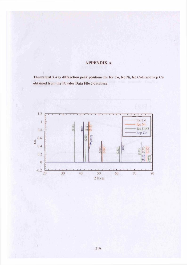
a.u.
APPENDIX A
Theoretical X-ray diffraction peak positions for fee Co, fee Ni, fee CoO and hep Co
obtained from the Powder Data File 2 database.
fee C o fee N i tee CoO hep Co
08r |06
0.4nr ' j
r ' lrn0.2
-02
2Theta
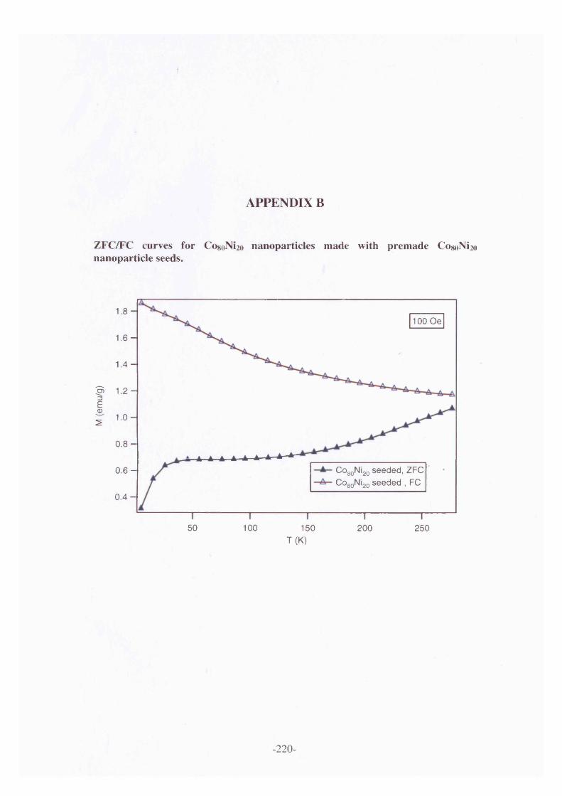
M (e
mu/
g)
APPENDIX B
ZFC/FC curves for C080N120 nanoparticles made with premade Co8oNi2o nanoparticle seeds.
1 .8 -100 Oe
1.6 -
1.4 -
1.2 -
1.0 -
0 .8 -
COgoNigo seeded, ZFC Co80Ni20 seeded , FC
0 . 6 -
0 .4 -
50 100 150 200 250T (K)
-220-
Copyright © 2022 FDOKUMEN