TPGS-based and S-thanatin functionalized nanorods ... - Nature

Journal of Photochemistry and Photobiology B: Biology 140 (2014) 69–78
Contents lists available at ScienceDirect
Journal of Photochemistry and Photobiology B: Biology
journal homepage: www.elsevier .com/locate / jphotobiol
Synthesis and characterization of titania nanorods from ilmenitefor photocatalytic annihilation of E. coli
http://dx.doi.org/10.1016/j.jphotobiol.2014.07.0071011-1344/� 2014 Elsevier B.V. All rights reserved.
⇑ Corresponding author.E-mail addresses: [email protected], [email protected] (S. Ramasamy).
Diptipriya Sethi a, Naresh Jada b, Rohit Kumar b, Sakthivel Ramasamy b,⇑, Sony Pandey b, Trupti Das b,Jayasankar Kalidoss b, Partha Sarathi Mukherjee b, Ashish Tiwari b
a Academy of Scientific and Innovative Research (AcSIR), Anusandhan Bhawan, 2 Rafi Marg, New Delhi 110 001, Indiab CSIR-Institute of Minerals and Materials Technology, Bhubaneswar 751 013, India
a r t i c l e i n f o
Article history:Received 1 April 2014Received in revised form 3 June 2014Accepted 3 July 2014Available online 22 July 2014
Keywords:IlmeniteTitania rich slagTitania nanorodsDegussa P25 titaniaAntibacterial propertyTransmission electron microscopyAbsorption edge
a b s t r a c t
Titania nanorod structures have been obtained by thermal plasma reduction of ilmenite (FeTiO3) fol-lowed by chemical treatments. Inherently present iron in the titania nanorods acts as a dopant whichresults in shifting the absorption edge of titania from ultraviolet to visible region. X-ray diffraction(XRD) study confirms the existence of rutile phase of titania. X-ray Photoelectron Spectroscopy (XPS)reveals the presence of Ti4+, O2�, Fe3+ and surface hydroxyl group. Transmission Electron Microscopy(TEM) confirms the formation of nanorod structure having width of 6 nm and length of 32 nm. Photocat-alytic annihilation property of titania nanorods derived from ilmenite (titania-I), rutile titania obtainedfrom titanium(IV) butoxide (titania-A) and Degussa P25 titania was studied under UV and UV–Visibleirradiation conditions separately and compared. The time required for complete photocatalytic annihila-tion of Escherichia coli cells are 10, 15 and 45 min under UV irradiation whereas it has taken 15, 10–15,30 min under UV–Visible irradiation for titania-A, Degussa P25 titania and titania-I respectively. It isobserved that titania-I shows significantly stronger antibacterial property under UV–Visible irradiationcompared to UV alone.
� 2014 Elsevier B.V. All rights reserved.
1. Introduction
Titanium dioxide is a non-toxic, chemically and mechanicallystable semiconducting material. It exists in three crystallographicforms such as anatase, rutile and brookite. Among these rutile isthe most common natural form of TiO2 [1,2]. It is used in the man-ufacturing of paints, varnishes, lacquer, paper, paperboard, printinginks, rubber, floor coverings, ceramics, food and pharmaceuticals[3,4]. Anatase phase of titania is used as a support material fornanogold particles for oxidation of carbon monoxide [5]. In plasticindustries, it is used as filler to improve the strength of plasticmaterials [6]. Nano-scaled titanium dioxide particles are primarilyused in sun screen lotions because they scatter visible light lessthan titanium dioxide pigments while still providing UV protection[7–9]. Nanostructured TiO2 finds new advanced applications in thefields of energy and environmental sciences such as dye-sensitizedsolar cells [10,11], photovoltaic devices [12] and photocatalysts[13,14]. More importantly, TiO2 is extensively studied as a photo-catalyst to remove pollutants such as bacteria, volatile organic
compounds and nitrogen oxides from water and air [15]. Itsabsorption edge shifts from UV to visible region when doped withnitrogen, sulfur etc. that helps for visible light photocatalytic appli-cations [16]. It has been reported that delaminated anatase nano-tubes and titanate nanoribbons are of potential interest ascatalytic supports and photocatalysts [17]. The major resourcesof titanium and titania are mineral ilmenite (FeTiO3) and rutile(natural TiO2). Among these, ilmenite is preferred as a raw materialfor manufacturing titanium dioxide pigment, as it contains a highamount of TiO2 (95%). But its availability is very less in nature, sothe naturally available ilmenite becomes the alternate source forfulfilling the demand of titanium dioxide production [18].
Large scale production of TiO2 requires abundant resources andnaturally available ilmenite is the major mineral resource of titaniaacross the world. India has vast ilmenite resources; large depositsof ilmenite are found in the beach sands of Kerala, Tamilnadu andOdisha coasts. Since it contains 40–60% TiO2, it has to be upgradedfor its effective utilization. Removal of iron from ilmenite is themajor challenge to obtain high quality rutile phase of titania. Con-ventional method of iron removal involves hydrochloric/sulfuricacid treatment of ilmenite. It has been reported that iron removalis better with HCl compared to H2SO4 at 80 �C [19]. However, this
70 D. Sethi et al. / Journal of Photochemistry and Photobiology B: Biology 140 (2014) 69–78
method produces a huge amount of environmentally hazardouswaste. In the carbothermal reduction, solid-state reaction betweenilmenite and carboneous reductant takes place between 1550 and1650 �C to produce metallic iron and titania slag [20].
In these processes, parameters such as temperature and reac-tant feed concentrations are to be optimized in order to producehigh grade titania rich slag. This slag is further subjected to thermaltreatment for easy leaching of iron and other impurities in HCltreatment [21]. Although bulk form of titania could be producedfrom these processes, obtaining nanosized titania with differentmorphology is difficult. In this paper, a novel method has beenput forward to synthesize TiO2 nanorods from the TiO2 rich slagobtained from thermal plasma treatment of ilmenite. The productso obtained has been compared with Degussa P25 titania and tita-nia synthesized from titanium(IV) butoxide (titania-A), both struc-turally as well as chemically through XRD, FE-SEM, TEM, UV–Visible spectrophotometry and XPS. Its efficacy as photocatalysthas also been done using antibacterial test with Escherichia coliand compared with other titania samples.
2. Materials and methods
Ilmenite obtained from IRE, Chatrapur was used as a startingmaterial for titania. It was subjected to reduction in a speciallydesigned thermal plasma reactor at temperature 1550–1650 �Cusing coke as a reductant. Chemical composition of low ash metal-lurgical (LAM) coke is fixed carbon 86.74%, Ash-12.33%, S-0.73%and P-0.025%. Design of thermal plasma reactor used in this inves-tigation and its operational procedures were reported elsewhere[21]. During the thermal plasma reduction, major portion of FeOpresent in the ilmenite was reduced to form metallic iron alongwith titania rich slag. The metallic iron was separated from the slagby hammering/crushing, whereas titania rich slag is subjected toheat treatment at 750 �C for further processing. The treated titaniarich slag was subjected to HCl treatment to leach out the remainingiron and other acid soluble impurities. The obtained enriched tita-nia product was further subjected to NaOH treatment to formsodium titanate, whereas impurities such as alumina (Al2O3) andsilica (SiO2) results in the formation of water soluble sodium alu-minate (Na2Al2O4) and sodium silicate (Na2SiO3) and they werewashed out with distilled water. The left out sodium titanate (Na2-
TiO3) was treated with HCl to produce titanium oxychloride fol-lowed by hydrolysis to produce titania nanorods. The chemicalcomposition of titania synthesized from ilmenite, raw ilmeniteand titania rich slag were shown in Table 1. The overall chemicalreaction schemes involved in the formation of titania nanorods isgiven below.
TiO2 þ 2NaOH! Na2TiO3 þH2O
Al2O3 þ 2NaOH! Na2Al3O4 þH2O
SiO2 þ 2NaOH! Na2SiO3 þH2O
Na2TiO3 þ FeOþ 6HCl! TiOCl2 þ FeCl2 þ 2NaClþ 3H2O
TiOCl2 þH2O! TiO2 þ 2HCl
Table 1Chemical composition of ilmenite, titania rich slag and titania-I.
Samples TiO2
(%)Fe2O3
(%)FeO(%)
Al2O3
(%)MgO(%)
V2O5
(%)Si(%
Ilmenite 50.84 12.13 33.51 1.02 0.72 0.24 –Titania rich slag 79.0 – 12.29 1.47 – – 1.Titania-I 98.1 – – – – – –
For comparative study, rutile phase of titania was synthesizedby water hydrolysis of 25 ml titanium(IV) butoxide (Aldrich)mixed with 75 ml of tert-butanol under vigorous stirring at roomtemperature. The obtained precipitate was aged in its motherliquor at room temperature for 10 h and separated out by centrifu-gation. Then, it was washed with distilled water, dried at 100 �C for12 h and calcined at 900 �C for 4 h. Titania synthesized from ilmen-ite and titanium (IV) butoxide (Aldrich) are denoted as titania-I andtitania-A respectively, whereas commercially available titania isrepresented as Degussa P25 titania.
Phase identification of ilmenite and its other derived productswere determined by X-ray diffraction (diffractometer: X’PERTPRO, PANalytical, Netherlands) using Cu Ka radiation source(wavelength 0.154056 nm). Surface morphology and elementalcomposition were studied using Field Emission Scanning ElectronMicroscope (FESEM, model ZEISS SUPRA55) and energy dispersivespectroscope (EDS) attached to it. The transmission electron micro-scopic (TEM) images and the selected area electron diffraction(SAED) pattern were achieved with a TECHNAI G2, FEI operatedat 200 kV. To get the TEM images, the powdered samples were dis-persed in iso-propanol by ultrasonication for 20 min. Then onedrop of the dispersed solution was deposited on the carbon coatedcopper grid. The deposited sample was dried completely under anIR lamp and taken for observation. The UV–Visible diffuse reflec-tance spectra (DRS) were recorded in the wavelength range 200–800 nm by a Varian Cary UV–Vis spectrophotometer. The Ramanspectra were recorded in a dispersive type micro-Raman Spec-trometer (Renishaw in via Reflex, UK). Fourier Transform Infrared(FTIR) spectra were recorded in the range of 400–4000 cm�1 wave-number on a Fourier Transform Infrared spectrophotometer (Spec-trum Gx Model of Perkin Elmer) and KBr was used as a reference.The X-ray photoelectron spectroscopy (XPS) spectra were carriedout using VG Scienta hemispherical energy analyser source havingresolution of 44.1 meV at 50 pass energy and Al Ka(hm = 1486.6 eV) source (S/N:10001, Prevac, Poland). Instrumentbase pressure of 5 � 10�10 mbar was maintained during dataacquisition. The specific surface area of samples was measuredfrom the nitrogen adsorption/desorption study carried out on thesurface area analyzer, Quantachrome autosorbiQ, Model ASiQ-MOV000-4.
The antibacterial property of the prepared sample was investi-gated by using E. coli (MTCC 739) as an indicator strain. Beforestarting the experiments, 0.85% aq. NaCl solution was preparedwith tap water in order to maintain the osmotic balance of bacte-rial cells and then sterilized at 121 �C under 15 lb pressure for20 min. Then, 25 ml of sterilized water was spiked with an initialconcentration of 107 CFU/ml of E. coli and subsequently mixed withcatalyst sample in a beaker. The catalyst sample concentration wasmaintained at 1 mg/ml of sterilized tap water. This mixed solutionwas placed on a magnetic stirrer in a bioreactor and irradiated withboth 8 W UV and 8 W visible light (Philips T5 8 W) sources and alsowith 8 W UV alone. At various intervals of time, 1 mL of the samplewas drawn from the beaker and it was serially diluted up to 10�4
dilution. From each diluted solution, 0.1 mL was drawn and spreadplated on the LBA plates before incubation at 37 �C under dark con-dition for 24 h. After incubation the number of colonies present onthe plates was counted. The Colony Forming Unit (CFU) per ml was
O2
)Fe (M)(%)
MnO(%)
Fe (T)(%)
S(%)
P(%)
Na(%)
Cl(%)
C(%)
– – – – – – – –02 2.84 0.80 – – – – – –
– – 0.42 – – 0.04 0.13 0.154

D. Sethi et al. / Journal of Photochemistry and Photobiology B: Biology 140 (2014) 69–78 71
calculated for each sample at different time intervals using the fol-lowing formula:
CFU=ml ¼ No: of colonies� Dilution factor=volume inoculated
Here dilution factor is the reciprocal of the dilution in which theplate count was taken, and volume inoculated is 0.1 mL.
3. Results and discussion
X-ray powder diffraction patterns of raw ilmenite and ilmeniterich slag obtained after thermal plasma treatment are depicted inFig. 1a whereas for titania nanorod sample derived from titaniarich slag (titania-I), titania synthesized from titanium(IV) butoxide(titania-A) and Degussa P25 titania are shown in Fig. 1b. The peakposition and relative intensity of raw ilmenite is very well matchedwith standard powder diffraction pattern of JCPDS No. 01-075-1211 which corresponds to the single phase of ilmenite (FeTiO3).However, in case of titania rich slag, anatase titania and Fe3Ti3O10
are found as major phases along with minor phases of ferrousoxide (FeO) and metallic iron (Fe). These phases are well matchedwith the JCPDS file No. 00-001-0562, 00-047-0421, 77-2355 and01-1267 respectively. Whereas in titania-I only tetragonal rutilephase of titania (JCPDS No. 00-001-1292) was found. Average crys-
Fig. 1a. X-ray powder diffraction patterns of ilmenite and titania rich slag.
Fig. 1b. X-ray powder diffraction patterns of various titania samples.
tallite size calculated based on the Debye–Scherrer equation [22]given below for various diffraction peaks of titania-I was foundto be 25 nm.
Dhkl ¼0:9k
b cos hð1Þ
where D is the crystallize size, k is the wavelength of X-ray (CuKa = 1.54056 Å), b is the full width at half maximum (FWHM) of apeak and h is diffraction angle. XRD pattern of titania-A shows onlyrutile phase whereas Degussa P25 titania contains mixed phase ofanatase and rutile (Fig. 1b). Laser Raman spectra of titania-I, tita-nia-A and Degussa P25 titania recorded from 100 cm�1 to800 cm�1 are shown in Fig. 2. It shows various bands at around
Fig. 2. Laser Raman spectra of various titania samples.
Table 2Raman spectral assignments of various titania samples.
Titania-I Titania-A Degussa P25 titania Spectral assignment
– – 196 Eg (Anatase)236 237 – O–O interaction– – 395 B1g (Anatase)446 445 445 Eg (Rutile)– – 517 A1g + B1g (Anatase)609 610 611 A1g (Rutile)
Fig. 3. FTIR spectra of various titania samples.

Table 3FTIR spectral band positions and their assignments for titania samples.
Band positions (cm�1) Assignments
Titania-A Titania-I Degussa P25
– – 532 O–Ti–O bending vibration606 627 – O–Ti–O bending vibration
– 709 – Ti–O stretching vibration1518 1521 1521 Bending vibration of surface O–H group1698 1689 1699 Bending vibration of O–H group due to adsorbed water molecule.3612 3577 3607 Stretching vibration of O–H group due to adsorbed water molecule.3737 3740 3740 Stretching vibration of surface O–H group
Fig. 4a. Ti2P XPS spectrum of titania-I sample.
Fig. 4b. O1s XPS spectrum of titania-I sample.
Fig. 4c. Fe2P XPS spectrum of titania-I sample.
72 D. Sethi et al. / Journal of Photochemistry and Photobiology B: Biology 140 (2014) 69–78
238, 448, 611 and 826 cm�1 which are assigned to rutile phase fortitania-I and titania-A according to reported literature [23–25].Whereas in case of Degussa P25 titania, in addition to these bandsother bands are also observed at 196, 395, 517 and 639 cm�1 corre-sponding to the anatase phase. This observation is well supportingthe XRD results confirming the existence of mixed phases of anataseand rutile. The Raman band positions and their correspondingvibration mode assignments are given in Table 2. All these bandscorrespond to O–Ti–O bending and Ti–O stretching mode vibra-tions. FTIR spectra of all three titania samples are shown in Fig. 3.
It shows stretching and bending vibration modes for O–Ti–O, Ti–Oand also for the surface hydroxyl group and adsorbed water mole-cules [26] and references therein). The observed spectral band posi-tions corresponding to different vibration modes of variousfunctional groups are also given in Table 3. In order to reveal thepresence of iron ion in titania-I sample and to define its oxidationstate, three areas of the XPS spectrum were examined and eachpeak was deconvoluted for better understanding: the Ti 2p regionbetween 455 and 470 eV (Fig. 4a), the O 1s region between 525and 540 eV (Fig. 4b) and the Fe 2p region between 705 and745 eV (Fig. 4c). The Ti 2p1/2 and Ti 2p3/2 spin–orbital splitting pho-toelectrons are located at binding energies of 465 and 459.4 eVrespectively which indicate the existence of Ti4+ ion as can be seenin Fig. 4a. This observation is in excellent agreement with dataobtained by Zhu et al. [27,28]. Fig. 4b shows the O 1s core levelspectrum, a peak observed at 530.6 eV is due to O2� ion in theTiO2 lattice whereas shoulder peak located at a binding energy of532.9 eV is attributed to the surface hydroxyl groups of chemi-sorbed water molecules on the titania [27,28]. The XPS spectrumof Fe present in titania-I is shown in Fig. 4c. It shows peak at thebinding energies of 712 and 726 eV corresponding to 2p3/2 and2p1/2 of Fe3+ ion respectively [29]. It is reported that presence ofFe3+ ion as a dopant in TiO2 matrix is beneficial for the generationof oxygen vacancies in the crystal lattice or on the surface of TiO2.When oxygen vacancies are on or near the particle surface, theycan favor adsorption of water molecules that helps creating surfacehydroxyl groups to promote the photocatalytic activity [28].
FE-SEM pictures of raw ilmenite and titania rich slag aredepicted in Figs. 5 and 6 respectively. It shows that ilmenite hasthe particle size range between 200 and 350 lm and their particlemorphology is distinctly visible with irregular shape. Transmissionelectron microscopic pictures of titania-I and its corresponding
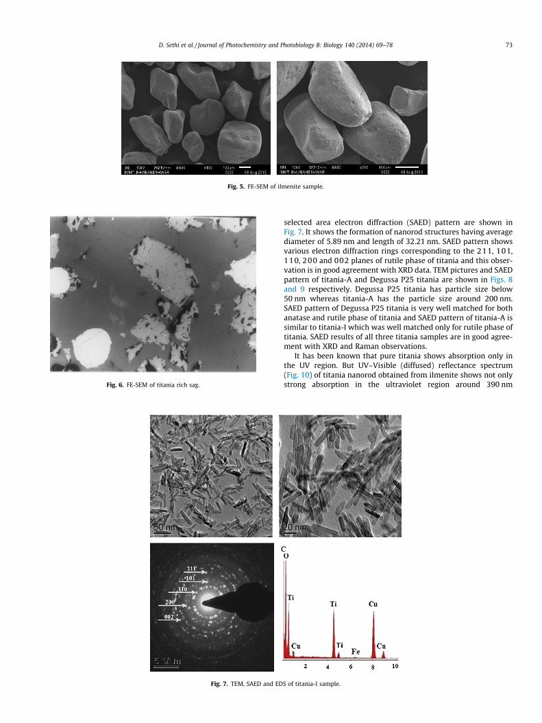
Fig. 5. FE-SEM of ilmenite sample.
Fig. 6. FE-SEM of titania rich sag.
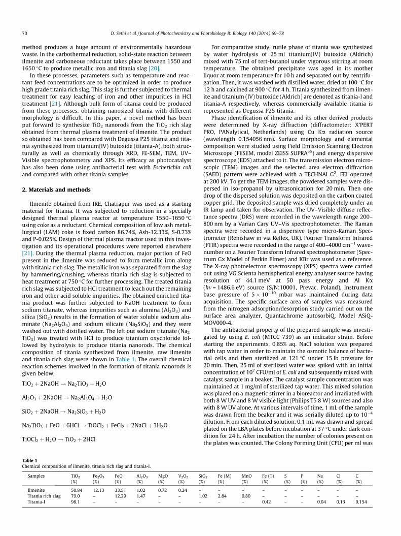
Fig. 7. TEM, SAED and ED
D. Sethi et al. / Journal of Photochemistry and Photobiology B: Biology 140 (2014) 69–78 73
selected area electron diffraction (SAED) pattern are shown inFig. 7. It shows the formation of nanorod structures having averagediameter of 5.89 nm and length of 32.21 nm. SAED pattern showsvarious electron diffraction rings corresponding to the 211, 101,110, 200 and 002 planes of rutile phase of titania and this obser-vation is in good agreement with XRD data. TEM pictures and SAEDpattern of titania-A and Degussa P25 titania are shown in Figs. 8and 9 respectively. Degussa P25 titania has particle size below50 nm whereas titania-A has the particle size around 200 nm.SAED pattern of Degussa P25 titania is very well matched for bothanatase and rutile phase of titania and SAED pattern of titania-A issimilar to titania-I which was well matched only for rutile phase oftitania. SAED results of all three titania samples are in good agree-ment with XRD and Raman observations.
It has been known that pure titania shows absorption only inthe UV region. But UV–Visible (diffused) reflectance spectrum(Fig. 10) of titania nanorod obtained from ilmenite shows not onlystrong absorption in the ultraviolet region around 390 nm
S of titania-I sample.

Fig. 8. TEM and SAED of titania-A sample.
74 D. Sethi et al. / Journal of Photochemistry and Photobiology B: Biology 140 (2014) 69–78
wavelength but also shows weak broad absorption in the visiblerange of the spectrum indicating the existence of Fe as an impuritywhich could be detected in the EDS (Fig. 7) as well as in the XPSspectra (Fig. 4c). DRS spectrum of titania-I is similar to the spec-trum reported for iron doped titania [30]. Degussa P25 titaniaand titania-A shows absorption only in the UV region (Fig. 10).
Antibacterial property has been examined separately under UVand UV–Visible light irradiation conditions for the titania-I, titania-A and commercially available Degussa P25 titania. The resultsobtained for all three samples show the increasing photocatalyticannihilation of E. coli cells present in the water with increase intime (Figs. 11 and 12). The time required for complete photocata-lytic deactivation/annihilation of E. coli cells are 10, 15 and 45 minfor titania-A, Degussa P25 titania and titania-I respectively underUV irradiation. Bacterial deactivation mechanism occurs due tointeraction between E. coli cells with reactive oxygen species suchas OH� radicals, superoxide ions (O2
��) and H2O2 generated during
photocatalysis. This further depends on availability of photogener-ated electrons in the conduction band (e�cb) and holes in the valenceband (hvb) when sample is irradiated with UV/UV–Visible light. Thereactive oxygen species generated by above mentioned process ini-tiate the damage of proteins and nucleic acids and also oxidationof phospholipids present in various layers of bacterial membrane[31,32] and this oxidation process is called lipid peroxidation.Although photocatalytic annihilation of E. coli cells is observed inall three samples, their trends differ from sample to sample andthese changes could be due to various reasons such as method ofpreparation of sample, availability of photogenerated electrons (e�cb)and holes (hvb) and their recombination etc. The drastically reducedphotocatalytic annihilation property observed for the sample titania-I could be due to presence of iron impurity which has been con-firmed in the XPS spectrum and it might act as centre for recombina-tion of photogenerated electrons and holes while irradiating with UVlight alone.
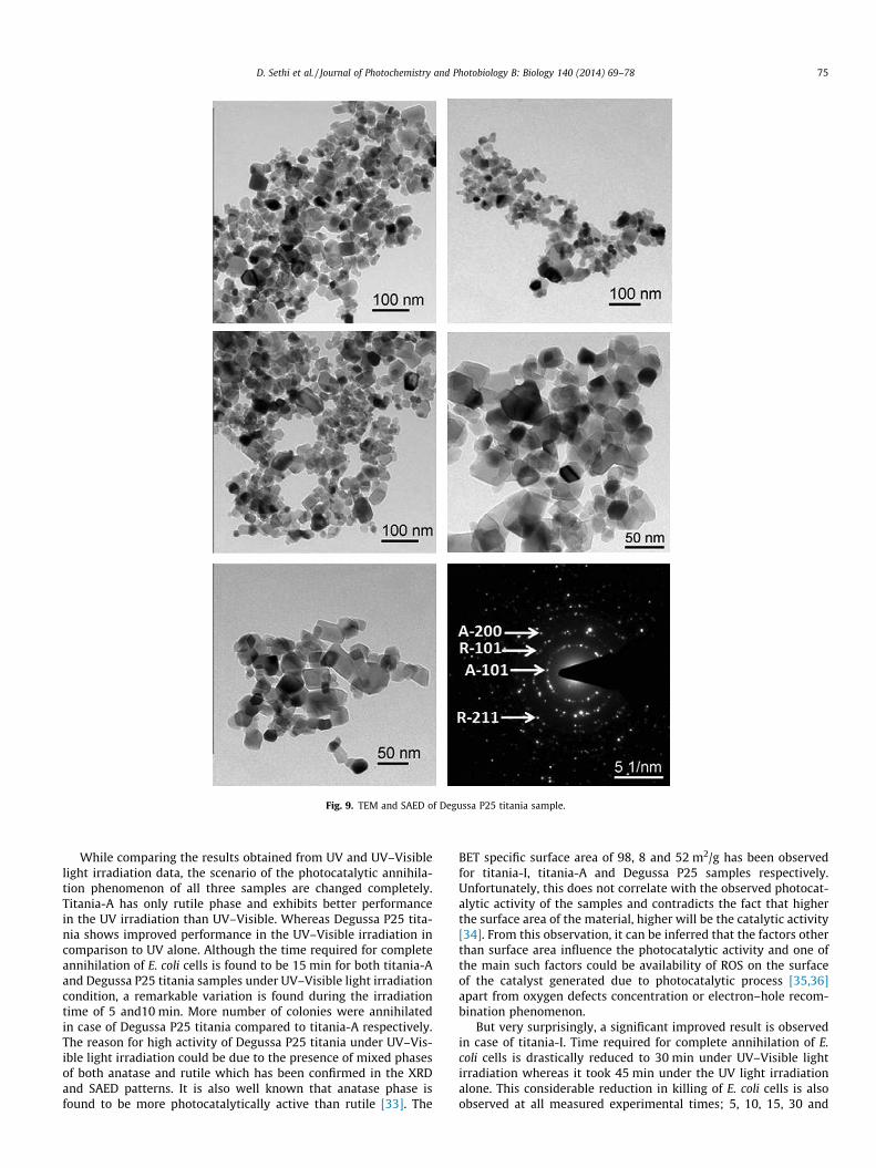
Fig. 9. TEM and SAED of Degussa P25 titania sample.
D. Sethi et al. / Journal of Photochemistry and Photobiology B: Biology 140 (2014) 69–78 75
While comparing the results obtained from UV and UV–Visiblelight irradiation data, the scenario of the photocatalytic annihila-tion phenomenon of all three samples are changed completely.Titania-A has only rutile phase and exhibits better performancein the UV irradiation than UV–Visible. Whereas Degussa P25 tita-nia shows improved performance in the UV–Visible irradiation incomparison to UV alone. Although the time required for completeannihilation of E. coli cells is found to be 15 min for both titania-Aand Degussa P25 titania samples under UV–Visible light irradiationcondition, a remarkable variation is found during the irradiationtime of 5 and10 min. More number of colonies were annihilatedin case of Degussa P25 titania compared to titania-A respectively.The reason for high activity of Degussa P25 titania under UV–Vis-ible light irradiation could be due to the presence of mixed phasesof both anatase and rutile which has been confirmed in the XRDand SAED patterns. It is also well known that anatase phase isfound to be more photocatalytically active than rutile [33]. The
BET specific surface area of 98, 8 and 52 m2/g has been observedfor titania-I, titania-A and Degussa P25 samples respectively.Unfortunately, this does not correlate with the observed photocat-alytic activity of the samples and contradicts the fact that higherthe surface area of the material, higher will be the catalytic activity[34]. From this observation, it can be inferred that the factors otherthan surface area influence the photocatalytic activity and one ofthe main such factors could be availability of ROS on the surfaceof the catalyst generated due to photocatalytic process [35,36]apart from oxygen defects concentration or electron–hole recom-bination phenomenon.
But very surprisingly, a significant improved result is observedin case of titania-I. Time required for complete annihilation of E.coli cells is drastically reduced to 30 min under UV–Visible lightirradiation whereas it took 45 min under the UV light irradiationalone. This considerable reduction in killing of E. coli cells is alsoobserved at all measured experimental times; 5, 10, 15, 30 and
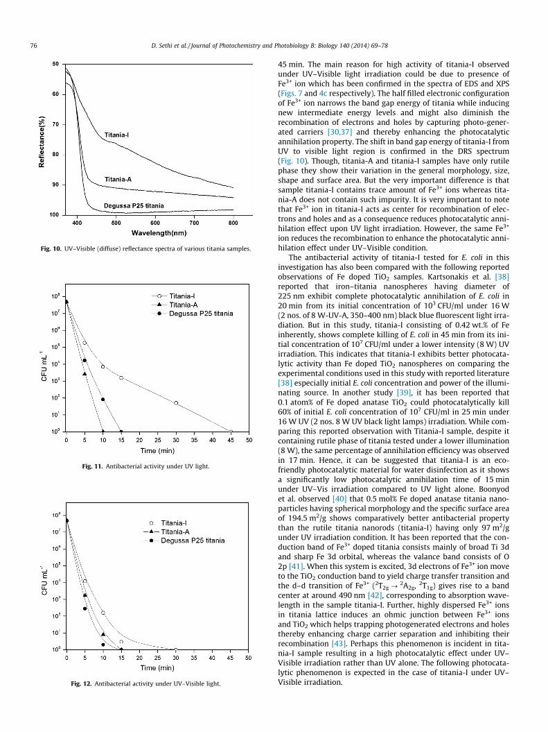
Fig. 10. UV–Visible (diffuse) reflectance spectra of various titania samples.
Fig. 11. Antibacterial activity under UV light.
Fig. 12. Antibacterial activity under UV–Visible light.
76 D. Sethi et al. / Journal of Photochemistry and Photobiology B: Biology 140 (2014) 69–78
45 min. The main reason for high activity of titania-I observedunder UV–Visible light irradiation could be due to presence ofFe3+ ion which has been confirmed in the spectra of EDS and XPS(Figs. 7 and 4c respectively). The half filled electronic configurationof Fe3+ ion narrows the band gap energy of titania while inducingnew intermediate energy levels and might also diminish therecombination of electrons and holes by capturing photo-gener-ated carriers [30,37] and thereby enhancing the photocatalyticannihilation property. The shift in band gap energy of titania-I fromUV to visible light region is confirmed in the DRS spectrum(Fig. 10). Though, titania-A and titania-I samples have only rutilephase they show their variation in the general morphology, size,shape and surface area. But the very important difference is thatsample titania-I contains trace amount of Fe3+ ions whereas tita-nia-A does not contain such impurity. It is very important to notethat Fe3+ ion in titania-I acts as center for recombination of elec-trons and holes and as a consequence reduces photocatalytic anni-hilation effect upon UV light irradiation. However, the same Fe3+
ion reduces the recombination to enhance the photocatalytic anni-hilation effect under UV–Visible condition.
The antibacterial activity of titania-I tested for E. coli in thisinvestigation has also been compared with the following reportedobservations of Fe doped TiO2 samples. Kartsonakis et al. [38]reported that iron–titania nanospheres having diameter of225 nm exhibit complete photocatalytic annihilation of E. coli in20 min from its initial concentration of 103 CFU/ml under 16 W(2 nos. of 8 W-UV-A, 350–400 nm) black blue fluorescent light irra-diation. But in this study, titania-I consisting of 0.42 wt.% of Feinherently, shows complete killing of E. coli in 45 min from its ini-tial concentration of 107 CFU/ml under a lower intensity (8 W) UVirradiation. This indicates that titania-I exhibits better photocata-lytic activity than Fe doped TiO2 nanospheres on comparing theexperimental conditions used in this study with reported literature[38] especially initial E. coli concentration and power of the illumi-nating source. In another study [39], it has been reported that0.1 atom% of Fe doped anatase TiO2 could photocatalytically kill60% of initial E. coli concentration of 107 CFU/ml in 25 min under16 W UV (2 nos. 8 W UV black light lamps) irradiation. While com-paring this reported observation with Titania-I sample, despite itcontaining rutile phase of titania tested under a lower illumination(8 W), the same percentage of annihilation efficiency was observedin 17 min. Hence, it can be suggested that titania-I is an eco-friendly photocatalytic material for water disinfection as it showsa significantly low photocatalytic annihilation time of 15 minunder UV–Vis irradiation compared to UV light alone. Boonyodet al. observed [40] that 0.5 mol% Fe doped anatase titania nano-particles having spherical morphology and the specific surface areaof 194.5 m2/g shows comparatively better antibacterial propertythan the rutile titania nanorods (titania-I) having only 97 m2/gunder UV irradiation condition. It has been reported that the con-duction band of Fe3+ doped titania consists mainly of broad Ti 3dand sharp Fe 3d orbital, whereas the valance band consists of O2p [41]. When this system is excited, 3d electrons of Fe3+ ion moveto the TiO2 conduction band to yield charge transfer transition andthe d–d transition of Fe3+ (2T2g ?
2A2g, 2T1g) gives rise to a bandcenter at around 490 nm [42], corresponding to absorption wave-length in the sample titania-I. Further, highly dispersed Fe3+ ionsin titania lattice induces an ohmic junction between Fe3+ ionsand TiO2 which helps trapping photogenerated electrons and holesthereby enhancing charge carrier separation and inhibiting theirrecombination [43]. Perhaps this phenomenon is incident in tita-nia-I sample resulting in a high photocatalytic effect under UV–Visible irradiation rather than UV alone. The following photocata-lytic phenomenon is expected in the case of titania-I under UV–Visible irradiation.

D. Sethi et al. / Journal of Photochemistry and Photobiology B: Biology 140 (2014) 69–78 77
TiO2 þ hv! hþ þ e�
Ti4þ þ e� ! Ti3þ
O2� þ hþ ! O�
Fe3þ þ Ti3þ ! Fe2þ þ Ti4þ trapping electron
Fe3þ þ O� ! Fe4þ þ O2� trapping hole
Fe4þ þ OH� ! Fe3þ þ OH�
Fe2þ þ O2 ! Fe3þ þ O�2
The relatively unstable Fe2+ and Fe4+ ions tend to furnish thetrapped carriers for the creation of ROS in water which can damagethe bacterial cell membrane [44]. Further oxidative attack of theROS causes a lethal destruction of intercellular components of thebacteria.
4. Conclusions
A novel method has been put forward to synthesize titaniananorods from ilmenite. The obtained titania nanorods containiron as an inherent impurity which acts as dopant in titania latticeand shifts the absorption edge from UV to visible region. In addi-tion to its great influence on the optical characteristics of the hostmaterial, iron in titania nanorod structures promotes effective pho-tocatalytic annihilation of E. coli present in water under UV–visibleirradiation condition than UV irradiation alone. The time requiredfor complete photocatalytic annihilation of E. coli cells is 45 and30 min under UV alone and UV–Visible irradiation respectively.TEM confirms the formation of nanorod structures having widthof 6 nm and length of 32 nm. XRD and Laser Raman studies sub-stantiated the existence of rutile phase of titania and XPS studyconfirmed the presence of iron as an dopant in the form of Fe3+
ion in the titania lattice. From the above study, it can be concludedthat titania nanorods derived from ilmenite source could be usedas a photocatalyst for effective annihilation of E. coli bacteria pres-ent in water.
Acknowledgments
We acknowledge the Ministry of Environment and Forest, NewDelhi for financial support and thanks to the Director of CSIR-Insti-tute of Minerals and Materials Technology, Bhubaneswar for hisencouragement. One of the authors Dipti Priya Sethi wishes toacknowledge the Ministry of Social Justice and Empowerment,Government of India for awarding Rajiv Gandhi NationalFellowship.
References
[1] X. Chen, S.S. Mao, Titanium dioxide nanomaterials: synthesis, properties,modifications, and applications, Chem. Rev. 107 (2007) 2891–2959.
[2] J. Ovenstone, K. Yanagisawa, Effect of hydrothermal treatment of amorphoustitania on the phase change from anatase to rutile during calcination, Chem.Mater. 11 (1999) 2770–2774.
[3] S. Samal, B.K. Mohapatra, P.S. Mukherjee, S.K. Chatterjee, Integrated XRD,EPMA and XRF study of ilmenite and titania slag used in pigment production, J.Alloys Comp. 474 (2009) 484–489.
[4] S. Samal, K.K. Rao, P.S. Mukhrejee, T.K. Mukherjee, Statistical modelling studieson leachability of titania-rich slag obtained from plasma melt separation ofmetallized ilmenite, Chem. Eng. Res. Des. 86 (2008) 187–191.
[5] R. Sakthivel, T. Ntho, M. Witcomb, M.S. Scurrell, CO oxidation over anatase TiO2
supported Au: effect of nitrogen doping, Catal. Lett. 130 (2009) 341–349.[6] F. Bondioli, A. Dorigato, P. Fabbri, M. Messori, A. Pegoretti, High-density
polyethylene reinforced with submicron titania particles, Polym. Eng. Sci. 48(2008) 448–457.
[7] G. Wakefield, M. Green, S. Lipscomb, B. Flutter, Modified titania nanomaterialsfor sunscreen applications – reducing free radical generation and DNA damage,Mater. Sci. Technol. 20 (2004) 985–988.
[8] A. Salvador, M.C. Pascual-Marti, J.R. Adell, A. Requeni, J.G. March, Analyticalmethodologies for atomic spectrometric determination of metallic oxides inUV sunscreen creams, J. Pharma. Biomed. Anal. 22 (2000) 301–306.
[9] R. Zallen, M.P. Moret, The optical absorption edge of brookite TiO2, Solid StateCommun. 137 (2006) 154–157.
[10] G.K. Mor, K. Shankar, M. Paulose, O.K. Varghese, C.A. Grimes, Use of highly-ordered TiO2 nanotube arrays in dye-sensitized solar cells, Nano Lett. 6 (2006)215–218.
[11] H.J. Koo, Y.J. Kim, Y.H. Lee, W.I. Lee, K. Kim, N.G. Park, Nano-embossed hollowspherical TiO2 as bifunctional material for high-efficiency dye-sensitized solarcells, Adv. Mater. 20 (2008) 195–199.
[12] T.W. Zeng, Y.Y. Lin, H.H. Lo, C.W. Chen, C.H. Chen, S.C. Liou, H.Y. Huang, W.F. Su,A large interconnecting network within hybrid MEH-PPV/TiO2 nanorodphotovoltaic devices, Nanotechnology 17 (2006) 5387–5392.
[13] Y.H. Tseng, C.S. Kuo, C.H. Huang, Y.Y. Li, P.W. Chou, C.L. Cheng, M.S. Wong,Visible-light-responsive nano-TiO2 with mixed crystal lattice and itsphotocatalytic activity, Nanotechnology 17 (2006) 2490–2497.
[14] S. Kwon, M. Fan, A.T. Cooper, H. Yang, Photocatalytic applications of micro- andnano-TiO2 in environmental engineering, Crit. Rev. Environ. Sci. Technol. 38(2008) 197–226.
[15] D. Sannino, V. Vaiano, G. Sarno, P. Ciambelli, Smart tiles for the preservation ofindoor air quality, Chem. Eng. Trans. 32 (2013) 355–360.
[16] S. Sakthivel, M. Janczarek, H. Kisch, Visible light activity andphotoelectrochemical properties of nitrogen-doped TiO2, J. Phys. Chem. B108 (2004) 19384–19387.
[17] A.R. Angstrom, G. Armstrong, C. Jesus, P.G. Bruce, TiO2-B nanowires, Chem.Commun. 2005 (2005) 2454–2456.
[18] S. Samal, D.W. Park, Nano-particle synthesis of titanium oxides from ilmenitein a thermal plasma reactor, Chem. Eng. Res. Des. 90 (2012) 548–554.
[19] A.A. Baba, F.A. Adekola, O.A. Arodola, L. Ibrahim, R.B. Bale, M.K. Ghosh, A.R.Sheik, Simultaneous recovery of total iron and titanium from ilmenite ore byhydrometallurgical processing, Metall. Mater. Eng. 18 (2012) 67–78.
[20] S. Samal, P.S. Mukherjee, A.K. Ray, Comparative study on energy consumptionand yield by various thermal plasma routes for production of titania slag,Plasma Chem. Plasma Process. 30 (2010) 413–428.
[21] S. Samal, B.K. Mahapatra, P.S. Mukherjee, The effect of heat treatment ontitania slag, J. Miner. Mater. Char. Eng. 9 (2010) 795–809.
[22] R.W.M. D’eye, E. Wait, X-ray Powder Photography in Inorganic Chemistry,Butterworths, London, 1960. p. 135.
[23] R. van de Krol, A. Goossens, Structure and properties of anatase TiO2 thin filmsmade by reactive electron beam evaporation, J. Vac. Sci. Technol. A 21 (2003)76–83.
[24] F.D. Hardcastle, Raman spectroscopy of titania (TiO2) nanotubular watersplitting catalysts, J. Arkanas Acad. Sci. 65 (2011) 43–48.
[25] T. Ohsaka, F. Izumi, Y. Fujiki, Raman spectrum of anatase, TiO2, J. RamanSpectrosc. 7 (1978) 321–324.
[26] D. Dvoranová, V. Brezová, M. Mazúra, M.A. Malati, Investigations of metal-doped titanium dioxide photocatalysts, Appl. Catal. B Environ. 37 (2002) 91–105.
[27] J. Zhu, F. Chen, J. Zhang, H. Chen, M. Anpo, Fe3+–TiO2 photocatalysts preparedby combining sol–gel method with hydrothermal treatment and theircharacterization, J. Photochem. Photobiol. A 180 (2006) 196–204.
[28] N.D. Abazovic, L. Mirenghi, I.A. Jankovic, N. Bibic, D.V. Sojic, B.F. Abramovic,M.I. Comor, Synthesis and characterization of rutile TiO2 nanopowders dopedwith iron ions, Nanoscale Res. Lett. 4 (2009) 518–525.
[29] A.P. Grosvenor, B.A. Kobe, M.C. Biesinger, N.S. Mcintyre, Investigation ofmultiplet splitting of Fe 2P XPS spectra and bonding in iron compounds, Surf.Interface Anal. 36 (2004) 1564–1574.
[30] Y. Liu, J.H. Wei, R. Xiong, C.X. Pan, J. Shi, Enhanced visible light photocatalyticproperties of Fe-doped TiO2 nanorod clusters and monodispersednanoparticles, Appl. Surf. Sci. 257 (2011) 8121–8123.
[31] J.C. Ireland, P. Klostermann, E.W. Rice, R.M. Clark, Inactivation of Escherichiacoli by titanium dioxide photocatalytic oxidation, Appl. Environ. Microbiol. 59(1993) 1668–1670.
[32] J.A. Ibanez, M.I. Litter, R.A. Pizarro, Photocatalytic bactericidal effect of TiO2 onenterobacter cloacae: comparative study with other Gram (�) bacteria, J.Photochem. Photobiol. A 157 (2003) 81–85.
[33] N.S. Allen, M. Edge, J. Verran, L. Caballero, C. Abrusci, J. Stratton, J. Maltby, C.Bygott, Photocatalytic surfaces: environmental benefits of nanotitania, OpenMater. Sci. J. 3 (2009) 6–27.
[34] L. Wang, L. Chang, B. Zhao, Z. Yuan, G. Shao, W. Zheng, Systematic investigationon morphologies, forming mechanism, photocatalytic and photoluminescentproperties of ZnO nanostructures constructed in ionic liquids, Inorg. Chem. 47(2008) 1443–1452.
[35] J.M. Slauch, How does the oxidative burst of macrophages kill bacteria? Still anopen question, Mol. Microbiol. 80 (2011) 580–583.
[36] P.C. Maness, S. Smolinski, D.M. Blake, Z. Huang, E.J. Wolfrum, W.A. Jacoby,Bactericidal activity of photocatalytic TiO2 reaction: toward anunderstanding of it killing mechanism, Appl. Environ. Microbiol. 65 (1999)4094–4098.
[37] M. Zhou, J. Yu, B. Cheng, Effects of Fe-doping on the photocatalytic activity ofmesoporous TiO2 powders prepared by an ultrasonic method, J. Hazard. Mater.137 (2006) 1838–1847.

78 D. Sethi et al. / Journal of Photochemistry and Photobiology B: Biology 140 (2014) 69–78
[38] I.A. Kartsonakis, P. Kontogiani, G.S. Pappas, G. Kordas, Photocatalytic action ofcerium molybdate and iron–titanium oxide hollow nanospheres on Escherichiacoli, J. Nanopart. Res. 15 (2013) 1759–1768.
[39] T.A. Egerton, S.A.M. Kosa, P.A. Christensen, Photocatalytic disinfection of E. colisuspensions by iron doped TiO2, Phys. Chem. Chem. Phys. 8 (2006) 398–406.
[40] S. Boonyod, W. Sutthisripok, L. Sikong, Antibacterial activity of TiO2 and Fe3+
doped TiO2 nanoparticles synthesized at low temperature, Adv. Mater. Res.214 (2011) 197–201.
[41] D.W. Hwang, J.S. Lee, W. Li, S.H. Oh, Electronic band structure andphotocatalytic activity of Ln2Ti2O7 (Ln = La, Pr, Nd), J. Phys. Chem. B 107(2003) 4963–4970.
[42] X. Li, P.L. Yue, C. Kutal, Synthesis and photocatalytic oxidation properties ofiron doped titanium dioxide nanosemiconductor particles, New J. Chem. 27(2003) 1264–1269.
[43] A.H. Gregory, J.B. Allen, Platinum/titanium dioxide (rutile) interface. Formationof ohmic and rectifying junctions, J. Phys. Chem. 87 (1983) 1979–1984.
[44] M.A. Khan, S.I. Woo, O.B. Yang, Hydrothermally stabilized Fe(III) doped titaniaactive under visible light for water splitting reaction, Int. J. Hydrogen Energy33 (2008) 5345–5351.
Copyright © 2022 FDOKUMEN