Syntac Scientific Documentation
35
Syntac ® Scientific Documentation
-
Upload
khangminh22 -
Category
Documents
-
view
4 -
download
0
Transcript of Syntac Scientific Documentation
Syntac®
Scientific Documentation
Scientific Documentation Syntac® Page 2 of 35
Table of contents
1. Introduction 3
1.1 Mechanics of dental adhesion 3
2. A brief history of adhesives 5
3. Classification of adhesives 5
3.1 Classification by generation 6
3.2 Classification by mechanism of adhesion / clinical steps 7
4. Ivoclar Vivadent Adhesives: Product Range 7
5. Syntac 8
5.1 Syntac mechanism 9
5.2 Selective-etch vs. total-etch 10
5.3 Trends in adhesive use 11
6. Technical Data 12
7. In vitro Investigations 13
7.1 Syntac and bond strength 13
7.2 Syntac and marginal integrity 15
8. Clinical Investigations 17
8.1 External controlled clinical studies 17
8.2 Internal controlled clinical studies 18
8.3 Literature review: Direct restorations with Syntac 21
8.4 Literature review: Indirect restorations with Syntac 24
8.5 Syntac and postoperative sensitivity 27
8.6 Summary 28
9. Biocompatibility 29
9.1 Introduction 29
9.2 Acute toxicity 29
9.3 Sensitisation and irritation 30
9.4 Conclusion 30
10. References 31
Scientific Documentation Syntac® Page 3 of 35
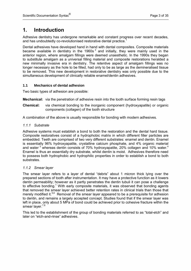
1. Introduction Adhesive dentistry has undergone remarkable and constant progress over recent decades, and has undoubtedly co-revolutionised restorative dental practice.1
Dental adhesives have developed hand in hand with dental composites. Composite materials became available in dentistry in the 1960s 2 and initially, they were mainly used in the anterior region, where amalgam fillings were deemed unaesthetic. In the 1990s they began to substitute amalgam as a universal filling material and composite restorations heralded a new minimally invasive era in dentistry. The retentive aspect of amalgam fillings was no longer necessary as the hole to be filled, had only to be as large as the demineralised tissue to be removed. This new development in restorative dentistry was only possible due to the simultaneous development of clinically reliable enamel/dentin adhesives.
1.1 Mechanics of dental adhesion
Two basic types of adhesion are possible:
Mechanical: via the penetration of adhesive resin into the tooth surface forming resin tags
Chemical: via chemical bonding to the inorganic component (hydroxyapatite) or organic components (collagen) of the tooth structure
A combination of the above is usually responsible for bonding with modern adhesives.
1.1.1 Substrate
Adhesive systems must establish a bond to both the restoration and the dental hard tissue. Composite restoratives consist of a hydrophobic matrix in which different filler particles are embedded. Teeth are comprised of two very different substrates: enamel and dentin. Enamel is essentially 96% hydroxyapatite, crystalline calcium phosphate, and 4% organic material and water 3 whereas dentin consists of 70% hydroxyapatite, 20% collagen and 10% water.4 Enamel is thus an essentially dry substrate, whilst dentin is moist. Adhesives therefore need to possess both hydrophobic and hydrophilic properties in order to establish a bond to both substrates.
1.1.2 Smear layer
The smear layer refers to a layer of dental “debris” about 1 micron thick lying over the prepared sections of tooth after instrumentation. It may have a protective function as it lowers dentin permeability; however as it partly penetrates the dentin tubuli it can pose a challenge to effective bonding.1 With early composite materials, it was observed that bonding agents that removed the smear layer achieved better retention rates in clinical trials than those that merely modified it.5,6 Removal of the smear layer appeared to be a prerequisite for adhesion to dentin, and remains a largely accepted concept. Studies found that if the smear layer was left in place, only about 5 MPa of bond could be achieved prior to cohesive fracture within the smear layer.7,8
This led to the establishment of the group of bonding materials referred to as “total-etch” and later on “etch-and-rinse” adhesives.
Scientific Documentation Syntac® Page 4 of 35
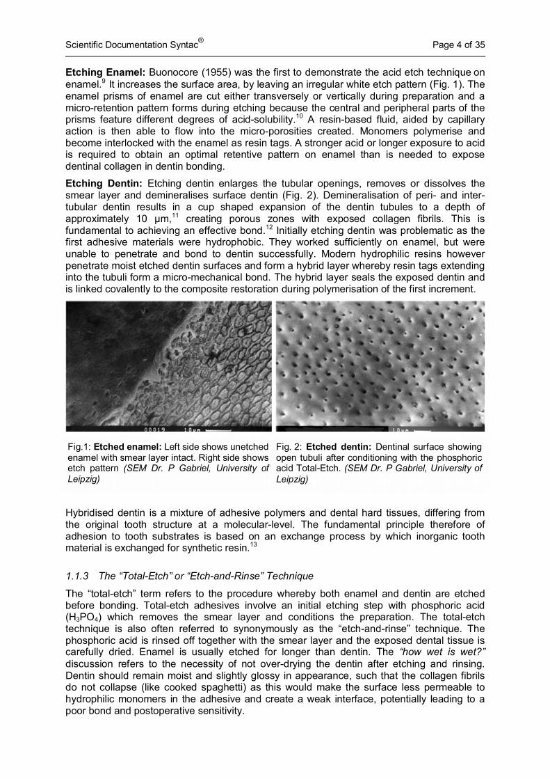
Etching Enamel: Buonocore (1955) was the first to demonstrate the acid etch technique on enamel.9 It increases the surface area, by leaving an irregular white etch pattern (Fig. 1). The enamel prisms of enamel are cut either transversely or vertically during preparation and a micro-retention pattern forms during etching because the central and peripheral parts of the prisms feature different degrees of acid-solubility.10 A resin-based fluid, aided by capillary action is then able to flow into the micro-porosities created. Monomers polymerise and become interlocked with the enamel as resin tags. A stronger acid or longer exposure to acid is required to obtain an optimal retentive pattern on enamel than is needed to expose dentinal collagen in dentin bonding.
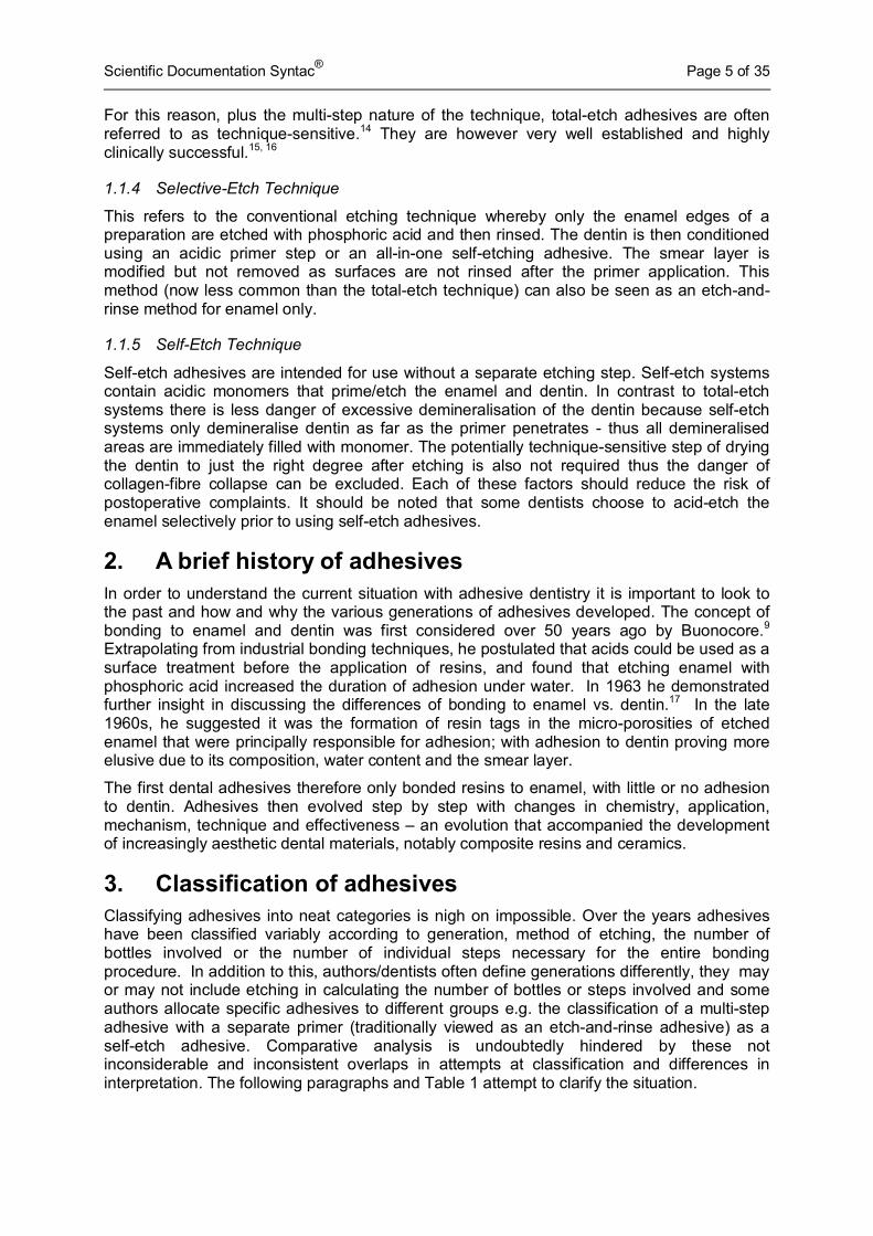
Etching Dentin: Etching dentin enlarges the tubular openings, removes or dissolves the smear layer and demineralises surface dentin (Fig. 2). Demineralisation of peri- and inter-tubular dentin results in a cup shaped expansion of the dentin tubules to a depth of approximately 10 µm,11 creating porous zones with exposed collagen fibrils. This is fundamental to achieving an effective bond.12 Initially etching dentin was problematic as the first adhesive materials were hydrophobic. They worked sufficiently on enamel, but were unable to penetrate and bond to dentin successfully. Modern hydrophilic resins however penetrate moist etched dentin surfaces and form a hybrid layer whereby resin tags extending into the tubuli form a micro-mechanical bond. The hybrid layer seals the exposed dentin and is linked covalently to the composite restoration during polymerisation of the first increment.
Hybridised dentin is a mixture of adhesive polymers and dental hard tissues, differing from the original tooth structure at a molecular-level. The fundamental principle therefore of adhesion to tooth substrates is based on an exchange process by which inorganic tooth material is exchanged for synthetic resin.13
1.1.3 The “Total-Etch” or “Etch-and-Rinse” Technique
The “total-etch” term refers to the procedure whereby both enamel and dentin are etched before bonding. Total-etch adhesives involve an initial etching step with phosphoric acid (H3PO4) which removes the smear layer and conditions the preparation. The total-etch technique is also often referred to synonymously as the “etch-and-rinse” technique. The phosphoric acid is rinsed off together with the smear layer and the exposed dental tissue is carefully dried. Enamel is usually etched for longer than dentin. The “how wet is wet?” discussion refers to the necessity of not over-drying the dentin after etching and rinsing. Dentin should remain moist and slightly glossy in appearance, such that the collagen fibrils do not collapse (like cooked spaghetti) as this would make the surface less permeable to hydrophilic monomers in the adhesive and create a weak interface, potentially leading to a poor bond and postoperative sensitivity.
Fig.1: Etched enamel: Left side shows unetched enamel with smear layer intact. Right side shows etch pattern (SEM Dr. P Gabriel, University of Leipzig)
Fig. 2: Etched dentin: Dentinal surface showing open tubuli after conditioning with the phosphoric acid Total-Etch. (SEM Dr. P Gabriel, University of Leipzig)
Scientific Documentation Syntac® Page 5 of 35
For this reason, plus the multi-step nature of the technique, total-etch adhesives are often referred to as technique-sensitive.14 They are however very well established and highly clinically successful.15, 16
1.1.4 Selective-Etch Technique
This refers to the conventional etching technique whereby only the enamel edges of a preparation are etched with phosphoric acid and then rinsed. The dentin is then conditioned using an acidic primer step or an all-in-one self-etching adhesive. The smear layer is modified but not removed as surfaces are not rinsed after the primer application. This method (now less common than the total-etch technique) can also be seen as an etch-and-rinse method for enamel only.
1.1.5 Self-Etch Technique
Self-etch adhesives are intended for use without a separate etching step. Self-etch systems contain acidic monomers that prime/etch the enamel and dentin. In contrast to total-etch systems there is less danger of excessive demineralisation of the dentin because self-etch systems only demineralise dentin as far as the primer penetrates - thus all demineralised areas are immediately filled with monomer. The potentially technique-sensitive step of drying the dentin to just the right degree after etching is also not required thus the danger of collagen-fibre collapse can be excluded. Each of these factors should reduce the risk of postoperative complaints. It should be noted that some dentists choose to acid-etch the enamel selectively prior to using self-etch adhesives.
2. A brief history of adhesives In order to understand the current situation with adhesive dentistry it is important to look to the past and how and why the various generations of adhesives developed. The concept of bonding to enamel and dentin was first considered over 50 years ago by Buonocore.9
Extrapolating from industrial bonding techniques, he postulated that acids could be used as a surface treatment before the application of resins, and found that etching enamel with phosphoric acid increased the duration of adhesion under water. In 1963 he demonstrated further insight in discussing the differences of bonding to enamel vs. dentin.17 In the late 1960s, he suggested it was the formation of resin tags in the micro-porosities of etched enamel that were principally responsible for adhesion; with adhesion to dentin proving more elusive due to its composition, water content and the smear layer.
The first dental adhesives therefore only bonded resins to enamel, with little or no adhesion to dentin. Adhesives then evolved step by step with changes in chemistry, application, mechanism, technique and effectiveness – an evolution that accompanied the development of increasingly aesthetic dental materials, notably composite resins and ceramics.
3. Classification of adhesives Classifying adhesives into neat categories is nigh on impossible. Over the years adhesives have been classified variably according to generation, method of etching, the number of bottles involved or the number of individual steps necessary for the entire bonding procedure. In addition to this, authors/dentists often define generations differently, they may or may not include etching in calculating the number of bottles or steps involved and some authors allocate specific adhesives to different groups e.g. the classification of a multi-step adhesive with a separate primer (traditionally viewed as an etch-and-rinse adhesive) as a self-etch adhesive. Comparative analysis is undoubtedly hindered by these not inconsiderable and inconsistent overlaps in attempts at classification and differences in interpretation. The following paragraphs and Table 1 attempt to clarify the situation.
Scientific Documentation Syntac® Page 6 of 35
3.1 Classification by generation
Dental adhesives can to a degree be categorised chronologically according to generation - a historical system of identification commonly used by adhesive manufacturers. The generation simply refers to when and in what order this type of adhesive was developed by the dental industry, ranging from 1st generation in the 1960s to modern 7th generation adhesives.
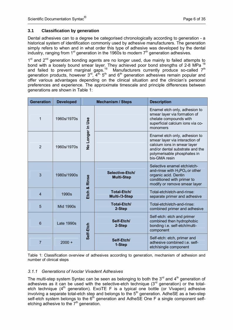
1st and 2nd generation bonding agents are no longer used, due mainly to failed attempts to bond with a loosely bound smear layer. They achieved poor bond strengths of 2-8 MPa 18 and failed to prevent marginal gaps.19 Manufacturers currently produce so-called 7th generation products, however 3rd, 4th, 5th and 6th generation adhesives remain popular and offer various advantages depending on the clinical situation and the clinician’s personal preferences and experience. The approximate timescale and principle differences between generations are shown in Table 1:
Generation Developed Mechanism / Steps Description
1 1960s/1970s
No
Lo
ng
er in
Use
Enamel etch only, adhesion to smear layer via formation of chelate compounds with superficial calcium ions via co-monomers
2 1960s/1970s
Enamel etch only, adhesion to smear layer via interaction of calcium ions in smear layer and/or dental substrate and the polymerisable phosphates in bis-GMA resin
3 1980s/1990s
Etc
h &
Rin
se Selective-Etch/
Multi-Step
Selective enamel etch/etch-and-rinse with H3PO4 or other organic acid. Dentin conditioned with primer to modify or remove smear layer
4 1990s Total-Etch/
Multi-/3-Step Total-etch/etch-and-rinse: separate primer and adhesive
5 Mid 1990s Total-Etch/
2-Step Total-etch/etch-and-rinse: combined primer and adhesive
6 Late 1990s
Sel
f-E
tch
Self-Etch/ 2-Step
Self-etch: etch and primer combined then hydrophobic bonding i.e. self-etch/multi-component
7 2000 + Self-Etch/
1-Step
Self-etch: etch, primer and adhesive combined i.e. self-etch/single component
Table 1: Classification overview of adhesives according to generation, mechanism of adhesion and number of clinical steps
3.1.1 Generations of Ivoclar Vivadent Adhesives
The multi-step system Syntac can be seen as belonging to both the 3rd and 4th generation of adhesives as it can be used with the selective-etch technique (3rd generation) or the total-etch technique (4th generation). ExciTE F is a typical one bottle (or Vivapen) adhesive involving a separate total-etch step and belongs to the 5th generation. AdheSE as a two-step self-etch system belongs to the 6th generation and AdheSE One F a single component self-etching adhesive to the 7th generation.
Scientific Documentation Syntac® Page 7 of 35
3.2 Classification by mechanism of adhesion / clinical steps
Whilst the generational system of classification is helpful for looking at adhesives from a historical perspective, with regard to adhesives currently on the market (generations 3-7), it may be more meaningful to classify them according to how they work and how many working steps are involved.
Modern dental adhesives can be classified into two basic types: etch-and-rinse and self- etch adhesives. Although the etch-and-rinse term is often used synonymously for total-etch adhesives theoretically it covers both total-etch and selective-etch adhesives (i.e. total-etch: both enamel and dentin are etched and rinsed; selective-etch: just the enamel is etched and rinsed). These systems can then be sub-categorized based on the number of clinical steps involved: e.g. multi-step, three-step and two-step etch-and-rinse systems and two-step and one-step self-etch systems.
The etch-and-rinse system is distinct in that it has a separate etch-and-rinse step prior to the priming and bonding steps. The three-step etch-and-rinse/total-etch system (using fourth-generation adhesives) follows the conventional “etch-rinse-prime-bond” approach. The two-step etch-and-rinse system (using fifth-generation adhesives, also known as one-bottle adhesives) combines the primer and the bonding agent into one application. The self-etch adhesive system eliminates the rinsing phase after etching by using non-rinse acidic monomers to etch and prime dentin simultaneously. The two-step self-etch system (involving sixth-generation adhesives) uses acidic monomers as self-etch primers in the initial step and an adhesive resin in the second step. The one-step self-etch system (using seventh generation adhesives, also known as all-in-one adhesives) combines the (self-etch) acidic primer with the adhesive resin in one application step. This allows for simultaneous infiltration of adhesive resin to the depth of demineralisation, which may reduce postoperative sensitivity.
To provide an overview of adhesives from both a historical and current perspective, Table 1 attempts to combine both methods of classification.
4. Ivoclar Vivadent Adhesives: Product Range Ivoclar Vivadent produces both total-etch and self-etch adhesives. There are valid pros and cons to both types of adhesive and of the multi-bottle/single bottle variants within these groups. Total-etch adhesives offer longer clinical experience and a more pronounced etch pattern in enamel and extensive removal of the smear layer. Self-etch adhesives on the other hand may be less technique sensitive,20 reducing the danger of collagen collapse and can be applied in fewer steps.
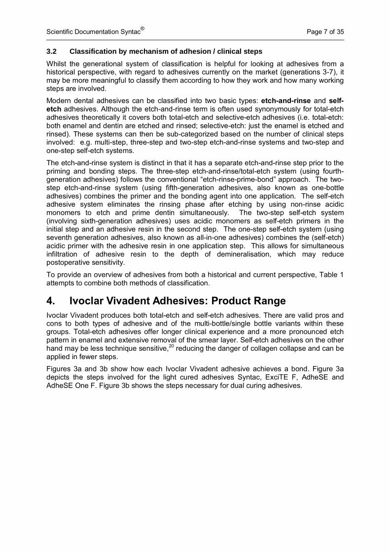
Figures 3a and 3b show how each Ivoclar Vivadent adhesive achieves a bond. Figure 3a depicts the steps involved for the light cured adhesives Syntac, ExciTE F, AdheSE and AdheSE One F. Figure 3b shows the steps necessary for dual curing adhesives.

Scientific Documentation Syntac® Page 8 of 35
Fig. 3a: Light curing adhesives. Fig. 3b: Dual curing adhesives
5. Syntac Syntac was introduced to the market in 1990. It is a traditional multi-step adhesive system often referred to in dental fields as “Syntac Classic” Although the suffix “classic” does not originate with Ivoclar Vivadent AG, the name is suitable as it embodies the traditional, long-term, well known and reliable aspects of the adhesive.
Syntac is a light-cured, multiple-component adhesive system with universal application fields. It can be used for bonding both direct restorations i.e. light, self and dual curing composites and compomers and indirect restorations i.e. metal-free restorations with light or dual curing luting composites such as Variolink II. The primer and adhesive contain no light curing initiators, thus Syntac is always applied in combination with the light-cured bonding material Heliobond.
Fig. 4: The Syntac Adhesive System: Syntac Primer, Syntac Adhesive and Heliobond
Scientific Documentation Syntac® Page 9 of 35
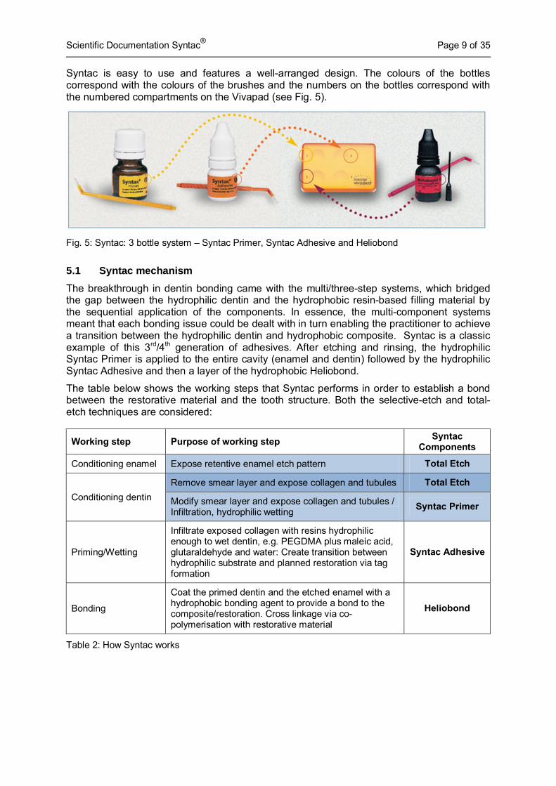
Syntac is easy to use and features a well-arranged design. The colours of the bottles correspond with the colours of the brushes and the numbers on the bottles correspond with the numbered compartments on the Vivapad (see Fig. 5).
Fig. 5: Syntac: 3 bottle system – Syntac Primer, Syntac Adhesive and Heliobond
5.1 Syntac mechanism
The breakthrough in dentin bonding came with the multi/three-step systems, which bridged the gap between the hydrophilic dentin and the hydrophobic resin-based filling material by the sequential application of the components. In essence, the multi-component systems meant that each bonding issue could be dealt with in turn enabling the practitioner to achieve a transition between the hydrophilic dentin and hydrophobic composite. Syntac is a classic example of this 3rd/4th generation of adhesives. After etching and rinsing, the hydrophilic Syntac Primer is applied to the entire cavity (enamel and dentin) followed by the hydrophilic Syntac Adhesive and then a layer of the hydrophobic Heliobond.
The table below shows the working steps that Syntac performs in order to establish a bond between the restorative material and the tooth structure. Both the selective-etch and total-etch techniques are considered:
Working step Purpose of working step Syntac
Components
Conditioning enamel Expose retentive enamel etch pattern Total Etch
Conditioning dentin
Remove smear layer and expose collagen and tubules Total Etch
Modify smear layer and expose collagen and tubules / Infiltration, hydrophilic wetting
Syntac Primer
Priming/Wetting
Infiltrate exposed collagen with resins hydrophilic enough to wet dentin, e.g. PEGDMA plus maleic acid, glutaraldehyde and water: Create transition between hydrophilic substrate and planned restoration via tag formation
Syntac Adhesive
Bonding
Coat the primed dentin and the etched enamel with a hydrophobic bonding agent to provide a bond to the composite/restoration. Cross linkage via co-polymerisation with restorative material
Heliobond
Table 2: How Syntac works
Scientific Documentation Syntac® Page 10 of 35
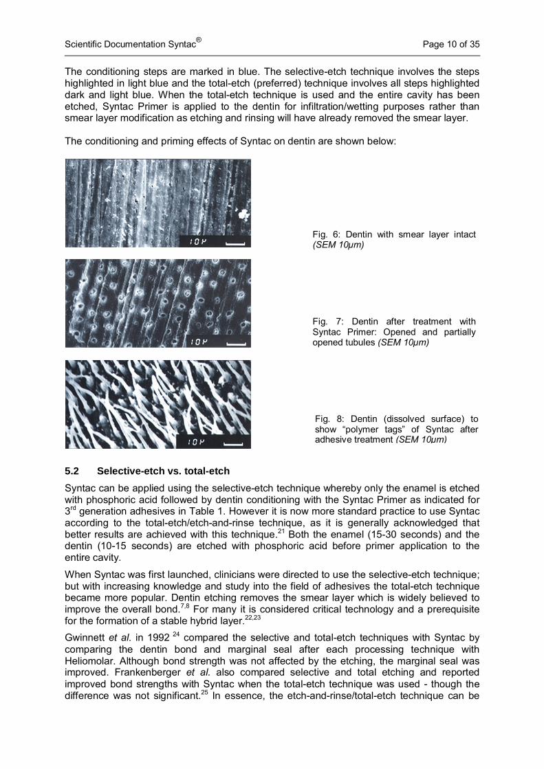
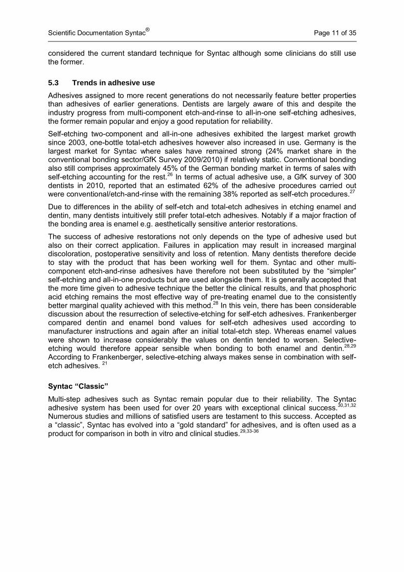
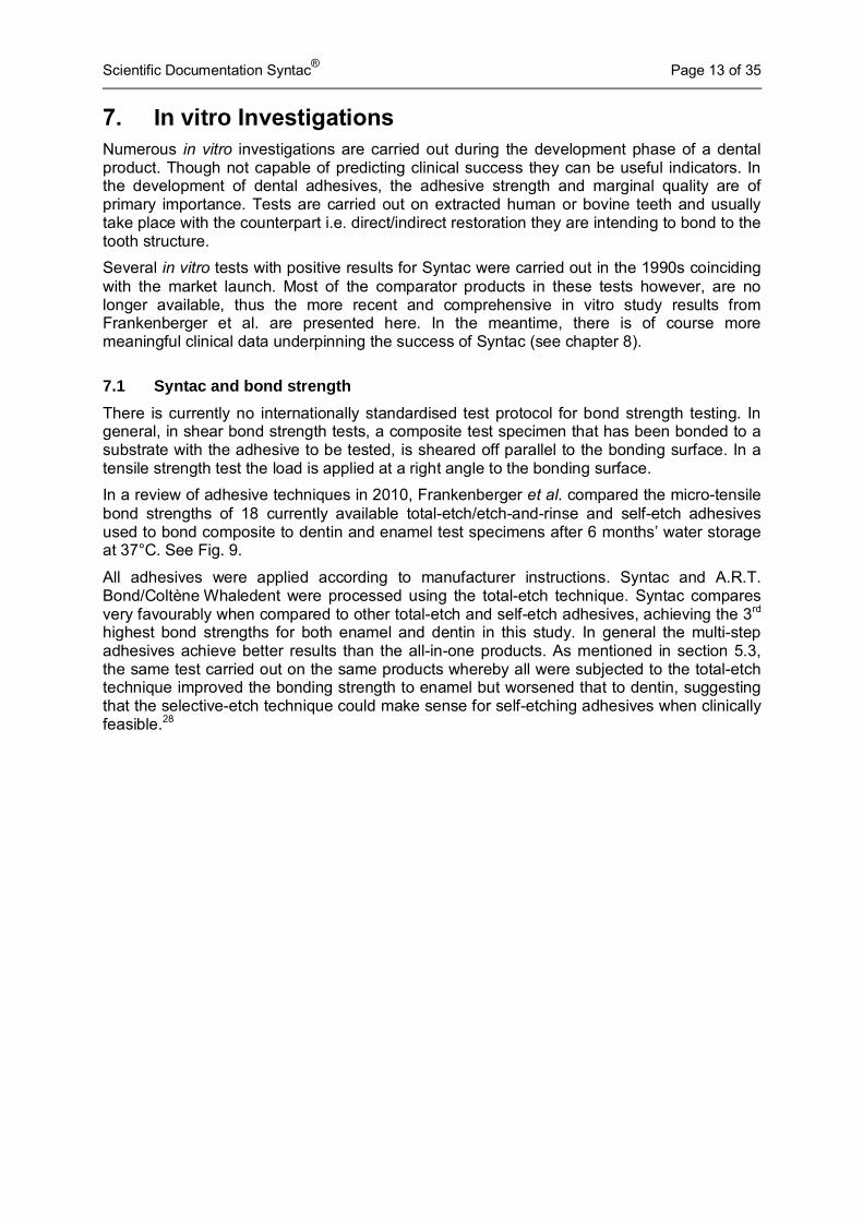
The conditioning steps are marked in blue. The selective-etch technique involves the steps highlighted in light blue and the total-etch (preferred) technique involves all steps highlighted dark and light blue. When the total-etch technique is used and the entire cavity has been etched, Syntac Primer is applied to the dentin for infiltration/wetting purposes rather than smear layer modification as etching and rinsing will have already removed the smear layer. The conditioning and priming effects of Syntac on dentin are shown below:
5.2 Selective-etch vs. total-etch
Syntac can be applied using the selective-etch technique whereby only the enamel is etched with phosphoric acid followed by dentin conditioning with the Syntac Primer as indicated for 3rd generation adhesives in Table 1. However it is now more standard practice to use Syntac according to the total-etch/etch-and-rinse technique, as it is generally acknowledged that better results are achieved with this technique.21 Both the enamel (15-30 seconds) and the dentin (10-15 seconds) are etched with phosphoric acid before primer application to the entire cavity.
When Syntac was first launched, clinicians were directed to use the selective-etch technique; but with increasing knowledge and study into the field of adhesives the total-etch technique became more popular. Dentin etching removes the smear layer which is widely believed to improve the overall bond.7,8 For many it is considered critical technology and a prerequisite for the formation of a stable hybrid layer.22,23
Gwinnett et al. in 1992 24 compared the selective and total-etch techniques with Syntac by comparing the dentin bond and marginal seal after each processing technique with Heliomolar. Although bond strength was not affected by the etching, the marginal seal was improved. Frankenberger et al. also compared selective and total etching and reported improved bond strengths with Syntac when the total-etch technique was used - though the difference was not significant.25 In essence, the etch-and-rinse/total-etch technique can be
Fig. 6: Dentin with smear layer intact (SEM 10µm)
Fig. 7: Dentin after treatment with Syntac Primer: Opened and partially opened tubules (SEM 10µm)
Fig. 8: Dentin (dissolved surface) to show “polymer tags” of Syntac after adhesive treatment (SEM 10µm)
Scientific Documentation Syntac® Page 11 of 35
considered the current standard technique for Syntac although some clinicians do still use the former.
5.3 Trends in adhesive use
Adhesives assigned to more recent generations do not necessarily feature better properties than adhesives of earlier generations. Dentists are largely aware of this and despite the industry progress from multi-component etch-and-rinse to all-in-one self-etching adhesives, the former remain popular and enjoy a good reputation for reliability.
Self-etching two-component and all-in-one adhesives exhibited the largest market growth since 2003, one-bottle total-etch adhesives however also increased in use. Germany is the largest market for Syntac where sales have remained strong (24% market share in the conventional bonding sector/GfK Survey 2009/2010) if relatively static. Conventional bonding also still comprises approximately 45% of the German bonding market in terms of sales with self-etching accounting for the rest.26 In terms of actual adhesive use, a GfK survey of 300 dentists in 2010, reported that an estimated 62% of the adhesive procedures carried out were conventional/etch-and-rinse with the remaining 38% reported as self-etch procedures.27
Due to differences in the ability of self-etch and total-etch adhesives in etching enamel and dentin, many dentists intuitively still prefer total-etch adhesives. Notably if a major fraction of the bonding area is enamel e.g. aesthetically sensitive anterior restorations.
The success of adhesive restorations not only depends on the type of adhesive used but also on their correct application. Failures in application may result in increased marginal discoloration, postoperative sensitivity and loss of retention. Many dentists therefore decide to stay with the product that has been working well for them. Syntac and other multi-component etch-and-rinse adhesives have therefore not been substituted by the “simpler” self-etching and all-in-one products but are used alongside them. It is generally accepted that the more time given to adhesive technique the better the clinical results, and that phosphoric acid etching remains the most effective way of pre-treating enamel due to the consistently better marginal quality achieved with this method.28 In this vein, there has been considerable discussion about the resurrection of selective-etching for self-etch adhesives. Frankenberger compared dentin and enamel bond values for self-etch adhesives used according to manufacturer instructions and again after an initial total-etch step. Whereas enamel values were shown to increase considerably the values on dentin tended to worsen. Selective-etching would therefore appear sensible when bonding to both enamel and dentin.28,29 According to Frankenberger, selective-etching always makes sense in combination with self-etch adhesives. 21
Syntac “Classic”
Multi-step adhesives such as Syntac remain popular due to their reliability. The Syntac adhesive system has been used for over 20 years with exceptional clinical success.30,31,32 Numerous studies and millions of satisfied users are testament to this success. Accepted as a “classic”, Syntac has evolved into a “gold standard” for adhesives, and is often used as a product for comparison in both in vitro and clinical studies.29,33-36

Scientific Documentation Syntac® Page 12 of 35
6. Technical Data Syntac Primer & Syntac Adhesive 2- phase adhesive system Standard – Composition (in weight %)
Primer Dimethacrylates 25.0
Maleic acid 4.0
Solvent 71.0
Stabilizer < 0.1
Adhesive Dimethacrylate 35.0
Maleic acid < 0.01
Glutaraldehyde 5.0
Water 60.0
Physical property Shear bond strength on dentin > 12 MPa
Heliobond Light curing bonding agent Standard – Composition
(in weight %)
Bis-GMA 59.5
Triethylenglycole dimethacrylate 39.7
Stabilizers and Catalysts 0.8
Physical properties Vickers hardness HV 0.2/30 180 MPa
Refraction index nD25 1.5129
Scientific Documentation Syntac® Page 13 of 35
7. In vitro Investigations Numerous in vitro investigations are carried out during the development phase of a dental product. Though not capable of predicting clinical success they can be useful indicators. In the development of dental adhesives, the adhesive strength and marginal quality are of primary importance. Tests are carried out on extracted human or bovine teeth and usually take place with the counterpart i.e. direct/indirect restoration they are intending to bond to the tooth structure.
Several in vitro tests with positive results for Syntac were carried out in the 1990s coinciding with the market launch. Most of the comparator products in these tests however, are no longer available, thus the more recent and comprehensive in vitro study results from Frankenberger et al. are presented here. In the meantime, there is of course more meaningful clinical data underpinning the success of Syntac (see chapter 8).
7.1 Syntac and bond strength
There is currently no internationally standardised test protocol for bond strength testing. In general, in shear bond strength tests, a composite test specimen that has been bonded to a substrate with the adhesive to be tested, is sheared off parallel to the bonding surface. In a tensile strength test the load is applied at a right angle to the bonding surface.
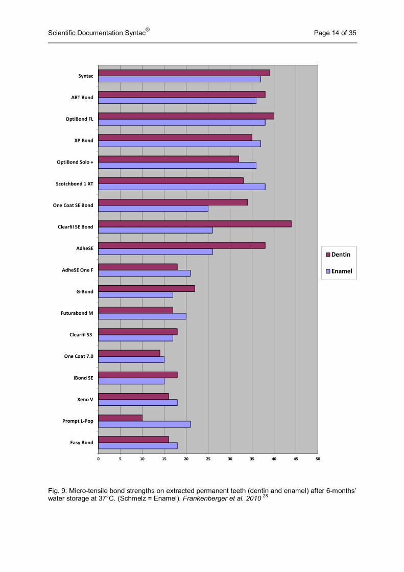
In a review of adhesive techniques in 2010, Frankenberger et al. compared the micro-tensile bond strengths of 18 currently available total-etch/etch-and-rinse and self-etch adhesives used to bond composite to dentin and enamel test specimens after 6 months’ water storage at 37°C. See Fig. 9.
All adhesives were applied according to manufacturer instructions. Syntac and A.R.T. Bond/Coltène Whaledent were processed using the total-etch technique. Syntac compares very favourably when compared to other total-etch and self-etch adhesives, achieving the 3rd highest bond strengths for both enamel and dentin in this study. In general the multi-step adhesives achieve better results than the all-in-one products. As mentioned in section 5.3, the same test carried out on the same products whereby all were subjected to the total-etch technique improved the bonding strength to enamel but worsened that to dentin, suggesting that the selective-etch technique could make sense for self-etching adhesives when clinically feasible.28
Scientific Documentation Syntac® Page 14 of 35
Fig. 9: Micro-tensile bond strengths on extracted permanent teeth (dentin and enamel) after 6-months’ water storage at 37°C. (Schmelz = Enamel). Frankenberger et al. 2010 28
0 5 10 15 20 25 30 35 40 45 50
Easy Bond
Prompt LPop
Xeno V
iBond SE
One Coat 7.0
Clearfil S3
Futurabond M
GBond
AdheSE One F
AdheSE
Clearfil SE Bond
One Coat SE Bond
Scotchbond 1 XT
OptiBond Solo +
XP Bond
OptiBond FL
ART Bond
Syntac
Dentin
Enamel
Scientific Documentation Syntac® Page 15 of 35
7.2 Syntac and marginal integrity
Microleakage can be defined as the clinically undetectable passage of bacteria, fluids or molecules between the cavity wall and the restorative material. Marginal leakage may cause sensitivity, discoloration of margins and secondary caries. It is usually measured using extracted teeth that have been subjected to temperature changes or mechanical loading. In functional evaluations, the marginal seal is assessed by means of dye penetration, whereas in morphological evaluations the quality is evaluated by means of replica investigation under the scanning electron microscope (SEM).
Frankenberger (2006) considered bonding to enamel and dentin and marginal quality amongst various total-etch/etch-and-rinse and self-etch adhesives used for bonding Class II cavities before and after chewing simulation. He evaluated the percentage of perfect margin and in enamel demonstrated that the total-etch adhesives produced better results than the self-etch 2-step and 1-step adhesives (respectively) both before and after chewing simulation. In dentin the multi-step systems produced clearly better results than the all-in-one adhesives.37
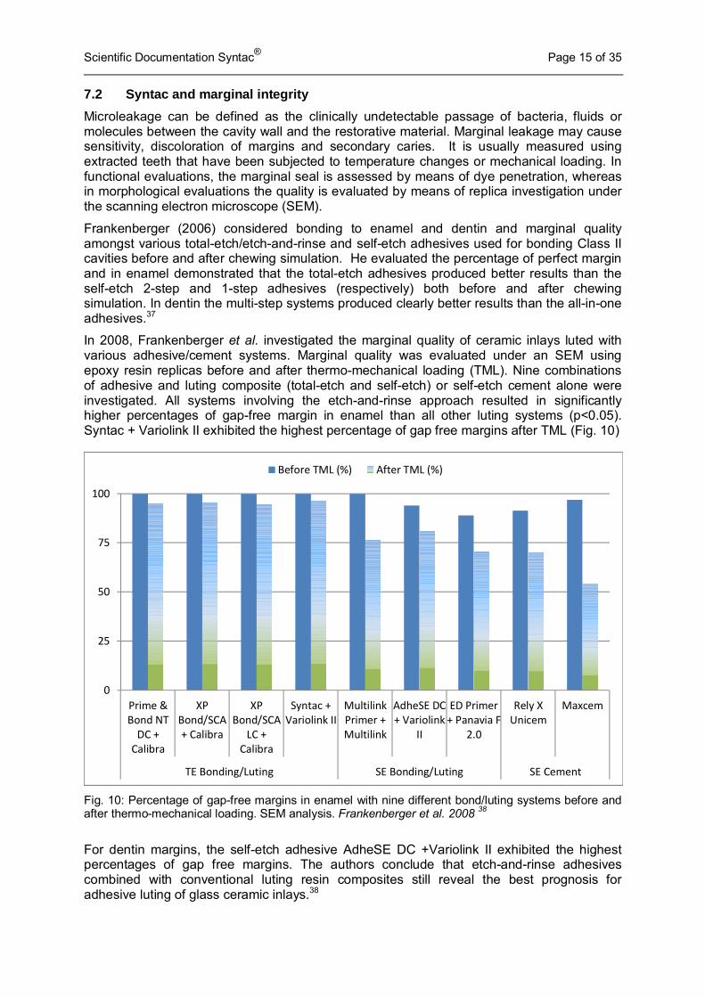
In 2008, Frankenberger et al. investigated the marginal quality of ceramic inlays luted with various adhesive/cement systems. Marginal quality was evaluated under an SEM using epoxy resin replicas before and after thermo-mechanical loading (TML). Nine combinations of adhesive and luting composite (total-etch and self-etch) or self-etch cement alone were investigated. All systems involving the etch-and-rinse approach resulted in significantly higher percentages of gap-free margin in enamel than all other luting systems (p<0.05). Syntac + Variolink II exhibited the highest percentage of gap free margins after TML (Fig. 10)
Fig. 10: Percentage of gap-free margins in enamel with nine different bond/luting systems before and after thermo-mechanical loading. SEM analysis. Frankenberger et al. 2008 38
For dentin margins, the self-etch adhesive AdheSE DC +Variolink II exhibited the highest percentages of gap free margins. The authors conclude that etch-and-rinse adhesives combined with conventional luting resin composites still reveal the best prognosis for adhesive luting of glass ceramic inlays.38
0
25
50
75
100
Prime &
Bond NT
DC +
Calibra
XP
Bond/SCA
+ Calibra
XP
Bond/SCA
LC +
Calibra
Syntac +
Variolink II
Multilink
Primer +
Multilink
AdheSE DC
+ Variolink
II
ED Primer
+ Panavia F
2.0
Rely X
Unicem
Maxcem
TE Bonding/Luting SE Bonding/Luting SE Cement
Before TML (%) After TML (%)
Scientific Documentation Syntac® Page 16 of 35
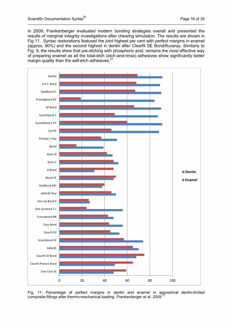
In 2009, Frankenberger evaluated modern bonding strategies overall and presented the results of marginal integrity investigations after chewing simulation. The results are shown in Fig 11. Syntac restorations featured the joint highest per cent with perfect margins in enamel (approx. 90%) and the second highest in dentin after Clearfil SE Bond/Kuraray. Similarly to Fig. 9, the results show that pre-etching with phosphoric acid, remains the most effective way of preparing enamel as all the total-etch (etch-and-rinse) adhesives show significantly better margin quality than the self-etch adhesives.21
Fig. 11: Percentage of perfect margins in dentin and enamel in approximal dentin-limited composite fillings after thermo-mechanical loading. Frankenberger et al. 2009 21
0 20 40 60 80 100
One Coat SE
Clearfil Protect Bond
Clearfil SE Bond
AdheSE
Scotchbond SE
Clearfil S3
Easy Bond
Futurabond NR
One Up Bond F +
One Up Bond F
AdheSE One
OptiBond AIO
iBond SE
GBond
Xeno V
Xeno III
iBond
Prompt LPop
ExciTE
Scotchbond 1 XT
Scotchbond 1
XP Bond
Prime&Bond NT
OptiBond FL
A.R.T. Bond
Syntac
Dentin
Enamel
Scientific Documentation Syntac® Page 17 of 35
8. Clinical Investigations Clinical trials remain the ultimate way to collect scientific evidence on the clinical effectiveness of an adhesive/restorative treatment.39 Both external and internal clinical trials were undertaken and as Syntac has been on the market for over two decades a review of the clinical literature was carried out.
8.1 External controlled clinical studies
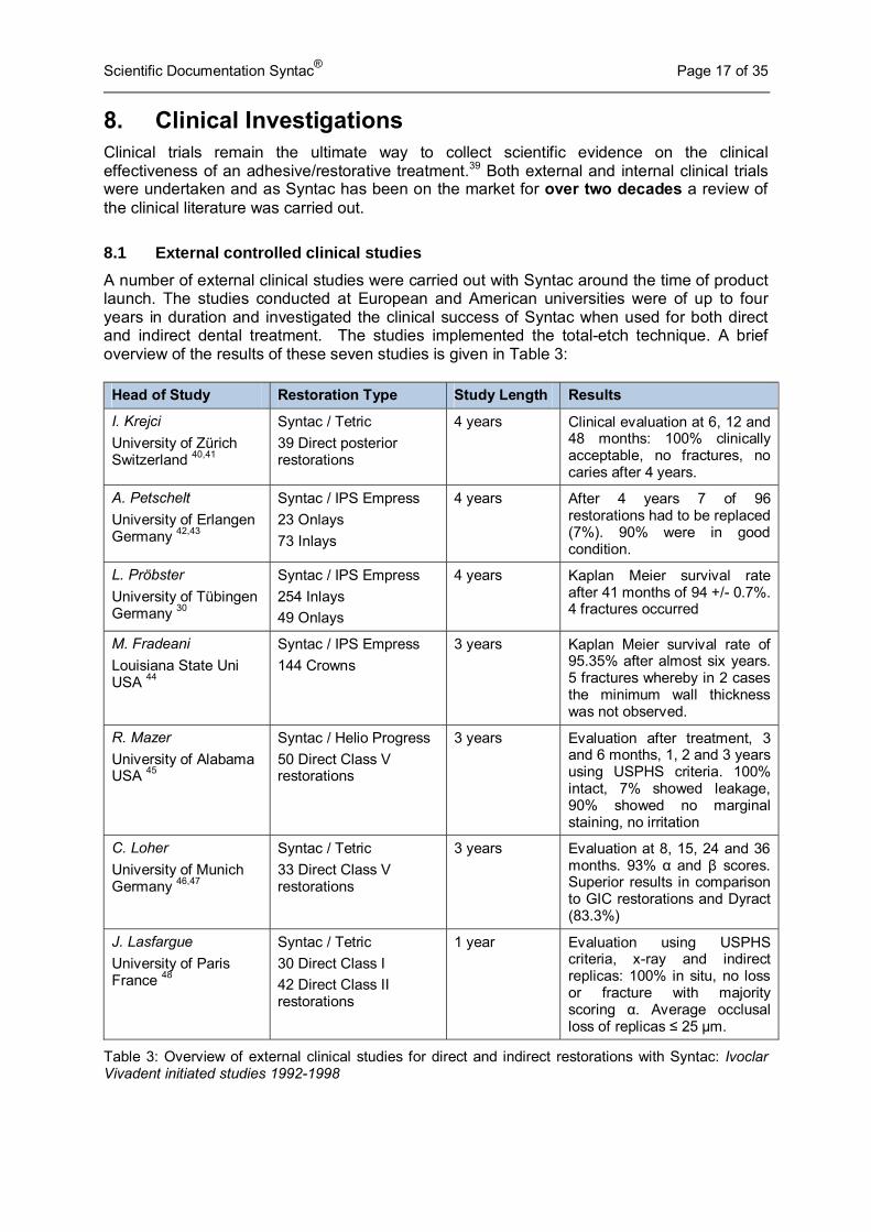
A number of external clinical studies were carried out with Syntac around the time of product launch. The studies conducted at European and American universities were of up to four years in duration and investigated the clinical success of Syntac when used for both direct and indirect dental treatment. The studies implemented the total-etch technique. A brief overview of the results of these seven studies is given in Table 3: Head of Study Restoration Type Study Length Results
I. Krejci
University of Zürich Switzerland 40,41
Syntac / Tetric
39 Direct posterior restorations
4 years Clinical evaluation at 6, 12 and 48 months: 100% clinically acceptable, no fractures, no caries after 4 years.
A. Petschelt
University of Erlangen Germany 42,43
Syntac / IPS Empress
23 Onlays
73 Inlays
4 years After 4 years 7 of 96 restorations had to be replaced (7%). 90% were in good condition.
L. Pröbster
University of Tübingen Germany 30
Syntac / IPS Empress
254 Inlays
49 Onlays
4 years Kaplan Meier survival rate after 41 months of 94 +/- 0.7%. 4 fractures occurred
M. Fradeani
Louisiana State Uni USA 44
Syntac / IPS Empress
144 Crowns
3 years Kaplan Meier survival rate of 95.35% after almost six years. 5 fractures whereby in 2 cases the minimum wall thickness was not observed.
R. Mazer
University of Alabama USA 45
Syntac / Helio Progress
50 Direct Class V restorations
3 years Evaluation after treatment, 3 and 6 months, 1, 2 and 3 years using USPHS criteria. 100% intact, 7% showed leakage, 90% showed no marginal staining, no irritation
C. Loher
University of Munich Germany 46,47
Syntac / Tetric
33 Direct Class V restorations
3 years Evaluation at 8, 15, 24 and 36 months. 93% α and β scores. Superior results in comparison to GIC restorations and Dyract (83.3%)
J. Lasfargue
University of Paris France 48
Syntac / Tetric
30 Direct Class I
42 Direct Class II restorations
1 year Evaluation using USPHS criteria, x-ray and indirect replicas: 100% in situ, no loss or fracture with majority scoring α. Average occlusal loss of replicas ≤ 25 µm.
Table 3: Overview of external clinical studies for direct and indirect restorations with Syntac: Ivoclar Vivadent initiated studies 1992-1998
Scientific Documentation Syntac® Page 18 of 35
8.2 Internal controlled clinical studies
Two internal studies at Ivoclar Vivadent were recently conducted using the composites Tetric EvoCeram and IPS Empress Direct. Syntac was used as the bonding agent in both studies:
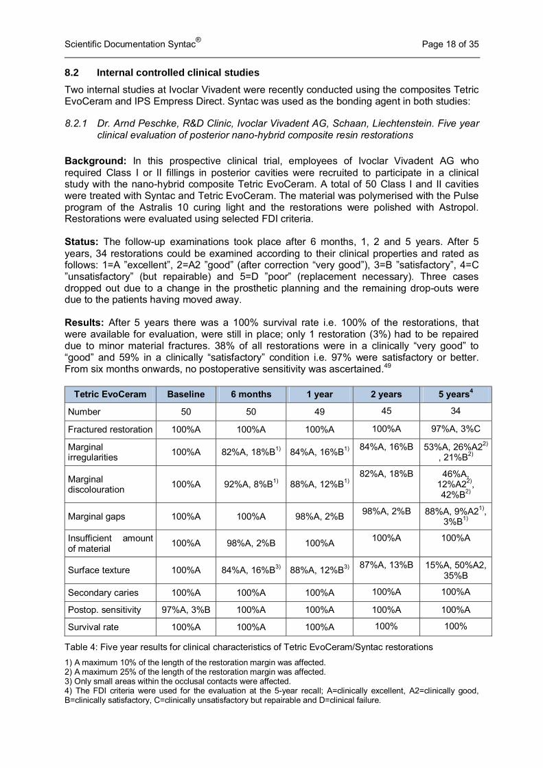
8.2.1 Dr. Arnd Peschke, R&D Clinic, Ivoclar Vivadent AG, Schaan, Liechtenstein. Five year clinical evaluation of posterior nano-hybrid composite resin restorations
Background: In this prospective clinical trial, employees of Ivoclar Vivadent AG who required Class I or II fillings in posterior cavities were recruited to participate in a clinical study with the nano-hybrid composite Tetric EvoCeram. A total of 50 Class I and II cavities were treated with Syntac and Tetric EvoCeram. The material was polymerised with the Pulse program of the Astralis 10 curing light and the restorations were polished with Astropol. Restorations were evaluated using selected FDI criteria. Status: The follow-up examinations took place after 6 months, 1, 2 and 5 years. After 5 years, 34 restorations could be examined according to their clinical properties and rated as follows: 1=A ”excellent”, 2=A2 ”good” (after correction “very good”), 3=B ”satisfactory”, 4=C ”unsatisfactory” (but repairable) and 5=D ”poor” (replacement necessary). Three cases dropped out due to a change in the prosthetic planning and the remaining drop-outs were due to the patients having moved away. Results: After 5 years there was a 100% survival rate i.e. 100% of the restorations, that were available for evaluation, were still in place; only 1 restoration (3%) had to be repaired due to minor material fractures. 38% of all restorations were in a clinically “very good” to “good” and 59% in a clinically “satisfactory” condition i.e. 97% were satisfactory or better. From six months onwards, no postoperative sensitivity was ascertained.49
Tetric EvoCeram Baseline 6 months 1 year 2 years 5 years4
Number 50 50 49 45 34
Fractured restoration 100%A 100%A 100%A 100%A 97%A, 3%C
Marginal irregularities
100%A 82%A, 18%B1) 84%A, 16%B1) 84%A, 16%B 53%A, 26%A22)
, 21%B2)
Marginal discolouration
100%A 92%A, 8%B1) 88%A, 12%B1) 82%A, 18%B 46%A,
12%A22), 42%B2)
Marginal gaps 100%A 100%A 98%A, 2%B 98%A, 2%B 88%A, 9%A21),
3%B1)
Insufficient amount of material
100%A 98%A, 2%B 100%A 100%A 100%A
Surface texture 100%A 84%A, 16%B3) 88%A, 12%B3) 87%A, 13%B 15%A, 50%A2,
35%B
Secondary caries 100%A 100%A 100%A 100%A 100%A
Postop. sensitivity 97%A, 3%B 100%A 100%A 100%A 100%A
Survival rate 100%A 100%A 100%A 100% 100%
Table 4: Five year results for clinical characteristics of Tetric EvoCeram/Syntac restorations
1) A maximum 10% of the length of the restoration margin was affected. 2) A maximum 25% of the length of the restoration margin was affected. 3) Only small areas within the occlusal contacts were affected. 4) The FDI criteria were used for the evaluation at the 5-year recall; A=clinically excellent, A2=clinically good, B=clinically satisfactory, C=clinically unsatisfactory but repairable and D=clinical failure.
Scientific Documentation Syntac® Page 19 of 35
Conclusion: After an observation period of 5 years, all restorations, that were available for evaluation, were still in place and no absolute failure was observed. Only one restoration required minor repair work due to chipping. Documented marginal flaws affected only small portions of the total margin length. The combination of Tetric EvoCeram and Syntac showed a very reliable clinical performance after 5 years in posterior restorations and an outstanding marginal quality.
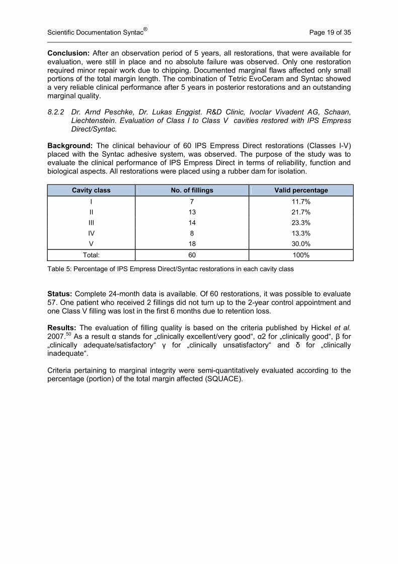
8.2.2 Dr. Arnd Peschke, Dr. Lukas Enggist. R&D Clinic, Ivoclar Vivadent AG, Schaan, Liechtenstein. Evaluation of Class I to Class V cavities restored with IPS Empress Direct/Syntac.
Background: The clinical behaviour of 60 IPS Empress Direct restorations (Classes I-V) placed with the Syntac adhesive system, was observed. The purpose of the study was to evaluate the clinical performance of IPS Empress Direct in terms of reliability, function and biological aspects. All restorations were placed using a rubber dam for isolation.
Table 5: Percentage of IPS Empress Direct/Syntac restorations in each cavity class Status: Complete 24-month data is available. Of 60 restorations, it was possible to evaluate 57. One patient who received 2 fillings did not turn up to the 2-year control appointment and one Class V filling was lost in the first 6 months due to retention loss. Results: The evaluation of filling quality is based on the criteria published by Hickel et al. 2007.50 As a result α stands for „clinically excellent/very good“, α2 for „clinically good“, β for „clinically adequate/satisfactory“ γ for „clinically unsatisfactory“ and δ for „clinically inadequate“. Criteria pertaining to marginal integrity were semi-quantitatively evaluated according to the percentage (portion) of the total margin affected (SQUACE).
Cavity class No. of fillings Valid percentage
I 7 11.7%
II 13 21.7%
III 14 23.3%
IV 8 13.3%
V 18 30.0%
Total: 60 100%
Scientific Documentation Syntac® Page 20 of 35
Percent Assessment
Criteria Classification
Class I-V overall
(SD) Class I and
II (SD) Class III
and IV (SD) Class V
(SD) %
of
all R
esto
rati
on
s
Evaluated restorations (24 months) - 95 90 100 94,4
Survival rate - 98.3 100 100 94,4
Material fracture / retention
α 93.1 94.4 90.9 94.4
α2 0 0 0 0
β 5,2 5.6 9.1 0
γ 0 0 0 0
δ 1,7 0 0 5,6
Secondary caries α 100 100 100 100
α2-δ 0 0 0 0
Postoperative sensitivity
α 100 100 100 100
α2-δ 0 0 0 0
% o
f M
arg
in *
Marginal irregularities
α 97,2 (±5.3) 96,7 (±5,4) 97 (±5.7) 98 (±4.7)
α2 2.8 (±5.3) 3.3 (±5,4) 3.0 (±5.7) 2.0 (±4.7)
β-δ 0 0 0 0
Marginal discoloration
α 99.5 (±2.8) 98,3 (±4,6) 99.8(±1.2) 98.8 (±4.9)
α2 0.5 (±2.8) 0.3 (±1.2) 0.2 (±1) 1.2 (±4.9)
β-δ 0 0 0 0
Marginal gaps α 100 100 100 100
α2-δ 0 0 0 0
Lack of filling material
Α 100 100 100 100
α2-δ 0 0 0 0
Margin fracture Α 100 100 100 100
α2-δ 0 0 0 0
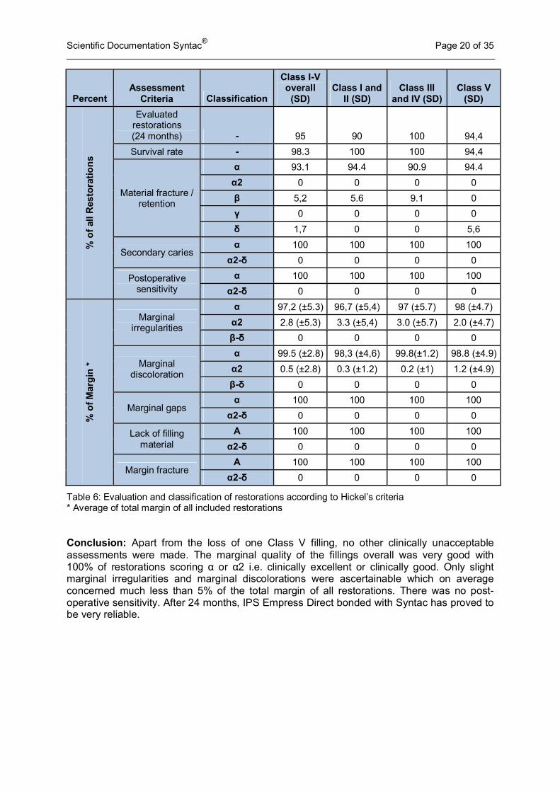
Table 6: Evaluation and classification of restorations according to Hickel’s criteria * Average of total margin of all included restorations Conclusion: Apart from the loss of one Class V filling, no other clinically unacceptable assessments were made. The marginal quality of the fillings overall was very good with 100% of restorations scoring α or α2 i.e. clinically excellent or clinically good. Only slight marginal irregularities and marginal discolorations were ascertainable which on average concerned much less than 5% of the total margin of all restorations. There was no post-operative sensitivity. After 24 months, IPS Empress Direct bonded with Syntac has proved to be very reliable.
Scientific Documentation Syntac® Page 21 of 35
8.3 Literature review: Direct restorations with Syntac
In contrast to dental composites, there are still considerable differences in performance among dental adhesives. This is impressively illustrated by recent reviews on clinical trials on posterior restorations 51 and adhesives.39 Posterior restorations using up-to-date composite materials mostly show annual failure rates of less than 3%.51 In contrast, annual failure rates of adhesive restorations in non-carious Class V lesions where macro-mechanical retention is at a minimum and thus the quality of adhesive bond paramount - vary between 0 – 48%. Furthermore, one-step self-etch adhesives (all-in-one) tend to exhibit significantly higher annual failure rates than multi-component, two-step total-etch and two-step self-etch adhesives. 52 A dentist’s choice of a clinically proven adhesive can therefore substantially contribute to the success of a restoration.
A search of the dental literature, yielded positive reinforcement for the use of Syntac in both direct and indirect clinical situations.
8.3.1 Class I and Class II restorations
Krämer et al. (2011), conducted a controlled prospective split-mouth study over six years to evaluate the clinical behaviour of two different resin composites in extended Class II cavities.53 Thirty patients received 68 direct resin composite restorations Solobond M + Grandio/Voco (n=36) or Syntac + Tetric Ceram (n=32). Restorations were examined according to modified USPHS criteria at baseline, after six months, one, two, four and six years. The survival rate for all restorations was 100% after six years, hypersensitivity was significantly reduced over time and no significant difference was found between the two restorative materials.53,54
Schirrmeister et al. evaluated the clinical performance of Ceram.X + an experimental one bottle etch-and-rinse adhesive (K-0127)/Dentsply compared to Tetric Ceram + Syntac which served as a control. This prospective study ran over four years with check-ups made at baseline, 1, 2 and 4 years. 43 patients received two (Ceram X/K-0127 and Tetric Ceram/Syntac) Class I or Class II molar restorations. At the 4 year recall, 27 patients could be examined. The cumulative failure rate for the Ceram X group was 7.4% and 3.7% in the Tetric Ceram group. Slight marginal discoloration was found in 19.2% of Ceram X restorations and in 15.4% of Tetric Ceram restorations. No sensitivity, recurrent caries or changes in surface texture were recorded after four years and no statistically significant differences were found between the two restorative systems (p > 0.05).36
Manhart et al. compared Quixfil plus the self-etch adhesive Xeno III/Dentsply (n= 46) with Tetric plus Syntac as a control (n=50) in a longitudinal randomised controlled trial over four years. Restorations were placed in stress–bearing Class I and II cavities in first or second molars. Clinical evaluation was performed at baseline and after 4 years using modified USPHS criteria. At the last recall 37 Quixfil and 46 Tetric Ceram restorations could be assessed. A total of 89.2% of Quixfil and 97.8% of Tetric Ceram composites were assessed to be clinically excellent or acceptable. Four Quixfil restorations failed due to bulk fracture, partial tooth fracture (n=2) and postoperative symptoms. One Tetric Ceram restoration was lost due to problems with tooth integrity. No significant differences between the two composites were detected at four years for any of the evaluated clinical criteria. Both sets of materials demonstrated good clinical results after four years.55
Scientific Documentation Syntac® Page 22 of 35
8.3.2 Class V restorations
Non-carious Class V restorations may be considered ideal for assessing the clinical effectiveness of adhesives as they provide little macro-mechanical retention, they involve at least 50% bonding to dentin and are widely available and accessible.39
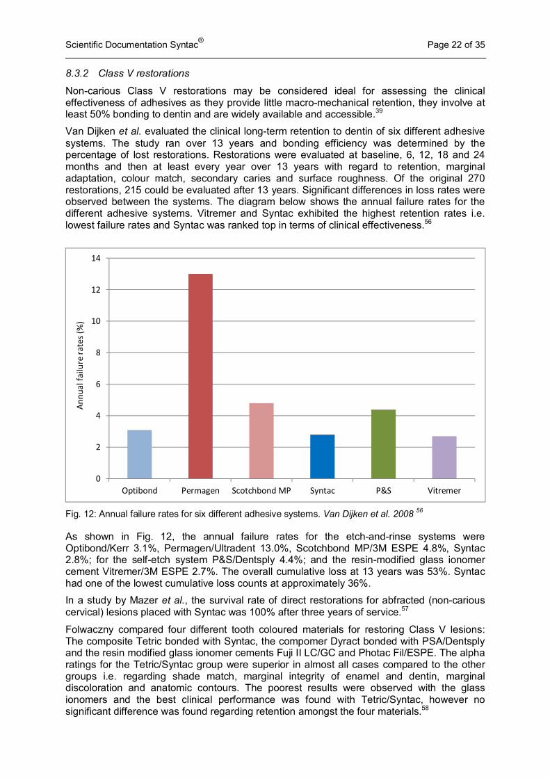
Van Dijken et al. evaluated the clinical long-term retention to dentin of six different adhesive systems. The study ran over 13 years and bonding efficiency was determined by the percentage of lost restorations. Restorations were evaluated at baseline, 6, 12, 18 and 24 months and then at least every year over 13 years with regard to retention, marginal adaptation, colour match, secondary caries and surface roughness. Of the original 270 restorations, 215 could be evaluated after 13 years. Significant differences in loss rates were observed between the systems. The diagram below shows the annual failure rates for the different adhesive systems. Vitremer and Syntac exhibited the highest retention rates i.e. lowest failure rates and Syntac was ranked top in terms of clinical effectiveness.56
Fig. 12: Annual failure rates for six different adhesive systems. Van Dijken et al. 2008 56
As shown in Fig. 12, the annual failure rates for the etch-and-rinse systems were Optibond/Kerr 3.1%, Permagen/Ultradent 13.0%, Scotchbond MP/3M ESPE 4.8%, Syntac 2.8%; for the self-etch system P&S/Dentsply 4.4%; and the resin-modified glass ionomer cement Vitremer/3M ESPE 2.7%. The overall cumulative loss at 13 years was 53%. Syntac had one of the lowest cumulative loss counts at approximately 36%.
In a study by Mazer et al., the survival rate of direct restorations for abfracted (non-carious cervical) lesions placed with Syntac was 100% after three years of service.57
Folwaczny compared four different tooth coloured materials for restoring Class V lesions: The composite Tetric bonded with Syntac, the compomer Dyract bonded with PSA/Dentsply and the resin modified glass ionomer cements Fuji II LC/GC and Photac Fil/ESPE. The alpha ratings for the Tetric/Syntac group were superior in almost all cases compared to the other groups i.e. regarding shade match, marginal integrity of enamel and dentin, marginal discoloration and anatomic contours. The poorest results were observed with the glass ionomers and the best clinical performance was found with Tetric/Syntac, however no significant difference was found regarding retention amongst the four materials.58
0
2
4
6
8
10
12
14
Optibond Permagen Scotchbond MP Syntac P&S Vitremer
An
nu
al fa
ilu
re r
ate
s (%
)
Scientific Documentation Syntac® Page 23 of 35
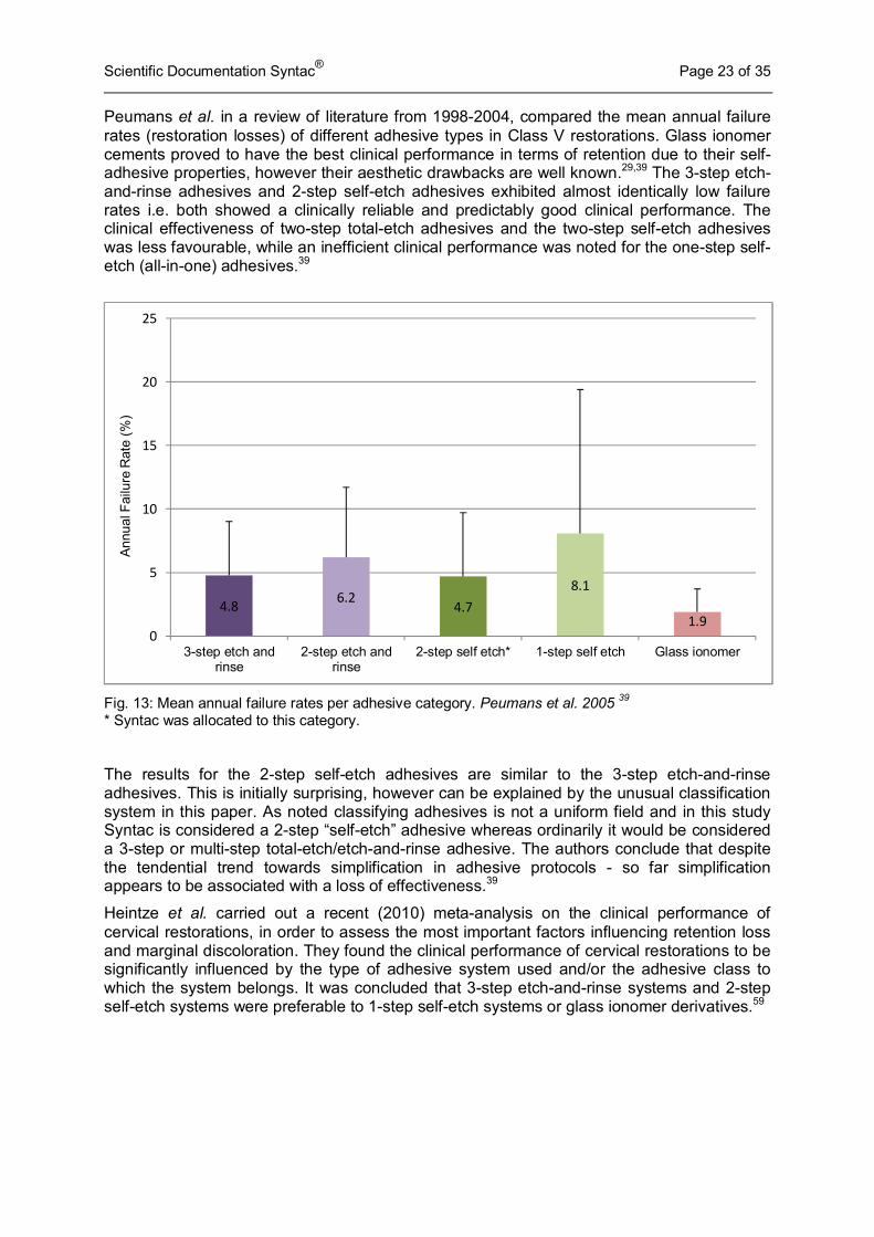
Peumans et al. in a review of literature from 1998-2004, compared the mean annual failure rates (restoration losses) of different adhesive types in Class V restorations. Glass ionomer cements proved to have the best clinical performance in terms of retention due to their self-adhesive properties, however their aesthetic drawbacks are well known.29,39 The 3-step etch-and-rinse adhesives and 2-step self-etch adhesives exhibited almost identically low failure rates i.e. both showed a clinically reliable and predictably good clinical performance. The clinical effectiveness of two-step total-etch adhesives and the two-step self-etch adhesives was less favourable, while an inefficient clinical performance was noted for the one-step self-etch (all-in-one) adhesives.39
Fig. 13: Mean annual failure rates per adhesive category. Peumans et al. 2005 39 * Syntac was allocated to this category.
The results for the 2-step self-etch adhesives are similar to the 3-step etch-and-rinse adhesives. This is initially surprising, however can be explained by the unusual classification system in this paper. As noted classifying adhesives is not a uniform field and in this study Syntac is considered a 2-step “self-etch” adhesive whereas ordinarily it would be considered a 3-step or multi-step total-etch/etch-and-rinse adhesive. The authors conclude that despite the tendential trend towards simplification in adhesive protocols - so far simplification appears to be associated with a loss of effectiveness.39
Heintze et al. carried out a recent (2010) meta-analysis on the clinical performance of cervical restorations, in order to assess the most important factors influencing retention loss and marginal discoloration. They found the clinical performance of cervical restorations to be significantly influenced by the type of adhesive system used and/or the adhesive class to which the system belongs. It was concluded that 3-step etch-and-rinse systems and 2-step self-etch systems were preferable to 1-step self-etch systems or glass ionomer derivatives.59
4.86.2
4.7
8.1
1.90
5
10
15
20
25
3-step etch andrinse
2-step etch andrinse
2-step self etch* 1-step self etch Glass ionomer
Ann
ual
Fai
lure
Rat
e (%
)
Scientific Documentation Syntac® Page 24 of 35
8.4 Literature review: Indirect restorations with Syntac
8.4.1 Inlays/Onlays
Survival rates of 92% after eight years’ service for IPS Empress inlays bonded with Syntac and the different composite systems: Tetric, Dual Cement, Variolink and Variolink Ultra, were reported by Krämer et al. in 2002.60,61 After 12 years’ service the restorations exhibited a survival rate of 84%, as 15 of the original 96 restorations had to be replaced, 12 due to bulk fracture. Significantly more bulk fractures were found when light-curing composite luting agents as opposed to dual-cured were used. No secondary caries was observed.62
In a later study, Krämer et al. conducted a split-mouth prospective clinical trial with 94 IPS Empress inlay/onlay restorations in 31 patients. Restorations were luted with either EBS Multi + Compolute/3M ESPE or Syntac + Variolink II (selective-etch method). Evaluation took place at baseline, and after 6 months, 1, 2, 4 and 8 years. The recall rate was 72% after eight years. The Kaplan Maier survival rate was 96% at 4 years and 90% at 8 years. Between the six recalls a statistically significant deterioration was found for marginal adaptation. Though not significant, numerically the EBS Multi group resulted in more postoperative sensitivity and the scans of the luting gap showed that Compolute was more prone to wear (p<0.05). Otherwise there were no significant differences between the luting systems.33,34
In a prospective comparison of the clinical performance of two different resin composites for luting IPS Empress inlays and onlays; 83 IPS Empress restorations were placed in 30 patients. 43 with the self-adhesive resin cement RelyX Unicem/3M ESPE and 40 with Syntac and Variolink II as a control. After 1 year of clinical service the Syntac group revealed significantly better results regarding colour match and integrity.35
Coelho Santos et al. compared sintered (Duceram/Dentsply-Degussa) and pressable (IPS Empress) ceramic inlays and onlays all luted with Syntac and Variolink II. After two years both systems demonstrated excellent clinical performance with 100% evaluated as clinically excellent or acceptable.63
CAD/CAM Inlays/Onlays
Bernhart et al. reported a survival rate of 95% after 3 years for Cerec Ceramic inlays luted with Syntac and Dual Cement. Restorations were carried out by dental students in their 4th semester after a short theoretical and practical course in the Cerec method. Good clinical results were thus possible despite limited operator experience, suggesting the low technique sensitivity of Syntac.64
Zimmer et al. conducted a 10 year study of Class I and II (inlays and onlays) CAD/CAM ceramic restorations bonded adhesively with Syntac and Vita Cerec Duo Cement/VITA. An initial 308 restorations were inserted into cavities in the posterior teeth of 95 patients between 1992 and 1994. 74 patients returned for the 10 year recall involving 226 restorations. Of these 39 were Class I and 187 Class II (23 onlays involving one or more cusp and 164 inlays). Kaplan-Meier survival analysis was carried out whereby the following criteria constituted failure: secondary decay, any kind of loss or fracture of the restoration, tooth fracture or marginal gap reaching dentin or base material. The survival rate was 94.7% after five years and 85.7% after 10 years. The authors also concluded that the Cerec 1 restorations as applied in this study were comparable to cast gold restorations.65
Scientific Documentation Syntac® Page 25 of 35
Reich et al. conducted a three year pilot study, whereby 58 large CAD/CAM-fabricated all ceramic restorations were placed on teeth with large coronal defects in 26 patients. Restorations varied as necessary from onlays, reduced crowns, classic crowns endo-crowns and veneers. Implant crowns were also included. The restorations were luted with Syntac and either Variolink Ultra or Tetric Ceram. After 3 years the restorations were evaluated according to USPHS criteria and 97% were rated “Bravo” (satisfactory) or better for marginal integrity, secondary caries, discoloration and anatomical form i.e. all but 2 of the adhesively luted restorations exhibited satisfactory clinical performance. These results were evaluated as exceptionally positive given the compromised nature of the teeth in this study i.e. including severe coronal destruction.66
Syntac clearly proves its suitability for use in the successful long term bonding of indirect CAD/CAM ceramic restorations.
8.4.2 Crowns
Guess et al. reported survival rates of 100% after three years for all ceramic IPS e.max Press crowns bonded with Syntac/Tetric. The study was designed to compare the clinical performance and survival of IPS e.max Press and CAD/CAM fabricated ProCAD crowns. 80 molars of 25 patients requiring at least 2 new crowns were treated in a split mouth prospective investigation. All crowns were luted with Syntac/Tetric. Clinical evaluation took place at baseline, 13, 25 and 36 months according to modified USPHS criteria. The results here are the mid-term (3-year) results of a 5 year study, and showed 100% survival for IPS e.max Press crowns and 97% for ProCAD due to one severe fracture at 9 months. There were no endodontic complications or cases of secondary caries; however both materials demonstrated significantly decreased marginal adaptation, discolorations and surface roughness over time. Both materials can be considered a reliable treatment option for the restoration of larger defects in the posterior dentition. Syntac proved itself a very reliable bonding agent.67
An eleven year study by Fradeani et al. investigated 125 IPS Empress crowns in 54 patients bonded with Syntac or Allbond 2/Bisco and luted with Dual Cement or Variolink. A survival rate for all crowns of 95.5% after eleven years was found with anterior crowns (98.9%) faring better than posterior (84.4%). In this paper it is not possible however to establish which crowns were bonded/luted with which material. 68
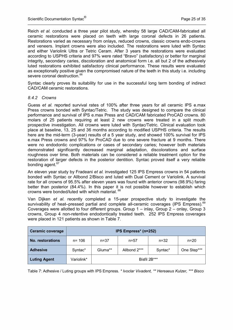
Van Dijken et al. recently completed a 15-year prospective study to investigate the survivability of heat–pressed partial and complete all-ceramic coverages (IPS Empress).69 Coverages were allotted to four different groups. Group 1 – inlay, Group 2 – onlay, Group 3 crowns, Group 4 non-retentive endodontically treated teeth. 252 IPS Empress coverages were placed in 121 patients as shown in Table 7.
Ceramic coverage IPS Empress* (n=252)
No. restorations n= 106 n=37 n=57 n=32 n=20
Adhesive Syntac* Gluma** Allbond 2*** Syntac* One Step***
Luting Agent Variolink* Bisfil 2B***
Table 7: Adhesive / Luting groups with IPS Empress. * Ivoclar Vivadent, ** Hereaeus Kulzer, *** Bisco
Scientific Documentation Syntac® Page 26 of 35
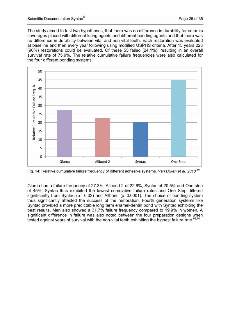
The study aimed to test two hypotheses, that there was no difference in durability for ceramic coverages placed with different luting agents and different bonding agents and that there was no difference in durability between vital and non-vital teeth. Each restoration was evaluated at baseline and then every year following using modified USPHS criteria. After 15 years 228 (90%) restorations could be evaluated. Of these 55 failed (24.1%), resulting in an overall survival rate of 75.9%. The relative cumulative failure frequencies were also calculated for the four different bonding systems.
Fig. 14: Relative cumulative failure frequency of different adhesive systems. Van Dijken et al. 2010 69 Gluma had a failure frequency of 27.3%, Allbond 2 of 22.6%, Syntac of 20.5% and One step of 45%. Syntac thus exhibited the lowest cumulative failure rates and One Step differed significantly from Syntac (p= 0.02) and Allbond (p=0.0001). The choice of bonding system thus significantly affected the success of the restoration. Fourth generation systems like Syntac provided a more predictable long term enamel-dentin bond with Syntac exhibiting the best results. Men also showed a 31.7% failure frequency compared to 19.9% in women. A significant difference in failure was also noted between the four preparation designs when tested against years of survival with the non-vital teeth exhibiting the highest failure rate.69,70
0
5
10
15
20
25
30
35
40
45
50
Gluma Allbond 2 Syntac One Step
Re
lati
ve
Cu
mu
lati
ve
Fa
ilu
re F
req
. %
Scientific Documentation Syntac® Page 27 of 35
8.5 Syntac and postoperative sensitivity
8.5.1 Introduction
Dentin hypersensitivity is a common condition, notably after dental restorative work. It is generally agreed that hypersensitivity occurs due to fluid movements within the dentin tubuli in response to stimuli such as cold, warmth or osmotically active substances such as sugar.71
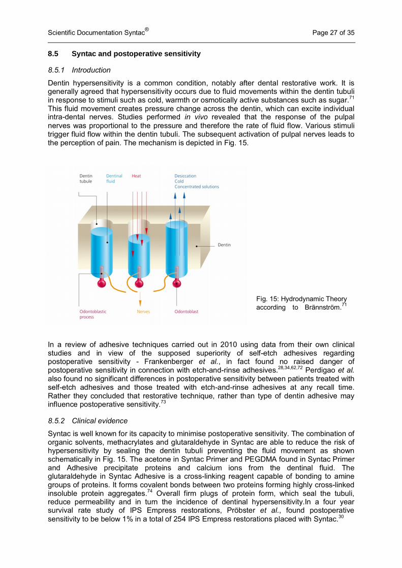
This fluid movement creates pressure change across the dentin, which can excite individual intra-dental nerves. Studies performed in vivo revealed that the response of the pulpal nerves was proportional to the pressure and therefore the rate of fluid flow. Various stimuli trigger fluid flow within the dentin tubuli. The subsequent activation of pulpal nerves leads to the perception of pain. The mechanism is depicted in Fig. 15.
In a review of adhesive techniques carried out in 2010 using data from their own clinical studies and in view of the supposed superiority of self-etch adhesives regarding postoperative sensitivity - Frankenberger et al., in fact found no raised danger of postoperative sensitivity in connection with etch-and-rinse adhesives.28,34,62,72 Perdigao et al. also found no significant differences in postoperative sensitivity between patients treated with self-etch adhesives and those treated with etch-and-rinse adhesives at any recall time. Rather they concluded that restorative technique, rather than type of dentin adhesive may influence postoperative sensitivity.73
8.5.2 Clinical evidence
Syntac is well known for its capacity to minimise postoperative sensitivity. The combination of organic solvents, methacrylates and glutaraldehyde in Syntac are able to reduce the risk of hypersensitivity by sealing the dentin tubuli preventing the fluid movement as shown schematically in Fig. 15. The acetone in Syntac Primer and PEGDMA found in Syntac Primer and Adhesive precipitate proteins and calcium ions from the dentinal fluid. The glutaraldehyde in Syntac Adhesive is a cross-linking reagent capable of bonding to amine groups of proteins. It forms covalent bonds between two proteins forming highly cross-linked insoluble protein aggregates.74 Overall firm plugs of protein form, which seal the tubuli, reduce permeability and in turn the incidence of dentinal hypersensitivity.In a four year survival rate study of IPS Empress restorations, Pröbster et al., found postoperative sensitivity to be below 1% in a total of 254 IPS Empress restorations placed with Syntac.30
Fig. 15: Hydrodynamic Theory according to Brännström.71
Scientific Documentation Syntac® Page 28 of 35
Cox and O’Neal reviewed the biological and clinical use of Syntac and Variolink adhesive systems for cohesive hybridisation of vital dentin to prevent patient postoperative hypersensitivity and bacterial micro-leakage following clinical preparation. In the clinical research facility of the authors, patients reporting pre-operative hypersensitivity before restoration with Syntac and Tetric, reported no postoperative hypersensitivity after 2 years – demonstrating its effective long term seal.75
A pilot clinical trial by Monaco et al. evaluated the clinical behaviour of 3-unit inlay fixed partial dentures (IFPDs) made of SR Adoro/Vectris and luted with either Syntac or Excite DSC over a period of 2 years. 39 inlay bridges were made, placed in 39 adult patients and evaluated according to USPHS criteria. Twenty restorations were luted with Syntac and 19 with Excite DSC. Variolink II was used to lute the restorations. At recalls patients were asked questions about hypersensitivity. Moderate to severe hypersensitivity was found during the first six months of the study. At the 1 week recall 95% received Alpha ���scores for postoperative sensitivity in the Syntac group compared to 61% in the Excite DSC group. At the last recall after 2 years this was 100% for the Syntac group i.e. no sensitivity and 95% for the Excite DSC group.76
If postoperative sensitivity is going to occur it tends to occur immediately and then wane or disappear completely. If there has been no sensitivity following restorative work within a few months it is unlikely to occur at all. Wilson et al. in a 4-year evaluation of Class II Tetric restorations placed with Syntac using a decoupling technique also found zero postoperative sensitivity at 1 month, 18 months, 2, 3 or 4 years.77. Schirrmeister et al. compared restorations restored with Syntac and Tetric Ceram or Ceram. X and an experimental one bottle etch-and-rinse adhesive K-0127/Dentsply DeTrey. Restorations were evaluated at baseline, 1, 2 and 4 years. Whereas 10% of patients showed slight symptoms of postoperative sensitivity at baseline, after one year no sensitivity was apparent in either group.36
8.6 Summary
Syntac lives up to its excellent 20 year reputation. It has and continues to prove itself as a suitable and clinically successful bonding agent in numerous medium and long term studies for direct, indirect, (CAD/CAM or traditionally manufactured) restorations in both anterior and posterior teeth. Survival rates of Syntac bonded restorations are notably high and uniform indicating the low technique sensitivity of Syntac despite its 3-bottle system. Syntac is the classic universal bonding agent for a sound chemical bond between composite material and tooth structure.
Scientific Documentation Syntac® Page 29 of 35
9. Biocompatibility Biocompatibility has been variously defined. One definition is “The ability of a material to perform with an appropriate host response in a specific application." 78
9.1 Introduction
The Syntac adhesive system consists of a primer, an adhesive, and a bonding agent. It creates a chemically stable bond to the composite material and dental substrate. It contains:
Syntac Primer: TEGDMA, PEGDMA, maleic acid, acetone in an aqueous solution
Syntac Adhesive: PEGDMA, glutaraldehyde in an aqueous solution
Heliobond: Bis-GMA, TEGDMA
These components are ubiquitous in dental resin-based materials.
9.2 Acute toxicity
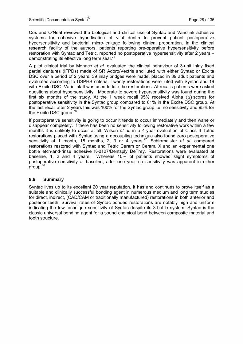
Acute oral toxicity data for all the major monomers contained in Syntac is available from external sources:
LD 50 Species Reference
PEGDMA 10,200 mg/kg Mouse 79
TEGDMA 10,837 mg/kg Rat 80
Maleic acid 700-2400 mg/kg Rat, mouse 81
Acetone 10.7 ml/kg Rat 82
Glutaraldehyde 820 mg/kg Rat 83
Table 8: Acute toxicity data for Syntac components
LD 50 is the amount of a material given all at once, which causes the death of 50% of a group of test animals. It is one method to measure the acute toxicity of a material. It is expressed here per kg of body weight of the test animal. The higher the LD 50 value the lower the toxicity i.e. more of a substance is necessary before it is toxic.
All the values shown in the table are above 700mg/kg. A typical application for one restoration rarely requires more than 40mg of material. Assuming an average human weighs 50kg one can estimate a safety factor of approximately 1000. That is, 35,000mg (700mg x 50) of substance would be the minimum necessary LD50 for an average human. Assuming a Syntac application of just 40mg, this results in a safety factor of 875 (35,000/40) i.e. approximately 1000.
Farmer et al.84 tested the histological compatibility of Syntac. In three days, there was no pulp reaction observed that derived from Syntac. After 80 days observation, the authors excluded the possibility of pulp irritation due to Syntac.
Scientific Documentation Syntac® Page 30 of 35
9.3 Sensitisation and irritation
Like all resin-based dental materials, Syntac contains methacrylate and acrylate derivatives. Such materials may have an irritating effect and may cause sensitisation. This can lead to allergic contact dermatitis. Allergic reactions are extremely rare in patients but are increasingly observed in dental personnel, who handle uncured composite material on a daily basis.85,86 These reactions can be minimized by clean working conditions and avoiding contact of unpolymerised material with the skin. Commonly employed gloves, made of latex or vinyl, do not provide effective protection against sensitisation to such compounds.
If Syntac is used incorrectly, the solution may come into contact with the oral mucosa. Any concentration of glutaraldehyde is soluble in water however and if rinsed with copious amounts of water immediately after contact, the tissues should suffer no damage. Unintended contamination of mucous membranes can however go unnoticed and provoke local tissue lesions.
Since the sensitising properties of the above mentioned components of Syntac are known and declared in the instructions for use, no specific tests for sensitisation were conducted with Syntac.
9.4 Conclusion
Syntac has been on the market since 1990 and no unexpected undesirable side effects have become apparent. According to current knowledge, if used as indicated, Syntac poses no risk for the patient, user or third party, and the benefits of the product exceed the residual risk.
Scientific Documentation Syntac ® 31 of 35
10. References
1. Kugel G. J Am Dent Assoc 131, No suppl 1 20S-25S
2. Bowen R L. Dental filling material comprising vinyl silane treated fused silica and a binder consisting of the reaction product of Bis phenol and glycidyl acrylate. 1962; Patent No: 3,066,112.
3. Eisenmann D R (1998). Enamel structure. In: Oral Histology Development, Structure and Function. A R Ten Cate editor. St. Louis: Mosby, pp. 218-235.
4. Schroeder H E. Oral Structural Biology. Thieme; New York 1991
5. Alhadainy H A, Abdalla AI. 2-year clinical evaluation of dentin bonding systems. Am J Dent 1996; 9: 77-79.
6. Van Meerbeek B, Peumans M, Verschueren M, Gladys S, Braem M, Lambrechts P, Vanherle G. Clinical status of ten dentin adhesive systems. J Dent Res 1994; 73: 1690-1702.
7. Gwinnett A J. Quantitative contribution of resin infiltration/hybridization to dentin bonding. Am J Dent 1993; 6:7-9.
8. Gwinnett AJ. Dentin bond strength after air drying and rewetting. Am J Dent 1994; 7: 144-148.
9. Buonocore M G. A simple method of increasing the adhesion of acrylic filling materials to enamel surfaces. J Dent Res 1955; 34: 849-853.
10. Silverstone L M, Saxton C A, Dogon I L, Fejerskov O. Variation in the pattern of acid etching of human dental enamel examined by scanning electron microscopy. Caries Res 1975; 9 (5): 373-387
11. Perdigao J, Lambrechts P, Van Meerbeen B, Tome A R, Vanherle G, Lopes A B. Morphological field emission-SEM study of the effect of six phosphoric acid etching agents on human dentin. Dent Mater 1996; 12: 262-71
12. Pashley D H, Ciucci B, Sano H, Horner J A. Permeability of dentin to adhesive agents. Quintessence Int. 1993; 24: 618-631
13. Van Meerbeek B, Vargas S, Inoue S, Yoshida Y, Peumans M, Lambrechts P, Vanherle G. Adhesives and cements to promote preservation dentistry. Operative Dentistry 2001. (Supplement 6) 119-144
14. Hashimoto M, Tay F R, Svizero N R, de Gee A J, Feilzer A J, Sano H, Kaga M, Pashley D H. The effects of common errors on sealing ability of total-etch adhesives. Dent Mater 2006; 22: 560-568.
15. van Dijken J W, Sunnegardh-Gronberg K. A four-year clinical evaluation of a highly filled hybrid resin composite in posterior cavities. J Adhes Dent 2005; 7: 343-349.
16. Knitter K, Lösche GM, Blunck U. Effectiveness of Excite/Tetric Ceram in Class-II-restorations after three years. J Dent Res 2005; 84 (Spec Iss B):Abstract# 0333.
17. Buonocore M G. Principles of adhesive retention and adhesive restorative materials. J. Am Dent Assoc. 1963 Sep; 67: 382-91.
18. Huget E F, Denniston J C, Vilca J M. Dentin adhesives: a perspective. Military Medicine 1979; 144: 619-620
19. Crim G A, Swartz M L Phillips R W. An evaluation of cavosurface design and microleakage. Gen Dent 1984; 32: 56-58
20. Haller B, Blunck U. Übersicht und Wertung der aktuellen Bonding-systeme. ZM 2003; 93 (7): 48-58
21. Frankenberger R. Adhäsivtechnik 2009 – Neuigkeiten Tipps und Trends. Quintessenz 2009; 60 (4) 415-423
22. Haller B. Mechanismus und Wirksamkeit von Dentinhaftvermittlern Dtsch Zahnärztl Z 49 (1994) 750-759
23. Cox CF, Suzuki S. Re-evaluating pulp protection: caIcium hydroxide liners vs. cohesive hybridization J Am Dent Assoc 1994;125: 823-831

Scientific Documentation Syntac ® 32 of 35
24. Gwinnett AJ, Dickerson WG, Yu S. Dentin bond shear strength and microleakage for Syntac/Heliomolar: a comparison between the manufacturer's and total etch technique. J Esthet Dent 1992; 4: 164-168
25. Frankenberger R, Krämer N, Petschelt A. Fatigue behaviour of different dentin adhesives. Clin Oral Invest. 1999; 3: 11-17
26. GfK Healthcare. DDM Jahresbericht 2010. Ivoclar Vivadent. Chairside Bereich
27. GfK Healthcare. ZaBus IV/2010
28. Frankenberger R, Schipper H M, Roggendorf M J. Adhäsivtechnik 2010 – Etch and Rinse oder Self Etch Systeme? Quintessenz 2010; 61 (5): 537-542
29. Cardoso M V, Yoshida Yasuhiro, Van Meerbeek B. Adhesion to tooth enamel and dentin – a view on the latest technology and future perspectives. Chapter 3 from: Roulet J-F, Kappert H F. Statements – Diagnostics and therapy in dental medicine today and in the future. Quintessenz Publishing 2009
30. Pröbster L, Ulmer HJ, Engel E; Four-year survival rate study of IPS Empress restorations; DGZPW 1996: 59
31. Folwaczny M, Mehl A, Kunzelmann K H, Hickel R. Tooth colored restorations of class V lesions using four different materials - five year results. J Dent Res 2000; 79: 361.
32. Mazer R, Cury C, Teixeira L, Leinfelder K. Influence of Maleic acid on the retention of abfracted lesion restorations. J Dent Res 1994; 73: 275
33. Krämer N, Ebert J, Petschelt A, Frankenberger R. Ceramic inlays bonded with two adhesives after 4 years. Dental Materials 2006; 22: 13-21
34. Krämer N, Taschner M, Lohbauer U, Petschelt A, Frankenberger R. Totally bonded ceramic inlays and onlays after eight years. J Adhes Dent 2008; 10: 307-314
35. Taschner M, Frankenberger R, Garcia-Godoy F, Rosenbusch S, Petschelt A, Krämer N. IPS Empress inlays luted with a self-adhesive resin cement after 1 year. 2009. Am J Dent. 22 1:55-59
36. Schirrmeister J F, Huber K, Hellwig E, Hahn P. Four-year evaluation of a resin composite including nanofillers in posterior cavities. J Adhes Dent 2009; 11: 399-404
37. Frankenberger R. Bonding 2006 – Zeitersparnis versus Langzeiterfolg. Quintessenz 2006, 57 (5) 485-495
38. Frankenberger R, Lohbauer U, Schaible R B, Nikolaeinko S A, Naumann M. Luting of ceramic inlays in vitro: Marginal quality of self-etch and etch-and –rinse adhesives versus self-etch cements. Dent Materials 2008; 24:185-191
39. Peumans M, Kanumilli P, De Munck J, Van Landuyt K, Lambrechts P, Van Meerbeek B. Clinical effectiveness of contemporary adhesives: A systematic review of current clinical trials. Dent Mater 2005; 21: 864-881.
40. Krejci I, Besek M, Lutz F Clinical and SEM study of Tetric resin composite in posterior teeth: 12-month results Am J Dent 1994; 7: 27-30
41. Boretti R, Krejci I, Lutz F Clinical and scanning electron microscopic evaluation of fine hybrid composite restorations in posterior teeth after four years of wear J Dent Res 1997; 76: 41
42. Krämer N, Frankenberger G, Dettenhofer J, Ebert M, Pelka M, Petschelt A; Clinical Evaluation of Ceramic Inlays and Onlays after Four Years; J Dent Res 1997; 76: 271
43. Pelka M, Teinelt C, Krämer N, Fassbender U, Petschelt A; In-vivo-Abrasion bei IPS Empress Inlays; Dtsch Zahnarzt Z 50 1995: 917-919
44. Fradeani M, Aquilano A; Clinical Experience with IPS Empress Crowns; Into J Prosthodontics 1997; 10: 241-247
45. Mazer RB, Cury C, Teixeira L, Leinfelder K; Influence of maleic acid on the retention of abfracted lesion restorations; J Dent Res 1994; 73: 275
46. Loher C, Kunzelmann KH, Hickel R; klinische Studie mit Hybridionomerzement-, Kompomer- und Kompositfüllungen in Klasse-V-kavitäten; Dtsch Zahnärztl Z 1997; 52: 8
47. Folwaczny M, Loher C, Mehl A, Benz C, Hickel R; Class V fillings with four different light curing materials: three years results; J Dent Res 1998; 77: 190
Scientific Documentation Syntac ® 33 of 35
48. Lasfargues J, Bonte E, Bnebot D, Pujol F, Bachelard B; Clinical performance of a microhybrid posterior composite resin restorative material: a one-year clinical report; J Dent Res 1997; 76: 1117
49. A. Peschke, L. Enggist, and R.Watzke, Poster: Five-Year Clinical Evaluation of Posterior Nano-Hybrid Composite Resin Restorations, Abstract Nr. 0237, 45th Meeting of the Continental European Division (CED) of IADR, Budapest, September 2011.
50. Hickel R, Roulette J F, Bayne S, Heintze S D, Major I A, Peters M, Rousson V, Randall R, Schmalz G, Tyas, M, Vanherle G. Recommendations for conducting controlled clinical studies of dental restorative materials. Science Committee Project 2/98--FDI World Dental Federation study design (Part I) and criteria for evaluation (Part II) of direct and indirect restorations including onlays and partial crowns. J. Adhes Dent. 2007; 9 Suppl 1: 121-47.
51. Manhart J, Chen H-Y, Hamm G, Hickel R. Review of the clinical survival of direct and indirect restorations in posterior teeth of the permanent dentition. Oper Dent 2004; 29: 481-508
52. Peumans M, Van Meerbeek B, Lambrechts P, Vuylsteke-Wauters M, Vanherle G. Five year clinical performance of porcelain veneers. Quintessence Int. 1998; 29: 211-221
53. Krämer N, Garcia-Godoy F, Reinelt C, Feilzer A J, Frankenberger R. Nanohybrid vs. fine hybrid composite in extended Class II cavities after six years. Dental Materials 2011; 27:455-464
54. Krämer N, Reinelt C, Richter G, Petschelt A, Frankenberger R. Nanohybrid vs. fine hybrid composite in Class II cavities: Clinical results and margin analysis after four years. Dental Materials 2009; 25: 750-759
55. Manhart J, Chen H Y, Hickel R. Clinical evaluation of the posterior composite Quixfil in Class I and II cavities: 4-year follow-up of a randomized controlled trial. J Adhes Dent 2010;12:237-243
56. van Dijken J W V, Pallesen U. Long-term dentin retention of etch-and-rinse and self-etch adhesives and a resin-modified glass ionomer cement in non-carious cervical lesions. Dental Materials 2008; 24: 915-922
57. Leinfelder K, University of Alabama, Birmingham, USA, 1994 Mazer RB, Cury C, Teixeira L, Leinfelder KF. Influence of maleic acid on the retention of abfracted lesion restorations. JDR 1994; 73, 275 Abstr. 1389
58. Folwaczny M, Loher C, Mehl A, Kunzelmann K H, Hickel R. Class V lesions restored with four different tooth-colored materials – 3-year results. Clin Oral Invest 2001; 5:31-39
59. Heintze S, Ruffieux C, Rousson V. Clinical performance of cervical restorations – a meta-analysis. Dental Materials 2010; 26: 993-1000
60. Krämer N et al.; Clinical study conducted at the University of Erlangen, Germany; IADR, Abstract 2002, San Diego
61. Krämer N, Frankenberger R. Clinical performance of bonded leucite-reinforced glass ceramic inlays and onlays after eight years. Dent Materials 2005 21: 262-271
62. Frankenberger R, Taschner M, Garvia-Godoy F, Petschelt A, Krämer N. Leucite-reinforced glass ceramic inlays and onlays after 12 years. J Adhes Dent 2008; 10: 393-398.
63. Coelho Santos M J M, Mondelli R F L, Lauris J R P, Navarro M F L. Clinical evaluation of ceramic inlays and onlays fabricated with two systems: Two-year clinical follow up. Operative Dent 2004; 29-2: 123-130
64. Bernhart J, Schulze D, Wrbas K-T. Evaluation of the clinical success of Cerec 3D inlays. Int J Comp Dent 2009; 12: 265-277
65. Zimmer S, Göhlich O, Rüttermann S, Lang H, Raab WH-M, Barthel C R. Long-term survival of Cerec restorations: A 10-year study. Operative Dentistry 2008; 33-5: 484-487
66. Reich S M, Wichmann M, Rinne H, Shortall A. Clinical performance of large, all-ceramic CAD/CAM-generated restorations after three years. A pilot study. JADA 2004; 135: 605-612
67. Guess P C, Strub J R, Steinhart N, Wolkewitz M, Stappert C F J. All-ceramic partial coverage restorations – midterm results of a 5-year prospective clinical split-mouth study. J Dent 2009 37: 627-637
Scientific Documentation Syntac ® 34 of 35
68. Fradeani M, Redemagni M. Klinische Evaluation von leuzitverstärkten glaskeramischen Kronen über 11 Jahre. Quintessenz 2003; 54; 4: 379-386
69. van Dijken J W V, Hasselrot L. A prospective 15-year evaluation of extensive dentin-enamel-bonded pressed ceramic coverages. Dental Materials 2010; 26: 929-939
70. Donovan T E, Anderson M, Becker W, Cagna D R, Hilton T J, Rouse J. Annual review of selected scientific literature: Report of the committee on scientific investigation of the American Academy of Restorative Dentistry. J Prosthet Dent 2011:106: 224-265
71. Brännström M, Linden LÅ, Åstrom A: The hydrodynamics of the dental tubule and of pulp fluid. A discussion of its significance in relation to dentinal sensitivity. Caries Res. 1967; 1: 310-317.
72. Frankenberger R, Krämer N, Lohbauer U, Nikolaenko S A, Reich S M. Marginal integrity: is the clinical performance of bonded restorations predictable in vitro? J. Adhes. Dent 2007; 9 (Suppl. 1): 107-116
73. Perdigao J, Geraldeli S, Hodges J S. Total-etch versus self-etch adhesive: effect on postoperative sensitivity. J Am Dent Assoc. 2003, 134 (12):1621-9
74. Faber K. Biotransformations in organic chemistry – a textbook. Springer-Verlag. Berlin. 1995.
75. Cox C F, O’Neal S J. Biologic and clinical evaluation of Syntac and Variolink sytems for cohesive pretreatment of hypersensitivity and definitive cementation. Signature Winter 1994. 3-7
76. Monaco C, Ferrari M, , Caldari M, Baldissara P Scotti R. Comparison of 2 bonding systems and survival of fiber–reinforced composite inlay fixed partial dentures. Int J Prosthodont 2006; 19: 577-585
77. Wilson N H, Cowan A J, Unterbrink G, Wilson M A, Crisp R J. A clinical evaluation of Class II composites placed using a decoupling technique. J Adhes Dent 2000; 2 (4): 319-329
78. D.F. Williams. The Williams dictionary of Biomaterials 1999
79. MSDS- Shin-Nakamura-Chemical Co.
80. Schmalz G. The biocompatibility of non-amalgam dental filling materials. Eur J Oral Sci 1998;106: 696-706
81. TOXNET. Maleic acid. Hazardous Substances Data Bank (HSDB) A database of the National Libarary of Medicines. TOXNET system report no. 666. March 5, 2003
82. TOXNET. Acetone. Hazardous Substances Data Bank (HSDB) A database of the National Libarary of Medicines. TOXNET system report no. 41. June 24, 2005
83. TOXNET. Glutaraldehyde. 2005-06-24
84. Farmer J B, Cox C F, White K , Ramus L. Histological evaluation of a new bonding agent. J Dent Res 1992; 71: 279
85. Geurtsen W. Biocompatibility of resin-modified filling materials. Crit Rev Oral Biol Med 2000; 11: 333-335.
86. Munksgaard EC, Hansen EK, Engen T, Holm U. Self reported occupational dermatological reactions among Danish dentists. Eur J Oral Sci 1996; 104: 396-402.
Scientific Documentation Syntac ® 35 of 35
This documentation contains a survey of internal and external scientific data (“Information”). The documentation and Information have been prepared exclusively for use in-house by Ivoclar Vivadent and for external Ivoclar Vivadent partners. They are not intended to be used for any other purpose. While we believe the Information is current, we have not reviewed all of the Information, and we cannot and do not guarantee its accuracy, truthfulness, or reliability. We will not be liable for the use of or reliance on any of the Information, even if we have been advised to the contrary. In particular, use of the information is at your sole risk. It is provided “as-is”, “as available” and without any explicit or implicit guarantee or warranty regarding usability or suitability (without limitations) for a particular purpose. The Information has been provided without cost to you and in no event will we or anyone associated with us be liable to you or any other person for any incidental, direct, indirect, consequential, special, or punitive damages (including, but not limited to, damages for lost data, loss of use, or any cost to procure substitute information) arising out of your or another’s use of or inability to use the Information even if we or our agents know of the possibility of such damages. Ivoclar Vivadent AG Research and Development Scientific Services Bendererstrasse 2 FL - 9494 Schaan Liechtenstein Contents: Joanna-C. Todd Issued: February 2012