Synaptic vesicle pool size, release probability and synaptic depression are sensitive to Ca2+...
17
Synaptic vesicle pool size, release probability and synaptic depression are sensitive to Ca 2+ buffering capacity in the developing rat calyx of Held R.M. Leão 1,2 and H. von Gersdorff 2 1 Departamento de Fisiologia, Faculdade de Medicina de Ribeirão Preto, Universidade de São Paulo, Ribeirão Preto, SP, Brasil 2 The Vollum Institute, Oregon Health and Science University, Portland, OR, USA Abstract The calyx of Held, a specialized synaptic terminal in the medial nucleus of the trapezoid body, undergoes a series of changes during postnatal development that prepares this synapse for reliable high frequency firing. These changes reduce short-term synaptic depression during tetanic stimulation and thereby prevent action potential failures during a stimulus train. We measured presynaptic membrane capacitance changes in calyces from young postnatal day 5–7 (p5–7) or older (p10–12) rat pups to examine the effect of calcium buffer capacity on vesicle pool size and the efficiency of exocytosis. Vesicle pool size was sensitive to the choice and concentration of exogenous Ca 2+ buffer, and this sensitivity was much stronger in younger animals. Pool size and exocytosis efficiency in p5–7 calyces were depressed by 0.2 mM EGTA to a greater extent than with 0.05 mM BAPTA, even though BAPTA is a 100-fold faster Ca 2+ buffer. However, this was not the case for p10–12 calyces. With 5 mM EGTA, exocytosis efficiency was reduced to a much larger extent in young calyces compared to older calyces. Depression of exocytosis using pairs of 10-ms depolarizations was reduced by 0.2 mM EGTA compared to 0.05 mM BAPTA to a similar extent in both age groups. These results indicate a developmentally regulated heterogeneity in the sensitivity of different vesicle pools to Ca 2+ buffer capacity. We propose that, during development, a population of vesicles that are tightly coupled to Ca 2+ channels expands at the expense of vesicles more distant from Ca 2+ channels. Keywords Calyx of Held; Neurotransmission; Calcium buffer; Capacitance measurements; Mammalian auditory brainstem; Synaptic development Introduction Action potential invasion of the presynaptic nerve terminal opens voltage-dependent Ca 2+ channels permitting Ca 2+ ions to move down their electrochemical gradient into the terminal. Because Ca 2+ enters via discrete sites, micro-or nanodomains of intracellular Ca 2+ may form where Ca 2+ may reach concentrations of 5–10 μM within 200 nm of the channel and up to 100 μM within 20 nm of the channels (1,2). This highly localized peak of synaptic Ca 2+ Correspondence to: R.M. Leão, Departamento de Fisiologia, FMRP, USP, Av. Bandeirantes, 3900, 14049-900 Ribeirão Preto, SP, Brasil [email protected]. Presented at the IV Miguel R. Covian Symposium, Ribeirão Preto, SP, Brazil, May 23–25, 2008. NIH Public Access Author Manuscript Braz J Med Biol Res. Author manuscript; available in PMC 2010 May 28. Published in final edited form as: Braz J Med Biol Res. 2009 January ; 42(1): 94–104. NIH-PA Author Manuscript NIH-PA Author Manuscript NIH-PA Author Manuscript
-
Upload
independent -
Category
Documents
-
view
2 -
download
0
Transcript of Synaptic vesicle pool size, release probability and synaptic depression are sensitive to Ca2+...
Synaptic vesicle pool size, release probability and synapticdepression are sensitive to Ca2+ buffering capacity in thedeveloping rat calyx of Held
R.M. Leão1,2 and H. von Gersdorff21Departamento de Fisiologia, Faculdade de Medicina de Ribeirão Preto, Universidade de SãoPaulo, Ribeirão Preto, SP, Brasil2The Vollum Institute, Oregon Health and Science University, Portland, OR, USA
AbstractThe calyx of Held, a specialized synaptic terminal in the medial nucleus of the trapezoid body,undergoes a series of changes during postnatal development that prepares this synapse for reliablehigh frequency firing. These changes reduce short-term synaptic depression during tetanicstimulation and thereby prevent action potential failures during a stimulus train. We measuredpresynaptic membrane capacitance changes in calyces from young postnatal day 5–7 (p5–7) or older(p10–12) rat pups to examine the effect of calcium buffer capacity on vesicle pool size and theefficiency of exocytosis. Vesicle pool size was sensitive to the choice and concentration of exogenousCa2+ buffer, and this sensitivity was much stronger in younger animals. Pool size and exocytosisefficiency in p5–7 calyces were depressed by 0.2 mM EGTA to a greater extent than with 0.05 mMBAPTA, even though BAPTA is a 100-fold faster Ca2+ buffer. However, this was not the case forp10–12 calyces. With 5 mM EGTA, exocytosis efficiency was reduced to a much larger extent inyoung calyces compared to older calyces. Depression of exocytosis using pairs of 10-msdepolarizations was reduced by 0.2 mM EGTA compared to 0.05 mM BAPTA to a similar extent inboth age groups. These results indicate a developmentally regulated heterogeneity in the sensitivityof different vesicle pools to Ca2+ buffer capacity. We propose that, during development, a populationof vesicles that are tightly coupled to Ca2+ channels expands at the expense of vesicles more distantfrom Ca2+ channels.
KeywordsCalyx of Held; Neurotransmission; Calcium buffer; Capacitance measurements; Mammalianauditory brainstem; Synaptic development
IntroductionAction potential invasion of the presynaptic nerve terminal opens voltage-dependent Ca2+
channels permitting Ca2+ ions to move down their electrochemical gradient into the terminal.Because Ca2+ enters via discrete sites, micro-or nanodomains of intracellular Ca2+ may formwhere Ca2+ may reach concentrations of 5–10 μM within 200 nm of the channel and up to 100μM within 20 nm of the channels (1,2). This highly localized peak of synaptic Ca2+
Correspondence to: R.M. Leão, Departamento de Fisiologia, FMRP, USP, Av. Bandeirantes, 3900, 14049-900 Ribeirão Preto, SP, [email protected] at the IV Miguel R. Covian Symposium, Ribeirão Preto, SP, Brazil, May 23–25, 2008.
NIH Public AccessAuthor ManuscriptBraz J Med Biol Res. Author manuscript; available in PMC 2010 May 28.
Published in final edited form as:Braz J Med Biol Res. 2009 January ; 42(1): 94–104.
NIH
-PA Author Manuscript
NIH
-PA Author Manuscript
NIH
-PA Author Manuscript

concentration elicits mobilization and fusion of nearby synaptic vesicles. The synapsetopography (i.e., the spatial relationship of vesicles to Ca2+ channels) is crucial for determiningthe Ca2+-coupling efficiency of exocytosis, and this relationship seems to vary among differentsynapses (3). In some synapses, like the squid giant synapse and the chick ciliary ganglion,vesicles are assumed to be in close contact with a single Ca2+ channel (4–6). However, atseveral mammalian central synapses, vesicles are thought to be located far (~200 nm) fromCa2+ channels and influenced by overlapping domains of clustered Ca2+ channels (3,7),because in these synapses release is very sensitive to relatively slow exogenous Ca2+ bufferssuch as ethylene glycol-bis(2-aminoethyl-ether)-N,N,N',N'-tetraacetic acid (EGTA) (8–10),suggesting that Ca2+ ions must travel further to reach their binding sites in the releasemachinery. Ca2+ buffer sensitivity varies between different types of synapses and this can beone factor that determines short-term synaptic plasticity (10–14) and the size of the readilyreleasable pool of vesicles (15).
The calyx of Held, due to its large size, is accessible to direct electrophysiological recordingand has been intensively studied as a model of a fast central synapse (16,17). This synapseundergoes developmental changes during the first two postnatal weeks, which prepare it formediating high frequency transmission after the onset of hearing at p12 in rodents. In moremature calyces (p12–15), synaptic depression is reduced compared to p5–7 (18), which isproposed to be a consequence of a reduced release probability, a larger vesicle pool size, anda reduced propensity for multivesicular release, which would lead to postsynaptic receptordesensitization (19). The evidence for a smaller pool size in immature synapses comes, in part,from presynaptic capacitance recordings obtained under a single Ca2+ buffer condition, namely0.2 mM EGTA, which is thought to be equivalent to the physiological Ca2+ bufferconcentration in this synapse (20,21). However, endogenous Ca2+ handling changes withdevelopment of the calyx: immature terminals have a slower Ca2+ extrusion system (22) andweaker expression of Ca2+ binding proteins (23,24) compared to more mature synapses.Perhaps because of these changes, short trains of stimuli produce higher levels of intra-calycealcalcium in young calyces than in mature calyces (25). Also, glutamate release from youngcalyces (p8) is more sensitive to 10 mM EGTA than release from terminals of more maturemice (p18), which exhibited higher Ca2+ cooperativity, suggesting tighter coupling of Ca2+
channels to release sites in mature synapses (26). Since Ca2+ buffering can determine short-term synaptic plasticity (10), developmental changes in this parameter could explain some ofdecrease in release probability and short-term depression in mature calyces. To investigate thishypothesis further, we measured exocytosis directly using membrane capacitancemeasurements (Cm) in the calyx of Held during two distinct developmental stages and withthree distinct Ca2+ buffering conditions. Our aim was to determine the effect of theseparameters on exocytotic efficiency, releasable pool size and vesicle pool depletion.
Material and MethodsSlice preparation
Brainstem transverse slices (200 μm thick) were obtained from postnatal day 5 (p5) to p12Sprague-Dawley rats as described previously (19), and kept in artificial cerebral spinal fluidcontaining 125 NaCl, 2.5 mM KCl, 1 mM MgCl2, 2 mM CaCl2, 25 mM glucose, 25 mMNaHCO3, 1.25 mM NaH2PO4, 0.4 mM ascorbic acid, 3 mM myoinositol, 2 mM Na-pyruvate,pH 7.3, when bubbled with a carbogenic mixture (95% O2, 5% CO2).
Whole-cell patch-clamp recording and capacitance measurementsWhole-cell patch-clamp recordings were performed at room temperature (21–23°C) in normalartificial cerebral spinal fluid containing 1 μM tetrodotoxin (TTX) and 10 mMtetraethylammonium chloride (TEA-Cl). The internal solution consisted of 90 mM Cs-
Leão and von Gersdorff Page 2
Braz J Med Biol Res. Author manuscript; available in PMC 2010 May 28.
NIH
-PA Author Manuscript
NIH
-PA Author Manuscript
NIH
-PA Author Manuscript
methanesulfonate, 20 mM CsCl, 1 mM MgCl2, 5 mM Na2-phosphocreatine, 40 mM HEPES,10 mM TEA-Cl, 0.2 or 5 mM EGTA, or 0.05 mM 1,2-bis(2-aminophenoxy)ethane-N,N,N',N'-tetraacetic acid (BAPTA), 2 mM ATP-Mg, 0.2 mM GTP, pH 7.3, with CsOH, 310 mOsm.Lucifer yellow (0.25 mg/mL) was added to the pipette solution to allow post hoc confirmationthat the recording was from a calyx. The calyx terminal was held at −70 or −80 mV. Patchpipettes were pulled from soft thin-walled glass (WPI, USA) and had an open tip resistance of3–7.0 MΩ. Series resistance (7–25 MΩ) was electronically compensated by 30–90%. Dataacquisition was controlled by “Pulse” software (Heka, Germany) and signals were recordedvia an EPC-9 (Heka) double patch-clamp amplifier. Exocytosis was elicited by single squaredepolarizations to 0 mV lasting 2, 10, 30, or 90 ms, or by a series of 30 action potential like(AP-like) stimuli (a 1-ms ramp to +40 mV followed by a 1-ms ramp back to the holdingpotential). This AP-like stimulation produces Ca2+ currents that are very similar to thoseobtained with a calyx AP waveform (Figures 3A and 6 of Ref. 27) generating a Ca2+ influx of1–2 pC per stimulus. Exocytosis was assessed by presynaptic membrane capacitancemeasurements performed as in Ref. 19 by the “sine + DC” method. Briefly, a sinusoidal voltagecommand (1 kHz and 60–70 mV peak-to-peak amplitude) was applied to the terminal inaddition to the holding command few milliseconds before and after the depolarizing commandstep. The resulting membrane currents were processed with a software lock-in amplifier(HEKA) that decomposed the current into its resistive and capacitive components, resulting inthe calculated series (Gs) and membrane (Gm) conductances and membrane capacitance(Cm). The estimate of the number of vesicles (Nv) was calculated using the formula Nv =ΔCm / (Cms × πd2 ) (28), where d is the vesicle diameter and Cms is the specific membranecapacitance of 10 fF/μm2. Vesicle diameter was assumed to be 50 nm in both developmentalgroups (19). Unpaired t-tests were performed to assess statistical significance of the data andmeans with two-tail P values below 0.05 were considered to be significantly different. Datawere analyzed using IGOR Pro (Wavemetrics). Statistics and curve fitting with a hyperbolicfunction were performed with Prism (GraphPad). Data are reported as means ± SEM.
Alterations in membrane capacitance in response to the Ca2+ currents were not followed byrespective calculated changes in series resistance (Rs change after 10 ms Ca2+ current was−0.001 ± 0.019 MΩ; Figure 1A; see also Ref. 19). A small current (peak of −37 ± 24 pA aftera 100 ms ICa) that decayed over approximately 200 ms was observed after Ca2+ currents longerthan 10 ms, and increased linearly with the increase in Ca2+ influx, in contrast to the capacitancejump that saturated with long depolarizations (Figure 1C). This current might be attributableto activation of the Na+/Ca2+ exchanger (29) or some other Ca2+-dependent conductance, sinceit was not observed when the internal solution contained 5 mM EGTA (data not shown). Toavoid interference from this conductance in the calculation of membrane capacitance, wecollected data after this current had decayed (gray rectangle in Figure 1A).
Time-resolved capacitance measurements using sinusoidal voltage stimuli are based on theassumption that the recorded cell is electrically equivalent to a single compartment composedof a membrane resistor in parallel with a membrane capacitor. Applying a step voltage, via anelectrode at the whole-cell voltage-clamp mode, and observing if the resulting capacitivecurrent decays with a single exponential time constant can identify a single-compartment cell(30). Since most calyces included in this study had different axon lengths, based on the luciferyellow signal and based on the number of exponentials needed to fit the time course ofmembrane charging currents, most calyces comprised more than one electrical compartment.However, comparison of results from calyces that presented a single electrical compartmentto those with more than one electrical compartment showed that Ca2+-dependent capacitancejumps were similar in both groups (data not shown), in agreement with previous observations(19,30).
Leão and von Gersdorff Page 3
Braz J Med Biol Res. Author manuscript; available in PMC 2010 May 28.
NIH
-PA Author Manuscript
NIH
-PA Author Manuscript
NIH
-PA Author Manuscript
ResultsCalyces of Held nerve terminals were voltage clamped at a holding potential of −80 mV andsubjected to step depolarizations to 0 mV of 2- to 90-ms duration. Ca2+ currents andcorresponding membrane capacitance changes were then recorded (Figure 1B). The resultingcapacitance jumps increased with the duration of depolarization, with half-maximal responsesoccurring at 11 ± 0.3 ms (Figure 1C) and reaching an apparent plateau after 90 ms (Figure 1C).Application of CdCl2 (0.1 mM) abolished both Ca2+ currents and capacitance jumps (Figure1C), as expected if changes in membrane capacitance reflect exocytosis of synaptic vesicles.Recording from postsynaptic cells produced very small capacitance changes (<50 fF after 90-ms depolarizations; Figure 1C), and were thus easily distinguishable from those recorded fromcalyces. We did not investigate the nature of the postsynaptic capacitance changes further.
Effects of exogenous Ca2+ buffer and development on exocytosisThe results illustrated in Figure 1 were recorded using a pipette solution containing 0.2 mMEGTA. Paired recordings indicate that 0.2 mM EGTA and 0.05 mM BAPTA are theapproximate endogenous Ca2+ buffering in p8–10 calyces for single action potentialstimulation (20,21). However, these buffers have quite distinct buffering kinetics, as seen inFigure 6. Due to its faster binding kinetics, 0.05 mM BAPTA is considerably more efficientin buffering Ca2+ than four times more EGTA, while 5 mM EGTA is very effective in inhibitingneurotransmission in the calyx of Held (20), although it produces a similar buffering profileas 0.05 mM BAPTA.
These discrepancies might be explained by differences in Ca2+ buffer capacity and kinetics.Probably the lower concentration of BAPTA (lower buffering capacity) makes it inefficient inbuffering the large local increase in calyceal Ca2+ after an action potential, because of quickbuffer saturation due to the fast binding rate of Ca2+ to BAPTA (10). On the other hand, thelarger buffer capacity of 5 mM EGTA and its lower binding kinetics to Ca2+ make buffersaturation negligible after Ca2+ influx evoked by an action potential. In order to better elucidatethe influence of Ca2+ buffering capacity on exocytosis in the calyx of Held, and how thisrelationship is affected during maturation of this synapse, we compared exocytosis in calycesat different stages of maturation (from p5–7 and p10–12 rats) under three Ca2+ bufferconditions: 0.05 mM BAPTA, 0.2 mM EGTA and 5 mM EGTA. These age groups were chosenbecause they flank a period during which the calyx undergoes a rearrangement of exocytoticactive zones (19), changes in the coupling of Ca2+ entry to exocytosis (25), and changes in thelevels of expression of endogenous Ca2+ buffers such as parvalbumin (24). All of these factorscould lead to changes in the sensitivity of exocytosis to exogenous buffer.
Exocytosis recorded from the two age groups and three buffer conditions are shown in Figure2A, where the data are plotted versus Ca2+ charge (QCa, integral of the Ca2+ current) to separatechanges in Ca2+ entry from exocytosis efficiency, since Ca2+ current amplitude increasedduring development (data not shown; see Ref. 31). We observed that the maximum number ofvesicles released by a long depolarization, as well as the number of vesicles released in responseto very short pulses, varied significantly during development.
First it was clear that the maximal capacitance jump (in response to a 90-ms depolarization)was strongly dependent on the choice of Ca2+ buffer in p5–7 calyces, with values in 0.05 mMBAPTA approximately two times those obtained with 0.2 mM EGTA (Figure 2; Table 1). Incontrast, in p10–12 calyces, maximum capacitance jumps did not differ between the twoCa2+ buffer conditions (Figure 2). By converting these capacitance jumps to the number ofvesicles (see Methods) we calculate apparent vesicle pool sizes, as shown in Figure 2B andTable 1, showing that the calyces can maximally release about 8500 vesicles when stimulatedwith our protocol and that this value is reduced by 50% in immature calyces perfused with 0.2
Leão and von Gersdorff Page 4
Braz J Med Biol Res. Author manuscript; available in PMC 2010 May 28.
NIH
-PA Author Manuscript
NIH
-PA Author Manuscript
NIH
-PA Author Manuscript
mM EGTA. Increasing the Ca2+ buffer to 5 mM EGTA reduced the releasable pool in bothage groups to similar levels (down to about 2000 vesicles; Figure 2B and Table 1).
The large difference in vesicle pool generated by 90-ms step depolarizations in 0.05 mMBAPTA versus 0.2 mM EGTA in immature calyces was surprising since these two bufferconditions mimic endogenous buffering for a single action potential stimulation in p8–10 rats(21). We hypothesized that prolonged step depolarizations might evoke exocytosis that issomehow different from what occurs during brief bursts of Ca2+ entry during an action potentialtrain. To address this, we first tested whether a train of 30 AP-like stimuli (Figure 3A; seeMethods) and a 30-ms step depolarization would evoke exocytosis from a common pool ofvesicles. Both protocols elicited similar capacitance jumps (Figure 3B,C; P > 0.05). Similar tothe response to a step depolarization, exocytosis in response to a train of AP-like stimuli waslarger with 0.05 BAPTA as the buffer than with 0.2 mM EGTA in immature calyces (Figure3C). In all ages and buffer conditions tested, exocytosis evoked by an AP-train was closelycorrelated with exocytosis evoked by a 30-ms depolarization in the same cell (r2 = 0.90; Figure3B,D). In addition, exocytosis evoked by a step depolarization was strongly reduced (by 62%on average; N = 3 in 0.05 mM BAPTA and N = 3 in 0.2 mM EGTA; p6-7 calyces) by a precedingtrain of AP-like stimuli (data not shown). Thus, trains of AP-like stimuli and stepdepolarizations evoke exocytosis from a common pool of vesicles.
We also observed that, for a given buffer condition, the Ca2+ entry required for half-maximumexocytosis (the Kd value obtained from curve fitting) was similar for the different ages studiedbut was about 50% lower in the low-buffer condition of 0.05 mM BAPTA (15.1 ± 7 pC forp5–7 and 13.95 ± 9 pC for p10–12) compared with 0.2 mM EGTA (24.9 ± 4 pC for p5–7 and34.3 ± 8 pC for p10–12). This higher potency observed in the low-buffer condition can bebetter appreciated if we compare the differences in exocytosis during the briefest (2 ms)depolarizations that released a small fraction of the vesicle pool that probably include thevesicles released by a single action potential since this protocol resulted in about 1.5–13.5 pCof Ca2+ entry (depending on the age group), or approximately twice the Ca2+ entry generatedby a single AP (21). A 2-ms depolarization released more vesicles from mature than immaturecalyces, and in both age groups the calyces released more vesicles in the presence of 0.05 mMBAPTA than in the presence of 0.2 or 5 mM EGTA (Figure 4B;Table 1). Figure 4A showsthat in immature calyces 5 mM EGTA was very effective in inhibiting exocytosis by a 2-msCa2+ current in a p5 calyx in contrast to a p11 calyx. Interestingly, in both age groups, thenumber of vesicles released by a 2-ms Ca2+ current in the presence of 0.05 mM BAPTA wassimilar to the number of docked vesicles as assayed by electron microscopy (19) (see Figure4B), suggesting an anatomical correlate for these vesicles.
However, the developmental differences in Cm jumps are perhaps mostly due to the largerCa2+ currents in more mature terminals since when these values were normalized by QCa bothage groups released a similar number of vesicles under the different buffering conditions,although the release was significantly bigger in the low buffer condition with 0.05 mM BAPTA(Figure 4B, Table 1) than with higher EGTA. It is interesting to note that for a 2-msdepolarization, release per Ca2+ ion with 5 mM EGTA (Figure 4B) was not significantlydifferent from that obtained with 0.2 mM EGTA in both age groups, suggesting that 0.2 mMEGTA is sufficient to block the release of all EGTA-sensitive vesicles released during a briefperiod of Ca2+ entry, which is different from what we observed with longer (>10 ms) Ca2+
currents.
Effects of exogenous Ca2+ buffer on paired-pulse depressionThe calyx of Held presents significant synaptic depression in response to high frequencystimulation. We have shown that manipulation of the Ca2+ buffer changes the number ofvesicles released in response to long and short pulses, and estimates of the apparent pool size
Leão and von Gersdorff Page 5
Braz J Med Biol Res. Author manuscript; available in PMC 2010 May 28.
NIH
-PA Author Manuscript
NIH
-PA Author Manuscript
NIH
-PA Author Manuscript

differ depending on the age of the calyx. We next investigated if changes in Ca2+ buffer couldexplain the observed decrease in presynaptic depression that occurs during development. Toaddress this question, we measured capacitance jump depression using two 10-msdepolarizations separated by a 70-ms interval at the holding potential and compared thecapacitance jump after each pulse (Figure 5A).
The capacitance change elicited by the second Ca2+ current was significantly smaller than thecapacitance change produced by the first Ca2+ current (P < 0.0001; Figure 5B), showing thatdepression of exocytosis occurs. Increasing Ca2+ buffering from 0.05 mM BAPTA to 0.2 mMEGTA significantly decreased capacitance jump depression, independently of the age group(Figure 5C). In 0.05 mM BAPTA, the depression was of 66 ± 7% at p5–7 and 73 ± 7% at p10–12 whereas in 0.2 mM EGTA the depression was of 43 ± 8% at p5–7 and 36 ± 6% at p10–12,values significantly different than those obtained with 0.05 mM BAPTA (P < 0.05).
It has been reported that calyceal Ca2+ current depression is responsible for short-termdepression of neurotransmission in the calyx of Held (32,33). Depression of the presynapticCa2+ current did occur during most double-pulse protocols (Figure 5B,C), but Ca2+ currentdepression was not significantly different between 0.05 mM BAPTA and 0.2 mM EGTA(depression of ICa-pre in 0.2 mM EGTA: 11.45 ± 3.7%, in 0.05 mM BAPTA: 15.1 ± 8.1%; P> 0.05; Figure 5B), which could not explain the differences observed in paired-pulse depressionin these two conditions. Moreover, no correlation was observed between Ca2+ currentdepression and capacitance jump depression (P = 0.97; r2 = 0.00015), indicating that Ca2+
current depression is not the main factor in producing paired-pulse depression, at least underthe present conditions.
Interestingly, we observed that a 10-ms Ca2+ current releases about 40% of the total vesiclepool, as calculated by the ratio between the exocytosis induced by a 10- and a 90-ms Ca2+
current (Figure 5B). In this situation we would expect a paired-pulse depression of 40%. Thisagrees with what we observed with 0.2 mM EGTA (Figure 5C) but not with 0.05 mM BAPTA,which produces a more pronounced depression (Figure 5B). We conclude that some other factorin addition to fractional release of the synaptic vesicle pool during the first stimuli and Ca2+
current depression determines the extent of paired-pulse depression, and this factor is sensitiveto buffers present in the calyx.
DiscussionA developmental increase in the size of the releasable pool and a decrease in the releaseprobability are proposed as adaptive mechanisms to avoid depletion of synaptic vesicle poolsin the calyx of Held during high frequency trains of APs (18,19). Here we studied theseparameters using direct measurements of vesicle exocytosis, and examined how theseparameters were affected by Ca2+ buffering capacity in different developmental stages.Recording exocytosis in two different developmental phases of the calyx of Held using threedifferent Ca2+ buffering conditions revealed that parameters, such as vesicle pool size, releaseprobability and synaptic depression, are strongly dependent on the Ca2+ buffer capacity,especially in immature calyces.
EGTA is a relatively slow buffer and is probably inefficient in buffering the fast rise in Ca2+
close to the Ca2+ channels after an action potential, although it does inhibit the excitatorypostsynaptic current when loaded in the calyx of Held synapses at millimolar concentrations(20,21), suggesting that Ca2+ ions must travel some distance in the synapse before reachingthe fusion sensor. This result contrasts with observations in the squid giant synapse (4,5) andsuggests that the immature calyx of Held does not have an organized distribution of Ca2+
channels and vesicles, as reported for other terminals (34,35). Meinrenken et al. (7) developed
Leão and von Gersdorff Page 6
Braz J Med Biol Res. Author manuscript; available in PMC 2010 May 28.
NIH
-PA Author Manuscript
NIH
-PA Author Manuscript
NIH
-PA Author Manuscript
a model in which the vesicles in the calyx of Held are randomly located across a cluster ofCa2+ channels (with the vesicle distance ranging from 30 to 300 nm) producing overlappingdomains of multiple channels controlling vesicle exocytosis, and recruitment of vesicles tothese domains has been demonstrated to be a limiting step for exocytosis in the calyx of Held(36). Changes in Ca2+ buffering in the terminal could thus strongly modulate vesicleexocytosis. Previous work suggests that immature synapses have a weaker Ca2+ bufferingsystem (23,25,31) and less tightly coupled docked vesicles to Ca2+ channels (26). Ourhypothesis was that these developmental changes in intracellular Ca2+ buffering capacity andvesicle coupling could be responsible at least in part for the developmental differences inrelease probability, vesicle pool size and synaptic depression observed in the calyx of Heldsynapse.
We used three different mobile Ca2+ buffer paradigms. The Ca2+ buffering capacity of thethree conditions is depicted in Figure 6 where we can see the expected exponential decay ofthe steady-state Ca2+ concentration around the pore of a Ca2+ channel in the presence of thethree Ca2+ buffering conditions. Because BAPTA at the concentration of 0.05 mM is equallyeffective in buffering Ca2+ as EGTA at the concentration of 5 mM, and we observed afacilitating effect of this BAPTA concentration on exocytosis after brief Ca2+ currents, weconclude that the fast rise in intracellular Ca2+ after a short Ca2+ current (e.g., 2 ms) is stillable to saturate the smaller concentration of buffer molecules, thus leaving no free buffermolecules for chelating the excess Ca2+ influx.
The overall sensitivity of exocytosis to Ca2+ buffer capacity (type and concentration of buffer)suggests that the vesicles in the young calyx of Held are loosely coupled to Ca2+ channels.Remarkably, in immature terminals low capacity Ca2+ buffering reveals a pool of vesicles thatare not released in the presence of medium or high buffer capacity, revealing the existence ofa group of weakly Ca2+-channel-coupled vesicles that are quite sensitive to slow Ca2+ buffers.This pool represents a large fraction of the fast releasable vesicles in immature terminals.Comparing the release per Ca2+ ion influx (units of fF/pC) at 2 ms we estimate that the poolof weakly Ca2+ channel-coupled vesicles represents 72% of the fast releasing vesicles in theimmature synapses, in contrast to 40% of these vesicles in the mature synapses.
It is important to note that the weak Ca2+ buffering capacity via 0.05 mM BAPTA could alsofacilitate exocytosis by different mechanisms such as Ca2+-dependent facilitation and/orpotentiation (11,13) by keeping resting or residual Ca2+ levels high. This could aid recruitmentof vesicles from the reserve to the releasable pool in immature synapses (37) or increase thepriming of docked vesicles by increasing Ca2+-dependent DOC2 protein translocation to themembrane (38). Despite these possibilities, we believe that the simplest explanation for theincreased exocytosis in immature calyces with 0.05 mM BAPTA is the weaker coupling ofvesicles to Ca2+ channels.
Another important observation was that the maximal number of vesicles released, or thereleasable vesicle pool size, is not a fixed value, but depends on the capacity of the Ca2+ buffersystem. Also, in immature calyces the size of the releasable pool was more sensitive to increasesin Ca2+ buffering capacity, suggesting that the releasable vesicle pool undergoes a maturationfrom very weakly coupled vesicles to more tightly coupled vesicles as the calyx develops,allowing these vesicles to be released even in a stronger Ca2+ buffering condition.Taschenberger et al. (19) suggested that a decreased releasable pool in immature terminals wasone of the causes of the increased synaptic depression observed in these terminals. On the otherhand, our data suggest that the size of the vesicle pool in immature terminals might not besmaller than the one in mature terminals, so that the stronger synaptic depression observed inimmature calyces might be generated mainly by an increased release probability due to weakerbuffering and/or a larger action potential half-width (18,23,25,31).
Leão and von Gersdorff Page 7
Braz J Med Biol Res. Author manuscript; available in PMC 2010 May 28.
NIH
-PA Author Manuscript
NIH
-PA Author Manuscript
NIH
-PA Author Manuscript
The observation that 0.2 mM EGTA is able to inhibit the recruitment of vesicles released afterlong Ca2+ influx (>10 ms) in immature terminals was surprising in light of the fact that duringa 50-ms depolarization the resulting Ca2+ concentration in the synapse could reach values ofapproximately 490 μM (assuming a calyx volume of about 0.4 pL) (see Ref. 39), which wouldbe enough to saturate the local 200 μM EGTA (Kd ~176 nM) loaded in the calyx, making thisbuffer concentration inefficient under such conditions. However, Ca2+ extrusion systems suchas the Na+-Ca2+ exchanger and Ca2+-pumps (29) could be acting more efficiently duringdevelopment to speed up Ca2+ extrusion from the terminal, so that local Ca2+ concentrationsnever reach such large values.
Finally, we observed an age-independent increase in depression of exocytosis in the presenceof a low capacity Ca2+ buffer that could not be explained only by increased fractional releaseof the vesicular pool or increased Ca2+ current inactivation. This effect could suggest a formof Ca2+-induced depression or “adaptation” of exocytosis at the calyx of Held synapse.However, it might also reflect the release and depletion of a pool of “reluctant” vesicles withlow release probability recruited after the exocytosis of the readily releasable pool of vesiclesas observed by Wu and Borst (40). In summary, we note that depression of exocytosis in thecalyx of Held is largely dependent on the Ca2+ buffering capacity of the terminal.
We conclude that the vesicle pool size of immature calyces is very sensitive to Ca2+ buffersand that release probability and synaptic depression are both increased by weak Ca2+ bufferingcapacity at both developmental stages. These effects agree with a model in which exocytosisof synaptic vesicles become more efficient as the synapses mature (19,26). This increasedefficiency can be attained by closer proximity of vesicles to Ca2+ channels or/and by adevelopmental change in the intrinsic release probability. Interestingly, in calyces from p8–10rats, the application of the slow endogenous mobile Ca2+ buffer parvalbumin (100 μM) slowsdown the Ca2+ transients after an action potential and reduces paired pulse facilitation in a waysimilar to that obtained with 0.1 mM EGTA (14). Also, expression of parvalbumin in theauditory system increases during development (23,24), suggesting that a presynaptic increasein parvalbumin expression in the calyx of Held could be a potential mechanism for regulatingCa2+ dynamics in this structure.
AcknowledgmentsWe thank Dr. Christopher Kushmerick (ICB, UFMG, Belo Horizonte, MG, Brazil) for revising and discussing themanuscript.
Research supported by NIH/NIDCD and Pew Foundation grants to H. von Gersdorff. Publication supported byFAPESP.
References1. Yamada WM, Zucker RS. Time course of transmitter release calculated from simulations of a calcium
diffusion model. Biophys J 1992;61:671–682. [PubMed: 1354503]2. Neher E. Vesicle pools and Ca2+ microdomains: new tools for understanding their roles in
neurotransmitter release. Neuron 1998;20:389–399. [PubMed: 9539117]3. Schneggenburger R, Neher E. Presynaptic calcium and control of vesicle fusion. Curr Opin Neurobiol
2005;15:266–274. [PubMed: 15919191]4. Adams DJ, Takeda K, Umbach JA. Inhibitors of calcium buffering depress evoked transmitter release
at the squid giant synapse. J Physiol 1985;369:145–159. [PubMed: 2419546]5. Adler EM, Augustine GJ, Duffy SN, Charlton MP. Alien intracellular calcium chelators attenuate
neurotransmitter release at the squid giant synapse. J Neurosci 1991;11:1496–1507. [PubMed:1675264]
Leão and von Gersdorff Page 8
Braz J Med Biol Res. Author manuscript; available in PMC 2010 May 28.
NIH
-PA Author Manuscript
NIH
-PA Author Manuscript
NIH
-PA Author Manuscript

6. Gentile L, Stanley EF. A unified model of presynaptic release site gating by calcium channel domains.Eur J Neurosci 2005;21:278–282. [PubMed: 15654866]
7. Meinrenken CJ, Borst JG, Sakmann B. Calcium secretion coupling at calyx of Held governed bynonuniform channel-vesicle topography. J Neurosci 2002;22:1648–1667. [PubMed: 11880495]
8. Atluri PP, Regehr WG. Delayed release of neurotransmitter from cerebellar granule cells. J Neurosci1998;18:8214–8227. [PubMed: 9763467]
9. Chen C, Regehr WG. Contributions of residual calcium to fast synaptic transmission. J Neurosci1999;19:6257–6266. [PubMed: 10414955]
10. Rozov A, Burnashev N, Sakmann B, Neher E. Transmitter release modulation by intracellular Ca2+ buffers in facilitating and depressing nerve terminals of pyramidal cells in layer 2/3 of the ratneocortex indicates a target cell-specific difference in presynaptic calcium dynamics. J Physiol2001;531:807–826. [PubMed: 11251060]
11. Sakaba T, Neher E. Quantitative relationship between transmitter release and calcium current at thecalyx of Held synapse. J Neurosci 2001;21:462–476. [PubMed: 11160426]
12. Awatramani GB, Price GD, Trussell LO. Modulation of transmitter release by presynaptic restingpotential and background calcium levels. Neuron 2005;48:109–121. [PubMed: 16202712]
13. Korogod N, Lou X, Schneggenburger R. Presynaptic Ca2+ requirements and developmentalregulation of posttetanic potentiation at the calyx of Held. J Neurosci 2005;25:5127–5137. [PubMed:15917453]
14. Muller M, Felmy F, Schwaller B, Schneggenburger R. Parvalbumin is a mobile presynaptic Ca2+
buffer in the calyx of Held that accelerates the decay of Ca2+ and short-term facilitation. J Neurosci2007;27:2261–2271. [PubMed: 17329423]
15. Burrone J, Neves G, Gomis A, Cooke A, Lagnado L. Endogenous calcium buffers regulate fastexocytosis in the synaptic terminal of retinal bipolar cells. Neuron 2002;33:101–112. [PubMed:11779483]
16. Forsythe ID. Direct patch recording from identified presynaptic terminals mediating glutamatergicEPSCs in the rat CNS, in vitro. J Physiol 1994;479(Part 3):381–387. [PubMed: 7837096]
17. Schneggenburger R, Forsythe ID. The calyx of Held. Cell Tissue Res 2006;326:311–337. [PubMed:16896951]
18. Taschenberger H, von Gersdorff H. Fine-tuning an auditory synapse for speed and fidelity:developmental changes in presynaptic waveform, EPSC kinetics, and synaptic plasticity. J Neurosci2000;20:9162–9173. [PubMed: 11124994]
19. Taschenberger H, Leao RM, Rowland KC, Spirou GA, von Gersdorff H. Optimizing synapticarchitecture and efficiency for high-frequency transmission. Neuron 2002;36:1127–1143. [PubMed:12495627]
20. Borst JG, Sakmann B. Calcium influx and transmitter release in a fast CNS synapse. Nature1996;383:431–434. [PubMed: 8837774]
21. Borst JG, Helmchen F, Sakmann B. Pre- and postsynaptic whole-cell recordings in the medial nucleusof the trapezoid body of the rat. J Physiol 1995;489(Part 3):825–840. [PubMed: 8788946]
22. Chuhma N, Koyano K, Ohmori H. Synchronisation of neurotransmitter release during postnataldevelopment in a calyceal presynaptic terminal of rat. J Physiol 2001;530:93–104. [PubMed:11136861]
23. Lohmann C, Friauf E. Distribution of the calcium-binding proteins parvalbumin and calretinin in theauditory brainstem of adult and developing rats. J Comp Neurol 1996;367:90–109. [PubMed:8867285]
24. Felmy F, Schneggenburger R. Developmental expression of the Ca2+-binding proteins calretinin andparvalbumin at the calyx of Held of rats and mice. Eur J Neurosci 2004;20:1473–1482. [PubMed:15355314]
25. Nakamura T, Yamashita T, Saitoh N, Takahashi T. Developmental changes in calcium/calmodulin-dependent inactivation of calcium currents at the rat calyx of Held. J Physiol 2008;586:2253–2261.[PubMed: 18238813]
26. Fedchyshyn MJ, Wang LY. Developmental transformation of the release modality at the calyx ofHeld synapse. J Neurosci 2005;25:4131–4140. [PubMed: 15843616]
Leão and von Gersdorff Page 9
Braz J Med Biol Res. Author manuscript; available in PMC 2010 May 28.
NIH
-PA Author Manuscript
NIH
-PA Author Manuscript
NIH
-PA Author Manuscript
27. Leao RM, von Gersdorff H. Noradrenaline increases high-frequency firing at the calyx of Heldsynapse during development by inhibiting glutamate release. J Neurophysiol 2002;87:2297–2306.[PubMed: 11976369]
28. Sun JY, Wu LG. Fast kinetics of exocytosis revealed by simultaneous measurements of presynapticcapacitance and postsynaptic currents at a central synapse. Neuron 2001;30:171–182. [PubMed:11343653]
29. Wolfel M, Schneggenburger R. Presynaptic capacitance measurements and Ca2+ uncaging revealsubmillisecond exocytosis kinetics and characterize the Ca2+ sensitivity of vesicle pool depletion ata fast CNS synapse. J Neurosci 2003;23:7059–7068. [PubMed: 12904466]
30. Sun JY, Wu XS, Wu W, Jin SX, Dondzillo A, Wu LG. Capacitance measurements at the calyx ofHeld in the medial nucleus of the trapezoid body. J Neurosci Methods 2004;134:121–131. [PubMed:15003378]
31. Chuhma N, Ohmori H. Postnatal development of phase-locked high-fidelity synaptic transmission inthe medial nucleus of the trapezoid body of the rat. J Neurosci 1998;18:512–520. [PubMed: 9412527]
32. Forsythe ID, Tsujimoto T, Barnes-Davies M, Cuttle MF, Takahashi T. Inactivation of presynapticcalcium current contributes to synaptic depression at a fast central synapse. Neuron 1998;20:797–807. [PubMed: 9581770]
33. Xu J, Wu LG. The decrease in the presynaptic calcium current is a major cause of short-termdepression at a calyx-type synapse. Neuron 2005;46:633–645. [PubMed: 15944131]
34. Harlow ML, Ress D, Stoschek A, Marshall RM, McMahan UJ. The architecture of active zone materialat the frog's neuromuscular junction. Nature 2001;409:479–484. [PubMed: 11206537]
35. Dresbach T, Qualmann B, Kessels MM, Garner CC, Gundelfinger ED. The presynaptic cytomatrixof brain synapses. Cell Mol Life Sci 2001;58:94–116. [PubMed: 11229820]
36. Wadel K, Neher E, Sakaba T. The coupling between synaptic vesicles and Ca2+ channels determinesfast neurotransmitter release. Neuron 2007;53:563–575. [PubMed: 17296557]
37. Becherer U, Moser T, Stuhmer W, Oheim M. Calcium regulates exocytosis at the level of singlevesicles. Nat Neurosci 2003;6:846–853. [PubMed: 12845327]
38. Friedrich R, Groffen AJ, Connell E, van Weering Jr, Gutman O, Henis YI, et al. DOC2B acts as acalcium switch and enhances vesicle fusion. J Neurosci 2008;28:6794–6806. [PubMed: 18596155]
39. Helmchen F, Borst JG, Sakmann B. Calcium dynamics associated with a single action potential in aCNS presynaptic terminal. Biophys J 1997;72:1458–1471. [PubMed: 9138591]
40. Wu LG, Borst JG. The reduced release probability of releasable vesicles during recovery from short-term synaptic depression. Neuron 1999;23:821–832. [PubMed: 10482247]
Leão and von Gersdorff Page 10
Braz J Med Biol Res. Author manuscript; available in PMC 2010 May 28.
NIH
-PA Author Manuscript
NIH
-PA Author Manuscript
NIH
-PA Author Manuscript
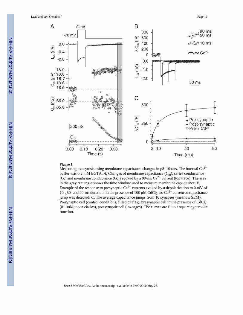
Figure 1.Measuring exocytosis using membrane capacitance changes in p8–10 rats. The internal Ca2+
buffer was 0.2 mM EGTA. A, Changes of membrane capacitance (Cm), series conductance(Gs) and membrane conductance (Gm) evoked by a 90-ms Ca2+ current (top trace). The areain the gray rectangle shows the time window used to measure membrane capacitance. B,Example of the response to presynaptic Ca2+ currents evoked by a depolarization to 0 mV of10-, 50- and 90-ms duration. In the presence of 100 μM CdCl2, no Ca2+ current or capacitancejump was detected. C, The average capacitance jumps from 10 synapses (means ± SEM).Presynaptic cell (control conditions; filled circles); presynaptic cell in the presence of CdCl2(0.1 mM; open circles), postsynaptic cell (lozenges). The curves are fit to a square hyperbolicfunction.
Leão and von Gersdorff Page 11
Braz J Med Biol Res. Author manuscript; available in PMC 2010 May 28.
NIH
-PA Author Manuscript
NIH
-PA Author Manuscript
NIH
-PA Author Manuscript

Figure 2.Membrane capacitance changes (ΔCm) in immature (p5–7) and more mature terminals (p10–12) under different Ca2+ buffering conditions. A, Plot of membrane capacitance in response todifferent Ca2+ current charge transfer (integrals of the Ca2+ current). The points correspond toICa elicited by 2-, 10-, 50-, and 90-ms step pulses to 0 mV. The curves were fitted using asquare hyperbolic function. Data are reported as means ± SEM. B, Changes in vesicle pool size(ΔCm elicited by a 90-ms pulse) for two age groups and different Ca2+ buffering conditions.*P < 0.05, significantly different compared to other buffer conditions in the same age group(unpaired t-test). N = 7–8 calyces for each age group (0.05 mM BAPTA; 0.2 mM EGTA) andN = 3 calyces for each age group for 5 mM EGTA.
Leão and von Gersdorff Page 12
Braz J Med Biol Res. Author manuscript; available in PMC 2010 May 28.
NIH
-PA Author Manuscript
NIH
-PA Author Manuscript
NIH
-PA Author Manuscript
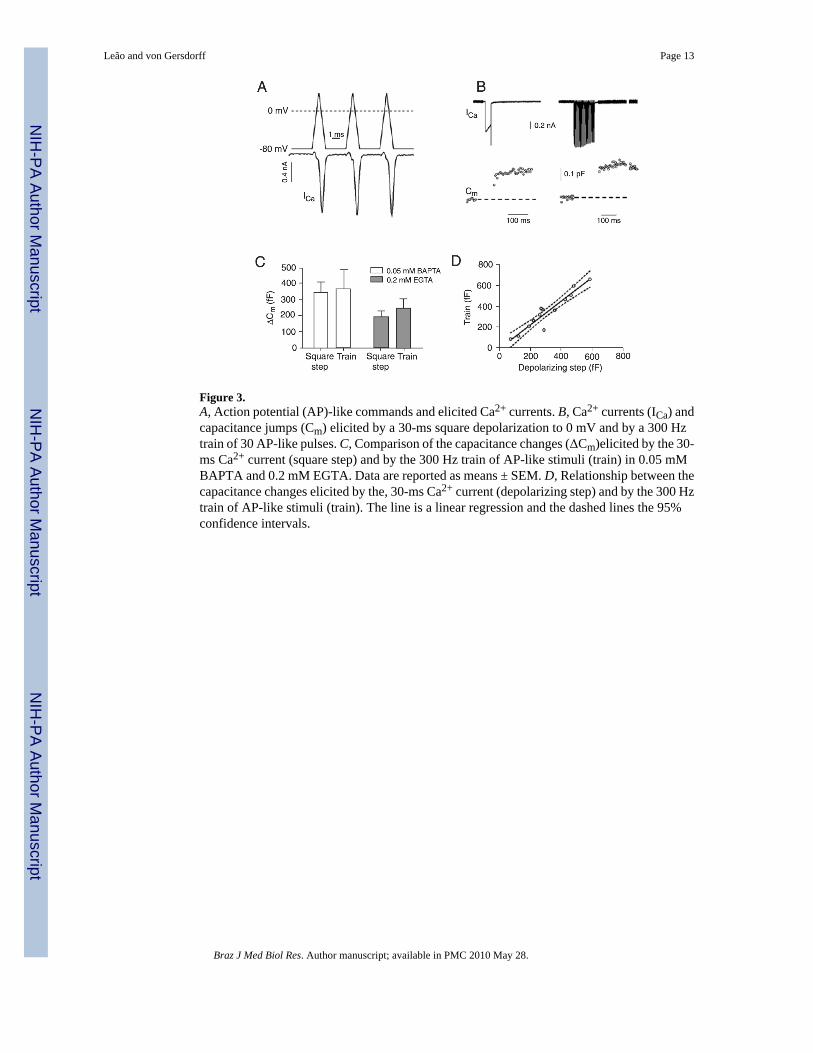
Figure 3.A, Action potential (AP)-like commands and elicited Ca2+ currents. B, Ca2+ currents (ICa) andcapacitance jumps (Cm) elicited by a 30-ms square depolarization to 0 mV and by a 300 Hztrain of 30 AP-like pulses. C, Comparison of the capacitance changes (ΔCm)elicited by the 30-ms Ca2+ current (square step) and by the 300 Hz train of AP-like stimuli (train) in 0.05 mMBAPTA and 0.2 mM EGTA. Data are reported as means ± SEM. D, Relationship between thecapacitance changes elicited by the, 30-ms Ca2+ current (depolarizing step) and by the 300 Hztrain of AP-like stimuli (train). The line is a linear regression and the dashed lines the 95%confidence intervals.
Leão and von Gersdorff Page 13
Braz J Med Biol Res. Author manuscript; available in PMC 2010 May 28.
NIH
-PA Author Manuscript
NIH
-PA Author Manuscript
NIH
-PA Author Manuscript

Figure 4.A, Examples of capacitance jumps (Cm) evoked by a 2-ms Ca2+ current in a p6 calyx (left) anda p11 calyx (right) loaded with 5 mM EGTA (note different time scale bars for Ca2+ currentand capacitance measurements). The average number of vesicles released by a 2-msdepolarization is shown for different buffer conditions. The number of docked vesiclesestimated by electron microscopy (EM) is from Taschenberger et al. (19). Data are reportedas means ± SEM.*P < 0.05, significantly different from the other buffer conditions (unpairedt-test). B, Changes in exocytosis per Ca2+ ion influx (vesicles/pC of ICa for 2 ms pulse; *P <0.05, significantly different from the other buffer conditions; unpaired t-test).
Leão and von Gersdorff Page 14
Braz J Med Biol Res. Author manuscript; available in PMC 2010 May 28.
NIH
-PA Author Manuscript
NIH
-PA Author Manuscript
NIH
-PA Author Manuscript

Figure 5.Changes in paired-pulse depression. A, Two 10-ms Ca2+ currents (ICa) were delivered 70 msapart (top trace) and the capacitance changes (bottom trace) were measured. Each capacitancepoint is the average of five consecutive data points. B, Comparison of capacitance changes(ΔCm) elicited by the first and the second 10-ms Ca2+ current in each age group and buffercondition. C, Depression of exocytosis and of Ca2+ current. Exocytosis from calyces: N = 8(0.2 mM EGTA p10–12), N = 10 (0.2 mM EGTA p5–7), N = 8 (0.05 mM BAPTA p10–12),N = 5 (0.05 mM BAPTA p5–7); Ca2+ current of calyces: N = 5 (0.2 mM EGTA p10–12), N =5 (0.05 mM BAPTA p10–12). Data are reported as means ± SEM. *P < 0.05 compared with0.2 mM EGTA (unpaired t-test).
Leão and von Gersdorff Page 15
Braz J Med Biol Res. Author manuscript; available in PMC 2010 May 28.
NIH
-PA Author Manuscript
NIH
-PA Author Manuscript
NIH
-PA Author Manuscript
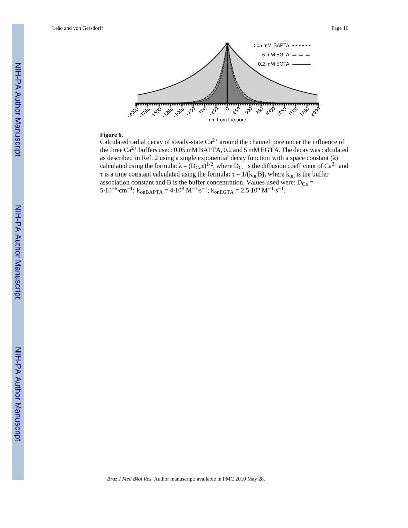
Figure 6.Calculated radial decay of steady-state Ca2+ around the channel pore under the influence ofthe three Ca2+ buffers used: 0.05 mM BAPTA, 0.2 and 5 mM EGTA. The decay was calculatedas described in Ref. 2 using a single exponential decay function with a space constant (λ)calculated using the formula: λ = (DCaτ)1/2, where DCa is the diffusion coefficient of Ca2+ andτ is a time constant calculated using the formula: τ = 1/(konB), where kon is the bufferassociation constant and B is the buffer concentration. Values used were: DCa =5·10−6·cm−1; konBAPTA = 4·108 M−1·s−1; konEGTA = 2.5·106 M−1·s−1.
Leão and von Gersdorff Page 16
Braz J Med Biol Res. Author manuscript; available in PMC 2010 May 28.
NIH
-PA Author Manuscript
NIH
-PA Author Manuscript
NIH
-PA Author Manuscript

NIH
-PA Author Manuscript
NIH
-PA Author Manuscript
NIH
-PA Author Manuscript
Leão and von Gersdorff Page 17
Tabl
e 1
Ves
icle
s rel
ease
d by
step
dep
olar
izat
ion
and
num
ber o
f doc
ked
vesi
cles
est
imat
ed b
y el
ectro
n m
icro
scop
y.
Step
p5–7
ves
icle
sp1
0–12
ves
icle
s
EG
TA
(0.2
mM
)B
APT
A (0
.05
mM
)E
GT
A (5
mM
)E
GT
A (0
.2 m
M)
BA
PTA
(0.0
5 m
M)
EG
TA
(5 m
M)
2 m
s40
616
8416
011
6626
6778
1
10 m
s16
9641
6067
129
7340
4916
84
50 m
s40
4871
7613
4465
1761
5620
22
90 m
s44
9189
4317
7380
5285
6023
58
Doc
ked
(EM
)16
7021
00
The
num
ber o
f ves
icle
s was
obt
aine
d by
mul
tiply
ing
the
capa
cita
nce
jum
p (in
fF) b
y th
e fa
ctor
12.
2. T
he n
umbe
r of d
ocke
d ve
sicl
es w
as d
eter
min
ed b
y el
ectro
n m
icro
scop
y (E
M) a
s des
crib
ed b
y Ta
sche
nber
ger
et a
l. (1
9).
Braz J Med Biol Res. Author manuscript; available in PMC 2010 May 28.
![Effect of different electrolytes on the swelling properties of calyx[4]pyrrole-containing polyacrylamide membranes](https://static.fdokumen.com/doc/165x107/631f4fc8d10f1687490fbd44/effect-of-different-electrolytes-on-the-swelling-properties-of-calyx4pyrrole-containing.jpg)