SUSCEPTIBILITY OF S.AUREUS TO METHYLENE BLUE HAEMATOPORPHYRIN, PHTALOCYANINES PHOTODYNAMIC EFFECTS
12
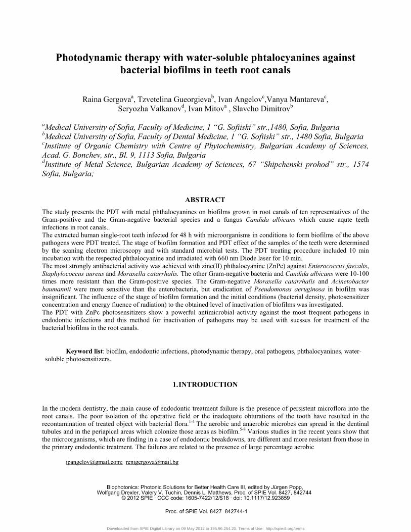
Photodynamic therapy with water-soluble phtalocyanines against bacterial biofilms in teeth root canals Raina Gergova a , Tzvetelina Gueorgieva b , Ivan Angelov c ,Vanya Mantareva c , Seryozha Valkanov d , Ivan Mitov a , Slavcho Dimitrov b a Medical University of Sofia, Faculty of Medicine, 1 “G. Sofiiski” str.,1480, Sofia, Bulgaria b Medical University of Sofia, Faculty of Dental Medicine, 1 “G. Sofiiski” str., 1480 Sofia, Bulgaria c Institute of Organic Chemistry with Centre of Phytochemistry, Bulgarian Academy of Sciences, Acad. G. Bonchev, str., Bl. 9, 1113 Sofia, Bulgaria d Institute of Metal Science, Bulgarian Academy of Sciences, 67 “Shipchenski prohod” str., 1574 Sofia, Bulgaria; ABSTRACT The study presents the PDT with metal phthalocyanines on biofilms grown in root canals of ten representatives of the Gram-positive and the Gram-negative bacterial species and a fungus Candida albicans which cause aqute teeth infections in root canals.. The extracted human single-root teeth infected for 48 h with microorganisms in conditions to form biofilms of the above pathogens were PDT treated. The stage of biofilm formation and PDT effect of the samples of the teeth were determined by the scaning electron microscopy and with standard microbial tests. The PDT treating procedure included 10 min incubation with the respected phthalocyanine and irradiated with 660 nm Diode laser for 10 min. The most strongly antibacterial activity was achieved with zinc(II) phthalocyanine (ZnPc) against Enterococcus faecalis, Staphylococcus aureus and Moraxella catarrhalis. The other Gram-negative bacteria and Candida albicans were 10-100 times more resistant than the Gram-positive species. The Gram-negative Moraxella catarrhalis and Acinetobacter baumannii were more sensitive than the enterobacteria, but eradication of Pseudomonas aeruginosa in biofilm was insignificant. The influence of the stage of biofilm formation and the initial conditions (bacterial density, photosensitizer concentration and energy fluence of radiation) to the obtained level of inactivation of biofilms was investigated. The PDT with ZnPc photosensitizers show a powerful antimicrobial activity against the most frequent pathogens in endodontic infections and this method for inactivation of pathogens may be used with sucsses for treatment of the bacterial biofilms in the root canals. Keyword list: biofilm, endodontic infections, photodynamic therapy, oral pathogens, phthalocyanines, water- soluble photosensitizers. 1. INTRODUCTION In the modern dentistry, the main cause of endodontic treatment failure is the presence of persistent microflora into the root canals. The poor isolation of the operative field or the inadequate obturations of the tooth have resulted in the recontamination of treated object with bacterial flora. 1-4 The aerobic and anaerobic microbes can spread in the dentinal tubules and in the periapical areas which colonize those areas as biofilm. 5-8 Various studies in the recent years show that the microorganisms, which are finding in a case of endodontic breakdowns, are different and more resistant from those in the primary endodontic treatment. The failures are related to the presence of large percentage aerobic [email protected]; [email protected] Biophotonics: Photonic Solutions for Better Health Care III, edited by Jürgen Popp, Wolfgang Drexler, Valery V. Tuchin, Dennis L. Matthews, Proc. of SPIE Vol. 8427, 842744 © 2012 SPIE · CCC code: 1605-7422/12/$18 · doi: 10.1117/12.923859 Proc. of SPIE Vol. 8427 842744-1 Downloaded from SPIE Digital Library on 09 May 2012 to 195.96.254.20. Terms of Use: http://spiedl.org/terms
-
Upload
independent -
Category
Documents
-
view
0 -
download
0
Transcript of SUSCEPTIBILITY OF S.AUREUS TO METHYLENE BLUE HAEMATOPORPHYRIN, PHTALOCYANINES PHOTODYNAMIC EFFECTS
Photodynamic therapy with water-soluble phtalocyanines against bacterial biofilms in teeth root canals
Raina Gergovaa, Tzvetelina Gueorgievab, Ivan Angelovc,Vanya Mantarevac,
Seryozha Valkanovd, Ivan Mitova , Slavcho Dimitrovb
aMedical University of Sofia, Faculty of Medicine, 1 “G. Sofiiski” str.,1480, Sofia, Bulgaria bMedical University of Sofia, Faculty of Dental Medicine, 1 “G. Sofiiski” str., 1480 Sofia, Bulgaria cInstitute of Organic Chemistry with Centre of Phytochemistry, Bulgarian Academy of Sciences, Acad. G. Bonchev, str., Bl. 9, 1113 Sofia, Bulgaria dInstitute of Metal Science, Bulgarian Academy of Sciences, 67 “Shipchenski prohod” str., 1574 Sofia, Bulgaria;
ABSTRACT
The study presents the PDT with metal phthalocyanines on biofilms grown in root canals of ten representatives of the Gram-positive and the Gram-negative bacterial species and a fungus Candida albicans which cause aqute teeth infections in root canals.. The extracted human single-root teeth infected for 48 h with microorganisms in conditions to form biofilms of the above pathogens were PDT treated. The stage of biofilm formation and PDT effect of the samples of the teeth were determined by the scaning electron microscopy and with standard microbial tests. The PDT treating procedure included 10 min incubation with the respected phthalocyanine and irradiated with 660 nm Diode laser for 10 min. The most strongly antibacterial activity was achieved with zinc(II) phthalocyanine (ZnPc) against Enterococcus faecalis, Staphylococcus aureus and Moraxella catarrhalis. The other Gram-negative bacteria and Candida albicans were 10-100 times more resistant than the Gram-positive species. The Gram-negative Moraxella catarrhalis and Acinetobacter baumannii were more sensitive than the enterobacteria, but eradication of Pseudomonas aeruginosa in biofilm was insignificant. The influence of the stage of biofilm formation and the initial conditions (bacterial density, photosensitizer concentration and energy fluence of radiation) to the obtained level of inactivation of biofilms was investigated. The PDT with ZnPc photosensitizers show a powerful antimicrobial activity against the most frequent pathogens in endodontic infections and this method for inactivation of pathogens may be used with sucsses for treatment of the bacterial biofilms in the root canals.
Keyword list: biofilm, endodontic infections, photodynamic therapy, oral pathogens, phthalocyanines, water-soluble photosensitizers.
1. INTRODUCTION
In the modern dentistry, the main cause of endodontic treatment failure is the presence of persistent microflora into the root canals. The poor isolation of the operative field or the inadequate obturations of the tooth have resulted in the recontamination of treated object with bacterial flora.1-4 The aerobic and anaerobic microbes can spread in the dentinal tubules and in the periapical areas which colonize those areas as biofilm.5-8 Various studies in the recent years show that the microorganisms, which are finding in a case of endodontic breakdowns, are different and more resistant from those in the primary endodontic treatment. The failures are related to the presence of large percentage aerobic
[email protected]; [email protected]
Biophotonics: Photonic Solutions for Better Health Care III, edited by Jürgen Popp, Wolfgang Drexler, Valery V. Tuchin, Dennis L. Matthews, Proc. of SPIE Vol. 8427, 842744
© 2012 SPIE · CCC code: 1605-7422/12/$18 · doi: 10.1117/12.923859
Proc. of SPIE Vol. 8427 842744-1
Downloaded from SPIE Digital Library on 09 May 2012 to 195.96.254.20. Terms of Use: http://spiedl.org/terms

and facultative anaerobic bacteria such as Enterococcus faecalis, Staphylococcus aureus and more rare Pseudomonas aeruginosa and members of family Enterobacteriaceae and Candida albicans.9-11 The eradication of the microbial biofilm by a conventional antibiotic therapy is exclusively difficult or almost impossible due the much higher resistance level of the bacteria’s. That is caused by the barrier effect of the builded exopolysaccharide protective matrix, and more importantly by heterogeneous genetic and metabolic adaptations mechanisms of the cells to new variant of growth that transforms the planktonic lifestyle into a sessile form.5,12,13 The capability of canals to efficient nutrient uptake by infusing fluid from the bulk phase into the biofilm, thereby optimized and focused to the exchange of nutrient and waste-product, demonstrates the first link between form and function.4 The invasion in dentinal tubules by root canal micro flora is a multifactorial event in which a limited number of oral bacterial species have the required properties to participate.3,7 The intercellular communication mechanisms are mediated by extracellular small signalling molecules (autoinducers) and coordinate gene expression of virulence factors such as ability to biofilm formation. It has been estimated that bacteria which are members of biofilms are generally less susceptible (about 1000-fold) to the effects of commonly used antimicrobial agents than are their analogic planktonic cells and highly resistant to phagocytosis by immune system phagocytes and the various arms of antimicrobial immunity are neutralized by the same biofilm formation.8,12,14 The source for contamination of root canals normally appears caries. The dental caries is lysis of the enamel, dentin or cementum of teeth due to microbial activities. Caries would initiated by direct demineralization of the enamel of teeth due to organic acids, which accumulate by bacterial metabolism in dental plaque. Dental plaque, adhering to the teeth, is a biofilm, in which the conglomerate of bacteria may reach a thickness of 300-500 cells on the surfaces of the tooth, including salivary polymers, and bacterial extracellular products. After initial weakening of the enamel, various oral bacteria such as Streptococcus mutans, Lactobacillus spp., Actinomyces spp., and various bacteria gain access to interior regions of the tooth. This leads to suggestion that they are secondary invaders that contribute to the progression of the destruction.4,5,7,10 Therefore the more sensitive to antibiotics microbial participants in infection are superimposed the more resistant bacterial representatives. Ones of the most frequent agents of root canal infection, E. faecalis is resistant to the most of the intracanal medicaments, as such as all of the cephalosporins and some of the other of antibiotics. Probably this is a fact due to its ability to regulate internal pH with an efficient proton pump.9,15 In 4 the authors reports that microorganisms grown in biofilms could be 1000-fold more resistant than the corresponding planktonic form. The binding of E. faecalis to the inner dentin walls of root canals explain with significant role of collagen-binding protein and serine protease produced by this organism. Pure cultures of E. faecalis added to calcium hydroxide medicated and nonmedicated root canals were able to form a biofilm structure on the canal walls. Methicillin resistance of S. aureus (MRSA) due to alteration penicillin binding proteins is a problem for therapy with all the penicillin’s and cephalosporin’s and in the most of cases it is a combinated with resistance to the others group antimicrobial agents.15 Their ability to grow as biofilms seems to be an additional important determinant of bacterial resistance to antimicrobial therapies and increases the virulence in an endodontic pathology. Therefore, the dentists urgently needed of new strategies for battling with clinically relevant biofilms, because particularly biofilm-forming microbes will be taking into consideration for more than two-thirds of human bacterial infections on the teeth.8 In the last decade, photodynamic therapy (PDT) appears as newest generation of methods used to destruct successfully endodontic microbial biofilm successfully.16-19 The PDT is a unique combination of a photosensitizer solution and a low-power light both locally applied to the selected object for treatment.14,15,20 The photosensitizer (PS), which is mostly dye compound, adheres to or gets absorbed of the target object. The PS for example binds to the walls or even enters into the cells. Further, the light activates the photosensitizer and creates a cascade of energy transfer and variable photochemical reactions in which a singlet oxygen and other reactive oxygen species (ROSs) can be produced.21,22 The PDT has found its high success at last years with positive results in several treatment of cancer, age-related macular degeneration, actinic keratosis, and acne vulgaris.23,24
PDT involves application of a photosensitizer (PS) which selectively localizes within the target cells and by irradiation with a proper light within the PS absorbance range (visible or near IR spectra) initiates the formation of PSs singlet excited state. This state relaxes to the singlet ground state by fluorescence emission and by non-radiative transition such as the intersystem crossing to the long-lived triplet excited state. The triplet energy state goes down to the ground state by exchanging energy with the surrounding molecules. The last photophysical process occurs in the irradiated area, which results in generation of singlet oxygen or the formation of ROSs. As known the radicals and ions are toxic to cells and their formation in the region near to the cellular membranes or within cells leads to oxidation of biomolecules and therefore to the selective damage of treated cells.
Proc. of SPIE Vol. 8427 842744-2
Downloaded from SPIE Digital Library on 09 May 2012 to 195.96.254.20. Terms of Use: http://spiedl.org/terms

PDT with the first generation porphyrins-like photosensitizers is approved for clinical applications. The further research on PDT agents is still actual due to limitations such as low absorbance in the visible spectrum (up to 630 nm), prolonged skin photosensitivity (more than a month) after application, chemical impurity and need of high doses of the approved porphyrins.25 The second-generation far-red absorbing photosensitizers with a fast release from skin were recognized among phthalocyanines (Pcs). The Pc-sensitizers are selected as promising PDT agents due to their strong and narrow Q-band (>670 nm) absorption properties. In addition, many of known metal-Pcs (MPcs) have very low dark toxicity to the tested cells in a wide range of concentrations. Unsubstituted MPcs have some limitations for their biomedical use such as very low solubility in biocompatible media and the poor selectivity towards targeted objects that somehow restrict their efficacy. On other hand, the uptake of PS depends on the hydrophilic/lipophilic balance and the chemical structure of the sensitizer molecule. An advantage of MPcs is that their molecular structure is flexible to tailoring to achieving better uptake to the cells. For example, the strong hydrophobic nature of the macromolecule easy is able to modify by attachment of substituents in peripheral positions, or to the coordinated metal ions.26-29
In the dental medicine field, PDT is approved for the palliative treatment of patients with advanced head and neck cancer in the European Union, Norway and Iceland, recently, and in Canada, for the treatment of periodontitis.2,25,30 The application of PDT for eradication pathogenic microbes in wound infections has been explored in animal models.14,30,31 In the last two years, the PDT was applied with success for endodontic treatments against E. faecalis.32-34 In recent times, the considerable increasing in number of paper devoted to application of PDT in dentistry was published.35-37
The aim of the presented study is a determination of bactericidal effect of PDT with three water-soluble phthalocyanines against biofilm in root canals, forming of ten representatives and problematic for standard medicine Gram-positive and Gram-negative bacteria species and fungus Candida albicans, which cause the teeth infections.
Scheme 1. . Chemical structure of used in experiments phthalocyanines - zinc (II) and silicon (IV) complexes.
2. MATERIALS AND METHODS
2.1. Phthalocyanines
Phthalocyanine complexes of zinc (II) (ZnPc and ZnPcMe) and silicon (IV) (SiPc) were evaluated as active bactericidal and fungicidal photodynamic agents.29,38 Synthesizes of phthalocyanines metal complexes studied in the present work were described in the previous publications.26,27,38 Scheme 1 presents the chemical structures of the used Pcs. The stock solutions of ZnPcMe, ZnPc and Si Pc1 were prepared and stored in the dark at 4-7°C. Prior the experiments the stocks were diluted and applied for inactivation of pathogen cells. The dilutions were performed in a sterile PBS were performed to obtain the final concentration 6 μg/L. The syringes were used for application to the treated teeth root.
Proc. of SPIE Vol. 8427 842744-3
Downloaded from SPIE Digital Library on 09 May 2012 to 195.96.254.20. Terms of Use: http://spiedl.org/terms

2.2. Microbial strains
The strains were obtained from American type culture collection bank. The Gram-positive Staphylococcus aureus (ATCC29213) and Enteroccocus faecalis (ATCC29212) were used in our study. The clinical isolates from patients’, which are representative’s multidrug resistant Gram-positive and Gram-negative aerobic and facultative anaerobic microorganisms, were included in the experiments. The following clinical isolates from patients were used: Enteroccocus faecalis (n=8; vancomycin S n=6; vancomycin R n=2), Enterococcus faecium MDR vancomycin R (n=2), Staphylococcus aureus – MSSA (n=3), and MRSA (n=3), Moraxella catarrhalis - bro2 beta-lactamase+ (n=2), Escherichia coli (n=4), Klebsiella pneumoniae ESBL (n=2), Klebsiella oxytoca (n=2), Enterobacter cloaceae (n=2), Pseudomonas aeruginosa (n=4), Acinetobacter baummannii MDR (n=4), Candida albicans (n=3). They were indentificated, reproduced and cultivated as biofilms in root canals for treatments.
2.3. Root Canals The 273 freshly extracted human single-root teeth were cleaned from the organic material with a periodontal
curette. Coronal parts of the teeth were removed and 15 mm of the root lengths were kept. All canals were instrumented using crown-down technique using Kerr (K3) files (Maillefer Instruments SA, Switzerland). Following the each file operation all canals were irrigated with 1 ml 5.25% NaOCl, than the the formed smear layer was removed with 1 ml of 17% EDTA and with 10 ml of NaOCl. The root canals were dried with sterile paper points. After that, the each tooth was placed in transparent microcentrifuge tube, autoclaved at 121ºC for 15 min and stored in sterile 0.01M phosphate-buffered saline (PBS) until used in the experiment.
2.4. Experimental procedure The bacterial strains were grown on a Brain heart infusion (BHI) agar overnight at 35ºC. The suspension of
every one of them was prepared in BHI broth to bacterial density reached at least 1,5-2x10⁸
colony-forming units (CFU)/ml and the endodontic canals inoculated with 200μl of a pure microbial culture (of a single isolate) or a microbial combination in ratio 1:1 and incubated 48 h at 35ºC. This period is enough for bacteria strains to build well-formed biofilms on investigated area.26 After incubation , the root canals of samples were washed with 1 mL sterile PBS and using serially 10-fold dilution in sterile PBS the value of CFU/ml on the BHI agar (initial concentration) were determinated. Then, the teeth were replaced from the cultures in the sterile tubes and were prepared to PDT. The number of CFU on the plates BHI agar was counted following 24-48 h incubation at 35°C aerobically. The teeth were irradiated with 300μl one of the dilution solutions of ZnPcMe, ZnPc or Si Pc1 for 5 min. The two controls of every sample were used: 1) only bacterial culture with photosensitizer, without light – dark control; 2) without photosensitizer, but with light – light control; a common control to every testing of sterile tooth with sterile BHI is used. After processing with PDT, the CFU/ml of the teeth samples was counted again (PDT treated). In the present study, the diode laser with wavelength lying within spectral diapason of absorption of photosensitizers (Fig. 1) was used. The continuous wave laser radiation with wavelength 660 nm and with output light power at the end of optical fibre of 200 mW was used in all PDT experiments. The irradiation method involved helicoidally traction movements of the fiber, from the apical to the cervical part of the teeth, lasting 5 minutes extracted human teeth. A few samples of the teeth, infected with bacterial biofilm before and after PDT were fixed with Gluteraldehyde buffer
2.5% 48h at 8ºC and prepared by vacuum evaporation with gold monolayer before scanning electron microscopy (SEM) using scanning electron microscope and X-ray Laser Particle Sizer "JCXA 733" (JEOL – Japan).
2.5.Statistics
The difference between the results was observed as a mean ± standard deviation (SD) and was compared by a Student’s test. The values of P<0.05 were considered as significant.
Proc. of SPIE Vol. 8427 842744-4
Downloaded from SPIE Digital Library on 09 May 2012 to 195.96.254.20. Terms of Use: http://spiedl.org/terms

Figure 1. Absorption spectra of used phthalocyanines
3. RESULTS AND DISCUSSION
The biofilms formed in the root canals, after 48h incubation with the selected microbial strains were studied with SEM. The typical images of biofilm developed for 48 h were visualized (Fig.2a). The effect of PDT on teeth canal surface was also imagined by SEM and is shown on Fig.2b. Obtained images from root canals demonstrated that after 48 h incubation with strains in canals, well formed biofilms were observed by SEM and the fine structure of dentinal tubules were hidden. After PDT treatment clear surface of root canals with well expressive structure of dentin structure and tubules were observed.
a) b)
Figure 2. Scanning electron micrograph of root canals tubules invaded by biofilm of Enterococcus faecalis and E. coli.(a)-
Magnification 4000x and micrograph of canals tubules without biofilm after PDT treating with ZnPcMe (b)-Magnification 3600x.
The obtained results for bacteria survival before and after PDT responces in 138 samples with biofilms against some of the most frequent and problematic bacterial isolates alone, using three phthalocyanines ZnPcMe, SiPc1, and ZnPc are shown on Figs.3-4. The PDT effectiveness on biofilms formed from polymicrobial associations of two or three strains are shown on Fig.5. The experiments shown that the most effective photoinactivation of the Gram-positive
Proc. of SPIE Vol. 8427 842744-5
Downloaded from SPIE Digital Library on 09 May 2012 to 195.96.254.20. Terms of Use: http://spiedl.org/terms

Figure 3. Survival of biofilms originated from investigated Gram-positive bacteria’s after photodynamic inactivation with water soluble metalphthalocyanine photosensitizers. .
bacteria in pure culture (Figs.3-4) and in combination of two or three strains E. faecalis, E. faecium, S. Aureus (MSSA) and by only one species of Gram-negative cocci (M. catarrhalis) was observed by PDT treating with ZnPcMe – more than 5 log10. Somewhat less (with 1-2 log10) efficacies were demonstrated with ZnPc and SiPc1 photosensitizers (Fig.5). The obtained low levels of dark toxicity – less than 1 log10 are due to relatively high for phthalocyanines concentrations of PSs used in experiments. This study aims is a determination of bactericidal effect of PDT against biofilms in root canals formed from problematic bacteria and fungi which can play significant role in progressing of infections and in same time are very resistant to treatment with ordinary antimicrobial therapy alone. The most of the microbial pathogens in dentistry as human medicine are highly resistant to the known antibacterial agents and because of that for our investigations we preferred this non-invasive and easy method as alternative for treating of great number multi drug resistant (MDR) bacteria, especially in cases of bacterial infections related to dental medicine which can not be influenced by the conventional antimicrobial chemotherapy.2,9,11 Because of fact, that mouth is main reservoir for most of bacteria’s, the suggestion that in building of tooth biofilms take place not only well known agent of concrete infection but also and nonspecific bacteria, we investigate effect of PDT inactivation on multibacterial biofilms. In contrast with32,34, where the combinations of PDT with chemomechanical debridement (CMD) with 6% NaOCl were used, the presented results for efficacy of decontamination were obtained only after PDT treatment. The investigated phthalocyanines inactivated and hence significant decreasing in value of viable cells were obtained for both the E. faecalis and E. faecium, with similar activity, irrespective of their resistance of antimicrobial agents, respectively of problematic vancomycin resistance. The effect of ZnPcMe, ZnPc and SiPc1 was stronger against E. faecalis, E. faecium and MSSA, but more weak to MRSA about with one log10 by treating with the three phthalocyanines. The effect of SiPc1 to Gram-positive and Gram-negative cocci was lowest in comparison with the others phthalocyanines. The lower effective photoinactivation of variety Gram-negative rods and C. albicans were obtained by irradiation with different light intensity for three phthalocyanines: ZnPcMe, ZnPc and Si Pc1 and the results are shown on Figs.3-5. The most effective against sensitive to antibiotics members of family Enterobacteriaceae and A. Baummannii was phthalocyanines ZnPcMe about 3.5 log10. The phthalocyanines ZnPc and SiPc1 were also active and show decreasing in survival levels with about 1.5-2 log10. A. baummannii, MDR and problematic bacterial species is very
Proc. of SPIE Vol. 8427 842744-6
Downloaded from SPIE Digital Library on 09 May 2012 to 195.96.254.20. Terms of Use: http://spiedl.org/terms

Figure 4. Survival of Gram-negative bacteria’s biofilms after photodynamic inactivation with zinc- and silicon- phthalocyanine complexes
sensitive (similar to sensitivity of staphylococci) to PDT with ZnPcMe. The producers of extended spectrum beta-lactamases enterobacteria (E. coli, K. pneumoniae, E. cloaceae) showed some difference about 0.5-1.5 log10 to the effect of PDT compared to the other representatives of same species from Enterobacteriacea. The lower effective inactivations about 0.5-1 log10 were demonstrated with ZnPc and SiPc1 compared with ZnPcMe. All of the enterobacteria and fungi demonstrated about 10-100 times more resistant to PDT mediated killing compared to E. faecalis, E. faecium, S. aureus, and M. catarrhalis. The results of dark and light toxicity demonstrated negligible effect compared this of PDT with the used photosensitizers (P<0.05). The effect of the three phthalocyanines: ZnPcMe, ZnPc, and Si Pc1 were insignificant or missing for the problematic MDR P. aeruginosa. That is the lowest effect compared to the similar problematic MDR A. baummannii and the other MDR Gram-positive and Gram-negative bacteria. Gram-positive bacteria’s are more than1000 times sensitive to PDT mediated killing compared to P. aeruginosa. In multibacterial biofilms with P. aeruginosa it was observed clear difference in inactivation levels for P. aeruginosa compared to the other strains (Fig.6). The biofilms of
Proc. of SPIE Vol. 8427 842744-7
Downloaded from SPIE Digital Library on 09 May 2012 to 195.96.254.20. Terms of Use: http://spiedl.org/terms
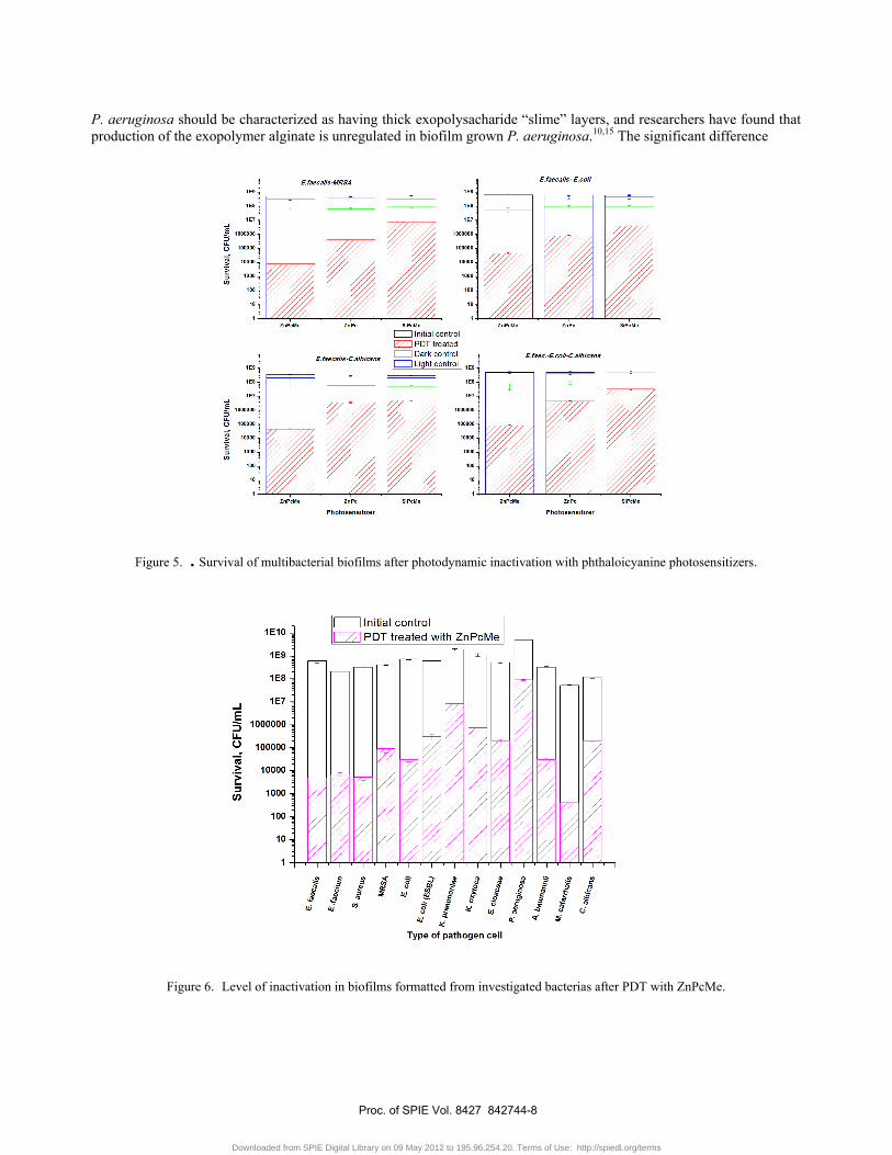
P. aeruginosa should be characterized as having thick exopolysacharide “slime” layers, and researchers have found that production of the exopolymer alginate is unregulated in biofilm grown P. aeruginosa.10,15 The significant difference
Figure 5. . Survival of multibacterial biofilms after photodynamic inactivation with phthaloicyanine photosensitizers.
Figure 6. Level of inactivation in biofilms formatted from investigated bacterias after PDT with ZnPcMe.
Proc. of SPIE Vol. 8427 842744-8
Downloaded from SPIE Digital Library on 09 May 2012 to 195.96.254.20. Terms of Use: http://spiedl.org/terms

of levels of inactivation in this situations and repeating the levels on inactivation for different strains in other cases of multi bacterial biofilms compared to the single bacterial biofilms mean that influence and interference between strains in biofilms seems to be slightingly low.
Figure 7. Obtained level of inactivation in multibacterial biofilms after PDT with ZnPcMe .
On Fig.6 and Fig.7 are shown the obtained level on inactivation for different microbial strains (Fig.6) or for multimicrobial formations (Fig.7) in root canals, achieved by using of most effective in our experiments ZnPcMe photosensitizer. The results confirm the next suggestion in previous publications.28,39,40,41 First– the Gram-positive strains are more sensitive to the PDT inactivation, than Gram-negative and yeast. This may be explained with differences in cells walls and as a result of these differences the level of uptake of PS to the cell - it is higher for Gram-positive than to the other two types of microorganisms. The more complex character of ceil wall by Gram-negative also contribute to the lowest levels of inactivation do to necessarily of higher concentration of singlet oxygen or ROS to destroying membrane to such of degree which leads to bacterial death. On the second place the higher initial concentrations of strains, which were used in experiments, lead to the lower efficacy of inactivation. Explanation of this fact is also in lower level of uptake of PSs molecules per cell by increasing the cell density.41,42 Therefore, obtained results for levels of decontamination by treatment of root canals against wide number of pathogen strains in oral cavity through using PDT with water soluble PCs shown that this method may be used alone or with combination with chemomechanical debridement (CMD) with 6% NaOCl 32-34 for successful endodontic treatments. In addition, the identical results for PDT efficacy were obtained when in PDT investigations for inactivation of some of samples was used red LED lamp on 640 nm with light power of 150 mW.
4. CONCLUSIONS
The obtained results demonstrated that by the PDT with using of cationic ZnPcMe photosensitizer in endodontic treatments on microbial biofilm statistically considerable reduction of pathogen bacterial population will be possible to be achieved. The PDT effect was more significant by the bacteria’s - E. faecalis, E. faecium, S. aureus, M. catarrhalis and A. baumannii. The PDT efficacy are in contrast with results after treatment of investigated pathogens with antibiotics, because of their resistance to conventional antibiotic therapy. The use of ZnPcMe is preferable for the photoinactivation of pathogenic Gram-positive and Gram-negative microorganisms compared to the ZnPc and SiPc1.
Proc. of SPIE Vol. 8427 842744-9
Downloaded from SPIE Digital Library on 09 May 2012 to 195.96.254.20. Terms of Use: http://spiedl.org/terms

The investigated three phthalocyanines- ZnPcMe, ZnPc and SiPc1 were not enough effective in eradication of problematic P. aeruginosa. The PDT efficacies against pathogens were demonstrated ex vivo as well on the pure microbial culture biofilms also by biofilms formed from polymicrobial combination in root canals. The PDT with water-soluble phthalocyanines in combination with unexpensive red LED lamps has high potential to be used as antimicrobial procedure in endodontic therapy.
ACKNOWLEDGEMENTS The work was supported by the National Science Fund, Sofia, Bulgaria (Grants DO-02-112/08 and DO-02-177/08)
REFERENCES [1] Bonsor S.J., Nichol R., Reid T.M., Pearson G.J., “Microbiological evaluation of photo-activated disinfection in endodontics (An in vivo study),” Brit Dent J., 200, 337–341 (2006). [2] Bonsor S.J., Nichol R., Reid T.M., Pearson G.J., “An alternative regimen for root canal disinfection,” Brit Dent J, 201, 101-105 (2005). [3] Gunnel S.T., Gunnar B., “Biofilms in endodontic infections,” Endodon Top, 9, 27–36 (2004). [4] Hall-Stoodley L., William J., Stoodley C.P., “Bacterial biofilms: from the natural environment to infectious diseases,” Nat Rev Microbiol., 2, 95-108 (2004). [5] Chavez D.E, Paz L. “Gram-positive organisms in endodontic infections,” Endodont Topics, 9, 79–96 (2004). [6] Hayati A.F, Okada A., Kitasako Y, Tagami J, Matin K., “An artificial biofilm induced secondary in vitro studies,” Australn Dent J, 56, 40–47 (2011). [7] Kunze B., Reck M., Dotsch A., Lemme A., Schummer D., Irschik H., Steinmetz H., Wagner-Dobler I. “Damage of Streptococcus mutans biofilms by carolacton, a secondary metabolite from the myxobacterium Sorangium cellulosum,” BMC Microbiol, 10, 199 (2010). [8] Usha H.L., Kaiwar A., Mehta D., “Biofilm In Endodontics: New Understanding To An Old Problem,” Int J of Contempor Dentistry, 3, 44-51 (2010). [9] Stuart C.H., Schwartz S.A., Beeson T.J., Sowatz C.B., “Enterococcus faecalis: Its Role in Root Canal Treatment Failure and Current Concepts in Retreatment,” JOE, 32, 93-98 (2006). [10] Ramos M.M.B., Gaetti-Jardim E.C., Caetti-Jardim Junior E., “Resistance to tetracycline and β-lactams and distribution of resistance markers in enteric microorganisms and pseudomonads isolated from the oral cavity,” J of Appl Oral Sci, 17, 13-18 (2009). [11] Qiang Y-G., Christine M.N.Y., Huang Z. “Combination of photodynamic therapy and immunomodulation - current status and future trends,” Med Res Rev, 28, 632–644 (2008). [12] Dror N., Mandel M., Hazan Z., Lavie G., “Advances in Microbial Biofilm Prevention on Indwelling Medical Devices with Emphasis on Usage of Acoustic Energy,” Sensors, 9, 2538-2554 (2009). [13] Izutani N., Imazato S., Nakajo K., Takahashi N., Takahashi Y., Ebisu S., Russell R.R.B., “Effects of the antibacterial monomer 12-methacryloyloxydodecylpyridinium bromide (MDPB) on bacterial viability and metabolism,” Eur J of Oral Sciences, 19, 175-181 (2011). [14] Soukos N.S., Goodston J.M, “Photodynamic therapy in the control of oral biofilms,” Periodontol, 55, 143–166 (2000). [15] Mahon C.R., Lehman D.C., Manuselis G.,[Textbook of Diagnostic microbiology third edition], 303-363 (2007). [16] Muller P., Guggenheim B., Schmidlin P.R., “Efficacy of gasiform ozone and photodynamic therapy on a multispecies oral biofilm in vitro,” Europ J of Oral Sci, 115, 77-80 (2007). [17] Lima J.P.M., Mary A.S.M., Fatima M.C.B., Alrieta H.T., Carolina S.O., Martines N., Lidiani K., Rodrigues A., Korbelik M., “PDT-Associated Host Response and its Role in the Therapy Outcome,” Lasers in Surg and Med, 38, 500–508 (2006). [18] Akilov O.E., Katie O., Sachiko K, Tayyaba H., “Photodynamic therapy against intracellular pathogens: Problems and potentials,” Medl Laser Appl, 21, 251–260 (2006). [19] Soukos N.S., Chen P. S-Y., Morris J.T., Ruggiero K., Abernethy A.D., Som S., Foschi F., Doucette S., Bammann L.L., Fontana C.R., Doukas A.G., Stashenko P.P., “Photodynamic Therapy for Endodontic Disinfection,” J Endodont, 32(10), 979 –984 (2006).
Proc. of SPIE Vol. 8427 842744-10
Downloaded from SPIE Digital Library on 09 May 2012 to 195.96.254.20. Terms of Use: http://spiedl.org/terms

[20] Scalise I., Durantini E.N., “Synthesis, properties and photodynamic inactivation of Escherichia coli using a cationic and noncharged Zn(II) pyridiloxyphthalocyanine derivates,” Bioorganic and Medical Chemistry, 13(8), 3037-3045 (2005). [21] Garcez A.S, Martha S.R., George P.T., Silva C.N., Antonio O.C.J., Michael R.H., “Antimicrobial Photodynamic Therapy Combined With Conventional Endodontic Treatment to Eliminate Root Canal Biofilm Infection,” Lasers in Surg and Med, 39, 59–66 (2007). [22] Iriana C.J.Z., “Evaluation of the antimicrobial effect of photodynamic antimicrobial therapy in an in situ model of dentine caries,” Europ J of Oral Sci, 117, 568–574 (2009). [23] Wainright M., Crossley K.B., “Photosensitising agents – Circumventing resistance and breaking down biofilms: A review,” International Biodeterioration and Biodegradation, 53(2), 119-126 (2004). [24] Dai, T., Huang Y-Y.,Hamblin M,R., “Photodynamic therapy for localized infections – State of the art,” Photodiagnosis and Photodynamic Therapy, 6(3-4), 170-188 (2009). [25] Juzeniene A., Moan J., “The history of PDT in Norway. Part one: Identification of basic mechanisms of general PDT,” Photodiagnosis and Photodynamic Therapy, 4, 3-11 (2007). [26] Mantareva V., Kussovski V., Angelov I., Wöhrle D., Dimitrov R., Dimitrov S., Popova E., “Non-aggregated Ga(III)-phthalocyanines in the photodynamic inactivation of planktonic and biofilm cultures of pathogenic microorganisms,” Photochem Photobiol Sci, 10(1), 92-102 (2011). [27] Mantareva V, Kussovski V., Angelov I., Borisova E., Avramov L., Schurpfeld G., Wöhrle D., “Photodynamic activity of water-soluble phthalocyanine zinc(II) complexes against pathogenic microorganisms,” Bioorg & Med Chem., 15, 4829–4835 (2007). [28] Mantareva, V., I. Angelov, V. Kussovski, D. Wöhrle, S. Dimitrov, “Metallophthalocyanines as photodynamic sensitizers for treatment of pathogenic bacteria. Uptake and photoinactivation properties,” Comp. Rend. Bulg. Acad. Sci., 63(1), 77-84 (2010). [29] Nyokong T., “Effects of substituents on the photochemical and photophysical properties of main group metal phthalocyanines,” Cordination Chemistry Reviews, 251, 1707-1722 (2007). [30] Douherty T.J., Charles J.G., Barbara W.H., Giulio J., David K., Korbelik M., Johan M., Quian P., “Photodynamic Therapy,” J of the Nat Cancer Inst, 90, 889-905 (1998). [31] Wainwright M. “Photodynamic antimicrobial chemotherapy (PACT),” J of Antimicrob Chemother, 42, 13–28 (1998). [32] Ng R.; Singh F.; Papamanou D.A. et al., “Endodontic Photodynamic Therapy Ex Vivo,” Jour. Of Endodontics, 37(2), 217-222 (2011). [33] Souza L.C.; Brito-Patricia R. R.; Machado de Oliveira J.C. et al., “Photodynamic Therapy with Two Different Photosensitizers as a Supplement to Instrumentation/Irrigation Procedures in Promoting Intracanal Reduction of Enterococcus faecalis,” Jour. Of Endodontics, 36 (2), 292-296 (2010). [34] Rios A., He Jianing; Glickman G.N. et al., “Evaluation of Photodynamic Therapy Using a Light-emitting Diode Lamp against Enterococcus faecalis in Extracted Human Teeth,” Jour. Of Endodontics, 37(6), 856-859 (2011). [35] Nagata J.Y., Hioka N., Kimura E., Batistela V.B., Terada R.S.S., Graciano A.X., Baesso M.L., Hayacibara M.,F., “Antibacterial photodynamic therapy for dental caries: Evaluation of the photosensitizers used and light source properties,” Photodiagnosis and Photodynamic Therapy, doi:10.106/j.pdpdt.2011.11.006 (2011). [36] Rolim J.P.M.L., de-Melo M.A.S., Guedes S.F. et al., “The antimicrobial activity of photodynamic therapy against Streptococcus mutans using different photosensitizers,” Jour. of Photochemistry and Photobiology B: Biology, 106, 40-46 (2012). [37] Jungueira J.C., Jorge A.O.C., Barbosa J.O. et al., “Photodynamic inactivation of biofilms formed by Candida spp., Trichosporon mucoides, and Kodamaea ohmeri by cationic nanoemulsion of zinc 2,9,16,23-tetrakis (phenylthio)-29H, 31H-phthalocyanine (ZnPc),” Lasers Med Sci, DOI 10.1007/s10103-012-1050-2 (2012). [38] Mantareva V., Angelov, I., Kussovski, V., Dimitrov, R., Lapok, L., Wöhrle, D., “Photodynamic efficacy of water-soluble Si(IV) and Ge(IV) phthalocyanines towards Candida albicans planktonic and biofilm cultures,” Europ. Jour. Of Medical Chemistry, 46(9), 4430-4440 (2011). [39] Angelov I., Mantareva, V., Kussovski, V., Wöhrle, D., Kisov, H., Belcheva, M., Georgieva, T., Dimitrov, S. “Susceptibility of representative dental pathogens to inactivation by the PDT with water-soluble photosensitizers,” Proceed. of SPIE, Vol. 7994, 79941A (2010). [40] Kussovski V., V. Mantareva, I. Angelov, P. Orozova, D. Wöhrle, G. Schnurpfeil, E. Borisova, L. Avramov, “Photodynamic inactivation of Aeromonas hydrophilla by cationic phthalocyanines with different hydrophobicity,” FEMS Microbiol Lett., 294, 133-140 (2009).
Proc. of SPIE Vol. 8427 842744-11
Downloaded from SPIE Digital Library on 09 May 2012 to 195.96.254.20. Terms of Use: http://spiedl.org/terms

[41] Angelov I., Mantareva, V., Kussovski, V., Wöhrle, D., Borisova, E., Avramov, L. “Improved antimicrobial therapy with cationic tetra- and octa-substituted phthalocyanines,” Proceed. of SPIE, 7027, art. no. 702717 (2008), [42] Demidova, T.N., Hamblin M.R., “Effect of cell-photosensitizer binding and cell density on microbial photoinactivation,” Antimicrobial Agents and Chemotherapy, 49(6), 2329-2335 (2005).
Proc. of SPIE Vol. 8427 842744-12
Downloaded from SPIE Digital Library on 09 May 2012 to 195.96.254.20. Terms of Use: http://spiedl.org/terms