Surgery After Neoadjuvant Stereotactic MRI Guided Adaptive ...
147
Henry Ford Health Henry Ford Health Henry Ford Health Scholarly Commons Henry Ford Health Scholarly Commons Radiation Oncology Meeting Abstracts Radiation Oncology 10-1-2021 Surgery After Neoadjuvant Stereotactic MRI Guided Adaptive Surgery After Neoadjuvant Stereotactic MRI Guided Adaptive Radiation in Pancreatic Cancer: Multi-institutional Toxicity and Radiation in Pancreatic Cancer: Multi-institutional Toxicity and Survival Outcomes Survival Outcomes Eric Schaff Henry Ford Health, [email protected] Celina Kirsch Henry Ford Health, [email protected] Parag J. Parikh Henry Ford Health, [email protected] Michael Chuong Roberto Herrera See next page for additional authors Follow this and additional works at: https://scholarlycommons.henryford.com/ radiationoncology_mtgabstracts Recommended Citation Recommended Citation Schaff E, Kirsch C, Parikh P, Chuong M, Herrera R, Asbun H, Jimenez R, Siddiqui F, Khan G, Aparo S, De Zarraga F, Ucar A, Shah R, Li P, Movsas B, and Kwon D. Surgery After Neoadjuvant Stereotactic MRI Guided Adaptive Radiation in Pancreatic Cancer: Multi-institutional Toxicity and Survival Outcomes. American Journal of Clinical Oncology-Cancer Clinical Trials 2021; 44(10):S57-S57. This Conference Proceeding is brought to you for free and open access by the Radiation Oncology at Henry Ford Health Scholarly Commons. It has been accepted for inclusion in Radiation Oncology Meeting Abstracts by an authorized administrator of Henry Ford Health Scholarly Commons.
-
Upload
khangminh22 -
Category
Documents
-
view
8 -
download
0
Transcript of Surgery After Neoadjuvant Stereotactic MRI Guided Adaptive ...

Henry Ford Health Henry Ford Health
Henry Ford Health Scholarly Commons Henry Ford Health Scholarly Commons
Radiation Oncology Meeting Abstracts Radiation Oncology
10-1-2021
Surgery After Neoadjuvant Stereotactic MRI Guided Adaptive Surgery After Neoadjuvant Stereotactic MRI Guided Adaptive
Radiation in Pancreatic Cancer: Multi-institutional Toxicity and Radiation in Pancreatic Cancer: Multi-institutional Toxicity and
Survival Outcomes Survival Outcomes
Eric Schaff Henry Ford Health, [email protected]
Celina Kirsch Henry Ford Health, [email protected]
Parag J. Parikh Henry Ford Health, [email protected]
Michael Chuong
Roberto Herrera
See next page for additional authors
Follow this and additional works at: https://scholarlycommons.henryford.com/
radiationoncology_mtgabstracts
Recommended Citation Recommended Citation Schaff E, Kirsch C, Parikh P, Chuong M, Herrera R, Asbun H, Jimenez R, Siddiqui F, Khan G, Aparo S, De Zarraga F, Ucar A, Shah R, Li P, Movsas B, and Kwon D. Surgery After Neoadjuvant Stereotactic MRI Guided Adaptive Radiation in Pancreatic Cancer: Multi-institutional Toxicity and Survival Outcomes. American Journal of Clinical Oncology-Cancer Clinical Trials 2021; 44(10):S57-S57.
This Conference Proceeding is brought to you for free and open access by the Radiation Oncology at Henry Ford Health Scholarly Commons. It has been accepted for inclusion in Radiation Oncology Meeting Abstracts by an authorized administrator of Henry Ford Health Scholarly Commons.

Authors Authors Eric Schaff, Celina Kirsch, Parag J. Parikh, Michael Chuong, Roberto Herrera, Horacio Asbun, Ramon Jimenez, Farzan Siddiqui, Gazala Khan, Santiago Aparo, Fernando De Zarraga, Antonio Ucar, Rupen Shah, Pin Li, Benjamin Movsas, and David Kwon
This conference proceeding is available at Henry Ford Health Scholarly Commons: https://scholarlycommons.henryford.com/radiationoncology_mtgabstracts/212

Proceedings of the American RadiumSociety®’s 103rd Annual Meeting
ORAL ABSTRACTS
(OA01) Interleukin-15 Rescues Radiation Related Lym-phopenia and Improves Tumor Control Outcomes in Pan-creatic CancerBhanu Prasad Venkatesulu, MD1, Cheng Cheng-En Hsieh, MD2,Byung Kyu Kim, PhD2, Keith L. Sanders, BS2, Amrish Sharma, PhD2,Pankaj Kumar Singh, PhD3, Sunil Krishnan, MD4; 1Loyola UniversityChicago,2 MD Anderson Cancer Centre,3 Mayo Clinic, Jacksonville,4Mayo Clinic, JacksonvilleBackground: There have been recent reports on the associationbetween radiation related lymphopenia and survival outcomes in solidtumors. Depletion of the circulating lymphocytes during the course ofradiation has been reported to cause inferior overall survival outcomesin gliomas, head and neck squamous cell carcinoma, nasopharyngealcancer, lung cancer, esophagus, hepatocellular carcinoma, pancreaticcancer as well as cervical cancer. The radiation dose to the circulatinglymphocyte population when the lymphocytes traverse the radiationportal as well as unintended dose to primary and secondary lymphoidorgans were postulated as the reasons for the lymphocyte depletion.This RT related lymphocyte depletion not only mitigates the beneficialeffect of RT but as well as reduces the effectiveness of T cell basedimmunotherapeutic agents like checkpoint inhibitors and adoptive Tcell transfer.Objectives: 1. To understand the alterations in the lymphocyte andcytokine kinetics in peripheral blood samples of patients with locallyadvanced pancreatic cancer who underwent definitive chemoradiationas part of a prospective phase 2 trial 2. Create radiation related lym-phopenia murine pancreatic cancer models 3. Devise rescue agents ofradiation related lymphopenia and assess if tumor control outcomes areimproved with enhancing lymphocyte population 4. Assess whichsubgroup of lymphocyte populations are responsible for improvementin tumor control outcomes.Methods: We analyzed the severity of lymphopenia, mean splenic dose,lymphocyte subpopulations (CD3, CD45, CD4, CD8, PD-1, Foxp3, Ki-67, CTLA-4 and the level of cytokines (IL-2, IL-6, IL-10, IL-7, IL-15,and TNF-alpha) in a cohort of 20 pancreatic cancer patients undergoingconventional 50.4 Gy in 28 fractions intensity modulated radiationtherapy (IMRT) for locally advanced pancreatic (LPAC) as part of aphase 2 clinical trial. Based on our clinical findings, we also designed amodel of radiation induced lymphopenia through splenic irradiation inmice and assessed the lymphocyte subpopulation dynamics in periph-eral blood and its impact on murine xenograft pancreatic tumors. Wealso devised potential rescue strategies to see if T lymphocytehomeostatic cytokines Interleukin-15 (IL-15) 7.5 ug for 7 days andInterleukin-7 (IL-7) 10 ug for 7 days supplemented to mice that hadradiation related lymphopenia could have its lymphopenia rescued andthe tumor outcomes could improve. In addition, we tested the efficacyof IL-15 super agonist which has a longer half-life than IL-15 aslymphorepletion agent and its impact on primary tumor as well assecondary non-irradiated tumor in murine pancreatic cancer models.
Results: Twenty patients with locally advanced pancreatic cancer wereenrolled as part of a single arm phase 2 clinical trial. All patientsreceived 50.4 Gy in 28 fractions of IMRT with concurrent daily cape-citabine and weekly nab-paclitaxel on days 1, 8, 15, 22, and 29 for atotal duration of 5-6 weeks. The mean splenic dose for severe lym-phopenia (grade 3, 4) was 4.92 Gy compared with 1.52 Gy in mildlymphopenia arm (grade 1, 2). The following cytokines IL-2, IL-6, IL-7, IL-15 and TNF-alpha were analyzed and homeostatic T cell cyto-kines IL-7, IL-15 fail to increase in patients with severe lymphopeniacompared with elevation of IL-7 in patients with mild lymphopenia.Analyses of the frequencies of T cells from the peripheral blood showsthat the patients who had higher mean splenic doses (4.92 Gy) com-pared with lower splenic doses (1.52 Gy) had significant depletion (Pvalue< 0.05) of CD3+ T cells (17.30% vs 40.80%), CD4+ T cells(11.87% vs 27.88%), CD8+ T cells (0.16% vs 0.52%) at week 1; CD3+T cells (19.28% vs 29.84%), CD4+ T cells (13.87% vs 27.86%), CD8+T cells (0.23% vs 0.98%) at week 5. We tested IL-7 and IL-15 inC57BL6 murine pancreatic cancer models. The tumors were irradiatedto a dose of 10 Gy in 5 fractions over 5 days and the spleen wasirradiated to 5 Gy in 5 fractions over 5 days with Philips 250 Kvorthovoltage irradiator. Splenic irradiation compromises the tumorcontrol outcomes provided by radiation. IL-15 was administered intra-peritoneally 24 hours post radiation 7.5 ug per day for 7 days IP and IL-7 was administered intra-peritoneally 24 hours post radiation 10 ug perday for 7 days. Treatment with IL-15 induced complete primary tumorregression in 75% of the treatment group whereas IL-7 did not inducecomplete primary tumor regression in any mice. IL-15 increased cyto-toxic CD8 T cell and natural killer cell infiltration into tumors andperipheral blood. IL-15 rescued the lymphopenia induced by radiationby increasing the circulating lymphocyte populations as well as byenhancing lymphocyte infiltration into the tumor as well as the resi-dential lymphocytes in the spleen. IL-15 super agonist which has alonger half-life and ease of administration was tested. IL-15 superagonist 4 ug per mice was administered 24 hours after radiation deliveryfor 2 doses one week apart. IL-15 super agonist supplementation inlymphodepleted mice enhances the tumor control at the primary site aswell as the secondary non-irradiated abscopal site.Conclusions: Our results show that lymphopenia is a commonaccompaniment of pancreatic cancer treatment with radiation being theprime contributor and lymphopenia correlates with suboptimal tumorcontrol outcomes. The mean splenic dose correlates with the severity oflymphopenia and the main lymphocyte subpopulations that are depletedare CD3, CD4, CD8 lymphocyte populations. Though IL-7 supple-mentation rescued CD4 counts, the reconstitution did not have a tan-gible effect on the tumor control outcomes in murine pancreatic cancermodels. These results add further strength to the notion that CD8 T cellsplay an important role in immunological cell death of pancreatic tumorand the reconstitution of the CD8 cells with IL-15 not only increases thelymphocyte count but improves tumor control outcomes. Our experi-ments provide impetus for design of clinical trials for use of IL-15 orIL-15 super agonist as an adjuvant therapy to rescue T cells and tofurther augment the benefit of radiation in pancreatic cancer (Figs. 1–3).
Copyright © 2021 Wolters Kluwer Health, Inc. All rights reserved.ISSN: 0277-3732/21/4410-00S1DOI: 10.1097/COC.0000000000000862
ABSTRACTS
American Journal of Clinical Oncology � Volume 44, Number 10, October 2021 www.amjclinicaloncology.com | S1
Copyright r 2021 Wolters Kluwer Health, Inc. All rights reserved.

FIGURE 1. Radiation of 50.4 Gray in 28 fractions given over 5.5 weeks in combination with capecitabine 825mg/m2 (Monday to Friday)and nab-paclitaxel IV over 30 minutes on days 1, 8, 15, 22, and 29 in locally advanced pancreatic cancer leads to lymphopenia andcauses distinct alterations in the cytokine levels in peripheral blood at baseline, week 1, week 3, week 5 and follow up after completion ofone month of treatment. (A) Illustrative summary of the design of the phase I clinical trial. The figures show the cytokine levels IL-2, IL-6,IL-7, IL-15, TNF-alpha in mild lymphopenia group (B) and severe lymphopenia group (C), percentage of CD3+ T cells (D), percentage ofCD4+ T cells (E), percentage of FOXp3+ cells (F), percentage of CD8+ T cells (G), percentage of CD19+ cells (H), percentage of CD11b-11C+ cells (I). Data are represented as means± SEM (n=20 patients). Statistical significance was assessed by unpaired T test; *, P<0.05;**, P<0.005; ***P<0.0005; ns-not significant.
Abstracts American Journal of Clinical Oncology � Volume 44, Number 10, October 2021
S2 | www.amjclinicaloncology.com Copyright © 2021 Wolters Kluwer Health, Inc. All rights reserved.
Copyright r 2021 Wolters Kluwer Health, Inc. All rights reserved.

FIGURE 2. Interleukin-15 rescues radiation induced lymphopenia and enhances tumor control outcomes in murine Pancreatic cancerxenograft models. (A) Experimental schema (B) shows the tumor growth delay curves when spleen is irradiated (C) shows that Inter-leukin-7 doesn’t improve tumor control outcomes (D) shows that IL-15 rescues the inferior tumor control that occurs with incidentalsplenic radiation (E) shows mean tumor weights. Unpaired T test with Welch’s correction was used to assess significance between thetumor growth delay groups; *, P<0.05; **, P<0.005; ***P<0.0005; ns-not significant. (Each group had mice n=8-10 per group).
American Journal of Clinical Oncology � Volume 44, Number 10, October 2021 Abstracts
Copyright © 2021 Wolters Kluwer Health, Inc. All rights reserved. www.amjclinicaloncology.com | S3
Copyright r 2021 Wolters Kluwer Health, Inc. All rights reserved.

FIGURE 3. Serial assessment of the peripheral blood lymphocyte subpopulations in mice bearing Panco2 xenograft tumor models in right thighshow that Interleukin-15 (A) and (C) increases circulating CD3+ T cells and CD8+ T cells with no impact on (B) CD4+ T cells. (D) representativeflow cytometry figures that show alterations in CD3 and CD19 populations during the course of radiation from day 1-to day 5 and after IL-15rescue from day 6 to day 13 and follow up till day 32. (E) representative flow cytometry figures that show alterations in CD8 and CD4 populationsduring the course of radiation from day 1-to day 5 and after IL-15 rescue from day 6 to day 13 and follow up till day 32. Tumors and spleen wereharvested on day 15 and the isolated tumor infiltrating lymphocytes and splenocytes were characterized by flow cytometry. The figures showpercentage of CD3+ T cells in tumor (F), percentage of CD4+ Tcells in tumor (G), percentage of CD8+ Tcells in tumor (H), percentage of NK cellsin tumor (I), percentage of CD3+ T cells in spleen (J), percentage of CD8+ T cells in spleen (K). Data are represented as means±SEM (n=6-8mice/group) statistical significance was assessed by unpaired T test; *, P<0.05; **, P<0.005; ***P<0.0005; ns-not significant.
Abstracts American Journal of Clinical Oncology � Volume 44, Number 10, October 2021
S4 | www.amjclinicaloncology.com Copyright © 2021 Wolters Kluwer Health, Inc. All rights reserved.
Copyright r 2021 Wolters Kluwer Health, Inc. All rights reserved.

(OA02) Long-term Pathologic and Survival Outcomes withChemotherapy and Stereotactic Body Radiotherapy inLocalized Pancreatic AdenocarcinomaColin Hill1, Shuchi Sehgal, BS2, Jeffrey Meyer, MS, MD1, JosephHerman, MD3, Amol Narang, MD1; 1Johns Hopkins University Schoolof Medicine, Department of Radiation Oncology and Molecular RadiationSciences, 2Philadelphia College of Osteopathic Medicine, 3NorthwellHealthBackground: Borderline resectable (BRPC) or locally advanced pan-creatic cancer (LAPC) patients are at high risk of margin positiveresection. Neoadjuvant stereotactic body radiation therapy (SBRT) mayhelp increase the proportion of patients that are surgically explored andresected with negative margins. We report long-term outcomes ofBRPC/LAPC patients after neoadjuvant CT (nCT) followed by5-fraction SBRT (nCT-SBRT) with a high proportion of patientsreceiving multi-agent (MA)-CT and surgically explored.Objectives: Consecutive BRPC/LAPC patients diagnosed from 2011-2019 who underwent resection following nCT-SBRT were retro-spectively reviewed to determine survival outcomes, pathologicalendpoints, and patterns of failure.Methods: One-sided and two-sided T-Test was used to comparecovariates of interest with P-value ≤ 0.05. Pathological endpoints andpatterns of failure are descriptively reported, and Kaplan-Meier methodwas used to analyze survival outcomes.Results: Of 274 patients, 156 patients (57%) were BRPC and 118patients (43%) were LAPC. The median follow-up was 25.3 months(range: 6.6–88.4) from diagnosis and 18.9 months (1.5–81.9) fromSBRT. The median age at diagnosis was 65.3 years of age (range: 39.7–84.1 y) and the tumor was more frequently located in the head/neck/uncinate regions (n= 192, 71%). For nCT, FOLFIRINOX (FFX) wasadministered in 203 patients (74%) and gemcitabine and nab-paciltaxel(GnP) was utilized in 91 patients (33%). 29 patients (11%) received adifferent regimen which included single agent gemcitabine, a combi-nation of gemcitabine, docetaxel, capecitabine, or gemcitabine andcisplatin. 45 patients (16%) received more than 1 line of CT beforeSBRT. The median total duration of nCT was 4.2 months (range: 0.5-18.0). SBRT median dose was 33 Gy (range: 25-40). At baseline,median cancer antigen (CA) 19-9 was 192.6 U/mL (range: 0–14,004.2)with 63% of cases ≥ 90 U/mL, but after neoadjuvant chemotherapy, themedian CA 19-9 was 41.3 U/mL (range: 0–3264.0) with only 28% ofcases ≥ 90 U/mL. In 31% of patients, the CA 19-9 reduction wasgreater than 80%. After SBRT, 250 patients (91%) were surgicallyexplored, and 226 patients (83%) were surgically resected. In resectedpatients with available pathology, 190 (91.3%) had negative margins,137 (61%) were node-negative, and 17 (8%) had a pathological com-plete response (pCR). Of the 156 BRPC patients, 112 (72%) wereexplored with 104 (67%) completing a resection and 98 (94%) weremargin-negative. Of the 118 LAPC patients, 138 (89%) were surgicallyexplored with 122 (78%) completing a resection and 110 (90%) weremargin-negative. In all resected patients, vascular reconstruction wasrequired in 84 cases (37%) with LAPC patients more frequentlyrequiring reconstruction (n= 47, 41%) compared with BRPC patients(n= 37, 24%). Only 81 patients (30%) received adjuvant chemotherapy.The median overall survival (OS) for all patients from SBRT was24.4 months (mo) and 30.7 mo from diagnosis. The median OS fromSBRT for BRPC (23.3 mo) and LAPC (26.17 mo) from SBRT weresimilar (P= 0.743). The 1- and 2-year probability for OS from SBRTwas 75.2% (95% CI: 70.0-80.4%) and 50.9% (95% CI: 44.7-57.1%).The 1- and 2-year probability for OS from diagnosis was 93.7% (95%CI 90.8-96.6%) and 59.9% (95% CI: 54.0-66.0%). Patients who weretaken to surgery had a significantly better median survival if they wereresected at 28.0 mo vs. 10.0 months for those explored (HR 3.14,P< 0.001) and vs. 10.1 mo (HR 3.35, P< 0.001) for those aborted.When comparing covariates in patients surviving longer (n= 82, 30%)or less than 36 mo (n= 192, 70%) after diagnosis, there was a sig-nificant difference in duration of neoadjuvant chemotherapy (median:4.9 vs. 4.0 mo, P= 0.005), successful resection (96% vs 77%,P< 0.001), (pCR (13% vs. 10%, P= 0.015), and node-negative status(76% vs. 52%, P< 0.001). From SBRT, the 3-year OS probability forresected patients was 44.8% (95% CI: 37.7-51.9%) versus 9.0% (95%CI: 0.0-18.3%) in non-resected patients. From SBRT, the median
progression-free survival was 11.4 mo, local (LPFS) was 24.8 mo, anddistant metastasis-free survival (DMFS) was 13.32 mo. The mostcommon pattern of first failure was distant in 100 patients (47%) fol-lowed by synchronous in 57 (27%) and local in 45 (21%) patients. InBRPC patients, local failure occurred first in 15 (7%), distant in 43(20%), and synchronous in 31 (15%) whereas in LAPC patients, localfailure was first in 30 (14%), distant in 57 (27%), and synchronous in 26(12%). Margin-negative patients had better LPFS with median LPFS of36.4 mo versus 16 mo in margin-positive patients (HR 0.51, P= 0.029).Conclusions: In a large cohort of BRPC/LAPC patients treated at asingle high-volume institution with SBRT following multi-agent che-motherapy, a high proportion of patients underwent successful resectionof their cancer (> 80%), of which a high proportion of resections weremargin negative (> 90%). Patients who underwent resection experi-enced significantly improved median and long-term survival. Despiteaggressive local therapy with SBRT and resection, local failureremained not insignificant, highlighting opportunity to continue torefine radiation therapy for this disease.
(OA03) Detection of Minimal Residual Disease in LocalizedBladder Cancer Patients Based on Single Nucleotide Var-iants and Copy Number Alterations in Urine Tumor DNAKevin Chen, BS1, Pradeep Chauhan, PhD1, Ramandeep Babbra, MD1,Wenjia Feng, MS1, Jeffrey Szymanski, MD, PhD2, Peter Harris, PhD1,Katherine Dienstbach, MPH3, Andrew Atkocius, BS3, Lenon Maguire,BS3, Faridi Qaium, BS1, Yi Huang, MS1, Brian Baumann, MD1, LiDing, PhD4, Dengfeng Cao, MD5, Melissa Reimers, MD3, Eric Kim,MD6, Vivek Arora, MD, PhD3, Zachary Smith, MD6, Aadel Chaudhuri,MD, PhD2; 1Department of Radiation Oncology, Washington Uni-versity School of Medicine in St. Louis, 2Washington University Schoolof Medicine in St. Louis, 3Department of Medicine, Washington Uni-versity School of Medicine in St. Louis, 4McDonnell Genome Institute,Washington University School of Medicine in St. Louis, 5Department ofPathology and Immunology, Washington University School of Medicinein St. Louis, 6Department of Surgery, Washington University School ofMedicine in St. LouisBackground: Standard-of-care for treating muscle-invasive bladdercancer involves radical cystectomy, which is a morbid procedure.Nonoperative treatment with chemoradiation is the alternative butrequires frequent invasive monitoring to assess for response andrecurrence.Objectives: We sought to develop a noninvasive liquid biopsyapproach by detecting single nucleotide variants (SNVs) and copynumber alterations (CNAs) in urine tumor DNA (utDNA) obtained
FIGURE 1. Scatterplot of highest utDNA variant allele fraction(vAF), shown as the square-root value, among non-silent, duplex-supported driver mutations detected in the urine of localizedbladder cancer patients (pCR vs. no pCR) and healthy adults. Pvalues were calculated using Mann-Whitney U test with an α of0.017 after Bonferroni correction.
American Journal of Clinical Oncology � Volume 44, Number 10, October 2021 Abstracts
Copyright © 2021 Wolters Kluwer Health, Inc. All rights reserved. www.amjclinicaloncology.com | S5
Copyright r 2021 Wolters Kluwer Health, Inc. All rights reserved.

pre-operatively from localized bladder cancer patients. These resultswere correlated with pathologic complete response (pCR) assessed bysurgery.Methods: We acquired urine samples from 42 localized bladder cancerpatients with an indication for radical cystectomy, often followingneoadjuvant chemotherapy. SNV-calling with Cancer PersonalizedProfiling by deep Sequencing (CAPP-Seq) was performed withouttumor mutational knowledge using a 145 kb panel of 49 consensusdriver genes. For each patient, we identified the non-silent, duplex-supported driver mutation with the highest variant allele fraction (vAF)after removing germline variants. Minimal residual disease (MRD)detection was defined using the optimal threshold of highest vAF thatclassified patients with residual disease in their cystectomy specimens(no pCR) against 15 healthy adults. Accuracy of pCR prediction basedon MRD was assessed by applying leave-one-out cross-validation to alogistic regression controlling for age and sex. Low-pass whole genomesequencing (LP-WGS) was performed on a subset of urine samples tocorrelate pCR with genome-wide CNAs.Results: The median difference in highest vAF between patients withno pCR (n= 26) and those who achieved pCR (n= 16) was 4.3% (4.3%vs. 0%; P= 0.002), while there was no median difference between pCRand healthy adults (0% vs 0%, P= 0.23) (Fig. 1). Using highest vAF todefine MRD and predict pCR in a logistic regression, 81% of cases inour cohort were correctly classified by cross-validation with 81% sen-sitivity and 81% specificity. Positive MRD correlated with worse pro-gression-free survival (HR= 7.4; P= 0.03) with a median follow-uptime of 183 days. Median utDNA fractions derived from genome-wideCNAs in patients who achieved pCR (n= 4) were also significantly
lower than those with no pCR (n= 4) by 12% (1% vs. 13%; P= 0.03)but not significantly different compared with four healthy adults (1% vs0%; P= 0.43). Representative genome-wide comparisons among twopatients and a healthy adult are shown in Figure 2.Conclusions: utDNA analysis of MRD in localized bladder cancerpatients significantly predicted pCR in the cystectomy specimen with81% sensitivity and 81% specificity. Patients who achieved pCR alsodemonstrated genomic copy number stability similar to healthy adults,while those with no pCR did not. In the future, this work could pave theway toward more precise and noninvasive response assessment inbladder-sparing chemoradiation patients.
(OA04) Benchmarking Outcomes After Ablative Radio-therapy for Molecularly Characterized UnresectableIntrahepatic CholangiocarcinomaBrian De, MD1, Ibrahim Abu-Gheida, MD1, Aashini Patel, BS1, SylviaNg, MD PhD MSc1, Mohamed Zaid, MD1, Connor Thunshelle, BA1,Dalia Elganainy, MD1, Milind Javle, MD1, Kanwal Raghav, MDMBBS1, Sunyoung Lee, MD1, Jean-Nicolas Vauthey, MD1, Ching-WeiTzeng, MD1, Hop Tran Cao, MD1, Ethan Ludmir, MD1, Bruce Minsky,MD1, Grace Smith, MD MPH1, Emma Holliday, MD1, Cullen Tani-guchi, MD PhD2, Albert Koong, MD, PhD1, Prajnan Das, MD MSMPH2, Eugene Koay, MD PhD2; 1MD Anderson Cancer Center, 2MDAndersonBackground: We previously showed that ablative radiotherapy (A-RT)with biologically effective dose (BED10) ≥ 80.5 Gy is associated with
FIGURE 2. Representative genome-wide comparisons of copy number alterations among a patient with no pCR, a patient with pCR, anda healthy adult. Y-axis indicates log2 copy number ratio. Color legend also indicates genomic locus-specific copy number levels.
Abstracts American Journal of Clinical Oncology � Volume 44, Number 10, October 2021
S6 | www.amjclinicaloncology.com Copyright © 2021 Wolters Kluwer Health, Inc. All rights reserved.
Copyright r 2021 Wolters Kluwer Health, Inc. All rights reserved.

longer survival for patients with unresectable intrahepatic chol-angiocarcinoma (ICC). Despite recent large-scale sequencing efforts inICC, RT outcomes based on genetic alterations have not been described.Objectives: To identify clinical and pathologic characteristics asso-ciated with disease control and survival after RT for ICC, and tobenchmark RT outcomes based on commonly mutated genes.Methods: We reviewed records of 156 consecutive patients (54%female) treated with A-RT for unresectable ICC from 2008-2020. For114 patients (73%), next generation sequencing using solid tumor tissueand/or cell-free DNA provided molecular profiles. The Kaplan-Meiermethod was used to estimate overall survival (OS), local control (LC),and both intrahepatic and extrahepatic distant metastasis-free survival(DMFS). Univariable Cox analysis was used to determine associationswith outcomes. Median age at RT was 66 years (range, 31-89 y).Median number of liver tumors was 1 (range, 1-5) and 51% had sat-ellitosis. Median tumor size was 7.3 cm (range, 2.2-18.2). AmericanJoint Committee on Cancer 8th Edition stages were I, II, III, and IV in12%, 22%, 38%, and 29%, respectively. Portal vein thrombus (PVT)was present in 10%. Systemic therapy before, concurrently with, andafter RT were delivered to 81%, 63%, and 58%, respectively. RTtechnique was photon in 73% and proton in 27%. RT median dose was67.5 Gy (range, 58.05-100) in a median 15 fractions (range, 10-28) for amedian BED10 of 98 Gy (range, 81-144 Gy).
Results: Median [95% confidence interval] follow-up was 50 [37-92]months from diagnosis and 35 [29-62] months from RT. Median OSwas 32 [29-37] months after diagnosis and 21 [17-25] months afterRT. One-year OS, LC, and intrahepatic DMFS were 73% [65-80%],81% [72-87%], and 35% [27-43%]. Among 111 (71%) patients withM0 disease at RT, 1-year extrahepatic DMFS was 60% [50-69%].Most common mutations were in IDH1 (24%), TP53 (21%), ARID1A(21%), FGFR2 (13%), BAP1 (12%), IDH2 (12%), and PIK3CA(11%). Sixteen (14%) patients had no somatic mutations identified.Outcomes stratified by the most commonly mutated genes are shownin Table 1. On univariable analysis, factors commonly associatedwith death were worse performance status, higher CA 19-9 levels,male sex, metastatic disease, PVT, satellitosis, D90% to gross tumorvolume, and IDH1 mutation. Factors associated with progressionincluded satellitosis, PVT, higher CA 19-9 levels, and IDH1 andTP53 mutations. Significant results for time-to-event endpoints areshown in Table 2.Conclusions: IDH1 mutations may be associated with poorer diseasecontrol and survival for patients with ICC receiving A-RT. However,favorable outcomes with A-RT were observed regardless of molecularprofile. Further investigation into the prognostic value and therapeuticimplications of individual mutations and combinations thereof iswarranted.
TABLE 1. Time-to-event Outcomes of Patients Stratified by the Most Commonly Mutated Genes
TABLE 2. Univariable Cox Analysis of Factors Associated With Time-to-event Outcomes
American Journal of Clinical Oncology � Volume 44, Number 10, October 2021 Abstracts
Copyright © 2021 Wolters Kluwer Health, Inc. All rights reserved. www.amjclinicaloncology.com | S7
Copyright r 2021 Wolters Kluwer Health, Inc. All rights reserved.

(OA05) Economic Evaluation of Total Neoadjuvant Ther-apy in Rectal Cancer: Short-course Radiation Therapy vs.Long-course ChemoradiationRe-I Chin, MD1, Ebun Otegbeye, MD1, Kylie Kang, MD2, Su-HsinChang, PhD1, Scott McHenry, MD1, Amit Roy, MD3, Shahed Badiyan,MD1, William Chapman Jr, MD1, Sean Glasgow, MD1, Lauren Henke,MD, MSCI4, Steven Hunt, MD1, Katrina Pedersen, MD1, PamelaSamson, MD, MPHS2, Matthew Silviera, MD1, Benjamin Tan, MD1,Paul Wise, MD1, Matthew Mutch, MD1, Hyun Kim, MD1; 1WashingtonUniversity School of Medicine, 2Department of Radiation Oncology,Washington University School of Medicine in St. Louis, 3WashingtonUniversity in St. Louis, [email protected]: Distant recurrence risk is high in patients treated forlocally advanced rectal cancer (LARC). Total neoadjuvant therapy(TNT) with either short-course radiation therapy (SC-TNT) or long-course chemoradiation (LC-TNT) has been proposed to lower this risk,but the economic implications of these two approaches are unknown.Objectives: To evaluate the cost-effectiveness of SC-TNT vs. LC-TNTin conjunction with total mesorectal excision for patients withresectable LARC.Methods: A decision analytic model with a 5-year time horizon wasconstructed. Markov modeling was used to model disease pro-gression and patient survival after treatment in 3-month cycles. Dataon probabilities and utilities were extracted from the literature. Costswere evaluated from Medicare payer’s perspective in 2020 US dol-lars (2020$). Sensitivity analyses were performed for key variables.Quality-adjusted life-years (QALYs) and total costs (2020$) werecomputed and discounted at 3% annually. Cost-effectiveness wasevaluated using the net-monetary benefit (NMB), QALYs × will-ingness-to-pay per QALY (WTP) - total costs, where WTP was set at$50,000.Results: Over the 5-year horizon, QALYs accrued were 2.20 for SC-TNT and 2.35 for LC-TNT. The total cost was $44,010 for SC-TNT and$53,463 for LC-TNT. The NMB was $66,134 for SC-TNT versus$64,165 for LC-TNT. The sensitivity analyses using WTP at $100,000and $150,000 demonstrated the same conclusion.Conclusions: SC-TNT is an economically preferred treatment strategywith a greater net-monetary benefit compared with LC-TNT.
(OA06) The Benefit of Whole Pelvis Radiation Therapyin Patients with High-Risk Prostate Cancer Relative tothe Risk of Nodal Metastases in a Multi-InstitutionalCohortLuca Valle, MD1, Michael Xiang, MD, PhD2, Amar Kishan, MD2,Tahmineh Romero, MS2, Jessica Wong3, Bradley Stish, MD4, DanielSpratt, MD5, Avinash Pilar, MD6, Jay Ciezki, MD7, Trude Wedde,MD8, Gregory Merrick, MD9, Richard Stock, MD10, Brian Moran,MD11, Phuoc Tran, MD12, Rafael Martinez-Monge, MD13, DanielKrauss, MD14, Ashley Ross, MD, PhD15, Derya Tilki, MD16, JonathanTward, MD, PhD17, Brian Davis, MD, PhD18, Michael Steinberg, MD2;1University of California, Los Angeles, 2UCLA, 3Fox Chase CancerCenter, 4Mayo Clinic Rochester, 5University of Michigan Departmentof Radiation Oncology, 6Princess Margaret Hospital, 7ClevelandClinic, 8iMaria Sklodowska-Curie Memorial Cancer Center, 9WheelingJesuit University, 10Icahn School of Medicine at Mount Sinai, 11Pros-tate Cancer Foundation of Chicago, 12Johns Hopkins University Schoolof Medicine, 13University of Navarra, 14Oakland University WilliamBeaumont School of Medicine, 15Northwestern University FeinbergSchool of Medicine, 16University Hospital Hamburg-Eppendorf,17Huntsman Cancer Institute at the University of Utah, 18Mayo Clinic
Background: The optimal selection of men with prostate cancer forwhole-pelvis radiation (WPRT) is controversial, although emergingdata suggest a benefit in very high-risk patients.Objectives: We evaluated the benefit of WPRT in a large, con-temporary, prostate-specific antigen (PSA) screen-detected multi-insti-tutional cohort of high-risk patients, stratified by risk of nodal upstagingon prostate-specific membrane antigen (PSMA) PET/CT.
Methods: The multi-institutional cohort comprised 1,863 patientstreated at 15 tertiary referral centers between 1995-2018 with high- andvery high-risk prostate cancer treated with definitive radiotherapy, withor without androgen deprivation therapy (ADT). Patients were stratifiedby risk of pelvic nodal upstaging by PSMA PET/CT (≤ 25% vs > 25%)using a nomogram built using logistic regression on four variables:initial PSA, biopsy Gleason grade group, percent positive cores, andclinical T category. Time-to-event outcomes were compared usingGray’s test (for biochemical recurrence [BCR], distant metastasis [DM],prostate cancer-specific mortality [PCSM]) and the log-rank test (forOS), allowing for competing risks and censoring. Multivariable analy-ses were performed using Fine-Gray regression (for BCR, DM, PCSM)and Cox regression (for OS) controlling for patient age, initial PSA,clinical T stage, Gleason grade group, percent positive cores, brachy-therapy boost, and ADT.Results: 69% (1,287/1,863) of patients in the high-risk multi-institu-tional cohort received WPRT. Median follow-up was 6.1 years. Onunadjusted analysis, WPRT was associated with significantly higher8-year freedom from BCR versus no WPRT in patients at high (71% vs58%, P= 0.007) and low risk (84% vs 78%, P= 0.02) of pelvic nodalupstaging; there were no differences in DM, PCSM, or OS. Afteradjustment for clinical features and brachytherapy, the BCR benefit ofWPRT persisted in high but not in low nodal risk patients. However,after additionally adjusting for ADT, the benefit of WPRT disappearedfrom all groups. This finding was unchanged in subgroup and inter-action analyses according to the status of brachytherapy or ADT.Conclusions: For PSA screen-detected patients without upfront PSMAscreening, the value of WPRT requires further study. Individualized andshared decision making is crucial (Figs. 1–3).
FIGURE 1. Chemical Structures and Binding Sites for MolecularPET-Tracers. The molecular structures for the three evaluated PETtracers are shown above, along with their relevant binding sites forprostate cancer imaging. 18F-FACBC is a fluorinated syntheticamino acid analog that is transported into prostate cancer cells byamino acid transporters that are upregulated in prostate cancer.18F-DCFPyl and 68Ga-PSMA-11 are both radiolabeled ligandsthat bind to the extracellular component of the prostate specificmembrane antigen (PSMA) transmembrane glycoprotein, which isoverexpressed in prostate cancer cells. Graphic created with Bio-Render.com.
Abstracts American Journal of Clinical Oncology � Volume 44, Number 10, October 2021
S8 | www.amjclinicaloncology.com Copyright © 2021 Wolters Kluwer Health, Inc. All rights reserved.
Copyright r 2021 Wolters Kluwer Health, Inc. All rights reserved.

FIGURE 2. PRISMA Diagram. PRISMA diagram depicting studies included in our systematic review.
FIGURE 3. Potential Management Changes Resulting from Molecular PET Findings. Schematic of potential changes to radiation dose(Red), radiation volume (green), or ADT use and duration (blue) as findings from molecular PET/CT are incorporated into patientmanagement. Potential revisions in treatment considerations are organized by anatomic region of PET-detected disease.
American Journal of Clinical Oncology � Volume 44, Number 10, October 2021 Abstracts
Copyright © 2021 Wolters Kluwer Health, Inc. All rights reserved. www.amjclinicaloncology.com | S9
Copyright r 2021 Wolters Kluwer Health, Inc. All rights reserved.
(OA07) Radiosensitivity, Microenvironment Inflammation,and Mutational Frequency of Non-Small Cell Lung CancerMetastases Across Host Tissue TypesAustin Sim, MD, JD1, Prithvi Shetty, BS2, Eric Welsh, PhD3, JamieTeer, PhD3, Steven Eschrich, PhD3, James Mule, PhD4, Javier Torres-Roca, MD1, Kamran Ahmed, MD1, G. Daniel Grass, MD, PhD1;1Department of Radiation Oncology, H. Lee Moffitt Cancer Center &Research Institute, 2University of South Florida Morsani College ofMedicine, 3Department of Biostatistics and Bioinformatics, H. LeeMoffitt Cancer Center & Research Institute, 4Department of Immu-nology, H. Lee Moffitt Cancer Center & Research InstituteBackground: Therapies for non-small cell lung cancer (NSCLC)metastases are minimally personalized, especially if a targetable muta-tion is lacking. Additionally, the influence of the metastasis host tissuetype is unknown.Objectives: To explore differences in radiosensitivity, tumor micro-environment inflammation (TMI), and mutational frequency (MF)among NSCLC metastases by host tissue.Methods: Metastatic NSCLC samples across 6 tissues (adrenal, bone,brain, liver, lymph node [LN], soft tissue [ST]) underwent microarraygene expression profiling. A previously described signature was used toestimate radiosensitivity (scale 0-1), known as the radiosensitivity index(RSI). Higher RSI values are associated with greater radioresistance(cutpoint 0.375). An additional 12-gene chemokine signature (12CK)was used to estimate the magnitude of TMI, where a higher scoreindicates greater inflammation. A subset of samples (n= 24) addition-ally underwent targeted exome sequencing of 1,327 known cancer-associated genes. The number of somatic mutations (MF) was calcu-lated and filtered for known germline/silent mutations and artifacts forenrichment. Differences across these metrics were compared acrosstissue types using the Kruskal-Wallis H test. RSI, 12CK, and MF werethen correlated using Spearman’s rho.Results: From 1998 to 2011, 154 metastatic samples were identifiedfrom unique patients. Median age at diagnosis was 62 (range 36-82).With a median follow-up of 168.3 months (95%CI 146.3-190.3),median survival was 28.5 months (95%CI 24.0-33.0). Median valuesfor RSI, 12CK, and MB were 0.420 (interquartile range [IQR] 0.319-0.491), 7.58 (IQR 6.28-8.92), and 90 (IQR 80.5-111), respectively.Significant differences existed among tissues for both RSI (P< 0.001)and 12CK (P< 0.001), but not for MF (P= 0.442). After Bonferronicorrection, RSI pairwise comparisons were significant for LN-Brain(P= 0.006), Adrenal-Brain (P= 0.020), and ST-Brain (P= 0.021).12CK pairwise comparisons remained significant for Liver-LN(P= 0.026), Liver-Adrenal (P= 0.026), Brain-ST (P= 0.003), Brain-LN (P< 0.001), Brain-Adrenal (P< 0.001). Significant correlationbetween RSI and 12CK was noted (rho= -0.420, P< 0.001). However,no significant correlation was found between MF and RSI or 12CK. Nosignificant differences were found for RSI or 12CK when stratified bypresence of ALK, EGFR, or KRAS mutations.
Conclusions: In this novel composite gene expression analysis ofNSCLC metastases, adrenal and LN metastases appear relatively radi-osensitive, while brain and liver metastases appear resistant, warrantingdose escalation. The concomitant low TMI scores of brain, liver, andbone metastases may refine patient selection for immunotherapy. Moreinflamed metastases appear to be more radiosensitive, except for inbone. Better elucidation of the effects of host tissue on the tumormicroenvironment may provide the opportunity for further treatmentpersonalization through therapy selection and sequencing (Table 1).
(OA08) Pulmonary Function Test Results of a Multi-institutional Phase II Clinical Trial for 4DCT-ventilationFunctional Avoidance Thoracic RadiotherapyBrian Kavanagh1, Yevgeniy Vinogradskiy2; 1University of Colorado,2Thomas Jefferson UniversityBackground: A novel form of lung function imaging has been pro-posed that uses 4DCT data along with image processing techniques togenerate 4DCT-based ventilation images (4DCT-ventilation). 4DCT-ventilation-based functional avoidance radiotherapy proposes to reducedose to functional portions of the lung (as measured by 4DCT-ven-tilation) with the hypothesis that reduced doses to functional lung willlead to reduced rates of pulmonary toxicity.Objectives: A phase II, multi-center, prospective study was initiated toevaluate 4DCT-ventilaiton functional avoidance radiotherapy. As partof the study, pulmonary function tests (PFTs) were collected at baselineand 3 months post radiotherapy. We report on a secondary trial end-point of PFT changes; specifically changes in Diffusing capacity forcarbon monoxide (DLCO), which have been shown to predict forclinical pulmonary toxicity (Guerra et al, IJROBP, 2012) are reported.Methods: Lung cancer patients receiving curative intent radiotherapy(prescription doses of 45-75 Gy) and planned curative intent chemo-therapy were accrued from 2 institutions. Each patient’s 4DCT imagesalong with image processing techniques (Guerrero et al, PMB, 2006) wereused to generate 4DCT-ventilation maps. Using favorable arc geometryand optimization techniques the 4DCT-ventilation images were used togenerate functional avoidance plans. The functional avoidance plansaimed to reduce doses to functional portions of the lung while deliveringthe prescribed tumor dose and respecting tolerances of organs-at-risk.Standard PFTs were acquired at baseline and 3 months post radiotherapy(median 3.4 mo, range 2.4–8.3 mo). We report on changes in DLCO as apercentage of predicted level (determined by sex, height, and weight).Results: 50 patients enrolled on the study with pre and post-treatment PFTswere evaluable for the current analysis. The majority (74%) of study patient’shad stage III disease. The median prescription dose was 60Gy (range45–66Gy) delivered in 30 fractions (range 15–33 fractions). Median DLCOvalues at baseline were 62% (range 32-100%) and 52% (27-106%) postchemo-radiation for a median difference of 10% (range -50% to +44%).
TABLE 1. Radiosensitivity Index Scores, 12-Chemokine Scores, and Mutational Frequencies by Tissue
Number of samples, median values, and interquartile range of Radiosensitivity Index scores, 12-Chemokine scores, and mutational frequencies of metastatic samplesstratified by host tissue type. RSI: Radiosensitivity Index, 12CK: 12-Chemokine score, MF: mutational frequency, IQR: interquartile range.
Abstracts American Journal of Clinical Oncology � Volume 44, Number 10, October 2021
S10 | www.amjclinicaloncology.com Copyright © 2021 Wolters Kluwer Health, Inc. All rights reserved.
Copyright r 2021 Wolters Kluwer Health, Inc. All rights reserved.

Conclusions: DLCO changes from pre- to post-treatment have been shownto be a predictor of pulmonary toxicity. For patients treated with standardthoracic chemo-radiation, DLCO values decrease by 20% on average(Guerra et al, IJROBP, 2012). Our results indicate DLCO reductions of10% with functional avoidance, providing evidence that functional avoid-ance results in improved preservation of clinically significant pulmonaryfunction when compared with standard thoracic radiotherapy.
(OA09) The Influence of Dosimetric Parameters on Qualityof Life for Early Stage Non-small Cell Lung CancerPatients Treated with Stereotactic Body Radiation TherapySuneetha Devpura, PhD1, Aharon Feldman, MD1, Samuel Rusu, MSc1,Stephen Brown, PhD1, Andrew Cook, MD2, Avielle Movsas, MD1,Zhen Sun, MSc1, Sean Vance, MD1, Michael Simoff, MD1, MuntherAjlouni, MD1, M. Salim Siddiqui, MD, PhD1, Benjamin Movsas, MD2,Indrin Chetty, PhD1; 1Henry Ford Cancer Institute, HFHS, 2Depart-ment of Radiation Oncology, Henry Ford Cancer InstituteBackground: Lung stereotactic body radiotherapy (SBRT) has become astandard treatment option for early stage non-small cell lung cancer(NSCLC) patients who are medically inoperable. The influence of radia-tion dose/volume parameters on quality of life is not known. Ourhypothesis is that clinically meaningful declines in quality of life over timewill be associated with increased radiation lung dose/volume parameters.Objectives: To investigate clinical toxicity and quality of life (QOL)outcomes of stage I NSCLC patients after SBRT as a function ofradiation dose/volume parameters.Methods: In this IRB-approved study, 55 stage I NSCLC patients whoreceived SBRT (12Gy x 4) and completed QOL forms were analyzed.Clinical symptoms and QOL were measured at baseline and at 3, 6, 12, 18,24, and 36 months post-SBRT. Clinical toxicity was graded using thecommon terminology criteria for adverse effects (CTCAE v4.0). Quality oflife was followed using the validated Functional Assessment of CancerTherapy-Trial Outcome Index (FACT-TOI) instrument. Dosimetricparameters, including the mean lung radiation dose (MLD), and the volumeof normal lung receiving > 5, 10, 13 or 20Gy (V5, V10, V13, and V20)were measured from the radiation treatment plan. Student’s t-test andPearson correlation analyses were used to examine the relationshipsbetween radiation lung metrics and clinically meaningful changes in QOLand/or clinical toxicities. Kaplan-Meier method was used to estimate ratesof local control (LC), disease free survival (DFS), and overall survival (OS).Results: With a median follow-up of 24 months, the 3 year LC, DFS, andOS were 93%, 65% and 84%, respectively, with 5.5% grade 3 toxicity and nograde 4 or 5 toxicities. Clinically meaningful declines in patient reported QOL(FACT-TOI, lung cancer subscale, physical well-being, and/or functionalwell-being) post-treatment significantly correlated with increased dosimetricparameters, such as V10, V13, and V20.Conclusions: While lung SBRT is associated with excellent LC and minimalclinical toxicity for early stage NSCLC, clinically meaningful declines in QOLsignificantly correlated with increasing lung dose/volume parameters. This suggeststhat further improvements in the techniques of lung SBRT have the potential tofurther enhance patients’ QOL following this treatment.
(OA10) Feasibility and Phase I/II Trial of PreoperativeProton Beam Radiotherapy with Concurrent Chemotherapyfor Resectable Stage IIIA Non-Small Cell Lung CancerNikhil Yegya-Raman, MD1, Charles Simone, MD, FACRO2, ShwethaManjunath, MD1, Jacob Shabason, MD, MTR1, Abigail Berman, MD,MSCE1, Vivek Verma, MD3, Lee Xu, MS1, Keith Cengel, MD, PhD1,William Levin, MD1, John Christodouleas, MD, MPH1, Roger Cohen,MD1, Corey Langer, MD1, Charu Aggarwal, MD, MPH1, JoshuaBauml, MD1, Taine Pechet, MD1, Sunil Singhal, MD1, JohnKucharczuk, MD1, Ramesh Rengan, MD, PhD4, Steven Feigenberg,MD1; 1University of Pennsylvania, 2New York Proton Center, 3Uni-versity of Texas M.D. Anderson Cancer Center, Department of Radi-ation Oncology, 4University of WashingtonBackground: Neoadjuvant chemoradiotherapy (CRT) followed bysurgical resection represents one treatment approach for patients withlocally advanced non-small cell lung cancer (LA-NSCLC). Benefits in
progression-free survival with trimodality therapy have been offset bysignificant treatment-related morbidity, necessitating strategies toenhance the safety of this approach. Due to its characteristic Braggpeak, proton beam therapy (PBT) can potentially minimize dose tonormal structures while facilitating full or escalated doses to targetvolumes.Objectives: To assess feasibility (first portion), followed by maximumtolerated dose (MTD) and pathologic complete response (pCR) (phaseI/II portion), of a trimodality approach with PBT.Methods: Patients with NSCLC (potentially resectable stage IIIA orsuperior sulcus tumors) were enrolled on this prospective trial(NCT01076231). Patients received neoadjuvant concurrent CRT withPBT, followed by restaging and surgical resection. For each patient,both a PBT plan and a photon therapy (IMRT) clinical backup planwere generated. The starting radiotherapy dose level was 50.4 Gy(feasibility phase), followed by sequential dose escalation to 59.4 Gyand 66.6 Gy in 1.8 Gy daily fractions to determine MTD based on dose-limiting toxicity. Primary outcomes were feasibility, MTD, and pCR.Additional outcomes included post-operative toxicity, late toxicity,progression-free survival (PFS), and overall survival (OS).Results: From 2011-2018, 21 patients were enrolled, of whom 19underwent surgical resection and were included in the final analysis. Thetrial was closed early before reaching MTD due to poor accrual. Medianage was 64 years. Radiotherapy doses were 50.4 Gy (n= 13, 68%) and59.4 Gy (n= 6, 32%). Concurrent chemotherapy consisted of cisplatin/etoposide (n= 15, 79%), carboplatin/paclitaxel (n= 3, 16%), and car-boplatin/pemetrexed (n= 1, 5%). Most patients (n= 16, 84%) underwentlobectomy. Primary endpoint of feasibility was met, as no patientreceived photon therapy for > 30% of their total treatment, all patientscompleted treatment within 10 days of planned completion date, notreatment breaks > 5 days were required, and no patient experienced anacute grade 3+ non-hematologic toxicity from PBT. pCR occurred in 5patients (26%), including 2/13 patients (15%) who received 50.4 Gy and3/6 patients (50%) who received 59.4 Gy (P= 0.26). Nodal pCRoccurred in 8/18 patients (44%) who were clinically node positive. Therewere no post-operative grade 4-5 toxicities or deaths within 30 days.One patient (5%) experienced late grade 4 toxicity (aspiration). Medianfollow-up was 85.7 months (95% CI 38.6-105 mo). Median PFS was22.3 months and median OS was 40.7 months.Conclusions: The first prospective report of neoadjuvant CRT withPBT for LA-NSCLC demonstrated this approach as feasible, with anacceptable toxicity profile and favorable nodal pCR and survival rates.
(OA11) Sustained Lung Cancer Radiotherapy QualityImprovement in a Statewide Collaborative RadiationOncology Quality ConsortiumShruti Jolly, MD1, Melissa Mietzel, MS1, Peter Paximadis, MD2, JamesHayman, MD, MBA1, Robert Dess, MD1, Reshma Jagsi, MD, DPhil1,Kent Griffith, MS1, Jean Moran, PhD1, Lori Pierce, MD1, MatthewSchipper, PhD1, Martha Matuszak, PhD1; 1University of Michigan,2Spectrum HealthBackground: Advancements in imaging and radiation therapy deliveryhave made it possible to provide more targeted treatment with lesstoxicity. With improved precision, the quality parameters required todeliver high level radiation become even more important. In 2011, astatewide collaborative quality initiative (CQI) was created focused onlung and breast cancer patients (later expanded to other select patientpopulations) to establish and disseminate best practice guidelines thatenable radiation oncology practitioners to optimize the delivery of cost-effective care. Using an incentive participation program, various qualitymeasures and targets were utilized to drive improvements.Objectives: To report the impact of a statewide CQI on the quality oflung cancer radiotherapy delivered.Methods: Using educational forums, in-person as well as virtualmeetings, and establishment of a lung cancer specific working group,four time-limited measures for lung cancer radiation therapy qualityimprovement have been developed over the course of the CQI. Thesemeasures focused on 1]evaluation of lung tumor motion management,2] tumor volume (GTV/ITV) definition as defined by the consortium, 3]TG-263 nomenclature compliance for heart and lungs, and 4] cardiac
American Journal of Clinical Oncology � Volume 44, Number 10, October 2021 Abstracts
Copyright © 2021 Wolters Kluwer Health, Inc. All rights reserved. www.amjclinicaloncology.com | S11
Copyright r 2021 Wolters Kluwer Health, Inc. All rights reserved.

dose reduction (mean heart dose <20 Gy while keeping target coverage> 95%). The rate of compliance of these measures was evaluated beforeinitiation of the measure and then annually. When consistentimprovement in the measure is noted, it is no longer tied to the incentiveparticipation program. Additional quality measures have been adoptedover time.Results: To date, 3846 lung cancer patients from 27 radiation treatmentcenters (academic and community practices) and over 125 membersparticipate in the collaborative by enrolling patients to this prospectiveobservational database. Adoption of lung motion assessment increasedfrom 57% to 93%. Even after removal of the incentive component ofthis measure in 2018, the rate of compliance did not decrease. See figurebelow. Target volume contouring per guidelines increased from 83% to96%. The current rate of implementation of nomenclature stand-ardization per TG-263 is 98%. The cardiac dose reduction and tumorcoverage measure increased from 44% to 85%.Conclusions: Across a statewide consortium, we have seen a sub-stantial improvement in lung cancer delivery and treatment. The longterm clinical impact of these improvements are being assessed by col-lection of cardiac and pulmonary toxicity outcomes (Fig. 1).
(OA12) Utility of Prophylactic Cranial Irradiation forLimited Stage Small Cell Lung Cancer in the Modern Erawith Magnetic Resonance Imaging SurveillanceSiddharth Ghanta1, Andrew Keller, MD2, Joshua Rodríguez-López,MD3, Ankur Patel, MD2, Sushil Beriwal, MD, MBA2; 1University ofPittsburgh School of Medicine, 2UPMC Hillman Cancer Center,3Department of Radiation Oncology, UPMC Hillman Cancer Center,University of Pittsburgh School of Medicine
Background: Limited stage small cell lung cancer (LS-SCLC) isgenerally treated with local radiation or surgery and chemotherapy.Since systemic therapy does not adequately cross the blood-brain bar-rier, brain metastases are common and prophylactic cranial irradiation(PCI) has been utilized to reduce the risk of brain metastases. Multiplemeta-analyses of prospective trials and retrospective reviews demon-strated the utility of PCI in patients with LS-SCLC who responded toupfront treatment (Aupérin et al NEJM 1999 and Meert et al BMCCancer 2001). However, all available prospective data includes patientstreated before 1998 before widespread MRI screening for brain meta-stases. The utility of PCI in patients with LS-SCLC in the modern era ofwidespread MRI screening has not yet been examined in publishedprospective trials and recent retrospective analyses have demonstratedconflicting data.Objectives: To retrospectively analyze the impact of prophylactic cra-nial irradiation (PCI) on survival and intracranial progression in patientswith limited stage small cell lung cancer (LS-SCLC) in the modern eraof widespread MRI brain screening.Methods: Patients with LS-SCLC treated within our network between2009-2020 who responded to initial therapy were stratified by receipt ofPCI and stage of disease. Propensity score match analysis was per-formed for stage II-III patients. Overall survival (OS) and neurologicsurvival (NS) were defined as time to death and presumed death due touncontrolled intracranial disease, respectively. Brain metastasis free-survival (BMFS) and symptomatic brain metastasis free-survival(SBMFS) were defined as freedom from intracranial progression andsymptomatic intracranial progression, respectively. The effect of PCI onthese outcomes was assessed using Kaplan-Meier and Cox-proportionalhazards models.Results: 243 (69.6%) of 349 patients received PCI. On multivariateanalysis (MVA) in the propensity matched stage II-III cohort, PCI was a
FIGURE 1. Change in lung motion assessment measure over time.
FIGURE 1. Kaplan-Meier plots of Overall Survival in entire cohort (1A), stage I cohorts (1B), and stage II-III cohorts (1C), stratified by use of PCI.
Abstracts American Journal of Clinical Oncology � Volume 44, Number 10, October 2021
S12 | www.amjclinicaloncology.com Copyright © 2021 Wolters Kluwer Health, Inc. All rights reserved.
Copyright r 2021 Wolters Kluwer Health, Inc. All rights reserved.

significant predictor of improved NS (HR: 0.23, 95% CI: 0.08-0.65;P= 0.01), BMFS (HR: 0.25, 95% CI: 0.12-0.51; P< 0.01) and SBMFS(HR: 0.21, 95% CI: 0.08-0.55; P< 0.01), but not improved OS. 2-yearNS estimates within the propensity matched cohort were 96.8% (95%CI: 87.6-99.2%) with PCI and 77.2% (95% CI: 63.0-86.4%) withoutPCI and 1- and 2-year estimates of incidence of brain metastases were3.9% (95% CI: 1.3-11.7%) and 11.7% (95% CI: 5.6-23.5%) in the PCIgroup and 31.6% (95% CI: 22.1-43.9%) and 40.4% (95% CI: 29.2-54.0%) in the no PCI group, respectively.Conclusions: In the modern era of MRI screening, PCI was associatedwith reduced incidence of intracranial progression in patients with stageII-III LS-SCLC who respond to initial therapy. This, importantly,translated to a decreased risk of neurologic death within our propensitymatched cohort, without significant improvement in overall survival(Figs. 1 and 2).
(OA13) Phase II Trial Evaluating Efficacy of a Fitbit Pro-gram for Improving the Health of Endometrial CancerSurvivorsElham Rahimy, MD1, Melissa Usoz, BS2, Rie von Eyben, MS1, DylannFujimoto, BS3, Darla Watanabe, RN1, Amer Karam, MD1, AratiJairam-Thodla, NP1, Margaret Mills, NP1, Oliver Dorigo, MD PhD1,Elisabeth Diver, MD1, Nelson Teng, MD PhD1, Diana English, MD4,Elizabeth Kidd, MD1; 1Stanford University, 2Duke University School ofMedicine, 3University of California, Irvine, School of Medicine, 4Uni-versity of South FloridaBackground: Endometrial cancer has a strong association with obesityand low physical activity. Despite the favorable prognosis of early stagedisease, mortality from cardiovascular disease in this patient demo-graphic is high.Objectives: We aimed to evaluate the efficacy of a Fitbit program toimprove physical activity in endometrial cancer survivors.Methods: Eligible patients were diagnosed with stage IA-IIIA endo-metrioid endometrial adenocarcinoma, at least three months out fromtreatment, and English- or Spanish-speaking. All participants received aFitbit Alta and initial exercise consultation and were randomized toreceive reminders/goal-setting counseling via telephone or electronic
methods (email/text). Communication was every two weeks for twomonths, then once during months four and five. Average daily stepswere assessed weekly for nine months. BMI and quality of life wereassessed at baseline and at three, six, and nine months follow-up.Changes in activity and health/quality of life metrics were evaluatedwith repeated measures models. Quality of life was evaluated with theFACT-G questionnaire which consists of four domains (physical,social, emotional, and functional well-being), each measured on a 24-28point scale.Results: The 46 analyzable patients demonstrated a baseline of 5,641median daily average steps. Average steps increased by 22% at 6 monthsbut decreased to baseline by nine months. Baseline activity level (dailysteps and walks per week) was the greatest predictor of activity level.Only the telephone intervention participants demonstrated increasedactivity level at several timepoints, although not maintained by ninemonths (Fig. 1). BMI was unchanged. There was mild improvement inphysical and social well-being in those with low baseline well-being(P= 0.009 and 0.014, respectively), regardless of intervention group.Emotional well-being correlated with step count (P= 0.005).Conclusions: Activity level was low and mildly improved on the Fitbitprogram with the telephone intervention, but effects did not persist bystudy completion. The program had the greatest impact on a selectgroup of telephone intervention patients with high baseline walkingfrequency and low baseline step count. Others may require more intenseintervention to promote more robust/persistent lifestyle changes.
(OA14) Survival Outcomes and Patterns of RecurrenceAfter Adjuvant Vaginal Cuff Brachytherapy and Chemo-therapy in Early-Stage Uterine Serous CarcinomaAndrew Cook, MD1, Ahmed Ghanem, MD PhD2, Miriana Hijaz, MD2,Charlotte Burmeister, MS3, Mohamed Elshaikh, MD2; 1Department ofRadiation Oncology, Henry Ford Cancer Institute, 2Henry Ford CancerInstitute, 3Henry Ford Health SystemBackground: Uterine serous carcinoma (USC) is a relatively rare his-tology that portends a poor prognosis. The optimal adjuvant therapy forearly-stage USC remains controversial; however, adjuvant vaginal cuffbrachytherapy (VB) and chemotherapy is a commonly utilized strategy.
FIGURE 2. Kaplan-Meier plots of Overall Survival (2A), Neurologic Survival (2B), Brain Metastasis Free-Survival (2C), and SymptomaticBrain Metastasis-Free Survival (2D) within the propensity matched stage II-II cohort, stratified by use of PCI.
FIGURE 1. Trends in daily steps overall and by intervention group. Average daily steps during the study for the whole cohort (left) andstratified by intervention groups (right). Predicted mean trend lines are shown in bold. Tudor-Locke classification of steps is shown alongthe y-axis.
American Journal of Clinical Oncology � Volume 44, Number 10, October 2021 Abstracts
Copyright © 2021 Wolters Kluwer Health, Inc. All rights reserved. www.amjclinicaloncology.com | S13
Copyright r 2021 Wolters Kluwer Health, Inc. All rights reserved.

Objectives: We sought to characterize predictors of survival endpointsand determine recurrence patterns in women with early-stage USC whoreceived adjuvant VB and chemotherapy.Methods: We queried our prospectively maintained database for patientswith 2009 FIGO stages I-II USC who underwent adequate surgicalstaging at our institution and received adjuvant chemotherapy with car-boplatin and paclitaxel along with VB. We excluded women with syn-chronous malignancies. Overall survival (OS), disease-specific survival(DSS), and recurrence-free survival (RFS) were assessed by Kaplan-Meierand log-rank tests. Univariate (UVA) and multivariate analyses (MVA)were performed to identify statistically significant predictors of survivalendpoints. Variables with P< 0.1 on UVA were included in a MVA andany variable with P<0.05 was considered statistically significant.Results: We identified 77 women who met our inclusion criteria whounderwent surgical staging between 1991 and 2018. The median follow-up time was 36 months (range 6-125). The median age was 66 years. Ofthe cohort, 70% were FIGO stage IA, 17% were stage IB, and 13% werestage II. The median number of dissected lymph nodes (LN) was 22.There were 10 women (13%) diagnosed with a recurrence with a mediantime to recurrence of 12.0 months. The main site of initial recurrencewas distant in seven patients (70%) with the remaining recurrences beingpelvic/para-aortic. The 5-year RFS for patients who experienced a dis-tant recurrence was 87% (95% Confidence Interval [CI] 0.75-0.94). Forthe entire cohort, 5-year OS, DSS, and RFS were 83% (95% CI 0.68-0.91), 92% (95% CI 0.78-0.97), and 83% (95% CI 0.71-0.91), respec-tively. The sole predictor of 5-year OS on UVA was receipt of omen-tectomy (P= 0.09). The predictors of 5-year DSS on UVA were pres-ence of positive peritoneal cytology (P= 0.03), number of LN examined(Hazard Ratio [HR] 1.10, 95% CI 1.00-1.21, P= 0.05), and number ofpara-aortic LN examined (HR 1.16 [95% CI 1.01-1.32], P= 0.03). Thesole independent predictor of DSS was the presence of positive peri-toneal cytology (HR 0.03 [95% CI 0.00-0.72], P= 0.03). Predictors offive-year RFS on UVA were robotic vs open surgery technique(P= 0.06), presence of positive peritoneal cytology (P= 0.01), percentmyometrial invasion (HR 5.59 [95% CI 0.84-37.46], P= 0.08), andpresence of lymphovascular space invasion (LVSI) (P= 0.05).Conclusions: Five-year survival outcomes were promising in this cohortof women with early-stage USC treated with adjuvant chemotherapy andVB; however, this study shows that the predominant pattern of relapse inthis population is distant, suggesting the need to optimize systemic ther-apy. Possible predictors of worse outcomes include positive peritonealcytology, deep myometrial invasion, and presence of LVSI. Multi-insti-tutional pooled analyses are warranted to confirm our study results.
(OA15) A Report on the Unique Secondary Malignancy RiskProfiles for Uterine Cancer Patients Stratified by TreatmentReceived: A SEER Database Study Spanning 40 YearsRyan Kraus, MD1, Matthew Parsons, MD1, Christopher Weil, MD1,Johnathan Chipman, PhD2, Gita Suneja, MD1, Lindsay Burt, MD1,Melissa Brackmann, MD3, David Gaffney, MD/PhD4; 1University ofUtah Huntsman Cancer Institute, 2University of Utah, 3University ofUtah Huntsman Cancer Institute TRIAL, 4Huntsman Cancer Institute atthe University of UtahBackground: As treatment outcomes for uterine cancer improve, thelate side effects of treatment gain increasing importance.Objectives: With this in mind, we sought to characterize the risk ofsecondary malignancies (SM) in patients with uterine cancer based ontreatment modality rendered.Methods: Patients diagnosed with uterine cancer between 1975 and2016 were identified using the National Cancer Institute’s Surveillance,Epidemiology and End Results Program database. Standardized inci-dence ratios (defined as observed-to-expected [O/E] relative to theendemic population), which account for patient years at risk, andabsolute excess risk of SM were assessed and quantified based ontreatment received. Given the standard removal of the ovaries andcervix at the time of uterine cancer surgical staging, we did not includemalignancies of these sites in our analysis; nor did we include non-melanoma skin cancers.Results: We identified 117,283 patients with uterine cancer accountingfor 1,323,710 patient years at risk. In this population, 33,566 were treated
with radiotherapy (RT) and 11,019 were treated with chemotherapy (CT).17,062 SM were observed in 14,744 (13%) patients which was similar toendemic rates (O/E 1.01, CI 1-1.03). Uterine cancer patients had higherrates of colon, rectal, breast, vaginal, vulvar, bladder, renal, and thyroidcancer compared with endemic rates (all P< 0.05). Bladder cancer wasidentified as a late occurring SM with significantly higher rates (O/E 1.94)occurring >20 years after diagnosis. There was an increased rate of SMin patients treated with CT (O/E 1.27, CI 1.18-1.37), as well as RT (O/E1.16, CI 1.13-1.19). There was no significant difference in the rate of SMbetween these two types of treatment. However, each form of treatmentwas associated with a unique risk profile of SM. When compared withpatients who received no therapy, patients treated with CT had higherrates of SM of the colon, lung/mediastinum, bladder, thyroid, and leu-kemia while treatment with RT was associated with a higher rate of SM ofthe colon, bone/joint, soft tissue, vulva, bladder, Non-Hodgkin lymphoma,and leukemia (all P<0.05). When evaluating the timing of SM, patientstreated with CT had significantly higher rates of secondary leukemia (O/E2.70, CI 1.69-4.09) within the first five years of therapy than patientstreated with no therapy (O/E 0.92, CI 0.76-1.11) or RT (O/E 1.22, CI0.89-1.62). When stratifying the associated site specific SM risk of dif-ferent RT modalities, we found there was a numerically higher rate of anySM in patients treated with larger volumes or higher doses of RT: bra-chytherapy (BT) 9.6% (O/E 1.11, CI 1.03-1.20), external bream radio-therapy (EBRT) 16.6% (O/E 1.14, CI 1.10-1.18), or combined EBRT andBT 17.9% (O/E 1.23, CI 1.17-1.29).Conclusions: In the largest population of uterine cancer patientsreported to date, we found that chemotherapy and RT were eachassociated with a unique profile of SM risks. This information may helpfacilitate targeted SM screenings in uterine cancer patients based on thetreatment received.
(OA16) Limited Time Penalty for Improved Dosimetry:Simplified Needle Insertion in Combined Tandem andOvoid + Interstitial Cases with Custom TemplatesThomas Niedermayr, PhD1, Elizabeth Kidd, MD1; 1Stanford UniversityBackground: In cervical brachytherapy the addition of interstitial (IS)needles to intra-cavitary (IC) applicators can significantly enhancedosimetry by improving target coverage without increasing normal tis-sues doses. Accurate placement of interstitial needles requires significantskill, time and imaging guidance proficiency, thereby limiting the ben-efits to a subset of practitioners and patients. Available commercialsolutions lead to unsatisfactory needle locations for optimized dosimetry.Objectives: We developed supplemental templates that attach to thetandem applicator and guide needles to optimized positions for differenttumor topologies. This TARGIT (Tandem Anchored Radially GuidingInterstitial Template, patent 63/124,784) is 3D printed from sterilizableand biocompatible materials. We compared the dosimetry betweentandem and ovoid (TO) implants with no needles (NN), freehand nee-dles (FH) or using TARGIT guided needles (TN) as well as the asso-ciated procedure length.Methods: From Feb 2017-Jan 2021, 302 implants from 70 patients (4-5implants per patient) were treated with TO only (n= 133), combinedTO/IS using either free-hand needles (n= 101) or TARGIT (Fig. 1)guided needles (n= 68). Interstitial needles were inserted to a pre-defined depth using the pre-procedure MRI and/or from previousfractions. Varian TO Fletcher titanium applicators were inserted witheither no, freehand, or TARGIT-guided needles. Post implant CT wasused for planning along with MR for clinical target volume (CTV)contouring. CTV, normal tissue metrics and procedure times werecompared between three groups: NN, FN or TN.Results: The average CTV volume for the NN, FN, TN groups was24.0 cc, 39.5 cc and 28.5 cc. The mean CTV V100% for the NN-FN-TN groups was 87.2%, 84.1% and 90.2% whereas the combinedexternal beam + HDR normal tissue mean EQD2 doses were 79.9 Gy,81.0 Gy and 82.0 Gy (Bladder); 62.3 Gy, 63.9 Gy and 64.7 Gy (Rec-tum); 66.3 Gy, 66.0 Gy and 68.2 Gy (Bowel). The difference betweenthe FN and TN groups was significant for the V100% (P< 0.00003) butnot for any OAR doses (P> 0.13), which shows the benefit of TAR-GIT-guided needles in improving the CTV coverage while preservingnormal tissue sparing. This improvement between FN and TN groups is
Abstracts American Journal of Clinical Oncology � Volume 44, Number 10, October 2021
S14 | www.amjclinicaloncology.com Copyright © 2021 Wolters Kluwer Health, Inc. All rights reserved.
Copyright r 2021 Wolters Kluwer Health, Inc. All rights reserved.

maintained for CTV volumes larger than 25cc with V100% of 83.7%and 89.9% (P< 0.0006) while having a significant (P= 0.07) dosedifference only for the bladder, increasing from 81.6 Gy to 84.8 Gy),still below the 85 Gy limit. The mean procedure time for the NN-FN-TN groups was 21 min (range, 7-58), 25 min (10-62) and 29 min (10-56), which in the TN group includes a non-negligible ultrasoundimaging component for design feedback purposes.Conclusions: The addition of TARGIT-guided needles improves CTVcoverage while maintaining normal tissue sparing, in particular for CTVlarger than 25cc. The simplified and optimized needle positioning withTARGIT offers an effective, economical and time-efficient solution.
(OA17) Is Substantial Lymphovascular Space Invasion Prog-nostic for Clinical Outcomes in Type II Endometrial Cancer?Sushil Beriwal, MD, MBA1, Phillip Pifer, MD,PhD2, Sruthi Jaishan-kar2, Rohit Bhargava, MD3, Andrew Keller, MD1, Hima BinduMusunuru, MD2, Ankur Patel, MD1, Paniti Sukumvanich, MD4,Madeleine Courtney-Brooks, MD4, Michelle Boisen, MD4, JessicaBerger, MD4, Sarah Taylor, MD4, Alexander Olawaiye, MD4, JamieLesnock, MD4, Robert Edwards, MD4, John Vargo, MD2; 1UPMCHillman Cancer Center, 2Department of Radiation Oncology, HillmanCancer Center, University of Pittsburgh Medical Center, 3Departmentof Pathology, Magee-Women’s Hospital, University of PittsburghMedical Center, 4Department of Gynecologic Oncology, Magee-Women’s Hospital, University of Pittsburgh Medical CenterBackground: Analysis of PORTEC 1&2 demonstrated substantiallymphovascular space invasion (LVSI) compared with none or focalLSVI was predictive for pelvic recurrence, distant metastasis, andoverall survival (OS) in Type I endometrial cancer (Bosse T, et al Eur JCancer, 2015). Subsequently, substantial LVSI has been associated withlymph node (LN) involvement in this histologic subtype (Pifer PM, et alGyn Onc 2020). Although LVSI has been associated with worse out-comes in Type II (clear cell and serous) endometrial cancer, the effect ofsubstantial LVSI has not been investigated.Objectives: We aimed to quantify the relationship of substantial LSVIon Type II endometrial cancer histologic subtypes and to correlateextent of LVSI with LN involvement, loco-regional disease-free sur-vival (LRDFS), distant metastasis-free survival (DMFS) and OS.Methods: A retrospective review was conducted on patients with Type IIendometrial cancer (serous and/or clear cell histology with or withoutendometrioid component) who underwent surgical staging from July 2017to December 2019. Patients were excluded if they received neoadjuvanttherapy or had synchronous cancers. χ2 was used to assess the correlationbetween the extent of LVSI and clinical/pathological factors. For uni-variate and multivariable analysis, LVSI was defined as none/focal versus
substantial. Binary logistic regression was used for univariate analysis. Forthe multivariate analysis with forward conditional selection, all variableswith P< 0.05 on univariate analysis were entered into the model. LRDFS,DMFS and OS were analyzed using the Kaplan-Meier method.Results: After surgical staging, 79 patients with Type II endometrialcarcinomas were identified (41 pure serous histology, 11 pure clear cellhistology and the remainder being of mixed histology). No LVSI waspresent in 48.1%, focal LVSI was present in 15.2%, and substantial LVSIwas present in 36.7% of samples. In patients with LN evaluation (72 outof 79), lymph nodes were involved in 0.0% without LVSI, 20.0% withfocal LVSI, and 60.0% with substantial LVSI (P<0.001). On univariateanalysis, myometrial invasion > 50% (OR 33.8, 95% CI 6.6-171.7),tumor size > 3.6 cm (OR 10.5, 95% CI 2.91-37.83), and substantial LVSIversus none/focal LVSI (OR 10.5, 95% CI 2.91-37.83) were significantpredictors for LN involvement. On multivariable analysis, myometrialinvasion > 50% and substantial LVSI versus none/focal LVSI remainedsignificant predictors of LN involvement (P=0.002 and P= 0.002). Witha median follow-up of 22.2 months, the 2-year LRDFS, DMFS, and OSwas 91.5% vs 71.4% (P=0.01), 90.2% vs 63.8% (P= 0.005), and 95.4%vs 72.3% (P=0.072) for none/focal versus substantial LVSI.Conclusions: Incidence of substantial LVSI is higher in Type 2endometrial pathologies compared with Type I disease. SubstantialLVSI is associated with higher risk of LN involvement and worseclinical outcomes in Type II endometrial cancer.
(OA18) Creation of Appropriate Use Criteria for Manage-ment of Uterine Clear Cell and Serous CarcinomasTracy Sherertz, MD1, Catheryn Yashar, MD2, Anuja Jhingran, MD3,Andrew Wahl, MD4, Mohamed Elshaikh, MD5, Robert Coleman, MD3,Matthew Biagioli, MD6, Elizabeth Kidd, MD7, David Gaffney, MD/PhD8, Matthew Harkenrider, MD9, Aradhana Venkatesan, MD3, ShrutiJolly, MD10, Lorraine Portelance, MD11, Karen Heskett, MSI12, WilliamSmall, MD, FACRO, FACR, FASTRO13; 1Kaiser Permanente Wash-ington, 2UCSD, 3MD Anderson Cancer Center, 4University of NebraskaMedical Center, 5Henry Ford Cancer Institute, 6Advent Health, 7Stan-ford University, 8Huntsman Cancer Institute at the University of Utah,9Loyola University Chicago, 10University of Michigan Department ofRadiation Oncology, 11University of Miami Health System, 12Universityof California San Diego, 13Loyola University Chicago, Stritch School ofMedicine, Cardinal Bernardin Cancer CenterBackground: Uterine serous carcinomas (USC) and uterine clear cellcarcinomas (UCCC) represent a subset of endometrial cancers that havea high propensity for peritoneal, lymphatic and distant spread, tend tobe more advanced at presentation, and carry a higher risk of recurrenceand death compared with most estrogen-mediated endometrioid
FIGURE 1. TARGIT templates with needles directed sideways towards hard-to-reach regions of the tumor.
American Journal of Clinical Oncology � Volume 44, Number 10, October 2021 Abstracts
Copyright © 2021 Wolters Kluwer Health, Inc. All rights reserved. www.amjclinicaloncology.com | S15
Copyright r 2021 Wolters Kluwer Health, Inc. All rights reserved.

carcinomas. Both USC and UCCC have typically been pooled intostudies that include other high-risk uterine cancers, but neither histologyhas been exclusively studied in large prospective clinical trials. Thus,the optimal treatment paradigm for each individual histology remainssomewhat undefined, especially in early stage disease limited to theuterus. Adjuvant treatment options include chemotherapy, vaginal cuffbrachytherapy (VBT) and pelvic +/- para-aortic external beam radio-therapy (EBRT), or both, with multimodality therapy typically con-sidered for these aggressive tumors.Objectives: We sought to rate the appropriateness of treatment proce-dures for a variety of USC and UCCC cases by a multidisciplinaryexpert panel including gynecological oncologists and radiation oncol-ogists with expertise in the treatment of uterine cancer.Methods: An extensive and updated analysis of current medical literaturefrom peer-reviewed journals was conducted from 1/1/1996-1/28/2020 usingthe Preferred Reporting Items for Systematic Reviews and Meta-analyses(PRISMA) guidelines to search the PubMed, Embase and Web of Sciencedatabases to retrieve a comprehensive set of relevant articles. We developedstrategies using subject and combinations of keywords search terms. Wereviewed the bibliographies of full articles for a comprehensive survey, andrelevant studies were included. The literature was reviewed for quality ofstudy design, cohort size, selection bias, variability of evaluation of partic-ipants in regard to time from exposure, and methods of assessments. Inaddition, two significant studies published in 12/20 and 2/21 were selectivelyincluded during writing of the manuscript. A well-established consensusmethodology (modified Delphi) was used to rate the appropriateness oftreatment procedures by the expert panel.Results: The panel recommends strongly vaginal cuff brachytherapy(VBT) alone or systemic therapy + VBT as adjuvant treatment for a non-invasive surgically staged FIGO stage IA UCCC or USC. The panelrecommends strongly that adjuvant chemotherapy and radiation therapyis usually appropriate for a typical case of FIGO stage IB UCS andUCCC; an exception is the option for pelvic EBRT alone for a surgicallystaged IB UCCC with minimal risk features. The panel recommendsstrongly that tumor volume-directed radiation treatment is usuallyappropriate in the adjuvant setting of patients with advanced stage USCor UCCC. IMRT is the recommended treatment technique when EBRTis recommended. For patients who undergo pelvic and para-aortic lymphnode sampling and are pN0, the panel recommends strongly omitting thepara-aortics from the external beam field, while there was some dis-agreement on whether to target para-aortic lymph nodes when the pelviclymph nodes are known to be involved yet the para-aortic sampling wasnegative. The panel strongly recommends against the routine use ofadjuvant WART outside of a clinical trial setting.Conclusions: Due to the rarity of uterine clear cell and serous carci-nomas, there is a paucity of prospective trials focusing exclusively onmanagement of these aggressive histologies and hence there is nostandard consensus for treatment strategy in patients with uterine clearcell and serous carcinoma. Given the aggressive nature of thesemalignancies, and until further research determines the most appropriateadjuvant therapy, it may be reasonable to counsel patients about com-bined modality treatment with systemic chemotherapy and RT. Furtherprospective studies or multi-institutional retrospective studies are war-ranted to determine optimal sequencing of therapy and appropriatemanagement of patients based on their unique risk factors.
(OA19) An Open-Label, Phase II, Multicenter, Single-ArmStudy of Induction and Concurrent Vismodegib Combinedwith Curative-Intent Radiation Therapy for LocallyAdvanced Basal Cell CarcinomaSue Yom, MD, PhD, MAS1, Sarah Arron, MD, PhD1, Alan Ho, MD,PhD2, Alain Algazi, MD1, Lara Dunn, MD2, C. Jillian Tsai, MD, PhD,MS2, Audrey Humphries, BS1, Carter Hultman, BS3, Roy Grekin, MD3, P.Daniel Knott, MD3, Christopher Barker, MD2; 1University of California,San Francisco, 2Memorial Sloan Kettering Cancer Center, 3UCSFBackground: Vismodegib is effective for basal cell carcinoma (BCC)that cannot be removed with surgery. Its feasibility and efficacy com-bined with definitive-intent radiation therapy (RT) have not been pre-viously established. This phase II, multicenter clinical trial assessed thedisease response and toxicity resulting from a combined-modality
course of vismodegib 150 mg daily before and concurrent with curative-intent RT to 66-70 Gy.Objectives: To assess the primary endpoint of local-regional control (LRC)rate at 12 months from completion of the protocol as well as the secondaryendpoints of rates of adverse events (AEs), overall response rate (ORR), andrates of progression-free survival (PFS) and overall survival (OS).Methods: Eligible patients were required to have inoperable BCC dueto surgical futility, functional morbidity or medical contraindication.Ineligibility criteria included prior RT overlapping planned RT, pres-ence of distant metastasis, Gorlin syndrome, administration of anycompeting therapies, or malignancy or HIV with life expectancy of<1 year such that the patient would not complete the trial endpoint.Patients received 12-14 weeks of vismodegib followed by 6.5-7 weeksof RT with concurrent vismodegib. The primary endpoint of LRC rateat 12 months from end of treatment (EOT) was defined as no evidenceof progression within the RT volume. A sample size of 24 yielded 81%power to detect an increase in LRC from 60% to 80%, assuming adirectional level of significance of 0.10 based upon a one-sample exactbinomial test. Secondary endpoints were AEs, ORR, PFS, and OS.Results: 24 patients, including 18 men and 6 women having a medianage of 68 years (range, 50-98), were enrolled. During induction, 1patient withdrew consent due to travel and 4 discontinued vismodegibdue to AEs. 1 discontinued vismodegib during RT due to AEs. 1expired just after EOT from pre-existing aortic aneurysm. Thus 17/24patients were evaluable for the primary endpoint. During the study, 1patient had grade 4 eye disorder, and 9 (38%) patients had grade 3 AEsincluding hyponatremia (13%), oral mucositis (8%), gastrointestinal(8%) or dermatologic (8%) disorder, liver function (4%), anemia (4%),and hypertension (4%). 19 (79.2%) patients had grade 2 AEs includingdermatitis (33%), dysgeusia (33%), myalgia (25%), fatigue (21%), oralmucositis (21%), and weight loss (17%). At end-of-induction, the ORRamong 19 evaluable patients was 74%, while at 3 months post-EOT,among 17 evaluable it was 88%. At 12 months post-EOT, all 17 werealive with LRC. For all 24 patients, the median followup was 33 months(range, 0-62), and the estimated 3-year PFS and OS were 82% (95% CI,59-93%) and 91% (95% CI, 68-98%), respectively. Among 17 evalu-able patients, the median followup was 31 months (range, 16-62), andthe 3-year PFS and OS were 88% (95% CI, 61-97%) and 100%,respectively. No patient developed distant metastasis.Conclusions: Vismodegib-RT was efficacious with an expected toxicityprofile. The potential advantages of this strategy are wound healing andcontracture of subclinical disease before RT as well as radiosensitizationduring RT. Concurrent vismodegib did not affect completion of definitive-intent RT. This is the first clinical trial reporting a novel strategy of com-bining targeted therapy with RT for unresectable, locally advanced BCC.
(OA20) Predictors of Pathologic Lymph Node Positivity inClinically Node Negative Oral Cavity Cancer PatientsEric Anderson, MD1, Michael Luu, MPH1, Diana Lu, MD1, EricChung, MD1, Christopher Gay, MD1, Kevin Scher, MD1, Alain Mita,MD1, Jon Mallen-St. Clair, MD1, Allen Ho, MD1, Zachary Zumsteg,MD1; 1Cedars-Sinai Medical CenterBackground: Most patients undergoing surgery for clinically node-negative (cN0) oral cavity cancer (OCC) require elective neck dis-section (END) based on randomized controlled trials showing improvedsurvival with END versus observation. Prior retrospective studies havesuggested that primary tumor depth of invasion (DOI) is the dominantpredictor of occult lymph node involvement and can be used to selectpatients for omission of END. However, these studies primarily focusedon oral tongue cancers, and it is unclear if the importance of DOI isapplicable to other oral cavity cancers. Moreover, the independentpredictive value of DOI in comparison with other correlated factors likeT-stage, grade, and lymphovascular invasion is not well established.Objectives: We sought to systematically examine predictors of pN+ forcN0 oral cavity cancers of various sites.Methods: Patients with cN0 oral cavity cancers with at least 2 mm DOIdiagnosed from 2010-2015 and undergoing primary surgery wereidentified in the National Cancer Data Base. Patients with missingpathologic variable data were excluded. Multivariable logistic regres-sion was performed to assess predictors of pN+.
Abstracts American Journal of Clinical Oncology � Volume 44, Number 10, October 2021
S16 | www.amjclinicaloncology.com Copyright © 2021 Wolters Kluwer Health, Inc. All rights reserved.
Copyright r 2021 Wolters Kluwer Health, Inc. All rights reserved.

Results: 5060 cN0 patients met inclusion criteria, including 1127 (22.3%) pN+ patients. pN+ rates varied by OCC subsites (oral tongue: 25.6%; floor ofmouth: 19.4%; other sites: 19.3%). In multivariable logistic regression, thepresence of lymphovascular invasion (LVI) was the strongest predictor of pN+(odds ratio [OR]=4.28, 95% confidence interval [CI] 3.34-5.44, P < 0.001).Histologic grade also strongly predicted pN+ (high- vs. low-grade: OR 3.18,95% CI 2.24-4.55, P < 0.001; intermediate- vs. low-grade: OR 2.20, 95% CI1.64-2.98, P < 0.001). Other predictors of pN+ were higher pT-stage and oraltongue primary site. DOI was not a predictor of pN+ among the full cohort ofOCC patients. However, DOI did independently correlate with pN+ in the oraltongue subgroup (≥20mm, OR 2.04, 95% CI 1.08-3.82, P=0.027; 10-19.99mm, OR 1.67, 95% CI 1.10-2.57, P=0.018), although LVI and graderemained the strongest predictors of pN+ in this anatomic site.Conclusions: LVI and histologic grade are the strongest predictors ofpN+ among patients with cN0 OCC. DOI predicts pN+ in patients withoral tongue cancer, but not for patients with OCCs of other anatomicsubsites. Even for oral tongue cancer, DOI was found to be a weakerpredictor than LVI or histologic grade. These factors should be givengreater consideration when determining which patients require END.
(OA21) 5-Year Longitudinal Analysis of Patient-ReportedOutcomes and Cosmesis in a Randomized Trial of Con-ventionally Fractionated versus Hypofractionated Whole-Breast IrradiationBenjamin Smith, MD1, Julius Weng, MD2, Xiudong Lei, PhD2, PamelaSchlembach, MD2, Elizabeth Bloom, MD2, Simona Shaitelman, MD,
MEd3, Isidora Arzu, MD, PhD2, Daniel Buchholz, MD4, GregoryChronowski, MD2, Tomas Dvorak, MD4, Emily Grade, MD5, KarenHoffman, MD, MPH2, George Perkins, MD, MBA6, Valerie Reed,MD2, Shalin Shah, MD2, Michael Stauder, MD2, Eric Strom, MD2,Welela Tereffe, MD2, Wendy Woodward, MD PhD1, Gabriel Horto-bagyi, MD2, Kelly Hunt, MD2, Thomas Buchholz, MD7; 1University ofTexas MD Anderson Cancer Center, 2MD Anderson Cancer Center,3Department of Radiation Oncology, The University of Texas MDAnderson Cancer Center, 4Orlando Health UF Health Cancer Center,5Banner MD Anderson Cancer Center, [email protected],7Scripps MD Anderson Cancer CenterBackground: Whole-breast irradiation (WBI) is well-tolerated by themajority of women with early breast cancer, however, a significantminority experience persistent treatment-related toxicity and suboptimalcosmetic results. There is little data on clinically relevant predictors ofpatient- and physician-reported outcomes after WBI plus a boost, andwhether unique subsets of patients experience better outcomes afterhypofractionation compared with conventional fractionation.Objectives: To characterize longitudinal PROs and cosmesis obtainedin a prospective, randomized trial comparing conventionally fractio-nated (CF) vs hypofractionated (HF) WBI plus boost.Methods: From 2011 to 2014, women age≥ 40 years with Tis-T2,N0-N1a, M0 breast cancer who underwent lumpectomy with negativemargins were randomized to CF-WBI (50 Gy/25 fractions plus boost)vs HF-WBI (42.56 Gy/16 fractions plus boost). At baseline (pre-radi-ation), 6 months, and yearly thereafter through 5 years, PROs includedthe Breast Cancer Treatment Outcome Scale (BCTOS), FunctionalAssessment of Cancer Therapy-Breast (FACT-B), and Body Image
FIGURE 1. BCTOS longitudinal multivariable mixed-effects growthcurve models. A lower score indicates a better outcome. a indicatesreferent group, * indicates P-value < 0.05, *** indicates P value <0.001, 95% CI=95% confidence interval, HF=hypofractionated,CF=conventionally fractionated, WBI=whole-breast irradiation.
FIGURE 2. FACT-B longitudinal multivariable mixed-effectsgrowth curve models. A higher score indicates a better outcome.a indicates referent group, * indicates P-value < 0.05, *** indicatesP value < 0.001, 95% CI=95% confidence interval, HF=hypofractionated, CF= conventionally fractionated, WBI=whole-breast irradiation.
American Journal of Clinical Oncology � Volume 44, Number 10, October 2021 Abstracts
Copyright © 2021 Wolters Kluwer Health, Inc. All rights reserved. www.amjclinicaloncology.com | S17
Copyright r 2021 Wolters Kluwer Health, Inc. All rights reserved.
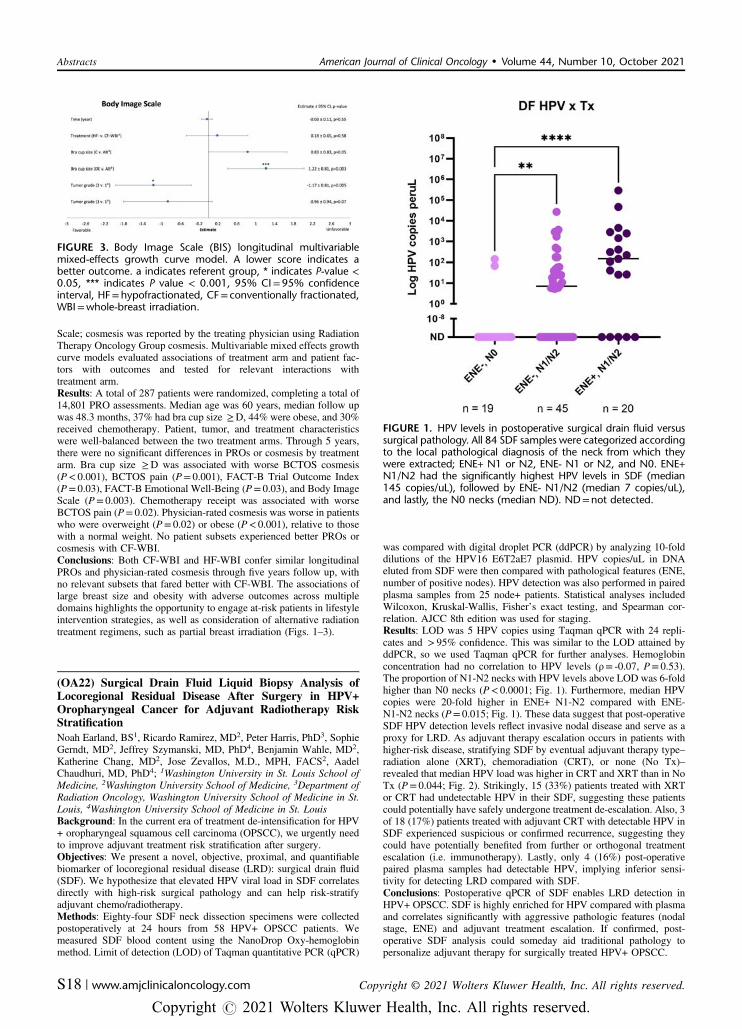
Scale; cosmesis was reported by the treating physician using RadiationTherapy Oncology Group cosmesis. Multivariable mixed effects growthcurve models evaluated associations of treatment arm and patient fac-tors with outcomes and tested for relevant interactions withtreatment arm.Results: A total of 287 patients were randomized, completing a total of14,801 PRO assessments. Median age was 60 years, median follow upwas 48.3 months, 37% had bra cup size ≥D, 44% were obese, and 30%received chemotherapy. Patient, tumor, and treatment characteristicswere well-balanced between the two treatment arms. Through 5 years,there were no significant differences in PROs or cosmesis by treatmentarm. Bra cup size ≥D was associated with worse BCTOS cosmesis(P< 0.001), BCTOS pain (P= 0.001), FACT-B Trial Outcome Index(P= 0.03), FACT-B Emotional Well-Being (P= 0.03), and Body ImageScale (P= 0.003). Chemotherapy receipt was associated with worseBCTOS pain (P= 0.02). Physician-rated cosmesis was worse in patientswho were overweight (P= 0.02) or obese (P< 0.001), relative to thosewith a normal weight. No patient subsets experienced better PROs orcosmesis with CF-WBI.Conclusions: Both CF-WBI and HF-WBI confer similar longitudinalPROs and physician-rated cosmesis through five years follow up, withno relevant subsets that fared better with CF-WBI. The associations oflarge breast size and obesity with adverse outcomes across multipledomains highlights the opportunity to engage at-risk patients in lifestyleintervention strategies, as well as consideration of alternative radiationtreatment regimens, such as partial breast irradiation (Figs. 1–3).
(OA22) Surgical Drain Fluid Liquid Biopsy Analysis ofLocoregional Residual Disease After Surgery in HPV+Oropharyngeal Cancer for Adjuvant Radiotherapy RiskStratificationNoah Earland, BS1, Ricardo Ramirez, MD2, Peter Harris, PhD3, SophieGerndt, MD2, Jeffrey Szymanski, MD, PhD4, Benjamin Wahle, MD2,Katherine Chang, MD2, Jose Zevallos, M.D., MPH, FACS2, AadelChaudhuri, MD, PhD4; 1Washington University in St. Louis School ofMedicine, 2Washington University School of Medicine, 3Department ofRadiation Oncology, Washington University School of Medicine in St.Louis, 4Washington University School of Medicine in St. LouisBackground: In the current era of treatment de-intensification for HPV+ oropharyngeal squamous cell carcinoma (OPSCC), we urgently needto improve adjuvant treatment risk stratification after surgery.Objectives: We present a novel, objective, proximal, and quantifiablebiomarker of locoregional residual disease (LRD): surgical drain fluid(SDF). We hypothesize that elevated HPV viral load in SDF correlatesdirectly with high-risk surgical pathology and can help risk-stratifyadjuvant chemo/radiotherapy.Methods: Eighty-four SDF neck dissection specimens were collectedpostoperatively at 24 hours from 58 HPV+ OPSCC patients. Wemeasured SDF blood content using the NanoDrop Oxy-hemoglobinmethod. Limit of detection (LOD) of Taqman quantitative PCR (qPCR)
was compared with digital droplet PCR (ddPCR) by analyzing 10-folddilutions of the HPV16 E6T2aE7 plasmid. HPV copies/uL in DNAeluted from SDF were then compared with pathological features (ENE,number of positive nodes). HPV detection was also performed in pairedplasma samples from 25 node+ patients. Statistical analyses includedWilcoxon, Kruskal-Wallis, Fisher’s exact testing, and Spearman cor-relation. AJCC 8th edition was used for staging.Results: LOD was 5 HPV copies using Taqman qPCR with 24 repli-cates and > 95% confidence. This was similar to the LOD attained byddPCR, so we used Taqman qPCR for further analyses. Hemoglobinconcentration had no correlation to HPV levels (ρ= -0.07, P= 0.53).The proportion of N1-N2 necks with HPV levels above LOD was 6-foldhigher than N0 necks (P< 0.0001; Fig. 1). Furthermore, median HPVcopies were 20-fold higher in ENE+ N1-N2 compared with ENE-N1-N2 necks (P= 0.015; Fig. 1). These data suggest that post-operativeSDF HPV detection levels reflect invasive nodal disease and serve as aproxy for LRD. As adjuvant therapy escalation occurs in patients withhigher-risk disease, stratifying SDF by eventual adjuvant therapy type–radiation alone (XRT), chemoradiation (CRT), or none (No Tx)–revealed that median HPV load was higher in CRT and XRT than in NoTx (P= 0.044; Fig. 2). Strikingly, 15 (33%) patients treated with XRTor CRT had undetectable HPV in their SDF, suggesting these patientscould potentially have safely undergone treatment de-escalation. Also, 3of 18 (17%) patients treated with adjuvant CRT with detectable HPV inSDF experienced suspicious or confirmed recurrence, suggesting theycould have potentially benefited from further or orthogonal treatmentescalation (i.e. immunotherapy). Lastly, only 4 (16%) post-operativepaired plasma samples had detectable HPV, implying inferior sensi-tivity for detecting LRD compared with SDF.Conclusions: Postoperative qPCR of SDF enables LRD detection inHPV+ OPSCC. SDF is highly enriched for HPV compared with plasmaand correlates significantly with aggressive pathologic features (nodalstage, ENE) and adjuvant treatment escalation. If confirmed, post-operative SDF analysis could someday aid traditional pathology topersonalize adjuvant therapy for surgically treated HPV+ OPSCC.
FIGURE 3. Body Image Scale (BIS) longitudinal multivariablemixed-effects growth curve model. A lower score indicates abetter outcome. a indicates referent group, * indicates P-value <0.05, *** indicates P value < 0.001, 95% CI=95% confidenceinterval, HF=hypofractionated, CF= conventionally fractionated,WBI=whole-breast irradiation.
FIGURE 1. HPV levels in postoperative surgical drain fluid versussurgical pathology. All 84 SDF samples were categorized accordingto the local pathological diagnosis of the neck from which theywere extracted; ENE+ N1 or N2, ENE- N1 or N2, and N0. ENE+N1/N2 had the significantly highest HPV levels in SDF (median145 copies/uL), followed by ENE- N1/N2 (median 7 copies/uL),and lastly, the N0 necks (median ND). ND=not detected.
Abstracts American Journal of Clinical Oncology � Volume 44, Number 10, October 2021
S18 | www.amjclinicaloncology.com Copyright © 2021 Wolters Kluwer Health, Inc. All rights reserved.
Copyright r 2021 Wolters Kluwer Health, Inc. All rights reserved.

(OA23) DNA Damage Response Inhibitors PotentiateImmunogenic Cell Death and Radiotherapy in Merkel CellCarcinomaPeter Goff, MD, PhD1, Rashmi Bhakuni, PhD1, Thomas Pulliam1,Ramesh Rengan, MD, PhD1, Paul Nghiem, MD, PhD1; 1University ofWashingtonBackground: Metastatic cancer with primary or acquired resistance toimmune checkpoint inhibitors requires novel management strategies.Merkel cell carcinoma (MCC) is immunogenic with the highestresponse rate to PD-1 pathway blockade among solid tumors such thatapproximately 50% of patients with advanced MCC attain durabledisease control. Cell cycle checkpoint and DNA damage response(DDR) proteins, including ATR, ATM & DNA-PK, have been recog-nized as promising therapeutic targets for decades. However, it is onlyrecently described in preclinical studies that targeting the DDR maystimulate anti-tumor immunity and augment existing immunotherapies.Importantly, specific and potent DRR inhibitors (DDRi) are in early-phase clinical investigation. The rapid growth of MCC is driven byderegulation of early cell cycle components (e.g. p53 & Rb inactivationpaired with Myc protein upregulation), suggesting high replicationstress and susceptibility to DDRi or DNA damaging agents. MCCtumors are particularly radiosensitive. The mechanisms governing howDDRi may interact with radiation to promote antitumor immunity arenot well understood; however, they may potentiate immunogenic celldeath (ICD) to recruit and activate antigen presenting cells.Objectives: We assessed if several classes of DDRi, alone or in com-bination with radiation, potentiated ICD and quantified their potenciesas radiosensitizers in MCC cell lines. These preclinical studies are anearly test of our hypothesis that DDRi could play a role in the treatmentof PD-1 pathway blockade-refractory MCC.Methods: Representative Merkel cell polyomavirus positive (VP) andnegative (VN) MCC cell lines were treated with various doses of singlefraction radiation (0, 2, 4, 8, 16 Gray; 160 kV x-rays) +/- inhibitors ofATR, ATM or DNA-PK (ATRi, ATMi and DNA-PKi). Cells wereassessed for viability and hallmarks of ICD including extracellular ATP
release (luciferase assay) and HSP70 secretion (ELISA) at 1 - 5 dayspost-treatment.Results: Inhibitors of ATR, ATM and DNA-PK are all effective radi-osensitizers of MCC, and DNA-PKi proved the most potent with anenhancement ratio of ∼5-fold compared with ∼2-fold or less for ATMior ATRi across cell lines. ATRi and ATMi induced extracellular releaseof ATP or HSP70 alone or in combination with low dose radiation (2 -4 Gy) more effectively than when combined with radiation doses of≥ 8 Gy in VP and VN cell lines. DNA-PKi, however, had limited abilityto potentiate these ICD markers alone and, in fact, abrogated extrac-ellular ATP release compared with radiation alone in a VP cell line.Conclusions: Either alone or in combination with low-dose radiation (1fraction of 2-4 Gy), ATRi and ATMi more effectively induced canon-ical markers of ICD in MCC cell lines compared with DNA-PKi. DNA-PKi was superior as a radiosensitizer; however, it apparently enhanced anon-immunogenic mechanism of cell death. Elucidating the cell deathpathways induced by radiation and DDRi in MCC is underway.Understanding why relatively lower radiation doses appear moreimmunogenic when combined with ATRi and ATMi is of particularinterest. Given its inherent immunogenicity, cell cycle checkpointdeficiencies and radiosensitivity, MCC is relevant for the preclinical andclinical evaluation of DDRi as novel agents to overcome PD-1 pathwayblockade resistance.
(OA24) What Is Reasonably Achievable? Evaluation of LeftBreast Mean Heart Dose Distributions to Improve the PeerReview ProcessAmit Sood, MD1, Rachel Hackett, CMD, RTT1, Mark Farrugia, MD,PhD1, Varun Chowdhry, MD1; 1Roswell Park Comprehensive CancerCenterBackground: To improve the peer review process, our RadiationOncology department defined disease site specific dose constraints fororgans at risk (OARs) based on QUANTEC and national protocols.However, there is opportunity to improve on these existing doseconstraints.Objectives: For patients undergoing radiotherapy to the left breast, ourdepartmental mean heart dose constraint is 3 Gy. However, in ourexperience this constraint is achievable in virtually all circumstances.To improve the process of peer review, we wanted to understand thedistribution of mean heart dose (MHD) for patients treated at our centerto identify plans that are potential outliers.Methods: We retrospectively reviewed aggregate data from peerreviewed treatment plans for patients treated at our center from 2019 to2020 utilizing Radiologica software (Radiologica LLC, St. Louis,MO). We analyzed MHD in patients being treated with hypofractio-nated radiotherapy (HFx, 40 Gy/15fx) +/- boost for left whole breast aswell as patients treated with conventional fractionated radiotherapy(CFx, 50 Gy/25fx) in patients undergoing regional nodal irradiation(RNI) +/- boost. All patients underwent deep inspiratory breath hold(DIBH).Results: A total of 191 patients were identified, including 162 in theHFx group and 29 in the CFx group. Using a student’s t-test, the use ofa boost did not impact the mean heart dose (P= 0.44). In both the HFxand CFx group, there were no patients who exceeded 3 Gy MHD. In theHFx group, MHD was 0.82 Gy (95% CI ,0.37 -1.72), and there were 7/162 (4.3%) of cases identified with the MHD exceeding two-standarddeviations (> 1.72 Gy). In the CFx group, MHD was 1.46 Gy (95%CI0.34-2.58), and there were 0/29 (0%) of cases identified with mean heartdose exceeding two-standard deviations (> 2.58 Gy).Conclusions: For patients undergoing radiotherapy for left sidedbreast cancer, a MHD of 3 Gy is not helpful to stratify cases that needmore detailed peer review. Based on our results, we recommend anadditional physician and dosimetrist review of contours and treatmentplan where mean heart dose falls two standard deviations outside themean (> 1.72 Gy). This standard will serve for a future qualityimprovement project. Understanding the distribution of doses to aparticular OAR can be helpful in identifying plans that could benefitfrom more careful peer review. This process can be used for otherOARs and disease sites.
FIGURE 2. HPV levels in postoperative surgical drain fluid byadjuvant treatment type. SDF was collected postoperatively (theSDF with the higher HPV copies was used for patients withbilateral neck dissections). There was a non-significant elevation inSDF HPV levels from patients who went on to receive chemo-radiation therapy (CRT) compared with only radiation therapy(XRT) (median 263 vs. 18 copies/uL; P=0.56). SDF from patientsin the CRT group was significantly enriched for HPV comparedwith no treatment (No Tx) (median 263 copies/uL vs. ND;P=0.026). The single patient that recurred in the No Tx groupwas deemed to be high-risk but declined adjuvant radiotherapy.ND=not detected.
American Journal of Clinical Oncology � Volume 44, Number 10, October 2021 Abstracts
Copyright © 2021 Wolters Kluwer Health, Inc. All rights reserved. www.amjclinicaloncology.com | S19
Copyright r 2021 Wolters Kluwer Health, Inc. All rights reserved.

(OA25) Intersection of Race or Ethnicity and Sex on SeniorFaculty Representation in Radiation Oncology: MixedChanges from 2000 to 2019James Janopaul-Naylor, MD1, Lauren Beck2, Sanford Roberts, MD3,Jeffrey Switchenko, PhD, MS1, Mylin Torres, MD1; 1Winship CancerInstitute at Emory University, 2University of Pennsylvania, 3Universityof Pennsylvania Department of Surgery
Background: Radiation Oncology (RO) is a small, specialized field ofmedicine traditionally included in Radiology (R) Departments until the 1980s.Given work hours primarily confined to the busines week with few emer-gencies, the opportunity to care for cancer patients, and favorable compen-sation, RO is a potentially attractive career. Advantages of a diverse physicianworkforce include higher patient satisfaction, patient compliance with physi-cian recommendations, and increased participation of under-representedminority (URM) patients in clinical research (Cooper-Patrick et al, JAMA1999; Branson et al, Am J Surg 2007). However, data from 2000 to 2020indicate that URM and female representation in RO continues to be poor, withthe field having less diversity than in medical schools and even other oncologyspecialties (Chapman et al IJROBP 2013; Deville et al, IJROBP 2020).Objectives: The goal of this study was to determine the proportion ofwomen and URMs senior faculty over the last two decades in ROrelative to other departments. We compared RO to Internal Medicine(IM), General Surgery (GS) and R given the large number of physiciansin IM and GS and the historical relationship RO has with R.Methods: AAMC data were used to determine the proportion of womenand URMs in senior faculty positions (Full or Associate Professors) in ROrelative to IM, GS, and R. Comparisons were assessed using the Cochran-Armitage trend test and multivariable linear regression models using SAS.Results: From 2000 to 2019, the proportion of women and Asian malesenior faculty increased among RO, IM, GS, and R Departments(P< 0.001). The proportion of senior faculty who were womenincreased faster in RO than GS or R (P< 0.001) but slower than in IM(P< 0.001). While the proportion of senior faculty that were non-Whiteincreased fastest in RO (1.00% per year, P< 0.001), this change inrepresentation was largely due to the increase in Asian males. Theincrease in proportion of non-white/non-Asian faculty who becamesenior faculty over time was significantly lower in RO (0.04% per year)than in GS (0.16% per year, P< 0.001), IM (0.24% per year, P< 0.001)or R (0.12% per year, P< 0.001). The proportion of Black male seniorfaculty in RO decreased 70% (Fig. 1) since 2000, while remainingstable in other departments. Full, Associate, or Assistant Professors whoidentified as American Indians, Alaskan Natives, Native Hawaiians, or
FIGURE 1. Proportion of each race/ethnicity and sex for radiationoncology pipeline in 2019.
FIGURE 2. Proportion of each race/ethnicity and sex for professors in radiation oncology from 2000 to 2019.
Abstracts American Journal of Clinical Oncology � Volume 44, Number 10, October 2021
S20 | www.amjclinicaloncology.com Copyright © 2021 Wolters Kluwer Health, Inc. All rights reserved.
Copyright r 2021 Wolters Kluwer Health, Inc. All rights reserved.

other Pacific Islanders decreased 20% in RO, while increasing in IM,GS, and R from 2000-2019. Among all clinical departments, RO wasamong lowest in representation of White or Hispanic women andHispanic men in senior faculty positions.Conclusions: Our findings indicate that sex and URM disparities in ROare large and improving at slower rate than in IM, GS, and R. Emphasison improving these gaps in representation is needed in our field toaddress the needs of our diverse patient population and reflect theincreasing diversity of medical students (Figs. 2 and 3).
(OA26) Epigenetic Methylation of Immune Synapse Genesin Primary and Metastatic Melanoma TumorsMatthew Mills, MD1, Anders Berglund, PhD2, Ryan Putney, PhD2, ImeneHamaidi, PhD2, James Mule, PhD3, Sungjune Kim, MD PhD2; 1H. LeeMoffitt Cancer Center and Research Institute, Department of RadiationOncology, 2H. Lee Moffitt Cancer Center and Research Institute, 3Depart-ment of Immunology, H. Lee Moffitt Cancer Center & Research InstituteBackground: Tumor cells often express immune synapse proteins toevade the immune system, which are frequently regulated by epigeneticmechanisms. Our prior work (Berglund, A., JCI, 2020, 10.1172/JCI131234) demonstrated that the methylation status of immune syn-apse genes in multiple tumor types was associated with functional T cellrecruitment and associated with survival.Objectives: To assess whether the methylation status of immune syn-apse genes predicts response to immunotherapy in metastatic melanoma.
Methods: A principal component analysis (PCA) of differentiallymethylated immune synapse genes in 8,186 solid tumors and 745 normaladjacent tissues from TCGA database was performed. Subsequently, thedysregulated methylation pattern of immune synapse genes was validatedin two independent cohorts (GSE120878 and GSE86355 datasets). Lastly,the capacity of PC1 to predict response to immunotherapy in melanomawas investigated. Pearson’s, Spearman’s and t-SNE correlation statistics,and the 2-sided Student’s t tests were performed as appropriate. The
FIGURE 1. A demonstrates response to immunotherapy by PC1status. B is a Kaplan Meier curve of overall survival stratified byPC1 status.
FIGURE 3. Proportion of each race/ethnicity and sex for full professors in all departments in 2019.
American Journal of Clinical Oncology � Volume 44, Number 10, October 2021 Abstracts
Copyright © 2021 Wolters Kluwer Health, Inc. All rights reserved. www.amjclinicaloncology.com | S21
Copyright r 2021 Wolters Kluwer Health, Inc. All rights reserved.
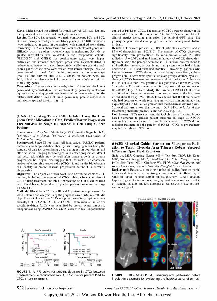
Kaplan-Meier method was utilized for overall survival (OS), with log-ranktesting to identify associated with methylation status.Results: The PCA has revealed two main components: PC1 and PC2.PC1 was mainly driven by co-stimulatory genes (i.e. CD40), frequentlyhypermethylated in tumor in comparison with normal adjacent tissue.Conversely, PC2 was characterized by immune checkpoint genes (i.e.HHLA2), which are often hypomethylated in melanoma. Such dysre-gulated methylation was validated in the independent cohorts,GSE120878 and GSE863, as costimulatory genes were hyper-methylated and immune checkpoint genes were hypomethylated inmelanoma compared with nevi. Importantly, a pilot analysis of a mel-anoma patient cohort who were treated with immunotherapy (N= 29)demonstrated a trend for superior response to immunotherapy(P= 0.15) and survival (HR 2.32, P= 0.15) in patients with lowPC1, which is characterized by relative hypomethylation of co-stimulatory genes.Conclusions: The selective hypomethylation of immune checkpointgenes and hypermethylation of co-stimulatory genes by melanomarepresents a crucial epigenetic mechanism of immune evasion, and therelative methylation levels of these genes may predict response toimmunotherapy and survival (Fig. 1).
(OA27) Circulating Tumor Cells, Isolated Using the Gra-phene Oxide Microfluidic Chip, Predict Shorter ProgressionFree Survival in Stage III Non-Small Cell Lung CancerPatientsEmma Purcell1, Zeqi Niu1, Shruti Jolly, MD2, Sunitha Nagrath, PhD1;1University of Michigan, 2University of Michigan Department ofRadiation OncologyBackground: Stage III non-small cell lung cancer (NSCLC) patientscommonly undergo radiation therapy, with imaging scans being thestandard of care for determining disease progression both during andafter radiation. Imaging technologies only detect progression after ithas occurred, which may be well after tumor growth or diseaseprogression has begun. We suggest that the molecular character-ization of circulating tumor cells (CTCs) found in the bloodstreamcan identify or predict disease progression before it is currentlydiagnosable.Objectives: The objective of this work is to determine whether CTCmetrics, including the number of CTCs, change in the number ofCTCs during treatment, and PD-L1 expression on CTCs, can be usedas a blood-based biomarker to predict patient outcomes in stageIII NSCLC.Methods: Blood from 26 stage III NSLC patients was processed forCTC isolation and analysis using the graphene oxide (GO) microfluidicchip. The GO chip isolates CTCs using immunoaffinity capture, takingadvantage of EPCAM, EGFR, and CD133 expression on CTCs forspecific isolation. CTCs were quantified by protein expression at sixtimepoints as being DAPI+/CK+, CD45- cells with two subpopulations
defined as PD-L1+/- CTCs. The number of CTCs, percent change in thenumber of CTCs, and the number of PD-L1+ CTCs were correlated toclinical metrics including progression free survival (PFS) time. Theprimary endpoint was disease progression, either locoregional, distant,or death.Results: CTCs were present in 100% of patients (n= 26/26), and at93% of timepoints (n= 102/110). The number of CTCs decreasedsignificantly from pre-treatment to mid-radiation (P= 0.02), afterradiation (P= 0.04), and mid-immunotherapy (P= 0.02) timepoints.By calculating the percent decrease in CTCs from pre-treatment tomid-radiation therapy, it was found that patients who had a largedecrease in CTCs had sustained stability. Conversely, patients whohad an increase in CTCs during radiation therapy were likely to haveprogression. Patients were split in two even groups, defined by a 75%change in CTCs between pre-treatment and mid-radiation. A decreasein CTCs of less than 75% precluded a significantly shorter PFS time,7 months vs 21 months average monitoring time with no progression(P= 0.005), Fig. 1A. Secondarily, the number of PD-L1+ CTCs werequantified and found to decrease from pre-treatment to the first weekof radiation therapy (P= 0.045). It was found that the three patientswho had progression while receiving anti-PD-L1 immunotherapy hada quantity of PD-L1+ CTCs greater than the median at all time points.Survival analysis shows that having > 50% PD-L1+ CTCs at pre-treatment potentially predicts a shorter PFS, Fig. 1B.Conclusions: CTCs isolated using the GO chip are a potential blood-based biomarker to predict patient outcomes in stage III NSCLCundergoing chemoradiation. Increase in the number of CTCs duringradiation treatment and the percent of PD-L1+ CTCs at pre-treatmentmay indicate shorter PFS time.
(OA28) Biological Guided Carbon-ion Microporous Radi-ation to Tumor Hypoxia Area Triggers Robust AbscopalEffects as Open Field RadiationJiade Lu, MD1, Qingting Huang, MSc1, Yun Sun, PhD1, Lin Kong,MD1, Weiwei Wang, MSc1, Lien-Chun Lin, MSc1, Yangle Huang,PhD2, Jing Yang, MD1, Xiaodong Wu, PhD1; 1Shanghai Proton andHeavy Ion Center, 2Fudan University Shanghai Cancer CenterBackground: Recently, a growing number of studies focus on partialtumor irradiation to induce the stronger non-target effects. However, thevalue of partial volume carbon ion radiotherapy (CIRT) targetinghypoxic region of a tumor under imaging guidance as well as its effectof inducing radiation induced abscopal effects (RIAEs) have not beenwell investigated.
FIGURE 1. 18F-FMISO PET/CT imaging was performed beforeirradiation treatment for evaluating the hypoxia status of tumors.
FIGURE 1. A, PFS curve for percent decrease in CTCs betweenpre-treatment and mid-radiation. B, PFS curve for percent PD-L1+CTCs at pre-treatment.
Abstracts American Journal of Clinical Oncology � Volume 44, Number 10, October 2021
S22 | www.amjclinicaloncology.com Copyright © 2021 Wolters Kluwer Health, Inc. All rights reserved.
Copyright r 2021 Wolters Kluwer Health, Inc. All rights reserved.

Objectives: To investigate the differences in abscopal effects inducedby CIRT to the entire tumor versus partial volume CIRT using thenewly developed Carbon Ion Microporous Radiation (CI-MPR) guidedby 18F-FMISO PET/CT targeting the tumor hypoxia region.Methods: We developed a technique of CI-MPR, guided by 18F-FMISO PET/CT, for partial volume irradiation targeting the hypoxiaarea of a tumor and investigated its capability of inducing abscopaleffects. Tumor-bearing mice were inoculated subcutaneously withbreast cancer 4T1 cells into the flanks of both hind legs of mouse.Mice were assigned to three groups: Group 1: control group with notreatment; Group 2: carbon ion open field radiation (CI-OFR group)targeting the entire tumor; And Group 3: partial volume carbon ion
microporous radiation (CI-MPR group) targeting the hypoxia region.And the tumors on the left hind legs of mice were irradiated withsingle fraction of 20 Gy of CIRT in Groups 2 and 3.Results: Mice treated with CI-MPR or CI-OFR showed that significantgrowth delay on both the irradiated and unirradiated of tumor ascompared with the control groups. Tumor regression of left tumorirradiated with CI-OFR was more prominent as compared with thetumor treated with CI-MPR; however, the regression of the unirradiatedtumor in both CI-MPR and CI-OFR group was similar.Conclusions: Biological-guided CIRT using the newly developed micro-porous technique targeting tumor hypoxia region could induce robustabscopal effects similar to CIRT covering the entire tumor (Figs. 1–3).
FIGURE 2. Mouse irradiation and dose profile. (A and B) The radiation area and dose profile of CI-MPR group. (C) The radiation area anddose profile of CI-OFR group.
American Journal of Clinical Oncology � Volume 44, Number 10, October 2021 Abstracts
Copyright © 2021 Wolters Kluwer Health, Inc. All rights reserved. www.amjclinicaloncology.com | S23
Copyright r 2021 Wolters Kluwer Health, Inc. All rights reserved.
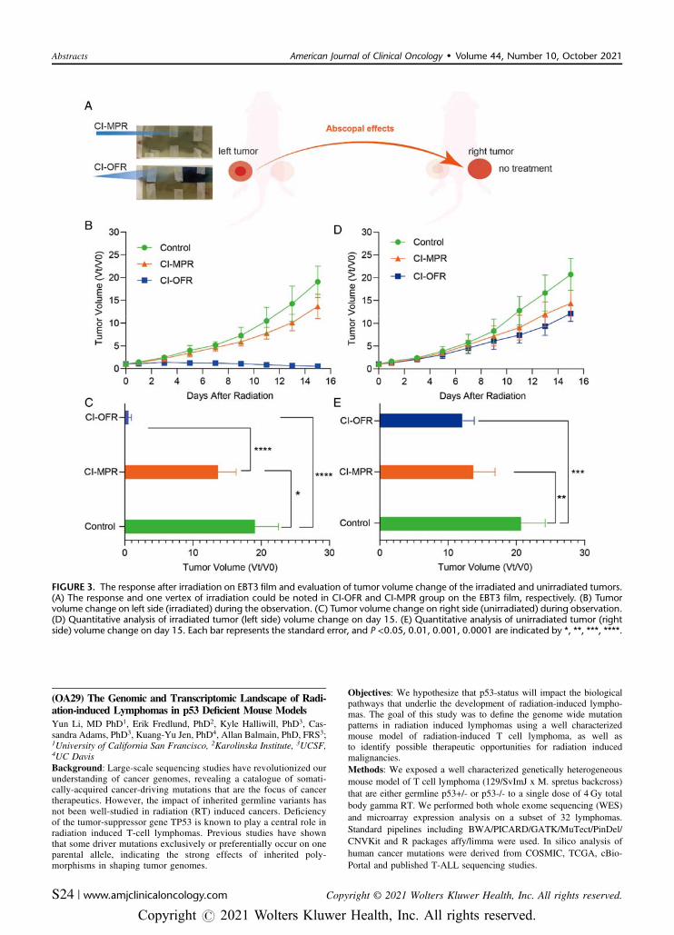
(OA29) The Genomic and Transcriptomic Landscape of Radi-ation-induced Lymphomas in p53 Deficient Mouse ModelsYun Li, MD PhD1, Erik Fredlund, PhD2, Kyle Halliwill, PhD3, Cas-sandra Adams, PhD3, Kuang-Yu Jen, PhD4, Allan Balmain, PhD, FRS3;1University of California San Francisco, 2Karolinska Institute, 3UCSF,4UC DavisBackground: Large-scale sequencing studies have revolutionized ourunderstanding of cancer genomes, revealing a catalogue of somati-cally-acquired cancer-driving mutations that are the focus of cancertherapeutics. However, the impact of inherited germline variants hasnot been well-studied in radiation (RT) induced cancers. Deficiencyof the tumor-suppressor gene TP53 is known to play a central role inradiation induced T-cell lymphomas. Previous studies have shownthat some driver mutations exclusively or preferentially occur on oneparental allele, indicating the strong effects of inherited poly-morphisms in shaping tumor genomes.
Objectives: We hypothesize that p53-status will impact the biologicalpathways that underlie the development of radiation-induced lympho-mas. The goal of this study was to define the genome wide mutationpatterns in radiation induced lymphomas using a well characterizedmouse model of radiation-induced T cell lymphoma, as well asto identify possible therapeutic opportunities for radiation inducedmalignancies.Methods: We exposed a well characterized genetically heterogeneousmouse model of T cell lymphoma (129/SvImJ x M. spretus backcross)that are either germline p53+/- or p53-/- to a single dose of 4 Gy totalbody gamma RT. We performed both whole exome sequencing (WES)and microarray expression analysis on a subset of 32 lymphomas.Standard pipelines including BWA/PICARD/GATK/MuTect/PinDel/CNVKit and R packages affy/limma were used. In silico analysis ofhuman cancer mutations were derived from COSMIC, TCGA, cBio-Portal and published T-ALL sequencing studies.
FIGURE 3. The response after irradiation on EBT3 film and evaluation of tumor volume change of the irradiated and unirradiated tumors.(A) The response and one vertex of irradiation could be noted in CI-OFR and CI-MPR group on the EBT3 film, respectively. (B) Tumorvolume change on left side (irradiated) during the observation. (C) Tumor volume change on right side (unirradiated) during observation.(D) Quantitative analysis of irradiated tumor (left side) volume change on day 15. (E) Quantitative analysis of unirradiated tumor (rightside) volume change on day 15. Each bar represents the standard error, and P <0.05, 0.01, 0.001, 0.0001 are indicated by *, **, ***, ****.
Abstracts American Journal of Clinical Oncology � Volume 44, Number 10, October 2021
S24 | www.amjclinicaloncology.com Copyright © 2021 Wolters Kluwer Health, Inc. All rights reserved.
Copyright r 2021 Wolters Kluwer Health, Inc. All rights reserved.

Results: All animals developed aggressive lymphomas <12 monthsafter RT. The p53-/- animals had shorter tumor latencies (107+/-36 d)compared with p53+/- mice (222+/-57 d, P= 2.36x10⁻¹⁰, log-rank test),and had 2-fold the point mutations compared with lymphomas from p53+/- mice (8 vs 4 mutations; P= 0.03, Wilcoxon test). Recurrentp53-dependent and/or strain-specific point and structural mutationswere identified at loci with known human homologs eg Notch1, Pten,and Ikzf1. Point mutations in the E1 ubiquitin activating enzyme Uba1were exclusively seen in lymphomas from p53-/- mice. These mutationsmap to the ubiquitin-activating and E2 interacting domains of Uba1,suggesting that the disruption of the ubiquitin pathway is a driver ofRT-induced lymphomas.Conclusions: Our data demonstrate the profound influence of bothgermline strain-specific polymorphisms and the timing of p53 loss onthe overall pathways to tumorigenesis, as well as on selection of specificmutations in potential drug target genes, such as Uba1.
(OA30) Racial Differences in Treatments and Toxicity inNon-Small Cell Lung Cancer Patients Treated withThoracic Radiation TherapyAnna Laucis, MD, MPhil1, Kimberly Hochstedler, MS1, MatthewSchipper, PhD2, Peter Paximadis, MD3, Thomas Boike, MD4, DerekBergsma, MD5, Benjamin Movsas, MD6, Munther Ajlouni, MD7,Annette Kretzler, MD8, Daniel Spratt, MD1, Robert Dess, MD2,Melissa Mietzel, MS2, Michael Dominello, DO9, Martha Matuszak,PhD2, Reshma Jagsi, MD, DPhil2, James Hayman, MD, MBA2, LoriPierce, MD, FASCO, FASTRO1, Shruti Jolly, MD1; 1University ofMichigan Department of Radiation Oncology, 2University of Michigan,3Lakeland Radiation Oncology, 421st Century Oncology, 5Departmentof Radiation Oncology at Mercy Health Saint Mary’s, 6Department ofRadiation Oncology, Henry Ford Cancer Institute, 7Henry Ford CancerInstitute, HFHS, 8Department of Radiation Oncology at Henry FordHospital, 9Wayne State UniversityBackground: Racial disparities are of particular concern for lungcancer patients given historical differences in surgery rates for Afri-can-American lung cancer patients that resulted in lower overallsurvival and higher recurrence rates compared with rates in Whitepatients.Objectives: The overall objective of this study was to examine racialdifferences in thoracic radiation therapy (RT) treatments and toxicitiesin a large cohort of patients from a multi-institutional consortiumdatabase of non-small cell lung cancer (NSCLC) patients.Methods: A large multi-institutional statewide prospectively col-lected patient-level database of locally advanced (stage II or III)NSCLC patients who received thoracic RT from March 2012 toNovember 2019 was analyzed to assess the associations between raceand treatment and toxicity variables. Race (White or African-Amer-ican) was defined by patient self-report or if not available then by theelectronic medical record system classification. Race categories otherthan White or African-American comprised a small minority ofpatients and were excluded from this analysis. Patient-reported tox-icity was determined by validated tools including the FunctionalAssessment of Cancer Therapy–Lung (FACT-L) quality of lifeinstrument. Provider-reported toxicity was determined by the Com-mon Terminology Criteria for Adverse Events (CTCAE) version 4.0.Uni-variable and multi-variable regression models were then fitted toassess relationships between primary outcomes by race and indicatorsof high-quality treatment and secondary analysis of symptoms.Spearman rank correlation coefficients were calculated betweenprovider reported toxicity and similar patient reported outcomes foreach race category.Results: A total of 1441 patients from 24 institutions with mean age of68 years (range 38–94) were evaluated; 226 patients were African-American, of whom 61% were treated at three facilities. Race was notsignificantly associated with RT treatment approach, use of concurrentchemotherapy, or the dose to the planning target volume (PTV) ororgans at risk including the heart and lungs. However, there wasincreased patient-reported general pain in African-American patients(compared with White patients) at several time points including pre-RT(22% (vs 15%), P= 0.02) and at the end of RT (30% (vs 17%),
P= 0.001). African-American patients were significantly less likely tohave provider-reported grade 2+ radiation pneumonitis (odds ratio (OR)0.36, P= 0.03), despite similar levels of patient-reported respiratorytoxicities such as cough and shortness of breath and even after con-trolling for known patient and treatment-related factors. Correlationcoefficients between provider- and patient-reported toxicities weregenerally similar across race categories.Conclusions: In this large multi-institutional observational study, wereassuringly found no evidence of differences in radiation treatment orchemotherapy approaches by race, in contrast to historical differencesby race in surgical care that led to worse survival and outcomes inminority race patients. However, we did unexpectedly find that African-American race was associated with lower odds of provider-reportedgrade 2+ radiation pneumonitis despite similar patient-reported tox-icities of shortness of breath and cough. There are several possibilitiesfor this finding including that pneumonitis is a multifactorial diagnosisthat relies on clinical as well as radiologic information and clinicalinformation alone may be insufficient. The Spearman correlation anal-ysis also revealed stronger correlations between patient- and provider-reported toxicities in White patients compared with African-Americanpatients, particularly for trouble swallowing/esophagitis. These findingstogether for pneumonitis and esophagitis discouragingly suggest pos-sible under-recognition of symptoms in black patients. Further inves-tigation is now warranted to better understand how these findingsimpact the care of racially diverse lung cancer patients.
(OA31) Tapping into the Power of Crowd Innovation toDevelop an Artificial Intelligence Tool for PancreaticRadiotherapyKarl Bush, PhD, MCCPM1, Diego Toesca, MD1, Richel Baclay, MD1,Daniel Chang, MD1; 1Stanford UniversityBackground: Crowd innovation is the process of inviting a large,worldwide community of highly proficient software developers, generallynot working in the healthcare industry, to rapidly produce a large numberof high-quality, and often novel, algorithmic solutions to a posed problem.Crowd innovation is most often implemented in a contest format, in whichthe programmer contestants compete for cash prizes to produce the highestscoring solution to the problem. This study offers a pivotal exploration intothe power and potential of crowd innovation for radiation oncology andradiology software automation.Objectives: In this work, we determine the efficacy of using the non-traditional approach of crowd innovation, to rapidly develop a diverse setof machine learning based algorithms for automatic pancreatic tumorsegmentation. The objective for the study was to probe the quality thatexpert programmers from outside healthcare could achieve in automatedsegmentation, and determine whether the approach could rival traditionalframeworks of graduate student/post-doctoral fellow algorithm develop-ment. The endpoint for the segmentation was for use in a pancreatic cancermanagement solution, both in prediction of pancreatic tumor surgicalresectibility, and in target delineation for radiation therapy.Methods: We conducted a 10-week, prize based, competition open toTopcoder, Inc. community contestants (total prize money of$30,000). Contestants were provided a training dataset containing120 CT scans and contour data for pancreatic tumors and sur-rounding vessels (superior mesenteric artery, celiac axis/commonhepatic artery, and superior mesenteric vein/portal vein), and com-peted in building algorithms to auto-segment a validation datasetcontaining 60 CT scans with no contours. An F-score metric wasused to rank the contestants’ algorithms using a separate, unrevealeddataset of 62 patients. F-score calculations included both the tumorand vessel contours, but with vessels weighted 7-times lower thanthe tumor. Contestants were offered a $3,000 prize bonus for algo-rithms exceeding an F-score of 0.7 to encourage quality algorithmsbeyond simply winning prize money.Results: 167 contestants registered for the competition, across 34countries, and submitted a total of 337 algorithms. The winning sol-ution (average F-score= 0.72) implemented a 3D-UNet model in a 3Dsemantic segmentation approach, which was found to be accurate intumor and vessel delineation. All other winning algorithms used 2Dmodel approaches. Top 5 algorithm performance was strongly
American Journal of Clinical Oncology � Volume 44, Number 10, October 2021 Abstracts
Copyright © 2021 Wolters Kluwer Health, Inc. All rights reserved. www.amjclinicaloncology.com | S25
Copyright r 2021 Wolters Kluwer Health, Inc. All rights reserved.

correlated by patient. In 82% of patient cases, all top 5 algorithmssucceeded in achieving an F-score > 0.6. The top 5 scoring solutionsprovided useful auto- segmentation capabilities for pancreatic tumorsand surrounding vessels, and have been integrated into a model forpredicting pancreatic tumor resection success before surgery.Conclusions: The use of crowd innovation for healthcare challengeshas seen limited use, but offers great potential for rapid development ofhigh quality segmentation algorithms with reasonable financial com-mitments (Figs. 1, 2 and Table 1).
(OA32) Immune Cell Infiltrates in Low Grade Glioma andImpact of Immunological Microenvironment AffectingTumor Biology and Oncologic OutcomeAnupam Rishi, MD1, Homan Mohammadi, MD2, Daniela Martir, MS3,Eric Welsh, PhD4, Timothy Robinson, MD, PhD5, Daniel Oliver, MD2,
Steven Eschrich, PhD2, Javier Torres-Roca, MD1, Hsiang-HsuanMichael Yu, MD2, G. Daniel Grass, MD, PhD1, Kamran Ahmed, MD1;1Department of Radiation Oncology, H. Lee Moffitt Cancer Center &Research Institute, 2H. Lee Moffitt Cancer Center & Research Institute,3Ponce Health Sciences University, 4Department of Biostatistics andBioinformatics, H. Lee Moffitt Cancer Center & Research Institute, 5H.Lee Moffitt Cancer Center and Research Institute, Department ofRadiation OncologyBackground: Prediction of response to immune therapies relates tothe presence, distribution, and activation of immune cell infiltrates(ICI). Little is known about the immune contexture of low-gradeglioma (LGG), and how ICI may predict the outcome. CIBERSORTprovides an estimate of ICI via a gene expression deconvolutionalgorithm.Objectives: In this novel study, we analyzed the presence of ICI, andthe neutrophil-to-lymphocyte (NLR) and platelet-to-lymphocyte (PLR)ratios with respect to outcomes in LGGs.
FIGURE 2. Tumor contour F-scores (left) and vessel contour F-scores (right), ordered in increasing order, for the 62 patient scoringdataset.
TABLE 1. Precision and Recall Scores for Tumor and Vessels for Top 5 Contestants’ Algorithms
FIGURE 1. 3D (left) and axial (right) views of an auto-segmented pancreatic tumor and surrounding vessels.
Abstracts American Journal of Clinical Oncology � Volume 44, Number 10, October 2021
S26 | www.amjclinicaloncology.com Copyright © 2021 Wolters Kluwer Health, Inc. All rights reserved.
Copyright r 2021 Wolters Kluwer Health, Inc. All rights reserved.

Methods: From an IRB-approved institutional tissue biorepository,we identified patients treated between 2004–2012, with LGG whounderwent excision and had complete gene expression profiling.Gene expression levels were assessed by Affymetrix Hu-RSTAassays. For ICI, the presence and high expression dichotomized bymedian were used to assess relationships with overall survival (OS)and progression-free survival (PFS). For NLR, 2.4 was used basedon published data in primary GBM. Time-to-event analyses wereperformed with Kaplan-Meier estimates and compared via log-ranktest. Associations of factors and outcomes were explored using Coxregression.Results: A total of 41 LGG patients were identified, with a medianage of 36.5 years (range, 18-65 y) and 43.9% (18 patients) females.All patients were either grade 2 astrocytoma, 16 (39%) patients oroligodendroglioma/oligo-astrocytoma, 25 (61%) patients. Allpatients underwent surgery, with gross total excision (GTR) ach-ieved in 19 (46.3%), and sub-total excision (STR) in 22 (53.7%)patients. Ten (24.4%) patients received adjuvant radiation therapy.The frontal lobe was the most common primary site in 27 (65.8%)patients. Median OS and PFS from date of surgery were
141 months (95% confidence interval (CI) 94.2–187.8 mo) and83 months (95% CI 47.4–118.6 mo), respectively. 5- and 10-yearOS was 64.2 and 35.1%, respectively. Among ICI, the presence ofmemory B-cells significantly improved OS (10-year OS 84.4% vs38%, hazard ratio (HR) 3.9, 95% CI 1.1-13.6; P= 0.02). In addi-tion, the presence of neutrophils adversely affected PFS (10-yearOS 7.5% vs 84.4%, HR 7.9, 95% CI 1.8–35.1; P= 0.002). No otherICI subtypes affected OS or PFS significantly. Patients with pre-surgery NLR > 2.4 had significantly worse OS (10-year OS 30.4%vs 66.7%, P= 0.05). PLR did not affect OS or PFS.Conclusions: LGGs have a distinct immunological microenviron-ment and ICIs may play a pivotal role in tumor biology affectingprogression and survival. The knowledge of ICI and NLR mayserve as a unique risk-stratification and guide clinical decisions(Figs. 1–3).
(OA33) Response to Whole-Lung Low-Dose RadiationTherapy (LD-RT) Predicts Freedom from Intubation inPatients Receiving Dexamethasone And/or Remdesevir forCOVID-19-Related PneumoniaClayton Hess, MD MPH1, Tony Eng, MD1, Tahseen Nasti, PhD1,Vishal Dhere, MD1, Troy Kleber, BS2, Jeffrey Switchenko, PhD, MS3,Brent Weinberg, MD4, Nadine Rouphael, MD1, Sibo Tian, MD1,Soumon Rudra, MD1, Luisa Taverna, (student)1, Alvaro Perez, (stu-dent)1, Rafi Ahmed, PhD1, Mohammad Khan, MD, PhD2; 1EmoryUniversity, 2Department of Radiation Oncology, Winship CancerInstitute of Emory University, 3Winship Cancer Institute at EmoryUniversity, 4Emory University HospitalBackground: Low-dose radiation is a known focal anti-inflammatorytreatment for various benign conditions. Phase I/II clinical trials haveexplored whole-lung low-dose radiotherapy (LD-RT) as a potentialtreatment for patients with COVID-19-related pneumonia. Initial find-ings require reproduction. Concomitant LD-RT administration withexisting therapies requires safety evaluation.Objectives: To explore the safety and efficacy of LD-RT in patientsreceiving concurrent dexamethasone and/or remdesevir. To get inves-tigate the predictive value of 3-day lab value response of C-reactiveprotein levels following LD-RT on 28-day intubation and recovery rates.Methods: Patients with COVID-19-related pneumonia receiving dex-amethasone and/or remdesevir were treated with 1.5 Gy whole-lungLD-RT, followed for 28 days or until hospital discharge, and comparedwith controls blindly matched by age, comorbidity, and diseaseseverity. Eligible patients were hospitalized, SARS-CoV-2 positive, hadradiographic consolidations, and required supplemental oxygen.
FIGURE 1. The Kaplan–Meier curve shows the overall survival issignificantly higher with the presence of memory B-cells (10-yearOS 84.4% vs 38%, HR 3.9, 95% CI 1.1-13.6, P=0.02; log-rank).
FIGURE 2. The Kaplan–Meier curve shows the overall survival issignificantly higher with pre-surgery NLR <= 2.4 (10-year OS30.4% vs 66.7%, P=0.05; log-rank).
FIGURE 3. The Kaplan–Meier curve shows the progression-freesurvival is significantly worse with the presence of neutrophils(10-year OS 7.5% vs 84.4%, HR 7.9, 95% CI 1.8–35.1; P=0.002;log-rank).
American Journal of Clinical Oncology � Volume 44, Number 10, October 2021 Abstracts
Copyright © 2021 Wolters Kluwer Health, Inc. All rights reserved. www.amjclinicaloncology.com | S27
Copyright r 2021 Wolters Kluwer Health, Inc. All rights reserved.

Endpoints included safety, clinical recovery, intubation, radiographicchanges, and biomarker response.Results: 40 total patients were available for analysis. 20 patientsreceiving concurrent dexamethasone and/or remdesevir were treatedwith whole-lung LD-RT between Jun 11 and Dec 7, 2020 and werecompared with 20 matched controls. Following LD-RT, freedom fromintubation improved from 68% in controls to 86% (P= 0.09). Predictiveinflammatory and cardiac markers rapidly fell following LD-RT,including C-reactive protein (CRP) (P= 0.02) and creatine kinase (CK)(P < 0.01), indicative of reduced cascading inflammation and cardiacinjury. Eighty percent of LD-RT patients experienced rapid decline inCRP within 3 days and were classified as LD-RT responders. Intuba-tion-free survival was 100% following LD-RT when the CRPresponded, vs 66% in matched controls (P= 0.01). Oxygenationrequirements were lower following LD-RT among CRP responderscompared with matched controls: 32% lower per individual (P= 0.03)and 56% lower for the cohort (P= 0.06). No patient whose CRPdeclined following LD-RT died or required intubation.Conclusions: A cohort of patients with COVID-19-related pneumoniatreated with LD- RT demonstrated superior freedom from intubationcompared with matched controls, especially LD-RT responders(P= 0.01). LD-RT appears safe to deliver with concurrent drugs. LD-RT lowered CRP and CK biomarkers. CRP response predicted favor-able outcome. Optimal timing for LD-RT after oxygen dependence butbefore intubation may extinguish immunopathology before systemicspread. Confirmatory clinical trials are warranted. Clinical Trial Reg-istration: NCT04366791.
(OA34) A Pilot Study to Assess the Combination of High-Dose Conformal Radiation Therapy and Pembrolizumab inModulating Local and Systemic T-cell Responses inAdvanced MalignanciesChristopher Luminais, MD1, Alexander Mathew1, Kimberly A. Bul-lock, PhD1, Gina R. Petroni, PhD1, Richard J. Price, PhD1, Timothy N.JBullock, PhD1, James Larner, MD1; 1University of VirginiaBackground: Although there has been tremendous interest in the use ofhigh-dose conformal radiation therapy (HDCRT) to enhance immuno-therapy (IO), the optimal sequencing of IO and HDCRT is unknownboth in terms of local control and distant (abscopal) responses.Objectives: To use repeated tissue biopsies to estimate the effect ofdifferent sequencing orders of IO and HDCRT and to describe theimmunologic effects.Methods: This was a Phase I, prospective, randomized trial. Twenty-one patients with metastatic solid malignancies who were eligible forHDCRT (24 Gy in 3 fractions) and pembrolizumab (200 mg) wererandomized to 3 arms: Arm A, concurrent HDCRT (day 1 start) andpembrolizumab (days 1, 43, 64, and 85); Arm B, pembrolizumab first(days 1, 43, 64, and 85) followed by HDCRT (day 22 start); Arm CHDCRT first (day 1 start) followed by pembrolizumab (days 22, 43, 64,and 85). Patients underwent biopsy of the treated lesion pre-treatmentand post-treatment on days 22 and 43. The primary endpoint was localCD8+ T cell and FoxP3+ T regulatory cell (Treg) counts. Post hoc,RNA sequencing of biopsy specimens was performed followed bybioinformatics analyses, including differential gene expression, gene setenrichment analysis, cell type deconvolution, and TCR analysis.Results: Primary malignancies treated included: breast (6), colon (5),salivary (2), sinus (1), esophagus (1), leiomyosarcoma (1), melanoma
(1), pancreatic (1), thyroid (1), larynx (1), and testicular (1). Repeatedmeasure modeling was used to assess the CD8+ T cell and Treg counts.No pattern for CD8+ T cell and Treg counts was observed within orbetween arms (Fig. 1). Transcripts associated with DNA damage,including DDB2, were upregulated 3 weeks after HDCRT in nearly allpatients in all arms (Fig. 2A). Upregulation of immune regulatorytranscripts consistent with activation of an adaptive immune response,such as IL17D, were only observed when pembrolizumab was admin-istered concomitant with or before HDCRT (Fig. 2B). Gene setsassociated with T-cell activation were significantly enriched afterpembrolizumab, with attenuation after HDCRT (Fig. 2C). Conversely,leukocyte deconvolution revealed that HDCRT increased the proportionof intratumoral M2 macrophage signatures, whereas pembrolizumabdecreased it (Fig. 2D).Conclusions: Surprisingly, the timing of IO and HDRT did not result ina consistent CD8+ T cell or Treg pattern within or between arms. Thiscould be a result of testing within a heterogeneous population withadvanced disease. RNA-sequencing revealed that while order of therapyadministration did not impact the ability of HDCRT to induce DNAdamage transcripts, it may influence pembrolizumab’s propensity toactivate anti-tumor immune mechanisms. Further, HDCRT acutelyinduced M2 macrophage signatures regardless of treatment arm.Together, these results underscore the importance of understanding theimmunological consequences of HDCRT and their influence onimmunotherapy.
FIGURE 1. CD8+ T cell and FoxP3+ T regulatory cell (Treg) countson pre and post treatment tumor biopsies.
Abstracts American Journal of Clinical Oncology � Volume 44, Number 10, October 2021
S28 | www.amjclinicaloncology.com Copyright © 2021 Wolters Kluwer Health, Inc. All rights reserved.
Copyright r 2021 Wolters Kluwer Health, Inc. All rights reserved.

(OA35) Should Immune System Sparing Dose ConstraintsBe Integrated into the QUANTEC Guidelines?Bhanu Prasad Venkatesulu, MD1, Prashanth Giridhar, MD2, RiturajUpadhyay, MD3, Amrish Sharma, PhD4, Hagar Elghazawy, MD5, Thir-aviyam Elumalai, MD6, Supriya Mallick, MD7, Cheng Cheng-En Hsieh,MD4; 1Loyola University Chicago, 2All India Institute of medical sciences,3The University of Texas MD Anderson Cancer Center, 4MD AndersonCancer Centre, 5Ain Shams University, 6Christie, 7National cancer instituteBackground: Multiple reports have shown that radiation delivery fortumors in close proximity to lymphoid organs such as bone marrow,spleen or unintended radiation to circulating pool of lymphocytes tra-versing organs such as heart and lung is known to deplete the circu-lating lymphocyte populations. This radiation-related lymphopenia isknown to cause inferior tumor control outcomes which translate intoinferior overall survival outcomes in high-grade gliomas, head and necktumors, lung cancer, pancreatic cancer, hepatocellular carcinoma, andGynecological malignancies. Currently, there are no standardized doseconstraints that are available to limit the dose to the resident and cir-culating lymphocyte populations. This is much more pertinent in thecurrent immunotherapy era wherein ongoing clinical trials are trying tooptimally time and sequence radiation and immunotherapy combina-tions for potentially synergistic and or additive effects.Objectives: Collate the radiation-related dosimetric parameters thatpredict the incidence of severe lymphopenia in solid tumorsMethods: We did a systematic review of PubMed, Cochrane Central,and Embase in accordance with the PRISMA statement and pooled thedosimetric parameters that have been related to radiation-related lym-phopenia and can be used to limit the incidence of lymphopenia.Results: 633 abstracts were shortlisted for abstract screenings and 35articles fulfilled the inclusion criteria. The CNS dose constraints thatwere most pertinent to spare the circulating lymphocytes in the brainwere Whole Brain mean dose <31 Gy, Whole-brain Dmin <2 GyV10< 68%, hypothalamus Dmax <56 Gy, and V25< 56% were sig-nificant predictors of no severe lymphopenia (SL). The thoracic doseconstraints were effective dose to immune cells (EDIC) (based on mean
lung and mean heart dose) the range of< 4.0, 4.0-6.5, 6.5-9.5,and> 9.5 Gy translated into 2-year OS rates of 72, 52, 49, and 15% forlocally advanced non-small-cell lung cancer, Mean lung dose of<12 Gy, lung V5 <45%, and mean heart dose of <5.2 Gy heartV5< 38%, mean thoracic dose <5 Gy, V20 of thoracic vertebra< 26%were predictors of no SL. In Esophagus cancer mean body doseexposure strong predictor for G4 nadir-Odds ratio 1.22 per Gray,increase of 1 Gy in mean splenic dose (MSD) predicted a 2.9% decreasein nadir absolute lymphocyte count (ALC), Heart V15< 73%, T-spineV5 < 72%, Body V10 < 18%, Lung V5 < 50%, and Aorta V5 < 93%were strong predictors of no grade 4 lymphopenia, the 2year OS rateswere 66.7%, 42.7%, and 16.7% for EDIC <2 Gy, 2-4 Gy and > 4 Gy.In gastric cancer mean splenic dose (MSD) ≥ 35 Gy poor prognosticfactor for OS and recurrence-free survival. In pancreatic cancer, dosi-metric parameters of spleen such as V10(32.6 vs 16%); V15(23.2 vs9.5%), and V20 (15.4 vs 4.6%), 1-Gy increase in MSD increased theodds of grade 3 lymphopenia by 18.6%. In liver cancer, MSD, V5, V25,and V30 of the spleen for the Min ALC were 227.72 cGy, 17.84, 0.98,and 0.42%, respectively. In rectal cancer, Pelvic bone marrow (PBM)volume receiving at least 30 Gy (V30) was significantly associated withlymphopenia. In Gynecological malignancies, bone marrow V10> 85%was associated with higher odds of developing lymphopenia, risk of SLwas associated with bone marrow V40 > 599cc.Conclusions: This systematic review is a preliminary attempt to providedose constraints to spare the immune system during radiation delivery.The aforementioned dose constraints require prospective validation inclinical trials to better spare the immune system and augment the effi-cacy of radiation.
(OA36) Primary Intraocular Lymphoma: Outcomes andToxicity After Radiation TherapyChelsea Pinnix, MD, PhD1, Ethan Damron, BS2, Jillian Gunther,MD, PhD1, Penny Fang, MD1, Susan Wu, MD1, Bouthaina Dabaja,MD1, Sattva Neelapu, MD1, Loretta Nastoupil, MD1, Sairah Ahmed,MD1, Paolo Strati, MD1, Jason Westin, MD1, Luis Fayad, MD1,
FIGURE 2. RNA sequencing results: A, DDB2 expression; B, IL17D expression; C, T-cell activation associated get set enrichment; D,intratumoral M2 macrophage signatures.
American Journal of Clinical Oncology � Volume 44, Number 10, October 2021 Abstracts
Copyright © 2021 Wolters Kluwer Health, Inc. All rights reserved. www.amjclinicaloncology.com | S29
Copyright r 2021 Wolters Kluwer Health, Inc. All rights reserved.

Raphael Steiner, MD1, Ranjit Nair, MD1, Dan Gombos, MD1, NathanFowler, MD1; 1MD Anderson Cancer Center, 2McGovern MedicalSchool at UTHealthBackground: Primary intraocular lymphoma (PIOL) is an aggressive,rare hematologic malignancy comprised of large B-cells primarilyinvolving the retina and vitreous. Radiation therapy (RT) is occasion-ally excluded over fears regarding toxicity.Objectives: We sought to examine the outcomes and toxicity associatedwith RT for PIOL patients (pts).Methods: We reviewed the charts of 26 PIOL pts treated between 2003-2020 at our institution. We included pts without additional sites of CNSinvolvement that received ocular RT as part of their treatment.Results: The median age at diagnosis was 69.5 years (range: 48–90); 21pts (81%) were female. The most common presenting symptoms (> 1possible) were blurred vision (n= 19, 75%), floaters (n= 15, 59%), and/or vision loss (n= 12, 46%). Ophthalmologic examination revealedsuspicious vitreous cells (n= 23, 88%) and/or retinal/choroidal infiltrate(n= 9, 35%). Eighteen pts (69%) had bilateral ocular involvement. Inall pts, gross disease was limited to the eyes. All pts underwent lumberpuncture (LP) to rule out CNS disease; 14 pts had LP flow analysiscompleted. In 3 pts (12%), CSF cytology revealed rare, atypical lym-phoid cells, but follow-up MRI studies were unremarkable. Twenty-twopts (85%) received a median of 5 (1–6) cycles of systemic chemo-therapy before RT. Seventeen pts (65%) were treated with rituximab(R), methotrexate (Mtx), procarbazine (P), and vincristine. Two pts(8%) received single agent Mtx, 2 pts (8%) received Mtx and R, and 1pt (4%) received Mtx and P. Before RT, 3 pts (12%) received intra-vitreal Mtx and 3 pts (12%) received intrathecal Mtx. Ten pts (38%) hadchemotherapy abbreviated due to toxicity. Nine pts (35%) receivedconsolidative cytarabine chemotherapy after RT and 6 pts (23%)underwent autologous bone marrow transplant. All pts received ocularRT, with 24 pts (92%) receiving treatment bilaterally, to a median doseof 36 Gy in 20 fractions (range 28.5–39.6 Gy in 15-24 fractions). Themedian follow up time was 41.8 months (95% CI 19.7–83) from start ofRT. Three pts (12%) relapsed in the eyes at a median of 4 months(1–42) post-RT. Eleven pts (42%) developed relapsed lymphoma in thebrain at a median of 31 months (5–53) post-RT. The median pro-gression free survival and overall survival was 24.1 (95% CI 4.9–36.3)and 42.5 (95% CI 3.3–75.8) months from start of RT, respectively.
Thirteen patients (50%) had cataracts removed before RT; 13 pts (50%)developed post-RT cataracts. Twenty pts (77%) had dry eyes thatpersisted > 3 months after RT. No pt developed optic neuropathy fol-lowing RT. Five pts (19%) developed keratopathy. One pt (4%)developed retinopathy post-RT, and 1 pt (4%) had preexisting diabeticretinopathy worsened by RT. No patient developed blindness aftertherapy.Conclusions: In this single-center review of combined treatment modalitytherapy for PIOL, severe visual complications were uncommon. Additionaltherapies to prevent distant CNS relapses are warranted.
(OA37) Low-Dose Radiotherapy for Low Grade Non-Hodgkin Lymphoma of the OrbitChristopher Wright, MD1, Jonathan Baron, BS1, Daniel Lee, BS1,Maribel Carpenter1, Cesar Briceno, MD2, Elise Chong, MD3, AmitMaity, MD PhD1, John Plastaras, MD PhD1, Ima Paydar, MD1;1Department of Radiation Oncology, University of Pennsylvania,2Department of Ophthalmology, University of Pennsylvania, 3Depart-ment of Medicine, Hematology/Oncology Division, University ofPennsylvaniaBackground: Orbital low grade non-Hodgkin Lymphomas (LGNHLs)are the most common tumor of the ocular adnexa. Marginal zonelymphoma (MZL) followed by follicular lymphoma (FL) are the mostcommonly represented histologies. Low-dose radiotherapy (LDRT),previously defined as 2 Gy x 2 fractions, for orbital LGNHLs hasresulted in response rates exceeding 90% (Fasola, et al IJROBP 2013).Additional data, however, suggests lower doses are associated withinferior response rates (Desai, et al Blood 2017). Radiotherapy (RT) is aproven treatment modality for this entity, but no prior data existscomparing response rates following LDRT to moderate-dose RT(MDRT, RT dose > 4 Gy).Objectives: Herein, we compare overall response rates (ORR) andcomplete response (CR) rates for patients with orbital LGNHLs treatedwith LDRT vs MDRT.Methods: We retrospectively reviewed 38 consecutive courses of RTacross 36 patients treated for LGNHL of the orbit at a single institution.Bilateral disease was present in 15 patients. Nine patients had advanced
FIGURE 1. Rates of complete response, partial response and stable disease following radiotherapy by low-dose radiotherapy andmoderate-dose radiotherapy groups.
Abstracts American Journal of Clinical Oncology � Volume 44, Number 10, October 2021
S30 | www.amjclinicaloncology.com Copyright © 2021 Wolters Kluwer Health, Inc. All rights reserved.
Copyright r 2021 Wolters Kluwer Health, Inc. All rights reserved.

stage disease or secondary orbital lymphoma following systemic dis-semination. Represented indolent histologies included MZL (20patients), FL (11 patients), and mantle cell lymphoma (MCL, 5patients). ORR were recorded according to Deauville or RECIST cri-teria with a response characterized as a complete response (CR) orpartial response. Univariate Cox regression analysis was performed forcharacteristics associated with the studied outcomes.Results: LDRT was delivered for 14 courses and MDRT for 24 courses.The median MDRT dose was 24 Gy (range 21-36 Gy in 1.5-2 Gyfractions). Concurrent or sequential Rituximab was used in 8 patients.Advanced stage (odds ratio (OR) 7, P= 0.021) and year of RT ≥ 2016(OR 24, P= 0.008) were associated with increased use of LDRT. Forthe LDRT group, ORR and CR were 100% and 50%, respectively,compared with 87.5% and 58.3%, respectively, for the MDRT group.No evaluated characteristics, including the use of LDRT, were sig-nificantly associated with inferior ORR. Unilateral orbital involvementwas associated with inferior CR rates (OR 0.22, P= 0.042) comparedwith bilateral orbital involvement.
Conclusions: RT for LGNHL of the orbit is an effective treatmentmodality with ORR exceeding 90%. Due to the exquisite radio-sensitivity of orbital LGNHL, LDRT produces similar response rates ascompared with more protracted RT courses. Present data suggests thatLDRT should be considered for patients with orbital LGNHL; however,further studies with larger patient numbers are warranted to show sig-nificant associations (Fig. 1 and Table 1).
(OA38) Rayos Contra Cancer Connects Volunteers andClinics to Impact Radiotherapy TogetherBenjamin Li, MD, MBA1, David Martinez, MD2, Gustavo Sarria, MD3,Fei Yang, BA4, Raymond Carter, BA5, Serguei Castaneda, MD6,Oluwadamilola Oladeru, MD, MA7, Daniel Wakefield, MD, MPH8,Soha Ahmed, MD, MBA9, Shada Wadi-Ramahi, PhD10, Maria Cai-cedo, MD11, Anuja Jhingran, MD12, Adam Shulman, MS13, ApoloSalgado, MD14, Layth Mula-Hussain, MD15, Dante Roa, PhD16; 1RayosContra Cancer, UCSF, 2Rayos Contra Cancer, Oncosalud AUNA,3Rayos Contra Cancer, University Medical Hospital Bonn, 4RayosContra Cancer, Vanderbilt University Medical Center, 5Rayos ContraCancer, St. Jude Children’s Research Hospital, 6Rayos Contra Cancer,Thomas Jefferson University, 7Rayos Contra Cancer, MassachusettsGeneral Hospital, 8Rayos Contra Cancer, University of TennesseeHealth Science Center; Harvard T.H. Chan School of Public Health,9Rayos Contra Cancer, Aswan University, 10University of PittsburghMedical Center, 11Rayos Contra Cancer, Instituto Nacional de Can-cerologia, 12MD Anderson Cancer Center, 13Rayos Contra Cancer,Sarah Cannon Cancer Center, 14Rayos Contra Cancer, InstitutoNacional del Cancer, 15Rayos Contra Cancer, The Ottawa HospitalCancer Centre, 16Rayos Contra Cancer, University of California IrvineBackground: Rayos Contra Cancer (RCC) is a 501(c)(3) non-profitorganization whose mission is to create sustainable access to timely,high-quality, and affordable radiation treatment for cancer in limited-resource settings globally. RCC has a clinic-centric approach andfocuses on education and training programs in areas where support isotherwise scarce for radiation therapy.Objectives: Demonstrate the potential of coordinated education andtraining to reach and improve radiation oncology team confidence,knowledge, and competence in clinics globally.Methods: From 2019 to 2021, RCC created, administered, and meas-ured 9 longitudinal curriculum programs to clinics with functionalmedical equipment but gaps in education and training. Each curriculumfocused on purely practical aspects of the clinic-wide transition to newradiotherapy techniques. RCC used the Project ECHO (Extension forCommunity Health Outcomes) telehealth model with professional peer-to-peer support and synchronous group learning in underservedcommunities via teleconferencing. Each curriculum consisted of live1-hour sessions given 1-2 times per week over 3-4 months. Programsused multi-institutional collaboration, involved medical professional,trainee, and student volunteers, and incorporated cloud-based
TABLE 1. Characteristics Associated with Complete Responseversus Partial or Worse Response to RT Using a Univariate LogisticRegression Analysis
FIGURE 1. Number of clinics trained from 2019–2021, by region.
American Journal of Clinical Oncology � Volume 44, Number 10, October 2021 Abstracts
Copyright © 2021 Wolters Kluwer Health, Inc. All rights reserved. www.amjclinicaloncology.com | S31
Copyright r 2021 Wolters Kluwer Health, Inc. All rights reserved.

technology. We measured participation, costs, and educational out-comes and gathered facility operation details to estimate patient impact.Results: 43 countries, 77 clinics, and 1,060 clinicians participated in 9training cohorts. Clinicians included 398 (37.5%) radiation oncologistsand residents, 240 (22.6%) medical physicists and physics trainees, 210(19.8%) radiation therapists, 77 (7.3%) other, and 141 (13.3%) notrecorded. A total 165 sessions were delivered by 9 education teamscomprised of 82 radiation oncology professionals, 28 residents or physicsgraduate students, 35 medical or pre-medical students, and 76 on-siteclinic coordinators. Educators included 37 (43.5%) medical physicists, 30(35.3%) radiation oncologists, 15 (17.6%) radiation therapists, 3 (3.5%)dosimetrists. Notably, 28 (37.8%) of 74 US-based educators were origi-nally from outside the US. RCC’s 3-year expenses totaled $43,273. TheUSD market value of all programs combined was $3,176,000, and RCCcharged $0. Paired measurements of pre- vs. post-curriculum clinicianconfidence, knowledge, and competence significantly improved acrossmultiple practical domains. Based on treatment facility data, the estimatedclinical benefit to date is 75,017 patient lives impacted per year.Conclusions: Based on 3 years of experience, each volunteer educatorhas the potential to impact approximately 1,000 patient lives per yearthrough participation in RCC education and training. Participatingclinics in RCC programs showed improved confidence, knowledge, andcompetence. With low-cost coordination at the volunteer studentthrough resident level, we show a vehicle to improve radiotherapyglobally (Fig. 1 and Table 1).
(OA39) 18F-Fluciclovine PET/CT to Distinguish RadiationNecrosis from Tumor Progression in Brain MetastasesTreated with Stereotactic Radiosurgery: A Prospective PilotStudyTimothy Smile, MD1, Martin Tom, MD2, Frank DiFilippo, PhD1,Stephen Jones, MD, PhD1, John Suh, MD3, Erin Murphy, MD1, Jen-nifer Yu, MD, PhD1, Alireza Mohammadi, MD1, Gene Barnett, MD,
MBA1, Steve Huang, MD1, Guiyun Wu, MD1, Nancy Obuchowski,PhD1, Manmeet Ahluwalia, MD1, David Peereboom, MD1, Glen Ste-vens, DO, PhD1, Robin Davis1, Megan Firman1, Samuel Chao, MD1;1Cleveland Clinic, 2Miami Cancer Institute, Baptist Health SouthFlorida, 3Department of Radiation Oncology, Taussig Cancer Institute,Cleveland ClinicBackground: 18F-Fluciclovine PET/CT is a widely available FDA-approved amino acid radiotracer for evaluation of suspected prostatecancer recurrence.Objectives: To determine if 18F-fluciclovine PET/CT has utility indistinguishing tumor progression (TP) from radiation necrosis (RN)among patients with brain metastases having undergone prior stereo-tactic radiosurgery (SRS).Methods: The primary objective is to estimate the accuracy of 18F-fluciclovine PET/CT in distinguishing RN from TP. We enrolled adultsubjects with brain metastases who underwent prior SRS and presentedwith a follow up MRI brain (with DSC MR perfusion) which wasequivocal for RN versus TP. Based on equivocal MRI before PET,subjects were divided into pre-PET subgroups of equivocal with sus-picion for TP or RN, or truly equivocal, by the study neuroradiologist.Within 30 days of equivocal MRI brain, subjects underwent 18F-fluciclovine PET/CT on a Siemens Biograph mCT scanner following a10 mCi bolus dose immediately before PET. PET data were collected inlist mode for 25 mins post-injection and were reconstructed as a staticimage of data 10-25 mins post-injection, and as a dynamic series of four5-min frames between 5-25 mins post-injection. Quantitative metrics foreach lesion were documented including SUV max, SUV mean, SUVpeak, and normal brain SUV mean. Lesion to normal brain ratios werecalculated. The reference standard was clinical follow up with MRIbrain (with DSC MR perfusion) every 2-4 months until multi-disciplinary consensus (or tissue confirmation) for diagnosis of RNversus TP.Results: From 7/2019-9/2020, 14 of 16 planned subjects enrolled andunderwent 18F-fluciclovine PET/CT for evaluation of 19 brain lesions.Primary histology was NSCLC in 6, breast in 4, melanoma in 3, and
TABLE 1. Training Program Sessions, Participants, and Patient Impact
Abstracts American Journal of Clinical Oncology � Volume 44, Number 10, October 2021
S32 | www.amjclinicaloncology.com Copyright © 2021 Wolters Kluwer Health, Inc. All rights reserved.
Copyright r 2021 Wolters Kluwer Health, Inc. All rights reserved.

endometrial in 1. Among all 19 imaged lesions, ranges of quantitativemetrics were: SUV max, 2.18-12.1; SUV mean, 1.16-7.37; SUV peak,1.06-4.45; normal brain SUV mean, 0.19-0.44; SUV max/normal ratio,7.5-45.4; SUV mean/normal ratio, 4.2-26.3; and SUV peak/normalratio, 3.9-26.4. Follow up was completed for 10 patients as described inthe Table. No adverse events have been reported.Conclusions: In this population, 18F-fluciclovine PET/CT produces awide range of lesion quantitative metric values and low uptake in thenormal brain. Ongoing follow up is required, however, preliminaryresults are promising and suggest 18F-fluciclovine PET/CT may be ableto accurately distinguish RN from TP. Final results of this pilot studyare expected within a year. Phase II and III studies are under devel-opment (Table 1).
(OA40) Multi-institutional Validation of Recursive Parti-tioning Analysis for Overall Survival in Patients Under-going Spine Radiosurgery for Spine MetastasisJonathan Hanan, BA1, Samuel Chao, MD2, Arjun Sahgal, MD3, WeiWei, MS2, Mikki Campbell, MHE3, Chandana Reddy, MS4, LilyanaAngelov, MD5, John Suh, MD6, Anthony Magnelli, MS6, Salim Balik,PhD6, Ping Xia, PhD6, Kailin Yang, MD, PhD6, Alireza Mohammadi,MD2, Ehsan Balagamwala, MD6; 1Case Western Reserve UniversitySchool of Medicine, 2Cleveland Clinic, 3Department of RadiationOncology, Sunnybrook Odette Cancer Centre, University of Toronto,4Department of Radiation Oncology, Taussig Cancer Institute, Cleve-land Clinic, Cleveland, OH, 5Department of Neurosurgery, ClevelandClinic, 6Department of Radiation Oncology, Taussig Cancer Institute,Cleveland ClinicBackground: The recently developed recursive partitioning analysis(RPA) classification system for overall survival (OS) in patientsundergoing spine stereotactic radiosurgery (sSRS) yielded three distinctprognostic groups for patients with spine metastasis. We sought toexternally validate this RPA with a larger patient cohort using a multi-institutional dataset.Objectives: Validate the recursive partitioning analysis (RPA) for spinestereotactic radiosurgery (sSRS).Methods: 444 patients were utilized to develop the recently publishedsSRS RPA predictive of OS in patients with spine metastases. The RPAidentified three prognostic classes: RPA Class 1 was defined as KPS >70and controlled systemic disease (n=142); RPA Class 2 was defined asKPS>70 with uncontrolled systemic disease or KPS ≤70, age ≥54 andabsence of visceral metastases (n=207); RPA Class 3 was defined as KPS≤70 and age <54 years or KPS≤70, age ≥54 years, and presence ofvisceral metastases (n=95). Data from a large tertiary care center wasutilized to validate this RPA. A total of 221 patients (467 spinal segments)
were in the validation cohort and divided based on their RPA classes.Kaplan-Meier method was used to estimate OS and log-rank test was usedto compare OS between RPA classes.Results: In the validation cohort (221 patients), the median OS was19.9 months and the median follow-up was 36 months (range, 0.1-70.8mo). The majority (85%) of patients received 24Gy (range, 20-35Gy)in 2 fx (range, 2-10). Forty-three (19.5%) patients were in RPA Class 1,165 (74.7%) patients in RPA Class 2 and 13 (5.9%) patients in RPA Class3. The median OS in the validation cohort based on RPA Class was54 months for Class 1, 15.8 months for Class 2 and 6.8 months for Class 3.Patients in RPA Class 1 had a significantly better OS compared with thosein Class 2 and Class 3 of the validation cohort (P<0.01). The difference inOS between RPA Class 2 and Class 3 was not statistically significant dueto small number of patients in RPA Class 3.Conclusions: The external dataset validated the spine sSRS RPA forRPA Classes 1 and 2. RPA Class 3 was not validated successfully,which was likely due to small patient numbers. Based on our validation,upfront spine SRS is strongly supported for patients in RPA Class 1.Additionally, either upfront spine SRS or conventional radiotherapy issupported for RPA Class 2 patients. Patients in RPA Class 3 wouldbenefit most from upfront conventional radiotherapy. Given successfulvalidation, this RPA will be utilized as enrollment criteria for anupcoming investigator-initiated trial.
(OA41) Concurrent versus Sequential Stereotactic Radio-surgery and Immune Checkpoint Inhibition in BrainMetastases: An International Cooperative Group StudyEric Lehrer, MD, MS1, Jason Gurewitz, BA2, Timothy Malouff, MD3,Kenneth Bernstein, MS2, Douglas Kondziolka, MD2, Brianna Jones,MD1, Phillip Bonney, MD4, Samir Patel, MD5, Joshua Palmer, MD6,Kareem Fakhoury, MD7, Chad G. Rusthoven, MD7, David Mathieu,MD8, Christopher Deibert, MD9, Piero Picozzi, MD10, Cheng-chia Lee,MD, PhD11, Sonam Sharma, MD1, Ajay Niranjan, MD12, L. DadeLunsford, MD12, Jason Sheehan, MD, PhD13, Manmeet Ahluwalia,MD, MBA14, Daniel Trifiletti, MD15; 1Icahn School of Medicine atMount Sinai, 2NYU Langone Medical Center, 3Mayo Clinic, 4Universityof Southern California, 5University of Alberta, 6The Ohio State Uni-versity Wexner Medical Center, 7University of Colorado School ofMedicine, 8Université de Sherbrooke, Centre de recherche du CHUS,9Emory University, 10IRCCS Humanitas Research Hospital, 11TaipeiVeteran General Hospital, 12University of Pittsburgh, 13University ofVirginia, 14Miami Cancer Institute, 15Department of Radiation Oncol-ogy, Mayo Clinic, Jacksonville, FL, USABackground: Multiple single institution retrospective analyses havesuggested that concurrent administration (within 4 wk) of stereotactic
TABLE 1. NSCLC: non-small cell lung carcinoma, RN: radionecrosis, TP: tumor progression, E: equivocal, Dx: diagnosis, SUV:standardized uptake value
American Journal of Clinical Oncology � Volume 44, Number 10, October 2021 Abstracts
Copyright © 2021 Wolters Kluwer Health, Inc. All rights reserved. www.amjclinicaloncology.com | S33
Copyright r 2021 Wolters Kluwer Health, Inc. All rights reserved.

radiosurgery (SRS) and immune checkpoint inhibitors (ICI) is asso-ciated with improved overall survival (OS) when compared withsequential administration (more than 4 wk) in patients with brainmetastases (BrM). However, there are limited data analyzing the risk ofdeveloping radiation necrosis (RN) and OS by timing of therapy.Objectives: To evaluate the risk of development of RN and OS inpatients with BrM treated with SRS/ICI.Methods: The International Radiosurgery Research Foundationapproved the analysis. Logistic and Cox regression models were used toidentify covariates associated with the development of RN and OS. TheKaplan-Meier method and log-rank test were used to compare OS at 1-and 2-years post SRS. Patients with < 3-months of follow up wereexcluded.Results: There were 546 patients (223 melanoma/287 NSCLC/36 RCC)across 10 institutions with 2,880 BrM. Median follow-up was15.5 months, median age was 66 years (interquartile range [IQR]: 56-72 y). Median Karnofsky Performance Status (KPS) was 90. Medianmargin dose was 20 Gy (IQR: 18-20 Gy) in 1 fraction. The mean totalBrM volume was 3.59cc, and mean V12 Gy was 8.33cc. Activeextracranial disease was present in 78.0% at time of SRS. There were 65patients (11.9%) who experienced RN (46.2% Grade 1, 40.0% Grade 2;12.3% Grade 3; 1.5% Grade 4). On multivariate analysis, the number ofSRS courses (Odds Ratio [OR]: 2.59, 95% confidence interval [CI]:1.72-4.00; P< 0.001), total volume of treated BrM (OR: 1.09; 95% CI:1.03-1.15; P< 0.001), melanoma histology (OR: 2.79; 95% CI: 1.39-5.68; P= 0.004), RCC histology (OR: 3.96; 95% CI: 1.23-11.44;P= 0.01), receipt of previous intracranial radiation therapy (RT) beforeSRS (OR: 8.88; 95% CI: 2.96-26.34; P< 0.001) were associated withthe development of RN. When compared with anti-PD-1 monotherapy,anti-CTLA-4+anti-PD-L1 therapy (OR: 0.22, 95% CI: 0.07-0.57;P= 0.004) was associated with a lower likelihood of developing RN.Concurrent over sequential therapy (P= 0.26) and V12 (P= 0.49) werenot significant. On multivariate analysis, female sex (Hazard Ratio[HR]: 0.62; 95% CI: 0.48-0.82; P< 0.001) and KPS (HR: 0.97; 95%CI: 0.96-0.99; P< 0.001) were prognostic for OS. Concurrent versussequential therapy (P= 0.55), melanoma histology (P= 0.20), and RCChistology (P= 0.21) were not significant. The OS at 1-year was 74.1%vs. 76.4% and at 2-years was 56.3% vs. 53.8% for the concurrent andsequential groups, respectively (P= 0.69).Conclusions: The risk of RN remains low at approximately 12% inpatients treated with ICI/SRS, and 6.5% of patients developsymptomatic RN. This risk is significantly increased when patientsundergo multiple SRS courses, have received prior cranial RT, andharbor more radioresistant tumor histologies (melanoma and RCC).Concurrent immunotherapy does not appear to increase this risk.
(OA42) Proton Radiation Therapy for PediatricChordomasMyrsini Ioakeim-Ioannidou1, Daniel Kim, MD MBA1, Andrzej Nie-mierko, PhD1, Paul Caruso, MD1, David ebb, MD1, Yen-lin chen, MD1,Thomas DeLaney, MD1, Shannon MacDonald, MD1; 1MassachusettsGeneral HospitalBackground: Chordomas are rare primary bone tumors that arise fromectopic remnants of the embryonic notochord with an overall incidenceof 0.08 per 100,000 in general population. Pediatric chordomas are veryrare and comprise 5% of all chordoma cases. The most commonlocation of pediatric chordomas is the base of the skull (BOS) followedby the mobile spine and the sacrum. Treatment for chordomas generallyinvolves high-dose proton radiation therapy (PRT) with or withoutsurgical resection.Objectives: We report outcomes for pediatric patients with BOS andspinal chordomas treated with PRT.Methods: Between 1981 and 2019, 236 pediatric patients with BOS(n= 194) or spinal (n-42) chordomas were treated with PRT or com-bined proton/photon approach (comboRT) at a single institution. Theprimary endpoints were overall survival (OS) and progression freesurvival (PFS). Cox proportional hazards regression and Kaplan Meierestimates were used for survival analyses.Results: A total of 133 female and 103 male patients were identified.Median age at diagnosis was 10.9 years (range, 0.8-21 y) and 13.1 years
(range, 3.6-21 y) in the BOS and spinal cohort, respectively. Themajority of the patients (n= 154) were diagnosed with conventionalchordoma, 57 with a chondroid subtype, 13 with atypical/cellular, and12 with poorly-differentiated chordoma. All patients underwent surgicalresection (range, 1-8). 54 patients underwent gross total resection, 171patients underwent STR while 11 patients had biopsy only. 23 patientsreceived pre-RT chemotherapy. 62 patients developed progressivedisease before the delivery of RT. Median total dose was 76 Gy (RBE)(range, 39.7-83.3 Gy) delivered in 1.2 - 2.2 Gy (RBE) fractions (1.2 Gyfor BID). The median maximum dose to the brainstem was 65.7 Gy(RBE) (range, 40.8 - 80). The median maximum dose to the spinal cordwas 63 Gy (RBE) (range, 30.6-68.5 Gy). At a median follow-up of8.7 years (range, 0.6–35.6) from the date of diagnosis 68 patientsrecurred (42 local, 18 distant and 8 iatrogenic). The 5- and 10-year OSrates for BOS chordomas were 83% and 77%, respectively. The 5- and10-year OS rates for spinal chordomas were 77% and 66%, respec-tively. The 5- and 10-year PFS rates in the BOS cohort were 75% and71%, respectively. The 5- and 10-year PFS rates in the spinal cohortwere 66% and 62%, respectively. RT was well tolerated, with mostacute side effects limited to moderate erythema, otitis media and cer-vical dysphagia. Late toxicities included 5 cases of radiation myelitis, 4cases of brainstem injury, 4 cases of temporal lobe injury and singlecase each of moyamoya disease, Lhermitte’s syndrome, and esophagealstricture. 3 patients developed secondary radiation related malignancies.Conclusions: This is the largest cohort of pediatric chordomas in theliterature to date. High dose PRT following surgical resection is aneffective treatment with a high rate of disease control and minimal latetoxicity (Figs. 1 and 2).
FIGURE 1. Kaplan-Meier survival curves for overall survival forskull base pediatric chordomas.
FIGURE 2. Kaplan-Meier survival curves for progression-free sur-vival for skull base pediatric chordomas.
Abstracts American Journal of Clinical Oncology � Volume 44, Number 10, October 2021
S34 | www.amjclinicaloncology.com Copyright © 2021 Wolters Kluwer Health, Inc. All rights reserved.
Copyright r 2021 Wolters Kluwer Health, Inc. All rights reserved.

(OA43) Risk Factors for the Development of Lep-tomeningeal Disease and Outcomes Following StereotacticRadiation for Patients with Breast Cancer BrainMetastasesMatthew Mills, MD1, Chetna Thawani, BS2, Nicholas Figura, MD1,Daniel Oliver, MD3, Timothy Robinson, MD, PhD1, Hsiang-HsuanMichael Yu, MD3, Peter Forsyth, MD3, Kamran Ahmed, MD4; 1H. LeeMoffitt Cancer Center and Research Institute, Department of RadiationOncology, 2USF Morsani College of Medicine, 3H. Lee Moffitt CancerCenter & Research Institute, 4Department of Radiation Oncology, H.Lee Moffitt Cancer Center & Research InstituteBackground: As the prognosis for patients with breast cancer brainmetastases (BCBM) improves due to significant improvements in sys-temic therapy, the number of patients at risk for leptomeningeal disease(LMD) has increased. While local therapies such as surgical resectionand stereotactic radiation (SRT) are known risk factors for LMD, theidentification of patients with BCBM most at risk for LMD remainsunclear.Objectives: To identify the risk factors for the development of LMD inpatients with BCBM after SRT.Methods: This is a retrospective review of 181 patients who underwentSRT to 664 BCBM from 2004 to 2019. The Kaplan-Meier method wasused to calculate time to LMD from SRT and overall survival (OS) fromthe diagnosis of LMD, with log-rank testing to identify significant cor-relations with patient and treatment characteristics. Univariate analysis(UVA) was completed with the Cox proportional hazards model.Results: Median follow up from SRT was 11.4 months. Of the 181patients, 60 (33%) had HR+/HER2- subtype, 30 (17%) had HR-/HER2+ subtype, 47 (26%) had HR+/HER2+ subtype, and 44 (24%) had HR-/HER2- subtype. The median interval of initial breast cancer diagnosis tobrain metastasis disease was 52 months (range: 0-321). Of the 664BCBMs, 534 (80%) received single fraction stereotactic radiosurgery(SRS) with a median dose of 21 Gy (range 12-24 Gy), and 130 (20%)received fractionated stereotactic radiation therapy (FSRT), with amedian dose of 25 Gy (range: 12.5-35 Gy) delivered in 3 to 5 fractions.There were 68 cases (10%) of SRT to a postoperative cavity. Of the 181patients, 24 (13%) developed LMD at a median of 18 months (1-57)from BCBM diagnosis and a median of 6.6 months (0.6-29) from SRT.Twelve and 24-month rates of LMD were 14% and 21%, respectively.Patients who were diagnosed with BCBM ≤ 52 months after originalbreast cancer (BC) diagnosis had a significantly greater risk of LMD(12 mo LMD 21% vs 7%, P= 0.026). There was a trend toward sig-nificance for patients with extracranial systemic metastasis for greaterrisk of LMD (12 mo LMD 26.8% vs 10.9%, P= 0.055) After diagnosisof LMD, 6 and 12-month rates of OS were 35% ad 18%, respectively.An interval between SRT and LMD diagnosis ≤ 6 months was the onlyidentified variable prognostic for inferior OS (6-month OS 0% vs 47%,P< 0.001).Conclusions: The risk of LMD for patients with BCBM after SRTremains relatively low, though patients with a short interval from BCdiagnosis to BCBM diagnosis are at high risk of LMD. While theprognosis for patients with LMD is dismal, OS is particularly poor forthose who develop LMD soon after SRT.
(OA44) Impact of Peri-operative Radiotherapy on Local-ized Sarcomas with TP53 Alterations: Retrospective PooledAnalysis of Two Precision Medicine TrialsElise Nassif1, Edouard Auclin2, Rastilav Bahleda3, Charles Honore3, OlivierMir3, Sarah Dumont3, Benoite Mery4, Khalil Hodroj4, Mehdi Brahmi4,Olivier Tredan4, Isabelle Ray-Coquard4, Jean-Yves Blay4, ChristopheMassard3, Axel Le Cesne3, Armelle Dufresne4, Antonin Levy3, Cecile LePechoux3, Ritsuko Komaki5; 1MD Anderson Cancer Center, 2APHP,3Gustave Roussy, 4Leon Berard, 5Baylor College of MedicineBackground: Sarcomas are rare diseases with a dismal prognosis. Wehave previously reported an unfavorable prognostic impact of TP53mutations on disease-free survival (DFS) in sarcomas and an increasedobjective response rate to chemotherapy.
Objectives: We sought to characterize the impact of peri-operativeradiation therapy (RT) in sarcomas with TP53 alterations. Our objec-tives were type of first relapse (local versus metastatic) and DFSaccording to peri-operative RT administration and TP53 status.Methods: MOSCATO and ProfiLER trials were precision oncologytrials in advanced pan-histology tumors. Genomic molecular profilingwas done by targeted next-generation sequencing and comparativegenomic hybridization array. This is an ad hoc analysis of sarcomapatients included in these trials. Molecular data from patients wasassociated to their clinical data in localized standard-of-care treatmentusing the national prospective French Sarcoma Group database.Homozygous deletions and mutations of TP53 were retained as sig-nificant alterations. For comparison of categorical variables, Fisher’sexact test was used. For comparison of DFS, univariate Cox modelswere used.Results: A total of 155 sarcomas, including 123 soft-tissue sarcomas(STS), were initially localized and surgically resected, amongst which46 leiomyosarcomas (30%), 14 undifferentiated pleomorphic sarcomas(9%) and 15 liposarcomas (10%). STS were grade 1, 2 and 3 in 19%,34% and 47% of patients, respectively. TP53 status was determined onprimary tumor and metastasis in 77 cases each (50%, 1 not available).Peri-operative RT was administered in 61 patients (39%). TP53 alter-ations were found in 40 sarcomas, including 35 STS, and were treatedwith peri-operative RT in 18 cases, including 16 STS patients. Irre-spective of RT, first recurrence was metastatic in 37 and local in 3 of theTP53 altered tumors, whereas it was metastatic in 81 and local in 31cases of TP53 wild-type (WT) sarcomas (P= 0.01; Table 1). In STSsubgroup, there was only one local relapse and 34 metastatic relapses inthe TP53 altered tumors (P= 0.01). There were 13% (N= 8/61) localrecurrences in patients receiving RT versus 28% (N= 26/94) withoutRT (P= 0.027). In TP53 altered sarcomas, there was one (6%, N= 1/18) and two (9%, N= 2/22) local relapses with or without RT,respectively (P= 1; Table 1). In TP53 WT sarcomas, there was 16%(N= 7/36) and 33% (N= 24/72) of local relapses, with or without RT(P= 0.04). In TP53 altered STS, the only local recurrence was in apatient without peri-operative RT. In TP53 altered sarcomas, medianDFS with or without peri-operative radiotherapy was 10 and 14 months(P= 0.5) and DFS rate at 12 months was 72% (95%CI= 54-96) and41% (95%CI= 25-68) with or without RT, respectively. In TP53 WTtumors, median DFS with or without peri-operative RT was 17 monthsin both groups (Fig. 1).Conclusions: In our retrospective cohort, TP53 alterations are asso-ciated with metastatic relapse, which warrants further validation. We arerecording the pathological response to radiotherapy, in sarcomas whounderwent pre-operative radiation therapy, which will be available forpresentation at ARS.
FIGURE 1. Disease-free survival according to TP53 status andperi-operative radiotherapy administration. XRT= Radiotherapy;WT=Wild-Type.
American Journal of Clinical Oncology � Volume 44, Number 10, October 2021 Abstracts
Copyright © 2021 Wolters Kluwer Health, Inc. All rights reserved. www.amjclinicaloncology.com | S35
Copyright r 2021 Wolters Kluwer Health, Inc. All rights reserved.

(OA-01JIT) Prognostic Value of Baseline Metabolic TumorVolume in Intermediate-Risk Hodgkin Lymphoma AfterChemo-radiation Therapy: FDG PET Parameter Analysisin a Subgroup from COG AHOD0031Sarah Milgrom, MD1, Jihyun Kim, MD2, Alin Chirindel, MD3, JonghoKim, MD PhD3, Qinglin Pei, PhD4, Lu Chen, PhD5, Allen Buxton,MS6, Sandy Kessel, BA6, Jeffrey Leal, BA3, Kathleen McCarten, MD7,Bradford Hoppe, MD8, Suzanne Wolden, MD9, Cindy Schwartz, MD10,Debra Friedman, MD11, Kara Kelly, MD12, Steve Cho, MD13; 1Uni-versity of Colorado, 2University of Wisconsin School of Medicine andPublic Health, 3Johns Hopkins University, 44. Children’s OncologyGroup, Statistics and Data Center, Department of Biostatistics, Uni-versity of Florida, 5University of Florida, 6Children’s Oncology Group,Statistics and Data Center, 7IROC, 8Mayo Clinic Florida, 9MemorialSloan Kettering Cancer Center, 10Medical College of Wisconsin,11Vanderbilt University, 1212. Pediatric Oncology, Roswell ParkComprehensive Cancer Center, 13Department of Radiology, Universityof Wisconsin School of Medicine and Public HealthBackground: Positron emission tomography (PET)-based measures ofbaseline total-body tumor burden may improve risk stratification inpediatric and adolescent patients with intermediate-risk Hodgkinlymphoma (HL).Objectives: To assess for an association of metabolic tumor volume(MTV) and total lesion glycolysis (TLG) with event-free survival (EFS)To define which thresholds, used to define the MTV and TLG, are themost prognostic.
Methods: Evaluable patients were identified from a cohort treatedhomogeneously with the same combined modality regimen on themulti-center, prospective Children’s Oncology Group AHOD0031study. Eligible patients had high-quality baseline PET scans. MTV andTLG were each measured based on 15 thresholds for every patient.Univariate and multivariable Cox regression and Kaplan-Meier survivalanalyses assessed for an association of MTV and TLG with EFS.Results: From the AHOD0031 cohort (n=1,712), 86 patients were iden-tified who i) were treated with 4 cycles of ABVE-PC chemotherapy fol-lowed by involved field radiotherapy and ii) had a baseline PET scan thatwas amenable to quantitative analysis. Based on univariate Cox regressionanalysis, 6 PET-derived parameters were significantly associated with EFS.For each of these, Kaplan-Meier analyses and the log-rank test were used tocompare patients with highest tumor burden (i.e. highest 15%) to theremainder of the cohort. EFS was significantly associated with all 6 PETparameters (all P<0.029). In a multivariable model controlling forimportant covariates including disease bulk and response to chemotherapy,MTV2BP (MTV defined by the threshold of 2 * blood pool SUVmean)was significantly associated with EFS (P=0.012).Conclusions: Multiple baseline PET-derived volumetric parameterswere associated with EFS. MTV2BP was highly associated with EFSwhen controlling for disease bulk and response to chemotherapy. Thesetwo factors, bulky disease and early response to chemotherapy, are usedin current trials to guide risk-adapted treatment. Thus, baseline PETmeasures of metabolic tumor burden may provide additional prognosticinformation and improve risk stratification for individualized treatmentregimens in intermediate-risk HL.
TABLE 1. Type of Relapse According to TP53 Alterations, Treatment Modalities and Major Clinic-Pathological Characteristics at Diagnosis
Abstracts American Journal of Clinical Oncology � Volume 44, Number 10, October 2021
S36 | www.amjclinicaloncology.com Copyright © 2021 Wolters Kluwer Health, Inc. All rights reserved.
Copyright r 2021 Wolters Kluwer Health, Inc. All rights reserved.

POSTER ABSTRACTS
(P001) Human-Induced Pluripotent Stem Cell DerivedCerebral Organoids as a Model of Normal Brain TissueResponse to Proton RadiationLawrence Bronk, PhD1, Julianna Bronk, MD, PhD2, Sanjay Singh,PhD1, Xiaochun Wang, PhD1, Dadi Jiang, PhD1, Fada Guan, PhD1,Albert Koong, MD, PhD1, Radhe Mohan, PhD1, Frederick Lang, MD1,David Grosshans, MD, PhD3; 1MD Anderson Cancer Center, 2Uni-versity of Texas MD Anderson Cancer Center, 3The University of TexasMD Anderson Cancer CenterBackground: Treatment-related sequelae following cranial irradiationhave life changing impacts for patients and their caregivers. Character-ization of the basic response of human brain tissue to irradiation ischallenging due to a lack of representative preclinical models. However,due to advances in stem cell biology, neuroscience, and tissue engineer-ing, the direct study of human brain tissue in vitro is becoming possible.Objectives: Cerebral organoids (COs), a novel human brain tissuemodel system, have previously been applied to study neurologicaldisease and disorders including Zika and COVID-19 virus exposure andautism spectrum disorder. However, COs have not been translated intothe field of neuro-oncology to study of the sequalae of oncologictreatment effects. We aim to establish a CO model to study the radio-response of normal human brain tissue.Methods: COs were grown using human induced pluripotent stem cellsand a modified Lancaster protocol (1,2), irradiated using protons to 0, 2,5, and 9 Gy and prepared for immunostaining at 2-days post-irradiation.To examine the effect of irradiation on the neural stem cell (NSC)population, sections were stained for SOX2 and Ki-67 expressiondenoting NSCs and proliferation respectively. Slides were imaged andscored using the CellProfiler software package. LDH release wasquantified over time as a marker of cell damage.Results: The percentage of proliferating NSCs 2-days post-irradiationwas found to be reduced for proton irradiated COs (23.7± 5.2%(P= 0.14), 17.8± 6.5% (P= 0.02), and 20.9± 2.1% (P= 0.05) at 2, 5,and 9 Gy-RBE respectively) compared with control (33.0± 5.5%). At2-days post irradiation to 2 Gy, increased apoptotic markers, cleavedcaspase-3 and cleaved PARP, were found. LDH release increased whenassayed at timepoints of 2, 4, and 6-days post proton irradiation to 9 Gyand decreased towards baseline on day 9 and 11.Conclusions: The loss of proliferating NSCs, upregulation of apoptosis,and time dependent acute LDH release demonstrate the early effects ofproton radiation in a model of human brain tissue. Our initial resultsindicate COs will be a valuable model to study the effects of radiationtherapy and immediate next steps will focus on defining the predom-inant mechanisms of and therapeutic interventions to mitigate protonradiation mediated brain tissue damage.
(P002) Measuring and Molecularly Defining Intra-TumoralHypoxia in High-Risk SarcomaDavid Shultz, MD, PhD1, Jennifer Dorsey, BS2, Patrick Veit-Haibach,MD.2, Rebecca Gladdy, MD, PhD2, Harley Chan, PhD2, Jay Wunder,MD.2; 1Princess Margaret Cancer Centre, 2University of TorontoBackground: Tumor hypoxia is associated with poor outcomes in softtissue sarcoma (STS) through mechanisms that are not fully understood.We are currently conducting a clinical trial of high-risk STS in whichwe are measuring hypoxia with FAZA-PET/MRI and pimonidazole(PIMO) uptake detected through immunohistochemistry (IHC).Objectives: To investigate hypoxia-specific epigenetic profiles, we planto isolate PIMO+ and PIMO- tumor cells from surgically resectedtumors and then assess DNA methylation in these populations using anarray-based approach. To generate preliminary data and establish thetechniques required to evaluate clinical samples, we prepared methylatedDNA from two primary cell lines (ST148 and ST548) generated frompatients with high-risk STS (undifferentiated pleomorphic sarcoma).Methods: Cells were grown in a hypoxic or normoxic chamber for72 hours. Bisulfite converted DNA was prepared and analyzed using an850K methylation site array. All computational analyses were performedin R. We used DMRforPairs to compare median methylation values in a
pairwise fashion. Regions with median differences in M values (dM) >0.5 in at least one pairwise comparison were considered relevant.Results: In ST148 and ST548, 93, and 27 relevant regions were iden-tified, respectively. Within each cell line, several of the identified regionswere within putative regulatory elements of the same gene. For example,in STS148, 5 of 93 identified regions were within or adjacent toSH3YL1, which has previously been identified as a hypoxia-repressedgene. Several of the same hypoxia associated genes were identified inboth cell lines (e.g. SLC6A13, DIP2C, KDM5A, and CBWD1).Conclusions: Our initial results demonstrate proof of principle thathypoxia-regulated gene expression in high risk STS is likely associatedwith changes in DNA methylation. This approach offers a method foridentifying potential mechanisms by tumor hypoxia contributes totreatment failure. We are currently seeking to replicate this analysisex vivo tumor samples.
(P003) Biological Effects of Pre-treatment Low Dose Radi-ation on Peri-tumoral and Tumoral Tissues Involved withSquamous Cell Skin CancerJames Welsh, MS, MD1, Jaime Barumen, MD2, Richard Blankenbecler,PhD3, Colin Donahoe3, Linda Ritchie3, Valeria Barrón-Palma, PhD4,Eligia Juárez-Torres, PhD4, Frank Melendez, MD5, Bhanu Venkatesulu,MD6; 1Hines VA, 2General Hospital of Mexico, 3BioGenome Ther-apeutics, 4Hospital General de México, 5Universidad NacionalAutónoma de México, 6Loyola University/Hines VABackground: Low dose radiation (LDR) may have paradoxicallybeneficial effects via an adaptive response. This adaptive response canmitigate against the potential adverse effects of high-dose radiationtherapy and chemotherapy. In a human clinical trial of radiation therapyfor skin cancer plus pre-treatment low dose irradiation, we aimed toidentify molecular characteristics of the adaptive response in terms ofmicroRNA expression profiles.Objectives: A human clinical trial (n= 8) was conducted to determinethe biological effects of integrating low dose radiation (LDR, 0.1 Gy)into standard radiotherapy (2 Gy fractions) over the first two weeks ofradiation treatment for epidermoid skin cancers with the specific aim ofdetermining adaptive responses. In this study we aimed to: 1. identifythe molecular details (in miRNAs) behind the adaptive response and 2.To integrate the LDR adaptive response into clinical practice.Methods: RNA was obtained from skin biopsies and processed for geneticexpression using Affymetrix Human gene chips Clarion-D (138K genes).The aim was to determine how low dose radiation modifies genetic expres-sion in both healthy and tumor tissue. To determine this, a human clinical trial(n=8) was conducted to ascertain the biological effects of integrating 0.1Gyof LDR into standard radiotherapy (2Gy fractions) over the first two weeksof radiotherapy for squamous cell skin cancers. Biopsy samples were col-lected and total RNA preparation (100 ng), labeled cRNA synthesis,hybridization, scanning, and image analysis were performed. Changes inbiological response were measured using Affymetrix microarrays. Pathwayanalysis was performed using the Affymetrix Transcriptome Analysis Con-sole software and the Wikipaths database.Results: The differences in gene expression in healthy tissue due toLDR exposure were low (325 genes, mostly downregulated). The 20most important biological processes that were affected include Sen-escence and Autophagy in Cancer, and Genotoxicity Pathway (bothdownregulated). The differences in gene expression in malignant tissuefollowing LDR exposure were significantly more pronounced (5,651genes, mostly upregulated). The 20 most important biological processesaffected are shown in Figures 5 and 6 and Tables 4-6, and includeAllograft rejection pathway in skin cancer (upregulated).Conclusions: Low doses of radiation (0.1 Gy) have a narrow effect ongene expression when administered to healthy peritumoral tissues.Downregulated genes predominate, and DNA repair mechanisms arestimulated by the first low dose to this healthy tissue. In contrast, a pro-nounced effect on gene expression was induced by LDR delivered tomalignant tissue. Upregulated genes predominate and are linked to anti-tumor immune response, especially Allograft rejection. This antitumorimmune rejection response is more noticeable after the first low-dosesession than after the second week, possibly due to immune cell depletioninduced by the intervening high dose fractions. Also, after the second
American Journal of Clinical Oncology � Volume 44, Number 10, October 2021 Abstracts
Copyright © 2021 Wolters Kluwer Health, Inc. All rights reserved. www.amjclinicaloncology.com | S37
Copyright r 2021 Wolters Kluwer Health, Inc. All rights reserved.

administration of LDR, inflammatory and oxidative stress processes andtumor growth predominate, the latter perhaps as a tumor reaction toovercome radiotherapy-induced damage via accelerated re-population.
(P004) Pattern and Timing of Recurrence in Node-positiveBreast Cancer Patients After Trimodality Therapy:Opportunity for Improved Oligometastases Detection andSalvage Local TherapyRojine Ariani, MS1, Lindsay Hwang, MD2, Jason Ye, MD1; 1KeckSchool of Medicine of USC, 2University of Southern CaliforniaBackground: Patients with node-positive breast cancer (BC) experi-ence high rates of metastatic recurrence in distant sites even aftercompletion of trimodality therapy with surgery, systemic therapy andradiation (RT). There is growing evidence that early detection andtreatment of oligometastatic disease (≤ 5 lesions) with aggressive localtherapies such as stereotactic body radiotherapy (SBRT) may improvesurvival, but national guidelines have yet to offer any recommendationfor routine screening imaging for distant metastatic disease in breastcancer patients without clinical signs or symptoms.Objectives: A single-institution retrospective review of high-risk BCpatients treated with curative-intent trimodality therapy was performed todescribe rates, patterns, and extent of recurrence as well as imagingmodalities used in initial detection, to identify effective surveillance imagingstrategies for prompt detection of asymptomatic oligometastases that maybenefit from SBRT before progression to diffuse metastatic disease.Methods: Ninety-four node-positive BC patients treated at a safety-nethospital between 2008-2019 were identified through the institutioncancer registry, of which 21 developed recurrence as reported in theinstitution electronic medical records. These patients were divided intosubgroups of oligometastatic disease (10 patients) or diffusely meta-static disease (11 patients).Results: The median recurrence-free survival (RFS) following RT inthe oligometastatic and diffusely metastatic subgroups was 18 monthsand 36 months, respectively. Adjuvant chemotherapy was significantlyassociated with improved RFS and high-grade disease trended towardsan association with improved RFS on univariable analysis, but onlyadjuvant chemotherapy maintained significance on multivariable anal-ysis. Following RT, the median overall survival (OS) for the oligo-metastatic subgroup was not reached. The median OS for the diffuselymetastatic cohort was 57 months. Four oligometastatic patients pro-gressed to diffusely metastatic disease in a median of 17 months, with amedian survival of 57 months following progression. All patients whorecurred had distant metastatic disease on initial detection, most com-monly in bone (14 patients). Recurrences were most frequently firstdetected by CT body imaging (11 patients).Conclusions: Recurrent disease, limited or diffuse, was detected in 22%of node-positive breast cancer patients within a median time of≤ 36 months following RT. Patients with limited oligometastatic diseasehad overall improved survival compared with those with diffuselymetastatic disease. All recurrences involved distant sites and were mostcommonly detected by CT imaging. This study highlights the need forimproved surveillance imaging strategies in high-risk node-positivebreast cancer patients to allow for the timely detection of asymptomaticoligometastatic disease while still amenable to aggressive local salvagetherapy before diffuse progression.
(P006) Proton Therapy for the Treatment of Men withBreast CancerJulie Bradley, MD1, Jayden Curry, BS2, Raymond Mailhot Vega, MD,MPH2, Xiaoying Liang, PhD2, Nancy Mendenhall, MD2; 1University ofFlorida Department of Radiation Oncology, 2University of FloridaBackground: While breast cancer treatment for men involves multi-modality therapy including radiotherapy (RT), few men have receivedproton therapy (PT) for breast cancer. One case report exists in the lit-erature (1), yet an NCDB review of male breast cancer found 13/4070men were treated with PT between 2004 and 2014 (2). Men have a higherbaseline risk of cardiac toxicity and there is an established correlationbetween heart dose and cardiac toxicity, supporting the use of PT in men.
Objectives: To provide a descriptive analysis of PT in men with breastcancer.Methods: Men who received PT for localized breast cancer between2012 and 2020 were identified from a prospective database. Toxicitieswere prospectively recorded using CTCAE v4.0.Results: Three patients were identified: 1 Black and 2 white. All hadER+ PR+ Her2neu- disease and received adjuvant endocrine therapy.None had positive genetic testing. All patients received PT to the leftchest wall and comprehensive regional lymphatics. Acute toxicityconsisted of 1 patient with grade 2 dermatitis and fatigue; no grade 3+toxicity. Table 1 provides PT details. Patient A was 75 years old atdiagnosis with diabetes, hypertension, prior myocardial infarction withcardiac stents, and atrial fibrillation with a pacemaker. After work-upfor a palpable retroareolar mass, he underwent mastectomy and sentinellymph node biopsy (SLNB), with grade 2 invasive ductal carcinoma(IDC), positive lymphovascular space invasion (LVSI), pT2 N1a(sn).At 3.8 years after PT, there was no evidence of disease, with the onlytoxicity of grade 1 telangiectasia. With comorbidities of DVT andhypertension, Patient B was 60 years old when diagnosed with bilateralretroareolar breast cancer. He underwent bilateral mastectomies andSLNB with left grade 1 IDC, pT2 N1mi(sn) and right pTis N0(sn). At13 months of follow-up, he had no evidence of disease and no toxicity.Patient C was a healthy 80-year-old with clinical T2 N1 M0 disease.After 4 months of neoadjuvant tamoxifen, he underwent left modifiedradical mastectomy, with grade 2 IDC, LVSI positive, with a 0.1-cmposterior margin. He had 18/20 nodes positive, ypT1cN3a. Grade 2lymphedema developed postoperatively, before PT. He received 6cycles of adjuvant paclitaxel and cyclophosphamide. Anastrozole wasstarted after completion of PT. Two months later, a subcarinal node thatwas mildly enlarged at diagnosis but biopsy-negative increased in sizeand FDG avidity. Systemic therapy changed to palbociclib and letro-zole. He remained under radiographic surveillance with no in-fieldrecurrence or other distant disease progression 6 months after PT.Conclusions: In a small case series for a rare diagnosis, PT to the chestwall and regional lymphatics including internal mammary nodesresulted in low cardiac exposure, high local-regional disease control,and minimal toxicity. PT should be considered for treatment of menwith breast cancer to achieve cardiac sparing.References:1. Giap BQ, Giap F, Einck JP, et al. A case study: proton therapy for
male breast cancer with previous irradiation. Int J Part Ther. 2016;2:579–583.
2. Yadav S, Karam D, Bin Riaz I, et al. Male breast cancer in the UnitedStates: treatment patterns and prognostic factors in the 21st century.Cancer. 2020;126:26–36.
(P007) Radiation Treatment for Breast Cancer in Women 40Years Old and Younger: Demographics, Outcomes, and ToxicityJulie Bradley, MD1, Jayden Curry, BS2, Christopher Morris, MS2,Nancy Mendenhall, MD2; 1University of Florida Department of Radi-ation Oncology, 2University of Florida
TABLE 1. Radiation Characteristics
Abstracts American Journal of Clinical Oncology � Volume 44, Number 10, October 2021
S38 | www.amjclinicaloncology.com Copyright © 2021 Wolters Kluwer Health, Inc. All rights reserved.
Copyright r 2021 Wolters Kluwer Health, Inc. All rights reserved.
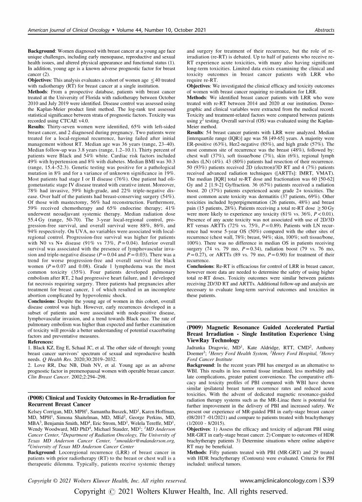
Background: Women diagnosed with breast cancer at a young age faceunique challenges, including early menopause, reproductive and sexualhealth issues, and altered physical appearance and functional status (1).In addition, young age is a known adverse prognostic factor for breastcancer (2).Objectives: This analysis evaluates a cohort of women age ≤ 40 treatedwith radiotherapy (RT) for breast cancer at a single institution.Methods: From a prospective database, patients with breast cancertreated at the University of Florida with radiotherapy between October2010 and July 2019 were identified. Disease control was assessed usingthe Kaplan-Meier product limit method. The log-rank test assessedstatistical significance between strata of prognostic factors. Toxicity wasrecorded using CTCAE v4.0.Results: Thirty-seven women were identified, 65% with left-sidedbreast cancer, and 2 diagnosed during pregnancy. Two patients weretreated for a local-regional recurrence, having failed after initialmanagement without RT. Median age was 36 years (range, 23–40).Median follow-up was 3.8 years (range, 1.2–10.1). Thirty percent ofpatients were Black and 54% white. Cardiac risk factors included49% with hypertension and 8% with diabetes. Median BMI was 30.3(range, 15.4–52.3). Genetic testing was positive for a pathologicalmutation in 8% and for a variance of unknown significance in 19%.Most patients had stage I or II disease (76%). One patient had oli-gometastatic stage IV disease treated with curative intent. Moreover,78% had invasive, 59% high-grade, and 22% triple-negative dis-ease. Over half of the patients had breast-conserving surgery (54%).Of those with mastectomy, 56% had reconstruction. Furthermore,59% received chemotherapy and 65% endocrine therapy; 41%underwent neoadjuvant systemic therapy. Median radiation dose55.4 Gy (range, 50-70). The 3-year local-regional control, pro-gression-free survival, and overall survival were 88%, 86%, and94% respectively. On UVA, no variables were associated with local-regional control. Progression-free survival was higher for patientswith N0 vs N+ disease (91% vs 73%, P= 0.04). Inferior overallsurvival was associated with the presence of lymphovascular inva-sion and triple-negative disease (P= 0.04 and P= 0.03). There was atrend for worse progression-free and overall survival for blackwomen (P= 0.07 and 0.08). Grade 1 lymphedema was the mostcommon toxicity (35%). Four patients developed pulmonaryembolism after RT, 2 had progressive heart failure, and 1 developedfat necrosis requiring surgery. Three patients had pregnancies aftertreatment for breast cancer, 1 of which resulted in an incompleteabortion complicated by hypovolemic shock.Conclusions: Despite the young age of women in this cohort, overalldisease control was high. However, early recurrences developed in asubset of patients and were associated with node-positive disease,lymphovascular invasion, and a trend towards Black race. The rate ofpulmonary embolism was higher than expected and further examinationof toxicity will provide a better understanding of potential exacerbatingfactors and preventative measures.References:1. Black KZ, Eng E, Schaal JC, et al. The other side of through: youngbreast cancer survivors’ spectrum of sexual and reproductive healthneeds. Q Health Res. 2020;30:2019–2032.2. Love RR, Duc NB, Dinh NV, et al. Young age as an adverseprognostic factor in premenopausal women with operable breast cancer.Clin Breast Cancer. 2002;2:294–298.
(P008) Clinical and Toxicity Outcomes in Re-Irradiation forRecurrent Breast CancerKelsey Corrigan, MD, MPH1, Samantha Buszek, MD1, Karen Hoffman,MD, MPH1, Simona Shaitelman, MD, MEd2, George Perkins, MD,MBA3, Benjamin Smith, MD4, Eric Strom, MD1, Welela Tereffe, MD1,Wendy Woodward, MD PhD4, Michael Stauder, MD1; 1MD AndersonCancer Center, 2Department of Radiation Oncology, The University ofTexas MD Anderson Cancer Center, [email protected],4University of Texas MD Anderson Cancer CenterBackground: Locoregional recurrence (LRR) of breast cancer inpatients with prior radiotherapy (RT) to the breast or chest wall is atherapeutic dilemma. Typically, patients receive systemic therapy
and surgery for treatment of their recurrence, but the role of re-irradiation (re-RT) is debated. Up to half of patients who receive re-RT experience acute toxicities, with many also having significantlong-term toxicities. Limited data exists examining the clinical andtoxicity outcomes in breast cancer patients with LRR whorequire re-RT.Objectives: We investigated the clinical efficacy and toxicity outcomesof women with breast cancer requiring re-irradiation for LRR.Methods: We identified breast cancer patients with LRR who weretreated with re-RT between 2014 and 2020 at our institution. Demo-graphic and clinical variables were extracted from the medical record.Toxicity and treatment-related factors were compared between patientsusing χ2 testing. Overall survival (OS) was evaluated using the Kaplan-Meier method.Results: 54 breast cancer patients with LRR were analyzed. Median[interquartile range (IQR)] age was 58 [49-65] years. A majority wereER-positive (63%), Her2-negative (85%), and high grade (57%). Themost common site of recurrence was the breast (48%), followed by:chest wall (37%), soft tissue/bone (7%), skin (6%), regional lymphnodes (LN) (4%). 43 (80%) patients had resection of their recurrence.50 (93%) patients received 2D (electron)/3D RT and 4 (7%) patientsreceived advanced radiation techniques ([ARTTs]: IMRT, VMAT).The median [IQR] total re-RT dose and fractionation was 60 [50-62]Gy and 2 [1.9-2] Gy/fraction. 36 (67%) patients received a radiationboost. 20 (37%) patients experienced acute grade 2+ toxicities. Themost common acute toxicity was dermatitis (37 patients, 69%). Othertoxicities included hyperpigmentation (26 patients, 48%) and breastpain (15 patients, 28%). Patients receiving a total re-RT dose ≥ 50 Gywere more likely to experience any toxicity (81% vs. 36%, P< 0.01).Presence of any acute toxicity was not associated with use of 2D/3DRT versus ARTTs (72% vs. 75%, P= 0.89). Patients with LN recur-rence had worse 5-year OS (50%) compared with the other sites ofrecurrence (chest wall, 78%; breast, 94%; skin, 100%; soft tissue/bone,100%). There was no difference in median OS in patients receivingsurgery (74 vs. 79 mo, P= 0.34), radiation boost (79 vs. 76 mo,P= 0.27), or ARTTs (89 vs. 79 mo, P= 0.90) for treatment of theirrecurrence.Conclusions: Re-RT is efficacious for control of LRR in breast cancer,however more data are needed to determine the safety of using highertotal re-RT doses. Toxicity outcomes were similar between patientsreceiving 2D/3D RT and ARTTs. Additional follow-up and analysis arenecessary to evaluate long-term survival outcomes and toxicities inthese patients.
(P009) Magnetic Resonance Guided Accelerated PartialBreast Irradiation - Single Institution Experience UsingViewRay TechnologyJadranka Dragovic, MD1, Kate Aldridge, RTT, CMD2, AnthonyDoemer3; 1Henry Ford Health System, 2Henry Ford Hospital, 3HenryFord Cancer InstituteBackground: In the recent years PBI has emerged as an alternative toWBI. This results in less normal tissue irradiated, less morbidity andlate complications, greater patient convenience. The comparative effi-cacy and toxicity profiles of PBI compared with WBI have shownsimilar ipsilateral breast tumor recurrence rates and reduced acutetoxicities. With the advent of dedicated magnetic resonance-guidedradiation therapy systems such as the MR-Linac there is potential forfurther improvement in the delivery of PBI and increased safety. Wepresent our experience of MR-guided PBI in early-stage breast cancer(08/2017 -01/2021) and compare to patients treated with brachytherapy(1/2010 - 8/2015).Objectives: 1) Assess the efficacy and toxicity of adjuvant PBI usingMR-GRT in early-stage breast cancer. 2) Compare to outcomes of HDRbrachytherapy patients 3) Determine situations where online adaptiveRT may be beneficial.Methods: Fifty patients treated with PBI (MR-GRT) and 29 treatedwith HDR brachytherapy (Contoura) were evaluated. Criteria for PBIincluded: unifocal tumors.
American Journal of Clinical Oncology � Volume 44, Number 10, October 2021 Abstracts
Copyright © 2021 Wolters Kluwer Health, Inc. All rights reserved. www.amjclinicaloncology.com | S39
Copyright r 2021 Wolters Kluwer Health, Inc. All rights reserved.

Results: Acute reactions were minimal with skin reactions mild to noneand limited to the lumpectomy site (RTOG score 0-1). Late effectswere localized mild skin hyper-pigmentation. One patient had a ribfracture. There are no recurrences to date, with a median follow up of12 months. None of the patients qualified for online adaptive planning,but end-inhale breath hold was utilized for left-sided tumors for cardiacsparing. In the brachytherapy group one had a local recurrence (at 3 y)and 12/29 had significant persistent seroma/thickening/retraction at thetreated site.Conclusions: PBI using MR-guided radiation therapy is a feasible, welltolerated regimen for early-stage breast cancer with a favorable acuteand late toxicity profile and excellent cosmetic result. The follow-up ispresently too short for recurrence evaluation, but so far no relapses wereseen at a median follow up of 12 months. When compared with bra-chytherapy the acute and late morbidity are significantly lower and thecosmetic result superior.
(P010) Predictors of Radiation Dermatitis and Esophagitisin Early Stage Breast Cancer Patients Receiving AdjuvantWhole Breast Radiation TherapyStephen Dzul, MD PhD1, Seongho Kim, PhD2, Hyejeong Jang, BS2,Michael Dominello, DO1, James Ninia, MS1; 1Wayne State University,2Karmanos Cancer InstituteBackground: Breast conserving surgery followed by adjuvant wholebreast radiation therapy is standard of care in the management of earlystage breast cancer. Radiation dermatitis is one of the most commontoxicities associated with whole breast radiation therapy and is fre-quently treated with topical ointments and NSAIDS.Objectives: This study investigates clinical factors associated withincreased incidence of Grade 2+ radiation dermatitis in patientsundergoing adjuvant whole breast radiation.Methods: An institutional database was developed to include allpatients with a history of breast cancer or DCIS undergoing adjuvantwhole breast radiotherapy at a single institution from 2013-2019.
Records were reviewed to identify patient’s age, BMI, radiationdose and fractionation, prone vs supine position, inclusion of boost,and inclusion of regional nodal irradiation. Radiation treatmentplans were reviewed to identify breast size, and volume of breastreceiving > 105% and > 107% prescription dose (V105%, V107%).Medical records were reviewed to identify which patients experi-enced at least patchy moist desquamation (silvadene prescriptionwas used a surrogate for this toxicity) during or immediately fol-lowing their course of radiotherapy. Univariable and multivariablelogistic regression analyses were performed to identify correlationsbetween patient factors associated with incidence of dermatitis andesophagitis.Results: 370 patients were included in the final analysis. 43% ofpatients were obese and 15% were morbidly obese. 42% of patientsreceived hypofractionated radiation treatment. 35% of patients weretreated with an additional supraclavicular field (SCF) to includeregional lymph nodes. 22% of patients were treated in prone position.Grade 2+ dermatitis was diagnosed in 39% of patients. On univariableanalysis, obese and morbidly obese patients were more likely todevelop dermatitis (Obese: OR 1.65, P = .032, Morbidly Obese: OR4.28, P <0.001). On multivariable analysis, larger breast volume (OR
TABLE 1. Logistic Regression Analyses of Patient FeaturesAssociating With Incidence of Dermatitis
Abstracts American Journal of Clinical Oncology � Volume 44, Number 10, October 2021
S40 | www.amjclinicaloncology.com Copyright © 2021 Wolters Kluwer Health, Inc. All rights reserved.
Copyright r 2021 Wolters Kluwer Health, Inc. All rights reserved.

2.75, P < .001) was associated with increased incidence of dermatitiswhile prone position (OR 0.42, P= .013) and hypofractionated radio-therapy was associated with decreased incidence of dermatitis (OR0.50, P= .027). V107% and presence of a SCF had a positive corre-lation and age had a negative correlation with dermatitis on univariableanalysis but these correlations were not present on multivariableanalysis. Addition of a boost to the lumpectomy cavity did not affectthe rate of dermatitis.Conclusions: Radiation dermatitis is a common toxicity observed inpatients undergoing adjuvant breast radiation therapy. Large breastvolume was associated with a higher rate of dermatitis while proneposition and hypofractionated radiation reduced rates of dermatitis.Addition of lumpectomy cavity boost did not increase the incidence ofdermatitis. Hypofractionated, prone radiotherapy with or without boostlikely decreases the risk of grade 2+ dermatitis, particularly for womenwith large breast volumes (Table 1).
(P011) Impact of Surgery Type and Radiation Fractiona-tion on Outcomes in Node-Negative Metaplastic BreastCancerYufei Liu, MD PhD1, Puneet Singh, MD1, Stacy Moulder, MD1,Wendy Woodward, MD PhD1, Simona Shaitelman, MD, MEd2, GeorgePerkins, MD, MBA3, Anthony Lucci, MD1, Lavinia Middleton, MD1,Benjamin Smith, MD1, Melissa Mitchell, MD PhD1; 1University ofTexas MD Anderson Cancer Center, 2Department of RadiationOncology, The University of Texas MD Anderson Cancer Center,[email protected]: Metaplastic breast cancer is a rare aggressive subtypewith poor outcomes and early recurrences. As practice patterns shifttoward hypofractionation in early stage breast cancer patients world-wide, we sought to assess local therapy use in node negative metaplasticbreast cancer in a single institution and associated oncologic outcomes.Objectives: 1. Assess the clinical outcomes of patients with metaplasticbreast treated with surgery with or without adjuvant radiation at ourinstitution. 2. Assess whether metaplastic breast cancer can be effec-tively treated with hypofractionation.Methods: After IRB approval, we retrospectively reviewed the charts ofmetaplastic patients treated between January 2014 and January 2020 atour institution, a timeframe with increased utilization of hypofractio-nation. Patients with non-inflammatory, non-metastatic breast cancerwith known radiation status were included in the analysis. All patientswere node negative clinically and pathologically. Data were collectedon chemotherapy use, radiation use, surgery type, tumor subtype, tumorstage, recurrences and deaths. Kaplan-Meier estimates of 2 yearrecurrence and survival were performed in SPSS 24.Results: A total of 50 metaplastic patients were identified who met theinclusion criteria, with a median follow-up time of 26 months fromdiagnosis. Median age was 58, range 24-84. Seventy-two percent ofpatients were White. Seventy-six percent of tumors were triple negative,18% hormone positive, and 6% Her2 positive. Most patients receivedchemotherapy, 82%, with 51% of these receiving it neoadjuvantly.Table 1 shows actuarial 2 year local recurrence, any recurrence and
overall survival by local therapy subtype. To date, all local recurrencescaptured had developed within 20 months of diagnosis; median time tolocal recurrence was 11 months. The minimum follow-up was16 months in patients treated with lumpectomy and hypofractionation.Clinical stage was T1 20%, T2 62%, T3 12% and T4b 6%. All T4bpatients received mastectomy and radiation. Patients selected for mas-tectomy alone were more likely to be early stage cT1/2 (100%), com-pared with patients receiving mastectomy and radiation cT1/2 (45%).Staging for lumpectomy patients receiving hypofractionation were cT133%, cT2 67%, as compared with lumpectomy with conventionalfractionation cT1 18%, cT2 76%, cT3 6%. All hypofractionationpatients were hormone negative, one was Her2 positive.Conclusions: While limited by low numbers, this analysis suggests thathypofractionation should be further studied in early stage metaplasticbreast cancer. Continued review of outcomes with larger patient num-bers is needed. Patients with higher stage disease have poorer outcome,despite aggressive local therapy with mastectomy and radiation.
(P012) Prospective Pilot Study of Radiation DermatitisAmong Patients with Breast Cancer Evaluated Using DeepMachine LearningSung Jun Ma, MD1, Shawn Matott, PhD1, Robert DeLeon, PhD1,Simon Fung-Kee-Fung, MD1, Saif Aljabab, MD1; 1Roswell ParkComprehensive Cancer CenterBackground: Patients and clinical studies often rely on experiencedradiation oncologists to evaluate the severity of radiation dermatitis(RD). However, available grading tools are heterogeneous and sub-jective. In recent years, deep machine learning (DML) algorithms haveemerged in diagnosing various skin disorders.Objectives: In this prospective feasibility pilot study, we explore theability of DML to detect RD in breast patients and correctly describe theskin findings and grading using the Common Terminology Criteria forAdverse Events (CTCAE v5.0).Methods: Our institutional review board approved this study. Videos ofbilateral breasts were obtained weekly during radiation and up to one-month post-treatment. Video recordings were split into frames andbinned by a radiation oncologist. Two separate convolutional neuralnetwork models were developed. One model classified frames by skinfindings and the other by CTCAE grade 0 to 4. Skin findings included:normal skin, irradiated healing skin, faint erythema/hyperpigmentation,brisk erythema/hyperpigmentation, dry desquamation, moist desqua-mation, moist desquamation with bleeding, skin ulceration, and skinnecrosis. Images were divided into 80% training set and 20%validation set.Results: A total of 12 women were enrolled (8 White and 4 AfricanAmerican) between June 2020 and October 2020. A total of 4,530images were collected and binned into normal skin (n= 2,624), irradi-ated healing skin (n= 317), faint erythema/hyperpigmentation (n= 867)and brisk erythema/hyperpigmentation (n= 577). Desquamation classeswere aggregated (n= 145) due to their small sample size for this initialanalysis. No skin ulceration or necrosis was observed. For our firstmodel to classify skin findings, overall accuracy for the model was84%. The model distinguished normal skin from any grade of radiationdermatitis or post-radiation skin with an accuracy of 95%. However,given small sample sizes of images with radiation dermatitis, themodel’s accuracy ranged from 57% to 74% for differentiating amongrecently irradiated skin, faint or brisk erythema or hyperpigmentation,and desquamation. For our second model to grade using CTCAE v5.0,images were binned into grade 0 (n= 1,844), grade 1 (n= 801), andgrade 2 (n= 301). Grade 3 images were excluded for analysis given itslimited sample size. No grade 4 or 5 toxicities were observed. Overallaccuracy was 82%. The model identified grade 0, 1, and 2 correctlywith the accuracy of 87%, 74%, and 74%, respectively. The model wasmore accurate on White patients with an overall accuracy of 85%,compared with that of 75% on African American patients.Conclusions: Our prospective pilot study demonstrates that using bothof our DML models to identify radiation dermatitis is feasible. We arenow planning to accelerate data collection to increase all classes’ samplesizes to further internally and externally validate our model.
TABLE 1. Outcomes of Metaplastic Breast Cancer PatientsTreated With Lumpectomy or Mastectomy With or WithoutRadiation
American Journal of Clinical Oncology � Volume 44, Number 10, October 2021 Abstracts
Copyright © 2021 Wolters Kluwer Health, Inc. All rights reserved. www.amjclinicaloncology.com | S41
Copyright r 2021 Wolters Kluwer Health, Inc. All rights reserved.

(P013) Five Day Accelerated Partial Breast Irradiation(APBI) Using Stereotactic Body Radiation Therapy (SBRT)in Stage 0-II Breast Cancer: A Report of 186 Patients withup to 3 Year Follow-upRufus Mark, MD1, Valerie Gorman, MD1, Steven McCullough, PhD1;1Baylor Scott & White Medical CenterBackground: Randomized trials in selected early stage breast cancerpatients with up to 10 year follow-up have proven that AcceleratedPartial Breast Irradiation (APBI) given via High Dose Rate (HDR)implant bid in 5 days is equivalent to whole breast External RadiationTherapy (XRT) given qd in 5-6 weeks in regard to breast tumor localrecurrence (LR) [1-2]. However, complications with APBI implant ina Medicare database review have been significant, with 3.95% ofwomen requiring Mastectomy, 16.2% developing infections, andanother 16.3% experiencing non-infection complications includingrib fractures, fat necrosis, and breast pain [3]. Recently APBI usingnon-invasive Intensity Modulated Radiation Therapy (IMRT) orStereotactic Body Radiation Therapy (SBRT) given qd in 5 fractionshas been shown in another randomized trial with 10 year follow-up tobe equivalent to qd XRT in 6 weeks, with respect to LR [4]. IMRT/SBRT was superior in regard to acute effects, late effects, andcosmesis.Objectives: In the randomized clinical trial of APBI IMRT/SBRT, theClinical Target Volume (CTV) was defined by the injection of indi-vidual fiducial markers bordering the surgical cavity. At our institution,we have used the Biozorb fiducial system to localize the CTV forSBRT. We sought to confirm the APBI SBRT/IMRT results with asimpler fiducial system.Methods: Between 2017 and 2020, 186 patients have undergoneSBRT targeted to a Biozorb defined CTV with the walls of the sur-gical cavity sewn to the Biozorb device. Eligible patients were olderthan age 40, had tumor sizes < 3 cm, negative surgical margins, andnegative sentinel node dissections. SBRT dose was 30 Gy given in 5fractions. Dose Constraints were as follows: V-30 Gy < 105%,Ipsilateral Breast V-15 Gy < 50%, Ipsilateral Lung V-10 Gy < 20%,Contralateral Lung V-5 Gy < 10%, Heart V-3 Gy < 20%, Contra-lateral Breast Dmax < 2 Gy and Skin Dmax < 27 Gy. The PlanningTarget Volume (PTV) ranged from 27 to 355 cc with a median of 80cc. PTV=CTV + 1-2 cm.Results: Follow-up ranged from 1-36 months with a median of18 months. LR has been 0% (0/186). There have been no biozorbrelated infections, reactions, or rejections. There have been no cases ofradiation induced seromas, skin reactions, or soft tissue necrosis. Fourpatients, 2.2% (4/186) developed pain around the Biozorb site. Threecases were resolved within 2 days on a 2 week course of steroids. Onepatient required 3 courses, with the last being 1 month. Cosmetic resultsas rated by the Surgeon, Radiation Oncologist, and Nurse, were ratedexcellent in 98.9% (184/186) of cases.Conclusions: Non-invasive APBI with SBRT given qd over 5 daystargeted to Biozorb has resulted in LR, complications, and cosmeticresults which compare favorably to invasive APBI given bid with HDRimplant. At last follow-up, there have been no LR, skin reactions, orcomplications. Cosmesis has been excellent in 98.9% of patients.
(P014) Predictors of Metastatic Breast Cancer Presentationin a Vulnerable Patient PopulationMary McGunigal, MD1, Rhea Bhatt, BS2, Nima Aghdam, MD3;1Georgetown University Hospital, 2Georgetown University, 3BethIsrael Deaconess Medical CenterBackground: Socially disadvantaged populations in the United Statesexperience worse outcomes compared with the general population whendiagnosed with breast cancer. One potentially vulnerable populationincludes patients under age 65 that qualify for Medicare due to debil-itation. This group includes patients who have received social securitydisability insurance for at least 2 years and those who have beendiagnosed with end-stage renal disease or amyotrophic lateral sclerosis;current literature suggests this population experiences worse oncologicoutcomes.
Objectives: Our study goal was to utilize the National Cancer Database(NCDB) and perform analyses to determine what factors amongpatients under the age of 65 who qualify for Medicare predict for ahigher likelihood of presenting with stage IV breast cancer.Methods: The NCDB was queried for breast cancer patients diagnosedfrom 2004-2014 who were below the age of 65 and qualified forMedicare. Patients were excluded if they presented with noninvasivedisease or if their AJCC stage was unknown. Patients were excluded ifany social determinants (race, income quartiles, Charlson-Deyocomorbidity score, travel distance) or hospital factors (facility type,facility location) were unknown. Pearson χ2 test was used to identifyassociations between categorical variables and stage. Univariate anal-ysis of sociodemographic factors was performed. Statistically sig-nificant factors were included in a multinomial logistic regression modelto determine independent covariates associated with stage IVpresentation.Results: 56,964 patients met study criteria. Based on P values (P <0.05) and strength of odds ratios, the following variables were includedin the final model: race, income, comorbidity score, travel distance,facility type, and facility location. All covariates except facility locationand distance were significant predictors of metastatic disease pre-sentation. Race was a significant predictor, with white patients lesslikely to have stage IV presentation (OR 0.73, P< 0.001) comparedwith black patients. Individuals earning <$38,000 annually were sig-nificantly more likely to present at stage IV (OR 1.15, P< 0.001).Patients who traveled shorter distances to treatment facility had non-significantly higher odds of presenting at stage IV (OR 1.37, P= 0.608).Higher comorbidity score was a significant predictor of Stage IV dis-ease at presentation.Conclusions: Multiple sociodemographic factors were powerful pre-dictors of a stage IV breast cancer presentation in this select populationof patients under 65 years old who qualify for Medicare due to debil-itation. A deeper understanding of these factors can help identifypatients at high risk of a metastatic cancer presentation to providescreening and preventative services at an earlier stage in this vulnerablepopulation.
(P015) Ductal Carcinoma in Situ (DCIS) of the Breast:Impact of Age, Grade, and Receptor Status on SurvivalMary Nittala, DrPH1, Satyaseelan Packianathan, MD, PhD2, GaryShultz, DO3, Eswar Mundra, MD3, William Woods, MSN. NPC3,Maria Smith, MSN, NPC3, Barbara Craft, MD3, Srinivasan Vijayaku-mar, MD, MPH3; 1The University of Mississippi Medical Center,2Univerity of Mississippi Medical Center, 3University of MississippiMedical CenterBackground: Ductal carcinoma in situ (DCIS) women who underwentsurgery (lumpectomy/mastectomy) are at risk for recurrence within thebreast; adjuvant radiotherapy (RT) often reduces the risk for recurrence.We decided to investigate the factors that improves the overall survival(OS) and loco-regional control (LRC) in DCIS women patients at anacademic medical center to help tailor treatment decisions.Objectives: The objective of this study was to evaluate LRC and OSrates of DCIS women patients treated with surgery ± RT and hormonalmodulation.Methods: This is a retrospective analysis of 44 DCIS patients treatedbetween 2007 and 2017 at an academic state institution. All of themunderwent lumpectomy and/or mastectomy (surgery) followed byadjuvant irradiation to a median total dose of 50 Gy (range 40.5–50.4 Gy). For all 44 DCIS patients, LRC and OS were estimated usingthe Kaplan-Meier method. The significance of survival variables wereanalyzed using the Cox univariate and multivariate proportional hazardsmodel. A P-value of less than 0.05 was considered statistically sig-nificant. The SPSS 24.0 software was used for data analysis.Results: The baseline characteristics of all 44 DCIS patients weredocumented over a median follow-up of 70 months (range 5–147 mo).Patients were stratified into three age groups (< 50 y (3) vs. 50- 70 y(31) vs. > 70 y (10); median age of 64 y; range 46–80 y). Overall,34.1% of DCIS diagnoses were low grade, 29.5% intermediate grade,and 36.4% were high grade. Older women were more likely to developlow grade DCIS than the younger women. Following surgery, 50% of
Abstracts American Journal of Clinical Oncology � Volume 44, Number 10, October 2021
S42 | www.amjclinicaloncology.com Copyright © 2021 Wolters Kluwer Health, Inc. All rights reserved.
Copyright r 2021 Wolters Kluwer Health, Inc. All rights reserved.

TABLE 1. Baseline Characteristics of DCIS Patients
American Journal of Clinical Oncology � Volume 44, Number 10, October 2021 Abstracts
Copyright © 2021 Wolters Kluwer Health, Inc. All rights reserved. www.amjclinicaloncology.com | S43
Copyright r 2021 Wolters Kluwer Health, Inc. All rights reserved.

patients had negative tumor margins; ER was positive in 88.6% and PRpositive in 79.5%. Ninety percent of patients had a lumpectomy and40.9% used Tamoxifen. The five-year rates for LRC, and OS were 0 vs.69 vs. 50% (P= 0.030) and 33.7 vs. 70.8 vs. 60% (P= 0.041),respectively. On Cox multivariate analysis, younger age [< 50 y](P= 0.023), African-American ethnicity (P= 0.011), and ER positivity(P= 0.007) were associated with a statistically improved significant OS.Conclusions: Outcomes in patients diagnosed with DCIS of the breastappears dependent upon age, ethnicity, and ER receptor status; thisinformation may potentially help tailor treatment decisions in thesepatients (Tables 1, 2 and Fig. 1).
(P016) Dosimetric and Acute Toxicity Analysis withHypofractionated Regional Nodal Irradiation for BreastCancerAdam Richman, MD1, Michael Schad, BS1, Emilia Diego, MD2,Priscilla McAuliffe, MD, PhD2, Ronald Johnson, MD2, Jennifer Stei-man, MD2, Donald Keenan, MD, PhD2, Joanna Lee, MD2, John Vargo,MD3, Sushil Beriwal, MD, MBA, FABS, FASTRO1; 1Department ofRadiation Oncology, UPMC Hillman Cancer Center, 2Department ofBreast Surgical Oncology, UPMC Hillman Cancer Center, 3Depart-ment of Radiation Oncology, Hillman Cancer Center, University ofPittsburgh Medical CenterBackground: Hypofractionated regional nodal irradiation (RNI) breastradiotherapy has been shown to be effective and safe in clinical trials.However, there is limited dosimetric analysis available with this regi-men to see which factors predict for pneumonitis. The ongoing RT-CHARM study limits the percentage of ipsilateral of lung volume thatreceives ≥ 18 Gy (V18) to 35-40% [1].Objectives: To evaluate the rates of acute pneumonitis in womentreated with hypofractionated RNI at our institution and to identifydosimetric factors predictive for pneumonitis.Methods: We performed a retrospective review of all breast cancerpatients treated with hypofractionated RNI defined as the undissectedaxilla and supraclavicular region +/- intramammary nodes followingeither lumpectomy or mastectomy at our institution from 9/2018 to 8/2020. The primary endpoints were acute and chronic symptomaticpneumonitis. Secondary endpoints included acute and chronic skintoxicity. The Kaplan–Meier method was used to estimate event-timeprobabilities for the primary endpoints. Predictors of symptomaticpneumonitis were analyzed using log rank tests between groups.P-values <0.05 were considered significant.Results: 126 patients qualified with a median follow up of11.1 months (interquartile range [IQR] 8.17–18.23). Median agewas 72 years (IQR 66–78) and 88.9% of patients had node-positivedisease. Overall, 56.3% of patients received whole breast RT and43.7% of patients received chest wall RT. 100 patients (79%)received 40.05 Gy in 15 fractions and 23 patients (18%) received42.56 Gy in 16 fractions. Surgical bed boost was given to 92.1% ofpatients. The median ipsilateral lung maximum dose (Dmax) was41.57 Gy (IQR 40.61–42.61) and mean dose (Dmean) was 9.78 Gy(IQR 8.39–11.48). Ipsilateral lung V5 was 40.73% (IQR35.17–46.30), V10 was 27.40% (IQR 22.59–32.35), and V20 was20.63% (IQR 16.72–25.74). Contralateral lung median Dmax andDmean were 2.79 Gy (IQR 1.68–5.24) and 0.20 Gy (IQR0.16–0.25), respectively. Mean heart Dmax was 13.71 Gy (standarddeviation [SD] 0.93), mean Dmean was 4.23 Gy (SD 2.97), andmedian V20 was 0.00% (IQR 0.00–0.00). For acute radiation der-matitis, the rate of grade 2 toxicity was 30.4% for lumpectomy and42.6% for mastectomy and the rate of grade 3 toxicity was 7.2% forlumpectomy and 1.9% for mastectomy. There was no grade 4+ acuteradiation dermatitis. Acute grade 2 pneumonitis was seen in onepatient (0.8%) and no patients developed acute grade 3+ or chronicgrade 2+ pneumonitis. Of the patients that underwent a post-radia-tion therapy chest CT (n= 38, 30.4%), 68.4% had radiographicevidence of grade 1 pneumonitis.Conclusions: The overall incidence of acute symptomatic pneumonitisin patients treated with hypofractionated RNI is very low. V20 < 26%appears to be safe to limit symptomatic pneumonitis. No clinical ordosimetric factors were predictive for symptomatic pneumonitis.
TABLE 2. Univariable &Multivariable Cox Regression for Overall Survival
FIGURE 1. Overall survival and loco regional control of DCISpatients categorized by age group.
Abstracts American Journal of Clinical Oncology � Volume 44, Number 10, October 2021
S44 | www.amjclinicaloncology.com Copyright © 2021 Wolters Kluwer Health, Inc. All rights reserved.
Copyright r 2021 Wolters Kluwer Health, Inc. All rights reserved.

(P017) Comparison of Skin Toxicity and Outcomes forThree and Four Week Hypofractionated Whole BreastIrradiation RegimensJuliana Runnels, MD1, Juliana Runnels, MD1, Alexander Rose, BS1,Ben Liem, MD2, David Lee, MD, PhD2, Thomas Schroeder, MD2;1University of New Mexico School of Medicine, 2Section of RadiationOncology, Division of Hematology-Oncology, Department of InternalMedicine, and University of New Mexico Comprehensive CancerCenter, University of New Mexico School of MedicineBackground: Multiple randomized trials have shown that whole breastradiation therapy delivered in fewer, larger fractions is safe and effec-tive. This recent evidence supports hypofractionation, but the optimaldose-fractionation schedule remains controversial. At our institution,physicians use either a 4-week course (42.56 Gy/16Fx, 10 Gy/4Fxsequential boost) or a 3-week course (48 Gy/40 Gy/15Fx, concomitantboost). Half our physicians perceived an increase in acute skin toxicitywith the 3-week regimen. The other half did not. Patients are referred tothe next available physician, similar to a randomization. A retrospectivereview of these patients may provide a comparison with fewer selectionbiases inherent in retrospective studies.Objectives: Acute skin toxicity is a common symptomatic side effectsof breast irradiation. We sought to compare the incidence of acute skintoxicity in patients treated with 3 versus 4 weeks of radiation therapy forbreast cancer.Methods: An IRB-approved retrospective review identified 342patients treated with hypofractionated radiation therapy from 2013 to2020. 195 treated with the 4-week regimen and 147 with a 3-weekregimen. Primary endpoint was acute skin toxicity assessed by: CTCAEv4.0 physician-scored toxicity during weekly visits, use of topical silversulfadiazine, and patient recollection of skin toxicity (assessed by tel-ephone survey done as part of the study). Secondary endpoints werepatient-reported cosmesis by BCTOS, overall survival, and localrecurrence-free survival. Log-rank tests were used to compare grade ≥2 skin toxicity between arms. Fischer’s exact test were used for com-paring toxicities between the two groups and two-sided P values werereported.Results: Median follow-up was 26 months (3-93 mo). Overall grade ≥2 CTCAE weekly assessed acute skin toxicity was 30% (33% 3-weekvs. 27% 4-week; P=NS). Topical silver sulfadiazine use was 39%3-week vs. 35% 4-week, P= .NS. Recollection of acute skin toxicity ontelephone survey was 77% 3-week vs. 69% 4-week, P= .008. 206(60%) responded to the telephone survey at time of data collection. Thisalso noted the 3-week course was more likely to report peak skin
toxicity after completion of RT (38% vs 18%, P= .005). BMI> 30 wasmore common in the 4-week group than the 3-week group (48% vs29%, P= 0.007). BCTOS showed no difference between the groups incosmesis, function, pain (1.55 vs. 1.63, 1.24 vs. 1.21, 1.51 vs. 1.42,P=NS). There was no difference in overall or local recurrence freesurvival (97.9% vs 95.9%, 98.9% vs 97.4%, P=NS). 3% (10 of 342)patients had died, 1% (3) from breast cancer, 1% (3) from non-breastsecond cancer, and 1% (4) due to other causes.Conclusions: In this comparison of two popular hypofractionationregimens, we observed comparable physician-reported acute skin tox-icity, cosmesis, and disease control outcomes. Limitations to this studyinclude the possibility of measurement bias and selection bias. The3-week group reported more peak skin toxicity after completion oftreatment, this could be associated with under-reporting of skin CTCAEweekly assessment on the 3-week regimen. A higher number of patientswith greater BMI was present in the 4-week regimen. Both could skewresults in favor of the 3-week regimen. Across treatment arms, therewas a higher incidence of silver sulfadiazine prescribed than physician-recorded acute toxicity. Though the treatments compared in this anal-ysis varied by a short time interval, hypofractionation could maketreatment more convenient and accessible for many patients. Additionalcomparative analyses that incorporate patient-reported outcome meas-ures are encouraged to determine the most safe and effective treatmentfor individuals.
(P018) Wrong Number: The Inappropriate Use of an AgeCutoff Heuristic to Allocate Adjuvant Therapy for High-risk, Early-stage Breast Cancer Patients Following BreastConserving SurgeryWesley Talcott, MD, MBA1, Daylian Cain, PhD, MBA2, Daniel Yang,MD1, Sarah Mougalian, MD1, Suzanne Evans, MD MPH3; 1YaleSchool of Medicine, 2Yale School of Management, 3Yale New HavenHealth/Smilow Cancer CenterBackground: Patient age is a continuous variable that should beincorporated into oncologic decision making. However, providers undertime pressure have been shown to process age by dividing patients into“older” vs “younger” categorical subgroups. Use of this cognitiveshortcut (“age cutoff heuristic”) simplifies incorporation of patient ageinto treatment decisions but may lead to inappropriate treatment allo-cation around the age cutoff used for dichotomization.Objectives: We hypothesized the existence of an age cutoff heuristicleading to non-evidence-based adjuvant treatment allocation among
FIGURE 1. Likelihood of a CALGB/PRIME ineligible patient being recommended adjuvant breast-directed radiation therapy followingbreast conserving surgery demonstrating early stage breast cancer with high-risk features, by age at diagnosis. Likelihood of beingrecommended adjuvant radiation therapy declines sharply for patients aged 69 versus age 70.
American Journal of Clinical Oncology � Volume 44, Number 10, October 2021 Abstracts
Copyright © 2021 Wolters Kluwer Health, Inc. All rights reserved. www.amjclinicaloncology.com | S45
Copyright r 2021 Wolters Kluwer Health, Inc. All rights reserved.

high-risk, early-stage breast cancer patients based on a patient’s agebeing ≥ 70, and characterized recommendation of adjuvant therapy inthis population.Methods: We used the National Cancer Data Base to identify womenaged 50-80 years with pT1-2 node-negative breast carcinoma diagnosed2004-2017, who underwent lumpectomy. Among these, we identified162,111 patients with CALGB 9343/PRIME II-ineligible features otherthan age (estrogen receptor negative, endocrine therapy not planned,final margins positive, or size > 3 cm)(cohort 1), and 394,946 hormonereceptor positive (HR+) patients with tumors > 5mm (cohort 2). Mul-tivariable logistic regressions (MVA) with odds ratios (OR) and 99%confidence intervals (CI) were performed for “year-over-year” agepairings, from patients aged 50 and 51 up to patients aged 79 and 80 atdiagnosis. Age at diagnosis, along with 21 additional disease andpatient factors potentially associated with treatment decisions, wereincluded in these MVA. Age was treated as a categorical variable inthese MVA to determine if any single year-over-year age difference wasindependently associated with a difference in likelihood of adjuvanttherapy recommendation.Results: Overall, 87.3% of patients with CALGB 9343/PRIME IIineligible breast cancer were recommended adjuvant radiation therapy(n= 140,513) and 92.3% of HR+ patients with tumor size > 5 mm wererecommended adjuvant endocrine therapy (n= 364,048). Among cohort1, rate of adjuvant radiation recommendation decreased sharply at age70, from 90-92% between the ages of 50 to 69, declining to 81% forthose aged 70 (Fig. 1). Regressions for each year-over-year age pairshowed year-over-year age difference was only an independent pre-dictor adjuvant radiation recommendation at age 70 vs 69 (OR 0.47, CI0.40-0.55, P< 0.001) (Fig. 2). For cohort 2, rates of endocrine therapyrecommendation showed a smaller absolute decline at age 70, but year-over-year age difference was again only a predictor of endocrine ther-apy recommendation at age 70 vs 69 (OR 0.86, CI 0.76-0.97,P= 0.001).Conclusions: We observed a sharp and unique step-off in adjuvanttherapy recommendation between ages 69 and 70. This suggests use ofan age cut off heuristic to process patient age in this population as acategorical, binary variable. This is a previously undescribed
phenomenon in high-risk, early-stage breast cancer, and a practice withconcerning implications for evidence-based care allocation.
(P019) Patient Reported Outcomes Version of the CommonTerminology Criteria for Adverse Events and CTCAE inBreast Cancer Patients Receiving Radiation Therapy:Single-center Prospective Registry ExperienceCameron Thorpe, MD1, Todd DeWees, PhD1, Michael Golafshar, MS1,Ronik Bhangoo, MD1, Tamara Vern-Gross, DO1, Lisa McGee, MD1,William Wong, MD2, Michele Halyard, MD1, Sameer Keole, MD2,Carlos Vargas, MD2; 1Mayo Clinic, 2Mayo Clinic ArizonaBackground: Adverse events during cancer treatment are recorded byproviders using the US National Cancer Institute’s (NCI) CommonTerminology Criteria for Adverse Events (CTCAE). However,symptomatic adverse events are often underreported by physicians anddo not fully represent the full scope of the impact that cancer treatmenthas on patientsObjectives: We sought to investigate the relationship between PRO-CTCAE and CTCAE for breast cancer (BC) patients undergoing radi-ation therapy (RT) from a prospective registry for a large, single-institution.Methods: From a prospectively maintained registry, all patients treatedwith RT for BC with curative intent from November, 2015 to June,2019 at our institution were eligible for this analysis. To be includedpatients were required to have at least one PRO-CTCAE survey. PRO-CTCAE questionnaires were administered at baseline, end-of-treatment,3, 6, 12 months, then annually. Correlation between CTCAE and PRO-CTCAE was measured using Pearson’s correlation coefficient (r) andunivariate Logistic regression was utilized to measure the clinicalmagnitude of that correlation.Results: Three-hundred and thiry-one (331) patients were included foranalysis. Patient and tumor characteristics included: 82% ER(+), 77%PR(+), 10% HER2(+), and 66% ECOG 0. Treatment characteristicsincluded: median dose 40.05 Gy (range, 40–50.4 Gy), 75% underwentlumpectomy, 39% received chemotherapy, 85% treated with photon,
FIGURE 2. Forest plot of odds ratios for being recommended adjuvant radiation therapy. Each point represents a multivariable regressionincluding the subset of patients with the corresponding age at diagnosis as well as the patients aged 1 year younger at diagnosis. The ORand 99% CI representing the association between each year-over-year age increase and likelihood of radiation recommendation areplotted. In this MVA age 70 at diagnosis was independently associated with significantly reduced odds of being recommended adjuvantradiation versus age 69 at diagnosis. No other year-over-year age increase was independently associated with likelihood of beingrecommended radiation.
Abstracts American Journal of Clinical Oncology � Volume 44, Number 10, October 2021
S46 | www.amjclinicaloncology.com Copyright © 2021 Wolters Kluwer Health, Inc. All rights reserved.
Copyright r 2021 Wolters Kluwer Health, Inc. All rights reserved.

15% treated with proton beam therapy, and 40% received a boost(median 10 Gy, range 5.4–14 Gy). “Moderate” or worse PRO-CTCAEcorrelated with CTCAE grade ≥ 2 (r= 0.35, P< 0.0001). ‘Severe’ orworse PRO-CTCAE correlated with CTCAE grade ≥ 3 (r= 0.19,P< 0.001). Furthermore, ‘Moderate’ or worse PRO-CTCAE was pre-dictive of developing CTCAE grade ≥ 3 (OR= 3.25, 95% CI [0.95-11.2], P= 0.06).Conclusions: PRO-CTCAE and CTCAE offer complimentary informa-tion and both should be reported in cancer trials. In this cohort of breastcancer patients, ‘Moderate’ or worse PRO-CTCAE predicted for devel-opment of CTCAE grade ≥3. Patients who report ‘Moderate’ or worsePRO-CTCAE should be monitored closely for development of toxicities.Future studies in breast cancer should report on both PRO-CTCAE andCTCAE to better understand how patients tolerate treatment.
(P020) Patterns of Care and Utilization of Radiation forWomen with High Grade Ductal Carcinoma in Situ: ANational Cancer Database AnalysisIrini Youssef, MD1, Benjamin Silver, MD1, Jared Pasetsky, MD1,Fleure Gallant, MD2, David Schreiber, MD1; 1SUNY Downstate HealthSciences University, 2Maimonides Cancer CenterBackground: Ductal carcinoma in situ (DCIS) currently comprises25% of newly diagnosed breast cancers, and this rate continues to risewith improvements in screening. While prospective studies have dem-onstrated a reasonably low risk of recurrence in “good-risk” patientswith lumpectomy without radiation, ECOG 5194 showed the risk ofdeveloping ipsilateral breast events in patients with high-nuclear gradeDCIS, who received lumpectomy alone, increased without plateau.Objectives: The purpose of this study was to retrospectively review theNational Cancer Database (NCDB) to assess patterns of care in patientswith high-grade DCIS treated with lumpectomy with or withoutradiation.Methods: The National Cancer Database (NCDB) was queried toidentify women treated with lumpectomy for high grade DCIS of thebreast from 2004-2016. Data regarding age, tumor size, endocrinetherapy use, ER receptor status, margin status, race, insurance type anddistance from the treatment center were collected. Radiation fractiona-tion was collected and categorized as hypofractionation, standardfractionation, and other, if the fraction size could not be clearly ascer-tained. Clinical and patient-related factors were compared betweenpatients who received radiation and no radiation. Frequency dis-tributions between categorical variables were compared using the χ2test. Multivariable logistic regression was used to identify co-variablesassociated with the use of radiation.Results: A total of 16,100 patients met the eligibility criteria. Of those,10, 378 (65%) received adjuvant radiation. On multivariable regression,patients who received radiation were more likely to be ER- (OR 1.68,95% CI 1.54-1.83, P< 0.001), and have tumor size of 1cm or larger(OR 1.23, 95% CI 1.13-1.33, P< 0.001). Those less likely to receiveradiation included great-circle distance of > 20 miles (OR 0.80, 95% CI0.71-0.91, p0.005), distance > 30 miles (OR 0.54, 95% CI 0.48-0.60,P< 0.001), and age > 65 years (OR 0.68, 95% CI 0.60-0.78,P< 0.005). There were no differences in radiation utilization based onthe year of diagnosis. Fractionation technique was categorized asstandard or hypofractionated in 64.3% of patients. Of those, the use ofhypofractionation increased from 1% in 2004 to 9.2% by 2010 and to51.7% by 2016.Conclusions: There appears to be an underutilization of radiationtherapy for women with high grade DCIS, particularly those who live> 20 miles from a treatment facility. Prospective evaluation of ultra-hypofractionated regimens and/or intra-operative regimens in DCISpatients is warranted and may improve utilization of adjuvant RT.
(P021) Clinical Factors Associated with Local Control orRadiation Necrosis for Patients Undergoing Single FractionSRSGregory Alexander, MD1, Jill Remick, MD2, Emily Kowalski, MD1,Kai Sun, MS3, Stewart Becker, Physicist1, Naru Lamichhane, PhD1,
Howard Eisenberg, MD1, Graeme Woodworth, MD1, William Regine,MD1, Mark Mishra, MD1; 1University of Maryland, 2Emory, 3Uni-versity of Maryland School of Medicine Department of Epidemiologyand Public HealthBackground: Single-fraction stereotactic radiosurgery (SF-SRS) for thetreatment of brain metastases can be delivered with either a Gamma-Knife platform (GK-SRS) or with a frameless linear accelerator (LA-SRS). These techniques vary based on patterns of prescribing, patientsetup and radiation delivery but the effect of these differences onclinical outcomes is unknown.Objectives: We seek to examine clinical factors associated with radi-ation necrosis and local failure in patients undergoing single fractionSRS in patients with metastatic brain cancer.Methods: Patients treated for metastatic brain cancer treated with SF-SRS from 2014-2020 were retrospectively reviewed and clinical out-comes were recorded on a per lesion basis. Covariates between groupswere compared using a χ2 analysis for dichotomous variables and t-testfor continuous variables. Median follow up was calculated using thereverse Kaplan Meier (KM) method. Primary endpoints of local control(LC) and symptomatic radiation necrosis (RN) were estimated using theKM method with salvage WBRT used as a censoring event. Outcomeestimates were compared using the log-rank test. Multivariate analysis(MVA) and Cox proportional hazards modeling were used for statisticalanalyses. Propensity score (PS) adjustments were used to reduce theeffects confounding variables. Further subset analyses of lesions treatedwith GK-SRS and LA-SRS were performed.Results: Overall, 119 patients with 287 lesions were included foranalysis which included 57 patients (127 lesions) treated with LA-SRScompared with 62 patients (160 lesions) treated with GK-SRS. In termsof lesion size, there was no statistically significant differences in per-centage of lesions > 2 cm treated with GK-SRS vs LA-SRS (14.17% vs8.75%; P = 0.147). For the entire cohort the median follow-up was11 months (15.2 mo GK-SRS vs 7.9 mo LA-SRS) with median OS15.9 months (16.2 mo GK-SRS vs 12.3 mo LA-SRS). On both multi-variate and univariate analysis, there was no statistically significantdifferences between GK-SRS and LA-SRS for local failure or in risk ofradiation necrosis analysis (multivariate adjusted P-value= 0.44).Tumor size (> 2 cm vs ≤ 2 cm) was predictive of both risk of radiationnecrosis and local failure. Subset analysis of lesions treated with LA-SRS did not find the addition of PTV margin or timing of MRI used fortreatment planning to be associated with clinical outcomes. Subsetanalysis of lesions treated with GK-SRS found that the first quartile ofdose rate was associated with increased risk of radiation necrosis,(P= 0.0001), but lesion characteristics were imbalanced betweengroups.Conclusions: In our retrospective cohort, we found no statisticallysignificant differences in the incidence of symptomatic radiationnecrosis or local control in patients treated with GK-SRS when com-pared with LA-SRS. Other clinical variables may be important andrequire consideration. Further study with well-matched prospectivecohorts is required to validate these results.
(P022) Stereotactic Radiosurgery in Patients with Trigemi-nal Neuralgia and Diabetes Mellitus: A RetrospectiveReviewJack Allen, Undergraduate Student1, Nathan Munson, MD1, MichaelRay, MD, PhD1, Shaneli Fernando, MD1; 1ThedaCare Regional CancerCenter, Appleton, WisconsinBackground: Trigeminal Neuralgia (TN) is a neurological disordercharacterized by intense unilateral pain in the face following a stimulus.TN treatments predominantly consists of stereotactic radiosurgery(SRS), microvascular decompression, or percutaneous rhizotomy. Type2 diabetes (T2D) is a disease that affects many patients with TN, andmay have an impact on TN treatment.Objectives: The objective of the current retrospective review is toexamine variances in treatment outcomes using SRS for TN in patientswith T2D and in the absence of T2D.Methods: Clinical data from 34 consecutive TN patients (2007-2013)was collected following treatment with SRS in our department. Patientswere allocated to T2D (n= 13) and T2D absent groups (DA) (n= 21).
American Journal of Clinical Oncology � Volume 44, Number 10, October 2021 Abstracts
Copyright © 2021 Wolters Kluwer Health, Inc. All rights reserved. www.amjclinicaloncology.com | S47
Copyright r 2021 Wolters Kluwer Health, Inc. All rights reserved.

Diabetic status was defined as hemoglobin A1C (HbA1C) > 6.5% orfasting blood glucose levels (FBGL)> 130 mg/dL. HbA1c results wereavailable for 11 patients within the T2D group. The remaining two T2Dpatients had FBGL > 130 mg/dL in three consecutive visits. Thepatient age was DA= 65.0 ± 16.3 and T2D= 70.5 ± 14.3. The DA groupconsisted of 14 females and 7 males, while the T2D group consisted of9 females and 4 males. Before SRS, pain levels were determined usingthe Barrow Neurological Institute (BNI) Pain Intensity Score. Treatmentoutcomes were measured using the BNI score one-week post operationand one year following SRS. The differential in BNI score following theoperation was then analyzed using a two sample t test for differencein mean.Results: Before SRS the DA group had a BNI score of 4.80± 0.4, theT2D group had a BNI score of 4.84 ± 0.37. One week followingtreatment the scores were DA= 1.92± 1.26 and T2D= 2.10 ± 1.51. Oneyear after SRS the results were DA= 2.41± 1.51 and T2D= 1.58 ±1.40. Comparison of the starting BNI score and the BNI score at theone-week post SRS visit of the DA group to the T2D group did notyield a significant difference in BNI score (P= 0.9066). Comparison ofthe DA group BNI score before SRS and BNI score one-year postoperation to the T2D group yields a significant mean difference(P= 0.0121). The DA group experienced a 1.417 greater BNI painreduction (95% CI: 2.499, 0.3347) as compared with the T2D BNIscore at the 1-year mark.Conclusions: This exploratory study suggests a relationship betweendecreased treatment efficacy of SRS one year following the operationand T2D in TN patients. Further studies are needed to examine apossible decrease in treatment efficacy for T2D patients with TN fortreatments such as microvascular decompression or percutaneousrhizotomy.
(P023) Radiotherapy for Management of Brain Metastasesfrom Thyroid Cancer: A National Cancer Database StudyErik Blomain, MD1, Michael Binkley, MD1, Duc Giao, Visiting Stu-dent1, Thomas Churilla, MD2, Scott Soltys, MD1, Erqi Pollom, MD1;1Stanford University, 2Northeast Radiation Oncology Center, GeisingerCommonwealth School of MedicineBackground: Brain metastasis is a rare presentation of thyroid cancerand there is a paucity of data regarding management. Although ster-eotactic radiosurgery (SRS) is a standard of care for patients acrossmany histologies, the current NCCN guidelines do not support a uni-versal role for this modality in thyroid cancer. Alternatives to SRSinclude whole brain radiotherapy (WBRT) and best supportive care;however, current practice patterns regarding the relative use of thesemodalities remains largely undefined.Objectives: In that context, the present study queried the NationalCancer Database to look at practice patterns and outcomes in patientswith brain metastases from thyroid cancer with a goal of informingmanagement.Methods: The Participant User File (PUF) for Thyroid Cancer wasobtained from the NCDB in accordance with all institutional andorganizational regulations. The data was formatted using SAS and
analyzed in R studio and Graphpad Prism. The PUF was screened bypatients who presented with brain metastases and/or who receivedbrain-directed radiotherapy. Kaplan–Meier analysis was performed foroverall survival, as was multivariable regression analysis. Descriptivestatistics were collected regarding patient demographics and treatmentinformation.Results: Overall, we identified a cohort of 289 patients. Median agewas 63 years old (range 19-90 years old) and 56.1% were female.68.5% of patients were white, 13.8% were black and 11.1% were latino(several other races were represented <5%). 16.6% received SRS,55.0% received other brain-directed radiotherapy, and 28.4% receivedno brain-directed RT. Histologic breakdown was as follows: Papillary45.7%, Anaplastic 15.2%, Other 10.7%, Medullary 10.0%, Follicular9.7% and Unknown 8.7%. In terms of practice patterns, there appearedto be a trend towards decreasing use of non-SRS brain radiotherapy,although this is potentially confounded by missing data from recently-treated patients. Median follow up was 213.2 weeks, and median overallsurvival was 40.9 weeks. On multivariable analysis, treatment modality(SRS versus other brain RT versus no brain RT) was not significant interms of OS (hazard ratio 1.09, P= 0.55); however, anaplastic histologywas significantly associated with worse overall survival (hazard ratio3.5, P= 0.00000005), and anaplastic patients’ median overall survivalwas 8.4 weeks.Conclusions: Brain metastases from thyroid cancer portend a poorprognosis (< 1 year median in our study), particularly for patients withanaplastic histology. The decision to treat with radiotherapy and thetype of radiotherapy utilized did not appear to affect overall survival,consistent with other retrospective and prospective data in brain meta-stases from other histologies. Of note, our case number was ratherlimited, potentially due to the relatively rare nature of this metastaticpresentation. Despite this limitation, the present study represents to ourknowledge the largest dataset to date of thyroid cancer brain metastasestreated with radiotherapy. Within the limitations inherent to NCDBstudies, our data suggest clinical equipoise in the management of thesepatients (Figs. 1 and 2).
(P024) Intracranial Response to Combination BRAF andMEK Inhibitor Therapy in Patients with MetastaticMelanomaCaressa Hui, MD1, Yufan (Fred) Wu, MD1, Kevin Liu, BS2, NavjotSandhu, BS1, Erik Blomain, MD1, Michael Binkley, MD1, MelanieGephart, MD, MAS1, Steven Chang, MD1, Gordon Li, MD1, SunilReddy, MD1, Iris Gibbs, MD1, Scott Soltys, MD1, Erqi Pollom, MD1;1Stanford University, 2Carnegie Mellon UniversityBackground: Combination BRAF+MEK inhibitor therapy is nowwidely used in the treatment of metastatic melanoma. However, data forintracranial response to these drugs are limited.Objectives: We aimed to evaluate the intracranial efficacy of BRAF+MEK inhibitors in patients with BRAF-mutant melanoma with brainmetastases and to determine patterns of failure of these new agents toinform optimal integration of local intracranial therapy.FIGURE 1. Overall Survival.
FIGURE 2. Cumulative incidence of death: anaplastic versus dif-ferentiated thyroid cancer.
Abstracts American Journal of Clinical Oncology � Volume 44, Number 10, October 2021
S48 | www.amjclinicaloncology.com Copyright © 2021 Wolters Kluwer Health, Inc. All rights reserved.
Copyright r 2021 Wolters Kluwer Health, Inc. All rights reserved.

Methods: We identified patients with BRAF-mutant melanoma withmetastasis to the brain with at least one untreated brain metastasis (BM)at the time of initiation of BRAF+MEK inhibitors at our institution from2006 to 2020. We obtained per-patient and per-lesion data on demo-graphics, treatment, and outcomes. Cumulative incidence of local (LF),distant intracranial (DF) and extracranial failure (EF) were calculatedwith competing risk analysis with death as a competing risk, and cen-sored at the last brain MRI follow up. LF was calculated on a per-lesionbasis while DF and EF were calculated on a per-patient basis. DF wasdefined as any new intracranial lesions. Overall survival (OS) wasanalyzed using Kaplan-Meier. Logistic regression was used to identifypredictors for LF.Results: We identified 10 patients with 63 untreated BM. Median agewas 50.5 years old. Median sum of the diameters of the 5 largestuntreated BM per patient was 20 mm (interquartile range 15-39 mm)and median diameter for all measurable lesions was 4 mm. Medianfollow up time was 9.0 months (range 1.4 mo-46.2 mo). Median OSwas 13.6 months. The 1-year cumulative incidence of LF, EF and DFwere 17.1%, 71.4%, and 88.6%, respectively. Median time to LF, DF,and EF from start of BRAF+MEK inhibitors was 9.0 months,4.7 months and 7.0 months, respectively. Larger size of the BM wasassociated with LF (odds ratio 1.13 per 1 mm increase in diameter, 95%confidence interval 1.019 to 1.308, P< 0.02). 6 patients (60%) expe-rienced significant toxicities with BRAF+MEK inhibitors, with 1patient switching to another BRAF plus MEK inhibitor, 4 patientsrequiring treatment breaks or decreased dose, and 1 patient stoppingtherapy. 2 (20%) patients eventually received stereotactic radiosurgery,and 2 (20%) received whole-brain radiotherapy for intracranialprogression.Conclusions: Although patients with BRAF-mutant melanoma withBM had fair LC on BRAF+MEK inhibitors, the competing risk ofdeath, distant intracranial and extracranial progression were high.Patients with larger BM may benefit from local therapy.
(P025) Treatment Fractionation and Lymphopenia inPatients Receiving Radiation for Glioblastoma: CouldHypofractionation Reduce Toxicity?Sean Koerner, MD, MS1, Joshua Rodríguez-López, MD2, ZaidSiddiqui, MD1, John Flickinger, MD, FACR1; 1UPMC Hillman CancerCenter, 2Department of Radiation Oncology, UPMC Hillman CancerCenter, University of Pittsburgh School of MedicineBackground: Prognosis in patients with glioblastoma (GBM) isimpacted by their ability to receive temozolomide (TMZ), both con-currently with radiation therapy (RT) and in the adjuvant setting.Lymphopenia is a common side effect of TMZ therapy, and post-RTlymphopenia may inhibit adjuvant TMZ treatment and thereforedecrease survival.Objectives: We sought to assess the impact of RT fractionation onlymphopenia.Methods: We identified patients treated with external beam radiationtherapy (EBRT) for GBM from at our institution from 2012 through2017. Treatment data, including fractions delivered, total dose, radiationtechnique, site of primary tumor, and age at diagnosis, was extractedfrom our treatment electronic medical record (EMR). Laboratory datafrom our hospital EMR was retrospectively assessed, and individualpatient lymphopenia during treatment and in the month followingradiation therapy was recorded by the Common Terminology Criteriafor Adverse Events (CTCAE), version 5.0, and collated with treatmentdata. We defined grade 2 lymphopenia (WBC <0.8) as sufficient toimpact TMZ administration, and hypofractionated treatment as ≤ 15fractions. Wilcoxon signed-rank test was performed to assess for acorrelation between number of fractions delivered and development oflymphopenia.Results: 472 patients with RT treatment data were assessed for qual-ifying laboratory data, and 340 patients had sufficient laboratory datafor assessment of lymphopenia according to our criteria. Median agewas 65, and 57.9% of patients received a conventionally fractionatedcourse, with 42.1% receiving hypofractionated treatment and themajority (91.3%) receiving concurrent TMZ. Patients who did notreceive concurrent TMZ were either medically ineligible before RT
initiation or declined chemotherapy. A total of 7 patients developedgrade ≥ 2 lymphopenia, an incidence of 2.03%; four, two, and one werecategorized as grade 2, 3, and 4, respectively. All patients who devel-oped RT-associated lymphopenia were in the conventionally fractio-nated cohort, and the correlation between fractions received andincreased CTCAE lymphopenia grade was significant (P< 0.001).Conclusions: Increased fractionation of RT is correlated with increasedincidence and CTCAE grade of lymphopenia. Hypofractionation mayreduce the likelihood of developing lymphopenia and impact patientprognosis through capacity to receive adjuvant temozolomide therapy.
(P026) Definitive Radiation Therapy for Chordoma: ASystematic ReviewNicholas Gravbrot1, Riley McDougall1, Baldassarre Stea, MD PhD2;1University of Arizona College of Medicine - Tucson, 2Banner/Uni-versity of Arizona/Dept. Radiation OncologyBackground: Chordomas are rare, slow-growing malignancies arisingfrom remnants of the primitive notochord. En bloc resection is not oftenachieved due to local invasion, and systemic adjuvant treatments are oflimited benefit. High rates of local recurrence and poor long-termprognoses are common after primary surgery alone. Traditionally, therole of radiotherapy (RT) is postoperatively after partial resection, butdefinitive high-dose RT has recently been explored as an alternative.Objectives: To review the literature on definitive RT in chordomatreatment.Methods: Following the Preferred Reporting items for SystematicReviews and Meta-Analyses guidelines, a systematic review was con-ducted. Four databases were queried: PubMed, ScienceDirect, Embase,and Cochrane. Studies included original research articles in peer-reviewed journals with patients undergoing definitive RT for biopsy-proven chordoma. Case reports, conference papers/abstracts, protocols,and book chapters were excluded.Results: Twenty studies were included for analysis. Follow-up dura-tions ranged from 1-5 years. Many series reported pooled data, com-bining chondrosarcoma and other pathologies, adjuvant or salvage RT,and combination or multiple monotherapies (e.g. proton or carbon ionor photon); as such, abstraction by pathology, disease state, or modalitywas not possible. 543 patients were included: 363 sacral, 117 skull base,23 mobile spine, and 40 location undisclosed. Carbon ion monotherapyin 196 patients (all sacral), proton in 65 (48 sacral, 10 skull, 2 spine, 5undisclosed), and photon in 14 (7 skull, 2 spine, 5 undisclosed). Therest of patients (n= 268) either received combination therapy (proton/carbon, photon/proton) with cumulative doses reported in Gy adjustedfor RBE of protons and carbon ions or were part of series with pooledmonotherapy results. Most patients received 60-78.3 Gy. The mean5-year local control (LC), overall survival (OS), progression-free sur-vival (PFS) were 78.9% (range 46.0-89.0%), 78.9% (44.0-100.0%), and90.9% (80-100%), respectively. For studies with shorter follow-up,3-year LC, OS, and PFS were 82.5% (53.0-92.0%), 88.9% (67.0-96.0%), and 79% (72.0-89.6%), respectively. Results were similarregardless of location or RT modality. Most studies used CTCAE tonote toxicities. Early grade 3-4 toxicities were seen in 42 patients anddelayed grade 3-4 toxicities in 48; no grade 5 toxicities were reported.Toxicities included acute dermatitis (4.6%), acute neuropathies (1.3%),chronic radiation dermatitis (6.1%), and chronic neuropathies (1.7%).Conclusions: Although en bloc gross total resection has been advocatedas the standard of care for chordomas, this is rarely possible andcommonly fraught with complications. Definitive treatment of biopsy-proven chordomas with high-dose, charged particle RT appears to bereasonably effective, offering comparable tumor control rates to thoseof upfront surgical debulking with acceptable toxicity.
(P027) American Radium Society Appropriate Use Criteriafor the Management of Brain Metastases in EGFR-mutatedNon-Small Cell Lung CancerSeema Nagpal, MD1, Veronica Chiang, MD2, Michael Milano, MD,PhD3, Scott Soltys, MD1, Manmeet Ahluwalia, MD, MBA4, SamuelChao, MD5, Amit Garg, MD6, Melanie Gephart, MD, MAS1, Lia
American Journal of Clinical Oncology � Volume 44, Number 10, October 2021 Abstracts
Copyright © 2021 Wolters Kluwer Health, Inc. All rights reserved. www.amjclinicaloncology.com | S49
Copyright r 2021 Wolters Kluwer Health, Inc. All rights reserved.
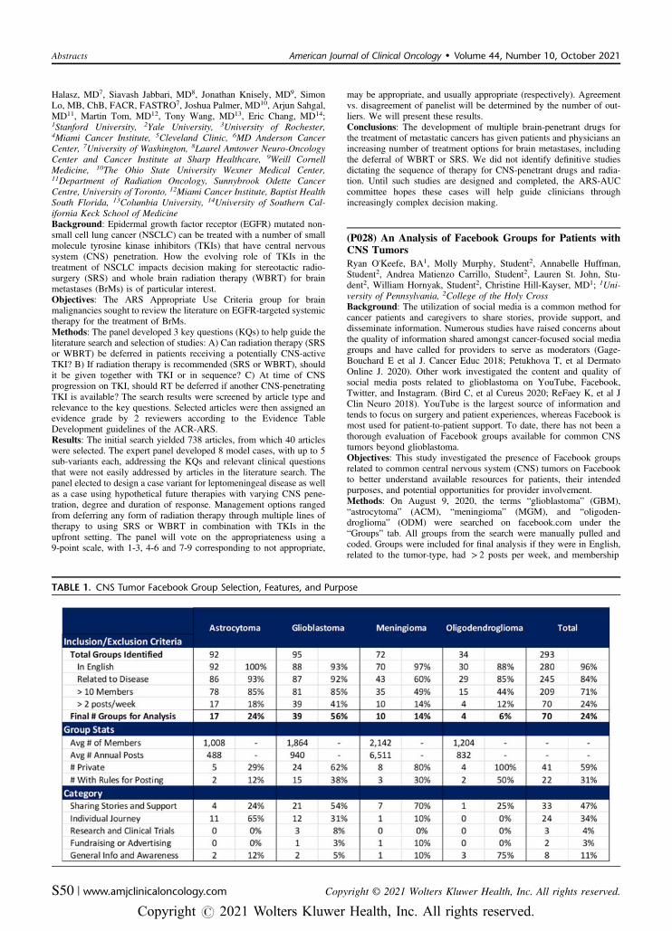
Halasz, MD7, Siavash Jabbari, MD8, Jonathan Knisely, MD9, SimonLo, MB, ChB, FACR, FASTRO7, Joshua Palmer, MD10, Arjun Sahgal,MD11, Martin Tom, MD12, Tony Wang, MD13, Eric Chang, MD14;1Stanford University, 2Yale University, 3University of Rochester,4Miami Cancer Institute, 5Cleveland Clinic, 6MD Anderson CancerCenter, 7University of Washington, 8Laurel Amtower Neuro-OncologyCenter and Cancer Institute at Sharp Healthcare, 9Weill CornellMedicine, 10The Ohio State University Wexner Medical Center,11Department of Radiation Oncology, Sunnybrook Odette CancerCentre, University of Toronto, 12Miami Cancer Institute, Baptist HealthSouth Florida, 13Columbia University, 14University of Southern Cal-ifornia Keck School of MedicineBackground: Epidermal growth factor receptor (EGFR) mutated non-small cell lung cancer (NSCLC) can be treated with a number of smallmolecule tyrosine kinase inhibitors (TKIs) that have central nervoussystem (CNS) penetration. How the evolving role of TKIs in thetreatment of NSCLC impacts decision making for stereotactic radio-surgery (SRS) and whole brain radiation therapy (WBRT) for brainmetastases (BrMs) is of particular interest.Objectives: The ARS Appropriate Use Criteria group for brainmalignancies sought to review the literature on EGFR-targeted systemictherapy for the treatment of BrMs.Methods: The panel developed 3 key questions (KQs) to help guide theliterature search and selection of studies: A) Can radiation therapy (SRSor WBRT) be deferred in patients receiving a potentially CNS-activeTKI? B) If radiation therapy is recommended (SRS or WBRT), shouldit be given together with TKI or in sequence? C) At time of CNSprogression on TKI, should RT be deferred if another CNS-penetratingTKI is available? The search results were screened by article type andrelevance to the key questions. Selected articles were then assigned anevidence grade by 2 reviewers according to the Evidence TableDevelopment guidelines of the ACR-ARS.Results: The initial search yielded 738 articles, from which 40 articleswere selected. The expert panel developed 8 model cases, with up to 5sub-variants each, addressing the KQs and relevant clinical questionsthat were not easily addressed by articles in the literature search. Thepanel elected to design a case variant for leptomeningeal disease as wellas a case using hypothetical future therapies with varying CNS pene-tration, degree and duration of response. Management options rangedfrom deferring any form of radiation therapy through multiple lines oftherapy to using SRS or WBRT in combination with TKIs in theupfront setting. The panel will vote on the appropriateness using a9-point scale, with 1-3, 4-6 and 7-9 corresponding to not appropriate,
may be appropriate, and usually appropriate (respectively). Agreementvs. disagreement of panelist will be determined by the number of out-liers. We will present these results.Conclusions: The development of multiple brain-penetrant drugs forthe treatment of metastatic cancers has given patients and physicians anincreasing number of treatment options for brain metastases, includingthe deferral of WBRT or SRS. We did not identify definitive studiesdictating the sequence of therapy for CNS-penetrant drugs and radia-tion. Until such studies are designed and completed, the ARS-AUCcommittee hopes these cases will help guide clinicians throughincreasingly complex decision making.
(P028) An Analysis of Facebook Groups for Patients withCNS TumorsRyan O’Keefe, BA1, Molly Murphy, Student2, Annabelle Huffman,Student2, Andrea Matienzo Carrillo, Student2, Lauren St. John, Stu-dent2, William Hornyak, Student2, Christine Hill-Kayser, MD1; 1Uni-versity of Pennsylvania, 2College of the Holy CrossBackground: The utilization of social media is a common method forcancer patients and caregivers to share stories, provide support, anddisseminate information. Numerous studies have raised concerns aboutthe quality of information shared amongst cancer-focused social mediagroups and have called for providers to serve as moderators (Gage-Bouchard E et al J. Cancer Educ 2018; Petukhova T, et al DermatoOnline J. 2020). Other work investigated the content and quality ofsocial media posts related to glioblastoma on YouTube, Facebook,Twitter, and Instagram. (Bird C, et al Cureus 2020; ReFaey K, et al JClin Neuro 2018). YouTube is the largest source of information andtends to focus on surgery and patient experiences, whereas Facebook ismost used for patient-to-patient support. To date, there has not been athorough evaluation of Facebook groups available for common CNStumors beyond glioblastoma.Objectives: This study investigated the presence of Facebook groupsrelated to common central nervous system (CNS) tumors on Facebookto better understand available resources for patients, their intendedpurposes, and potential opportunities for provider involvement.Methods: On August 9, 2020, the terms “glioblastoma” (GBM),“astrocytoma” (ACM), “meningioma” (MGM), and “oligoden-droglioma” (ODM) were searched on facebook.com under the“Groups” tab. All groups from the search were manually pulled andcoded. Groups were included for final analysis if they were in English,related to the tumor-type, had > 2 posts per week, and membership
TABLE 1. CNS Tumor Facebook Group Selection, Features, and Purpose
Abstracts American Journal of Clinical Oncology � Volume 44, Number 10, October 2021
S50 | www.amjclinicaloncology.com Copyright © 2021 Wolters Kluwer Health, Inc. All rights reserved.
Copyright r 2021 Wolters Kluwer Health, Inc. All rights reserved.

> 10 people. Data points of interest were extracted from each page’s“About” tab between August 10-22, 2020.Results: 292 Facebook groups were found using the search terms, 70met inclusion criteria (Table 1). GBM-related groups were most com-mon and MGM-related groups had the largest average membership sizewith the most average annual posts (Table 1). 41 (58.6%) were privategroups, and 22 (31.4%) had rules regarding posting content. Mostgroups were created for the purpose of sharing support and stories or todescribe an individual’s journey with their disease (Table 1). Astrocy-toma-related groups were predominantly created to describe an indi-vidual’s journey, with 9 of 11 being pediatric patients. Groups createdto share general information about the disease, to discuss research orclinical trials, or to fundraise were less common.Conclusions: MGM-related groups on FB had the most activity, pos-sibly due to its high prevalence amongst CNS tumors and longer lifeexpectancy. Most groups identified were not currently active. In linewith previous work, groups focused on individual journeys and supportwere most common. There were few public groups focused on sharinggeneral information or discussing research, potentially limiting theopportunities for providers to contribute. Future work should explorethe content of discussion in such groups, and the desire for, and effectsof, physician involvement.
(P029) Outcomes of Adults Treated with Brain Hypo-fractionated Stereotactic Radiosurgery in an EstablishedCNS Multidisciplinary Clinic for Radiation Oncology andNeurosurgeryWencesley Paez, MD1, Samantha Bowen, BA2, Rohi Gheewala, BS3,Brandon Lucke-Wold, MD, PhD4, Jeremy Ciporen, MD2, Timur Mitin,MD, PhD1; 1Department of Radiation Medicine, Oregon Health &Science University, 2Department of Neurological Surgery, OregonHealth & Science University, 3School of Medicine, Oregon Health &Science University, 4Department of Neurosurgery, University ofFloridaBackground: Our multidisciplinary central nervous system (CNS)clinic provides synchronous consultation and follow-up visits with aneurosurgeon and a radiation oncologist at a community hospital center.Hypofractionated stereotactic radiosurgery (hfSRS) is commonly usedfor management of primary and secondary malignant disease in thebrain. A significant challenge in the follow-up of patients treated withhfSRS to brain lesions is to distinguish pseudoprogression (PP) fromtumor recurrence (TR) for which treatment and prognosis are different.Objectives: To determine the rate of tumor control (TC), pseudoprog-ression (PP) and tumor recurrence (TR) among adult patients with brainprimary and secondary malignant disease treated at a multidisciplinaryCNS community hospital center clinic with hfSRS.Methods: We conducted an IRB-approved retrospective review oftreatment outcomes for consecutive patients treated with hfSRS forbrain tumors between June 2017 and December 2019. Post-treatment
imaging and/or histology when available were reviewed for assessmentof treatment response.Results: Forty-six brain lesions in 18 consecutive patients were treatedwith hfSRS over a period of 30 months. The rate of tumor control withno evidence of progression or pseudoprogression was 67% (31 out of46 lesions). Among the remaining 15 lesions, TR was determined ineight lesions for an overall rate of TR of 17% (8 out of 46 lesions). Sixlesions (13%) were determined to represent PP. Table 1 summarizesmanagement and outcomes among patients with progressing lesionsafter hfSRS.Conclusions: Adult patients with brain primary and secondary malig-nant disease evaluated and treated in our multidisciplinary CNS clinichave a high rate of disease control in line with previously publishedresults from large academic institutions and multi-institutional clinicaltrials. The integrated evaluation and management of radiographic pro-gression after hfSRS allows for a nuanced multidisciplinary inter-pretation of radiographic and clinical findings. Thereby, reducingunnecessary clinical visits, tests, procedures and leading to improvedclinical care through prompt systemic, surgical and radiation treatmentmodalities that minimizes re-treatment of PP and avoids undertreatmentof TR.
(P030) American Radium Society's Appropriate Use Cri-teria on Postoperative Management of Lower GradeGliomasMartin Tom, MD1, Michael Milano, MD, PhD2, Scott Soltys, MD3,Arjun Sahgal, MD4, Jonathan Knisely, MD5, Veronica Chiang, MD6,Seema Nagpal, MD3, Simon Lo, MB, ChB, FACR, FASTRO7, SamuelChao, MD8, Siavash Jabbari, MD9, Tony Wang, MD10, Joshua Palmer,MD11, Melanie Gephart, MD, MAS3, Amit Garg, MD12, ManmeetAhluwalia, MD, MBA13, Lia Halasz, MD7, Eric Chang, MD14; 1MiamiCancer Institute, Baptist Health South Florida, 2University of Roches-ter, 3Stanford University, 4Department of Radiation Oncology, Sunny-brook Odette Cancer Centre, University of Toronto, 5Weill CornellMedicine, 6Yale University, 7University of Washington, 8ClevelandClinic, 9Laurel Amtower Neuro-Oncology Center and Cancer Instituteat Sharp Healthcare, 10Columbia University, 11The Ohio State Uni-versity Wexner Medical Center, 12MD Anderson Cancer Center,13Miami Cancer Institute, 14University of Southern California KeckSchool of MedicineBackground: “Lower grade gliomas” refer to WHO grade 2 and 3gliomas. Their optimal postoperative management remains con-troversial, especially in light of contemporary prognostic and predictivemolecular characteristics which were not available during the design ofearlier clinical trials.Objectives: The American Radium Society (ARS) Appropriate UseCriteria Brain Malignancy Committee sought to develop consensusguidelines for the postoperative management of lower grade gliomas.Methods: The committee developed 4 key questions regarding: 1)Timing of postoperative therapy (i.e. adjuvant vs. salvage approaches);
TABLE 1. Management and Outcomes Among Patients (n=8) With Clinical Evidence of Progression After Brain hfSRS
Brain lesions (n= 15) with radiographic evidence of progression after brain hfSRS. *Avastin (A), Steroids (S), Redo-craniotomy (C), Re-irradiation (R).
American Journal of Clinical Oncology � Volume 44, Number 10, October 2021 Abstracts
Copyright © 2021 Wolters Kluwer Health, Inc. All rights reserved. www.amjclinicaloncology.com | S51
Copyright r 2021 Wolters Kluwer Health, Inc. All rights reserved.

2) Monotherapy vs. combined modality therapy (i.e. postoperativeradiotherapy [RT] vs. chemotherapy vs. chemoradiotherapy); 3) Typeof chemotherapy used in conjunction with radiotherapy (i.e. procarba-zine, lomustine, and vincristine [PCV] vs. temozolomide [TMZ]); and4) Radiotherapy dose. The panel systematically reviewed the literatureaccording to the Preferred Reporting Items for Systematic Reviews andMeta-Analyses guidelines, and voted accordingly on the appropriate-ness of various management options for representative case variants.Results: The search identified 6,244 studies, of which 38 were includedafter screening and in-depth review. The committee designed 6 casevariants, each consisting of 1-2 subvariants, with emphasis on patientage, patient performance, patient symptomatology, tumor size, tumorlocation, tumor grade, tumor molecular classification, and extent ofresection. Management options included initial observation, adjuvantmonotherapy (RT, PCV, or TMZ), adjuvant combined modality therapy(RT with PCV or RT with various combinations of TMZ), various RTdoses, and proton therapy. After two rounds of voting, disagreementremained for some treatment options within the case variants. Thecommittee recommended a third round of voting for these scenarios.Conclusions: Multiple postoperative management options are available forlower grade gliomas, which are heavily influenced by molecular classi-fication and other clinical characteristics. Until recently completed andongoing clinical trials using updated molecular classification are published,the ARS Brain Malignancy Committee endeavors for this Appropriate UseCriteria document to aid decision making for clinicians. Final voting isunderway and will be followed shortly by the consensus guideline.
(P031) Patterns of Care and Treatment Disparities in theManagement of Benign Central Nervous System MeningiomaFelicia Lew, DVM1, Christopher Weil, MD2, Calvin Rock, MD2, RyanKraus, MD2, Donald Cannon, MD2, Randy Jensen, MD PhD2, LindsayBurt, MD2; 1Elson S Floyd College of Medicine, 2University of UtahHuntsman Cancer InstituteBackground: Observation/active surveillance, surgery and radio-therapy (RT) are all standard of care treatment options in the man-agement of benign central nervous system (CNS) meningioma.Objectives: In this analysis, we sought to describe the patterns of carein the management of benign CNS meningioma and identify temporaland sociodemographic trends in the utilization of surgery and radio-therapy, including the use of stereotactic radiosurgery (SRS) comparedwith external beam radiotherapy (EBRT).Methods: We queried the National Cancer Database (NCDB) to identifyall patients with benign meningioma of the CNS diagnosed from 2004 to2017. Clinical characteristics, demographic information and treatmentapproaches were analyzed using standard T and χ2 tests. Predictors ofreceipt of any treatment and radiotherapy technique were identified withunivariable and multivariable logistic regression modeling.Results: We identified 252,258 patients diagnosed with benign CNSmeningioma between 2004 and 2017, of which 133,008 (53%) receivedno treatment and 119,250 (47%) received either surgery, RT or both. Ofthe patients who received treatment, 98,611 (83%) had surgery only,14,556 (12%) received RT and 6,077 (5%) had surgery and RT. Of thepatients who received RT, 61% received single- or multi-fraction SRSand 39% received fractioned EBRT. On multivariable analysis, patientswere more likely to be observed if they were diagnosed from 2009-2012(Odds ratio [OR] 0.66, 95% Confidence Interval 0.65-0.68) and 2013-2017 (OR 0.57, 0.55-0.58), had no insurance (OR 0.60, 0.57-0.63),Medicaid (OR 0.71, 0.68-0.74), Medicare (OR 0.50, 0.48-0.51), or aCharlson co-morbidity score of 3+ (0.75, 0.71-0.79). Those more likelyto receive treatment were Asian (OR 1.2, 1.1-1.3) or Hispanic-Whiteethnicity (1.1, 1.03-1.1), from the geographic West (OR 1.2, 1.2-1.3), ortreated at an academic/research institution (OR 3.4, 3.24-3.56). Blackpatients were more likely to receive RT than Whites (OR 1.2, 1.1-1.2)and less likely to receive surgery (OR 0.96, 0.93-0.98). Blacks andHispanic-Whites were more likely to receive EBRT than SRS comparedwith Non-Hispanic Whites (OR 1.3, 1.2-1.4, and OR 1.5, 1.3-1.7,respectively). Black patients were also less likely to receive both sur-gery and RT compared with Whites (OR 0.71, 0.68-0.73), whilepatients treated at academic/research institutions compared with com-munity hospitals were more likely to receive both (OR 5.8, 4.9-6.4).
Conclusions: Active surveillance as a treatment strategy for CNSmeningiomas has been increasing over the past decade, however thereare noticeable disparities in which patients receive treatment. At riskpopulations, including the uninsured and minorities, are less likely toreceive treatment and less likely to receive less toxic treatments likeSRS. Further evaluation is warranted to address the underlying cause ofthe inequities.
(P032) Impact of Prior Y90 Dosimetry on Toxicity andOutcomes Following Stereotactic Body Radiation Therapyfor Hepatocellular CarcinomaShauna Campbell, DO1, Aditya Juloori, MD2, Timothy Smile, MD1,Danielle LaHurd, PhD1, Naichang Yu, PhD1, Neil Woody, MD1, KevinStephans, MD1; 1Cleveland Clinic, 2University of ChicagoBackground: Stereotactic body radiation therapy (SBRT) is the onlynon-invasive local therapy for the treatment of hepatocellular carcinoma(HCC) and is often utilized following transarterial embolization. Thereis a paucity of data reporting safety and outcomes of patients (pts)receiving SBRT after yttrium-90 (Y90).Objectives: We sought to evaluate the impact of Y90 and SBRTcomposite plan dosimetry on toxicity and outcomes of HCC pts.Methods: An IRB-approved registry of 260 liver SBRT pts was used.Pts included in analysis received Y90 before SBRT and had post-Y90PET imaging (measuring Bremsstrahlung component of Y90). Volumeof ablated liver was estimated using the sharp edge of PET gradient andthe SBRT volume receiving at least 30 Gy (V30). Characteristicsbetween groups were compared using χ2 and independent t-test. Clin-ical, imaging, and laboratory follow up are reported.Results: Twenty eights pts with median follow up of 14 m (4.6-49.6)were included. Pts received 1 (N= 15), 2 (N= 10), or 3 (N= 3) priorY90 treatments, to a median volume of 286 cm3 (21-1512) a median 4.4m (1.1-82.9) before SBRT. SBRT dose ranged from 24-50 Gy in 3-5fractions with a median PTV of 117 cm3 (26-857). Y90 and V30overlap ranged from 0-577 cm3 with a median of 80 cm3. Treatmentintent was definitive in 23 pts, to downstage within transplant criteria in4 pts, and as a bridge to transplant in 1 pt. Baseline Child-Pugh (CP)score was A, B, and C in 23, 3, 2 pts, respectively. At a median 9 m(1.5-46.4) after SBRT local control was 93%. Liver, nodal, and distantfailure had an incidence of 11%, 3.6%, and 11%, respectively. Changein CP score was analyzed at 1-2 m and 3 m post SBRT. Twenty sixpatients had at least 1 calculable time point. Change in CP class from Ato B was seen in 6 pts (26%), of which two recovered back to CP A by 3m. The 3 CP B pts did not change class, and the 2 CP C pts receivedsuccessful liver transplants 2-3 m after SBRT downstaging. Factorsassociated with an increase in CP score within 3 m of SBRT includeSBRT mean liver dose (P= 0.001), SBRT V30 (P= 0.049), and<1200 cm3 of un-ablated liver (P= 0.003). Volume of liver treated byY90 and time between Y90 and SBRT was not associated with anincrease in CP score within 3 m of SBRT. Non-hematologic toxicityinclude 2 acute biliary stricture (grade 2&3), 1 late biliary stricture(grade 2), 1 late chest wall pain (grade 1), and 1 late grade 2 radio-graphic myonecrosis. One CP A pt died 4 weeks after SBRT of car-diopulmonary comorbidities.Conclusions: Time between Y90 and SBRT, volume of liver exposedto prior Y90, and overlap between V30 and Y90 volumes do not appearto adversely impact CP score within 3 m of SBRT. Dosimetric prop-erties including mean liver dose, V30, and un-ablated liver volume<1200 cm3 are associated with an increase in CP score within 3 m ofSBRT. Pts with HCC previously treated with Y90 appear to tolerateSBRT well with no clinically significant change in CP score, minimaltoxicity, and excellent local control.
(P033) Causes of Death Among Unresectable Non-meta-static Pancreas Cancer Patients Receiving MR-guidedSABRMichael Chuong, MD1, Roberto Herrera, BS1, Matthew Hall, MD,MBA1, Rupesh Kotecha, MD1, Antonio Ucar, MD2, Fernando DeZarraga, MD1, Santiago Aparo, MD1, Sarah Joseph, MD1, Kathryn
Abstracts American Journal of Clinical Oncology � Volume 44, Number 10, October 2021
S52 | www.amjclinicaloncology.com Copyright © 2021 Wolters Kluwer Health, Inc. All rights reserved.
Copyright r 2021 Wolters Kluwer Health, Inc. All rights reserved.
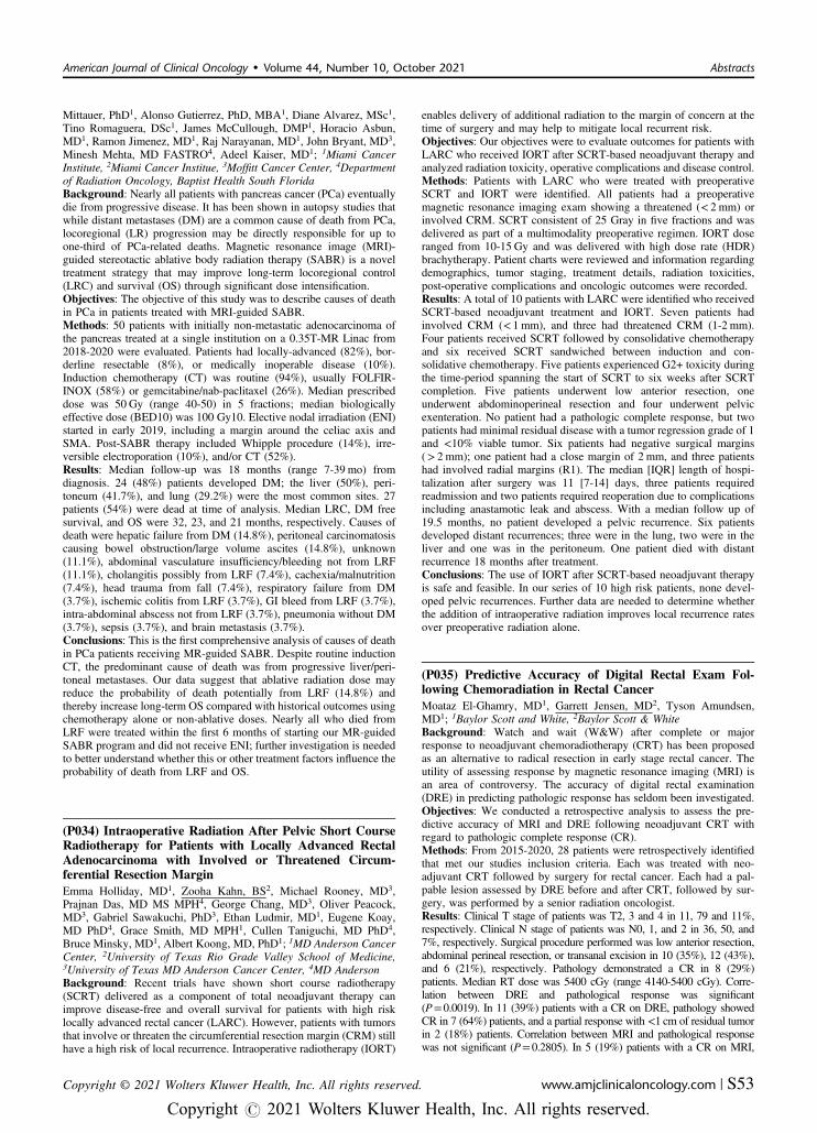
Mittauer, PhD1, Alonso Gutierrez, PhD, MBA1, Diane Alvarez, MSc1,Tino Romaguera, DSc1, James McCullough, DMP1, Horacio Asbun,MD1, Ramon Jimenez, MD1, Raj Narayanan, MD1, John Bryant, MD3,Minesh Mehta, MD FASTRO4, Adeel Kaiser, MD1; 1Miami CancerInstitute, 2Miami Cancer Institue, 3Moffitt Cancer Center, 4Departmentof Radiation Oncology, Baptist Health South FloridaBackground: Nearly all patients with pancreas cancer (PCa) eventuallydie from progressive disease. It has been shown in autopsy studies thatwhile distant metastases (DM) are a common cause of death from PCa,locoregional (LR) progression may be directly responsible for up toone-third of PCa-related deaths. Magnetic resonance image (MRI)-guided stereotactic ablative body radiation therapy (SABR) is a noveltreatment strategy that may improve long-term locoregional control(LRC) and survival (OS) through significant dose intensification.Objectives: The objective of this study was to describe causes of deathin PCa in patients treated with MRI-guided SABR.Methods: 50 patients with initially non-metastatic adenocarcinoma ofthe pancreas treated at a single institution on a 0.35T-MR Linac from2018-2020 were evaluated. Patients had locally-advanced (82%), bor-derline resectable (8%), or medically inoperable disease (10%).Induction chemotherapy (CT) was routine (94%), usually FOLFIR-INOX (58%) or gemcitabine/nab-paclitaxel (26%). Median prescribeddose was 50 Gy (range 40-50) in 5 fractions; median biologicallyeffective dose (BED10) was 100 Gy10. Elective nodal irradiation (ENI)started in early 2019, including a margin around the celiac axis andSMA. Post-SABR therapy included Whipple procedure (14%), irre-versible electroporation (10%), and/or CT (52%).Results: Median follow-up was 18 months (range 7-39 mo) fromdiagnosis. 24 (48%) patients developed DM; the liver (50%), peri-toneum (41.7%), and lung (29.2%) were the most common sites. 27patients (54%) were dead at time of analysis. Median LRC, DM freesurvival, and OS were 32, 23, and 21 months, respectively. Causes ofdeath were hepatic failure from DM (14.8%), peritoneal carcinomatosiscausing bowel obstruction/large volume ascites (14.8%), unknown(11.1%), abdominal vasculature insufficiency/bleeding not from LRF(11.1%), cholangitis possibly from LRF (7.4%), cachexia/malnutrition(7.4%), head trauma from fall (7.4%), respiratory failure from DM(3.7%), ischemic colitis from LRF (3.7%), GI bleed from LRF (3.7%),intra-abdominal abscess not from LRF (3.7%), pneumonia without DM(3.7%), sepsis (3.7%), and brain metastasis (3.7%).Conclusions: This is the first comprehensive analysis of causes of deathin PCa patients receiving MR-guided SABR. Despite routine inductionCT, the predominant cause of death was from progressive liver/peri-toneal metastases. Our data suggest that ablative radiation dose mayreduce the probability of death potentially from LRF (14.8%) andthereby increase long-term OS compared with historical outcomes usingchemotherapy alone or non-ablative doses. Nearly all who died fromLRF were treated within the first 6 months of starting our MR-guidedSABR program and did not receive ENI; further investigation is neededto better understand whether this or other treatment factors influence theprobability of death from LRF and OS.
(P034) Intraoperative Radiation After Pelvic Short CourseRadiotherapy for Patients with Locally Advanced RectalAdenocarcinoma with Involved or Threatened Circum-ferential Resection MarginEmma Holliday, MD1, Zooha Kahn, BS2, Michael Rooney, MD3,Prajnan Das, MD MS MPH4, George Chang, MD3, Oliver Peacock,MD3, Gabriel Sawakuchi, PhD3, Ethan Ludmir, MD1, Eugene Koay,MD PhD4, Grace Smith, MD MPH1, Cullen Taniguchi, MD PhD4,Bruce Minsky, MD1, Albert Koong, MD, PhD1; 1MD Anderson CancerCenter, 2University of Texas Rio Grade Valley School of Medicine,3University of Texas MD Anderson Cancer Center, 4MD AndersonBackground: Recent trials have shown short course radiotherapy(SCRT) delivered as a component of total neoadjuvant therapy canimprove disease-free and overall survival for patients with high risklocally advanced rectal cancer (LARC). However, patients with tumorsthat involve or threaten the circumferential resection margin (CRM) stillhave a high risk of local recurrence. Intraoperative radiotherapy (IORT)
enables delivery of additional radiation to the margin of concern at thetime of surgery and may help to mitigate local recurrent risk.Objectives: Our objectives were to evaluate outcomes for patients withLARC who received IORT after SCRT-based neoadjuvant therapy andanalyzed radiation toxicity, operative complications and disease control.Methods: Patients with LARC who were treated with preoperativeSCRT and IORT were identified. All patients had a preoperativemagnetic resonance imaging exam showing a threatened (< 2 mm) orinvolved CRM. SCRT consistent of 25 Gray in five fractions and wasdelivered as part of a multimodality preoperative regimen. IORT doseranged from 10-15 Gy and was delivered with high dose rate (HDR)brachytherapy. Patient charts were reviewed and information regardingdemographics, tumor staging, treatment details, radiation toxicities,post-operative complications and oncologic outcomes were recorded.Results: A total of 10 patients with LARC were identified who receivedSCRT-based neoadjuvant treatment and IORT. Seven patients hadinvolved CRM (< 1 mm), and three had threatened CRM (1-2 mm).Four patients received SCRT followed by consolidative chemotherapyand six received SCRT sandwiched between induction and con-solidative chemotherapy. Five patients experienced G2+ toxicity duringthe time-period spanning the start of SCRT to six weeks after SCRTcompletion. Five patients underwent low anterior resection, oneunderwent abdominoperineal resection and four underwent pelvicexenteration. No patient had a pathologic complete response, but twopatients had minimal residual disease with a tumor regression grade of 1and <10% viable tumor. Six patients had negative surgical margins(> 2 mm); one patient had a close margin of 2 mm, and three patientshad involved radial margins (R1). The median [IQR] length of hospi-talization after surgery was 11 [7-14] days, three patients requiredreadmission and two patients required reoperation due to complicationsincluding anastamotic leak and abscess. With a median follow up of19.5 months, no patient developed a pelvic recurrence. Six patientsdeveloped distant recurrences; three were in the lung, two were in theliver and one was in the peritoneum. One patient died with distantrecurrence 18 months after treatment.Conclusions: The use of IORT after SCRT-based neoadjuvant therapyis safe and feasible. In our series of 10 high risk patients, none devel-oped pelvic recurrences. Further data are needed to determine whetherthe addition of intraoperative radiation improves local recurrence ratesover preoperative radiation alone.
(P035) Predictive Accuracy of Digital Rectal Exam Fol-lowing Chemoradiation in Rectal CancerMoataz El-Ghamry, MD1, Garrett Jensen, MD2, Tyson Amundsen,MD1; 1Baylor Scott and White, 2Baylor Scott & WhiteBackground: Watch and wait (W&W) after complete or majorresponse to neoadjuvant chemoradiotherapy (CRT) has been proposedas an alternative to radical resection in early stage rectal cancer. Theutility of assessing response by magnetic resonance imaging (MRI) isan area of controversy. The accuracy of digital rectal examination(DRE) in predicting pathologic response has seldom been investigated.Objectives: We conducted a retrospective analysis to assess the pre-dictive accuracy of MRI and DRE following neoadjuvant CRT withregard to pathologic complete response (CR).Methods: From 2015-2020, 28 patients were retrospectively identifiedthat met our studies inclusion criteria. Each was treated with neo-adjuvant CRT followed by surgery for rectal cancer. Each had a pal-pable lesion assessed by DRE before and after CRT, followed by sur-gery, was performed by a senior radiation oncologist.Results: Clinical T stage of patients was T2, 3 and 4 in 11, 79 and 11%,respectively. Clinical N stage of patients was N0, 1, and 2 in 36, 50, and7%, respectively. Surgical procedure performed was low anterior resection,abdominal perineal resection, or transanal excision in 10 (35%), 12 (43%),and 6 (21%), respectively. Pathology demonstrated a CR in 8 (29%)patients. Median RT dose was 5400 cGy (range 4140-5400 cGy). Corre-lation between DRE and pathological response was significant(P=0.0019). In 11 (39%) patients with a CR on DRE, pathology showedCR in 7 (64%) patients, and a partial response with <1 cm of residual tumorin 2 (18%) patients. Correlation between MRI and pathological responsewas not significant (P=0.2805). In 5 (19%) patients with a CR on MRI,
American Journal of Clinical Oncology � Volume 44, Number 10, October 2021 Abstracts
Copyright © 2021 Wolters Kluwer Health, Inc. All rights reserved. www.amjclinicaloncology.com | S53
Copyright r 2021 Wolters Kluwer Health, Inc. All rights reserved.

pathology showed CR in 3 (60%) patients. Of 4 patients with a CR on bothMRI and DRE, 3 (75%) had a pathological CR.Conclusions: A DRE performed by an experienced physician beforeand after CRT has higher accuracy than MRI in detecting CR. DRE, inaddition to MRI, may help better identify patients with rectal cancerwho are candidates for a W&W approach.
(P036) Magnetic Resonance-guided Adaptive RadiationTherapy for Treatment of Locally-recurrent PancreaticAdenocarcinomaKylie Kang, MD1, John Lattin, BS2, Joshua Schiff, MD3, Olga Green,PhD3, Alex Price, MS3, Deshan Yang, PhD3, William Hawkins, MD3,Ryan Fields, MD3, Pamela Samson, MD, MPHS1, Shahed Badiyan,MD3, Lauren Henke, MD, MSCI4, Hyun Kim, MD3; 1Department ofRadiation Oncology, Washington University School of Medicine in St.Louis, 2St. Louis University School of Medicine, 3Washington Uni-versity School of Medicine, [email protected]: There are limited treatment options for locally recurrentpancreatic adenocarcinoma, especially after prior resection or radiationtherapy. The use of magnetic resonance (MR)-guided adaptive radiationtherapy (MRgART) has been reported for the treatment of inoperablepancreatic adenocarcinoma, but there is a paucity of data on its appli-cation in locally recurrent cases.Objectives: We aim to report on our institutional experience withMRgART for treatment of locally-recurrent pancreatic adenocarcinoma.Methods: Eighteen patients with locally recurrent pancreatic adeno-carcinoma were treated at our institution between 2015-2019 withMRgART. Demographic and treatment-related characteristics werecollected from the institutional electronic medical record and treatmentplanning system. Kaplan-Meier with log-rank analysis was conducted,with statistical significance defined as P< .05.Results: Median time from diagnosis to first local failure (LF) was17.8 months (range: 6.4 - 56.6mo). Six patients had definitive fractionatedchemoradiation before their local recurrence to a median dose of 52.2 Gy(range: 45 - 55 Gy). Salvage MRgART was delivered to a median of50 Gy (BED10=100 Gy; range: 50 - 67.5 Gy) for radiation-naïve patientsand a median of 32.5 Gy (BED10=53.6 Gy; range: 25 - 50 Gy) for thosewho had received prior radiation; two patients did not receive salvageMRgART at first LF (had salvage chemotherapy alone) but underwentMRgART at subsequent progression. Aside from one salvage regimen of67.5 Gy over 15 fractions, all patients received salvage MRgART overfive fractions. Median planning target volume (PTV) was 132.6 cc (range:16.2 - 292.3 cc). The most common organs at risk requiring adaptationwere small bowel and stomach, with 67% of cases requiring adaptation forevery fraction. Median overall survival from time of diagnosis was34.7 months; median survival after salvage MRgART was 8.7 months.One- and three-year local control rates were both 66%, and one- and three-year overall survival rates were 78% and 18%, respectively. There was noassociation between local control post-MRgART or overall survival andPTV, BED10, or receipt of salvage chemotherapy.Conclusions: Locally recurrent pancreatic adenocarcinoma can betreated with MRgART with acceptable local control rates, even in casesof prior definitive chemoradiation. MRgART was utilized for everyfraction in the majority of cases and allowed for plan adjustments tomeet luminal OAR constraints.
(P037) Stereotactic MRI-guided Adaptive Radiation Ther-apy for Non-metastatic Pancreatic Cancer; Outcomes andToxicity Analysis for Patients Treated in an UnderservedUrban CenterCelina Kirsch, PAC1, Eric Schaff, MD2, Parag Parikh, MD2, BenjaminMovsas, MD3, Farzan Siddiqui, MD, PhD, CPE3, David Kwon, MD2,Pin Li, PhD2, Gazala Khan, MD2, Rupen Shah, MD2; 1Henry FordCancer Institute, 2Henry Ford Health System, 3Department of Radia-tion Oncology, Henry Ford Cancer InstituteBackground: Stereotactic MRI-guided Adaptive Radiation Therapy(SMART) is an emerging technology for treatment of pancreatic cancerpatients. Initial results show favorable survival and toxicity. However,
data is still sparse overall, and especially in underserved patient pop-ulations. The purpose of this study is to review SMART outcomes atour underserved urban academic cancer.Objectives: Stereotactic MRI-guided Adaptive Radiation Therapy(SMART) is an emerging technology for treatment of pancreatic cancerpatients. Initial results show favorable survival and toxicity. However,data is still sparse overall, and especially in underserved patient pop-ulations. The purpose of this study is to review SMART outcomes atour underserved urban academic cancer.Methods: In this IRB approved retrospective chart review we reviewed98 patients with non-metastatic pancreatic cancer, who completedSMART between November 2018-January 2021. All 98 patients weretreated with 50 Gy in 5 daily fractions with adaptive technique as deemedappropriate by treating radiation oncologist. The primary endpoints wereoverall survival (OS), progression free survival (PFS), and both acute andlate grade 3+ GI toxicity. OS, PFS, locoregional control and distantcontrol were estimated by Kaplan-Meier method and compared using log-rank test. The effect of clinical features on OS was assessed using uni-variate and multivariate Cox proportional hazard models. OS and PFSwere calculated from completion of radiation. Grade 3+ GI toxicityprobably or definitively related to radiation was recorded. All incidencesof GI bleeding, regardless of attribution, were also recorded.Results: Median follow up was 20.9 months from time of diagnosis and14 months from radiation. 21 (21%) patients were borderline resectable,42 (43%) locally advanced, 22 (22%) medically inoperable and 13(13%) resectable. Neoadjuvant chemotherapy was given to 86 (88%)patients with a median of 3.5 months of chemotherapy (range 1-12),leaving 11 (12%) patients who did not have systemic chemotherapy.Median overall survival from radiation for the whole group was15.7 months, and 1-year OS was 58%. There was a statistically sig-nificant worsening of overall survival from diagnosis between ECOG 2+and ECOG 0/1 patients (HR 1.94, 1.05-3.57). 27 (27%) patients went onto have surgical resection with 23 (82%) having R0 resection, and 3(11%) have an R1 resection. Improved OS was seen in patients withsurgical resection (HR 0.06, 0.02-0.23). Acute grade 3+ GI toxicity fromradiation was seen in 4 (4%) patients and late toxicity from radiation wasseen in 6 (6%) patients. GI bleeding was seen in 16(16%) patients, 10(62%) of which were on anticoagulation at the time of GI bleed and 5(19%) of which had surgery. Portal vein complications occurred with 7(7%) having portal vein thrombosis and 6 (6%) portal vein stenosis.Conclusions: SMART showed durable responses in pancreatic cancerpatients with an acceptable toxicity profile. Attention needs to be paid tothe moderate incident of GI bleeding, however further work is necessaryto determine if bleeding was due to radiation, surgery, or disease pro-gression. Surgical resection as well as performance status of ECOG 0-1were associated with improved overall survival. Further follow up willbe necessary to determine further durability of treatment response andlong-term survival in these patients.
(P038) Outcomes of MR-guided Stereotactic Body Radio-therapy (SBRT) or yttrium-90 Transarterial Radio-embolization for Hepatocellular Carcinoma Treated at anUrban Liver Transplant CenterEvan Liang, MD1, Garret Greer, MD1, Charlotte Burmeister, MS1, PeterMassa, MD1, Jadranka Dragovic, MD1, Parag Parikh, MD1; 1HenryFord Health SystemBackground: There are overlapping indications for both stereotactic bodyradiotherapy (SBRT) and yttrium-90 (Y90) trans-arterial radioembolization aslocoregional treatments for hepatocellular cancer, though most centers pref-erentially use one modality over the other. MR-guided radiation allows botheffective on-table localization and integrated motion management as com-pared with many traditional linear accelerators, allowing SBRT to be donemore easily. Y90 radioembolization has been a well-established modality todeliver highly conformal dose due to the localization of the microspheres tothe vascular supply of a tumor. We looked at patient characteristics andtreatment outcomes for patients receiving MR-guided SBRT or Y90 at anurban transplant center.Objectives: To compare patient characteristics and treatment outcomesof MR-guided SBRT with Y90 transarterial radioembolization in a livertransplant center.
Abstracts American Journal of Clinical Oncology � Volume 44, Number 10, October 2021
S54 | www.amjclinicaloncology.com Copyright © 2021 Wolters Kluwer Health, Inc. All rights reserved.
Copyright r 2021 Wolters Kluwer Health, Inc. All rights reserved.

Methods: This retrospective single-institution study analyzed patientswith HCC treated with SBRT or Y90 from August 2017 to September2020. To select a patient population eligible for either treatmentmodality, any Y90 procedures for lesions > 10 cm or for treatmentvolumes > 1000 cc were omitted from the cohort. A total of 239patients were included in the analysis, receiving a total of 98 courses ofSBRT and 187 courses of Y90 treatment. Local control (LC), freedomfrom liver progression (FFLP), and overall survival (OS) rates weremeasured from treatment completion date to death date or last follow-up. All outcomes were censored at time of loss to follow-up; LC andFFLP were censored at time of liver transplant if applicable. Coxregression models were used for survival, with significant factors on theunivariate analysis further analyzed with a multivariate model.Results: Median time to follow-up was 11 months (0-44 mo). Themean size of lesions treated with SBRT were smaller than thosetreated with Y90 (2.7 cm vs 4.3 cm, P< 0.01). The groups of patientsdiffered in liver disease characteristics, with SBRT patients havingfewer Child-Pugh A disease (62% vs 80%, P< 0.01), more havingreceived locoregional treatments to the liver in the past (81% v 35%,P< 0.01), and more disease in previously treated liver (57% vs 25%,P< 0.01). Dose of radiation for SBRT was 45-50 Gy administered in5 fractions; dose of Y90 radiation to tumor was prescribed to amedian of 235.2 Gy (range 55.8-512.3 Gy). There was a higher rate ofone year LC in the SBRT cohort (77% vs 57%, P< 0.01), whilemedian FFLP (9 mo vs 8 mo, P=NS) and median OS were notsignificantly different (24 mo vs 21 mo, P=NS). Multivariate anal-ysis revealed size of largest lesion (P< 0.01) was correlated withdecreased local control; a 1 cm increase in tumor size was associatedwith a 25% increased risk of local failure. Subsequent transplant(P< 0.01) was the remaining significant factor. Treatment modalitydid not remain an independent predictor of LC. Predictors of OS inmultivariate analysis included age (P= 0.01), prior liver treatments(HR 2.86, P< 0.01), size of largest lesion (P< 0.01), Child-Pughstage (P< 0.01), portal vein thrombosis (HR 1.6, P= 0.04), andsubsequent liver transplant (HR 0.08, P< 0.01).Conclusions: These findings support the effectiveness of both MR-guided SBRT and Y90 transarterial radioembolization in locoregionalmanagement of HCC at a single institution despite clear differences inthe patient cohorts. Though survival outcomes were comparable, localcontrol differences favored the cohort treated by SBRT, in large partdue to differences in tumor size. This data supports further investigationin a randomized study between SBRT and Y90.
(P039) Induction Chemotherapy for Locally AdvancedEsophageal Cancer: Can Additional PET/CT ParametersInfluence Patient Selection?Jordan McDonald, BS1, Rutika Mehta, MD, MPH2, Anupam Rishi,MD3, Austin Sim, MD, JD3, Kutjim Latifi, PhD3, Jessica Frakes, MD3,Jose Pimiento, MD4, Jacques Fontaine, MD4, Sarah Hoffe, MD3; 1USF
Health Morsani College of Medicine, 2Department of Medical Oncol-ogy, H. Lee Moffitt Cancer Center & Research Institute, 3Department ofRadiation Oncology, H. Lee Moffitt Cancer Center & Research Insti-tute, 4Department of Surgical Oncology, H. Lee Moffitt Cancer Centerand Research InstituteBackground: In the concurrent chemoradiation (CRT) setting, max-imum standard uptake value (SUVmax) on PET/CT has been lessprognostically predictive than other metabolic parameters. The CALGB80803 trial evaluated SUVmax response as a surrogate marker to guidetreatment decisions in the induction chemotherapy (IC) setting. As theSUVmax only measures the most metabolically active voxels of tumor,it overlooks volumetric change.Objectives: To determine if integrating metabolic tumor volume(MTV) and Total Lesion Glycolysis (TLG) into post-IC assessmentbetter predict treatment response before concurrent chemoradiation(CRT).Methods: We identified sequential clinical stage III and IV (by virtue ofnon-regional adenopathy) EC patients treated with Folinic acid, Fluo-rouracil, and Oxaliplatin (FOLFOX) IC with pre and post IC PET/CTperformed at our institution before CRT to 50.4 Gy. SUVmax wasevaluated on these scans to assess response. As per the CALBG trial,those with SUVmax decline of ≥ 35% in the primary tumor wereclassified as responders while those <35% were non-responders.Analysis of initial and post IC scans manually imported into Miradasoftware was performed to identify additional metabolic parameters atdifferent thresholds including the TLG and MTV.Results: Between 2017 and 2019, we identified 15 patients, 11 males(73.3%) and 4 females (26.7%), treated with induction FOLFOX(median age 67 and median initial stage III). The majority of tumorswere adenocarcinomas (80%, n= 12) and in the distal esophagus/gastroesophageal junction (73.3%, n= 11). Responders showed thegreatest difference in TLG compared with MTV at all thresholds(21.7% difference in median TLG reduction at variable, 18.8% at 2.5,and 24.5% at 4.0, compared with 27% difference in median MTVreduction at variable, 22.9% at 2.5, and 24.7% at 4.0) , with all thoseinitial TLG > 260 exhibiting response. The median reduction inSUVmax was 42.0% (Range: -7.1-92.5%, Interquartile Range: 28.2-73.2%). With a median follow up of 2.5 years (range 1.97-3.96 y) 10(66.7%) patients remain alive at this time with a crude median sur-vival of 2.42 (range 1.97-3.96) years, while 5 (33.3%) patients aredeceased with a crude median survival of 1.60 (range 0.59-2.45)years from diagnosis. 8 (53.3%) patients have remained free of pro-gression, recurrence, or metastasis and one (6.7%) patient experi-enced recurrence but was successfully treated. Survivors had amedian TLG reduction of 98.3% at variable threshold, 90.1% at 2.5,and 96.6% at 4.0.Conclusions: TLG parameters may be used for additional prog-nostic value in the locally advanced EC setting to determine extentof tumor response to IC. Further prospective study of additionalmetabolic parameters on pre and post IC imaging is warranted(Figs. 1 and 2).
FIGURE 1. Larger change in median pre and post IC TLG values among responders vs. non-responders at all thresholds.
American Journal of Clinical Oncology � Volume 44, Number 10, October 2021 Abstracts
Copyright © 2021 Wolters Kluwer Health, Inc. All rights reserved. www.amjclinicaloncology.com | S55
Copyright r 2021 Wolters Kluwer Health, Inc. All rights reserved.

(P040) Validation of the Neoadjuvant Rectal Cancer (NAR)Scores for Prognostication Following Total NeoadjuvantTherapy (TNT) for Locally Advanced Rectal CancerApril Metzger, MD1, Stephen Abel, DO1, Alexander Kirichenko, MD,PhD1, James McCormick, DO1, Dulabh Monga, MD1, Rodney Wegner,MD1; 1Allegheny Health NetworkBackground: The neoadjuvant rectal cancer (NAR) score is a prog-nostic tool for locally advanced rectal cancer treated with total neo-adjuvant therapy (Valentini V, et. al. J Clin Oncol 2011). It has beenpreviously validated as an endpoint that predicts survival more accu-rately than pathologic complete response (pCR) (Raissouni S, et. al. JClin Oncol 2014) and is the primary endpoint of the ongoing NRG-GI002 Phase II trial. The score uses the variables of clinical tumorstage, pathologic tumor stage, and pathologic nodal stage which arecommonly available, furthering its utility in the clinical setting.Objectives: Using the National Cancer Database (NCDB) we aimed tovalidate the NAR score’s ability to predict survival in a large hospitalbased dataset.Methods: We queried the NCDB from 2004-2014 to identify all stageII and III rectal cancer patients that received total neoadjuvant therapy(TNT) followed by surgical resection. Selection for patients receivingTNT included receipt of preoperative multi-agent chemotherapy andradiation. Patients were excluded if they had non-adenocarcinomahistology, unknown clinical or pathologic staging, less than 6 monthsof follow up, and positive margins. Overall survival (OS) was cal-culated using Kaplan-Meier curves evaluating NAR score and pCRseparately. A multivariable Cox proportional hazards model was usedto identify factors associated with survival. Multivariate regressionwas used to evaluate characteristics associated with a favorable (< 15)NAR score.Results: Our final patient cohort yielded 209 patients for analysis witha median age of 62. The median follow up time was 43.8 months(Range: 7.1–135.3 mo). Factors associated with worse survivalincluded age > 62 years old (P= 0.046), lower income (P= 0.03),and unfavorable (≥ 15) NAR score (P= 0.04). Comorbid score, CEAlevel, race, lymphovascular invasion, perineural invasion, andinsurance status did not predict for survival. On multivariate regres-sion, tumors with perineural invasion and a higher comorbidity score(> 1) were less likely to have a favorable NAR response (P= 0.0093and P= 0.0117). Of note, pCR was not associated with improvedsurvival (P= 0.0949).Conclusions: This NCDB analysis further validates the utility of theNAR score as a prognostic tool in patients receiving TNT for locallyadvanced rectal cancer. Tumors with perineural invasion and patientswith a higher comorbidity score had worse NAR scores.
(P041) The Implications of Treatment Delays in AdjuvantTherapy for Resected Cholangiocarcinoma PatientsMatthew Parsons, MD1, Shane Lloyd, MD2, Skyler Johnson, MD3,Courtney Scaife, MD1, Robin Kim, MD2, Rebecca Kim, MD MPH2,Heloisa Soares, MD PhD2, Ignacio Garrido-Laguna, MD PhD2, RandaTao, MD3; 1University of Utah Huntsman Cancer Institute, 2Universityof Utah, Huntsman Cancer Institute, 3Huntsman Cancer Institute at theUniversity of UtahBackground: The role of timing of adjuvant therapy in the setting ofCCA has not yet been studied. The BILCAP study initially requiredpatients to start adjuvant therapy within 8 weeks of surgery, howeverthe protocol was later adjusted to allow initiation within 12 weeks andlater extended again to 16 weeks. The SWOG trial required enrollment
FIGURE 2. Percent reduction in TLG is increased in responders vs.non-responders at all thresholds.
FIGURE 1. Fifteen-year overall survival for patients who weretreated before and after various time points. Data are shown forpatients treated before and after 59 days (the median).
Abstracts American Journal of Clinical Oncology � Volume 44, Number 10, October 2021
S56 | www.amjclinicaloncology.com Copyright © 2021 Wolters Kluwer Health, Inc. All rights reserved.
Copyright r 2021 Wolters Kluwer Health, Inc. All rights reserved.

within 8 weeks. However, the impact of these time points has not beenformally analyzed. With the emergence of COVID-19 delaying therapyfor a number of oncology patients, we set out to evaluate factorsassociated with delays in the initiation of adjuvant therapy for CCA aswell as the impact of these delays on survival outcomes.Objectives: To understand factors associated with timing of adjuvant therapyfor cholangiocarcinoma and the impact of delays on overall survival (OS).Methods: Data from the National Cancer Database (NCDB) for patientswith non-metastatic bile duct cancer from 2004 to 2015 were analyzed.Patients were included only if they underwent surgery and adjuvantchemotherapy and/or radiotherapy (RT). Patients who underwent neo-adjuvant therapy or palliative treatments were excluded. Pearson’s χ2dtest and multivariate logistic regression analyses were used to assess thedistribution of demographic, clinical, and treatment factors. After pro-pensity-score matching with inverse probability of treatment weighting,OS was compared between patients initiating therapy past various timepoints using Kaplan Meier analyses and doubly-robust estimation withmultivariate Cox proportional hazards modeling.Results: In total, 7,733 of 17,363 (45%) patients underwent adjuvanttreatment. The median time to initiation of adjuvant therapy was 59 days(interquartile range 45-78 d). Age over 65, black and Hispanic race, andtreatment with RT alone were among the factors associated with laterinitiation of adjuvant treatment (Table 1). Patients with larger tumors andhigh grade disease were more likely to initiate treatment early. After pro-pensity score weighting, there was an OS decrement to initiation oftreatment beyond the median of 59 days after surgery (Fig. 1).Conclusions: We identified patient and disease characteristics that arerelated to the timing of adjuvant therapy in patients with biliary cancers.Troublingly, there are racial disparities associated with the timely initiationof adjuvant therapy. There was an OS decrement associated with delaysbeyond the median time point of 59 days. This finding may be especiallyrelevant given the treatment delays seen as a result of COVID-19.
(P042) Surgery After Neoadjuvant Stereotactic MRIGuided Adaptive Radiation in Pancreatic Cancer: Multi-institutional Toxicity and Survival OutcomesEric Schaff, MD1, Celina Kirsch, PAC1, Parag Parikh, MD1, MichaelChuong, MD2, Roberto Herrera, BS2, Horacio Asbun, MD2, RamonJimenez, MD2, Farzan Siddiqui, MD, PhD, CPE3, Gazala Khan, MD1,Santiago Aparo, MD2, Fernando De Zarraga, MD2, Antonio Ucar,MD4, Rupen Shah, MD1, Pin Li, PhD1, Benjamin Movsas, MD3, David
Kwon, MD1; 1Henry Ford Health System, 2Miami Cancer Institute,3Department of Radiation Oncology, Henry Ford Cancer Institute,4Miami Cancer InstitueBackground: Favorable toxicity and survival outcomes after doseescalated stereotactic MR guided adaptive radiation therapy (SMART)have been recently published for locally advanced (LA) and borderlineresectable (BR) pancreatic cancer. Perioperative morbidity and mor-tality are not well understood after ablative radiation therapy, whichmay temper enthusiasm for offering surgery.Objectives: The purpose of this study was to investigate survival andtoxicity in resected pancreas cancer patients after neoadjuvantablative SMART.Methods: In this IRB approved analysis, we retrospectively reviewed33 consecutive patients with resectable, BR, and LA pancreatic cancerbased on NCCN 2.2021 staging criteria who were treated at 2 institu-tions from 2017-2020 with neoadjuvant SMART 50 Gy in 5 fractionson a 0.35T MR Linac and later underwent definitive surgical resection.Overall survival (OS) and locoregional control (LRC) were evaluatedby Kaplan-Meier method.Results: Median follow up was 22.4 months from diagnosis and17.8 months from last day of RT. Most had BR (55%), otherwiseinitially resectable (33%) or LA (12%) pancreatic cancer. Medianduration of induction chemotherapy was 3.5 (SD 1.6) months with mostcommon regimens being FOLFIRINOX (74%), gemcitabine/abraxane(24%) and FOLFOX (3%). Performance status was ECOG 0, 1, 2 in 16(48.5%), 12 (36.4%), and 5 (15.2%), respectively. Whipple was per-formed in 27 (82%) of patients, distal pancreatectomy in 4 (12%), andtotal pancreatectomy in 2 (6%). The median duration from SMARTcompletion to surgery was 6.9 weeks (4.7-44.1). R0 resections wereachieved in 28 (84.8%) of patients with the rest being R1, all in BRpatients. Vascular resection/reconstruction was performed of the portalvein (PV) in 8 (24.2%) patients, SMV in 4 (12%), SMA in 1 (3%), andcommon hepatic artery in 2 (6%). Vascular resection/reconstruction wasperformed in all LA patients. Median OS, 1-year OS, and 2-year OSfrom diagnosis were 29.6 months, 93.8%, 81.5%, respectively. MedianOS from RT was not yet reached; 1-year OS was 90.9%. LRC at 1 and2 years was 97% and 93%, respectively. Radiation related acute and lategrade 3+ gastrointestinal toxicity was seen in 2 (6%) and 2 (6%)patients. Post-op mortality at 30 and 90 days was seen 2 (6%) and 3(9%) of patients with 1 death from GI bleed attributed to surgery and 1death from hepatic ischemia related to PV resection.Conclusions: To the best of our knowledge, this is the first reportsuggesting that surgery for pancreas cancer after dose escalated5-fraction SMART is feasible. Further clarification is needed withrespect to ideal patient selection and timing for surgery, the safety ofarterial versus venous resection/reconstruction, and histopathologicresponse after delivery of ablative versus non-ablative radiation dose.
(P043) Executive Summary of the American Radium Soci-etyTM (ARS) Appropriate Use Criteria (AUC) for Loco-Regional Gastric Adenocarcinoma: Systematic Review andGuidelinesLeila Tchelebi, MD1, Rachit Kumar, MD2, Navesh Sharma, DO, PhD3,Christopher Anker, MD4, Joseph Herman, MD5, Karyn Goodman,MD6, William Jones, MD7, Nelson Yee, MD3, Madappa Kundranda,MD, PhD2, Timothy Kennedy, MD, MBA8, Percy Lee, MD9, SuzanneRusso, MD10, William Small, MD, FACRO, FACR, FASTRO11,Warren Suh, MD12, Salma Jabbour, MD8; 1PennState Health, 2BannerMD Anderson Cancer Center, 3Penn State Health, 4University ofVermont, 5Northwell Health, 6Icahn School of Medicine Mount Sinai,7University of Texas Health Science Center at San Antonio, 8RutgersCancer Institute of New Jersey, Robert Wood Johnson Medical School,Rutgers University, 9University of Texas MD Anderson Cancer Center,10Case Western Reserve University School of Medicine and UniversityHospitals, 11Loyola University Chicago, Stritch School of Medicine,Cardinal Bernardin Cancer Center, 12University of California at LosAngeles, Ridley-Tree Cancer CenterBackground: Gastric cancer is a leading cause of cancer mortalityworldwide. Most patients present with locally advanced or advanceddisease for which multi-modal therapy is often indicated.
TABLE 1. Predictors of Initiation of Adjuvant Therapy Within theFirst Half of Patients (≤59 d) Assessed by Multivariable Analysis
American Journal of Clinical Oncology � Volume 44, Number 10, October 2021 Abstracts
Copyright © 2021 Wolters Kluwer Health, Inc. All rights reserved. www.amjclinicaloncology.com | S57
Copyright r 2021 Wolters Kluwer Health, Inc. All rights reserved.
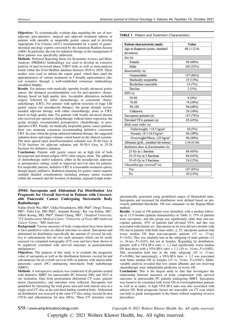
Objectives: To systematically evaluate data regarding the use of neo-adjuvant, peri-operative, surgical and adjuvant treatment options inpatients with operable or inoperable gastric cancer and to developAppropriate Use Criteria (AUC) recommended by a panel of gastro-intestinal oncology experts convened by the American Radium Society(ARS). In particular, the role for radiation therapy in the management ofthese patients was specifically addressed.Methods: Preferred Reporting Items for Systematic reviews and Meta-Analyses (PRISMA) methodology was used to develop an extensiveanalysis of peer-reviewed phase 2/2R/3 trials as well as meta-analysesfound within the Ovid Medline database between 2010 to 2020. Thesestudies were used to inform the expert panel, which then rated theappropriateness of various treatments in 5 broadly representative clin-ical scenarios through a well-established consensus methodology(modified Delphi).Results: For patients with medically operable locally advanced gastriccancer, the strongest recommendation was for peri-operative chemo-therapy based on high quality data. Acceptable alternatives includedsurgery followed by either chemotherapy or concurrent chemo-radiotherapy (CRT). For patients with upfront resection of stage I-IIIgastric cancer (no neoadjuvant therapy), the group strongly recom-mended adjuvant therapy with either chemotherapy alone or CRT,based on high quality data. For patients with locally advanced diseasewho received pre-operative chemotherapy without tumor regression, thegroup strongly recommended postoperative chemotherapy or post-operative CRT. Finally, for medically inoperable gastric cancer patients,there was moderate consensus recommending definitive concurrentCRT. In cases when the group endorsed radiation therapy, the suggestedradiation doses and target volumes varied based on the clinical scenario.The most agreed upon dose/fractionation schedule was 45-46 Gray in25-26 fractions for adjuvant radiation and 50-50.4 Gray in 25-28fractions for definitive radiation.Conclusions: Patients with gastric cancer are at high risk of bothlocoregional and distant relapse (49%) after surgery alone. The additionof chemotherapy and/or radiation, either in the neoadjuvant, adjuvant,or perioperative setting, result in improved survival rates for patients.For inoperable patients, definitive CRT is a reasonable treatment option,though largely palliative. Radiation planning for gastric cancer requiresmultiple detailed considerations including primary tumor locationwithin the stomach and the location of draining, regional lymph nodes.
(P044) Sarcopenia and Abdominal Fat Distribution ArePrognostic for Overall Survival in Patients with Unresect-able Pancreatic Cancer Undergoing Stereotactic BodyRadiotherapyYufan (Fred) Wu, MD1, Chika Nwachukwu, MD, PhD2, Diego Toesca,MD1, Yufei Liu, MD PhD3, Amanda Koong1, Rie von Eyben, MS1,Albert Koong, MD, PhD4, Daniel Chang, MD1; 1Stanford University,2UT Southwestern Medical Center, 3University of Texas MD AndersonCancer Center, 4MD AndersonBackground: Various measures of body composition have been shownto have predictive value on clinical outcomes in cancer. Sarcopenia andabdominal fat distribution (specifically the amount of visceral fat rela-tive to subcutaneous fat) are two such measures which can be easilyassessed via computed tomography (CT) scan and have been shown tobe negatively correlated with survival outcomes in gastrointestinalmalignancies.Objectives: The purpose of this study is to evaluate the prognosticvalue of sarcopenia as well as the distribution between visceral fat andsubcutaneous fat on overall survival (OS) in patients with unresectablepancreatic cancer (PC) undergoing stereotactic body radiotherapy(SBRT).Methods: A retrospective analysis was conducted of all patients treatedwith definitive SBRT for unresectable PC between 2001 and 2015 atour institution. Data from pretreatment abdominal CT scans, patient/tumor characteristics, and outcomes were collected. Sarcopenia wasquantified by measuring the total psoas area and total muscle area at asingle axial CT slice at the mid-third lumbar vertebral body. Abdominalfat distribution was assessed at the same CT slice using visceral fat area(VFA) and subcutaneous fat area (SFA). These CT structures were
automatically generated using predefined ranges of Hounsfield units.Sarcopenia and increased fat distribution were defined based on pre-viously published thresholds. OS was estimated via the Kaplan-Meiermethod.Results: A total of 196 patients were identified, with a median follow-up of 12.9 months (patient characteristics in Table 1). 75% of patientswere sarcopenic, and this group was significantly older than non-sar-copenic patients. 43% of patients had elevated VFA, and this wasassociated with female sex. Sarcopenia on its own did not correlate withOS, but in patients with body mass index ≥ 25, sarcopenic patients hadworse median OS than non-sarcopenic patients (13 vs. 17 mo,P= 0.01). This was similarly true in the subgroup of male patients (11vs. 24 mo, P= 0.03), but not in females. Regarding fat distribution,patients with a VFA:SFA ratio > 1.2 had significantly worse medianOS than those with a VFA:SFA ratio < 1.2 (10 vs. 14 mo, P= 0.003).This association held true in the male subgroup (10 vs. 13 mo,P= 0.006), but interestingly, a VFA:SFA ratio > 1.2 was associatedwith better median OS in females (15 vs. 14 mo, P= 0.047). Multi-variable analysis revealed that low serum albumin and not receivingchemotherapy were independent predictors of worse OS.Conclusions: This is the largest study to date that investigates therelationship between measures of body composition with survivaloutcomes in unresectable PC patients undergoing SBRT. Sarcopeniawas found to be correlated with worse OS in overweight/obese patientsas well as in males. A high VFA:SFA ratio was also associated withinferior OS. Both prognostic factors are assessable via CT scan whichcan help to guide management in the future without requiring invasiveprocedures.
TABLE 1. Patient and Treatment Characteristics
Abstracts American Journal of Clinical Oncology � Volume 44, Number 10, October 2021
S58 | www.amjclinicaloncology.com Copyright © 2021 Wolters Kluwer Health, Inc. All rights reserved.
Copyright r 2021 Wolters Kluwer Health, Inc. All rights reserved.

(P045) PSA: Declining Utilization of Brachytherapy for theTreatment of Prostate CancerLauren Andring, MD1, Todd A Pezzi, MD, MBA2, Chad Tang, MD1,Gary Walker, MD3; 1UT MD Anderson Cancer Center, 2MD AndersonCancer Center, 3Banner HealthBackground: Despite a growing body of literature demonstratingexpanding indications for the use of brachytherapy, multiple priorstudies have shown that over time rates of brachytherapy utilizationhave been decreasing.Objectives: To analyze rates of brachytherapy use for prostate cancerover time and evaluate patient characteristics, demographics and factorspredictive for its utilization.Methods: Data was retrospectively analyzed from the National CancerDatabase (NCDB) for patients with localized prostate cancer treatedbetween 2010 and 2015. Patients were included if they had biopsyconfirmed localized adenocarcinoma of the prostate, were treated withradiation as definitive local therapy, and were at least 18 years old.Utilization rates of external beam radiation (EBRT), brachytherapy(BT) and combination (EBRT + BT) were evaluated over time. Uni-variable (UVA) and backwards elimination multivariable (MVA)analysis were performed to determine characteristics predictive forbrachytherapy use.Results: We analyzed 178,837 patients with localized adenocarcinomaof the prostate treated between 2010 and 2015 with radiation therapy.During the period from 2010-2015, the use of EBRT increased from67% to 78%, BT (both monotherapy and combination with EBRT)decreased from 33% to 22%, BT monotherapy decreased from 25% to16% and EBRT + BT decreased from 8% to 6%. Age > 70, governmentfunded insurance or lack of insurance, intermediate or high-risk diseaseand treatment at an academic center were associated with significantlylower utilization of brachytherapy (all P< 0.001), while higher mediancounty income was associated with increased use (P= 0.02). On MVApatient age, insurance provider, treatment facility, and NCCN riskcategory were independent predictors for brachytherapy utilization(Table 1). Notably, on both UVA and MVA brachytherapy practicedecreased with increasing year of diagnosis (OR 0.881, 95% CI 0.853-0.910, P< 0.001).Conclusions: Rates of brachytherapy utilization for the treatment ofprostate cancer continue to decrease over time. Treatment at an aca-demic center was associated with reduced likelihood of brachytherapyuse. This has significant implications for the training of future radiationoncology residents/fellows and direct consequences for both ourpatients and healthcare expenditure.
(P046) Does the Addition of Brachytherapy And/orAndrogen Deprivation Therapy to External Beam Radio-therapy Correlate with Improved Survival in Men withUnfavorable Intermediate-risk Prostate Cancer?Neal Andruska, MD PhD1, Temitope Agabalogun, BS1, BenjaminFischer-Valuck, MD2, Randall Brenneman, MD PhD1, Yi Huang, MS1,Hiram Gay, MD1, Jeff Michalski, MD, MBA1, Ruben Carmona, MDMStat3, Brian Baumann, MD1; 1Department of Radiation Oncology,Washington University School of Medicine in St. Louis, 2Department ofRadiation Oncology, Emory University School of Medicine, 3Depart-ment of Radiation Oncology, University of Miami School of MedicineBackground: External beam radiotherapy (EBRT) ± brachytherapyboost (BT) ± androgen deprivation therapy (ADT) is currently rec-ommended for men with unfavorable intermediate-risk prostate cancer(UIR-PCa). However, the ideal radiotherapy regimen for UIR-PCa isnot well-defined since clinical trials have rarely addressed treatmentdecisions in this cohort, with most trials grouping UIR patients witheither favorable intermediate-risk or high-risk disease. Three clinicaltrials have shown that adding BT to EBRT improves biochemicalprogression-free survival but did not report differences in metastatic-free survival, cancer-specific survival, or overall survival. Given thathigher-risk patients were included and likely derived the most benefitfrom intensifying treatment, it remains unclear whether men with UIR-PCa derived the same benefit from a BT boost.Objectives: The addition of BT to EBRT+ADT in UIR-PCa has aradiobiologic advantage, permitting further dose escalation beyonddoses that can be delivered routinely with EBRT. We thereforehypothesized that EBRT+BT ±ADT would be associated withimproved overall survival (OS) relative to EBRT ±ADT in men withUIR-PCa.Methods: 32,246 men diagnosed between 2004-2015 with UIR-PCatreated with EBRT ±ADT (≥ 72 Gy in 1.8-2.0 Gy per fraction) orEBRT+BT (40-50.4 Gy EBRT, followed by HDR-BT or LDR-BT)were identified in the National Cancer Database (NCDB). Patients witha Charlson-Deyo comorbidity index (CDCI) score > 1, who receivedsystemic therapy other than ADT, or missing key information wereexcluded. Inverse propensity of treatment-weighted (IPTW) multi-variable analysis (MVA) using Cox regression modeling was used tocompare OS hazard ratios. Covariables included age, race, ethnicity,year of diagnosis, CDCI score, insurance status, educational and soci-oeconomic metrics, treatment at an academic center, PSA as diagnosis,Gleason score, clinical T-stage, and receipt of ADT.Results: Patients were stratified into four treatment groups: (i) EBRT(n= 13,265), (ii) EBRT+ADT (n= 13,123), (iii) EBRT+BT (n= 3,440),(iv) EBRT+ADT+BT (n= 2,418). Propensity-weighted MVA showedthe following. EBRT+ADT correlated with improved OS relative to
TABLE 1. Multivariable Logistic Regression Model forBrachytherapy Treatment
FIGURE 1. Propensity weighted Kaplan-Meier curve stratified byEBRT+ADT versus EBRT+BT without ADT. P value representP-log rank.
American Journal of Clinical Oncology � Volume 44, Number 10, October 2021 Abstracts
Copyright © 2021 Wolters Kluwer Health, Inc. All rights reserved. www.amjclinicaloncology.com | S59
Copyright r 2021 Wolters Kluwer Health, Inc. All rights reserved.

EBRT alone (Hazard Ratio (HR): 0.92, [95% Confidence Interval: 0.87-0.98], P= .005). Compared with EBRT+ADT, EBRT+BT (HR: 0.77[0.69-0.85], P= 3x10⁻⁷) and EBRT+BT+ADT (HR: 0.75 [0.67-0.83],P= 6x10⁻⁸) were both associated with improved OS. Relative to EBRT+BT, EBRT+BT+ADT was not associated with improved OS (HR:0.99 [0.87-1.11], P= .82). 10-year OS for the EBRT+ADT versusEBRT+BT without ADT was 55% and 70%, respectively (P< 0.0001)(Fig. 1).Conclusions: The addition of brachytherapy to EBRT correlated withreduced mortality in men with UIR-PCa. While ADT+EBRT wasassociated with better OS relative to EBRT, EBRT+BT+ADT resultedin comparable survival to EBRT+BT, suggesting that omitting ADTwith EBRT+BT may be an option for men who want to avoid ADT.
(P047) A Prospective Study of MR-guided Focal SalvageHDR Brachytherapy for Radiorecurrent Prostate Cancer:Updated Results of 30 PatientsHans Chung, MD1, Mark Corkum, MD1, Andrew Loblaw, MD1, Chia-Lin Tseng, MD1, Ananth Ravi, PhD1, Melanie Davidson, PhD1, MattWronski, MD1, Masoom Haider, MD2, Gerard Morton, MD1; 1Sunny-brook Odette Cancer Centre, 2University of TorontoBackground: Salvage therapies for localized radiorecurrent prostatecancer often carry significant short- and long-term morbidity. Focalsalvage high dose rate (HDR) brachytherapy is an appealing treatmenttechnique which delivers an ablative dose of radiotherapy to the portionof the prostate containing recurrent disease; however, limited pro-spective data is available.Objectives: We sought to explore the toxicities, health related quality oflife and efficacy of focal salvage HDR brachytherapy after previousdefinitive radiotherapy.Methods: Patients with locally recurrent prostate cancer after previousexternal beam radiotherapy (EBRT) and/or brachytherapy were enrolledon a prospective clinical trial. Patients received MRI-guided, ultra-sound-based focal HDR brachytherapy delivered over two fractions of13.5 Gy delivered 1-2 weeks apart. Adjuvant androgen deprivationtherapy (ADT) was not used. Toxicity was measured using CTCAE v4.Posttreatment response was evaluated using MRI 1-2 years after sal-vage. Biochemical failure was defined as PSA nadir + 2 ng/mL.Results: Thirty patients were treated between November 2012 andSeptember 2019. Median follow-up was 35 months (range: 13–92 mo).Fifteen patients were initially treated with EBRT, 3 with low dose rate(LDR) brachytherapy monotherapy, 1 with EBRT and LDR brachy-therapy boost, 2 with EBRT and HDR brachytherapy boost, and 9 withHDR brachytherapy as monotherapy (all 19 Gy in a single fraction).Median clinical target volume (CTV) at time of salvage was 5.22 mL(range: 2.18–15.71 mL), corresponding to a median of 20.0% of theprostate volume (range: 8.8–39.2%). Median PSA at salvage was 4.46ng/mL (range: 0.99–11.63 ng/mL). The median CTV V100 was 96.5%(range: 90.5–99.5%), and median CTV D90 was 15.1 Gy per fraction(range: 13.6–18.1 Gy). Seventeen patients experienced subsequentbiochemical failure, and 9 have received ADT and/or further localsalvage. No patients have died from prostate cancer. Median time tobiochemical failure was 41.5 months, and median time to ADT/salvagetherapy was 70.6 months. The three-year biochemical failure-free eventrate was 61.8% (95% CI 44.0–86.6%), and three-year ADT/salvagetherapy-free event rate was 86.0% (95% CI 74.1–99.8%). No acutegrade ≥ 3 GU/GI toxicity was observed. One late grade 3 GU toxicity
event occurred, cystitis at 42 months post treatment, which did notpersist on follow-up. No late grade ≥ 3 GI toxicity was seen. Of the 28patients who had a post-treatment MRI, 26 had evidence of a localtreatment response.Conclusions: In our updated results, we found focal salvage HDRbrachytherapy is well tolerated with a favorable side effect profile and3-year biochemical control rates in line with other salvage therapies forradiorecurrent prostate cancer. While early MRI response at the treatedsite is common, this does not preclude subsequent biochemical failure.
(P048) Comparative in Silico Analysis of Intensity Modu-lated Scanning Beam Proton Therapy (IMPT) and Volu-metric Modulated Arc Photon Radiotherapy (VMAT) forthe Post-Operative Treatment of Prostate CancerIan Cruickshank, BS RT(T)1, Hao Chen, PhD2, Aditya Halthore, MD2,Heng Li, PhD3, Curtiland Deville, MD2; 1Howard University College ofMedicine, 2Department of Radiation Oncology and Molecular Radia-tion Sciences, The Johns Hopkins Hospital, 3Johns Hopkins UniversityBackground: Limited data exists regarding the dosimetric techniquesand potential advantages of modern scanning beam proton therapyrelative to conventional photon therapy modalities for prostate cancerpatients in the post-operative setting.Objectives: Our purpose was to evaluate the dosimetric differences inIMPT versus IMRT for the post-operative treatment of prostate cancer.Methods: The 3DCT data of 7 consecutive, post-prostatectomy patientstreated at our institution with adjuvant or salvage IMPT in 2020 wereused to generate VMAT and IMPT plans using 3 different beamarrangements: 2-field (opposed laterals), 3-field (opposed laterals infe-riorly matched to posterior beam superiorly), and 4-field (opposed lat-erals inferiorly matched superiorly to 2 posterior oblique beams). Pre-scription was 50 Gy in 2 Gy fractions delivered to elective pelvic nodalregions (i.e., whole pelvis) and 20 Gy in 2 Gy fractions delivered to theprostate bed for a total dose of 70 Gy in 35 fractions. PT doses arereported in Gy (RBE)= 1.1 Gy. Dose goals for target and relevantorgans at risk (OAR), as well as dose-volume histogram parameterswere assessed. The paired 2-sided Wilcoxon signed-rank test was usedto compare the 4-field IMPT versus VMAT plans, with P< 0.05 indi-cating statistical significance.Results: CTV coverage met pre-specified dose goals for all plans with99% of CTVs receiving ≥ 100% of the prescription doses. The 2-, 3-,and 4-field IMPT plans showed similar doses to the bladder and bladderminus CTV (bladderless-CTV), while the 4-field IMPT plan showed thelowest mean and low to intermediate doses to the bowel cavity andrectum. For example, mean bowel cavity V15 and V45 for the 2-, 3-,and 4-field IMPT plans were respectively 549.2± 282.6 cc and110.7± 39.8 cc, 303.0± 102.0 cc and 92.7± 32.6 cc, and 263.4± 97.3 ccand 90.6± 40.6 cc. Mean rectum V50 and V60 for the 2-, 3-, and 4-fieldIMPT plans were 26.8± 8.9% and 18.7 ± 7.4%, 28.9 ± 10.5% and19.5 ± 8.0%, 24.3 ± 4.8% and 16.7 ± 4.1% respectively. When compar-ing the 4-field IMPT and VMAT plans, the rectal, bowel, and bladderparameters in Table 1 differed significantly.Conclusions: The 4-field IMPT beam arrangement showed the greatestreductions in dose to the bowel cavity and rectum compared withVMAT and the 2- and 3-field IMPT arrangements. These data caninform the future clinical management and delivery of proton therapyfor prostate cancer in the post-prostatectomy setting (Fig. 1).
Abstracts American Journal of Clinical Oncology � Volume 44, Number 10, October 2021
S60 | www.amjclinicaloncology.com Copyright © 2021 Wolters Kluwer Health, Inc. All rights reserved.
Copyright r 2021 Wolters Kluwer Health, Inc. All rights reserved.

FIGURE 1. Dose volume histogram comparison of 2-, 3-, and 4-field IMPT and IMRT for the clinical target volumes and organs at riskincluding bladder, bladder minus CTV (bladderless-CTV), and bowel cavity.
American Journal of Clinical Oncology � Volume 44, Number 10, October 2021 Abstracts
Copyright © 2021 Wolters Kluwer Health, Inc. All rights reserved. www.amjclinicaloncology.com | S61
Copyright r 2021 Wolters Kluwer Health, Inc. All rights reserved.
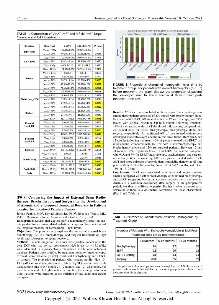
(P049) Comparing the Impact of External Beam Radio-therapy, Brachytherapy, and Surgery on the Developmentof Anemia and Subsequent Temporal Recovery in PatientsTreated for Localized Prostate CancerJordan Fenlon, MD1, Krystal Straessler, PhD1, Jonathan Tward, MD,PhD1; 1Huntsman Cancer Institute at the University of UtahBackground: Studies that explore pelvic radiotherapy’s effect on ane-mia predate intensity-modulated radiation therapy and have not trackedthe temporal recovery of Hemoglobin (Hgb) levels.Objectives: The present study explores the impact of external beamradiotherapy (EBRT), brachytherapy, and surgical treatments on Hgblevels and subsequent temporal recovery.Methods: Patients diagnosed with localized prostate cancer after theyear 2008 who had normal pretreatment Hgb levels (> = 13.2 g/dL)were identified in a prospectively maintained institutional outcomesdatabase. Patients were stratified by treatment modality (brachytherapy,external beam radiation [EBRT], combined brachytherapy and EBRT,or surgery. The proportion of patients who became mildly (Hgb 10–13.2 g/dL) or moderately/severely (Hgb <10 g/dL) anemic was eval-uated at time bins of 0-6 months, 6-12 months, and 12 - 24 months. Forpatients with multiple Hgb levels in a time bin, the average value wasused. Patients were censored at the initiation of any additional cancertherapies.
Results: 3285 men were included in the analysis. Treatment regimensamong these patients consisted of 239 treated with brachytherapy alone,88 treated with EBRT, 206 treated with EBRT/brachytherapy, and 2752treated with surgical resection. Up to 6 months following treatment,41% of men treated with EBRT developed mild anemia, compared with22, 6, and 50% for EBRT/brachytherapy, brachytherapy alone, andsurgery, respectively. An additional 6% of men treated with surgerydeveloped moderate/severe anemia in this time frame. Between 6 and12 months following treatment, 40% of patients treated with EBRT hadmild anemia, compared with 0% for both EBRT/brachytherapy andbrachytherapy alone and 12% for surgical patients. Between 12 and24 months, 33% of patients treated with EBRT had anemia, comparedwith 9, 5, and 7% for EBRT/brachytherapy, brachytherapy, and surgery,respectively. When considering ADT use, patients treated with EBRT/ADT had more episodes of anemia than trimodality therapy at all timepoints (50 vs. 21% at 0-6 months, 56 vs. 0% at 6–12 months, and 33 vs.13% at 12–24 mo).Conclusions: EBRT was associated with more and longer durationanemia compared with either brachytherapy or combined brachytherapyand EBRT, suggesting brachytherapy boost reduces the risk of anemia.Anemia is a common occurrence after surgery in the perioperativeperiod, but then is unlikely to persist. Further studies are required todetermine if there is a dosimetric correlation for these observations(Fig. 1 and Table 1).
TABLE 1. Comparison of VMAT IMRT and 4-field IMPT TargetCoverage and OAR Constraints
FIGURE 1. Proportional change of hemoglobin over time bytreatment group. For patients with normal hemoglobin (>13.2)before treatment, the graph displays the proportion of patientsthat developed mild to severe anemia at three distinct post-treatment time bins.
TABLE 1. Number of Patients With Evaluable Hemoglobin byTreatment Group
For patients with normal pre-treatment hemoglobin (> 13.2), the number ofpatients with evaluable hemoglobin by treatment group in each distinct post-treatment time bin is displayed.
Abstracts American Journal of Clinical Oncology � Volume 44, Number 10, October 2021
S62 | www.amjclinicaloncology.com Copyright © 2021 Wolters Kluwer Health, Inc. All rights reserved.
Copyright r 2021 Wolters Kluwer Health, Inc. All rights reserved.

(P050) Androgen Deprivation Therapy and RadiotherapyPractice Patterns for Unfavorable Intermediate Risk Pros-tate CancerShaakir Hasan, D.O.1, Stanislav Lazarev, MD2, Madhur Garg, MD3,Keyur Mehta, MD3, Robert Press, MD1, Arpit Chhabra, MD1, J IsabelleChoi, MD1, Charles Simone, MD, FACRO1, Daniel Gorovets, MD4;1New York Proton Center, 2Mount Sinai, 3Montefiore, 4Memorial SloanKettering Cancer CenterBackground: Per National Comprehensive Cancer Network (NCCN)guidelines, the nonsurgical treatment algorithm for unfavorable inter-mediate risk (UIR) prostate cancer (PC) includes external beam radio-therapy (EBRT) with short-course androgen deprivation therapy (ADT),or EBRT with brachytherapy (BT) with or without ADT. The currentpractice patterns and patient selection criteria in this setting are unclear.Objectives: Using the National Cancer Database (NCDB), radiationtreatment patterns for UIR PC were evaluated.Methods: Clinical and demographic characteristics associated witheach treatment were assessed via multivariable binomial regressionanalysis, expressed as odds ratios (OR) with 95% confidence intervals.Results: There were 68,283 eligible men with a median age of 70 yearstreated with radiotherapy between the years 2004-2015 and who met theNCCN criteria for UIR PC. The most common treatment was EBRT +ADT (38.0%), then EBRT alone (32.2%), BT alone (9.7%), EBRT +BT (8.4%), EBRT + BT + ADT (7.1%), and BT + ADT (4.7%). ADTutilization decreased over time, from 58% in 2004 to 49% in 2015, asdid BT utilization, from 39% in 2004 to 22% in 2015 (P <0.001).Patients were classified as UIR because of Gleason 4+3 disease(60.5%), Gleason 3+4 with PSA between 10-20 (18.5%), clinical T2bor T2c (17.5%), or 50% or more cores positive (3.5%). On multivariableanalysis, Gleason 4+3 (HR= 1.45, 1.39-1.50), high volume disease(OR= 1.53, 1.59-1.72), stage T2b/T2c (OR= 1.66, 1.59-1.72), PSAbetween 10-20 (OR= 1.86,1.79-1.93), older age (OR= 1.004, 1.001-1.006), treatment at a non-academic center (OR= 1.37, 1.28-1.44),Black patients (OR= 1.11, 1.06-1.17), Hispanic patients (OR= 1.17,1.09-1.28), lower income areas (OR-1.12, 1.05-1.19), rural areas(OR= 1.18, 1.09-1.27), treatment before 2008 (OR= 1.41, 1.35-1.48),and EBRT (vs BT or combination) (OR= 1.81, 1.75-1.88) were allassociated with increased ADT use. Gleason 4+3 (HR= 0.94,0.89-0.98), T2b/T2c (OR= 0.91,0.87-0.95), PSA between 10-20 (OR= 0.73,0.70-0.76), older age (OR= 0.96, 0.957-0.963), Hispanic patients(OR= 0.71, 0.64-0.78), Black patients (OR= 0.84, 0.79-0.88), non-rural areas (OR= 0.74, 0.68-0.81) treatments after 2007 (OR= 0.68,0.65-0.71) were each independently associated with decreased BT uti-lization. Lower PSA (OR= 1.28), Gleason 3+4 (OR= 1.17), andtreatment at academic centers (OR= 1.92) were independent predictorsof hypofractionation (all P < .05) The two non-surgical NCCN-rec-ommended treatments for UIR PC, EBRT+ADT and EBRT+BT, wereanalyzed in a separate subset. Independent correlates of EBRT+BTinclude Gleason 3+4 (OR= 1.28, 1.19-1.38), low volume disease(OR= 1.32, 1.18-1.46), T1c/T2a (OR= 1.21, 1.12-1.31), PSA under 10
(OR= 2.22, 2.06-2.39), younger age (1.06, 1.055–1.065), non-Hispanic(OR= 1.55, 1.30-1.85), and white patients (OR= 1.40, 1.27-1.53).Conclusions: Only approximately half of UIR PC patients treated withradiotherapy between 2004-2015 adhered to the current NCCN rec-ommendations, with a trend away from ADT and BT in more recentyears. Older patients with more aggressive UIR criteria were morelikely to receive ADT and less likely to receive BT (Figs. 1 and 2).
(P051) Was There a Differential Delay in Urologic OncologyDiagnoses Due to the COVID 19 Pandemic?Kevin Healey, Medical Student 1st year1, Mitchell Finkelstein, HighSchool Student2, Miriam Williams, CAO1, Steven Finkelstein, MD,DABR, FACRO2; 1AUI, 2FCA / US Oncology NetworkBackground: In community urology, a delay in performing routinescreening evaluation and treatment procedures secondary to theCOVID-19 pandemic delayed the number of urological oncologypatient seeking care according to data from Medicare and United Statesinsurers. Thus, it is unclear if this delay influenced urologic oncologyand non-oncology equally.Objectives: Our hypothesis is that there may be a differential delay inurologic oncology diagnoses due to the COVID 19 Pandemic.Methods: This is a retrospective review of prospectively collected data;new patients referred to a large multispecialty community urologyorganization before COVID 19 (quarter 2 April-June 2019) werecompared with during COVID 19 (quarter 2 April-June 2020). Data wasobtained using electronic medical records for completeness. Urologydisease data were recorded and compared.Results: As compared with previous year quarter 2, there was an overalldecrease in new patient referrals to a large multispecialty urologyorganization. With respect to urologic oncology and urologic non-oncology, there was a significant decrease in new patient visits. Withrespect to urologic oncology, prostate, bladder, and testicular weresignificantly affected; diagnostic procedures related to workup weredecreased accordingly. Lastly, referrals to radiation oncology forprostate, bladder, and testicular were impacted negatively with a dif-ferential delay by disease subsite.Conclusions: The data suggest a differential delay in urologic oncologydiagnoses due to the COVID 19 pandemic in some large communityurology organizations. Data over time will assess these changesacross 2021.
(P052) Radiotherapy Does Not Increase the Frequency ofRevision of Perineal Urethrostomy in Penile Cancer: A20-year International Multicenter ExperiencePeter Johnstone, MD, FACR, FASTRO1, Hielke-Martijn de Vries, na2,Juan Chipollini, Assistant Professor3, Franklin Boyd4, FernandoKorkes, MD, PhD5, Maarten Albersen6, Eduard Roussel, Department ofUrology6, James Jiang, MD7, Viraj Master, MD PhD FACS8, OscarBrouwer, Netherlands Cancer Institute2, Philippe Spiess, MD, MS,FACS1; 1Moffitt Cancer Center, 2Netherlands cancer institute, 3TheUniversity of Arizona, 4University of South Florida Morsani College ofFIGURE 1. Type of treatment partitioned by risk factor.
FIGURE 2. ADT use by year.
American Journal of Clinical Oncology � Volume 44, Number 10, October 2021 Abstracts
Copyright © 2021 Wolters Kluwer Health, Inc. All rights reserved. www.amjclinicaloncology.com | S63
Copyright r 2021 Wolters Kluwer Health, Inc. All rights reserved.

Medicine, 5ABC Medical School, 6University Hospitals Leuven, 7EmoryUniversity School of Medicine, 8Emory UniversityBackground: Perineal urethrostomy (PU) is the definitive form ofurinary diversion in patients with locally advanced or anatomicallyunfavorable penile cancer (PeCa) requiring total penectomy. Priorstudies would suggest PU failure rates in up to 30% of cases, althoughthis has been infrequently reported in patients with cancer.Objectives: Because of lack of clinical evidence, radiotherapy (RT) isseldom recommended for pN1/pN2 PeCa lesions but may be used forextra-nodal (pN3) disease. We previously described a large, multi-institutional experience of PU for PeCa with respect to complications,and now describe the role of perioperative RT in the process.Methods: In our cohort, 299 patients from seven international centers inBelgium, Brazil, China, Netherlands, UK and the United Statesunderwent PU for urinary diversion for PeCa between 2000 and 2019.Median patient age was 67 years and median follow-up was 24 months.Demographic and clinicopathologic characteristics were reviewed. Sixpatients received pre-operative RT; five of them with chemotherapy. 43received RT post-operatively, 28 (65%) with chemotherapy.Results: 75 patients (25%) developed a 30-day post-operative compli-cation with a single postoperative death. Wound infection (49%) anddehiscence (19%) were most common. Over 65% (n= 50) of compli-cations were minor (Clavien–Dindo Grade I and II). Only adjuvantchemoradiation was significantly associated with post-operative com-plication (OR: 2.39, 95%CI 1.09-5.24). Urethral stenosis occurred in12% (n= 36) of cases. Almost 80% occurred in the first year (median5.7 m; IQR 3.1-12 m). Twenty-seven patients underwent surgicalrevision and 9 were treated conservatively. Pre-operative or adjuvanttherapy was not significantly related to subsequent PU stenosis.Conclusions: When RT is delivered for PeCa it is usually to the pre-pubic fat, groin and lateral pelvis, and not to the course of the PU.Given the large number of patients and diverse circumstances and erasinvolved, it appears that post-operative RT may be delivered withoutsignificant increase in the risk of stenosis and surgical revision. Weacknowledge limitations of this retrospective study and its implicitselection bias. However, our follow-up is sufficient for perioperativecomplications and early stenosis rates contributing to revision.
(P053) Outcomes of Axumin PET/CT-detected ProstateCancer Nodal Recurrences: A Single Institution ExperienceAndrew Keller, MD1, Ankur Patel, MD1, Leonard Appleman, MD,PhD1, David Friedland, MD1, Roby Thomas, MD1, Adam Olson, MD2;1UPMC Hillman Cancer Center, 2Department of Radiation Oncology,UPMC Hillman Cancer Center, University of Pittsburgh School ofMedicineBackground: Nodal recurrence from prostate cancer is an emergingdisease state that represents a therapeutic dilemma for oncologists. Theadvent of advanced PET imaging modalities offers greater sensitivity todetect oligometastatic disease in this patient cohort and renders radia-tion therapy, such as stereotactic body radiation therapy (SBRT) orIntensity Modulated Radiation Therapy (IMRT), an attractive alter-native to androgen deprivation therapy alone.Objectives: Whether addition of local therapy confers long-term bio-chemical control is uncertain. We evaluated outcomes of prostate cancerpatients with recurrent nodal disease detected by Axumin PET/CT.Methods: A retrospective analysis of 23 patients with prostate adeno-carcinoma nodal recurrences detected by Axumin PET/CT scan treatedwith SBRT, EBRT, or ADT alone from 2017-2018 was performed.Median patient age was 67 (range 49-83). Initial definitive treatmentconsisted of radical prostatectomy for 14 patients and RT +/- ADT for 9patients. Median PSA at time of Axumin PET/CT scan was 5.5 ng/mL(range 0.7-39.0 ng/mL). 11 patients underwent SBRT, 6 patientsunderwent IMRT with simultaneous or sequential boost to gross nodaldisease, and 8 patients underwent ADT alone. 7 of 17 patients treatedwith RT received ADT at time of RT. SBRT was to a total dose of 35-50 Gy in 5 fractions. IMRT was to a dose of 45-72 Gy in 25-40 frac-tions targeting pelvic nodes plus/minus prostatic fossa or PA nodalregion, with boost to gross disease. Local control of treated diseasefollowing RT, biochemical control, ADT-free interval, freedom fromnew sites of disease, and overall survival, were calculated from date of
Axumin utilizing the Kaplan-Meier estimator. Biochemical control wasdefined as the time until sustained PSA rise > 0.2 ng/mL above PSA attime of Axumin PET/CT scan. Log-rank test was utilized for univariateanalysis.Results: Median follow-up was 27 months. Median overall survivalwas not reached, with all patients surviving until last follow-up. Medianbiochemical control was 32.0 mos (95% CI 20.7-43.3 mos). For patientsnot receiving ADT at time of RT, median ADT-free interval was 16.0mos (95% CI 4.0-28.0 mos). Follow-up imaging was available in 18patients. Of patients receiving RT, 2-year local control of treated diseasewas 100%. Median freedom from new sites of disease was 27 mos(95% CI 16.7-37.3 mos), with 7 patients developing additional sites ofdisease, including 5 cases of additional nodal-only failure. Of the 18patients with follow-up imaging, 10 (56%) had no evidence of radio-graphic progression. There were no cases of grade 3+ toxicity followingRT. On univariate analysis, Gleason score, time from initial definitivemanagement to Axumin PET/CT, and addition of RT were not stat-istically significant predictors of biochemical control or freedom fromadditional sites of disease.Conclusions: RT in the form of SBRT or IMRT are well-tolerated,effective local treatment for patients with recurrent nodal disease sec-ondary to prostate cancer. Prognostic factors associated with long-termbiochemical control remain unclear and require a larger sample size todetect.
(P054) Improvements in Rectal Dosimetry Using RectalHydrogel Spacer in Patients with Prostate Cancer Under-going HDR BrachytherapyCharles Lee1, Iavor Veltchev1, Mengying Deng1, Rebecca Shulman,MD1, Teh Lin1, Robert Price1, Mark Hallman1, Eric Horwitz1, JessicaWong1; 1Fox Chase Cancer CenterBackground: High-dose-rate brachytherapy (HDR-BT) is highlyeffective in the definitive and salvage treatment of prostate cancer.Acute grade 2+ rectal toxicity may occur from 5% of prostate cancerpatients treated with HDR-BT monotherapy to over 20% of patients inthe salvage or re-irradiation setting. Recently, polyethylene glycol-based rectal hydrogel spacers (RHS) have been increasingly used tocreate separation between the rectum and prostate. Studies investigatingits use in the external beam radiotherapy (EBRT) setting demonstrateimproved dosimetry and reduced rectal mucosal injury. However,sparse data exist investigating its use with HDR-BT. This is the firststudy investigating the dosimetric effect of RHS placement on patientsundergoing HDR-BT by utilizing a novel replanning technique.Objectives: Rectal V75% (rV75) is directly correlated with acute rectaltoxicity. We aim to demonstrate improvement in rV75 in patients withRHS undergoing HDR-BT by comparing plans in the same patients.Secondary objective is to quantify changes in prostate (target volume)coverage in plans with and without RHS.
FIGURE 1. Correlation of change in rectal V75% and RHSvolume.
Abstracts American Journal of Clinical Oncology � Volume 44, Number 10, October 2021
S64 | www.amjclinicaloncology.com Copyright © 2021 Wolters Kluwer Health, Inc. All rights reserved.
Copyright r 2021 Wolters Kluwer Health, Inc. All rights reserved.

Methods: A prospective institutional database was queried for patientsundergoing HDR-BT with RHS in place from January 2020–November2020 for either definitive, adjuvant, or salvage treatment of prostatecancer. A novel replanning technique was used, negating the RHSvolume and using the negated contours as a surrogate for the rectum.New plans were generated for each patient using this technique, withthe goal of at least 95% prostate coverage. Wilcoxon signed rank testsand Spearman correlation were used to assess dosimetric differencesbetween original treatment plans and regenerated plans for correlationsbetween separation and volume.Results: Twenty-one HDR-BT procedures were performed in 12patients. Five had HDR-BT monotherapy (2700 cGy in 2 fractions), 4patients received salvage HDR-BT (2400 cGy in 2 fractions), and 3patients received HDR-BT boost (1500 cGy in 1 fraction followingEBRT 4600 cGy in 23 fractions). Median RHS volume was 7.44 cm3(IQR: 4.59-9.56 cm3). Rectal dosimetric parameters were significantlyworse in regenerated plans compared with original plans. However, all21 regenerated plans were clinically acceptable based on institutionalcriteria. In the original treatment plans, rV75 was 0.0 cm3 in all cases;in regenerated plans, median rectum V75% (rV75*) was 0.74cm3 (IQR:0.62-0.87 cm3), P< 0.001. Spearman correlation of the differencebetween rV75* vs rV75 (ΔrV75) was found to be significantly corre-lated with RHS volume (rho= 0.52, P= 0.016, Fig. 1), prostate volume(rho= 0.51, P= 0.018), and radiation dose (rho= 0.46, P= 0.035).Median prostate V100% in original cases was 98.56% (IQR: 98.06-99.08) and was 96.07% in replanned cases (IQR:95.06-97.30),P< 0.001. There were no significant differences between medianprostate V150% and V200%.Conclusions: RHS significantly reduces rectum V75% in patientsundergoing HDR-BT. Larger RHS volumes inserted appear correlatedwith larger magnitude of rectum V75% reduction. RHS should beconsidered for all patients, especially those undergoing combination orsalvage treatment, to limit rectal toxicity. Reduction in prostate V100%in replanned cases likely underestimates actual attainable prostateV100% in patients undergoing HDR-BT without RHS.
(P055) Dose Differentiated HDR Prostate Brachytherapy –
a Feasibility Assessment of MRI Guided Dose Escalation toDominant Intraprostatic LesionsChristopher Luminais, MD1, David Cousins, MD1, Hamidezra Nour-zadeh, PhD1, Eric Aliotta, PhD1, Bruce Libby, PhD1, Timothy Show-alter, MD1; 1University of VirginiaBackground: Prostate brachytherapy is routinely performed withTRUS or CT based planning. Neither modality can delineate dominantintraprostatic lesions (DILs). Prostate MRIs performed during workupcan identify high risk DILs. DILs are known to be a high risk locationsfor local recurrence and there is clinic interest in dose escalating DILs.MRI registration at the time of brachytherapy allows DIL delineationand dose escalation.Objectives: This retrospective study assesses the maximum achievabledose escalation to DILs while respecting OAR objectives.Methods: Our institution offers combination HDR prostate brachy-therapy (15 Gy in 1 fx) + pelvic EBRT (45 Gy in 25 fx) to patients withunfavorable intermediate or high risk prostate cancer. We identified 24patients treated with combination therapy from 2013–2016 who had anavailable pre-treatment prostate MRI with 1-3 visualized DILs. Eachpatient’s MRI was rigidly co-registered to the intra-procedure TRUSand used to contour any visible DILs. DIL contours were transferred tothe TRUS based planning system. Original TRUS based prostate andOAR volumes were not altered. Original treatment plans were
experimentally re-optimized to dose escalate the DILs. Dosimetricindices from the original and the reoptimized plans were comparedusing a two-tailed paired T-test. Each reoptimized plan was deemedacceptable if it achieved all of the following criteria: prostate D90 >100%, prostate V100 > 90%, urethra D10 < 118%, rectum V80 <0.5 cc, bladder D1cc < 75%, or if it did not exceed OAR doses of theoriginal plan.Results: All reoptimized plans met the acceptability criteria. Achieveddosimetry is reported in Table 1. Mean DIL D90 was significantlyincreased from 134% on the original plans to 154% on the re-optimizedplans. Mean urethra D10 and mean bladder D1cc were significantlyreduced from 123% to 117% and 72% to 65%, respectively. Meanrectum V80 did not change significantly. Prostate D90 and V100 werereduced from 106% to 102% and 93% to 91%, respectively. Decreasedprostate coverage was considered acceptable as D90 > 100% and V100> 90% were achieved. We estimate that image registration and DILdose escalation would increase procedure time by 15 minutes usingstandard treatment planning software.Conclusions: Using MRI for delineation of DILs, we were able toreoptimize HDR brachytherapy plans to dose escalate DILs to a meanD90 of > 150% while maintaining favorable prostate coverage andOAR doses. Based on this finding, we estimate that a goal DIL D90dose of > 150% (or 22.5 Gy) is an aggressive but achievable goal whilemeeting OAR dose constraints. We will use this analysis to inform anupcoming prospective clinical trial of MRI guided prostate HDR bra-chytherapy at our center.
(P056) Early Toxicity Outcomes for Initial Implementationof Cs-131 Seeds for Low-dose-rate Prostate BrachytherapyDavid Marshall, MD, MS1, Shauna McVorran, MD2, Bradley DePaoli,MD2, Bethany Broekhoven, MS2, Elizabeth Hilliard, MS3, Hong Li,PhD2; 1Department of Radiation Oncology, Medical University ofSouth Carolina / Hollings Cancer Center, 2Medical University of SouthCarolina, 3SUNY Upstate Medical UniversityBackground: Low dose rate (LDR) prostate brachytherapy is anattractive definitive treatment option for localized prostate cancer withequivalent oncologic outcomes compared with external beam radiationtherapy (EBRT) and radical prostatectomy (RP) with the benefit ofbeing a convenient outpatient minimally invasive procedure. As theutility of LDR brachytherapy has been established, the focus has shiftedto improving the side effect profile while maintaining oncologic out-comes. Most data in this sphere were gathered using I-125 and Pd-103seeds, but since Cs-131 was introduced in 2004, several institutionshave adopted its use with the hypothesis that its shorter half-life andslightly higher average energy would produce dosimetric advantagesthat may improve outcomes and decrease duration or severity of sideeffects.Objectives: To report acute toxicity outcomes with the first 50 patientstreated with Cs-131 LDR brachytherapy at our institution, comparedwith our experience with I-125 and Pd-103.Methods: An IRB-approved retrospective chart review was performedon the first 50 patients treated with Cs-131 LDR brachytherapy forlocalized prostate cancer at our institution. Patients receiving either afull or partial implant were included; these patients were treated to dosesof 100 Gy and 80 Gy prostate minimal peripheral dose, respectively.Patients receiving partial implants generally received 45 Gy externalbeam radiotherapy starting 6-8 weeks after implant. These data werecompared with cohorts of the last 50 patients treated with I-125 bra-chytherapy and the last 50 patients treated with Pd-103 brachytherapy tostandard doses. Data regarding AUA prostate symptom score, SexualHealth Index for Men (SHIM), and RTOG GI toxicity score wereassessed pretreatment and at 1-month and 7-month time points. Dem-ographic and clinical data were also collected. Descriptive statisticswere used to summarize variables. A linear mixed model was used tocompare means and to identify variables as prognostic for acute toxicity(P≤ 0.05).Results: The mean AUA score at 1-month post-treatment was sig-nificantly higher for patients treated with Cs-131 (P< 0.001) butdeclined rapidly and was equivalent to the scores seen with I-125 andPd-103 at 7 months. A similar trend was seen with the mean RTOG GI
TABLE 1. Original Plan and MRI Reoptimized Dosimetry
American Journal of Clinical Oncology � Volume 44, Number 10, October 2021 Abstracts
Copyright © 2021 Wolters Kluwer Health, Inc. All rights reserved. www.amjclinicaloncology.com | S65
Copyright r 2021 Wolters Kluwer Health, Inc. All rights reserved.

toxicity score. The mean SHIM score at 1-month post-treatment wassignificantly lower (P< 0.001) with Cs-131, but this also improved by7 months to be equivalent to those seen with I-125 and Pd-103,respectively. On multivariate analysis, the type of isotope used did notpredict the AUA, SHIM, or RTOG toxicity score grade or the need for acatheter post brachytherapy.Conclusions: In patients treated in our initial experience with Cs-131LDR prostate brachytherapy, acute GU, GI, and sexual side effectspeaked higher but decreased faster compared with patients treated withI-125 or Pd-103 isotopes. The isotope used did not predict for AUA,SHIM, or RTOG toxicity score or the need for a catheter postbrachytherapy.
(P057) Disparities in Radiation Therapy Access for ProstateCancer in the United StatesShearwood McClelland III, MD1, Ulysses Gardner, MBA, RTT2,Curtiland Deville, MD3; 1Department of Radiation Oncology, IndianaUniversity School of Medicine, 2Wright State University BoonshoftSchool of Medicine, 3Department of Radiation Oncology and MolecularRadiation Sciences, The Johns Hopkins HospitalBackground: Major advances in the role of radiation oncology fortreating prostate cancer increases the importance of equity in access toradiation therapy (RT). In 2016, the first comprehensive investigationexamining the pervasiveness of health disparities in RT access foundthat the most prominently studied organ site was prostate.Objectives: As disparities research has continued to dramaticallyincrease, we sought to assess the evolution of RT access disparities inprostate cancer since that initial investigation.Methods: A comprehensive literature search was undertaken in June2020 using the PubMed database (www.pubmed.gov) and the query[prostate AND (radiation OR proton) AND (disparities OR “socio-economic status” OR “health services research” OR inequity OR race[Title])] from 2017-2020. Studies were excluded which were not basedin the United States, did not examine health inequities, did not examineRT, or did not examine prostate cancer.Results: Forty-four of 184 studies found met inclusion criteria. Healthdisparities were most prominently reported by race (35 studies) fol-lowed by insurance status (12 studies), age (6 studies), socioeconomicstatus (8 studies), geographic location (9 studies), and practice char-acteristics (6 studies).Conclusions: Health disparities in access to radiation therapy for prostatecancer remain substantial despite significant advancements in radiationoncology. Access to radiation therapy for prostate cancer is most likelymultifactorial; several considerations must be included when assessingsystems to decrease existing inequalities. High-level evidence researchfocusing on reducing disparities is needed and highly anticipated toimprove health and survival outcomes for all patients with prostate cancer.
(P058) Association of Second-Generation Antiandrogenswith Depression in Prostate CancerKevin Nead, MD, MPhil1, Malgorzata Nowakowska, BS2, XiudongLei, PhD1, Mackenzie Wehner, MD, MPhil1, Paul Corn, MD1, SharonGiordano, MD1; 1MD Anderson Cancer Center, 2Baylor College ofMedicineBackground: Previous studies have shown a consistent link betweenhormone therapy (HT) to treat prostate cancer, such as androgen dep-rivation therapy (ADT), and depression risk (Dinh KT, et al JCO 2016;Nead KT, et al Urol Oncol 2017). However, the association betweensecond-generation antiandrogens (AAs) and depression is unknown.Objectives: To test the hypothesis that second-generation AAs carry anincreased risk of depression, including compared with traditionalforms of HT.Methods: This retrospective cohort study analyzed Surveillance,Epidemiology, and End Results (SEER)-Medicare and Texas CancerRegistry (TCR)-Medicare data from 2011-2015, with a follow-up periodof 2 years after diagnosis. Data were analyzed from February to May2021. Of 210,804 patients diagnosed with prostate cancer in 2011-2015,we identified 30,069 patients with primary prostate cancer diagnosed at
age > 66 years without a second cancer in 12 months. We includedpatients with continuous Medicare Parts A, B, and D coverage. Weexcluded individuals who received any form of HT before prostate cancerdiagnosis and those previously diagnosed with depression. The followingtreatment groups were compared: (1) no HT group, (2) traditional HTgroup (HT without second-generation AA exposure), and (3) second-generation AA group. We implemented time-varying exposure multi-variable Cox proportional hazards models to determine the association ofADT use with depression using the inverse probability treatment weighted(IPTW) method.Results: Of 30,069 patients, 17,710 (59%) received no HT, 11,311(38%) received traditional-HT only, and 1,048 (3%) received a second-generation AA. The cumulative incidence of depression at 2 years washighest among second-generation AA users; 13.7% versus 7.2% and4.8% in the traditional-HT only and no-HT groups, respectively;P< 0.001. Multivariable Cox proportional hazards analysis showed thatthe second-generation AA group had an increased risk of depressioncompared with the no-HT group (HR= 2.16, 95%CI 1.81-2.59;P< 0.001) and the traditional-HT group (HR= 2.26, 95%CI 1.88-2.73;P< 0.001), including with stratification by localized (HR= 2.73, 95%CI 2.19-3.42; P< 0.001) and regional/distant disease (HR= 2.56, 95%CI 1.81-3.61; P< 0.001).Conclusions: Patients with prostate cancer who received a second-generation AA had a large and clinically significant absolute increasedrisk of depression compared with patients who received traditional HTalone or no HT.
(P059) Short-Term ADT and Dose-Escalated IMRT inPatients with Intermediate-Risk Prostate Cancer: Benefit orCaution?Arthur Hung, MD1, Carl Post, MD1, Jenna Kahn, MD1, Claire Turina,BS1, Tomasz Beer, MD, FACP1; 1Oregon Health & SciencesUniversityBackground: In the era of dose-escalated prostate radiation therapy(RT), the use of androgen deprivation therapy (ADT) is undefined forintermediate-risk prostate cancer (IR). Randomized data demonstratesan improvement in biochemical control without an associatedimprovement in overall survival. Yet there is also growing concern ofthe risk of ADT to be detrimental to quality of life and increase cardiacevents.Objectives: This single-institution retrospective analysis aimed toevaluate outcomes of IR prostate cancer patients treated with RT with orwithout concurrent/adjuvant short-term ADT.Methods: Data was collected from 559 consecutive patients treatedwith dose-escalated IMRT with daily IGRT for newly diagnosedprostate cancer from December 2002–March 2016. IMRT prescriptionwas 78 Gy / 39 fractions or 70 Gy / 28 fractions. Biochemical recur-rence-free survival (BCRFS), distant metastasis-free survival (DMFS),prostate cancer-specific survival (PCSS), and overall survival (OS) werecalculated using Kaplan-Meier methodology. We focused our results onthe patients with favorable (FIR) or unfavorable intermediate-risk (UIR)prostate cancer.
TABLE 2. Clinical Characteristics of the IR Population
Abstracts American Journal of Clinical Oncology � Volume 44, Number 10, October 2021
S66 | www.amjclinicaloncology.com Copyright © 2021 Wolters Kluwer Health, Inc. All rights reserved.
Copyright r 2021 Wolters Kluwer Health, Inc. All rights reserved.

Results: With a median follow-up of 93 months, 260 patients had IR, ofwhich 69.6% (N= 181) had UIR disease, 78.8% (N= 205) were treatedwith moderately hypofractionated RT to 70 Gy in 28 fractions, and36.2% (N= 94) received ADT, 89 UIR and 5 FIR, with median ADTduration of 6 months (range, 3-30; 90% received 6 mo). 7-yr BCRFSwas 94.1% vs. 86.2%, P = 0.067, for ADT and no ADT respectively,and no difference in DMFS or PCSS was observed. ADT was asso-ciated with significantly worse 7-yr OS (80.0% vs. 91.3%, P = 0.010).When stratified by age, patients <70 years who received ADT had evenworse 7-yr OS (93.4% vs. 77.3%, P = 0.002), whereas those ≥ 70 yearshad similar OS (P= 0.918). Analysis of the UIR cohort alone, showedsimilar results; 7-yr BCRFS and 7-yr OS in patients who received ADTversus no ADT were 93.7% vs. 85.9% (P = 0.093), and 79.0% vs.90.6% (P = 0.019), respectively.Conclusions: In our 15-year experience treating IR prostate cancer withdose-escalated IMRT with daily IGRT, short-term concurrent ADT wasassociated with a statistically significant worse OS as well as non-stat-istically significant improvement in BCRFS. Additional studies are neededto determine if ADT is beneficial or detrimental for patients with IRprostate cancer treated with dose-escalated radiation (Table 2 and Fig. 1).
(P060) Second Pelvic Cancers (SPCs) Following Radio-therapy for Localized Prostate Cancer: Analysis of Sys-tematic Reviews/Meta-analysis (S/M), SEER Data andRandomized Clinical Trials (RTCs)Mack Roach, MD1, Stella Ling, MD1, Yun Li, MD, PhD1, ANNLAZAR, PHD2; 1UCSF, 2UCSF Helen Diller Family ComprehensiveCancer Center
Background: Radiation Oncologist are frequently confronted withquestions concerning the risk of SPCs (rectal, colon and or bladder)following the use of definitive external beam radiotherapy (EBRT) inmen with localized prostate cancer. Several systematic reviews/meta-analyses (S/M), retrospective studies, and data from a limited number ofrandomized trials have attempted to shed light on this question. Of note,data from Johns Hopkin’s revealed that men undergoing a radicalprostatectomy (RP) had a standardized mortality ratio (SMR)= 0.43(95% CI:0.29-0.57) for subsequent cancers of the colon, rectum, anusand 0.47 (95% CI:0.22-0.73) for subsequent bladder cancer (BC),compared with men in the general population (GP) (Eifler et al, J ofUrol, 2012). This observation suggests that studies using post radicalprostatectomy (RP) patients as a control for estimating the risk of SPCsafter RT may be associated with substantial bias.Objectives: To determine whether: (1) the literature is in agreementwith the estimated risk of SPCs; (2) whether the S/M published to dateare likely to be biased by using a RP control group: (3) whether theevidence from RT vs No RT from RCTs supports the magnitude of therisk of SPCs.Methods: A review of S/M and selected retrospective studies wasperformed using the search terms “second cancers”, “systematicreviews” and “radiotherapy treatment” and “prostate Cancer”, for theyears 2001-2021. In addition, relevant papers identified in the S/Ms, andpapers identified through the search (n= 225) were screened by twoauthors (MR, SL). Twenty-four papers were identified and selected forthis analysis, with the most common reason for exclusion being casereports, review articles, modeling dosimetric only analysis, anothercancer site or “other”. Six of these papers were characterized as S/Mand 15 used Surveillance Epidemiology End Results (SEER) data. TwoRCTs involving systemic therapy +/- RT (n= 2) were reviewed for dataconcerning the risk of SPCs.Results: Table 1 summarizes the results of the six S/M reported(2014–2018). The standardized incidence rates (SIR), odds ratio (OR),and RR > 1 ranging from 1.33 to 1.79 with the vast majority of studiesusing post RP patients as the control group. The SEER based studiesselected for this analysis, generally resulted in similar estimates of therisk of SPCs. Neither the S/M nor SEER based analyses acknowledgedthe RP vs RT selection biases, although some performed “propensitymatching” and some tested bias (e.g. Ottawa-Newcastle scale). Nostatistically significant increased risk of second cancers was foundinvolving the rectum or colon for either RCT but a 2% higher risk ofbladder cancer was noted beyond 10 years in Scandinavian ProstateCancer Groups-7 trial.Conclusions: Based on the available data from S/Ms and population-based studies there appears to be a relatively low but increased risk ofSPCs. Given the selection bias associated with the use of RP patients asthe major RT comparison group in most of these studies, the actual riskof SPCs is likely to be substantially lower than the estimates reported.
(P061) Non-adaptive MR-guided Radiotherapy for ProstateSBRT: Less Time, Equal ResultsMaria Sandoval, MD1, Kujtim Latifi, PhD1, G. Daniel Grass, MD,PhD2, Javier Torres-Roca, MD2, Stephen Rosenberg, MD MS1, Kosj
FIGURE 1. A, BCRFS for IR patients treated with and without ADT.B, OS for IR patients treated with and without ADT.
TABLE 1. Second Pelvic Cancers following Radiotherapy forLocalized Prostate Cancer: Systematic Reviews/Meta-analysis,SEER Data and Randomized Clinical Trials
Second cancers after radiotherapy: Systematic Reviews and SEER Data.
American Journal of Clinical Oncology � Volume 44, Number 10, October 2021 Abstracts
Copyright © 2021 Wolters Kluwer Health, Inc. All rights reserved. www.amjclinicaloncology.com | S67
Copyright r 2021 Wolters Kluwer Health, Inc. All rights reserved.

Yamoah, MD PhD1, Peter Johnstone, MD, FACR, FASTRO1; 1MoffittCancer Center, 2Department of Radiation Oncology, H. Lee MoffittCancer Center & Research InstituteBackground: The use of stereotactic body radiation therapy (SBRT) iswidely utilized for treatment of localized prostate cancer. Magneticresonance guided radiotherapy (MRgRT) was introduced in 2014 andhas recently been implemented in SBRT for prostate cancer, as itprovides opportunity for smaller margins and adaptive daily planning.Currently, both publications of MRgRT for prostate SBRT describeEuropean clinical experiences (Alongi, et al Rad Oncol 2020; Bruyn-zeel, et al Int J Radiat. Oncol. Biol. Phys 2019), which utilized adaptiveplanning. However, adaptive planning adds significantly to the timerequired for daily treatment.Objectives: Since prostate SBRT has demonstrated acceptable toxicityfor several years, we did not consider daily adaptation critical to theprocess of prostate SBRT. After Institutional Review Board approval,we analyzed and now report our experience using MRgRT withoutadaptation.Methods: Between September 25, 2019 and December 21, 2020, 36consecutive patients were treated with MRgRT prostate SBRT at ourcenter. One patient had Gleason Grade Group (GG) 1, 23 patients hadGG 2 and 12 had GG 3 prostate cancer. Nine patients (25%) receivedadjuvant leuprolide for a median of 4.5 months (range 4–6 m). Ourclinical pathway allows for a maximum prostate gland volume of 60cc;median prostate volume of this cohort was 35.0 cc (range 17–58.4 cc).Median pre-treatment PSA was 6.30 (range 2.55–16.77). Each patientwas treated with 36.25 Gy delivered in five fractions over 2 weeks withurethral sparing to a maximal dose of 35 Gy. Target volumes includedthe prostate gland and proximal seminal vesicles with a 3 mm margin.Results: Median follow-up as of March 15, 2021 was 288 days (range60 - 523). At a median of 33 days after completion of SBRT (range19–102), all patients had first follow-up data available. The median PSAat first visit was 2.75 (range 0.02–9.00) with a median AUA symptomscore of 10 (range 1–24). Second follow-up data are available for 31(86%) patients at a median of 132 days after completion of SBRT(range 77–267). At second follow-up, the median PSA was 1.83 (range0.02–5.40) with a median AUA symptom score of 6 (range 1–33).Twelve (33.3%) patients had third follow-up data with a median of294 days (range 141–370) after SBRT. The median PSA was 1.14(range 0.14–4.61) with an AUA score 8.5 (1–22) at the third follow-up.The most common toxicity was grade 2 urethritis, managed in all casesby tamsulosin. One patient developed grade 2 tenesmus relieved bytopical steroids.Conclusions: By avoiding the extra time required for plan adaptation,MRgRT without daily adaptation allows for successful prostate SBRTwith manageable toxicity. We continue to reserve our limited adaptivetreatment slots for preoperative pancreatic and ultra-central lung SBRTpatients, which require time intensive respiratory gating and adaptiveplanning.
(P062) Early Results of Patient Reported Quality-of-lifeAfter Moderately Hypofractionated Intensity ModulatedProton Therapy Targeting the Prostate and the RegionalPelvic Lymph Nodes for Prostate CancerWilliam Wong, MD1, David Hillman, MS2, Carlos Vargas, MD1, JeanClaude Rwigema, MD1, Thomas Daniels, MD3, Sameer Keole, MD1,Bradley Stish, MD2, Thomas Pisansky, MD2, Richard Choo, MD2;1Mayo Clinic Arizona, 2Mayo Clinic Rochester, 3New Yok UniversityBackground: High-risk prostate cancer (PCa) has an increased risk ofoccult pelvic nodal metastasis and inclusion of pelvic lymph nodes(PLN) in the clinical target volumes can be considered. Proton therapyhas a dosimetric advantage over photons by reducing the integral doseto normal tissues in a large pelvic field, potentially lowering treatmenttoxicity and improving patient-reported quality-of-life (PRQOL).Objectives: To report early changes in patient reported PRQOL usingthe 26-item Expanded Prostate Index Composite (EPIC-26) ques-tionnaire in a prospective study of moderately hypofractionated inten-sity modulated PT (H-IMPT) targeting the prostate, seminal vesicles(SV) and PLN concurrently.
Methods: 56 patients (pts) with high-risk or unfavorable intermediate-risk PCa were enrolled. H-IMPT consisted of 45 Gy (RBE 1.1) to PLNand 67.5 Gy (RBE 1.1) to prostate and SV concurrently in 25 dailyfractions. All pts received androgen deprivation therapy. PRQOL wasassessed by the urinary incontinence (UI), urinary irritative/obstructivesymptoms (UO) and bowel function (BF) domains of EPIC-26. Meanchanges in domain scores were analyzed from baseline, at end ofH-IMPT and 3 months thereafter. A clinically meaningful change wasdefined as a score change > 50% of the baseline standard deviation.Results: 55 pts completed the planned H-IMPT. Median follow-up was25 months. 62% and 2% experienced acute CTCAE grade 1 and 2gastrointestinal (GI) adverse events (AE), respectively. 65% and 35%had acute CTCAE grade 1 and 2 genitourinary (GU) AE, respectively.There was no acute grade≥ 3 GI or GU AE. The mean scores of UO,UI and BF at baseline were 84.6, 91.1 and 95.3, respectively. All threescores declined at the end of H-IMPT (mean score change from base-line: -13.4, -2.74 and -13.7, respectively). The decline in UO and BFscores were both statistically significant and clinically meaningful,while the decline in UI score was not clinically meaningful. At the endof H-IMPT, clinically meaningful decline in UO, UI and BF scoresoccurred in 54%, 25% and 73% of the pts, respectively. At 3 monthsafter H-IMPT, most acute GI and GU AE resolved, while all 3 PRQOLscores improved compared with baseline (mean score change frombaseline: 0.64, -0.41 and -6.18, respectively). Fewer pts had clinicallymeaningful decline in UO, UI and BF scores at 3 months (18%, 20%and 43.9%, respectively) and there was no significant reduction in themean UO and UI scores compared with baseline. Although mean BFscore improved at 3 months compared with the end of H-IMPT, itremained lower than the baseline, and the reduction was clinicallymeaningful.Conclusions: This prospective study of H-IMPT targeting both PLNand the prostate/SV showed a decline of UO, UI and BF domains ofPRQOL in the early post-treatment phase. UO and UI scores improvedat 3 months and were similar to the baseline. However, BF scoreremained lower at 3 months and the decline was clinically meaningful.Follow-up is continuing to assess the late changes in PRQOL.
(P063) Factors Affecting Perioperative Length of Stay forSurgically Guided Interstitial Brachytherapy for Gyneco-logic MalignanciesJames Cantrell1, Gabriel Vidal1, Pawan Acharya2, Patrick Newbury1,Erich Schnell1, Sara Vesely2, Laura Holman3, Spencer Thompson1;1Department of Radiation Oncology/Stephenson Cancer Center/OUHSC, 2Department of Biostatistics and Epidemiology/OUHSC,3Department of Gynecologic Oncology/Stephenson Cancer Center/OUHSCBackground: Interstitial brachytherapy (IB) utilizes trans-perinealplacement of needle-catheters to deliver a conformal radiation dose, andis used in the treatment of bulky tumors or sites of recurrence not well-covered with traditional intracavitary devices. Historically, the appli-cation of IB was without the aid of direct visualization or imageguidance, and was associated with significant complication rates. Cur-rently, needle placement is performed under image guidance (ultra-sound) or direct visualization (laparoscopy or laparotomy) to improvesafety and target coverage. Surgical guidance also allows for creation ofan omental shield providing protection to the organs at risk (OARs)from radiation or mechanical injury from the needles.Objectives: The goal of this study is to explore factors affecting peri-operative length of stay (LOS) in patients treated with surgically-guidedIB (SGIB) for locally advanced primary or recurrent gynecologicalcancer.Methods: Patients undergoing SGIB between 2010-20 were identified,and both Low Dose Rate (LDR) and High Dose Rate (HDR) cases wereincluded. We used the Kaplan-Meier method with log-rank test foroverall survival (OS) and χ2 test to assess the univariate associationbetween LOS and background characteristics. Survival duration wasdefined as the time from end of brachytherapy to death or last contact.Results: A total of 38 patients were treated with SGIB. Mean follow uptime was 2.56 years (range 0.1-9.27 y). Patient, disease and treatmentcharacteristics are summarized in Table 1. In the HDR cohort, mean
Abstracts American Journal of Clinical Oncology � Volume 44, Number 10, October 2021
S68 | www.amjclinicaloncology.com Copyright © 2021 Wolters Kluwer Health, Inc. All rights reserved.
Copyright r 2021 Wolters Kluwer Health, Inc. All rights reserved.
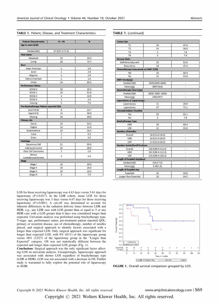
LOS for those receiving laparoscopy was 4.63 days versus 5.61 days forlaparotomy (P= 0.017). In the LDR cohort, mean LOS for thosereceiving laparoscopy was 3 days versus 6.67 days for those receivinglaparotomy (P= 0.001). A cut-off was determined to account forinherent differences in the radiation delivery times between LDR andHDR, e.g., any LDR case with LOS greater than or equal to 5 or anyHDR case with a LOS greater than 6 days was considered longer thanexpected. Univariate analysis was performed using brachytherapy type,T-stage, age, performance status, pre-treatment patient reported QOL,primary or recurrent disease, use of chemotherapy, number of needlesplaced, and surgical approach to identify factors associated with alonger than expected LOS. Only surgical approach was significant forlonger than expected LOS, with 0% (0/11) of the laparoscopy groupversus 48% (12/27) of the laparotomy group in the “Longer thanExpected” category. OS was not statistically different between theexpected and longer than expected LOS groups (Fig. 1).Conclusions: Surgical approach was the only significant factor affect-ing LOS on univariate analysis. Unsurprisingly, laparoscopic approachwas associated with shorter LOS regardless of brachytherapy type(LDR or HDR). LOS was not associated with a decrease in OS. Furtherstudy is warranted to fully explore the potential role of laparoscopyin SGIB. FIGURE 1. Overall survival comparison grouped by LOS.
TABLE 1. Patient, Disease, and Treatment Characteristics TABLE 1. (continued)
American Journal of Clinical Oncology � Volume 44, Number 10, October 2021 Abstracts
Copyright © 2021 Wolters Kluwer Health, Inc. All rights reserved. www.amjclinicaloncology.com | S69
Copyright r 2021 Wolters Kluwer Health, Inc. All rights reserved.

(P064) Characteristics and Early Outcomes of the First 100Patients with Cervical Cancer at the National CancerCenter of CambodiaJacob Gardner1, Rath Beauta, MD2, Poly Bunpa, MD2, Mora Mel,MD2, Vannyat Ath2, Sarameth Thou, MD2, Tracy Sherertz, MD3; 1CaseWestern Reserve University School of Medicine, 2National CancerCenter, Calmette Hospital, 3Kaiser Permanente WashingtonBackground: Cervical cancer is the most common cancer reportedamong women in Cambodia, with an estimated 1500 new cases and 800deaths annually. The history of cancer care in Cambodia is unfortu-nately dominated by the Khmer Rouge period from the mid-1970s untilthe end of the 1990s, during which upwards of 25% of the populationwas killed. Another casualty was the country’s healthcare infrastructure;hospitals were abandoned, equipment was destroyed, and healthcareprofessionals were persecuted. On January 15th, 2018, Cambodiaopened their first dedicated cancer hospital - the National Cancer Center(NCC) - at Calmette Hospital in Phnom Penh to provide modernmultidisciplinary cancer care to Cambodian citizens and advancedoncology training for Cambodian health care professionals.Objectives: We sought to gather descriptive statistics on the demo-graphic, diagnostic, treatment, and outcome data of the first 100 patientswith cervical cancer under care at the NCC.Methods: The medical records of the first 100 patients with cervicalcancer treated at the NCC were reviewed for demographic, diagnostic,treatment, and outcome data. Data was drawn from archived paperrecords and from electronic records on the ARIA Oncology InformationSystem. Descriptive statistics of each metric were calculated in Excel,and included frequency, range, mean and median.Results: Median age was 55 years. 40% of patients lived in PhnomPenh. 23% of patients presented with stage I disease, 35% with stage IIdisease, 20% with stage III disease, and 14% with stage IV disease.42% of patients were treated with definitive surgery, 89% receivedsystemic chemotherapy, 96% received external beam radiation therapy,and 73% received brachytherapy. Of the patients who received che-motherapy, 49.44% were treated concurrently with radiation therapy.For those patients who received definitive-intent radiotherapy, themedian cumulative dose (EQD2) from external beam and brachytherapywas as follows: HRCTV D90: 90.8 Gy, bladder maximum 2 cc:77.45 Gy, and rectal maximum 2cc: 69.95 Gy. The median overalltreatment time was 148 days. The median follow-up period was402 days. For the 25 patients for whom follow-up data has been col-lected, the frequency of acute toxicity - assessed at each patient’s lastfollow-up - was as follows: 25 (100%) had Grade 1 bladder toxicity; 21(84%) had Grade 1 rectal toxicity; & 4 (16%) had Grade 2 rectal tox-icity. For the 14 patients with at least 1 year follow-up, 13 (93%) werealive without recurrence at 12 months.Conclusions: The opening of the NCC is a watershed moment forimproving access to cancer care in Cambodia. The data presented hereillustrate future challenges for care and hope for improving outcomesfor patients with cervical cancer in Cambodia. This early sample servesas an initial step toward gaining a better understanding of the patientpopulation in Cambodia, and also highlights a need to continue out-comes-based review as the NCC broadens its scope and capacity toprovide care to its patients.
(P065) Human Papillomavirus Infection Among Cambo-dian Patients with Cervical CancerJacob Gardner1, Rath Beauta, MD2, Michel Pluot, MD3, SaramethThou, MD2, Aun Korn, MD3, Jae Jung, PhD4, Tracy Sherertz, MD5;1Case Western Reserve University School of Medicine, 2NationalCancer Center, Calmette Hospital, 3Calmette Hospital, 4LernerResearch Institute, Cleveland Clinic Foundation, 5Kaiser PermanenteWashingtonBackground: Cervical cancer is the most common cancer reportedamong women in Cambodia, with an estimated 1500 new cases and 800deaths annually. Cervical cancer can be prevented by vaccinationagainst its primary causative agent: human papillomavirus (HPV).Cambodia has yet to implement a nationwide HPV vaccination cam-paign, in large part because it remains unknown which subtypes of HPVpose the greatest oncogenic threat to the Cambodian population. Dozens
of oncogenic HPV subtypes have been identified around the world, andthe prevalence and distribution of these various subtypes can varydrastically across geographic regions. It is therefore important to betterunderstand the prevalence of various oncogenic HPV subtypes amongCambodian patients who develop cervical cancer so that the mosteffective vaccination policy can be developed.Objectives: 1). To determine which subtypes of HPV are represented ina survey of 100 patients with biopsy-proven cervical cancer at CalmetteHospital in Phnom Penh, Cambodia. 2). To help inform HPV vacci-nation policy in Cambodia.Methods: HPV DNA is isolated from the cervical tumor biopsies of100 patients from Calmette Hospital in Phnom Penh, Cambodia afterhistological confirmation of cervical squamous cell carcinoma or ade-nocarcinoma. Fifty samples comprise the “retrospective” cohort, col-lected from the archives of Calmette’s Pathology Department. Fiftyadditional samples comprise the “prospective” cohort, collected fromnew patients presenting to Calmette for evaluation and diagnosis ofcervical cancer. A microtome is used to section 10, 10μm-thick curlsfrom paraffin blocks containing each patient’s biopsy tissue. Commer-cially available kits are used to deparaffinize the tissue samples andextract and isolate the HPV DNA. A polymerase chain reaction bead-based assay is used to amplify and genotype the viral DNA and identifywhich subtypes are present in each tissue sample. For each HPV sub-type detected, the prevalence among the study population is calculated.Results: This study is still being carried out and has not produced anyresults yet. We hypothesize that the HPV subtype prevalence dis-tribution among patients with cervical cancer in Cambodia will besimilar to that of patients in Cambodia’s neighbor countries Thailandand Vietnam, where such information is already known from priorstudies.Conclusions: The results of this study will be submitted to Cambodia’sMinistry of Health for consideration in the development of their HPVvaccination policy.
(P066) Incorporation of Vaginal Brachytherapy to ExternalBeam Radiotherapy in Adjuvant Therapy for High-RiskEarly-Stage Cervical Cancer: A Comparative StudyShahil Mehta, MD, MPH1, Boniface Yarabe, BA1, Omar Ragab, MD2,David Nusbaum, MD3, Muneaki Shimada, MD4, Laurie Brunette, MD1,Lynda Roman, MD1, Koji Matsuo, MD, PhD1; 1University of SouthernCalifornia, 2University of Southern California Keck School of Medi-cine, 3University of Chicago Medicine, 4Tohoku University, School ofMedicineBackground: For most patients with early stage T1-2 cervical cancer,treatment options include surgical intervention with a hysterectomy orconcurrent chemotherapy and external beam radiotherapy (EBRT) fol-lowed by a vaginal brachytherapy (VBT) boost. Following hysterec-tomy there is a role for adjuvant radiation therapy in patients withintermediate risk factors of lympho-vascular space invasion (LVSI),stromal invasion, and large tumor size. Concurrent chemotherapy andEBRT also benefit patients with the high risk factors of positive pelvicnodes, positive surgical margins and/or positive parametria. The role ofVBT boost for patients with high risk factors is unclear. The NationalComprehensive Cancer Network (NCCN) clinical practice guidelinessuggest that it may be most helpful for those with close or positivemargins, although the paucity of evidence is noted. Overall the literaturedoes not provide a clear answer as to whether adjuvant EBRT with VBTboost provides additional benefit to high risk early stage cervical cancerpatients. The objective of the current study was to examine trends,characteristics, and outcomes of additional VBT after EBRT for high-risk early-stage cervical cancer in the United States.Objectives: To examine trends, characteristics, and outcomes related toaddition of vaginal brachytherapy (VBT) to external beam radiotherapy(EBRT) for adjuvant radiotherapy in high-risk early-stage cervicalcancer.Methods: This comparative study is a retrospective observationalanalysis of the National Cancer Institute’s Surveillance, Epidemiology,and End Results (SEER) Program. Surgically-treated women with stageT1-2 cervical cancer who had high-risk factors (nodal metastasis and/or
Abstracts American Journal of Clinical Oncology � Volume 44, Number 10, October 2021
S70 | www.amjclinicaloncology.com Copyright © 2021 Wolters Kluwer Health, Inc. All rights reserved.
Copyright r 2021 Wolters Kluwer Health, Inc. All rights reserved.

parametrial invasion) and received adjuvant radiotherapy from 2000-2018 were examined. Propensity score inverse probability of treatmentweighting was used to assess the survival estimates for addition ofVBT use.Results: Among 2,470 women with high-risk factors receiving EBRT, 760(30.8%) had additional VBT. During the study period, there was anincreasing trend of VBT use from 27.4% to 36.1% (P<0.001). In a mul-tivariable analysis, year of diagnosis and high-risk tumor factors: para-metrial involvement, large tumor size, and use of chemotherapy remainedindependent characteristics associated with VBT use (all, P<0.05). Inpropensity score weighted models, VBT use with EBRT and EBRT alonehad comparable overall survival (OS) (5-year rates 73.8% versus 77.4%,hazard ratio 1.07, 95% confidence interval 0.92-1.25). Non-significantassociation was also observed in squamous vs non-squamous tumors,young vs old age, low vs high lymph node ratio, chemotherapy use, andsimple or radical hysterectomy (all, P>0.05). Lastly, the addition of VBTwas not associated with cervical cancer-specific survival (subdistributionhazard ratio 1.15, 95% confidence interval 0.94-1.41, P=0.170).Conclusions: Utilization of VBT with EBRT for adjuvant radiotherapyin high-risk early stage cervical cancer is increasing in the UnitedStates. Addition of VBT is associated with neither overall survival norcancer specific survival.
(P067) Clinical and FDG-PET Tumor CharacteristicsAssociated with Early Versus Late Distant Metastasis Fol-lowing Curative Chemoradiation of Cervical CancerSuvidya Lakshmi Pachigolla, BSE1, Alexander Lin, MD/PhD2, LeslieMassad, MD1, Premal Thaker, MD1, David Mutch, MD1, MatthewPowel, MD1, Farrokh Dehdashti, MD1, Barry A SIEGEL SIEGEL,MD3, Julie Schwarz, MD/PhD1, Stephanie Markovina, MD/PhD1, PerryGrigsby, MD1; 1Washington University in St. Louis, 2WashingtonUniversity in Saint Louis, 3Washington UniversityBackground: Distant failure is the predominant pattern of failure forlocally advanced cervical cancer treated with modern image-guidedradiation therapy (Tan et al IJROBP 2019). Distant recurrence can be
apparent as early as 3 months after completion of therapy (Schwarz et alIJROBP 2012), which may signal an aggressive tumor biology. Iden-tification of factors associated with early distant metastasis could helpselect patients most likely to benefit from adjuvant systemic therapy.Objectives: To determine risk factors associated with early vs late dis-tant recurrence in cervical cancer following definitive chemoradiation.Methods: We included patients treated at an academic medical center withFIGO Stage IB-IVA cervical cancer staged with FDG-PET who completedcurative chemoradiation therapy with brachytherapy, with a minimum of3 months follow-up and restaging FDG-PET. Patients who subsequentlydeveloped biopsy-proven distant metastasis were categorized to have ear-liest (3-6mo), early (6-24mo) or late (>24mo) recurrence followingcompletion of therapy. Fisher’s exact test and Mann-Whitney U test werecomputed for categorical and continuous variables, respectively. Logisticregression was used to calculate odds ratios (ORs) with 95% confidenceintervals (CI). Kaplan-Meier analysis demonstrated differences in the timingof distant only vs concurrent distant + local failure.Results: Of 574 patients treated with curative chemoradiation, 143(25%) developed distant metastasis: 45 (31.5%) earliest, 65 (45.5%)early, and 33 (23%) late. Among these three groups, there were nosignificant differences in age, race, FIGO 2009 stage, metabolic tumorvolume, SUVmax, treatment length, or baseline/end-of-treatment squ-amous cell carcinoma antigen (SCCA) levels. New areas of FDG-uptake on 3-month post-treatment scan predicted the development ofdistant metastasis within 6 months in 86% of cases and 100% of casesby 2 years out from treatment. Compared with patients with late distantmetastasis, patients with earliest and early distant recurrences weremore likely to have PET-positive lymph nodes at diagnosis. Neitherpre-treatment nor end-of treatment variables were significantly differentcomparing the earliest vs early groups, so these patients were combinedfor comparison with the late group. Patients with distant metastasiswithin 2 years of radiation were more likely to have PET-positivelymph nodes at diagnosis (OR: 3.0, 95% CI 1.4–6.9, P= 0.006). Therewere 39 (27.3%) patients who also experienced local recurrence con-currently with their distant metastasis. Figure 1 shows that patients withearly distant metastasis were more likely to have concurrent local failurethan patients with late distant metastasis.
FIGURE 1. Kaplan-Meier curve showing time to recurrence stratified by distant alone recurrence vs. concurrent local and distant failure.
American Journal of Clinical Oncology � Volume 44, Number 10, October 2021 Abstracts
Copyright © 2021 Wolters Kluwer Health, Inc. All rights reserved. www.amjclinicaloncology.com | S71
Copyright r 2021 Wolters Kluwer Health, Inc. All rights reserved.

TABLE 1. Patient, Tumor, and Treatment Characteristics
*Number of Cases Missing Data: Metabolic Tumor Volume–49; Cervix SUV Max–27; SCCA at Diagnosis–78; Delta SCCA–90; SCCA at RT Completion–90
Abstracts American Journal of Clinical Oncology � Volume 44, Number 10, October 2021
S72 | www.amjclinicaloncology.com Copyright © 2021 Wolters Kluwer Health, Inc. All rights reserved.
Copyright r 2021 Wolters Kluwer Health, Inc. All rights reserved.

Conclusions: Lymph node metastasis at time of diagnosis was the onlydistinguishing risk factor for earliest/early vs. late distant metastasis.The biological mechanisms that trigger late metastasis deserve furtherexploration (Table 1).
(P068) Clinical and FDG-PET Tumor CharacteristicsAssociated with Early versus Late Distant Metastasis FollowingCurative Chemoradiation of Cervical CancerAlexander Lin, MD, PhD1, Suvidya Lakshmi Pachigolla, BSE2, LeslieMassad, MD2, Premal Thaker, MD2, David Mutch, MD2, MatthewPowel, MD2, Farrokh Dehdashti, MD2, Barry A Siegel Siegel, MD3,Julie Schwarz, MD, PhD2, Stephanie Markovina, MD, PhD2, PerryGrigsby, MD2; 1Washington University in Saint Louis, 2WashingtonUniversity in St. Louis, 3Washington UniversityBackground: Distant failure is the predominant pattern of failure forlocally advanced cervical cancer treated with modern image-guidedradiation therapy (Tan et al IJROBP 2019). Distant recurrence can beapparent as early as 3 months after completion of therapy (Schwarz et alIJROBP 2012), which may signal an aggressive tumor biology. Iden-tification of factors associated with early distant metastasis could helpselect patients most likely to benefit from adjuvant systemic therapy.Objectives: To determine risk factors associated with early vs latedistant recurrence in cervical cancer following definitive chemo-radiation.Methods: We included patients treated at an academic medical centerwith FIGO Stage IB-IVA cervical cancer staged with FDG-PET whocompleted curative chemoradiation therapy with brachytherapy, with aminimum of 3 months follow-up and restaging FDG-PET. Patientswho subsequently developed biopsy-proven distant metastasis were
categorized to have earliest (3-6 mo), early (6-24 mo) or late (> 24mo)recurrence following completion of therapy. Fisher’s exact test andMann-Whitney U test were computed for categorical and continuousvariables, respectively. Logistic regression was used to calculate oddsratios (ORs) with 95% confidence intervals (CI). Kaplan-Meier analysisdemonstrated differences in the timing of distant only vs concurrentdistant + local failure.Results: Of 574 patients treated with curative chemoradiation, 143(25%) developed distant metastasis: 45 (31.5%) earliest, 65 (45.5%)early, and 33 (23%) late. Among these three groups, there were nosignificant differences in age, race, FIGO 2009 stage, metabolic tumorvolume, SUVmax, treatment length, or baseline/end-of-treatment squ-amous cell carcinoma antigen (SCCA) levels. New areas of FDG-uptake on 3-month post-treatment scan predicted the development ofdistant metastasis within 6 months in 86% of cases and 100% of casesby 2 years out from treatment. Compared with patients with late distantmetastasis, patients with earliest and early distant recurrences weremore likely to have PET-positive lymph nodes at diagnosis. Neitherpre-treatment nor end-of treatment variables were significantly differentcomparing the earliest vs early groups, so these patients were combinedfor comparison with the late group. Patients with distant metastasiswithin 2 years of radiation were more likely to have PET-positivelymph nodes at diagnosis (OR: 3.0, 95% CI 1.4–6.9, P= 0.006). Therewere 39 (27.3%) patients who also experienced local recurrence con-currently with their distant metastasis. Figure 1 shows that patients withearly distant metastasis were more likely to have concurrent local failurethan patients with late distant metastasis.Conclusions: Lymph node metastasis at time of diagnosis was the onlydistinguishing risk factor for earliest/early vs. late distant metastasis.The biological mechanisms that trigger late metastasis deserve furtherexploration (Table 1).
FIGURE 1. Kaplan-Meier curve showing time to recurrence stratified by distant alone recurrence vs. concurrent local and distant failure.
American Journal of Clinical Oncology � Volume 44, Number 10, October 2021 Abstracts
Copyright © 2021 Wolters Kluwer Health, Inc. All rights reserved. www.amjclinicaloncology.com | S73
Copyright r 2021 Wolters Kluwer Health, Inc. All rights reserved.

TABLE 1. Patient, Tumor, and Treatment Characteristics
*Number of Cases Missing Data: Metabolic Tumor Volume–49; Cervix SUV Max–27; SCCA at Diagnosis–78; Delta SCCA–90; SCCA at RT Completion – 90.
Abstracts American Journal of Clinical Oncology � Volume 44, Number 10, October 2021
S74 | www.amjclinicaloncology.com Copyright © 2021 Wolters Kluwer Health, Inc. All rights reserved.
Copyright r 2021 Wolters Kluwer Health, Inc. All rights reserved.

(P069) Clinical Outcomes and Dosimetric Data for Patientswith Gynecologic Malignancies: A Comparison Between aProton and Photon Matched CohortAriel Pollock, MD1, Hunter Risher, MS2, Kai Sun, MS3, Ishan Oza4,Kimberly Marter, CMD1, David Alicia, CMD1, Gautam Rao, MD5, DanaRoque, MD5, Elizabeth Nichols, MD6, Pranshu Mohindra, MD, MBBS7;1Department of Radiation Oncology, University of Maryland MedicalCenter, 2University of Maryland School of Medicine, 3University ofMaryland School of Medicine Department of Epidemiology and PublicHealth, 4University of Maryland College Park, 5Division of GynecologicOncology, University of Maryland School of Medicine, 6Department ofRadiation Oncology, University of School of Medicine, 7Department ofRadiation Oncology, University of Maryland School of MedicineBackground: Radiation (RT) for treatment of gynecologic (gyn)malignancies confers a risk of gastrointestinal (GI), genitourinary (GU),and hematologic (heme) toxicity. Proton therapy (PT) may confersuperior dosimetric sparing of bowel, bladder and marrow than volu-metric arc therapy (VMAT), though supporting clinical data is lacking.Objectives: In this IRB-approved retrospective matched-pair analysis,we analyzed clinical outcomes and dosimetric differences betweenintensity modulated proton therapy (IMPT) and VMAT when treatinggyn patients.Methods: Patients with gyn malignancies treated with definitive oradjuvant whole-pelvic ( ± para-aortic) IMPT and VMAT between2014-2020 were analyzed. Patients with prior in-field RT wereexcluded. IMPT patients were matched 1:2 with a VMAT cohort (basedon receipt/timing of chemotherapy (ChT), RT field, and receipt ofsurgery). The Kaplan-Meier (KM) method was used to estimate overallsurvival (OS). A conditional logistic regression was performed to
compare incidence of grade 2+ and grade 3+ toxicity. A t-test analysiswas performed to compare mean dosimetric parameters.Results: The cohort included 28 patients treated with IMPT and 51treated with VMAT (total N= 79). Sites included uterus (n= 52), cervix(n= 16), vagina (n= 3), vulva (n= 5) and ovary (n= 3). Median initialdose for IMPT and VMAT was 50.4 and 45 Gy, respectively. Overall,43% of patients received adjuvant RT, 75% received ChT of which38% was concurrent. With a median follow up of 23 months, the 1-yearOS was 92% (95% CI 82-100%) and 88% (9% CI 80-96%) for IMPTand VMAT cohort, respectively, P= 0.6. The differences in incidenceof any grade 2+ acute toxicity (n=9, 32% vs n=18, 35%), any grade 3+acute toxicity (n=2, 7% vs n=10, 20%), any grade 2+ late toxicity (n=6,21% vs n=10, 20%) and GI/GU grade 3+ late toxicity (n=1, 4% vs n=4,8%) between IMPT and VMAT were not statistically significant. All acutegrade 3+ toxicity in proton cohort were heme while acute grade 3+ photontoxicities included heme, small bowel obstruction (SBO) and diarrhea. LateGI/GU grade 3+ toxicity in proton cohort were RT enteritis in one patientversus SBO (2 patients) and fistula (2 patients) in VMAT cohort. Upondosimetric comparison, bone marrow V10Gy (P < 0.001) and V20Gy (P< 0.001), small bowel V15Gy (P<0.001) and V45Gy (P<0.001) werelower with IMPT but rectum V40Gy was lower with VMAT (P=0.03).No significant differences were seen in bone marrow V40Gy and bladderV40Gy.Conclusions: This initial clinical report of gyn cancer patients treatedwith IMPT demonstrates excellent tolerance and efficacy with possiblesignal of acute (marrow) and late (GI/GU) toxicity reduction withIMPT, corroborated by dosimetric superiority of IMPT in reducingmarrow and small bowel dose. This hypothesis generating data supportsneed for an appropriately powered prospective trial to test utility ofIMPT (Fig. 1).
FIGURE 1. Acute and late grade 2+ and grade 3+ toxicity.
American Journal of Clinical Oncology � Volume 44, Number 10, October 2021 Abstracts
Copyright © 2021 Wolters Kluwer Health, Inc. All rights reserved. www.amjclinicaloncology.com | S75
Copyright r 2021 Wolters Kluwer Health, Inc. All rights reserved.

(P070) Patterns of Care and Outcomes of RadiotherapyAnd/or Hormone Therapy in Medically Inoperable Endo-metrial AdenocarcinomaLeonid Reshko, MD1, Jeremy Gaskins, PhD2, Alyssa Farley, BS2,Abbas Rattani, MD2, Grant McKenzie, MD2, Scott Silva, MD, PhD1;1Department of Radiation Oncology/University of Louisville, 2Universityof LouisvilleBackground: Medically inoperable endometrioid adenocarcinoma ofthe endometrium is a challenging clinical entity as definitive surgerywith risk-adaptive adjuvant therapy is the standard of care (Abu-Rustum, et al NCCN 2021 and Colombo, et al Int J of Gyn Cancer2016). It is unknown whether definitive radiotherapy (RT) and/or hor-mone therapy (HT) provide the optimal oncologic outcomes due to theabsence of randomized data (Schwarz, et al Brachytherapy 2015;Kupelian, et al IJROBP 1993; Podzielinski, et al Gynecologic Oncology2012; Nikolopoulos, et al Obstet Gynecol Sci 2020, Macchia, et alOncology Letters 2016).Objectives: The goal of this study is to evaluate the patterns of care andefficacy of RT and/or HT in the treatment of medically inoperableendometrial adenocarcinoma.Methods: We performed a query of the National Cancer Database(NCDB) of patients with medically inoperable endometrioid adeno-carcinoma of the endometrium diagnosed between 2004 and 2016.Included in this analysis were patients in which definitive surgery wasnot performed because it was contraindicated due to patient comor-bidities. Additionally, patients were only included if they receivedeither RT, HT or both; did not receive chemotherapy; were neither stage0 nor 4; were treated within 4 months of diagnosis and had a follow-uptime of at least 4 months. A Cox model was constructed to evaluate
TABLE 1. Multinomial Logistic Regression Model of Factors that Affect Hormone Therapy, Radiotherapy or Combination of HormoneTherapy and Radiotherapy Utilization
TABLE 2. Cox Proportional Hazards Analysis of Factors that AffectSurvival
Abstracts American Journal of Clinical Oncology � Volume 44, Number 10, October 2021
S76 | www.amjclinicaloncology.com Copyright © 2021 Wolters Kluwer Health, Inc. All rights reserved.
Copyright r 2021 Wolters Kluwer Health, Inc. All rights reserved.

survival after controlling for confounding variables. A multinomiallogistic regression model was used to assess predictors of RT and/orHT use.Results: A total of 1,063 patients were included in this cohort. Patientswho received RT compared with HT were more likely to be older, bediagnosed in earlier years, have government-issued insurance, be treatedat a lower-case volume center, have high-grade disease, have seriousmedical comorbidities and live outside of metro areas as shown inTable 1. Unadjusted 5-yr survival was 43.2% in patients receiving HTcompared with 37.3% in patients receiving RT. However, on multi-variate analysis, patients who received HT had worse overall survivalcompared with patients who received RT (adjusted hazard ratio (aHR)1.25; 95% CI 1.02–1.54; P= 0.0322). Only 38 patients received bothRT and HT compared with 275 who received HT alone and 750 treatedwith RT alone. There was borderline evidence that the combination ofRT and HT did worse than RT alone (aHR 1.48; 95% CI 1.00–2.19;P= 0.0524). Other factors associated with worse survival were olderage, presence of serious medical comorbidities, government-issuedinsurance, treatment at a lower-case volume center and high-gradehistology as shown in Table 2.Conclusions: We identified factors associated with the receipt of RTand/or HT in medically inoperable endometrial cancer patients. Thereceipt of RT appears to correlate with superior survival than treatmentwith HT alone when accounting for confounding variables. There wasan insufficient number of patients treated with a combination of RT andHT to draw a conclusion regarding the efficacy of this regimen.
(P071) Robotically Assisted Intra-Operative RadiationTherapyHouman Vaghefi, MD, PhD1, Pamela Stone, MD1, James Wheeler,MD, PhD1, John Lowden, DABR2, Samantha Korda, DABR2, IrinaSparks, MD2, Grace Darnell, NP2; 1Goshen Cancer Center, 2GoshenCenter for Cancer CareBackground: Robotic surgery is one of the standards of care in thesurgical management of gynecologic malignancy. One of the goals ofan oncologic resection is to achieve negative margins. However, insome instances an R0 resection is not achieved, which could lead toincreased local failure. Local control could be achieved by adjuvantradiotherapy, commonly delivered with external beam radiotherapy.Intra-operative Radiotherapy (IORT) is a technique where a singleintraoperative dose of radiation is delivered to the target. In roboticresection IORT is usually not considered due to small port sites that can
preclude the IORT applicator. However, transvaginal approach allowsadequate and safe physical space to deliver IORT.Objectives: We describe the first published report of robotic surgeryusing a transvaginal approach to safely deliver balloon based IORT.Methods: 75-year-old female with a nebulous past medical historysignificant for a gynecologic malignancy, treated with TAH/BSO in1994 and radiation therapy. She presented with vaginal pain anddrainage. Physical exam showed a mass in the fornix, extending to thevaginal cuff. Pelvic MRI revealed a 2.8 cm pelvic mass adjacent to thevaginal cuff (Fig. 1). PET/CT showed no metastasis. She underwent arobotically assisted radical upper vaginectomy, bilateral pelvic nodemapping and sampling, and bilateral pelvic node dissection. Intra-operatively, it became apparent that the resection of the tumor resultedin the dissection to the left levator ani muscle over the rectum withoutentry into the rectal mucosa. All margins were marked, and tissue wassent for rapid frozen section and were noted to be positive for tumor.Therefore, a decision was made to deliver IORT. The target adjacent tothe rectum was estimated to be superficial and within the adequate rangeof the 50KvP source. A 3-4 cm XOFT (TM) catheter was selected andfilled to 30mL. The IORT catheter was introduced with a transvaginalapproach after removal of the specimen. The catheter was dockedthrough the opened vaginectomy site and secured for IORT. Thebladder and the right ureter were tethered away from the radiation fieldby the robot. Trendelenburg position ensured that the bowel was movedout of the radiation field and 10 Gy was delivered (Fig. 2). The patienthad no apparent complications and was discharged in post-operativeday 1.Results: Pathology showed G2, moderately differentiated invasivesquamous cell carcinoma with the tumor originating at the vaginal cuffand extending deeply into the paravaginal tissue, positive for LVI andPNI. The deep margin was positive, with 14 negative nodes. She wasstaged to have a rpT2bN0 squamous cell carcinoma.Conclusions: For select patients who have undergone prior radiationtherapy and whose imaging studies indicate a high risk for encounteringpositive margins at the time of salvage surgery, IORT may be a feasibletreatment option using the XOFT balloon applicator even in con-junction with robotic surgery.
(P072) Vulvar Recurrence and Survival in PatientsUndergoing Adjuvant Radiation for High-Risk VulvarCancerAlison Yoder, MD, MPH1, David Lakomy, BA2, Kelsey Corrigan, MD,MPD1, Anuja Jhingran, MD1, Ann H. Klopp, MD, PhD3, MelissaJoyner, MD, MBA4, Lauren Colbert, MD, MSCR1, Patricia Eifel, MD1,Lilie Lin, MD1; 1MD Anderson Cancer Center, 2Dartmouth CollegeGeisel School of Medicine, 3The University of Texas MD AndersonCancer Center, 4M.D. Anderson Cancer CenterBackground: Vulvar cancer is rare and primarily treated with surgicalresection. Adjuvant radiation therapy (RT) is recommended for patientswith close or positive margins or positive nodal disease.
FIGURE 1. MRI Axial T1 post-constrast image of the mass adjacentto the vaginal cuff.
FIGURE 2. Intra-operative photo representing the IORT catheter,introduced through the vagina, secured by the robotic arm, whilethe bladder is tethered away out of the radiation field.
American Journal of Clinical Oncology � Volume 44, Number 10, October 2021 Abstracts
Copyright © 2021 Wolters Kluwer Health, Inc. All rights reserved. www.amjclinicaloncology.com | S77
Copyright r 2021 Wolters Kluwer Health, Inc. All rights reserved.

Objectives: We evaluated demographic, clinical, and treatment varia-bles that might be associated with vulvar recurrence (VR) in patientswith vulvar cancer treated with postoperative RT with or withoutchemotherapy.Methods: After obtaining IRB approval, we retrospectively reviewedthe records of 69 vulvar cancer patients who were treated at our insti-tution with vulvar resection followed by RT between 1999 and 2019.The median total dose of RT to the vulva was 54 Gy (range 40-64) in 25(20-33) fractions, with median RT duration of 39 days (27-53). RTboost was given to the vulva in the majority of cases with close orpositive margins, median dose of 6 Gy (0-20). All time variables werecalculated from the start of RT. Variables associated with VR wereanalyzed using cox proportional hazard model. The median follow-upduration was 53 months.Results: Most patients were White (n= 46/69, 67%) and the majority(n= 60, 87%) had squamous cell carcinomas. Thirteen patients (18.6%)had a history of lichen sclerosis (LS) diagnosed either before theircancer diagnosis or at time of surgical resection. Most patients had onlyone prior surgical resection before RT (n= 44, 64%), though 22 (32%)had two prior resections and 3 (4%) underwent 3 total resections beforeRT. Fourteen patients had VR after receiving adjuvant RT; median timeof 15 months. Patients with positive margins (HR 0.66, P= 0.50), non-squamous histopathology (HR 0.51, P= 0.52), or who required > 1surgery (HR 1.20, P= 0.73) were not at an increased risk of VR.Patients with positive lymph nodes and those with LS were more likelyto have VR (HR 8.7, P= 0.04; HR 4.1, P= 0.01, respectively). Of the14 patients with VR, 6 (43%) had LS. Patients with LS were more likelyto be older (mean age 71 vs 59, P= 0.1). Of the 25 patients who hadmultiple prior resections, 13 (52%) had re-excisions for positive
margins while 12 (48%) were treated for recurrence after prior definitivesurgery. In the 13 patients who required re-excision, 8 had negativemargins after re-excision. Margin status after re-excision was notassociated with VR (HR 1.48, P= 0.78). The risk of VR was notincreased if patients required re-excision compared with all otherpatients (HR 0.64, P= 0.56). Patients were not at an increased risk ofVR if they were recurrent at time of presentation (HR 1.87, P= 0.29).At last contact, 48 patients (70%) had no evidence of disease, 13 (19%)had disease, and 8 (12%) had unknown disease status. Eight patients inthis cohort died of disease.Conclusions: Re-excision to achieve negative margins or delivery ofadditional boost dose to areas of close or positive margins appear tobe effective strategies to reduce the risk of VR. The presence oflichen sclerosis is a strong predictor of vulvar relapse and maywarrant investigation as an indication for treatment intensification(Figs. 1 and 2).
(P073) Two-year Patient-Reported Outcomes After Ipsi-lateral IMRT for T1-2 N2b Squamous Cell Carcinoma ofthe TonsilsChike Abana, MD, PhD1, Adam Garden, MD1, Andrew Frankart, MD2,Natalie Geier, MD3, Houda Bahig, MDCM, PhD4, Gary Gunn, MD1,Abdallah S.R. Mohamed, MD1, Vinita Takiar, MD, PhD2, Jack Phan,MD, PhD1; 1The University of Texas M.D. Anderson Cancer Center,2University of Cincinnati, 3The Christ Hospital, 4University of MontrealHospital CenterBackground: Although unilateral neck irradiation is frequentlyimplemented in patients with well-lateralized stage T1-2 HPV+ tonsillarsquamous cell carcinoma (SCC) to minimize toxicity, its role in patientswith N2b (AJCC-7 staging) nodal disease remains controversial. Wehave separately reported on contralateral nodal recurrence risk inpatients with N2b disease receiving unilateral neck radiation. Thepatient reported outcomes (PRO) in these patients have not been pre-viously reported.Objectives: To evaluate the quality of life PRO profile in patients withAJCC 7th edition T1-2 N2b tonsillar squamous cell carcinoma treatedwith unilateral neck radiation.Methods: The PROs of 44 AJCC-7 T1-2 N2b tonsillar squamous cellcarcinoma (SCC) patients from two academic institutions treated withdefinitive, ipsilateral intensity modulation radiation therapy (IMRT)with or without systemic therapy were reviewed retrospectively. PROswere completed using either the MD Anderson Symptom Inventory-Head and Neck module (MDASI-HN) at the first institution, or theEuropean Organization for Research and Treatment of Cancer Qualityof Life Questionnaire Head and Neck Module 35 (EORTC QLQ-H&N35) at the second institution. The primary endpoint was the headand neck-related PROs at 2 years after completion of radiation.Results: Twenty-eight of 30 and 4 of 14 patients from the first andsecond institutions, respectively, met the primary endpoint and werefurther analyzed. Of these, 34.4% received radiation alone withoutsystemic therapy and 93.7% received ipsilateral-only neck IMRT. Themost common head and neck-related PRO was dry mouth at the firstand second institutions (64.2% and 100%, respectively); the leastcommon was difficulty with voice or speech at the both institutions(14% and 0%, respectively). There were also no reports of mouth painor soreness, trouble with teeth, opening mouth, or swallowing liquid/pureed food at the second institution. The overall symptom severity forall PRO categories in both modules were in the mild range, with thehighest mean score reported for dry mouth: 2.54 out of 10 on MDASI-HN for the first institution and 2.5 out of 4 on EORTC QLQ-H&N35for the second institution. The rest of the mean scores for the firstinstitution were < 1.5, while the remainder for the second institutionwere ≤ 2. The locoregional control and overall survival after a medianfollow-up of 4.5 years were each 100%.Conclusions: We assessed the 2-year PROs for ipsilaterally radiatedtonsillar SCC from 2 institutions using either MDASI-HN or EORTCQLQ-H&N35, and most reported mild symptom burden. These findingssuggest that ipsilateral-only IMRT is well tolerated in patients withAJCC-7 N2b disease and can be safely employed as a de-escalationstrategy.
FIGURE 1. Lichen sclerosis and vulvar recurrence. Kaplan-Meiercurve comparing vulvar recurrence in patients going throughadjuvant radiation for vulvar carcinoma with and without lichensclerosis, log-rank P=0.005.
FIGURE 2. Vulvar recurrence and re-excision. Kaplan-Meier curvecomparing vulvar recurrence in patients that required re-excisionfor positive margins compared with those that did not receive re-excision, log-rank P=0.056.
Abstracts American Journal of Clinical Oncology � Volume 44, Number 10, October 2021
S78 | www.amjclinicaloncology.com Copyright © 2021 Wolters Kluwer Health, Inc. All rights reserved.
Copyright r 2021 Wolters Kluwer Health, Inc. All rights reserved.

(P074) Association Between Radiation Dose to AdjacentOrgans at Risk and Acute Patient Reported OutcomeDuring Radiation Treatment for Head and Neck CancersMona Arbab, MD1, Yu-Hui Chen, PhD2, Roy Tishler, MD2, JasonGlass2, Jo Ann Fugazzotto2, Joseph Killoran, PhD3, Rosh Sethi, MD2,Eleni Rettig, MD3, Donald Annino, MD3, Laura Goguen, MD3, Rav-indra Uppaluri, MD2, Carolyn Hsu2, Elaine Burke3, Glenn Hanna,MD2, Jochen Lorch, MD2, Robert Haddad, MD2, Danielle Margalit,MD2, Jonathan Schoenfeld, MD2; 1Indiana University, 2Dana FarberCancer Institute, 3Brigham and Women’s HospitalBackground: Anticipating patient-reported side effects during radiationtherapy (RT) based on RT dose to Organs At Risk (OARs) may pro-mote focused patient-centered management during RT.Objectives: We studied the association between dose to OARs andpatient reported outcomes (PRO) measured during head and neck RT.Methods: We performed a retrospective evaluation of the associationbetween dose to OARs and PROs obtained weekly during treatment inhead and neck cancer patients receiving definitive or postoperativeradiation treatment with at least three prospectively-acquired PROsurveys.Results: Between 2015-2018, 169 patients had definitive (61%) orpostoperative (39%) intensity modulated radiation therapy (IMRT) forHead & Neck (H&N) cancer and completed at least 3 PRO surveysduring RT. The majority of patients had oropharyngeal SCC (50%). Weidentified associations between patient reports of: “Difficulty swal-lowing/chewing” and increased mean RT dose to the oral cavity, larynxand pharyngeal constrictor muscles (PCM) (P= 0.0005, P= 0.02 andP= 0.046 respectively); “choking/coughing” and larynx mean dose(P= 0.003); “problems with mucus in mouth and throat” and oralcavity, parotid and PCM mean dose (P< 0.001, 0.002, 0.04 respec-tively); “difficulty with voice/speech” and oral cavity, parotid and lar-ynx mean dose (P= 0.03, 0.03 and 0.02); and “dry mouth” and oralcavity and PCM mean dose (P= 0.0002, 0.02). There were no associ-ations between PROs and T-classification, smoking and whether RTwas delivered definitively or in the adjuvant setting.Conclusions: Increased OAR dose to oral cavity, larynx, PCM andparotids are associated with increased severity of patient-reportedsymptoms including difficulty swallowing/chewing, choking/coughing,problem with mucus in mouth and throat, difficulty with voice/speechand dry mouth. Evaluating dose to OARs can assist in anticipatingpatient-reported side effects of therapy and promote patient-specificacute management.
(P075) Who Really Receives Single Modality Therapy?Oropharynx CancerNina Burbure, MD1, Mengying Deng1, Elizabeth Handorf, PhD1, JohnRidge, MD PhD1, Christopher Yao, MD1, Jessica Bauman, MD1,Anshu Giri, MD1, Jeffrey Liu, MD1, Thomas Galloway, MD1; 1FoxChase Cancer CenterBackground: Either radiation (RT) or transoral surgery + neck dis-section promise the potential of single modality curative therapy forappropriately selected oropharynx cancer patients. How many patientsare actually managed with single modality therapy is unknown.Objectives: We queried the National Cancer Database (NCDB) todetermine what proportion of early stage oropharynx cancer patientsreceive single modality therapy and to identify factors associated withmulti-modality therapy in this group of patients.Methods: We identified 4678 patients treated curatively for early stage(7th ed cT1-T2, cN0-N1) oropharyngeal squamous cell carcinomabetween 2010-2016. For those treated with primary surgery, the oddsratio (OR) of requiring adjuvant therapy was determined using logisticregression and adjusting for age, sex, race, comorbidity score, insurancestatus, facility type, diagnosis year, disease subsite, clinical T and Ncategory and HPV status. Multinomial logistic regression was used todetermine the association between covariates and specific adjuvanttherapies (chemoradiation [CRT] or RT alone). For the primary RTgroup, surgery within 20 weeks of RT completion was consideredmulti-modality.
Results: A majority of patients were managed with primary surgery(2554/4678, 55%). Patients managed with surgery were younger (meanage 61 vs 66 y old) with smaller primaries (T1: 51% v 31%,P= < 0.0001). HPV status was known in 2910 patients, of which 63%had HPV positive tumors. Surgery within 20 weeks of definitive radi-ation was rare (13/2124, 0.6%). By contrast 51% of surgical patientsreceived adjuvant therapy and nearly half (46%) of these patientsreceived adjuvant CRT (24% of all surgical patients). Factors associatedwith increased likelihood of adjuvant therapy include treatment at acommunity center (versus academic), tonsil subsite, T2 stage desig-nation and N1 stage designation. The odds ratios of requiring adjuvanttherapy (vs no adjuvant therapy) for these variables are tabulated. Foradjuvant CRT specifically, the OR was increased significantly forclinical N1 disease (8.37, P= < 0.0001). In the group with known HPVstatus, the OR for requiring adjuvant therapy was increased for HPVpositive tumors (1.39, P= 0.0076).Conclusions: A majority of early stage oropharynx cancer patients whoundergo transoral surgery receive adjuvant therapy, while <1% ofsimilarly staged patients require adjuvant surgery following RT. Moreadvanced disease is associated with a higher odds ratio for adjuvanttherapy after surgery (Table 1).
(P076) Prospective Evaluation of Oral Cavity DosimetricParameters as Predictors of Quality of Life Outcomes inPatients Undergoing Chemoradiation for Locally AdvancedOropharyngeal Squamous Cell CarcinomaAndrew Cook, MD1, Ankit Modh, MD2, Haythem Ali, MD3, JawadSheqwara, MD3, Steven Chang, MD4, Tamer Ghanem, MD, PhD4,Suhael Momin, MD4, Vivian Wu, MD4, Samantha Tam, MD, MPH4,Xiaoxia Han, PhD5, Lamis Fakhoury, BA6, Benjamin Movsas, MD1,Farzan Siddiqui, MD, PhD, CPE1; 1Department of Radiation Oncology,Henry Ford Cancer Institute, 2Department of Radiation Oncology,Carle Cancer Institute, 3Department of Hematology/Oncology, HenryFord Cancer Institute, 4Department of Otolaryngology, Henry FordCancer Institute, 5Department of Public Health Sciences, Henry FordCancer Institute, 6Henry Ford Cancer InstituteBackground: Concurrent chemoradiation (chemoRT) for orophar-yngeal squamous cell carcinoma (OPSCC) has improved survival;however, this intensive treatment comes at the expense of increasedtoxicity and worse quality of life (QOL). Data from a prospectiverandomized trial was used to assess the impact of oral cavity (OC) doseon QOL outcomes.Objectives: Based off this prospective data, we would like to report onthe association between OC dose and quality of life outcomes.Methods: All patients (n= 56) had AJCC 7th ed stage III-IV OPSCCand underwent concurrent chemoRT. RT dose was 70 Gy in 35 frac-tions using a VMAT technique. To assess QOL, patients completed thePatient-Reported Oral Mucositis Symptom (PROMS) scale at baseline,weekly during treatment, and at 6-week follow-up (higher scoresindicate worse QOL). The OC was contoured using consensus guide-lines (Brouwer, et al Radiother Oncol 2015). For dosimetric data, wecollected OC max and mean doses (Gy) and volume (%) receiving x Gy(Vx) in 10 Gy increments. PROMS scores at week 7 (end of treatment)and follow-up timepoints were used for analysis. In addition to the totalPROMS score, we analyzed question 1 individually (PROMS1-mouthpain) and questions 4, 5, 6, and 9 combined (PROMS4569-difficulty
TABLE 1. Odds Ratio of Receiving Adjuvant Therapy AfterSurgery for Several Variables
American Journal of Clinical Oncology � Volume 44, Number 10, October 2021 Abstracts
Copyright © 2021 Wolters Kluwer Health, Inc. All rights reserved. www.amjclinicaloncology.com | S79
Copyright r 2021 Wolters Kluwer Health, Inc. All rights reserved.

eating and swallowing). PROMS scores were dichotomized at themedian to create two comparison groups, and dosimetric differenceswere assessed using Wilcoxon-rank sum tests (P< 0.05 consideredsignificant).Results: At the week 7 endpoint, there were no differences noted in OCdoses when the total PROMS score was dichotomized at the median.For PROMS1, there were significant dosimetric differences noted in OCmean dose (56.8 Gy vs 62.2 Gy, P= 0.04) and V50 (64.7% vs 81.5%,P= 0.05) in the low and high score arms, respectively. Additionally,differences in OC V40 (84.8% vs 92.7%, P= 0.08), V60 (48.6% vs71.2%, P= 0.05), and V70 (28.4% vs 40.4%, P= 0.06) trended towardssignificance in the low and high score arms, respectively. ForPROMS4569, there were no significant differences noted in the dosi-metric parameters. At follow-up examining total PROMS score, therewas a significant difference in OC V70 (25.6% vs 38.2%, P= 0.05) inthe low and high score arms, respectively. Additionally, OC V60(47.8% vs 61.1%, P= 0.09) trended towards significance in the low andhigh score arms, respectively. For PROMS1 at follow-up, there were nosignificant differences noted in the dosimetric parameters. There werealso no significant findings for PROMS4569 at follow-up; however, OCV70 (27% vs 38.2%, P= 0.09) trended towards significance in the lowand high score arms, respectively.Conclusions: We found that patients who had worse QOL scores at theend of treatment, especially related to mouth pain, had significantlyhigher OC mean and V50 dosimetric parameters. Additionally, higherOC V60 and V70 parameters may predict for longer term detriment toQOL. These variables should be optimized during treatment planning toimprove outcomes.
(P077) 18FDG Positron Emission Tomography Mining forMetabolic Imaging Biomarkers of Radiation-inducedXerostomia in Patients with Oropharyngeal CancerClifton Fuller, MD, PhD1, Hesham Elhalawani, MD, MSc2, CarlosCardenas, PhD1, Stefania Volpe, MD3, Souptik Barua, PhD3, SonjaStieb, MD3, Calvin Rock, MD3, Timothy Lin, MD3, Pei Yang, MD3,Haijun Wu, MD3, Jhankruti Zaveri, BS3, Baher Elgohari, MD3, LamiaaAbdallah, MD3, Amit Jethanandani, MD3, Abdallah S.R. Mohamed,MD4, Laurence E. Court, PhD1, Katherine Hutcheson, PhD3, GaryGunn, MD4, David Rosenthal, MD1, Steven Frank, MD1, Adam Gar-den, MD4, Arvind Rao, PhD5; 1MD Anderson Cancer Center, 2Brighamand Women’s Hospital/Dana-Farber Cancer Institute, 3University ofTexas MD Anderson Cancer Center, 4The University of Texas M.D.Anderson Cancer Center, 5University of MichiganBackground: Head and neck cancers radiotherapy (RT) is associatedwith inevitable injury to parotid glands and subsequent xerostomia.
Objectives: We investigated the utility of SUV derived from 18FDG-PET to develop metabolic imaging biomarkers (MIBs) of RT-relatedparotid injury.Methods: Data for oropharyngeal cancer (OPC) patients treated withRT at our institution between 2005-2015 with available planningcomputed tomography (CT), dose grid, pre- & first post-RT 18FDG-PET-CT scans, and physician-reported xerostomia assessment at3-6 months post-RT (Xero 3-6 ms) per CTCAE, was retrieved, fol-lowing an IRB approval. A CT-CT deformable image co-registrationfollowed by voxel-by-voxel resampling of pre & post-RT 18FDGactivity and dose grid were performed. Ipsilateral (Ipsi) and contra-lateral (contra) parotid glands were sub-segmented based on thereceived dose in 5 Gy increments, i.e. 0-5 Gy, 5-10 Gy sub-volumes,etc. Median and dose-weighted SUV were extracted from whole parotidvolumes and sub-volumes on pre- & post-RT PET scans, using in-housecode that runs on MATLAB. Wilcoxon signed-rank and Kruskal-Wallistests were used to test differences pre- and post-RT.Results: 432 parotid glands, belonging to 108 OPC patients treated withRT, were sub-segmented & analyzed. Xero 3-6 ms was reported as:non-severe (78.7%) and severe (21.3%). SUV-median values weresignificantly reduced post-RT, irrespective of laterality (P= 0.02). Asimilar pattern was observed in parotid sub-volumes, especially ipsiparotid gland sub-volumes receiving doses 10-50Gy (P<0.05). Kruskal-Wallis test showed a significantly higher mean RT dose in the contraparotid in the patients with more severe Xero 3-6mo (P= 0.03). Multiplelogistic regression showed a combined clinical-dosimetric-metabolicimaging model could predict the severity of Xero 3-6mo; AUC=0.78(95%CI: 0.66-0.85; P<0.0001).Conclusions: We sought to quantify pre- and post-RT 18FDG-PETmetrics of parotid glands in patients with OPC. Temporal dynamics ofPET-derived metrics can potentially serve as MIBs of RT-relatedxerostomia in concert with clinical and dosimetric variables (Figs. 1–3).
FIGURE 1. Parotid dose sub-volume generation.
FIGURE 2. Illustration of dose-weighted SUV computation.
FIGURE 3. Correlation between severity of post-radiotherapyxerostomia and (A) Age; (B) Mean contralateral parotid gland dose(Gy); (C) Post-radiotherapy contralateral parotid gland SUV-median;and (D) Post-radiotherapy ipsilateral parotid gland SUV-median.
Abstracts American Journal of Clinical Oncology � Volume 44, Number 10, October 2021
S80 | www.amjclinicaloncology.com Copyright © 2021 Wolters Kluwer Health, Inc. All rights reserved.
Copyright r 2021 Wolters Kluwer Health, Inc. All rights reserved.

(P078) Assessing Oral Intake Outcomes in Head and NeckCancer Patients Treated with Definitive Radiation with orWithout ChemotherapyRicardo Engel, BS1, Patrick Etta, BS1, Talha Gahzi, BS2, Amy Wil-liams, PhD3, Samantha Tam, MD, MPH4, Suhael Momin, MD4, StevenChang, MD4, Tamer Ghanem, MD, PhD4, Diana Walker, MA5, FarzanSiddiqui, MD, PhD, CPE6, Haythem Ali, MD7, Vivian Wu, MD4,Jawad Sheqwara, MD7, Megan Carlson, MS8; 1Wayne State UniversitySchool of Medicine, 2Michigan State Univ College of Human Medicine,3Department of Otolaryngology-Head and Neck Surgery, Henry FordHospital, 4Department of Otolaryngology, Henry Ford Cancer Insti-tute, 5Speech Language Pathology and Otolaryngology, Henry FordHealth System, 6Department of Radiation Oncology, Henry FordCancer Institute, 7Department of Hematology/Oncology, Henry FordCancer Institute, 8Speech-Language Pathology, Henry Ford HealthSystemBackground: Head and neck cancer treatment modalities can sig-nificantly impact functional outcomes of patients, especially oral intake(Brizel, et al N Engl J Med 1998; Kamal, et al Support Care Cancer2019). Radiation therapy in particular has been associated with post-treatment xerostomia and dysphagia (Adelstein, et al J Clin Oncol 2003;Hutcheson, et al Cancer 2013) which can affect quality of life andimpair weight gain, contributing to worse long-term outcomes (Paya-kachat, et al Head Neck, 2013). Early speech-language pathologyintervention has shown to be effective in improving these functionaloutcomes in this population (Greco, et al Int J Radiat Oncol BiolPhys 2018).Objectives: The purpose of this study is to evaluate oral intake out-comes of patients undergoing definitive radiation therapy with orwithout chemotherapy for head and neck squamous cell carcinoma.Methods: A cohort of patients with stage III or IV squamous cellcarcinoma of the oropharynx, larynx, and hypopharynx treated withdefinitive radiation therapy with or without chemotherapy wereextracted from system database. Patients with evidence of distantmetastases were excluded. Swallow function was assessed both pre- andpost-treatment (within four months of treatment initiation or conclusion)with the Functional Oral Intake Scale (FOIS) (Crary et al, Arch PhysMed Rehabil, 2005) as measured by a Speech-Language Pathologist(SLP) involved in the patient’s care. Body mass index (BMI) wasevaluated within four months of treatment initiation and one year aftertreatment completion. The use of enteral feeding at one-year post-treatment was also assessed. Data was analyzed with descriptive stat-istical methods, Wilcoxon sign rank tests, and χ2d tests.
Results: The sample included 152 patients. Table 1 highlights patientbaseline characteristics, tumor location, and treatment. FOIS scoresdecreased from pre-treatment to post-treatment, with 75% of patientshaving a FOIS of 7 at pre-treatment compared with only 13.8% at thepost-treatment time point (Table 1). Median BMI also decreased frompre-treatment to one-year post-treatment (Table 2). At one-year post-treatment, 23.7% patients (n= 33) required enteral feeding.Conclusions: Definitive radiation therapy with or without chemo-therapy in the treatment of head and neck cancer is associated withimpaired oral intake. Treatment is also associated with decreases inBMI and longer use of enteral feeding, which may reflect sequelae ofimpaired oral intake. These factors have a negative impact on quality oflife and can lead to long-term morbidity. Integrative treatment planswould therefore benefit from speech-language pathology interventionsthroughout the treatment process.
(P079) Single Fraction Radiotherapy as PostoperativeTreatment (SF-PORT) for Resected, Stage I/II Merkel CellCarcinomaPeter Goff, MD, PhD1, Stephanie Schaub, MD1, Kristina Lachance,MS1, Emily Huynh1, Nikhil Harikrishnan1, Maclean Cook1, YolandaTseng, MD1, Jay Liao, MD1, Smith Apisarnthanarax, MD1, KentWallner, MD1, Paul Nghiem, MD, PhD1, Upendra Parvathaneni,MBBS, FRANZCR1; 1University of WashingtonBackground: In the absence of randomized prospective data, the role ofpostoperative radiotherapy (PORT) for localized Merkel cell carcinoma(MCC) is controversial with varying practice patterns. Although amajority of patients with resected stage I/II MCC without adversefeatures may be cured with surgery alone, those with adverse featuresincluding head and neck (HN) primary tumors have local recurrencerates of 20% or more without PORT (Takagishi et al Adv Radiat Oncol,2016). Extrapolating from our experience demonstrating high rates oflocal control with single fraction radiotherapy (SFRT) for metastaticMCC (Iyer et al Cancer Med, 2015), we hypothesized that SFRT (8 Gy)may be effective as PORT to optimize local control for stage I/II MCCwith adverse risk features [e.g. HN disease, immunosuppression,recurrent disease, non-oncologic resection, lymphovascular spaceinvasion (LVSI)], especially in patients for whom a conventional RTcourse may not be feasible.Objectives: Determine if single fraction radiotherapy (8 Gy) may beeffective as PORT to optimize local control for stage I/II Merkel cellcarcinoma with adverse risk features.Methods: A single-institution, retrospective review of patients withclinical or pathological stage I/II MCC receiving SFRT (8Gy) as post-operative treatment (SF-PORT) was completed from a prospectively-enrolled database.Results: Thirty patients (median age 78 y) were identified who receivedSFRT after wide local excision (n= 24; 80%), Mohs surgery (n= 1;3%), shave or excisional biopsy only (n= 5; 17%) of the primary.Patients had pathological stage I (n= 15; 50%), clinical stage I (n= 10:33%), pathological stage II (n= 3; 10%) or clinical stage II (n= 2; 7%)disease. All patients received SF-PORT (8 Gy) at a median of 43 days(range: 14-203) following resection or biopsy. Five patients (4 with HNdisease) received SFRT to draining lymph nodes. The majority ofpatients had HN tumors (77%), LVSI was present in 23%,17% wereimmunosuppressed and 7% had locally recurrent disease (n= 2). Atmedian follow-up of 11.3 months (range: 3-45.7), no local or in-field
TABLE 1. Description of Baseline Population
TABLE 2. Swallowing Outcomes and BMI
American Journal of Clinical Oncology � Volume 44, Number 10, October 2021 Abstracts
Copyright © 2021 Wolters Kluwer Health, Inc. All rights reserved. www.amjclinicaloncology.com | S81
Copyright r 2021 Wolters Kluwer Health, Inc. All rights reserved.

recurrences were observed. No local or distant recurrences, MCC spe-cific deaths, or RT related toxicities grade > 1 (CTCAE v5.0) wereobserved. There were two out-of-field regional nodal recurrences in HNpatients who did not receive elective nodal RT.Conclusions: Our early experience demonstrates a high rate of in-fieldlocal control in patients with stage I/II MCC with adverse featuresmanaged with SF-PORT. This approach warrants further study.
(P080) Sex Differences in Health Related Quality of Life inHead & Neck Cancer One Year After TreatmentMadeline Goosmann, MD1, Steven Chang, Attending1, Amy Williams,PhD2, Samantha Tam, MD, MPH3; 1Henry Ford Hospital, 2Departmentof Otolaryngology-Head and Neck Surgery, Henry Ford Hospital,3Department of Otolaryngology, Henry Ford Cancer InstituteBackground: Head and neck cancer (HNC) makes up about 3% of allcancers and is treated with systemic therapy, radiation, surgery, or acombination of these. HNC treatment can be associated with decreasedpatient reported health related quality of life (HR-QoL), which can leadto depression.1 The majority of studies found that females reportedworse patient reported HR-QoL than males,2-7 however, there were afew that did not have a significant difference in overall patient reportedQoL.8-9 With the discovery of patient oriented outcomes (PROs) inclinical practice affecting patient satisfaction, provider-patient rela-tionship, and overall patient mortality,10 it is vital to include PROs inthe creation of treatment plans.Objectives: The objectives of this project are to highlight the differ-ences in HR-QoL between men and women. Ultimately, using thesePROs clinically will help to improve patient care, augment patient-provider trust, and optimize treatment plans. Using PROs and recog-nizing where unconscious biases of providers come into play is pin-nacle, and this project aims to highlight how men and women’s expe-riences are different in the treatment of HNC.Methods: Participants were given the FACT-H&N instrument one yearafter treatment for head and neck cancer at a single tertiary academiccenter to assess different aspects of Hr-QoL. Sex differences wereanalyzed between the groups. A Wilcoxon Rank Sum test was per-formed to assess associations with sex and survey responses, as well asto assess associations with total laryngectomy and survey responses.Results: There were 100 participants from a single academic center ofwhich 73% were men and 27% women. Several of the questions hadsignificant differences between men and women: “I feel ill(P= 0.0299),” “I am satisfied with my family communication about myillness P= 0.0075),” “I am satisfied with my sex life (P= 0.0496),”“My voice has its usual quality and strength (P= 0.0057),” “I canswallow naturally and easily (P= 0.0437),” and “I can eat solid foods(P= 0.0248).” There were no significant differences between men andwomen with laryngectomies.Conclusions: Overall, men felt more ill, were less satisfied with theirsex lives, were less likely to feel a normal strength and quality of voice,felt decreased ability to swallow normally, and felt they could not eatsolid foods; women were less satisfied with communication about theirdisease to their families. For those who had undergone laryngectomy,there were no significant differences between men and women. Dif-ferent aspects of quality of life for men and women are affected by headand neck cancer. Monitoring PROs are becoming increasingly standardof care for patients, and providers need to be equipped understand howto interpret data accordingly and understand the inherent biases.
(P081) More TORS, More Problems? Institution-levelPatterns of Care for Early Stage Oropharynx Cancers inthe USJames Janopaul-Naylor, MD1, Yuan Liu, MS, PhD2, William Stokes,MD1; 1Winship Cancer Institute at Emory University, 2Rollins School ofPublic Health at Emory UniversityBackground: Increasing use of TORS in early-stage oropharyngealcancer (esOPC) has stimulated debate over the role of radiotherapy,both as definitive non-surgical treatment and as adjuvant therapy
following surgical resection. Underutilization of adjuvant radiotherapy(aRT) or chemoradiotherapy (aCRT) may indicate inappropriatepathologic risk stratification and treatment selection, which mayadversely impact survival (OS) at an institutional level.Objectives: Examine institution-level patterns of surgery, aRT, andaCRT for esOPC in the United States and their associations with OS.Methods: Cases of cT1-2 N0-1 M0 oropharynx squamous cell carci-noma who received upfront surgery from 2004-2015 were identifiedfrom the NCDB. Cases treated at the same institution were grouped forinstitution-level analysis. Survival distributions were estimated withKaplan-Meier and log-rank tests. Clustering effects by institution were
FIGURE 1. Kaplan-Meier plot of overall survival based on insti-tutional rate of trimodality therapy (surgery followed by adjuvantchemoradiation).
FIGURE 2. Kaplan-Meier plot of overall survival based on insti-tutional rate of adjuvant radiation (surgery followed by adjuvantradiotherapy).
Abstracts American Journal of Clinical Oncology � Volume 44, Number 10, October 2021
S82 | www.amjclinicaloncology.com Copyright © 2021 Wolters Kluwer Health, Inc. All rights reserved.
Copyright r 2021 Wolters Kluwer Health, Inc. All rights reserved.

calculated with optimal cut search using bias-adjusted log-rank tests.Cox proportional hazards models were built with backward selection.Results: 19,508 patients treated within 2,290 institution-years receivedupfront surgery. Institution-level median rate of aRT was 9.0% (IQR0-25%), and median rate of aCRT was 3.4% (IQR 0.0-20.0%), despitemedian rate of positive margins of 25.0% (IQR 0.0-50.0%). From 2004-2007 to 2012-2015, median rate of upfront surgery increased from 42% to50% (P<0.01), the median rate of aRT increased from 6% to 9%(P=0.11), and the median rate of aCRT increased from 0% to 8%(P<0.01). On UVA, OS was longer for patients treated at institutions withhigher case volume, higher rates of upfront surgery, higher rates of aRT,and higher rates of aCRT (all P≤0.04). Applying optimal cut points, five-year OS was lower for patients at institutions with <11.9% of patientsreceiving aRT (65.0% vs 69.1%; P<0.01) and with <10.4% of patientsreceiving aCRT (65.5% vs 69.4%; P<0.01). On MVA there were sig-nificant associations for race, comorbidities, income, lymph node size,lymphovascular invasion, and HPV status with OS, but non-significantassociations for lower institutional use of aRT (HR 1.05; 95%CI 1.00-1.11,P=0.06) or aCRT (HR 1.02; 95%CI 0.96-1.08, P=0.55).Conclusions: Low institutional rates of aRT and aCRT for esOPC donot appear to be independently associated with OS. However, institu-tion-level utilization of both upfront surgery and aCRT are increasing intandem, suggesting that patients previously considered poor or bor-derline surgical candidates are increasingly being offered surgery.Moreover, institutions offering surgery exhibit a troubling mismatchbetween reported risk factors and adjuvant therapy: one-quarter reportpositive margins in ≥ 50% of cases, but three-quarters provide aCRT in≤ 20% of cases. These findings underscore the importance of multi-disciplinary care in optimal selection of upfront and adjuvant therapy inesOPC (Figs. 1 and 2).
(P082) Improved Local Control in p16 Negative Orophar-yngeal Cancers with Hypermethylated MGMTGarrett Jensen, MD1, Gabriel Axelrud, MD2, David Fink, DO3,Arundhati Rao, MD4, Kendall Hammonds, MPH4, Kimberly Walker,MSHA4, Marcus Volz, BS4, Gowan Alan, MD4, Niloyjyoti Deb, MD4,Sameer Jhavar, MD4; 1Baylor Scott & White, 2Covenant Health, 3IncyteDiagnostics, 4Baylor Scott and WhiteBackground: Patients with oropharyngeal cancers that are p16 negative(p16-) have worse outcomes than those who are p16 positive (p16+)and there is an unmet need for prognostic markers in this population.O6-Methylguanine (O6-MG)-DNA-methyltransferase (MGMT) genemethylation has been associated with response to chemoradiotherapy(CRT) in glioblastoma.Objectives: We sought to find if MGMT promoter methylation wasassociated with outcomes of locally advanced oropharyngeal and oralcavity squamous cell carcinoma (OOSCC) in patients treated withdefinitive concurrent CRT.Methods: Patients were identified with primary OOSCC, known p16status, retrievable pre-treatment biopsies, and at least 6 months of fol-low-up who received definitive concurrent CRT from 2004 to 2015.Biopsies were tested for MGMT hypermethylation (MGMT+) using aQiagen pyrosequencing kit (Catalog number 970061). Outcomes weresubsequently recorded and analyzed.Results: Fifty-eight patients were included with a median follow up of78 (range 6-196) months. Fourteen patients (24.1%) had oral cavitycancer and 44 (75.9%) had oropharyngeal cancer. A significant differ-ence was found for local recurrence free survival (LRFS) by combinedMGMT and p16 status (P= 0.0004). Frequency of LR in MGMT+/p16+, MGMT+/p16-, MGMT-/p16+, and MGMT-p16- patients was 14.3,14.3, 13.0, and 69.2%, respectively (P= 0.0019). A significant differ-ence was not found for distant recurrence free survival (P= 0.6165) oroverall survival (P= 0.1615). LRFS remained significant on analysisrestricted to oropharyngeal cancer patients (P-value= 0.0038).Conclusions: Patients who are p16- and MGMT+ with oropharyngealand oral cavity squamous cell carcinoma have significantly better LCwith definitive CRT than those who are p16- and MGMT-. Prospectivestudies are needed to verify these findings (Fig. 1).
(P083) Analysis of Oral Microbial Flora in PatientsUndergoing Chemoradiation for Head and Neck CancerRafi Kabarriti, MD1, Shalom Kalnicki, MD1, Chandan Guha, MD,PhD1, Madhur Garg, MD2; 1Montefiore Medical Center, 2MontefioreBackground: Patients undergoing chemoradiation for head and neckcancer experience treatment related side effects including mucositis andxerostomia which can significantly alter oral microbiome and provokemicrobial dysbiosis, further compounding and contributing to thecomplex pathogenesis of the treatment related side-effects. In this study,we prospectively evaluated the oral microbiome in patient undergoingchemoradiotherapy for head and neck cancer.Objectives: In this study, we prospectively evaluated the oral micro-biome in patient undergoing chemoradiotherapy for head and neckcancer.Methods: Patients undergoing definitive or postoperative radiationtherapy with concurrent chemotherapy were prospectively enrolled inan investigator initiated clinical trial assessing the supportive careduring chemoradiation for head and neck cancers. As part of the
FIGURE 1. Local recurrence free survival by MGMT and p16status of a) patient cohort, b) oropharyngeal cancer, and c) oralcavity cancer.
American Journal of Clinical Oncology � Volume 44, Number 10, October 2021 Abstracts
Copyright © 2021 Wolters Kluwer Health, Inc. All rights reserved. www.amjclinicaloncology.com | S83
Copyright r 2021 Wolters Kluwer Health, Inc. All rights reserved.

correlative portion of the study, oral swabs were collected and sent forbacterial culture.Results: There were a total of 60 oral swabs from fifteen patients with amedian of 5 swabs per patient. 19 of these were collected during che-moradiotherapy and remaining 41 were collected following completionof radiotherapy (up to 15 months post-treatment). Of the 19 swabsduring chemoradiotherapy, 89.4% (17) showed normal or mixed oralflora and 10.5% (2) were abnormal with one showing staphylococcusaureus and another with candida albicans. Of the 41 swabs post-che-moradiotherapy, 58.5% (24) swabs showed normal or mixed oral floraand 41.5% (17) swabs were abnormal with 10 showing many staph-ylococcus aureus, 4 showing candida albicans, 1 showing many yeastnot candida albicans, 1 showing Beta Streptococcus group F and 1showing Beta Streptococcus group C.Conclusions: Patients with head and neck cancer undergoing chemo-radiotherapy had an increase in abnormal oral microbial swabs post-chemoradiotherapy with the most common microbial isolates includingstaphylococcus aureus and candida albicans. Strategies to modulate theoral microbial community for the treatment of chemoradiotherapytreatment related side-effects are ongoing.
(P084) Intensity Modulated Proton Therapy Better SparesNon-Adjacent Organs and Reduces the Risk of SecondaryMalignant Neoplasms in the Treatment of SinonasalCancersLuke Lewis, MD1, Paul Kreinbrink, MD1, Yongbin Zhang, MS1,Madeline Doberstein, MS, RT(T)1, Max Richardson, MS, CMD,RT(T)1, Morgan Westerfield, MS, CMD, RT(T)1, Kevin Redmond,MD1, Vinita Takiar, MD, PhD1; 1University of CincinnatiBackground: Sinonasal cancers (SC) are a rare and heterogeneousgroup of malignancies arising from the nasal cavity, with a male pre-dominance, a diversity of histologic sub-types and largely locallyadvanced presentation (Youlden D, et al Cancer Epidemiol 2013).
Surgery is the mainstay of treatment for SC but is often insufficient toachieve widely clear margins and satisfactory local control, thus adju-vant radiotherapy is frequently employed to improve outcomes. (RobinsK, et al Head Neck 2011). Despite the challenges posed by adjacentanatomy and post-operative target delineation, advances in radiationtechnology now allow for optimal SC treatment while reducing treat-ment morbidity.Objectives: The potential dosimetric indications for treating SC withintensity modulated proton therapy (IMPT) versus volumetric modu-lated arc therapy (VMAT) are unclear. This study compares dosimetricparameters and risk of secondary malignant neoplasms (SMNs) usingIMPT and VMAT plans for the treatment of SC.Methods: After IRB-approval, 10 patients previously treated withIMPT for cancers of the ethmoid, sphenoid, maxillary, or cavernoussinuses were identified. VMAT plans were generated for comparison.Volume coverage and dose to organs at risk (OAR) were recorded andcompared using paired t-tests. Cases were divided into three groupsbased on primary tumor location (ethmoid sinus, sphenoid sinus andother) to evaluate for differences between proton and photon dis-tribution by disease subsite. Organ equivalent dose (OED) of tissuesoutside of the treatment volume was used to define the excess absoluteand relative risk of SMNs.Results: In all cases, both VMAT and IMPT provide acceptable targetvolume coverage and are able to meet OAR constraints. IMPT issuperior for brain V10 (P< 0.001), V30 (P= 0.012), and mean(P< 0.001), brainstem maximum point dose (P= 0.027), ipsilateralcochlea V30 (P= 0.003), contralateral cochlea mean (P< 0.001),contralateral lacrimal gland mean (P= 0.007), contralateral parotidmean (P= 0.029), spinal cord D0.01 (P= 0.015) and body outside ofthe CTV V10 (P< 0.001), V20 (P< 0.001), and V30 (P= 0.009).VMAT is superior for ipsilateral eye mean (P= 0.004), ipsilateral lensmean (P< 0.001), CTV V100 (P= 0.003) and maximum dose(P= 0.013). Ethmoid sinus tumors (n= 4) benefit more from IMPTdosimetry while tumors of the sphenoid sinus (n= 2) or other sites(maxillary, cavernous sinus or multiple sinuses, n= 4) did not favor one
FIGURE 1. Parameters by anatomic location. Photons (solid), protons (dashed). Brain mean (A) and body minus CTV V10 (B) arerepresentative dosimetric parameters that favor IMPT due to decreased dose to distant organs and total dose respectively. Ipsilateral eyemean (C) and maximum hot spot (D) are representative dosimetric parameters favoring VMAT for adjacent organ dose and targetcoverage homogeneity respectively.
Abstracts American Journal of Clinical Oncology � Volume 44, Number 10, October 2021
S84 | www.amjclinicaloncology.com Copyright © 2021 Wolters Kluwer Health, Inc. All rights reserved.
Copyright r 2021 Wolters Kluwer Health, Inc. All rights reserved.

modality over the other. The relative risk of SMNs with VMAT com-pared with IMPT was 3.35 (95% CI, 1.92-5.89).Conclusions: For the treatment of SC, IMPT spares OARs that are notimmediately adjacent to the treatment volume and reduces the risk ofSMNs when compared with VMAT. However, VMAT spares OARsabutting the target volume better than IMPT and has more homoge-neous target coverage (Figs. 1 and 2). These differences suggest sino-nasal tumors located superiorly, such as those of the ethmoid sinus,benefit most from IMPT while neither modality is superior for tumors inother locations.
(P085) Recurrence of Primary Mucosal Head and NeckSquamous Cell Carcinoma in Solid Organ TransplantRecipientsEvan Liang, MD1, Marissa Gilbert, BS1, Farzan Siddiqui, MD, PhD,CPE2; 1Henry Ford Health System, 2Department of Radiation Oncol-ogy, Henry Ford Cancer InstituteBackground: Patients that undergo a solid organ transplant have beenshown to have a higher risk of developing cancer and even subsequentrecurrences due to the immunosuppressants required to prevent rejec-tion. Most established literature has been in the setting of cutaneousmalignancies. In this study, we examine patients diagnosed with pri-mary mucosal head and neck squamous cell carcinomas (HNSCC)diagnosed post-transplant to analyze their disease characteristics andclinical outcomes.Objectives: To retrospectively characterize patients with primarymucosal HNSCC with history of prior solid organ transplant to definepatient and tumor factors as well as analyze their long-term outcomes.Methods: IRB approval was obtained for a retrospective evaluationutilizing our institutional head and neck cancer database. The analysisincluded patients who had previously undergone a solid organ
transplant and subsequently were diagnosed with a primary mucosalHNSCC. These included patients diagnosed from March 2006 to March2021. The onset of recurrence was analyzed to identify long-term healthimplications for this patient cohort. Kaplan-Meier analyses were per-formed to calculate overall and disease-free survival.Results: Out of 1,221 patients in our database, 24 patients met theinclusion criteria. Three patients were excluded due to lack of treatmentor follow-up information, creating a sample of 21 patients. Of these, 13(61.9%) received a liver, 4 (19%) received a kidney, 1 (4.8%) received alung, and 3 (14.3%) received two transplants. After receiving thetransplant, the median time to a HNSCC diagnosis was 6.4 years (rangeof 0.5 y to 18.5 y). The primary tumors included 8 (36.3%) orophar-yngeal, 8 (36.3%) oral cavity, 5 (22.7%) laryngeal, and 1 (4.5%)hypopharyngeal lesion for a total of 22 lesions, with one patient havingconcurrent primaries of the oral cavity and oropharynx. The cohortincluded 1 (4.7%) stage 0, 7 (33.3%) stage I, 3 (14.3%) stage II, 3(14.3%) stage III, and 7 (33.3%) stage IV; no patients had distantmetastasis at time of diagnosis. Of the patients, 7 (33.3%) were treatedwith surgery alone, 6 (28.6%) received post-operative radiation/che-moradiation, 6 (28.6%) were treated with definitive chemoradiation, and2 (9.5%) received definitive radiation. Median overall survival was31 months. After treatment, 6 (28.6%) patients experienced a recur-rence. Disease-free survival was 72.1% at 12 months. All patients whohad a recurrence also died within the follow-up period. The median timeof death after recurrence for all six patients was 11.5 months (range of1 month to 22 mo).Conclusions: Solid organ transplant patients are at a higher risk ofdeveloping many different cancers. Treatment of primary mucosalHNSCC is frequently done with curative intent and can be associatedwith significant morbidity. A better understanding of how solid organtransplant history modifies the disease course can help properly guidetreatment decisions. In particular, this series highlights a high rate ofmortality among patients who experience a disease recurrence. Furtherresearch is needed to better understand the risks associated withrecurrence in solid organ transplant patients.
FIGURE 2. Representative comparative plans. IMPT plans are shown on the left and VMAT plans are shown on the right of each panel.Panel A demonstrates low dose spread with VMAT with a sphenoid tumor. Panel B demonstrates hotspots and heterogenous dosedistribution with IMPT with a tumor involving multiple sinuses. Panel C demonstrates the sharper dose falloff of VMAT around the eyeswith an ethmoid tumor. Panel D demonstrates more effective sparing of contralateral structures with IMPT with a maxillary sinus tumor.
American Journal of Clinical Oncology � Volume 44, Number 10, October 2021 Abstracts
Copyright © 2021 Wolters Kluwer Health, Inc. All rights reserved. www.amjclinicaloncology.com | S85
Copyright r 2021 Wolters Kluwer Health, Inc. All rights reserved.

(P086) Development of a Novel Accelerator System andNew Targeted Drugs for BNCTTioga Martin, PhD1, Kataro Matsumoto, PhD2, Fuyuhiko Tamanoi,PhD2, Michael Torgov, PhD3, Linnette Capo, MS1, Maki Ikeura, BA4,Chunying Zhang, MS1, Maria Malinao1, James Welsh, MD5, KendallMorrison, PhD1; 1TAE Life Sciences, 2Institute for Integrated Cell-Material Sciences, Institute for Advanced Study, Kyoto University,3Company, 4TAE life sciecnes, 5Department of Radiation Oncology;Loyola University Stritch School of Medicine and Edward Hines Jr VAMedical CenterBackground: Boron neutron capture therapy (BNCT) is a binaryradiation modality: following infusion with a 10B-containing com-pound that targets cancer cells, the tumor is irradiated with a low-energyneutron beam that triggers fission of the 10B isotope. The fissionreleases nuclear fragments that cause dsDNA breaks resulting inapoptotic cell death. BNCT development has been hindered by anumber of practical hurdles, especially the need for a nuclear reactor asa source of neutrons and the reliance on poorly soluble bor-onophenylalanine (BPA) for 10B delivery. We are developing a newneutron source and drugs to overcome these hurdles.Objectives: For successful BNCT at least 20 μg of 10B needs to bedelivered per gram of tumor and the minimal tumor:healthy cell boronratio 3:1. BPA has several inefficiencies including solubility andinadequate boron delivery. We aim to overcome these by developingnew boron-carrying entities including unnatural amino acids, smallpeptides and boron enriched nanoparticles.Methods: Boronated amino acids are designed based on known LAT-1substrates and synthesized by adding boronic acid and purified usingpreparative HPLC. The aqueous solutions are formulated to achievehigh concentrations. Nanoparticles are based on periodic mesoporousorganosilica that can be complexed with BPA. All boronated aminoacids and BPMOs are tested for their uptake using FaDu and other celllines representing the cancer indication currently treatable by BNCT.Intracellular boron concentration is measured by ICP OES.Results: We developed a compact 2.5MeV tandem accelerator neutronsource operating at 10mA with a lithium target generating neutrons inthe epithermal spectrum ideal for BNCT. We are testing novel boro-nated artificial amino acids possessing good solubility, and with cellularuptake and retention, and easier formulation than BPA. TDP-747, anamino acid analogue that is taken up by the large neutral amino acidtransporter (LAT-1), which is upregulated in many cancers. In vivo,TDP-747 has shown encouraging boron delivery. In addition, we havegenerated boronated biodegradable periodic mesoporous organosilicananoparticles (BPMO) that carry boron through attachment of BPA.They are approximately 200 nm in size and are able to carry largeamounts of BPA and target tumors by the EPR (enhancer permeabilityand retention) effect. We have shown in the CAM tumor model thatthey are effective for BNCT using neutrons from the KUR1 reactor inKyoto, Japan.Conclusions: A compact tandem accelerator suitable for BNCT inhospital settings has been developed along with novel boron drugs withimproved tumor selectivity and 10B loads. BNCT is experiencing nowa revival due to the introduction of accelerator-based neutron sourcesand it was approved in April 2020 for the treatment of recurrent headand neck cancer. We are readying our accelerator system for regulatorysubmission in the US and in Europe, with a target date of 2022 and agoal to start clinical trials in in the next 2 to 3 years.
(P087) Utilizing the Genomic Profile to Characterize Radi-osensitivity and Progression in Non-melanoma SkinCancersRussell Palm, MD1, Daniela Martir, MS2, G. Daniel Grass, MD, PhD3,Steven Eschrich, PhD4, Eric Welsh, PhD5, Javier Torres-Roca, MD3,Evan Wuthrick, MD3, Kamran Ahmed, MD3; 1Moffitt Cancer Center,2Ponce Health Sciences University, 3Department of Radiation Oncol-ogy, H. Lee Moffitt Cancer Center & Research Institute, 4H. Lee MoffittCancer Center & Research Institute, 5Department of Biostatistics andBioinformatics, H. Lee Moffitt Cancer Center & Research Institute
Background: Radiotherapy is employed in both the adjuvant anddefinitive settings in the management of basal and squamous cellskin cancers. The radiosensitivity index (RSI) is an extensivelyvalidated signature of radiosensitivity. Higher RSI values (scale 0-1)are associated with greater radioresistance. The CIBERSORTalgorithm is a computational method that infers the presence oftumor infiltrating leukocytes (TILs). Previous studies have shownthe presence of TILs have varying significance amongst cancerhistologies.Objectives: In this study, we characterized RSI and correlated thepresence of TILs assessed through the CIBERSORT algorithm withclinical outcomes in basal and squamous cell skin cancers from ourinstitution’s tissue biorepository.Methods: A retrospective collection of clinical data for patients treatedat our institution with transcriptionally profiled tissue samples wasperformed. A previously defined cut point for RSI (greater than or equalto 0.375) was used to define greater radioresistance. A total of 11 basalcell and 16 squamous cell skin cancer samples were identified from ourinstitutional biorepository and stored between the dates of August 2006through August 2010. All samples were surgically excised. Cox pro-portional hazard analyses were conducted to assess the presence of 22individual TILs and risk of progression. Time-to-event analysis wasconducted with the Kaplan-Meier (KM) method with differencesassessed via log-rank.Results: Median follow-up of samples following tissue collection was12.7 months (range: 0.4-142.7 mo). Median patient age was 61 (33-78 y). The 12-month KM rates of time to progression were 80% and60% for basal and squamous cell samples, respectively P= 0.12. Therewere no significant differences between the RSI of basal cell (median0.47, range: 0.28-0.86) and squamous cell samples (median 0.42, range:0.26-0.54), P= 0.2. The majority of basal and squamous cell sampleswere radioresistant, 73% and 69%, respectively. Significant differenceswere noted in the TIL profiles between samples noted to undergoprogression and those that did not. Elevated expression of neutrophils(HR: 4.3; 95% CI 1.8-28.1; P= 0.03) and decreased resting mast cells(HR 2.9; 95% CI 0.8-10.9; P= 0.09) trended towards predicting pro-gression. No other variables were found to be significant forprogression.Conclusions: The majority of basal and squamous cell samples wereradioresistant in our analysis. Significant differences were noted in thepresence of TILs between samples noted to undergo progression andthose that did not. These results may help inform clinical decisionmaking to identify tumors warranting treatment escalation.
(P088) Evaluating the Use of Primary Radiotherapy inMetastatic Head and Neck Cancers: A RetrospectiveReviewRiley McDougall1, Nicholas Gravbrot1, Charles Hsu, MD, PhD2, JaredRobbins, MD3; 1University of Arizona College of Medicine - Tucson,2University of Arizona Cancer Center, 3University of ArizonaBackground: Squamous cell carcinoma of the Head and Neck(SCCHN) presents with distant metastases in ∼10% of cases. In thisscenario, systemic therapy is the standard of care, but some patientsreceive local radiation therapy (RT). Understanding the prognosis andoutcomes for these patients is important to guide management anddetermine the benefit of primary site RT in this population.Objectives: To review treatment practices and evaluate the role ofprimary site RT in SCCHN with de novo distant metastatic.Methods: Utilizing the National Cancer Database, a retrospectiveanalysis was conducted using data from 2010 to 2015. Only patientswith SCCHN and de novo distant metastatic disease were included. Alltreatment modalities were evaluated to determine treatment patterns.Univariate and multivariate analyses were performed for overallsurvival (OS). Case-controlled matching was used to compare chemo-therapy (CHT) vs chemoradiation (CRT) with definitive intent. Com-parisons were made with and without a landmark analysis beforematching. Once matched, frequencies were analyzed. The 1-year OS,3-year OS, and median OS were reported.
Abstracts American Journal of Clinical Oncology � Volume 44, Number 10, October 2021
S86 | www.amjclinicaloncology.com Copyright © 2021 Wolters Kluwer Health, Inc. All rights reserved.
Copyright r 2021 Wolters Kluwer Health, Inc. All rights reserved.

Results: 4667 patients were included: 33.3% oral cavity, 27.4% oro-pharynx, 11.2% nasopharynx, 24.2% larynx, and 3.8% had a sinusprimary tumor. Osseous metastases alone were seen in 20.8% ofpatients, lung in 49.0%, liver in 7.50%, brain in 1.5% and multi-organin 21.2% of patients. Primary-site RT alone was delivered to 444patients, metastatic-site RT +/- CHT to 304, CHT alone to 1116, con-current CRT (cCRT) to 679, sequential CRT (sCRT) to 536, surgicalintervention +/- other modalities (SURG) to 530 (adjuvantly 303 withCHT and 248 with RT), and 1058 received no treatment. CRT patientshad relatively more advanced disease and nodal involvement whencompared with CHT. On multivariate analysis age, race, insurancestatus, Charlson-Deyo score, primary location, T4 status, metastaticlocation, and treatment modalities were significant. Median OS beganfrom the start of therapeutic intervention: primary RT alone was4.12 months, metastatic site RT +/- CHT was 5.65 months, CHT alonewas 8.57 months, cCRT was 9 months, sCRT was 14.34 months,SURG was 10.5 months, and no treatment was 2.27 months. 2211received RT, with 1410 receiving definitive intent (dRT) and 801receiving palliative RT (pRT) (465 to primary, 336 to metastatic site):16.2% receiving RT died in 30 days of receiving treatment (10.9% dRT,25.5% pRT). In the matched cohort analysis, CHT vs CRT with andwithout a landmark analysis noted 44.2% vs 58.5% and 43.0% vs54.7% OS at 1-year, 9.0% vs 20.8% and 9.7% vs 19.4% OS at 3-years,and a median OS of 11.14 vs 15.17 and 10.29 vs 14.58 months(P< 0.001) respectively.Conclusions: Definitive primary-site RT added to CHT may improveoverall survival in select patients with SCCHN with de novo distantmetastases. Further study in prospective and randomized trials iswarranted.
(P090) Evaluating Quality of Life and Functional Outcomesin Salvage Surgery for Head and Neck CancerJohn Riley, DO1, Amy Williams, PhD2, Keisha Best, MS3, NitikaTripathi, BS3, Steven Chang, MD4, Tamer Ghanem, MD, PhD4, SuhaelMomin, MD4, Farzan Siddiqui, MD, PhD, CPE5, Vivian Wu, MD4,Samantha Tam, MD, MPH4; 1Henry Ford Macomb Hospital,2Department of Otolaryngology-Head and Neck Surgery, Henry FordHospital, 3School of Medicine, Wayne State University, 4Department ofOtolaryngology, Henry Ford Cancer Institute, 5Department of Radia-tion Oncology, Henry Ford Cancer InstituteBackground: Unique challenges surround treatment for residual or recurrenthead and neck squamous cell carcinoma (HNSCC). Of the limited treatmentoptions for residual or recurrent HNSCC, salvage surgery is often the bestoption. However, salvage surgery can result in significant morbidity, affectingboth quality of life (QoL) and functional outcomes. Few studies haveexamined QoL outcomes following salvage surgery in the setting of HNSCC.Objectives: To analyze head and neck related quality of life andfunctional outcomes in patients with head and neck cancer whounderwent salvage surgery.Methods: In this IRB approved study, FACT-HN Version 4 wasadministered pre-operatively and 6 months post-operatively to patientsundergoing salvage surgery for HNSCC between November 4, 2014and April 27, 2020. Retrospective cohort analysis was performed onthis population with major outcome being postoperative QoL score.Functional outcomes included postoperative tracheostomy and feedingtube status. QoL outcomes were compared with paired t-tests. Uni-variate logistic regression was used to determine characteristics asso-ciated with presence of permanent tracheostomy and feeding tube,defined as presence greater than 30 days.Results: Overall, 25 patients undergoing salvage surgery for HNSCCwere included in this analysis. Primary tumor sites were larynx/hypo-pharynx (44.0%), oral cavity (24.0%), oropharynx (20.0%), salivary(4.0%), skin (4.0%), and unknown primary (4.0%). Salvage surgeriesconsisted of total laryngectomy (36.0%), definitive neck dissection(24.0%), mandibulectomy (16.0%), parotidectomy (8.0%), with totallaryngectomy/total glossectomy, radical tonsillectomy, TORS base oftongue excision, and transoral laser laryngeal excision all comprising 4%of cases. Total QoL scores were not significantly different preoperativelyto postoperatively (mean 108.7, 95% CI= 97.7 to 119.7 vs. 103.8, 95%CI: 93.1 to 114.5; P= 0.436, with maximum total score of 148). Patients
with lower preoperative Emotional Well-Being (EWB) subscores dem-onstrated significantly worse EWB subscores postoperatively (post-operative mean: 17.0, 95% CI: 14.5 to 19.4 vs. 21.7, 95% CI: 20.0 to23.4; P= 0.002). Of patients who underwent tracheostomy tube place-ment, 53.8% (N= 7/13) remained tracheostomy dependent long-term(> 30 d). Of patients who underwent feeding tube placement, 81.0%(N= 17/21) remained feeding tube dependent long-term (> 30 d). Tra-cheostomy and feeding tubes remained in place with median durations of3.02 months (range 0.16 to 20.55) and 10.13 months (range 0 to 24.89),respectively. All patients with T3/4 disease undergoing salvage surgeryrequired long-term feeding tube (N= 6).Conclusions: This study provides important information about qualityof life and functional outcomes for patients undergoing salvage surgeryfor HNSCC. There is a high rate of long-term tracheostomy and feedingtube dependence following salvage surgery. While no difference wasfound in head and neck related quality of life total score and sub-scoresat 6 months postoperatively, general emotional well-being pre-operatively was most associated with general emotional well-beingpostoperatively. This information should be taken into conside-ration when counseling and managing patients with residual orrecurrent HNSCC.
(P091) A Two-decade, Single-institution Experience ofSuperficial Radiation for Squamous Cell and Basal CellCarcinoma of the Skin in Select PatientsSumi Sinha, MD1, Priyanka Ghosh, MD2, William Silveira, MD, PhD3,Jason Chan, MD4, Sue Yom, MD, PhD, MAS5; 1University of Cal-ifornia San Francisco, Department of Radiation Oncology, 2WeillCornell Medical College, 3Clovis Community Medical Center, 4Uni-versity of California San Francisco, 5University of California, SanFranciscoBackground: Skin cancer remains the most common form of cancer inthe United States. Non-melanoma skin cancers (NMSC) includingsquamous cell carcinomas (SCC) and basal cell carcinomas (BCC) arelargely considered curable disease with primary surgical resection.However, surgery may not be recommended in patients with cancers incosmetically sensitive areas or who are of advanced age or with sig-nificant co-morbidities. Radiotherapy is also recommended for patientswith positive margins after surgery, recurrent disease, or who prefer anon-surgical approach. Superficial radiotherapy has historically dem-onstrated excellent outcomes but its use has declined in recent years.Objectives: The study aims to assess appropriate candidacy, therapy,and outcomes of patients with SCC or BCC of the skin undergoingtreatment with superficial radiotherapy.Methods: A single-institution cancer registry was used to identify patientswho received superficial radiation for SCC or BCC from 10/2003–8/2020and records were retrospectively reviewed. Toxicity was assessed usingCTCAE version 5.0. Local control (LC) and overall survival (OS) wereestimated using Kaplan-Meier curves with 95% confidence intervals (CI).All statistical analysis was performed in STATA (version 14.2).Results: Sixty-six patients were identified, 18 with SCC and 48 withBCC. The median age was 78.6 years (interquartile range, IQR, 67.8–88.4 y) and 43% were female. A history of melanoma was uncommon(9%) though history of other NMSC was common (62%). Patients werepredominantly referred for lesions of the face (72%) or for multiplelesions (15%) with the remaining having lesions in the head and neck(12%). All treatments were given at 100 KV. The median number oftreated lesions was 1 (IQR 1-1) though up to 7 lesions were treated in asingle patient. Median lesion size was 15 mm (IQR 8-25 mm). Referralwas most often for surgical contraindications (32%) compared withmedical contraindications (21%), patient preference (23%), or positivemargins (18%). The most common treatment regimen was 51 Gy in 17fractions (44%, biologically equivalent dose, BED= 66.3 Gy) followedby 40 Gy in 10 fractions (11%, BED= 56 Gy). Treatment was toleratedwell with no grade 4 or higher toxicity though 3 patients (5%) expe-rienced grade 3 toxicity. At a median follow-up of 1.9 years, overalloutcomes were excellent with 1 year LC 94.2% (95% CI 83.4-98.1%)and 2 year local control 85.2% (95% CI 69.0-93.3%). One year OS was94.1% (95% CI 82.8-98.1%) with no additional deaths at 2 years.
American Journal of Clinical Oncology � Volume 44, Number 10, October 2021 Abstracts
Copyright © 2021 Wolters Kluwer Health, Inc. All rights reserved. www.amjclinicaloncology.com | S87
Copyright r 2021 Wolters Kluwer Health, Inc. All rights reserved.

Conclusions: Superficial radiotherapy is an excellent treatment optionfor patients who have contraindications to surgical management or whoprefer a non-surgical approach. These patients are more likely to be ofadvanced age and/or have lesions on the face. Superficial radiotherapyshould be considered as a major primary treatment for SCC and BCC ofthe skin.
(P092) Utility of Chemotherapy in Intensity Modulated andProton Beam Reirradiation Therapy of Recurrent Headand Neck Squamous Cell MalignanciesMoaaz Soliman, MD1, Chidinma Anakwenze, MD1, Courtney Pollard,MD, Phd1, Mark Edson, MD2, Amy Moreno, MD1, Jack Phan, MD,PhD3; 1MD Anderson Cancer Center, 2Kaiser, 3The University of TexasM.D. Anderson Cancer CenterBackground: More conformal radiation techniques have led toincreased interest in reirradiation of head and neck cancer (HNC). Thebenefit of the addition of chemotherapy in the setting of reirradiation ofrecurrent HNC is currently unknown. We evaluated our 16-year insti-tutional experience using intensity modulated radiation therapy (IMRT)and proton beam therapy (PBT) for reirradiation of recurrent HNCfocusing on chemotherapy related outcomes.Objectives: We aim to review the effects of using systemic therapy onsurvival outcomes, including induction chemotherapy (ICT) and/orconcurrent chemotherapy (CC), in the setting of IMRT or PBT reirra-diation in patients with recurrent HNC.Methods: We retrospectively reviewed the records of consecutivelytreated patients between 1999–2016 with definitive IMRT or PBTreirradiation for recurrent HNC at one site to definitive doses. Onlypatients with Squamous Cell Carcinoma (SCC) histology and treatedwith curative intent were included in the analysis.Results: Median follow-up for 209 eligible patients was 25.1 months(range 1-167). Patients with a complete response to induction systemictherapy demonstrated improved 2-year overall survival (OS) whencompared with those patients that had either a partial response, noresponse, or progressive disease to induction chemotherapy (76% vs.49%, Ρ= 0.026). There was no difference between partial respondersand non-responders to induction chemotherapy with regards to OS. Inaddition, treatment with concurrent cisplatin doublet therapy demon-strated improved 2-year locoregional control (LRC) when comparedwith treatment with concurrent single agent carboplatin (Ρ= 0.030) orno concurrent chemotherapy (Ρ= 0.023) and trended towardsimprovement when compared with treatment with concurrent singleagent cisplatin (Ρ= 0.064). Single agent platinum regimens did notimprove LRC when compared with no concurrent chemo during HNCreirradiation.Conclusions: Response to induction chemotherapy can be predictive ofoutcome in the setting of reirradiation of recurrent head and neckmalignancies. Concurrent chemotherapy with particular platinum dou-blet regimens may improve LRC in the reirradiation setting.
(P093) Clinical Outcomes of Gamma Knife Boost for Headand Neck Cancers with Skull Base ExtensionAlan Sosa, MD1, Catherine Wang, PhD2, Theresa Nguyen, BS2, JayReddy, MD2, Amy Moreno, MD2, Adam Garden, MD3, DavidRosenthal, MD2, Steven Frank, MD2, Clifton Fuller, MD, PhD2, ShalinShah, MD2, William Morrison, MD2, Gary Gunn, MD3, Shirley Su,MBBS, FRACS2, Jack Phan, MD, PhD3; 1MD Anderson, 2MDAnderson Cancer Center, 3The University of Texas M.D. AndersonCancer CenterBackground: Many Head and Neck Cancers (HNC) have a predilectionfor perineural spread which can track proximally and lead to skull baseinvolvement. In cases of skull base involvement, including gross/residual disease after surgery, it can be challenging to deliver tumor-icidal radiotherapy dose without exceeding nearby critical structureconstraints. The use of Gamma Knife stereotactic radiosurgery as aboost (GK-boost) to the overall radiotherapy (RT) plan may furtherimprove sparing of these critical structures while escalating dose todisease in the skull base.
Objectives: We report the clinical outcomes of patients receiving a GK-boost to the skull base as a component of their overall RT plan.Methods: Patients with HNC and gross/residual skull base disease aftersurgery treated with conventionally fractionated radiation therapy(IMRT/IMPT) plus a GK-boost were retrospectively reviewed. Sixpatients had squamous cell carcinomas, one patient had an adenoidcystic carcinoma, and another patient had an initially unclassified epi-thelioid malignancy most consistent with melanoma. Treatment tox-icity, survival, and local control were evaluated.Results: Of the patients evaluated, 5 received adjuvant radiotherapywithin a mean of 7 weeks after surgery and 3 were treated withdefinitive radiotherapy. The median dose for IMRT was 66 Gy in 30-35 Fx. The median GK-boost dose was 8-12 Gy in a single fraction.The boost was delivered to the trigeminal nerve tract in 6 patientsand the facial nerve tract in two patients. Median follow-up was38 months. There were no local failures within the boost field butthere was one local (and distant) failure in one patient with mela-noma. Another patient developed a regional recurrence and expired.The regional recurrence occurred 11.2 months after completingtreatment in the left orbit outside the prescribed PTV, which received30 Gy or less. There were no grade 3 or higher acute toxicitiesassociated with GK-boost but there were three patients that devel-oped asymptomatic temporal lobe radionecrosis. The median onsetof radionecrosis was at 12.8 months (range 9.3-33.9 mo) with a peakin radiographic necrosis after a mean of 11.9 months followed by agradual improvement in two patients.Conclusions: Gamma Knife boost to the skull base is well toleratedand provides a durable local control benefit. An ongoing dosimetriccomparative study will assess the extent of normal tissue dosesparing. A larger prospective study is needed to validate theseclinical findings.
(P094) Association of Gene Alterations and Patterns ofFailure in HNSCCWhitney Sumner, MD1, Xenia Ray, PhD2, Leisa Sutton, MAS2, DanielRebibo, BS2, Parag Sanghvi, MD2, Vitali Moiseenko, PhD2, Ida Dei-chaite, PhD2; 1University of California, San Diego, 2Moores CancerCenter, University of California, San DiegoBackground: Head and neck squamous cell carcinoma (HNSCC)represents a profoundly heterogeneous collection of malignancies withrapidly evolving treatment paradigms. Next-generation sequencing hasbeen increasingly utilized in patients with recurrent or metastatic dis-ease, which generates tremendous potential to understand associationsof genetic alterations with treatment outcomes in this population fol-lowing radiation therapy (RT). The augmentation and validation ofthese associations is critical to the development of radiogenomic data-bases with a goal of personalized, risk-adapted therapeutic regimens.Objectives: This study presents the findings of an initial evaluation ofnext-generation sequencing data in patients with HNSCC to identifyassociations with treatment outcomes.Methods: Next-generation sequencing data was collected from HNSCCpatients who received RT for primary or locally recurrent HNSCC. Thetwelve most commonly altered genes were included for analysis asgenes of interest (GOI). Relevant patient characteristics and outcomedata including local recurrence (LR) and distant metastasis (DM) werecollected including site of metastatic disease. Associations with LR andDM were assessed for each of the GOI.Results: A total of 35 patients met inclusion criteria. On bivariateanalysis, LR was significantly associated with alterations in TP53(r= 0.44, P= 0.009), CDK2NA (r= 0.49, P= 0.003), CASP8 (r= 0.35,P= 0.04) and NFE2L2 (r= 0.35, P= 0.04). Analysis of DM revealedthat BRCA2 was associated with a higher likelihood of DM (r= 0.44,P= 0.01). Of patients who experienced DM, BRCA2 alterations werefound in nearly 40% compared with 0% without DM. Only two patientsexperienced brain metastases, however these patients each harboredalterations in four GOIs with BRCA2, NOTCH1 and PIK3CA alter-ations found in both patients.Conclusions: Assessment of gene alterations in HNSCC represents apromising area of exploration as we seek to predict treatment responseto RT and understand the populations at greatest risk for LR or DM.
Abstracts American Journal of Clinical Oncology � Volume 44, Number 10, October 2021
S88 | www.amjclinicaloncology.com Copyright © 2021 Wolters Kluwer Health, Inc. All rights reserved.
Copyright r 2021 Wolters Kluwer Health, Inc. All rights reserved.

Here, we present next-generation sequencing in a cohort of HNSCCpatients demonstrating associations of gene alterations with both LRand DM. Swift expansion of analyses in this domain are critical for bothprognostication and treatment optimization in this complex population.
(P095) Re-irradiation of Recurrent or Second PrimaryHead and Neck Cancer After Prior Radiation: InitialFindings of an American Radium SocietyTM (ARS)Appropriate Use Criteria Systematic ReviewMatthew Ward, MD1, Shlomo Koyfman, MD2, Richard Bakst, MD3,Danielle Margalit, MD4, Beth Beadle, MD5, Jonathan Beitler, MD,MBA6, Steven Chang, MD7, Allen Chen, MD8, Jay Cooper, MD9,Thomas Galloway, MD10, John Ridge, MD, PhD11, Jared Robbins,MD12, Minh-Tam Truong, MBBS13, C. Jillian Tsai, MD, PhD, MS14,Sue Yom, MD, PhD, MAS15, Farzan Siddiqui, MD, PhD, CPE16;1Levine Cancer Institute, Atrium Health, 2Cleveland Clinic, 3MountSinai, 4Dana Farber Cancer Institute, 5Stanford, 6Knox County HealthClinic, 7Department of Otolaryngology, Henry Ford Cancer Institute,8UC Irvine, 9Retired, 10Fox chase cancer center, 11Fox Chase, 12TheUniversity of Arizona College of Medicine - Tucson, 13Boston Uni-versity, 14Memorial Sloan Kettering Cancer Center, 15University ofCalifornia, San Francisco, 16Department of Radiation Oncology, HenryFord Cancer InstituteBackground: In 2011, the Appropriateness Criteria for Retreatment ofRecurrent Head and Neck Cancer After Prior Definitive Radiation werepublished. The American Radium Society (ARS) Appropriate Use Cri-teria (AUC) committee performed an updated literature review in 2021.Objectives: Update the literature review to prepare for the upcomingAUC.Methods: The ARS convened a multidisciplinary expert panelcomposed of radiation, medical and surgical oncologists. A search ofthe medical literature was conducted using subject-specific keywordsin PubMed, Embase and Scopus databases. Five key questions wereapproved by the committee. Screening identified articles containingoriginal data published from January 2000-December 2020 in theEnglish language. Articles were removed if they focused on salvagetherapies other than re-irradiation or were not otherwise relevant toone of the key questions. The final data tables contained the detailsof all systematic reviews, meta-analyses, and prospective random-ized trials and the highest level of evidence available for each keyquestion.Results: The key questions are listed in Table 1. From the initialsearch, 686 citations were identified, of which 231 were duplicates.An additional 8 citations were identified through other sources. Ofthe resulting 463 abstracts, 189 were excluded because they werereview manuscripts (N= 66) or because the primary subject focusedon the initial course of therapy (N= 43), were in non-English lan-guage (N= 23), or were published before 2000 (N= 23). In total, 274manuscripts informed the panel discussion, of which 9 were sys-tematic reviews/meta-analyses, 5 were prospective randomized trials,28 were single-arm prospective studies, 15 were multi-institutionretrospective studies, 188 were single-institution retrospective stud-ies and 29 were original manuscripts of other types (models, casereports, etc.). Table 2 presents the 30 studies identified by type, withthose included in the detailed data table shaded (a single study maybe included in multiple cells).
Conclusions: The AUC head and neck committee completed a literaturesearch intended to update the original reirradiation publication in 2011.This updated literature review will inform the forthcoming updated AUC.
(P096) Expanding Our Understanding of Adherence: TheRole of Health Literacy and Cognitive Function in Adher-ence and Outcomes in Head and Neck CancerAmy Williams, PhD1, Maria Olex, PsyD2, Mary Kate Miller, PsyD3,Marissa Gilbert, BS4, Farzan Siddiqui, MD, PhD, CPE5; 1Departmentof Otolaryngology-Head and Neck Surgery, Henry Ford Hospital,2Medical College of Wisconsin, 3UMass Memorial Medical Center,4Henry Ford Health System, 5Department of Radiation Oncology,Henry Ford Cancer InstituteBackground: Health literacy is the degree to which a person has thecapacity to obtain, process, and understand basic information andservices needed to make decisions about their health care. Poorhealth literacy has been associated with difficulties managing med-ications, assessing and evaluating health information, completingmedical and financial forms, and comparing nutritional informationof foods. As such, health literacy is closely related to adherence tomedical treatment. Cognitive function contributes to one’s healthliteracy, though also independently contributes to adherence.Patients with head and neck cancers require complex, often multi-modal care, and both health literacy and cognitive function havebeen found to be lower than the general population. However nostudy has examined the interaction between cognitive function andhealth literacy within treatment for head and neck cancer andoutcomes.Objectives: To examine the role of cognitive function and health lit-eracy in adherence to definitive and adjuvant radiotherapy and che-moradiotherapy and disease-free and overall survival in patients withhead and neck cancer.Methods: 149 patients who received either definitive or adjuvantradiotherapy or chemoradiotherapy for squamous cell carcinoma of thehead and neck and were assessed by psycho-oncology provider beforeinitiating treatment were included. Patients between August 2017through March 2020 were included. Patients were administered theMontreal Cognitive Assessment (MoCA) and the Rapid Estimate ofAdult Literacy in Medicine (REALM-SF) by the psych-oncologistbefore starting treatment. Cancer and treatment related variables,including adherence, were obtained via chart review. Adherence wasdefined as having completed the treatment recommended by the Multi-disciplinary Tumor Board.Results: Patients were predominantly male (78%), white (73%), with anaverage age of 62 years (SD= 9.1). The average years of education was13.6 years (SD= 2.6). The mean health literacy score was 6.3 out of 7(SD= 1.3, range 0-7), indicating reading at 7-8th grade level. The meancognitive function score was 23.8 out of 30 (SD= 3.6, range 10-30,scores less than 26 are indicative of cognitive impairment). Sixteenpercent of patients were non-adherent to treatment recommendations
TABLE 1. Key Questions
TABLE 2. Number and Types of Studies Identified in theLiterature Search
American Journal of Clinical Oncology � Volume 44, Number 10, October 2021 Abstracts
Copyright © 2021 Wolters Kluwer Health, Inc. All rights reserved. www.amjclinicaloncology.com | S89
Copyright r 2021 Wolters Kluwer Health, Inc. All rights reserved.

and this was not associated with either health literacy or cognitivefunction (P= 0.5 & 0.36, respectively). Lower health literacy wasassociated with later stage at presentation (P< 0.05). Health literacywas not associated with disease-free or overall survival (P= 0.66 &0.11, respectively). However, cognitive function was associated withoverall survival (P< 0.0001) but not disease-free survival (P= 0.22).Conclusions: Psychosocial variables such as health literacy and cog-nitive function are infrequently considered or studied in head and neckcancer. However, there exists significant evidence that patients withhead and neck cancer tend to have higher rates of cognitive impairmentand lower health literacy than the general population. Further, literacyand cognitive function are known to contribute to health outcomes inother populations. The current study found that cognitive impairment,but not health literacy, is associated with overall survival, while notbeing associated with treatment adherence. Further research is neededinto the pathways that cognitive function interacts with cancer care andsurvival. This study highlights the need for assessment of cognitivefunction in patients with head and neck cancer, as identification andintervention with these patients can aid in survival outcomes and qualityof life.
(P097) Survival of Mucosal Melanoma Patients in theImmune Checkpoint Inhibitor EraCharles Hsu, MD, PhD1, Jessica Xing, MD2, Baldassarre Stea, MD,PhD3, Shona Dougherty, MB, ChB, PhD2, Montaser Shaheen, MD2, LeChristopher, MD, FACS2, Jared Robbins, MD4; 1University of ArizonaCancer Center, 2University of Arizona, Tucson, 3Banner/University ofArizona/Dept. Radiation Oncology, 4University of ArizonaBackground: Mucosal melanoma (MM) is an uncommon subtype ofmelanoma with a poor prognosis due to its aggressive behavior,typically arising in the head and neck, anorectal, and vulvovaginalregions. Surgical resection with adjuvant radiation or definitive doseradiation therapy for gross or residual disease have shown variablelocal control with frequent distant metastatic disease, poor responseto cytotoxic chemotherapy, and generally worse survival than cuta-neous melanoma (CM). The role of immune checkpoint inhibitors(ICI) is well-established for CM, but the utility of radiation therapyand ICI is poorly characterized for MM presenting with localizeddisease.Objectives: Our goal was to explore practice patterns of treatment andexamine survival of ICI and radiation therapy for MM presenting withlocalized/locally advanced disease.Methods: We reviewed cases with histologically confirmed mucosalmelanoma without distant metastasis (DM) at presentation who weretreated with RT in the adjuvant or definitive setting at our institutionfrom 2010–2021. Rates of local control (LC), DM, and overall survival(OS) were calculated with the Kaplan-Meier method and measured fromthe end of radiation therapy. The log-rank test was used to assess dif-ferences in survival.Results: Of 17 patients with MM treated with RT, primary sites wereN= 15 (88%) head and neck and N= 2 (12%) vagina with median
follow-up of 19.2 months. 52.9% of patients were female, and meanage at the time of RT was 68.2 years (11.8% <50 y and47.1%> 70 y). At diagnosis, 23.5% of head and neck and 0% ofvaginal had nodal involvement. Surgical resection included partialbiopsy (5.9%), excisional biopsy (11.8%), wide local excision(47.1%), and radical resection (35.3%), with 64.7% of patients havinggross or residual disease. Technique used included fractionatedIMRT/3D in N= 14 (82.4%, mean dose 60 Gy, mean fractions 28.6),3D conformal (45 Gy/25fxns) with brachytherapy boost (15 Gy/5fxs)in N= 1 (5.9%), brachytherapy alone (30 Gy/5) in N= 1 (5.9%), andSBRT alone (30 Gy/5) in N= 1 (5.9%). Mean BED a/b= 2.5 was 110(92.4-129.3) with mean EQD2 of 61.3 Gy (median 60.2 Gy, 51.4 Gy-71.8 Gy). Of N= 12 with tumor sequencing, none were BRAF pos-itive and N= 1(8.3%) PD-L1 positive. 94.1% of patients were treatedwith ICI with N= 3 with CTLA-4 inhibitor alone (17.7%), N= 6 withCTLA-4/PD-1 inhibitor (35.3%), and N= 7 with PD-1 inhibitor alone(41.2%). N= 2 (12%) received ICI as initial treatment, N= 7 (41%)adjuvant after surgery and/or RT, and N= 7 (41%) after recurrence.Overall survival: As of last follow-up, 35.3% of patients weredeceased, and median OS was 50.2 months with 1-, 2-, and 5-year OSof 92.9%, 71.6%, and 35.8%. Median OS was longer for thosewithout gross disease after surgery (91.1 mos vs. 32.1 mos) as well asthose treated to EQD2> = 60 Gy (50.2 mos vs. 20.4 mos) but notstatistically significantly different (log-rank P> 0.05). Local control:N= 7 (41.2%) had local recurrence: 2 patients progressed locallybefore completion of RT, another 5 progressed after completion ofRT with median local-recurrence free survival (LRFS) of 48.0 monthand 1-, 2- and 5-year LRFS of 76.2%, 76.2%, and 38.1%. Thoughpatients without gross disease had median LRFS of 60.9 months,even those with gross disease treated with adjuvant or definitive doseRT had fair LC with LRFS of median 48.0 months, log-rank P= 0.15.Median LRFS was greater but not statistically significantly differentfor those treated to EQD2> = 60 Gy (> = 60 Gy 48.0 mos vs.<60 Gy 11.3 mos, log-rank P= 0.81). Distant metastases: N= 13(76.5%) of patients developed distant metastases. Median DM-freesurvival (DMFS) was 13.2 months, with 1-, 2-, and 5-year DMFS of53.7%, 30.7%, and 10.2%. Median DMFS did not differ by grossresidual tumor (none 13.2 vs. yes 15.2 mos, log-rank P= 0.77) or byEQD2> = 60 Gy (< 60 Gy 11.3 months vs. > = 60 Gy 17.2 months,log-rank P= 0.16). OS, LRFS, and DMFS did not differ by type andtiming of ICI (log-rank P> 0.05).Conclusions: Among patients treated with either adjuvant or defini-tive RT for localized mucosal melanoma of the vagina and nasal/paranasal cavities, nearly all patients received ICI, though timing andtype of ICI was variable. Gross total resection and dose of radiationseemed to influence OS, though these factors were not statisticallysignificantly associated with outcomes due to the small number ofpatients. Unfortunately, distant metastases affected the majority ofpatients and appeared to drive outcomes. We were unable to deter-mine how ICI administration influenced their survival as mostpatients were treated with these drugs. Further prospective/random-ized studies are necessary to better evaluate optimal methods andsequencing of therapy to improve survival in this rare and deadlydisease (Figs. 1 and 2).
FIGURE 1. Overall survival for all patients, and by gross disease and RT dose.
Abstracts American Journal of Clinical Oncology � Volume 44, Number 10, October 2021
S90 | www.amjclinicaloncology.com Copyright © 2021 Wolters Kluwer Health, Inc. All rights reserved.
Copyright r 2021 Wolters Kluwer Health, Inc. All rights reserved.

(P098) A Prospective Trial Evaluating Patient ReportedOutcomes (PROs) of Oral Stents Fabricated Using IntraoralScanning (IOS) and 3D Printing for Head and Neck CancerPatients Receiving Radiotherapy (RT)Mohamed Zaid, MD1, Nimit bajaj, BDS, MPH2, Ryan Mathew, BS2,Millicent Roach, BS2, Anshuman Agrawal, BS2, Christopher Wilke,MD, PhD3, Caroline Chung, MD, MSc2, Clifton Fuller, MD, PhD1,Gary Gunn, MD4, Jack Phan, MD, PhD4, William Morrison, MD1,Adam Garden, MD4, Steven Frank, MD1, David Rosenthal, MD1,Richard Cardoso, DDS, MS2, Ruth Aponte Wesson, DDS, MS2,Adegbenga Otun, BDS2, Mark Chambers, DMD, MS2, Eugene Koay,MD PhD5; 1MD Anderson Cancer Center, 2The University of Texas,MD Anderson Cancer Center, 3University of Minnesota, 4The Uni-versity of Texas M.D. Anderson Cancer Center, 5MD AndersonBackground: Oral stents can be useful in improving the therapeuticindex of RT in patients (pts) with head and neck cancer (HNC).However, standard stents require extensive resources to fabricate, whichare less likely to exist outside academic institutions and therefore limittheir utilization in community practices.Objectives: Previously we reported that 3D printed stents are non-inferior to standard stents in terms of PROs and positioning reprodu-cibility. Also, we showed that 3D printed stents require significantlyless time and resources to fabricate (Zaid, et al Oral Oncology 2020).Here, we report the interim results of the second cohort of this trial, inwhich we utilize IOS and virtual articulating techniques to createthe stent.Methods: Prospectively, we evaluated pts with HNC who weredispatched to receive RT and prescribed mouth-opening tongue-depressing stents. Pts had their traditional impressions and biterecords acquired (∼2 cm mouth opening) to construct a stone modelthat would be used to manually craft the traditional wax pattern forsimulation and the acrylic stent for RT. Then, we used IOS to scanthe patient’s upper and lower teeth, and the closed bite registration.Scan files were imported to computer aided design (CAD) software,where we used a home-developed virtual articulator to simulate the2 cm mouth opening. Then, we designed and 3D printed the stents aspreviously described (Zaid, et al RO 2019). Pts inserted the 3Dprinted stent and the traditional stent for 5–10 minutes at 3 timepoints (TP): 1) before simulation (wax pattern), 2) pre-treatment(acrylic stent), and 3) mid-treatment (weeks 3–5 of RT, acrylicstent). After each TP for each stent, pts were asked to fill out aquestionnaire covering the design domains of oral stents thatincluded comfort, ease of insertion, gagging, jaw pain, roughnessand stability in treatment position (Kaanders, et al IJROBP 1992).Each question was graded on a scale of 0 (best) to 10 (worst).We used Matched-Pairs test for statistical analysis at significancelevel of <0.05.Results: With a target of 10 evaluable pts, four pts fulfilled ourinclusion criteria, and completed the three TPs (age 26–73 years, 4
male, 3 oropharyngeal cancer and 1 nasopharyngeal cancer, medianRT dose= 68 Gy). At the first TP, there was no significant differencebetween the 3D printed stent and the wax pattern scores (mean dif-ference= − 2.3, 95%CI [5, − 10], P= 0.36). Similarly, there was nosignificant difference in the overall scores of the 3D printed (meandifference= − 1.25, 95%CI [2, − 4], P= 0.3) and acrylic stentscombining TPs 2 and 3.Conclusions: The interim results indicate that 3D printed stentsdesigned using IOS, virtual articulators and CAD are not superior, andpotentially not inferior to the traditional stent in terms of PROs. Thisupdated digital workflow can further expand the utilization of thesedevices to radiation oncology practices that currently lack the support tomake these devices.
(P099) Compromising Care of Cervical Cancer PatientsThrough Disincentivizing Brachytherapy: Exploring theImpact of Reimbursement on Utilization and OutcomesDavid Boyce-Fappiano, MD1, Kevin A. Nguyen, MS2, MitchellKamrava, MD3, Olsi Gjyshi, MD, PhD1, Lauren Andring, MD4, Ann H.Klopp, MD, PhD5, Catheryn Yashar, MD6, Peter F. Orio III, DO, MS7,Nikhil G. Thaker, MD8, Albert Chang, MD, PhD9; 1MD AndersonCancer Center, 2David Geffen School of Medicine, University of Cal-ifornia Los Angeles (UCLA), 3Cedars-Sinai Medical Center, 4UT MDAnderson Cancer Center, 5The University of Texas MD AndersonCancer Center, 6UC San Diego School of Medicine, 7Dana-FarberCancer Institute, Brigham and Women’s Hospital, 8Arizona Oncology,9University of California Los AngelesBackground: Optimal management of locally advanced cervicalcancer (CC) involves multi-modality therapy with chemotherapy andcombined external beam radiation therapy (EBRT) and brachy-therapy (BT). There are growing concerns regarding decreased BTutilization despite evidence displaying compromised outcomeswithout BT.Objectives: We sought to explore patterns of BT utilization andrelate these findings to changes in reimbursement while exploring theimpact of the new radiation oncology alternative payment model(RO-APM).Methods: This study included a total of 21,153 patients from theSurveillance, Epidemiology, and End Results registry with a diag-nosis of cervical cancer between 1988 and 2015 who receivedradiation therapy without any surgery to their primary site. Uti-lization patterns to be reflective of the RO-APM were conducted onall patients. A sub-cohort of patients with locally advanced (stageIB2-IVA) non-metastatic disease were utilized to determine uti-lization patterns and survival outcomes (n= 14,121). Overall (OS)and disease-specific survival (DSS) were compared between
FIGURE 2. Local recurrence and distant metastasis free survival.
American Journal of Clinical Oncology � Volume 44, Number 10, October 2021 Abstracts
Copyright © 2021 Wolters Kluwer Health, Inc. All rights reserved. www.amjclinicaloncology.com | S91
Copyright r 2021 Wolters Kluwer Health, Inc. All rights reserved.

radiation modalities within this sub-cohort. Medicare episodes andreimbursement rates for EBRT and BT were obtained from2009-2020. Reimbursement levels were compared using multiplemethodologies including MPFS, Ambulatory Payment Classifica-tion (APC), comprehensive APC (C-APC) and the newly proposedRO-APM.Results: In total, 10,107 (47.8%) and 5,888 (41.7%) patientsreceived EBRT while 11,046 (52.2%) and 8,233 (58.3%) receivedEBRT + BT in the overall and sub-cohort respectively. Overall from1988-2015 BT utilization declined by 22.7%. Within the sub-cohortmedian OS was 2.33 and 5.92 years while median DSS was 4.0 vs.17.4 years for EBRT and EBRT + BT respectively (P<0.0001). Treat-ment with BT was associated with improved OS (HR: 0.68, 95%CI:
0.65-0.71, P< 0.0001) and DSS (HR: 0.68, 95%CI: 0.64-0.71,P< 0.0001). Black race, increasing age and higher stage disease wereassociated with inferior outcomes. Medicare claims for BT declined by42.1% from 2009-2020 with the steepest decline occurring in 2017(42.2%) correlating with a 66.7% cut in reimbursement with C-APCimplementation. On comparison of payment methodology for 2018 RO-APM final rule resulted in a 31.7% cut from the current C-APC model,and a 51.5% reduction from the APC model.Conclusions: Brachytherapy utilization continues to decline and closelymirrors changes in reimbursement. In its present form, RO-APM willamplify the already substantial reductions in reimbursement thusthreatening the sustainability of BT and compromising care for CCpatients (Figs. 1–3).
FIGURE 1. Trends in radiation modality utilization for cervical cancer from 1988 to 2015.
FIGURE 2. Kaplan Meier curves comparing (A) overall survival by receipt of radiation (B) disease-specific survival by receipt of radiationfor patients with locally advanced cervical cancer (stage IB2-IVA).
Abstracts American Journal of Clinical Oncology � Volume 44, Number 10, October 2021
S92 | www.amjclinicaloncology.com Copyright © 2021 Wolters Kluwer Health, Inc. All rights reserved.
Copyright r 2021 Wolters Kluwer Health, Inc. All rights reserved.

(P100) Impact of Socioeconomic Status on Cervical CancerPrevention and ScreeningChristine Hill-Kayser, MD1, Ronak Dave, BA2, Michael LaRiviere,MD1, Carolyn Vachani, MSN, RN, AOCN1, Margaret Hampshire, RN,BSN, OCN1, James Metz, MD1, Christina Bach, MSW, MBE, LCSW3,Karen Arnold-Korzeniowski, BSN, RN1, Marisa Healy, BSN, RN1;1University of Pennsylvania, 2Augusta University/University of GeorgiaMedical Partnership, 3OncolinkBackground: Cervical cancer (CC) screening effectively reduces ratesof CC, and is recommended by the U.S. Preventative Services TaskForce, American College of Gynecology, and American Cancer Soci-ety. (Curry S, et al, JAMA 2018; Practice Bulletin No. 168, Obstetricsand Gynecology 2016, Fontham E, et al, A Cancer Journal for Clini-cians 2020) Prior research has shown that over 50% of invasive CC incountries with screening programs are in women who areunderscreened, and differences in CC screening are associated withrace, age, income, education, geography, and healthcare access. (Bos A,et al, International Journal of Cancer 2006; Akers A, et al, CurrentProblems in Cancer 2007).
Objectives: We attempted to determine how income, family history,and Medicaid expansion were related to utilization of CC screening and/or vaccination and avoidance of risk factors.Methods: A convenience sample was obtained from users voluntarilycompleting an Internet-based survey designed to assess individualcancer risk (oncolink.org).(LaRiviere M, et al, ASCO 2019) Analysiswas limited to users who self-identified as women living in the U.S.Users were classified as lower income (LI) [annual household income(AHHI) < $25,000] and higher income (HI, AHHI > $100,000) andstratified by age (≤ 25 vs 26+). Comparisons of before vs. after Med-icaid expansion were limited to users living in states that had imple-mented expansion of Medicaid. Due to the large number of compar-isons, the 2-tailed P-value cutoff was set to 0.0026.Results: Of eligible users, 1,774 identified as LI (64% ≤ 25 y, range<18-80, 70% white), and 2,257 identified as HI (31% ≤ 25 y, range<18-74, 84% white). With the exception of rates of vaccination in those> 25y and pap smears in those ≤ 25y, HI patients tended to accesspreventative services and avoid risk factors for CC at the highest rates(Table 1). Further examination of LI group demonstrated that FH of CCdid not impact rates of vaccination against HPV or risky sexual activity
FIGURE 3. Comparison of Medicare claims filed for cervical cancer brachytherapy and expected annual reimbursement utilizing theambulatory payment methodology for HOPDs (APC FFS from 2009-2016 and C-APC from 2017-2020).
TABLE 1. Effect of Income on Rates of Cervical Cancer Preventionuse and Risk Factor Avoidance in Higher vs. Lower IncomePatients, Stratified by Age
Results are presented with highest frequency user group (lower vs higherincome) listed in each age group and for each mechanism of prevention. Com-parisons refer to higher income vs. lower income.
TABLE 2. Effect of Either a Family History of Cervical Cancer(Stratified by Age) or Medicaid Expansion on Rates of CervicalCancer Prevention Use and Risk Factor Avoidance in LowerIncome Patients
Results for family history are presented as negative family history vs. positivefamily history. Results for Medicaid expansion are presented as before expansionvs. after expansion.
American Journal of Clinical Oncology � Volume 44, Number 10, October 2021 Abstracts
Copyright © 2021 Wolters Kluwer Health, Inc. All rights reserved. www.amjclinicaloncology.com | S93
Copyright r 2021 Wolters Kluwer Health, Inc. All rights reserved.

(Table 2). Of LI users living in states with Medicaid expansion, 63responses were submitted before expansion (46% ≤ 25 y, age range 18-73, 75% white), and 75 responses were submitted after (45% ≤ 25 y,age range 18-64, 61% white). Medicaid expansion did not significantlyaffect rates of adherence with pap smears or HPV vaccination (Table 2).Conclusions: Individuals with greater income demonstrated better access topreventative care and avoidance of risk factors associated with cervical cancer.In the LI user group neither FH of CC nor Medicaid expansion impacted ratesof access to preventative services or avoidance of risk factors. These findingspoint to need for education surrounding CC risk and prevention, particularlyfor LI women, regardless of age, insurance status, or family history.
(P101) Your Cancer Journey: The Development of a Cul-turally Informed Patient Education Material to ImproveWillingness to Pursue Radiation Therapy in AmericanIndian & Alaska Native Individuals (View Study)Sasha Ebrahimi, PhD1, Todd DeWees, PhD2, Farhia Omar, MPH2, TimothyMathews, FNP – BC3, Donald Northfelt, MD2, Samir Patel, MD2; 1MayoClinic Arizona, 2Mayo Clinic, 3Phoenix Indian Medical CenterBackground: Patients from American Indian and Alaska Native (AI/AN)these communities are often diagnosed with advanced stage cancers and donot complete guideline concordant cancer care. A recent study by our groupat the Mayo Clinic, in collaboration with the Phoenix Indian Medical Center(PIMC), shows that many AI/AN patients are concerned about side effects ofradiation therapy (RT) and their concerns are directly associated with theinability of their providers to explain their treatments adequately (Patel SH,et al Cancer Control 2020). Studies show that standardized patient educationthat use diverse cultural representation and simple language can lead to betterclinical outcomes – particularly in populations with low health literacy. Here
we propose a project to address the communication barrier AI/AN patientsface by using a culturally informed brochure.Objectives: 1. To design an educational aid that addresses concerns andquestions specific to the AI/AN population in a culturally informedmanner with input from all stakeholders. 2. To subsequently use thismaterial to assess improvements in patient anxiety, perception of infor-mation received, and RT completion rate in a randomized controlled trial.Methods: Members of the PIMC staff and the AI/AN patient appearingin the brochure served as community advisors and helped us refine thebrochure’s language and images in an iterative process. 100 adult AI/AN patients receiving cancer treatment at the PIMC will be recruited.50 patients will receive the standard Mayo Clinic educational bookletwhile the other 50 patients will also receive our educational brochure.All patients will be surveyed twice, once at their clinical visit whereradiation therapy referral is made, and a subsequent visit 1-2 weeks laterto assess patient anxiety and perception of the information received.Results: Community advisors identified the following key elements for thebrochure: 1. Graphic representation of native communities and Arizonalandscapes, 2. Employing motifs of windows and doorways to elaborate oncultural themes of illness as a journey, 3. Depicting native patients and theirfamily in a story-telling manner, 4. Avoiding complex topics such asfractionation, 5. Including reassuring verbiage, and 6. Highlighting impor-tance of exercise, eating healthy, and family support. The prototype wasdesigned through an unique collaborative effort with the Phoenix IndianMedical Center and the Mayo Clinic (Figs. 1 and 2). The project hasreceived IRB approval and is currently proceeding with activation.Conclusions: Adapting medical educational literature to make thelanguage and imagery inclusive, accessible, and understandable may bea powerful tool towards equitable care. This study serves as a prototypethat can be utilized in addressing underserved communities facingcommunication barriers in other clinical contexts.
FIGURE 1. Bifold brochure, outside view.
Abstracts American Journal of Clinical Oncology � Volume 44, Number 10, October 2021
S94 | www.amjclinicaloncology.com Copyright © 2021 Wolters Kluwer Health, Inc. All rights reserved.
Copyright r 2021 Wolters Kluwer Health, Inc. All rights reserved.

(P102) Changing Landscape of Community Oncology Dif-ferential Delay in Cancer Diagnoses Due to the COVID 19PandemicMitchell Finkelstein, High School Student1, Kevin Healey, MedicalStudent 1st year2, Steven Finkelstein, MD, DABR, FACRO1; 1FCA/USOncology Network, 2AUIBackground: In March 2020, the U.S. health-care system delayedperforming routine procedures secondary to the COVID-19 pandemic.The number of cancer screenings plummeted according to data fromMedicare, insurers, and electronic medical records. It is unclear if thisdelay effected all facets of community oncology equally.Objectives: To test the hypothesis of a differential delay in cancerdiagnoses by disease subsite in some community oncology organ-izations due to the COVID 19 pandemic.Methods: This is a retrospective review of prospectively collected data;new patients referred to a large multispecialty community oncologyorganization before COVID 19 (quarter 2 April-June 2019) were comparedwith during COVID 19 (quarter 2 April-June 2020). Data was obtainedusing Centricity and iKnowMed electronic medical records for complete-ness. Cancer disease subsites data (n=24) were recorded and compared.Results: As compared with previous year quarter 2 (analogous timeperiod before COVID 19 pandemic), there was an overall decreasein new patients cancer referrals to a large multispecialty communityoncology organization. Out of twenty four different cancers sub-sites, 14 (58%) were decreased. Brain, carcinoid, and breast (DCIS)were greatest decreased by 75%, 50%, and 40% respectively.Common cancers skin, breast, and lung were decreased by 6.8%,5.3%, and 11.7%. Interestingly, head and neck (unknown primary),GI (upper), and multiple myeloma were increased by 150%, 52.6%,and 45.4%.Conclusions: The data suggest a differential delay in cancer diagnosesby disease subsite in some community oncology organizations. Inter-estingly, head and neck (unknown primary) was increased by 150%suggesting referral before complete staging. Further data over time willassess if these changes persist into the future.
(P103) Worsening Disparities in Intensity-modulatedRadiotherapy Utilization Among Non-Hispanic BlackPatientsRyan Hutten, MD1, Christopher Weil, MD2, David Gaffney, MD,PhD1, Kristine Kokeny, MD3, Shane Lloyd, MD4, Charles Rogers,PhD, MPH, MS, MCHES5, Gita Suneja, MD2; 1Huntsman CancerInstitute at the University of Utah, 2University of Utah HuntsmanCancer Institute, 3Huntsman Cancer Hospital, University of Utah,4University of Utah, Huntsman Cancer Institute, 5University of UtahSchool of MedicineBackground: Across many cancer sites, intensity-modulated radio-therapy (IMRT) results in improved quality of life, decreased acute andlate toxicity, and opportunities for dose escalation compared withstandard 3D conformal radiotherapy (3DCRT). Sparse published
FIGURE 2. Bifold brochure, inside view.
TABLE 1. Likelihood of IMRT Utilization in Non-Hispanic BlackPatients
American Journal of Clinical Oncology � Volume 44, Number 10, October 2021 Abstracts
Copyright © 2021 Wolters Kluwer Health, Inc. All rights reserved. www.amjclinicaloncology.com | S95
Copyright r 2021 Wolters Kluwer Health, Inc. All rights reserved.

literature from the early IMRT era suggests non-White patients hadlower rates of IMRT utilization, yet the magnitude IMRT-related dis-parities has not been reported as IMRT utilization has increased.Objectives: To evaluate temporal trends in IMRT utilization whenstratifying by race and ethnicity.Methods: The National Cancer Database was queried to identify 10disease sites with the highest total number of cancer patients treatedwith definitive-intent IMRT in 2017, the most recent year of availabledata. Patients who were stage IV at diagnosis, <18 years of age, hadunknown insurance status or race, or treated with palliative intentradiation were excluded. Race and ethnicity were classified as Asian,Hispanic, Hawaiian/Pacific islander, Native American/Eskimo, non-Hispanic Black (NHB), and non-Hispanic White (NHW). Using clinicaland demographic covariates, multivariable logistic regression for IMRTutilization was conducted for each disease site for both early (2004-2010) and contemporary (2011-2017) cohorts.Results: Among 10 disease sites (see Table 1), 1,010,292 patientsreceived radiotherapy as part of definitive treatment with rates of IMRTutilization increasing from 22.0% to 57.8% between 2004 and 2017.When adjusting for clinical and sociodemographic covariates, NHBpatients were significantly less likely to receive IMRT in 1 of 10 disease
sites in the 2004-2010 cohort, and 5 of 10 disease sites in the 2011-2017cohort compared with NHW patients (see Table 1). Of 119,601 NHBpatients, 33.3% had private insurance, 46.3% had Medicare, 46.3% hadMedicaid, and 4.61% had no insurance. Compared with patients withprivate insurance in the contemporary cohort, patients insured byMedicare were more likely to receive treatment with IMRT in 6 of 10.Subset analyses by insurance status revealed that NHB patients withprivate insurance or Medicare were less likely to receive IMRT in 3 of10 and 2 of 10 disease sites, respectively (see Table 2).Conclusions: Despite greater awareness of racial disparities in cancercare and outcomes, this study demonstrates that as IMRT utilization hasincreased over time, the disparity in utilization of IMRT between NHBand NHW patients has worsened. Compared with private insurance,patients with Medicare appear to have higher likelihood of IMRT uti-lization on multivariate analysis, however disparities exist even whenstratifying by insurance type. Further investigation of the underlyingdrivers of differential IMRT use—including the role of prior author-ization for IMRT utilization in patients with private insurance and socialdeterminants of health such as insurance access—is warranted.
(P104) Design and Implementation of an Internet-BasedCancer Risk Assessment Tool: Use over 10 YearsMichael LaRiviere, MD1, Ryan O’Keefe, BS1, Maribel Carpenter1,Hann-Hsiang Chao, MD, PhD2, Isabella Amaniera1, Carolyn Vachani,MSN, RN, AOCN1, Margaret Hampshire, RN, BSN, OCN1, ChristinaBach, MSW, MBE, LCSW3, Karen Arnold-Korzeniowski, BSN, RN1,James Metz, MD1, Christine Hill-Kayser, MD1; 1University of Penn-sylvania, 2Hunter Holmes McGuire Veterans Administration MedicalCenter, 3OncolinkBackground: Prevention and early intervention can improve survivaland quality of life across cancers. Patient understanding of risk factors,and associated actionable lifestyle changes and screening programs, isnot well understood by clinicians.Objectives: We sought to understand individuals’ non-modifiable andmodifiable risk factors for the development of cancer.Methods: An Internet-based tool, Reduce My Risk, was created in 2009and made available on oncolink.org after beta testing and review by amultidisciplinary advisory board. Users voluntarily completed a survey
TABLE 2. Subset Analysis of IMRT Usage Based on Insurance inNon-Hispanic Black Patients
FIGURE 1. Geographic location of respondents outside of North America (“Where have you spent the majority of your life?”).
Abstracts American Journal of Clinical Oncology � Volume 44, Number 10, October 2021
S96 | www.amjclinicaloncology.com Copyright © 2021 Wolters Kluwer Health, Inc. All rights reserved.
Copyright r 2021 Wolters Kluwer Health, Inc. All rights reserved.

regarding demographics and cancer risk factors (Table 1), and receivedinformation about their cancer risk. Research related to these data hasbeen IRB-approved.Results: 28,001 surveys were completed from 2009-2019. Median ageamong respondents was 26y (18-101); 60% were female, 87% lived in
North America (Fig. 1), 76% were White/Non-Hispanic, 37% had atleast a bachelor’s degree, and 22% had household income > $100,000/y. Household size was 3 or less among 54% of respondents. Most livedin a city/suburb (81%). Users reported on behavioral/modifiable riskfactors: 13% were current smokers, 23% previous smokers, 10% used
TABLE 1. Survey Questions
American Journal of Clinical Oncology � Volume 44, Number 10, October 2021 Abstracts
Copyright © 2021 Wolters Kluwer Health, Inc. All rights reserved. www.amjclinicaloncology.com | S97
Copyright r 2021 Wolters Kluwer Health, Inc. All rights reserved.

cigars or pipe tobacco, 6% used oral snuff/chew/quid, and 29% reportedsecondhand smoke exposure. Other substance use included marijuana(22% former users, 10% current users), alcohol (52% current users, 8%of those ≥ 14 drinks/week), and areca nut, betel leaf, or exotic smoking(1%). Body mass index (BMI) was ≥ 30 in 19%; 74% of all surveysreported dietary risks and 36% reported infrequent exercise. Intercourse<18y and > 10 sexual partners were reported by 43% and 10%,respectively. Excess UV exposure was reported by 19%, and exposureto known carcinogens 14%. Sixty-two percent of respondents reportedhaving received the hepatitis B virus vaccine, 49% of women and 23%
of men between 18-25y received the human papilloma virus (HPV)vaccine. Among women surveyed, 36% reported performing breast self-examinations monthly, and 50% reported receiving a breast examina-tion by a clinician at least once every 3y. Sixty seven percent of menbetween 55-75y reported undergoing screening prostate specific antigentesting, and 50% of men in this age group annual digital rectalexaminations. Nonmodifiable risk factors included family cancer his-tory (64%, Table 2) or genetic syndrome (3%), and cancer-predisposinghealth condition (26%); 31% of women began menstruating <12y and33% gave birth to a first child > 30y.Conclusions: This free, publicly accessible cancer risk assessment toolhas a large user population. Users have higher educational status andsmaller household than the general population. Not only did 97% ofusers report behavioral risk factors, but 60% of all individuals reportedat least 4 modifiable risk factors. By understanding detailed character-istics of a large number of respondents, in-depth analysis of these datahas the potential to improve educational interventions to reduce cancerrisk through behavioral modification and cancer screening across thegeneral public.
(P105) Trends in Diagnosis and Treatment of MetastaticCancer in the United StatesEric Lehrer, MD, MS1, Kelsey Stoltzfus, MPH2, Brianna Jones, MD1,Niraj Gusani, MD, MS2, Vonn Walter, PhD3, Ming Wang, PhD2,Daniel Trifiletti, MD4, Shankar Siva, MBBS, PhD5, Alexander Louie,MD, MSc, PhD6, Nicholas Zaorsky, MD, MS3; 1Icahn School ofMedicine at Mount Sinai, 2Penn State College of Medicine, 3Penn StateCancer Institute, 4Department of Radiation Oncology, Mayo Clinic,Jacksonville, FL, USA, 5Peter MacCallum Cancer Centre, 6OdetteCancer Centre - Sunnybrook Health Sciences CentreBackground: Metastatic cancer has historically been considered fatal;however, there is a paucity of evidence characterizing the epidemiologyof incidence, treatment, and outcomes.Objectives: We aim to: (I) characterize the epidemiology of metastaticcancer over time; (II) characterize treatment trends for metastatic
TABLE 2. Reported Family History of Cancer by PrimaryCancer Type
FIGURE 1. Age-adjusted incidence rates per 100,000 by primary cancer site and age at diagnosis. A) The y-axis depicts the age-adjustedincidence rate of metastatic cancer per 100,000, and the x-axis depicts the age group at diagnosis in years. Age groups are divided into5 year increments. The colors depict the primary cancer site. (B) The y-axis depicts the relative incidence rate of metastatic cancer per100,000 compared with other metastatic cancer patients, and the x-axis depicts the age group at diagnosis in years. For patients underage 25 soft tissue cancers, kidney, and liver cancers make up the plurality of cases. For patients ages 25 to 50, lung, colorectal, breast, andovarian cancers have the highest incidence rates. For patients over age 50, the plurality of cases are seen in lung, colorectal, pancreatic,breast, and prostate cancers.
Abstracts American Journal of Clinical Oncology � Volume 44, Number 10, October 2021
S98 | www.amjclinicaloncology.com Copyright © 2021 Wolters Kluwer Health, Inc. All rights reserved.
Copyright r 2021 Wolters Kluwer Health, Inc. All rights reserved.

disease; (III) evaluate if survival has improved for metastatic cancerpatients over time.Methods: The Surveillance, Epidemiology, and End Results (SEER)and the National Cancer Database (NCDB), 1998-2015 to determineincidence rates, annual percent change (APC), descriptive epidemio-logical statistics, and odds ratios for survival.Results: There were a total of 1,055,860 patients with metastatic cancer.The most frequent primary cancers were lung (42.6%), colorectal(9.5%), and ovarian (5.5%). Metastatic lung and colorectal cancerincidence decreased, APC: − 1.57 (P< 0.001) and APC: − 1.48(P< 0.001), respectively; metastatic pancreatic cancer incidenceincreased, APC: 0.62 (P= 0.001). The use of local therapies decreasedfor almost all sites, and the use of systemic therapies increased acrossmultiple sites: single-agent chemotherapy in kidney cancer (2.54%increase/year), female breast cancer (1.14% increase/year) and prostate(1.08% increase/year); multi-agent chemotherapy, most notably in pan-creas (2.23% increase/year), uterus (1.81% increase/year), and colorectalcancer (1.54% increase/year). Increased utilization of immunotherapywas observed across the majority of sites, most notably in melanoma(2.14% increase/year). Patients diagnosed from 2006-2010 had 17.4%higher odds of surviving at least 60 months compared with 1998-2002.Conclusions: Metastatic disease has been shown to have unique epi-demiological patterns, and survival has improved. Continued researchon metastatic disease is important in understanding and addressing thedistinct health concerns of this population (Figs. 1 and 2).
(P106) Characterizing Geographic Isolation from RT FacilitiesSean Maroongroge, MD, MBA1, David Wallington, MD2, Paige Tay-lor, MS1, David Followill, PhD1, Diana Zhu, BA2, Beverly Gua-dagnolo, MD, MPH1, Benjamin Smith, MD1, James Yu, MD, MHS3,
Leslie Ballas, MD4; 1University of Texas MD Anderson Cancer Center,2Yale University, 3Yale School of Medicine, 4University of SouthernCalifornia Keck School of MedicineBackground: Across multiple contexts, travel distance has been shownto impact the timing and type of care cancer patients receive. (AmbroggiM, et al Oncologist 2015) Radiation therapy (RT) is no different, and infact, may be more prone to the effect of travel distance given the dailytreatments required. However, the current distribution of RT facilities inthe US is not well established. A comprehensive inventory of US RTfacilities was last assessed in 2005, based on data from state regulatoryagencies and international dosimetric quality assurance bodies. (BallasL, et al IJROBP 2006) We updated this database to characterize pop-ulation-level measures of geographic access to RT in the US andanalyze changes over the past 15 years. We also sought to identifycharacteristics of counties that were furthest from existing RT facilitiesto better understand challenges faced by patients who are geo-graphically isolated from RT resources.Objectives: Our objectives include refining our “gold standard” data-base of RT facilities in the US, mapping the data to allow for thevisualization of spatial and temporal trends in access over time, andidentifying characteristics of counties that face the greatest geographicbarriers to receiving RT.Methods: We compiled data from state regulatory agencies and inde-pendent quality assurance bodies to identify US facilities that deliveredRT for human medical treatment in 2018-2020. Addresses were geo-coded, mapped, and analyzed with Geographic Information Services(GIS) software. Geographic isolation was defined as a Euclidean dis-tance of greater than 50 miles between a county centroid and the nearestRT facility. We assessed changes in multiple measures of geographicaccess over time. Univariate and multivariate logistic regression anal-yses (with a forward selection process) were performed at the countylevel to identify features associated with geographic isolation.
FIGURE 2. Trends in the utilization of various treatment options in metastatic cancer patients. (Top row) The y-axis depicts the percent utilizationof the particular treatment (beam radiation, surgery, single-agent chemotherapy. The x-axis depicts the year, from 2004 to 2015. The colors depictthe primary cancer site. (Bottom row) The y-axis depicts the percent utilization of the particular treatment (multi-agent chemotherapy, hormonetherapy, immunotherapy). The x-axis depicts the year, from 2010 to 2015. The colors depict the primary cancer site.
American Journal of Clinical Oncology � Volume 44, Number 10, October 2021 Abstracts
Copyright © 2021 Wolters Kluwer Health, Inc. All rights reserved. www.amjclinicaloncology.com | S99
Copyright r 2021 Wolters Kluwer Health, Inc. All rights reserved.

Results: In 2020, a total of 2,313 US RT facilities were reportedcompared with 1,986 in 2005, representing a 16.5% growth in facilitiesover nearly 15 years. 513 of 3,143 (16.9%) of counties met our defi-nition for geographic isolation in the 2020 dataset compared with 589 of3,143 (18.7%) in 2005. 233 counties are located more than 75 milesfrom the nearest RT facility, which is notably the upper limit of distancefor which the Department of Health and Human Services allows forlocal transportation reimbursement in its latest guidance regarding thelocal transportation safe harbor (85 FR 77684). We also found thatincreased distance to RT was significantly associated with countymeasures of rural status, less insurance coverage, older median age, andlower rates of cancer death.Conclusions: Based on a 50-mile threshold, we found that one-sixth ofcounties in the US can be considered geographically isolated from RTfacilities. Further consideration should be given to policies and tech-nologies that would support the rural patients most vulnerable to thefinancial toxicity of cancer care to address the health disparities theyface (Fig. 1).
(P107) Impact of the Early COVID-19 Pandemic on SexParticipation in Academic Publishing in RadiationOncologyRobert Miller, MD1, Nancy Anabaraonye2, Jillian Tsai, MD, PhD3,Hina Saeed, MD4, Fumiko Chino, MD5, Oscar Garcia, MPH6, EkaeteEkpo, BA7, Sudeep Ahuja, BA8, Christine Lauro, MD9; 1University ofTennessee Medical Center Knoxville, 2University of Pennsylvania,3Memorial Sloan Kettering, 4University of Wisconsin, 5Memorial SloanKettering Cancer Center, 6University of Southern California, 7JohnsHopkins, 8University of Maryland Baltimore County, 9University ofTennessee KnoxvilleBackground: There is a known sex gap in oncology publishing withworse disparities within specialty fields such as radiation oncology.There has been a dramatic increase in the number of manuscriptssubmitted to academic journals during the pandemic. Several analyseshave suggested that the pandemic has had a disproportionate impact onacademic productivity of women in academia, as measured by manu-script publication rates.Objectives: To determine if female authors published fewer manu-scripts proportionally in the early months of the COVID-19 pandemic inASTRO Advances in Radiation Oncology.
Methods: A comparison of the sex of first/co-first and corresponding/co-corresponding authors, as well as early career versus mid/late careerstatus and manuscript type, for all papers published by Advances fromits inception in December 2015 to the end of February 2020 was madeto those published after the spread of COVID-19 to North America fromMarch to the end of May 2020.Results: This examination of papers published during COVID-19 didnot indicate a statistically significant decrease in the overall proportionof women publishing in Advances. For early career female authors, thisproportion fell just short of statistical significance (39% vs. 19%,P= 0.051). When only scientific manuscripts were considered, therewas a statistically significant decrease in publications by early careerfemale first authors during the early months of the pandemic (37% vs.11%, P= 0.02).Conclusions: In the early months of the pandemic, early career femaleinvestigators published fewer manuscripts proportionally comparedwith historical rates. Although the results of this study were incon-clusive, they do shed light on the broader problem of sex inequality inacademia and raise questions regarding methods to remediate this issue.A sex gap in academic publishing exists. It has been speculated thatfamily and parental obligations may affect the trajectory of careersdifferently between women and men.
(P108) How Do Radiation Oncology Program DirectorsIncrease Diversity in Residency Training Programs?Arif Musa, MS1, James Ninia, MS2, Julianne Flowers, BS1, MaysoonAl-Hihi, MD3, Richard Jennelle, MD4, Eric Chang, MD4, Omar Ragab,MD4; 1Wayne State University School of Medicine, 2Wayne StateUniversity, 3Detroit Medical Center, 4University of Southern CaliforniaKeck School of MedicineBackground: Radiation oncology remains one of the least diversespecialties in medicine. Despite attempts to increase the number offemales and under-represented minorities (URM), females make uponly 33.3% of residents and under-represented minorities (URMs) only6.9% (1). Recently, an overall decline in the number of applicantscoincided with a decline in Black applicants to radiation oncologyresidency (2). As a result, there has been a call to action to increasediversity in radiation oncology residency programs by optimizing theresident selection process (3).Objectives: This cross-sectional study of residency training programdirectors was performed to identify which strategies were most
FIGURE 1. Service area map of RT facilities in the US. Salmon-colored regions represent areas more than 50 miles from the nearest RTfacility. Green circles represent areas with new coverage in the latest dataset (2020). Deep red circles represent areas that have lostcoverage since 2005. Blue shaded regions represent areas that have maintained coverage through both periods.
Abstracts American Journal of Clinical Oncology � Volume 44, Number 10, October 2021
S100 | www.amjclinicaloncology.com Copyright © 2021 Wolters Kluwer Health, Inc. All rights reserved.
Copyright r 2021 Wolters Kluwer Health, Inc. All rights reserved.

preferred to increase the number of female and URM radiation oncol-ogy residents.Methods: An anonymous, electronic survey was submitted to a list ofresidency training program directors in the specialty of radiationoncology. The survey included questions about program characteristics,demographic and representation data, and strategies to potentiallyincrease diversity. The level of agreement or disagreement for eachstrategy was quantified based on a 5-point scale (1= disagree,2= somewhat disagree, 3= neutral, 4= somewhat agree, 5= agree). Aweighted average (WA) was calculated for each strategy. Data werecollected from March to May 2021. A lottery incentive was included.Results: Responses were received from 15.4% of 91 residency programdirectors from university and hybrid programs located in urban andsuburban regions of the United States. Program directors most sup-ported increasing the number of female and URM faculty, recruitingfemale and URM medical students directly, and promoting mentorshipamong female and URM applicants (WA= 4.9) to increase diversityfollowed by establishing a “safe space” to discuss workplace issues(WA= 4.8). Other highly supported strategies included holistic appli-cation review, pipeline program establishment, and concrete measuresto track success of such initiatives (WA= 4.7). Supporting “Women inRadiation Oncology” and similar groups was also believed to increasediversity according to program directors (WA= 4.6). Respondents alsoexpressed somewhat less agreement towards establishing a missionstatement related to increasing diversity, unconscious bias training, andforming a subcommittee dedicated to increasing diversity (WA= 4.4).Similar levels of agreement were expressed for establishing an office ofdiversity, conducting research regarding diversity, cultural competencetraining, and “Second Look Days” for applicants (WA= 4.3). In addi-tion, program directors somewhat agreed that a webpage dedicated todiversity (WA= 4.2), increasing funding for diversity initiatives(WA= 4.2), and advertising the specialty at diverse medical schools(WA= 4.0) could increase representation. By comparison, programdirectors expressed less support for deemphasizing exam scores(WA= 3.9), Grand Rounds presentations related to diversity (WA=3.7), accepting a a minimum number of female, URM or internationalapplicants (WA= 3.4), and de-identifying applications (WA= 3.1).Conclusions: Radiation oncology residency training program directorsthat responded to our survey expressed varying levels of support forseveral strategies to increase diversity. Principally endorsed strategiesincluded increasing the number of female and URM faculty, promotingmentorship of female and URM residents, and direct recruitment ofinterested female and URM applicants. Several diversity-related ini-tiatives such as a dedicated website, mission statement, and residentselection committee were also endorsed to a lesser degree. However,deemphasizing exam scores, accepting a minimum number of appli-cants, and de-identifying applications garnered less support. Thesestrategies may be useful to residency program directors and selectioncommittees seeking to increase representation. Future research isneeded to determine which strategy or combination of strategies issignificantly associated with increasing the percentage of female andURM residents in radiation oncology training programs.
(P109) Impact of the Radiation Oncology Alternative PaymentModel (RO-APM) on Reimbursement for MRI-GuidedRadiotherapy and Adaptive ReplanningRussell Palm, MD1, Kurt Eicher2, Austin Sim, MD, JD3, Susan Pene-guy1, Stephen Rosenberg, MD MS1, Stuart Wasserman1, Peter John-stone, MD, FACR, FASTRO1; 1Moffitt Cancer Center, 2GuidehouseConsulting, 3Department of Radiation Oncology, H. Lee Moffitt CancerCenter & Research InstituteBackground: The pending implementation of Radiation OncologyAlternative Payment Model (RO-APM) has raised concerns regardingthe adoption and development of new technology in radiation oncology.A novel technique that will be impacted by a deviation from a fee-for-service (FFS) model is MRI-guided adaptive radiotherapy (MRgART)which is intensive of both physician and medical physicist time.Objectives: We sought to model a typical patient load and distributionfor an MRI-Linac (MRL) and compare current reimbursement underFFS (Model A) to the proposed RO-APM Final Rule as published on
9/29/2020 (Model B). We then derived a new modifier that could beused in the RO-APM to account for the additional resources necessaryto provide this type of adaptive therapy (Model C).Methods: A sample MRL patient load and distribution consisted of anannual throughput of 200 patients with 5 primary cancer diagnoses:breast (31%), lung (13%), colorectal (15%), pancreas/hepatobiliary(28%), and prostate (13%). Online adaptive treatment planning wasutilized in both lung and pancreatic cancer, and MRI guidance withoutadaptive planning was used for breast, colorectal, and prostate cancer.All treatment regimens except colorectal cancer were hypofractionated.We utilized nationwide average Truvian FFS Medicare reimbursementfor current procedural terminology (CPT) codes associated with thetechnical fees for treatment planning and delivery (Model A) comparedwith payment for the technical component of treatment associated withthe diagnosis in RO-APM (Model B). Within the framework of the RO-APM, we added a modifier to account for the proportion of treatmentsthat were adapted across the total treatment course (Model C); thisretained standard reimbursement for MR guided treatments that did notrequire adaptation.Results: Reimbursement for all selected diagnoses were lower in Model Bcompared with Model A, with the largest differences in the adaptivetreatments for lung cancer (−89%) and pancreatic cancer (−83%) andmore moderate differences for nonadaptive treatments (breast: −75%;colorectal: −65%; prostate: −51%). A total annual reimbursement dis-crepancy in Model B would amount to -79%. Without implementation ofadaptive replanning there was no difference in reimbursement in breast,colorectal and prostate cancer between Model B and Model C. Accom-modating online adaptive treatments in Model C would result in a differ-ence from the FFS (Model A) of −55% for lung cancer and −53% forpancreatic cancer with an overall difference of −59%.Conclusions: Without adjustment, the viability of MRgART as a newtreatment strategy is threatened under the RO-APM. Modifications thatallow for the proportion of adaptive fractions to be billed would allowthe evidence supporting this technique to grow while still allowing forsignificant cost-savings by encouraging high-value care.
(P110) To Biopsy or Not to Biopsy: Harnessing the NCDBto Examine Overall Survival Differences as a Function ofStaging Method in Early-Stage Non-Small Cell Lung CancerTreated with SBRTJames Cantrell1, Pawan Acharya2, Sara Vesely2, Tyler Gunter1;1Department of Radiation Oncology/Stephenson Cancer Center/OUHSC, 2Department of Biostatistics and Epidemiology/OUHSCBackground: The use of stereotactic body radiation therapy (SBRT) forearly-stage non-small cell carcinoma of the lung (ES-NSCLC) has beenincreasing, and many patients are being treated with SBRT withoutcomplete mediastinal staging, contrary to NCCN Guideline recom-mendations (NCCN 2021). There is a possibility that forgoing invasivestaging in these patients increases the risk of nodal recurrence, therebydecreasing survival.Objectives: This study aims to identify overall survival (OS) differ-ences for ES-NSCLC depending on staging method, using the NationalCancer Database’s (NCDB) data set. Further, we intend to identify anypatient, tumor, or treatment characteristics associated with survival.Methods: Using the NCDB, a retrospective observational cohort studywas performed investigating survival outcomes and prognostic indica-tors for ES-NSCLC patients receiving SBRT. Only those patients withES-NSCLC treated with SBRT (defined as 1-5 fractions with allowabledose of 3000-7000cGy) within 1 year of diagnosis were included.Patients receiving any surgical or systemic therapy were excluded.Kaplan Meier method, log-rank test, and Cox proportional hazardsregression were performed to determine characteristics associatedwith OS.Results: A total of 12,413 patients from 2010 to 2015 met inclusioncriteria for analysis with 876 (7.1%) receiving nodal sampling (NS) and11537 (92.9%) receiving no nodal sampling (NNS) as part of theirstaging work-up. Median survival (MS) for all ES-NSCLC patientsreceiving SBRT was 35.8 months (95% CI 35.1-36.8 mo), with 2-yearand 5-year OS estimates of 66.2% and 28.6%. There was no differencein OS between the NS and the NNS group on univariate or multivariate
American Journal of Clinical Oncology � Volume 44, Number 10, October 2021 Abstracts
Copyright © 2021 Wolters Kluwer Health, Inc. All rights reserved. www.amjclinicaloncology.com | S101
Copyright r 2021 Wolters Kluwer Health, Inc. All rights reserved.

analysis (MVA) (HR 0.99 (95% CI 0.9-1.09), P= 0.83) (Fig. 1,Table 1). Negative prognostic factors on MVA include age greater than75, male sex, Charlson-Deyo Score of 1 or greater, increasing tumorsize, and biological effective dose < 100 Gy. Positive prognostic factorson MVA include Black or other non-white race, adenocarcinoma his-tology, and shorter interval from diagnosis to SBRT. On MVA therewas a 3% decrease in OS for every additional month from diagnosis tostart of SBRT (P= 0.0075).Conclusions: The use of SBRT to treat ES-NSCLC continued toincrease over the period of this study, with most patients proceeding to
SBRT without nodal staging. These findings suggest similar OSbetween patients with ES-NSCLC treated with SBRT who underwentmediastinal nodal staging versus those without nodal staging. Fur-thermore, this study highlights numerous patient, disease, and treatmentrelated prognostic variables to consider when counseling and planningtherapy for similar patients. Prospective randomized controlled trials arerequired to confirm the lack of survival difference between stagingapproaches, but these results support the NCCN Guidelines recom-mendation that certain tumors do not necessarily require invasive nodalstaging.
(P111) Utilization of a Modified Frailty Index in PredictingOncologic Outcomes and Toxicity for Patients with EarlyStage Non-small Cell Lung Cancer Treated with Hypo-fractionated RadiotherapyJustin Cohen, MD1, Maria Sanchez, BS2, Kai Sun, MS3, MelissaVyfhuis, MD/PhD4, Akshar Patel, MD4, Sara Dudley, MD5, PranshuMohindra, MD, MBBS6; 1University of Maryland Medical CenterDepartment of Radiation Oncology, 2University of Maryland School ofMedicine, 3University of Maryland School of Medicine Department ofEpidemiology and Public Health, 4Baltimore Washington MedicalCenter Department of Radiation Oncology, 5Central Maryland Radi-ation Oncology, 6Department of Radiation Oncology, University ofMaryland School of MedicineBackground: Moderately hypofractionated regimens have emerged asan alternative to stereotactic body radiotherapy for patients (pts) withcentral/ultracentral non-small cell lung cancer (NSCLC) who aremedically inoperable due to age, poor performance status (PS) orcomorbidity and who may be at a higher risk of severe toxicity.Objectives: Utilize a modified frailty index (mFI) to identify frail ptsand demonstrate that frailty could better stratify clinical outcomes inthese pts.Methods: We retrospectively analyzed 41 consecutive previously uni-rradiated pts with stage I-IIB or recurrent NSCLC treated with definitivehypofractionated (8-15 fractions) radiation therapy (RT) in our multi-site practice. An 11 factor mFI score was calculated based on the fol-lowing variables: ECOG score ≥ 2, impaired sensorium, diabetes,chronic lung disease, myocardial infarction within 6 months, hospital-ization within 6 months for heart failure, coronary/cardiac disease, HTNon medication, history of transient ischemic attack, stroke with deficits,and peripheral vascular disease. Kaplan-Meier (KM) method was usedto estimate 1-year overall-survival (OS), local control (LC), freedomfrom progression (FFP) and logistic regression for severe toxicity (≥grade 3) with patients stratified by mFI score (0-3 vs ≥ 4), median age(< 78 vs ≥ 78) and ECOG PS (0-1 vs ≥ 2).Results: Most pts were elderly (median 78 y; range 58-94), White(78%), with squamous cell carcinoma (43.9%). Twenty pts (48.8%) hadStage I disease, 12 (29.2%) had Stage II, and 9 (30%) had recurrentdisease. Pts were treated with 8-15 fractions (fx) with a dose range of50-70 Gy; 70 Gy in 10 fx was most common. At a median follow-up of17 months, KM estimated 1-year OS for the whole cohort was 87%.When stratified by mFI score (0-3 vs ≥ 4), there was no significantdifference in 1-year LC (84.3% vs 100%), FFP (76.5% vs 76.5%), orOS (86.5% vs 92.3%). When stratified by median age (< 78 vs ≥ 78),there was no significant difference in 1-year LC (82.1% vs 92.9%), FFP(67.3% vs 84.8%), or OS (94.4% vs 80.7%). When stratified by ECOGPS (0-1 vs ≥ 2), there was no significant difference in 1-year LC(89.3% vs 93.8%), FFP (80.7% vs 71.4%), or OS (86.5% vs 87.2%).There were 4 instances of severe grade 3 toxicity (all cardiopulmonary),all in the frailer group (mFI score ≥ 4) which was significant onmultivariate analysis (P= 0.0136). Stratification by median age and PSwas not significant.Conclusions: We found a strong association between higher frailtyscore and toxicity outcomes in patients with early-stage NSCLC treatedwith hypofractionated RT, a difference that was not predicted by age orPS. Despite the toxicity risk, hypofractionated RT allowed even frailpatients to achieve comparable tumor control and survival when com-pared with non-frail patients. Future analyses in larger datasets couldexplore the balance between the two competing end-points of toxicityversus tumor control/survival in frail patients.
TABLE 1. Univariate and Multivariate Analysis
FIGURE 1. Overall survival comparison of NS and NNS groups.
Abstracts American Journal of Clinical Oncology � Volume 44, Number 10, October 2021
S102 | www.amjclinicaloncology.com Copyright © 2021 Wolters Kluwer Health, Inc. All rights reserved.
Copyright r 2021 Wolters Kluwer Health, Inc. All rights reserved.

(P112) Examining Outcomes and Toxicity When UsingHypofractionated Radiotherapy to Early-Stage Tumors forPatients with Non-Small Cell Lung CancerAbigail Copella, MD1, Karthik Devarajan, PhD1, Sameera Kumar,MD1; 1Fox Chase Cancer CenterBackground: Stereotactic Body Radiation Therapy (SBRT) is standardof care for medically-inoperable, peripheral, non-small cell lung cancer(NSCLC). While SBRT has been used in peripheral early-stageNSCLC, the use of SBRT in central lesions is controversial. The mosthigh-risk central lesions are ultracentral, which come within 5mm orless of the primary or secondary bronchi. Hypofractionated radiationtherapy (HFRT) for these tumors is a new paradigm that remains largelyunexplored. A prospective study of 47 patients with ultracentral tumorslooking at efficacy and safety of a 6000 cGy in 12 fractions regimenfound 38% of patients experienced grade ≥ acute/late toxicity, with 1patient experiencing grade 4 and 7 patients experiencing grade 5hemoptysis (Tekatli H, et al JTO 2016). A second prospective studydemonstrated feasibility of accelerated radiation therapy for non-oper-able, early stage, NSCLC by using HFRT. This study delivered6987cGy in 411cGy fractions, over 17 fractions, with toxicity com-parable with conventionally fractionated radiation (Bogart J, et alJCO 2010).Objectives: The aim of this study is to present local progression freesurvival (L-PFS), progression free survival (PFS), overall survival (OS)and toxicity data for a single institution experience using an HFRTregimen, 6987cGy in 17 fractions.Methods: This is a retrospective review of patients with early stage (I orII) NSCLC treated at a single institution between 2011 and 2019.Kaplan-Meier curves, log-rank tests and Cox proportional hazardsmodels were used to analyze L-PFS and PFS. Toxicity rates wereevaluated for patients with ultra-central tumors based on presence ofbronchial stricture/necrosis (BSN), hemoptysis and/or pneumonitis aftertreatment. IRB approval was obtained.Results: A total of 35 patients with NSCLC were included in this study.Twenty patients had ultra-central tumors, 7 had central tumors and 8had peripheral tumors. Median age of patients was 79 years. Forty-sixpercent of patients were female. Fifty-one percent had squamous his-tology while 49% had adenocarcinoma histology. One and 2 yearL-PFS were 81.6% (95% CI 0.68-0.98) and 73% (95% CI 0.54-0.97)respectively. The PFS was 60% (95% CI 0.4-0.82) and 51% (95% CI0.33–0.79) at 1 and 2 years respectively. The OS at 1, 2, and 3 yearswas 62% (95% CI 0.48-0.81), 38% (95% CI 0.24-0.61) and 26% (95%CI 0.13-0,49) respectively. In patients with ultra-central tumors, 4patients (20%) experienced BSN and 2 patients (10%) experiencedpneumonitis. Hemoptysis occurred in 1 patient (5%) with an ultracentraltumor in the setting of local recurrence.Conclusions: This study demonstrates that the HFRT regimen 6987cGyin 17 fractions is both safe and feasible, and thus, is an attractivetreatment option for central and ultra-central lung tumors. The toxicityprofile is limited with early data demonstrating acceptable L-PFSand PFS.
(P113) Impact of Dosimetric Factors on Distant Failure inEarly Stage Non-Small Cell Lung Cancer Patients Receiv-ing Stereotactic Body Radiation TherapyShahed Badiyan, MD1, James Cordova, MD, PhD2, Randall Brenne-man, MD PhD2, Yi Huang, MS2, Kylie Kang, MD2, Gregory Vlacich,MD, PhD2, Clifford Robinson, MD2, Pamela Samson, MD, MPHS2,Matthew Spraker, MD, PhD2, Aadel Chaudhuri, MD, PhD3; 1Wash-ington University School of Medicine, 2Department of RadiationOncology, Washington University School of Medicine in St. Louis,3Washington University School of Medicine in St. LouisBackground: Stereotactic body radiation therapy (SBRT) has localcontrol (LC) rates of > 90% for early stage non-small cell lung cancer(NSCLC) (Timmerman et al JAMA 2010). Despite excellent LC, dis-tant failure (DF) is common with upwards of 31% of patients failing at5 years (Timmerman et al IJROBP 2014). Identification of risk factorspredictive of DF may help identify those who would benefit fromadjuvant systemic therapy.
Objectives: To identify clinical and dosimetric risk factors for DF in acohort receiving SBRT for early stage NSCLC to assist in riskstratification.Methods: Using a prospectively maintained, IRB-approved registry,patients with early stage NSCLC were reviewed for failure. DF wasdefined as new distant tumor focus on CT with evidence of viability bybiopsy or FDG PET. The following variables were recorded for eachcase: GTV and ITV volume, target min/mean/max dose, BED (α/β= 10), histology, clinical T stage, lobe, PET SUVmax, type of SBRTdelivery, and ± abdominal compression. ITV/GTV ratio was calculatedas a surrogate for respiratory motion. The effect of each factor on DFwas evaluated with the Hoeffding’s D statistic, and correlation betweenfactors was assessed with Pearson’s correlation coefficient.
FIGURE 1. Impact of clinical and treatment factors on time to DF.ITV volume, ITV mean, ITV min, GTV mean, and GTV volume werethe factors most strongly associated with DF. Levels of categoricalvariables: Histology (squamous cell carcinoma, adenocarcinoma,NSCC-NOS, and no biopsy / non-diagnostic biopsy); Deliverytype (IMRT and 3D); T stage (T1, T2, and T3); Lobe involved (LUL,LLL, RUL, RML, and RLL); Abdominal compression (present orabsent). Units: Dose (Gy), Volume (cc).
FIGURE 2. Probit regression curve for GTV volume versus DF.Estimated baseline probability of DF was 22% while 50% pre-dicted probability of DF (horizontal red dashed line) was seenwith GTV volume ≥ 130 cc (vertical black dashed line). Units:Volume (cc).
American Journal of Clinical Oncology � Volume 44, Number 10, October 2021 Abstracts
Copyright © 2021 Wolters Kluwer Health, Inc. All rights reserved. www.amjclinicaloncology.com | S103
Copyright r 2021 Wolters Kluwer Health, Inc. All rights reserved.

Uncorrelated variables associated with DF were evaluated using uni-variate and multivariate Cox regression. Probit regression was used tobuild a continuous model predicting probability of DF. Significance wasset at α < 0.05 for all comparisons.Results: Of those treated between October 2005 and March 2015, 305patients with complete dosimetric data were identified for analysis.Median follow up time was 19.5 months (range, 0-168 mo), and DF wasnoted in 25.2% of cases. The cohort consisted of 95.5%, 0.7%, and3.8% T1, T2, and T3 tumors, respectively. Histology included squ-amous cell carcinoma (36.1%), adenocarcinoma (38.7%), and NSCLCnot otherwise specified (NSCC-NOS, 18.1%); 7.1% were treatedempirically. ITV volume, ITV mean and min, GTV mean, and GTVvolume were most strongly associated with DF (Fig. 1). ITV volume,ITV mean, and ITV min were highly correlated with GTV volume andGTV mean, and were omitted from regression analysis. On univariateanalysis, GTV volume was significantly associated with DF (HR: 1.01,P= 0.01). GTV volume remained significant on multivariate analysis(HR: 1.01, P= 0.01) when controlling for GTV mean and ITV/GTVratio in the model. Probit analysis estimated the baseline probability ofDF to be 22% (range, 17-28%) with 50% probability of DF at GTVvolume ≥ 130 cc (P= 0.02, Fig. 2).Conclusions: In this early stage NSCLC cohort, larger GTV volumewas associated with a higher likelihood of DF following SBRT.Baseline probability of DF exceeded 20% while a GTV ≥ 130 cc(equivalent to a 6.3 cm spherical tumor) predicted a 50% probability ofDF. Larger studies evaluating the predictive capacity of dosimetricquantities, including GTV volume, are warranted to validate models forrisk stratification in early stage NSCLC.
(P114) A Novel STING Agonist in Combination withRadiotherapy to Treat Local and Systemic PD1-resistantLung AdenocarcinomaHampartsoum Barsoumian, PhD1, Livia Cox2, Duygu Sezen, MD2,Kewen He, MD2, Ethan Hsu, BA2, Yun Hu, PhD3, Nahum Puebla-Osorio, PhD2, Maria Angelica Cortez, PhD2, James Welsh, MD1; 1MDAnderson Cancer Center Welsh Lab, 2MD Anderson Cancer Center,3Welsh Lab Radiation Oncology MD Anderson Cancer CenterBackground: Immunotherapy has revolutionized the oncologicallandscape in recent years, proving to be an effective and minimallytoxic cancer therapeutic in the treatment of solid tumors. However,resistance to immunotherapy remains a substantial challenge. In mosttumor types, immunotherapy treatment maintains a 20-40% responserate, with one third of initial responders developing acquired resistanceto the immunotherapy (1). The combination of immunotherapy withstereotactic body radiation therapy (SBRT) has become a powerfulclinical strategy in the treatment of solid tumors, as well as an effectivetool to combat immunotherapy resistance. This combination simulta-neously regulates immune pathways, while promoting an abscopalresponse in which treatment to the primary tumor site exposes theimmune system to antigens, propagating a vaccine-like response to non-irradiated metastatic sites (2). This study uses an innate agonist tostimulator of interferon genes protein (STING). When co-administeredwith checkpoint inhibitor(s) and SBRT, the STING agonist furtheractivates antigen presenting cells and subsequently T-cells to promoteabscopal antitumor responses (3, 4).Objectives: This study uses a novel STING agonist in combination withradiotherapy (RT) to overcome α-PD1 resistance and enhance abscopaloccurrence in murine models resembling non-small cell lung adeno-carcinoma (NSCLC). By using this therapeutic combination, immuno-logically “cold” tumors convert to immunologically “hot” (5), thereforerestricting the growth of both primary and secondary metastatic tumors.Methods: Bilateral tumor models were established in 129Sv/Ev micewith either 344SQ-Parental (344SQ-P) or 344SQ α-PD1-Resistant(344SQ-R) lung adenocarcinoma cells (6). Primary and secondarytumors were implanted subcutaneously on right and left hind legsrespectively. When primary tumors reached around 7mm in diameter,they were irradiated with a Cesium source with a dose of either 5 Gy x 3fractions or 12 Gy x 3 fractions. Both primary and secondary tumorswere measured twice per week using digital calipers and mice wereeuthanized when average tumor diameter reached 14mm. STING
agonist (BMS-986301) was injected into primary tumors 3, 9, and19 days after the last fraction of RT for the parental model; and 2, 7, and16 days post-RT for the resistant model. Systemic anti-PD1 (200µg/IPinjection) was administered twice per week starting on day 5 post-primary tumor inoculation. At defined endpoints, lungs were also har-vested, stained with Bouin’s fixative, and enumerated for lung lesions.Log-Rank tests were used to compare survival curves, while two-wayANOVA was used to compare tumor growth curves.Results: When coupled with RT, the STING agonist and α-PD1 com-bination significantly improved control of both the primary and secon-dary tumors in the 344SQ-P as well as 344SQ-R models. Moreover,comparing the efficacy of 5 Gyx3 + STING to 12Gyx3 + STING, bothgroups were equally capable to delay the growth of primary and sec-ondary tumors in the 344SQ-P model (5 Gyx3 vs 5 Gyx3 + STING,P< 0.0001; 12 Gyx3 vs 12 Gyx3 + STING, P< 0.0001). Therefore, weselected the 5 Gy x 3 dose to proceed with the next set of experiments inthe resistant model and to reduce the chance of any potential toxicity. In344SQ-R, the triple combination of RT + STING agonist + α-PD1significantly prolonged survival time relative to all other groups (5 Gyx3median survival was 25 days vs 5 Gyx3 + STING 36 days vs tripletherapy 43 days; 5 Gyx3 vs 5 Gyx3 + STING P= 0.0210; 5 Gyx3 vstriple therapy P= 0.0039). The triple therapy also achieved prominentabscopal responses in 344SQ-R as measured by secondary tumor growthcurves (5 Gyx3 vs triple therapy, P< 0.0001). In addition, although thedual treatment of RT and STING reduced lung metastases comparedwith control or RT only, the triple combination of RT + STING agonist+ α-PD1 led to significantly lower lung metastasis counts compared withRT alone (P= 0.0235) in the aggressively growing 344SQ-R model.Conclusions: Based on these preclinical results, radiotherapy alongwith local STING agonist may be a potent therapeutic approach to treatNSCLC cases with anti-PD1 resistance, prolong survival, and maximizeabscopal responses.
(P115) Care Patterns for Stereotactic Radiosurgery in SmallCell Lung Cancer Brain MetastasesOlsi Gjyshi, MD, PhD1, Steven H. Lin, MD, PhD1, Todd A. Pezzi, MD,MBA1, Matthew Ning, MD, MPH1, Junsheng Ma, PhD1, Suyu Liu,PhD1, Chad G Rusthoven, MD2; 1MD Anderson Cancer Center,2University of Colorado School of MedicineBackground: The historical standard of care for brain metastases(BMs) from small cell lung cancer (SCLC) has been whole-brainradiotherapy (WBRT). However, there is growing interest in upfrontstereotactic radiosurgery (SRS) for select SCLC patients.Objectives: To determine the current practices and rate of SRS vs.WBRT use in patients with SCLC BMs.Methods: We invited United State-based Radiation Oncologists(ROs) via email to answer an anonymous survey using a branchinglogic system addressing their use of SRS and WBRT for SCLC BMs.Wilcoxon rank-sum test and Fisher’s exact test were used to comparedifferences in continuous and categorical variables, respectively. Mul-tivariable logistic regression analyses were fitted for outcome variablesincluding covariates with P< 0.10 obtained on univariable analysis.Results: In total, 309 ROs completed the survey and 290 (95.7%)reported that they would consider SRS for SCLC BMs under certainclinical circumstances. Across patient characteristics, the number ofBMs was the most heavily weighted factor (mean 4.3/5 in importance),followed by performance status, cognitive function, and response toprior therapy. Fewer BMs was correlated with increased SRS use(55.8% offered SRS ‘very frequently’ [> 75% of cases] or ‘often’ [51-75% of cases] for 1 BM vs. 1.1% for > 10 BM, P< 0.001). In situationswhere WBRT was preferred, concern for rapid intracranial progression(45.3%) and lack of high-level data (36.9%) were the most importantfactors. The majority (60.6%) were aware of a large recent internationalretrospective analysis (FIRE-SCLC) reporting similar OS betweenupfront SRS and WBRT; awareness of this study was the onlyrespondent variable predictive of SRS use for limited BMs (19.2% ofthose aware of the study preferring SRS for limited [≤ 4] BMs beforevs. 61% preferring SRS after the publication, P< 0.001). 88.2%expressed a willingness to enroll patients on a recently opened coop-erative group phase III trial of SRS vs. hippocampal-avoidance WBRT.
Abstracts American Journal of Clinical Oncology � Volume 44, Number 10, October 2021
S104 | www.amjclinicaloncology.com Copyright © 2021 Wolters Kluwer Health, Inc. All rights reserved.
Copyright r 2021 Wolters Kluwer Health, Inc. All rights reserved.

Conclusions: In the first survey of SRS for SCLC BMs, we observed ahigh level of physician openness to upfront SRS in SCLC, particularlyfor patients with limited numbers of BMs, as well as significant interestin generating prospective randomized data to clarify the role of SRS inthis population.
(P116) Cardiac Events Following Chemoradiotherapy forLocally Advanced Non-Small Cell Lung Carcinoma withPhoton and Proton Beam RadiotherapyTimothy Kegelman, MD, PhD1, Nikhil Yegya-Raman, MD1, KristineKim, MD1, Steven Feigenberg, MD1, Srinath Adusumalli, MD1, CoreyLanger, MD1, Roger Cohen, MD1, Rupal O’Quinn, MD1, Bonnie Ky,MD1, Michael Kallan, MS1, Srinivas Denduluri, PhD1, Keith Cengel,MD, PhD1, Aditi Singh, MD1, Charu Aggarwal, MD, MPH1, JoshuaBauml, MD1, William Levin, MD1, Abigail Berman, MD, MSCE1;1University of PennsylvaniaBackground: Cardiac toxicity secondary to chemoradiotherapy (CRT)for locally advanced non-small cell lung cancer (LA-NSCLC) is ofparticular concern in a patient population with underlying cardiac riskfactors. Heart radiation dose correlates with adverse cardiovascularevents (CVEs) following CRT. A strategy to minimize this dose is toutilize Proton Beam RT(PBRT).Objectives: We aimed to test if type of radiation received correlatedwith decreased risk of incurring a post-RT CVE, or with decreasedgrade of post-RT CVE in a single-center retrospective study.Methods: Between December 2008 and November 2016, 187 consec-utive patients with LA-NSCLC were treated definitively with CRTutilizing either PBRT or photon RT. No patient received adjuvantimmunotherapy as this cohort predates the results of the PACIFIC trial.Clinical characteristics, RT details, and outcomes were collected. Usingthe institutional electronic medical record, we conducted a screen forfirst instance of CVE, including myocardial infarction (MI), atrialfibrillation (AFIB), stroke, aortic aneurysm, coronary artery disease,heart failure, and graded these events. Associations with these eventswith RT type were calculated by using Fisher’s Exact test, and survivalby log-rank test from Kaplan-Meier analysis.Results: All patients received CRT to a median dose in both groups of66.6 Gy (range 52.2–74.0 Gy). 89 (47.6%) received photon RT, while98 patients (52.4%) received PBRT. Median follow up was 29 months(range 4–134). The photon cohort was significantly younger, medianage 63.8 years compared with 69.4 years (P= 0.01) and had a lessextensive smoking history (median 30 vs 40 pack-years, P= 0.035).Mean heart dose (MHD) was significantly higher in the photon group(median 15.0 vs 6.7 Gy, P< 0.001) as were the proportions of patientsreceiving > 10 Gy (61.8% vs 28.6%, P< 0.001) and > 20 Gy MHD(29.2% vs 7.1%, P <0.001). The photon cohort had a lower burden ofCVEs before RT, 34.8% compared with 54.1% of the PBRT group(P= 0.012). After RT, overall CVEs recorded were similar, observed in42.7% of the proton group compared with 40.8% of the PBRT group(P= 0.88). Following treatment, more total (5.7%, n= 5 vs 1.1%, n= 1)and grade 3+ (n= 3 vs n= 1) MIs occurred in the photon group com-pared with the PBRT group, although the association did not reachstatistical significance (P= 0.88 and 1 respectively). Likewise, numer-ically more strokes occurred in the photon cohort compared with thePBRT cohort (4.8%, n= 4 vs 0, P= 0.052). Median overall survivalwas similar between the groups (29.2 vs 29.0 months, P= 0.6). Type ofradiation received did not correlate with occurrence of other CVEs or inevaluating time to individual CVEs.Conclusions: In our retrospective analysis of definitive treatment of LA-NSCLC, the use of PBRT lowered the mean radiation dose to the heart.While we observed fewer total and grade 3+ MIs and strokes in the PBRTgroup, the differences in CVEs did not reach statistical significance.
(P117) Pulmonary Hemorrhage in Patients Treated withThoracic Stereotactic Ablative Radiotherapy and Anti-Angiogenic AgentsBrianna Lau, BA1, Hyunsoo No, MD, CMD1, Yufan (Fred) Wu, MD2,Max Devine, BS1, Ryan Ko, BS3, Billy Loo, MD, PhD1, Maximilian
Diehn, MD, PhD1, Alex Chin, MD, MBA1, Lucas Vitzthum, MD,MAS1; 1Stanford University Radiation Oncology, 2Stanford University,3Oakland UniversityBackground: Hemoptysis is a rare but potentially fatal toxicity asso-ciated with thoracic stereotactic ablative radiotherapy (SABR). Priorstudies have suggested that vascular endothelial growth factor inhibitors(VEGFI) may potentiate the risk for pulmonary hemorrhage in patientstreated with SABR for centrally located lung tumors. However, thetoxicity severity relative to timing or sequence of treatment deliverybetween SABR and VEGFI, as well as location of the treated tumor, isnot well defined.Objectives: The purpose of this study is to evaluate the combinedtoxicity of VEGFIs and SABR for peripheral, central, or ultra-centraltumors.Methods: We retrospectively evaluated patients with primary ormetastatic lung tumors treated with SABR between 2008 and 2018 at asingle institution. Baseline patient, tumor and treatment characteristicswere evaluated. Pulmonary bleeding events were graded using CTCAEversion 5.0. Rates of a grade three or higher (G3+) or any ipsilateralpulmonary hemorrhage at three years were estimated using the Kaplan-Meier method. We compared rates of bleeding by tumor location,treatment with a VEGFI, sequence of therapy and VEGFI within90 days of SABR using the log-rank test.Results: This inclusion criteria identified a total of 925 pulmonarytumors treated with SABR in 691 patients. Of this cohort, 44 patientswere treated with a VEGFI (bevacizumab, sorafenib, pazopanib, suni-tinib or ramucirumab), with the majority receiving bevacizumab(n= 38, 86.3%). Median follow-up was 32.2 months for the overallcohort and 46.4 months for VEGFI patients. The rate of G3+ hemor-rhage was significantly higher in patients treated with a VEGFI (7.3 vs0.8%, P < 0.01). The rate of any grade hemorrhage did not significantlyvary between groups (9.4 vs 2.7%, P= 0.1). Among those treated with aVEGFI, the median interval between VEGFI therapy and SABR was 16weeks, ranging from zero days to 3.7 years, with 15 (34.0%) patientstreated within 90 days of SABR. Among patients treated with SABRand VEGFI, there was no significant difference in rates of hemorrhagewhen the interval was > or ≤ 90 days (12.0 vs 0.0%, P= 0.17).Similarly, there was no significant difference between rates of G3+hemorrhage when VEGFI was given before (12.9%), after (8.3%), orbefore and after SABR (0.0%). Patients were treated with SABR toperipheral (738, 79.8%), central (137, 14.8%) and ultra-central (50,5.4%) locations with a median BED10 of 87.5 Gy. When stratified bylocation, both central/ultra-central tumors (21.1 vs 3.2%, P= 0.03) andperipheral tumors (3.4 vs 0.1%, P= 0.03) had increased rates of G3+hemorrhage when treated with a VEGFI.Conclusions: VEGFI therapy was associated with an increased rate ofhigh-grade hemorrhage in patients undergoing SABR to pulmonarytumors. Rates of high-grade hemorrhage were increased with VEGFIfor both central/ultra-central and peripheral tumors although the abso-lute rate was low for peripheral tumors. While limited by low samplesize and event rate, there was no correlation observed between intervalor sequence of VEGFI and SABR and rate of high-grade hemorrhage.
(P118) Pulmonary Adverse Events After Real WorldAdjuvant Immune Checkpoint Inhibitor (ICI) Therapy andIts Impact on Survival for Locally Advanced Non-SmallCell Lung CancerZhongxing Liao, MD, FASTRO, FAAWR1, Lirong Wu, MD, PhD2,Ting Xu, MD, PhD3, Saumil Gandhi, MD, PhD4, Steven H. Lin, MD,PhD5, Wang Jing, MD, PhD6, Quynh-Nhu Nguyen, MD3, Aileen Chen,MD3, Joe Chang, MD, PhD3, Mehmet Altan, MD3, Anne Tsao, MD3,Roza Nurieva, PhD3, Ajay Sheshadri, MD4, James Welsh, MD7, PercyLee, MD3; 1The University of Texas MD Anderson Cancer Center,2Jiangsu Cancer Hospital, 3UT MD Anderson Cancer Center, 4UT M DAnderson Cancer Center, 5MD Anderson Cancer Center, 6ShandongCancer Hospital, 7MD Anderson Cancer Center Welsh LabBackground: In the PACIFIC trial, only lung cancer patients withoutdisease progression and who did not have symptomatic pneumonitisfrom concurrent CRT were included in the randomization, excludingmany patients from the benefit of ICI due to concern over severe
American Journal of Clinical Oncology � Volume 44, Number 10, October 2021 Abstracts
Copyright © 2021 Wolters Kluwer Health, Inc. All rights reserved. www.amjclinicaloncology.com | S105
Copyright r 2021 Wolters Kluwer Health, Inc. All rights reserved.

pulmonary adverse events. In real world practice, however, ICI iscommonly given to any patient after concurrent CRT, even though it isnot clear whether deviation from PACIFC patient selection for com-bining ICI with CRT will increase the risk of lung complications or not.Objectives: 1) To assess the impact of adjuvant immune checkpointinhibitor (ICI) on the incidence of pulmonary adverse events (PAE),and 2) to assess the impact of ICI and PAE on survival in locallyadvanced non-small cell lung cancer (LA-NSCLC).Methods: Patients with a diagnosis of LA-NSCLC who received con-current chemotherapy with curative intention were included in thisstudy. Information on patient-, disease-, treatment-characteristics, andoutcomes (survival and toxicities) have been collected on an IRBapproved protocol for retrospective chart review and informed consentshas been waived. Characteristics were compared by t-test for continuousvariables and χ2 test for categorical variables. Survival and PAE werecalculated from the end of radiotherapy. PAEs included pneumonitis,pneumonia, fibrosis, and pleural effusion. Outcomes were comparedbetween patients who received ICI versus who did not by Cox pro-portional hazard regression and Kaplan-Meier analysis. The impact onsurvival due to ICI interruption caused by pulmonary toxicities was alsoassessed.Results: Total 336 patients treated from July 2010 to November 2019were included in this study, among them, 217 patients received standardconcurrent chemoradiation (CRT) and 119 patients treated afterDecember 2014 received additional ICI (CRTI). Patient- and disease-characteristics were similar between CRT and CRTI groups. The 2-yearsurvival rates were 62.7% and 86.6% for CRT and CRTI groupsrespectively (P< 0.0001); and the survival curves starts to diverge at theend of radiotherapy which was the beginning of OS assessment timepoint. In multi-variable analysis, CRTI patients had statistically sig-nificantly improved 2-year survival (HR= 0.43, 95% CI 0.24-0.76,P= 0.004), adjusted for age, sex, race, smoking packyear, ECOG score,histology, stages, gross tumor volume, tumor location, total radiationdose, mean lung dose and radiation modality. ECOG score, smokingpack-year, mean lung dose and adjuvant chemotherapy were also sig-nificant independent factors for 2-year survival. Additionally, CRTIpatients had improved 2-year PFS with HR of 0.58 (95% CI 0.39-0.85,P< 0.006). Incidence of ≥ grade 2 PAEs was 34.1% in CRT groupcompared with 68.9% in CRTI group (CRT vs. CRTI: HR= 0.46, 95%CI 0.34-0.64, P< 0.0001). Finally, compared with those who completedICI after developing PAEs, patients who had to discontinue ICI due tosevere PAEs had worse 2-year OS (68% vs 98%, P< 0.0001) andhigher rates of distant metastasis (69% vs 79%, P= 0.015) (Fig. 1).Conclusions: Immune checkpoint immunotherapy has improved 2-yearsurvival and disease control of patients with LA-NSCLC treated withconcurrent chemoradiation. Patients who received CRTI were morelikely to develop PAEs compared with those who received CRT. TheICI discontinuation due to PAEs negatively impacted 2-year overall anddistant metastasis free survival.
(P119) Patterns of Care in Maintenance Therapy in U.S.Patients Undergoing Definitive Chemoradiation for Stage 3Non-Small Cell Lung Cancer (NCSLC)Jason Liu, MD1, Emily Bratton, PhD2, Xinyan Yu, BS2, Colton Lad-bury, MD1, Joseph Wagner, MPH2, Mackenzie Small, BS2, AryaAmini, MD1; 1City of Hope National Medical Center, 2IQVIA
Background: The recommended treatment for patients with unresect-able stage 3 NSCLC is definitive chemoradiation followed by 1 year ofmaintenance durvalumab.Objectives: To assess use of maintenance durvalumab based on patientand physician characteristics among stage 3 NSCLC patients in the U.S.Methods: Analyses were conducted in both open claims (IQVIApharmacy and medical claims data) and adjudicated closed claims(IQVIA PharMetrics Plus Health Plan Claims Database). Patients werefiltered with the following criteria: (1) ≥ 1 lung cancer diagnosis codeNov 2017–Nov 2020, (2) retain any metastatic/advanced codes, (3)
FIGURE 1. Two-year disease outcomes in NSCLC patients devel-oped TRPT stratified by completion of ICI treatment.
FIGURE 1. Steps of analytic dataset creation for stage 3 NSCLCcohorts (durvalumab users and non-durvalumab users) in openclaims.
FIGURE 2. Steps of analytic dataset creation for stage 3 NSCLCcohorts (durvalumab users and non-durvalumab users) in closedclaims.
Abstracts American Journal of Clinical Oncology � Volume 44, Number 10, October 2021
S106 | www.amjclinicaloncology.com Copyright © 2021 Wolters Kluwer Health, Inc. All rights reserved.
Copyright r 2021 Wolters Kluwer Health, Inc. All rights reserved.

exclude SCLC patients, and (4) limit to single primary tumor lookingback to Nov 2015. Lastly, patients must have been ≥ 18 years, have nolung cancer surgery codes during the observation period, and receivedchemoradiation after the index date and before May 31, 2020. Logisticregression was used to evaluate differences between patients whoreceived durvalumab following chemoradiation compared with thosewho did not.Results: N= 8071 NSCLC patients were included from the open claimssource; 1794 (22.2%) received maintenance durvalumab (Table 1).Overall, distributions of baseline characteristics were similar betweendurvalumab and non-durvalumab patients. Durvalumab patients had ahigher probability of older age (≥ 65 y) (odds ratio [OR] 1.2, 95% CI1.1–1.3) and being treated in the Midwest (OR 1.2, 95% CI 1.0–1.4),respectively. Among non-durvalumab users (N= 6277), chemotherapy(n= 2785; 44.4%) and pembrolizumab (n= 2047; 32.6%) were mostcommonly used, with combination chemotherapy treatment given ton= 1629 (26.0%) patients. N= 357 NSCLC patients were includedfrom the closed claims source; 127 (35.6%) received maintenancedurvalumab (Table 1b). Overall, distributions of baseline characteristicswere similar between durvalumab and non-durvalumab patients. Noneof the baseline characteristics differed between exposure groups (all95% CIs crossed the null). Among non-durvalumab users (N= 230),chemotherapy (n= 124; 53.9%) and pembrolizumab (n= 82; 35.6%)were most commonly used, with combination chemotherapy treatmentgiven to n= 63 (27.4%) patients.Conclusions: The rate of durvalumab utilization was overall low inboth the open and closed claims data sources (22.2% and 35.6%respectively). In the open claims data source, durvalumab utilizationwas higher for patients ≥ 65 years and treated in the Midwest; similartrends were observed in the closed claims database but did not reach
significance. Interesting findings include a higher rate of pem-brolizumab utilization than expected. Future studies are needed to betterunderstand these current practice patterns in the U.S. (Figs. 1 and 2).
(P120) Multiple Primary Lung Cancers: Treatment Out-comes After Stereotactic Body Radiotherapy (SBRT)Gyujae Choi, MD1, Angela Zhu, MD1; 1Baylor College of MedicineBackground: Patients with lung cancer may often present with multipleprimary lung cancers (MPLCs), whether found to have two primary fociof disease at the same time (synchronous) or developing a second lesionafter treatment of their initial disease (metachronous). Surgery is con-sidered the mainstay of treatment for patients with stage I-II non-small-cell lung cancer (NSCLC), with stereotactic body radiation therapy(SBRT) an acceptable alternative for patients unfit for surgery or whorefuse surgery.Objectives: We investigated our institutional treatment outcomes fol-lowing SBRT for patients with early-stage MPLCs.Methods: From June 2011 to March 2020, patients receiving SBRT forMPLC were reviewed. Patients underwent review of imaging andpathology at a multi-disciplinary tumor board before undergoingdefinitive treatment. Dose and fractionation varied with the mostcommon prescriptions being 50 Gy/5 fractions, 56 Gy/4 fractions, and55 Gy/5 fractions.Results: A total of 38 patients were treated for 80 metachronous andsynchronous lesions, which were comprised of 68 T1 lesions and 12 T2lesions. Median follow-up was 25.9 months, with local control (LC)rates calculated per lesion to be 98.6%, 93.3%, and 88.2% at 1, 2, and3 years. Median overall survival (OS) was 43.5 months, with OS rates at
TABLE 1. Summary of NSCLC Patient Characteristics based on Open Claims (left) (Nov 2017- Nov 2020) and Closed Claims (right) (Nov2017- Nov 2020)
1 – Maximum recorded age is 85 years for all patients to ensure protection of patient identity. 2 – If physician was affiliated with any academic institution, thenacademic practice was coded as ‘yes’. 3 – Urban/rural status associated with chemoradiation claim. 4 – First adjuvant regimen following chemoradiation and within90 days of treatment end date.
American Journal of Clinical Oncology � Volume 44, Number 10, October 2021 Abstracts
Copyright © 2021 Wolters Kluwer Health, Inc. All rights reserved. www.amjclinicaloncology.com | S107
Copyright r 2021 Wolters Kluwer Health, Inc. All rights reserved.

83.6%, 67.8%, and 52.3% at 1, 2, and 3 years. 62 of the 80 (77.5%)treated lesions were not associated with any subsequent acute or latetoxicity. The 18 (22.5%) lesions associated with toxicity were com-prised of 9 acute and 9 late events. All reported toxicity were grade 1(13 of 18) or grade 2 (5 of 18).Conclusions: SBRT for early-stage MPLC achieves high control rateswith limited toxicity. Patients deemed unfit for surgical management ofMPLC should be considered for definitive SBRT.
(P121) Single Institution Experience of Stereotactic BodyRadiation Therapy (SBRT) in Lung CancerMary Nittala, DrPH1, Eswar Mundra, MD2, Satyaseelan Packianathan,MD, PhD3, Maria Smith, MSN, NPC2, William Woods, MSN, NPC2,rahul bhandari, MD4, Robert Allbright, MD2, Srinivasan Vijayakumar,MD, MPH2; 1The University of Mississippi Medical Center, 2Universityof Mississippi Medical Center, 3Univerity of Mississippi Medical Cen-ter, 4G.V. (Sonny) Montgomery VA Medical CenterBackground: Stereotactic body radiation therapy [SBRT] is an effec-tive treatment for early stage non- small cell cancer [NSCLC] patientswho do not undergo surgery, either medically inoperable or who declinesurgery. SBRT provides enhanced tumor control and overall survival[OS] results in medically inoperable early stage NSCLC patients. In thisstudy, we investigated the effectiveness of two different radiationfractions used and presented our institutional experience.Objectives: To determine the clinical outcomes between two treatmentregiments (50 Gy vs. 55 Gy) among stage I NSCLC patients treatedwith SBRT at a state academic medical center.Methods: A retrospective analysis of 114 patients with stage I (T1-2N0M0) NSCLC lesions treated at a state academic medical centerbetween October, 2009 and April, 2019. Survival analysis with treat-ment regiments of 50 Gy and 55 Gy were conducted to evaluate thesignificant effect of scheduled fractionation. The primary endpoints ofthis retrospective study included rates of OS, local control [LC] anddisease free survival [DFS]. Log rank test and the Kaplan-Meier methodwas used to analyze the survival function between populations of twotreatment intervals. The SPSS v.24.0 was used for statistical analysis.Results: The 114 early stage NSCLC patients (median age, 68 y; range12-87 y) had a median follow- up of 23 months (range 2- 64). Numberof males (n= 72; 63.2%) were diagnosed and treated for NSCLCcompared with females (n= 42; 36.8 %). The majority of patients in thisstudy were Caucasians (n= 68; 59.6%) compared with African-Americans (n= 46; 40.4%). The majority of patients (n= 76; 66.7%)were treated with 50 Gy in 5 fractions, and 38 patients (33.3%) with55 Gy in 5 fractions. The 1, 2 & 3-year OS and DFS rates were betterfor the patients treated with 55 Gy group [OS, 81.7% vs. 72.8%; 81.7%vs. 58.9%; 81.7% vs. 46.7% (P= 0.049)], [DFS, 69.7% vs. 69.7%;61.9% vs. 55.7%; 61.9% vs. 52.0% (P= 0.842)] compared with the
patients treated with 50 Gy. Adenocarcinoma was the most commonpathology in both groups comprising (51.3% & 68.4%) lesions. A totalnumber of [39 (34.2%) vs. 12 (8.5%)] failed. Among these, 11 (9.6%)vs. 6 (5.3%) local, 18 (15.8%) vs. 3 (2.6%) regional, and 10 (8.8%) vs.3 (2.6%) distant failures were observed between two groups treated with50 Gy vs.55 Gy.Conclusions: Early stage NSCLC lesions treated with modified SBRTfractionation group 55 Gy shows better local control, overall survivaland disease free survival rates compared with the interval group ofpatients treated with 50 Gy (Tables 1, 2 and Fig. 1).
TABLE 1. 1-, 2-, and 3-year Overall Survival and Disease FreeSurvival (%)
TABLE 2. Number of Patients Who Failed Categorized
FIGURE 1. Overall survival and disease free survival years sinceend of SBRT.
Abstracts American Journal of Clinical Oncology � Volume 44, Number 10, October 2021
S108 | www.amjclinicaloncology.com Copyright © 2021 Wolters Kluwer Health, Inc. All rights reserved.
Copyright r 2021 Wolters Kluwer Health, Inc. All rights reserved.

(P122) Treatment Patterns for Isolated Nodal Recurrencesin Non-Small Cell Lung Cancer After Definitive StereotacticAblative RadiotherapyHyunsoo No, MD, CMD1, Max Devine, BS1, Brianna Lau, BA1, BillyLoo, MD, PhD1, Maximilian Diehn, MD, PhD1, Alex Chin, MD, MBA1,Lucas Vitzthum, MD, MAS1; 1Stanford University Radiation OncologyBackground: Stereotactic ablative radiotherapy (SABR) results in high ratesof primary tumor control for early-stage non-small cell lung cancer (NSCLC)1. However, medical literature notes up to 2-9% of patients may developisolated nodal recurrence (INR) after initial treatment2-6. An optimal treat-ment paradigm for those experiencing INR has not been well defined.Objectives: The objective of this study is to determine the rates of INRafter SABR for treatment of early-stage NSCLC and to analyze thepatterns of care and outcomes for early-stage NSCLC patients whodeveloped post-SABR INR.Methods: This retrospective cohort study included patients with StageT1-3 N0 M0 NSCLC treated with definitive SABR from 2003-2018.Exclusion criteria included synchronous primaries, first-line systemictherapy, and patients with <3 months follow up. We investigated theincidence of INR and baseline factors between patients who did and didnot exhibit INR. Among patients who experienced INR, we describedtreatment patterns and outcomes including overall (OS) and progressionfree survival (PFS) from the time of nodal failure using the Kaplan-Meier method.Results: A total of 342 patients met inclusion criteria for the study. Themedian follow-up post-SABR was 3.3 years with a 3-year INR rate of10.6% (95% CI, 6.6% –13.4%). The median OS for patients with INRwas 2.1 years with an estimated 3-year survival of 39.3% (95% CI,24.4–63.3%). The median PFS was 1.4 years with an estimated 3-yearPFS of 26.7% (95% CI, 14.1–50.3%). Both OS and PFS differed bytreatment type. Thirty of the 34 patients exhibiting INR had knownsalvage treatment courses, including chemoradiotherapy (CRT) (43.3%, n= 13), RT alone (26.7 %, n= 8), chemotherapy alone (13.3%,n= 4), and observation (16.7%, n = 5). Patients treated with CRT hadthe best survival outcomes with an estimated 3-year OS and PFS of81.5% (95% CI, 61.1–100.0%) and 63.9% (95% CI, 40.7–100.0%),respectively. No patients treated with chemotherapy, RT alone, orobservation were alive without a censoring event at 3 years.Conclusions: INR occurred in approximately 10% of patients treated withSABR for early-stage NSCLC. The highest rates of survival amongpatients with INR were observed in those treated with chemoradiotherapy.
(P123) Hypofractionated vs. Standard Radiotherapy forLocally Advanced Limited-Stage Small Cell Lung CancerNadia Saeed, BA1, Lan Jin, PhD, MSc2, Alexander Sasse, BA3, AryaAmini, MD4, Vivek Verma, MD5, Nataniel Lester-Coll, MD6, Po-HanChen, MD, PhD7, Roy Decker, MD, PhD8, Henry Park, MD, MPH9;1Yale University School of Medicine, 2Yale School of Medicine,Department of Surgery, 3Yale University, 4City of Hope NationalMedical Center, 5University of Texas M.D. Anderson Cancer Center,Department of Radiation Oncology, 6University of Vermont LarnerCollege of Medicine, Division of Radiation Oncology, 7Yale School ofMedicine, Department of Pathology, 8Yale School of Medicine,Department of Therapeutic Radiology, 9Yale School of MedicineBackground: There is increasing interest in hypofractionated radiationtherapy (HFRT) in locally advanced limited-stage small cell lung cancer(SCLC), particularly during the COVID-19 pandemic. HFRT with 40-45 Gy in 15 fractions is preferred in many Western countries but notused regularly in the U.S. HFRT is likely more convenient and lessexpensive than standard radiation therapy (RT), and reduces exposureduring the pandemic. The real-world impact of definitive HFRT withchemotherapy in this setting remains unclear.Objectives: We sought to determine if: (1) HFRT is associated withdifferent patient characteristics & practice patterns but similar overallsurvival (OS) compared with standard RT in locally advanced limited-stage SCLC; (2) the relationship between fractionation schedule and OSis impacted by chemotherapy timing.Methods: This retrospective cohort analysis included patients in theNational Cancer Database with unresected primary stage II-III SCLCdiagnosed in 2008-2016. All patients underwent chemotherapy initiated
within six months of starting either HFRT (40-45 Gy in 15 fractions) orstandard RT (45 Gy in 30 fractions or 60-70 Gy in 30-35 fractions).Associations among sociodemographic and clinicopathologic variableswith fractionation were assessed using χ2, Fisher’s exact test, ANOVA,and multivariable logistic regression. Kaplan-Meier estimator, log-ranktest, and multivariable Cox proportional hazards regression modelingwere used to evaluate OS. Propensity score matching (PSM) analysiswas used to reduce confounding on OS. Subset analyses were per-formed to evaluate the effect of chemotherapy timing in relation to RT.Results: Our analysis included 7,143 patients: 25.8% received 45 Gy in30 fractions, 72.1% 60-70 Gy in 30-35 fractions, and 2.1% HFRT.Median age was 64 years. 85.2% of patients had stage III disease and71.9% underwent early concurrent chemotherapy (RT and chemo-therapy initiated within 30 d of each other). In the PSM cohort(N= 292), median OS was similar between standard RT (22.9 mo [95%CI 18.2-30.4]) vs. HFRT (21.2 mo [CI 16.3-24.7]; P= 0.13). MVA onthe whole cohort (N= 7,143) also yielded comparable OS (HR forHFRT 1.09, CI 0.90-1.32, P= 0.37). On PSM, OS was numericallyshorter with HFRT in the early concurrent chemotherapy subset (16.0[CI 13.5-19.3] vs. 19.1 mo [CI 15.4-24.8], P= 0.20) and numericallylonger with HFRT in the non-early concurrent chemotherapy subset(30.0 mo [CI 21.4-37.7] vs. 18.6 mo [CI 15.0-21.5], P= 0.075).Conclusions: OS with HFRT appears similar to standard RT in locallyadvanced limited-stage SCLC, although the timing of chemotherapymay modify the effect of fractionation on OS. HFRT may be consideredover standard RT in select patients (particularly those unable to receiveearly concurrent chemotherapy), which may offer certain advantagesboth during and after the pandemic era. Given the limitations of thisretrospective analysis and lack of data on toxicity and tumor control,further investigation is needed.
(P124) Hypofractionated vs Conventionally FractionatedRadiation Therapy with Concurrent Chemotherapy forStage III Non-small Cell Lung CancerAlexander Sasse, BA1, Vikram Jairam, MD2, Nadia Saeed, BA3, HenryPark, MD, MPH2; 1Yale University, 2Yale School of Medicine, 3YaleUniversity School of MedicineBackground: A standard definitive treatment option for locallyadvanced non-small cell lung cancer (NSCLC) includes conventionallyfractionated radiation therapy (CFRT) with concurrent chemotherapy.Moderately hypofractionated radiation therapy (HFRT), a potentialalternative, is frequently used outside the United States. HFRT hasincreased convenience for patients and minimizes exposure to thehealthcare setting, which is particularly beneficial during the COVID-19pandemic.Objectives: The aims of this study were to evaluate practice patternsand compare overall survival (OS) between patients who receivedconcurrent chemoradiotherapy with HFRT vs CFRT for stageIII NSCLC.Methods: The National Cancer Database (NCDB) was queried forpatients with stage III NSCLC diagnosed between 2004 and 2015 withknown vital status who received concurrent chemoradiation, defined aschemotherapy received within 30 days of radiation. CFRT was definedas delivery of 60 Gy in 30 fractions. HFRT was defined as 55-60 Gy in20-24 fractions with 2.5-3 Gy/fraction. Univariable and multivariablelogistic regression was used to identify factors associated with receipt ofHFRT. We evaluated the association between fractionation scheduleand OS using the Kaplan-Meier method, and significance was deter-mined using the log-rank test. Univariable and multivariable Cox pro-portional hazards regressions were used to identify predictors of OS.Propensity score matching (PSM) was performed to account for knownconfounders of OS. PSM was performed using the variables with aP-value less than 0.1 after multivariable cox regression analysis.Results: A total of 6,914 patients were included in this analysis, amongwhom 6751 (97.6%) received CFRT and 163 (2.4%) received HFRT.On χ2 analysis, HFRT was associated with lower vs. higher educationlevel (56% vs. 44%, P= 0.007) and a higher vs. lower T-stage (63% vs.51%, P= 0.002), but was not associated with age, N-Stage, comorbidityscore, income, or insurance. In the whole cohort, HFRT was sig-nificantly associated with decreased OS compared with CFRT on
American Journal of Clinical Oncology � Volume 44, Number 10, October 2021 Abstracts
Copyright © 2021 Wolters Kluwer Health, Inc. All rights reserved. www.amjclinicaloncology.com | S109
Copyright r 2021 Wolters Kluwer Health, Inc. All rights reserved.

univariable (median 14.9 [CI 13.0-16.7] vs. 21.6 mo [CI 20.9-22.1],P< 0.001) and multivariable (HR 1.39, 95% CI 1.14-1.69, P= 0.003)analysis. After PSM, similar findings were noted on univariable analysisbut were no longer statistically significant (median 15.2 [CI 12.8-17.1]vs. 21.7 [15.1-27.1] months, P= 0.10).Conclusions: Our findings suggest that moderately hypofractionatedradiotherapy with concurrent chemotherapy may not be a viable alter-native to concurrent chemoradiotherapy with conventionally fractio-nated radiation therapy for routine utilization in stage III NSCLC, evenduring the COVID-19 pandemic. Given that selection bias may affectthe observed outcomes after HFRT, prospective studies will be neededto directly compare these regimens.
(P125) Residing in Food Priority Areas Correlates withUnique Socioeconomic Demographics and Patterns-of-carein Patients with Stage III Non-small Cell Lung CancerBansi Savla, MD1, Muhammad Hamza, Medical Student2, KanavMarkan, Medical Student3, Dan Yacubovich, Undergraduate Student4,Stephen Cobbs, Undergraduate Student5, Katherine Scilla, MD6,Whitney Burrows, MD7, Ranee Mehra, MD8, Robert Miller, MD,MBA, FASTRO9, Christian Rolfo, MD, MBA8, Pranshu Mohindra,MD, MBBS2, Melissa Vyfhuis, MD, PhD10; 1University of Maryland,2Department of Radiation Oncology, University of Maryland School ofMedicine, 3American University of Integrative Sciences, 4University ofMaryland Baltimore County, 5Stonehill College, 6University of Mary-land Department of Medical Oncology, 7University of Maryland Divi-sion of Thoracic Surgery, 8University of Maryland Division of MedicalOncology, 9Division of Radiation Oncology, University of TennesseeMedical Center, 10Baltimore Washington Medical Center Departmentof Radiation OncologyBackground: Nutritional deficiencies have been linked with poorprognosis in multiple cancer sites. Patients living in food priority areas(FPAs) may have limited access to healthy foods. To our knowledge,there is no published data analyzing how residence in an FPA mayimpact treatments rendered or outcomes for patients with locallyadvanced non-small cell lung cancer (LA-NSCLC).Objectives: We aim to characterize socioeconomic and cancer demo-graphics of stage III NSCLC patients living in zip code-designated FPAstreated with curable intent at a single institution. We hypothesize thatresidence in an FPA will impact treatments rendered and cancer outcomes.Methods: This is an IRB-approved single institution retrospective studyof 515 LA-NSCLC patients treated with curative intent from January2000-January 2019. χ2 tests were done to compare variables stratifiedby residence in FPAs. Mann-Whitney U-test was used to comparedifferences between continuous variables. Kaplan-Meier analysis andcox proportional hazard models were used to analyze overall survival(OS) and freedom from recurrence (FFR). Cox regression with forwardmodel selection was used for multivariate analysis (MVA).Results: Twenty-eight percent of our LA-NSCLC patients live in FPAs(N= 144). Patients living in an FPA were more likely than patients notliving in an FPA to self-identify as black (86% vs 18.7%, P< 0.0001),single (70.6% vs 39.5%, P< 0.001), younger than 60 (52.1% vs 35.7%,P= 0.001), uninsured (13.9% vs 10.5% P< 0.0001), with lowermedian income ($28548 vs. $66200, P< 0.001) and worse perform-ance status compared with non-FPA patients (ECOG PS > 1.40%, P=0.025). Patients in FPAs had lower pre-chemoradiation (CRT) albumin(P= 0.006), took longer to finish RT [Avg. 48d (28-78) vs. 46d (1-77),P= 0.044] and were less likely to undergo trimodality therapy (14% vs.86%; P= < 0.001). Most patients received concurrent CRT (91%) withdefinitive doses (82% of patients received ≥ 60 Gy) of RT. There wasno difference in OS (23.1m vs. 25m, P= 0.46) or FFR (15.4m vs18.4m, P= 0.32) between the two groups. On MVA, insurance status(HR: 0.38, 95% CI: 0.242-0.618, P< 0.001), consolidation chemo-therapy receipt (HR: 0.556, 95% CI: 0.409-0.755, P< 0.0001), maritalstatus (HR: 1.37, 95%CI: 1.049-1.884, P= 0.037), pre-CRT albumin(HR: 0.628, 95% CI: 0.507-0.765, P< 0.001) and post-CRT BMI (HR:0.968, 95% CI: 0.943-0.994, P= 0.017) were all predictors of OS.Conclusions: In this institutional analysis examining impact of FPAs inpatients with LA- NSCLC, we demonstrate a socioeconomic divide inthis population, where residing in FPAs reflects distinct patient
demographics and receipt of less aggressive therapy (i.e. surgery).Though cancer outcomes were not different between the groups,important nutritional factors were predictive of OS in LA-NSCLC. Toovercome limitations of a retrospective analysis, we are currentlycharacterizing nutritional needs of our patients in a prospective study.
(P126) Integration of Anti-TIGIT and anti-Lag3 withNBTXR3-mediated Immunoradiation Therapy ImprovesAbscopal Effect and Induces Long-term Memory AgainstCancerYun Hu, PhD1, Lily Schuda2, Sébastien Paris, PhD3, HampartsoumBarsoumian, PhD4, Angelica Cortez, PhD4, James Welsh, MD4; 1WelshLab Radiation Oncology MD Anderson Cancer Center, 2MD AndersonExperimental Radiation Oncology Welsh Lab, 3Nanobiotix, 4MDAnderson Cancer Center Welsh LabBackground: TIGIT and Lag3 are inhibitory receptors expressed oncytotoxic CD8+ T cells and NK cells and directly inhibit the activationand proliferation of these cells. We proposed that blockade of TIGITand Lag3 could improve antitumor immune response in a mouse modelof anti-PD1-resistant mice.Objectives: In this study, we aim to improve the therapeutic outcome ofradioimmunotherapy by combining a radio-enhancer (NBTXR3), radi-ation, anti-PD1, anti-LAG3, and anti-TIGIT.Methods: 129Sv/Ev mice were inoculated with 50,000 anti-PD1-re-sistant 344SQR cells in the right leg on day 0 (primary tumor) and with50,000 cells in the left leg on day 4 (secondary tumor). Primary tumorswere injected with NBTXR3 (radioenhancer nanoparticles) on day 7and irradiated with 12 Gy on days 8, 9, and 10. Anti-PD1 (200 μg), anti-Lag3 (200 μg), and anti-TIGIT (200 μg) were given to mice by intra-peritoneal injections on days 5, 8, 11, 14, 21, 28, 35, and 42. On day 21,primary tumors, secondary tumors, and blood samples were harvestedand analyzed with flow cytometry to evaluate changes in immune cellpopulations. Mice in which tumors were completely eradicated were re-challenged with another 50,000 344SQR cells in the right flank at leasttwo months post radiation; no further treatment was given to these mice,and tumor growth was monitored.Results: The addition of anti-TIGIT, anti-Lag3, or anti-TIGIT+anti-LAG3 to NBTXR3+XRT+anti-PD1 therapy significantly improvedcontrol of both primary and secondary tumors, and the addition ofanti-TIGIT plus anti-LAG3 also led to fewer spontaneous lungmetastases. The addition of either anti-TIGIT or anti-Lag3 toNBTXR3+XRT+anti-PD1 extended the mouse survival time relativeto NBTXR3+XRT+anti-PD1. None of the 8 mice in either theNBTXR3+XRT+anti-PD1+anti-TIGIT group or the NBTXR3+XRT+anti-PD1+anti-Lag3 group survived more than 32 days; in contrast,3 of the 8 mice that received NBTXR3+XRT+anti-PD1+anti-TIGIT+anti-Lag3 survived until the end of the experiment. These survivingmice were found to have developed memory against 344SQR cells,and no further tumor growth was observed after re-challenge. Flowcytometry analysis showed that adding anti-TIGIT+anti-Lag3 toNBTXR3+XRT+anti-PD1 increased the percentages of proliferatingCD8+ T cells in primary tumors, secondary tumors, and blood, andincreased the percentage of NK cells in the secondary tumors.Conclusions: Blockade of TIGIT and Lag3 with NBTXR3+XRT+anti-PD1 improved CD8+ T-cell proliferation, augmented the antitumorresponse at both irradiated and unirradiated (abscopal) tumors, andinduced potent long-term antitumor memory in mice.
(P127) Radiotherapy Targeting One or Two Metastases inthe Setting of Oligoprogressive Lung CancerRebecca Shulman, MD1, Mengying Deng1, Martin Edelman, MD1,Sameera Kumar, MD1; 1Fox Chase Cancer CenterBackground: Improved outcomes for metastatic lung cancer achievedwith immunotherapy and other innovative treatments have raisedexpectations that progression-free survival of patients with oligoprog-ressive lung cancer may be prolonged with SBRT and hypofractionatedradiotherapy (RT).
Abstracts American Journal of Clinical Oncology � Volume 44, Number 10, October 2021
S110 | www.amjclinicaloncology.com Copyright © 2021 Wolters Kluwer Health, Inc. All rights reserved.
Copyright r 2021 Wolters Kluwer Health, Inc. All rights reserved.

Objectives: This retrospective study tested the hypothesis that RTtargeting one or two metastases in patients with oligoprogressive lungcancer will delay metastatic progression with little added toxicity.Methods: A retrospective chart review was conducted for lung cancerpatients treated from 2018 to 2020 at a single institution. Covariatesexamined included age, sex, length of illness, regimen of systemic treat-ment, sites of metastases, and toxic side effects. Oligoprogression beforeRT was defined as radiographic evidence of progression of one or twometastases while all other metastases remained stable. The patients’ lungcancer was considered to have progressed after RT if surveillance imagingduring a follow-up period ranging from 32 and 471 days demonstratedextension of at least one metastasis, including any metastasis at a non-radiated site. Kaplan-Meier curves were calculated for PFS.Results: Twenty five patients were identified who had received RT foroligoprogressive lung cancer, including 24 patients with NSCLC (19with adenocarcinoma, 4 with squamous cell, 1 with mixed adeno-carcinoma and squamous) and one patient with small cell lung cancer.One patient had not undergone surveillance imaging post-RT and wasexcluded from further study. Sites of oligoprogression in the remaining24 patients included lung (71%), mediastinal/supraclavicular lymphnodes (21%), bone (4%), and adrenal gland (4%). The median pro-gression free survival time for all patients was 9.5 months. Afterreceiving RT, 13 of 24 patients (54%) demonstrated no further diseaseprogression during the follow-up period. Of the 11 patients with pro-gression post-RT, 9 involved extracranial lesions, 1 involved intra-cranial lesions and 1 involved both. Seven patients (64%) with pro-gressive disease had received immunotherapy and four patients (36%)had received a form of targeted systemic therapy. For all 25 studypatients receiving RT for oligoprogressive disease, toxicity was short-term and no worse than grade 2, limited chiefly to cough anddysphagia.Conclusions: A retrospective chart review reveals that 54% ofpatients who received RT targeting 2 or fewer metastases for oligo-progressive lung cancer showed no evidence of disease progressionduring a follow-up period ranging from 32 to 471 days. The medianprogression free survival time for all patients was found to be9.5 months. Toxicity associated with RT for oligoprogressive diseasewas mild and transient.
(P128) Radiation Therapy (RT) for Diffuse Large B-cellLymphoma (DLBCL) of the Orbit: A Single Institution’sCohort ExperienceJonathan Baron, BS1, Christopher Wright, MD1, Daniel Lee, BS1,Maribel Carpenter1, Cesar Briceno, MD2, Amit Maity, MD PhD1, JohnPlastaras, MD, PhD1, Ima Paydar, MD1; 1Department of RadiationOncology, University of Pennsylvania, 2Department of Ophthalmology,University of PennsylvaniaBackground: Orbital DLBCL can arise as a primary ocular adnexallymphoma or, more commonly, as a secondary manifestation of alreadysystemic disease. RT has been established as a potential treatment option,but due to the limited number of patients presenting with orbital DLBCL,the efficacy and safety of this modality needs to be further characterized.Objectives: Our purpose was to describe a single institution’s experi-ence of treating patients with orbital DLBCL with RT, as well as toassess the safety and outcomes in this population.Methods: We retrospectively analyzed data from 10 patients withorbital DLBCL treated with RT between May 2009 and January 2019 atthe University of Pennsylvania. Patient, tumor, radiation, and systemictherapy details were retrospectively recorded from the patients’ medicalrecords. Acute (< 3 mo) and late (> 3 mo) radiation toxicity weregraded by CTCAEv5. Follow-up time was defined as time from start ofRT to event or last follow-up. Overall response was defined as ach-ieving a complete or partial response by clinical exam or imaging asdefined by Deauville or RECIST criteria. Progression free survival wasestimated using the Kaplan-Meier method.Results: Median follow-up time was 24.8 months (range 0.8-87.5). Themajority of the patients were male (70%), with a median age of 60 (22-74). Nine patients presented with unilateral and 1 patient with bilateralorbital involvement. Median maximum dimension of disease treatedwas 26mm (range 15-52). Most patients (70%) had systemic
lymphoma preceding secondary orbital involvement. Ocular muscleswere the most common orbital subsite involved (60%), while 4 patientshad lacrimal gland and eye lid involvement, and 3 patients hadextension of disease to bone. Five patients received rituximab beforeRT, 2 patients after RT, and 3 patients did not receive systemic ther-apy. Median RT dose used was 17.5 Gy (range 3-36 Gy) in 1.5-2 Gyfractions. Photon RT was used in 9 patients (IMRT in 6 patients &3DCRT in 3 patients) and proton RT in 1 patient. Grade 1 (G1) and G2acute RT toxicities were present in 7 patients and 2 patients, respec-tively. No G3 or higher acute toxicities occurred. Dry eye syndromeoccurred in two patients, one with G1 toxicity first noted 3 months afterRT (treated with 10.5 Gy) and one with G2 toxicity first noted 1 monthafter RT (treated with 25 Gy); both patients developed chronic dryeyes. No other long-term toxicities occurred. The overall response ratewas 90%, with 60% of patients achieving a complete response. Nopatients had orbital disease recurrence, while 4 patients experiencedsystemic progression (Fig. 1).Conclusions: RT for the treatment of orbital DLBCL resulted infavorable outcomes, with no local recurrence and with modest long-term toxicity. Future studies may be undertaken to further clarify thedose and technique of RT to optimize local control and toxicity.
(P129) Radiation Therapy for Early Stage Low GradeFollicular Lymphoma Is Associated with Improved OverallSurvival in the Rituximab EraJordan Fenlon, MD1, Ryan Hutten, MD1, Skyler Johnson, MD1, DavidGaffney, MD, PhD1, Randa Tao, MD1; 1Huntsman Cancer Institute atthe University of UtahBackground: National Comprehensive Cancer Network (NCCN)treatment guidelines for early stage follicular lymphoma (FL) includesinvolved-site radiation therapy (ISRT). Up to 50% of early stage FLtreated with radiation therapy (RT) remain relapse-free at 10 years andare likely cured. Despite this, RT utilization has been decreasing.Objectives: To compare overall survival (OS) for stage I and II, grade 1and 2 FL between various treatment regimens, in the era that ISRT isstandard and rituximab became widely available. Secondary objectiveswere to evaluate trends in national practice patterns over time, andidentify predictors for receipt or omission of RT.Methods: The National Cancer Database (NCDB) was queried toidentify patients with stage I-II and grade 1-2 nodal or extranodal FLdiagnosed between 2011 and 2017. Patients were stratified by treat-ment group including observation, RT, rituximab, RT + rituximab,and chemotherapy + rituximab. OS was calculated using a Kaplan-Meier estimator and analyzed with log rank testing. MultivariableCox proportional hazards modeling was utilized to evaluate associ-ations between clinical variables and survival. Multivariable logisticregression was used to identify predictors for recommendation andreceipt of RT vs. observation. The observation and RT groups were
FIGURE 1. Progression Free Survival.
American Journal of Clinical Oncology � Volume 44, Number 10, October 2021 Abstracts
Copyright © 2021 Wolters Kluwer Health, Inc. All rights reserved. www.amjclinicaloncology.com | S111
Copyright r 2021 Wolters Kluwer Health, Inc. All rights reserved.

propensity matched to mitigate confounding factors and again ana-lyzed with multivariable Cox, Kaplan-Meier estimator, and log ranktesting.Results: 11,645 patients were included in the analysis with a meanfollow up time of 45 months who underwent treatment with either RTonly (n= 2,399), observation (n= 5,214), rituximab (n= 808), RT +rituximab (n= 242), or chemotherapy + rituximab (n= 1,165). Five-year OS was 90.6% for RT only, 81.1% for observation, 84.3% forrituximab, 89.5% for RT + rituximab, and 87.3% for chemo + ritux-imab. Compared with RT alone, observation was associated with worseOS outcomes (HR 1.41, P < .001), while the other treatment groupswere not significantly different. Compared with patients with privateinsurance, those with Medicaid had worse survival (HR 2.26, P < .001).Patients who made > 46,000 dollars per year had better survival thanthose who made < 30,000 (HR 0.75, P= .03). Treatment at an academiccenter was predictive for both the recommendation (OR 1.30, P < .01)and receipt (OR 1.39, P < .01) of observation instead of RT. Between2011 and 2017, RT monotherapy decreased from 33.4% to 22.4%,chemotherapy + rituximab increased from 0.5% to 15%, and rituximabmonotherapy increased from 0.6% to 10.2%. After propensity matchingthe RT and observation groups, observation remained associated withworse overall survival (HR 1.23, P= .02).Conclusions: In early stage low-grade FL, RT was associated withimproved survival compared with observation in propensity matched
cohorts. Despite lack of evidence supporting a change in patterns ofcare, RT utilization continues to decline and systemic therapy isincreasing (Figs. 1 and 2).
(P130) Clinical Outcomes for Patients with Diffuse LargeB-cell Lymphoma Treated with Radiation Therapy andLenalidomideSungjune Kim, MD, PhD1, Shreni Shah, Medical Student2, Su-YuanYu, Medical Student3, Matthew Mills, MD1, Nicholas Figura, MD1,Timothy Robinson, MD, PhD1; 1H. Lee Moffitt Cancer Center andResearch Institute, Department of Radiation Oncology, 2USF MorsaniCollege of Medicine, 3University of South Florida, Morsani College ofMedicineBackground: First-line treatment for diffuse large B-cell lymphoma(DLBCL) consists of combination immunochemotherapy with ritux-imab, cyclophosphamide, doxorubicin, vincristine, and prednisone (R-CHOP). Patients who develop relapsed or refractory (R/R) diseasefollowing second-line salvage high-dose chemotherapy and autologousstem cell transplantation have a dismal overall prognosis (Crump M,et al, Blood 2017).Objectives: Lenalidomide is an immunomodulatory agent that has beenshown to demonstrate efficacy in the setting of R/R DLBCL; however,there is currently minimal evidence on the role of combining lenali-domide with radiotherapy (RT).Methods: This is a single institution retrospective review of DLBCLpatients who underwent RT within 3 months of lenalidomideadministration. Local control (LC), progression-free survival (PFS),and overall survival (OS) were estimated using the Kaplan-Meiermethod from the start date of RT and were compared with the log-rank test.Results: The study identified 10 DLBCL patients who received RT to12 distinct disease sites within 3 months of lenalidomide administration.Median clinical follow-up post-RT was 6.9 months (range, 0.1-118.0 mo). The majority of patients were stage IV (n= 7, 70%) with amedian age of 69 years (range, 42-93 y). Three patients demonstratedBCL2 rearrangement while one patient demonstrated MYC rearrange-ment; however, there were no cases of double- or triple-hit DLBCL.The median number of systemic regimens before lenalidomide was 2.5(range, 0-6). The 12 disease sites treated with RT received a mediandose of 36 Gy (range, 18-50 Gy) to a planned target volume of 553cc(range, 16-4764 cc). The 12-month LC, PFS, and OS were 87.5%,34.4%, and 62.5%, respectively. There were no episodes of acute norlate grade ≥ 3 radiation-related toxicities.Conclusions: The combination of radiotherapy with concurrent lenali-domide appears to be a well-tolerated treatment option for patients withR/R DLBCL. While early outcomes data appear comparable with his-torical controls, further evaluation of long-term clinical outcomes andtoxicities remains warranted.
(P131) Upfront versus Delayed Stereotactic Radiosurgeryfor Brain Metastases in Epidermal Growth Factor Receptor(EGFR) Mutant Non-Small Cell Lung Cancer Treated withEGFR Tyrosine Kinase InhibitorsAmmoren Dohm, MD1, Joseph Tang, BA1, Matthew Mills, MD1,Bradford Perez, MD1, Timothy Robinson, MD, PhD2, Ben Creelan,MD1, Jhanelle Gray, MD1, Arnold Etame, MD1, Michael Vogelbaum,MD, PhD1, Peter Forsyth, MD1, Hsiang-Hsuan Michael Yu, MD1,Daniel Oliver, MD1, Kamran Ahmed, MD3; 1H. Lee Moffitt CancerCenter & Research Institute, 2H. Lee Moffitt Cancer Center andResearch Institute, Department of Radiation Oncology, 3Department ofRadiation Oncology, H. Lee Moffitt Cancer Center & Research InstituteBackground: Epidermal growth factor receptor-tyrosine kinase inhib-itors (EGFR-TKI) and stereotactic radiosurgery (SRS) have shownresponse in the management of non-small cell lung cancer (NSCLC)brain metastases (BM). However, whether to withhold metastasis-
FIGURE 1. Survival by treatment group. Kaplan-Meier analysis forpatients by treatment group. All patients had stage I-II, grade 1-2 FL.Abbreviations: RT, radiotherapy; FL, follicular lymphoma; R, rituximab.
FIGURE 2. Treatment trends over time. Treatment trends over timecomparing observation, RT, R, RT + R, and chemo + R between2011 and 2017. Abbreviations: RT, radiotherapy; R, rituximab.
Abstracts American Journal of Clinical Oncology � Volume 44, Number 10, October 2021
S112 | www.amjclinicaloncology.com Copyright © 2021 Wolters Kluwer Health, Inc. All rights reserved.
Copyright r 2021 Wolters Kluwer Health, Inc. All rights reserved.

directed therapy until progression of asymptomatic intracranial diseasewhile on EGFR-TKI or to treat with upfront SRS remains an area ofinvestigation, especially in the era of 3rd generation EGFR-TKI.Objectives: We evaluated clinical outcomes in NSCLC BM treatedwith SRS and EGFR-TKI to determine optimal management. Our pri-mary objective was to evaluate the effect of SRS timing with EGFR-TKI on distant intracranial control (DIC).Methods: A total of 30 NSCLC BM patients, treated between 2015and 2019, to a total of 110 intact (non-operative) BM over 43 singlefraction SRS sessions were included. EGFR-TKI therapies includedosimertinib (n= 10), afatinib (n= 3), erlotinib (n= 13), and gefitinib(n= 4). Time-to-event analysis was conducted with the Kaplan-Meiermethod, with log-rank testing to determine differences in outcomesby subgroup. Outcomes included DIC and local control (LC) calcu-lated from SRS and overall survival (OS) from the date of BMdiagnosis.Results: Median follow-up from BM diagnosis was 11.0 months (1.1-53.1 mo) and 9.3 months from SRS (0.1-52.4 mo). SRS was conductedbefore, concurrently, and after EGFR-TKI therapy in 15, 87, and 8lesions, respectively. No significant differences in age (P= 0.77), sex(P= 0.07), presence of systemic metastasis (P= 0.07), SRS dose(P= 0.40), KPS (P= 0.23) or EGFR-TKI generation (P= 0.45) werenoted between treatment groups. The median DIC was 26.2, 21.0, and3.2 months (P= 0.086) for SRS upfront, concurrently, and after EGFR-TKI, respectively; only SRS timing was significant on univariateanalysis favoring upfront SRS (HR 0.13, P= 0.048). Median OS was42.8, 11.9, and 4.0 months for SRS upfront, concurrently, and afterEGFR-TKI, respectively (P= 0.0009). No differences were noted in LC(P= 0.58) based on timing of SRS. Rates of radiation necrosis did notdiffer between treatment groups and were 0%, 3.5%, and, 0%,for upfront, concurrent, and after EGFR-TKI groups, respectively,P= 0.49.Conclusions: In our analysis of NSCLC BM patients treated with SRS,we demonstrate that upfront SRS before EGFR-TKI was associatedwith a trend towards improved DIC and improved OS. Further evalu-ation of concurrent treatment with SRS and third-generation EGFR-TKIs is warranted.
(P132) A Patient-level Data Meta-analysis of the AbscopalEffectSteven Hatten, Medical Student1, Eric Lehrer, Resident Physician2,Jenn Liao, Medical Student1, Mike Sha, Medical Student1, DanielTrifiletti, MD3, Shankar Siva, Physician4, Sean McBride, Physician5,David Palma, Physician6, Sheldon Holder, Physician7, Nicholas Zaor-sky, Physician8; 1Department of Radiation Oncology, Penn StateCancer Institute, Hershey, PA, USA, 2Department of RadiationOncology, Icahn School of Medicine at Mount Sinai, New York, NY,USA, 3Department of Radiation Oncology, Mayo Clinic, Jacksonville,FL, USA, 4Sir Peter MacCallum Department of Oncology, University ofMelbourne, Melbourne, Australia, 5Department of Radiation Oncology,Memorial Sloan Kettering Cancer Center, New York, NY, USA,6Department of Radiation Oncology, London Health Sciences Centre,London, Ontario, Canada, 7Division of Hematology and Oncology,Penn State Cancer Institute, Hershey, PA, USA, 8Department of PublicHealth Sciences, Penn State College of Medicine, Hershey, PA, USABackground: The abscopal effect is defined by local therapy resultingin response within a targeted tumor but also reducing tumor burden indistant, untreated areas.Objectives: Number one is to systematically review cases of theabscopal effect. Number two is to perform a patient-level data analysisfor clinical predictors of duration of response and survival.Methods: PICOS/PRISMA/MOOSE was used for articles publishedbefore 09/2019 in MEDLINE/PubMed and Google Scholar. Inclusioncriteria were: (1) population: patients with reported abscopal response;(2) intervention: documented treatment(s); (3) control: none; (4) out-comes: overall and progression-free survival; (5) setting: retrospectivecase reports. Time from treatment until abscopal response and timefrom abscopal response until progression or death were calculated.
Univariate and multivariate analyses were conducted for survivaloutcomes.Results: 50 studies (n= 55) were included. Median age was 65 years,(interquartile range, IQR: 58-70) and 62% were male. 54 (98%)patients received radiotherapy; 34 (62%) received radiotherapyalone, 5 (9.1%) underwent surgery, 4 (7.3%) received chemotherapy,and 11 (20%) received immunotherapy. Median total dose was 32 Gy(IQR: 25.5–48 Gy) and median dose per fraction was 3 Gy (IQR:2-7.2). Median time until abscopal response was 4 months (IQR: 1-5,min 0.5, max 24). At 5-years, overall survival was 63% anddistant progression-free survival was 45%. No variables had stat-istical significance (all P> 0.05) in predicting duration of response orsurvival.Conclusions: Almost all reported cases of the abscopal response areafter radiation therapy; however, there are no known predictors ofduration of response or survival in this population (Figs. 1 and 2).
FIGURE 1. PRISMA flow diagram.
FIGURE 2. Outcome survival and progression free survival.
American Journal of Clinical Oncology � Volume 44, Number 10, October 2021 Abstracts
Copyright © 2021 Wolters Kluwer Health, Inc. All rights reserved. www.amjclinicaloncology.com | S113
Copyright r 2021 Wolters Kluwer Health, Inc. All rights reserved.

(P133) Normalized Standardized Uptake Value Character-ization of Adrenal Metastases for Biology-Guided RadiationTherapy Utilizing FDG-PETCalvin Huntzinger, MS1, Daniel Palmieri, BS2, Shervin Shirvani, MD1,Ronald Lalonde, PhD2, Adam Olson, MD3; 1RefleXion Medical, 2Universityof Pittsburgh Medical Center, 3Department of Radiation Oncology, UPMCHillman Cancer Center, University of Pittsburgh School of MedicineBackground: Biology-guided radiation therapy (BgRT) is a tracked dosedelivery method utilizing positron emissions emanating from tumors todeliver radiation to moving targets, while minimizing dose to adjacent normaltissue. This novel approach may be beneficial in the application of ablativeradiation to treat adrenal metastases in non-small cell lung cancer (NSCLC).Because BgRT is predicated on detecting the SUV of active tumors, itrequires PET contrast between the surrounding tissue and the metastasis,which is quantified as the normalized standardized uptake (NSUV).Objectives: In this study, we reviewed various calculations of NSUVfor adrenal metastases, a common site of metastatic spread for non-small cell lung cancer (NSCLC), to determine optimal numericalthresholds for BgRT delivery.Methods: We measured NSUV values in a retrospective cohort of patientsat a single center who received an FDG PET-CT before SBRT for adrenalmetastases between January 1, 2007 and July 1, 2019. We excluded patientswho received systemic therapy between PET-CT and SBRT or whose PET-CT was more than 180 days before SBRT. NSUV was calculated bydividing max SUV within the gross tumor volume (GTV) by mean SUVsin 5, 10, and 15-mm rings around the GTV. The liver area was croppedfrom the rings surrounding right adrenal metastases while bowel andstomach areas were cropped from rings surrounding left adrenal metastases.All contouring was performed in an Eclipse treatment planning system. Atwo-tailed T-test was used for any comparisons. A prespecified NSUV of 3was assumed as the minimum NSUV for BgRT and potential factorsassociated with NSUVs above this threshold were explored.Results: Eighteen patients were ultimately included in the analysis. Ten(56%) received SBRT for a right adrenal metastasis and 8 (44%) for a leftadrenal metastasis. The average age at time of treatment was 69 (range 44-87) with 13 male (72%) and 5 female (28%) patients. The average timeelapsed between PET-CT and SBRT was 43 days (range 15-133). Ten(56%) patients received systemic therapy in the year before their PET-CTand 8 (44%) received no systemic therapy. The NSUVs are presented in thefollowing table: GTV Ring Size NSUV (Mean±SD) NSUV Range 5mm3.95±2.24 1.94-9.76 10mm 4.66±2.80 2.16-12.19 15mm 5.03±3.022.23-13.09. There was no significant difference in NSUV ratio between the5mm and 15mm ring method (P=0.233). Tumor size was not associatedwith a higher odds of an NSUV of 3 or greater (P=0.3978 for 5mm ring,P=0.7597 for 10/15mm ring) nor was right vs. left adrenal metastasis(P=1.0 for 5mm ring, P=0.1176 for 10/15mm rings).Conclusions: Biology guided radiation therapy (BgRT) shows promisein the treatment of adrenal metastases. Mean NSUVs increased as theGTV ring size increased, but the difference between the smallest andlargest increase was not significant. A 5 mm ring around the GTV couldprovide a robust and simple method to calculate NSUVs to see whetheradequate contrast is present for BgRT. Assuming an NSUV of 3 isrequired for optimal functioning of BgRT, most of the adrenal meta-stases would be BgRT candidates. This investigation will inform futureinvestigations of BgRT for NSCLC metastases in the upper abdomen.
(P134) Improved Accuracy in Predicting Survival WhenSupplementing a Predictive Model with Clinical IntuitionJohnny Kao, MD1, Amanda Zucker, DO2, Michelle Urso, RN1, PawelKarwowski, BA1, Giavanna Bustamante, RN1, Deborah Lugo, RN3,LuAnn Rowland, RN, OCN, CNS1; 1Good Samaritan Hospital MedicalCenter, 2New York College of Osteopathic Medicine, 3Memorial SloanKettering Cancer Center Regional Care NetworkBackground: Neither clinical intuition or validated prognostic modelsadequately predict survival for patients with advanced cancer.Objectives: We hypothesized that experienced clinicians could predictsurvival of patients with metastatic cancer better than validated modelsalone thereby demonstrating the value of clinician intuition.Methods: This prospective, single institution, cohort study conducted ata community hospital recruited 73 patients with advanced cancer
referred to radiation oncology between October 2016 and December2017. A survival estimate was calculated using the validated NEATmodel based on number of active tumors, ECOG performance status,albumin, and primary tumor site. The consulting nurse and physicianwere prospectively surveyed on whether the patient would survive alonger or shorter duration than the NEAT estimate. Concurrent pre-dictions made by the validated Chow and TEACHH models were usedas a control. Overall survival was calculated from time of enrollment.The accuracy of predictions compared with observed survival betweengroups was assessed using the McNemar’s χ2d test.Results: The median survival for enrolled patients was 7.8 months. Theaccuracy of nursing and physician predictions was similarly accurate (61.6%vs. 60.3%, P=0.85). The accuracy of the TEACHHmodel and Chowmodelswere also similar (49.3% vs. 48.0%, P=0.74). The accuracy of human pre-dictions was significantly improved compared with the accuracy of computermodels alone (61.0% vs. 48.6%, P=0.016). Radiation dose intensity wasinformed by predicted survival and median survival was significantly higher inpatients receiving an EQD2≥40 (17mo vs. 2mo, P<0.001).Conclusions: Experienced clinicians, both nurses and oncologists, haveinsight that can modestly supplement the accuracy of validated modelsto predict survival in patients with advanced cancer (Fig. 1).
(P135) Local Control of 1-5 Fraction Radiotherapy Regi-mens for Spinal Metastases: An Analysis of the Impacts ofBiologically Effective Dose and Primary HistologyRoman Kowalchuk, MD1, David Cousins, MD2, Kelly Spencer, MS3,K. Richardson, MS3, James Larner, MD2, Timothy Showalter, MD2,William McAllister, MD4, Jason Sheehan, MD, PhD2, C. Kersh, MD3,Sunil Dutta, MD5; 1Mayo Clinic - Rochester, 2University of Virginia,3University of Virginia / Riverside, 4Riverside Regional Medical Center,5Emory UniversityBackground: Ablative doses of radiotherapy for the treatment of oli-gometastatic disease can improve overall survival (OS). Despite thisbenefit, toxicity can be severe and patient selection is critical.
FIGURE 1. Overall survival by radiation dose intensity.
FIGURE 1. The local control rates of the different primary his-tologies are shown.
Abstracts American Journal of Clinical Oncology � Volume 44, Number 10, October 2021
S114 | www.amjclinicaloncology.com Copyright © 2021 Wolters Kluwer Health, Inc. All rights reserved.
Copyright r 2021 Wolters Kluwer Health, Inc. All rights reserved.

Objectives: This analysis evaluates the impacts of biologically effectivedose (BED) and primary histology on local control (LC) of spinalmetastases treated with highly conformal radiotherapy to moderatelyescalated doses per fraction.Methods: Patients were treated at two institutions with either stereotacticbody radiation therapy (SBRT) or tomotherapy for spinal metastases from2010-2020, and at least one clinical or imaging follow-up was required.Patients treated with less than 5 Gy per fraction or 8 Gy in 1 fraction wereexcluded. The dataset was divided into three RPA classes predictive ofsurvival (Chao et al IJROBP 2012). Class 1 required > 30 months fromprimary disease diagnosis and Karnofsky performance status (KPS) > 70;class 2 included > 30 months from primary diagnosis and KPS ≤ 70 or≤ 30 months from diagnosis and age < 70 years; class 3 involved ≤30 months from diagnosis and age ≥ 70. The primary endpoint was LC;secondary endpoints were toxicity and OS. Key statistical methodsincluded the Kaplan-Meier method and Cox hazards analysis. An alpha/beta ratio of 10 Gy was used for BED calculations.Results: 223 patients with 248 treatments met inclusion criteria. Patientshad a median KPS of 80, and common primary disease sites includedbreast (29.4%), non-small cell lung cancer (15.7%), and prostate (13.3%).Patients presented at a median age of 67.4 years at a median 42.8 monthsafter primary diagnosis. A median 24 Gy (6-36) was delivered in a median3 fractions (1-5) to a median planning target volume (PTV) of 37.3 cc(1.1-2436.0). Median BED was 38.4 Gy (9.6-79.2). At a median clinicalfollow-up of 12.85 months, there were 44 local failures. 2-year LC was75.7%, and 2-year OS was 42.1% via the Kaplan-Meier method. Nodifference in LC was noted between SBRT and tomotherapy, but PTV >30 cc was indicative of increased local failure (P=0.01). LC was variedamong primary histology in the cohort (Fig. 1), and increased BED waspredictive of improved LC for primary prostate cancer (HR=0.85 95% CI[0.74–0.99]). When matched according to RPA class, patients withfavorable survival (class 1) had improved LC with BED ≥ 40 Gy(P= 0.05, Fig. 2). This relationship was also shown using an ROC curve.Patients in the intermediate and poor survival groups failed to show thisimprovement. Toxicity was minimal, with only 6 total instances of grade 2toxicity reported, and no grade 3-5 toxicities.Conclusions: Treatment of spinal metastases with a range of moderatelyescalated doses per fraction using modern radiation techniques was effi-cacious and well-tolerated. BED ≥ 40 Gy may improve LC, particularlyfor prostate cancer and patients with favorable survival; however, a multi-disciplinary approach may be necessary for larger tumor volumes.
(P136) Adoption of Immunotherapy in the Treatment ofMetastatic Malignant MelanomaTodd A. Pezzi, MD, MBA1, Ashley Pezzi, MD2, Amanda Shreders,MD3, William Hammond, MD3, Jennifer Crozier, MD3, ChristopherPezzi, MD3; 1MD Anderson Cancer Center, 2Baylor College of Medi-cine Dermatology, 3Baptist MD Anderson Cancer CenterBackground: Major advances have occurred in the treatment ofmetastatic melanoma since 2011 leading to improvements in responseto therapy and overall survival–notably the usage of immunotherapy
and targeted agents. Using the National Cancer Database (NCDB) weexamined the utilization of such treatments overtime and attempted toidentify potential barriers of use.Objectives: The objective is to explore trends in the utilization ofimmunotherapy/targeted therapy in the treatment of metastatic melanoma.Methods: The NCDB was queried for patients with MM from 2004-2014.Sociodemographic, treatment, and overall survival data was examined.Results: The use of immunotherapy/chemotherapy in healthy patientsincreased overtime to 54% in 2004 to 72.1% in 2014. Patients were lesslikely to receive immunotherapy if they lacked insurance, were of olderage, and received care at a community practice as compared with
FIGURE 2. Patients in RPA class 1 demonstrated improved localcontrol with BED ≥ 40Gy (P=0.05).
TABLE 1. Trends of Immunotherapy Over Time
TABLE 2. Multivariate Analysis of Predictors of ReceivingImmunotherapy
Multivariate analysis of predictors of receiving immunotherapy for treatmentof metastatic melanoma in the National Cancer Data Base, 2004-2014.
American Journal of Clinical Oncology � Volume 44, Number 10, October 2021 Abstracts
Copyright © 2021 Wolters Kluwer Health, Inc. All rights reserved. www.amjclinicaloncology.com | S115
Copyright r 2021 Wolters Kluwer Health, Inc. All rights reserved.

academic centers. Those who received immunotherapy had greateroverall survival.Conclusions: Immunotherapy and targeted agents have becomestandard of care in those with MM. Adoption of these new therapies hasbeen slow despite the improved response and survival compared withother treatments (Tables 1 and 2).
(P137) Disparities in Survival and Surveillance Imaging inLong-Term Survivors with Brain Metastasis Treated withStereotactic RadiosurgeryBenjamin Rich, MD1, Deukwoo Kwon, PhD2, Yash Soni3, DannyJohn3, Jonathan Bell, MD, PhD1, Gregory Azzam, MD1, Eric Mellon,MD1, Raphael Yechieli, MD1, Carolina Benjamin, MD4, Ronald Ben-veniste, MD4, Matthew Abramowitz, MD1, Ricardo Komotar, MD4,
Michael Ivan4, Jacques Morcos4, Tejan Diwanji, MD1; 1University ofMiami/Sylvester Comprehensive Cancer Center, Department of Radi-ation Oncology, 2University of Miami/Sylvester Comprehensive CancerCenter, 3University of Miami Miller School of Medicine, 4University ofMiami, Department of NeurosurgeryBackground: Guidelines recommend surveillance magnetic resonanceimaging (MRI) every three months in asymptomatic patients with brainmetastasis following treatment based on the majority of patients withcentral nervous system (CNS) disease-free survival (DFS) under oneyear (Patel SH, et al Am J Clin Oncol. 2012). Patients with long-termCNS DFS with brain metastasis treated with stereotactic radiosurgery(SRS) are an increasingly important population, but survival disparitiesand follow-up patterns are not established.Objectives: Our objective is to evaluate disparities in survival andfollow-up surveillance imaging in patients with brain metastasis treatedwith SRS with long-term CNS DFS.
TABLE 1. Patient Characteristics
Abstracts American Journal of Clinical Oncology � Volume 44, Number 10, October 2021
S116 | www.amjclinicaloncology.com Copyright © 2021 Wolters Kluwer Health, Inc. All rights reserved.
Copyright r 2021 Wolters Kluwer Health, Inc. All rights reserved.

Methods: We identified a cohort of 373 consecutive patients with brainmetastasis treated with SRS in the definitive or postoperative setting atour institution from 9/2014 to 9/2019. Eligible patients had at least oneyear of CNS DFS from SRS treatment for newly diagnosed brainmetastasis. Patient socioeconomic and disease characteristics were ret-rospectively collected. The MRI brain scans from one-year post-treat-ment during CNS DFS were collected with a Poisson regression uni-variable (UVA) and multivariable (MVA) analysis conducted to assessimpact of select factors on imaging follow-up. Overall survival (OS)was calculated as time from SRS treatment to death. Survival wascensored at death or last follow-up. Kaplan-Meier univariate analysiswas conducted to assess impact of factors on OS. P-value of <0.05 wasused for significance.Results: 55 patients met eligibility criteria for inclusion in this study(median follow-up 2.48 years, median age at treatment 60 y). Themedian CNS disease-free survival and OS was not reached. 11 patients(20.0%) had intracerebral progression at median time of 2.26 years. Amedian of 4 MRIs were performed during CNS DFS after one year. OnUVA, males (RR 1.32, P= 0.034) had increased MRI frequency,whereas increased income (middle income RR 0.67, P= 0.036; upperincome RR 0.65, P= 0.037) and more than 12 months between radia-tion oncology follow-ups (RR 0.65, P= 0.003) were associated withlower MRI frequency. On MVA, there was a trend for radiationoncology follow-ups and income (P< 0.10).Conclusions: Patients with metastatic disease to the brain treateddefinitively or postoperatively with SRS who have CNS DFS at oneyear have a good prognosis. Factors that predict survival at time oftreatment do not appear to be associated with survival in this selective
population. A low level of income and regular follow-ups with aradiation oncologist were associated with increased follow-up imagingfrequency on UVA. While these findings did not remain significant onMVA, the cohort may be underpowered to detect this difference. Webelieve these findings warrant further investigation with a multi-insti-tutional cohort (Tables 1 and 2).
(P138) Determining Factors of Salvage Radiation TherapyModality for Distant Brain Metastasis Failure After InitialStereotactic RadiosurgeryYash Soni1, Benjamin Rich, MD2, Deukwoo Kwon, PhD3, Wei Zhao3,Danny John1, Crystal Seldon, MD2, Carolina Benjamin, MD4, RonaldBenveniste, MD4, Ricardo Komotar, MD4, Macarena De La Fuente,MD5, Maria del Pilar Prieto, MD5, Gregory Azzam, MD2, Eric Mellon,MD2, Matthew Abramowitz, MD2, Tejan Diwanji, MD2; 1University ofMiami Miller School of Medicine, 2University of Miami/SylvesterComprehensive Cancer Center, Department of Radiation Oncology,3University of Miami/Sylvester Comprehensive Cancer Center, 4Uni-versity of Miami, Department of Neurosurgery, 5University of Miami/Sylvester Comprehensive Cancer Center, Department of NeurologyBackground: The likelihood of distant brain metastasis failure isincreased in patients initially undergoing stereotactic radiosurgery (SRS)versus whole brain radiation therapy (WBRT) for brain metastasis(LeCompte MC, et al J Neuroncol 2020). Although the factors influ-encing survival outcomes have been studied extensively, those used todetermine salvage reirradiation with SRS versus WBRT are unknown.
TABLE 2. Effect of Selected Variable on MRI Brain Frequency After One Year From SRS Treatment
American Journal of Clinical Oncology � Volume 44, Number 10, October 2021 Abstracts
Copyright © 2021 Wolters Kluwer Health, Inc. All rights reserved. www.amjclinicaloncology.com | S117
Copyright r 2021 Wolters Kluwer Health, Inc. All rights reserved.

Objectives: To determine the clinical and demographic characteristicsassociated with treating distant brain metastasis failure (DBF) withsalvage SRS vs. salvage WBRT after initial SRS treatment.
Methods: We identified a cohort of 374 consecutive patients with brainmetastases treated with SRS in the definitive or postoperative setting, ata single institution, from August 2014 to September 2019. Eligiblepatients received subsequent salvage radiation at our institution withWBRT or SRS due to DBF at least one month after initial SRS. Clinicaland demographic characteristics were retrospectively recorded. Uni-variate (UVA) and stepwise multivariate analyses (MVA) were per-formed to determine if there was an association between these factorsand the use of salvage SRS versus WBRT. Odds ratios (ORs) andcorresponding P-values were estimated from a logistic regressionmodel. A survival analysis was also performed to assess the hazard ratio(HR) of these factors on survival. All tests were two sided and P-valueof <0.05 was used for significance.Results: A total of 84 patients (median age 59.6, median time to salvageradiation 5.4 months, median follow-up from initial treatment1.45 years and 0.84 years from salvage treatment) met eligibility criteriafor inclusion in this study. 69 patients received SRS and 15 patientsreceived WBRT at the time of first DBF. The following factors wereassociated with decreased use of salvage SRS on UVA: ≥ 3 sites ofDBF (OR 0.06, P= 0.009) and brain metastasis velocity ≥ 4 (OR=0.17, P= 0.025).On MVA, ≥ 3 sites of DBF was associated withdecreased use of salvage SRS (OR 0.07, P= 0.012). There were nosocioeconomic or other clinical factors associated with receipt of sal-vage SRS versus WBRT on UVA or MVA. Median overall survivalfrom retreatment was 21 months. On MVA, progressing extracranialdisease was associated with decreased survival (HR 2.96, P= 0.018)and receipt of salvage SRS versus WBRT increased survival (HR 0.30,P= 0.006).Conclusions: In this analysis, the only factor independently asso-ciated with selection of salvage WBRT vs. SRS for DBF is thenumber of new intracranial lesions. This is congruent with currentguidelines. No socioeconomic disparities were found to significantlyinfluence modality of salvage therapy. In this cohort, salvage SRSwas associated with a statistically significant improvement in OS;however, this may represent a selection bias. We await results of theaccruing randomized trial NRG-BN009, which is evaluating these
TABLE 1. Baseline Patient Characteristics by SalvageTherapy Type
TABLE 2. Effect of Selected Variable on Receipt of Salvage SRS orSalvage WBRT (WBRT= Reference)
Abstracts American Journal of Clinical Oncology � Volume 44, Number 10, October 2021
S118 | www.amjclinicaloncology.com Copyright © 2021 Wolters Kluwer Health, Inc. All rights reserved.
Copyright r 2021 Wolters Kluwer Health, Inc. All rights reserved.

salvage treatment modalities, for further guidance in this patientpopulation (Tables 1 and 2).
(P139) CT-guided Interstitial HDR Brachytherapy Is a Safeand Effective Treatment Option for Central and Ultra-central Lung MalignanciesStephanie Yoon, MD1, Robert Suh, MD1, Fereidoun Abtin, MD1,Mitchell Kamrava, MD2, Puja Venkat, MD1, Alan Lee, MD1, AlbertChang, MD, PhD1; 1University of California Los Angeles, 2Cedars-Sinai Medical CenterBackground: Central lung tumors lie in close proximity to criticalorgans that, if untreated, can cause significant morbidity. Effectivetreatment options are limited with an increased risk for toxicity. CT-guided interstitial high-dose rate (HDR) brachytherapy is a relativelynovel therapeutic option for lung malignancies, and has the potential toovercome limitations faced with current therapeutic options, such asimage-guided thermal ablation or stereotactic body radiotherapy, forcentral lung tumors. Few institutions globally offer HDR brachytherapyfor pulmonary malignancies with our institution, to our knowledge,being the first to offer this therapy in the United States.Objectives: The aim of this investigation was to assess the long-termsafety and efficacy of interstitial HDR brachytherapy for pulmonarymalignancies.Methods: From September 2015 to August 2019, 25 patients with 37pulmonary tumors were treated with interstitial HDR brachytherapy.CT-guided placement of interstitial catheters was performed followedby radiation delivery. Twenty-three patients received a median totaldose of 21.5 Gy (range, 15-27.5) in a single fraction. Two patientsreceived median total dose of 24.75 Gy (range 24-25.5 Gy) over 2-3fractions. Patients were followed every 3 months with chest CT andphysical exam to assess tumor response and treatment-related toxicity.
Descriptive statistics were reported for patient demographics, clinicalfeatures, procedural complications, and toxicity. Fisher’s exact testassessed the association between LC and several covariates. Kaplan-Meier method estimated local tumor control (LC), progression-freesurvival (PFS) and overall survival (OS).Results: Of 37 treated tumors, 88% of patients had lung metastases.24.3% and 54.1% of tumors were centrally and ultra-centrally located,respectively. Average tumor volume was 11.6 cm3 (SD 12.4, range0.57-62.8). Median follow-up was 19 months (range 3-48). Two and3-year LC were 96.2% (Fig. 1). No clinical or treatment covariates wereassociated with LC. Two and 3-year PFS were 29.7%. Two and 3-yearOS were 65.5%. Thirteen of 39 (33.3%) procedures were associatedwith trace minor pneumothorax requiring no intervention, 4 (10.3%)procedures were associated with major pneumothorax requiring chesttube insertions, and 1 (2.5%) procedure with minor radiographicpulmonary hemorrhage. All procedural complications resolved within24 hours from treatment (Table 1). Four patients developed acute grade1-2 toxicity. No patients developed late toxicity.Conclusions: CT-guided interstitial HDR brachytherapy is a safe andeffective treatment. Brachyablation should be considered in the multi-disciplinary management of centrally-located pulmonary tumors.
(P140) Psycho-social Impact of COVID-19 on HematologicCancer Patients and CaregiversIsabella Amaniera1, Christina Bach, MSW, MBE, LCSW2, CarolynVachani, MSN, RN, AOCN1, Karen Arnold-Korzeniowski, BSN, RN1,James Metz, MD1, Christine Hill-Kayser, MD1, Margaret Hampshire,RN, BSN, OCN1, Marisa Healy, BSN, RN1, Alexander Rodriguez1,Courtney Misher, MPH,BSRT(T)1, Laura Kendrick, MHA, BSRT(T)1;1University of Pennsylvania, 2OncolinkBackground: Patients with hematologic cancers (HC) are at higher riskfor severe Covid-19 infection, due to the nature of their disease andtreatment. This risk is even higher for those undergoing transplant. Atbaseline, the HC population is at risk for increased feelings of isolation,psychosocial challenges, and financial distress.FIGURE 1. Kaplan-Meier curve for local tumor control.
TABLE 1. Rates of Procedural Complications and Treatment-related Toxicities Following CT-guided HDR InterstitialBrachytherapy Ablation
American Journal of Clinical Oncology � Volume 44, Number 10, October 2021 Abstracts
Copyright © 2021 Wolters Kluwer Health, Inc. All rights reserved. www.amjclinicaloncology.com | S119
Copyright r 2021 Wolters Kluwer Health, Inc. All rights reserved.

Objectives: Further understand the impact of the COVID-19 pandemicon feelings of isolation, psychosocial challenges, and financial distressin the HC population.Methods: An Internet-based survey was available on Oncolink.org overa 3-month period from April to June 2020. It was completed by aconvenience sample consisting of individuals diagnosed with cancerover the age of 18 or their caregivers. Demographics, treatment-relatedvariables, practical needs (transportation, caregiving responsibilities)financial impact (costs of cancer treatment, loss of a job, insurance,reduction in income) and factors surrounding access to food werecollected.Results: There were 281 respondents. Caregivers and patients with HCmade up 37% of respondents (n= 104). Of these, 89% were patientsand 11% were caregivers. Age ranged from 28 to 81 (median 61). Themajority of participants were White (87%) and lived in the UnitedStates (88%). Hematologic diagnoses included multiple myeloma(66%), non-Hodgkin lymphoma (17%), acute leukemia (5%), Wal-denstrom macroglobulinemia (5%), chronic leukemia (4%), andHodgkin lymphoma (2%). Most (76%) participants reported undergoingactive cancer treatment, with 96% receiving chemotherapy (alone orcombined with other therapies). In total, 37% of participants reportedthat their treatments were changed or delayed during this time period.Reasons for this change or delay of treatment included: decision byoncology team (63%), worry about COVID exposure at treatmentcenter (13%), and worry about increased COVID risk related to treat-ment (11%). The majority of participants reported feeling increasedsocial isolation (81%) since the beginning of the COVID19 pandemic,and reported that their mental health (worry, mood, sleep) was affectedby the pandemic (70%). Those reporting feeling more socially isolatedwere also more likely to report negative impact on mental health(P< 0.00001). Participants reporting that COVID-19 was affecting theirworry, mood and sleep were more likely to report an increase inexpenses (P = .003) and reduced access to food (P= .002). They werealso more likely to report COVID-19 impacting their ability to pay fortheir monthly expenses (P< 0.005).Conclusions: Patients and caregivers with HC represented the top usergroup completing our survey. Our findings demonstrate that early in thepandemic, patients experienced treatment delays and changes. Thesepatients reported feeling more socially isolated and this was related tocompromised mental health. Our results also illustrate the inter-connectedness between mental health and practical needs. As weemerge from the pandemic, oncology providers should be aware of thelingering effects of social isolation and how this impacts mental health.
(P141) Standardization of PRISMA-Based SystematicReview Practices for American Radium Society Appro-priate Use Criteria GuidelinesChristopher Anker, MD1, Jonathan Beitler, MD, MBA2, KristinaNovick, MD, MS3, Sue Yom, MD, PhD, MAS4, William Small, MD,FACRO, FACR, FASTRO5, Karen Heskett, MSI6; 1University ofVermont Larner College of Medicine, Division of Radiation Oncology,2Knox County Health Clinic, 3University of Pennsylvania, 4Universityof California, San Francisco, 5Loyola University Chicago, StritchSchool of Medicine, Cardinal Bernardin Cancer Center, 6University ofCalifornia San DiegoBackground: The American Radium Society (ARS) Appropriate UseCriteria (AUC) Committees consist of disease-site expert panels that pro-vide modified Delphi-derived treatment guidelines based on an rigorousreview of the literature. The Preferred Reporting Items for SystematicReviews and Meta-Analyses (PRISMA) guidelines have establishedmethodologic standards for systematic reviews, including most recently viathe 2020 statement (Page, et al BMJ 2021) and accompanying explanationand elaboration document (Page, et al BMJ 2021). However, adherence toPRISMA guidelines is variable among publications noting their use.Objectives: Standardize systematic review methodology to improvereproducibility, quality, and transparency of the ARS AUC guidelinedevelopment process.Methods: To tailor the PRISMA 2020 checklist to the ARS AUCmethodology, we performed an extensive analysis of resources
available on the EQUATOR network (Enhancing the QUAlity andTransparency Of health Research), assessing the PRISMA documentsas well as the The Cochrane handbook (https://training.cochrane.org/handbook, accessed March 29, 2021).Results: The essential elements of the PRISMA 2020 checklist thatpertain to ARS AUC procedures were defined. A topic proposaldocument based on PRISMA-P is submitted for approval to theDisease-Site Committee Chair and the Steering Committee beforereference screening. Topic proposals will use the Cochrane FINERCriteria (Feasible, Interesting, Novel, Ethical, and Relevant) to for-mulate focused PICOTS (Population, Intervention, Comparison,Outcome, Timing, Study type) question(s). The search strategy mustbe optimized through consultation with a librarian or informationspecialist. The search must involve ≥ 1 databases, including Medline(via Ovid or PubMed) and Embase (strongly recommended). Toconfirm the search’s sensitivity we suggest checking results against apre-selected validation set of articles. Article titles/abstracts must bereviewed by ≥ 2 reviewers and full text by at least 1. Backwardcitation searching (from references within the selected articles) andforward citation searching (for papers that cite the selected articles)is encouraged. Study quality rating using modified American Collegeof Radiology (ACR) definitions will be assigned by ≥ 2 reviewers(Table 1). Certainty assessment will be defined via ACR “Strength ofEvidence” and “Strength of Recommendation” categorization. Thefinal publications are to include the search strategies reported in full,the PRISMA 2020 flowsheet detailing the record selection process,and the PRISMA 2020 checklist.Conclusions: We have redefined the ARS AUC guideline developmentprocess using PRISMA 2020 documents, including the checklist. Theresulting reproducibility and transparency will increase the quality ofARS AUC guidelines.
(P142) Normal Pressure Hydrocephalus: A Reversible LateSide-Effect of Whole Brain RadiationAbigail Copella, MD1, Scott Shepard, MD2, Elizabeth Handorf, PhD1,Shelly Hayes, MD1, Stephanie Weiss, MD1; 1Fox Chase CancerCenter, 2Temple University Hospital
TABLE 1. American College of Radiology (ACR) StudyQuality Rubric
Abstracts American Journal of Clinical Oncology � Volume 44, Number 10, October 2021
S120 | www.amjclinicaloncology.com Copyright © 2021 Wolters Kluwer Health, Inc. All rights reserved.
Copyright r 2021 Wolters Kluwer Health, Inc. All rights reserved.
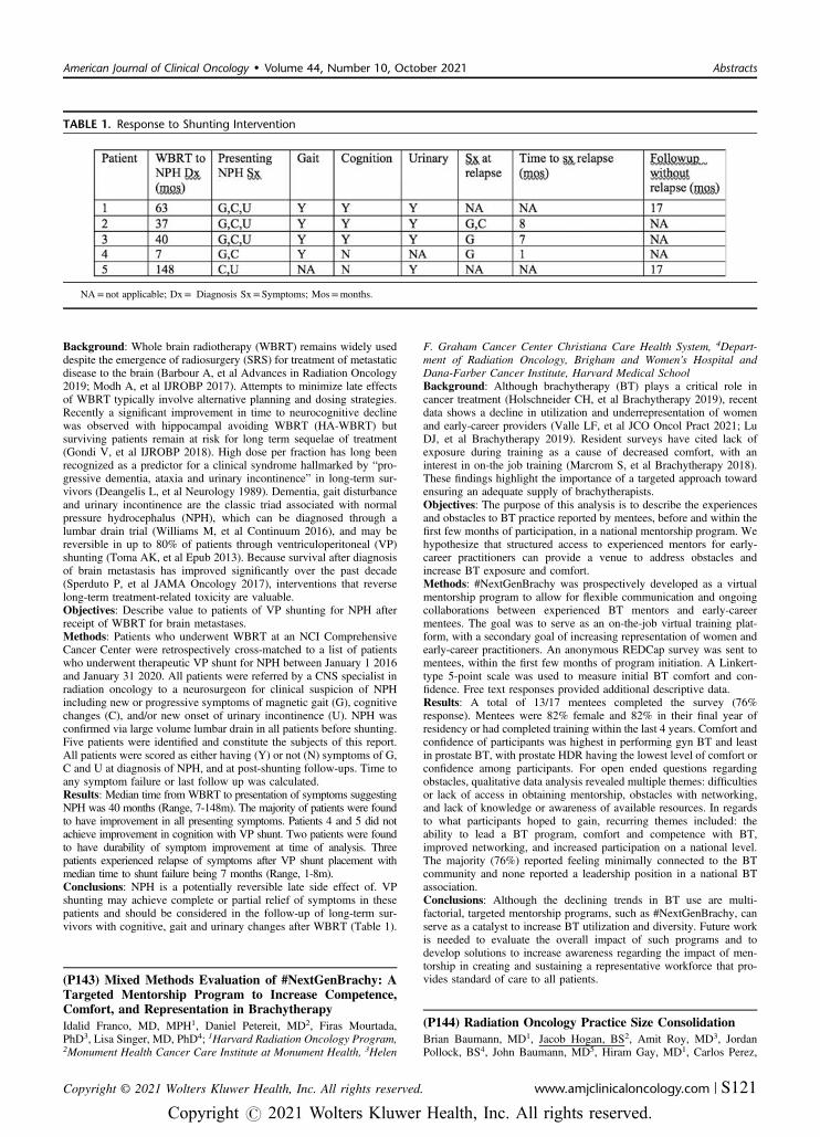
Background: Whole brain radiotherapy (WBRT) remains widely useddespite the emergence of radiosurgery (SRS) for treatment of metastaticdisease to the brain (Barbour A, et al Advances in Radiation Oncology2019; Modh A, et al IJROBP 2017). Attempts to minimize late effectsof WBRT typically involve alternative planning and dosing strategies.Recently a significant improvement in time to neurocognitive declinewas observed with hippocampal avoiding WBRT (HA-WBRT) butsurviving patients remain at risk for long term sequelae of treatment(Gondi V, et al IJROBP 2018). High dose per fraction has long beenrecognized as a predictor for a clinical syndrome hallmarked by “pro-gressive dementia, ataxia and urinary incontinence” in long-term sur-vivors (Deangelis L, et al Neurology 1989). Dementia, gait disturbanceand urinary incontinence are the classic triad associated with normalpressure hydrocephalus (NPH), which can be diagnosed through alumbar drain trial (Williams M, et al Continuum 2016), and may bereversible in up to 80% of patients through ventriculoperitoneal (VP)shunting (Toma AK, et al Epub 2013). Because survival after diagnosisof brain metastasis has improved significantly over the past decade(Sperduto P, et al JAMA Oncology 2017), interventions that reverselong-term treatment-related toxicity are valuable.Objectives: Describe value to patients of VP shunting for NPH afterreceipt of WBRT for brain metastases.Methods: Patients who underwent WBRT at an NCI ComprehensiveCancer Center were retrospectively cross-matched to a list of patientswho underwent therapeutic VP shunt for NPH between January 1 2016and January 31 2020. All patients were referred by a CNS specialist inradiation oncology to a neurosurgeon for clinical suspicion of NPHincluding new or progressive symptoms of magnetic gait (G), cognitivechanges (C), and/or new onset of urinary incontinence (U). NPH wasconfirmed via large volume lumbar drain in all patients before shunting.Five patients were identified and constitute the subjects of this report.All patients were scored as either having (Y) or not (N) symptoms of G,C and U at diagnosis of NPH, and at post-shunting follow-ups. Time toany symptom failure or last follow up was calculated.Results: Median time from WBRT to presentation of symptoms suggestingNPH was 40 months (Range, 7-148m). The majority of patients were foundto have improvement in all presenting symptoms. Patients 4 and 5 did notachieve improvement in cognition with VP shunt. Two patients were foundto have durability of symptom improvement at time of analysis. Threepatients experienced relapse of symptoms after VP shunt placement withmedian time to shunt failure being 7 months (Range, 1-8m).Conclusions: NPH is a potentially reversible late side effect of. VPshunting may achieve complete or partial relief of symptoms in thesepatients and should be considered in the follow-up of long-term sur-vivors with cognitive, gait and urinary changes after WBRT (Table 1).
(P143) Mixed Methods Evaluation of #NextGenBrachy: ATargeted Mentorship Program to Increase Competence,Comfort, and Representation in BrachytherapyIdalid Franco, MD, MPH1, Daniel Petereit, MD2, Firas Mourtada,PhD3, Lisa Singer, MD, PhD4; 1Harvard Radiation Oncology Program,2Monument Health Cancer Care Institute at Monument Health, 3Helen
F. Graham Cancer Center Christiana Care Health System, 4Depart-ment of Radiation Oncology, Brigham and Women's Hospital andDana-Farber Cancer Institute, Harvard Medical SchoolBackground: Although brachytherapy (BT) plays a critical role incancer treatment (Holschneider CH, et al Brachytherapy 2019), recentdata shows a decline in utilization and underrepresentation of womenand early-career providers (Valle LF, et al JCO Oncol Pract 2021; LuDJ, et al Brachytherapy 2019). Resident surveys have cited lack ofexposure during training as a cause of decreased comfort, with aninterest in on-the job training (Marcrom S, et al Brachytherapy 2018).These findings highlight the importance of a targeted approach towardensuring an adequate supply of brachytherapists.Objectives: The purpose of this analysis is to describe the experiencesand obstacles to BT practice reported by mentees, before and within thefirst few months of participation, in a national mentorship program. Wehypothesize that structured access to experienced mentors for early-career practitioners can provide a venue to address obstacles andincrease BT exposure and comfort.Methods: #NextGenBrachy was prospectively developed as a virtualmentorship program to allow for flexible communication and ongoingcollaborations between experienced BT mentors and early-careermentees. The goal was to serve as an on-the-job virtual training plat-form, with a secondary goal of increasing representation of women andearly-career practitioners. An anonymous REDCap survey was sent tomentees, within the first few months of program initiation. A Linkert-type 5-point scale was used to measure initial BT comfort and con-fidence. Free text responses provided additional descriptive data.Results: A total of 13/17 mentees completed the survey (76%response). Mentees were 82% female and 82% in their final year ofresidency or had completed training within the last 4 years. Comfort andconfidence of participants was highest in performing gyn BT and leastin prostate BT, with prostate HDR having the lowest level of comfort orconfidence among participants. For open ended questions regardingobstacles, qualitative data analysis revealed multiple themes: difficultiesor lack of access in obtaining mentorship, obstacles with networking,and lack of knowledge or awareness of available resources. In regardsto what participants hoped to gain, recurring themes included: theability to lead a BT program, comfort and competence with BT,improved networking, and increased participation on a national level.The majority (76%) reported feeling minimally connected to the BTcommunity and none reported a leadership position in a national BTassociation.Conclusions: Although the declining trends in BT use are multi-factorial, targeted mentorship programs, such as #NextGenBrachy, canserve as a catalyst to increase BT utilization and diversity. Future workis needed to evaluate the overall impact of such programs and todevelop solutions to increase awareness regarding the impact of men-torship in creating and sustaining a representative workforce that pro-vides standard of care to all patients.
(P144) Radiation Oncology Practice Size ConsolidationBrian Baumann, MD1, Jacob Hogan, BS2, Amit Roy, MD3, JordanPollock, BS4, John Baumann, MD5, Hiram Gay, MD1, Carlos Perez,
TABLE 1. Response to Shunting Intervention
NA= not applicable; Dx= Diagnosis Sx= Symptoms; Mos=months.
American Journal of Clinical Oncology � Volume 44, Number 10, October 2021 Abstracts
Copyright © 2021 Wolters Kluwer Health, Inc. All rights reserved. www.amjclinicaloncology.com | S121
Copyright r 2021 Wolters Kluwer Health, Inc. All rights reserved.

MD6; 1Department of Radiation Oncology, Washington UniversitySchool of Medicine in St. Louis, 2Washington University School ofMedicine, 3Washington University in St. Louis, 4Mayo Clinic,5Princeton Radiation Oncology, 6Washington University in St. Louis,Department of Radiation OncologyBackground: In recent years, there has been evidence of practiceconsolidation in US healthcare. To our knowledge, a detailed quanti-tative study of recent changes in radiation oncology practice size has notbeen performed.Objectives: We aim to evaluate radiation oncology practice sizechanges between 2012 and 2020 in the US overall, for all regions, andfor both male and female physicians.Methods: Using the Medicare Physician Compare Database, we iden-tified practices employing radiation oncologists using their TaxpayerIdentification Number and individual radiation oncologists using theirNational Provider Identifier. We assigned physicians with multiplepractice affiliations to the practice with the largest size. Practice sizeincludes the number of physicians of any specialty employed at thatpractice, not just radiation oncologists. We compared the number ofindividual radiation oncologists and the number of practices in eachpractice size category between 2012 and 2020. Further analysis by UScensus region and sex were performed. Cochran-Armitage test for trendwas used to detect significant differences.Results: Between 2012 and 2020, the total number of practicing radi-ation oncologists identified increased by 9% (4,300 to 4,679) while thenumber of practices employing radiation oncologists decreased by11.5% (1,606 to 1,422). The number of radiation oncologists in prac-tices of size 1-2, 3-9, 10-24, and 25-49 decreased by 3.7%, 4.7%, 4.9%and 2%, respectively, while the number of radiation oncologists inpractices of size 50-99, 100-499, and 500+ increased by 1.4%, 2.1%,and 11.8%, respectively (all 500+ practices are multi-specialty groups).The increase in practice size at both the individual and practice levels
was consistent in all regions and for both genders (FDR-adjusted P <0.001 in all cases).Conclusions: Our analysis provides objective evidence that practicesize consolidation is occurring within the US Radiation Oncologyworkforce. The potential implications of practice consolidation forphysicians and patients should be considered by specialty leaders,hospital administrators and government officials when determiningfuture financial policy (Figs. 1 and 2).
(P145) Feasibility of a Phase II Study Utilizing SpatiallyFractionated Radiation Therapy for Bulky or Radio-resistant Tumors of the Head and Neck, Thorax, Abdomen,Pelvis, and ExtremitiesElizabeth Jeans, MEd, MD1, Michael Grams, PhD1, Randi Finley,CMD1, William Breen, MD1, Dawn Owen, MD, PhD1, Sean Park, MD,PhD1, Bradley Stish, MD2, Scott Lester, MD1, Ivy Petersen, MD1, SafiaAhmed, MD1, Kimberly Corbin, MD1, Mark Waddle, MD1, MichaelHaddock, MD1, Kenneth Merrell, MD1, Christopher Hallemeier, MD1,Nadia Laack, MD1, Daniel Ma, MD1; 1Mayo Clinic, Rochester, 2MayoClinic RochesterBackground: Spatially fractionated radiotherapy (SFRT), the modern-day implementation of grid therapy, delivers a heterogenous dose,consisting of high-dose distribution of “hotspots” interspersed with lowdose “valleys”, to a designated target volume.Objectives: Bulky or radioresistant tumors have specific treatmentchallenges. SFRT allows for high dose delivery with sparing of closelyapproximated tissues and the potential to stimulate the radiation-induced bystander effect and non-specific immune system cells. Thisstudy reports the feasibility of developing and accruing to a prospectiveinstitutional protocol utilizing SFRT for curative or palliative purposes.Methods: Patients are registered to a curative- or palliative-intent arm.SFRT delivery options include a brass aperture to deliver a static field orlattice SFRT, utilizing VMAT. SFRT technique, dose, and optional con-solidative radiotherapy dose/fractionation are prescribed at physician’sdiscretion. The primary outcome is radiographic local control rate, utilizingRECIST criteria, at 3 - 6 months. The secondary objectives include acuteand late adverse event profile by assessment of grade 3 or higher adverseevents (NCI-CTCAE version 5 criteria), overall survival, regional pro-gression, and distant disease control. An enrollment of 120 patients, 70palliative and 50 definitive, is planned based on a 95% exact binomialconfidence interval for local failure rate of ≤ 10% (curative), ≤ 30%(palliative) and an estimation of adverse (grade 3+) events within 90 daysof ≤ 5%. Patient disease and treatment-related characteristics, as well asfollow-up details, including disease site-specific patient reported outcomesare maintained in a prospective database.
FIGURE 1. Physician selection flowchart. Flowchart of physicianselection using the Physician Compare dataset (n=number in2012, number in 2020).
FIGURE 2. Bar plot of overall changes in group practice size. Barplot showing (A) the number of radiation oncologists and (B) thenumber of group practices in each size category for 2012and 2020.
Abstracts American Journal of Clinical Oncology � Volume 44, Number 10, October 2021
S122 | www.amjclinicaloncology.com Copyright © 2021 Wolters Kluwer Health, Inc. All rights reserved.
Copyright r 2021 Wolters Kluwer Health, Inc. All rights reserved.
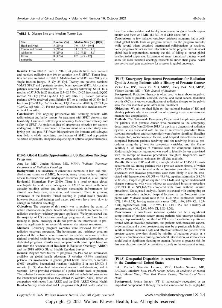
Results: From 01/2020 until 01/2021, 24 patients have been accruedand received palliative (n= 19) or curative (n= 5) SFRT. Tumor loca-tion and size are listed in Table 1. Median dose of SFRT was 20 Gy in asingle fraction [range, 18 Gy–25 Gy]. Twenty-one patients receivedVMAT SFRT and 3 patients received brass aperture SFRT. All curativepatients received consolidative RT 1-2 weeks following SFRT to amedian of 57.5 Gy in 25 fractions [35–62.5 Gy, 10–25 fractions], EQD2median 58.9 Gy [39.4 Gy–65.1 Gy, α/β ratio 10]. Eleven palliativepatients (58%) received consolidative RT to a median of 30 Gy in 5fractions [20–30 Gy, 3–5 fractions], EQD2 median 40.0 Gy [27.7 Gy–40.0 Gy, α/β ratio 10]. For the patient’s enrolled to date, median follow-up is 4.1 months.Conclusions: This ongoing prospective trial accruing patients withradioresistant and bulky tumors for treatment with SFRT demonstratesfeasibility. Continued follow-up is necessary to determine efficacy andsafety of SFRT. An understanding of appropriate selection for patientsundergoing SFRT is warranted. Co-enrollment on a sister study ana-lyzing pre- and post-RT frozen biospecimens for immune cell subtypesmay help to elude underlying mechanisms of SFRT and appropriateselection of patients, alongside sequencing of optimal adjunct therapies.
(P146) Global Health Opportunities in US Radiation OncologyProgramsAmy Le, MD1, Jordan Holmes, MD, MPH1; 1Indiana UniversityDepartment of Radiation OncologyBackground: The incidence of cancer has increased in low- and mid-dle-income countries (LMIC), however, many countries have limitedaccess to cancer care with inadequate resources (Jemal A, et al CancerEpidemiol Biomarkers Prev 2010). There is a need to train globaloncologists to work with colleagues in LMIC to assist with localcapacity-building efforts and develop sustainable infrastructure forclinical oncology care, education, and research. There is growingawareness of the benefits formal training in global health provideshowever formalized training and career pathways have been slow toemerge in radiation oncology.Objectives: The purpose of this study was to explore the extent ofonline information on global health education available to potential USradiation oncology residency program applicants. We hypothesized thatthe majority of US radiation oncology programs do not have formaltraining in global oncology or do not provide the information to pro-spective applicants in an accessible manner via internet resources.Methods: Residency program websites were reviewed for 89 USradiation oncology programs. The homepages and residency programportion of the websites were examined for specific mention of inter-national or global health initiatives, rotations, research collaborations, ordedicated programs. Results were compared with prior report based ondata from the Association of Residents in Radiation Oncology (ARRO)and the 2018 ARRO Global Health Resident Survey.Results: Program websites for 76 programs (85.4%) had no informationavailable on global health education, 3 websites (3.4%) mentionedpotential for involvement in general global health initiatives, 5 websites(5.6%) described international rotations (including 2 in non-LMIC), 1website (1.1%) described international research collaborations, and 4websites (4.5%) provided evidence of a global health track or program.The websites for some residency programs did not include information onthe international opportunities they offered (5 websites, 5.6%), based oncomparison with report from ARRO and the 2018 ARRO Global HealthResident Survey which identified 11 programs with global health initiatives
based on active resident and faculty involvement in global health oppor-tunities and focus on LMIC (Li BC, et al Glob Onco 2021).Conclusions: A few radiation oncology residency programs had a dedi-cated global health track or program detailed on the program website,while several others described international collaborations or rotations.Some programs did not include information on the program website aboutglobal health opportunities, running the risk of failing to attract globalhealth-minded applicants. Expansion of more formalized training wouldallow for more radiation oncology residents to enrich their global healthperspective and gain experience for a career in global oncology.
(P147) Emergency Department Presentations for RadiationCystitis Among Patients with a History of Prostate CancerVictor Lee, BS1, James Yu, MD, MHS1, Henry Park, MD, MPH1,Vikram Jairam, MD1; 1Yale School of MedicineBackground: Radiation therapy is often used to treat abdominopelviccancers such as prostate, cervical, uterine, and rectal cancer. Radiationcystitis (RC) is a known complication of radiation therapy to the pelvicarea that can manifest years after initial treatment.Objectives: We aim to shed light on the national burden of RC andidentify patients who at increased risk of requiring invasive measures tomanage this complication.Methods: The Nationwide Emergency Department Sample was queriedfor patients with prostate cancer who presented to the emergencydepartment (ED) from 2006 to 2015 for a primary diagnosis of radiationcystitis. Visits associated with the use of an invasive procedure (tran-surethral procedures and cystectomies) were further identified. Baselinedemographic, socioeconomic factors, and hospital characteristics werecompared between patients receiving and not receiving invasive pro-cedures using the χ2 test for categorical variables, and the Mann-Whitney U or analysis of variance tests for continuous variables.Multivariable logistic regression was used to identify factors associatedwith the receipt of invasive procedures. Weighted frequencies wereused to create national estimates for all data analysis.Results: Between 2006 and 2015, a weighted total of 17,456 ED visitsoccurred for RC among patients with a prostate cancer history, of which5,925 (33.9%) were treated with an invasive procedure. ED visitsassociated with invasive procedures were more likely to also be asso-ciated with hypertension (53.3% vs 48.9%), inpatient admission (98.7%vs 86.2%), longer length of stay (6.6 d vs 4.6 d), and higher median totalcharges compared with visits without an associated invasive procedure($36,213.00 vs $19,166.70) compared with those without invasiveprocedures. On adjusted analysis, factors associated with undergoing aninvasive procedure included hospital location in the Northeast com-pared with Midwest (odds ratio [OR], 1.34; 95% confidence interval[CI], 1.04-1.73), having metastatic cancer (OR, 1.46; 95% CI, 1.05-2.04), hypertension (OR, 1.31; 95% CI, 1.10-1.55), and a history ofprostatectomy (OR, 2.96; 95% CI, 1.86-4.70).Conclusions: In conclusion, radiation cystitis can be a significantcomplication of prostate cancer among patients who undergo radiationtherapy. Approximately one third of ED visits for radiation cystitis aretreated with an invasive procedure, and patients with metastatic cancer,hypertension, and a prior prostatectomy may be at particularly high risk.While radiation remains a safe and effective treatment for patients withprostate cancer, providers should be mindful of radiation cystitis as apotential complication requiring procedural intervention, as severe casescould lead to significant bleeding or anemia. Patients at greatest risk forthis complication should be monitored closely in the outpatient setting.
(P148) Geospatial Disparities in Access to Proton Therapyin the Continental United StatesLuke Maillie1, Stanislav Lazarev, MD2, Charles Simone, MD,FACRO3, Matthew Sisk, PhD4; 1Icahn School of Medicine at MountSinai, 2Mount Sinai, 3New York Proton Center, 4University of NotreDameBackground: Proton therapy (PT) is increasingly recognized as animportant component of therapy for select cancers due to its negligible
TABLE 1. Disease Site and Median Tumor Size
American Journal of Clinical Oncology � Volume 44, Number 10, October 2021 Abstracts
Copyright © 2021 Wolters Kluwer Health, Inc. All rights reserved. www.amjclinicaloncology.com | S123
Copyright r 2021 Wolters Kluwer Health, Inc. All rights reserved.

exit dose and, as a result, improved normal tissue sparing and lower riskof adverse effects compared with conventional photon-based radio-therapy techniques. It is often purported that due to the existence ofrelatively few centers in the continental United States (US), inequities inaccess to PT may be present, however no formal study of geospatialaccess to proton centers has been conducted to date.Objectives: This study examined access to PT in the continental US byquantifying travel time to PT centers.Methods: Population data from 2014-2018 were available for 320.7million people in 32,644 zip code tabulation areas (ZCTAs) in thecontinental US. Addresses for all PT centers in operation were geo-coded. ArcGIS Pro Mapping software was used to determine travel timefrom the center point of each ZCTA to the nearest PT center. Twomodels were constructed, one for adults (≥ 18 y) and one for children(< 18 y) due to one proton center being available only for children. Dataon age, race, health insurance status, household income, educationalattainment, and geographic area were also analyzed.Results: A total of 36 proton centers were identified in the continentalUS. The median travel time was 1.61 (IQR 0.67-3.36) hours for chil-dren and 1.64 (IQR 0.69-3.33) hours for adults. 35.7% of children and34.7% of adults were found to live within one hour of the nearest PT
center, while 29.0% of children and 29.1% of adults were found to liveover three hours from the nearest PT center. Significant variation intravel time to the nearest PT center was observed between states, with11 states having a median travel time of less than 1 hour and 20 stateshaving a median travel time of over 3 hours. Catchment, defined as thepopulation located closest to a facility, varied widely between facilitieswith a median of 5.71 (IQR 3.75-9.30) million per facility for adults and1.45 (IQR 1.02-2.75) million per facility for children. The West has alonger median travel time of 3.51 (IQR 1.15-7.13) hours when com-pared with the Midwest (1.70, IQR 0.79-2.69), South (1.60, IQR 0.61-3.12), and Northeast (1.04, IQR 0.57-2.01). Native Americans wereseen to live at further distances when compared with other races.Households within the highest income quartile were observed to livecloser to PT centers when compared with lower income quartiles. Timeto nearest proton center did not differ significantly based on insurancestatus or education level.Conclusions: Average travel time to the nearest PT center is high, andtravel times vary significantly based on the state of residence. Whilebeing from the West or being Native American confers a higher mediantravel time and being in the highest income quartile confers a lowermedian travel time, travel time does not significantly vary based on
FIGURE 1. Time to the nearest PT center for children and adults. The boundary of each ZCTA in the mainland U.S. is shown.
Abstracts American Journal of Clinical Oncology � Volume 44, Number 10, October 2021
S124 | www.amjclinicaloncology.com Copyright © 2021 Wolters Kluwer Health, Inc. All rights reserved.
Copyright r 2021 Wolters Kluwer Health, Inc. All rights reserved.

health insurance status or educational attainment. Disparities in geo-graphic access for certain populations warrant further examination toimprove access to this important cancer treatment modality (Figs. 1, 2and Table 1).
(P149) OncMonic: A Novel Visual Proprietary Study Paradigmfor the Oncologist-in-trainingGohar Manzar, MD PhD1; 1The University of Texas MD AndersonCancer Center
Background: In recent years, a new generation of visual mnemoniclearning devices has emerged to assist with medical education forboard licensure examinations. SketchyMedical↓ and Picmonic↓ haverevolutionized the study of pathophysiology, microbiology, andpharmacology. The use of such memory anchor devices has dem-onstrated increased long-term memory retention by 331% and animprovement in test scores after one week by 50% among medicalstudents in a randomized double-blind controlled study1. The basisfor this is a dual coding phenomenon whereby distinct verbal andvisual pathways to encode information diversifies the basis for recall,as well as the Von Restorff effect, whereby a potently stimulatedsensorium offers superior context for layering memory as comparedwith plain bulleted texts. There is a general lack of embracement ofsuch pedagogical innovation among medical sub-specialties.Oncology is an ideal candidate for supplementation of tradi-tional study approaches with the exponential increase in dataand its meaningful utilization in multidisciplinary tumor boards toguide clinical decision making, particularly in the space of clinicaltrials.Objectives: To develop and validate an assistive visual mnemonicparadigm for the study of major clinical trials under the domain ofradiation oncology.Methods: An IRB proposal will investigate the recall of landmarkclinical trials presented with the use of pictorial mnemonics amongclinical radiation oncology residents participating in a weekly didacticseminar series. A separate clinical trial included within the same pre-sentation by the same speaker without the visual assistive device willserve as the internal control.Results: A survey will be collected among consenting residentsimmediately post-seminar, one week post-seminar, and eight weekspost-seminar. Metrics for recall of specific symbolic cues will includethe study name or acronym, primary and/or secondary outcomes of thetrial, and clinical criteria the study generated. Statistical analysis will beconducted using a Student’s t test.Conclusions: Rich visual tools are transforming medical education withless reliance of traditional lackluster memorization. Embracing suchpedagogical advances within radiation oncology by leveraging provenresearch in memory recall will empower residents to better utilize thevast array of data at their disposal and may potentially influence clinicaldispositions.
(P150) Optimizing Practical Implementation of AutomatedRadiotherapy Treatment Planning Tool: A SurveyGwendolyn McGinnis, MD, MS1, Matthew Ning, MD, MPH2, RemigioMakufa, MS3, Carlos Cardenas, PhD2, Surbhi Grover, MD4, LaurenceE. Court, PhD2, Grace Smith, MD MPH2; 1UT MD Anderson CancerCenter, 2MD Anderson Cancer Center, 3Gaborone Private Hospital,4University of PennsylvaniaBackground: Low-to-middle income countries (LMIC) have inequit-able access to radiation treatment secondary to shortages in bothavailable technology and personnel training. The use of automatedplanning software can lessen this disparity. The Radiation PlanningAssistant (RPA) is an automated treatment-planning tool designed forlimited resource environments. In anticipation of international deploy-ment, understanding practical implementation of the RPA in LMIC iscritical to optimizing its value.Objectives: We conducted a survey to better characterize the optimalapproach to RPA deployment in LMIC clinical settings.Methods: Providers in three countries expressing interest in pilotingRPA were approached for survey participation, with 100% response.Providers received an initial 1-hour remote, live videoconferencelearning session supported by interactive learning using breast and headand neck cancer dummy radiation plans. Providers were surveyed onanticipated need for training and ongoing support.Results: Five institutions were included with 25 total participants.Countries included South Africa (n= 13), Tanzania (n= 1), and Gua-temala (n= 12). Most commonly, participants were between the ages of31-50 years old (72%) and in practice for > 5 years (68%). The
FIGURE 2. Time to the nearest PT center by state for adults.States are shown in order of increasing median travel time tonearest PT center.
TABLE 1. Time to Nearest PT Center Based by DemographicVariables
Comparison of access to PT by demographic and geographic characteristicsin the continental US.
American Journal of Clinical Oncology � Volume 44, Number 10, October 2021 Abstracts
Copyright © 2021 Wolters Kluwer Health, Inc. All rights reserved. www.amjclinicaloncology.com | S125
Copyright r 2021 Wolters Kluwer Health, Inc. All rights reserved.

distribution of included respondent roles were as follows: physician(32%), dosimetry (24%), physicist (32%), resident/registrar (4%),radiation therapist (4%), and administrator (4%). Most participantsreported they would find printed materials (96%), on-site in-persontraining (96%), off-site in-person training (92%), interactive onlinetraining (100%), and online video tutorials (100%) helpful. When askedto rate how helpful each of these resources would be on a scale of 1 to 5(5 being most preferred), on-site in-person training (mean 4.25, SD=0.9) scored the highest followed by interactive online training (mean4.2, SD= 0.8), online video tutorials (mean 4.0, SD= 1.3), off-site in-person training (mean 3.74, SD= 1.1), and printed materials (mean3.71, SD= 1.2). Only 44% of participants had previously used onlinetraining to learn about software designed for radiation treatment plan-ning. When asked how comfortable they would be using the RPA aftercompleting only an online training program on a scale of 1 to 5 (5 beingvery comfortable), participants rated a mean score of 3.8 (SD= 1.3). Interms of ongoing support for use of the RPA, participants reported thatthey would find telephone (88%), online chat (100%), email (92%), inperson (88%), scheduled online (96%), and online discussion (88%)support helpful. Online chat (mean 4.1, SD= 1.1) was rated as the mosthelpful on a scale of 1 to 5 (5 being the most helpful) followed by email(mean 4.0, SD= 1.2), online discussion (mean 3.9, SD= 1.3) in person(mean 3.9, SD= 1.4), scheduled online (mean 3.8, SD= 1.3), and tel-ephone (mean 2.5, SD= 1.4) support.Conclusions: This survey indicates various strategies may be effectivein training providers to use the RPA. However, these data provide earlysignal that although many providers may not have had similar onlineinteractive training experiences, such an approach appears bothacceptable and effective for training the target users of the RPA. Onlinetraining would be expected to be substantially less costly and havepotentially wider reach than in-person trainings globally. These datahelp create a framework for global training to promote uptake andproficiency in use of automated radiation therapy planningtechnologies.
(P151) Diversity in Medical Physics Research Leadership:Quantifying the Representation of Racial, Cultural, and SexIdentity Minority GroupsRichard Castillo, PhD1, Jillian Rankin2, Brendan Whelan, PhD3,Julianne Pollard-Larkin, PhD4, Matthew Scarpelli, PhD5, Keyvan Far-ahani, PhD6; 1Emory University, 2Emory University School of Medi-cine, 3University of Sydney, 4MD Anderson Cancer Center, 5Barrow
Neurological Institute, 6National Cancer Institute, National Institutes ofHealthBackground: The existence of disparities in the representation ofminority groups has been an ongoing, systemic issue in STEM fields.Prior work has shown this is exacerbated for leadership positions andawards recipients. However, a characterization of racial, ethnic, and sexminority representation in medical physics research leadership has notyet been established.Objectives: The purpose of this study was to investigate the presenceand potential extent of disproportionate racial, ethnic, and sex repre-sentation in medical physics research leadership.Methods: NIH grant funding was used as a proxy for research lead-ership. A list of AAPM members who received NIH grant funding asprincipal investigator between 1985-2020 was generated using theAAPM research database (Whelan B, et al Med. Phys. 2017). Anony-mized demographics data for the group of NIH-funded AAPM members(N= 416), as well as for all current AAPM members (N= 8690), wasprovided by the AAPM. Fisher’s Exact test for independence was usedto test association between grant funding status and racial, ethnic, andsex demographic factors. Two-sample z-tests were used to compareproportions of individual demographic groups between the two studypopulations. Member profiles for which race, ethnicity, or sex were notreported were excluded from analysis.Results: While not significantly associated with ethnicity, relativerepresentation in research leadership was significantly associatedwith sex (P < 0.05) and had an association nearing significance withrace (P= 0.055). Amongst demographic groups, women were sig-nificantly underrepresented as research leaders relative to theirrepresentation in the medical physics workforce (P < 0.05).Although not statistically significant due to low absolute repre-sentation in both groups, decreased relative representation amongstgrant recipients was observed for individuals identifying as Black orAfrican American, or as being of Hispanic, Latino, or Spanish ori-gin. Within the group of research leaders, there were no individualswho identified as American Indian, Alaskan Native, transgender, orandrogynous. Demographic data for AAPM was limited due to the∼40% rate of members self-declaring a racial or ethnic identitywithin membership profiles.Conclusions: Our results suggest that the demographic makeup of thepool of NIH-funded medical physicists is distinct from that of practicingmedical physicists in general, with significant differences in sexinclusivity observed. While we did not establish a statistically sig-nificant association between race or ethnicity and grant funding, anal-ysis was limited due to a severe lack of minority representation inAAPM membership. Efforts to increase diversity and inclusion within
FIGURE 1. Gender identity demographic profiles of AAPM members and AAPM members with NIH grant funding. Relative repre-sentation in research leadership is significantly associated with sex (P < 0.05). Amongst AAPMmembers, women are underrepresented asNIH grant recipients relative to their male counterparts. Individuals identifying as transgender or androgynous have little representationin either group, but are notably absent from the NIH grant recipient group.
Abstracts American Journal of Clinical Oncology � Volume 44, Number 10, October 2021
S126 | www.amjclinicaloncology.com Copyright © 2021 Wolters Kluwer Health, Inc. All rights reserved.
Copyright r 2021 Wolters Kluwer Health, Inc. All rights reserved.

AAPM will help yield more meaningful results. Further investigation isnecessary to inform actionable policy to ensure diverse, equitable, andinclusive representation, both in research funding and within the pro-fessional body at large (Figs. 1 and 2).
(P152) Decision Making Process and Operations for Irra-diation of COVID-19 Positive PatientsQuynh-Nhu Nguyen, MD1, Julius Weng, MD2, Bouthaina Dabaja,MD2, Prajnan Das, MD, MS, MPH3, Albert Koong, MD, PhD2, Eliz-abeth Bloom, MD2, Gary Gunn, MD4, Cody Wages, BS, CMD2,Gregory Chronowski, MD2, Ryan Ritchey, RT (R) (T)2, Ann H. Klopp,MD, PhD5, Kham Nguyen, BS, CMD, RT (T)2, Andrew Bishop, MD2,Danna Fullen, RT3, Ivy Robinson, RT (R) (T)2, Denise Zaebst, RN,MSN2, Jared Brokaw, RT (R) (T)2, Greg Swiedom, RT (R), MR2,Christopher Sanders, BS, RT (R) (T)2, Zhongxing Liao, MD, FASTRO,FAAWR5, Mary McAleer, MD5; 1UT MD Anderson Cancer Center,2MD Anderson Cancer Center, 3MD Anderson, 4The University ofTexas M.D. Anderson Cancer Center, 5The University of Texas MDAnderson Cancer CenterBackground: During the COVID-19 pandemic, patients who test positiveexperience unique challenges of risking a treatment break in their radiationtherapy course which can affect their clinical outcome. Many cancer centersquickly adopted policies and CDC guidelines into their daily operations toensure continued patient care and treatment during the pandemic. It isimportant to develop and operationalize a safe process involving clinicalleaders and teams to deliver safe and effective radiation treatments for onco-logic emergencies and definitive radiation courses. Treatment of COVID-19positive patients for both routine and urgent RT indications requires carefulconsideration and modification of workflow to balance potential therapeuticbenefits and risk of transmission to other patients and staff.Objectives: To report decision making process and workflow to irra-diate COVID-19 positive patients at a tertiary cancer center from 4treatment locations.Methods: The Radiation Oncology Covid-19 committee (RO COVIDRT) developed an integrated process to triage, provide consensustreatment recommendations, and safely deliver radiation therapy topatients who could not have a 21-day treatment break and patients who
needed emergent RT. Radiation oncology specific COVID policieswere created for each center by physicians, nurses, and radiationtherapists, which was then approved by the institution’s COVID com-mittee. All COVID-19 positive patients were presented to the ROCOVID RT group, evaluated for clinical urgency, benefit with radia-tion, and life expectancy, and, if deemed necessary, a limited plannedbreak, modification of radiation dose, and hypofractionated course wasrecommended. To minimize risk to staff and other vulnerable oncologypatients, inpatients were treated at our hospital’s main radiation facilityand outpatients were treated at three of our six ambulatory centers. Weconducted a retrospective review of patients treated with these COVIDpolicies between June 2020 to July 2021.Results: A total of 20 patients were treated using COVID precautionsdue to testing positive (n= 12, 60%) or for exposure/suspected COVID(n= 8, 40%). Six (30%) patients received treatment for an emergentindication (spinal cord compression, symptomatic leptomeningeal dis-ease, cauda equina syndrome, and gastrointestinal bleeding). Theremaining patients were already receiving RT at the time of their pos-itive COVID test and underwent treatment breaks for 7-14 days.Thirteen (65%) patients were asymptomatic and were tested for routinepre-radiation screening or due to concerns of COVID exposure. Alltreatments were successfully completed without known spread ofCOVID to staff. Sixteen patients (80%) were alive at last follow up.Conclusions: In this study, COVID positive patients were effectivelytreated with radiation therapy without significant delays in access orCOVID transmission. We found that prospective review of cases atCOVID Operations Leadership meetings was essential in determiningurgency of RT, recommending hypofractionated regimens, and opti-mizing operational workflow of treating COVID patients.
(P153) ESAS Based Palliative Care Intervention in a RadiationOncology ClinicRonica Nanda, MD1, Alexandra Owens, MD2, Heather Jim, PhD1,Roberto Diaz, MD, PhD3, Sunny Raiker, BS2, Nicholas Figura, MD4,Peter Johnstone, MD, FACR, FASTRO3; 1H. Lee Moffitt Cancer Centerand Research Institute, 2Morton Plant Hospital, 3Moffitt Cancer
FIGURE 2. Racial and ethnic demographic profiles of AAPM members and AAPM members with NIH grant funding. Minority racial andethnic groups are severely underrepresented in both the all-AAPM member population and AAPM member-NIH grants recipientspopulation. Decreased relative representation amongst grant recipients is observed for individuals identifying as Black or AfricanAmerican, or as being of Mexican, Mexican American, Chicano, Cuban, Puerto Rican, or other Hispanic, Latino, or Spanish origin. Withinthe group of research leaders, there are no individuals who identify as American Indian or Alaskan Native.
American Journal of Clinical Oncology � Volume 44, Number 10, October 2021 Abstracts
Copyright © 2021 Wolters Kluwer Health, Inc. All rights reserved. www.amjclinicaloncology.com | S127
Copyright r 2021 Wolters Kluwer Health, Inc. All rights reserved.

Center, 4H. Lee Moffitt Cancer Center and Research Institute,Department of Radiation OncologyBackground: Symptom palliation as a component of routine cancercare has been proven to improve overall survival and quality of life forpatients with advanced cancer. However, acute palliative care resourcesare limited at many cancer centers, especially in the outpatient setting;therefore, strategies to optimize the identification and selection ofpatients who require palliative care (PC) services are needed. Wepiloted a program at our institution in March 2018 to provide outpatientpalliative care for cancer patients undergoing radiotherapy (RT) withreferral based on high Edmonton Symptom Assessment Scale (ESAS)scores to divert resources toward patients in greatest need.Objectives: To prospectively determine the impact of a palliative careconsultation and follow up care on patient for radiation oncologypatients with symptoms exceeding predetermined thresholds. Wepiloted a program at our institution to reflexively refer patients foroutpatient palliative care should they report high symptom burden, asmeasured by the clinically validated Edmonton Symptom AssessmentScale (ESAS) score to divert resources toward patients in greatest needin a community-based cancer center.Methods: The Radiation Oncology clinic established use of a modifiedESAS tool (clinically validated in the outpatient oncology setting) in2017, assessing 12 symptom domains on a 10-point scale. Higher scoresindicate higher symptom burden. Each patient was asked to completethe modified ESAS at each consult, follow up, or weekly visit, and totalscores were calculated. Patients with either a high cumulative ESASscore or a high score (> 6) on a single symptom domain were referredto the palliative care physician. Demographic and individual patientdata were obtained from the electronic medical record. Individualdomain and total scores were recorded in a deidentified database. Thenet change between highest and lowest total scores as well as netchange in total scores from initial encounters and last follow up werecalculated to determine the effect of a standard symptom-based pallia-tive care referral. Patients without a subsequent clinical encounter andaccompanying ESAS score following the palliative care consult wereexcluded from final analysis.Results: Between March 2018 and January 2021, a total of 91 patientsconsulted with outpatient palliative care at least once. A total of 75patients met the criteria for final analysis. ECOG performance status forall patients was 0-2. Mean total score from initial radiation oncologyencounter was 49.4 +/- 22.3 (range 0 to 97); at initial PC consultation,mean total score was 53.2 +/- 22.9 (standard deviation) (range 1-101).At time of last follow up, mean total scores was 45.1 +/- 25.6 (range0-103). The mean lowest total score was 30.0 +/- 22.1. (range 0-88) andhighest total scores mean was 66.6 +/- 21.7 (range 13-107). Meanchange in total scores from initial radiation oncology visit to last followup was -4.28 points +/- 25.6 (range -86 to 61) and from initial PCconsult to last follow up was -8.03 points +/- 22.1 (range -88 to 40).84% of patients had improvement in the highest individual score by atleast one point, and of those who had an improvement, 70% had anindividual score improve from severe (> 6) to not severe (< = 6). Ofthe 21 patients who passed away during this interval, twelve had loweror stable total ESAS scores at last follow up.Conclusions: Radiation oncology patients referred to outpatient pal-liative care have high symptom burden at baseline. Palliative careconsultation and follow up were correlated with a decrease in totalESAS scores of 8 points. Previous literature suggests that improvementin ESAS scores is associated with improved quality of life and decreasein total scores of 3-4 points is clinically meaningful. Patients at end oflife in this cohort appeared to have stable ESAS scores suggesting asignificant benefit from early palliative care intervention.
(P154) Malignant Cord Compression and Timing of Post-operative RadiotherapyApril Vassantachart, MD1, Phillip Bonney, MD1, Eugene Lin, MD,MS2, Shelly Bian, MD2; 1LAC+USC Medical Center, 2Keck School ofMedicine of USCBackground: Malignant spinal cord compression (MSCC) can result inpermanent neurologic injury and often requires prompt neurosurgicaldecompression followed by radiation therapy (RT). Post-surgical care
can include rehabilitation and time for wound healing which can lead todelays in post-operative RT.Objectives: This study aims to retrospectively review the time to ini-tiation (TTI) of RT after surgical intervention for MSCC and its asso-ciation with ambulatory status at 3 months. We also compare TTI inpatients discharged to home, versus inpatient rehabilitation, or a skillednursing facility.Methods: Our institutional database was queried for patients with solidmalignancies and spinal metastases with Bilsky 2 or 3 MSCC whounderwent surgical intervention from 2015-2020 and postoperative RT.In patients who were ambulatory peri-operatively, we computed therelative risk of decreased ambulation at 3 month follow up using dif-ferent TTI cutoff values of 30, 42, and 56 days. P values were generatedusing a 1-sided Fisher’s Exact Test. TTI was also evaluated in allpatients who underwent post-operative RT based on their discharge toeither home, inpatient rehabilitation, or a skilled nursing facility.Results: We found 85 patients with MSCC who underwent surgery.Within this group, 56 received post-operative RT with known TTI. Forour TTI analysis, we excluded 14 patients who were not peri-oper-atively ambulatory and 10 patients who did not have at least 3 monthsof follow up. Three-month ambulatory rates for TTI of ≤ 30 vs> 30 days, ≤ 42 vs > 42 days, and ≤ 56 vs > 56 days were 86% vs78% (P= 0.460), 87% vs 67% (P= 0.203), and 89% vs 40%(P= 0.034), respectively. For all patients who received post-operativeRT without wound complications causing delays in RT, TTI based ondischarge to home (n= 35), a skilled nursing facility (n= 4), or inpatientrehab (n= 16) was 28 days (IQR 19-41), 28 days (IQR 16-41), and35 days (IQR 24-53), respectively. Six patients were found to havelocally recurrent disease causing cord compression—only one recurredafter post-operative RT while five recurred before post-operative RT.Three had TTI > 100 days due to recurrent disease at 33 days and56 days requiring two re-resections, transportation difficulties fromskilled nursing facility, and continued oncologic care at an outsidehospital who did not refer the patient for RT. The other three patientsreceived RT within 30 days, however two recurred before RT and onerecurred at 465 days.Conclusions: Initiation of post-operative radiation more than 56 daysafter surgical resection for MSCC showed lower ambulatory rates at3 months. Further studies evaluating the optimal timing of post-oper-ative RT and causes for delay are needed (Fig. 1).
(P155) Prisoner of the Heart – Primary Unresectable CardiacAngiosarcoma in an African American ManJehan Yahya, MD1, David Henkin, MD2, Joseph Montecalvo, MD3,Rami Khoury Abdulla, MD3; 1OHSU: Oregon Health & ScienceUniversity, 2UT Health, 3Henry Ford Hospital
FIGURE 1. Forest plot of relative risk for impaired ambulationbased on time to treatment initiation of radiation. RR= relativerisk. CI= confidence interval.
Abstracts American Journal of Clinical Oncology � Volume 44, Number 10, October 2021
S128 | www.amjclinicaloncology.com Copyright © 2021 Wolters Kluwer Health, Inc. All rights reserved.
Copyright r 2021 Wolters Kluwer Health, Inc. All rights reserved.
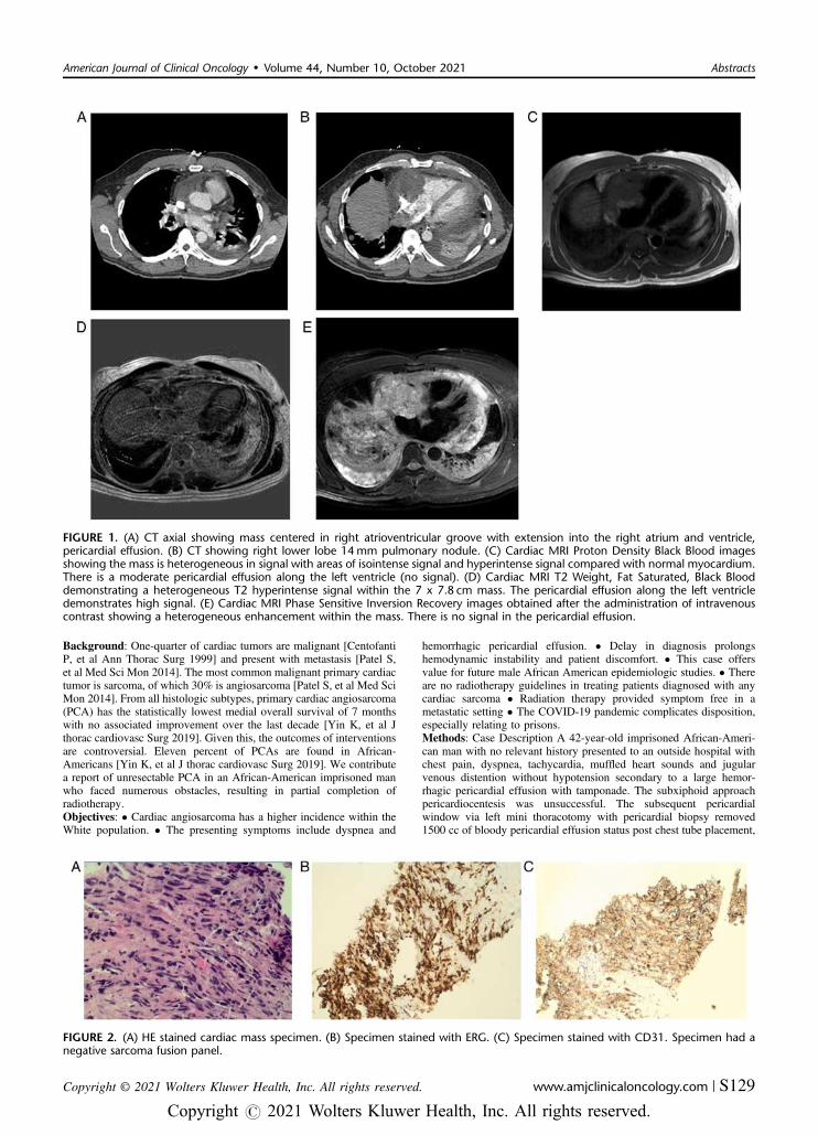
Background: One-quarter of cardiac tumors are malignant [CentofantiP, et al Ann Thorac Surg 1999] and present with metastasis [Patel S,et al Med Sci Mon 2014]. The most common malignant primary cardiactumor is sarcoma, of which 30% is angiosarcoma [Patel S, et al Med SciMon 2014]. From all histologic subtypes, primary cardiac angiosarcoma(PCA) has the statistically lowest medial overall survival of 7 monthswith no associated improvement over the last decade [Yin K, et al Jthorac cardiovasc Surg 2019]. Given this, the outcomes of interventionsare controversial. Eleven percent of PCAs are found in African-Americans [Yin K, et al J thorac cardiovasc Surg 2019]. We contributea report of unresectable PCA in an African-American imprisoned manwho faced numerous obstacles, resulting in partial completion ofradiotherapy.Objectives: • Cardiac angiosarcoma has a higher incidence within theWhite population. • The presenting symptoms include dyspnea and
hemorrhagic pericardial effusion. • Delay in diagnosis prolongshemodynamic instability and patient discomfort. • This case offersvalue for future male African American epidemiologic studies. • Thereare no radiotherapy guidelines in treating patients diagnosed with anycardiac sarcoma • Radiation therapy provided symptom free in ametastatic setting • The COVID-19 pandemic complicates disposition,especially relating to prisons.Methods: Case Description A 42-year-old imprisoned African-Ameri-can man with no relevant history presented to an outside hospital withchest pain, dyspnea, tachycardia, muffled heart sounds and jugularvenous distention without hypotension secondary to a large hemor-rhagic pericardial effusion with tamponade. The subxiphoid approachpericardiocentesis was unsuccessful. The subsequent pericardialwindow via left mini thoracotomy with pericardial biopsy removed1500 cc of bloody pericardial effusion status post chest tube placement,
FIGURE 1. (A) CT axial showing mass centered in right atrioventricular groove with extension into the right atrium and ventricle,pericardial effusion. (B) CT showing right lower lobe 14mm pulmonary nodule. (C) Cardiac MRI Proton Density Black Blood imagesshowing the mass is heterogeneous in signal with areas of isointense signal and hyperintense signal compared with normal myocardium.There is a moderate pericardial effusion along the left ventricle (no signal). (D) Cardiac MRI T2 Weight, Fat Saturated, Black Blooddemonstrating a heterogeneous T2 hyperintense signal within the 7 x 7.8 cm mass. The pericardial effusion along the left ventricledemonstrates high signal. (E) Cardiac MRI Phase Sensitive Inversion Recovery images obtained after the administration of intravenouscontrast showing a heterogeneous enhancement within the mass. There is no signal in the pericardial effusion.
FIGURE 2. (A) HE stained cardiac mass specimen. (B) Specimen stained with ERG. (C) Specimen stained with CD31. Specimen had anegative sarcoma fusion panel.
American Journal of Clinical Oncology � Volume 44, Number 10, October 2021 Abstracts
Copyright © 2021 Wolters Kluwer Health, Inc. All rights reserved. www.amjclinicaloncology.com | S129
Copyright r 2021 Wolters Kluwer Health, Inc. All rights reserved.
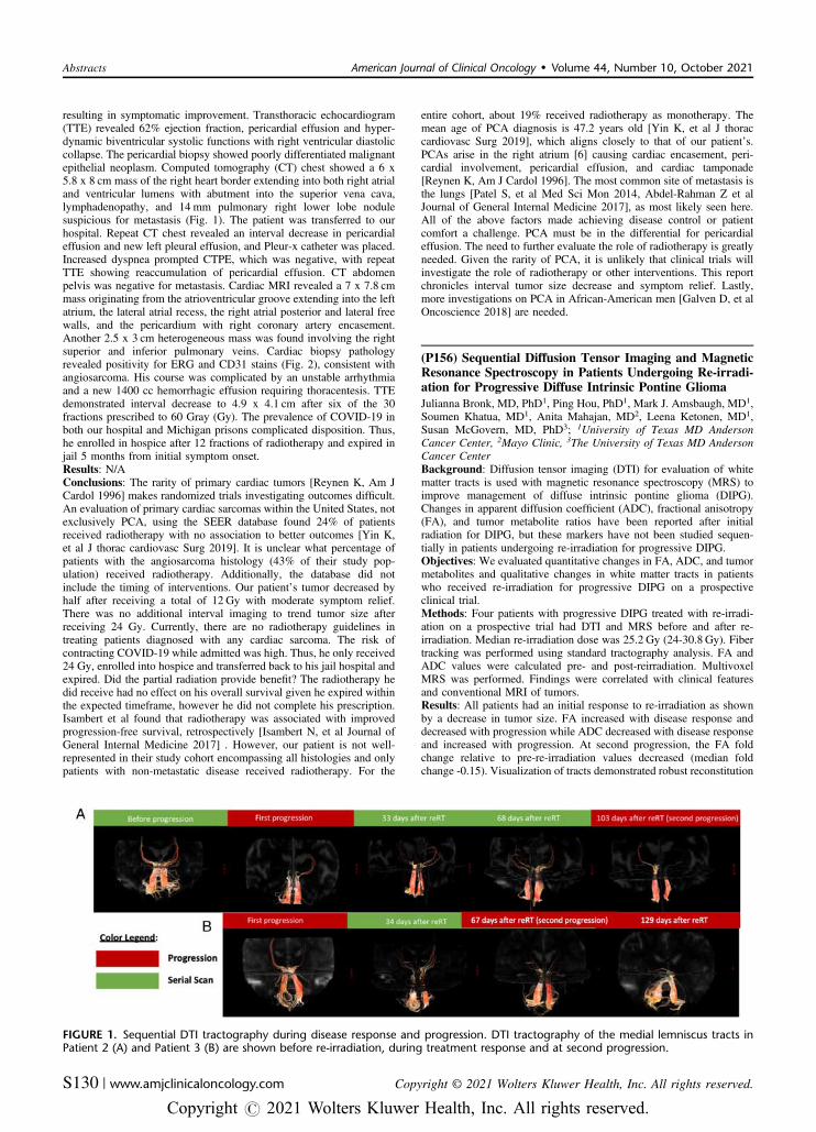
resulting in symptomatic improvement. Transthoracic echocardiogram(TTE) revealed 62% ejection fraction, pericardial effusion and hyper-dynamic biventricular systolic functions with right ventricular diastoliccollapse. The pericardial biopsy showed poorly differentiated malignantepithelial neoplasm. Computed tomography (CT) chest showed a 6 x5.8 x 8 cm mass of the right heart border extending into both right atrialand ventricular lumens with abutment into the superior vena cava,lymphadenopathy, and 14 mm pulmonary right lower lobe nodulesuspicious for metastasis (Fig. 1). The patient was transferred to ourhospital. Repeat CT chest revealed an interval decrease in pericardialeffusion and new left pleural effusion, and Pleur-x catheter was placed.Increased dyspnea prompted CTPE, which was negative, with repeatTTE showing reaccumulation of pericardial effusion. CT abdomenpelvis was negative for metastasis. Cardiac MRI revealed a 7 x 7.8 cmmass originating from the atrioventricular groove extending into the leftatrium, the lateral atrial recess, the right atrial posterior and lateral freewalls, and the pericardium with right coronary artery encasement.Another 2.5 x 3 cm heterogeneous mass was found involving the rightsuperior and inferior pulmonary veins. Cardiac biopsy pathologyrevealed positivity for ERG and CD31 stains (Fig. 2), consistent withangiosarcoma. His course was complicated by an unstable arrhythmiaand a new 1400 cc hemorrhagic effusion requiring thoracentesis. TTEdemonstrated interval decrease to 4.9 x 4.1 cm after six of the 30fractions prescribed to 60 Gray (Gy). The prevalence of COVID-19 inboth our hospital and Michigan prisons complicated disposition. Thus,he enrolled in hospice after 12 fractions of radiotherapy and expired injail 5 months from initial symptom onset.Results: N/AConclusions: The rarity of primary cardiac tumors [Reynen K, Am JCardol 1996] makes randomized trials investigating outcomes difficult.An evaluation of primary cardiac sarcomas within the United States, notexclusively PCA, using the SEER database found 24% of patientsreceived radiotherapy with no association to better outcomes [Yin K,et al J thorac cardiovasc Surg 2019]. It is unclear what percentage ofpatients with the angiosarcoma histology (43% of their study pop-ulation) received radiotherapy. Additionally, the database did notinclude the timing of interventions. Our patient’s tumor decreased byhalf after receiving a total of 12 Gy with moderate symptom relief.There was no additional interval imaging to trend tumor size afterreceiving 24 Gy. Currently, there are no radiotherapy guidelines intreating patients diagnosed with any cardiac sarcoma. The risk ofcontracting COVID-19 while admitted was high. Thus, he only received24 Gy, enrolled into hospice and transferred back to his jail hospital andexpired. Did the partial radiation provide benefit? The radiotherapy hedid receive had no effect on his overall survival given he expired withinthe expected timeframe, however he did not complete his prescription.Isambert et al found that radiotherapy was associated with improvedprogression-free survival, retrospectively [Isambert N, et al Journal ofGeneral Internal Medicine 2017] . However, our patient is not well-represented in their study cohort encompassing all histologies and onlypatients with non-metastatic disease received radiotherapy. For the
entire cohort, about 19% received radiotherapy as monotherapy. Themean age of PCA diagnosis is 47.2 years old [Yin K, et al J thoraccardiovasc Surg 2019], which aligns closely to that of our patient’s.PCAs arise in the right atrium [6] causing cardiac encasement, peri-cardial involvement, pericardial effusion, and cardiac tamponade[Reynen K, Am J Cardol 1996]. The most common site of metastasis isthe lungs [Patel S, et al Med Sci Mon 2014, Abdel-Rahman Z et alJournal of General Internal Medicine 2017], as most likely seen here.All of the above factors made achieving disease control or patientcomfort a challenge. PCA must be in the differential for pericardialeffusion. The need to further evaluate the role of radiotherapy is greatlyneeded. Given the rarity of PCA, it is unlikely that clinical trials willinvestigate the role of radiotherapy or other interventions. This reportchronicles interval tumor size decrease and symptom relief. Lastly,more investigations on PCA in African-American men [Galven D, et alOncoscience 2018] are needed.
(P156) Sequential Diffusion Tensor Imaging and MagneticResonance Spectroscopy in Patients Undergoing Re-irradi-ation for Progressive Diffuse Intrinsic Pontine GliomaJulianna Bronk, MD, PhD1, Ping Hou, PhD1, Mark J. Amsbaugh, MD1,Soumen Khatua, MD1, Anita Mahajan, MD2, Leena Ketonen, MD1,Susan McGovern, MD, PhD3; 1University of Texas MD AndersonCancer Center, 2Mayo Clinic, 3The University of Texas MD AndersonCancer CenterBackground: Diffusion tensor imaging (DTI) for evaluation of whitematter tracts is used with magnetic resonance spectroscopy (MRS) toimprove management of diffuse intrinsic pontine glioma (DIPG).Changes in apparent diffusion coefficient (ADC), fractional anisotropy(FA), and tumor metabolite ratios have been reported after initialradiation for DIPG, but these markers have not been studied sequen-tially in patients undergoing re-irradiation for progressive DIPG.Objectives: We evaluated quantitative changes in FA, ADC, and tumormetabolites and qualitative changes in white matter tracts in patientswho received re-irradiation for progressive DIPG on a prospectiveclinical trial.Methods: Four patients with progressive DIPG treated with re-irradi-ation on a prospective trial had DTI and MRS before and after re-irradiation. Median re-irradiation dose was 25.2 Gy (24-30.8 Gy). Fibertracking was performed using standard tractography analysis. FA andADC values were calculated pre- and post-reirradiation. MultivoxelMRS was performed. Findings were correlated with clinical featuresand conventional MRI of tumors.Results: All patients had an initial response to re-irradiation as shownby a decrease in tumor size. FA increased with disease response anddecreased with progression while ADC decreased with disease responseand increased with progression. At second progression, the FA foldchange relative to pre-re-irradiation values decreased (median foldchange -0.15). Visualization of tracts demonstrated robust reconstitution
FIGURE 1. Sequential DTI tractography during disease response and progression. DTI tractography of the medial lemniscus tracts inPatient 2 (A) and Patient 3 (B) are shown before re-irradiation, during treatment response and at second progression.
Abstracts American Journal of Clinical Oncology � Volume 44, Number 10, October 2021
S130 | www.amjclinicaloncology.com Copyright © 2021 Wolters Kluwer Health, Inc. All rights reserved.
Copyright r 2021 Wolters Kluwer Health, Inc. All rights reserved.

of previously disrupted paths during tumor response; conversely, therewas increased fiber tract disruption and infiltration during tumor pro-gression. MRS analysis revealed a decrease in Chol:Cr (median 2.5) andChol:NAA ratios (median 1.4) during tumor response and increaseduring progression (median 3.6 for Chol:Cr, median 2.1 for Chol:NAA).Conclusions: This study is the first to prospectively examine changes inwhite matter tracts and tumor metabolism in DIPG patients undergoingre-irradiation. Changes related to tumor response and progression areobserved even after only 24-30.8 Gy re-irradiation (Figs. 1 and 2).
(P157) MRI Radiomics Features Correlate with HistologicTumor Necrosis Following Neoadjuvant Radiation Therapyin Extremity Soft Tissue SarcomaCrystal Seldon, MD1, James Grosso, MD, PhD2, Ty Subhawong, MD3,Gautam Shrivastava, ScM1, Melanie Fernandez, BS1, Raphael Yechieli,MD1, Matthew Mason, BS4; 1University of Miami/Sylvester Compre-hensive Cancer Center, Department of Radiation Oncology, 2Universityof Miami, Department of Diagnostic Radiology, 3University of Miami,Departmentof Diagnostic Radiology, 4Univerisity of Miami, Depart-ment of Diagnostic RadiologyBackground: Radiomics has become a new method of extracting datafrom radiographic images for quantifying pixelated data far beyondwhat the human eye can perceive. Unique differences at the pixel levelbetween subsets of patients can help radiologists interpret radiographicimages in a clinically meaningful way to predict response to oncologictreatments, predict prognosis, and help in diagnosis of tumor types.Objectives: The purpose of this study is to identify MRI radiomicfeatures of sarcomas treated neoadjuvantly with radiation that correlatewith excised tumor histologic necrosis.Methods: Patients with intermediate- or high-grade extremity soft tissuesarcomas who underwent neoadjuvant radiation therapy, and who hadpost-treatment MRI scans were included. We employed MINT Lesionradiomics software for extraction of quantitative imaging data from con-trast-enhanced sequences. The lesion Regions of Interest (ROIs) weremanually contoured on a single slice demonstrating largest tumor cross-secitonal areas; 3 ROIs were analyzed as replicates within each scan, andaveraged before final analysis. Following neoadjuvant treatment, tumorswere excised and histologic tumor necrosis was determined. Using Pearsoncorrelation statistics, the percent necrosis was compared with radiomicfeatures of post treatment scans. Only radiomic features with a P value<0.05 were considered statistically significant which correlated with aPearson value of less than -0.25 or greater than 0.25.Results: 11 patients, treated between 2013 and 2019 were included in thestudy. The clinical characteristics were: male–(45%) and female- (55%); typeof sarcomas included Synovial, Spindle, Liposarcoma, and Myxofi-brosarcoma. The average necrosis on final pathology was 71% +/- 38%std. A total of 42 radiomic features were extracted from the ROIs. 12 of the
delta-radiomics parameters were significantly correlated with necrosis(P-value < 0.05). The significantly correlated radiomic features includesfirst and second order values representing the distribution of voxel inten-sities within the segmented image regions and the statistical inter-relationships between neighboring voxels, respectively. The highest cor-related radiomic features included(names directly from Mint lesion):secondorder.Information_correlation_1, secondorder.Dissimilarity, andsecondorder.Difference_average.Conclusions: Radiomics features, including both first and second orderfeatures, are associated with histologic necrosis in neoadjuvantly treatedextremity soft tissue sarcomas. This data supports multi-parametricimage texture mapping in the development of prediction models forneoadjuvant treatment response in sarcoma.
(P158) Vertebral Body Growth Retardation FollowingProton Craniospinal RadiationShannon MacDonald, MD1, Myrsini Ioakeim-Ioannidou1, AndrzejNiemierko, PhD1, Drosoula giantsoudi, PhD1, Roshan Sethi, MD1,Daniel Kim, MD MBA1, joseph Simeone, MD1; 1Massachusetts Gen-eral HospitalBackground: Craniospinal irradiation (CSI) is a standard component ofcurative therapy for several childhood brain tumors. Long-term survi-vors are at risk of late complication from CSI. A known side-effect ofCSI is growth impairment of the vertebral column resulting in decreasedsitting height for survivors
FIGURE 2. Quantitative measurements of FA, ADC, and metabolites before and after re-irradiation and at second progression. A.Absolute values for FA and ADC for the corticospinal and medial lemniscus tracts at first progression, after reRT, and at second progressionin patients who underwent reRT for progressive DIPG. B. Median fold change in FA and ADC during disease response and at secondprogression for patients with serial imaging following re-irradiation. C and D. Median Chol:Cr (C) and Chol:NAA (D) ratios at firstprogression (n=1 scan), during response to re-irradiation (n=6 scans), and at second progression (n=3 scans).
FIGURE 1. Growth retardation as a function of follow-up time.
American Journal of Clinical Oncology � Volume 44, Number 10, October 2021 Abstracts
Copyright © 2021 Wolters Kluwer Health, Inc. All rights reserved. www.amjclinicaloncology.com | S131
Copyright r 2021 Wolters Kluwer Health, Inc. All rights reserved.
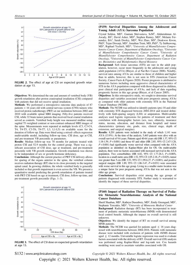
Objectives: We determined the rate and amount of vertebral body (VB)growth retardation after proton craniospinal irradiation (CSI) comparedwith patients that did not receive spinal irradiation.Methods: We performed a retrospective outcome data analysis of 87patients < 16 years old with central nervous system (CNS) tumors whoreceived proton radiotherapy (PRT) at our institution between 2002 and2010 with available spinal MRI imaging. Fifty-five patients receivedCSI, while 32 brain tumor patients that received focal cranial irradiationserved as controls. Vertebral body height was measured midline usingsagittal T1-weighted contrast or non-contrast enhanced MRI images ofthe spine. Measurements were repeated at multiple levels (C3, C3-C4,T4, T4-T5, C3-T6, T4-T7, L3, L1-L5) on available scans for theduration of follow-up. Data were fitted using a mixed- effects regressionmultivariable model, including follow-up time, CSI dose, age at CSI,and pre-treatment VB percentile as parameters.Results: Median follow-up was 70 months for patients treated withproton CSI and 52.9 months for the control group. There was a sig-nificant association of CSI dose, age at treatment, and pre-treatmentpercentile with VB growth retardation. Growth retardation was shownto be independent of sex or growth hormone deficiency.Conclusions: Although the current practice of PRT CSI delivery allowsfor sparing of the organs anterior to the spine, the vertebral columnreceives radiation therapy (RT) due to its close proximity to the targetedspinal canal. In growing children, the whole vertebral body is generallyincluded so that growth impairment is even across the VB. We present aquantitative model predicting the growth retardation of patients treatedwith PRT CSI based on age at treatment, CSI dose, follow-up time, andpre-treatment growth percentile (Figs. 1–3).
(P159) Survival Disparities Among the Adolescent andYoung Adult (AYA) Sarcoma PopulationCrystal Seldon, MD1, Gautam Shrivastava, ScM2, Abdurrahman Al-Awady, BS3, David Asher, MD3, Stephen Ramey, MD3, Melanie Fer-nandez, BA3, Sarah Dooley, MD3, Wei Zhao, MS4, Deukwoo Kwon,PhD2, Tejan Diwanji, MD1, Jonathan Trent, MD, PhD3, Ty Subhawong,MD3, Raphael Yechieli, MD1; 1University of Miami/Sylvester Compre-hensive Cancer Center, Department of Radiation Oncology, 2Universityof Miami/Sylvester Comprehensive Cancer Center, 3University ofMiami/Sylvester Comprehensive Cancer, Department of RadiationOncology, 4University of Miami/Sylvester Comprehensive Cancer Cen-ter, Biostatistics and Bioinformatics Shared ResourceBackground: Soft tissue sarcomas (STS) are rare in the adult pop-ulation, however, occur more frequently in the adolescent and youngadult population (AYA) (Bleyer, et al Nat Rev Cancer 2008). Notably,survival rates among AYAs are similar to those of children and higherthan in adults, however, this is not seen in STS (American CancerSociety, Cancer Facts & Figures 2020). Poorer prognosis is attributed tonumerous factors including more aggressive clinical characteristics ofSTS in the AYA population compared with those diagnosed in children,poor clinical trial participation of AYAs, and lack of data regardingprognostic factors in this age group (Bleyer, et al Cancer 2005).Objectives: In this retrospective study, we sought to determine survivaldisparities and variables associated with poor prognosis among AYAsas compared with older patients with extremity STS in the NationalCancer Database (NCDB).Methods: The NCDB was utilized to identify patients (pts) 18 and olderwith extremity STS diagnosed between 2004-2014 and treated defini-tively with limb-sparing surgery (LSS) or amputation. Multivariableanalyses used logistic regressions for patterns of treatment and theircorrelation with demographic factors (sex, race, ethnicity, insurancestatus, income, education, and distance from hospital) and tumorcharacteristics (primary site, grade, size, clinical stage, depth ofextension, and surgical margins).Results: 8,201 patients were included in the study of which 1,141 wereAYA (13.9%). At the time of the analysis, 5,465 patients were alive with anoverall survival of 9.6 years. Patients aged 40-64 years old (HR 1.37, 95% CI1.18-1.59, P<0.001) and those 65 and older (HR 2.85, 95% CI 2.47-3.28,P<0.001) had significantly worse survival when compared with the AYApopulation as identified on Kaplan-Meier plot for OS. On multivariableanalysis, there were no factors associated with increased survival in the AYApopulation. Two comorbidities (HR 6.82, 95% CI 1.77-26.3, P=0.005),location in a small metro area (HR 1.52, 95% CI 1.05-2.18, P=0.025), tumorsize greater than 5 cm (HR 3.53, 95% CI 1.99-6.27, P<0.001), and positivesurgical margins (HR 1.89, 95% CI 1.06-3.47, P=0.031) were associatedwith worse survival in this age group. Living in a smaller metro location wasa unique factor for poor prognosis among AYAs that was not seen in theolder age groups.Conclusions: Survival disparities exist among the age groups ofpatients diagnosed with extremity STS. Further study is warranted toidentify the impact of these survival disparities.
(P160) Impact of Radiation Therapy on Survival of Pedia-tric Metastatic Neuroblastoma: Analysis of the NationalCancer DatabaseSherif Shaaban, MD1, Kathryn Dusenbery, MD1, Emily Greengard, MD1,Stephanie Terezakis, MD1; 1University of Minnesota Medical CenterBackground: Radiation therapy (RT) has been incorporated in treat-ment of pediatric metastatic neuroblastoma (MNB) with demonstratedlocal control benefit. Although the impact on overall survival is stillunknown.Objectives: We identify the impact of RT on overall survival amongMNB patients.Methods: The NCDB was queried for patients aged ≤ 18 years diag-nosed with neuroblastoma between 2004-2016. Patients with metastaticdisease were included with exclusion of patients with 4S/MS diseaseaged ≤ 12 months. Univariable logistic regression was used to evaluatefactors associated with the delivery of RT. Overall survival (OS) analysiswas performed using Kaplan-Meier and log-rank test. Cox hazardsmodeling were used to ascertain variables associated with OS.
FIGURE 2. The effect of age at CSI on expected growth retar-dation at age 15.
FIGURE 3. The effect of CSI dose on expected growth retardationat age 15.
Abstracts American Journal of Clinical Oncology � Volume 44, Number 10, October 2021
S132 | www.amjclinicaloncology.com Copyright © 2021 Wolters Kluwer Health, Inc. All rights reserved.
Copyright r 2021 Wolters Kluwer Health, Inc. All rights reserved.

Results: 504 patients met the study criteria; 257 patients (51%) receivedRT and 247 (49%) did not. Surgery was performed more frequentlyamong RT group (88%) versus the non-RT group (32%). The numberof patients who underwent autologous bone marrow transplant (ABMT)in the RT group was significantly higher than those in the non-RT group(73% versus 26%, P= 0.0001). The most common utilized RTmodalities were 3D CRT (50%) and IMRT (34%). The most commonused RT dose was 2,160 cGy (58%) and the most frequent boost dosewas 1,440 cGy. Logistic regression analysis confirmed that surgery withpositive surgical margins, use of ABMT and immunotherapy covariateswere associated with increased likelihood of receiving RT. Afterstratification by RT receipt, the median OS was significantly higheramong the RT group, 67 months (95% CI, 62.53-71.4 mo) versus56 months among the non-RT group (95% CI, 51.64-61.38 mo) (P=0.008). Cox hazards modeling confirmed that non-receipt of RT wasassociated with poorer OS on univariate (HR 1.51, 95% CI 1.11- 2.05,P= 0.009) and multivariate (HR 1.52, 95% CI 1.115- 2.07, P= 0.008)analysis. Presence of bone metastases was significantly associated withworse OS on univariate analysis (HR 1.80, 95% CI 1.08- 2.97,P= 0.022). ABMT receipt was associated with better OS on univariate(HR 0.333, 95% CI, 0.145- 0.763, P= 0.009) and multivariate (HR0.234, 95% CI, 0.147- 0.897, P= 0.002) analysis.Conclusions: Impact of RT on OS has not been widely investigated. Ourstudy demonstrated that RT was significantly associated with improvedOS. Future prospective studies are warranted to identify the impact RT onsurvival in high risk and metastatic neuroblastoma (Fig. 1).
(P161) Impact of Strict Brainstem Dosimetric Parameterson Symptomatic Brainstem Injury and Survival AfterProton Beam Radiation in Pediatric Brain TumorsRituraj Upadhyay, MD1, David Grosshans, MD, PhD1, SusanMcGovern, MD, PhD1, Mary McAleer, MD1, Kristina Woodhouse,MD1, Wafik Zaky, MD1, Murali Chintagumpala, MD2, Anita Mahajan,MD3, Arnold Paulino, MD1; 1The University of Texas MD AndersonCancer Center, 2Texas Children's Cancer Center, Baylor College ofMedicine, 3Mayo ClinicBackground: Brainstem toxicity after radiation is a devastating com-plication and is a particular concern with protons, given the biologicaldifferences and range uncertainties. Dosimetric predictors of brainstem
injury are not well defined and the prescription dose is often decreasedto limit the risk, especially in tumors involving the posterior fossa.Objectives: To investigate the difference in survival and risk ofbrainstem injury in pediatric brain tumors treated with proton therapywhen using strict brainstem dosimetric.Methods: All patients < 18 years with infratentorial brain and pinealtumors, treated with proton beam radiation at our institution from2007–2019, with a brainstem Dmean of > 30 Gy and/or Dmax >50.4 Gy were included. We excluded patients with primary brainstemtumors. In 2014, our institution started using strict brainstem doseconstraints of Dmax ≤ 57 Gy, Dmean ≤ 52.4 Gy and V54 ≤ 10% todecide final dose to the target(s). We analyzed Symptomatic brainsteminjury (SBI) and survival before and since incorporation of theseguidelines. SBI was defined as any new or progressive cranial neuro-pathy, bulbar weakness, ataxia, dysmetria and/or motor weakness withcorresponding radiographic abnormality within the brainstem. Kaplan-Meier test was used for survival analysis.Results: A total of 595 patients were reviewed and 362 (medullo-blastoma= 209, ependymoma= 86, ATRT= 43, pineoblastoma= 9,others= 15) met our inclusion criteria. Median age at RT was 5 years(range 0.7–17.9 y) and median prescribed RT dose was 54 CGyE (range39.6–59.4 CGyE). About 32% patients were prescribed > 54 Gy before2014, compared with only 9% since 2014. Median follow up was40 months (range 1–152). 2-year progression free survival in patientstreated before and since 2014 was 70.3% and 70.6% (P= 0.649), whileoverall survival was 80.8% and 86.1% (P= 0.77) respectively. Tenpatients (2.7%) developed SBI, at a median of 4 months after radiation(range 1-6). Asymptomatic imaging changes within brainstem wereseen in 47 patients (13%) at a median of 4 months after RT, with mostcommon findings being increased T2 flair, focal enhancement andencephalomalacia. The incidence of SBI decreased from 4.2% (2007-2013) to 0.7% (2014-2019) (P= 0.047) while asymptomatic imagingchanges remained similar (11.6% vs 15.1%, P= 0.33). On univariateanalysis, only timing of radiation (before/since 2014) was an inde-pendent predictor of SBI (P= 0.028), while no significant associationwas observed with extent of resection, number of resections, age atradiation, prescribed RT dose, cone down and systemic treatment.Conclusions: Although our study is limited given the heterogenouspopulation and different time-period of comparison, our results suggestthat the use of strict brainstem dose constraints to determine final doseto the tumor/ tumor bed results in a lower incidence of SBI, withoutcompromising the survival.
FIGURE 1. Overall Survival of all study population (A) and stratified by radiotherapy treatment (B).
American Journal of Clinical Oncology � Volume 44, Number 10, October 2021 Abstracts
Copyright © 2021 Wolters Kluwer Health, Inc. All rights reserved. www.amjclinicaloncology.com | S133
Copyright r 2021 Wolters Kluwer Health, Inc. All rights reserved.

(P162) Patterns of Care and Utilization Disparities inProton Radiation Therapy for Pediatric Central NervousSystem MalignanciesChristopher Weil, MD1, Lew Felicia, DVM2, Vonetta Williams, MD/PhD3, Ralph Ermoian, MD3, Matthew Poppe, MD1; 1University of UtahHuntsman Cancer Institute, 2Washington State University Elson S.Floyd College of Medicine, 3University of WashingtonBackground: Proton radiotherapy (PR) is well established in thetreatment of pediatric malignancies in the central nervous system (CNS)given established dosimetric advantages that may reduce the late effectsof radiotherapy.Objectives: In this analysis, we sought to update our 2016 publishedpatterns of care data on the receipt of PR in children with primary CNSmalignancies since both the number of proton centers and availabilityhave significantly increased in the interim. We aimed to evaluate theutilization of PR in this population and characterize the clinical andsociodemographic factors predictive of receipt of PR.
Methods: The National Cancer Data Base (NCDB) was queried toidentify all pediatric patients with primary CNS malignancies treatedwith radiotherapy (RT) with curative intent from 2004 to 2017. Clinicalcharacteristics and demographics were analyzed using standard T andχ2 testing. Predictors of receipt of PR were identified with univariableand multivariable logistic regression.Results: We identified 9,126 patients age ≤ 18 years treated with RTbetween 2004 and 2017, of which 1,045 (11.5%) received PR. PR usagecontinued to increase significantly, from <1% in 2004 to 28% in 2017.This increase was most striking for ependymoma and medulloblastoma,where PR was utilized in 45% and 47% of cases, respectively, in 2017.The proportion of White and Asian patients receiving PR for non-highgrade glioma and non-meningioma CNS malignancies over the studyperiod rose from <1% for both to 35% and 44%, respectively, while inBlack patients the proportion rose from <1% to 26%. Multivariablepredictors of receipt of PR include year of diagnosis, age under 6 years,income level, distance from PR facility, and histology; multivariablepredictors of receipt of non-proton radiotherapy include Black race,rural residence and Medicaid insurance. These factors remained sig-nificant when isolating the most recent five years of data.Conclusions: The usage of PR for CNS malignancies continues toincrease and has doubled over the last four years. Despite the potentialclinical advantages to PR for pediatric primary CNS malignancies, thereare notable socioeconomic, geographic and racial disparities in thereceipt of PR that have persisted despite the increased availability andaccessibility over the study period. Further study is warranted toidentify how to address the disparities and better support these patients(Tables 1–3).
TABLE 1. Baseline Patient Characteristics Between Photon andProton Radiotherapy Groups
TABLE 2. Univariable and Multivariable Predictors of Receipt ofProton Radiotherapy
Abstracts American Journal of Clinical Oncology � Volume 44, Number 10, October 2021
S134 | www.amjclinicaloncology.com Copyright © 2021 Wolters Kluwer Health, Inc. All rights reserved.
Copyright r 2021 Wolters Kluwer Health, Inc. All rights reserved.

(P163) A Dose Accumulation Assessment of AlignmentErrors During Spatially Fractionated Radiation TherapyJohn Ginn, PhD1, Sai Duriseti, MD, PhD1, Thomas Mazur, PhD1,Matthew Spraker, MD, PhD1, James Kavanaugh, PhD2; 1Department of
Radiation Oncology, Washington University School of Medicine in St.Louis, 2Washington University School of MedicineBackground: Spatially-fractionated radiotherapy (SFRT) treatmenttechniques produce high-dose peaks and low-dose valleys by inter-weaving a matrix of high- and low-dose spheres through a techniquetermed Lattice. Due to the highly localized spatial dose gradientsassociated with SFRT, it is critical for patients be aligned correctly fortreatment.Objectives: Here we report the dosimetric impact of daily alignmentsetup uncertainty through a dose accumulation study.Methods: Dose accumulation was retrospectively performed for tenpatients enrolled on a completed prospective trial evaluating Lattice forlarge tumors (NCT04133415). SFRT was completed in 5 fractions with20 Gy prescribed to the entire tumor volume and 66.7 Gy prescribed tothe high-dose spheres of 1.5cm diameter. Daily alignment error wasquantified through a landmark analysis with paired landmarks manuallyselected in both the planning computed tomography (CT) scan and theregistered daily cone beam CT image. The dosimetric impact ofalignment errors was quantified by translating the isocenter in thetreatment planning system by the daily average alignment error for eachfraction. To understand the effect of varying magnitude and frequencyof alignment errors, we also performed a simulated error analysis bytranslating isocenter by 5 mm and 10 mm for one and two fractions,with alignment errors separately assessed for the superior-inferior andaxial dimensions. We hypothesized misalignment errors will reduce thevolume of tumor receiving both 66.7 Gy and 20 Gy while also blurringthe dose gradient between the high- and low-dose spheres. Therefore,we quantified the ratio of the mean dose in the high-dose and adjacentlow-dose spheres (DR) as well as the mean dose (Dmean) and standarddeviation of the mean in the 1.5 cm ring surrounding the high-dosespheres.Results: The total average and standard deviation alignment error was1.8 mm and 0.6 mm across all patients. Visual inspection revealedalignment errors tend to blur the edges of the high- and low-dose dis-tributions. Quantitatively, mean DR decreased from 3.42 to 3.32 in thedose accumulation study. On average Dmean in the 1.5 cm ring was35.5 Gy in both the initial plans and accumulated dose, while thestandard deviation of the dose in this region decreased by 25.5 cGyfrom the initial plan to 10.7 Gy. The simulated worst case was aninferior-superior shift of 10 mm for two fractions. In this case averageDR decreased to 2.72, and the Dmean and standard deviation of thedose in the surrounding ring decreased 24.5 cGy and 148.0 cGy,respectively.Conclusions: The dose accumulation study revealed on average DRonly decreased from 3.42 to 3.32. However, setup errors > 5 mm resultin larger dosimetric degradation than those observed clinically. Asindicated by our simulation study, even a single large setup error cansubstantially reduce the DR and overall SFRT plan quality,
TABLE 3. Univariable andMultivariable Predictors of Receipt of ProtonRadiotherapy Limited to Low Grade Glioma, Ependymoma, Medullo-blastoma, Primitive Neural Ectodermal Tumors, Craniopharyngiomas,Germ Cell Tumors and Atypical Teratoid/Rhabdoid Tumors
FIGURE 1. Example of a spatially fractionated dose distribution. A coronal view of a spatially fractionated dose distribution showing thehigh- and low-dose spheres dispersed throughout the target.
American Journal of Clinical Oncology � Volume 44, Number 10, October 2021 Abstracts
Copyright © 2021 Wolters Kluwer Health, Inc. All rights reserved. www.amjclinicaloncology.com | S135
Copyright r 2021 Wolters Kluwer Health, Inc. All rights reserved.
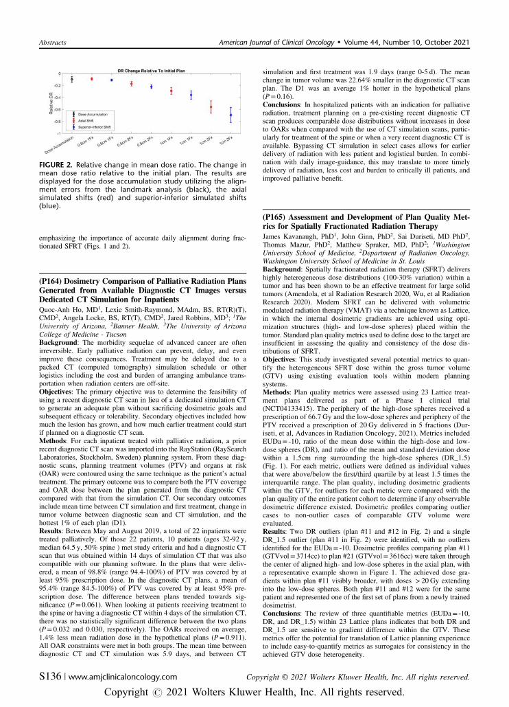
emphasizing the importance of accurate daily alignment during frac-tionated SFRT (Figs. 1 and 2).
(P164) Dosimetry Comparison of Palliative Radiation PlansGenerated from Available Diagnostic CT Images versusDedicated CT Simulation for InpatientsQuoc-Anh Ho, MD1, Lexie Smith-Raymond, MAdm, BS, RT(R)(T),CMD2, Angela Locke, BS, RT(T), CMD2, Jared Robbins, MD3; 1TheUniversity of Arizona, 2Banner Health, 3The University of ArizonaCollege of Medicine - TucsonBackground: The morbidity sequelae of advanced cancer are oftenirreversible. Early palliative radiation can prevent, delay, and evenimprove these consequences. Treatment may be delayed due to apacked CT (computed tomography) simulation schedule or otherlogistics including the cost and burden of arranging ambulance trans-portation when radiation centers are off-site.Objectives: The primary objective was to determine the feasibility ofusing a recent diagnostic CT scan in lieu of a dedicated simulation CTto generate an adequate plan without sacrificing dosimetric goals andsubsequent efficacy or tolerability. Secondary objectives included howmuch the lesion has grown, and how much earlier treatment could startif planned on a diagnostic CT scan.Methods: For each inpatient treated with palliative radiation, a priorrecent diagnostic CT scan was imported into the RayStation (RaySearchLaboratories, Stockholm, Sweden) planning system. From these diag-nostic scans, planning treatment volumes (PTV) and organs at risk(OAR) were contoured using the same technique as the patient’s actualtreatment. The primary outcome was to compare both the PTV coverageand OAR dose between the plan generated from the diagnostic CTcompared with that from the simulation CT. Our secondary outcomesinclude mean time between CT simulation and first treatment, change intumor volume between diagnostic scan and CT simulation, and thehottest 1% of each plan (D1).Results: Between May and August 2019, a total of 22 inpatients weretreated palliatively. Of those 22 patients, 10 patients (ages 32-92 y,median 64.5 y, 50% spine ) met study criteria and had a diagnostic CTscan that was obtained within 14 days of simulation CT that was alsocompatible with our planning software. In the plans that were deliv-ered, a mean of 98.8% (range 94.4-100%) of PTV was covered by atleast 95% prescription dose. In the diagnostic CT plans, a mean of95.4% (range 84.5-100%) of PTV was covered by at least 95% pre-scription dose. The difference between plans trended towards sig-nificance (P= 0.061). When looking at patients receiving treatment tothe spine or having a diagnostic CT within 4 days of the simulation CT,there was no statistically significant difference between the two plans(P= 0.032 and 0.030, respectively). The OARs received on average,1.4% less mean radiation dose in the hypothetical plans (P= 0.911).All OAR constraints were met in both groups. The mean time betweendiagnostic CT and CT simulation was 5.9 days, and between CT
simulation and first treatment was 1.9 days (range 0-5 d). The meanchange in tumor volume was 22.64% smaller in the diagnostic CT scanplan. The D1 was an average 1% hotter in the hypothetical plans(P= 0.16).Conclusions: In hospitalized patients with an indication for palliativeradiation, treatment planning on a pre-existing recent diagnostic CTscan produces comparable dose distributions without increases in doseto OARs when compared with the use of CT simulation scans, partic-ularly for treatment of the spine or when a very recent diagnostic CT isavailable. Bypassing CT simulation in select cases allows for earlierdelivery of radiation with less patient and logistical burden. In combi-nation with daily image-guidance, this may translate to more timelydelivery of radiation, less cost and burden to critically ill patients, andimproved palliative benefit.
(P165) Assessment and Development of Plan Quality Met-rics for Spatially Fractionated Radiation TherapyJames Kavanaugh, PhD1, John Ginn, PhD2, Sai Duriseti, MD PhD2,Thomas Mazur, PhD2, Matthew Spraker, MD, PhD2; 1WashingtonUniversity School of Medicine, 2Department of Radiation Oncology,Washington University School of Medicine in St. LouisBackground: Spatially fractionated radiation therapy (SFRT) delivershighly heterogeneous dose distributions (100-30% variation) within atumor and has been shown to be an effective treatment for large solidtumors (Amendola, et al Radiation Research 2020, Wu, et al RadiationResearch 2020). Modern SFRT can be delivered with volumetricmodulated radiation therapy (VMAT) via a technique known as Lattice,in which the internal dosimetric gradients are achieved using opti-mization structures (high- and low-dose spheres) placed within thetumor. Standard plan quality metrics used to define dose to the target areinsufficient in assessing the quality and consistency of the dose dis-tributions of SFRT.Objectives: This study investigated several potential metrics to quan-tify the heterogeneous SFRT dose within the gross tumor volume(GTV) using existing evaluation tools within modern planningsystems.Methods: Plan quality metrics were assessed using 23 Lattice treat-ment plans delivered as part of a Phase I clinical trial(NCT04133415). The periphery of the high-dose spheres received aprescription of 66.7 Gy and the low-dose spheres and periphery of thePTV received a prescription of 20 Gy delivered in 5 fractions (Dur-iseti, et al, Advances in Radiation Oncology, 2021). Metrics includedEUDa= -10, ratio of the mean dose within the high-dose and low-dose spheres (DR), and ratio of the mean and standard deviation dosewithin a 1.5cm ring surrounding the high-dose spheres (DR_1.5)(Fig. 1). For each metric, outliers were defined as individual valuesthat were above/below the first/third quartile by at least 1.5 times theinterquartile range. The plan quality, including dosimetric gradientswithin the GTV, for outliers for each metric were compared with theplan quality of the entire patient cohort to determine if any observabledosimetric difference existed. Dosimetric profiles comparing outliercases to non-outlier cases of comparable GTV volume wereevaluated.Results: Two DR outliers (plan #11 and #12 in Fig. 2) and a singleDR_1.5 outlier (plan #11 in Fig. 2) were identified, with no outliersidentified for the EUDa= -10. Dosimetric profiles comparing plan #11(GTVvol= 3714cc) to plan #21 (GTVvol= 3616cc) were taken throughthe center of aligned high- and low-dose spheres in the axial plan, witha representative example shown in Figure 1. The achieved dose gra-dients within plan #11 visibly broader, with doses > 20 Gy extendinginto the low-dose spheres. Both plan #11 and #12 were for the samepatient and represented one of the first set of plans from a newly traineddosimetrist.Conclusions: The review of three quantifiable metrics (EUDa= -10,DR, and DR_1.5) within 23 Lattice plans indicates that both DR andDR_1.5 are sensitive to gradient difference within the GTV. Thesemetrics offer the potential for translation of Lattice planning experienceto include easy-to-quantify metrics as surrogates for consistency in theachieved GTV dose heterogeneity.
FIGURE 2. Relative change in mean dose ratio. The change inmean dose ratio relative to the initial plan. The results aredisplayed for the dose accumulation study utilizing the align-ment errors from the landmark analysis (black), the axialsimulated shifts (red) and superior-inferior simulated shifts(blue).
Abstracts American Journal of Clinical Oncology � Volume 44, Number 10, October 2021
S136 | www.amjclinicaloncology.com Copyright © 2021 Wolters Kluwer Health, Inc. All rights reserved.
Copyright r 2021 Wolters Kluwer Health, Inc. All rights reserved.
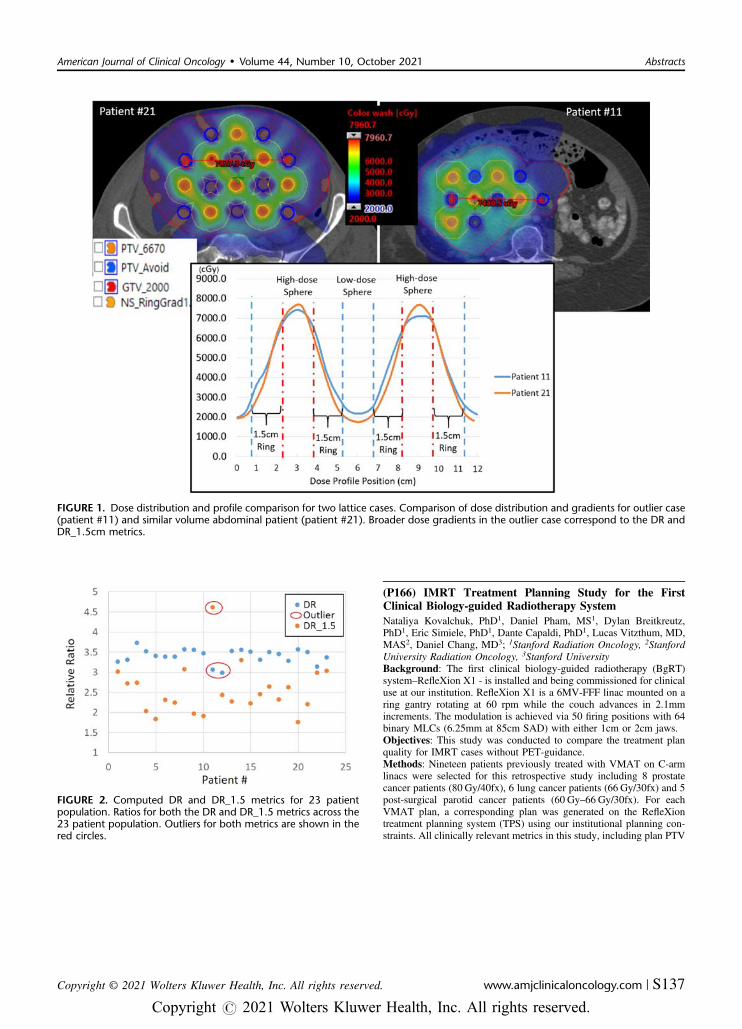
(P166) IMRT Treatment Planning Study for the FirstClinical Biology-guided Radiotherapy SystemNataliya Kovalchuk, PhD1, Daniel Pham, MS1, Dylan Breitkreutz,PhD1, Eric Simiele, PhD1, Dante Capaldi, PhD1, Lucas Vitzthum, MD,MAS2, Daniel Chang, MD3; 1Stanford Radiation Oncology, 2StanfordUniversity Radiation Oncology, 3Stanford UniversityBackground: The first clinical biology-guided radiotherapy (BgRT)system–RefleXion X1 - is installed and being commissioned for clinicaluse at our institution. RefleXion X1 is a 6MV-FFF linac mounted on aring gantry rotating at 60 rpm while the couch advances in 2.1mmincrements. The modulation is achieved via 50 firing positions with 64binary MLCs (6.25mm at 85cm SAD) with either 1cm or 2cm jaws.Objectives: This study was conducted to compare the treatment planquality for IMRT cases without PET-guidance.Methods: Nineteen patients previously treated with VMAT on C-armlinacs were selected for this retrospective study including 8 prostatecancer patients (80 Gy/40fx), 6 lung cancer patients (66 Gy/30fx) and 5post-surgical parotid cancer patients (60 Gy–66 Gy/30fx). For eachVMAT plan, a corresponding plan was generated on the RefleXiontreatment planning system (TPS) using our institutional planning con-straints. All clinically relevant metrics in this study, including plan PTV
FIGURE 1. Dose distribution and profile comparison for two lattice cases. Comparison of dose distribution and gradients for outlier case(patient #11) and similar volume abdominal patient (patient #21). Broader dose gradients in the outlier case correspond to the DR andDR_1.5cm metrics.
FIGURE 2. Computed DR and DR_1.5 metrics for 23 patientpopulation. Ratios for both the DR and DR_1.5 metrics across the23 patient population. Outliers for both metrics are shown in thered circles.
American Journal of Clinical Oncology � Volume 44, Number 10, October 2021 Abstracts
Copyright © 2021 Wolters Kluwer Health, Inc. All rights reserved. www.amjclinicaloncology.com | S137
Copyright r 2021 Wolters Kluwer Health, Inc. All rights reserved.

D95%, PTV D1%, Conformity Index (CI), and organs at risk con-straints were analyzed and compared between the VMAT andRefleXion plans using paired t-tests.Results: Clinically acceptable plans were obtained with both techni-ques. For the prostate, lung, and head and neck sites, no statisticallysignificant difference was observed in PTV D95%, PTV D1%, or CIbetween the VMAT and RefleXion treatment plans. Due to inherentOAR dose penalties in the RefleXion optimizer, dose reduction tocertain critical structures was observed in RefleXion plans: brainstemDmax (-4.8 Gy, P< 0.008), contralateral cochlea Dmean (-4.8 Gy,P< 0.01), lips Dmax (-3.5 Gy, P< 0.007) for head and neck cases;esophagus Dmean (-2.3 Gy, P< 0.039), spinal cord Dmax (-3.6 Gy,P< 0.001) for lung cases; femurs left and right Dmax (-4.8 Gy,P< 0.003 and -5.0 Gy, P< 0.01) and rectum V75% (-1.6%, P< 0.01)for prostate cases.Conclusions: The RefleXion TPS provided comparable plan quality toVMAT plans (Table 1).
(P167) Lower Baseline Apparent Diffusion CoefficientValues Associated with Poor Prognosis in Locally AdvancedPancreatic CancerAnna Laucis, MD, MPhil1, Yilun Sun, PhD1, Yue Cao, PhD1, TheodoreLawrence, MD, PhD, FASTRO1, Mahmoud Al-Hawary, MD2, ThomasChenevert, PhD2, Kyle Cuneo, MD1; 1University of MichiganDepartment of Radiation Oncology, 2University of Michigan Depart-ment of RadiologyBackground: A challenge in locally advanced pancreatic cancer is thedifficulty in obtaining adequate tumor tissue to personalize therapy.Non-invasive diffusion magnetic resonance imaging (dMRI) has thepotential to tell us about the biology of a tumor and its responsiveness totherapy. Our prior work found a significant association between pre-treatment apparent diffusion coefficient (ADC) and pathologicalresponse in patients with resectable pancreatic cancer undergoing pre-operative chemoradiation.Objectives: The goal of the current study was to prospectively inves-tigate the relationship between dMRI characteristics and outcomes afterchemoradiation in locally advanced pancreatic cancer patients.Methods: Patients with locally advanced pancreatic cancer wereprospectively enrolled onto an IRB-approved clinical trial inves-tigating dMRI characteristics at multiple time points before andduring chemoradiation. Each pancreatic tumor was delineated by tworadiation oncologists on T1-weighted MRI and dMRI images.Baseline and mid-treatment ADC values were then analyzed andcompared with clinical outcomes including time to local failure(TTLF), time to distant failure (TTDF), progression free survival(PFS), and overall survival (OS). Additionally, CA-19-9 values wereobtained for each patient pre-radiation and at four months post-
radiation and compared with clinical outcomes. Univariable Coxproportional hazard models, Student’s t-tests, and Kaplan Meiermethods were used for statistical analysis.Results: A total of 23 MRI scans were obtained in nine patientsreceiving gemcitabine-based chemoradiation. There were six maleand three female patients, with median age of 64 years (range 52-73).The median PFS was 18 months and median OS was 25.3 monthsfrom diagnosis for the cohort. Pre-treatment baseline MRIs wereobtained a median of 25 days (range 1–35) before radiation. Wefound a significant association between lower baseline ADC valuesand several clinical outcomes. Specifically, lower mean baselinetumor ADC values were associated with lower OS, lower PFS, shorterTTLF, and shorter TTDF. The difference between baseline and mid-treatment ADC values were also found to be significantly differentbetween patients with and without distant failure (Fig. 1, P= 0.049).In comparison, there was no significant relationship between CA-19-9lab values or mid-treatment ADC with any clinical outcome. Table 1summarizes the baseline and mid-treatment ADC value results.Conclusions: Lower baseline tumor ADC values were found to sig-nificantly correlate with clinical outcomes including worse survival inlocally advanced pancreatic cancer patients. This is concordant withprior studies showing poor pathological responses in resectable pan-creatic cancer patients with lower baseline ADC values. Furtherresearch is needed in a larger cohort of patients to study how this non-invasive imaging biomarker may help with clinical decision-makingand treatment planning.
TABLE 1. Comparison of Average Dosimetric Indices for Head and Neck, Lung, and Prostate Cases
Colors signify RefleXion plan superiority (green) or inferiority (red).
FIGURE 1. Difference between baseline and mid-radiation (mid-RT) ADC values were significantly different for patients with andwithout distant failure (DF), P=0.049.
Abstracts American Journal of Clinical Oncology � Volume 44, Number 10, October 2021
S138 | www.amjclinicaloncology.com Copyright © 2021 Wolters Kluwer Health, Inc. All rights reserved.
Copyright r 2021 Wolters Kluwer Health, Inc. All rights reserved.

(P168) A Diode-based 64X64 X-ray Detector for CancerRadiotherapyMekhail Anwar1, Kyoungtae Lee2, Bruce Faddegon1, Jessica Scholey1,Kavita Mishra1, Michel Maharbiz2; 1UCSF, 2UC BerkeleyBackground: The ability to monitor radiation dose delivered in real timeto the tumor and organs at risk (OAR), is critical for radiotherapy qualityand safety, particularly for targets subject to motion or near critical OARs.A highly sensitive in vivo microdosimeter (IVµD) implanted within thetumor and OAR via biopsy needle solves this problem by monitoring andrelaying the dose delivered within the patient in real time. We havedeveloped a real time millimeter IVµD [1]. We hypothesize that the deviceis capable of measuring photons via secondary electrons generated by pairproduction and Compton scattering. Here we present the first results forphoton detection: the IVµD is capable of counting the number of secondaryelectrons generated by x-ray photons with the form factor and powerconsumption compatible with in vivo implantation.Objectives: An IVµD must satisfy the following requirements: 1)measure and relay dose in real-time, 2) be millimeter scale forimplantation through a core biopsy needle, 3) high sensitivity, and 4)low power (to minimize battery size or wireless power transferrequirements). To accomplish these requirements, we exploit CMOSfabrication technology (i.e. ‘computer chip technology’) that enablesdirect integration of complex circuitry with highly sensitive sensingelements in a small form factor.Methods: A P-N diode was used to detect the secondary electrons gen-erated from a photon interacting with the silicon. About ⅓ of the energydeposited in the depletion region creates electron-hole pairs (EHPs), whichare then integrated on a nearby capacitor, transforming to a voltage pulse.Because the voltage pulse amplitude is inversely proportional to the par-asitic capacitance, a nearly minimum size of 1X1µm2 P-type diode wasused to maximize the signal. To readily detect photons while preservingsensor sensitivity, the diodes are arrayed in a 64X64 structure. Each pixelhas its own amplifying circuit resulting in a total detection area of 512X512µm2 with the fill factor of 1/64. The duration of the voltage pulse (PW -pulse width) is quantized by a 10-bit digital converter, the PW and pixellocation on chip are conveyed off chip. The device was tested underSiemens Avant-Garde clinical 6 MeV X-ray linear accelerator. The totalradiated dose was 5Gy with 3 Gy/min dose rate.Results: At baseline, the flux (0 Gy/min) is 0 counts/sec. At 3 Gy/min,the response is near instantaneous, and measured flux was 4,097 counts/seconds/mm2. The standard deviation of the flux when the beam is on is32.9 counts/sec/mm2.Conclusions: A new diode based 64X64 X-ray detector was developedand verified under the clinical beam setting. We envision that theproposed work can be used to improve the effectiveness of the cancerradiotherapies.
(P169) Normal Tissue and Tumor Segmentation UsingV-Net Regularized by YOLOJohn Lucas, K:1, Chih-Yang Hsu, PhD1; 1St. Jude Childrens ResearchHospitalBackground: Automated tumor segmentation from Fluorodeox-yglucose (18FDG)-positron emission tomography and computedtomography (18FDG-PET/CT) has the potential to aid diagnosis and
treatment response evaluation in multiple tumor systems. Manualdelineation is time consuming and subject to human bias. Conventionalsegmentation methods are often obscured by radiotracer uptake fromnormal tissues, including brain, heart, liver, bilateral kidneys, andbladder. Specifically, heart, liver, and kidneys are variably avid onroutine 18FDG-PET examinations, imposing additional challenge foraccurate segmentation.Objectives: In this work, we propose a semantic segmentation networkbased on autoencoder architecture reinforced by an object detectionbranch for segmenting normal tissue and tumor from 3D 18FDG-PET/CT. The autoencoder network consists of symmetrical encoder anddecoder with ResNet blocks and skip connections. The object detectionbranch is derived from You only look once (YOLO) -real time objectdetection which is added at the end of the encoder. Our hypothesis isthat the object detection branch not only regularizes the network butalso imposes location constraints based on anatomy.
TABLE 1. Cox Proportional Hazards Model of Mean PancreaticTumor ADC
TABLE 1. Encoder and Decoder Structure
The encoder starts with a 1x1x1 convolution and uses ResNet blocks [12]where each block consists of three convolutions with ReLU activation followedby identity skip connection. After skip connection, the image dimensions aredownsampled using convolutions with strides of 2 and kernel size 3x3x3. Werepeat the process four times and reach an encoder output size of 8x8x16. Allconvolutions are 3x3x3 in kernel size and 32 filters. Table 1 outlines the encoderstructure. The number of convolutions at each level of the decoder and is thussymmetrical to the encoder. Each level begins with upsampling by a factor of 2using transpose convolution followed by concatenation from matching encoderlevel. ResNet blocks with three convolutions and ReLU is then applied and theprocess is repeated four times. A dropout layer with rate of 0.5 is connectedbefore the final convolution layer of 1x1x1 kernel size for dense inference of eightclasses with softmax activation. Table 2 outlines the encoder structure.
American Journal of Clinical Oncology � Volume 44, Number 10, October 2021 Abstracts
Copyright © 2021 Wolters Kluwer Health, Inc. All rights reserved. www.amjclinicaloncology.com | S139
Copyright r 2021 Wolters Kluwer Health, Inc. All rights reserved.

Methods: We test our hypothesis in a limited cohort consists of 34pediatric patients with Hodgkin lymphoma with pre-treatment PET/CTimages. We used F1 score and Hausdorff distance at 95th percentile toquantify segmentation accuracy and compare the results with estab-lished networks.Results: The proposed method achieved the overall best segmentationaccuracy and improved the average F1 score by 0.19 in normal tissuesand tumor segmentation compared with V-Net. The overall Hausdorffdistance improved by 83% compared with V-Net.Conclusions: Automatic detection and segmentation of normal tissuesand tumors by combining segmentation and regularization networkssubstantially improves the clinical detection and treatment responseevaluation of PET and CT images relative to existing models (Table 1and Figs. 3, 4).
(P170) Large Random Sporadic Patient-Specific StereotaxicUncertainties Demand a New Workflow for Frame-BasedIntracranial RadiosurgeryLIJUN MA, PhD1, Nam Woo Cho, MD, PhD1, Benjamin Ziemer,PhD1, Lauren Boreta, MD2, Penny Sneed, MD1, Steve Braunstein, MD,PhD1; 1UCSF School of Medicine, 2UCSFBackground: Frame-based MR-imaging-only stereotaxy has been thegold standard for single-fraction intracranial radiosurgery. With adventof integrated online 3D stereotactic imaging such as voxel-by-voxel
mapped Gamma Knife Icon platform, small patient-specific uncertain-ties associated with rigid frame-based MR-imaging-only stereotacticdefinition can now be promptly quantified before treatment delivery.Objectives: In this study, we investigated patient-specific uncertaintiesassociated with classic rigid metal frame-based MR-imaging-onlystereotactic definitions specific to single-fraction stereotactic radio-surgery.Methods: Pre-treatment phantom-based stereotactic definition meas-urements as well as pre-treatment patient-specific stereotactic definitionmeasurements were performed for a randomly sampled patientsundergoing brain radiosurgery (N= 16) treated since 2016 at ourinstitution, with the most common indication of trigeminal neuralgia,with voxel-by-voxel stereotactic mapping available with use of a ster-eotactic CBCT (sCBCT) unit. Independent patient-specific measure-ments were performed with sCBCT scans as well as stereotactic serialCT (ssCT) scans in conjunction with the classic frame-based MR-imaging-only stereotactic definitions. The phantom measurements wereperformed via a Elekta grid phantom as previously reported. Pre-treatment patient-specific measurements were performed via comparingidentical MR imaging studies that mapped stereotactic coordinates via(1) stereotactic localizer box attached to the frame for conventionalstereotactic MR scans with a slice thickness of 0.6 mm to 1.5 mm, (2)stereotactic localizer box attached to the frame for independent ssCTscans, and (3) sCBCT scans acquired immediately before the treatmentdelivery. For each case, 6-12 anatomical and/or device landmarks werefirst identified via auto-segmentation of the same window/level/
FIGURE 3. Proposed network structure. The proposed approach utilizes autoencoder based convolutional neural network (CNN)architecture with a symmetrical encoder and decoder. The object detection branch is added at the end of the encoder.
FIGURE 4. Shows the average F1 score for each class in the validation set throughout training.
Abstracts American Journal of Clinical Oncology � Volume 44, Number 10, October 2021
S140 | www.amjclinicaloncology.com Copyright © 2021 Wolters Kluwer Health, Inc. All rights reserved.
Copyright r 2021 Wolters Kluwer Health, Inc. All rights reserved.

thresholding, and the stereotactic coordinates of these landmarks werethen compared across the different stereotactic definition methods.Results: Pre-treatment phantom measurements produced < 0.8 mmdeviations among all three stereotactic definition methods in agreementwith our previous studies. However, discrepancy among three stereo-tactic definitions ranged from 0.2 to 2.5 mm (0.81 ± 0.67 mm) amongpatient cases studied. Large shifts > 1.0 mm were predominatelyobserved in the peripheral brain region and mostly along the longi-tudinal z-axis. These shifts were random and sporadic acrossMRscanning sequences, slice thickness, target sizes, isocenter locations,and the patient head-tilt angles for the setups. Of note, such shifts werewithin 1.0 mm when only comparing ssCT and sCBCT based meas-urements for the studied cases.Conclusions: Sporadic patient-dependent uncertainties exceeding 2 mmwere observed with conventional frame-based MR-imaging-only ster-eotactic definitions. Such a result is alarming and demands furtherinvestigations toward independent validations of stereotaxy, review ofclinical outcomes and possible revision of the workflow for the currentframe-based brain radiosurgery.
(P171) GammaKnife Auto-planning Using Artificial Intel-ligence: A Preliminary Comparison of ML and DL Treat-ment Planning Parameters for Vestibular SchwannomaLijun, MA, PhD1, Tom Nano, PhD2, Christopher Pang, MSc3, YannetInterian, PhD3, Gilmer Valdes, PhD4, Olivier Morin, PhD4; 1UCSFSchool of Medicine, 2University of California San Francisco, 3USF,4UCSFBackground: Gamma Knife treatment planning is a manual and iter-ative approach that is tedious, time-consuming and produces highlyvariable plans which depends on planner patients and experience.Consequently, automatic and inverse planning techniques have beenpreviously developed. However, automatic planning methods are notclinical implemented due to poor performance. The purpose of thiswork is to develop artificial intelligence models that predict treatmentplanning parameters for vestibular schwannoma cases.Objectives: The objectives of this work are to develop and comparemachine learning (ML) and deep learning (DL) models for predictingtreatment planning parameters (number of shots, shot coordinates, shotweights and collimator settings) and the dosimetry impact of predictionerrors.Methods: A total of 223 vestibular schwannoma cases treated onLeksell Gamma Knife Icon with a frame setup were used to build MLand DL models. Each case consisted of a single target prescribed to12.5 Gy to the 50% isodose line. The range of target sizes was 0.7–6.1cc and the range of shots used were 1–61, varying in weight and col-limator settings that were manually planned by three experiencedphysicist. MR images, target masks and organs-at-risk masks (cochleaand brainstem) were used as model inputs. A linear regression MLmodel with Lasso regularization was trained on radiomic image featuresextracted from MR images multiplied by the target mask. A 3D con-volutional neural network (ResNet-34 architecture) was trained on3-channel inputs containing MR image, target mask and OAR masks.An 80/20 data split was used for training and testing subsamples andmean-squared error (MSE) and R2-score was used to evaluate modelpredictions.Results: The ML model has a MSE mean of 53.77 and 31.48 in thetraining and test datasets respectively for all treatment planningparameters predicted. The R2 score in the test dataset was 0.63,showing a good-fit for our ML regression model. The DL model has aMSE mean of 0.15 and 75.21 in the training and test datasets respec-tively for all treatment planning parameters predicted. The R2 score inthe test dataset was 0.42, showing a worst correlation in the DL modelpredictions relative to ML predictions.Conclusions: We have developed ML and DL model for predictingGamma Knife treatment planning parameters (number of shots, shotcoordinates, shot weights and collimator settings). Model predictionsfrom ML are more generalizable to out-of-sample performance than DLpredictions, indicating that ML models are superior than DL for auto-matic Gamma Knife treatment planning. Vestibular schwannoma caseswere chosen because of their simplicity (single target, average number
of shots less than 20). We will evaluate the dosimetric impact (targetcoverage, gradient index, selectivity, beam-on time) of ML and DLprediction uncertainties relative to clinical plans. In the future, thismethod will be expanded to multi-target lesions.
(P172) Generation of Synthetic X-ray Images Using DeepLearning for Evaluation of Real-time TrackingTom Nano, PhD1, Sicheng Zhou, MSc2, Yannet Interian, PhD2, GilmerValdes, PhD3, Olivier Morin, PhD3, Martina Descovich, PhD2; 1Uni-versity of California San Francisco, 2USF, 3UCSFBackground: Real-time tracking using projection x-ray images duringstereotactic-body radiation therapy (SBRT) requires accurate identi-fication of targets relative to planning computed tomography (CT).Target localization is highly dependent on co-registration of digitallyreconstructed radiographs (DRRs) that are generated from planning CT.However, current DRRs do not contain the same fine-detail as projec-tion x-rays and are ultimately limited in tracking accuracy of sharpedges in fiducials and bony-spine anatomy. The purpose of this work isto develop a deep learning model that generates synthetic x-ray that canbe used to improve real-time tracking accuracy.Objectives: The objective of this work is to develop a deep learningmodel that will convert DRRs into synthetic x-ray images which will bea more accurate representation of real-time patient x-ray images. Themodel will be trained on image-to-image domain transformation (DRRto x-ray image domains) rather than pixel-to-pixel mapping which willallow for synthetic x-rays to be generated from multiple treatment sites(intracranial, lung, abdomen and pelvis).Methods: DRR-to-x-ray image pairs were extracted from 128 Cyber-Knife patient treatments of lung cases. For each case, the best co-registered image pair during the entire treatment for each orthogonalview (camera A and B) were chose for model building from 256 imagepairs as it provides the most stationary case. The "processed DRR" and“processed x-ray” images as provided by the CyberKnife system (andused to determine tracking parameters) consisted of a 512 by 512 matrixfor our model input and output respectively. A 2D convolutional neuralnetwork using cycle-consistent generative adversarial networks(CycleGANs) was trained on 80% of the data and testing on a 20%hold-out set. All images (synthetic x-rays, real x-rays, and DRRs) werez-score normalized for fair a comparison removing mean pixel valueand gain variations between image domains. The mean-squared error(MSE), maximum absolute error MAE) and structural similarity indexmetric (SSIM) was used to compare synthetic x-ray accuracy to realx-ray images.Results: Mean and standard deviation between DRR and real x-rayimages was 0.23 +/- 0.17, 0.35 +/- 0.14 and 0.96 +/- 0.03 for MSE,MAE and SSIM respectively. Mean and standard deviation betweenCycleGAN synthetic x-ray predictions to real x-ray images was 0.09 +/-0.08, 0.13 +/- 0.10 and 0.99 +/- 0.01 for MSE, MAE and SSIMrespectively. CycleGAN synthetic x-rays contain more fine detail atbone edges, and appear shaper with less blurring, then DRRs whichfundamentally cannot contain this information because they are gen-erated from CT.Conclusions: We have developed a method of generating syntheticx-rays using deep learning that are more similar to real-time x-rayprojections than conventional DRRs. Synthetic x-rays that more accu-rately match with real x-rays provide more accurate real-time tracking.We will evaluate registration performance with synthetic x-rays andevaluate synthetic x-ray generation for other treatment sites (intra-cranial, lung, abdomen and pelvis).
(P173) Prediction of 3D Dose Distribution in Head and NeckCancer with Convolutional Neural NetworkSimeng Zhu, MD1, Zhenzhen Dai, MS1, Ning (Winston) Ning, PhD,MBA1; 1Henry Ford Cancer InstituteBackground: Intensity-modulated radiation therapy (IMRT) is a com-monly used technique for head and neck cancer, but the treatmentplanning process can be time-consuming due to the inherent complexity
American Journal of Clinical Oncology � Volume 44, Number 10, October 2021 Abstracts
Copyright © 2021 Wolters Kluwer Health, Inc. All rights reserved. www.amjclinicaloncology.com | S141
Copyright r 2021 Wolters Kluwer Health, Inc. All rights reserved.
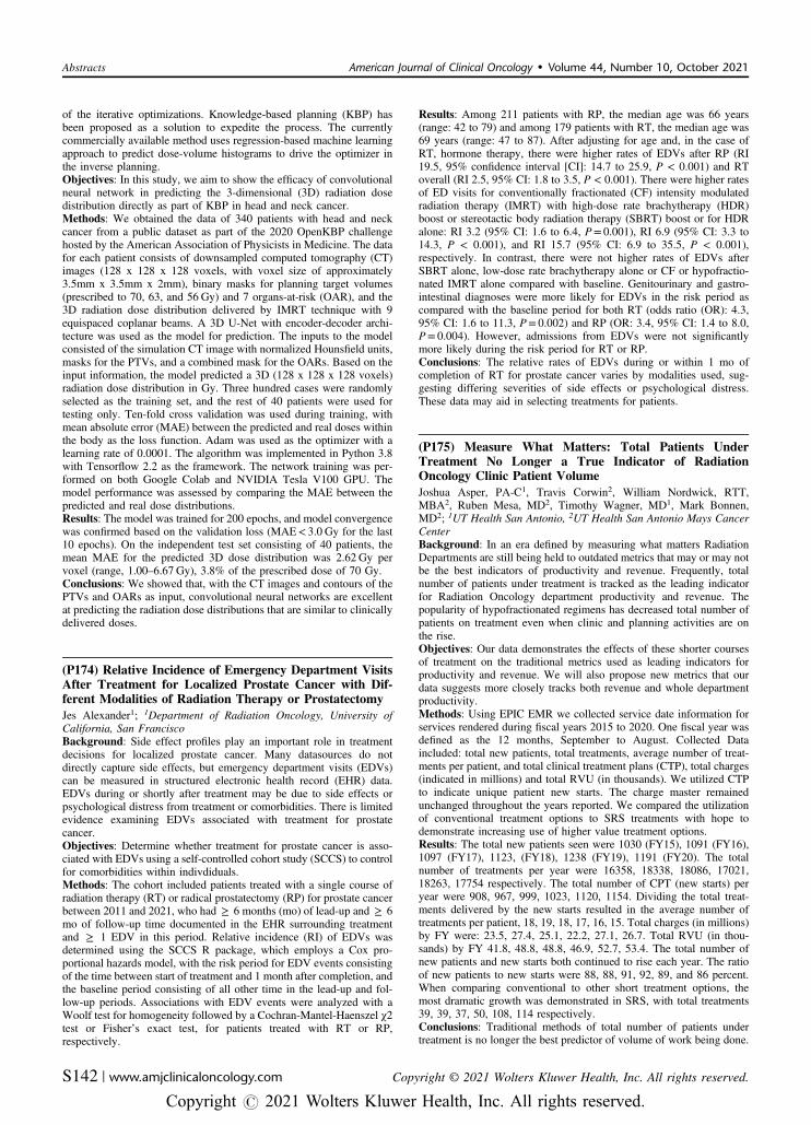
of the iterative optimizations. Knowledge-based planning (KBP) hasbeen proposed as a solution to expedite the process. The currentlycommercially available method uses regression-based machine learningapproach to predict dose-volume histograms to drive the optimizer inthe inverse planning.Objectives: In this study, we aim to show the efficacy of convolutionalneural network in predicting the 3-dimensional (3D) radiation dosedistribution directly as part of KBP in head and neck cancer.Methods: We obtained the data of 340 patients with head and neckcancer from a public dataset as part of the 2020 OpenKBP challengehosted by the American Association of Physicists in Medicine. The datafor each patient consists of downsampled computed tomography (CT)images (128 x 128 x 128 voxels, with voxel size of approximately3.5mm x 3.5mm x 2mm), binary masks for planning target volumes(prescribed to 70, 63, and 56 Gy) and 7 organs-at-risk (OAR), and the3D radiation dose distribution delivered by IMRT technique with 9equispaced coplanar beams. A 3D U-Net with encoder-decoder archi-tecture was used as the model for prediction. The inputs to the modelconsisted of the simulation CT image with normalized Hounsfield units,masks for the PTVs, and a combined mask for the OARs. Based on theinput information, the model predicted a 3D (128 x 128 x 128 voxels)radiation dose distribution in Gy. Three hundred cases were randomlyselected as the training set, and the rest of 40 patients were used fortesting only. Ten-fold cross validation was used during training, withmean absolute error (MAE) between the predicted and real doses withinthe body as the loss function. Adam was used as the optimizer with alearning rate of 0.0001. The algorithm was implemented in Python 3.8with Tensorflow 2.2 as the framework. The network training was per-formed on both Google Colab and NVIDIA Tesla V100 GPU. Themodel performance was assessed by comparing the MAE between thepredicted and real dose distributions.Results: The model was trained for 200 epochs, and model convergencewas confirmed based on the validation loss (MAE< 3.0 Gy for the last10 epochs). On the independent test set consisting of 40 patients, themean MAE for the predicted 3D dose distribution was 2.62 Gy pervoxel (range, 1.00–6.67 Gy), 3.8% of the prescribed dose of 70 Gy.Conclusions: We showed that, with the CT images and contours of thePTVs and OARs as input, convolutional neural networks are excellentat predicting the radiation dose distributions that are similar to clinicallydelivered doses.
(P174) Relative Incidence of Emergency Department VisitsAfter Treatment for Localized Prostate Cancer with Dif-ferent Modalities of Radiation Therapy or ProstatectomyJes Alexander1; 1Department of Radiation Oncology, University ofCalifornia, San FranciscoBackground: Side effect profiles play an important role in treatmentdecisions for localized prostate cancer. Many datasources do notdirectly capture side effects, but emergency department visits (EDVs)can be measured in structured electronic health record (EHR) data.EDVs during or shortly after treatment may be due to side effects orpsychological distress from treatment or comorbidities. There is limitedevidence examining EDVs associated with treatment for prostatecancer.Objectives: Determine whether treatment for prostate cancer is asso-ciated with EDVs using a self-controlled cohort study (SCCS) to controlfor comorbidities within indivdiduals.Methods: The cohort included patients treated with a single course ofradiation therapy (RT) or radical prostatectomy (RP) for prostate cancerbetween 2011 and 2021, who had ≥ 6 months (mo) of lead-up and ≥ 6mo of follow-up time documented in the EHR surrounding treatmentand ≥ 1 EDV in this period. Relative incidence (RI) of EDVs wasdetermined using the SCCS R package, which employs a Cox pro-portional hazards model, with the risk period for EDV events consistingof the time between start of treatment and 1 month after completion, andthe baseline period consisting of all other time in the lead-up and fol-low-up periods. Associations with EDV events were analyzed with aWoolf test for homogeneity followed by a Cochran-Mantel-Haenszel χ2test or Fisher’s exact test, for patients treated with RT or RP,respectively.
Results: Among 211 patients with RP, the median age was 66 years(range: 42 to 79) and among 179 patients with RT, the median age was69 years (range: 47 to 87). After adjusting for age and, in the case ofRT, hormone therapy, there were higher rates of EDVs after RP (RI19.5, 95% confidence interval [CI]: 14.7 to 25.9, P < 0.001) and RToverall (RI 2.5, 95% CI: 1.8 to 3.5, P < 0.001). There were higher ratesof ED visits for conventionally fractionated (CF) intensity modulatedradiation therapy (IMRT) with high-dose rate brachytherapy (HDR)boost or stereotactic body radiation therapy (SBRT) boost or for HDRalone: RI 3.2 (95% CI: 1.6 to 6.4, P= 0.001), RI 6.9 (95% CI: 3.3 to14.3, P < 0.001), and RI 15.7 (95% CI: 6.9 to 35.5, P < 0.001),respectively. In contrast, there were not higher rates of EDVs afterSBRT alone, low-dose rate brachytherapy alone or CF or hypofractio-nated IMRT alone compared with baseline. Genitourinary and gastro-intestinal diagnoses were more likely for EDVs in the risk period ascompared with the baseline period for both RT (odds ratio (OR): 4.3,95% CI: 1.6 to 11.3, P= 0.002) and RP (OR: 3.4, 95% CI: 1.4 to 8.0,P= 0.004). However, admissions from EDVs were not significantlymore likely during the risk period for RT or RP.Conclusions: The relative rates of EDVs during or within 1 mo ofcompletion of RT for prostate cancer varies by modalities used, sug-gesting differing severities of side effects or psychological distress.These data may aid in selecting treatments for patients.
(P175) Measure What Matters: Total Patients UnderTreatment No Longer a True Indicator of RadiationOncology Clinic Patient VolumeJoshua Asper, PA-C1, Travis Corwin2, William Nordwick, RTT,MBA2, Ruben Mesa, MD2, Timothy Wagner, MD1, Mark Bonnen,MD2; 1UT Health San Antonio, 2UT Health San Antonio Mays CancerCenterBackground: In an era defined by measuring what matters RadiationDepartments are still being held to outdated metrics that may or may notbe the best indicators of productivity and revenue. Frequently, totalnumber of patients under treatment is tracked as the leading indicatorfor Radiation Oncology department productivity and revenue. Thepopularity of hypofractionated regimens has decreased total number ofpatients on treatment even when clinic and planning activities are onthe rise.Objectives: Our data demonstrates the effects of these shorter coursesof treatment on the traditional metrics used as leading indicators forproductivity and revenue. We will also propose new metrics that ourdata suggests more closely tracks both revenue and whole departmentproductivity.Methods: Using EPIC EMR we collected service date information forservices rendered during fiscal years 2015 to 2020. One fiscal year wasdefined as the 12 months, September to August. Collected Dataincluded: total new patients, total treatments, average number of treat-ments per patient, and total clinical treatment plans (CTP), total charges(indicated in millions) and total RVU (in thousands). We utilized CTPto indicate unique patient new starts. The charge master remainedunchanged throughout the years reported. We compared the utilizationof conventional treatment options to SRS treatments with hope todemonstrate increasing use of higher value treatment options.Results: The total new patients seen were 1030 (FY15), 1091 (FY16),1097 (FY17), 1123, (FY18), 1238 (FY19), 1191 (FY20). The totalnumber of treatments per year were 16358, 18338, 18086, 17021,18263, 17754 respectively. The total number of CPT (new starts) peryear were 908, 967, 999, 1023, 1120, 1154. Dividing the total treat-ments delivered by the new starts resulted in the average number oftreatments per patient, 18, 19, 18, 17, 16, 15. Total charges (in millions)by FY were: 23.5, 27.4, 25.1, 22.2, 27.1, 26.7. Total RVU (in thou-sands) by FY 41.8, 48.8, 48.8, 46.9, 52.7, 53.4. The total number ofnew patients and new starts both continued to rise each year. The ratioof new patients to new starts were 88, 88, 91, 92, 89, and 86 percent.When comparing conventional to other short treatment options, themost dramatic growth was demonstrated in SRS, with total treatments39, 39, 37, 50, 108, 114 respectively.Conclusions: Traditional methods of total number of patients undertreatment is no longer the best predictor of volume of work being done.
Abstracts American Journal of Clinical Oncology � Volume 44, Number 10, October 2021
S142 | www.amjclinicaloncology.com Copyright © 2021 Wolters Kluwer Health, Inc. All rights reserved.
Copyright r 2021 Wolters Kluwer Health, Inc. All rights reserved.

Given the current trend of shorter course higher value treatments theaverage number of patients on treatment and total treatments no longerare best indicators of treatment value. We suggest using new treatmentsstarts as the new indicator as a predictor of value of work being done.Secondly, we suggest that clinics should follow the ratio of new patientsto new starts to ensure that new patients being seen in clinic result innew starts. Total patients on treatment at any given point is a meas-urement of the past.
(P176) Proton Therapy at the End of Life: A RetrospectiveReviewMina Bakhtiar, BA1, Anish Butala, MD2, Neil Taunk, MD, MS3, JohnNicholas Lukens, MD3, Joshua Jones, MD, MA3, Ima Paydar, MD4;1Perelman School of Medicine, University of Pennsylvania, 2Depart-ment of Radiation Oncology, Perelman School of Medicine, Universityof Pennsylvania, 3Hospital of the University of Pennsylvania,4Department of Radiation Oncology, University of PennsylvaniaBackground: Cancer patients often receive proton beam radiotherapy(PBT) to reduce side-effects and in the re-irradiation setting. A pro-portion of patients receive radiotherapy near the end of life. The indi-cations for and acute toxicities associated with proton beam therapynear the end of life are poorly understood.Objectives: To identify the characteristics, treatment indications, andacute toxicities among patients who received PBT in the final year oflife at a tertiary academic medical center.Methods: A retrospective review of 175 patients treated with PBTwithin the last year of life was performed. Electronic medical recordswere reviewed for patient and treatment details. Follow-up was calcu-lated from start of PBT until death. Acute (< 3mo) and chronic(> 3 mo) toxicities were graded using the CTCAE v5.0. Simple logisticregression was used to evaluate factors associated with acute toxicityand grade (G) 3 or 4 toxicity.Results: Mean age was 66 years (19-94 y), with 57% male and 43%female patients. The most common cancers were non-small cell lungcancer (31%), hepatocellular carcinoma (13%), and small cell lungcancer (7%). Concurrent systemic therapy was delivered to 47%.Median dose was 4988 cGy (180-7388 cGy), with 38% receiving <50 Gy. Mean treatment time was 38 days (1-189 d). Median time fromfirst fraction to death was 185 days (1-363 d). On average, patients spent27% of the remaining days of life receiving PBT. The most commonindications were LC (67%) and pain (24%), along with other clinicalsymptoms including bleeding and dysphagia (9%). Forty-six percentreceived PBT for reirradiation. Of 56 patients treated for symptoms,69%, 27%, and 4% had partial/complete resolution, stable symptoms, orprogression of symptoms. Acute toxicity of any grade was noted in 87%of patients (55% G1, 39% G2, 6% G3). The G3 toxicities includeddyspnea, fatigue, and dermatitis. Seven patients (4%) experiencedchronic toxicity; the most severe was a tracheo-esophageal fistula (G4).Among patients who received PBT for re-irradiation, 6 patients (8%)experienced acute and 1 patient (< 1%) experienced chronic G3 orgreater toxicity. In the simple logistic regression model, higher dosewas positively associated with concurrent systemic therapy (B= 0.026,P= 0.025). Higher dose was also positively associated with any gradeacute toxicity (B= 0.09, P< 0.001). No patient or treatment factorswere associated with >G3 toxicity.Conclusions: Patients receiving PBT at the end of life have high ratesof symptom control. However, some patients spend a large portion oftheir remaining days on treatment. Also, toxicities are moderate andassociated with higher dose. Therefore, further research is needed toidentify the best candidates for PBT at the end of life.
(P177) Analysis of Approaches to Missing Patient-ReportedData in Radiotherapy TrialsJackson Howell1, Jordan Kharofa, MD2, Christopher Anker, MD3;1University of Cincinnati College of Medicine, 2University of CincinnatiCollege of Medicine, Department of Radiation Oncology, 3University ofVermont Larner College of Medicine, Division of Radiation Oncology
Background: The use of patient-reported outcomes (PROs) inrandomized clinical trials (RCTs) improves the sensitivity of adverseevent detection and enhances the appreciation of patient quality oflife concerns. However, PRO data collection is often incomplete—for reasons varying from logistical obstacles to patient death, exitfrom trials, and refusal—and the statistical analysis of theseincomplete data sets so as to minimize bias and maintain conclusionintegrity imposes a formidable challenge to trialists (Chowdhry et al,Int. J. Radiat. Oncol. Biol. Phys., 2021). The Setting InternationalStandards in Analyzing Patient-Reported Outcomes and Quality ofLife Endpoints Data Consortium (SISAQOL) was formed to estab-lish PRO analysis recommendations, including guidance for antici-pating and addressing missing PRO data (Coens, et al, LancetOncol., 2020).
TABLE 1. Summary of National Clinical Trials Network RadiotherapyRandomized Controlled Trials Included in This Analysis
CNS indicates central nervous system; PRO, patient-reported outcome.
TABLE 2. Summary of Relevant Patient-reported Outcome (PRO)Data Analysis Recommended Statements Established by SettingInternational Standards in Analyzing Patient-Reported Outcomesand Quality of Life Endpoints Data Consortium (SISAQOL) and aSurvey of These Features in the Trial Set Analyzed in This Work(Coens, et al, Lancet Oncol., 2020)
QOL indicates quality of life.
American Journal of Clinical Oncology � Volume 44, Number 10, October 2021 Abstracts
Copyright © 2021 Wolters Kluwer Health, Inc. All rights reserved. www.amjclinicaloncology.com | S143
Copyright r 2021 Wolters Kluwer Health, Inc. All rights reserved.

Objectives: Survey recently completed radiotherapy (RT) RCTsthat utilized PROs to evaluate how they approached the challengeof missing PRO data and compare their methods to SISAQOL rec-ommendations to identify specific areas for improvement in futuretrials.Methods: In prior work, a survey of all RT RCTs initiated by theNational Cancer Institute’s National Clinical Trials Network organ-izations between 2000 and 2020 was performed (Howell et al, Int. J.Radiat. Oncol. Biol. Phys., 2021). Trial protocols were acquired fromthe Cancer Trial Support Unit, and published manuscripts presentingthe trials’ data were collected. Only completed trials from this set thatpublished PRO data in manuscript form were considered. The protocolsand manuscripts produced by these trials were carefully examined, withthe data analysis methods and presentation of results compared with theSISAQOL recommendations for the assessment of missing PRO data(Coens, et al, Lancet Oncol., 2020).Results: Twelve trials met these criteria (Table 1), and evaluationobjectives are summarized in Table 2. Only 75% specified a clearstrategy for addressing missing PRO data in the protocol, and 78% ofthose trials adhered to the a priori strategy. Saliently, 67% of trialsanalyzed recorded reasons for incomplete PRO data collection, which isimportant when designing imputation methods for estimating missingdata. Importantly, 67% of trials at least partially incorporated a listwisedeletion strategy into their PRO data analysis, which can weaken theanalysis.Conclusions: Areas for improvement in planning future RCTsinclude increasing specificity of and adherence to a priori strategiesfor analyzing PRO data, collecting reasons for missing data, andutilizing data imputations methods, rather than listwise deletionmethods, when missing data arises. These methods will improve thequality of PRO data analysis by utilizing the maximum amount ofcollected data and decreasing the role of assumptions regardingmissing data.
(P178) The Impact of the Clinical and TranslationalResearch Infrasturcture Network (CTR-IN) Program onOncology Research in the Mountain West (MW) RegionParvesh Kumar, MD1, Chad Cross, Associate Professor1, tony Ward,Professor2, Curtis Noonan, Professor2; 1University of Nevada LasVegas, 2University of MontanaBackground: The MW CTR-IN grant is a U-54 Institutional Devel-opment Award (IDeA) funded by the NIH / NIGMS to increase andenhance research capacity. The CTR-IN Program (ctrin.unlv.edu) isonly one of 12 clinical and translational research centers funded byNIGMS in the US. The CTR-IN involves a partnership with the 13major public universities in the MW region (i.e., Nevada, New Mexico,Wyoming, Montana, Idaho, Alaska and Hawaii) that covers 1/3rd of theUS land mass and almost 1/3rd of all IDeA states. Of the 13 CTR-INpartner public universities, only 4 have schools of medicine (SOM). Toincrease and enhance research capacity, the CTR-IN Program initiated apilot grant (PG) program for eligible research faculty of the 13 MWUniversity partners in 2013 that is now in its 8th year with a verysuccessful track record of producing independent extramural grantfunding.Objectives: We conducted a retrospective review of our CTR-IN PGsto determine their impact on oncology research in the Mountain Westregion. All of our PGs undergo a rigorous “NIH-like” review processand our funding percentile is typically in the 25th - 30% range. Thetypical funding range for our PGs is usually $60 - $66k each with aduration of one year.Methods: During the initial grant cycle (2013 - 2017), a total of 83 PGswere funded for approximately $60 - $66k each in many different areasincluding cancer. Eight (9.6%) of the 83 PGs had a cancer focus. Weanalyzed and compared various major outcome metrics such as theJournal Impact Factor (JIF) of the published manuscripts, the Extra-mural Grant Funding (EMGF) and Return on Investment (ROI= totalEMGF amount/total awarded PG amount x 100%) for the cancerfocused PGs (n= 8) vs. the non-cancer PG awards (n= 75). Exactcomparisons of the means of the major outcomes (i.e., JIF, EMGF,
ROI) between the cancer focused PGs vs. the non-cancer PGs wereconducted.Results: Of the 8 pilot grants funded in cancer research, only 2 werefunded for faculty at a SOM while the others were funded at otherschools (i.e., Biological Sciences, Allied Health, Social Work,Nursing and Computer/Mathematical Sciences) at our 13 partneruniversities. The major outcomes for the cancer focused PGs vs. thenon-cancer focused PGs were as follows, respectively: (1) MeanJIF= 3.94 (SD= 4.83) for 24 publications for the cancer focused PGsvs. Mean JIF= 3.05 (SD= 1.69) for 126 publications for the non-cancer focused PGs, P= 0.102; (2) Mean Amount of EMGF=$1,255,913 (SD= $294,931) for a total EMGF amount of$2,511,826 involving 4 grants (all from faculty at non-SOM Uni-versities) vs. Mean Amount of EMGF= $2,317,870 (SD=$3,669,986) for a total EMGF amount of $46,358,174 involving 43grants, P= 0.765; (3) ROI was 2,002% (SD= 792%) vs. 3,644 %(SD= 6,643%), P= 0.789.Conclusions: Despite not having medical schools at majority of our13 partner institutions, our comparative analysis of major scholarshipoutcomes (i.e., JIF, EMGF and ROI) reveals that our cancer focusedPG awardees were just as successful as our non-cancer focused PGawardees. The MW CTR-IN Program has been instrumental inseeding a culture of investigator initiated clinical and translationalresearch through its pilot grant program including oncology researchdespite the sparsity of medical schools in the MW region.
(P179) Radiotherapy Patients’ Procedural Stress and Anxiety:A National Survey Study of Radiation Therapists’ Experiencein Daily TreatmentNina Mayr, MD1, Daniel Hippe, MS2, Dean Baraba, RTT2, Simon Lo,MB, ChB, FACR, FASTRO2, Jamie Bartalamay, MBA, RTT2, WilliamYuh, MD, MSEE2, Kai Borm, MD3, Stephanie Combs, MD3, Alex-andra Chadderdon, Psy D4; 1Nina A. Mayr, 2University of Washington,3Technische Universitaet Muenchen, 4University of ColoradoBackground: Patients’ anxiety and stress towards radiation therapyhave been well recognized. However, little is known about their effectson daily treatment delivery/workflow and about anxiety management inthe radiotherapy environment.Objectives: We sought to assess prevalence, impact and management ofpatient anxiety/stress in external radiotherapy from the perspective ofthe radiation therapists (RTTs) who have the closest daily interactionswith patients.Methods: A national survey of RTTs was conducted to collect theirexperiences regarding prevalence and perceived importance of patients’anxiety/stress, impact on their daily workflows, approaches to evalua-tion and management, and responsibilities for anxiety management inradiotherapy patients.Results: Among 739 responders, the importance of managing radio-therapy-associated patient anxiety/stress in the RTT’s practice wasjudged as high by 97% (extremely important: 61%; very important:36%). Anxiety-related interferences were reported by 89% for con-ventional and by 87% for advanced/complex (SBRT/IGRT/breathhold)radiotherapy procedures. Interferences consisted predominately ofdelays in fraction completion, need for re-imaging, patients’ inability tocooperate and increased pain. Anxiety was assessed most commonlythrough unstructured verbal discussion (82%). Assessment was mostcommonly performed by nurses (83%), followed by RTT’s and radia-tion oncologists. The majority reported that RTTs carried the mostresponsibility for anxiety management in their clinics. Management wasvariable. Methodically unstructured measures including empathicattention and patient education prevailed (Fig. 1). Requests for phar-macological anxiolytic therapy were common (72%), while most inte-grative health approaches, with the exception of breathing exercises,were rare.Conclusions: Patients’ procedural anxiety/stress has profound impacton daily external radiotherapy delivery and workflows. Anxiety man-agement varies widely and relies heavily on RTTs. Integrative healthapproaches that are used in radiology and other medical proceduresappear underrepresented. Our observations may help inform strategies
Abstracts American Journal of Clinical Oncology � Volume 44, Number 10, October 2021
S144 | www.amjclinicaloncology.com Copyright © 2021 Wolters Kluwer Health, Inc. All rights reserved.
Copyright r 2021 Wolters Kluwer Health, Inc. All rights reserved.

to advance patient anxiety management within the radiation oncologycare team, decrease its interfering effects on radiotherapy delivery, andimprove patient experience.
(P180) Dosimetric Parameters Associated with Long-TermPatient-Reported Quality of Life Following DefinitiveChemoradiation for Anal CancerMichael Rooney, MD1, Brian De, MD1, Kelsey Corrigan, MD, MPD1,Graciela Nogueras Gonzalez, MS1, Prajnan Das, MD, MS, MPH2,Cullen Taniguchi, MD, PhD2, Grace Smith, MD MPH1, Bruce Minsky,MD1, Albert Koong, MD, PhD1, Eugene Koay, MD PhD2, EthanLudmir, MD1, Craig Messick, MD1, Nancy You, MD MHSc1, GeorgeChange, MD1, Ouida Westney, MD1, Van Morris, MD1, Emma Hol-liday, MD1; 1MD Anderson Cancer Center, 2MD AndersonBackground: Data describing long-term patient-reported outcomes(PROs) following intensity-modulated radiation therapy (IMRT)-basedchemoradiation (CRT) for the treatment of squamous cell carcinoma ofthe anus (SCCA) remain scarce.1 Furthermore, clinical and dosimetricrisk factors associated with adverse PROs are unknown.Objectives: To identify dosimetric parameters which may be associatedwith patient-reported quality of life after treatment with chemoradiationfor SCCA.Methods: We identified 248 patients with SCCA treated at our insti-tution from 2010-2018 who were alive and without recurrence. Werequested that all patients complete five validated PRO instruments:Fecal Incontinence QOLScale (FIQOL), Low Anterior Resection Syn-drome Score (LARS), International Consultation on IncontinenceQuestionnaire (ICIQ) male and female lower urinary tract symptoms(MLUTS and FLUTS, respectively), International Index of ErectileFunction (IIEF), and Female Sexual Function Index (FSFI). For thosecompleting surveys, we retrospectively segmented pelvic organs at risk
using published atlases and calculated prespecified dosimetric param-eters (anal PTV D1, bowel bag V30(cc)/V35/V40/V45/D1, small bowelV30(cc)/V35/V40/V45/D1, bladder V40(%)/V45/V50, vagina V40(%)/V45/V50/mean dose(Gy), external genitalia V30/V20, penile bulbmean dose, and prostate mean dose) using approved treatment plans.After selecting factors with intuitive potential causal relations withPROs and those with association on univariate analysis (P <0.05), wegenerated multiple regression models to identify factors independentlyassociated with PROs.Results: One-hundred eight (44%) patients completed surveys. Eightypercent of respondents were female, mean (standard deviation) age was60 (8.9) years, and 23% had T3/T4 disease. Mean bowel bag D1 was49.9 Gy (3.3), and mean small bowel D1 was 48.1 Gy (4.6). Averagevaginal mean dose was 47.0 Gy (6.3), average penile bulb mean dosewas 49.2 Gy (7.1), and average prostate mean dose was 49.3 Gy (6.5).Median [IQR] time from CRT to the completion of the PRO survey was51 [37-84] months. Multiple regression modeling revealed thatincreasing bowel bag D1 was significantly associated with lowerFIQOL score (P= 0.001) and higher LARS score (P= 0.003), bothsignifying worse bowel function. Higher bladder V40 was associatedwith increased ICIQ score (P = 0.001), indicating worse urinaryfunction. Increasing vaginal V40 was associated with lower FSFI(P= 0.011), correlating with worse sexual function. There were nosignificant dosimetric relationships detected with IIEF or PROMISscores.Conclusions: Multiple dosimetric parameters are associated withpatient-reported quality of life across multiple symptomatic domainsfollowing receipt of IMRT-based chemoradiation for the treatment ofSCCA. Minimizing hotspots in the bowel as well as V40 of the bladderand vagina may improve long-term bowel, urinary and sexual functionfor patients undergoing definitive chemoradiation. The dosimetricparameters from this study could potentially be used to develop radi-otherapy planning constraints that optimize patient reported qualityof life.
FIGURE 1. Method(s) used to manage radiation oncology patients’ procedural anxiety. Methods for the management of patient anxietyused by responders are shown in descending frequency. Multiple choices were permitted.
American Journal of Clinical Oncology � Volume 44, Number 10, October 2021 Abstracts
Copyright © 2021 Wolters Kluwer Health, Inc. All rights reserved. www.amjclinicaloncology.com | S145
Copyright r 2021 Wolters Kluwer Health, Inc. All rights reserved.