Globalization, Deglobalization, and New Paradigms in Business
Upload
independentCategory
view
2download
0
CHAPTER 7
Seeking New Paradigms in Epilepsy: StereotacticRadiosurgery
Jean Régis, MD, Romain Carron, MD, Fabrice Bartolomei, PhD, and Patrick Chauvel, PhD
Radiosurgery is a neurosurgical approach consisting of thedelivery of a high energy confined to a small, sharply lim-
ited target with stereotactic accuracy in a single session either tocreate a lesion or to induce a desired biological effect.1,2 Sincethe first attempt by the pioneers in Stockholm, the practice ofradiosurgery has changed dramatically. Nowadays, in the vastmajority of the indications, Gamma Knife (GK) radiosurgeryentails the use of nondestructive low dose to induce subtlebiological effects like apoptosis in tumors or endothelial pro-liferation in arteriovenous malformations (AVMs).
THE DIFFERENTIAL EFFECT CONCEPTThis is the classic clinical observation that in 85% of
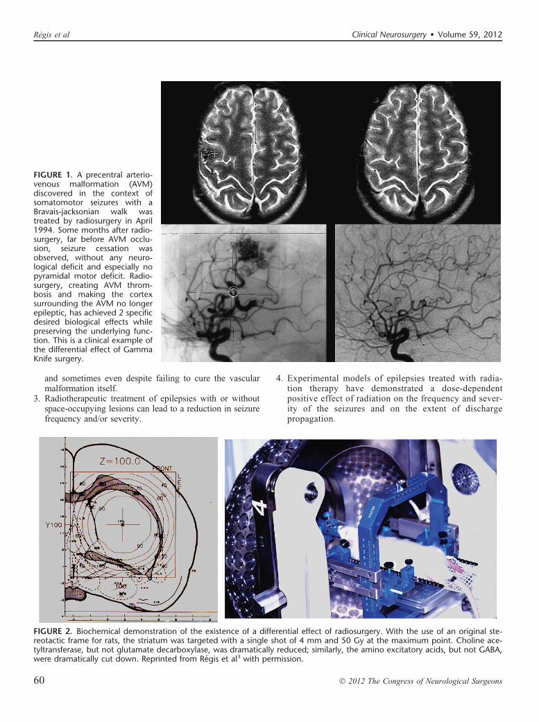
the AVMs associated with a drug-resistant or medicallyintractable epilepsy, radiosurgery is followed by seizurecessation or a dramatic improvement of epilepsy occurslong before the occlusion of the AVM itself (Figure 1).Moreover, when the AVM is located in a highly functionalarea, the seizure cessation is obtained without clinical def-icit. This observation led us to put forth the concept ofclinical differential effect in 1992: Radiosurgery can inducea functional effect such as rendering the cortex surroundingthe AVM no longer epileptic without destroying the under-lying function, thanks to its capacity to alter some systemsspecifically while sparing others. The first proof of conceptcame from the demonstration of the existence of such aneffect at the biochemical level in the striatum of rats.3 Withthe help of an original rat frame,4 a group of rats receiveda single isocenter of 4 mm in the left striatum with a max-imum dose of 50 Gy using the GK (Figure 2). Biochemicalanalyses demonstrated no change in the level of the gluta-mate decarboxylase but a significant decrease in the cholineacetyltransferase level, indicating an injury of the cate-cholaminergic system with concomitant sparing of theGABAergic system. Similarly, GABA was unchangeddespite a major decrease in the amino excitatory acids (glu-tamate and aspartate). This experimental demonstration ledus to consider radiosurgery as a neuromodulation therapy1-3
and encouraged us to organize several prospective clinicaltrials.5-9 More recently, the demonstration of the existenceof a differential effect at the cellular level came from the
Charlottesville group.10 In epileptic rats irradiated with40 Gy in the temporal lobe with the GK, immunohisto-chemical study suggested that at least 1 subtype of hippo-campal interneurons was selectively vulnerable to GKradiosurgery. Neuronal cells appear to have undergonea phenotypic shift with respect to calbindin and glutamatedecarboxylase-67 expression. There is now a growing bodyof evidence in favor of a neuromodulatory effect ofradiosurgery.1,2,11
A series of successive clinical trials has been carriedout in Marseille to evaluate GK surgery in epilepsy. In1993, we organized a phase II prospective trial in 4 mesialtemporal lobe epilepsy (MTLE) patients with a goal of doseranging and toxicity evaluation.7,9 In 1995, the good safetyand impressive efficacy in the patient receiving the 24-Gydosage led us to organize a phase III prospective single-center study in 4 MTLE patients (24 Gy, 7-8 cm3) to eval-uate the reproducibility of the efficacy.5 In 1996, we orga-nized a prospective multicentric European study (21 MTLEpatients), confirming the reproducibility of the safety effi-cacy.8 In 1998, a dose de-escalation study (24, 20, 18 Gy)showed that the efficacy decreased dramatically when themarginal doses were , 24 Gy.6 Finally, the neurologistsfrom our team performed a long-term evaluation (. 5-yearfollow-up) of the first 15 consecutive patients treated ac-cording our standard protocol.12 This study confirmed thegood safety and efficacy of GK surgery in this group ofpatients over the long term with a rate of 60% of Engel Iat a mean follow-up of 8 years,12-16 comparing well with thesafety and efficacy of open surgery over the long term.More recently, a multicenter prospective trial in the UnitedStates confirmed all our findings.17 Radiosurgery has beenthe current practice for selected pure MTLE in our groupever since.18
There are convincing arguments for such an investiga-tion of the potential role of radiosurgery in epilepsy surgery.We know the following:
1. The safety and efficacy of radiosurgery for the treatment ofnumerous small, deep-seated intracerebral tumors or mal-formations have been well documented since the 1950s.
2. Radiosurgical treatment of small corticosubcortical lesionsassociated with epilepsy is known to be associated withseizure cessation in a high percentage of cases (58%-80%in AVMs) long before the expected occlusion of the lesion
Copyright © 2012 by The Congress of Neurological Surgeons0148-396X
Clinical Neurosurgery � Volume 59, 2012 59
and sometimes even despite failing to cure the vascularmalformation itself.
3. Radiotherapeutic treatment of epilepsies with or withoutspace-occupying lesions can lead to a reduction in seizurefrequency and/or severity.
4. Experimental models of epilepsies treated with radia-tion therapy have demonstrated a dose-dependentpositive effect of radiation on the frequency and sever-ity of the seizures and on the extent of dischargepropagation.
FIGURE 1. A precentral arterio-venous malformation (AVM)discovered in the context ofsomatomotor seizures with aBravais-jacksonian walk wastreated by radiosurgery in April1994. Some months after radio-surgery, far before AVM occlu-sion, seizure cessation wasobserved, without any neuro-logical deficit and especially nopyramidal motor deficit. Radio-surgery, creating AVM throm-bosis and making the cortexsurrounding the AVM no longerepileptic, has achieved 2 specificdesired biological effects whilepreserving the underlying func-tion. This is a clinical example ofthe differential effect of GammaKnife surgery.
FIGURE 2. Biochemical demonstration of the existence of a differential effect of radiosurgery. With the use of an original ste-reotactic frame for rats, the striatum was targeted with a single shot of 4 mm and 50 Gy at the maximum point. Choline ace-tyltransferase, but not glutamate decarboxylase, was dramatically reduced; similarly, the amino excitatory acids, but not GABA,were dramatically cut down. Reprinted from Regis et al3 with permission.
Régis et al Clinical Neurosurgery � Volume 59, 2012
60 � 2012 The Congress of Neurological Surgeons

Different kinds of radiation to treat epilepsies havealready been proposed. Lars Leksell conceived GK radiosurgeryas a tool for functional neurosurgery.19,20 Accordingly, he usedGK in movement disorders, trigeminal neuralgia, and other painsyndromes but not for epilepsy surgery.21 The first radiosurgicaltreatments for epilepsy surgery were performed by Talairach inthe 1950s.22 Talairach was another pioneering expert in stereo-taxis. Unlike Leksell, he had specific involvement in epilepsysurgery and led one of the first large comprehensive programsfor epilepsy surgery. As early as 1974, he reported the use ofradioactive yttrium implants in patients with MTLE withoutspace-occupying lesions and showed a high rate of seizurecontrol in patients with epilepsies confined to the mesial struc-tures of the temporal lobe.22 In 1980, Elooma,23 apparentlyunaware of the pioneer work of Talairach, promoted the ideaof focal irradiation for the treatment of temporal lobe epilepsybased on the preliminary reports of Tracy,23a Von Wieser,24
and Baudouin et al.15 Furthermore, clinical experience withthe use of GK- and linear accelerator–based radiosurgery inAVMs and corticosubcortical tumors (mostly metastases andlow-grade glial tumors) revealed an antiepileptic effect ofradiosurgery in the absence of a necrotizing effect.25-27
A series of experimental studies in small animals confirmedthis effect28,29 and emphasized its relationship to the dose
delivered.30-33 Barcia Salorio et al34 and later Lindquistet al35-37 reported small and heterogenous groups of patientstreated with the aim of seizure cessation; however, resultswere poor. Unfortunately, these data were never publishedin peer-reviewed articles, and precise data are unavailable.
In the Department of Stereotactic and FunctionalSurgery in Marseille, the 2 major fields of expertise areepilepsy surgery and radiosurgery. This context has thereforefacilitated the investigation and development of a potentialrole for GK radiosurgery in the treatment of intractableepilepsy. The first attempt to treat an MTLE was made inMarseille in March 1993.7 Since then, 217 cases of epilepsysurgery with GK radiosurgery have been performed.
Among 11 066 GK surgery procedures accomplished inour neurosurgical unit within a 20-year period (between July1992 and December 2012), only 217 were proposed in pa-tients referred for epilepsy surgery (roughly 10 patients peryear). During the same period, we performed 759 nonradio-surgical neurosurgical operations for epilepsy surgery. Thephilosophy of our team is to define the niche of patients inwhom the safety-to-efficacy ratio makes it advantageous or inwhom it at least compares favorably with open neurosur-gery.38 Obviously, this represents a small subset of patientsin our present experience (23%). However, a portion of the
FIGURE 3. Typical planning of aGamma Knife surgery for a MTLEon the dominant side in a patientwith a high level of functioning.This patient presented with aseries of risk factors for a post-operative verbal memory declinein case of microsurgical resec-tion: Dominant-side MTLE withno atrophy or hippocampal scle-rosis, few preoperative neuro-psychological deficit in patientwith a high level of functioning.Additionally, a Wada test pre-dicted a severe verbal memorydeficit in case of amygdalohippo-campectomy, and the patientwas contraindicated for a microsurgery. In October 2003, the patient was treated with a dose of 24 Gy at the 50% isodose lineaccording to our standard protocol. Seizure cessation occurred 1 year after the radiosurgical intervention with no side effects. At 2years, the neuropsychological testing was improved, especially the verbal memory items (before drug reduction).
FIGURE 4. Clinical results of theradiosurgical treatment of a leftmesial temporal lobe epilepsyperformed on December 17,1997. Nine years after the radio-surgical intervention performedon the dominant side of a right-handed patient, the neuro-psychological performances weredramatically improved, particu-larly on the verbal memory items(. 2 standard deviation).
Clinical Neurosurgery � Volume 59, 2012 Epilepsy Radiosurgery
� 2012 The Congress of Neurological Surgeons 61

patients we now treat by GK surgery are directly referred tous for this specific procedure. The actual percentage of pa-tients coming from our own clinical program of investigationfor epilepsy and operated on by GK surgery is only 14.6%.
HYPOTHALAMIC HAMARTOMASHypothalamic hamartomas (HHs) may be asymptomatic,
associated with precocious puberty or with neurologicaldisorders (including epilepsy, behavior disturbances, andcognitive impairment), or both. The seizures usually beginearly in life and are often particularly drug resistant from theoutset. The evolution is unfavorable in most patients because ofbehavioral symptoms (particularly aggressive behavior) andmental decline, which occur as a direct consequence of theseizures,39 in the setting of an epileptic encephalopathy. Inter-estingly, in our experience, reversal of this encephalopathy
after radiosurgery seems to start even before complete cessa-tion of the seizures and seems to be correlated to the improve-ment in background electroencephalograph activity. Thesecontinuous discharges might lead to the disorganization ofseveral systems, including the limbic system. Their disappear-ance may well account for the improvement seen in attention,memory, cognitive performance, and impulsive behavior.Thus, the goal of radiosurgery is the reversal of the epilepticencephalopathy rather than seizure cessation stricto sensu.Consequently, we consider it essential to operate on theseyoung patients as early as possible, whatever the surgicalapproach (resection or radiosurgery).
The intrinsic epileptogenicity of HHs has been demon-strated,40,41 even though the mechanisms of the epilepsy asso-ciated with HHs are still debatable. The boundary of the targetzone of treatment is that of the lesion visualized on magneticresonance (MR) imaging. This contrasts greatly with cases ofMTLE in which there is no such clear delineation of an epi-leptogenic zone.
We retrospectively analyzed radiosurgery in a series of10 patients collected from centers around the world.42 The verygood safety-to-efficacy ratio (all improved, 50% cured, and noadverse effects except 1 case of poikilothermia) led us to orga-nize a prospective multicenter trial. Our trial of 64 prospec-tively evaluated patients is unique with regard to the number ofpatients and the strict methodology. We have published a pre-liminary report of this study,43-45 and the final evaluation is inprogress. These 64 patients operated on between 1999 and2007 have all been followed up for. 3 years (36-107 months).According to our policy, the patient and the family are offereda second radiosurgery in case of partial benefit when the lesionis anatomically small and well defined. Because of the signif-icant but partial efficacy, 25 patients (62.5%) were treatedtwice. The preoperative cognitive functions, behavioral distur-bance, seizure severity, and anatomic type were character-ized.46,47 The goal of the preoperative workup was toadequately select the candidates for inclusion and to evaluate
FIGURE 5. Anterior corpus cal-losotomy with the Gamma Knifein an 18-year-old patient witha severe drug-resistant bifrontalepilepsy since 5 years of age withfrequent falls. After a stereo-electroencephalographic inve-stigation demonstrating thebifrontal extent of the epilepto-genic zone, a corpus callos-otomy was decided. Left, theplanning of the Gamma Knifesurgery (80 Gy at the 50% iso-dose line). The main benefit ofthis callosotomy was to reducethe frequency of seizures withfalls. Right, 1 year after radio-surgery, no deficit was observed,and the tractography demon-strated the section of the corpuscallosum fibers.
FIGURE 6. The activity of radiosurgery for epilepsy is reservedfor highly selected patients, but this activity is sustained overyears. Hypothalamic hamartomas (HHs) and mesial temporallobe epilepsy (MTLE) are the 2 major indications. Epi, epilepsy;TN, trigeminal neuralgia; VIN, ventral intermediate nucleusradiosurgery for tremor.
Régis et al Clinical Neurosurgery � Volume 59, 2012
62 � 2012 The Congress of Neurological Surgeons

the baseline neurological and endocrinological functions. Allradiosurgical procedures were carried out with a model B, C,4C, or Perfexion GK (Elekta Instrument, Stockholm). We con-sistently elaborated multi-isocentric complex dose planning ofhigh conformity and selectivity. We used low peripheral dosesbecause of the close relationship with optic pathways andhypothalamus (median, 17 Gy; range, 13-26 Gy). The lesionstreated are generally small (median, 9.5 mm; range, 5-26). Wepay special attention to the dose delivered to the mammillarybody and fornix, and we always try to tailor the dose plan foreach patient on the basis of the use of a single run of shots withthe 4-mm collimator. Patients were evaluated with respect toseizures, cognition, behavior, and endocrine status 6, 12, 18,24, and 36 months after radiosurgery and then every year.Results demonstrate that in 65% of these patients, an EngelI or II is achieved. An Engel III is observed in 20% of thepatients. The frequency of seizures before radiosurgery was92 seizures per month (mean, 427 6 1009; minimum, 3.3)and dropped to 6 seizures per month afterward (mean,34.6 6 78; minimum, 0; maximum, 425). In the majority ofthese patients, a dramatic behavioral and cognitive improve-ment was observed. Psychiatric and cognitive comorbidity wascured in 28%, improved in 56%, and stable in 8% of thepatients. Globally, a very good result was obtained in 60%of the patients. A microsurgical approach was performed in 6patients (9.3%) with quite large HHs and poor efficacy ofradiosurgery. Microsurgery was performed in 6 patients(17%), (33%) and (50%). After this microsurgical approach,1 patient was cured (Engel I), 2 were improved (Engel III), and3 were not improved (Engel IV). No permanent or even tran-sient neurological deficits were seen.
A transient increase of seizure was observed in 7patients (17.5%). Transient nondisabling poikilothermia wasobserved in 3 cases.
Topographic classification of the lesion based on a goodhigh-resolution MR imaging is a key feature of the decision-making process.43 Previous classifications were based on ana-tomic48-50 or surgical51 criteria. These classifications do notdescribe the large diversity of these lesions and ignore theirtherapeutical consequences. As underlined by Palmini andcoworkers,52 the exact location of the lesion in relation tothe interpeduncular fossa and the walls of the third ventriclecorrelates with the extent of excision, seizure control, andcomplication rate. On this basis, we classify the HHs accord-ing to their topography using our own classification.43,44 Inour experience, this classification correlates well with theclinical semiology and severity and is especially useful inthe selection of the surgical strategy.
Type I (small HHs located inside the hypothalamusextending more or less into the third ventricle) are certainlythe best candidates for GK surgery. In this population, the riskof microsurgical removal is likely to be high.
In type II (the lesion is small and mainly in the thirdventricle), radiosurgery may be the safer alternative. Althoughthe endoscopic and transcallosal interforniceal approaches havebeen attempted, the risks of short-term memory worsening,endocrinological disturbances (hyperphagia with obesity, low
thyroxine, sodium metabolism disturbance), and thalamic orthalamocapsular infarcts have been reported by the mostenthusiastic and skillful neurosurgeons. However, for verysevere repeated status epilepticus, we propose as a salvagesurgery either a transcallosal interforniceal approach or anendoscopic approach (depending on the width of thirdventricle). In emergency situations, if the lesion is small andthe third ventricle is wide, the endoscopic approach is chosen.
In type III (lesion located essentially in the floor), theextremely close relationship between the mammillary body,the fornix, and the lesion clearly leads us to prefer GKsurgery. We speculate that sessile HHs (type IV) always havemore or less an “extension” in the hypothalamus close to themammillary body. Thus, when a lesion is classified as a typeII, the lesion appears on the MR as located mainly in the thirdventricle but is likely to have a “root” in the hypothalamus.The same assumption is made for type III.
In type IV (the lesion sessile in the cistern), a disconnec-tion can be discussed (pterional approach with or withoutorbitozygomatic osteotomy). However, if the lesion is small,GK surgery can be recommended because of its safety and itscapability to reach at the same time the small componentlocated within the hypothalamus itself, frequently visible onthe high-resolution MR. In the Delalande and Fohlen16 expe-rience, only 2 patients among 14 were seizure free after a singledisconnection through a pterional approach. Consequently, weuse this approach for lesions too large for GK surgery as a firststep of a staged approach.
In most circumstances, the patient is improved but notseizure free after the first microsurgical step, and GK surgeryis scheduled at 3 months as a second step.
Type V (pediculate) is rarely epileptogenic and caneasily be cured by radiosurgery or disconnection througha pterional approach. In case of severe epilepsy, the secondtherapeutic modality allows faster cessation of seizures.However, a distant extension of the HH into the hypothalamusclose to the mammillary bodies must be cautiously looked foron high-resolution MR. Its visualization will prompt us toconsider GK surgery to treat both parts of the lesion, especiallyin cases when the cisternal component is small.
Type VI (giant) does not represent a good indication forfirst intention radiosurgery because, in nearly all cases,a combination of several therapeutic modalities should beused. Even if GK surgery does not seem to be suitable whenthe lesion is large, “radiosurgical” disconnection has beenenvisaged (radiosurgery targeting only the superior part inthe hypothalamus and/or the third ventricle, leaving untreatedall the lesion lower than the floor) but has been systematicallydisappointing. In our opinion, this strategy may cause loss ofprecious time for the child to be treated effectively. Conse-quently, we do not advocate this strategy. When microsurgi-cal resection has left a small remnant in the third ventricle andactive epilepsy, reoperation can be envisaged by GK surgery.
Two major questions remain. First, we know thatcomplete treatment or resection of the lesion is not alwaysmandatory,53-55 but we do not know how to predict in anindividual patient the amount (and mapping) of the HH that
Clinical Neurosurgery � Volume 59, 2012 Epilepsy Radiosurgery
� 2012 The Congress of Neurological Surgeons 63

must be treated to obtain a complete antiepileptic effect. Sec-ond, we know that these patients frequently present with anelectroclinical semiology, suggesting involvement of the tem-poral or frontal lobe that can mimic a secondary epilepto-genesis phenomenon.41,56 In our experience, some of thesepatients can be cured completely by the isolated treatmentof the HH, whereas a partial result is obtained in others withresidual seizures despite significant overall psychiatric andcognitive improvement. In this second group, it is temptingto propose that such a secondary epileptogenic area accountsfor the partial failure.
Our initial results indicate that GK surgery is aseffective as and much safer than microsurgical resection.57
GK surgery also avoids the vascular risk related to radiofre-quency lesioning or stimulation. The disadvantage of radio-surgery lies in its delayed action. Longer follow-up ismandatory for proper evaluation of the role of GK surgery.Results are faster and more complete in patients with smallerlesions inside the third ventricle (stage II). The early effect onsubclinical electroencephalographic discharges appears toplay a major role in the dramatic benefit concerning at oncethe sleep quality, behavior, and cognitive developmentalfunctions. GK surgery can safely lead to the reversal of theepileptic encephalopathy.42,44,45,58
Considering the very poor clinical prognosis of thiscondition and the invasiveness of microsurgical resection, GKcan now be regarded as the first-line option for small tomiddle-sized HHs associated with epilepsy because it canlead to dramatic improvements in the future of these youngpatients. The role of secondary epileptogenesis of widespreadcortical dysgenesis in these patients needs to be betterevaluated and understood to optimize patient selection andto define the best treatment period.
MESIAL TEMPORAL LOBE EPILEPSYThe first GK surgery operations for MTLE were
performed in Marseille in March 1993. Because no similarexperience in the literature was available at that time, we wereobliged to base our technical choices on hypothesis andexperience in radiosurgery for other pathological conditions.Four patients were treated with different technical strategies(dose, volume, target definition). The impressive delayedradiological changes observed some months after radiosur-gery59 drove us to stop for a while and follow up these first 4patients. As a result of the clinical safety of the procedure inthese patients and the gradual disappearance of the acute MRchanges after some months, we treated several new series ofpatients under strict prospective controlled trial conditions(with ethical committee approval). The treatment for the next16 patients was based on that of the first patient who hada successful outcome (as opposed to the 3 others who hadpartial or no effect).
This “classic planning” was based on the use of two 18-mm shots covering a volume of about 7 cm3 at the 50% iso-dose (24 Gy) and has turned out to produce a high rate ofseizure cessation.60,61 For epileptological and safety reasons,the targeting was very much centered on the parahippocampal
cortex and spared a significant part of the amygdaloidal com-plex and hippocampus. The refinement of the GK surgerytechnique and the need to find a dose that would create morelimited MR alterations during the acute phase led us to reducethe dose from 24 to 20 and 18 Gy at the margin. However, thiscaused a significant decrease in the rate of seizure cessation.We have reviewed the long-term follow-up of our first 15patients operated on by state-of-the-art GK surgery for MTLE(24 Gy) (Figure 3).6 The mean follow-up was 8 years, and atthe last follow-up, 73% were seizure free. These long-termresults compare favorably with microsurgical results. No per-manent neurological deficit was reported except a visual fielddeficit in 9 patients.12 After microsurgery for MTLE on thedominant side, a verbal memory deficit is classically observedin 30% to 50% of the patients.62,63 It is of special importance tonote that none of our patients has harbored any neuropsycho-logical worsening (using the evaluation published by Clus-mann et al13) and especially no verbal memory decline(Figure 4).5,6,8,12 This finding from our 4 prospective trialshas been confirmed by a US prospective trial.64
The timetable of events after radiosurgery and thefollow-up is quite standardized. Patients are informed that themain drawback of radiosurgery lies in its delayed efficacy.Typically, the frequency of the seizures is not modifiedsignificantly for the first few months. Thereafter, there isa rapid and significant increase in auras for some days orweeks before the seizures eventually disappear. Usually. thepeak in seizure cessation is observed around the 8th to 18thmonth with a clear variability in the delay in onset. In 1patient, this occurred 26 months after GK radiosurgery. Weusually consider a delay of 2 years as the minimum forpostradiosurgery follow-up. In the absence of initial radio-logical changes or clinical benefit, the recommendation is towait for the onset of the MR imaging changes and theirsubsequent disappearance. All our patients had the samepattern of MR changes regardless of the margin dose (18-24Gy) and treatment volume (5-8.5 cm3). However, the degreeof these changes and their delay of onset varied according tothe dose delivered to the margin, the volume treated, and theindividual patient. To allow an optimal evaluation, we recom-mend that subsequent microsurgery not be considered beforethe third year after radiosurgery. Similarly, we believe thata patient who undergoes a cortectomy before the onset of theMR changes has occurred cannot be assumed to have failedradiosurgical treatment. Of course, before consideration ofany further surgery, the reason for the failure needs to beaddressed. After a review of the medical records of patientstreated with radiosurgery for MTLE, it was sometimes possi-ble to identify likely causes of failure, such as the following:
1. Poor patient selection (eg, patients with epilepsy involvingmore than the MTL structures)
2. Patients with a diagnosis of treatment failure (, 3 years)who had been operated on too soon after radiosurgery65
3. Targeting of the amygdala and hippocampus (which in ouropinion is not the optimal target in terms of safety andefficacy) instead of the parahippocampal cortex66
4. Insufficient dose65-67
Régis et al Clinical Neurosurgery � Volume 59, 2012
64 � 2012 The Congress of Neurological Surgeons

Our current strategy of treatment is based on our firstseries of MTLE patients who were strictly selected andtreated systematically with a very simple but very reproduc-ible dose planning strategy.5,7 Identifying putative improve-ments in the methodology requires a systematic analysis ofthe influence of the technical data from our experience andfrom the literature on the outcome of those patients.
THE “TECHNICAL” QUESTIONS
The Dose IssueThe first targets used in functional GK radiosurgery
(capsulotomy, thalamotomy of ventral intermediate nucleus.,or the centromedianum, pallidotomy) were treated with a highdose (300-150 Gy) delivered in very small volumes (3-5 mmin diameter).21 The goal was to destroy a predefined, verysmall anatomic structure with stereotactic precision. Quitea significant variability in the delay and amplitude of theMR changes has been reported with a fixed dose regi-mens.41,68 Barcia Salorio et al13 have presented several timesa small and heterogeneous group of patients treated withdifferent kinds of devices and dose regimens. Apparently,some of those patients had no expanding lesion and weretreated with very large volumes and very low doses (about10 Gy). On the basis of this experience, several teams havemade the assumption that very low doses, as low as 10 to 20Gy at the margin, should be as effective as the 24-Gy protocol(at the margin) that we used for our first series of patients withMTLE.5 A cautious examination of the last proceeding ofBarcia Salorio et al34 shows that individual information onthe dose at the margin, the volume, and the topography of theepileptogenic zone is not provided. Moreover, among the 11patients reported, the actual rate of seizure cessation is appar-ently only 36% (4 of 11), which is much lower than whatwould be expected with resection in MTLE. In a heteroge-neous group of 176 patients, Yang et al67 confirmed that onlya very low rate of seizure control is achieved when low doses(9-13 Gy at the margin) are used.
The experience with the radiosurgical treatment of HHsindicates that 18 Gy at the margin appears to be a threshold interms of probability of seizure cessation.42 In this group ofpatients (36 cases), only 1 patient showed MR changes. Themajority of the AVM cases with worsening of the epilepsywere treated with a range of doses between 15 and 18 Gy.Similarly, poor results have been reported by Cmelak et al65
in 1 MTLE patient treated with linear accelerator–basedradiosurgery, with 15 Gy at the 60% isodose line, who under-went surgical resection 1 year later. In this case, the authorsfirst observed a slight improvement followed by an obviousworsening. A recent de-escalation study has allowed us todemonstrate poorer results in patients receiving doses of 18or 20 Gy compared with 24 Gy at the margin.61,69 As a resultof the rate of seizure cessation achievable by conventionalresection, a radiosurgical strategy associated with a muchlower rate of seizure cessation appears unacceptable. Frac-tionated stereotactically guided radiotherapy has been dem-onstrated to fail systematically in controlling seizures. Among12 patients treated by Grabenbauer and colleagues,60,70 none
has achieved seizure cessation; only seizure reduction wasobtained in this series.
Experimental studies on small animals have demon-strated the antiepileptic effect of radiosurgery,30,32,71 the dosedependence of this effect,30-32,72 and the possibility of obtain-ing clear antiepileptic effect without macroscopic necrosis.31
Of course, the rodent models of epilepsy are far from beinggood models of human MTLE. However, taking into accountthe huge difference in volume of the target, it is intriguing tonotice that according to our clinical experience in humans,a similar maximum dose range of 40 to 50 Gy is currentlyproviding the optimal safety-to-efficacy ratio.
The Target DefinitionWhen the target is a lesion that can be precisely
depicted with optimal imaging sequences, the question ofthe selection of the marginal dose is quite easily addressed bycorrelating safety-efficacy and individual outcome to themarginal dose. This can be refined with stratification accord-ing to volume, location, age, etc. However, in patientspresenting with MTLE, this process is invalid for 2 reasons.First, there is no consensus regarding the extent of MTLresection required. Second, the concept of MTLE syndromewith a stable extent of the epileptogenic zone and surgicaltarget is increasingly a topic of debate.14,73
Volume (in association with marginal dose) is wellknown to be a major determinant of the tissue effect, as shownin integrated risk/dose volume formulas.74 In our first series ofpatients, this marginal isodose volume (or prescription isodosevolume) was approximately 7 cm3 (range, 5-8.5 cm3).
An attempt to correlate dose/volume and the effect onseizures and on the MR changes (as evaluated by volume ofthe contrast enhancement ring, extent of the high T2 signal,and the importance of the mass effect) has been publishedrecently.61 In this study, we found, not surprisingly, that thehigher the dose and volume were, the higher the risk was ofhaving more severe MR changes, but also the higher thechance was of achieving seizure cessation. However, thesedata have limited value. Hence, more precise identification ofthose structures of the MTL that need to be “covered” by theradiosurgical treatment may allow more selective, but just asefficacious, dose planning strategies despite smaller prescrip-tion isodose volumes.
Growing evidence supports the organization of theepileptogenic zone into networks, meaning that severaldifferent and possibly distant structures fire simultaneouslyat the onset of the electroclinical seizure. This kind oforganization explains why the risk of failure is so high whena simple topectomy (without preoperative investigations) insevere drug-resistant epilepsies is associated with a benignlesion.75 This has also been reported in MTLE.14,73 Certainnuclei of the amygdaloidal complex, head, body, tail ofthe hippocampus, perirhinal cortex, entorhinal cortex, andparahippocampal cortex may be associated with the genesisof the seizures. The role of the entorhinal cortex in epilepsyis supported by experimental studies in animals.59,76 Theentorhinal cortex is considered to be the amplifier of the
Clinical Neurosurgery � Volume 59, 2012 Epilepsy Radiosurgery
� 2012 The Congress of Neurological Surgeons 65

“amygdalohippocampal epileptic system.” The pattern of theassociated structures, including that of the structure playingthe leader role, can vary significantly from patient topatient.14,73 A subgroup of patients have clonic dischargesand involvement of the entorhinal cortex, amygdala, andhead of the hippocampus, with a clear leader role of theentorhinal cortex. Wieser et al77 analyzed the postoperativeMR images of patients operated on by Yasargil (amygdalo-hippocampectomy) and were able to correlate the quality ofthe resection of each substructure of the MTL area and theoutcome with respect to seizures.77 Only the quality of theremoval of the anterior parahippocampal cortex was stronglycorrelated with a higher chance of seizure cessation. Wetried to perform a similar study in patients treated withGK radiosurgery.61 We defined and manually drew the limitsof subregions on the stereotactic images of all these patients.The amygdala, head, body, and tail of the hippocampus werefirst delineated. The white matter, parahippocampal cortex,and cortex of the anterior wall of the collateral fissure werethen drawn separately and divided into 4 sectors in the ros-trocaudal axis corresponding to the amygdala, head, body,and tail of the hippocampus.61
PATIENT SELECTIONWhang and Kwon78 (without having performed specific
preoperative epileptological workup beforehand) treated pa-tients with epilepsy associated with slowly growing lesionsand observed seizure cessation in only 38% (12 of 31) of thepatients. This kind of observation emphasizes the importanceof preoperative definition of the extent of the epileptic zoneand of its relationship with the lesion.75,79 In our institution,the philosophy is to tailor the investigations for each individ-ual case. In some patients, electroclinical data, structural andfunctional imaging, and neuropsychological examination aresufficiently concordant for surgery of the temporal lobe to beproposed without depth electrode recordings. In other cases,the level of evidence for MTLE is deemed insufficient, anda stereoelectroencephalographic study is performed. Thestrategy of stereoelectroencephalographic implantation isbased on the primary hypothesis (mesial epileptogenic zone)and alternative hypotheses (early involvement of the temporalpole, lateral cortex, basal cortex, insular cortex, or other cor-tical areas). The goal of these studies is to record the patient’shabitual seizures to establish the temporospatial pattern ofinvolvement of the cortical structures during these seizures.In these patients, the high resolution of depth electroderecording clearly allows fine tailoring of surgical resection,according to the precise temporospatial course of the seizures.
The main limitation of radiosurgery is the size of thetarget (prescription isodose volume). The radiosurgical treat-ment of MTLE is certainly the most selective surgical therapyfor this group of patients. The requirement for precision andaccuracy in the definition of the epileptogenic zone isconsequently higher. Furthermore, if depth electrode investi-gation enables demonstration of a particular subtype ofMTLE, this can lead to tailoring of the treatment volumeand frequently allows the volume to be reduced.
THE POTENTIAL CONCERNSThe risk of long-term complications must always be
scrutinized cautiously in functional neurosurgery. Radio-therapy is most frequently used in the brain for short-termlife-threatening pathologies. The use of radiotherapy inyoung patients with benign disease such as pituitaryadenomas or craniopharyngioma has been associated witha significant rate of cognitive decline80,81 and tumor gene-sis,82 including some carcinogenesis.83 If the risk of radia-tion-induced tumor were similar with radiosurgery, weshould by now have observed numerous cases. However,such reported cases84-86 are extremely rare and frequentlyfail to meet the classic criteria by which tumors are deemedto be radiation induced.16 In fact, if this risk exists, it islikely to be about 1 in 10 000, which is far lower than themortality risk associated with temporal lobectomy.68,87-90
Epilepsy is a life-threatening condition. The risk ofsudden unexplained death in epileptic patients is wellknown.91,92 This risk is higher in patients treated with . 2antiepileptic drugs with an IQ , 70 (as independent factors).Because seizure cessation after surgery reduces the mortalityrisk to that of the general population,92 microsurgical resectionof the epileptogenic zone may confer better benefit in terms ofthe possibility of immediate seizure cessation and thereforereduced mortality risk compared with the more delayed bene-fits of radiosurgical treatment. Our patients are systematicallyinformed about this disadvantage of radiosurgery.
WHAT ARE THE CURRENT INDICATIONS?We still consider the use of radiosurgery for MTLE to
be experimental. The demonstrated advantages of radio-surgery are the comfort of the procedure, the absence ofgeneral anesthesia, the absence of surgical complicationsand mortality, the very short hospital stay, and the imme-diate return to the previous level of functioning andemployment. Potential sparing of memory function is stilla matter of debate and needs to be established withcomparative studies. There is also a need for furtherdemonstration of long-term efficacy and safety of radio-surgery. Worldwide, microsurgical cortectomies for MTLEhave proven to be very satisfactory owing to the rarity ofsurgical complications and a high rate of seizure freedom. Inour experience, the most important selection parameters arethe demonstration of the purely mesial location of theepileptogenic zone and the clear understanding by thepatient of the advantages, disadvantages, and limitations.One other very good indication in our experience is provenMTLE but previous failure of microsurgery, supposedlyresulting from insufficient posterior extent of the resection.The best candidates are young patients with mid-severityepilepsy (working, socially well adjusted), a high level offunctioning (able to understand clearly the limits andconstraints of radiosurgery), a very high risk of memorydeficit with microsurgery (MTLE on the dominant side withlittle or no atrophy, few deficits of the verbal memorypreoperatively), and potentially huge social and professionalconsequences in case of postoperative memory deficit.2,18,93
Régis et al Clinical Neurosurgery � Volume 59, 2012
66 � 2012 The Congress of Neurological Surgeons

CONCLUSIONSThe field of epilepsy surgery is a new and promising
one for radiosurgery. However, the determination of theextent of the epileptogenic zone requires specific expertise,which is crucial for achieving a reasonable rate of seizurecessation. In addition, the huge impact of fine technical detailson the efficacy and possible toxicity of the procedure meansthat, at present, its use for these indications remains underevaluation, and further prospective study is absolutelyrequired. It is difficult to know whether we really are at thedawning of broader indications for the use of radiosurgery; inthe upcoming years, our ability to identify the correcttechnical strategies should determine whether this is so.
DisclosureThe authors have no personal financial or institutional
interest in any of the drugs, materials, or devices described inthis article.
REFERENCES1. Régis J, Bartolomei F, Hayashi M, Chauvel P. Gamma Knife surgery,
a neuromodulation therapy in epilepsy surgery! Acta Neurochir Suppl.2002;84:37-47.
2. Régis J, Carron R, Park M. Is radiosurgery a neuromodulation therapy?A 2009 Fabrikant Award lecture. J Neurooncol. 2010;98(2):155-162.
3. Régis J, Kerkerian-Legoff L, Rey M, et al. First biochemical evidence ofdifferential functional effects following Gamma Knife surgery. StereotactFunct Neurosurg. 1996;66(suppl 1):29-38.
4. Rey M, Valliccioni PA, Vial M, et al. Experimental radiosurgery in ratsusing gamma a “gamma knife”: description of a stereotactic device forsmall laboratory animals [in French]. Neurochirurgie. 1996;42(6):289-293.
5. Régis J, Bartolomei F, Rey M, et al. Gamma Knife surgery for mesialtemporal lobe epilepsy. Epilepsia. 1999;40(11):1551-1556.
6. Regis J, Levivier M, Motohiro H. Radiosurgery for intractable epilepsy.Tech Neurosurg. 2003;9(3):191-203.
7. Régis J, Peragui JC, Rey M, et al. First selective amygdalohippocampcalradiosurgery for ‘mesial temporal lobe epilepsy’. Stereotactic Funct Neu-rosurg. 1994;64(suppl 1):191-201.
8. Régis J, Rey M, Bartolomei F, et al. Gamma Knife surgery in mesialtemporal lobe epilepsy: a prospective multicenter study. Epilepsia. 2004;45(5):504-515.
9. Regis J, Semah F, Bryan RN, et al. Early and delayed MR and PETchanges after selective temporomesial radiosurgery in mesial temporallobe epilepsy. AJNR Am J Neuroradiol. 1999;20(2):213-216.
10. Tsuchitani S, Drummond J, Kamiryo T, et al. Selective vulnerability ofinterneurons to low dosage radiosurgery [abstract]. Presented at: Societyfor Neuroscience Annual Meeting; New Orleans; 2003.
11. Quigg M, Rolston J, Barbaro NM. Radiosurgery for epilepsy: clinicalexperience and potential antiepileptic mechanisms. Epilepsia. 2012;53(1):7-15.
12. Bartolomei F, Hayashi M, Tamura M, et al. Long-term efficacy ofGamma Knife radiosurgery in mesial temporal lobe epilepsy. Neurology.2008;70(19):1658-1663.
13. Barcia-Salorio JL, Barcia JA, Hernández G, López-Gómez L. Radiosur-gery of epilepsy: long-term results. Acta Neurochir Suppl (Wien). 1994;62:111-113.
14. Bartolomei F, Wendling F, Bellanger JJ, Régis J, Chauvel P. Neuralnetworks involving the medial temporal structures in temporal lobe epi-lepsy. Clin Neurophysiol. 2001;112(9):1746-1760.
15. Baudouin A, Stuhl L, Perrard AC. Un cas d’épilepsie focale traité par laradiothérapie. Rev Neurol (Paris). 1951;84(1):60-63.
16. Cahan W, Woodard H, Highinbotham N, Stewart F, Coley B. Sarcomaarising in irradiated bone: report of eleven cases. Cancer. 1948;1(1):3-29.
17. Barbaro NM, Quigg M, Broshek DK, et al. A multicenter, prospectivepilot study of Gamma Knife radiosurgery for mesial temporal lobe epi-lepsy: seizure response, adverse events, and verbal memory. Ann Neurol.2009;65(2):167-175.
18. Régis J, Bartolomei J, Chauvel P. Epilepsy. Prog Neurol Surg. 2007;20:267-278.
19. Leksell L. The stereotaxic method and radiosurgery of the brain. ActaChir Scand. 1951;102(4):316-319.
20. Leksell L. Stereotaxic radiosurgery in trigeminal neuralgia. Acta ChirScand. 1971;137(4):311-314.
21. Lindquist C, Kihlström L, Hellstrand E. Functional neurosurgery:a future for the Gamma Knife? Stereotact Funct Neurosurg. 1991;57(1-2):72-81.
22. Talairach J, Bancaud J, Szikla G, Bonis A, Geier S, Vedrenne C. Ap-proche nouvelle de la neurochirurgie de l’epilepsie: méthodologie stéréo-taxique et résultats thérapeutiques. Neurochirurgie. 1974;20(suppl 1):92-98.
23. Elomaa E. Focal irradiation of the brain: an alternative to temporal loberesection in intractable focal epilepsy? Med Hypotheses. 1980;6(5):501-503.
23a.Tracy S. High-frequency high potential currents, and X-radiations in thetreatement of epilepsy. N York M J. 1905;81:422-424.
24. Von Wieser W. Die Roentgentherapie der traumatischen Epilepsie.Mschr Psychiat Neurol. 1939;101:422-424.
25. Heikkinen ER, Konnov B, Melnikow L. Relief of epilepsy by radiosur-gery of cerebral arteriovenous malformations. Stereotactic Funct Neuro-surg. 1989;53(3):157-166.
26. Rogers LR, Morris HH, Lupica K. Effect of cranial irradiation on seizurefrequency in adults with low-grade astrocytoma and medically intractableepilepsy. Neurology. 1993;43(8):1599-1601.
27. Rossi GF, Scerrati M, Roselli R. Epileptogenic cerebral low-grade tu-mors: effect of interstitial stereotactic irradiation on seizures. Appl Neuro-physiol. 1985;48(1-6):127-132.
28. Barcia Salorio JL, Roldan P, Hernandez G, Lopez Gomez L. Radio-surgical treatment of epilepsy. Appl Neurophysiol. 1985;48(1-6):400-403.
29. Gaffey CT, Montoya VJ, Lyman JT, Howard J. Restriction of the spreadof epileptic discharges in cats by mean of Bragg peak, intracranial irra-diation. Int J Appl Radiat Isotope. 1981;32(11):779-787.
30. Chen ZF, Kamiryo T, Henson SL, et al. Anticonvulsant effects of gammasurgery in a model of chronic spontaneous limbic epilepsy in rats. JNeurosurg. 2001;94(2):270-280.
31. Maesawa S, Kondziolka D, Dixon CE, Balzer J, Fellows W,Lunsford LD. Subnecrotic stereotactic radiosurgery controlling epilepsyproduced by kainic acid injection in rats. J Neurosurg. 2000;93(6):1033-1040.
32. Mori Y, Kondziolka D, Balzer J, et al. Effects of stereotactic radiosurgeryon an animal model of hippocampal epilepsy. Neurosurgery. 2000;46(1):157-165; discussion 165-168.
33. Ronne-Engström E, Kihlström L, Flink R, et al. Gamma Knife surgery inepilepsy: an experimental model in the rat. Presented at: European Soci-ety for Stereotactic and Functional Neurosurgery; 1993.
34. Barcia Salorio JL, Garcia JA, Hernandez G, Lopez Gomez L. Radio-surgery of epilepsy: long-term results. Presented at: European Society forStereotactic and Functional Neurosurgery; 1993.
35. Lindquist C. Gamma knife surgery in focal epilepsy. 1 year follow-up in4 cases. 1992.
36. Lindquist C, Hellstrand E, Kilström L, Abraham-Fuchs K, Jernberg B,Wirth A. Stereotactic localisation of epileptic foci by magnetoencepha-lography and MRI followed by gamma surgery. Presented at: Interna-tional Stereotactic Radiosurgery Symposium; 1991.
37. Lindquist C, Kihlström L, Hellstrand E, Knutsson E. Stereotactic radio-surgery instead of conventional epilepsy surgery. Presented at: EuropeanSociety for Stereotactic and Functional Neurosurgery; 1993.
38. Régis J, Bartolomei F, Hayashi M, Roberts D, Chauvel P, Peragut JC.The role of Gamma Knife surgery in the treatment of severe epilepsies.Epileptic Disord. 2000;2(2):113-122.
39. Deonna T, Ziegler AL. Hypothalamic hamartoma, precocious pubertyand gelastic seizures: a special model of “epileptic” developmental dis-order. Epileptic Disord. 2000;2(1):33-37.
Clinical Neurosurgery � Volume 59, 2012 Epilepsy Radiosurgery
� 2012 The Congress of Neurological Surgeons 67

40. Kuzniecky R, Guthrie B, Mountz J, et al. Intrinsic epileptogenesis ofhypothalamic hamartomas in gelastic epilepsy. Ann Neurol. 1997;42(1):60-67.
41. Munari C, Kahane P, Francione S, et al. Role of the hypothalamichamartoma in the genesis of gelastic fits (a video-stereo-EEG study).Electroencephalogr Clin Neurophysiol. 1995;95(3):154-160.
42. Régis J, Bartolomei F, de Toffol B, et al. Gamma Knife surgery forepilepsy related to hypothalamic hamartomas. Neurosurgery. 2000;47(6):1343-1351; discussion 1351-1352.
43. Régis J, Hayashi M, Eupierre LP, et al. Gamma Knife surgery for epi-lepsy related to hypothalamic hamartomas. Acta Neurochir Suppl. 2004;91:33-50.
44. Régis J, Scavarda D, Tamura M, et al. Epilepsy related to hypothalamichamartomas: surgical management with special reference to gamma knifesurgery. Childs Nerv Syst. 2006;22(8):881-895.
45. Régis J, Scavarda D, Tamura M, et al. Gamma knife surgery for epilepsyrelated to hypothalamic hamartomas. Semin Pediatr Neurol. 2007;14(2):73-79.
46. Frattali CM, Liow K, Craig GH, et al. Cognitive deficits in children withgelastic seizures and hypothalamic hamartoma. Neurology. 2001;57(1):43-46.
47. Weissenberger AA, Dell ML, Liow K, et al. Aggression and psychiatriccomorbidity in children with hypothalamic hamartomas and their unaf-fected siblings. J Am Acad Child Adolesc Psychiatry. 2001;40(6):696-703.
48. Arita K, Ikawa F, Kurisu K, Sumida M, et al. The relationship betweenmagnetic resonance imaging findings and clinical manifestations ofhypothalamic hamartoma. J Neurosurg. 1999;91(2):212-220.
49. Debeneix C, Bourgeois M, Trivin C, Sainte-Rose C, Brauner R. Hypo-thalamic hamartoma: comparison of clinical presentation and magneticresonance images. Horm Res. 2001;56(1-2):12-18.
50. Valdueza JM, Cristante L, Dammann O, et al. Hypothalamic hamartomas:with special reference to gelastic epilepsy and surgery. Neurosurgery.1994;34():949-958; discussion 958.
51. Delalande O, Fohlen M. Disconnecting surgical treatment of hypotha-lamic hamartoma in children and adults with refractory epilepsy andproposal of a new classification. Neurol Med Chir (Tokyo). 2003;43(2):61-68.
52. Palmini A, Chandler C, Andermann F, et al. Resection of the lesion inpatients with hypothalamic hamartomas and catastrophic epilepsy. Neu-rology. 2002;58(9):1338-1347.
53. Pascual-Castroviejo I, Moneo JH, Viaño J, García-Segura JM,Herguido MJ, Pascual Pascual SI. Hypothalamic hamartomas: controlof seizures after partial removal in one case. Rev Neurol. 2000;31(2):119-122.
54. Rosenfeld JV, Harvey AS, Wrennall J, Zacharin M, Berkovic SF.Transcallosal resection of hypothalamic hamartomas, with control ofseizures, in children with gelastic epilepsy. Neurosurgery. 2001;48(1):108-118.
55. Watanabe T, Enomoto T, Uemura K, Tomono Y, Nose T. Gelastic seiz-ures treated by partial resection of a hypothalamic hamartoma. No ShinkeiGeka. 1998;26(10):923-928.
56. Cascino GD, Andermann F, Berkovic SF, et al. Gelastic seizures andhypothalamic hamartomas: evaluation of patients undergoing chronicintracranial EEG monitoring and outcome of surgical treatment. Neurol-ogy. 1993;43(4):747-750.
57. Wait SD, Abla AA, Killory BD, Nakaji P, Rekate HL. Surgicalapproaches to hypothalamic hamartomas. Neurosurg Focus. 2011;30(2):E2.
58. Mathieu D, Deacon C, Pinard CA, Kenny B, Duval J. Gamma Knifesurgery for hypothalamic hamartomas causing refractory epilepsy: pre-liminary results from a prospective observational study. J Neurosurg.2010;113(suppl):215-221.
59. Jones R, Heinemann U, Lambert J. The entorhinal cortex and generationof seizure activity: studies of normal synaptic transmission and epilepto-genesis in vitro. In: Avanzini G, Engel J, Fariello R, Heinemann U, eds.Neurotransmitters in Epilepsy. New York, NY: Elsevier Science; 1992:173-180.
60. Grabenbauer GG, Reinhold Ch, Kerling F, et al. Fractionated stereotacti-cally guided radiotherapy of pharmacoresistant temporal lobe epilepsy.Acta Neurochir Suppl. 2002;84:65-70.
61. Hayashi M, Bartolomei F, Rey M, Farnarier P, Chauvel P, Regis J. MRchanges after Gamma Knife radiosurgery for mesial temporal lobe epi-lepsy: an evidence for the efficacy of subnecrotic doses. In:Kondziolka D, ed. Radiosurgery. Basel, Switzerland: Karger; 2002:192-202.
62. Clusmann H, Schramm J, Kral T, et al. Prognostic factors and outcomeafter different types of resection for temporal lobe epilepsy. J Neurosurg.2002;97(5):1131-1141.
63. Stroup E, Langfitt J, Berg M, McDermott M, Pilcher W, Como P. Pre-dicting verbal memory decline following anterior temporal lobectomy(ATL). Neurology. 2003;60(8):1266-1273.
64. Quigg M, Broshek DK, Barbaro NM, et al. Neuropsychologicaloutcomes after Gamma Knife radiosurgery for mesial temporal lobeepilepsy: a prospective multicenter study. Epilepsia. 2011;52(5):909-916.
65. Cmelak AJ, Abou-Khalil B, Konrad PE, Duggan D, Maciunas RJ. Low-dose stereotactic radiosurgery is inadequate for medically intractablemesial temporal lobe epilepsy: a case report. Seizure. 2001;10(6):442-446.
66. Kawai K, Suzuki I, Kurita H, Shin M, Arai N, Kirino T. Failure of low-dose radiosurgery to control temporal lobe epilepsy. J Neurosurg. 2001;95(5):883-887.
67. Yang KJ, Wang KW, Wu HP, Qi ST. Radiosurgical treatment of intrac-table epilepsy with low radiation dose. Di Yi Jun Yi Da Xue Xue Bao.2002;22(7):645-647.
68. Muracciole X, Cowen D, Régis J. Radiosurgery and brain radio-inducedcarcinogenesis: update [in French]. Neurochirurgie. 2004;50(2-3 pt 2):414-420.
69. Hayashi M, Régis J, Hori T. Current treatment strategy with GammaKnife surgery for mesial temporal lobe epilepsy [in Japanese]. No ShinkeiGeka. 2003;31(2):141-155.
70. Stefan H, Hummel C, Grabenbauer GG, et al: Successful treatment offocal epilepsy by fractionated stereotactic radiotherapy. Eur Neurol.1998;39(4):248-250.
71. Barcia Salorio JL, Vanaclocha V, Cerda M, Roldan P. Focus irradiationin epilepsy: experimental study in the cat. Appl Neurophysiol. 1985;48(1-6):152.
72. Maesawa S, Kondziolka D, Balzer J, Fellows W, Dixon E,Lunsford LD. The behavioral and electroencephalographic effectsof stereotactic radiosurgery for the treatment of epilepsy evaluatedin the rat kainic acid model. Stereotact Funct Neurosurg. 1999;73(1-4):115.
73. Spencer SS, Spencer DD. Entorhinal-hippocampal interactions in medialtemporal lobe epilepsy. Epilepsia. 1994;35(4):721-727.
74. Flickinger JC. An integrated logistic formula for prediction of complica-tions from radiosurgery. Int J Radiat Oncol Biol Phys. 1989;17(4):879-885.
75. Régis J, Bartolomei F, Kida Y, et al. Radiosurgery for epilepsy associ-ated with cavernous malformation: retrospective study in 49 patients.Neurosurgery. 2000;47(5):1091-1097.
76. Wilson WA, Swartzwelder HS, Anderson WW, Lewis DV. Seizureactivity in vitro: a dual focus model. Epilepsy Res. 1988;2(5):289-293.
77. Wieser HG, Siegel AM, Yasargil GM. The Zurich amygdalo-hippocampectomy series: a short up-date. Acta Neurochir Suppl(Wien). 1990;50:122-127.
78. Whang CJ, Kwon Y. Long-term follow-up of stereotactic Gamma Kniferadiosurgery in epilepsy. Stereotactic Funct Neurosurg. 1996;66(suppl 1):349-356.
79. Kitchen N. Experimental and clinical studies on the putative therapeuticefficacy of cerebral irradiation (radiotherapy) in epilepsy. Epilepsy Res.1995;20(1):1-10.
80. Glosser G, McManus P, Munzenrider J, et al. Neuropsychological func-tion in adults after high dose fractionated radiation therapy of skull basetumors. Int J Radiat Oncol Biol Phys. 1997;38(2):231-239.
81. McCord MW, Buatti JM, Fennell EM, et al. Radiotherapy for pituitaryadenoma: long-term outcome and sequelae. Int J Radiat Oncol Biol Phys.1997;39(2):437-444.
82. Strasnick B, Glasscock ME 3rd, Haynes D. The natural history ofuntreated acoustic neuromas. Laryngoscope. 1994;104(9):1115-1119.
Régis et al Clinical Neurosurgery � Volume 59, 2012
68 � 2012 The Congress of Neurological Surgeons
83. Simmons NE, Laws ER Jr. Gliomas occurrence after sellar irradiation:case report and review. Neurosurgery. 1998;42(1):172-178.
84. Kaido T, Hoshida T, Uranishi R, et al. Radiosurgery-induced braintumor: case report. J Neurosurg. 2001;95(4):710-713.
85. Shamisa A, Bance M, Nag S, et al. Glioblastoma multiforme occurring ina patient treated with Gamma Knife surgery: case report and review ofthe literature. J Neurosurg. 2001;94(5):816-821.
86. Yu JS, Yong WH, Wilson D, Black KL. Glioblastoma induction afterradiosurgery for meningioma. Lancet. 2000;356(9241):1576-1577.
87. Ganz JC. Gamma Knife radiosurgery and its possible relationship tomalignancy: a review. J Neurosurg. 2002;97(5 suppl):644-652.
88. Loeffler JS, Niemierko A, Chapman PH. Second tumors after radiosur-gery: tip of the iceberg or a bump in the road? Neurosurgery. 2003;52(6):1436-1440; discussion 1440-1442.
89. Lunsford LD, Niranjan A, Flickinger JC, Maitz A, Kondziolka D. Radio-surgery of vestibular schwannomas: summary of experience in 829 cases.J Neurosurg. 2005;102(suppl):195-199.
90. Rowe J, Grainger A, Walton L, Silcocks P, Radatz M, Kemeny A. Riskof malignancy after gamma knife stereotactic radiosurgery. Neurosur-gery. 2007;60(1):60-65; discussion 65-66.
91. Ficker DM, So EL, Shen WK, et al. Population-based study of theincidence of sudden unexplained death in epilepsy. Neurology. 1998;51(5):1270-1274.
92. Sperling MR, Feldman H, Kinman J, Liporace JD, O’Connor MJ. Seizurecontrol and mortality in epilepsy. Ann Neurol. 1999;46(1):45-50.
93. Regis J, Arkha Y, Yomo S, Bartolomei F, Peragut JC, Chauvel P. Radio-surgery for drug-resistant epilepsies: state of the art, results and perspec-tives [in French]. Neurochirurgie. 2008;54(3):320-331.
Clinical Neurosurgery � Volume 59, 2012 Epilepsy Radiosurgery
� 2012 The Congress of Neurological Surgeons 69
Copyright © 2022 FDOKUMEN