Bioassay-Guided Interpretation of - LIRIAS Resolver
17
Bioassay-Guided Interpretation of Antimicrobial Compounds in Kumu, a TCM Preparation From Picrasma quassioides’ Stem via UHPLC- Orbitrap-Ion Trap Mass Spectrometry Combined With Fragmentation and Retention Time Calculation Haibo Hu 1,2 *, Changling Hu 3 , Jinnian Peng 2 , Alokesh Kumar Ghosh 1 , Ajmal Khan 1 , Dan Sun 1,4 and Walter Luyten 1 * 1 Department of Biology, Animal Physiology and Neurobiology Section, KU Leuven, Leuven, Belgium, 2 National Engineering Research Center for Modernization of Traditional Chinese Medicine - Hakka Medical Resources Branch, School of Pharmacy, Gannan Medical University, Ganzhou, China, 3 Laboratory for Functional Foods and Human Health, Center for Excellence in Postharvest Technologies, North Carolina Agricultural and Technical State University, North Carolina Research Campus, Kannapolis, NC, United States, 4 College of Life Sciences, NanKai University, Tianjin, China The stem of Picrasma quassioides (PQ) was recorded as a prominent traditional Chinese medicine, Kumu, which was effective for microbial infection, inflammation, fever, and dysentery, etc. At present, Kumu is widely used in China to develop different medicines, even as injection (Kumu zhusheye), for combating infections. However, the chemical basis of its antimicrobial activity has still not been elucidated. To examine the active chemicals, its stem was extracted to perform bioassay-guided purification against Staphylococcus aureus and Escherichia coli. In this study, two types of columns (normal and reverse-phase) were used for speedy bioassay-guided isolation from Kumu, and the active peaks were collected and identified via an UHPLC-Orbitrap-Ion Trap Mass Spectrometer, combined with MS Fragmenter and ChromGenius. For identification, the COCONUT Database (largest database of natural products) and a manually built PQ database were used, in combination with prediction and calculation of mass fragmentation and retention time to better infer their structures, especially for isomers. Moreover, three standards were analyzed under different conditions for developing and validating the MS method. A total of 25 active compounds were identified, including 24 alkaloids and 1 triterpenoid against S. aureus, whereas only β-carboline-1-carboxylic acid and picrasidine S were active against E. coli. Here, the good antimicrobial activity of 18 chemicals was reported for the first time. Furthermore, the spectrum of three abundant β-carbolines was assessed via their IC 50 and MBC against various human pathogens. All of them exhibited strong antimicrobial activities with good potential to be developed as antibiotics. This study clearly showed the antimicrobial chemical basis of Kumu, and the results demonstrated that HRMS Edited by: Mukhlesur Rahman, University of East London, United Kingdom Reviewed by: Qiang Ma, Chinese Academy of Inspection and Quarantine (CAIQ), China Ting Han, Second Military Medical University, China *Correspondence: Haibo Hu [email protected] Walter Luyten [email protected] Specialty section: This article was submitted to Ethnopharmacology, a section of the journal Frontiers in Pharmacology Received: 20 August 2021 Accepted: 16 September 2021 Published: 27 October 2021 Citation: Hu H, Hu C, Peng J, Ghosh AK, Khan A, Sun D and Luyten W (2021) Bioassay-Guided Interpretation of Antimicrobial Compounds in Kumu, a TCM Preparation From Picrasma quassioides’ Stem via UHPLC- Orbitrap-Ion Trap Mass Spectrometry Combined With Fragmentation and Retention Time Calculation. Front. Pharmacol. 12:761751. doi: 10.3389/fphar.2021.761751 Frontiers in Pharmacology | www.frontiersin.org October 2021 | Volume 12 | Article 761751 1 ORIGINAL RESEARCH published: 27 October 2021 doi: 10.3389/fphar.2021.761751
-
Upload
khangminh22 -
Category
Documents
-
view
2 -
download
0
Transcript of Bioassay-Guided Interpretation of - LIRIAS Resolver

Bioassay-Guided Interpretation ofAntimicrobial Compounds in Kumu, aTCM Preparation From Picrasmaquassioides’ Stem via UHPLC-Orbitrap-Ion Trap Mass SpectrometryCombined With Fragmentation andRetention Time CalculationHaibo Hu1,2*, Changling Hu3, Jinnian Peng2, Alokesh Kumar Ghosh1, Ajmal Khan1,Dan Sun1,4 and Walter Luyten1*
1Department of Biology, Animal Physiology and Neurobiology Section, KU Leuven, Leuven, Belgium, 2National EngineeringResearch Center for Modernization of Traditional Chinese Medicine - Hakka Medical Resources Branch, School of Pharmacy,Gannan Medical University, Ganzhou, China, 3Laboratory for Functional Foods and Human Health, Center for Excellence inPostharvest Technologies, North Carolina Agricultural and Technical State University, North Carolina Research Campus,Kannapolis, NC, United States, 4College of Life Sciences, NanKai University, Tianjin, China
The stem of Picrasma quassioides (PQ) was recorded as a prominent traditional Chinesemedicine, Kumu, which was effective for microbial infection, inflammation, fever, anddysentery, etc. At present, Kumu is widely used in China to develop different medicines,even as injection (Kumu zhusheye), for combating infections. However, the chemicalbasis of its antimicrobial activity has still not been elucidated. To examine the activechemicals, its stem was extracted to perform bioassay-guided purification againstStaphylococcus aureus and Escherichia coli. In this study, two types of columns(normal and reverse-phase) were used for speedy bioassay-guided isolation fromKumu, and the active peaks were collected and identified via an UHPLC-Orbitrap-IonTrap Mass Spectrometer, combined with MS Fragmenter and ChromGenius. Foridentification, the COCONUT Database (largest database of natural products) and amanually built PQ database were used, in combination with prediction and calculation ofmass fragmentation and retention time to better infer their structures, especially forisomers. Moreover, three standards were analyzed under different conditions fordeveloping and validating the MS method. A total of 25 active compounds wereidentified, including 24 alkaloids and 1 triterpenoid against S. aureus, whereas onlyβ-carboline-1-carboxylic acid and picrasidine S were active against E. coli. Here, thegood antimicrobial activity of 18 chemicals was reported for the first time. Furthermore,the spectrum of three abundant β-carbolines was assessed via their IC50 and MBCagainst various human pathogens. All of them exhibited strong antimicrobial activitieswith good potential to be developed as antibiotics. This study clearly showed theantimicrobial chemical basis of Kumu, and the results demonstrated that HRMS
Edited by:Mukhlesur Rahman,
University of East London,United Kingdom
Reviewed by:Qiang Ma,
Chinese Academy of Inspection andQuarantine (CAIQ), China
Ting Han,Second Military Medical University,
China
*Correspondence:Haibo Hu
[email protected] Luyten
Specialty section:This article was submitted to
Ethnopharmacology,a section of the journal
Frontiers in Pharmacology
Received: 20 August 2021Accepted: 16 September 2021
Published: 27 October 2021
Citation:Hu H, Hu C, Peng J, Ghosh AK,
Khan A, Sun D and Luyten W (2021)Bioassay-Guided Interpretation of
Antimicrobial Compounds in Kumu, aTCM Preparation From Picrasmaquassioides’ Stem via UHPLC-
Orbitrap-Ion Trap Mass SpectrometryCombined With Fragmentation and
Retention Time Calculation.Front. Pharmacol. 12:761751.
doi: 10.3389/fphar.2021.761751
Frontiers in Pharmacology | www.frontiersin.org October 2021 | Volume 12 | Article 7617511
ORIGINAL RESEARCHpublished: 27 October 2021
doi: 10.3389/fphar.2021.761751
coupled with MS Fragmenter and ChromGenius was a powerful tool for compoundanalysis, which can be used for other complex samples. Beta-carbolines reported hereare important lead compounds in antibiotic discovery.
Keywords: Picrasma quassioides, kumu, beta-carboline, orbitrap elite, MS Fragmenter, fragmentation prediction
INTRODUCTION
Picrasma quassioides (D. Don) Benn, a prominent medicinal herbfrom southern and eastern Asia, named “Kumu,” “Kudanmu,”“Shanxiongdan,” and “Kupizi” in China due to its extremelybitter and lasting taste, is a deciduous tree of the Simaroubaceaefamily. It was described with a scientific name in 1884 (Flora ofChina Editorial Committee, 1997; Flora of China EditorialCommittee, 2008), but as a medicine, its stem in slices orpowder was first recorded as “Shanxiongdan” in Xin Yi Xue(AD 1972) for the treatment of dysentery, infections of the biliarytract, and burns and wounds (Medical Team of, 1972; ChineseMateria Medica Editoral Committee of National Administrationof TCM, 1999). Although PQ does not have as long a documentedhistory as other TCMs (traditional Chinese medicines), it hasreceived much attention due to its effectiveness in inflammation,infection, and cancer. By now, PQ is widely used in China indifferent medicinal products, such as Kumu Xiaosanpian (tablet),Fufang Kumu Xiaoyanjiaonang (capsule), Kumu Zhusheye(injection) for treating influenza, upper respiratory tractinfections, acute tonsillitis, enteritis, and bacterial dysentery.According to the Chinese Pharmacopeia, its dried stem andleaf are used as Kumu for anti-inflammation, microbialinfection, fever, dysentery, and snake or insect bites. However,PQ’s thick stems are commonly used in TCM markets. Also, theTamang people exploit its wood for fever and joint pain in Nepal(Ambu et al., 2020). In Korea and Japan, the heartwood of PQ,named ‘Picrasma wood’, is used as a herbal drug, consisting ofchips, slices, or short pieces of wood (Ministry of Food and DrugSafety, 2012; The ministry of health and labour and welfare,2016).
Phytochemical studies showed that alkaloids (β-carbolines,canthinones, and cinnamamides), triterpenoids (quassinoids,apotirucallanes, tirucallanes, and apotirucallanes), neolignans,and flavonoids are the major compounds in PQ (Jiao et al.,2010a; Xu et al., 2016; Bai et al., 2020; Mohd Jamil et al., 2020;Ren et al., 2020). They exhibit antitumor (Xie et al., 2020), anti-inflammatory (Jiao et al., 2011), antimicrobial (Chen et al.,2009b; Zhang J. et al., 2019), antiparasitic (Curcino Vieiraand Braz-Felho, 2006), anti-dyspepsia (Babu Rao et al.,2013), antihypertensive (Zhao et al., 2013), anti-asthma (Shinet al., 2014), antioxidant (Jung et al., 2012), anti-osteoporosis(Kong et al., 2017), neuroprotective (Zhao et al., 2019c; Guoet al., 2019), and other biological activities. PQ also showedsome toxicity and adverse effects in zebrafish embryos (Gonget al., 2018), cultured cells (Lou et al., 2018), and in clinicalstudies (Weiqi et al., 2019). However, comprehensive studiesinvestigating PQ’s antimicrobial chemicals are still scarce, andthe NMR chemical identification usually required milligrams ofpure compound, which is time-consuming to purify. In recent
years, the advanced Orbitrap Elite Hybrid Mass Spectrometer,which combines a novel high-field orbitrap mass analyzer with apremier dual-pressure linear ion trap mass spectrometer, greatlyimproved MS and MSn performance and versatility, and it hasbecome a reliable technique for a targeted and a non-targetedanalysis of the chemical structure, requiring only picogram ofthe sample (Denisov et al., 2012; Jiang et al., 2020; Zhao et al.,2020). LC-HRMS/MS (high-resolution mass spectrometry) andMSn provide retention time, extract m/z and its fragmentation,from which the structure normally can be inferred based oncomparison with standards, or published spectra of compounddatabases, MS/MS libraries, and molecular networking (Allenet al., 2014; Schymanski et al., 2014; Fisher et al., 2021). But, it isstill a challenge to distinguish isomers with similar structures.Recently, several computer-assisted structure elucidationsoftware packages for MS were developed offering new waysfor isomer interpretation, such as MS fragmentation prediction(MFP) (Tyrkkö et al., 2010; Hu et al., 2021; Krettler andThallinger, 2021), retention time calculation (RTC)(Noreldeen et al., 2018; Aalizadeh et al., 2019), and ionmobility spectrometry tools (IMS)(Campuzano et al., 2012;Dodds et al., 2017). With contrast to MFP and RTC, IMSmeasures the collisional cross section of compounds based ontheir time-of-flight via a buffer gas in a drift tube, which is notavailable for all instruments, including orbitraps. Hence, in thisstudy, MFP and RTC (MS Fragmenter and ChromGenius fromACD/Structure Elucidator Suite) were used to performfragmentation and retention time calculations, aiding for theMS elucidation.
To figure out the active compounds in Kumu, two types ofcolumns (normal and reverse phases) were utilized for fastseparation of the more widely used PQ stem. Based on thebioassay test, the active peaks were collected and detected byLC-HRMS/MS for MSn. Meanwhile, a compound database ofPQ was manually built for MS interpretation, and chemicalstructures were elucidated based on MSn ion fragments andretention time, comparing with several standards and thedatabase. Notably, for “unknown” compounds (lack ofauthentic standards and scarce information, such as spectraor fragmentation), the fragmentation prediction program (MSFragmenter) and the retention time calculation program(ChromGenius) were used to evaluate the chemical structuresvia predicting mass spectral fragments based on the cleavagerules, and calculating their retention time based on chemicalsimilarity searching. Then all the active compounds wereidentified. This study demonstrates a rapid, reliable methodto isolate and identify natural products via columns, LC-HRMS/MS, and computer-assisted interpretation softwares. The resultswill provide a basis for further development and utilizationof Kumu.
Frontiers in Pharmacology | www.frontiersin.org October 2021 | Volume 12 | Article 7617512
Hu et al. 25 Antimicrobial Compounds of Kumu

MATERIALS AND METHODS
Preparation of Reagents and MaterialsFor MS samples, three standard compounds were used in thisstudy: methylnigakinone, nigakinone, and β-carboline-1-carboxylic acid. The first two were previously isolated andidentified in our lab via MS and NMR comparison (Gong et al.,2016), and β-carboline-1-carboxylic acid (LOT: 2015-0005238)was purchased from Enamine Ltd (Kyiv, Ukraine). The purityof all chemicals was over 95%. Water was generated by a Milli-Qsystem (Millipore, Bedford, MA, United States), and formic acid,acetonitrile, methanol, and ammonium acetate (LC/MS grade)were from Thermo Fisher Scientific (Fair Lawn, NJ,United States). The stock solutions of three standards inmethanol were prepared at 4 μg/ml and an additional mixedsolution for MS detection, stored at 4°C until use.
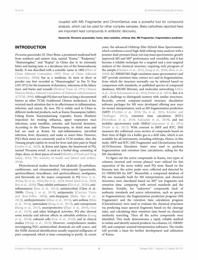
For samples of antimicrobial tests, the plant materials werecollected from Jinpen Mountain, Xinfeng County, Ganzhou,China (Figure 1). According to Flora of China, the samples wereidentified as Picrasma quassioides (D. Don) Benn. by ProfessorHaibo Hu. The voucher specimen was kept in our lab at GannanMedical University (No. PQ-XF1701). PQ stems (Kumu) were driedat 40°C, and then grounded into a fine powder. One gram of eachpowder was extracted in 10ml of H2O, MeOH, EtOAC, and hexanewith the help of sonication (4 × 15 min with 4–6 h gap). Then after5 min centrifuging at 3500 rpm, a 1-ml aliquot of each supernatantwas sampled and evaporated in a fume hood (hexane and ethylacetate), or dried (methanol and water) in a SpeedVac concentrator(SVC 200H, Stratech Scientific, London, United Kingdom). Thedried extracts and three standards were redissolved in DMSO as a20mg/ml for antimicrobial assessment. All the samples were storedat 4°C for further testing.
Antimicrobial AssayThe antibacterial activity of crude extracts was tested against 20human pathogenic strains, including five fungi [Candida albicans(SC 5314), Candida parapsilosis (ATCC 22019), Candida auris(OS299), Candida glabrata (ATCC 2001), and Saccharomycescerevisiae (ATCC 7754)], six Gram-positive bacteria
[Staphylococcus aureus (ATCC6538, Rosenbach),Staphylococcus epidermidis (ATCC 1457), Micrococcus luteus(DPMB 3), Listeria innocua (LMG 11387), Enterococcusfaecalis (HC-1909-5), and Bacillus cereus (LMG9610)], andnine Gram-negative bacteria [Escherichia coli (ATCC 47076),Pseudomonas aeruginosa (PAO1), Shigella sonnei (LMG 10473),Acinetobacter baumannii (RUH134), Enterobacter aerogenes (orKlebsiella aerogenes, ATCC 13048), Brevundimonas diminuta (agift from Prof. Rob Lavigne at KU Leuven), Shigella flexneri(LMG10472), Salmonella enterica subsp. enterica (ATCC 13076),and Aeromonas hydrophila (ATCC 7966)]. The antimicrobialtests were performed by a 96-well microdilution method. In brief,for bacteria and fungi, 10 and 5 μl, respectively, of a two-folddilution series of each extract (20 mg/ml stock), blank controls(DMSO) and positive controls (ciprofloxacin, 100 μg/ml stockand miconazole, 200 μg/ml stock) were transferred into 96-wellplates with the test microogranism (OD: 0.003) and cultured for20 h. Then the inhibition value (%, IV) was obtained by dividingthe sample’s OD value minus that of an extract control withoutinoculation by the average OD of the blank controls (solvent) andmultiplying by 100.
All the tests were repeated for confirmation, and IC50
(minimum inhibitory concentration to inhibit the growth of50%) was calculated by dose-response fitting using non-linearleast-squares sigmoid regression (GraphPad Prism 7.0, SanDiego, CA). The data were clustered using a webtool (https://biit.cs.ut.ee/clustvis/) to obtain clustered heatmaps. MBC(minimum bactericidal concentration) was obtained directlyfrom the agar tests of serial chemical dilution.
Fractionation and Isolation of Constituents100 g of PQ stem was extracted three times aided by sonication (4× 30min with 4–6 h gap) with the selected solvent (methanol)according to the antimicrobial activities, and then dried in aRotavapor (R-100, BÜCHI, Flawil, Switzerland). Then, 5.6 g ofextract was finally obtained and mixed with 25 g silica gel (60 Å,LOT#MKCK 1888; Sigma-Aldrich, Germany). Afterward, alarge-scale separation was performed on a silica gel column(ECOPLUS 35 × 500 mm, YMC, Kyoto, Japan) in a
FIGURE 1 | Antimicrobial activities (IC50) (μg/ml) of different solvent extracts of Picrasma quassioides stem; IV: inhibition value; The abbreviations are the initials ofthe scientific names of the testedmicroorganism, including five fungi [Candida albicans (CA),Candida parapsilosis (CP),Candida auris (CAU),Candida glabrata (CG), andSaccharomyces cerevisiae (SC)], nine G– bacteria [Escherichia coli (EC), Pseudomonas aeruginosa (PA), Shigella sonnei (SS), Acinetobacter baumannii (AB),Enterobacter aerogenes (EA),Brevundimonas diminuta (BD), Shigella flexneri (SF), Salmonella enterica subsp. enterica (SLE), and Aeromonas hydrophila (AH)], andsix G+ bacteria [Staphylococcus aureus (SA), Staphylococcus epidermidis (SE), Micrococcus luteus (ML), Listeria innocua (LI), Enterococcus faecalis (EF), and Bacilluscereus (BC)], marked in blue, black, and red, respectively.
Frontiers in Pharmacology | www.frontiersin.org October 2021 | Volume 12 | Article 7617513
Hu et al. 25 Antimicrobial Compounds of Kumu

preparative liquid chromatography setup (Waters Delta600 multi-solvent quaternary pump, Waters detector 2487,Waters 600 controller, Massachusetts, United States) using amobile phase consisting of hexane (A), ethyl acetate (B),methanol (C), and 25% acetic acid in methanol (D),performed a flow rate of 40 ml/min, with a step gradient from95% A and 5% B to 100% D (5–20% step every 10 min). Finally,218 fractions were collected (1 fraction per minute) and 1 ml ofeach fraction was sampled and dried; then 100 μl DMSO wasadded per sample to prepare stocks for antimicrobial testing.
Based on the bioactivity results of 218 fractions, seven activefractions (F47, F60, F88, F108, F118, F125, and F181) were selectedfor further separation via HPLC (LC-20AT pumps combined with aSPD-M20A detector and DGU-20A3R degasser, SHIMADZU,Kyoto, Japan). Separation conditions of each fraction wereoptimized on several analytical columns (250 × 4.6 mm, 5 μm,C18, Symmetry, Waters; 150 × 4.6 mm, 4 μm, Polar-RP, Synergi,Phenomenex; 250 × 4.6 mm, 4 μm, Hydro-RP, Synergi,Phenomenex), and then were geometrically transferred to semi-preparative columns to ensure the same selectivity for peakcollections. F47 and F60 were separated on a semi-preparativeC18 column (250 × 10mm, 5 μm, C18, Sunfire, Waters) and therest by a Polar-RP column (250 × 10mm, 4 μm, Polar-RP, Synergi,Phenomenex) using a mobile phase consisting of water (A) andacetonitrile (B), both containing 0.1% formic acid. For each fraction,the injected amount was kept around 2mg, and all the subfraction (1per minute) were collected via gradient elution with a 4 ml/min flowrate using the conditions listed in Table 1. After drying each fractionin a SpeedVac, 15 μl DMSO were added to each tube forantimicrobial testing. To eliminate possible errors during a singlerun, all subfractions were collected and tested twice. This was alsodone for all the active peaks collected under the same condition toconfirm the activities. Then the confirmed peaks were collected,dried, and kept in 4°C for the MS analysis.
UHPLC and MS/MS ConditionsThe UHPLC-MS/MS analysis was performed with an Ultimate 3000UHPLC system (Dionex Thermo Scientific) equipped with anEasySpray C18 column (2 μm, 100 Å, 50 μm × 15 cm, ThermoScientific), and Orbitrap Elite High-Field Orbitrap Hybrid MassSpectrometer (Thermo Scientific, United States), which consisted oforbitrap high-resolution selection and ion trap scanning, was usedfor the chemical analysis of natural small-molecular compounds.With a 0.3 μl·min−1 flow rate, the C18 column was set at 35°C andthe injection volume was 5 μl. The mobile phases were composed ofwater (eluent A) and 80% acetonitrile with 20% water (eluent B),
both containing 0.1% formic acid (for negative mode detection, 0.1%ammonium acetate was used instead of formic acid). The gradientswere set as follows: 0–3min, 5% B; 3–20min, 5%–100% B;20–23min, 100% B; 23–24min, 100%–5% B; and 24–34min, 5%B to re-equilibrate the system.
For MS detection, the Orbitrap-Ion Trap mass spectrometerwas fitted with a heated electrospray ionization (ESI) ion source,and both negative and positive ionization modes were performedat a full scan mode ranging from m/z 100–2000. To aid thestructural identification of the components, top 20 ions’ MS/MSfragmentation (dd-MS2-TOP 20) was operated with anm/z rangeof 50–2000. The MS parameters were optimized as reportedpreviously (Hu et al., 2021) with the followings: spray voltage−2.1 Kv/+2.1 Kv; capillary temperature, 275°C; multipole RFamplifier, 800 Vp-p; reagent ion source temperature, 160°C.The stepped ITMS (Ion trap mass spectrometry) was set to35 eV for MS/MS acquisition of the most intense ions fromFTMS (Fourier transform mass spectrometry).
Data Processing and AnalysisAll the MS data acquisition and analyses were done by Xcalibur 4.2(Thermo Scientific, United States), ACD/MS Workbook Suite2020, combined with MS Fragmenter, and ChromGenius(ACD/labs, Canada). The original UHPLC-MS data of eachsample were exported, and their background was subtractedbased on MS data of the blank solvent for both positive andnegative ions. Then the processed data were imported into ACD/labs to extract peaks and align chromatograms to make thecomparison more accurate than manually, including ITA(itellitarget) and IX 2.0 (IntelliXtract), which are algorithms toanalyze LC(GC)/MS datasets for targeted and untargetedcomponent detection and deconvolution, respectively. Theassignment accuracy was set to 5 ppm for both.
Moreover, the published components of PQ were collected bychecking SciFinder, Web of Science, the Dictionary of NaturalProducts, China National Knowledge Infrastructure (CNKI), andother databases, and then Spectrus DB (ACD/labs) was used tocreate a manually built compound library, including chemicalnames, molecular formulas, exact molecular mass, andstructures. The MS1 and MS2 spectra of active peaks wereanalyzed by comparing the detected formulas and MSfragments with the component library we built and theCOCONUT database (Sorokina et al., 2021) (the largestpublicly available natural product database), and all the activecompounds were targeted and identified. In detail, the spectrum ofstandards can figure out themselves easily, and those compounds
TABLE 1 | The HPLC gradient conditions of the selected fractions.
Fractions Gradients
F47 0–8 min, 50% B; 8–65 min, 50–100% B; 65–80 min, 100% BF60 0–8 min, 25% B; 8–60 min, 25–50% B; 60–70 min, 50–100% B; 70–80 min, 100% BF88 0–5 min, 15% B; 5–55 min, 15–30% B; 55–65 min, 30–50% B; 65–70 min, 30–100% B; 70–80 min, 100% BF108 0–8 min, 10% B; 8–15 min, 10–30% B; 15–65 min, 30–55% B; 65–70 min, 55–100% B; 70–80 min, 100% BF118 0–8 min, 10% B; 8–15 min, 10–30% B; 15–65 min, 30–65% B; 65–70 min, 65–100% B; 70–80 min, 100% BF125 0–8 min, 10% B; 8–60 min, 10–60% B; 60–70 min, 60–100% B; 70–80 min, 100% BF181 0–5 min, 10% B; 5–10 min, 10–20% B; 10–63 min, 20–60% B; 63–70 min, 60–100% B; 70–80 min, 100% B
Frontiers in Pharmacology | www.frontiersin.org October 2021 | Volume 12 | Article 7617514
Hu et al. 25 Antimicrobial Compounds of Kumu

without standards can be elucidated by their ion fragments andretention time (tR) according to fragmentation prediction by MSFragmenter and retention time calculation by ChromGenius, fromwhich all the fragmentation patterns of each compound in thisstudy were summarized and further verified.
RESULTS AND DISCUSSION
Antimicrobial Activities of KumuThus far, antimicrobial tests mainly focused on PQ compounds,especially for its alkaloids. A previous study showed that the extractof total alkaloids exhibited antimicrobial activities againstStreptococcus hemolytic-β, Staphylococcus aureus, Shigellacastellani, Bacillus subtilis subsp, and Sporosarcina (Meng, 2007).Total alkaloids were reported to exhibit a wider spectrum and betterantimicrobial activity than single alkaloids, possibly due tosynergistic effects (Guihua, 2011). Notably, the fat-solublealkaloids provided good inhibition against both wild type andtwo highly virulent strains of E. coli, even higher than berberinesulfate, mequindox, and ofloxacin, while water-soluble alkaloidshardly had bacteriostatic effects on these strains (He et al., 2008).Also, the essential oil of PQ inhibited both bacterial (e.g. Bacillussubtilis) and fungal (e.g. Ganoderma lucidum) species (Hanif et al.,2010). However, as a TCM, Kumu was always used as a whole
extract. Comprehensive studies investigating the bioactivities of itstotal extracts are still scarce. Hence, in this study, four differentsolvents (hexane, ethyl acetate, methanol, and water) were used toextract the stem of PQ, and test these extracts against 20 differenthuman pathogens. The results show that Kumu extracts can inhibitfive G+, four G– bacteria, and one fungus (Figure 1), in whichKumu exhibited strongest activity against Staphylococcus aureus(IC50 � 275 μg/ml). These results support Kumu’s medicinalapplication as an anti-infection agent. Moreover, the resultsshow that methanol is the best solvent to extract Kumu for itsantimicrobial activity, and it was used to perform the large-scaleextracting for bioassay guided purification work.
Bioassay-Guided IsolationBased on the above antimicrobial activities, it is worth performingbioassay-guided purification for identifying the antimicrobialcompounds of Kumu. By now, bioassay-guided isolation ofnatural products have been widely used for different activities,such as anti-inflammatory (Erdemoglu et al., 2008), antioxidant(Yang et al., 2020), anti-seizure (Brillatz et al., 2020b) (Brillatz et al.,2020a), vasorelaxant (Othman et al., 2006), aldose reductaseinhibitory (Logendra et al., 2006), anti-plasmodial (Baldé et al.,2021), antiviral (Parvez et al., 2020), antibacterial (He et al.,2017), and antifungal (Yan et al., 2020). But most of these studieswere performed with multiple purification steps, which consumed
FIGURE 2 | Separation of antibacterial compounds from Kumu based on successive chromatographic columns (normal and reversed-phase chromatography) andthe heatmap of the bioactivity of their factions against a representative Gram-positive strain (SA: S. aureus) and Gram-negative strain (EC: E. coli). F: fractions ofpreparative silica gel column, D: negative control, DMSO, P1-8: positive control (ciprofloxacin) in different concentrations, IV: inhibition value, T: time for collection inminutes, such as 24’ and 41’.
Frontiers in Pharmacology | www.frontiersin.org October 2021 | Volume 12 | Article 7617515
Hu et al. 25 Antimicrobial Compounds of Kumu

long time and large amounts of solvents. With the rapiddevelopment of chromatographic techniques, many differentcolumns have been already used for fast collection of small-amount compounds. Hence, in this study, two types of columnswere selected for the separation: a large normal-phase silica gelcolumn and semi-preprative reverse-phase columns. The aim was torapidly determine and collect the active fractions/peaks to increasethe purification efficiency.
For the bioassays, Staphylococcus aureus (Gram-positive) andEscherichia coli (Gram-negative), which belong to top dangeroussuperbugs worldwide (World Health Organization, 2017), wereselected for the antimicrobial activities of Kumu. After separationon a preparative silica gel column, 218 fractions (F1-218) wereobtained for antimicrobial testing, and seven active groups werefound including F46-52, F54-60, F88-96, F105-112, F113-119, F122-129, and F177-187 (Figure 2). From each, a representative fractionwas selected for further purification based on their inhibition values:F47, F60, F88, F108, F118, F125, and F181. By comparing differentcolumns and separation conditions, F47 and F60 were further
separated on a semi-preparative C18 column, and the remainingfractions by the Polar-RP column. In either case, we used a mobilephase consisting of water (A) and acetonitrile (B), both containing0.1% formic acid. Afterward, each HPLC-subfraction was testedagainst the two aforementioned bacteria, and their activities areshown as a heatmap (Figure 2). The subfractions showed much lessactivity against E. coli than S. aureus, presumably because G– bacteriahave a multilayer outer membrane preventing many xenobioticspassing through the cell membrane, so it is more difficult to findactivity against G– (Randall et al., 2013). Finally, 3, 5, 1, 10, 10, 2, and5 active peaks were obtained from the seven fractions, respectively,and all these 36 peaks were collected for chemical identification viaLC-HRMS/MS (Figure 3).
Identification of Active Compounds inSelected Fractions via LC-HRMS/MSBefore the MS analysis, all peaks were collected and tested toconfirm their activities. The results showed that all 36 peaks were
FIGURE 3 | HPLC–UV chromatograms and active peaks of the selected fractions from the stem of Picrasma quassioides. The peaks active against S. aureus aremarked in blue, while the ones active against both S. aureus and E. coli are numbered in orange.
Frontiers in Pharmacology | www.frontiersin.org October 2021 | Volume 12 | Article 7617516
Hu et al. 25 Antimicrobial Compounds of Kumu

TABLE 2 | Mass information of antimicrobial chemicals identified from the stem of Picrasma quassioides.
No Name tR(min)
Formula Ionmode
Calculatedm/z
Observedm/z
Diff.(ppm)
MS/MS fragments
1 1-Acetyl-β-carboline
18.90 C13H10N2O [M+H]+ 211.0866 211.0864 −0.90 211.1406, 194.0908, 193.0662, 185.9508, 169.0260, 166.9848
2 Methylnigakinone 19.22 C16H12N2O3 [M+H]+ 281.0921 281.0919 −0.60 266.9752, 266.0262, 265.0667, 249.0384, 247.9462, 237.8373, 236.9872, 220.9998, 219.98723 Melianone 20.30 C30H46O4 [M+H]+ 471.3469 471.3472 0.67 453.2476, 441.2062, 435.3094, 425.3902, 399.3133, 395.2563, 381.1285, 313.2863, 221.1231, 218.99064 1-
Methoxycarbonyl-β-carboline
15.98 C13H10N2O2 [M+H]+ 227.0815 227.0810 −2.22 212.9858, 195.0121, 184.9885, 167.0067[M−H]− 225.0670 225.0669 −0.23 240.0292, 236.9905, 227.0269, 213.0110, 212.0007
5 Dehydrocrenatine 14.00 C14H12N2O [M+H]+ 225.1022 225.1017 −2.40 225.0706, 211.0065, 210.0435, 192.9132, 181.9036[M−H]− 223.0877 223.0877 0.06 209.1688, 208.1098, 195.1732, 167.6166
6 Picrasidine D 14.33 C15H14N2O2 [M+H]+ 255.1128 255.1129 0.38 255.1180,241.0491, 240.0522, 222.9341[M−H]− 253.0983 253.0983 0.19 239.0415, 238.1265,223.0002, 118.9094, 88.9246
7 Dehydrocrenatidine 14.45 C15H14N2O2 [M+H]+ 255.1128 255.1126 −0.80 255.0867, 240.9951, 240.0045, 227.0230, 222.8967, 214.0931, 196.1431[M−H]− 253.0983 253.0983 0.19 245.6254,240.2685,238.1152,223.0610,207.9691
8 Canthin-6-one 18.25 C14H8N2O [M+H]+ 221.0709 221.0706 −1.54 221.0535, 193.0897, 166.12879 Kumudine D 17.63 C28H24N4O4 [M+H]+ 481.1870 481.1862 −1.73 480.2195, 463.1156, 449.1460, 431.1321, 281.3585, 229.0555, 210.9864
[M−H]− 479.1725 479.1720 −1.00 461.2052, 447.1585, 433.2364, 373.2896, 352.232610 Nigakinone 17.28 C15H10N2O3 [M+H]+ 267.0764 267.0761 −1.19 267.0483, 252.9591, 251.9883, 239.0147, 225.0800, 220.9192, 210.8483
[M−H]− 265.0619 265.0617 −0.62 251.0105, 250.1427, 248.9544, 247.6245, 238.0508, 237.0907, 221.082011 Picrasidine A 16.15 C27H22N4O3 [M+H]+ 451.1765 451.1758 −1.48 451.1954, 253.0459, 238.0994, 229.0569, 223.0212, 211.0181, 199.0065
[M−H]− 449.1619 449.1611 −1.81 435.2018, 434.1852, 237.0396, 234.9474, 224.9442, 212.0330, 211.0741, 196.007712 Picrasidine H 16.49 C28H25N4O4
+ [M+H]+ 481.1870 481.1862 −1.73 481.2508, 449.2208, 253.0801, 237.9780, 229.0756, 213.9998[M−H]− 479.1725 479.1719 −1.21 465.1142, 464.1550, 449.1692, 237.0669, 235.9578, 234.9786, 211.9767, 211.0386
13 Quassidine K 16.92 C28H25N4O3+ [M]+ 465.1921 465.1914 −1.54 465.2053, 267.1114, 254.0098, 253.0535, 238.0208, 212.9936, 197.9832
[M−2H]− 463.1776 463.1772 −0.79 449.1972, 448.2293, 433.2408, 265.2134, 237.0870, 235.0272, 221.0503, 195.051414 Kumudine B 16.60 C28H22N4O5 [M+H]+ 495.1663 495.1653 −2.01 495.1435, 477.1572, 281.0458, 266.9739, 253.0456, 214.9882
[M−H]− 493.1517 493.1513 −0.90 478.2079, 475.2882, 451.2625, 293.2664, 266.0257, 265.0934, 251.0856, 250.0576, 211.129415 Picrasidine C 16.93 C29H26N4O4 [M+H]+ 495.2027 495.2024 −0.57 495.2255, 463.2053, 267.0492, 253.0370, 243.0504, 228.0106
[M−H]− 493.1881 493.1877 −0.87 479.0698, 478.1480, 464.1842, 463.2394, 252.1857, 237.1095, 235.0246, 226.0647, 225.0798, 213.0634, 211.000116 Quassidine J 17.02 C29H27N4O4
+ [M]+ 495.2027 495.2023 −0.77 495.2697, 465.1927, 267.0480, 253.0714, 243.0592, 238.0275, 228.0461, 212.9872[M−2H]− 493.1881 493.1877 −0.87 478.1840, 364.1169, 251.0574, 234.9872, 225.0534, 211.0231
17 Quassidine L 17.52 C29H27N4O3+ [M]+ 479.2078 479.2060 −3.69 479.2004, 464.1393, 449.1678, 447.2045, 267.0383, 255.0499, 237.0422, 227.9599, 212.9119
[M−2H]− 477.1932 477.1929 −0.66 463.2171, 462.2030, 447.1887, 267.1133, 252.0062, 237.0820, 225.0665, 211.0365, 195.047918 Quassidine M 17.68 C30H29N4O4
+ [M]+ 509.2183 509.2172 −2.22 509.2563, 494.1823, 479.1444, 281.0834, 267.0577, 255.0575, 228.0182, 213.8599, 184.9867[M−2H]− 507.2038 507.2037 −0.15 493.2179, 492.1960, 477.2439, 267.1083, 252.0768, 251.1288, 237.0118, 234.9831, 224.9680, 211.0773
19 Picrasidine S 18.08 C30H29N4O4+ [M]+ 509.2183 509.2172 −2.22 509.2216, 479.1555, 478.2160, 295.1020, 266.9344, 255.0566, 242.1013, 227.9459, 194.0148
[M−2H]− 507.2038 507.2038 0.04 492.1746, 477.2206, 252.0334, 251.1116, 235.0350, 225.0618, 211.023920 Picrasidine G 17.25 C28H25N4O2
+ [M]+ 449.1972 449.1967 −1.12 449.2177, 434.1952, 251.0516, 237.1038, 224.9942, 212.9841, 198.0559[M−2H]− 447.1826 447.1825 −0.33 433.1703, 432.2199, 222.0418, 221.1324, 207.9999, 196.0633, 195.0490, 182.0178
21 Quassidine I 17.02 C29H27N4O3+ [M]+ 479.2078 479.2073 −0.97 479.2784, 448.2095, 447.1479, 266.9980, 249.0569, 238.0862, 225.0882, 212.9724, 197.9255
[M−2H]− 477.1932 477.1929 −0.66 463.1853, 462.2612, 447.2323, 445.2561, 251.1134, 225.0477, 221.0837, 210.0430, 195.039422 Picrasidine F 17.88 C29H27N4O3
+ [M]+ 479.2078 479.2072 −1.18 479.1259, 464.1355, 448.1689, 447.1413, 266.9610, 249.1028, 227.9948[M−2H]− 477.1932 477.1931 −0.24 462.2571, 461.2220, 447.1597, 267.4268, 252.1014, 237.1253, 226.0685, 225.0949, 211.0312, 195.0119
23 Quassidine A 18.08 C29H26N4O3 [M+H]+ 479.2078 479.2069 −1.81 479.2355, 449.1487, 448.1759, 265.0763, 250.9949, 226.9919, 211.0252[M−H]− 477.1932 477.1935 0.60 462.1905, 447.2411, 267.0988, 252.0888, 237.0792, 225.0339, 211.0342, 195.0640, 182.0502
24 Picrasidine I 12.47 C14H12N2O2 [M+H]+ 241.0972 241.0970 −0.64 241.0725, 223.0385, 221.3182, 194.9022, 180.8792[M−H]− 239.0826 239.0825 −0.42 221.9767, 221.0481, 196.0063, 195.0665
25 β-Carboline-1-carboxylic acid
13.88 C12H8N2O2 [M+H]+ 213.0659 213.0660 0.68 212.9659, 195.0410, 166.9938[M−H]− 211.0513 211.0515 0.94 211.2809, 167.9854, 167.0509, 72.8464
The standards used in this experiment are marked in bold.
Frontiersin
Pharm
acology|w
ww.frontiersin.org
October
2021|V
olume12
|Article
7617517
Huet
al.25
Antim
icrobialCom
poundsof
Kum
u

active against S. aureus, but only four peaks were also active againstE. coli, including the subfractions at 59, 54, 26 , and 68min of F108,118, 125, and 181, respectively. Furthermore, mass responds ofpure chemicals were easily affected by various noises ofbackground in total ion chromatograms, due to the highsensitivity of mass detection. Hence, all 36 peaks and severalblank samples were analyzed both in positive and negative ionmode by an UHPLC-Orbitrap-Ion-TrapMass Spectrometer, basedon the optimized experimental condition. Then all chemicalanalyses were confirmed by both positive and negative ions,except in a few cases where data were only available from oneionmode. By comparing with available standards, fragmentation ofMS Fragmenter and retention time calculations, a total of 25 activecompounds (some peaks were identified as same compounds, inFigure 3) were identified (Table 2), three of which were only
detected in positive ion mode under our experiment conditions: 1-acetyl-β-carboline, methylnigakinone, and melianone. All thechemical structures are shown in Figure 4, in whichcompounds 1–2 and 4–25 are alkaloids, while compound 3 is atriterpenoid. Notably, only picrasidine S (19) and β-carboline-1-carboxylic acid (25) showed strong inhibition against both S.aureus and E. coli.
Structure Elucidation Based onComparisonWith Published Spectra, Standards, and MSFragmenterFor component identification, comparison of the MS1 and MS2
spectra with published ones was extensively used, but for most PQcomponents no matching published sources could be available.
FIGURE 4 | Antimicrobial chemicals identified from the stem of Picrasma quassioides.
Frontiers in Pharmacology | www.frontiersin.org October 2021 | Volume 12 | Article 7617518
Hu et al. 25 Antimicrobial Compounds of Kumu

Thus, several standards were first analyzed under the samecondition to assist the MS interpretation, includingmethylnigakinone, nigakinone, and β-carboline-1-carboxylicacid. As shown in Figure 5, the β-carboline-1-carboxylic acidstandard (25) was detected in both negative and positive ionmode, with m/z 239.0826 and 241.0969, respectively, asprecursor ions. The detected active peaks of F125 showed quitesimilar retention time (tR) andm/z compared to the standard bothin MS1 and MS2, indicating that it was identical to the standard,compound 25. Meanwhile, another several active peaks of F47, 88,108, and 118 (shown in Figure 3) were similarly identified asmethylnigakinone (2) and nigakinone (10).
For the other compounds where no standards or publishedspectra were available, the software, MS Fragmenter (Zhou et al.,2014; Zhang Y. et al., 2019) was used to predict and interpret theirfragmentation in both positive and negative modes. Taking thefourth active peak in F108 as an example, it was detected in positiveand negative ion mode with strong procedures, m/z 465.1914[C28H25N4O3]
+ and m/z 463.1772, [C28H23N4O3]-,respectively.
After searching its structure via Mass Suite Book (ACD/labs) inthe COCONUT database (Sorokina et al., 2021) (the biggest publicly
available natural product database) and a manually built the PQdatabase (some components were not included in the COCONUTdatabase), 15 compounds with chemical formula C28H25N4O3
+ orC28H26N4O3 were retrieved. Then their mass fragmentation spectrawere compared one by one via MS Fragmenter (Tyrkkö et al., 2010),and the best matched one, quassidine K (13), was identified as thetarget, also according to the comparisons of retention time and MS1
spectra in positive and negative ion mode. Its fragmentaion inpositive ion mode, shown as an example in Figure 6, mainlyoriginates from the cleavage of nitrogen-containing heterocycles.Via the fragmentation analysis, most active peaks of Kumu could beidentified and are shown in Table 1.
Isomers Distiguished by ChromGenius andMS FragmenterAlthough MS Fragmenter can distinguish different fragmentationpattern of compounds, it was still difficult to see the differences forsome compounds with quite similar structures. Hence, retentiontime (tR) calculation can be utilized as a supplementary method todistinguish isomers. Notably, lots of isomers have been reported in
FIGURE 5 |MS1 and MS2 spectra of β-carboline-1-carboxylic acid (25) in the negative (NSI-) (A) and the positive (NSI+) (B) ion mode (upper panel: sample, lowerpanel: standard).
Frontiers in Pharmacology | www.frontiersin.org October 2021 | Volume 12 | Article 7617519
Hu et al. 25 Antimicrobial Compounds of Kumu

PQ, and were also detected in this study. Fraction 60 and 108showed active peaks with identical m/z 481.1862 in the positive ionmode, and m/z 479.1720 and 479.1719 in the negative ion mode.Hence, they were identified as the chemical formula, C28H24N4O4 orC28H25N4O4
+. To distiguish their structure, three compounds:kumudine D, picrasidine H, and picrasidine T, were consideredafter MS Fragmenter analysis as mentioned in the above becausethey have the same calculated m/z (481.1870 in positive and479.1725 in the negative ion mode).
Then ChromGenius was utilized for their retention time (tR)calculation, which was performed based on chemical similaritysearching (Willett et al., 1998). The calculation was set as theUHPLC-MS/MS condition and showed good predictionaccording to the main parameters, correction equation(−0.03*tR2 + 1.936*tR −6.696) and method coefficient(0.9039). The calculated results are listed in Figure 7E, inwhich the retention factor k’ was calculated as: k’ � (tR –t0)∕t0, where tR stands for retention time, t0 implied deadtime, and the similarity coefficient (sim coeff) is calculatedbased on the hamming distance coefficient (the closer to 1, thebetter). The predicted tRs of these isomers are shown inFigure 7A, and 25 best-matched points for the calculation areregressed in the curves (Figures 7B–D). Finally, based on thepredicted tR, the active peaks in F60 and 108 were identified askumudine D (9) and picrasidine H (12), respectively.
Four more sets of isomers were similarly analyzed based on theircalculated tR: isomers of m/z 255.1128 (C15H14N2O2) in F60 werepicrasidine D (6) and dehydrocrenatidine (7); isomers of m/z479.2078 (C29H27N4O3
+) in F108, 118, and 181 were identified as
quassidine L (17), quassidine I (21), and picrasidine F (22),respectively; isomers of m/z 509.2183 (C30H29N4O4
+) in F108,118, and 181 were suggested to be quassidine M (18) andpicrasidine S (19), while m/z 495.2027 (C29H26N4O4) in F108and 118 were picrasidine C (15) and quassidine J (16), respectively.
To confirm the accuracy of fragmentation and tR calculation foridentification, three standards were also subjected to the samecalculated method. Taking β-carboline-1-carboxylic acid (25) as anexample, the exact masses were 213.0659 ([M+H]+) and 211.0513([M−H]−), and its precursors were detected as 213.0656 and211.0513 in MS1; thus, the chemical formula was calculated asC12H8N2O2. Searching in the COCONUT and PQ databases 14compounds were retrieved. After assigning the MS2 fragmentationvia MS Fragmenter, three compounds were considered as the best-matching ones, and then, tR calculations were performed todistinguish them. Finally, the compound was identified asβ-carboline-1-carboxylic acid. Also, the other two standardscould be identified by the same approach. Hence, this can beconsidered as a method validation for this study.
The Antimicrobial Spectrum of ThreeRepresentative β-carbolinesThe standards of three β-carbolines were also assessed for theirIC50 (minimum inhibitory concentration to inhibit the growth of50%) and MBC (minimum bactericidal concentration):methylnigakinone (2), nigakinone (10), and β-carboline-1-carboxylic acid (25). Among them, only methylnigakinone’santimicrobial activity was reported in a Chinese patent against
FIGURE 6 | Total ion chromatography analysis (XIC) and MS1-2 spectra (A,C) of quassidine K (13) in the positive (m/z 465.1914, [C28H25N4O3]+) and the negative
(m/z 463.1772, [C28H23N4O3]-) ion mode, and its main mass fragmentation pathway (B) in the positive ion mode generated by MS Fragmenter.
Frontiers in Pharmacology | www.frontiersin.org October 2021 | Volume 12 | Article 76175110
Hu et al. 25 Antimicrobial Compounds of Kumu

Staphylococcus aureus (MIC � 0.76mg/ml), Escherichia coli(3.09 mg/ml), Pseudomonas aeruginosa (3.12 mg/ml),Streptococcus hemolyticus (2.46 mg/ml), and Streptococcuspneumoniae (1.23 mg/ml) (Wu et al., 2007). As far as we know,our study is the first systematic evaluation of their antimicrobialactivities. The tests were done by a 96-well microdilution methodagainst 20 different microorganisms. All three β-carbalinesstrongly inhibit the growth of many microorganisms and evenkilled them. Also the GraphPad was used to calculate the IC50 whileMBC was obtained directly from the agar tests of serial chemicaldilution (Table 3, Figure 8).
From the result of antimicrobial test, compound 2 (C2) caninhibit four Gram-positive, one Gram-negative bacteria, andthree fungi; however, most of the IC50 values were above 100 μg/ml. C10 was active against more microorganisms, includingsix G+, four G– bacteria, and three fungi, and showed mostlysomewhat higher activity than C2. C10 differs from C2 by amethylation at the 4’-hydroxy position. The methylation canreduce the chemical sensitivity, which was one of mechanism ofantimicrobial resistance (Malone and Gordon, 2016; Warrieret al., 2016), while other study also indicated the methylation
can also improve antimicrobial potency and broaden thespectrum (Benhamou et al., 2015), indicating thatmethylation worked differently for diverse compounds.According to the IC50 and MBC results, it was clear that themethylation reduced the antibacterial, but not the antifungalactivity of C10, suggesting its different mechanism of action onbacteria vs. fungi.
An interesting result was obtained for Candida auris, animportant emerging fungal pathogen, which was firstdescribed in 2009 and has spread across six continents as acause of infections in hospitals (Meis and Chowdhary, 2018;Proctor et al., 2021). Both C2 and C10 were found to stronglysuppress the growth of C. auris with a similar IC50 (around 30 μg/ml). Furthermore, C25 exhibited higher antibacterial activity andbroader spectrum than both C2 and C10, including six G+ andeight G– bacteria, but it showed no antifungal activity. Notably,most G– bacteria, such as E. coli, S. sonnei, S. flexneri, B. diminuta,A. hydrophila, and S. enterica subsp. enterica were sensitive forC25 (with IC50 mostly below 20 μg/ml). Therefore, all threecompounds were considered as having potential for antibioticdevelopment.
FIGURE 7 | Retention time prediction in chromatography (A), regression curves of three isomers [kumudine D (B), picrasidine T (C), and picrasidine H (D)] viaChromGenius and the calculation parameters (E).
Frontiers in Pharmacology | www.frontiersin.org October 2021 | Volume 12 | Article 76175111
Hu et al. 25 Antimicrobial Compounds of Kumu

Bioactivity Discussion of Kumu CompoundsTo better comparing the bioactivities of the chemicals detected in thisstudy, all the related literatures were checked, especially for theirantimicrobial activity. 1-acetyl-β-carboline (1)(OHMOTO andKOIKE, 1982), methylnigakinone (2)(Jiang and Zhou, 2008), andmelianone (3)were identified from fraction 47, where C1was alreadyreported as active against several laboratory and clinical strains(Savidge and Sorg, 2019)(Chen et al., 2018), such as S. aureus,with IC50 below 260 μg/ml (Shin et al., 2010). C2 has shown antiulcereffects (Omoja et al., 2014), significant cytotoxicity against CNE2 andHepG2 cancer cells (Jiang and Zhou, 2008; Mohd Jamil et al., 2020),and antiviral activity against Coxsackie virus B3 (with an IC50 as7.4 μM (Zhang et al., 2013)), and its antimicrobial activity wasdocumented and patented (Mitscher et al., 1975; Wu et al., 2007).Moreover, C3 (Zhao et al., 2019) was active against Salmonella ser.typhi with both MIC and MBC of 0.053 µM (Veni et al., 2020)against S. aureus with agar diffusion inhibition zone of 9mm(Biavatti et al., 2001), and it was also documented for anticancer,antivirus, anti-gout, anti-depressant, and antifeeding activities (Suet al., 1990; Ishaq, 2016; Liu Y. et al., 2019; Zhao et al., 2019; Anitaand Praveena, 2020; Sundar and Aarthy, 2020).
In fraction 60, six active chemicals were identified: 1-methoxycarbonyl-β-carboline (4), dehydrocrenatine (5),picrasidine D (6), dehydrocrenatidine (7), canthin-6-one (8), andkumudine D (9). Compound 4 can inhibit tobacco mosaic virus(TMV) replication with EC50 of 0.179mM (Chen et al., 2009b) andhad significant inhibitory activity on various plant pathogenicbacteria and fungi (Gao et al., 2020). C5 was reported forantimalarial, anti-inflammatory, antiparasitic, and antiprotozoalactivities (Kita et al., 2004; Van Baelen et al., 2009; Liu P. et al.,2019; Saiin et al., 2019), but ours is the first report of its antibacterial
activity to the best of our knowledge. C6 and C7 were reported fortheir cytotoxicity, anti-inflammatory, and antivirus activities(Ohmoto and Koike, 1988; Han et al., 2001; Chen et al., 2009b;Lee et al., 2009; Liu P. et al., 2019); their antibacterial activity isreported here for the first time. C8 showed anticancer, antimalarial,anti-inflammatory, antidiabetic, anti-plasmodium, antiprotozoal,antibacterial, antifungal, and antivirus activities (Lagoutte et al.,2008; Ross et al., 2008; Almeida et al., 2011; Arias et al., 2011;Cebrian-Torrejon et al., 2011; Agrawal et al., 2013; Rashed et al.,2013; Amanulla et al., 2017; Cho et al., 2018; Makong et al., 2019); itwas active against S. aureus (of inhibition zone diameter of 6.1 mm),but no activity against E. coli was observed (Casciaro et al., 2019),which is the same as our result. C9 showed cytotoxicity againstHepG2 and 3B cells, but there are no reports thus far about itsantimicrobial activity.
Fraction 88 only showed one active peak, identified asnigakinone (10), which has documented cytotoxicity, antivirus,antimalarial, and anti-inflammatory activities (Kuo et al., 2003;Chen et al., 2009b; Liu P. et al., 2019). Both fraction 108 and 118had 10 active peaks, as shown in Figure 3. The active peaks inF108 were identified as nigakinone (10), picrasidine A (11),picrasidine H (12), (±)-quassidine K (13), kumudine B (14),picrasidine C (15), quassidine J (16), quassidine L (17),quassidine M (18), and picrasidine S (19). Many similarcompounds were found in F118, including C10, 13, 16, 17, 18,and 19, in addition to four other compounds: picrasidine G (20),quassidine I (21), picrasidine F (22), and quassidine A (23). It canbe expected that the composition of compounds eluting fromsuccessive steps in a gradient may overlap, especially if the stepsare small. For their bioactivities, C11 was reported as a TMVinhibitor (Chen et al., 2009a) and C12 (KOIKE and OHMOTO,
TABLE 3 | Antimicrobial activity (μg/ml) of three β-carbolines
Microbials Methylnigakinone (2) Nigakinone (10) β-Carboline-1-carboxylic acid (25)
Positive controla
IC50 MBC IC50 MBC IC50 MBC IC50 MBC
S. aureus 205.70 >500 55.35 >125 47.70 >125 12.81 >125S. epidermidis NT NT 69.18 >125 50.88 >125 14.90 125M. luteus 137.10 >250 87.29 >250 33.99 64 21.14 64L. innocua NT NT 35.04 >250 117.80 >125 15.89 >125E. faecalis 109.00 >500 50.07 >125 70.66 >125 94.40b 125b
B. cereus 102.40 >250 38.75 >250 30.48 >125 21.62 >125E. coli NT NT NT NT 19.17 >125 22.31 32P. aeruginosa NT NT NT NT NT NT 12.58 >125S. sonnei NT NT NT NT 14.81 >125 20.68 64S. flexneri 194.80 >250 29.99 >250 3.96 125 12.04 64A. baumannii NT NT NT NT 30.28 >125 17.33 32E. aerogenes NT NT NT NT 93.65 >125 14.90 >125B. diminuta NT NT 10.46 >64 4.50 64 22.58 64A. hydrophila NT NT 68.64 >64 10.03 64 10.79 32S. enterica subsp. enterica NT NT 490.12 >500 19.34 >64 12.33 64C. parapsilosis 236.00 >500 201.5 >500 NT NT 10.01c >125cC.albicans 356.90 >500 493.8 >500 NT NT 17.43c >125cC. auris 32.82 >125 31.91 >125 NT NT 12.10c >125cC. glabrata NT NT NT NT NT NT 18.77c >125cS. cerevisiae NT NT NT NT NT NT 7.83c 32c
aPositive control: Ciprofloxacin.bChloramphenicol.cMiconazole.
Frontiers in Pharmacology | www.frontiersin.org October 2021 | Volume 12 | Article 76175112
Hu et al. 25 Antimicrobial Compounds of Kumu

1986) showed none of anti-inflammatory effects and cytotoxicityagainst RAW 264.7 cells (Liu P. et al., 2019), while C13 had goodcytotoxicity against the human cervical HeLa cell line, with anIC50 value around 20 μM (Guo et al., 2020). C14 has anti-hepatoma potential l (Zhao et al., 2019d), and C15 exhibitedanti-inflammatory activity (Liu P. et al., 2019) and cytotoxicityagainst RAW 264.7 (Liu P. et al., 2019), esophageal squamouscarcinoma cells (Shi et al., 2018), and also beneficial effects in
cerebral protection (Sasaki et al., 2016). From the literatures, C16,C19, C20, and C21 had cytotoxicity against human cervical HeLa,gastric MKN-28 cancer, and mouse melanoma B-16 cancer cells(Jiao et al., 2015), and C21 also strongly inhibited osteoclastformation (Kong et al., 2017). C17 exerted beneficial effects in thecerebral protection (Sasaki et al., 2016), and the trifluoroacetate ofboth C17 and C18 showed potential anti-Alzheimer’s diseaseactivity (Zhang et al., 2020). C20 had antiprotozoal activity (Kita
FIGURE 8 | Inhibition of different microorganisms by three β-carbolines from the stem of Picrasma quassioides.
Frontiers in Pharmacology | www.frontiersin.org October 2021 | Volume 12 | Article 76175113
Hu et al. 25 Antimicrobial Compounds of Kumu

et al., 2004) and was also active against RAW 264.7 (Liu P. et al.,2019) and triple-negative breast cancer cells (MDA-MB 468)(Yamashita et al., 2017). C22 was toxic against HeLa cells andactive against several methicillin-sensitive and -resistant strains ofS. aureus (MRSA), while C19 and C20 were also reported for theiranti-MRSA activity (Guo-hua et al., 2015). For C23, only its anti-inflammatory activity was studied (Jiao et al., 2010b).
Only two active compounds were found in F125: picrasidine I(24) and β-carboline-1-carboxylic acid (25). C24 was known tohave a strong antiosteoclastogenic effect (Kong et al., 2017) but noother effects were reported thus far. For C25, the previousbioactivity studies implied it can inhibit the growth of K-562and SGC-7901 cell lines (Lai et al., 2014) and had potential forattenuating bleomycin-induced pulmonary fibrosis (Cui et al.,2019), while none anti-inflammatory activity (Liu P. et al., 2019)and none photocytotoxicity against lung (A549) and colon(Colon-26) cancer cells (Reddy et al., 2019).
To summarize, 18 compounds identified in our study are firstreported here for their activity against S. aureus or E. coli,including 1-methoxycarbonyl-β-carboline (4),dehydrocrenatine (5), picrasidine D (6), dehydrocrenatidine(7), kumudine D (9), nigakinone (10), picrasidine A (11),picrasidine H (12), (±)-quassidine K (13), kumudine B (14),picrasidine C (15), quassidine J (16), quassidine L (17),quassidine M (18), quassidine I (21), quassidine A (23),Picrasidine I (24), and β-carboline-1-carboxylic acid (25).Notably, 24 active compounds were β-carbolines or dimersthereof. This class is a promising scaffold for the design anddevelopment of antibacterial compounds. It is worth to find lotsof related bioactive compounds in plant extracts, since thebiosynthesis of secondary metabolites often generatessignificant amounts of intermediates of the biosyntheticpathway, some of which may share bioactivities. The numberof bioactive compounds identified from a single plant is large inthis study, although it cannot be sure that all the identifiedβ-carbolines originate from a single biosynthetic pathway here,which could be helpful and pending for future studies.
CONCLUSION
In summary of this study, the interpretation method using two typesof columns for chemical isolation and UHPLC-Orbitrap- Ion-Trap
Mass Spectrometer for chemical identification was developed. Thestandard comparison, database searching, MS Fragmenter, andChromGenius were introduced for the structure analysis. Theresults showed that high-resolution mass spectrometry combinedwith mass fragmentation and retention time calculation was apowerful tool to identify natural products, especially fordistinguishing isomers with similar structures. A total of 25 activecompounds were isolated and identified from Kumu against S.aureus or E. coli, including five types of isomers, in which 18components’ antimicrobial activities were firstly reported in thisstudy. Notably, 24 active chemicals belonged to β-carboline or itsdimer, which class exhibited as a promising scaffold for the designand development of potent and selective antibacterial compounds.The antimicrobial components reported here provide a basis for themedicinal usage of themselves and Kumu, and are beneficial for thescientific development of quality standards and its therapeuticutilization, indicating that the method can be used to figure outactive chemical basis for other complex materials.
DATA AVAILABILITY STATEMENT
The raw data supporting the conclusions of this article will bemade available by the authors, without undue reservation.
AUTHOR CONTRIBUTIONS
Conceptualization, HH, CH, and WL; methodology, HH, AK,and AG; software, HH, JP, and DS.; validation, HH.; formalanalysis, HH; investigation, HH, JP, and CH; resources, HH, JP,and CH; data curation, HH; writing—original draft preparation,HH; writing—review and editing, HH andWL; visualization, HH;supervision, WL; project administration, WL and HH; fundingacquisition, WL and HH. All authors have read and agreed to thepublished version of the manuscript.
FUNDING
This work was funded by the National Natural ScienceFoundation of China (Grant Nos. 81660639). WL largelyfunded himself.
REFERENCES
Aalizadeh, R., Nika, M. C., and Thomaidis, N. S. (2019). Development andApplication of Retention Time Prediction Models in the Suspect and Non-target Screening of Emerging Contaminants. J. Hazard. Mater. 363, 277–285.doi:10.1016/j.jhazmat.2018.09.047
Agrawal, R., Sethiya, N. K., and Mishra, S. H. (2013). Antidiabetic Activity ofAlkaloids of Aerva Lanata Roots on Streptozotocin-Nicotinamide InducedType-II Diabetes in Rats. Pharm. Biol. 51, 635–642. doi:10.3109/13880209.2012.761244
Allen, F., Greiner, R., and Wishart, D. (2014). Competitive FragmentationModeling of ESI-MS/MS Spectra for Putative Metabolite Identification.Metabolomics 11, 98–110. doi:10.1007/s11306-014-0676-4
Amanulla, S. S. D., Veerakumar, S., and Ramanathan, K. (2017). Screening ofPotential Plant Compounds as Survivin Inhibitors and its Anti-Cancer Efficacyby Molecular Docking. Cei 13, 41–48. doi:10.2174/1573408012666160727165940
Ambu, G., Chaudhary, R. P., Mariotti, M., and Cornara, L. (2020). Traditional Usesof Medicinal Plants by Ethnic People in the Kavrepalanchok District, CentralNepal. Plants (Basel) 9, 759. doi:10.3390/plants9060759
A. Mitscher, L., Shipchandler, M., D. Hollis Showalter, H., and S. Bathala, M.(1975). Antimicrobial Agents from Higher Plants. Synthesis in the Canthin-6-One (6H-Indolo[3,2,1-De][1,5]naphthyridin-6-One) Series. Heterocycles 3,7–14. doi:10.3987/r-1975-01-0007
Anita, B. R., and Praveena, B. (2020). A Review on Anti-cancer Potential ofPhytochemicals Isolated from Simarouba Glauca. Indo Am. J. Pharm. Sci. 7,281–286. doi:10.5281/zenodo.3746442
Frontiers in Pharmacology | www.frontiersin.org October 2021 | Volume 12 | Article 76175114
Hu et al. 25 Antimicrobial Compounds of Kumu

Arias, J. L., Cebrián-Torrejón, G., Poupon, E., Fournet, A., and Couvreur, P. (2011).Biodegradable Polymeric Nanoformulation Based on the Antiprotozoal Canthin-6-One. J. Nanopart Res. 13, 6737–6746. doi:10.1007/s11051-011-0580-z
Bai, M., Zhao, W.-Y., Xu, W., Zhang, Y.-Y., Huang, X.-X., and Song, S.-J. (2020).Triterpenoids from Picrasma Quassioides with Their Cytotoxic Activities.Phytochemistry Lett. 39, 128–131. doi:10.1016/j.phytol.2020.07.014
Baldé, M. A., Tuenter, E., Matheeussen, A., Traoré, M. S., Cos, P., Maes, L., et al.(2021). Bioassay-guided Isolation of Antiplasmodial and AntimicrobialConstituents from the Roots of Terminalia Albida. J. Ethnopharmacol. 267,113624. doi:10.1016/j.jep.2020.113624
Benhamou, R. I., Shaul, P., Herzog, I. M., and Fridman, M. (2015). Di-N-Methylation of Anti-Gram-Positive Aminoglycoside-Derived MembraneDisruptors Improves Antimicrobial Potency and Broadens Spectrum toGram-Negative Bacteria. Angew. Chem. Int. Ed. Engl. 54, 13617–13621.doi:10.1002/ange.20150681410.1002/anie.201506814
Biavatti, M. W., Vieira, P. C., da Silva Mfg, M. F. G. F., Fernandes, J. B.,Albuquerque, S., Magalhães, C. M., et al. (2001). Chemistry and Bioactivityof Raulinoa Echinata Cowan, an Endemic Brazilian Rutaceae Species.Phytomedicine 8, 121–124. doi:10.1078/0944-7113-00016
Brillatz, T., Jacmin, M., Queiroz, E. F., Marcourt, L., Slacanin, I., Petit, C., et al.(2020a). Zebrafish Bioassay-Guided Isolation of Antiseizure Compounds fromthe Cameroonian Medicinal Plant Cyperus Articulatus L. Phytomedicine 70,153175. doi:10.1016/j.phymed.2020.153175
Brillatz, T., Kubo, M., Takahashi, S., Jozukuri, N., Takechi, K., Queiroz, E. F., et al.(2020b). Metabolite Profiling of Javanese Ginger Zingiber Purpureum andIdentification of Antiseizure Metabolites via a Low-Cost Open-SourceZebrafish Bioassay-Guided Isolation. J. Agric. Food Chem. 68, 7904–7915.doi:10.1021/acs.jafc.0c02641
Campuzano, I., Bush, M. F., Robinson, C. V., Beaumont, C., Richardson, K., Kim,H., et al. (2012). Structural Characterization of Drug-like Compounds by IonMobility Mass Spectrometry: Comparison of Theoretical and ExperimentallyDerived Nitrogen Collision Cross Sections. Anal. Chem. 84, 1026–1033.doi:10.1021/ac202625t
Casciaro, B., Calcaterra, A., Cappiello, F., Mori, M., Loffredo, M. R., Ghirga, F., et al.(2019). Nigritanine as a New Potential Antimicrobial Alkaloid for theTreatment of Staphylococcus Aureus-Induced Infections. Toxins (Basel) 11,511. doi:10.3390/toxins11090511
Cebrian-Torrejon, G., Spelman, K., Leblanc, K., Munoz-Durango, K., Gutierrez, S.T., Ferreira, M. E., et al. (2011). The Antiplasmodium Effects of a TraditionalSouth American Remedy: Zanthoxylum Chiloperone Var. Angustifoliumagainst Chloroquine Resistant and Chloroquine Sensitive Strains ofPlasmodium Falciparum. Rev. Bras. Farmacogn. 21, 652–661. doi:10.1590/S0102-695X2011005000104
Chen, J., Yan, X. H., Dong, J. H., Sang, P., Fang, X., Di, Y. T., et al. (2009a). TobaccoMosaic Virus (TMV) Inhibitors from Picrasma Quassioides Benn. J. Agric. FoodChem. 57, 6590–6595. doi:10.1021/jf901632j
Chen, Q., Zhang, S., and Xie, Y. (2018). Characterization of a NewMicrobial Pictet-Spenglerase NscbB Affording the β-carboline Skeletons from NocardiopsisSynnemataformans DSM 44143. J. Biotechnol. 281, 137–143. doi:10.1016/j.jbiotec.2018.07.007
Chinese Materia Medica Editorial Committee of National Administration of TCM(1999). Chinese Materia Medica (Volumn 5). Shanghai: Shanghai Science andTechnology Press, 13–14.
Cho, S. K., Jeong, M., Jang, D. S., and Choi, J. H. (2018). Anti-inflammatory Effectsof Canthin-6-One Alkaloids from Ailanthus Altissima. Planta Med. 84,527–535. doi:10.1055/s-0043-123349
Cui, Y., Jiang, L., Yu, R., Shao, Y., Mei, L., and Tao, Y. (2019). β-Carboline AlkaloidsAttenuate Bleomycin Induced Pulmonary Fibrosis in Mice through InhibitingNF-Kb/p65 Phosphorylation and Epithelial-Mesenchymal Transition.J. Ethnopharmacol. 243, 112096. doi:10.1016/j.jep.2019.112096
Curcino Vieira, I. J., and Braz-Filho, R. (2006). “Quassinoids: Structural Diversity,Biological Activity and Synthetic Studies,” In Studies In Natural ProductsChemistry 33, 433–492. doi:10.1016/S1572-5995(06)80032-3
de Souza Almeida, E. S., Filho, V. C., Niero, R., Clasen, B. K., Balogun, S. O., and deOliveira Martins, D. T. (2011). Pharmacological Mechanisms Underlying theAnti-ulcer Activity of Methanol Extract and Canthin-6-One of SimabaFerruginea A. St-Hil. In Animal Models. J. Ethnopharmacol. 134, 630–636.doi:10.1016/j.jep.2011.01.009
Denisov, E., Damoc, E., Lange, O., and Makarov, A. (2012). Orbitrap MassSpectrometry with Resolving powers above 1,000,000. Int. J. Mass Spectrom.325-327, 80–85. doi:10.1016/j.ijms.2012.06.009
Dodds, J. N., May, J. C., andMcLean, J. A. (2017). Investigation of the Complete Suite ofthe Leucine and Isoleucine Isomers: Toward Prediction of Ion Mobility SeparationCapabilities. Anal. Chem. 89, 952–959. doi:10.1021/acs.analchem.6b04171
Erdemoglu, N., Akkol, E. K., Yesilada, E., and Caliş, I. (2008). Bioassay-guidedIsolation of Anti-inflammatory and Antinociceptive Principles from a FolkRemedy, Rhododendron Ponticum L. Leaves. J. Ethnopharmacol. 119, 172–178.doi:10.1016/j.jep.2008.06.021
Fisher, C. M., Croley, T. R., and Knolhoff, A. M. (2021). Data Processing Strategiesfor Non-targeted Analysis of Foods Using Liquid Chromatography/high-Resolution Mass Spectrometry. Trac Trends Anal. Chem. 136, 116188.doi:10.1016/j.trac.2021.116188
Flora of China Editorial Committee (1997). flora of china (Chinese Version,Volumn 43). Beijing: Science Press, 7–8.
Flora of China Editorial Committee (2008). Flora of China (English Version,volumn11). St. Louis: Missouri Botanical Garden Press, 102.
Gao, X., Sheng, T., Wu, H., and Gu, Q. (2020). Inventor; Nanjing AgriculturalUniversity, Assignee. Preparation of 1-substituted β-carboline Derivatives forAntibacterial Agents. Chinese patent CN 111303148.
Gong, G., Jiang, L., Lin, Q., Liu, W., He, M. F., Zhang, J., et al. (2018). In Vivo toxicEffects of 4-Methoxy-5-Hydroxy-Canthin-6-One in Zebrafish Embryos viaCopper Dyshomeostasis and Oxidative Stress. Comp. Biochem. Physiol. CToxicol. Pharmacol. 204, 79–87. doi:10.1016/j.cbpc.2017.11.014
Gong, G., Lin, Q., Xu, J., Ye, F., Jiang, L., Liu,W., et al. (2016). In Vivo SAR and STRAnalyses of Alkaloids from Picrasma Quassioides Identify 1-Hydroxymethyl-8-Hydroxy-β-Carboline as a Novel Natural Angiogenesis Inhibitor. RSC Adv. 6,9484–9494. doi:10.1039/C5RA22391A
Guihua, D. (2011). Study on chemical constituents and quality evaluation ofRamulus et Folium Picrasmae. Master’s Thesis. Guangzhou (GD):Guangzhou University of Chinese Medicine.
Guo, E., Hu, Y., Du, T., Zhu, H., Chen, L., Qu, W., et al. (2019). Effects of PicrasmaQuassioides and its Active Constituents on Alzheimer’s Disease In Vitro and InVivo. Bioorg. Chem. 92, 103258. doi:10.1016/j.bioorg.2019.103258
Guo, X. M., Li, F., Zheng, F. F., Gong, N. N., Li, Y., Feng, W. Z., et al. (2020).(±)-Quassidine K, a Pair of Cytotoxic Bis-β-Carboline Alkaloid Enantiomersfrom Picrasma Quassioides. Nat. Prod. Res. 34, 489–493. doi:10.1080/14786419.2018.1489388
Guo-hua, S., Wei-hua, J., Fan, Y., and Hou-wen, L. (2015). Three Bis-β-CarbolineAlkaloids from Picrasma Quassioides and Their Bioactivities. Chin. Tradit.Herb. Drugs 6, 803–807. doi:10.7501/j.issn.0253-2670.2015.06.004
Han, J. H., Hong, S. R., Ji, O. P., Kwon, H. C., Lee, B. G., Lee, G. N., et al. (2001).Inventor. Antiinflammatory Composition Containing -carboline Derivatives.Korean patent KR, 2001037653.
Hanif, M. A., Bhatti, H. N., Jamil, M. S., Anjum, R. S., Jamil, A., and Khan, M. M.(2010). Antibacterial and Antifungal Activities of Essential Oils Extracted fromMedicinal Plants Using CO2 Supercritical Fluid Extraction Technology. AsianJ. Chem. 22, 7787–7798.
He, Y., Hu, Z., Li, Q., Huang, J., Li, X. N., Zhu, H., et al. (2017). Bioassay-GuidedIsolation of Antibacterial Metabolites from Emericella Sp. TJ29. J. Nat. Prod. 80,2399–2405. doi:10.1021/acs.jnatprod.7b00077
He, Y., Liu, W., Chen, Z., Zhao, B., Zhong, Z., and Wei, Y. (2008). Research on theIn Vitro Bacteriostatic Effect of Quassiawood Alkaloid on Escherichia coli.J. Anhui Agri.Sci. 36, 2777–2778.
Hu, H., Lee-Fong, Y., Peng, J., Hu, B., Li, J., Li, Y., et al. (2021). Comparative Research ofChemical Profiling in Different Parts of Fissistigma Oldhamii by Ultra-High-Performance Liquid Chromatography Coupled with Hybrid Quadrupole-Orbitrap Mass Spectrometry. Molecules 26, 960. doi:10.3390/molecules26040960
Hwisa, N. T., Gindi, S., Rao, C. B., Katakam, P., Rao Chandu, B., and Info, A.(2013). Evaluation of Antiulcer Activity of Picrasma Quassioides BennettAqueous Extract in Rodents. VRI-PM 1, 27. doi:10.14259/pm.v1i1.35
Ishaq, H. (2016). Anxiolytic and Antidepressant Activity of Different MethanolicExtracts of Melia Azedarach Linn. Pak. J. Pharm. Sci. 29, 1649–1655.
Jiang, C., Arthur, C. J., and Gates, P. J. (2020). A Computational and ExperimentalStudy of the Fragmentation of L-Leucine, L-Isoleucine and L-Allo-Isoleucineunder Collision-Induced Dissociation Tandem Mass Spectrometry. Analyst145, 6632–6638. doi:10.1039/d0an00778a
Frontiers in Pharmacology | www.frontiersin.org October 2021 | Volume 12 | Article 76175115
Hu et al. 25 Antimicrobial Compounds of Kumu

Jiang, M. X., and Zhou, Y. J. (2008). Canthin-6-one Alkaloids from PicrasmaQuassioides and Their Cytotoxic Activity. J. Asian Nat. Prod. Res. 10,1009–1012. doi:10.1080/10286020802277956
Jiao, W. H., Chen, G. D., Gao, H., Li, J., Gu, B. B., Xu, T. T., et al. (2015).(±)-Quassidines I and J, Two Pairs of Cytotoxic Bis-β-Carboline AlkaloidEnantiomers from Picrasma Quassioides. J. Nat. Prod. 78, 125–130.doi:10.1021/np500801s
Jiao, W. H., Gao, H., Li, C. Y., Zhao, F., Jiang, R. W., Wang, Y., et al. (2010b).Quassidines A-D, Bis-Beta-Carboline Alkaloids from the Stems of PicrasmaQuassioides. J. Nat. Prod. 73, 167–171. doi:10.1021/np900538r
Jiao, W. H., Gao, H., Li, C. Y., Zhou, G. X., Kitanaka, S., Ohmura, A., et al. (2010a).Beta-carboline Alkaloids from the Stems of Picrasma Quassioides. Magn.Reson. Chem. 48, 490–495. doi:10.1002/mrc.2602
Jiao, W. H., Gao, H., Zhao, F., Lin, H. W., Pan, Y. M., Zhou, G. X., et al. (2011).Anti-inflammatory Alkaloids from the Stems of Picrasma QuassioidesBENNET. Chem. Pharm. Bull. (Tokyo) 59, 359–364. doi:10.1248/cpb.59.359
Jung, Y.-T., Lee, I.-S., Whang, K., and Yu, M.-H. (2012). Antioxidant Effects ofPicrasma quassioides and Chamaecyparis obtusa (S. et Z.) ENDL Extracts.J. Life Sci. 22, 354–359. doi:10.5352/jls.2012.22.3.354
Kita, K., Yabu, Y., Nagai, K., Minagawa, N., and Hosokawa, K. , Inventor; ArigenPharmaceuticals Inc., Can. , Assignee. Indole Alkaloids as Enhancers forAntiprotozoal Activity of Ascofuranone, Their Compositions and Kits, andTreatment of Protozoan Diseases with Them. Japanese patent JP 2004231601(2004).
Koike, K., and Ohmoto, T. (1986). Studies on the Alkaloids from PicrasmaQuassioides Bennet. VII. Structures of .BETA.-carboline Dimer Alkaloids,Picrasidines-H and -R. Chem. Pharm. Bull. 34, 2090–2093. doi:10.1248/cpb.34.2090
Kong, L., Wang, B., Yang, X., Guo, H., Zhang, K., Zhu, Z., et al. (2017). Picrasidine Ifrom Picrasma Quassioides Suppresses Osteoclastogenesis via Inhibition ofRANKL Induced Signaling Pathways and Attenuation of ROS Production. Cell.Physiol. Biochem. 43, 1425–1435. doi:10.1159/000481874
Krettler, C. A., and Thallinger, G. G. (2021). AMap of Mass Spectrometry-Based InSilico Fragmentation Prediction and Compound Identification inMetabolomics. Brief. Bioinform 22. doi:10.1093/bib/bbab073
Kuo, P. C., Shi, L. S., Damu, A. G., Su, C. R., Huang, C. H., Ke, C. H., et al. (2003).Cytotoxic and Antimalarial Beta-Carboline Alkaloids from the Roots ofEurycoma Longifolia. J. Nat. Prod. 66, 1324–1327. doi:10.1021/np030277n
Lagoutte, D., Nicolas, V., Poupon, E., Fournet, A., Hocquemiller, R., Libong, D.,et al. (2008). Antifungal Canthin-6-One Series Accumulate in Lipid Dropletsand Affect Fatty Acid Metabolism in Saccharomyces cerevisiae. Biomed.Pharmacother. 62, 99–103. doi:10.1016/j.biopha.2007.07.014
Lai, Z.-Q., Liu, W.-H., Ip, S.-P., Liao, H.-J., Yi, Y.-Y., Qin, Z., et al. (2014). SevenAlkaloids from Picrasma Quassioides and Their Cytotoxic Activities. Chem.Nat. Compd. 50, 884–888. doi:10.1007/s10600-014-1106-6
Lee, J. J., Oh, C.-H., Yang, J. H., Baek, N.-I., Kim, S.-H., Cho, C. H., et al. (2009).Cytotoxic Alkaloids from the wood of Picrasma Quassioides. J. Korean Soc.Appl. Biol. Chem. 52, 663–667. doi:10.3839/jksabc.2009.110
Liu, P., Li, H., Luan, R., Huang, G., Liu, Y., Wang, M., et al. (2019a). Identificationof β-carboline and Canthinone Alkaloids as Anti-inflammatory Agents but withDifferent Inhibitory Profile on the Expression of iNOS and COX-2 inLipopolysaccharide-Activated RAW 264.7 Macrophages. J. Nat. Med. 73,124–130. doi:10.1007/s11418-018-1251-5
Liu, Y., Lai, L., Ju, Y., Liu, C., and Meng, D. (2019b). Chemical Constituents andSynergistic Anti-gout Studies on Eurycoma Longifolia and PotentialMechanisms Evaluation Based on Systemic Analysis Approach. Bioorg.Chem. 92, 103302. doi:10.1016/j.bioorg.2019.103302
Logendra, S., Ribnicky, D. M., Yang, H., Poulev, A., Ma, J., Kennelly, E. J., et al.(2006). Bioassay-guided Isolation of Aldose Reductase Inhibitors fromArtemisia Dracunculus. Phytochemistry 67, 1539–1546. doi:10.1016/j.phytochem.2006.05.015
Lou, L. L., Yao, G. D., Wang, J., Zhao, W. Y., Wang, X. B., Huang, X. X., et al.(2018). Enantiomeric Neolignans from Picrasma Quassioides ExhibitDistinctive Cytotoxicity on Hepatic Carcinoma Cells through ROSGeneration and Apoptosis Induction. Bioorg. Med. Chem. Lett. 28,1263–1268. doi:10.1016/j.bmcl.2018.03.043
Makong, Y. S., Mouthé Happi, G., Djouaka Bavoua, J. L., Wansi, J. D., Nahar, L.,Kamdem Waffo, A. F., et al. (2019). Cytotoxic Stilbenes and Canthinone
Alkaloids from Brucea Antidysenterica (Simaroubaceae). Molecules 24, 4412.doi:10.3390/molecules24234412
Malone, K. M., and Gordon, S. V. (2016). Antibiotic Methylation: A NewMechanism of Antimicrobial Resistance. Trends Microbiol. 24, 771–772.doi:10.1016/j.tim.2016.08.003
Medical team of, C. P. L. A. (1972). Clinical Application of Chinese HerbalMedicine. Shanxiongdan. J. New Med. 26, 0956.
Meis, J. F., and Chowdhary, A. (2018). Candida Auris: a Global Fungal PublicHealth Threat. Lancet Infect. Dis. 18, 1298–1299. doi:10.1016/S1473-3099(18)30609-1
Meng, C. (2007).Alkaloids from Picrasma Quassioies (D.Dom)Benn. And TheirAnti-inflammatory and Antibacterial Activities. Master’s Thesis. Yantai: YantaiUniversity.
Ministry of Food and Drug Safety (2012). Korean Pharmacopeia (TenthEdition,Monographs,Part II). Cheongju, Korea: Ministry of Food and DrugSafety, 1344.
Mohd Jamil, M. D. H., Taher, M., Susanti, D., Rahman, M. A., and Zakaria, Z. A.(2020). Phytochemistry, Traditional Use and Pharmacological Activity ofPicrasma Quassioides: A Critical Reviews. Nutrients 12, 2584. doi:10.3390/nu12092584
Noreldeen, H. A. A., Liu, X., Wang, X., Fu, Y., Li, Z., Lu, X., et al. (2018).Quantitative Structure-Retention Relationships Model for Retention TimePrediction of Veterinary Drugs in Food Matrixes. Int. J. Mass Spectrom.434, 172–178. doi:10.1016/j.ijms.2018.09.022
Ohmoto, T., and Koike, K. (1988). Antiherpes Activity of Simaroubaceae AlkaloidsIn Vitro. Shoyakugaku Zasshi 42, 160–162.
Ohmoto, T., and Koike, K. (1982). Studies on the Constituents of PicrasmaQuassioides Bennet. I. On the Alkaloidal Constituents. Chem. Pharm. Bull.30, 1204–1209. doi:10.1248/cpb.30.1204
Omoja, V., Ihedioha, T., Eke, G., PeterAjuzie, I., and Okezie, S. (2014). Evaluationof the Acute Toxicity, Phytochemical Constituents and Anti - Ulcer Propertiesof Methanolic Leaf Extract of Annona Muricata in Mice. J. IntercultEthnopharmacol 3, 37–43. doi:10.5455/jice.20140111103203
Othman, R., Ibrahim, H., Mohd, M. A., Mustafa, M. R., and Awang, K. (2006).Bioassay-guided Isolation of a Vasorelaxant Active Compound from KaempferiaGalanga L. Phytomedicine 13, 61–66. doi:10.1016/j.phymed.2004.07.004
Parvez, M. K., Al-Dosari, M. S., Arbab, A. H., Al-Rehaily, A. J., and Abdelwahid, M.A. S. (2020). Bioassay-guided Isolation of Anti-hepatitis B Virus FlavonoidMyricetin-3-O-Rhamnoside along with Quercetin from Guiera SenegalensisLeaves. Saudi Pharm. J. 28, 550–559. doi:10.1016/j.jsps.2020.03.006
Proctor, D. M., Dangana, T., Sexton, D. J., Fukuda, C., Yelin, R. D., Stanley, M., et al.(2021). Integrated Genomic, Epidemiologic Investigation of Candida AurisSkin Colonization in a Skilled Nursing Facility. Nat. Med. 27, 1401–1409.doi:10.1038/s41591-021-01383-w
Randall, C. P., Mariner, K. R., Chopra, I., and O’Neill, A. J. (2013). The Target ofDaptomycin Is Absent from Escherichia coli and Other Gram-Negative Pathogens.Antimicrob. Agents Chemother. 57, 637–639. doi:10.1128/AAC.02005-12
Rashed, K., Said, A., and Ahmed, M. (2013). Antiviral Activity and PhytochemicalAnalysis of Ailanthus Excelsa Roxb Bark. J. For. Prod. Ind. 2, 30–33.
Reddy, P. O. V., Shekar, K. P. C., Khandagale, S. B., Hara, D., Son, A., Ito, T., et al.(2019). Easy Access to Water-Soluble Cationic Porphyrin-β -CarbolineConjugates as Potent Photocytotoxic and DNA Cleaving Agents. AsianJ. Org. Chem. 8, 269–274. doi:10.1002/ajoc.201800649
Ren, J.-X., Bai, M., Zhao, W.-Y., Huang, X.-X., and Song, S.-J. (2020). ChemicalConstituents from Picrasma Quassioides (D.Don) Benn. And Their NetworkAnalysis of Chemotaxonomic Significance. Biochem. Syst. Ecol. 93, 104160.doi:10.1016/j.bse.2020.104160
Ross, S. A., Krishnaven, K., Radwan, M. M., Takamatsu, S., and Burandt, C. L.(2008). Constituents of Zanthoxylum Flavum and Their Antioxidant andAntimalarial Activities. Nat. Product. Commun. 3, 1934578X0800300–794.doi:10.1177/1934578x0800300521
Saiin, C., Junwised, J., Junwised, J., Nuengchamnong, N., and Ingkaninan, K.(2019). Determination of β-carbolines in Thai Picrasma Javanica Bl.; the Sourceof Potential Antimalarial Agents. Adv. Med. Plant Res. 7, 61–67. doi:10.30918/ampr.72.19.025
Sasaki, T., Li, W., Ohmoto, T., and Koike, K. (2016). Evaluation of CanthinoneAlkaloids as Cerebral Protective Agents. Bioorg. Med. Chem. Lett. 26,4992–4995. doi:10.1016/j.bmcl.2016.09.006
Frontiers in Pharmacology | www.frontiersin.org October 2021 | Volume 12 | Article 76175116
Hu et al. 25 Antimicrobial Compounds of Kumu

Savidge, T., and Sorg, J. A. (2019). Role of Bile in Infectious Disease: the Gall of 7α-Dehydroxylating Gut Bacteria. Cell Chem. Biol. 26, 1–3. doi:10.1016/j.chembiol.2018.12.010
Schymanski, E. L., Jeon, J., Gulde, R., Fenner, K., Ruff, M., Singer, H. P., et al.(2014). Identifying Small Molecules via High Resolution Mass Spectrometry:Communicating Confidence. Environ. Sci. Technol. 48, 2097–2098.doi:10.1021/es5002105
Shi, Y., Liu, X., Fredimoses, M., Song, M., Chen, H., Liu, K., et al. (2018). FGFR2Regulation by Picrasidine Q Inhibits the Cell Growth and Induces Apoptosis inEsophageal Squamous Cell Carcinoma. J. Cel. Biochem. 119, 2231–2239.doi:10.1002/jcb.26385
Shin, H. J., Lee, H. S., and Lee, D. S. (2010). The Synergistic Antibacterial Activity of1-Acetyl-Beta-Carboline and Beta-Lactams against Methicillin-ResistantStaphylococcus aureus (MRSA). J. Microbiol. Biotechnol. 20, 501–505.doi:10.4014/jmb.0910.10019
Shin, N. R., Shin, I. S., Jeon, C. M., Hong, J. M., Oh, S. R., Hahn, K. W., et al. (2014).Inhibitory Effects of Picrasma Quassioides (D.Don) Benn. On AirwayInflammation in a Murine Model of Allergic Asthma. Mol. Med. Rep. 10,1495–1500. doi:10.3892/mmr.2014.2322
Sorokina, M., Merseburger, P., Rajan, K., Yirik, M. A., and Steinbeck, C. (2021).COCONUT Online: Collection of Open Natural Products Database.J. Cheminform. 13, 2. doi:10.1186/s13321-020-00478-9
Su, R. H., Kim, M., Yamamoto, T., and Takahashi, S. (1990). AntifeedingConstituents of Phellodendron Chinense Fruit against ReticulitermesSperatus. J. Pestic. Sci. 15, 567–572. doi:10.1584/jpestics.15.567
Sundar, J. V., and Aarthy, T. (2020). Structure-based Screetning of PotentialTriterpenoids for Anti-viral Activity against SARS-CoV-2 and RelatedCoronaviruses [Preprint]. Available at: https://chemrxiv.org/engage/chemrxiv/article-details/60c74e64567dfe3d57ec54ef (Accessed September 15, 2021).
The Ministry of Health and Labour and Welfare (2016). The JapanesePharmacopoeia. Tokyo: Pharmaceuticals and Medical Devices Agency. 17edition.
Tyrkkö, E., Pelander, A., and Ojanperä, I. (2010). Differentiation of StructuralIsomers in a Target Drug Database by LC/Q-TOFMS Using FragmentationPrediction. Drug Test. Anal. 2, 259–270. doi:10.1002/dta.134
Van Baelen, G., Hostyn, S., Dhooghe, L., Tapolcsányi, P., Mátyus, P., Lemière, G.,et al. (2009). Structure-activity Relationship of Antiparasitic and CytotoxicIndoloquinoline Alkaloids, and Their Tricyclic and Bicyclic Analogues. Bioorg.Med. Chem. 17, 7209–7217. doi:10.1016/j.bmc.2009.08.057
Veni, A., Lokeswari, T. S., Krishna Kumari, G. N., Gayathri, D., and Sudandiradoss,C. (2020). Bioactivity of Melianone against Salmonella and In Silico Predictionof a Membrane Protein Target. 3 Biotech. 10, 460. doi:10.1007/s13205-020-02441-9
Warrier, T., Kapilashrami, K., Argyrou, A., Ioerger, T. R., Little, D., Murphy, K. C.,et al. (2016). N-methylation of a Bactericidal Compound as a ResistanceMechanism in Mycobacterium tuberculosis. Proc. Natl. Acad. Sci. U S A.113, E4523–E4530. doi:10.1073/PNAS.1606590113
Weiqi, F., Jinhua, W., Wan, L., Lin, W., and Guanhua, D. (2019). The historicalcognition and evaluation of the toxicity of picrasmaeramulus et folium.Pharmacol. Clin. Chin. Mater. Med. 35, 154–156. doi:10.13412/j.cnki.zyyl.2019.02.035
Willett, P., Barnard, J. M., and Downs, G. M. (1998). Chemical SimilaritySearching. J. Chem. Inf. Comput. Sci. 38, 983–996. doi:10.1021/ci9800211
World Health Organization (2017). WHO Publishes List of Bacteria for WhichNew Antibiotics Are Urgently Needed. Available at: https://www.who.int/news/item/27-02-2017-who-publishes-list-of-bacteria-for-which-new-antibiotics-are-urgently-needed (Accessed September 15, 2021).
Wu, W., Shao, M., Zhai, D., Liu, J., and Teng, H. (2007). Inventor; Shengke NaturalDrug Academy Co., Ltd. Hainan, Assignee. Medical Composition Having Anti-inflammatory and Anti-infectious Function. Beijing: China National IntellectualProperty Administration. CN 101036647A.
Xie, D. P., Gong, Y. X., Jin, Y. H., Ren, C. X., Liu, Y., Han, Y. H., et al. (2020). Anti-tumor Properties of Picrasma Quassioides Extracts in H-RasG12V Liver CancerAre Mediated Through ROS-dependent Mitochondrial Dysfunction.Anticancer Res. 40, 3819–3830. doi:10.21873/ANTICANRES.14371
Xu, J., Xiao, D., Lin, Q. H., He, J. F., Liu, W. Y., Xie, N., et al. (2016). CytotoxicTirucallane and Apotirucallane Triterpenoids from the Stems of PicrasmaQuassioides. J. Nat. Prod. 79, 1899–1910. doi:10.1021/acs.jnatprod.5b01137
Yamashita, N., Kondo, M., Zhao, S., Li, W., Koike, K., Nemoto, K., et al. (2017).Picrasidine G Decreases Viability of MDA-MB 468 EGFR-OverexpressingTriple-Negative Breast Cancer Cells through Inhibition of EGFR/STAT3Signaling Pathway. Bioorg. Med. Chem. Lett. 27, 2608–2612. doi:10.1016/j.bmcl.2017.03.061
Yan, Y. F., Yang, C. J., Shang, X. F., Zhao, Z. M., Liu, Y. Q., Zhou, R., et al. (2020).Bioassay-guided Isolation of Two Antifungal Compounds from MagnoliaOfficinalis, and the Mechanism of Action of Honokiol. Pestic. Biochem.Physiol. 170, 104705. doi:10.1016/j.pestbp.2020.104705
Yang, L., Liu, S., Liu, R., and He, J. (2020). Bioassay-guided Isolation ofCyclooxygenase-2 Inhibitory and Antioxidant Phenylpropanoid Derivativesfrom the Roots of Dendropanax Dentiger. Bioorg. Chem. 104, 104211.doi:10.1016/j.bioorg.2020.104211
Zhang, J., Wang, C. X., Song, X. J., Li, S., Fan, C. L., Chen, G. D., et al. (2019a). ANew Cinnamamide Derivative and Two New β-carboline Alkaloids from theStems of Picrasma Quassioides. Fitoterapia 139, 104375. doi:10.1016/j.fitote.2019.104375
Zhang, J., Zhao, S. S., Xie, J., Yang, J., Chen, G. D., Hu, D., et al. (2020). N-methoxy-β-carboline Alkaloids with Inhibitory Activities against Aβ42 Aggregation andAcetylcholinesterase from the Stems of Picrasma Quassioides. Bioorg. Chem.101, 104043. doi:10.1016/j.bioorg.2020.104043
Zhang, Y., Liu, Y. B., Li, Y., Ma, S. G., Li, L., Qu, J., et al. (2013). Sesquiterpenes andAlkaloids from the Roots of Alangium Chinense. J. Nat. Prod. 76, 1058–1063.doi:10.1021/np4000747
Zhang, Y., Pan, Z., Rong, C., Shao, Y., Wang, Y., and Yu, K. (2019b).Transformation of Antibacterial Agent Roxithromycin in SodiumHypochlorite Disinfection Process of Different Water Matrices. Separat.Purif. Tech. 212, 528–535. doi:10.1016/j.seppur.2018.11.061
Zhao,W., Yu, J., Su, Q., Liang, J., Zhao, L., Zhang, Y., et al. (2013). AntihypertensiveEffects of Extract from Picrasma Quassiodes (D. Don) Benn. In SpontaneouslyHypertensive Rats. J. Ethnopharmacol. 145, 187–192. doi:10.1016/j.jep.2012.10.049
Zhao, W. Y., Chen, J. J., Zou, C. X., Zhang, Y. Y., Yao, G. D., Wang, X. B., et al.(2019). New Tirucallane Triterpenoids from Picrasma Quassioides with TheirPotential Antiproliferative Activities on Hepatoma Cells. Bioorg. Chem. 84,309–318. doi:10.1016/j.bioorg.2018.11.049
Zhao, W. Y., Song, X. Y., Zhao, L., Zou, C. X., Zhou, W. Y., Lin, B., et al. (2019c).Quassinoids from Picrasma Quassioides and Their Neuroprotective Effects.J. Nat. Prod. 82, 714–723. doi:10.1021/acs.jnatprod.8b00470
Zhao, W. Y., Zhou, W. Y., Chen, J. J., Yao, G. D., Lin, B., Wang, X. B., et al. (2019d).Enantiomeric β-carboline Dimers from Picrasma Quassioides and Their Anti-hepatoma Potential. Phytochemistry 159, 39–45. doi:10.1016/j.phytochem.2018.12.002
Zhao, Y., Zhao, Q., and Lu, Q. (2020). Purification, Structural Analysis, andStability of Antioxidant Peptides from Purple Wheat Bran. BMC Chem. 14,58–12. doi:10.1186/s13065-020-00708-z
Zhou, J., Weber, R. J., Allwood, J. W., Mistrik, R., Zhu, Z., Ji, Z., et al. (2014).HAMMER: Automated Operation of Mass Frontier to Construct In Silico MassSpectral Fragmentation Libraries. Bioinformatics 30, 581–583. doi:10.1093/bioinformatics/btt711
Conflict of Interest: The authors declare that the research was conducted in theabsence of any commercial or financial relationships that could be construed as apotential conflict of interest.
Publisher’s Note: All claims expressed in this article are solely those of the authorsand do not necessarily represent those of their affiliated organizations, or those ofthe publisher, the editors, and the reviewers. Any product that may be evaluated inthis article, or claim that may be made by its manufacturer, is not guaranteed orendorsed by the publisher.
Copyright © 2021 Hu, Hu, Peng, Ghosh, Khan, Sun and Luyten. This is an open-access article distributed under the terms of the Creative Commons AttributionLicense (CC BY). The use, distribution or reproduction in other forums is permitted,provided the original author(s) and the copyright owner(s) are credited and that theoriginal publication in this journal is cited, in accordance with accepted academicpractice. No use, distribution or reproduction is permitted which does not complywith these terms.
Frontiers in Pharmacology | www.frontiersin.org October 2021 | Volume 12 | Article 76175117
Hu et al. 25 Antimicrobial Compounds of Kumu