An implicit surface triangulation based on exact surface curvature
Upload
khangminh22Category
view
0download
0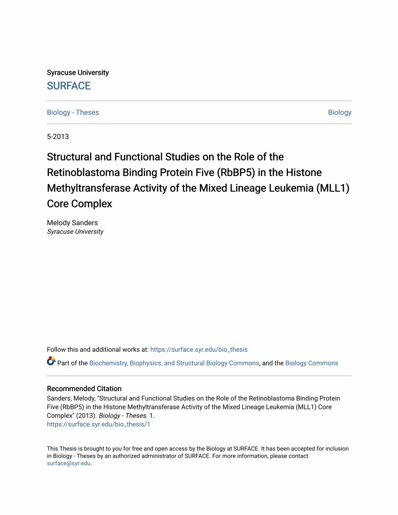
Syracuse University Syracuse University
SURFACE SURFACE
Biology - Theses Biology
5-2013
Structural and Functional Studies on the Role of the Structural and Functional Studies on the Role of the
Retinoblastoma Binding Protein Five (RbBP5) in the Histone Retinoblastoma Binding Protein Five (RbBP5) in the Histone
Methyltransferase Activity of the Mixed Lineage Leukemia (MLL1) Methyltransferase Activity of the Mixed Lineage Leukemia (MLL1)
Core Complex Core Complex
Melody Sanders Syracuse University
Follow this and additional works at: https://surface.syr.edu/bio_thesis
Part of the Biochemistry, Biophysics, and Structural Biology Commons, and the Biology Commons
Recommended Citation Recommended Citation Sanders, Melody, "Structural and Functional Studies on the Role of the Retinoblastoma Binding Protein Five (RbBP5) in the Histone Methyltransferase Activity of the Mixed Lineage Leukemia (MLL1) Core Complex" (2013). Biology - Theses. 1. https://surface.syr.edu/bio_thesis/1
This Thesis is brought to you for free and open access by the Biology at SURFACE. It has been accepted for inclusion in Biology - Theses by an authorized administrator of SURFACE. For more information, please contact [email protected].
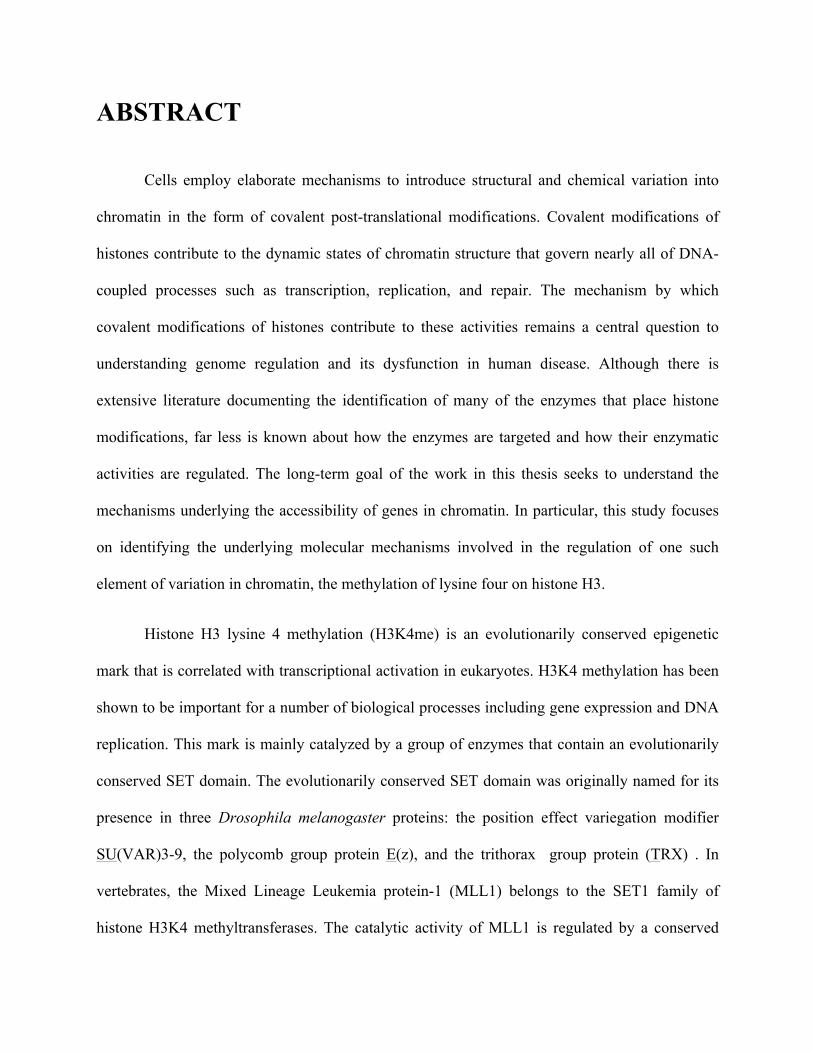
ABSTRACT
Cells employ elaborate mechanisms to introduce structural and chemical variation into
chromatin in the form of covalent post-translational modifications. Covalent modifications of
histones contribute to the dynamic states of chromatin structure that govern nearly all of DNA-
coupled processes such as transcription, replication, and repair. The mechanism by which
covalent modifications of histones contribute to these activities remains a central question to
understanding genome regulation and its dysfunction in human disease. Although there is
extensive literature documenting the identification of many of the enzymes that place histone
modifications, far less is known about how the enzymes are targeted and how their enzymatic
activities are regulated. The long-term goal of the work in this thesis seeks to understand the
mechanisms underlying the accessibility of genes in chromatin. In particular, this study focuses
on identifying the underlying molecular mechanisms involved in the regulation of one such
element of variation in chromatin, the methylation of lysine four on histone H3.
Histone H3 lysine 4 methylation (H3K4me) is an evolutionarily conserved epigenetic
mark that is correlated with transcriptional activation in eukaryotes. H3K4 methylation has been
shown to be important for a number of biological processes including gene expression and DNA
replication. This mark is mainly catalyzed by a group of enzymes that contain an evolutionarily
conserved SET domain. The evolutionarily conserved SET domain was originally named for its
presence in three Drosophila melanogaster proteins: the position effect variegation modifier
SU(VAR)3-9, the polycomb group protein E(z), and the trithorax group protein (TRX) . In
vertebrates, the Mixed Lineage Leukemia protein-1 (MLL1) belongs to the SET1 family of
histone H3K4 methyltransferases. The catalytic activity of MLL1 is regulated by a conserved
group of proteins that include the Tryptophan-Aspartate-repeat protein-5 (WDR5), the
retinoblastoma-binding protein-5 (RbBP5) and the absent small homeotic-2-like protein
(Ash2L).
The focus of this investigation is the RbBP5 component of the MLL1 core complex. The
RbBP5 subunit of the MLL1 core complex has been shown to be required for enzymatic activity
and disruption of RbBP5 is frequently observed in patients with malignant primary brain tumors.
To gain insight into the functional role of RbBP5 in the regulation of the enzymatic activity of
the MLL1 core complex, mutations targeting individual residues in a highly conserved stretch of
amino acid residues in RbBP5 were generated. Biochemical and biophysical analyses of the
variant proteins were utilized to assess the functional role of each amino acid on the intrinsic
properties of RbBP5 alone or when assembled within the context of the entire complex. These
studies identify several conserved aromatic residues and one acidic residue in RbBP5 required
for interaction with MLL1 and the overall dimethyltransferase activity of the complex. The
residues identified constitute a previously uncharacterized MLL1 interaction motif located in
RbBP5. Therefore, this study provides important insights into how the RbBP5 subunit of the
MLL1 core complex contributes to its overall enzymatic activity. Understanding the role that
RbBP5 plays in facilitating proper H3K4 methylation may provide insight into how
misregulation of RbBP5 can lead to brain tumor genesis.
Structural and Functional Studies on the Role of the Retinoblastoma Binding Protein Five (RbBP5) in the
Histone Methyltransferase Activity of the Mixed Lineage Leukemia (MLL1) Core Complex
By
Melody Sanders B.S. 2009
Syracuse University
THESIS
Submitted in partial fulfillment of the requirements for the degree Masters of Science in the Graduate School of Syracuse University
May 2013

© Copyright 2013 Melody Sanders
All Rights Reserved
v
ACKNOWLEDGEMENTS
It gives me great pleasure in expressing my gratitude to all those people who have
supported me and had their contribution in making this thesis possible. However, any attempt to
list the people and opportunities to which my life has been richly blessed would be like trying to
count the stars in the heavens. Therefore for anyone that I may have left out, of course, the
responsibility is entirely my own.
I express my profound sense of reverence and gratitude to my principal investigator and
advisor Dr. Michael Cosgrove. You have been patient and encouraging in times of new ideas and
difficulties. I have considered you a steady influence throughout my graduate career. Your
ability to select and to approach compelling research problems, your high scientific standards,
and your passion for science have been indispensable to my growth not only as a scientist but
also as a young woman.
It is also my fortune to gratefully acknowledge Dr. Gina Lee Glauser. This feat would not
have been possible without you. Thank you doesn’t seem sufficient but it is said with
appreciation and respect for your support, encouragement, care, understanding, and precious
guidance.
Dr. Laila Kobrossy and Dr. Anamika Patel, words fail me when expressing my sincere
love and appreciation. My deepest gratitude goes toward the two of you. I will always remember
the generosity and encouragement the two of you expressed to me in the midst of tragedy. A long
journey is easier travelled together. To Anamika, you are and will remain my best role model for
a scientist, mentor, and friend.
Last, but certainly not least I would like to thank Dr. Dean Stith with help towards my
graduate and postgraduate affairs. I would also like to thank as well the members of my thesis

vi
committee, Dr. Ramesh Raina, Dr. Stewart Loh, and Dr. Phil Borer for insightful discussions and
suggestions. I would like to make special mention to Dr. Raina, for his constant guidance,
support, motivation, and an ever-ready helping attitude during the completion of my degree.
vii
DEDICATION
I would like to dedicate this thesis to my beautiful family, with special regards to my boys. I know times are hard for us right now and our futures are uncertain, but God willing I promise to make a way. Always remember to work hard, pride yourselves on self-discipline, and unrelenting devotion and determination on the things you want to see happen. Most importantly, love one another and NEVER stop dreaming.
I would also like to dedicate this thesis to Michelle and Peter. Thank you for opening up your home to me when I had nowhere else to go. This thesis could never have reached its completion without the safe and peaceful place in your home and in your hearts that you gave me.
With Love,
Mel
viii
TABLE OF CONTENTS
ABSTRACT……………………………………………………………..i
TITLE PAGE……………………...……...…………………………….iii
COPYRIGHT NOTICE………………………………………………...iv
ACKNOWLEDGEMENTS………………………………………..........v
TABLE OF CONTENTS……………………………………………...vii
LIST OF FIGURES……………………………………………………..x
LIST OF TABLES……………………………………………............xiii
CHAPTER 1: INTRODUCTION…………………………….……….1
BACKGROUND AND SIGNIFICANCE ……….……………….2
RATIONAL OF STUDY………………………………………...25
CHAPTER 2: A CONSERVED PATCH OF AMINO ACIDS IN RbBP5 REQUIRED FOR INTERACTION WITH THE SET DOMAIN OF MLL IN THE MLL1 CORE COMPLEX……………………………………………………............29
TITLE PAGE…..……………………….…….………………….29
INTRODUCTION……...………………………………………..30
EXPERIMENTAL PROCEDURES….………………………….35
RESULTS………...…...…………………………………………39
ix
DISCUSSION……….....………………………………………...82
CHAPTER 3: BIOCHEMICAL AND BIOPHYSICAL CHARACTERIZATION OF TWO STABLE SUBDOMAINS IDENTIFIED IN THE RETINOBLASTOMA BINDING PROTEIN FIVE BY LIMITED PROTEOLYSIS.…………………..………………………………….87
TITLE PAGE…..……………………….…….………………….87
INTRODUCTION……...………………………………………..88
EXPERIMENTAL PROCEDURES….………………………….90
RESULTS………………………………………………………..93
DISCUSSION…………………………………………………..102
OVERALL CONCLUSIONS AND FUTURE
PROSPECTIVES……………………………………………............104
APPENDIX..........................................................................................106
BIOGRAPHICAL DATA…………………………………………..138
x
LIST OF FIGURES
FIGURE 1.1. Schematic Diagram of the effects of histone acetylation on chromatin structure……………………………………………………………………………………………4 FIGURE 1.2. Generic model of chromatin remodeling by ATP dependent protein machineries………………………………………………………………………………………..6
FIGURE 1.3. Schematic illustration of the members of the MLL1 core complex……………......................................................................................................................13
FIGURE 1.4. Structural analysis of a RbBP5 peptide bound to WDR5…………………………21
FIGURE 2.1. Schematic representation of the human retinoblastoma binding protein five (RbBP5) showing the domain architecture of recombinantly expressed full-length RbBP5……………………………………………………………………………………………32
FIGURE 2.2. Schematic representation of the human retinoblastoma binding protein five (RbBP5) showing the domain architecture of recombinantly expressed full-length and truncated RbBP5 constructs used in this investigation……………………………………………………..41
FIGURE 2.3 ClustalW2 multiple sequence alignment of the predicted globular domain of RbBP5 showing only a subgroup of residues, and high throughput methyltransferase assays showing activity of the RbBP5 protein with mutations in conserved residues identified in its predicted globular region…………………………………………………………………………………...46
FIGURE 2.4. Biophysical characterization of wild-type and mutant RbBP5 proteins by analytical ultracentrifugation………………………………………………………………………………..50
FIGURE 2.5. Biophysical characterization of wild-type and mutant RbBP5 proteins by circular dichroism………………………………………………………………………………………...52
FIGURE 2.6 MALDI-TOF mass spectrometry spectra of MWRAD and MWAD complexes………………………………………………………………………………………..54
FIGURE 2.7. Determination of rate constants from single turnover progress curves measured by MALDI-TOF mass spectrometry………………………………………………………………...58
FIGURE 2.8. Biophysical characterization of wild-type WRAD and WR(W329A)AD proteins by analytical ultracentrifugation…………………………………………………………………….61
FIGURE 2.9. Biophysical characterization of wild-type WRAD and WR(F332A)AD proteins by analytical ultracentrifugation…………………………………………………………………….62
FIGURE 2.10. Biophysical characterization of wild-type WRAD and WR(E347A)AD proteins by analytical ultracentrifugation…………………………………………………………………….63
FIGURE 2.11. Biophysical characterization of wild-type MWRAD and MWR(W329A)AD proteins by analytical ultracentrifugation…………………………………………………………………65
xi
FIGURE 2.12. Biophysical characterization of wild-type MWRAD and MWR(F332A)AD proteins by analytical ultracentrifugation…………………………………………………………………66
FIGURE 2.13. Biophysical characterization of wild-type MWRAD and MWR(F336A)AD proteins by analytical ultracentrifugation…………………………………………………………………67
FIGURE 2.14. Biophysical characterization of wild-type MWRAD and MWR(E347A)AD proteins by analytical ultracentrifugation…………………………………………………………………68
FIGURE 2.15. Biophysical characterization of wild-type MWR(W329A)AD protein by analytical ultracentrifugation………………………………………………………………………………..69
FIGURE 2.16. Biophysical characterization of wild-type and mutant RbBP5 proteins by circular dichroism…………………………………………………………………………………………71
FIGURE 2.17. Determination of rate constants from single turnover progress curves measured by MALDI-TOF mass spectrometry………………………………………………………………...73
FIGURE 2.18. Biophysical characterization of wild-type MWRAD, MWR(W329F)AD, and MWR(W329Y)AD by analytical ultracentrifugation……………………………………………….75
FIGURE 2.19. Biophysical characterization of wild-type MWRAD, and MWR(W329R)AD, by analytical ultracentrifugation…………………………………………………………………….76
FIGURE 2.20. Biophysical characterization of RbBP5 wild-type and RbBP5 (V375A) and RbBP5 (V377A) proteins……………………………………………………………………………………79
FIGURE 2.21. Determination of rate constants from single turnover progress curves measured by MALDI-TOF mass spectrometry………………………………………………………………...80
FIGURE 2.22. Biophysical characterization of WDR5-RbBP5 (V375A) and WDR5-RbBP5(V377A) interactions……………………………………………………………………………………….81
FIGURE 2.23. Model for multiple methylation by the MLL1 core complex……………………86
FIGURE 3.1. Schematic representation of the human retinoblastoma binding protein five (RbBP5) showing the domain architecture of recombinantly expressed full-length RbBP5……………………………………………………………………………………………89
FIGURE 3.2. Limited proteolysis of full-length RbBP5………………………………...............95
FIGURE 3.3. Biophysical characterization of wild-type RbBP5 (#1-495) proteins by analytical ultracentrifugation………………………………………………………………………………..96
FIGURE 3.4. Biophysical characterization of RbBP5 wild-type and RbBP5 (#1-495) proteins by circular dichroism………………………………………………………………………………..97
FIGURE 3.5. Biophysical characterization of RbBP5 (#1-337) by analytical ultracentrifugation………………………………………………………………………………..98
xii
FIGURE 3.6. Biophysical characterization of RbBP5 (#1-337) by circular dichroism………………………………………………………………………………………...99
FIGURE 3.7. Determination of rate constants from single turnover progress curves measured by MALDI-TOF mass spectrometry……………………………………………………………….100
xiii
LIST OF TABLES
Table 1.1 Histone posttranslational modification types and the residues modified…………………………………………………………………………………………...7
Table 2.1 RbBP5 mutations resulting in decreased enzymatic activities………………………..48
Table 2.2 Summary of sedimentation coefficients derived from sedimentation velocity analyses of the Retinoblastoma Binding Protein Five (RbBP5) expressed with mutations in various positions or expressed as truncated constructs…………………………………………………...51
Table 2.3 Summary of Rate Constants derived from globally fitting experimental data……………………………………………………………………………………………….59
Table 2.4 Summary of Rate Constants derived from globally fitting experimental data……………………………………………………………………………………………….74
Table 3.1 Summary of Rate Constants derived from globally fitting experimental data……………………………………………………………………………………...............101
1
Chapter One:
Introduction
2
BACKGROUND AND SIGNIFICANCE:
1.1 The Chromatin Landscape
Eukaryotes have evolved elaborate mechanisms to regulate the expression of genes by
packaging the genome into chromatin. The fundamental repeating unit of chromatin, the
nucleosome core particle, consists of approximately 147 bp of genomic DNA wrapped around a
histone octamer, containing one tetramer of histones H3 and H4 and two H2A-H2B dimers
(Kornberg, 1974). Nucleosomes are packaged into progressively higher order structures to
ultimately form chromosomes. In its extended form, chromatin can be visualized as an array of
nucleosomes or “beads on a string”, but in the nucleus of the cell, the chromatin fiber undergoes
extensive degrees of folding resulting in increasing degrees of chromatin compaction (Schwarz
and Hansen, 1994). Since the recognition of chromatin structure as a repeating unit of DNA
wrapped around histones, it has been suggested that its function extends beyond merely just a
storage vehicle for DNA. The discovery that nucleosomes impede transcription in vitro (Wolffe
and Hayes, 1999), in addition to experiments in vivo showing that deletions of histones or their
basic tails elicited specific effects on gene expression, provided a glimpse of chromatin’s
importance in the regulation of the expression of genetic programs (Allison, 1996; Hayes and
Wolffe, 1992; Lee et al., 1993). It has now become widely recognized that the structure of
chromatin largely affects DNA-templated processes such as DNA transcription, replication,
recombination, and repair. Within this realm, access to DNA must be tightly regulated to allow
sequence-specific DNA binding factors, chromatin regulators, and the general transcription
machinery to bind.
3
As stated above, the packaging of the DNA into nucleosomes affects all stages of DNA-
templated processes. Therefore, eukaryotes employ proteins, often in complexes, that modify
chromatin through the highly coordinated introduction or removal of post-translational
modifications on histone proteins to regulate access to the underlying genome- resulting in the
correct establishment and propagation of gene expression patterns. Each of the core histones that
make up the nucleosome are predominantly globular with the exception of their N-terminal tails,
which are unstructured and protrude from the lateral surface of the histone octamer. One striking
feature of the flexible N-terminal tails of histones is that they serve as platforms that can be
targeted by enzymes for the introduction or removal of distinct chemical moieties known as
covalent posttranslational modifications. Post-translational modifications themselves can
significantly affect the degree of chromatin compaction by creating generally more condensed
“heterochromatic” or more open “euchromatic’ regions that impact the procession of
chromosome processes such as transcription.
Currently, there are two characterized mechanisms for how modifications affect
chromatin structure. One is that chromatin packaging is altered directly (either by change in
electrostatic charge or through internucleosomal contacts), to open or close the DNA polymer
(Figure 1.1), thus controlling access of DNA-binding proteins such as transcription factors
(Bannister and Kouzarides, 2011; Berger, 2007; Gardner et al., 2011; Jenuwein and Allis, 2001;
Kouzarides, 2002; Wolffe and Hayes, 1999). The second mechanism posits that the attached
chemical moieties alter the nucleosome surface to promote or occlude the association of
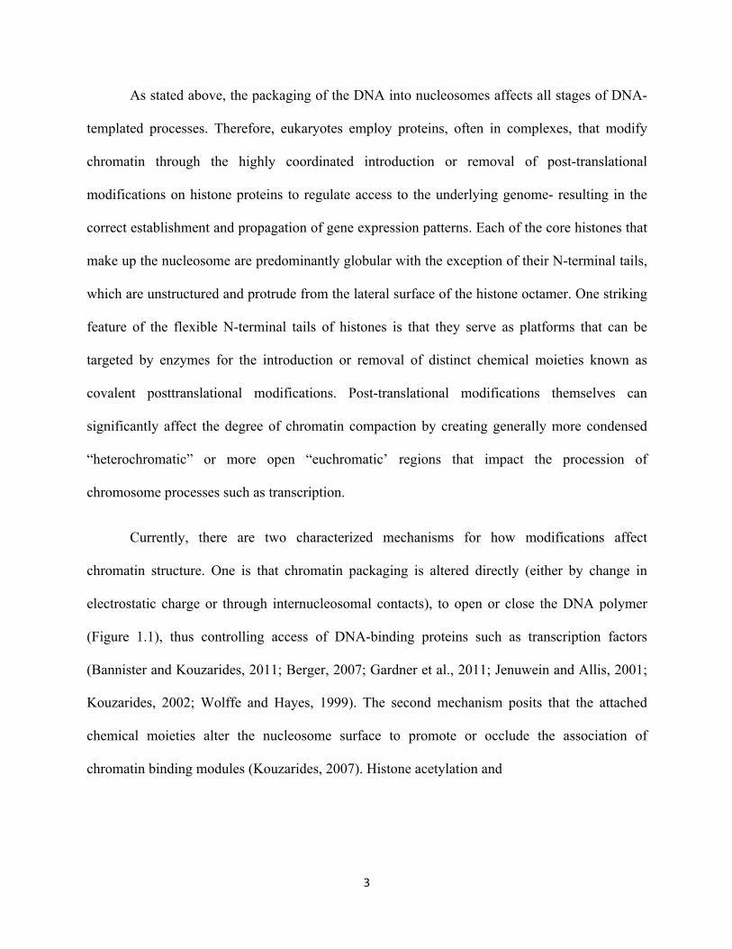
chromatin binding modules (Kouzarides, 2007). Histone acetylation and

4
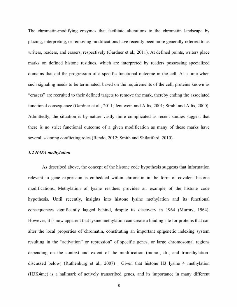
Figure 1.1. Schematic diagram of the effects of histone acetylation on chromatin structure.
The thin protruding blue lines represent the amino-terminal tails of histones, and the winding black lines represent DNA. The lower panel depicts a more “open” structure for acetylated chromatin. Conversely, histone deacetylation results in condensed, transcriptionally inactive chromatin (upper panel).
5
phosphorylation are two examples of post-translational modifications that likely weaken histone-
DNA contacts, leading to chromatin de-repression (Figure 1.1). Although difficult to
demonstrate in vivo, considering only the electrostatic requirements for folding of the chromatin
fiber, histone acetylation through the neutralization of positive charge, and histone
phosphorylation through addition of a negative charge would likely cause decondenstation of the
chromatin polymer (Brownell and Allis, 1996; Schwarz and Hansen, 1994). Histone
modifications can also indirectly affect chromatin structures by serving as marks for the
recruitment of enzymatic machineries that remodel the nucleosome and/or chromatin structure
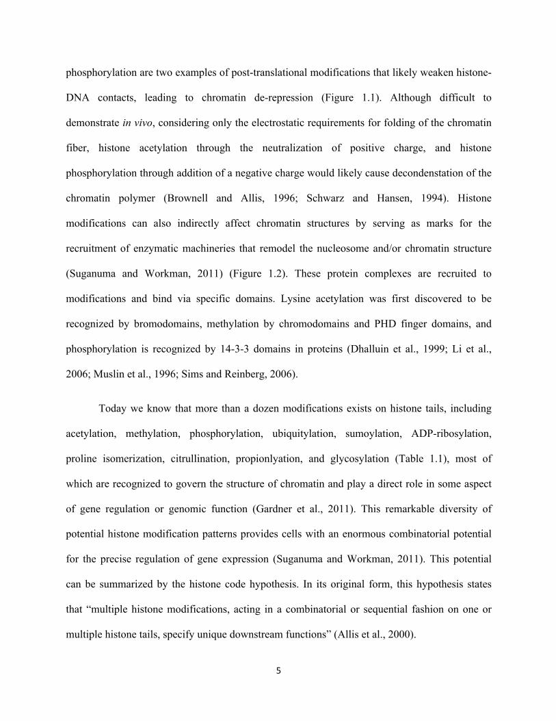
(Suganuma and Workman, 2011) (Figure 1.2). These protein complexes are recruited to
modifications and bind via specific domains. Lysine acetylation was first discovered to be
recognized by bromodomains, methylation by chromodomains and PHD finger domains, and
phosphorylation is recognized by 14-3-3 domains in proteins (Dhalluin et al., 1999; Li et al.,
2006; Muslin et al., 1996; Sims and Reinberg, 2006).
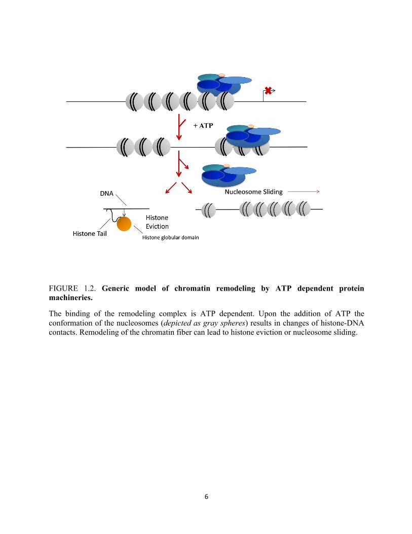
Today we know that more than a dozen modifications exists on histone tails, including
acetylation, methylation, phosphorylation, ubiquitylation, sumoylation, ADP-ribosylation,
proline isomerization, citrullination, propionlyation, and glycosylation (Table 1.1), most of
which are recognized to govern the structure of chromatin and play a direct role in some aspect
of gene regulation or genomic function (Gardner et al., 2011). This remarkable diversity of
potential histone modification patterns provides cells with an enormous combinatorial potential
for the precise regulation of gene expression (Suganuma and Workman, 2011). This potential
can be summarized by the histone code hypothesis. In its original form, this hypothesis states
that “multiple histone modifications, acting in a combinatorial or sequential fashion on one or
multiple histone tails, specify unique downstream functions” (Allis et al., 2000).
6
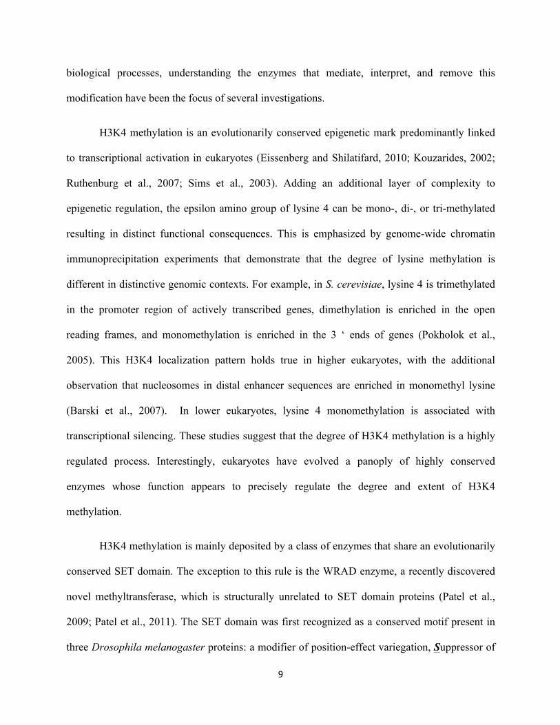
FIGURE 1.2. Generic model of chromatin remodeling by ATP dependent protein machineries.
The binding of the remodeling complex is ATP dependent. Upon the addition of ATP the conformation of the nucleosomes (depicted as gray spheres) results in changes of histone-DNA contacts. Remodeling of the chromatin fiber can lead to histone eviction or nucleosome sliding.
7
Table 1.1 Histone posttranslational modification types and the residues modified.
Modification types Residue(s) modified
Acetylation Lysine
Phosphorylation Serine/threonine
Methylation Lysine/arginine
Ubiquitylation Lysine
Sumoylation Lysine
ADP-ribosylation Lysine
Citrullination Arginine
Butyrylation Lysine
Propionlyation Lysine
Glycosylation Serine/threonine
8
The chromatin-modifying enzymes that facilitate alterations to the chromatin landscape by
placing, interpreting, or removing modifications have recently been more generally referred to as
writers, readers, and erasers, respectively (Gardner et al., 2011). At defined points, writers place
marks on defined histone residues, which are interpreted by readers possessing specialized
domains that aid the progression of a specific functional outcome in the cell. At a time when
such signaling needs to be terminated, based on the requirements of the cell, proteins known as
“erasers” are recruited to their defined targets to remove the mark, thereby ending the associated
functional consequence (Gardner et al., 2011; Jenuwein and Allis, 2001; Strahl and Allis, 2000).
Admittedly, the situation is by nature vastly more complicated as recent studies suggest that
there is no strict functional outcome of a given modification as many of these marks have
several, seeming conflicting roles (Rando, 2012; Smith and Shilatifard, 2010).
1.2 H3K4 methylation
As described above, the concept of the histone code hypothesis suggests that information
relevant to gene expression is embedded within chromatin in the form of covalent histone
modifications. Methylation of lysine residues provides an example of the histone code
hypothesis. Until recently, insights into histone lysine methylation and its functional
consequences significantly lagged behind, despite its discovery in 1964 (Murray, 1964).
However, it is now apparent that lysine methylation can create a binding site for proteins that can
alter the local properties of chromatin, constituting an important epigenetic indexing system
resulting in the “activation” or repression” of specific genes, or large chromosomal regions
depending on the context and extent of the modification (mono-, di-, and trimethylation-
discussed below) (Ruthenburg et al., 2007) . Given that histone H3 lysine 4 methylation
(H3K4me) is a hallmark of actively transcribed genes, and its importance in many different
9
biological processes, understanding the enzymes that mediate, interpret, and remove this
modification have been the focus of several investigations.
H3K4 methylation is an evolutionarily conserved epigenetic mark predominantly linked
to transcriptional activation in eukaryotes (Eissenberg and Shilatifard, 2010; Kouzarides, 2002;
Ruthenburg et al., 2007; Sims et al., 2003). Adding an additional layer of complexity to
epigenetic regulation, the epsilon amino group of lysine 4 can be mono-, di-, or tri-methylated
resulting in distinct functional consequences. This is emphasized by genome-wide chromatin
immunoprecipitation experiments that demonstrate that the degree of lysine methylation is
different in distinctive genomic contexts. For example, in S. cerevisiae, lysine 4 is trimethylated
in the promoter region of actively transcribed genes, dimethylation is enriched in the open
reading frames, and monomethylation is enriched in the 3 ‘ ends of genes (Pokholok et al.,
2005). This H3K4 localization pattern holds true in higher eukaryotes, with the additional
observation that nucleosomes in distal enhancer sequences are enriched in monomethyl lysine
(Barski et al., 2007). In lower eukaryotes, lysine 4 monomethylation is associated with
transcriptional silencing. These studies suggest that the degree of H3K4 methylation is a highly
regulated process. Interestingly, eukaryotes have evolved a panoply of highly conserved
enzymes whose function appears to precisely regulate the degree and extent of H3K4
methylation.
H3K4 methylation is mainly deposited by a class of enzymes that share an evolutionarily
conserved SET domain. The exception to this rule is the WRAD enzyme, a recently discovered
novel methyltransferase, which is structurally unrelated to SET domain proteins (Patel et al.,
2009; Patel et al., 2011). The SET domain was first recognized as a conserved motif present in
three Drosophila melanogaster proteins: a modifier of position-effect variegation, Suppressor of
10
variegation 3-9 (Suv(var)3-9), the Polycomb-group chromatin regulator, Enhancer of zeste
(E(z)), and the trithorax-group chromatin regulator, Trithorax (Trx) (Dillon et al., 2005). The
function of the SET-domain proteins is to transfer a methyl group from S-adenosyl-L-methionine
to the amino group of a lysine residue on the histone and in some cases non-histone proteins,
leaving a methylated lysine residue and the cofactor product. Seven main families of SET-
domain proteins are known, all of which differ with respect to their substrate specificity,
processivity, and the presence of additionally associated domains (Dillon et al., 2005; Jenuwein
et al., 1998; Rea et al., 2000).
While there are several SET domain enzymes, members of the SET1 family share the
virtues that they all methylate H3K4 and interact with an evolutionarily conserved core complex
of proteins that function together to control the degree of H3K4 methylation (Cosgrove and
Patel, 2010; Crawford and Hess, 2006; Dillon et al., 2005; Dou et al., 2006; Jenuwein et al.,
1998; Rea et al., 2000; Southall et al., 2009). The first H3K4 methyltransferase to be identified
was S. cerevisiae SET1p (Miller et al., 2001; Roguev et al., 2001). In mammals, the number of
H3K4 methyl writers is much greater, as there are over six SET1-related proteins including
Set1a, Set1b, and four members of the Mixed Lineage Leukemia (MLL) family, all of which are
capable of catalyzing the methylation of H3K4. MLL1, the human homologue of the Drosophila
protein Trithorax, has been the most intensively studied because of its involvement in genetic
rearrangements that occur in infant acute leukemias and other therapy related malignancies.
11
1.3 MLL1
The MLL gene located in the human genome at chromosome 11, band q23, was initially
identified through its recurring involvement in reciprocal translocations found in numerous cases
of acute myeloid leukemia (AML) and acute lymphoblastic leukemia (ALL) (Ernst et al., 2002;
Hess, 2004; Slany, 2009). MLL1 was later shown to encode a major H3K4- specific histone
methyltransferase enzyme that functions to maintain gene expression during development and
hematopoiesis (Ernst et al., 2002; Milne et al., 2005; Ono et al., 2005). Despite the important
biological role of MLL1 and its involvement in human leukemias, the underlying molecular
details for the regulation of the histone methyltransferase activity of MLL1 remain poorly
understood.
The MLL1 protein is a member of the SET1 family of H3K4 methyltransferases that
functions to maintain gene expression during development and hematopoiesis (Hess et al., 1997;
Milne et al., 2002;). The most well studied target genes of MLL1 encompass the homeobox
transcription factors, better known as HOX genes, which are important for specifying segment
identity and cell fate during metazoan development (Milne et al., 2002; Milne et al., 2005).
These genes also play a role in leukemogenesis, which is why they are important in the context
of MLL’s role in cancer (Ernst et al., 2002; Guenther et al., 2005; Hess, 2004; Milne et al., 2005;
Ono et al., 2005; Slany, 2009; Yokoyama et al., 2004; Yu et al., 1995). A unique pattern of
H3K4 methylation has been observed in HOX gene clusters, where large continuous regions of
H3K4 methylation are observed spanning multiple genes and intergenic regions (Guenther et al.,
2005). Genetic studies in mice have demonstrated that MLL1-null mutations result in embryonic
lethality and are associated with multiple development defects and hematopoietic abnormalities
(Yu et al., 1995).

12
1.4 Mechanistic Determinants of Multiple Lysine Methylation Catalyzed by the MLL1 Core
Complex
Like most histone modifying enzymes, MLL1 exists in a multi-protein complex and
regulates the degree of H3K4 methylation (Cosgrove and Patel, 2010). Although the subunit
composition of SET1 family members varies to some degree, each SET1 family member is
capable of interacting with a conserved core group of proteins that include the WD-40 repeat
protein-5 (WDR5), the Retinoblastoma binding protein-5 (RbBP5), the Absent small homeotic 2-
like protein (Ash2L), and Dumpy 30 (DPY30), which is required for distinct states of H3K4
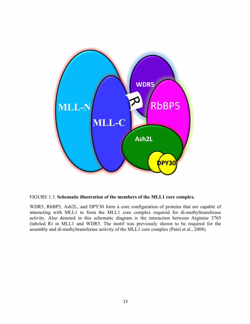
methylation (Figure 1.3) (Cao et al., 2010; Cosgrove and Patel, 2010; Crawford and Hess, 2006;
Dou et al., 2006; Patel et al., 2009; Ruthenburg et al., 2007; Southall et al., 2009). Because of the
central role of H3K4 methylation in transcriptional regulation, understanding how different
H3K4 methylation states are established and maintained is crucial for understanding how MLL1
is misregulated.
Recent research suggests several models for the establishment and maintenance of the
degree of methylation by the SET1 family of histone lysine methyltransferases. One model
suggests that multiple lysine methylation is achieved by the successive addition of a methyl
group catalyzed by distinct histone lysine methyltransferases (Patel et al., 2011). In this model
the sequential addition of methyl groups is catalyzed by distinct enzymes that differ in their
abilities to use unmodified, mono-, and di-methylated peptide histones as substrates- a
phenomenon known as product specificity.
13
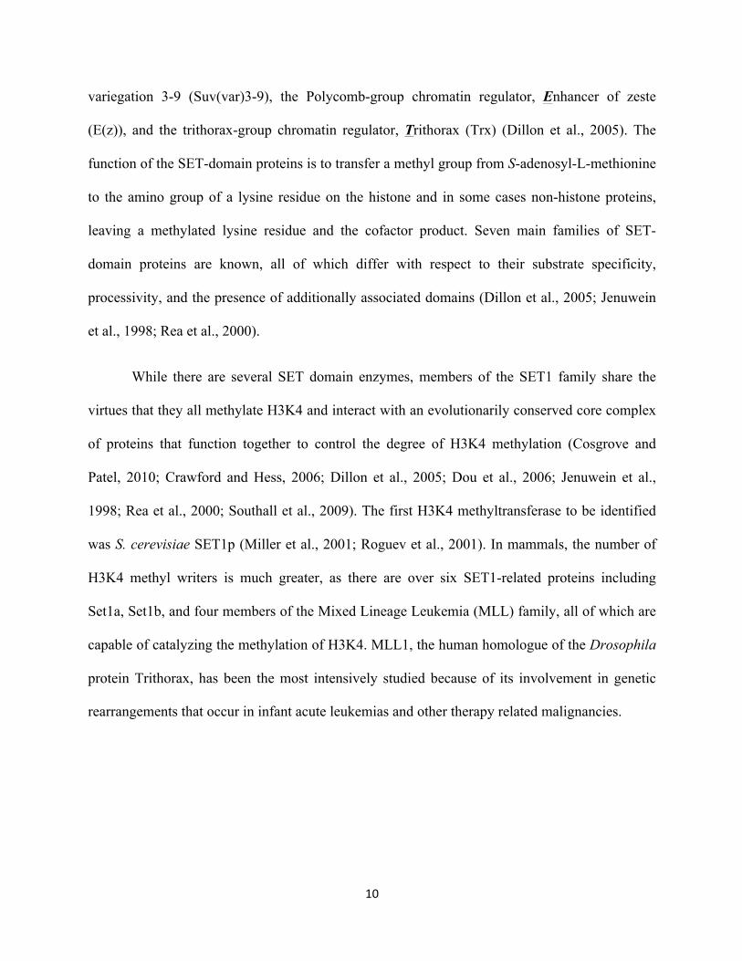
FIGURE 1.3. Schematic illustration of the members of the MLL1 core complex.
WDR5, RbBP5, Ash2L, and DPY30 form a core configuration of proteins that are capable of interacting with MLL1 to form the MLL1 core complex required for di-methyltransferase activity. Also denoted in this schematic diagram is the interaction between Arginine 3765 (labeled R) in MLL1 and WDR5. The motif was previously shown to be required for the assembly and di-methyltransferase activity of the MLL1 core complex (Patel et al., 2008).
14
Consistent with this hypothesis, several SET domain enzymes have been discovered that display
different product specificities toward histone lysine residues. Biochemical and structure-function
studies suggest that the product specificity of SET domain enzymes is governed by the presence
of a tyrosine or phenylalanine at a conserved position in the SET domain active site, called the
“Phe/Tyr switch” position (Collins et al., 2005; Couture et al., 2008; Dillon et al., 2005; Patel et
al., 2011; Southall et al., 2009). In general, SET domain enzymes are predicted to be lysine
monomethyltransferases based on the presence of a tyrosine at the switch position, whereas in
most di- and tri-methyltransferases, a phenylalanine or another hydrophobic amino acid is
located in this site. SET1- related enzymes possessing tyrosine in the switch position generally
have smaller active sites allowing them to only catalyze mono-methylation. In contrast, SET1-
related enzymes possessing a phenylalanine in the switch position have larger active sites that
can accommodate the rotation of pre-methylated substrates, therefore allowing a processive
mechanism (Collins et al., 2005; Couture et al., 2008; Dillon et al., 2005; Patel et al., 2011;
Southall et al., 2009). Although mutagenesis experiments have validated this Phe/Tyr switch
hypothesis for several SET domain containing enzymes, enzymes from the SET1 family appear
to contradict this rule. Interestingly, all of the SET1 family enzymes posses a tyrosine in the
critical switch position, which is predicted to limit their enzymatic activity to that of a
monomethyltransferases (Collins et al., 2005; Cosgrove and Patel, 2010; Couture et al., 2008;
Dillon et al., 2005; Southall et al., 2009) However, mono-, di-, and tri-methylation have been
attributed to SET1 family complexes in vivo and in vitro (Cao et al., 2010; Cosgrove and Patel,
2010; Crawford and Hess, 2006; Dou et al., 2006; Eissenberg and Shilatifard, 2010; Miller et al.,
2001; Ruthenburg et al., 2007). These early observations laid the groundwork for a large body of
subsequent studies to resolve this paradox. It is postulated that the product specificity of the
15
SET1 family enzymes is regulated by proteins that bind to and alter the conformation of the
catalytic SET domain active site, in turn altering their ability to catalyze the addition of each
methyl group to the lysine side chain.
In an effort to identify the minimal MLL1 core complex components necessary for mono-
, di-, and tri-methylation, Roeder and colleagues (Dou et al., 2006) developed a baculovirus
system to co-express and immunopurify recombinant components from insect cells. In this
system, results suggest that the minimal complex required for mono-, di-, and tri-methylation
includes a core configuration of proteins comprised of the 180kDa MLL-C terminal fragment,
WDR5, RbBP5, and Ash2L. Notably, they observed that optimal methyltransferase activity was
contingent upon MLL-C terminal associations with the other structural components and that the
MLL-C terminus alone could not efficiently catalyze H3K4 methylation (Dou et al., 2006).
MLL1 subcomplexes that lacked any of the other three components had compromised
methyltransferase activity. Omission of either RbBP5 or Ash2L resulted in substantial loss of
H3K4 activity, whereas the absence of WDR5 resulted in complete loss of H3K4 methylation.
With respect to the degree of H3K4 methylation in the absence of RbBP5 or Ash2L, H3K4
trimethylation was completely abolished, H3K4 dimethylation was significantly decreased, while
monomethylation remained unchanged. Surprisingly, they were also able to show through these
studies that the WDR5-RbBP5-Ash2L sub-complex associates with the MLL1 SET domain, but
can exist independently of the catalytic subunit, providing a structural platform that can associate
with the SET domains of different MLL-family members (Dou et al, 2006). These results are
consistent with the idea that the WDR5-RbBP5-Ash2L sub-complex regulates the product
specificity of the MLL1 C-terminal SET domain. The versatility of this reconstitution system
allowed for the structure-function analysis of the MLL1 core complex, and provided insights into
16
the regulation of its enzymatic activity. Since the same group of proteins are conserved from
yeast to humans, the conclusions from this study can be generalized for other members of the
SET1 family of methyltransferases. These studies along with others have lead to a model by
which the degree of H3K4 methylation is regulated by these components that interact with MLL
to induce a conformation change in the active site therefore regulating the product specificity of
the MLL1 SET domain (Couture et al., 2008; Crawford and Hess, 2006; Dou et al., 2006;
Southall et al., 2009). In support of the conformation change hypothesis, a separate independent
group was able to demonstrate that inclusion of equimolar amounts of either Ash2L or RbBP5
significantly promotes methyltransferase activity of a minimal MLL1 SET domain construct in
vitro (Cao et al., 2010).
In spite of these studies, the molecular details that describe how MLL1 catalyzes multiple
lysine methylation remained elusive until recently. This in part stemmed from the fact that the
intrinsic product specificity of the isolated SET domain remained unknown because of no
observable catalytic activity without the presence of interacting proteins (Cao et al., 2010; Dou et
al., 2006; Southall et al., 2009). To resolve these issues, an in vitro model system was developed
by Cosgrove and colleagues that allowed for the identification of the protein structural features
that are responsible for regulation of H3K4 methylation by the human MLL1 core complex.
Using this system, Cosgrove and colleagues were able to establish that the intrinsic product
specificity of an isolated MLL1 SET domain is indeed that of an H3K4 mono-methyltransferase
consistent with the prediction of the “Phe/Tyr switch” hypothesis (Cheng and Zhang, 2007;
Collins et al., 2005; Cosgrove and Patel, 2010; Couture et al., 2008; Dillon et al., 2005).
Mutagenesis of conserved tyrosine 3942 to a phenylalanine in the switch position of the MLL
SET domain alters the product specificity of MLL1 to that of a tri-methyltransferase (Patel et al.,
17
2009). In addition, in the absence of interacting proteins, the isolated MLL1 SET domain is a
slow mono-methyltransferase (Patel et al., 2009). However, when the MLL1 SET domain is
assembled with a complex containing WDR5, RbBP5, Ash2L, and DPY30, robust
dimethyltransferase activity is readily observed. At first glance, these results appear to support
the hypothesis that the MLL1 SET domain interacting proteins regulate the product specificity of
the MLL1 SET domain, however further experimental analyses suggests a different mechanism.
Recall that one model for regulation of multiple lysine methylation is that it is achieved
by allosteric control of a single SET domain. Therefore one explanation for the results obtained
above is that a protein-interaction induced conformational change alters the position of Tyr3942
in the MLL1 SET domain active site, like what occurs when this tyrosine is replaced with
phenylalanine. However, the kinetic behavior observed with the assembled MLL1 core complex
is substantially different from the behavior that would be anticipated from the simple movement
of Tyr3942 in the MLL1 SET domain active site. First, while the Y3942F MLL1 SET domain
readily trimethylates H3K4, the assembled MLL1 core complex only di-methylates H3K4 in
assays (Patel et al., 2009). Secondly, despite the ability of the Y3492A enzyme to readily
catalyze mono-, di-, and tri- methylation of H3 peptides in vitro, the rate constants of
methylation by the wild-type and the Y3942 MLL1 SET domain are similar. In contrast, the rate
constant for the reaction catalyzed by the MLL1 core complex increases 600-fold when
compared to the MLL1 SET domain fragment alone. Taken together, these results suggest that
the rate limiting step for di-methylation by the MLL1 core complex is distinct from that of the
monomethylation activity of the isolated MLL1 SET domain. Even more critically, it is
important to note that single turnover kinetic experiments reveal that the mechanism of
dimethylation by the MLL1 core complex involves the transient accumulation of a
18
monomethylated intermediate, while no such intermediate is observed with the Y3942F MLL1
SET domain (Patel et al., 2009). This transient accumulation of monomethylated species in the
reaction catalyzed by the MLL1 core complex implies that the monomethylated peptide could
potentially be released from the MLL1 SET domain active site before dimethylation occurs.
Encouraged to understand this phenomena further, Cosgrove and colleagues proposed an
alternative hypothesis that suggests that in order for multiple lysine methylation to occur, each
methyl group is added by a distinct methyltransferase. Unexpectedly, it was shown that the non-
SET domain components of the MLL1 core complex possess a previously unrecognized histone
methyltransferase activity that catalyzes H3K4 dimethylation within the MLL1 core complex
(Patel et al., 2009; Patel et al., 2011). In addition, it was shown that the non-SET domain
components of the MLL1 core complex (WDR5, RbBP5, Ash2L, and DPY30 or WRAD)
possess monomethyltransferase activity in the absence of the MLL1 SET domain (Patel et al.,
2011). Because the WRAD components lack homology to a conserved SET or DOT1-like
methyltransferase fold, this enzyme constitutes a novel previously uncharacterized
methyltransferase (Patel et al., 2011). Like the activity of other known lysine methyltransferases,
the WRAD enzyme is zinc dependent, inhibited by the co-factor product S-adensoyl
homocysteine, and displays Michaelis-Menten kinetics (Patel et al., 2011). This work has
opened up a whole new area of investigation involving a core configuration of proteins that have
already been shown to be intimately involved in several other cellular processes including
differentiation, transcription, multicellular development, and cancer (Ang et al., 2011; Bralten et
al., 2010; Gopal et al., 2012; Hsu et al., 1995; Hsu and Meyer, 1994; Jiang et al., 2011;
Riemenschneider et al., 1999; Riemenschneider et al., 2003; South et al., 2010; Stoller et al.,
2010; Vardanyan et al., 2008; Wan et al., 2012; Wysocka et al., 2005). Further studies are thus
19
necessary to determine the catalytic motif in this enzyme and what role, this enzyme may play in
oncogenesis. In light of the above findings the subject of the work in this thesis focuses on the
RbBP5 component of the MLL1 core complex.
1.5 The Retinoblastoma Binding Protein Five is a Critical Component of the MLL1 Core
Complex
The Retinoblastoma Binding Protein Five (RbBP5) was originally identified through
direct screening of cDNA expression libraries to isolate distinct clones of cellular proteins that
bind to the retinoblastoma protein, perhaps one of the best studied tumor suppressors (Classon
and Harlow, 2002; Classon and Settleman, 2000; Ferreira et al., 1998; Lai et al., 1999;
Magnaghi-Jaulin et al., 1998; Paggi et al., 1996; Suryadinata et al., 2011; Sutcliffe et al., 2000;
Williams et al., 2006). This report demonstrated that the human RbBP5 gene encodes a protein
of approximately 66kDa that localizes in the nucleus (Saijo et al., 1995). Although currently no
three dimensional structure of the full length human retinoblastoma binding protein exists,
sequence analysis, domain predictions, and homology modeling of RbBP5 predicts that this
protein is composed of a N-terminal seven blade β-propeller domain, a short hinge region
consisting of a stretch of conserved residues, and a C-terminal domain of unknown function
(please see chapter 2: Figure 2.1).
Since its discovery, human RbBP5 is now established as a well known conserved
component of several multisubunit histone H3 lysine four methyltransferases, one representative
example being the Mixed Lineage Leukemia-1 (MLL1) core complex. More recently,
misregulation of this protein has been implicated in malignant glioblastomas (Riemenschneider
et al., 2003). Within the MLL1 core complex, RbBP5 exists in a multisubunit core configuration
20
of proteins including MLL1, WDR5, Ash2L, and DPY30. The essential role of the RbBP5
subunit in regulating the MLL1 core complex’s methyltransferase activity has been demonstrated
with reports showing that the deletion or down regulation this core component protein leads to an
overall reduction in observable levels of methyltransferase activity, especially di- and tri-
methylation of H3K4 (Cao et al., 2010; Crawford and Hess, 2006; Dou et al., 2006; Patel et al.,
2009). Additionally, initial studies on the MLL1 core complex revealed through a detailed
biophysical analyses on the organization and assembly of the MLL1 core complex, that the
RbBP5 component of the MLL1 core complex forms important interactions with both the WDR5
and Ash2L subunits of the complex (Patel et al., 2009). Later reports have confirmed these
interactions, and have mapped the domains in RbBP5 responsible for these contacts. As a whole,
these studies have shown that the C terminus, a region outside of RbBP5’s WD40 repeat domain
is responsible for its interaction with WDR5 and Ash2L.
1.6 RbBP5 Binds WDR5 Using a Segment Neighboring its β-propeller domain
Subsequent to the discovery of WDR5 as a binding partner for the RbBP5 protein within
the MLL1 core complex, several groups sought to map the minimal structural features of RbBP5
mediating this interaction. Based on pull-down experiments from two independent laboratories,
it has been demonstrated the RbBP5’s interaction with WDR5 can be precisely mapped to a short
21
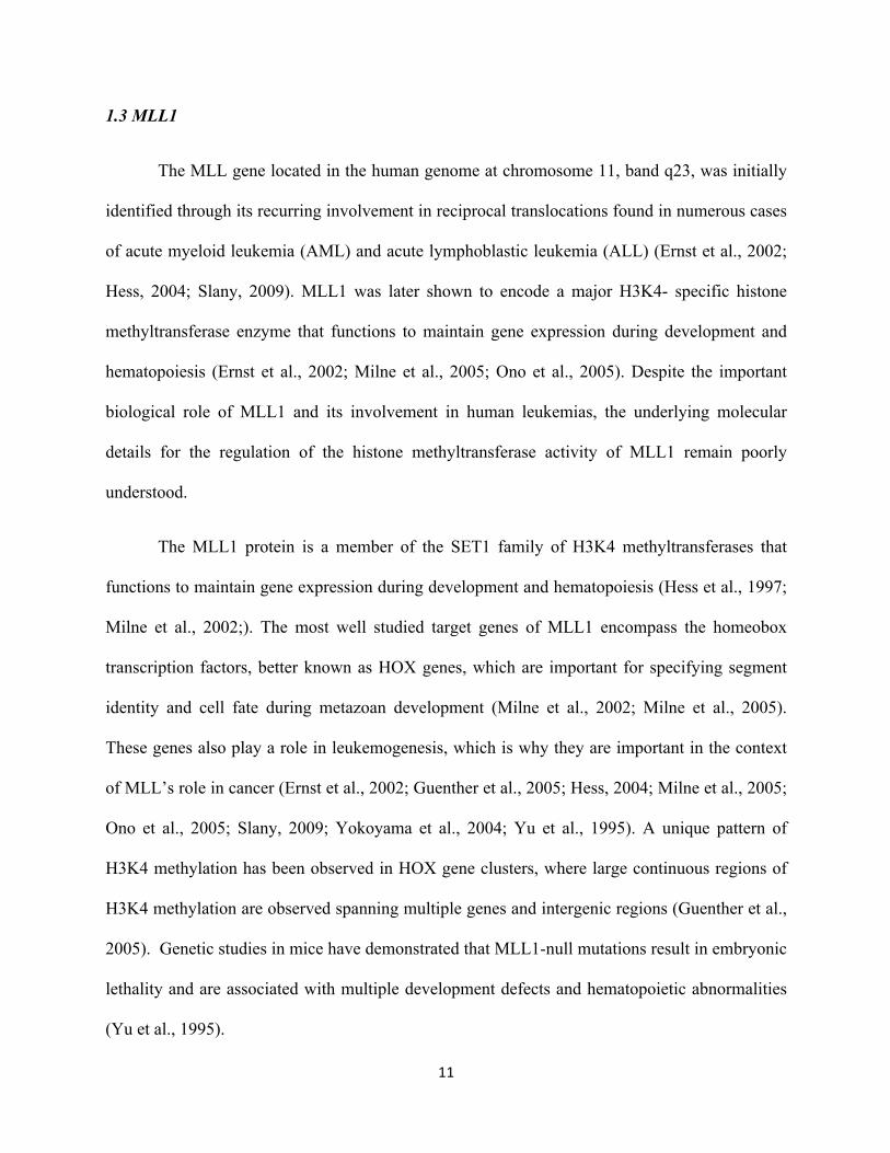
FIGURE 1.4. Structural analysis of a RbBP5 peptide bound to WDR5.
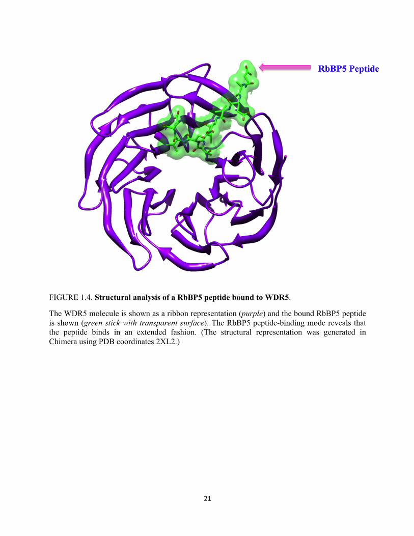
The WDR5 molecule is shown as a ribbon representation (purple) and the bound RbBP5 peptide is shown (green stick with transparent surface). The RbBP5 peptide-binding mode reveals that the peptide binds in an extended fashion. (The structural representation was generated in Chimera using PDB coordinates 2XL2.)
22
segment of the unstructured C-terminal tail segment in RbBP5 (Avdic et al., 2011; Odho et al.,
2010). To gain insight into the molecular basis of this interaction, crystals were obtained of
WDR5 and a synthetic RbBP5 peptide containing a small segment of residues responsible for
binding to WDR5. Structural analysis of the RbBP5 peptide-binding mode reveals that the
peptide binds in an extended fashion on the opposite face of WDR5 from the canonical binding
site that has been shown to accommodate the WDR5 interaction motif of MLL1 (Figure1.4).
Closer inspection of this structure reveals that the RbBP5 binding site on WDR5 has strong
hydrophobic character that can accommodate two residues Val-375 and Val-377 present on the
RbBP5 binding motif (Odho et al., 2010). When these two residues are mutated to glutamic acid,
binding to WDR5 shown by immunoprecipitation experiments is severely compromised (Odho
et al., 2010).
To investigate the effect of the addition of RbBP5 in the presence and absence of WDR5
on MLL1 SET domain mediated methyltransferase activity, a series of methyltransferase activity
assays were carried out with the MLL1 SET domain using unmodified histone H3 peptide as a
substrate. In addition, the ability of the RbBP5 proteins containing glutamic acid substitutions in
Valine residues 375 and 377 shown to be required for interaction with WDR5 were also tested
for their ability to further stimulate MLL1 SET domain mediated activity in the presence and
absence of WDR5. Previous studies have shown that MLL1 alone is slow
monomethyltransferases and the addition of WDR5 does not significantly enhance MLL1 SET
domain enzymatic activity. Consistent with previous results, the addition of WDR5 alone did not
significantly enhance MLL1 SET domain catalyzed methyltransferase activity. However, when
the RbBP5 wild-type protein was added, a small increase in activity of the MLL1 SET domain is
observed (Odho et al., 2010). Significantly, the addition of both WDR5 and RbBP5 resulted in a
23
much higher level of activity and enhancement. In a second separate series of methyltransferase
assays, carried out with the MLL1 SET and the RbBP5 proteins containing glutamic acid
substitutions in Valine residues at positions 375 and 377, the mutations alone did not interfere
with the ability of these proteins to further stimulate MLL1 methyltransferase activity when
compared to the stimulatory effect of the wild-type RbBP5 protein (Odho et al., 2010).
Interestingly enough, in the presence of WDR5, previous enhancement of MLL1
methyltransferase activity was lost with the RbBP5 variant proteins. Taken together, these results
suggest, that WDR5 may act as a structural scaffold for the assembly of the MLL1 core complex
by bridging interactions between RbBP5 and MLL1, to allow for RbBP5 mediated stimulation of
MLL1 SET domain methyltransferase activity. Consistent with this hypothesis, other studies
have demonstrated that RbBP5’s β-propeller domain and a short region of the tail was sufficient
enough to stimulate methyltransferase activity of the MLL1 SET domain (Avdic et al., 2011).
1.7 The Ash2L SPRY Domains Binds a Stretch of Acidic Residues in RbBP5
In addition to binding WDR5, RbBP5 forms another important interaction with the
Ash2L subunit within the MLL1 core complex. Recent reports have show that the SPRY domain
of Ash2L recognizes an acidic stretch of residues of RbBP5. It has been hypothesized that this
interaction between RbBP5 is more than likely mediated by electrostatic contacts (Chen et al.,
2011). In support of this hypothesis, previous data has shown that substitution of Arginine 343 in
Ash2L is enough to completely abolish its interaction with RbBP5. Not to mention, the mutation
of several other positively charged residues has resulted in the weakening of these contacts,
although to variable extents (Chen et al., 2011).
24
1.8 Studies in Yeast Reveal that the RbBP5 Homolog Swd1 is Required for SET Protein
Stability, Histone Methylation and Gene Expression
Many reports have suggested that RbBP5 of the MLL1 core complex is capable of
establishing direct contacts with the MLL1 SET domain to stimulate its intrinsic
methyltransferase activity. In spite of these studies, there has yet to be any study that identifies
the surfaces on MLL1 and RbBP5 required for this interaction, until recently. One recent study
on the homologous yeast Complex of Proteins Associated with SET1 (COMPASS) has shed
many important insights into the regulation of methyltransferase activity by MLL1 complex
members, specifically by Swd1 (RbBP5). In defining the mechanism of interaction between
SET1 and Swd1 (RbBP5), Briggs and colleagues have shown that two patches of acidic residues
found in the C-terminal domain of Swd1, is important for maintaining SET1 protein levels and
H3K4 methylation in vivo. Likewise, they were also able to identify that deletion of a basic
patch of residues on the conjugate protein SET1, abrogates the interactions between SET1 and
SWD1. Furthermore, the deletion of either the acidic or basic patches in either protein, resulted
in severe growth defects, loss of telomere silencing, and decreased gene expression.
Additionally, this study shows that this acidic patch of Swd1 is also conserved in human RbBP5,
and is necessary for protein–protein interactions between SETd1a1 and RbBP5 (Mersman et al.,
2012). Additional experiments indicate that RbBP5 is capable of forming a direct interaction to
the nSET domain of SET1, but does not form a direct interaction with MLL1. Based on these
results, although the complex members between human and yeast may be evolutionarily
conserved, the assembly and mode of regulation may differ. For example, although work in this
study suggests that Swd1 can interact with SET1 in the absence of other complex components,
work done on MLL1 shows that the human homolog of Swd1, (RbBP5) requires WDR5 to stably
25
interact with MLL1. Additionally, studies suggest that human SET1 complex components are
prerequisite for the stability of SET1A and SET1B protein levels, however, this does not appear
to be the case for MLL1. Taken together, the data suggest that although the SET1A/B and MLL1
exists in complexes along side an evolutionarily conserved core configuration of protein, the
manner in which they interact and assemble into multisubunit complexes to regulate H3K4
methylation are likely distinct.
Rational of Study
Although there have been several reports focused on RbBP5’s interaction with other
members of the MLL1 core complex, a detailed characterization of RbBP5 is still lacking. In
particular, relatively little is known about the contribution of RbBP5 in facilitating and/or
regulating H3K4 methylation within the context of the MLL1 core complex. Several lines of
evidence have emerged that suggests that the RbBP5 component of the MLL1 core complex is
involved in the catalytic mechanism of H3K4 methylation mediated by the MLL1 core complex
(Avdic et al., 2011; Cao et al., 2010; Crawford and Hess, 2006; Dou et al., 2006; Odho et al.,
2010; Patel et al., 2009; Southall et al., 2009). Several studies have also suggested that RbBP5
may directly interact with MLL1 SET domain to enhance methyltransferase activity (Avdic et
al., 2011; Cao et al., 2010; Crawford and Hess, 2006; Dou et al., 2006; Odho et al., 2010; Patel et
al., 2009; Southall et al., 2009). In addition, RbBP5 is a member of WRAD, a novel
methyltransferase whose enzymatic activity has only recently been reported (Patel et al., 2011).
This WRAD sub-complex of the MLL1 core complex is able to bind MLL1 and stimulate its
methyltransferase activity. Lastly, it has also been reported in an independent study that only an
Ash2L/RbBP5 heterodimer is required for weak methyltransferase activity (Cao et al., 2010).
Collectively, these studies suggest that RbBP5 is important for proper H3K4 methylation. In
26
spite of this body of work, the underlying mechanism of exactly how the RbBP5 protein
contributes to these methyltransferases activities remains undefined. Therefore, additional
biochemical and structural studies are pivotal in order to understand how RbBP5 contributes to
H3K4 methylation.
Therefore, to better understand the role of RbBP5 in H3K4 methylation, the work in this
thesis seeks to address the central question of what are the protein structural features of RbBP5
that are required for the di-and tri-methyltransferase activity of the MLL1 core complex. The
RbBP5 protein could be playing a number of roles in the MLL1 core complex including: 1.
RbBP5 is serving as a bridge to bring other components of the complex together; 2.RbBP5 is
interacting with MLL1 to stimulate SET domain activity; 3. RbBP5 binds the H3 histone
substrate or cofactor SAM; or 4. RbBP5 is part of a shared active site within the WRAD sub-
complex. By taking a structure-function approach the research in the following chapters of this
thesis seeks to identify novel protein structural features of RbBP5 responsible for the enzymatic
of the MLL1 core complex. By defining the role of RbBP5 we hope to further delineate the
underlying mechanism of H3K4.
In order to study the role of RbBP5 in H3K4 methylation by the MLL1 core complex,
extensive domain mapping experiments on the full-length human RbBP5 protein were performed
by Anamika Patel to identify the minimal domain requirements for enzymatic activity. These
studies reveal that the minimal domain of RbBP5 required to recapitulate methyltransferase
activity consists of residues 323-402 (Patel, unpublished data). Close inspection of the residues
in the sequence of the minimal domain of RbBP5 (323-402) reveals a highly conserved stretch of
amino acids with no known function. In light of these findings, to probe the functional role of
each of these residues in MLL1 core complex mediated methyltransferase activity, point
27
mutations were introduced into conserved residues in this domain. Once mutations were
introduced into the RbBP5 protein and confirmed, high-throughput methyltransferase enzymatic
assays were carried out to identify mutated residues that resulted in an overall decrease in the di-
methyltransferase activity of the MLL1 core complex. Further validation of residues resulting in
reduced activity were verified through quantitative MALDI-TOF methyltransferase assays. Out
of these screens, residues W329, F332, F336, and E347 were identified and confirmed to have
reduced di-methyltransferase activity within the MLL1 core complex. Further experiments
demonstrate that these residues are required for interaction with the MLL1 SET domain.
Although numerous studies have suggested that RbBP5 is required for full activity of the
complex, to date there have been no reports on the inherent structural features required for
protein-protein interactions and enzymatic activity involving RbBP5. Taken together, these
studies constitute the identification a novel structural motif in RbBP5 required for interaction
with MLL1 within the MLL1 core complex and consequently the overall dimethyltransferase
activity of the complex. This work is detailed in chapter 2, and encompasses the major body of
work done for this thesis. Furthermore, previously determined Valine residues 375 and 377 in
RbBP5 identified to be important for the interaction with WDR5 were further characterized and
confirmed taking a more stringent biophysical and kinetic approach. This work is described at
the end of chapter two, and provides further validation of the WDR5-RbBP5 interaction motif.
The third chapter of this thesis, reports on the identification and characterization of two
stable sub -domains of RbBP5 identified using limited proteolysis experiments to facilitate high-
resolution structure determination of RbBP5. Currently, there is no three-dimensional structure
of full-length RbBP5, and although several attempts were made to try to crystallize the full-
length protein using the hanging drop vapor diffusion method and sparse matrix screening, no
28
crystals have emerged from crystallization trials. Using limited proteolysis to identify flexible
regions in RbBP5 that may prevent this protein from crystallization, I have succeeded in
identifying two potentially well -ordered regions of RbBP5 containing residues 1-495 and 1-337.
The identity of these domains was confirmed utilizing N-terminal sequencing and their exact
masses were confirmed via electrospray ionization mass spectrometry. Although no crystals have
emerged from early crystallization trials of these newly identified stable subdomains in RbBP5,
this study illustrates alternative and varied approaches in studying the role of a protein without
the availability of a three-dimensional structure. The final part of this thesis provides an
overview of the body of work and proposed future directions in light of the findings of these
studies.

29
CHAPTER TWO:
A Conserved Patch of Amino Acids in RbBP5 is
Required for Interaction with the SET domain of
MLL1 in the MLL1 Core Complex
30
INTRODUCTION:
The correct establishment and propagation of genetic programs in eukaryotes depends in
part on enzymes that are responsible for the deposition and removal of covalent posttranslational
modifications on histone proteins. The methylation of histone H3 at lysine 4 (H3K4) is a well-
studied epigenetic mark required for the recruitment of enzymatic machineries that maintain
transcriptionally permissible states of chromatin. This mark is mainly catalyzed by a group of
enzymes that contain an evolutionarily conserved Suppressor of Variegation, Enhancer of Zeste,
and Trithorax (SET) domain (Ruthenburg et al., 2007). Abnormalities in H3K4 methylating
enzymes of the SET1 family have been observed in various cancers (Bhaumik et al., 2007; Chi et
al., 2010) the most prominent example resulting from aberrations of the Mixed Lineage
Leukemia gene. Studies on the archetypal member of the SET1 family, human MLL1, have shed
light on several mechanistic determinants underlying the mode of regulation for these enzymes.
Of specific interest, immunoprecipitation experiments have shown that unlike other SET domain
enzymes, members of the SET1 family are found in a multisubunit core configuration of proteins
comprised of WDR5, RbBP5, Ash2L, and DPY30 (WRAD). Growing bodies of studies have
highlighted the important role that WRAD plays in the overall methyltransferase activity of the
core complex. The key common finding of these studies is that deletion or down regulation of a
core complex protein leads to an overall reduction in the observed level of methyltransferase
activity (Avdic et al., 2011; Cao et al., 2010; Cosgrove and Patel, 2010; Crawford and Hess,
2006; Dou et al., 2006; Odho et al., 2010; Patel et al., 2009; Southall et al., 2009). More
importantly, it has been demonstrated that although WDR5 by itself does not stimulate
methyltransferase activity of the MLL1 SET domain in vitro, in contrast, both RbBP5 and Ash2L
31
contribute to the overall catalytic activity. When stoichiometric amounts of RbBP5 are added to
a MLL1 SET domain construct and WDR5, a small 2-fold increase in the overall reaction rate in
observed, when compared to the reaction catalyzed by MLL1 alone (Chi et al., 2010; Patel et al.,
2009). When Ash2L was added to the MLL1 SET domain construct along with WDR5 and
RbBP5, a considerable increase in the overall reaction rate was observed when compared with
that of the MLL1 SET domain protein alone (Chi et al., 2010; Patel et al., 2009). Although
several reports have emerged on the Ash2L subunit and its contribution to this activity, far less is
known about the contribution of RbBP5 in facilitating and/or regulating H3K4 methylation
within the context of the MLL1 core complex.
The retinoblastoma binding proteins five (RbBP5) was originally discovered because it is
capable of directly binding the Retinoblastoma protein (pRb), the protein product of the
Retinoblastoma gene. The Rb family members are essential regulators of cell cycle progression.
Studies of the retinoblastoma gene (Rb) have shown that its protein product (pRb) acts to restrict
cell proliferation, inhibit apoptosis, and promote cell differentiation (Lai et al., 1999; Magnaghi-
Jaulin et al., 1998; Paggi et al., 1996). The RbBP5 protein contains a predicted B-propeller
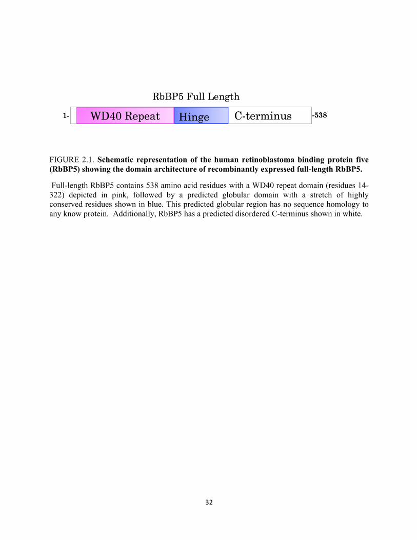
domain followed by a short hinge region, and a less conserved C-terminus (Figure 2.1).
Recently, this protein has garnered much attention because of its presence as a conserved core
component of the MLL1 core complex. The RbBP5 subunit of the MLL1 core complex has been
shown to be required for enzymatic activity and disruption of RbBP5 has been frequently
32
FIGURE 2.1. Schematic representation of the human retinoblastoma binding protein five (RbBP5) showing the domain architecture of recombinantly expressed full-length RbBP5.
Full-length RbBP5 contains 538 amino acid residues with a WD40 repeat domain (residues 14-322) depicted in pink, followed by a predicted globular domain with a stretch of highly conserved residues shown in blue. This predicted globular region has no sequence homology to any know protein. Additionally, RbBP5 has a predicted disordered C-terminus shown in white.
33
observed in patients with malignant primary brain tumors (Bralten et al., 2010). Importantly, the
absence of RbBP5 results in the loss of H3K4 dimethylation in vitro and the loss of H3K4 di-
and tri- methylation in vivo (Dou et al., 2006). However, the exact functional role of RbBP5 in
MLL1 core complex H3K4 dimethyltransferase activity is not well understood. One of the
possible means by which RbBP5 functions within the MLL1 core complex is that it may be
directly involved in the H3K4 di- and trimethylation reaction. This is supported by experiments
in vivo that demonstrate that RNAi knockdown of RbBP5 in HeLa cells severely impairs the
ability of the MLL1 core complex to catalyze H3K4 di- and trimethylation (Dou et al., 2006).
This study is further corroborated by experiments in vitro that show there is no detectable H3K4
di-methylation activity in the absence of RbBP5 by the MLL1 core complex (Dou et al., 2006).
Not to mention, several other lines of evidence suggest that the RbBP5 subunit could be directly
interacting with the SET domain of MLL1 to stimulate enzymatic activity (Odho et al., 2010;
Patel et al., 2009). Collectively, these findings convey the important functional role RbBP5 takes
within MLL1 core complex. In spite of these studies, the molecular details underlying RbBP5’s
catalytic influence on the MLL1 core complex activity have yet to be determined.
In this thesis, this research seeks to understand the mechanism behind RbBP5 mediated
stimulation of the MLL1 core complex methyltransferase activity. By comprehensive mapping of
RbBP5 we identify a minimal domain of RbBP5 required for enzymatic activity within the
MLL1 core complex. Within this domain sequence analysis uncovers a highly conserved stretch
of amino acids that exists among 19 homologs of the human Retinoblastoma binding protein-5
(RbBP5) subunit of the MLL1 core complex. The organisms range for yeast to humans, along
with plants and animals. To gain insight into the functional role of this conserved stretch of
residues, mutations targeting individual residues in this highly conserved stretch were generated.
34
Biochemical and biophysical analyses of the protein with various mutations in conserved
residues were utilized to assess the functional role each may endow on the intrinsic properties of
RbBP5 alone or when assembled within the context of the entire complex. Importantly, we find
that mutation of several conserved aromatic residues and one acidic residue in RbBP5 results in a
significant reduction in the overall dimethyltransferase activity of the MLL1 core complex.
Additionally we find that these residues when mutated diminish WRAD’s ability to interact with
MLL1. The residues identified constitute a previously uncharacterized novel MLL1 interaction
motif located in RbBP5. Taken together, our study provides fundamental insights into how the
RbBP5 subunit of the MLL1 core complex contributes to the overall enzymatic activity of
WRAD and the MLL1 core complex. Understanding the role that RbBP5 plays in facilitating
proper H3K4 methylation may provide insight into how misregulation of RbBP5 leads to its
association with brain tumorigenesis and progression. In addition, understanding the functions of
H3K4 methyltransferases and their interacting partners will be key in deciphering the “epigenetic
code” and how this mark contributes to fundamental regulatory and cell developmental fate
decisions.
35
EXPERIMENTAL PROCEDURES
Small Scale Expression of RbBP5 Variants For High-throughput Assays: RbBP5 wild-type and
mutant plasmid DNA were transformed into Escherichia coli Rosetta II plysS cells.
Transformation reactions were plated on LB plates containing 50ug/mL carbenicillin and
20ug/mL chloramphenicol and incubated at 37°C overnight. Isolated single colonies were picked
from wild-type and mutant plates the following day and inoculated in 5mL of Terrific Broth,
containing 50ug/mL carbenicillin and 20ug/mL chloramphenicol. Cultures were allowed to grow
overnight at 37°C. The next day, 50uL of overnight cultures were added to 5mL of Terrific Broth
containing 50ug/mL of carbenicillin. The cultures were incubated with shaking for 2 hours at
37°C. After 2 hours, the cells were induced with 750uM isopropyl-1-thio-D-galactopyranoside.
Small scale cultures were then placed on a shaker at 15 °C and grown overnight. The following
day, the small scale cultures were spun down and the pellets were stored at -80 °C until further
use.
For Small Scale lysis: 50 mL of lysis buffer (50 mM Tris (pH 7.4) 300 mM Sodium Chloride
(NaCl), 3 mM dithiothreitol (DTT) was prepared. One EDTA free protease inhibitor tablet,
0.1mM phenylmethylsulfonyl fluoride (PMSF), and 750uM sarkosyl was added to the 50 mL
buffer preparations. A 100uL aliquot of freshly prepared lysis buffer cocktail was removed from
the solutions and used to resuspend the crude pellet. To the sample cell lysis mixture 10uL of
10X Bug Buster, followed by the addition of 2.5uL DNAse 1 was added. The samples were then
placed on a rotator at 4 °C, and cells were allowed to lyse with gentle turning for approximately
4 hours. Cell lysis mixtures were spun down at 4 °C in a micro centrifuge at 10,000 rpm for 20
36
minutes. The supernatant was removed and placed in a carefully labeled tube. The enzyme
mixture was stored at -80°C until use.
Protein expression and purification: The RbBP5 gene was purchased from open bio systems and
sub-cloned into a pHis parallel vector, which encodes a Tobacco Etch Virus (TEV) cleavage N-
terminal 6x-His fusion tag. Versions of full-length human RbBP5 (1-538) and RbBP5 constructs
were overexpressed in Escherichia coli (Rosetta II, Novagen), by growing cells containing a
given plasmid at 37°C in Terrific Broth medium containing 50ug/mL carbenicillin. The
temperature was then lowered to 15 °C and cells were induced for 16–18 hours with 750uM
isopropyl-1-thio-D-galactopyranoside (IPTG). Cells were harvested, re-suspended in a lysis
buffer (50 mM Tris, pH 7.4, 300 mM NaCl, 3 mM dithiothreitol, 0.1 mM
phenylmethylsulfonylfluoride, and EDTA-free protease inhibitor mixture (RocheApplied
Science)), lysed with a microfluidizer cell disrupter, and clarified by centrifugation. Supernatants
containing the RbBP5 His-tagged proteins were purified by nickel affinity chromatography
(HisTrap column, GE Healthcare). For the first step of purification, the crude lysate was passed
through a HisTrap column (GE healthcare) containing nickel beads. The bound 6x-His-RbBP5
was eluted from the column using a linear gradient of elution buffer containing 500mM
imidazole. The peak fractions that contained RbBP5 were collected, pooled, and then dialyzed
against column buffer that contained 50 mM Tris pH 7.4, 300mM NaCl, 30 mM Imidazole, and
3mM DTT at 4°C to remove the excess imidazole and to cleave the 6X-His tag in the presence of
TEV (Tobacco Etch Virus) protease. The dialysis buffer was changed three times. For the
second step of purification, the dialyzed protein was passed through the His-Trap column and the
flow through fractions that contained the untagged version of RbBP5 were pooled and combined.
As a final step of purification, the protein was passed through a gel filtration column (Superdex
37
200TM GE Healthcare) pre-equilibrated with the sample buffer containing 20mM Tris (pH 7.5),
300mM NaCl, 1mM Tris (2-carboxyethyl) phosphine (TCEP), and 1µM zinc chloride. All other
proteins used in methyltransferase assays were purified as previously described.
Mutagenesis and Oligonucleotide Primers: Point mutations were introduced into RbBP5 using
the QuickChange site directed mutagenesis kit (Stratagene). Plasmids were sequenced to verify
the presence of the intended mutations and the absence of additional mutations. Integrated DNA
Technologies synthesized oligonucleotide primers were used in the mutagenesis.
3[H] methyltransferase assay: Radiolabelled methyltransferase assays were conducted by
combining 4µM of enzyme with 500 µM histone H3 peptide containing residues 1-20 (with
GGK-Biotin on the C-terminus) and one microcurrie of 3H-methyl-S-adenosyl-methionine (3H-
SAM, GE Healthcare) in 50mM Tris, pH 8.5, 200mM NaCl, 3mM DTT, 5mM MgCl2, and 5%
glycerol. The reactions were incubated at 15 °C for 8 hours, stopped by the addition of SDS-
loading buffer to 1X, and separated by SDS-PAGE on a 4-12% gradient gel (Invitrogen). The gel
then was soaked in an autoradiography enhancer solution (Enlightning, Perkin Elmer), dried, and
exposed to film at -80°C for 24 hours to 5 days.
Western Blotting : Equal volumes of RbBP5 crude lysates were loaded in the wells of an SDS-
PAGE gel along with molecular weight markers. SDS -PAGE were performed on RbBP5 crude
lysates and the proteins were transferred from the gel to a nitrocellulose membrane. Membranes
were blocked with a blocking solution containing 5% nonfat dry dairy milk in a 1X phosphate
buffered saline solution with tween 20 (0.1 %) (PBST) at room temperature with gentle agitation
for 1 hour. Blocking solution was decanted and the membrane was incubated with primary
antibody for 2 hours with gentle agitation (RbBP5 anti-body purified from Bethyl laboratories;
working dilution 1:1000). After the membrane was incubated with the primary antibody, the
38
membranes were washed with 1X (PBST 0.1 %) for 10 minutes 3 times. The membrane was then
incubated with a secondary antibody (Pierce goat anti-rabbit; working dilution 1:5,000) with
gentle agitation for 1 hour. Membranes were washed again with 1X PBST 0.1% for 10 minutes
three times. After washing the membranes, membranes were placed in weigh boats and a fresh
chemilumescence solution was prepared by adding 1mL of SuperSignal Peroxide Solution and 1
mL of SuperSignal Luminol Enhancer directly on the membranes. The solution was distributed
evenly. The weigh boat was then covered with foil and incubated in signal solution for 2
minutes. After incubation, membrane edges were bloated on paper towels to soak up excess
signal solution. Human RbBP5 was detected with chemilumescence at various exposures.
MALDI-TOF mass spectrometry methyltransferase assay: Mass spectrometry assays were
conducted as previously reported; 7 µM of enzyme was incubated with 250 µM s-adenosyl-
methionine (SAM) and 10 µM histone H3 peptide (1-20) at 15 °C in 50mM TrisCl pH 9.0,
200mM NaCl, 3 mM DTT, and 5% glycerol. The reactions were quenched at various time points
by the addition of trifluoroacetic acid to 0.5%. The quenched samples were diluted 1:4 with α-
cyano-4-hydroxycinnamic acid. MALDI TOF mass spectrometry was performed on a Bruker
AutoFlex mass spectrometer (State University of New York, ESF) operated in reflectron mode.
Final spectra were averaged from 100 shots/position at 10 different positions.
Analytical Ultracentrifugation: Analytical ultracentrifugation experiments were carried out using
a Beckman Coulter ProteomeLabTM XL-A analytical ultracentrifuge equipped with absorbance
optics and a 4-hole An-50 Ti analytical rotor. Sedimentation velocity experiments were carried
out at 10°C and 50,000 rpm (200,000 x g) using 3 mm two sector charcoal-filled Epon
centerpieces with quartz windows. For each sample, 300 scans were collected with the time
interval between scans set to zero. Protein samples in 20mM Tris (pH 7.5), 300mM NaCl, 1mM
39
TCEP and 1µM ZnCl2 were run at various concentrations. Sedimentation boundaries were
analyzed by the continuous distribution (c(s)) method using the program SEDFIT. The program
SEDNTERP version 1.09 was used to correct the experimental S-value (s*) to standard
conditions at 20°C in water (S20,w) and to calculate the partial specific volume of each protein.
Circular Dichroism Spectroscopy: CD spectra were collected on an AVIV Model 420 ds
spectropolarimeter equipped with a Neslab CFT-33 refrigerated circulator at 10°C using a 0.1
mm path length cell. Spectra for each sample were obtained at a protein concentration of
0.2mg/mL in a buffer contain 20mM tris (pH 7.5), 200 mM NaCl, 1mM TCEP and 1uM Zinc
chloride. The background contribution of the buffer alone was subtracted from each protein
spectrum.
RESULTS
2.1 The Hinge Domain of RbBP5 is Required for Methyltransferase Activity in WRAD and the
MLL1 Core Complex
Recent studies suggest a sequential mechanism for H3K4 di-methylation by the MLL1
core complex (Gardner et al., 2011; Patel et al., 2009). In this proposed model, the MLL1 SET
domain of MLL1 catalyzes monomethylation of histone H3 at active site one. Following this
event, the monomethylated peptide is subsequently transferred to a second active site on the
WRAD sub-complex that then catalyzes dimethylation of histone H3. WRAD therefore
constitutes a previously uncharacterized methyltransferase. Initial studies have shown that not
only is RbBP5 essential for the methyltransferase activity within the MLL1 core complex, but is
also essential for the newly discovered WRAD activity (Patel et al., 2009; Patel et al., 2011).
40
Nevertheless, the functional role of the RbBP5 protein in these two enzymatic activities has
remained unexplored.
One approach to better understand the function of RbBP5 is to delineate the role of
specific domains within RbBP5 in facilitating H3K4 methylation. Based on sequence analysis
RbBP5 contains a predicted N-terminal WD40 repeat Beta-propeller domain, a reported hinge
domain (Avdic et al., 2011), which contains several highly conserved residues, and a less
conserved C-terminus (Figure 2.2a). In order to study the function RbBP5 and its various
domains, RbBP5 constructs corresponding to residues 1-538 (Full-length), 1-402 (β-propeller
+Hinge), 1-322 (β-propeller), and 323-402 (Hinge Region) were expressed as 6X his tagged
proteins in E.coli, and purified to near homogeneity (Figure 2.2b). The individual constructs
were then tested for their ability to catalyze H3K4 methylation in the presence of the other
members of the MLL1 core complex (MLL1, WDR5, ASH2L, and DPY30) using an unmodified
H3 peptide as a substrate in an in vitro histone methyltransferase activity assay. Given the strong
conservation of residues in the hinge region of RbBP5, we hypothesized that this region will be
necessary not only for the activity of the MLL1 core complex, but also for activity within the
WRAD sub-complex, and could possibly serve as part of a putative catalytic domain when
assembled with other members of WRAD.
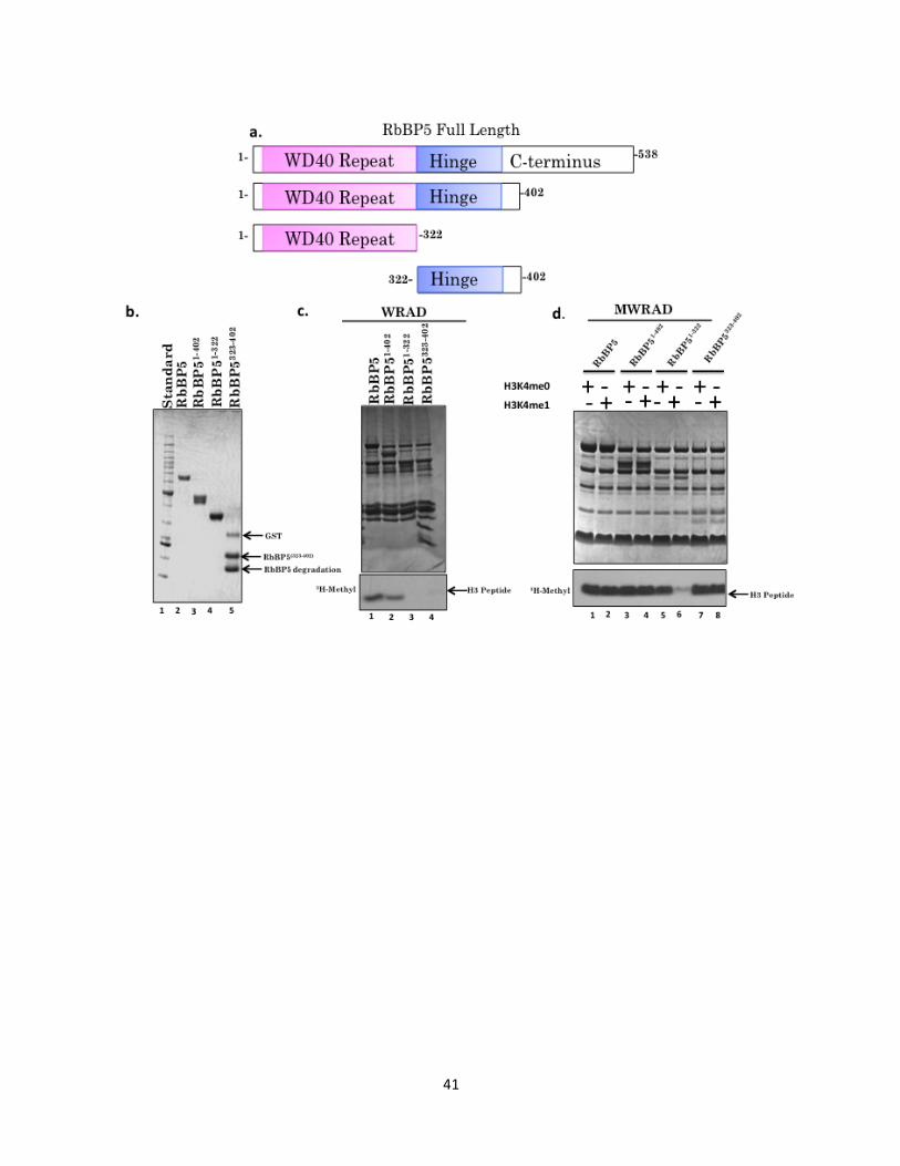
41
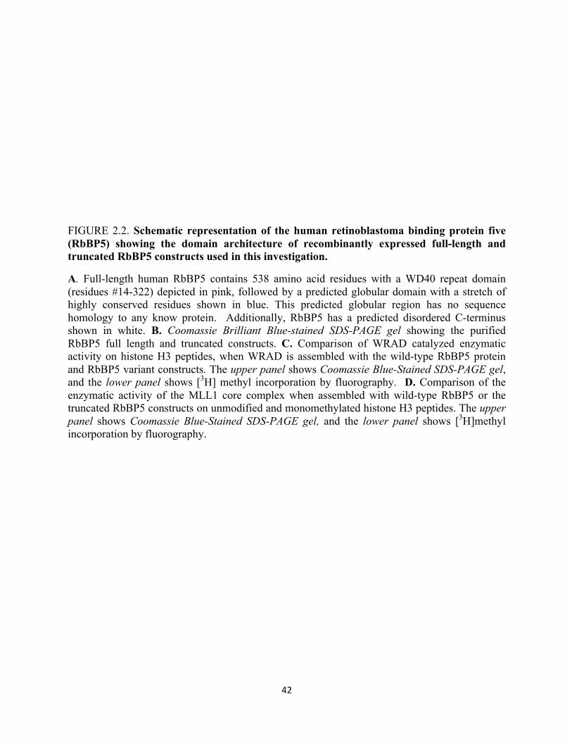
42
FIGURE 2.2. Schematic representation of the human retinoblastoma binding protein five (RbBP5) showing the domain architecture of recombinantly expressed full-length and truncated RbBP5 constructs used in this investigation.
A. Full-length human RbBP5 contains 538 amino acid residues with a WD40 repeat domain (residues #14-322) depicted in pink, followed by a predicted globular domain with a stretch of highly conserved residues shown in blue. This predicted globular region has no sequence homology to any know protein. Additionally, RbBP5 has a predicted disordered C-terminus shown in white. B. Coomassie Brilliant Blue-stained SDS-PAGE gel showing the purified RbBP5 full length and truncated constructs. C. Comparison of WRAD catalyzed enzymatic activity on histone H3 peptides, when WRAD is assembled with the wild-type RbBP5 protein and RbBP5 variant constructs. The upper panel shows Coomassie Blue-Stained SDS-PAGE gel, and the lower panel shows [3H] methyl incorporation by fluorography. D. Comparison of the enzymatic activity of the MLL1 core complex when assembled with wild-type RbBP5 or the truncated RbBP5 constructs on unmodified and monomethylated histone H3 peptides. The upper panel shows Coomassie Blue-Stained SDS-PAGE gel, and the lower panel shows [3H]methyl incorporation by fluorography.
43
To test this hypothesis, the ability of each RbBP5 construct to catalyze methylation of
unmodified histone H3 was tested in in vitro methyltransferase assays containing either wild-
type or various truncated RbBP5 constructs with the remaining members of the WRAD sub
complex. As shown in Figure 2.2 c, comparing lanes 1 and 2, when assembled with the
remaining members of WRAD the truncated construct RbBP5 (1-402) has similar
methyltransferase activity comparable to that of the wild-type WRAD complex (Figure 2.2 c,
lanes 1 and 2). However, when the residues constituting the hinge domain are deleted in the
RbBP5 (1-322) construct WRAD’s activity could not be observed (Figure 2.2 c, lane 3).
Assembly of the RbBP5 fragment containing only residues 323-402 in WRAD shows that these
residues alone are not sufficient on their own to restore activity compared to that of the full-
length protein when assembled in the WRAD sub-complex, although a trace amount of activity
is observed (Figure 2.2c, lane 4). In sum, the WD40 repeat and hinge domains of (RbBP5 (1-
402)) constitutes the minimal domain that is sufficient and necessary for the activity of the
WRAD sub complex.
2.3 Domains of RbBP5 Required for H3K4 dimethyltransferase Activity within the MLL1 Core
Complex
Given that the minimal domain of RbBP5 required to reconstitute the activity of WRAD
consists of residues 1-402, we anticipated that this fragment of RbBP5 would also be sufficient
to recapitulate di-methyltransferase activity within the MLL1 core complex. Therefore, we
assembled the RbBP5 constructs we previously generated to test for enzymatic activity within
the holo-MLL1 core complex, using both unmodified histone H3 peptide and mono-methylated
histone H3 peptide as substrates. Consistent with previous published results, when the MLL1
core complex was assembled with the full-length RbBP5 protein it is capable of methylating an
44
unmodified and a previously mono-methylated peptide on histone H3K4 (Figure 2.2 d, lanes 1
and 2). As expected, the RbBP5 (1-402) construct containing the WD40-repeat region and the
hinge region of RbBP5 possesses H3K4 mono- and dimethyltransferase activity similar to that of
the wild type MLL1 core complex (Figure 2.2d lanes 3 and 4). However, the shorter construct,
lacking the hinge domain exhibited full activity on an unmodified peptide (Figure 2.2d lane 5),
but was not able to fully restore activity on a histone H3 peptide previously monomethylated at
H3K4. (Figure 2.2d lane 6). Surprisingly, the small construct of RbBP5 containing only the
hinge domain that lacked activity with WRAD when assembled in the MLL1 core complex
exhibited activity similar to that of wild-type on both unmodified and mono-methylated peptides
(Figure 2.2d lane 7 and 8). These results suggest that (residues 323-402) in the hinge region of
RbBP5 are not only important for catalytic activity within WRAD, but more specifically contain
catalytic residues important for di-methyltransferase activity mediated by the MLL1 core
complex.
2.4 Alanine Scanning Mutagenesis Identifies Residues Involved in the Dimethyltransferase
Activity of the MLL1 Core Complex
Intrigued by the finding that the RbBP5 (323-402) construct was capable of facilitating
the di-methyltransferase activity of the MLL1 core complex, we decided to further investigate
the role of the hinge domain residues in RbBP5. We hypothesized that putative catalytic residues
responsible for the di-methyltransferase activity of the MLL1 core complex probably reside
within the 323-402 sequence of RbBP5. A multiple sequence alignment among 19 RbBP5
homologs reveals that this region possesses a 25 amino acid stretch that is highly conserved
(Figure 2.3a). To gain insight into why this region displays such a high degree of conservation,
we conducted an alanine scanning mutagenesis screen on this stretch of residues. To do so, point
45
mutations were introduced into the hinge region of RbBP5 for each residue from W329-K359
(letters represent the single letter codes for each amino acid). Alanine residues at positions 331
and 333 were mutated to serine residues. Each of the mutant proteins were expressed in E. coli,
lysed, and crude extracts were used in assays to compare wild-type and mutant RbBP5 proteins
using a H3K4me1 peptide as a substrate. The incorporation of a methyl group from 3[H] SAM
into the peptide was monitored and visualized by fluorography. From this mutagenesis screen,
we were able to identify several residues that when mutated reduced dimethyltransferase activity
to varying degrees within the MLL1 core complex. As controls, reactions were set up with wild-
type RbBP5 crude lysate (induced), and a reaction that contained the uninduced wild type
RbBP5. To control for expression, crude lysates were probed with an anti-RbBP5 antibody to
ensure that any observable decrease in the dimethylation activity of these enzymes assembled
within the complex was not a result of the protein not being expressed (Figure 2.3b lower panel).
The RbBP5 variant proteins that resulted in reduced di-methyltransferase activity are listed in
Table 2.1.
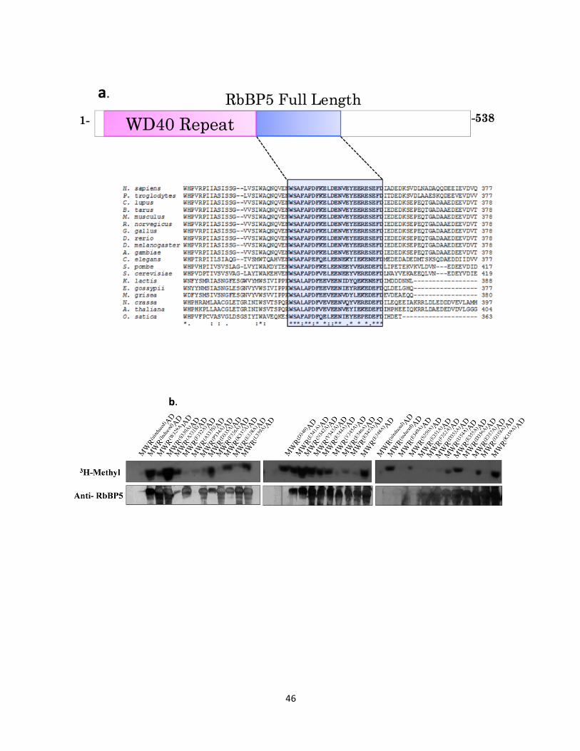
46
47
FIGURE 2.3 ClustalW2 multiple sequence alignment of the predicted globular domain of RbBP5 showing only a subgroup of residues, and high throughput methyltransferase assays showing activity of the RbBP5 protein with mutations in conserved residues identified in its predicted globular region.
A. The conserved globular motif of RbBP5 is highlighted in blue. Conserved residues are denoted underneath the alignment by an asterisk, conservative substitutions are denoted by a colon, and semiconservative substitutions are denoted by a period. B. Comparison of the enzymatic activity of the MWRAD complex on mono-methylated histone H3 (1-20) peptide when assembled with mutant RbBP5 proteins. The upper panel shows [3H] methyl incorporation into histone peptides as determined by fluorography. The lower panel shows immunoblotting of RbBP5 crude lysates used in this assay.
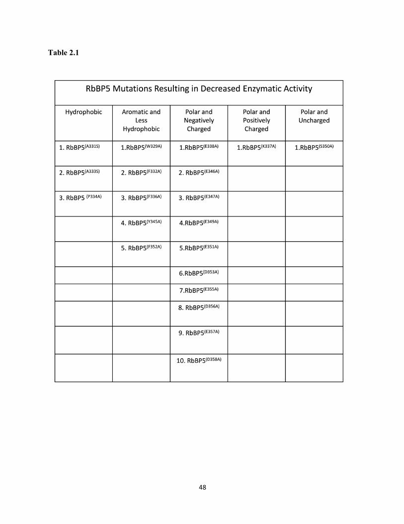
48
Table 2.1
49
Based on this screen, we decided to take a more quantitative approach to validate the
results of this screen. Therefore, RbBP5 proteins containing mutations that resulted in reduced
dimethyltransferase activity were expressed and purified as previously described (Patel et al.,
2008). To ensure that point mutations were not altering the overall hydrodynamic shape of the
proteins, sedimentation velocity analytical ultracentrifugation was conducted on individual
proteins. Sedimentation boundaries obtained were analyzed by the continuous sedimentation
coefficient distribution (c(s)) method in the program SEDFIT. Fitting of the boundaries
demonstrates that these mutant RbBP5 proteins sediment as monodispersed species and with a
sedimentation value of 2.4 s* for all proteins (Figure 2.4), similar to that of the wild-type RbBP5
protein (Patel et al., 2009). These results suggest that the mutations introduced in the RbBP5
variants do not significantly alter the overall hydrodynamic shape of RbBP5. Hydrodynamic
parameters for all mutant RbBP5 proteins are summarized in Table 2.2. Circular dichroism
spectroscopy was also performed on mutant RbBP5 proteins to determine if the point mutations
alter the secondary structure fold of RbBP5. Superimposition of CD spectra of all RbBP5
variants suggest that no drastic changes in the secondary structure occurs (Figure 2.5).
Once the correct folding of each mutant RbBP5 protein was established, their individual
enzymatic contributions to the MLL1 core complex di-methyltransferase activity were assayed
utilizing an independent assay. Using a quantitative Matrix-Assisted Laser Desorption Ionization
Time of Flight Mass Spectrometry (MALDI-TOF) methyltransferase assay, the ability for each
mutant RbBP5 protein to catalyze mono- and di-methylation when assembled in the presence of
other complex members on an unmodified peptide was tested in vitro.
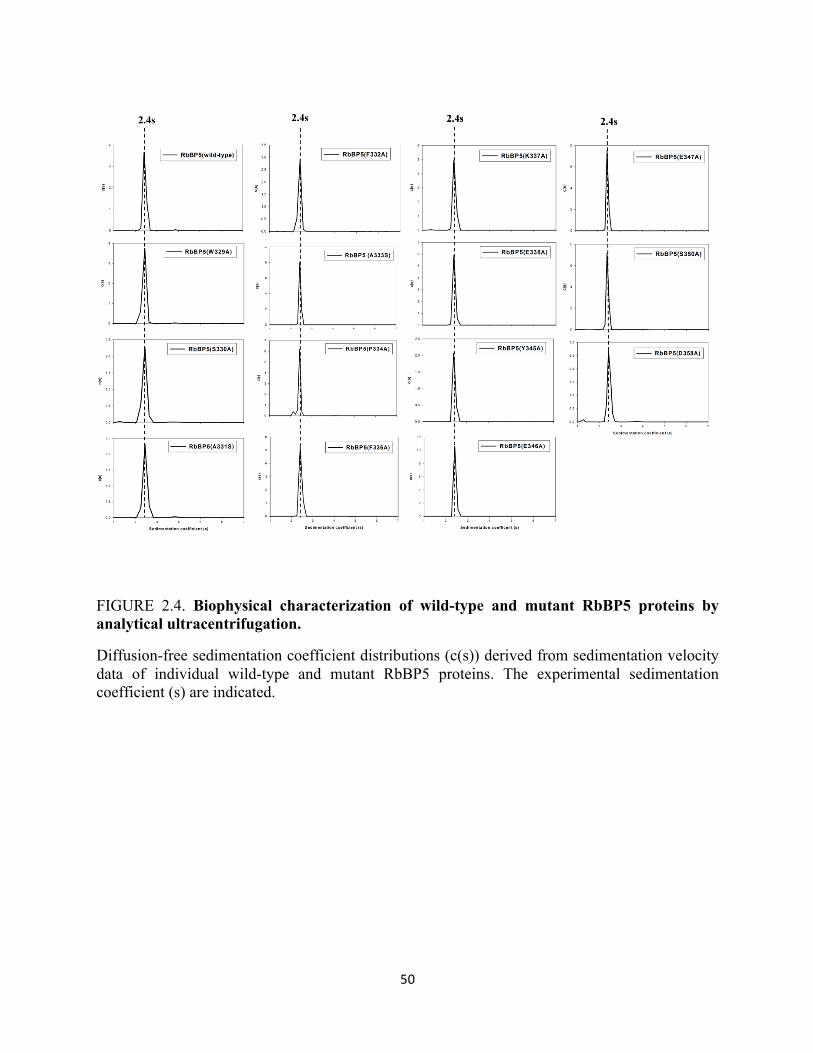
50
FIGURE 2.4. Biophysical characterization of wild-type and mutant RbBP5 proteins by analytical ultracentrifugation.
Diffusion-free sedimentation coefficient distributions (c(s)) derived from sedimentation velocity data of individual wild-type and mutant RbBP5 proteins. The experimental sedimentation coefficient (s) are indicated.
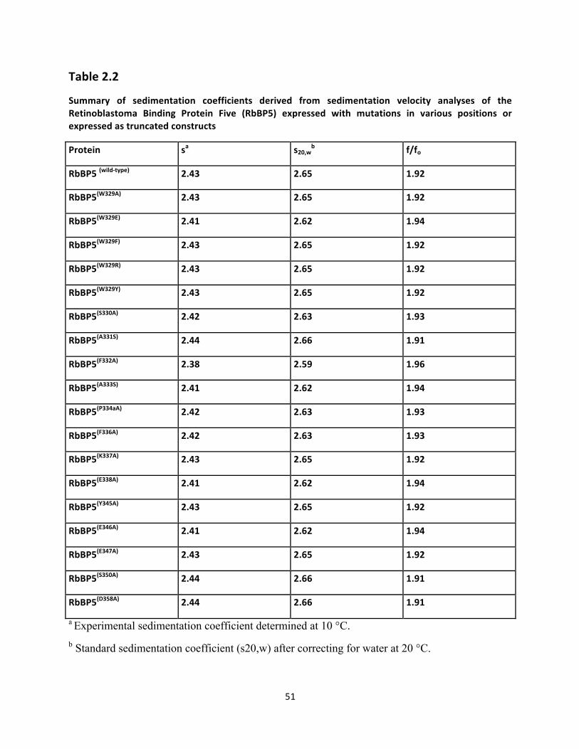
51
Table 2.2
Summary of sedimentation coefficients derived from sedimentation velocity analyses of the Retinoblastoma Binding Protein Five (RbBP5) expressed with mutations in various positions or expressed as truncated constructs
Protein sa s20,wb f/fo
RbBP5 (wild-‐type) 2.43 2.65 1.92
RbBP5(W329A) 2.43 2.65 1.92
RbBP5(W329E) 2.41 2.62 1.94
RbBP5(W329F) 2.43 2.65 1.92
RbBP5(W329R) 2.43 2.65 1.92
RbBP5(W329Y) 2.43 2.65 1.92
RbBP5(S330A) 2.42 2.63 1.93
RbBP5(A331S) 2.44 2.66 1.91
RbBP5(F332A) 2.38 2.59 1.96
RbBP5(A333S) 2.41 2.62 1.94
RbBP5(P334aA) 2.42 2.63 1.93
RbBP5(F336A) 2.42 2.63 1.93
RbBP5(K337A) 2.43 2.65 1.92
RbBP5(E338A) 2.41 2.62 1.94
RbBP5(Y345A) 2.43 2.65 1.92
RbBP5(E346A) 2.41 2.62 1.94
RbBP5(E347A) 2.43 2.65 1.92
RbBP5(S350A) 2.44 2.66 1.91
RbBP5(D358A) 2.44 2.66 1.91
a Experimental sedimentation coefficient determined at 10 °C. b Standard sedimentation coefficient (s20,w) after correcting for water at 20 °C.
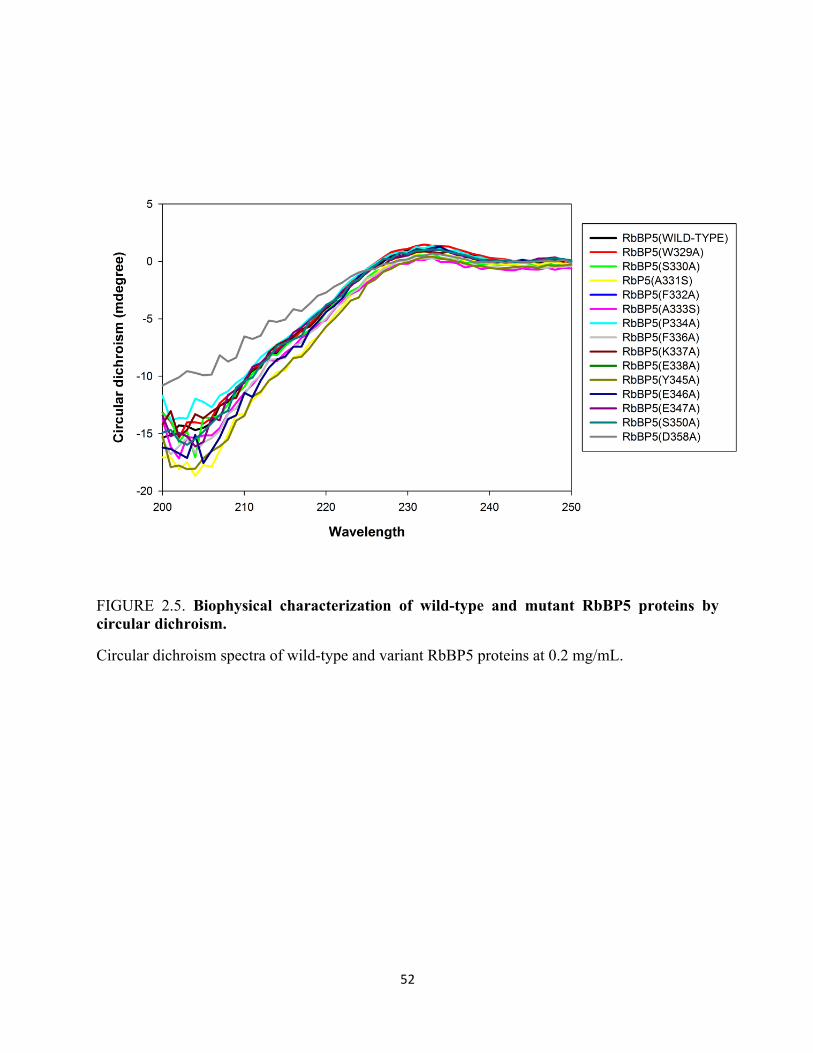
52
FIGURE 2.5. Biophysical characterization of wild-type and mutant RbBP5 proteins by circular dichroism.
Circular dichroism spectra of wild-type and variant RbBP5 proteins at 0.2 mg/mL.
53
Relative amounts of unmodified, monomethylated, and di-methylated H3K4 methylated species
were quantitated over 24 hours. As negative controls, experiments containing only the MLL1
SET domain or the MLL1 core complex lacking the RbBP5 subunit were also performed.
Similar to previous results with the MLL1 SET domain, the MLL1 SET domain displayed only
mono-methyltransferase activity when using the unmodified histone H3 peptide substrate
(Supplementary Figure 5, (S5)). Additionally consistent with earlier findings, absence of the
RbBP5 subunit in the MLL1 core complex (MWAD) results in loss of H3K4 dimethylation
(Figure 2.6). Unexpectantly, a number of mutants that were identified to have reduced activity in
the high-throughput assay when tested for activity in the MALDI-TOF methyltransferase assay
displayed enzymatic activity similar to that of the wild-type complex. Discrepancies between
these two assays could potentially be attributed to differences in the substrates in the two assays,
as well as the use of far less 3H- SAM in the high-throughput assay, as opposed to the MALDI-
TOF based methyltransferase activity assay. Figures showing the enzymatic activity and kinetic
behavior of these RbBP5 mutants along with any biophysical characterizations of these proteins
are included in the appendices. However, given that the primary interest of this research is
focused on identifying residues involved in the stimulation of the MLL1 mediated
dimethyltransferase activity, the remaining parts of this chapter will focus on the mutations that
displayed decreased dimethyltransferase activity in the high-throughput assay whose decreased
activity were validated in separate experiments via the MALDI-TOF based methyltransferase
assay.
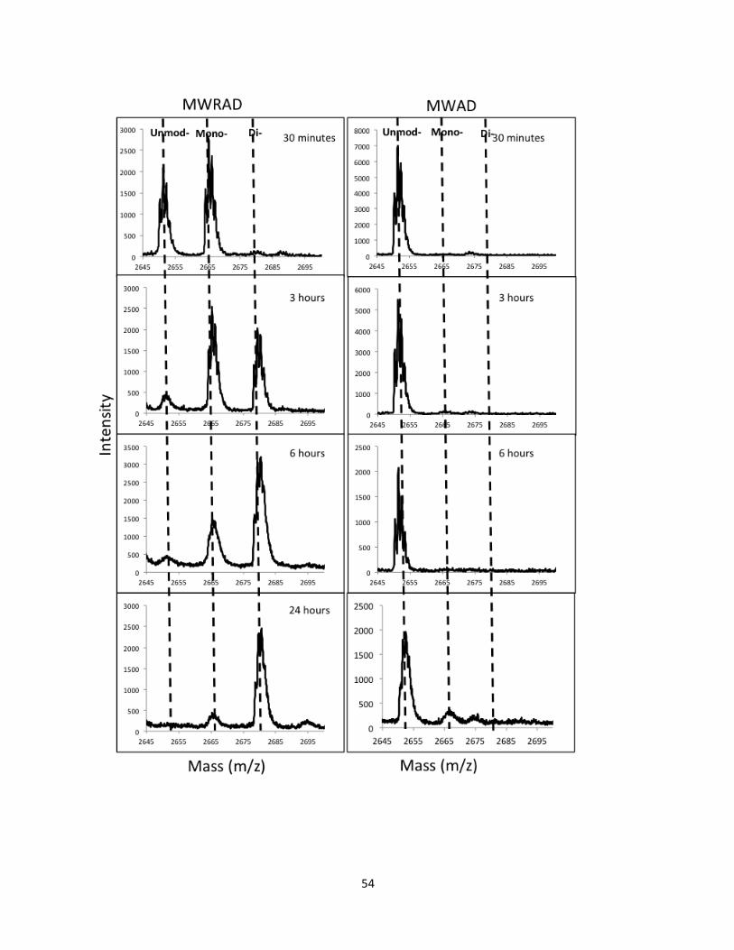
54
55
FIGURE 2.6. MALDI-TOF mass spectrometry spectra of MWRAD and MWAD complexes.
MALDI-TOF mass spectrometry of enzymatic reactions at 30 minutes, 3 hours, 6 hours and 24 hours. MALDI-TOF spectra in the left panel corresponds to the MLL1 core complex enzymatic activity of the complex assembled with full-length wild-type RbBP5. MALDI-TOF spectra in the right panel corresponds to the MLL1 core complex enzymatic activity of the complex assembled in the absence of full-length wild-type RbBP5.
56
Despite these discrepancies, we still were able to identify residues in RbBP5 that when
mutated showed reduced dimethyltransferase activity. These residues include W329, F332, F336,
and E347. In order to determine how these residues are specifically influencing the kinetic
behavior of the MLL1 core complex, we used quantitative MALDI-TOF mass spectrometry to
monitor the kinetic progression of H3K4 mono-, di- and tri-methylation of a histone H3 peptide
under pre-steady state conditions. The average data obtained based on triplicate experiments
were fitted with equations for kinetic models described for multiple irreversible consecutive
reactions using the program, Dynafit. Using the following model, we were able to determine
pseudo first order rate constants for the first and second methylation events for each individual
mutant RbBP5 protein in the MLL1 complex (Figure 2 .7).
H3K4 → H3K4me1 → H3K4me2
We were able to fit the MWRAD profiles with or without the addition of RbBP5 variant proteins
by using the three equations listed below (Patel et al., 2009). The results of kinetic parameters
obtained are summarized in Table 2.3.
1. [A]= [A]0 exp(-k1t)
2. [B]= [A]0 k1 ×[exp(-k1t)− exp(-k2t)
k2- k1
3. [C]= [A]0 {1+1 [k2 exp(-k1t)-k1-k1exp(-k2t)]
k1-k2
k1 k2
57
[A]0 is the concentration of the unmodified peptide at time (t) zero. [B] and [C] represent the
concentrations of the monomethylated and di-methylated species in single turnover curves,
respectively. The terms k1 and k2 represent the pseudo first order rate constants for the
conversion of A è B and BèC respectively (Patel et al., 2009; Patel et al., 2011). As shown in
Figure 2.7 b, replacing W329 with alanine results in ~5-fold reduction in the k1 rate and a ~25
fold reduction in the k2 rate compared to the MLL1 core complex assembled with the wild-type
RbBP5. Replacing, F336 with an alanine results in a ~3-fold reduction in the k1 rate and ~23-
fold reduction in the k2 rate of the reaction. Significantly, replacement of E347 with alanine
results in almost complete abolishment of the k2 rate and a 56-fold reduction in k1. Interestingly,
replacement of F332 with alanine in RbBP5 increases the overall k1 rate, but results in a 7-fold
decrease in the overall k2 rate. Taken together, these results suggest that the residues, W329,
F332, F336, and E347 are critical for the methylation kinetics of the MLL1 core complex.
Collectively, these results suggest direct participation by the RbBP5 component in H3K4 di-
methylation.
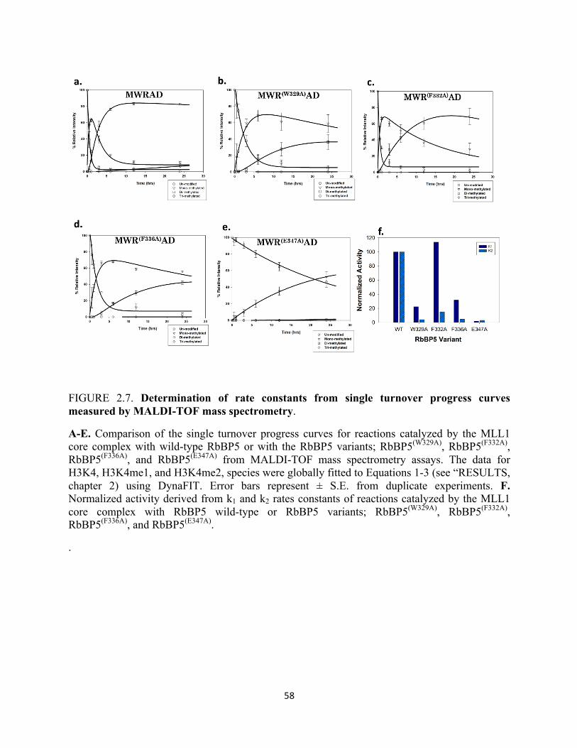
58
FIGURE 2.7. Determination of rate constants from single turnover progress curves measured by MALDI-TOF mass spectrometry.
A-E. Comparison of the single turnover progress curves for reactions catalyzed by the MLL1 core complex with wild-type RbBP5 or with the RbBP5 variants; RbBP5(W329A), RbBP5(F332A), RbBP5(F336A), and RbBP5(E347A) from MALDI-TOF mass spectrometry assays. The data for H3K4, H3K4me1, and H3K4me2, species were globally fitted to Equations 1-3 (see “RESULTS, chapter 2) using DynaFIT. Error bars represent ± S.E. from duplicate experiments. F. Normalized activity derived from k1 and k2 rates constants of reactions catalyzed by the MLL1 core complex with RbBP5 wild-type or RbBP5 variants; RbBP5(W329A), RbBP5(F332A), RbBP5(F336A), and RbBP5(E347A).
.
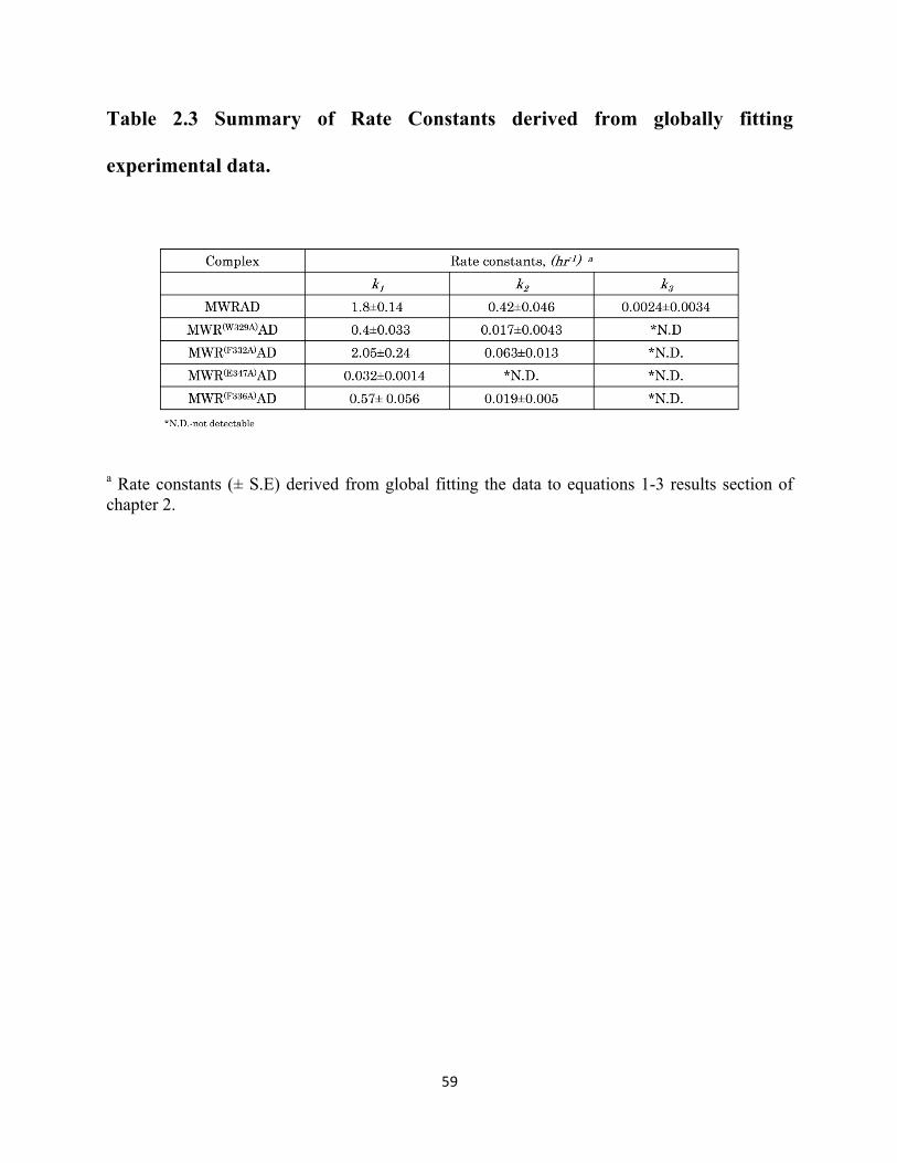
59
Table 2.3 Summary of Rate Constants derived from globally fitting
experimental data.
a Rate constants (± S.E) derived from global fitting the data to equations 1-3 results section of chapter 2.
60
2.5 Alanine substitutions in RbBP5 at positions W329, F332, F366, and E347 disrupt WRAD’s
ability to bind MLL1
To determine whether the mutations generated affected the ability of RbBP5 variants to
form the WRAD sub-complex and MLL1 core complex, sedimentation velocity analytical
ultracentrifugation was used to compare wild-type and variant RbBP5 proteins for their ability to
interact with members of the complex. Based on these experiments, we show that compared to
the wild-type WRAD complex, sedimentation velocity AUC reveals that the W329A, F332A,
F336A, and E347A, proteins are all capable of assembling into the WRAD complex with
sedimentation coefficients similar to that of wild type RbBP5 (Figures 2.8-2.10). This suggests
that mutations at positions W329A, F332A, F336A (data not shown), and E347A, do not
interfere with the mutant proteins’ ability to form WRAD. Varying the concentrations produces
slight shifts in the WRAD complexes assembled with the different mutations suggesting that the
interactions are less stable within the WRAD complex. This behavior is still consistent with the
conclusion that these mutations are not affecting the overall stability of the WRAD complex, as
previously published reports observe the same type of behavior over a similar concentration
range for wild-type WRAD (Patel et al., 2009; Patel et al., 2011).
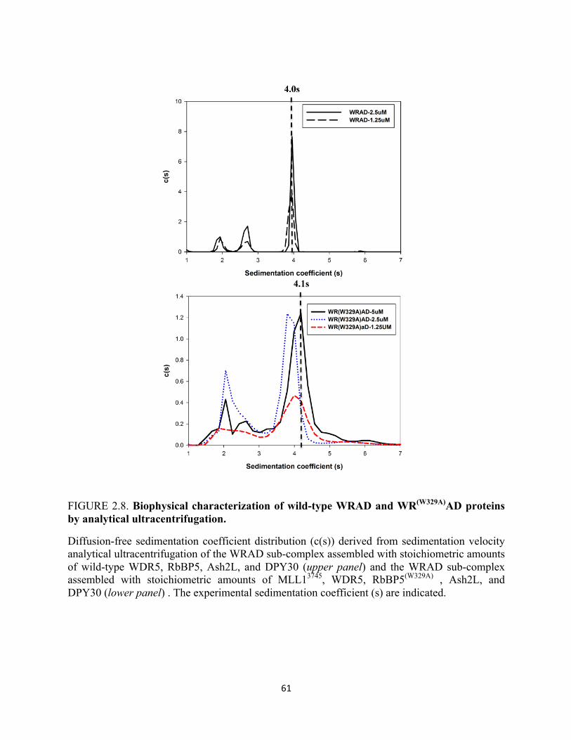
61
FIGURE 2.8. Biophysical characterization of wild-type WRAD and WR(W329A)AD proteins by analytical ultracentrifugation.
Diffusion-free sedimentation coefficient distribution (c(s)) derived from sedimentation velocity analytical ultracentrifugation of the WRAD sub-complex assembled with stoichiometric amounts of wild-type WDR5, RbBP5, Ash2L, and DPY30 (upper panel) and the WRAD sub-complex assembled with stoichiometric amounts of MLL13745, WDR5, RbBP5(W329A) , Ash2L, and DPY30 (lower panel) . The experimental sedimentation coefficient (s) are indicated.
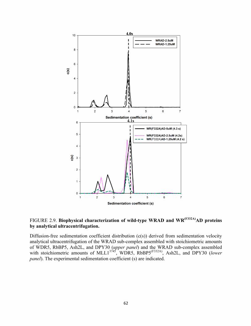
62
FIGURE 2.9. Biophysical characterization of wild-type WRAD and WR(F332A)AD proteins by analytical ultracentrifugation.
Diffusion-free sedimentation coefficient distribution (c(s)) derived from sedimentation velocity analytical ultracentrifugation of the WRAD sub-complex assembled with stoichiometric amounts of WDR5, RbBP5, Ash2L, and DPY30 (upper panel) and the WRAD sub-complex assembled with stoichiometric amounts of MLL13745, WDR5, RbBP5(F332A), Ash2L, and DPY30 (lower panel). The experimental sedimentation coefficient (s) are indicated.
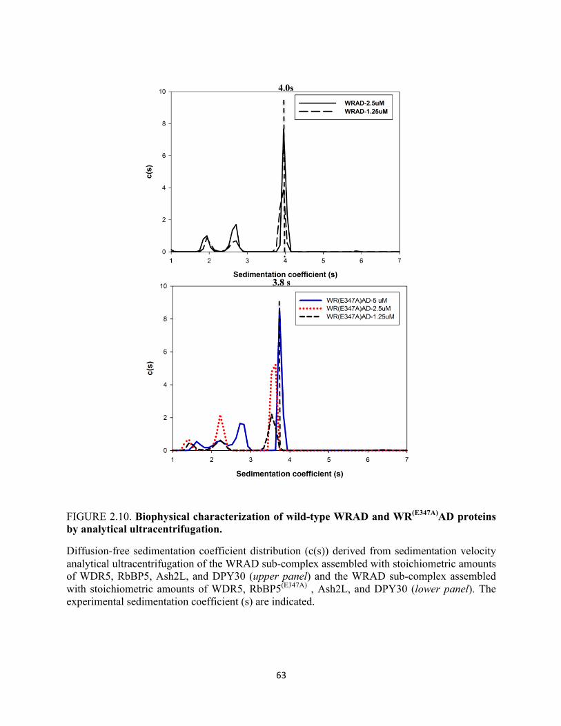
63
FIGURE 2.10. Biophysical characterization of wild-type WRAD and WR(E347A)AD proteins by analytical ultracentrifugation.
Diffusion-free sedimentation coefficient distribution (c(s)) derived from sedimentation velocity analytical ultracentrifugation of the WRAD sub-complex assembled with stoichiometric amounts of WDR5, RbBP5, Ash2L, and DPY30 (upper panel) and the WRAD sub-complex assembled with stoichiometric amounts of WDR5, RbBP5(E347A) , Ash2L, and DPY30 (lower panel). The experimental sedimentation coefficient (s) are indicated.

64
We next added stoichiometric amounts of MLL1 to the individual WRAD complexes,
and tested for their ability to interact with the MLL1 SET domain (Figure 2.11-2.14).
Sedimentation velocity analytical ultracentrifugation reveals that the addition of MLL1 to the
WR(W329A)AD sub complex does not shift the sedimentation coefficient of WRAD indicating
that this complex is unable to bind the MLL1 SET domain (Figure 2.11). Similar results are
obtained when MLL1 is added in a stoichiometric amount to the WR(F332A)AD, and
WR(E347A)AD complexes (Figure 2.12 and 2.14). A slight difference in behavior is observed
when MLL1 is added to the WR(F336A)AD complex (Figure 2.13). At a higher concentration,
the complex sediments with an s- value intermediate between wild-type WRAD and the wild-
type MLL1 core complex (Figure 2.13). Dilution of this mixture, resulted in the sedimentation
peak shifting to a lower s value, suggesting that the interaction between the MLL1 and
WR(F336A)AD dissociates on a relatively rapid time scale when compared to the time scale of
sedimentation (Figure 2.13).
In addition, to test if there are some weak interactions that exist between
WR(W329A)AD and the MLL1 SET domain, the MLL1 protein was added in excess to WRAD.
Titration of excess MLL1 reveals no shift in the WR(W329A)AD sedimentation profile
consistent with the hypothesis that MLL1 does not interact with WRAD (Figure 2.15.). As a
whole, these results suggests that although the interaction is much weaker for the
WR(F336A)AD complex between the MLL1 SET domain, the WR(F336A)AD still can form
some contacts with the MLL1 SET domain. In summary, these results provide evidence that the
residues W329, F332, F336, and E347 are essential for interaction between MLL1 and WRAD
and are indispensable for H3K4 dimethyltransferase activity of the MLL1 core complex.
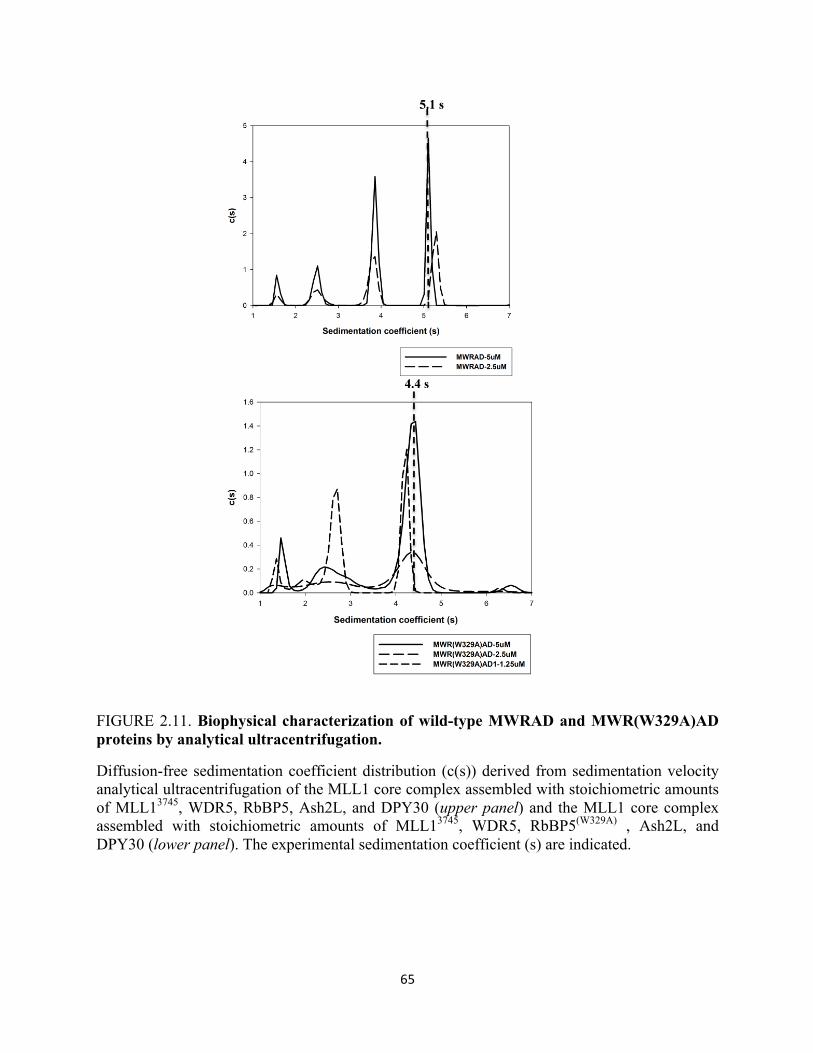
65
FIGURE 2.11. Biophysical characterization of wild-type MWRAD and MWR(W329A)AD proteins by analytical ultracentrifugation.
Diffusion-free sedimentation coefficient distribution (c(s)) derived from sedimentation velocity analytical ultracentrifugation of the MLL1 core complex assembled with stoichiometric amounts of MLL13745, WDR5, RbBP5, Ash2L, and DPY30 (upper panel) and the MLL1 core complex assembled with stoichiometric amounts of MLL13745, WDR5, RbBP5(W329A) , Ash2L, and DPY30 (lower panel). The experimental sedimentation coefficient (s) are indicated.
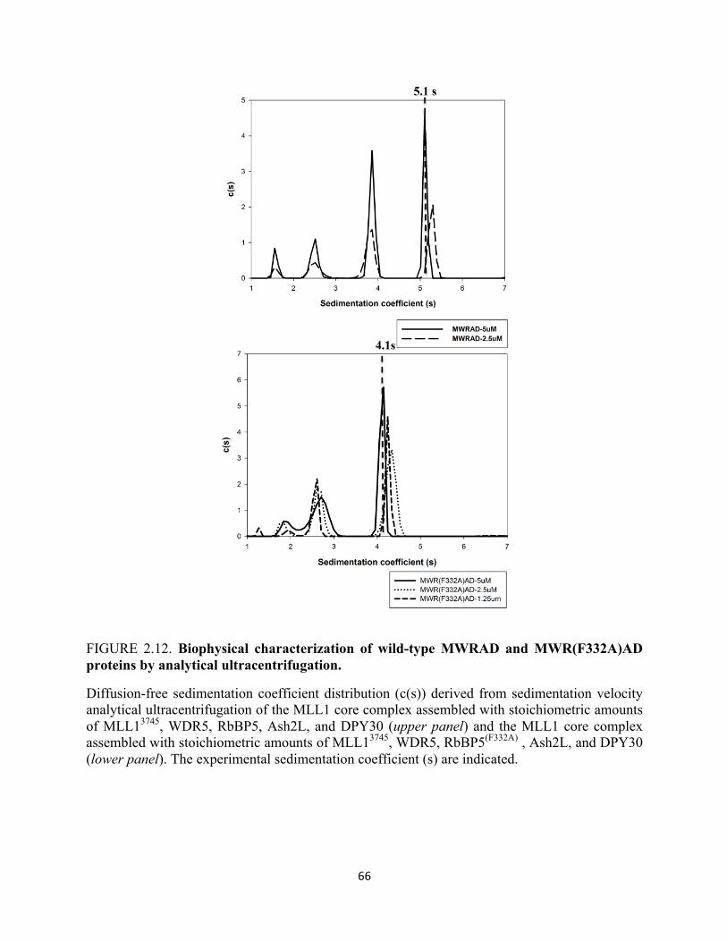
66
FIGURE 2.12. Biophysical characterization of wild-type MWRAD and MWR(F332A)AD proteins by analytical ultracentrifugation.
Diffusion-free sedimentation coefficient distribution (c(s)) derived from sedimentation velocity analytical ultracentrifugation of the MLL1 core complex assembled with stoichiometric amounts of MLL13745, WDR5, RbBP5, Ash2L, and DPY30 (upper panel) and the MLL1 core complex assembled with stoichiometric amounts of MLL13745, WDR5, RbBP5(F332A) , Ash2L, and DPY30 (lower panel). The experimental sedimentation coefficient (s) are indicated.
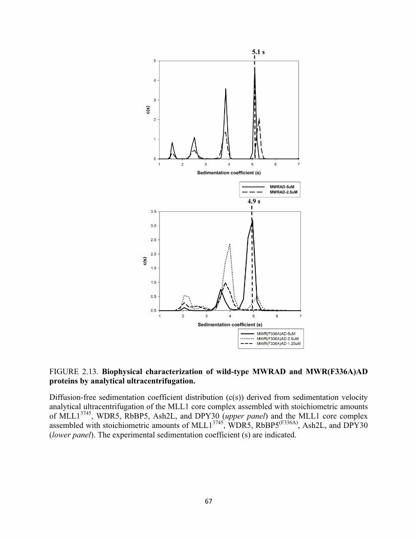
67
FIGURE 2.13. Biophysical characterization of wild-type MWRAD and MWR(F336A)AD proteins by analytical ultracentrifugation.
Diffusion-free sedimentation coefficient distribution (c(s)) derived from sedimentation velocity analytical ultracentrifugation of the MLL1 core complex assembled with stoichiometric amounts of MLL13745, WDR5, RbBP5, Ash2L, and DPY30 (upper panel) and the MLL1 core complex assembled with stoichiometric amounts of MLL13745, WDR5, RbBP5(F336A), Ash2L, and DPY30 (lower panel). The experimental sedimentation coefficient (s) are indicated.
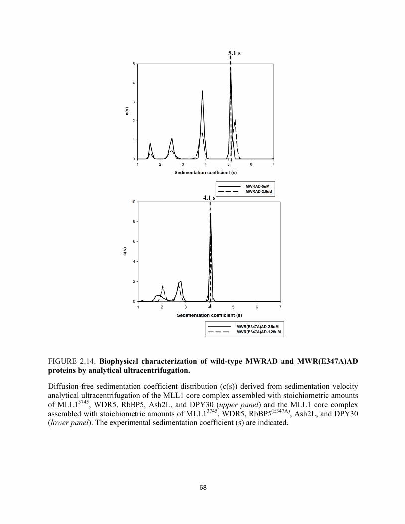
68
FIGURE 2.14. Biophysical characterization of wild-type MWRAD and MWR(E347A)AD proteins by analytical ultracentrifugation.
Diffusion-free sedimentation coefficient distribution (c(s)) derived from sedimentation velocity analytical ultracentrifugation of the MLL1 core complex assembled with stoichiometric amounts of MLL13745, WDR5, RbBP5, Ash2L, and DPY30 (upper panel) and the MLL1 core complex assembled with stoichiometric amounts of MLL13745, WDR5, RbBP5(E347A), Ash2L, and DPY30 (lower panel). The experimental sedimentation coefficient (s) are indicated.
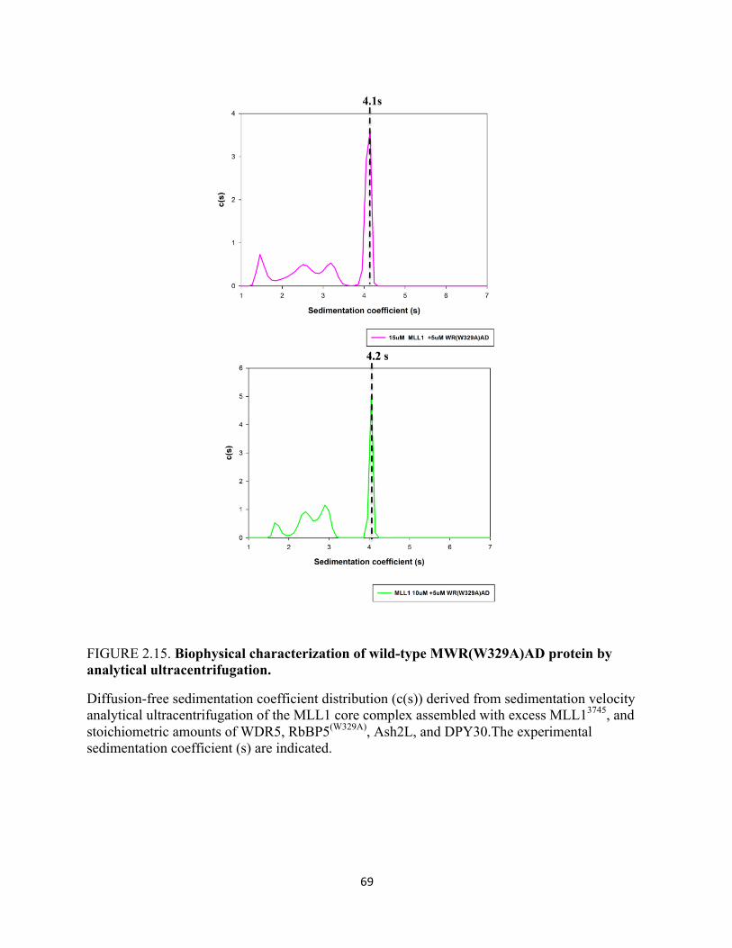
69
FIGURE 2.15. Biophysical characterization of wild-type MWR(W329A)AD protein by analytical ultracentrifugation.
Diffusion-free sedimentation coefficient distribution (c(s)) derived from sedimentation velocity analytical ultracentrifugation of the MLL1 core complex assembled with excess MLL13745, and stoichiometric amounts of WDR5, RbBP5(W329A), Ash2L, and DPY30.The experimental sedimentation coefficient (s) are indicated.

70
The results presented emphasize a more intricate role of MLL1 SET domain methyltransferase
activity than previously thought.
Notably, three out of the four residues identified in the high-throughput screen important
for interaction with MLL1 were aromatic residues. Intrigued by this finding, we wondered if the
loss of activity observed by replacing the aromatic residues with an alanine replacement could be
rescued with another aromatic residue. Therefore, we replaced W329 with both phenylalanine
and tyrosine. We also replaced the conserved tryptophan residue with an arginine, an amino acid
residue with some aromatic properties, to see what effect these residues would have on the
enzymatic activity and the assembly of the MLL1 core complex. We hypothesized that
replacement of tryptophan at position 329 with aromatic residues would restore di-
methyltransferase activity, and restore the ability of the RbBP5 protein to incorporate back into
the MLL1 core complex. Therefore, we expressed and purified these proteins as previously
described. CD spectra of these variant proteins show that the mutations do not introduce
secondary structure alterations (Figure 2.16). Methylation kinetics were monitored under pre-
steady state conditions using quantitative MALDI-TOF mass spectrometry.
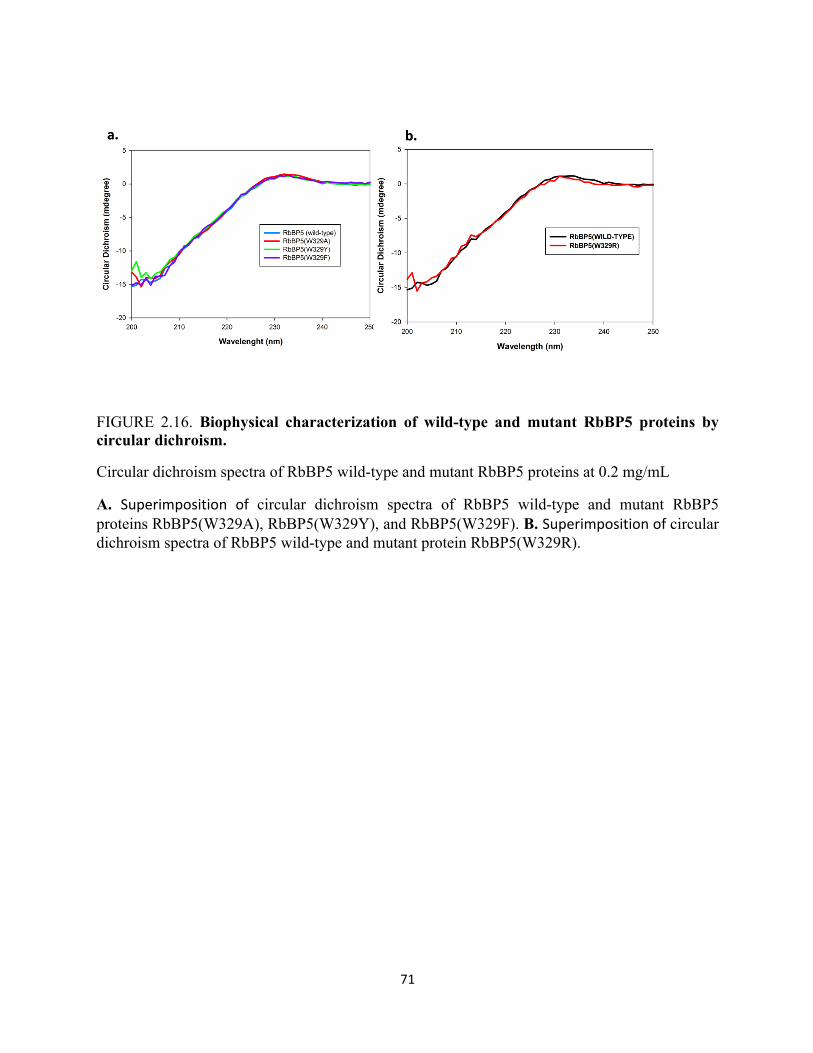
71
FIGURE 2.16. Biophysical characterization of wild-type and mutant RbBP5 proteins by circular dichroism.
Circular dichroism spectra of RbBP5 wild-type and mutant RbBP5 proteins at 0.2 mg/mL
A. Superimposition of circular dichroism spectra of RbBP5 wild-type and mutant RbBP5 proteins RbBP5(W329A), RbBP5(W329Y), and RbBP5(W329F). B. Superimposition of circular dichroism spectra of RbBP5 wild-type and mutant protein RbBP5(W329R).
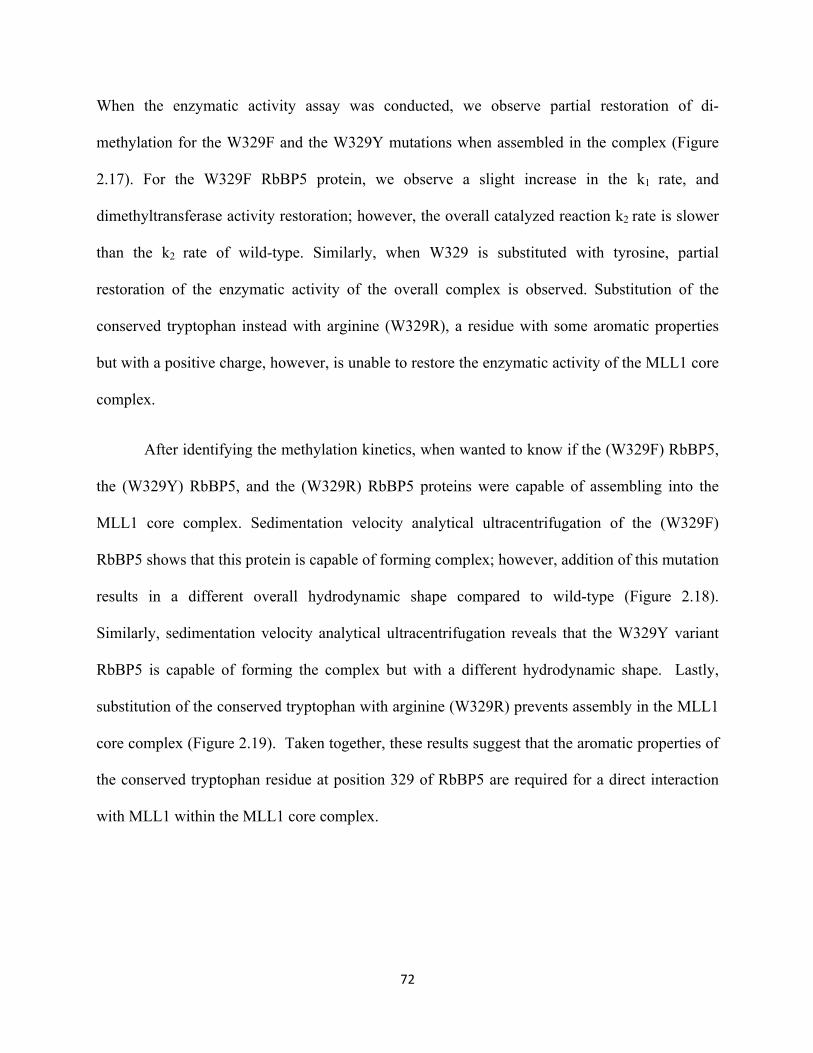
72
When the enzymatic activity assay was conducted, we observe partial restoration of di-
methylation for the W329F and the W329Y mutations when assembled in the complex (Figure
2.17). For the W329F RbBP5 protein, we observe a slight increase in the k1 rate, and
dimethyltransferase activity restoration; however, the overall catalyzed reaction k2 rate is slower
than the k2 rate of wild-type. Similarly, when W329 is substituted with tyrosine, partial
restoration of the enzymatic activity of the overall complex is observed. Substitution of the
conserved tryptophan instead with arginine (W329R), a residue with some aromatic properties
but with a positive charge, however, is unable to restore the enzymatic activity of the MLL1 core
complex.
After identifying the methylation kinetics, when wanted to know if the (W329F) RbBP5,
the (W329Y) RbBP5, and the (W329R) RbBP5 proteins were capable of assembling into the
MLL1 core complex. Sedimentation velocity analytical ultracentrifugation of the (W329F)
RbBP5 shows that this protein is capable of forming complex; however, addition of this mutation
results in a different overall hydrodynamic shape compared to wild-type (Figure 2.18).
Similarly, sedimentation velocity analytical ultracentrifugation reveals that the W329Y variant
RbBP5 is capable of forming the complex but with a different hydrodynamic shape. Lastly,
substitution of the conserved tryptophan with arginine (W329R) prevents assembly in the MLL1
core complex (Figure 2.19). Taken together, these results suggest that the aromatic properties of
the conserved tryptophan residue at position 329 of RbBP5 are required for a direct interaction
with MLL1 within the MLL1 core complex.
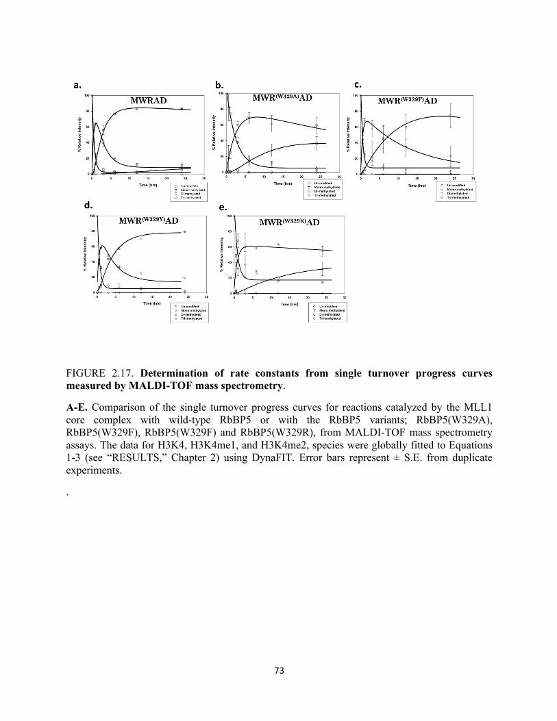
73
FIGURE 2.17. Determination of rate constants from single turnover progress curves measured by MALDI-TOF mass spectrometry.
A-E. Comparison of the single turnover progress curves for reactions catalyzed by the MLL1 core complex with wild-type RbBP5 or with the RbBP5 variants; RbBP5(W329A), RbBP5(W329F), RbBP5(W329F) and RbBP5(W329R), from MALDI-TOF mass spectrometry assays. The data for H3K4, H3K4me1, and H3K4me2, species were globally fitted to Equations 1-3 (see “RESULTS,” Chapter 2) using DynaFIT. Error bars represent ± S.E. from duplicate experiments.
.
74
Table 2.4 Summary of Rate Constants derived from globally fitting experimental data.
a Rate constants (± S.E) derived from global fitting the data to equations 1-3 results section of
chapter 2.
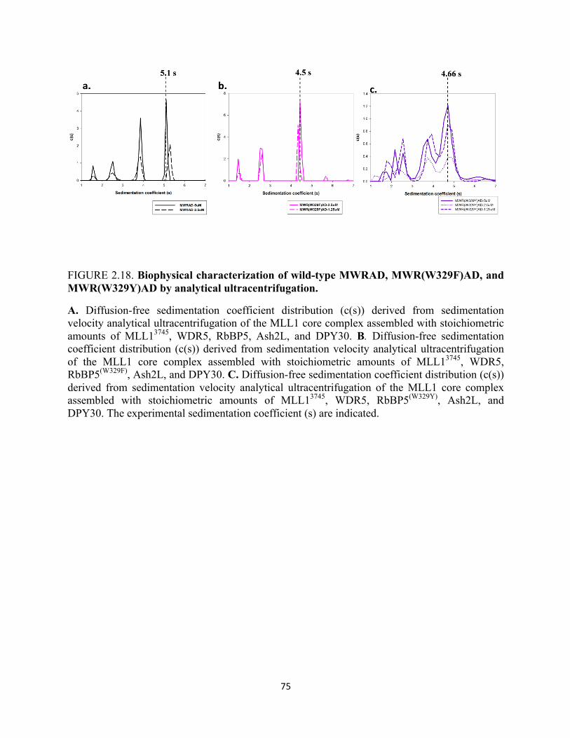
75
FIGURE 2.18. Biophysical characterization of wild-type MWRAD, MWR(W329F)AD, and MWR(W329Y)AD by analytical ultracentrifugation.
A. Diffusion-free sedimentation coefficient distribution (c(s)) derived from sedimentation velocity analytical ultracentrifugation of the MLL1 core complex assembled with stoichiometric amounts of MLL13745, WDR5, RbBP5, Ash2L, and DPY30. B. Diffusion-free sedimentation coefficient distribution (c(s)) derived from sedimentation velocity analytical ultracentrifugation of the MLL1 core complex assembled with stoichiometric amounts of MLL13745, WDR5, RbBP5(W329F), Ash2L, and DPY30. C. Diffusion-free sedimentation coefficient distribution (c(s)) derived from sedimentation velocity analytical ultracentrifugation of the MLL1 core complex assembled with stoichiometric amounts of MLL13745, WDR5, RbBP5(W329Y), Ash2L, and DPY30. The experimental sedimentation coefficient (s) are indicated.
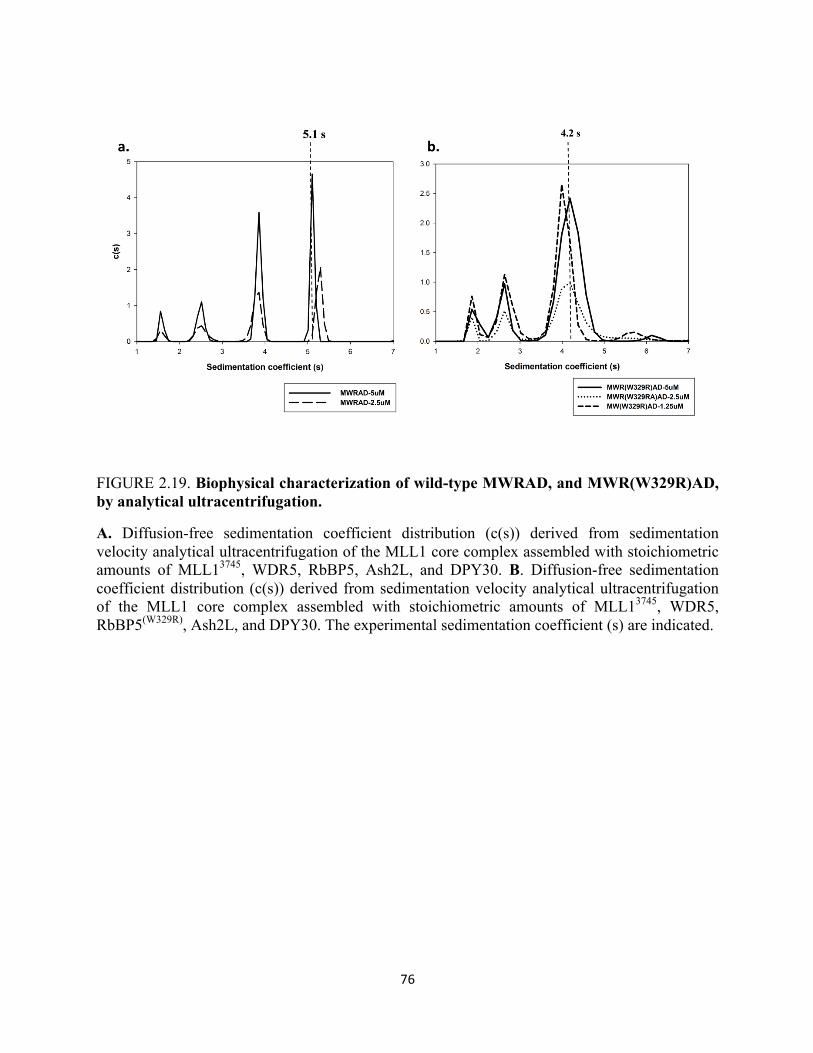
76
FIGURE 2.19. Biophysical characterization of wild-type MWRAD, and MWR(W329R)AD, by analytical ultracentrifugation.
A. Diffusion-free sedimentation coefficient distribution (c(s)) derived from sedimentation velocity analytical ultracentrifugation of the MLL1 core complex assembled with stoichiometric amounts of MLL13745, WDR5, RbBP5, Ash2L, and DPY30. B. Diffusion-free sedimentation coefficient distribution (c(s)) derived from sedimentation velocity analytical ultracentrifugation of the MLL1 core complex assembled with stoichiometric amounts of MLL13745, WDR5, RbBP5(W329R), Ash2L, and DPY30. The experimental sedimentation coefficient (s) are indicated.
77
2.6 Confirmation that Valine Residues 375 and 377 are Critical in the RbBP5-WDR5 Interaction
Previous reports have suggested that residues Valine 375 and Valine 377 of RbBP5
confer its binding to WDR5 (Odho et al., 2010). Taking a more stringent biophysical and kinetic
approach, I have been able to further characterize the WDR5-RbBP5 interaction. Given that
Valine 375 and Valine 377 of RbBP5 were identified as major residues recognized by WDR5 to
establish an interaction, I decided to mutate both Valine 375 and Valine 377 to alanine residues.
All plasmids were sequenced and verified to ensure that they contained the correct mutation and
no additional unwanted mutations. These proteins were recombinantly expressed in E.coli, and
purified as described in the experiment procedures section. Sedimentation velocity analytical
ultracentrifugation experiments were performed demonstrating that mutating Valine 375 and
Valine 377 to alanine residues did not distort the overall hydrodynamic shape of the mutant
proteins. As shown in Figure 2.20, sedimentation velocity analytical ultracentrifugation of the
isolated RbBP5 variants containing alanine substitutions at positions 375 and 377 reveal
monodispersed proteins in solution with an experimental sedimentation coefficient of 2.4 s like
that of the wild-type protein (Figure 2.20). Circular dichroism spectroscopy was also performed
to establish that the mutant proteins also displayed unaltered overall secondary structures. As
show in Figure 2.20b there are no major conformation changes in CD spectra of the mutant
RbBP5 proteins when compared to the wild-type protein.
After confirmation that these mutations did not disrupt the overall structure of RbBP5, I
wanted to determine how these proteins influence the enzymatic behavior of the MLL1 core
complex when assembled in the presence of other interacting proteins. To do so, MALDI-TOF
methyltransferase assays were conducted and rate constants by globally fitting single turnover
kinetic curves measured by the MALDI-TOF mass spectrometry were determined (Figure
78
2.21a). As shown in Figure 2.21 substituting Valine 375 with alanine results in ~2-fold reduction
in the k1 rate and a ~4-fold reduction in the k2 rate. Replacing, Valine 377 with an alanine results
in a small overall reduction in the k1 rate and an ~ 3- fold overall reduction in the k2 rate. These
results suggests that although Valine 375 and Valine 377 may establish important contacts with
WDR5, these are not the only contributions to this interaction, and the ability of the RbBP5-
Ash2L-DPY30 sub complex to interact with the MLL-WDR5, is not entirely abolished. The
most likely scenario is that the M-W sub complex is in rapid exchange with the RbBP5-Ash2L-
DPY30 sub complex and the activity observed is reminiscent of these transient interactions. To
determine whether the Valine mutations in RbBP5 disrupt its interactions with WDR5,
sedimentation velocity analytical ultracentrifugation was performed of stoichiometric amounts of
WDR5-RbBP5 binary complexes (Figure 2.22) Results suggest that mutations of Valines 375
and 377 do not completely abolish the protein’s ability to interact with WDR5. Taking an
alternative approach I have been able to validate the previous findings on the WDR5-RbBP5
interaction. All results here are consistent with previous findings.
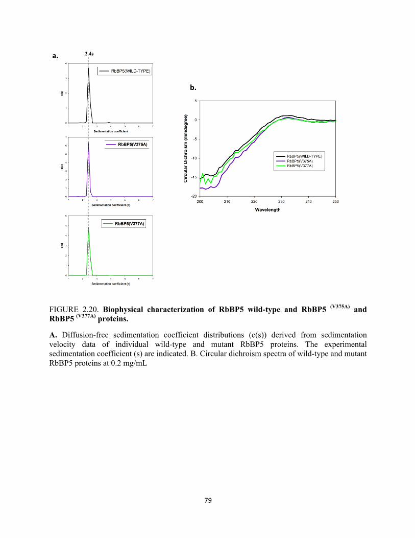
79
FIGURE 2.20. Biophysical characterization of RbBP5 wild-type and RbBP5 (V375A) and RbBP5 (V377A) proteins.
A. Diffusion-free sedimentation coefficient distributions (c(s)) derived from sedimentation velocity data of individual wild-type and mutant RbBP5 proteins. The experimental sedimentation coefficient (s) are indicated. B. Circular dichroism spectra of wild-type and mutant RbBP5 proteins at 0.2 mg/mL
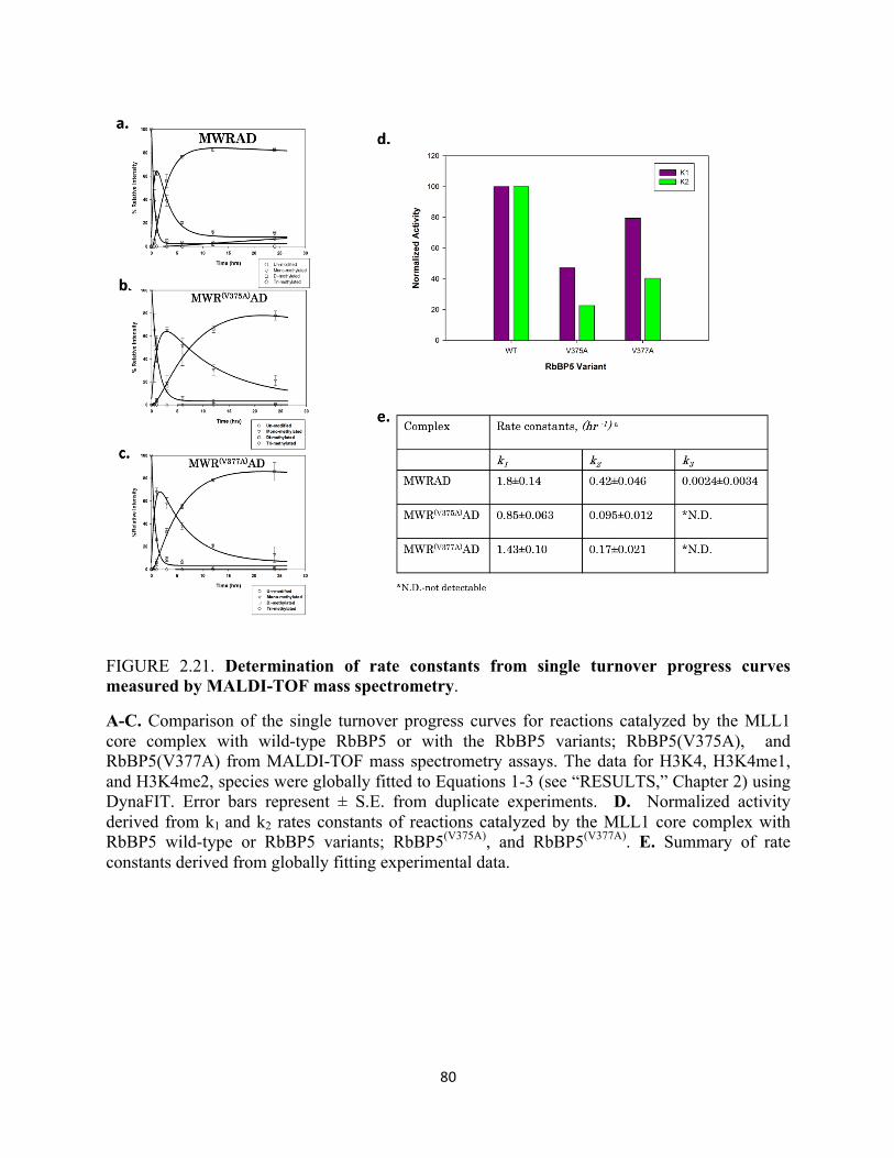
80
FIGURE 2.21. Determination of rate constants from single turnover progress curves measured by MALDI-TOF mass spectrometry.
A-C. Comparison of the single turnover progress curves for reactions catalyzed by the MLL1 core complex with wild-type RbBP5 or with the RbBP5 variants; RbBP5(V375A), and RbBP5(V377A) from MALDI-TOF mass spectrometry assays. The data for H3K4, H3K4me1, and H3K4me2, species were globally fitted to Equations 1-3 (see “RESULTS,” Chapter 2) using DynaFIT. Error bars represent ± S.E. from duplicate experiments. D. Normalized activity derived from k1 and k2 rates constants of reactions catalyzed by the MLL1 core complex with RbBP5 wild-type or RbBP5 variants; RbBP5(V375A), and RbBP5(V377A). E. Summary of rate constants derived from globally fitting experimental data.
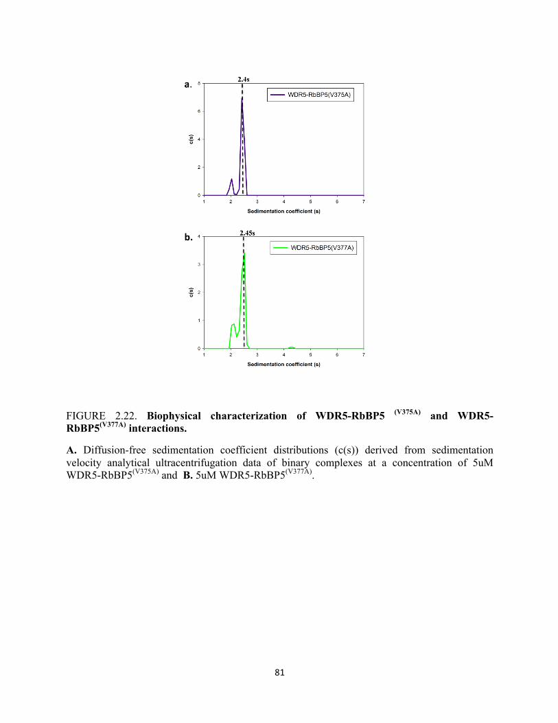
81
FIGURE 2.22. Biophysical characterization of WDR5-RbBP5 (V375A) and WDR5-RbBP5(V377A) interactions.
A. Diffusion-free sedimentation coefficient distributions (c(s)) derived from sedimentation velocity analytical ultracentrifugation data of binary complexes at a concentration of 5uM WDR5-RbBP5(V375A) and B. 5uM WDR5-RbBP5(V377A).
82
DISCUSSION:
Histone H3K4 methylation is a prevalent mark that positively regulates transcriptional
activation. The SET1 family of enzymes, which include MLL1-MLL4 and SET1A and SET1B
carry out the bulk of H3K4 di- and trimethylation in vivo. As noted above, studies on the
representative member of this family, MLL1, has provided several insights on mechanistic
determinants underlying the regulation of multiple lysine methylation. In common with the vast
majority of lysine methyltransferases, MLL1 contains an evolutionarily conserved SET domain
required for its enzymatic activity. However, unlike most other lysine methyltransferase, the
MLL1/SET1 class of enzymes can only catalyze multiple methylation in the context of a multi-
subunit core configuration of proteins, which includes WDR5, RbBP5, Ash2L, and DPY30.
These proteins can exist independent of MLL1 and form a sub -complex called WRAD. WRAD
binds in close proximity to the catalytic SET domain of MLL1 and together forms what we call
the MLL1 core complex. Currently, there are two proposed models that have emerged detailing
the mechanism behind multiple methylation catalyzed by the MLL1 core complex. The first
model “the one active site model” posits that mono-, di-, and tri-methylation of H3K4 can be
attributed solely to the methyltransferase activity of the SET domain alone, and that the WRAD
module merely functions as an allosteric regulator of the MLL1 catalytic SET domain (Couture
et al., 2008; Dillon et al., 2005; Southall et al., 2009). However, it has been more recently
demonstrated that an isolated MLL1 SET domain is predominantly a mono-methyltransferase
(Patel et al., 2009). This is consistent with the hypothesis that the presence of a conserved
tyrosine in the active site of SET domain enzymes constrains them to be
monomethyltransferases. Unexpectantly, it has also been shown that WRAD forms an
83
independent sub-complex that possesses an intrinsic histone methyltransferase activity of its own
in the absence of the MLL1 SET domain (Patel et al., 2011). Furthermore, when WRAD is
assembled within the MLL1 core complex it is capable of catalyzing H3K4 di-methylation.
These studies have thereby lead to a new model by which the degree of methylation of histone
H3 at lysine 4 is regulated in a sequential fashion catalyzed by a multi-subunit complex that
contains two distinct active sites for the addition of each methyl group. In agreement with this
model, additional studies reveal that the loss of the WDR5, RbBP5, or Ash2L subunit of WRAD
results in the loss of di- methylation in vitro, without significant changes in H3K4 mono-
methylation. Taken together, these studies provide evidence that suggests members of the
conserved core configuration of proteins in the WRAD complex possess essential protein
structural features underlying the shared enzymatic activities of the MLL1 core complex.
Although, there has been a wealth of reports on the assembly and function of the MLL1 core
complex’s enzymatic activity, there is a lack of information that exists on the functions and
contributions of individual conserved components and their detailed characterizations.
In this thesis, I sought to thoroughly characterize the role of the RbBP5 hinge domain in
facilitating the di-methyltransferase activity in the MLL1 core complex. RbBP5 is a member of
the Rb pathway, and the absence of RbBP5 in the MLL1 core complex results in no observable
dimethylation in vitro (Patel et al., 2011). To begin to explore the role of this protein in the
MLL1 core complex di-methyltransferase activity, a series of RbBP5 constructs were engineered
to identify the minimal domain required for activity. A small fragment of RbBP5 consisting of
residues (323-402) was able to restore di-methylation near to wild-type levels. Further sequence
alignments reveal that this fragment contains an absolutely conserved stretch of amino acid
residues. High-throughput scanning alanine mutagenesis of this stretch identified a number of
84
residues that when mutated result in decreased levels of dimethylation relative to that of wild-
type. Further characterization of residues identified in this screen, reveal that residues W329,
F332, F336, and E347 are all required for di-methylation within the MLL1 core complex, as
mutations in any of these residues result in significantly reduced di-methyltransferase activity of
the complex. Surprisingly, when any of the mutations were assembled within the MLL1
complex, they diminish WRAD’s ability to directly interact with the MLL1 SET domain.
Although, previous studies have implicated interaction surfaces between RbBP5 and the MLL1
SET domain, to date there have been no studies that have clearly demonstrated potential surfaces
of RbBP5 involved in this interaction. Therefore, through extensive biochemical and biophysical
characterization of the RbBP5 hinge domain, we have been able to uncover a novel, previously
unidentified MLL1 interaction motif in this B-propeller protein that preferentially recognizes the
SET domain of MLL1. In light of our finding that three out of the four residues identified
responsible for contacts with MLL are aromatic, we propose that these residues may create a
hydrophobic interface with consensus hydrophobic residues on the surface of MLL1. Mutation
of conserved amino acids residues in the MLL1 SET domain will facilitate the identification
surfaces in MLL1 that confer its binding to RbBP5. Preferential binding of RbBP5 to MLL1
through this motif, may alter the conformation of the MLL1 SET domain, therefore enhancing
catalysis. We envision an intriguing scenario whereby the RbBP5 component binds to the MLL1
SET domain, so that these aromatic residues may directly participate in catalysis, by facilitating
the optimal alignment of the lysine substrate and the co-factor SAM (Figure 2.23). It has been
well- established from the most well-studied SET domains, that the formation of the substrate
lysine binding channel involves the inclusion of several aromatic residues which insert
themselves between histone H3 and s-adensoyl methionine to achieve a favorable physio-
85
chemical environment for the transfer of a methyl group from SAM to the amine group of lysine
four on H3. Strikingly, the recent crystal structure report of the MLL1 SET domain reveals that
the SET-I subdomain is not in an optimal alignment to form this archetypal binding site and
lacks the highly conserved aromatic residues. Therefore, it is quite possible that conserved
aromatic residues in RbBP5 elicit binding with MLL1 to achieve this key structural
characteristic. It is important to note that one residue, E347 identified in this thesis was neither
hydrophobic nor aromatic that was also shown to be important for binding MLL1, it’s role in the
catalysis remains to be explored. In our model, we also propose that in addition to binding to
MLL1 to form the hydrophobic channel that accommodates and connects the substrate and
cofactor, this channel also links the first active site to the second active site in WRAD for
transfer of the peptide substrate to WRAD. In this scenario, RbBP5 when in complex with other
members of WRAD, binds the MLL1 SET domain enhancing catalysis by first proper alignment
of substrate and cofactor and increasing the overall rate of mono-methylation, by forming the
hydrophobic channel, while simultaneously facilitating the transfer of the peptide to the second
active site in WRAD. In WRAD RbBP5 is needed for making important contacts between the
WDR5 and Ash2L subunits to mediate MLL1 core complex dimethyltransferase activity by
aiding in formation of the second active site in WRAD. The results presented in this thesis
suggests this mode of catalysis, however solving the high-resolution crystal structure of the
MLL1 core complex with bound peptide will prove imperative in determining if this mechanism
of catalysis is at play.
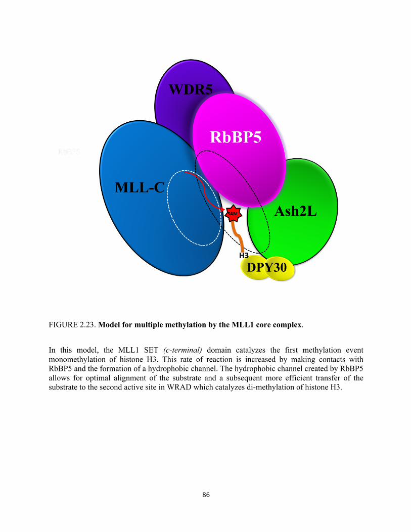
86
FIGURE 2.23. Model for multiple methylation by the MLL1 core complex.
In this model, the MLL1 SET (c-terminal) domain catalyzes the first methylation event monomethylation of histone H3. This rate of reaction is increased by making contacts with RbBP5 and the formation of a hydrophobic channel. The hydrophobic channel created by RbBP5 allows for optimal alignment of the substrate and a subsequent more efficient transfer of the substrate to the second active site in WRAD which catalyzes di-methylation of histone H3.
87
Chapter Three:
Crystallization Trials and Probing the Structure of
RbBP5 By Limited Proteolysis Identifies Two
Previously Uncharacterized Stable Subdomains
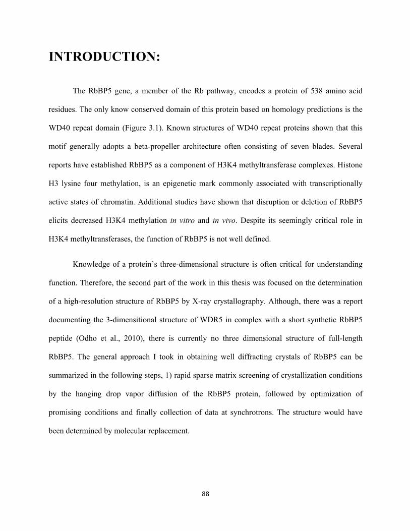
88
INTRODUCTION:
The RbBP5 gene, a member of the Rb pathway, encodes a protein of 538 amino acid
residues. The only know conserved domain of this protein based on homology predictions is the
WD40 repeat domain (Figure 3.1). Known structures of WD40 repeat proteins shown that this
motif generally adopts a beta-propeller architecture often consisting of seven blades. Several
reports have established RbBP5 as a component of H3K4 methyltransferase complexes. Histone
H3 lysine four methylation, is an epigenetic mark commonly associated with transcriptionally
active states of chromatin. Additional studies have shown that disruption or deletion of RbBP5
elicits decreased H3K4 methylation in vitro and in vivo. Despite its seemingly critical role in
H3K4 methyltransferases, the function of RbBP5 is not well defined.
Knowledge of a protein’s three-dimensional structure is often critical for understanding
function. Therefore, the second part of the work in this thesis was focused on the determination
of a high-resolution structure of RbBP5 by X-ray crystallography. Although, there was a report
documenting the 3-dimensitional structure of WDR5 in complex with a short synthetic RbBP5
peptide (Odho et al., 2010), there is currently no three dimensional structure of full-length
RbBP5. The general approach I took in obtaining well diffracting crystals of RbBP5 can be
summarized in the following steps, 1) rapid sparse matrix screening of crystallization conditions
by the hanging drop vapor diffusion of the RbBP5 protein, followed by optimization of
promising conditions and finally collection of data at synchrotrons. The structure would have
been determined by molecular replacement.
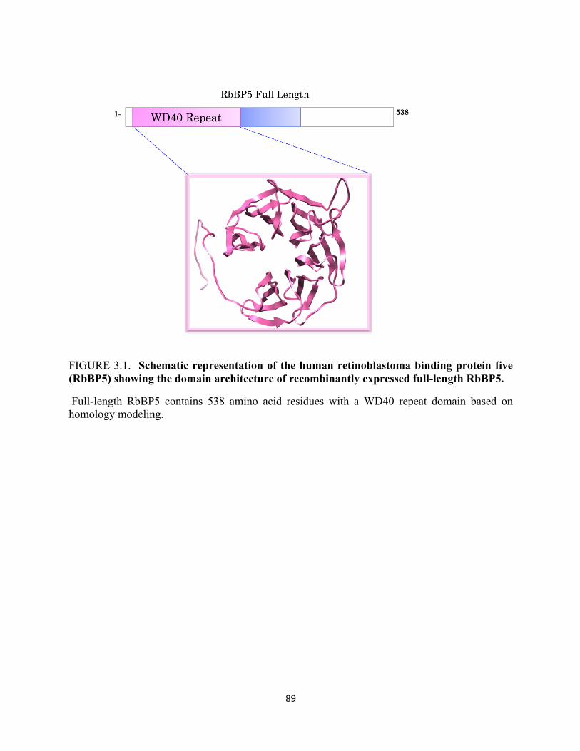
89
FIGURE 3.1. Schematic representation of the human retinoblastoma binding protein five (RbBP5) showing the domain architecture of recombinantly expressed full-length RbBP5.
Full-length RbBP5 contains 538 amino acid residues with a WD40 repeat domain based on homology modeling.
90
Perhaps, the most challenging part of this goal of my project was the crystallization and structure
determination of RbBP5 itself. Unfortunately, no suitable crystals were obtained by this first
approach taken on the full- length human RbBP5 protein. Therefore, I subjected the full -length
protein to limited proteolysis treatment to identify a stable construct that was amendable to
crystallization. From the limited proteolysis, I was able to obtain and identify two stable
subdomains of the RbBP5 protein. These minimal domains were re-cloned, purified, and
subjected to more sparse matrix screening crystallization trials. Although no crystals emerged
from early crystallization trials, I was able to biophysically characterization the intrinsic
properties of the two stable domains identified. The experiment procedures and results are
presented below.
EXPERIMENTAL PROCEDURES
Protein expression and purification: The RbBP5 gene was purchased from open biosystems and
subcloned into a pHis parallel vector which encodes a tobacco etch virus (TEV) cleavage N-
terminal 6x-His fusion. Versions of full-length human RbBP5 (1-538) and RbBP5 constructs
were overexpressed in Escherichia coli (Rosetta II, Novagen), by growing cells containing a
given plasmid at 37°C in Terrific Broth medium containing 50ug/mL carbenicillin. The
temperature was then lowered to 15 °C and cells were induced for 16–18 h with 750uM
isopropyl-1-thio-D-galactopyranoside (IPTG). Cells were harvested, re-suspended in lysis buffer
(50 mM Tris, pH 7.4, 300 mM NaCl, 3 mM dithiothreitol, 0.1 mM
phenylmethylsulfonylfluoride, and 1 EDTA-free protease inhibitor tablet (RocheApplied
Science)), lysed with a microfluidizer cell disrupter, and clarified by centrifugation. Supernatants
containing the RbBP5 His-tagged proteins were purified by nickel affinity chromatography
91
(HisTrap column, GE Healthcare). For the first step of purification, the crude lysate was passed
through a HisTrap column (GE healthcare) containing nickel beads. The bound 6x-His-RbBP5
was eluted from the column using a linear gradient of elution buffer containing 500mM
imidazole. The peak fractions that contained RbBP5 were collected, pooled, and then dialyzed
against column buffer that contains 50 mM Tris pH 7.4, 300mM NaCl, 30 mM Imidazole, and
3mM DTT at 4°C to remove the excess imidazole and to cleave the 6X-His tag in the presence of
TEV (Tobacco Etch Virus) protease. The dialysis buffer was changed three times. For the
second step of purification, the dialyzed protein was passed through the His-Trap column and the
flow through fractions that contained the untagged version of RbBP5 were pooled and combined.
As a final step of purification, the protein was passed through a gel filtration column (Superdex
200TM GE Healthcare) pre-equilibrated with the sample buffer containing 20mM Tris (pH 7.5),
300mM NaCl, 1mM Tris (2-carboxyethyl) phosphine (TCEP), and 1µM zinc chloride. All other
proteins used in methyltransferase assays were purified as previously described (Patel et al.,
2008).
Mutagenesis: Premature stop codons were introduced into human RbBP5 clones using the
QuickChange site directed mutagenesis kit (Stratagene) to create truncated RbBP5 constructs.
Plasmids were sequenced to verify the presence of the intended mutations and the absence of
additional mutations. Integrated DNA Technologies synthesized oligonucleotide primers used in
the mutagenesis.
Limited Proteolysis Experiments: Fresh Trypsin Stock was prepared at 0.2ng/uL. For maximum
of activity sequencing grade modified trypsin was resuspended in trypsin resuspension buffer
and the trypsin enzyme mixture was allowed to warm at room temperature for 15 minutes. 100uL
reactions with trypsin at a final concentration of 0.01ng/uL and a final protein concentration of
92
0.5mg/mL were prepared in 1.5mL micro centrifuge tubes. The ingredients were mixed gently by
flicking. Limited proteolysis of the protein was carried out in a 37 °C water bath. The reactions
were quenched at various time points by the addition of PMSF. SDS-PAGE followed by
coomassie staining was used to visualize any stable subdomains identified in the reaction
mixture at various times points and their approximate molecular weights. Experiments were
repeated in triplicate sets to confirm appropriate conditions for the limited proteolysis of the
protein. Reaction mixtures with stable subdomain masses were confirmed using ESI Mass
Spectrometry (Yale School of Medicine, W.M. Keck Biotechnology Resource Laboratory).
Sequences were confirmed using N-terminal sequencing (Johns Hopkins Institute for Nano
biotechnology, Proteometrics Synthesis and Sequencing Facility.)
MALDI-TOF mass spectrometry methyltransferase assay: Mass spectrometry assays were
conducted as mentioned earlier. 7 µM of enzyme was incubated with 250 µM s-adenosyl-
methionine and 10 µM histone H3 peptide (1-20) at 15 °C in 50mM Tris pH 9.0, 200mM NaCl,
3 mM DTT, and 5% glycerol. The reactions were quenched at various time points by the addition
of trifluoroacetic acid to 0.5%. The quenched samples were diluted 1:4 with α-cyano-4-
hydroxycinnamic acid. MALDI TOF mass spectrometry was performed on a Bruker AutoFlex
mass spectrometer (State University of New York, ESF) operated in reflectron mode. Final
spectra were averaged from 100 shots/position at 10 different positions.
Analytical Ultracentrifugation: Analytical ultracentrifugation experiments were carried out using
a Beckman Coulter ProteomeLabTM XL-A analytical ultracentrifuge equipped with absorbance
optics and a 4-hole An-50 Ti analytical rotor. Sedimentation velocity experiments were carried
out at 10°C and 50,000 rpm (200,000 x g) using 3 mm two sector charcoal-filled Epon
centerpieces with quartz windows. For each sample, 300 scans were collected with the time

93
interval between scans set to zero. Protein samples in 20mM Tris pH 7.5, 300mM NaCl, 1mM
TCEP and 1µM ZnCl2 were run at various concentrations as described in results. Sedimentation
boundaries were analyzed by the continuous distribution (c(s)) method using the program
SEDFIT. The program SEDNTERP version 1.09 was used to correct the experimental S-value
(s*) to standard conditions at 20°C in water (S20,w) and to calculate the partial specific volume of
each protein.
Circular Dichroism Spectroscopy: CD spectra were collected on an AVIV Model 420 ds
spectropolarimeter equipped with a Neslab CFT-33 refrigerated circulator at 10°C using a 0.1
mm path length cell. Spectra for each were obtained at a protein concentration of 0.2mg/mL in a
buffer contain 20mM TRIS (pH 7.5), 200 mM NaCl, 1mM TCEP and 1uM Zinc Chloride. The
background contribution of the buffer alone was subtracted from each protein spectrum.
RESULTS:
Through limited proteolysis of the RbBP5 protein (Figure 3.2), two stable subdomains
were identified and confirmed through the use of N-terminal sequencing, electrospray ionization
mass spectrometry, and an in silico digestion of predicted cleavage sites in full-length RbBP5.
These subdomains correspond to residues #1-495 and #1-337 in RbBP5. To characterize the
hydrodynamic properties of these constructs in isolation sedimentation velocity analytical
ultracentrifugation was performed. Sedimentation velocity profiles were fitted to an array of
Lamn equations solutions with a set of experimental parameters to determine the diffusion free
sedimentation coefficient distributions (c(s)). As show in Figure 3.3, RbBP5 (#1-495) sediments
as a monodispersed protein with a sedimentation coefficient like that of wild-type. (Figure 3.3)
CD spectra of this protein reveals that the deletion of the C-terminal residues does not induce any
94
drastic conformational changes in this protein (Figure 3.4). In contrast sedimentation velocity
analytical ultracentrifugation of the RbBP5 (#1-337) proteins reveals that this proteins sediments
as a monodispersed protein as well with a c(s) value of 2.16 (Figure 3.5). CD spectra of this
protein reveals, that the deletion of its C-terminus alters in overall secondary structural
conformation (Figure 3.6).
To determine the methylation kinetics of these two protein constructs, enzymatic assays
were set up under pre-steady state conditions, and methylation of an unmodified histone H3
peptide was monitored using MALDI-TOF mass spectrometry. As shown in Figure 3.7, the
overall k1 and k2 rates of reactions catalyzed by MWR(#1-495)AD are much slower compared to
that of the wild-type, although similar levels of dimethylation are observed after 24 hours. With
respect to MWR(#1-337)AD, a loss of di-methylation is readily observed with a slow mono-
methylation rate. This could possibly be attributed to less stability of the complex when the
constructs are assembled with the other components.
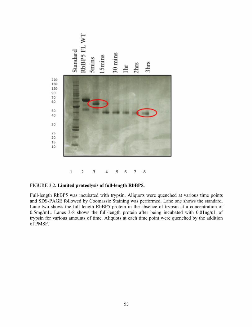
95
FIGURE 3.2. Limited proteolysis of full-length RbBP5.
Full-length RbBP5 was incubated with trypsin. Aliquots were quenched at various time points and SDS-PAGE followed by Coomassie Staining was performed. Lane one shows the standard. Lane two shows the full length RbBP5 protein in the absence of trypsin at a concentration of 0.5mg/mL. Lanes 3-8 shows the full-length protein after being incubated with 0.01ng/uL of trypsin for various amounts of time. Aliquots at each time point were quenched by the addition of PMSF.
1 2 3 4 5 6 7 8

96
FIGURE 3.3. Biophysical characterization of wild-type RbBP5 (#1-495) proteins by analytical ultracentrifugation.
Diffusion-free sedimentation coefficient distributions (c(s)) derived from sedimentation velocity data of the RbBP5 (#1-495) construct. The experimental sedimentation coefficient (s) are indicated.
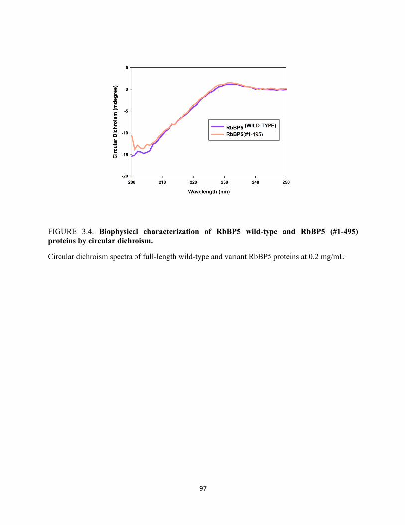
97
FIGURE 3.4. Biophysical characterization of RbBP5 wild-type and RbBP5 (#1-495) proteins by circular dichroism.
Circular dichroism spectra of full-length wild-type and variant RbBP5 proteins at 0.2 mg/mL
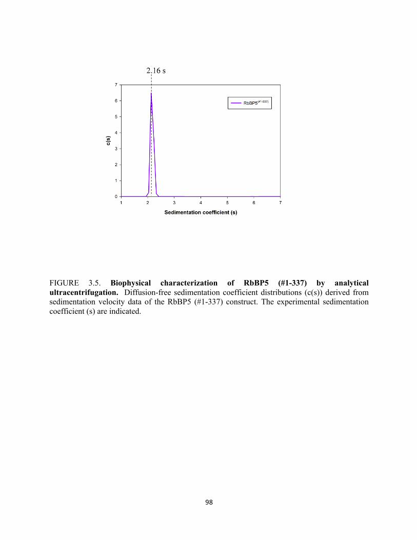
98
FIGURE 3.5. Biophysical characterization of RbBP5 (#1-337) by analytical ultracentrifugation. Diffusion-free sedimentation coefficient distributions (c(s)) derived from sedimentation velocity data of the RbBP5 (#1-337) construct. The experimental sedimentation coefficient (s) are indicated.
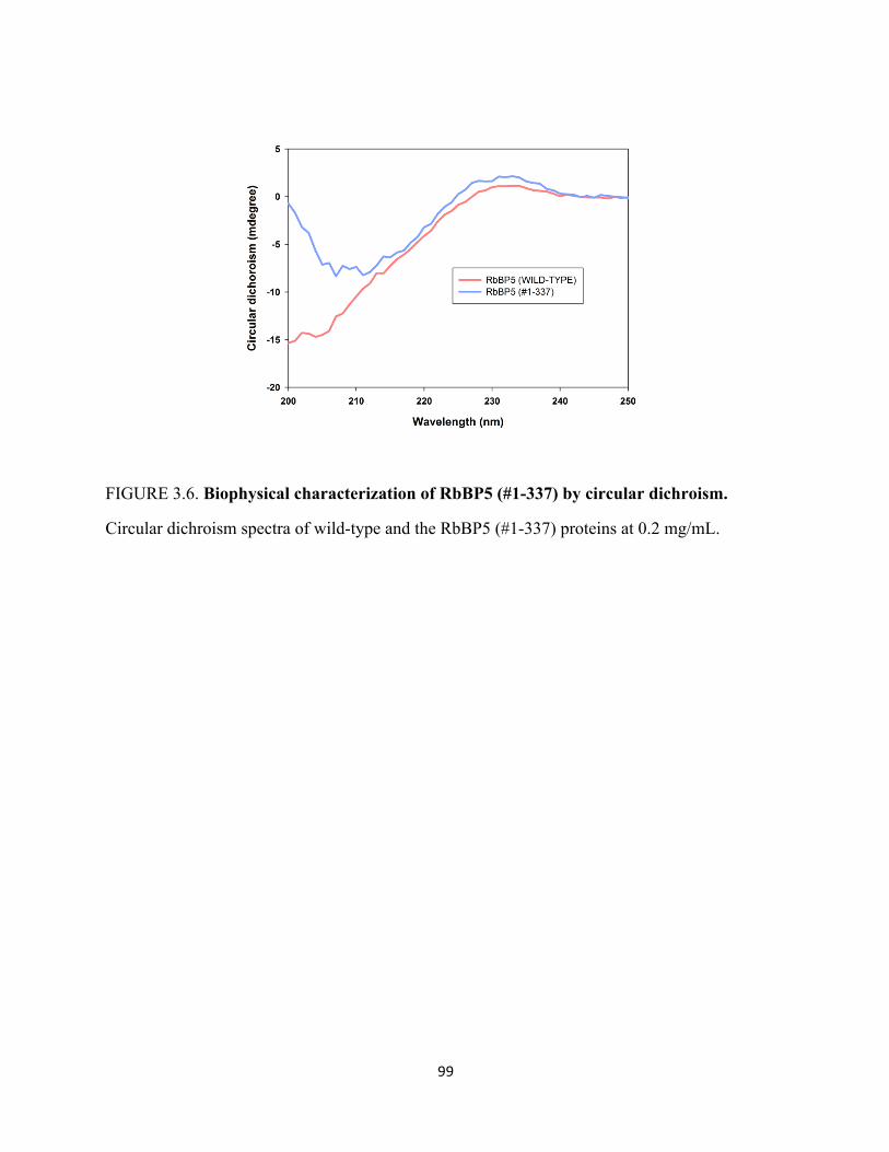
99
FIGURE 3.6. Biophysical characterization of RbBP5 (#1-337) by circular dichroism.
Circular dichroism spectra of wild-type and the RbBP5 (#1-337) proteins at 0.2 mg/mL.
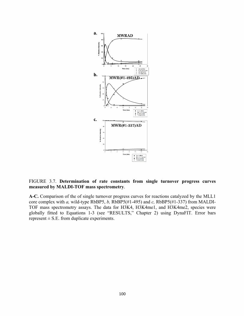
100
FIGURE 3.7. Determination of rate constants from single turnover progress curves measured by MALDI-TOF mass spectrometry.
A-C. Comparison of the of single turnover progress curves for reactions catalyzed by the MLL1 core complex with a, wild-type RbBP5, b, RbBP5(#1-495) and c, RbBP5(#1-337) from MALDI-TOF mass spectrometry assays. The data for H3K4, H3K4me1, and H3K4me2, species were globally fitted to Equations 1-3 (see “RESULTS,” Chapter 2) using DynaFIT. Error bars represent ± S.E. from duplicate experiments.
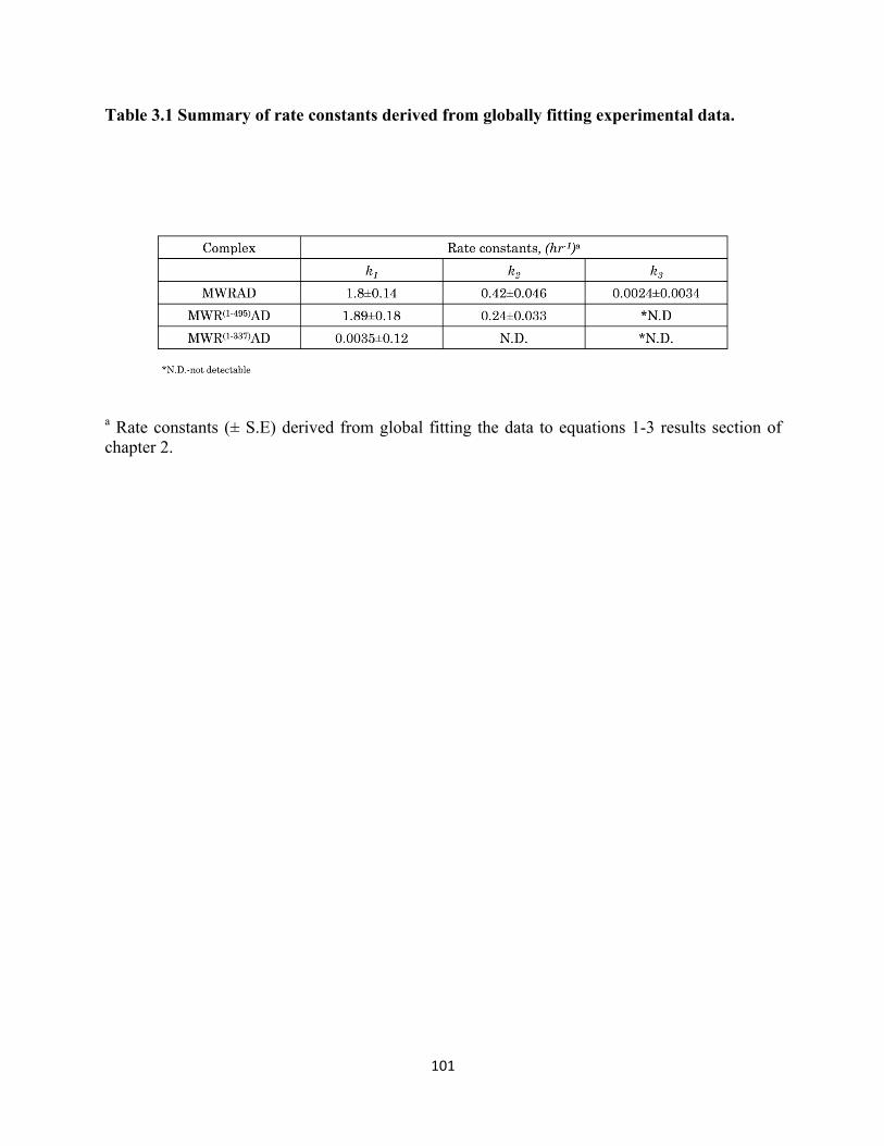
101
Table 3.1 Summary of rate constants derived from globally fitting experimental data.
a Rate constants (± S.E) derived from global fitting the data to equations 1-3 results section of chapter 2.

102
DISCUSSION:
The RbBP5 gene encodes a nuclear protein which belongs to a family of WD-repeat
proteins. The encoded protein binds directly to the retinoblastoma protein, which regulates cell
proliferation. Despite RbBP5’s critical role in mammalian methyltransferase complexes
relatively little is know about this protein’s involvement in this activity and currently there is no
high resolution structure of the protein available. One goal of this thesis was to attempt to solve
the three dimensional structure of RbBP5. After extensive sparse matrix screening on full-length
RbBP5, suitable crystals were not obtained.
Therefore, I chose to take an alternative approach utilizing limited proteolysis of full-
length RbBP5 in attempt to identify and obtain a stable truncated protein that may potentially be
more suitable for crystallization. Given that proteins generally have a modular structure
consisting of several distinctive domains tethered by flexible regions, I hypothesized that these
flexible regions could potentially be creating a source of heterogeneity preventing crystallization
with full-length RbBP5. One general aspect of protein crystallization that is universally
recognized is that rigid, stable proteins are much more likely to crystallize than proteins that are
internally flexible or have dynamic surfaces. Thus, deleting or re- moving the flexible regions at
the termini or within a protein may help in the crystallization by minimizing any interfering
effects from the micro heterogeneity. Taking a limited proteolysis approach, I was able to
identify two stable subdomains in RbBP5 consisting of residues #1-495 and residues #1-337.
These domains were cloned and characterized. These results presented in the aforementioned
further confirm domain mapping experiments performed by Anamika Patel. Consistent with
previous findings, deleting the end most c-termini as in the construct containing residues #1-495,
103
does not significantly effect the overall enzymatic activity of the complex, although it does
results in a rate fold reduction. Deletion of residues containing the hinge domain of RbBP5 result
in a complete lost of di-methyltransferase activity within the MLL1 core complex. When the
RbBP5 constructs were put into sparse matrix screening crystallization trials, no crystals
emerged from early screening trials. However, the identification of these two stable sub-domains
provide a strong starting point for more crystal screening trials with the potential of identifying
suitable crystals for the high resolution x-ray crystallography structure determination of stable
domains in RbBP5.
104
OVERALL CONCLUSIONS AND FUTURE
PROSPECTIVES:
Understanding the molecular means by which enzymes that introduce variation into
histones in the form of covalent posttranslational modifications is anticipated to lead to a deeper
understanding of how eukaryotic transcription is controlled in the context of chromatin. Such
knowledge would be valuable in understanding how the perturbation of this enzymatic
machinery can cause developmental defects and diseases. In turn these studies could lead to the
development of targeted therapeutics for human diseases associated with the misregulation of
chromatin modifications. The work in this thesis focuses on understanding the molecular
mechanisms underlying one particular modification H3K4 methylation. H3K4 methylation is a
prominent mark that is required for the inheritance of transcriptionally active states of chromatin.
In humans, this mark is predominantly catalyzed by the Mixed Lineage Leukemia (MLL) family
of enzymes. The MLL proteins contain evolutionarily conserved SET domains and associate
with a core configuration of proteins including WDR5, RbBP5, Ash2L, and DPY30.
The work in this thesis has focused on the beta propeller protein RbBP5 a critical
component of the MLL1 core complex. Through a careful biochemical dissection of the
functional role of RbBP5, the work in this thesis has shown that in the absence of RbBP5, the
MLL1 core complex loses di-methyltransferase activity. Additionally, the studies in this thesis
have identified several residues that when mutated result in the loss of dimethyltransferase
activity. Surprisingly, the same residues appear to be important for interaction with the MLL1
SET domain subunit within the context of the MLL1 core complex. This finding constitutes a
105
previously unidentified SET domain interaction motif located in the hinge region of RbBP5.
Based on the data in these studies we propose a model whereby RbBP5 plays a unique role in
creating a structural platform where in the presence of WDR5, RbBP5 mediates interaction with
the MLL1 c-terminal SET domain for optimal catalytic activity and to bridge the other complex
components namely, Ash2L and DPY30, to form the second catalytic site. For future studies it
will be interesting to identify the specific sites of interaction in the MLL1 SET domain required
for its interaction with RbBP5. Additionally, defining from a structural perspective where the
interaction surfaces between the MLL1 SET domain and RbBP5 lie would be crucial in
identifying RbBP5’s contribution to the MLL1 SET domain mediated catalysis and substrate
interactions. Such future studies could lead to an essential understanding in how the regulation of
H3K4 methylation by the MLL1 core complex is integrated in eukaryotic transcriptional
programs, how these programs are misregulated, and to better design and development of drugs
that target this misregulation in patients with certain forms of leukemia.
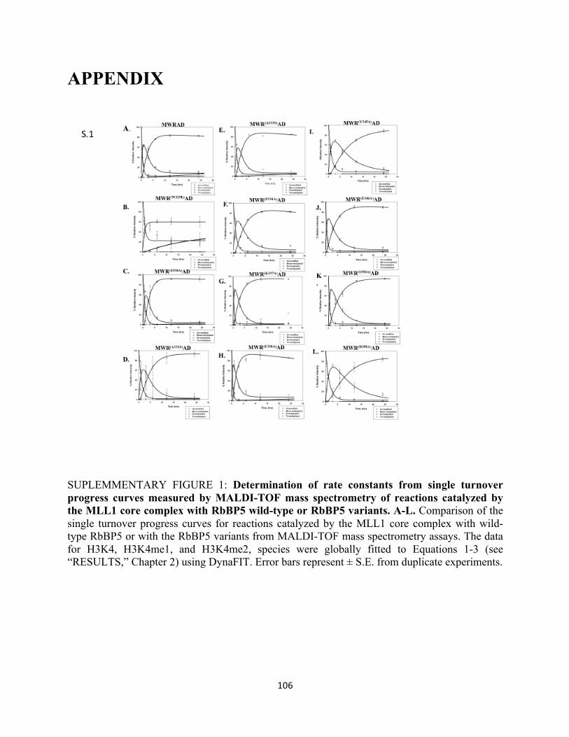
106
APPENDIX
SUPLEMMENTARY FIGURE 1: Determination of rate constants from single turnover progress curves measured by MALDI-TOF mass spectrometry of reactions catalyzed by the MLL1 core complex with RbBP5 wild-type or RbBP5 variants. A-L. Comparison of the single turnover progress curves for reactions catalyzed by the MLL1 core complex with wild-type RbBP5 or with the RbBP5 variants from MALDI-TOF mass spectrometry assays. The data for H3K4, H3K4me1, and H3K4me2, species were globally fitted to Equations 1-3 (see “RESULTS,” Chapter 2) using DynaFIT. Error bars represent ± S.E. from duplicate experiments.
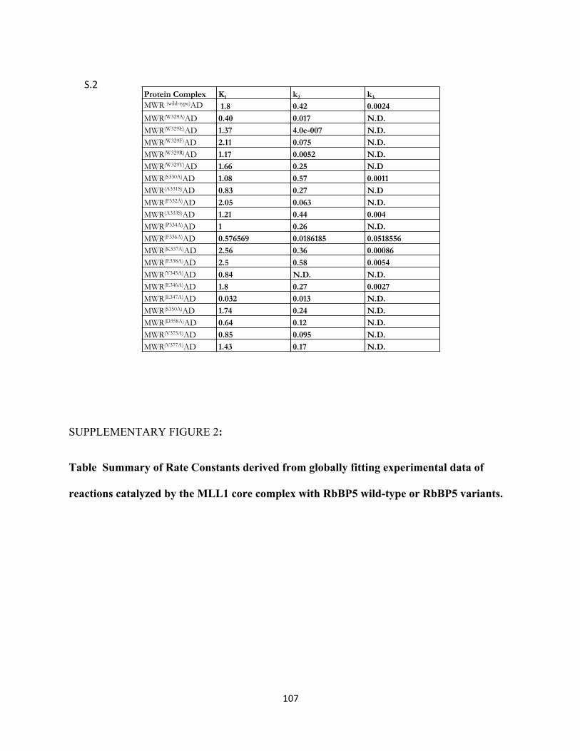
107
SUPPLEMENTARY FIGURE 2:
Table Summary of Rate Constants derived from globally fitting experimental data of
reactions catalyzed by the MLL1 core complex with RbBP5 wild-type or RbBP5 variants.
Protein Complex K1 k2 k3 MWR (wild-type)AD 1.8 0.42 0.0024 MWR(W329A)AD 0.40 0.017 N.D. MWR(W329E)AD 1.37 4.0e-007 N.D. MWR(W329F)AD 2.11 0.075 N.D. MWR(W329R)AD 1.17 0.0052 N.D. MWR(W329Y)AD 1.66 0.25 N.D MWR(S330A)AD 1.08 0.57 0.0011 MWR(A331S)AD 0.83 0.27 N.D MWR(F332A)AD 2.05 0.063 N.D. MWR(A333S)AD 1.21 0.44 0.004 MWR(P334A)AD 1 0.26 N.D. MWR(F336A)AD 0.576569 0.0186185 0.0518556 MWR(K337A)AD 2.56 0.36 0.00086 MWR(E338A)AD 2.5 0.58 0.0054 MWR(Y345A)AD 0.84 N.D. N.D. MWR(E346A)AD 1.8 0.27 0.0027 MWR(E347A)AD 0.032 0.013 N.D. MWR(S350A)AD 1.74 0.24 N.D. MWR(D358A)AD 0.64 0.12 N.D. MWR(V375A)AD 0.85 0.095 N.D. MWR(V377A)AD 1.43 0.17 N.D.
S.2$
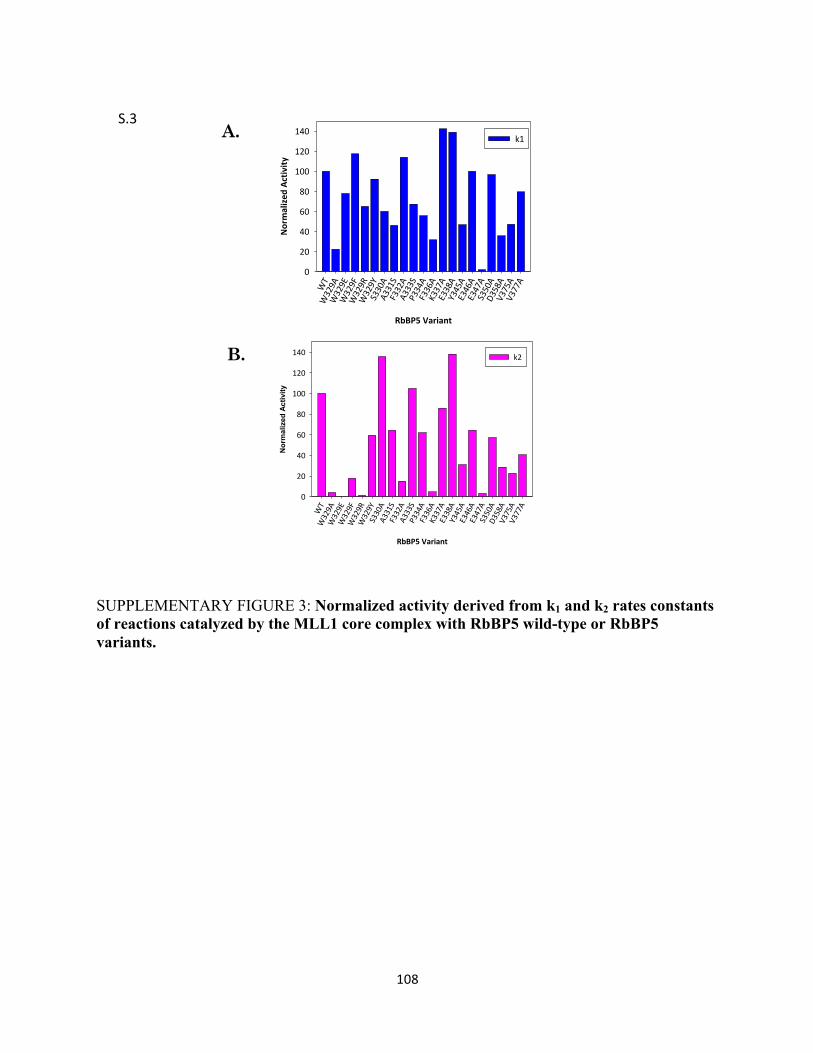
108
SUPPLEMENTARY FIGURE 3: Normalized activity derived from k1 and k2 rates constants of reactions catalyzed by the MLL1 core complex with RbBP5 wild-type or RbBP5 variants.
RbBP5&Variant
WT
W329A
W329E
W329F
W329R
W329Y
S330A
A331S
F332A
A333S
P334A
F336A
K337A
E338A
Y345A
E346A
E347A
S350A
D358A
V375A
V377A
Nor
mal
ized
Act
ivity
0
20
40
60
80
100
120
140 k2
RbBP5&VariantWT
W329A
W329E
W329F
W329R
W329Y
S330A
A331S
F332A
A333S
P334A
F336A
K337A
E338A
Y345A
E346A
E347A
S350A
D358A
V375A
V377A
Normalized
&Activity
0
20
40
60
80
100
120
140k1A.
B.
S.3$

109
SUPPLEMENTARYY FIGURE 4: A. Normalized activity derived from k1 and k2 rates constants of reactions catalyzed by the MLL1 core complex with RbBP5 wild-type or RbBP5 variants.
RbBP5&Variant
WT
W329A
W329E
W329F
W329R
W329Y
S330A
A331S
F332A
A333S
P334A
F336A
K337A
E338A
Y345A
E346A
E347A
S350A
D358A
V375A
V377A
Normalized
&Activity
0
20
40
60
80
100
120
140 k1k2
A.
S.4$

110
SUPPLEMENTARY FIGURES S5-S25. The following pages are raw spectra obtained from
MADLI-TOF mass spectrometry methyltransferase assays. The left column represents the wild-
type (MWRAD) control and the right column represents MWRAD assembled with mutant
RbBP5 proteins. Times points are indicated on each panel.
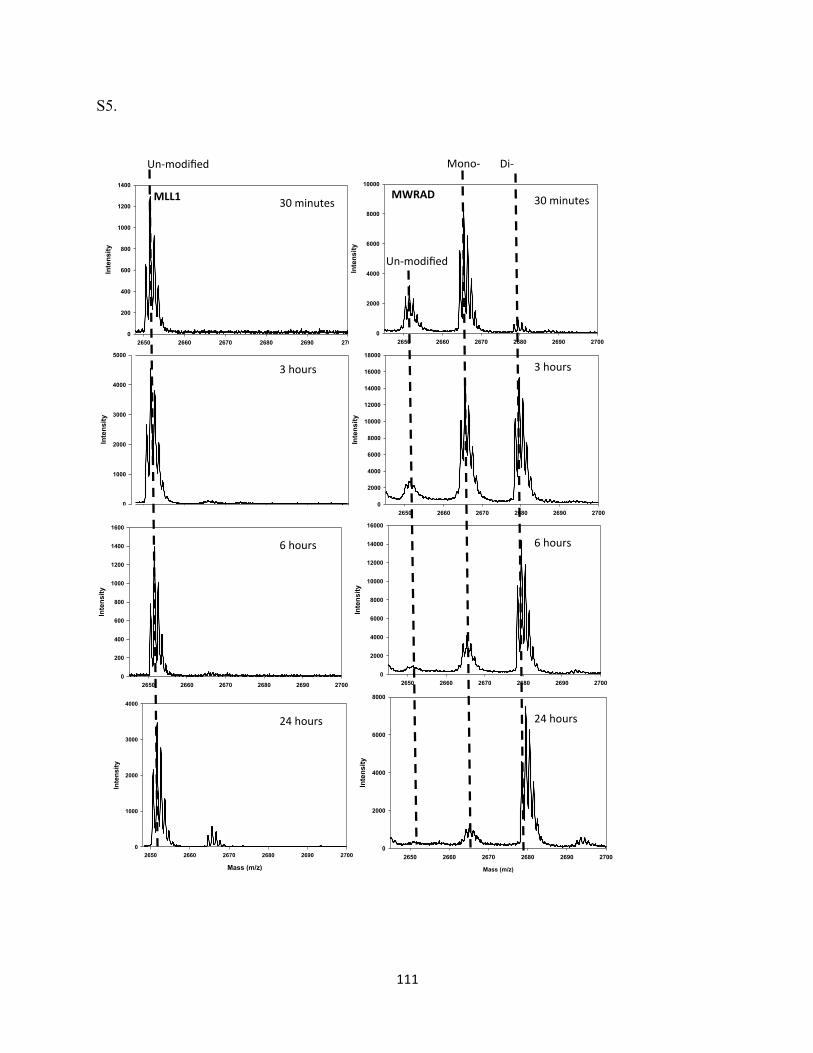
111
S5.
Mass (m/z)
2650 2660 2670 2680 2690 2700
Inte
nsity
0
1000
2000
3000
4000
2650 2660 2670 2680 2690 2700
Intensity
0
1000
2000
3000
4000
5000
2650 2660 2670 2680 2690 2700
Intensity
0
200
400
600
800
1000
1200
1400
2650 2660 2670 2680 2690 2700
Intensity
0
200
400
600
800
1000
1200
1400
1600
MLL1$ 30#minutes#
3#hours#
6#hours#
24#hours#
Mass (m/z)
2650 2660 2670 2680 2690 2700
Inte
nsity
0
2000
4000
6000
8000
2650 2660 2670 2680 2690 2700
Intensity
0
2000
4000
6000
8000
10000
12000
14000
16000
2650 2660 2670 2680 2690 2700
Intensity
0
2000
4000
6000
8000
10000
12000
14000
16000
18000
2650 2660 2670 2680 2690 2700
Intensity
0
2000
4000
6000
8000
10000
MWRAD$ 30#minutes#
3#hours#
6#hours#
24#hours#
Un2modified##
Un2modified##
Mono2## Di2##
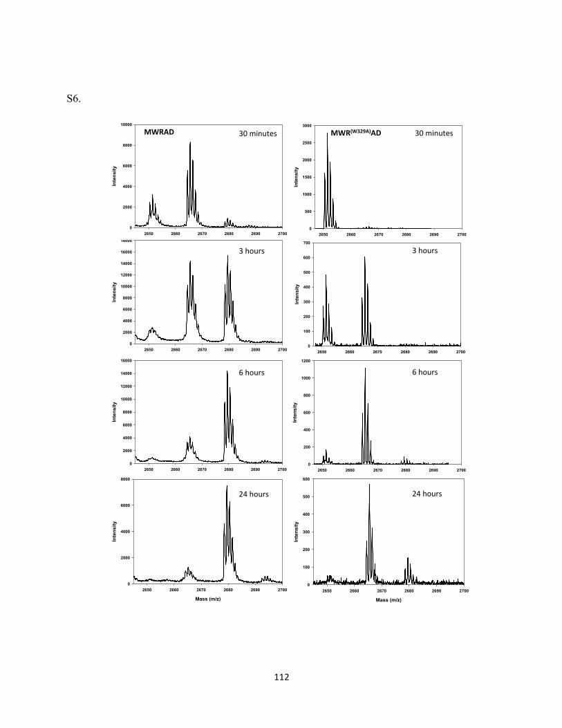
112
S6.
Mass (m/z)
2650 2660 2670 2680 2690 2700
Inte
nsity
0
100
200
300
400
500
600
2650 2660 2670 2680 2690 2700
Intensity
0
200
400
600
800
1000
1200
2650 2660 2670 2680 2690 2700
Intensity
0
100
200
300
400
500
600
700
2650 2660 2670 2680 2690 2700
Intensity
0
500
1000
1500
2000
2500
3000
Mass (m/z)
2650 2660 2670 2680 2690 2700
Inte
nsity
0
2000
4000
6000
8000
2650 2660 2670 2680 2690 2700
Intensity
0
2000
4000
6000
8000
10000
12000
14000
16000
2650 2660 2670 2680 2690 2700
Intensity
0
2000
4000
6000
8000
10000
12000
14000
16000
180002650 2660 2670 2680 2690 2700
Intensity
0
2000
4000
6000
8000
10000
MWRAD& 30#minutes#
3#hours#
6#hours#
24#hours#
30#minutes#
3#hours#
6#hours#
24#hours#
MWR(W329A)AD&
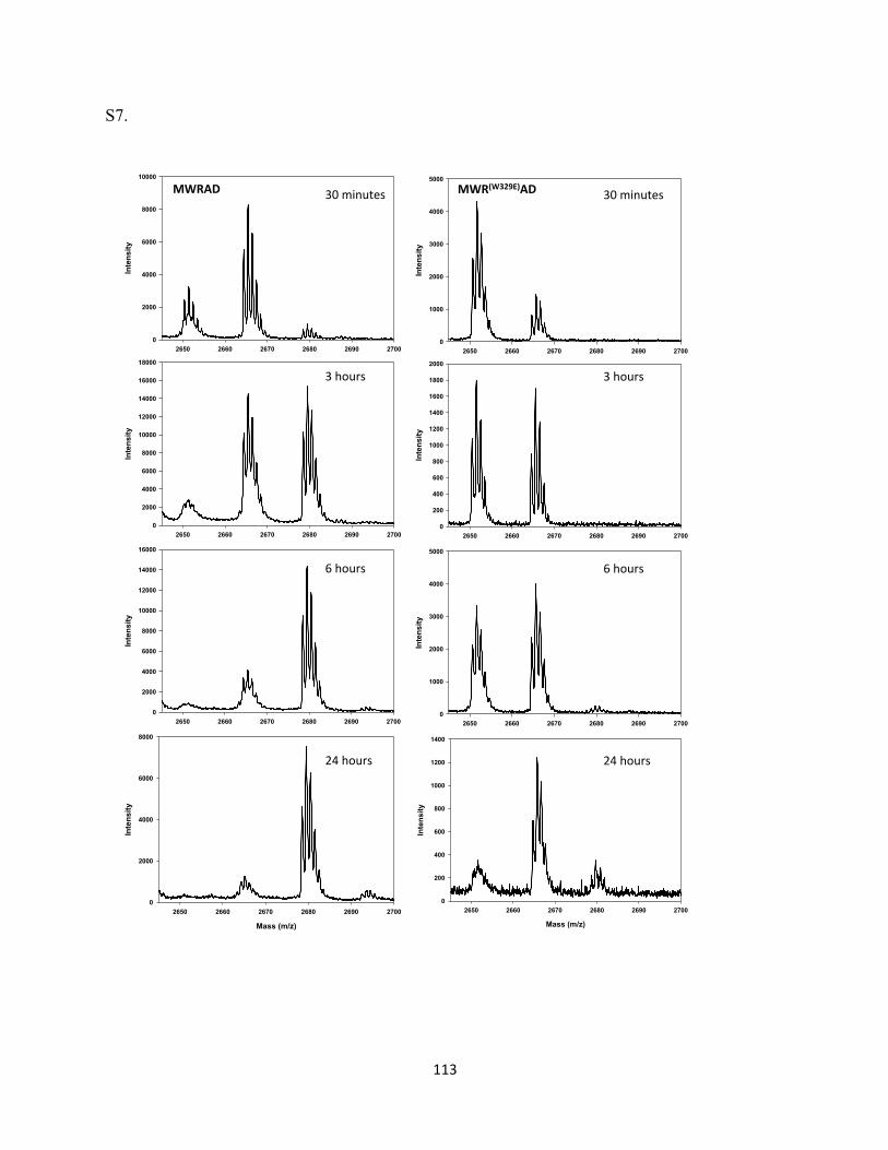
113
S7.
Mass (m/z)
2650 2660 2670 2680 2690 2700
Inte
nsity
0
200
400
600
800
1000
1200
1400
2650 2660 2670 2680 2690 2700
Intensity
0
1000
2000
3000
4000
5000
2650 2660 2670 2680 2690 2700
Intensity
0
200
400
600
800
1000
1200
1400
1600
1800
2000
Mass (m/z)
2650 2660 2670 2680 2690 2700
Inte
nsity
0
2000
4000
6000
8000
2650 2660 2670 2680 2690 2700
Intensity
0
2000
4000
6000
8000
10000
12000
14000
16000
2650 2660 2670 2680 2690 2700
Intensity
0
2000
4000
6000
8000
10000
12000
14000
16000
18000
2650 2660 2670 2680 2690 2700
Intensity
0
2000
4000
6000
8000
10000
MWRAD& 30#minutes#
3#hours#
6#hours#
24#hours#
2650 2660 2670 2680 2690 2700
Intensity
0
1000
2000
3000
4000
5000
30#minutes#
3#hours#
6#hours#
24#hours#
MWR(W329E)AD&

114
S8.
Mass (m/z)
2650 2660 2670 2680 2690 2700
Inte
nsity
0
200
400
600
800
1000
1200
1400
2650 2660 2670 2680 2690 2700
Intensity
0
1000
2000
3000
4000
5000
6000
7000
2650 2660 2670 2680 2690 2700
Intensity
0
1000
2000
3000
4000
5000
Mass (m/z)
2650 2660 2670 2680 2690 2700
Inte
nsity
0
2000
4000
6000
8000
2650 2660 2670 2680 2690 2700
Intensity
0
2000
4000
6000
8000
10000
12000
14000
16000
2650 2660 2670 2680 2690 2700
Intensity
0
2000
4000
6000
8000
10000
12000
14000
16000
180002650 2660 2670 2680 2690 2700
Intensity
0
2000
4000
6000
8000
10000
MWRAD& 30#minutes#
3#hours#
6#hours#
24#hours#
2650 2660 2670 2680 2690 2700
Intensity
0
500
1000
1500
2000
2500
3000
MWR(W329F)AD& 30#minutes#
3#hours#
6#hours#
24#hours#
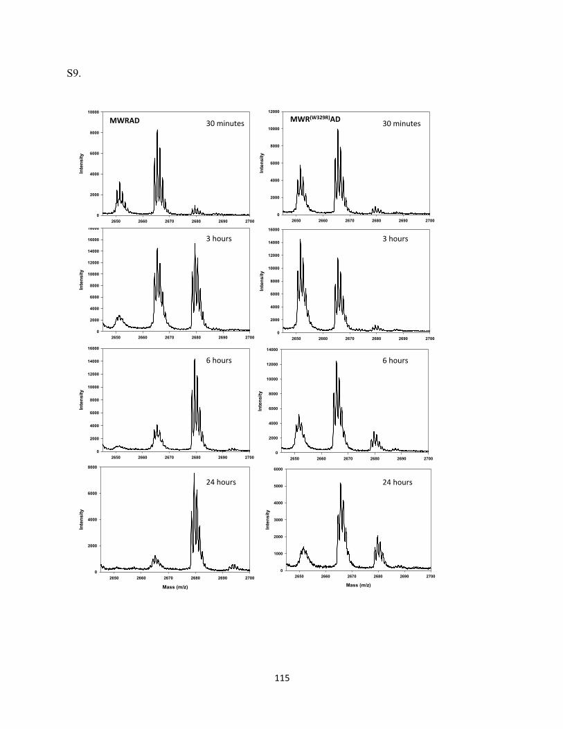
115
S9.
Mass (m/z)
2650 2660 2670 2680 2690 2700
Inte
nsity
0
1000
2000
3000
4000
5000
6000
2650 2660 2670 2680 2690 2700
Intensity
0
2000
4000
6000
8000
10000
12000
14000
2650 2660 2670 2680 2690 2700
Intensity
0
2000
4000
6000
8000
10000
12000
14000
16000
Mass (m/z)
2650 2660 2670 2680 2690 2700
Inte
nsity
0
2000
4000
6000
8000
2650 2660 2670 2680 2690 2700
Intensity
0
2000
4000
6000
8000
10000
12000
14000
16000
2650 2660 2670 2680 2690 2700
Intensity
0
2000
4000
6000
8000
10000
12000
14000
16000
180002650 2660 2670 2680 2690 2700
Intensity
0
2000
4000
6000
8000
10000
MWRAD& 30#minutes#
3#hours#
6#hours#
24#hours#
2650 2660 2670 2680 2690 2700
Intensity
0
2000
4000
6000
8000
10000
12000
MWR(W329R)AD& 30#minutes#
3#hours#
6#hours#
24#hours#
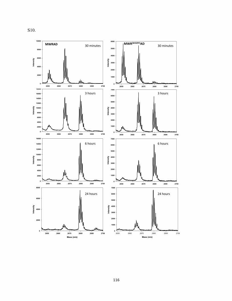
116
S10.
Mass (m/z)
2650 2660 2670 2680 2690 2700
Inte
nsity
0
1000
2000
3000
4000
5000
6000
7000
2650 2660 2670 2680 2690 2700
Intensity
0
1000
2000
3000
4000
5000
6000
7000
2650 2660 2670 2680 2690 2700
Intensity
0
1000
2000
3000
4000
5000
6000
7000
Mass (m/z)
2650 2660 2670 2680 2690 2700
Inte
nsity
0
2000
4000
6000
8000
2650 2660 2670 2680 2690 2700
Intensity
0
2000
4000
6000
8000
10000
12000
14000
16000
2650 2660 2670 2680 2690 2700
Intensity
0
2000
4000
6000
8000
10000
12000
14000
16000
180002650 2660 2670 2680 2690 2700
Intensity
0
2000
4000
6000
8000
10000
MWRAD& 30#minutes#
3#hours#
6#hours#
24#hours#
2650 2660 2670 2680 2690 2700
Intensity
0
1000
2000
3000
4000
5000
6000MWR(W329Y)AD& 30#minutes#
3#hours#
6#hours#
24#hours#
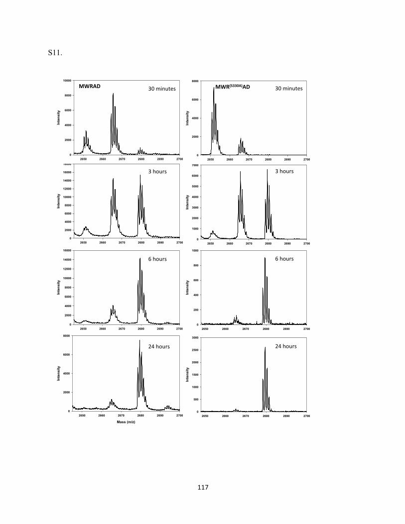
117
S11.
2650 2660 2670 2680 2690 2700
Intensity
0
500
1000
1500
2000
2500
3000
2650 2660 2670 2680 2690 2700
Intensity
0
200
400
600
800
1000
2650 2660 2670 2680 2690 2700
Intensity
0
1000
2000
3000
4000
5000
6000
7000
Mass (m/z)
2650 2660 2670 2680 2690 2700
Inte
nsity
0
2000
4000
6000
8000
2650 2660 2670 2680 2690 2700
Intensity
0
2000
4000
6000
8000
10000
12000
14000
16000
2650 2660 2670 2680 2690 2700
Intensity
0
2000
4000
6000
8000
10000
12000
14000
16000
180002650 2660 2670 2680 2690 2700
Intensity
0
2000
4000
6000
8000
10000
MWRAD& 30#minutes#
3#hours#
6#hours#
24#hours#
2650 2660 2670 2680 2690 2700
Intensity
0
2000
4000
6000
8000
30#minutes#
3#hours#
6#hours#
24#hours#
MWR(S330A)AD&
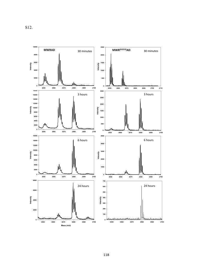
118
S12.
2650 2660 2670 2680 2690 2700
Intensity
0
100
200
300
400
500
600
700
2650 2660 2670 2680 2690 2700
Intensity
0
1000
2000
3000
4000
5000
2650 2660 2670 2680 2690 2700
Intensity
0
500
1000
1500
2000
2500
3000
2650 2660 2670 2680 2690 2700 2710
Intensity
0
500
1000
1500
2000
2500
Mass (m/z)
2650 2660 2670 2680 2690 2700
Inte
nsity
0
2000
4000
6000
8000
2650 2660 2670 2680 2690 2700
Intensity
0
2000
4000
6000
8000
10000
12000
14000
16000
2650 2660 2670 2680 2690 2700
Intensity
0
2000
4000
6000
8000
10000
12000
14000
16000
180002650 2660 2670 2680 2690 2700
Intensity
0
2000
4000
6000
8000
10000
MWRAD& 30#minutes#
3#hours#
6#hours#
24#hours#
30#minutes#
3#hours#
6#hours#
24#hours#
MWR(A331S)AD&
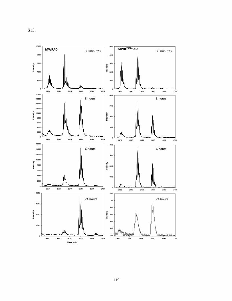
119
S13.
2650 2660 2670 2680 2690 2700
Intensity
200
400
600
800
1000
1200
1400
2650 2660 2670 2680 2690 2700
Intensity
0
1000
2000
3000
4000
2650 2660 2670 2680 2690 2700
Intensity
0
1000
2000
3000
4000
Mass (m/z)
2650 2660 2670 2680 2690 2700
Inte
nsity
0
2000
4000
6000
8000
2650 2660 2670 2680 2690 2700
Intensity
0
2000
4000
6000
8000
10000
12000
14000
16000
2650 2660 2670 2680 2690 2700
Intensity
0
2000
4000
6000
8000
10000
12000
14000
16000
180002650 2660 2670 2680 2690 2700
Intensity
0
2000
4000
6000
8000
10000
MWRAD& 30#minutes#
3#hours#
6#hours#
24#hours#
2650 2660 2670 2680 2690 2700
Intensity
0
1000
2000
3000
4000
5000
30#minutes#
3#hours#
6#hours#
24#hours#
MWR(F332A)AD&
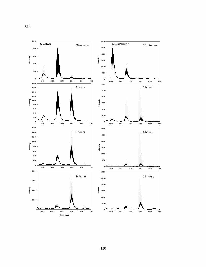
120
S14.
2650 2660 2670 2680 2690 2700
Intensity
0
2000
4000
6000
8000
10000
12000
2650 2660 2670 2680 2690 2700
Intensity
0
1000
2000
3000
4000
5000
6000
2650 2660 2670 2680 2690 2700
Intensity
0
500
1000
1500
2000
2500
3000
Mass (m/z)
2650 2660 2670 2680 2690 2700
Inte
nsity
0
2000
4000
6000
8000
2650 2660 2670 2680 2690 2700
Intensity
0
2000
4000
6000
8000
10000
12000
14000
16000
2650 2660 2670 2680 2690 2700
Intensity
0
2000
4000
6000
8000
10000
12000
14000
16000
180002650 2660 2670 2680 2690 2700
Intensity
0
2000
4000
6000
8000
10000
MWRAD& 30#minutes#
3#hours#
6#hours#
24#hours#
2650 2660 2670 2680 2690 2700
Intensity
0
5000
10000
15000
20000
25000
30000
30#minutes#
3#hours#
6#hours#
24#hours#
MWR(A333S)AD&
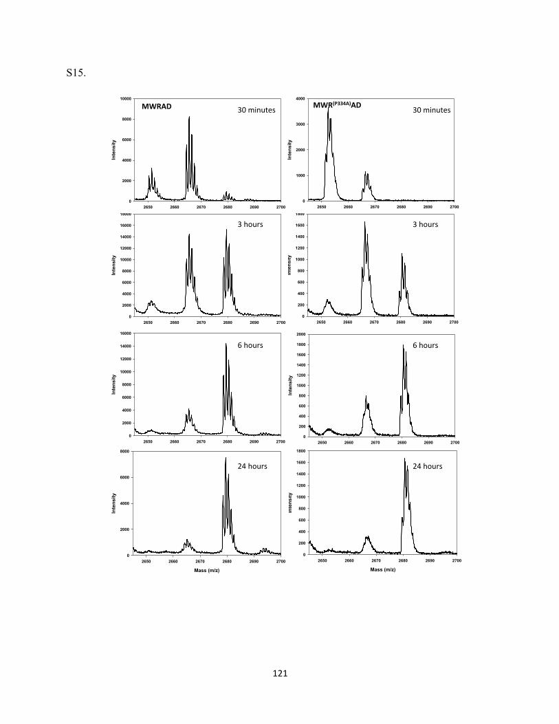
121
S15.
Mass (m/z)
2650 2660 2670 2680 2690 2700
Inte
nsity
0
200
400
600
800
1000
1200
1400
1600
1800
2650 2660 2670 2680 2690 2700
Intensity
0
200
400
600
800
1000
1200
1400
1600
1800
2000
2650 2660 2670 2680 2690 2700
Intensity
0
200
400
600
800
1000
1200
1400
1600
1800
Mass (m/z)
2650 2660 2670 2680 2690 2700
Inte
nsity
0
2000
4000
6000
8000
2650 2660 2670 2680 2690 2700
Intensity
0
2000
4000
6000
8000
10000
12000
14000
16000
2650 2660 2670 2680 2690 2700
Intensity
0
2000
4000
6000
8000
10000
12000
14000
16000
180002650 2660 2670 2680 2690 2700
Intensity
0
2000
4000
6000
8000
10000
MWRAD& 30#minutes#
3#hours#
6#hours#
24#hours#
2650 2660 2670 2680 2690 2700
Intensity
0
1000
2000
3000
4000
MWR(P334A)AD& 30#minutes#
3#hours#
6#hours#
24#hours#
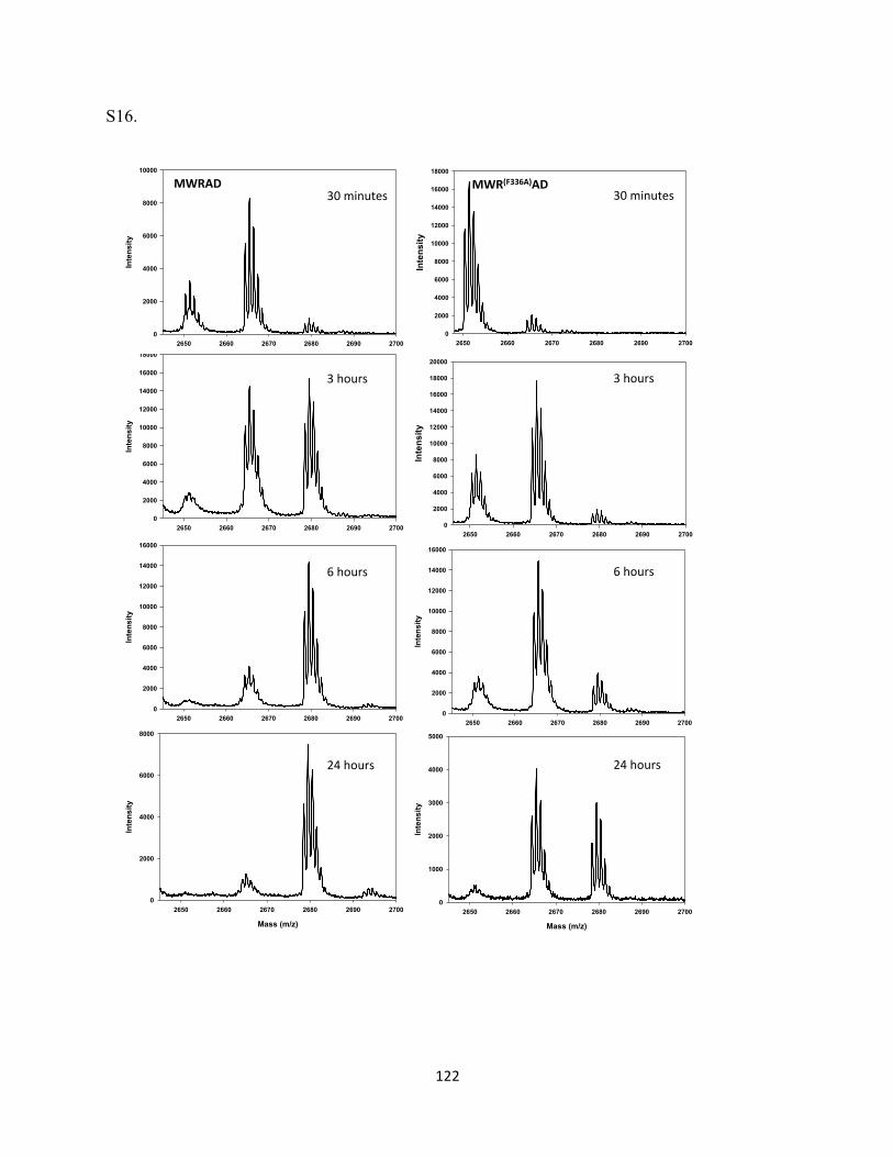
122
S16.
Mass (m/z)
2650 2660 2670 2680 2690 2700
Inte
nsity
0
1000
2000
3000
4000
5000
2650 2660 2670 2680 2690 2700
Intensity
0
2000
4000
6000
8000
10000
12000
14000
16000
2650 2660 2670 2680 2690 2700
Intensity
0
2000
4000
6000
8000
10000
12000
14000
16000
18000
20000
Mass (m/z)
2650 2660 2670 2680 2690 2700
Inte
nsity
0
2000
4000
6000
8000
2650 2660 2670 2680 2690 2700
Intensity
0
2000
4000
6000
8000
10000
12000
14000
16000
2650 2660 2670 2680 2690 2700
Intensity
0
2000
4000
6000
8000
10000
12000
14000
16000
180002650 2660 2670 2680 2690 2700
Intensity
0
2000
4000
6000
8000
10000
MWRAD&30#minutes#
3#hours#
6#hours#
24#hours#
2650 2660 2670 2680 2690 2700
Intensity
0
2000
4000
6000
8000
10000
12000
14000
16000
18000
30#minutes#
3#hours#
6#hours#
24#hours#
MWR(F336A)AD&
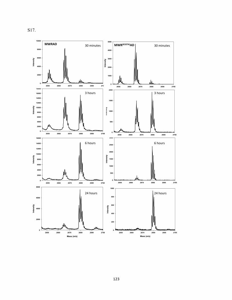
123
S17.
Mass (m/z)
2650 2660 2670 2680 2690 2700
Inte
nsity
0
200
400
600
800
1000
2650 2660 2670 2680 2690 2700
Intensity
0
500
1000
1500
2000
2500
3000
2650 2660 2670 2680 2690 2700
Intensity
0
500
1000
1500
2000
Mass (m/z)
2650 2660 2670 2680 2690 2700
Inte
nsity
0
2000
4000
6000
8000
2650 2660 2670 2680 2690 2700
Intensity
0
2000
4000
6000
8000
10000
12000
14000
16000
2650 2660 2670 2680 2690 2700
Intensity
0
2000
4000
6000
8000
10000
12000
14000
16000
180002650 2660 2670 2680 2690 2700
Intensity
0
2000
4000
6000
8000
10000
MWRAD& 30#minutes#
3#hours#
6#hours#
24#hours#
2650 2660 2670 2680 2690 2700
Intensity
0
1000
2000
3000
4000
5000
MWR(K337A)AD& 30#minutes#
3#hours#
6#hours#
24#hours#
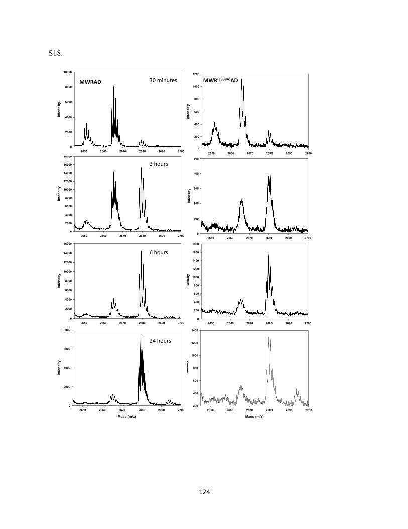
124
S18.
Mass (m/z)
2650 2660 2670 2680 2690 2700
Inte
nsity
200
400
600
800
1000
1200
1400
2650 2660 2670 2680 2690 2700
Intensity
0
200
400
600
800
1000
1200
1400
1600
1800
2650 2660 2670 2680 2690 2700
Intensity
0
100
200
300
400
500
Mass (m/z)
2650 2660 2670 2680 2690 2700
Inte
nsity
0
2000
4000
6000
8000
2650 2660 2670 2680 2690 2700
Intensity
0
2000
4000
6000
8000
10000
12000
14000
16000
2650 2660 2670 2680 2690 2700
Intensity
0
2000
4000
6000
8000
10000
12000
14000
16000
180002650 2660 2670 2680 2690 2700
Intensity
0
2000
4000
6000
8000
10000
MWRAD& 30#minutes#
3#hours#
6#hours#
24#hours#
2650 2660 2670 2680 2690 2700
Intensity
0
200
400
600
800
1000
1200
MWR(E338A)AD&
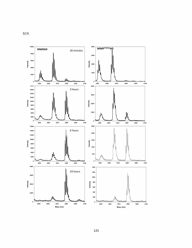
125
S19.
Mass (m/z)
2650 2660 2670 2680 2690 2700
Inte
nsity
0
200
400
600
800
1000
1200
1400
2650 2660 2670 2680 2690 2700
Intensity
0
500
1000
1500
2000
2500
2650 2660 2670 2680 2690 2700
Intensity
0
1000
2000
3000
4000
Mass (m/z)
2650 2660 2670 2680 2690 2700
Inte
nsity
0
2000
4000
6000
8000
2650 2660 2670 2680 2690 2700
Intensity
0
2000
4000
6000
8000
10000
12000
14000
16000
2650 2660 2670 2680 2690 2700
Intensity
0
2000
4000
6000
8000
10000
12000
14000
16000
180002650 2660 2670 2680 2690 2700
Intensity
0
2000
4000
6000
8000
10000
MWRAD& 30#minutes#
3#hours#
6#hours#
24#hours#
2650 2660 2670 2680 2690 2700
Intensity
0
1000
2000
3000
4000MWR(Y345A)AD&
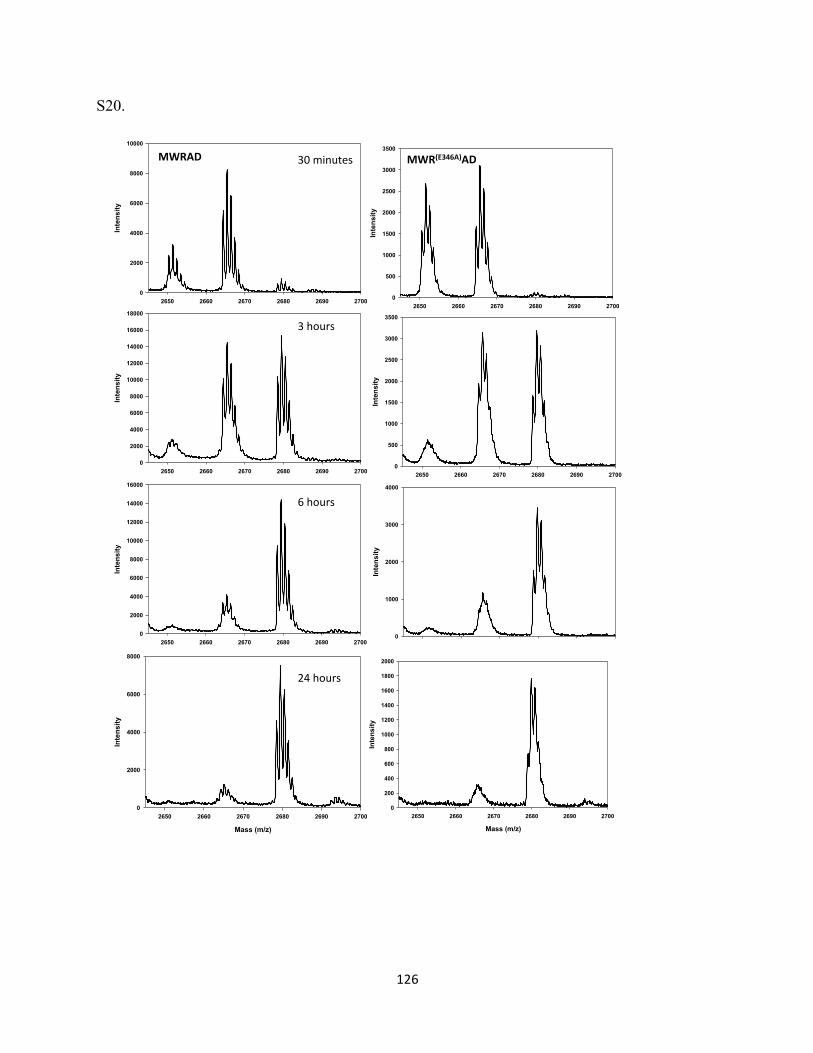
126
S20.
2650 2660 2670 2680 2690 2700
Intensity
0
1000
2000
3000
4000
2650 2660 2670 2680 2690 2700
Intensity
0
500
1000
1500
2000
2500
3000
3500
Mass (m/z)
2650 2660 2670 2680 2690 2700
Inte
nsity
0
2000
4000
6000
8000
2650 2660 2670 2680 2690 2700
Intensity
0
2000
4000
6000
8000
10000
12000
14000
16000
2650 2660 2670 2680 2690 2700
Intensity
0
2000
4000
6000
8000
10000
12000
14000
16000
18000
2650 2660 2670 2680 2690 2700
Intensity
0
2000
4000
6000
8000
10000
MWRAD& 30#minutes#
3#hours#
6#hours#
24#hours#
2650 2660 2670 2680 2690 2700
Intensity
0
500
1000
1500
2000
2500
3000
3500
MWR(E346A)AD&
Mass (m/z)
2650 2660 2670 2680 2690 2700
Inte
nsity
0
200
400
600
800
1000
1200
1400
1600
1800
2000
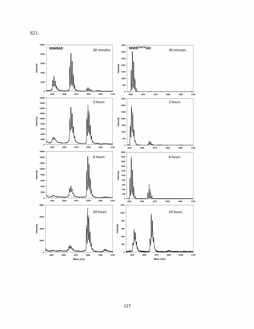
127
S21.
Mass (m/z)
2650 2660 2670 2680 2690 2700
Inte
nsity
0
200
400
600
800
1000
1200
2650 2660 2670 2680 2690 2700
Intensity
0
200
400
600
800
1000
1200
1400
1600
1800
2000
2650 2660 2670 2680 2690 2700
Intensity
0
500
1000
1500
2000
2500
3000
3500
Mass (m/z)
2650 2660 2670 2680 2690 2700
Inte
nsity
0
2000
4000
6000
8000
2650 2660 2670 2680 2690 2700
Intensity
0
2000
4000
6000
8000
10000
12000
14000
16000
2650 2660 2670 2680 2690 2700
Intensity
0
2000
4000
6000
8000
10000
12000
14000
16000
18000
2650 2660 2670 2680 2690 2700
Intensity
0
2000
4000
6000
8000
10000
MWRAD& 30#minutes#
3#hours#
6#hours#
24#hours#
2650 2660 2670 2680 2690 2700
Intensity
0
500
1000
1500
2000
2500
3000
3500
MWR(E347A)AD& 30#minutes#
3#hours#
6#hours#
24#hours#
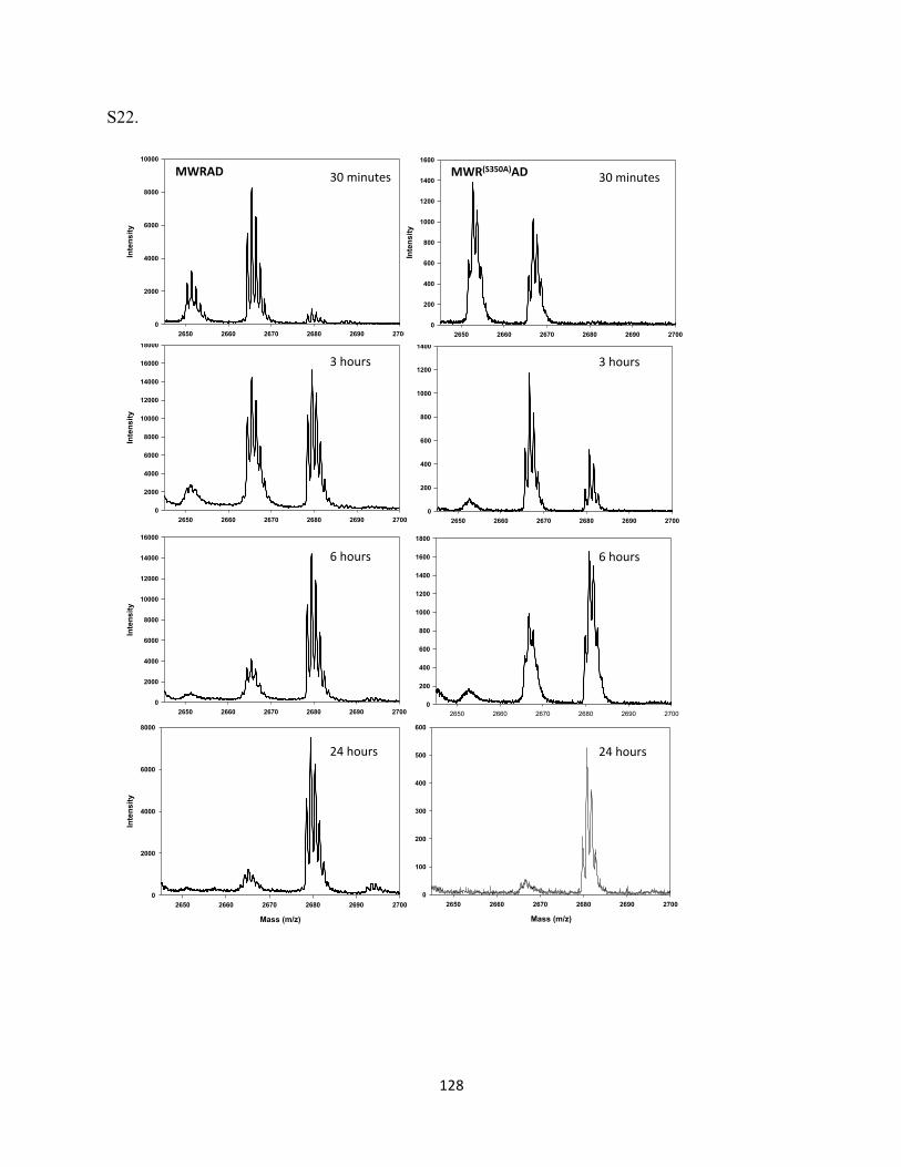
128
S22.
Mass (m/z)
2650 2660 2670 2680 2690 2700
Inte
nsity
0
100
200
300
400
500
600
2650 2660 2670 2680 2690 2700
Intensity
0
200
400
600
800
1000
1200
1400
1600
1800
2650 2660 2670 2680 2690 2700
Intensity
0
200
400
600
800
1000
1200
1400
Mass (m/z)
2650 2660 2670 2680 2690 2700
Inte
nsity
0
2000
4000
6000
8000
2650 2660 2670 2680 2690 2700
Intensity
0
2000
4000
6000
8000
10000
12000
14000
16000
2650 2660 2670 2680 2690 2700
Intensity
0
2000
4000
6000
8000
10000
12000
14000
16000
180002650 2660 2670 2680 2690 2700
Intensity
0
2000
4000
6000
8000
10000
MWRAD& 30#minutes#
3#hours#
6#hours#
24#hours#
2650 2660 2670 2680 2690 2700
Intensity
0
200
400
600
800
1000
1200
1400
1600
MWR(S350A)AD& 30#minutes#
3#hours#
6#hours#
24#hours#
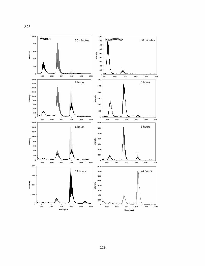
129
S23.
Mass (m/z)
2650 2660 2670 2680 2690 2700
Inte
nsity
0
200
400
600
800
1000
1200
1400
1600
2650 2660 2670 2680 2690 2700
Intensity
0
200
400
600
800
1000
1200
1400
2650 2660 2670 2680 2690 2700
Intensity
0
500
1000
1500
2000
2500
3000
Mass (m/z)
2650 2660 2670 2680 2690 2700
Inte
nsity
0
2000
4000
6000
8000
2650 2660 2670 2680 2690 2700
Intensity
0
2000
4000
6000
8000
10000
12000
14000
16000
2650 2660 2670 2680 2690 2700
Intensity
0
2000
4000
6000
8000
10000
12000
14000
16000
18000
2650 2660 2670 2680 2690 2700
Intensity
0
2000
4000
6000
8000
10000
30#minutes#
3#hours#
6#hours#
24#hours#
2650 2660 2670 2680 2690 2700
Intensity
0
200
400
600
800
1000
1200
1400
1600
1800
MWR(D358A)AD+MWRAD+ 30#minutes#
3#hours#
6#hours#
24#hours#
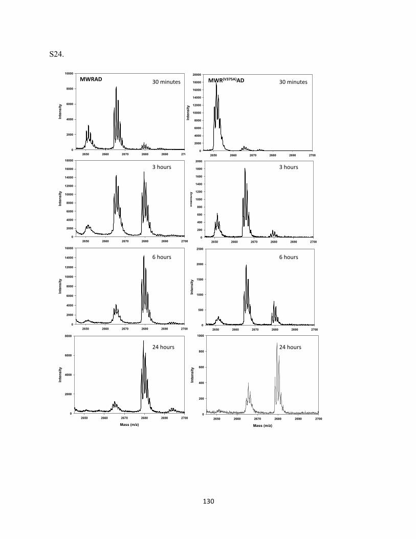
130
S24.
Mass (m/z)
2650 2660 2670 2680 2690 2700
Inte
nsity
0
200
400
600
800
1000
2650 2660 2670 2680 2690 2700
Intensity
0
500
1000
1500
2000
2500
2650 2660 2670 2680 2690 2700
Intensity
0
200
400
600
800
1000
1200
1400
1600
1800
2000
Mass (m/z)
2650 2660 2670 2680 2690 2700
Inte
nsity
0
2000
4000
6000
8000
2650 2660 2670 2680 2690 2700
Intensity
0
2000
4000
6000
8000
10000
12000
14000
16000
2650 2660 2670 2680 2690 2700
Intensity
0
2000
4000
6000
8000
10000
12000
14000
16000
18000
2650 2660 2670 2680 2690 2700
Intensity
0
2000
4000
6000
8000
10000
MWRAD& 30#minutes#
3#hours#
6#hours#
24#hours#
2650 2660 2670 2680 2690 2700
Intensity
0
2000
4000
6000
8000
10000
12000
14000
16000
18000
20000
MWR(V375A)AD& 30#minutes#
3#hours#
6#hours#
24#hours#
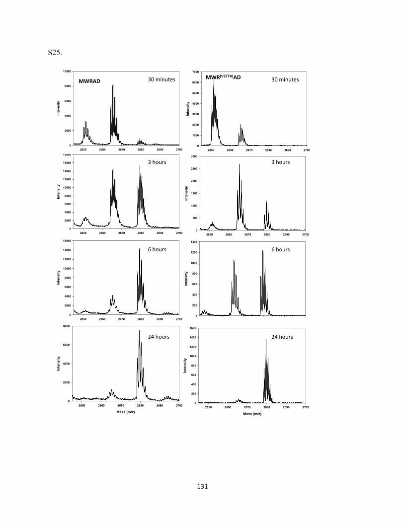
131
S25.
2650 2660 2670 2680 2690 2700
Intensity
0
200
400
600
800
1000
1200
1400
2650 2660 2670 2680 2690 2700
Intensity
0
500
1000
1500
2000
2500
3000
Mass (m/z)
2650 2660 2670 2680 2690 2700
Inte
nsity
0
2000
4000
6000
8000
2650 2660 2670 2680 2690 2700
Intensity
0
2000
4000
6000
8000
10000
12000
14000
16000
2650 2660 2670 2680 2690 2700
Intensity
0
2000
4000
6000
8000
10000
12000
14000
16000
180002650 2660 2670 2680 2690 2700
Intensity
0
2000
4000
6000
8000
10000
MWRAD& 30#minutes#
3#hours#
6#hours#
24#hours#
2650 2660 2670 2680 2690 2700
Intensity
0
1000
2000
3000
4000
5000
6000
7000
MWR(V377A)AD&
Mass (m/z)
2650 2660 2670 2680 2690 2700
Inte
nsity
0
200
400
600
800
1000
1200
1400
1600
30#minutes#
3#hours#
6#hours#
24#hours#
132
References Allison, S.P. (1996). Perioperative artificial nutrition in elective adult surgery. Clinical nutrition 15, 233-‐235. Ang, Y.S., Tsai, S.Y., Lee, D.F., Monk, J., Su, J., Ratnakumar, K., Ding, J., Ge, Y., Darr, H., Chang, B., et al. (2011). Wdr5 mediates self-‐renewal and reprogramming via the embryonic stem cell core transcriptional network. Cell 145, 183-‐197. Avdic, V., Zhang, P., Lanouette, S., Groulx, A., Tremblay, V., Brunzelle, J., and Couture, J.F. (2011). Structural and biochemical insights into MLL1 core complex assembly. Structure 19, 101-‐108. Bannister, A.J., and Kouzarides, T. (2011). Regulation of chromatin by histone modifications. Cell research 21, 381-‐395. Barski, A., Cuddapah, S., Cui, K., Roh, T.Y., Schones, D.E., Wang, Z., Wei, G., Chepelev, I., and Zhao, K. (2007). High-‐resolution profiling of histone methylations in the human genome. Cell 129, 823-‐837. Berger, S.L. (2007). The complex language of chromatin regulation during transcription. Nature 447, 407-‐412. Bhaumik, S.R., Smith, E., and Shilatifard, A. (2007). Covalent modifications of histones during development and disease pathogenesis. Nature structural & molecular biology 14, 1008-‐1016. Bralten, L.B., Kloosterhof, N.K., Gravendeel, L.A., Sacchetti, A., Duijm, E.J., Kros, J.M., van den Bent, M.J., Hoogenraad, C.C., Sillevis Smitt, P.A., and French, P.J. (2010). Integrated genomic profiling identifies candidate genes implicated in glioma-‐genesis and a novel LEO1-‐SLC12A1 fusion gene. Genes, chromosomes & cancer 49, 509-‐517. Brownell, J.E., and Allis, C.D. (1996). Special HATs for special occasions: linking histone acetylation to chromatin assembly and gene activation. Current opinion in genetics & development 6, 176-‐184. Cao, F., Chen, Y., Cierpicki, T., Liu, Y., Basrur, V., Lei, M., and Dou, Y. (2010). An Ash2L/RbBP5 heterodimer stimulates the MLL1 methyltransferase activity through coordinated substrate interactions with the MLL1 SET domain. PloS one 5, e14102. Chen, Y., Wan, B., Wang, K.C., Cao, F., Yang, Y., Protacio, A., Dou, Y., Chang, H.Y., and Lei, M. (2011). Crystal structure of the N-‐terminal region of human Ash2L shows a winged-‐helix motif involved in DNA binding. EMBO reports 12, 797-‐803. Cheng, X., and Zhang, X. (2007). Structural dynamics of protein lysine methylation and demethylation. Mutation research 618, 102-‐115. Chi, P., Allis, C.D., and Wang, G.G. (2010). Covalent histone modifications-‐-‐miswritten, misinterpreted and mis-‐erased in human cancers. Nature reviews Cancer 10, 457-‐469.
133
Classon, M., and Harlow, E. (2002). The retinoblastoma tumour suppressor in development and cancer. Nature reviews Cancer 2, 910-‐917. Classon, M., and Settleman, J. (2000). Emerging concepts in tumor progression and therapy. Seminars in cancer biology 10, 393-‐397. Collins, R.E., Tachibana, M., Tamaru, H., Smith, K.M., Jia, D., Zhang, X., Selker, E.U., Shinkai, Y., and Cheng, X. (2005). In vitro and in vivo analyses of a Phe/Tyr switch controlling product specificity of histone lysine methyltransferases. The Journal of biological chemistry 280, 5563-‐5570. Cosgrove, M.S., and Patel, A. (2010). Mixed lineage leukemia: a structure-‐function perspective of the MLL1 protein. The FEBS journal 277, 1832-‐1842. Couture, J.F., Dirk, L.M., Brunzelle, J.S., Houtz, R.L., and Trievel, R.C. (2008). Structural origins for the product specificity of SET domain protein methyltransferases. Proceedings of the National Academy of Sciences of the United States of America 105, 20659-‐20664. Crawford, B.D., and Hess, J.L. (2006). MLL core components give the green light to histone methylation. ACS chemical biology 1, 495-‐498. Dhalluin, C., Carlson, J.E., Zeng, L., He, C., Aggarwal, A.K., and Zhou, M.M. (1999). Structure and ligand of a histone acetyltransferase bromodomain. Nature 399, 491-‐496. Dillon, S.C., Zhang, X., Trievel, R.C., and Cheng, X. (2005). The SET-‐domain protein superfamily: protein lysine methyltransferases. Genome biology 6, 227. Dou, Y., Milne, T.A., Ruthenburg, A.J., Lee, S., Lee, J.W., Verdine, G.L., Allis, C.D., and Roeder, R.G. (2006). Regulation of MLL1 H3K4 methyltransferase activity by its core components. Nature structural & molecular biology 13, 713-‐719. Eissenberg, J.C., and Shilatifard, A. (2010). Histone H3 lysine 4 (H3K4) methylation in development and differentiation. Developmental biology 339, 240-‐249. Ernst, P., Wang, J., and Korsmeyer, S.J. (2002). The role of MLL in hematopoiesis and leukemia. Current opinion in hematology 9, 282-‐287. Ferreira, R., Magnaghi-‐Jaulin, L., Robin, P., Harel-‐Bellan, A., and Trouche, D. (1998). The three members of the pocket proteins family share the ability to repress E2F activity through recruitment of a histone deacetylase. Proceedings of the National Academy of Sciences of the United States of America 95, 10493-‐10498. Gardner, K.E., Allis, C.D., and Strahl, B.D. (2011). Operating on chromatin, a colorful language where context matters. Journal of molecular biology 409, 36-‐46. Gopal, R., Foster, K.W., and Yang, P. (2012). The DPY-‐30 domain and its flanking sequence mediate the assembly and modulation of flagellar radial spoke complexes. Molecular and cellular biology 32, 4012-‐4024.
134
Guenther, M.G., Jenner, R.G., Chevalier, B., Nakamura, T., Croce, C.M., Canaani, E., and Young, R.A. (2005). Global and Hox-‐specific roles for the MLL1 methyltransferase. Proceedings of the National Academy of Sciences of the United States of America 102, 8603-‐8608. Hayes, J.J., and Wolffe, A.P. (1992). Histones H2A/H2B inhibit the interaction of transcription factor IIIA with the Xenopus borealis somatic 5S RNA gene in a nucleosome. Proceedings of the National Academy of Sciences of the United States of America 89, 1229-‐1233. Hess, J.L. (2004). MLL: a histone methyltransferase disrupted in leukemia. Trends in molecular medicine 10, 500-‐507. Hsu, D.R., Chuang, P.T., and Meyer, B.J. (1995). DPY-‐30, a nuclear protein essential early in embryogenesis for Caenorhabditis elegans dosage compensation. Development 121, 3323-‐3334. Hsu, D.R., and Meyer, B.J. (1994). The dpy-‐30 gene encodes an essential component of the Caenorhabditis elegans dosage compensation machinery. Genetics 137, 999-‐1018. Jenuwein, T., and Allis, C.D. (2001). Translating the histone code. Science 293, 1074-‐1080. Jenuwein, T., Laible, G., Dorn, R., and Reuter, G. (1998). SET domain proteins modulate chromatin domains in eu-‐ and heterochromatin. Cellular and molecular life sciences : CMLS 54, 80-‐93. Jiang, H., Shukla, A., Wang, X., Chen, W.Y., Bernstein, B.E., and Roeder, R.G. (2011). Role for Dpy-‐30 in ES cell-‐fate specification by regulation of H3K4 methylation within bivalent domains. Cell 144, 513-‐525. Kornberg, R.D. (1974). Chromatin structure: a repeating unit of histones and DNA. Science 184, 868-‐871. Kouzarides, T. (2002). Histone methylation in transcriptional control. Current opinion in genetics & development 12, 198-‐209. Kouzarides, T. (2007). Chromatin modifications and their function. Cell 128, 693-‐705. Lai, A., Lee, J.M., Yang, W.M., DeCaprio, J.A., Kaelin, W.G., Jr., Seto, E., and Branton, P.E. (1999). RBP1 recruits both histone deacetylase-‐dependent and -‐independent repression activities to retinoblastoma family proteins. Molecular and cellular biology 19, 6632-‐6641. Lee, D.Y., Hayes, J.J., Pruss, D., and Wolffe, A.P. (1993). A positive role for histone acetylation in transcription factor access to nucleosomal DNA. Cell 72, 73-‐84. Li, H., Ilin, S., Wang, W., Duncan, E.M., Wysocka, J., Allis, C.D., and Patel, D.J. (2006). Molecular basis for site-‐specific read-‐out of histone H3K4me3 by the BPTF PHD finger of NURF. Nature 442, 91-‐95. Magnaghi-‐Jaulin, L., Groisman, R., Naguibneva, I., Robin, P., Lorain, S., Le Villain, J.P., Troalen, F., Trouche, D., and Harel-‐Bellan, A. (1998). Retinoblastoma protein represses transcription by recruiting a histone deacetylase. Nature 391, 601-‐605.
135
Mersman, D.P., Du, H.N., Fingerman, I.M., South, P.F., and Briggs, S.D. (2012). Charge-‐based interaction conserved within histone H3 lysine 4 (H3K4) methyltransferase complexes is needed for protein stability, histone methylation, and gene expression. The Journal of biological chemistry 287, 2652-‐2665. Miller, T., Krogan, N.J., Dover, J., Erdjument-‐Bromage, H., Tempst, P., Johnston, M., Greenblatt, J.F., and Shilatifard, A. (2001). COMPASS: a complex of proteins associated with a trithorax-‐related SET domain protein. Proceedings of the National Academy of Sciences of the United States of America 98, 12902-‐12907. Milne, T.A., Briggs, S.D., Brock, H.W., Martin, M.E., Gibbs, D., Allis, C.D., and Hess, J.L. (2002). MLL targets SET domain methyltransferase activity to Hox gene promoters. Molecular cell 10, 1107-‐1117. Milne, T.A., Dou, Y., Martin, M.E., Brock, H.W., Roeder, R.G., and Hess, J.L. (2005). MLL associates specifically with a subset of transcriptionally active target genes. Proceedings of the National Academy of Sciences of the United States of America 102, 14765-‐14770. Murray, K. (1964). The Occurrence of Epsilon-‐N-‐Methyl Lysine in Histones. Biochemistry 3, 10-‐15. Muslin, A.J., Tanner, J.W., Allen, P.M., and Shaw, A.S. (1996). Interaction of 14-‐3-‐3 with signaling proteins is mediated by the recognition of phosphoserine. Cell 84, 889-‐897. Odho, Z., Southall, S.M., and Wilson, J.R. (2010). Characterization of a novel WDR5-‐binding site that recruits RbBP5 through a conserved motif to enhance methylation of histone H3 lysine 4 by mixed lineage leukemia protein-‐1. The Journal of biological chemistry 285, 32967-‐32976. Ono, R., Nosaka, T., and Hayashi, Y. (2005). Roles of a trithorax group gene, MLL, in hematopoiesis. International journal of hematology 81, 288-‐293. Paggi, M.G., Baldi, A., Bonetto, F., and Giordano, A. (1996). Retinoblastoma protein family in cell cycle and cancer: a review. Journal of cellular biochemistry 62, 418-‐430. Patel, A., Dharmarajan, V., Vought, V.E., and Cosgrove, M.S. (2009). On the mechanism of multiple lysine methylation by the human mixed lineage leukemia protein-‐1 (MLL1) core complex. The Journal of biological chemistry 284, 24242-‐24256. Patel, A., Vought, V.E., Dharmarajan, V., and Cosgrove, M.S. (2008). A conserved arginine-‐containing motif crucial for the assembly and enzymatic activity of the mixed lineage leukemia protein-‐1 core complex. The Journal of biological chemistry 283, 32162-‐32175. Patel, A., Vought, V.E., Dharmarajan, V., and Cosgrove, M.S. (2011). A novel non-‐SET domain multi-‐subunit methyltransferase required for sequential nucleosomal histone H3 methylation by the mixed lineage leukemia protein-‐1 (MLL1) core complex. The Journal of biological chemistry 286, 3359-‐3369. Pokholok, D.K., Harbison, C.T., Levine, S., Cole, M., Hannett, N.M., Lee, T.I., Bell, G.W., Walker, K., Rolfe, P.A., Herbolsheimer, E., et al. (2005). Genome-‐wide map of nucleosome acetylation and methylation in yeast. Cell 122, 517-‐527.
136
Rando, O.J. (2012). Combinatorial complexity in chromatin structure and function: revisiting the histone code. Current opinion in genetics & development 22, 148-‐155. Rea, S., Eisenhaber, F., O'Carroll, D., Strahl, B.D., Sun, Z.W., Schmid, M., Opravil, S., Mechtler, K., Ponting, C.P., Allis, C.D., et al. (2000). Regulation of chromatin structure by site-‐specific histone H3 methyltransferases. Nature 406, 593-‐599. Riemenschneider, M.J., Buschges, R., Wolter, M., Reifenberger, J., Bostrom, J., Kraus, J.A., Schlegel, U., and Reifenberger, G. (1999). Amplification and overexpression of the MDM4 (MDMX) gene from 1q32 in a subset of malignant gliomas without TP53 mutation or MDM2 amplification. Cancer research 59, 6091-‐6096. Riemenschneider, M.J., Knobbe, C.B., and Reifenberger, G. (2003). Refined mapping of 1q32 amplicons in malignant gliomas confirms MDM4 as the main amplification target. International journal of cancer Journal international du cancer 104, 752-‐757. Roguev, A., Schaft, D., Shevchenko, A., Pijnappel, W.W., Wilm, M., Aasland, R., and Stewart, A.F. (2001). The Saccharomyces cerevisiae Set1 complex includes an Ash2 homologue and methylates histone 3 lysine 4. The EMBO journal 20, 7137-‐7148. Ruthenburg, A.J., Allis, C.D., and Wysocka, J. (2007). Methylation of lysine 4 on histone H3: intricacy of writing and reading a single epigenetic mark. Molecular cell 25, 15-‐30. Schwarz, P.M., and Hansen, J.C. (1994). Formation and stability of higher order chromatin structures. Contributions of the histone octamer. The Journal of biological chemistry 269, 16284-‐16289. Sims, R.J., 3rd, Nishioka, K., and Reinberg, D. (2003). Histone lysine methylation: a signature for chromatin function. Trends in genetics : TIG 19, 629-‐639. Sims, R.J., 3rd, and Reinberg, D. (2006). Histone H3 Lys 4 methylation: caught in a bind? Genes & development 20, 2779-‐2786. Slany, R.K. (2009). The molecular biology of mixed lineage leukemia. Haematologica 94, 984-‐993. Smith, E., and Shilatifard, A. (2010). The chromatin signaling pathway: diverse mechanisms of recruitment of histone-‐modifying enzymes and varied biological outcomes. Molecular cell 40, 689-‐701. South, P.F., Fingerman, I.M., Mersman, D.P., Du, H.N., and Briggs, S.D. (2010). A conserved interaction between the SDI domain of Bre2 and the Dpy-‐30 domain of Sdc1 is required for histone methylation and gene expression. The Journal of biological chemistry 285, 595-‐607. Southall, S.M., Wong, P.S., Odho, Z., Roe, S.M., and Wilson, J.R. (2009). Structural basis for the requirement of additional factors for MLL1 SET domain activity and recognition of epigenetic marks. Molecular cell 33, 181-‐191. Stoller, J.Z., Huang, L., Tan, C.C., Huang, F., Zhou, D.D., Yang, J., Gelb, B.D., and Epstein, J.A. (2010). Ash2l interacts with Tbx1 and is required during early embryogenesis. Experimental biology and medicine 235, 569-‐576.
137
Strahl, B.D., and Allis, C.D. (2000). The language of covalent histone modifications. Nature 403, 41-‐45. Suganuma, T., and Workman, J.L. (2011). Signals and combinatorial functions of histone modifications. Annual review of biochemistry 80, 473-‐499. Suryadinata, R., Sadowski, M., Steel, R., and Sarcevic, B. (2011). Cyclin-‐dependent kinase-‐mediated phosphorylation of RBP1 and pRb promotes their dissociation to mediate release of the SAP30.mSin3.HDAC transcriptional repressor complex. The Journal of biological chemistry 286, 5108-‐5118. Sutcliffe, J.E., Brown, T.R., Allison, S.J., Scott, P.H., and White, R.J. (2000). Retinoblastoma protein disrupts interactions required for RNA polymerase III transcription. Molecular and cellular biology 20, 9192-‐9202. Vardanyan, A., Atanesyan, L., Egli, D., Raja, S.J., Steinmann-‐Zwicky, M., Renkawitz-‐Pohl, R., Georgiev, O., and Schaffner, W. (2008). Dumpy-‐30 family members as determinants of male fertility and interaction partners of metal-‐responsive transcription factor 1 (MTF-‐1) in Drosophila. BMC developmental biology 8, 68. Wan, M., Liang, J., Xiong, Y., Shi, F., Zhang, Y., Lu, W., He, Q., Yang, D., Chen, R., Liu, D., et al. (2012). The Trithorax Group Protein Ash2l is Essential for Pluripotency and Maintaining Open Chromatin in Embryonic Stem Cells. The Journal of biological chemistry. Williams, J.P., Stewart, T., Li, B., Mulloy, R., Dimova, D., and Classon, M. (2006). The retinoblastoma protein is required for Ras-‐induced oncogenic transformation. Molecular and cellular biology 26, 1170-‐1182. Wolffe, A.P., and Hayes, J.J. (1999). Chromatin disruption and modification. Nucleic acids research 27, 711-‐720. Wysocka, J., Swigut, T., Milne, T.A., Dou, Y., Zhang, X., Burlingame, A.L., Roeder, R.G., Brivanlou, A.H., and Allis, C.D. (2005). WDR5 associates with histone H3 methylated at K4 and is essential for H3 K4 methylation and vertebrate development. Cell 121, 859-‐872. Yokoyama, A., Wang, Z., Wysocka, J., Sanyal, M., Aufiero, D.J., Kitabayashi, I., Herr, W., and Cleary, M.L. (2004). Leukemia proto-‐oncoprotein MLL forms a SET1-‐like histone methyltransferase complex with menin to regulate Hox gene expression. Molecular and cellular biology 24, 5639-‐5649. Yu, B.D., Hess, J.L., Horning, S.E., Brown, G.A., and Korsmeyer, S.J. (1995). Altered Hox expression and segmental identity in Mll-‐mutant mice. Nature 378, 505-‐508.
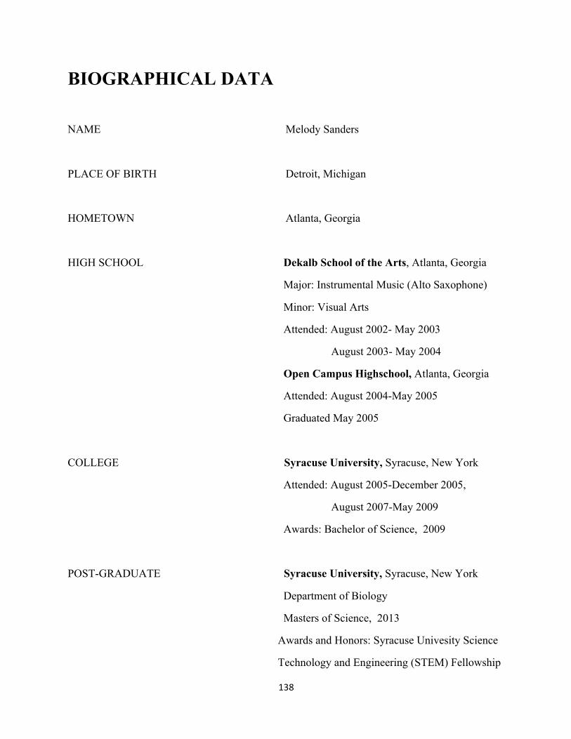
138
BIOGRAPHICAL DATA
NAME Melody Sanders
PLACE OF BIRTH Detroit, Michigan
HOMETOWN Atlanta, Georgia
HIGH SCHOOL Dekalb School of the Arts, Atlanta, Georgia
Major: Instrumental Music (Alto Saxophone)
Minor: Visual Arts
Attended: August 2002- May 2003
August 2003- May 2004
Open Campus Highschool, Atlanta, Georgia
Attended: August 2004-May 2005
Graduated May 2005
COLLEGE Syracuse University, Syracuse, New York
Attended: August 2005-December 2005,
August 2007-May 2009
Awards: Bachelor of Science, 2009
POST-GRADUATE Syracuse University, Syracuse, New York
Department of Biology
Masters of Science, 2013
Awards and Honors: Syracuse Univesity Science
Technology and Engineering (STEM) Fellowship

Copyright © 2022 FDOKUMEN