Strain-specific responses of Emiliania huxleyi to changing seawater carbonate chemistry
Upload
independentCategory
view
3download
0
APPLIED AND ENVIRONMENTAL MICROBIOLOGY, May 2005, p. 2564–2575 Vol. 71, No. 50099-2240/05/$08.00�0 doi:10.1128/AEM.71.5.2564–2575.2005Copyright © 2005, American Society for Microbiology. All Rights Reserved.
Suppressive Subtractive Hybridization of and Differences in GeneExpression Content of Calcifying and Noncalcifying Cultures of
Emiliania huxleyi Strain 1516Binh Nguyen, Robert M. Bowers, Thomas M. Wahlund, and Betsy A. Read*
Department of Biological Sciences, California State University San Marcos, 333 S. Twin Oaks Valley Road,San Marcos, California 92096-0001
Received 14 July 2004/Accepted 19 November 2004
The marine coccolithophorid Emiliania huxleyi is a cosmopolitan alga intensely studied in relation to globalcarbon cycling, biogeochemistry, marine ecology, and biomineralization processes. The biomineralizationcapabilities of coccolithophorids have attracted the attention of scientists interested in exploiting this abilityfor the development of materials science and biomedical and biotechnological applications. Although it hasbeen well documented that biomineralization in E. huxleyi is promoted by growth under phosphate-limitedconditions, the genes and proteins that govern the processes of calcification and coccolithogenesis remainunknown. Suppressive subtractive hybridization (SSH) libraries were constructed from cultures grown inphosphate-limited and phosphate-replete media as tester and driver populations for reciprocal SSH proce-dures. Positive clones from each of the two libraries were randomly selected, and dot blotting was performedfor the analysis of expression patterns. A total of 513 clones from the phosphate-replete library and 423 clonesfrom the phosphate-limited library were sequenced, assembled, and compared to sequences in GenBank usingBLASTX. Of the 103 differentially expressed gene fragments from the phosphate-replete library, 34% showedsignificant homology to other known proteins, while only 23% of the 65 differentially expressed gene fragmentsfrom the phosphate-limited library showed homology to other proteins. To further assess mRNA expression,real-time RT-PCR analysis was employed and expression profiles were generated over a 14-day time course forthree clones from the phosphate-replete library and five clones from the phosphate-limited library. Thefragments isolated provide the basis for future cloning of full-length genes and functional analysis.
Coccolithophorids are unicellular marine algae that producea wide variety of highly sculpted calcium carbonate cell cover-ings. Calcification in some coccolithophorids occurs intracel-lularly and is thought to begin with the conversion of calciumand bicarbonate ions into crystals of calcium carbonate that aredeposited in a defined array in association with an organicmatrix (35). A coccolith vesicle developing from the Golgiapparatus provides the microenvironment that promotes theformation of the crystallographically intricate coccolith struc-tures, which are subsequently extruded from the cell to formthe coccosphere (7, 20). The coccolith vesicle fuses with theplasma membrane to release the individual coccoliths. Whilecalcification and coccolithogenesis are presumed to occur in agenetically controlled manner, the functional significance andgenetic control of these complex structures and the variation incrystal shape, orientation, order, and connectivity that occursacross species are poorly understood
The size and morphology of the calcite structures are asdiverse as the coccolithophorids that produce them. Some ofthe calcite elements are simple rhombohedral units, while oth-ers appear as ornate oval platelets, extended spicules, or elab-orate coronets. The mechanisms behind the natural nanofab-rication of the highly sculpted calcium carbonate coccoliths areincreasingly being investigated. Scanning electron and atomic
force microscopy have defined many of the structural featuresof various types of coccoliths (13, 41). Ecophysiological andenvironmental studies have related biomineralization to nutri-ent status, light, cell growth and division, and the life cycle ofcoccolithophorids (28). Biochemical analysis has led to theisolation of several macromolecules associated with the or-ganic matrix (5, 20, 22, 34), while more recently, expressedsequence tags (ESTs) have been used to catalog genes andproteins expressed during calcification (36, 37). Despite theseefforts, our understanding of the mechanisms governing bio-mineralization in coccolithophorids is very limited. This limi-tation has historically been due to the lack of molecular andgenetic approaches for addressing questions regarding the bi-ology of these globally significant marine algae.
Emiliania huxleyi (Fig. 1) is the most prominent coccolith-ophorid and is rapidly gaining status as the model organism forstudying the biology of these organisms. E. huxleyi is foundthroughout the world’s oceans, forming massive blooms some-times covering 100,000 square kilometers (3, 14). In the labo-ratory, cells can be grown in batch cultures and manipulatedwith ease (17, 25, 30). In addition, the genome of E. huxleyistrain 1516 is presently being sequenced in a collaborativeeffort between our laboratory and the Department of Energy.Although the amount of research on biomineralization in E.huxleyi is growing, research aimed at identification of genesand proteins involved in calcification is still in its infancy. Todate, only one protein, designated GPA, has been implicated inthe process of biomineralization in E. huxleyi (5). GPA, whichis rich in glutamic acid, proline, and alanine, contains a Ca2�
* Corresponding author. Mailing address: Department of BiologicalSciences, California State University, San Marcos, 333 S. Twin OaksValley Rd., San Marcos, CA 92096-0001. Phone: (760) 750-4129. Fax:(760) 750-3063. E-mail: [email protected].
2564
on February 27, 2016 by guest
http://aem.asm
.org/D
ownloaded from

motif and was isolated from intracellular polysaccharide frac-tions. GPA is thought to play a role in coccolith formation bycontrolling either crystal nucleation and growth or Ca2� trans-port.
Until this study, biomineralization in E. huxleyi had onlybeen investigated using physiological and morphological tech-niques, and no studies have been done employing molecularapproaches for evaluating differential gene expression patternsin calcifying versus noncalcifying cells. To understand the mo-lecular processes governing biomineralization in E. huxleyi,relevant subsets of differentially expressed genes must be iden-tified, cloned, and studied in detail. In this report, we have usedsuppressive subtractive hybridization (SSH) to identify E. hux-leyi genes expressed when cells are grown in phosphate-limitedmedia, a condition known to promote biomineralization in E.huxleyi (1, 23, 25). SSH, developed by Diatchenko and col-leagues (8), is a sophisticated technique that combines normal-ization and cDNA subtraction to enrich and isolate differen-tially expressed genes. SSH has been widely applied in plantsfor the identification of disease-related (6), developmentallyassociated (21), tissue-specific (16), and other differentiallyexpressed genes (18, 19, 38). More recently, SSH-based meth-ods have been employed with algae (12, 43).
There is a growing body of evidence indicating that theprocesses of coccolith production and calcification are closelyrelated to phosphate metabolism in E. huxleyi (1, 23, 24, 32,33). Not only are E. huxleyi blooms in the open ocean foundpredominately in oligotrophic regions, where phosphorus andnitrate are limited (32), but in the laboratory, Paasche andBruback (24) and van Bleijswijk et al. (33) reported an increasein the ratio of carbon deposited in coccoliths to carbon incor-porated in photosynthesis when cells are starved of phospho-rus. Paasche (23) further demonstrated increases in the num-ber of coccoliths and in the molar Ca2�/C content of thecultures and of the coccoliths in cells grown in batch cultureand/or in chemostats under phosphate-limited conditions.Andersen (1) showed that coccoltih formation could be revers-ibly induced in naked E. huxleyi cells, also known as N cells, bygrowing them in a phosphorus-deficient medium. While themechanisms remain to be defined, and whether it is a direct orindirect effect remains to be determined, it is clear that phos-phate limitation is linked to biomineralization and coccolithproduction in E. huxleyi.
Mutant strains that fail to calcify regardless of nutrient statusare also available; however, these noncalcifying phenotypesmay be caused by a point mutation in a single gene. Hence, ourrationale for this study was to employ cells similar to thosedescribed by Andersen (1): cells that cease to produce cocco-liths when grown under normal growth conditions and calcifywhen starved of phosphorus. This strategy afforded greaterpotential in terms of obtaining information regarding genes(and proteins) involved in coccolith production and calcifica-tion than any experimental approach previously employed. Inthe present investigation for SSH analysis, E. huxleyi strain1516 was grown under noncalcifying conditions in phosphate-replete medium and compared to cells grown under calcifyingconditions in phosphate-limited medium.
During the course of this investigation, we identified 168gene sequences that were differentially expressed by compar-ing E. huxleyi cultures grown in phosphate-limited and phos-
FIG. 1. (A) Scanning electron micrograph of coccolith-bearing cellof E. huxleyi CCMP1516 showing overlapping coccoliths (magnifica-tion, �15,000; courtesy of J. R. Young). (B and C) Light microscopyimages of CCMP 1516 cells showing (B) up-regulation of calcificationin phosphate-limited (f/50) medium and (C) down-regulation in phos-phate-replete (f/2) medium. Magnification, �2,000.
VOL. 71, 2005 SSH GENE EXPRESSION IN E. HUXLEYI 2565
on February 27, 2016 by guest
http://aem.asm
.org/D
ownloaded from
phate replete media using reciprocal SSH. Of these transcripts,63 were specific to the phosphate-limited conditions that pro-mote calcification, while 105 were specific to the phosphate-replete conditions that retard calcification. The differential ex-pression pattern of a subset of these genes over a 14-day timecourse is discussed based on real-time RT-PCR analysis. To-gether, these results provide novel information for developingmolecular genetic approaches for addressing important ques-tions on the biology of coccolithophorids, including the bio-mineralization and coccolithogenesis processes in these organ-isms.
MATERIALS AND METHODS
Strains and growth conditions. E. huxleyi strain 1516 was obtained from theProvasoli-Guillard National Center for Culture of Marine Phytoplankton andcultured in either f/2 (phosphate replete; 41.7 �M NaH2PO4 · H2O) or f/50(phosphate limited; 1.7 �M NaH2PO4 · H2O) artificial seawater medium asdescribed previously (11, 17). Cells were grown in batch culture by inoculating�5 � 107 cells into 1 liter of medium in 4-liter flasks. Cultures were grownphotoautotrophically at 17 to 18°C under cool white fluorescent light (660 �mol· m�2 · s�2) under a discontinuous light (12 h dark, 12 h light) cycle.
RNA isolation. RNA was isolated from 3 liters of late-log-phase cultures (8days after inoculation) grown in both f/2 and f/50 cultures. Dissolution of calciumcarbonate coccolith material was accomplished prior to RNA extraction bylowering the pH of the culture with HCl (0.1 N) to 5.0 for 2 min and then rapidlyreadjusting the pH to 8.0 with NaOH (1 N). Cultures grown in both f/2 and f/50media were treated in this manner. A standard guanidinium isothiocyanateprocedure (31) was used to isolate total RNA. Briefly, cells were lysed bygrinding them in liquid nitrogen with a mortar and pestle. Membranes werefurther disrupted, and the activity of ribonucleases was inhibited when the cellmaterial was resuspended in extraction buffer (4 M guanidinium isothiocyanate,25 mM sodium citrate, 0.5% Sarkosyl, 0.1 M �-mercaptoethanol). To separateRNA from other cellular components, a phenol extraction was performed, fol-lowed by an isopropanol precipitation with 3 M sodium acetate (pH 5.2). Asecond precipitation using 2 M lithium chloride was carried out to further purifythe RNA. The concentration and purity of the RNA concentration were deter-mined spectrophotometrically by measuring the absorbance at 260 and 280 nm,and the integrity of the RNA was assessed using denaturing agarose gel electro-phoresis.
cDNA synthesis and suppressive subtractive hybridization. Reciprocal SSHwas performed by Evrogen, JSC (Moscow, Russia) using the method of Diatch-enko and colleagues (8). Amplification of double-stranded cDNA was performedusing 1 �g of total RNA isolated from cells grown in phosphate-replete andphosphate-limited media, using CLONTECH’s SMART reverse transcriptasetemplate-switching approach. The SMART Oligo II and CDS primers (Table 1)were used to synthesize and anchor first-strand cDNA, which was then used forPCR amplification with the SMART PCR primer (Table 1). Eighteen PCRcycles (each cycle included 95°C for 7 s, 65°C for 20 s, and 72°C for 3 min) wereperformed. The SMART-amplified cDNA samples were digested with RsaI toobtain shorter blunt-ended fragments for SSH procedures.
Subtractive hybridizations were performed using the SSH method in bothdirections (f/2 versus f/50 and f/50 versus f/2) as described previously (8, 9). Forisolation of cDNAs associated with coccolith morphology, the cDNA from thephosphate-limited (f/50) E. huxleyi cells was used as the “tester,” and the cDNAfrom the phosphate-replete (f/2) cells was used as the “driver.” For isolation of
cDNAs associated with non-coccolith-bearing morphology, the cDNA from thephosphate-replete (f/2) cells was used as the “tester,” and the cDNA from thephosphate-limited (f/50) cells was used as the “driver.” For each direction, twotester populations were created separately by ligating suppression adapters 1 and2R (Table 1) to the blunt-ended RsaI-digested cDNA synthesis products. Thetwo tester populations were mixed with 30� driver excess (driver cDNA had noadaptors) in two separate tubes, denatured, and allowed to renature. After thisfirst hybridization, the two samples were mixed and hybridized together. Thesubtracted cDNA was then amplified in a primary PCR consisting of 26 cycleswith primer 1 (Table 1). A secondary PCR of 11 cycles was then performed usingthe nested primers 1 and 2R (Table 1).
Subtracted library construction. Two subtracted cDNA samples enriched withdifferentially expressed sequences (f/2 specific and f/50 specific) obtained bysecondary PCR were used for library construction. In each case, approximately40 ng of purified cDNA was cloned into the pAL16 vector and used for Esche-richia coli transformation. For both libraries, a white:blue colony ratio of 60:40was obtained, and of the white colonies, no less than 90% contained plasmidswith inserts.
Dot blot differential screening of clones. One 96-well plate of randomly pickedwhite clones from the tester f/2-specific library and one 96-well plate of randomlypicked white clones from the driver f/50-specific library were used for differentialdot blot screening. The clones were grown in LB medium containing ampicillin(75 �g/ml) for 6 h at 37°C. PCR amplification of plasmid inserts was accom-plished using 1 �l of culture and plasmid-specific primers. Approximately 100 ngof PCR-amplified insert DNA was arrayed in a 96-well format onto duplicatednylon membranes and hybridized with 32P-labeled f/2- and f/50-subtracted cDNAprobes.
Virtual Northern blots. Virtual Northern blots were prepared using 2.5 �g ofSMART-amplified f/2- and f/50-unsubtracted cDNAs. The cDNA was resolvedon a 1% agarose gel, processed, and blotted onto Hybond-N membranes (RocheDiagnostics, Mannheim, Germany). Individual cDNA clones generated from theSSH procedures were labeled using 32P and used as probes. Membranes werehybridized and washed according to standard procedures.
DNA sequencing. The Phi29 DNA polymerase-based rolling-circle amplifica-tion method was used to prepare sequencing templates from randomly pickedf/2- and f/50-specific clones. Single-pass sequencing was performed by WindsorPond Associates (Chicago, Ill.) using the dideoxy chain termination and dyetermination chemistry (Applied Biosystems). Analysis was completed on an ABI3100 fluorescence automated sequencer.
Data handling and analysis. Expressed sequence tags from both f/2- andf/50-specific libraries were edited and assembled using the programs PHRED,CROSS_MATCH, and CONSED from PHRAP (P. Green [http://bozeman.mbt.washington.edu/]). Contaminating vector sequences were removed, and se-quences having a PHRED quality score of at least 20 and a minimum size of 300bases were taken for further analysis. Sequences from both libraries weregrouped into classes of identical overlapping sequences (contigs) using PHRAPand were viewed to assess the accuracy of assembly using CONSED. For theassembly of contigs, PHRAP parameters were set at a minimum match of 20 witha default score of 30. CROSS_MATCH parameters included a minimum matchof 10 and score of 20.
To identify potential homologues to the E. huxleyi genes, searches were per-formed against the nonredundant protein database in GenBank (National Cen-ter for Biotechnology Information) using BLASTX with the BLOSUM62 matrix.For all other parameters, such as filtering and word size, BLASTX defaultsettings were employed. Similarities to known proteins were considered signifi-cant when database searches returned matches with e values of less than 1.0 �10�2.
Measuring temporal expression of select clones by real-time RT-PCR analy-
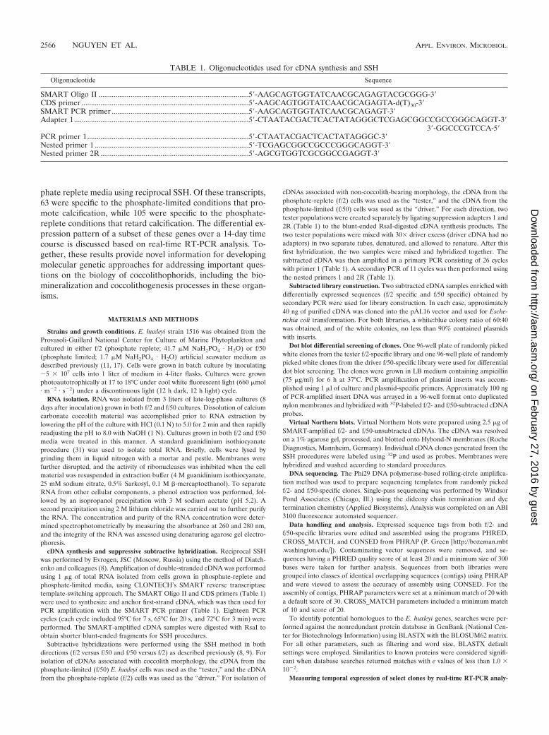
TABLE 1. Oligonucleotides used for cDNA synthesis and SSH
Oligonucleotide Sequence
SMART Oligo II ................................................................................5�-AAGCAGTGGTATCAACGCAGAGTACGCGGG-3�CDS primer .........................................................................................5�-AAGCAGTGGTATCAACGCAGAGTA-d(T)30-3�SMART PCR primer .........................................................................5�-AAGCAGTGGTATCAACGCAGAGT-3�Adapter 1.............................................................................................5�-CTAATACGACTCACTATAGGGCTCGAGCGGCCGCCGGGCAGGT-3�
3�-GGCCCGTCCA-5�PCR primer 1......................................................................................5�-CTAATACGACTCACTATAGGGC-3�Nested primer 1 ..................................................................................5�-TCGAGCGGCCGCCCGGGCAGGT-3�Nested primer 2R ...............................................................................5�-AGCGTGGTCGCGGCCGAGGT-3�
2566 NGUYEN ET AL. APPL. ENVIRON. MICROBIOL.
on February 27, 2016 by guest
http://aem.asm
.org/D
ownloaded from

sis. The mRNA expression levels of five of the f/50- and three of the f/2-specificclones were measured over a 14-day time course. Cells were grown in f/50 and f/2media, and total RNA was extracted from cultures at 4, 7, 10, and 14 dayspostinoculation. First-strand cDNA synthesis was performed using an Omni-script cDNA synthesis kit (QIAGEN Inc., Valencia, CA). For cDNA synthesis,2 �g of total RNA from each time point was used as a template in a 20-�lreaction mixture. Each reaction mixture contained 2 �g template RNA, 1�buffer reverse transcriptase (RT), 0.5 mM deoxynucleoside triphosphate, 1 �Moligo(dT) primer, 10 U of RNase inhibitor, and 4 U of Omniscript reversetranscriptase. The reaction mixture was incubated for 60 min at 37°C. cDNA wasdiluted 1:100, and 5 �l of the dilution was used in a SYBR Green RT-PCR.
Real-time PCR experiments were carried out using SYBER Green chemistryfor amplicon detection. The SYBER Green assays were performed on the iCy-cler iQ System (Bio-Rad). Amplification of target genes, along with a template-minus control, was performed in triplicate in a 96-well plate. Each 25-�l reactionmixture contained 5 �l cDNA, 7.1 �l 2� SYBER Green Master Mix, and 0.24�M (each) of forward and reverse primers. The cycling conditions for amplifi-cation included a 10-min polymerase activation at 95°C followed by 40 cycles of95°C for 10 seconds, 60°C for 30 seconds, and 82°C for 30 seconds. Fluorescencewas measured at 82°C at each cycle. The cycle threshold (CT) value is inverselyproportional to the quantity of cDNA present in the reaction mixture at thatparticular cycle. After PCR amplification, melt curves were generated by dena-turing the sample for 1 min at 95°C, cooling it to 55°C for 1 min, and thenramping the temperature 0.5°C every 10 seconds beginning at 55°C until a finaltemperature of 95°C was reached. Real-time PCR data were plotted using SigmaPlot version 8.0 by taking the mean of three replicates per time point. Therelative difference in expression between f/2 and f/50 cDNA clones was measuredusing the Basic �CT method as recommended by the Bio-Rad iCycler manufac-turer (Bio-Rad Laboratories). Change between f/2 and f/50 cDNA clones wascalculated as 2�CT. Primers were designed using Primer Express software version1.0 (PE Applied Biosystems) and are listed in Table 2. Results from the sequencedetection software were exported as tab-delimited text files and imported intoMicrosoft Excel for further analysis.
RESULTS
Differential screening of subtracted libraries. Figure 1shows the effect of phosphate levels on calcification in E. hux-leyi CCMP 1516 cells grown under phosphate-replete (f/2 me-dium) (Fig. 1B) versus phosphate-limited (f/50 medium) (Fig.1C) conditions. The dramatic difference in the amount of cal-cification occurring in response to the phosphate concentrationpermitted us to employ this as a tool for looking at geneexpression differences using SSH. Following construction of
our reciprocal SSH libraries, dot blotting and virtual Northernblots were employed to screen the differential cDNA frag-ments from the two libraries (Fig. 2 and 3). Differential screenresults yielded 44 f/50-specific clones (Fig. 2A) and 41 f/2-specific clones (Fig. 2B), indicating that both libraries containabout 40% positive clones. Additional clones from each of thetwo libraries exhibited various degrees of more subtle differ-ential expression that may warrant further investigation. Toverify differential screen results, virtual Northern blots werealso performed using six of the dot blot clones from eachsubtracted library. Results from the virtual Northern blots areshown in Fig. 3 top (f/50-specific clones) and bottom (f/2-specific clones). Differential expression of all 12 genes from thesubtracted libraries was confirmed by our virtual Northern blotresults.
Sequence assembly and annotation. In order to obtain moresequence information in the form of longer reads, we se-quenced and assembled several hundred clones from each li-brary. Table 3 describes the general characteristics of the ESTsthat were sequenced from each of the two SSH libraries. Of the423 ESTs sequenced from the phosphate-limited library, 27were singletons and the remainder were assembled into 38contiguous sequences (contigs) with an overall EST redun-dancy of 93% (calculated as the number of ESTs in contigsdivided by the number of ESTs). Of the 513 ESTs sequencedfrom the phosphate-replete library, 53 were singletons and theremainder were assembled into 50 contigs. The overall ESTredundancy for the phosphate-replete library was 90%. Assem-bled sequences from each library were annotated by BLASTXsearch (e value � 10�2) against the nonredundant proteindatabase from GenBank. Overall, 23% and 34% of the iden-tified genes from the phosphate-limited and phosphate-repletelibraries, respectively, encode polypeptides with similarity toproteins with known functions in GenBank. The remainingsequences fell into the unclassified category of proteins withunknown functions (expressed as putative or hypothetical pro-teins) or with no GenBank match.
A complete list of the ESTs and their GenBank homologues
TABLE 2. Primers used for amplification of transcripts by real-time RT-PCR
Primer name Primer sequence (5�–3�)a Primer %GC Ampliconsize (bp)
Amplicon Tmb
expected observed
F/2Contig26 For: CACCATCAATACATCGTCGGAT 45 82 78 81.5Rev: CTGGATCATAATCACGCACGAA 45
F/2Contig34 For: TTTGTCGAACTTCCGCATGA 45 78 83 85.5Rev: TCGTTGGCGCACATACTGA 53
F/2Contig22 For: AGGCCATCTACAACCACTACAAACC 48 77 81 83Rev: CCTCATGCAATTCGTGTTCCA 48
F/50Contig29 For: AGTTGAAGTGCTGCCCTGGTT 52 97 82 85.2Rev: TCGTAGACAGTCCGTGGTTCCT 55
F/50Contig31 For: CCTAGGACAGTTCATCCAAACGA 48 96 79 83.5Rev: GCAAAGCACAAGGACTCATCAA 45
F/50Contig18 For: GACTTCGCCAGCTACAATCCA 52 84 81 84Rev: GGCGAACTTTGCCATATCCTT 48
F/50Contig36 For: GTGTCACGCCCTCTCAATCAGT 55 76 83 85Rev: TGTTCGCCAGAATCGTTTCAC 48
F/50Contig33 For: CCATCCTGCAGTCCTTGATACC 55 84 82 85.5Rev: AGATGCCGTTTGCGCTTT 50
a For, forward; Rev, reverse.b Tm, melting temperature.
VOL. 71, 2005 SSH GENE EXPRESSION IN E. HUXLEYI 2567
on February 27, 2016 by guest
http://aem.asm
.org/D
ownloaded from

for each of the SSH libraries is presented in Tables 4 and 5.The vast majority of the most abundant transcripts in thephosphate-limited data set were of unknown function, whiletranscripts encoding ribosomal proteins and RNA polymerasesubunits were abundant in the phosphate-replete data set.Transcripts encoding proteins involved in photosynthesis, suchas the photosystem I subunits and the chloroplast ATP syn-thase subunit proteins, were also present among the mostabundant clusters in the phosphate-replete data set.
Temporal expression of selected genes by real-time RT-PCRanalysis. Real-time RT-PCR was conducted to verify expres-sion of a small subset of eight differentially expressed genesusing independently prepared RNA that was extracted fromcultures of f/2- and f/50-grown cells 4, 7, 10, and 14 days afterinoculation. The 14-day time course was intended to determinetranscripts (cDNA clones) that may be directly involved inbiomineralization, as calcification is known to occur most no-tably during the late log and stationary phases of growth (2),which generally occur between 7 and 14 days of growth (17).
The amplification profiles (along with their respective stan-dard deviations) and dissociation curves for each of the se-lected genes, three from the phosphate-replete library and fivefrom the phosphate-limited library, are shown in Fig. 4. Whilecontigs 18, 29, 31, and 36 from the f/50 library showed differ-
ential mRNA expression in cultures grown in phosphate-lim-ited compared to phosphate-replete media from day 4 onward,none of the transcripts were induced immediately. Maximaldifferences in expression were reached 14 days following inoc-ulation. In all cases, with the exception of contig 18, contig-specific expression was detectable under both growth condi-tions. By day 14, changes in gene expression for contigs 31 and35 were 3 orders of magnitude greater for cells grown in phos-phate-limited versus phosphate-replete media (Fig. 4E and G).Changes of more than 2 orders of magnitude were noted forcontigs 29 and 18 at day 14 (Fig. 4D and F). Contig 18-specificexpression, however, was barely detectable in cells grown inphosphate-replete medium. More modest changes in gene ex-pression were evident for contig 33 from the f/50 library (Fig.4H).
Real-time RT-PCR also confirmed candidates from the f/2library whose down-regulation during phosphate limitationwas revealed via SSH procedures. For example, the estimatedchange for contig 34 across the 14-day time course highlightsthe ability of SSH to identify genes that are expressed differ-entially, but at low levels. Down-regulation of this transcript inthe phosphate-deficient cells varied between 1.5- and 3.6-fold(Fig. 4B). Transcript levels for one of the P700 reaction centerproteins exhibited an interesting expression pattern across the
FIG. 2. Dot blot showing differential screening results of subtracted library clones from a phosphate-limited (f/50 tester) (A) and a phosphate-replete (f/2 tester) (B) plate. Clones on each plate were differentially screened with tester-specific and driver-specific cDNA probes.
2568 NGUYEN ET AL. APPL. ENVIRON. MICROBIOL.
on February 27, 2016 by guest
http://aem.asm
.org/D
ownloaded from

14 days. The transcript was down-regulated at 4 days (�2-fold)under phosphate-limited conditions but then appeared to beup-regulated (�3- to 5-fold) during the later stages of growth(days 10 to 14) (Fig. 4A). The dissociation curves for all sam-ples showed a single peak at the expected temperature, indi-cating target-specific amplification.
DISCUSSION
PCR-based suppressive subtractive hybridization was usedsuccessfully in the present investigation to detect differences ingene expression in coccolith-bearing E. huxleyi cells grown inphosphate-limited medium and non-coccolith-bearing cellsgrown in phosphate-replete medium. A total of 168 differen-
tially expressed transcripts were identified, some of which maybe related to phosphate starvation and some of which presum-ably are involved in calcification and coccolithogenesis. Thehigh redundancy rates of 93% and 90% that were obtainedafter sequencing 423 clones from the phosphate-limited and513 clones from the phosphate-replete libraries, respectively,may be a function of the SSH methodology. Alternatively,these rates may indicate that the potential for novel genediscovery under these conditions is nearly exhausted, suggest-ing that only a small number of genes, perhaps less than 100,are required for biomineralization in E. huxleyi.
SSH is a powerful technology that enriches for differentiallyexpressed genes, but it is by no means perfect. It is difficult tocontrol for sequence-dependent factors, such as cDNA synthe-sis and PCR amplification efficiencies. Construction of leakylibraries in which the same transcript is present in the tworeciprocal libraries can also be problematic. While the recip-rocal libraries constructed in the present investigation weremutually exclusive, with membership in each library specific tothe particular growth conditions, overrepresentation of specificclones was noted in both libraries. SSH includes a normaliza-tion step to overcome the problem of differences in mRNAabundance (8). All differentially regulated clones are expectedto be equally represented after normalization. Overrepresen-tation of clones, such as the various ribosomal proteins,Orf122, and hypothetical protein TC0128 in the phosphate-replete library and the photosystem II reaction center proteinD1, the putative senescence-associated protein, and the differ-ent hypothetical (contigs 38 and 32), unknown proteins in thephosphate-deficient library, is an artifact.
Nearly half of the clones recovered were estimated to bespecific by dot blot hybridization, being either completely ab-sent or substantially different in the two different cell types.Further tests of a smaller subset of select gene sequences usingNorthern blots and/or real-time RT-PCR corroborated someof the more distinctly different gene expression profiles, such asthose of contigs 31 and 36 from the f/50 SSH library. Repre-sentation of quantitatively different cDNAs in the SSH librar-ies is also apparent, as several gene sequences present in thephosphate-replete SSH library, including the ribosomal pro-teins and photosynthesis transcripts, are fundamental house-keeping genes. The differential expression data of these par-ticular genes support previous findings that suggest thatcalcification and coccolithogenesis are stimulated under con-ditions that result in reduced cell growth and increased cell sizeand are inhibited under conditions that favor cell growth and adecrease in cell size (4, 26, 28, 29).
The SSH method applied here has also proved to be effec-tive in uncovering some of the distinguishing, albeit less abun-dant, transcripts from coccolith-bearing and non-coccolith-bearing E. huxleyi cells. This is evidenced by the fact thatcombined, 70% of the ESTs identified in this investigation(49% from the phosphate-limited and 21% from the phos-phate-replete libraries) were not represented in our previouslydescribed EST data sets (36, 37) that were derived from non-normalized cDNA libraries. The effectiveness of subtractionwas also demonstrated by the real-time RT-PCR data, in whichthe expression of eight genes tested over a 14-day time courseexperiment validated results obtained from these libraries. Thesuppressive subtraction was successful in detecting small dif-
FIG. 3. Virtual Northern blot analysis of differential clones ob-tained from phosphate-limited (f/50) and phosphate-replete (f/2) sub-tracted libraries (see text for details).
TABLE 3. Comparative analysis of E. huxleyi 1516 f/2 and f/50cDNA libraries
Descriptive category Phosphatelimiteda
Nutrientrepleteb
No. of clones sequenced 423 513No. of contigs 38 50No. of singletons 27 53No. of unigenes 65 103No. of EST matches with e value of 1 � 10�2 37 19No. of EST matches with e value of 1 � 10�2 15 35No. of EST with no GenBank matchc 13 49
a RNA extracted from E. huxleyi 1516 grown in f/50 medium (ca. 1.5 �Mphosphate) as driver.
b RNA extracted from E. huxleyi 1516 grown in f/2 medium (ca. 36 �Mphosphate) as driver.
c Searches yielding e values of 10�2.
VOL. 71, 2005 SSH GENE EXPRESSION IN E. HUXLEYI 2569
on February 27, 2016 by guest
http://aem.asm
.org/D
ownloaded from

ferences in the expression of highly expressed, as well as lessabundant, transcripts. These results demonstrate the impor-tance of SSH-based EST sequencing as an approach comple-mentary to existing EST resources for functional genomicsresearch on marine coccolithophorids.
The most distinguishing feature of the SSH library preparedfrom the coccolith-bearing cells derived from the phosphate-
limited media is the large number of transcripts that show nosignificant homology to sequences in GenBank. We obtainedand sequenced the corresponding cDNA clones for six of theeight SSH clones that were subjected to real-time RT-PCRanalysis. Since contig 26 showed significant homology to theP700 chlorophyll a apoprotein A2 from E. huxleyi, it was notcharacterized any further, and since contig 29 from the f/50
TABLE 4. BLASTX results of most prevalent transcripts from f/2-specific SSH librarya
SeqID Putative ID e value Reads Length
Sig1 Unknown 0.001 1 865Sig2 ORF497, homologous to Porphyra ORF491 (Odontella sinensis) 2.47E-28 1 797Sig3 Unknown 0.001 1 828Sig4 Unknown 0.001 1 543Sig5 Unknown 0.001 1 479Sig6 Apocytochrome f, chloroplast precursor 7.499E-13 1 866Sig7 RNA polymerase beta apos; apos; subunit (Cyanidium caldarium) 3.593E-09 1 628Sig8 Pentose-5-phosphate-3-epimerase (Pseudomonas fluorescens PfO-1) 6.42E-16 1 804Con2 Ribosomal protein S9 (Crocosphaera watsonii WH 8501) 1.36E-29 1 960Con3 RNA polymerase beta prime subunit (Gloeobacter violaceus PCC 7421) 4.26E-38 1 913Con4 Ribosomal protein S3 (Nostoc punctiforme) 1.09E-31 1 803Con5 Ribosomal protein S3 (Porphyra purpurea) 3.29E-28 1 807Con6 Ribosomal protein S3 (Porphyra purpurea) 1.72E-16 1 916Con7 Unknown 0.001 1 801Con8 Orf122-(Chlorobium tepidum) 5.686E-10 1 1,002Con9 Unknown 0.001 2 322Con10 Hypothetical protein (Trichodesmium erythraeum IMS101) 3.08E-40 2 634Con11 Unknown 0.001 2 433Con12 Unknown 0.001 2 416Con14 Ribosomal protein S2 (Guillardia theta) 4.21E-24 2 863Con15 Unknown 0.001 2 1,174Con16 Ribosomal protein S4 (Nephroselmis olivacea) 5.61E-26 2 447Con17 Unknown 0.001 2 419Con18 Hypothetical protein (Bacillus megaterium) 8.79E-17 2 396Con19 Unknown 0.001 2 392Con20 Unknown 0.001 2 405Con21 Unknown 0.001 2 645Con22 Unknown 0.001 2 455Con23 Signal transduction histidine kinase (Crocosphaera watsonii WH 8501) 1.93E-30 2 781Con24 Unknown 0.001 2 301Con25 Elongation factor Tu GTP binding domain (Bacillus anthracis A2012) 3.32E-33 2 897Con26 Photosystem I P700 chlorophyll a apoprotein A2 (Emiliania huxleyi) 1.76E-74 2 616Con27 Hypothetical protein (Plasmodium yoelii yoelii) 5.41E-19 3 961Con28 ATP synthase delta chain, chloroplast 1.37E-30 3 572Con29 Unknown 0.001 3 402Con30 ABC transporter (Cyanophora paradoxa) 2.18E-49 3 621Con31 Ribosomal protein S9 (Crocosphaera watsonii WH 8501) 1.18E-32 3 587Con32 Hypothetical protein (Nostoc sp. strain PCC 7120) 3.50E-18 3 869Con33 50S ribosomal protein L3 (Prochlorococcus marinus) 1.08E-19 3 537Con34 Unknown 0.001 4 1,021Con35 Hypothetical chloroplast RF7 (Guillardia theta) 6.85E-04 5 364Con36 Hypothetical protein Avar020180 (Anabaena variabilis ATCC 29413) 0.0001666 5 514Con37 Preprotein translocase subunit Sec Y (Porphyra purpurea) 8.27E-20 6 866Con38 COG0087: ribosomal protein L3 (Trichodesmium erythraeum IMS101) 1.79E-11 7 465Con39 Chloroplast ATP synthase a chain precursor (ATPase subunit IV) 1.54E-76 7 745Con40 Subunit epsilon of ATPase (Ochrosphaera neapolitana) 8.68E-30 7 1,020Con41 Photosystem 1 subunit III (Guillardia theta) 1.11E-39 8 1,027Con42 Photosystem 1 subunit XI (Guillardia theta) 2.82E-35 8 1,152Con43 Predicted protein (Methanosarcina acetivorans strain C2A) 1.02895 10 619Con44 Orf122 (Chlorobium tepidum) 8.41E-19 10 971Con45 Ribosomal protein S2 (Guillardia theta) 1.41E-56 10 566Con46 Hypothetical protein TC0128 (imported)—Chlamydia muridarum (strain Nigg) 0.116837 12 692Con47 COG0092: ribosomal protein S3 (Nostoc punctiforme) 6.21E-31 19 651Con48 Hypothetical protein (Chlamydophila pneumoniae AR39) 2.60643 56 1,570Con49 COG0092: ribosomal protein S3 (Anabaena variabilis ATCC 29413) 1.27E-40 71 1,128Con50 COG0048: ribosomal protein S12 (Crocosphaera watsonii WH 8501) 1.81E-37 151 1,766
a BLASTX results following PHRAP assembly are listed as follows: SeqID (sequence identification number) for singletons (Sig) and contigs (Con), Reads (numbersof EST fragments per assembled sequence), and Length (average sequence length in base pairs).
2570 NGUYEN ET AL. APPL. ENVIRON. MICROBIOL.
on February 27, 2016 by guest
http://aem.asm
.org/D
ownloaded from

SSH lacked a correlate in our previously described unigene setof 4,561 sequences from E. huxleyi (36), the clone and full-length sequence could not be readily obtained. In an attemptto characterize the six other sequences, we used NCBI’sBLASTX and ORF-finder to identify the most likely openreading frame and employed the Protein Prediction MetaServer available through Columbia University to search forsequence motifs, domains, low-complexity regions, and proteinlocalization signals. Secondary structure, transmembrane re-
gions, solvent accessibility, disulfide bonds, and posttranslationmodifications were also predicted.
Only a few biomineralization proteins have been identifiedin marine organisms. Some of these are proteins such as lustrinand perlucin from the mollusk shell; nacre and pearlin fromthe nacreous layer of the oyster; and MS130, SpP16, and SpP19from the sea urchin. While these particular proteins show littlesequence homology to each other or any other known biomin-eralization proteins (15, 27, 39), they do share a number of
TABLE 5. BLASTX results of most prevalent transcripts from f/50-specific SSH librarya
SeqID Putative ID e value Reads Length
Sig1 Unknown 0.001 1 740Sig2 Unknown 0.001 1 1,108Sig3 Subunit beta of ATPase (Ochrosphaera neapolitana) 1.93E-44 1 612Sig4 Unknown 0.001 1 663Sig5 Hypothetical protein (Plasmodium yoelii yoelii) 1.4E-14 1 668Sig6 Unknown 0.001 1 781Sig7 Unknown 0.001 1 1,378Sig8 Unknown 0.001 1 537Sig9 NADH dehydrogenase subunit 1 (Emiliania huxleyi) 6.56E-40 1 746Sig10 Unknown 0.001 1 479Sig11 Unknown 0.001 1 604Sig12 Unknown 0.001 1 721Sig13 Unknown 0.001 1 407Sig14 Unknown 0.001 1 596Sig15 Translation elongation factor Tu (Chromobacterium violaceum) 1.51E-52 1 802Sig16 Unknown 0.001 1 790Sig17 Unknown 0.001 1 831Sig18 Unknown 0.001 1 849Uni1 Unknown 0.001 1 785Uni2 Unknown 0.001 1 893Uni4 Unknown 0.001 1 571Uni5 Unknown 0.001 1 661Uni6 Unknown 0.001 1 655Uni7 347L (Invertebrate iridescent virus 6) 2.6E-07 1 559Con8 Cytochrome c oxidase subunit 2 (Emiliania huxleyi) 8.43E-31 2 587Con9 Unknown 0.001 2 242Con10 Photosystem I P700 apoprotein A2 (Guillardia theta) 5.21E-16 2 503Con11 Unknown 0.001 2 426Con12 Unknown 0.001 2 328Con13 Unknown 0.001 2 433Con14 Unknown 0.001 2 775Con15 Unknown 0.001 2 766Con17 Unknown 0.001 3 718Con18 Unknown 0.001 3 287Con19 Unknown 0.001 3 506Con20 Unknown 0.001 4 510Con21 Unknown (Vibrio vulnificus CMCP6) 1.9E-05 4 509Con22 Unknown 0.001 4 358Con23 Unknown 0.001 4 528Con24 Unknown 0.001 5 170Con25 Unknown 0.001 5 348Con26 Unknown 0.001 8 640Con27 Ribosomal protein S3 (Porphyra purpurea) 6.32E-17 8 636Con28 Unknown 0.001 8 596Con29 Unknown 0.001 16 787Con30 Photosystem II reaction center protein D1 (Emiliania huxleyi) 5.39E-37 18 841Con31 347L (Invertebrate iridescent virus 6) 4.15E-06 18 1,279Con32 Unknown 0.001 18 422Con33 Hypothetical protein (Nostoc punctiforme) 8.19E-06 19 1,170Con35 Putative senescence-associated protein (Pisum sativum) 9.65E-23 25 1,014Con36 Unknown 0.001 29 1,980Con37 Unknown 0.001 42 968Con38 Hypothetical protein 0.00022 105 2,391
a BLASTX results following PHRAP assembly are listed as follows: SeqID (sequence identification number) for singletons (Sig) and contigs (Con), Reads (numbersof EST fragments per assembled sequence), and Length (average sequence length in base pairs).
VOL. 71, 2005 SSH GENE EXPRESSION IN E. HUXLEYI 2571
on February 27, 2016 by guest
http://aem.asm
.org/D
ownloaded from

FIG. 4. Real-time RT-PCR results showing temporal expression patterns of individual cDNA transcripts from the f/2-specific (A to C) andf/50-specific (D to H) SSH libraries over a 14-day period. Total RNA was isolated from E. huxleyi cells grown in either f/2 (F) or f/50 (E) medium.mRNA transcript abundance is inversely proportional to the cycle threshold (CT) value. Change (n-fold; bars) at each time point was calculatedas 2�Ct and expressed as the relative difference in expression in f/50 versus f/2. The data represent the mean of three replicates per time point,and standard deviations are expressed as error bars. d, days.
2572
on February 27, 2016 by guest
http://aem.asm
.org/D
ownloaded from

FIG. 4—Continued.
VOL. 71, 2005 SSH GENE EXPRESSION IN E. HUXLEYI 2573
on February 27, 2016 by guest
http://aem.asm
.org/D
ownloaded from

chemical and physical features. They generally are small acidicproteins or glycoproteins that are rich in aspartic acid or glu-tamic acid (10) and manifest prominent repeat sequences (40,42). They also tend to exhibit extended or elastomeric confor-mations possessing little or no distinct secondary structure(42). Unfortunately, none of the gene products we examinedshowed homology to previously identified biomineralizationproteins, nor were they noticeably acidic, nor did they containobvious amino acid repeat elements. This result is not surpris-ing given the limited number of biomineralization proteinsidentified to date and the lack of understanding of the detailsof the structure and function of these proteins in relation tobiomineralization processes.
Contig 22 from the SSH f/2 library, which showed littledifferential expression until day 14, when it was up-regulatedover 40-fold, exhibited no significant homology to any proteindeposited in GenBank and showed no distinct motif or domainmatches. The transcript is predicted to be an extracellular orcell wall protein and appears to contain three dominant alphahelices. Two of the f/50 SSH contigs that showed markedup-regulation—contig 31, which was expressed at a level 1,000-fold greater, and contig 18, which was expressed at a level500-fold greater—in cells under phosphate-limited conditionson day 14 relative to phosphate-replete conditions also re-turned no significant GenBank BLASTX hits and exhibited nodistinct motif or domain match. Although the cellular locationof the protein correlates cannot be predicted, both proteins areexpected to be membrane bound. Of the three alpha helicesthat can be discerned for each of the two proteins, one incontig 31 and two in contig 18 have a high probability of beingtransmembrane helices.
The sequences of three transcripts exhibited significant ho-mology to proteins in GenBank. The sequence of one of thetranscripts from the f/2 SSH library was up-regulated slightly(1.5- to 4-fold) across the 14-day time course in the non-coccolith-bearing cells and showed significant homology(BLASTX; 2E-07) to a hypothetical protein of unknown func-tion from Leptospira interrogans. The protein, which may residein the nucleus (PSORT), features two prominent alpha-helicalregions interspersed with short stretches of beta-sheets butshows no other outstanding characteristics. The most likelyprotein product encoded by contig 33 from the f/50 SSH libraryshowed significant homology to a hypothetical protein fromNostoc (1E-14), with more distant homology to bacterial andfungal methylase enzymes (1E-7). This particular transcriptwas marginally up-regulated (2- to 5-fold) in coccolith-bearingcells during the first 10 days of growth but by day 14 wassignificantly up-regulated (30-fold).
One of the more interesting sequences was that of contig 36from the f/50 SSH library, which at day 14 was expressed in thephosphate-limited coccolith-bearing cells at a level that was1,400-fold greater than in the non-coccolith-bearing cells main-tained in phosphate-replete medium. The full-length sequenceof the corresponding cDNA clone showed significant homol-ogy (BLASTX) to a number of ligand-gated anion channels,including that of the AVR-15 protein from Caenorhabditiselegans (4E-7), a glutamate-gated chloride channel from Hae-monchus contortus (9E-06), and rat gamma-aminobutyric acidA receptor (1E-04). Four strong transmembrane helices likelyto form the ion channel were predicted (using bioinformatics
tools such as TMHMM, TMpred, and RPS BLAST), alongwith a distinct arginine-rich region outside the membrane thatmay represent the ligand binding domain. Our laboratory iscurrently trying to develop tools to determine the membranelocation of the contig 36 gene product with the goal of evalu-ating the physiological consequences of blocking the channelor of knocking out expression of the gene to elucidate itsphysiological role. These data may indicate whether it is ahighly selective ion channel or an indiscriminate activatedchannel that functions in low-affinity nutrient uptake, signal-ing, and/or ion transport.
The expression profiles of the phosphate-limited contigs 31,18, 36, and 29 are intriguing, with progressively greater differ-ential expression occurring during the later stages of growth.This is consistent with what we know about biomineralizationand growth of E. huxleyi in batch culture (2). The effects ofphosphate limitation, however, are also expected to becomeprogressively more severe over time, and hence, we cannotunequivocally relate the differential expression patterns ofthese genes to biomineralization (4, 20, 27, 30).
Based upon what is presently known of biomineralizationproteins, one cannot logically argue that any of the aforemen-tioned proteins is directly involved in calcium carbonate bio-mineralization. However, the differential expression data sup-port the possibility that these proteins may be peripherallyinvolved in the processes of biomineralization and coccolitho-genesis. Perhaps of greater significance is the fact that the datapresented in this study provide novel and important informa-tion to the research community for developing molecular andbiochemical approaches to determine the metabolic roles ofthese genes and gene products differentially expressed undercalcifying conditions in E. huxleyi.
In summary, our results support the value of SSH-basedEST sequencing to complement existing EST resources forfunctional genomics research in marine coccolithophorid al-gae. We have detected 168 differentially expressed transcriptsin comparing coccolith-bearing and non-coccolith-bearing E.huxleyi cells maintained in phosphate-limited and phosphate-replete media, respectively, by screening SSH libraries. Thespecific expression of a subset of these genes was confirmed bya combination of dot blotting, virtual Northern blotting, andreal-time RT-PCR. Given the fact that methods for genetictransformation and mutagenesis have not been developed forE. huxleyi, or for any coccolithophorid, additional full-lengthcDNA cloning, combined with more detailed functional studiesusing temporal cDNA microarray analysis or RNA interfer-ence methodologies, will help us to understand the molecularmechanisms underlying biomineralization and coccolithogen-esis.
ACKNOWLEDGMENTS
We thank Jeremy Young (Department of Palaeontology, The Nat-ural History Museum, London, United Kingdom) for kindly providingthe scanning electron micrograph of E. huxleyi. We also thank PatrickQuinn for his helpful input on the manuscript.
This work was supported by a grant from the National Institutes ofHealth (GM 059833).
REFERENCES
1. Andersen, O. K. 1981. Coccolith formation and calcification in an N-cellculture of Emiliania huxleyi during phosphorus-limited growth in batch andchemostat cultures. Ph.D. thesis. University of Oslo, Oslo, Norway.
2574 NGUYEN ET AL. APPL. ENVIRON. MICROBIOL.
on February 27, 2016 by guest
http://aem.asm
.org/D
ownloaded from

2. Balch, W. M., P. M. Holligan, and K. A. Kilpatrick. 1992. Calcification,photosynthesis and growth of the bloom-forming coccolithophore Emilianiahuxleyi. Cont. Shelf Res. 12:1353–1374.
3. Brown, C. W., and J. A. Yoder. 1994. Coccolithophorid blooms in the globalocean. J. Geophys. Res. 99:7467–7482.
4. Buitenhuis, E. T., H. J. W. de Baar, and M. J. W. Veldhuis. 1999. Photo-synthesis and calcification by Emiliania huxleyi (Prymnesiophyceae) as afunction of inorganic carbon species. J. Phycol. 35:949–959.
5. Corstjens, P. L. A. M., A. van der Kooij, C. Linschooten, G.-J. Brouwers, P.Westbroek, and E. W. de Vrind-de Jong. 1998. GPA, a calcium-bindingprotein in the coccolithophorid Emilianis huxleyi (Prymnesiophyceae). J.Phycol. 34:622–630.
6. Cramer, R. A., and C. B. Lawrence. 2004. Identification of Alternaria bras-sicicola genes expressed in planta during pathogenesis of Arabidopsis thali-ana. Fungal Genet. Biol. 41:115–128.
7. de Vrind-de Jong, E. W., and J. P. M. de Vrind. 1997. Algal deposition ofcarbonates and silicates, p. 267–307. In J. F. Banfield and K. H. Nealson(ed.), Geomicrobiology: interactions between microbes and minerals. Min-eralogical Society of America, Washington, D.C.
8. Diatchenko, L., Y.-F. C. Lau, A. P. Campbell, A. Chenchik, F. Moqadam, B.Huang, S. Lukyanov, K. Lukyanov, N. Gurskaya, E. D. Sverdlov, and P. D.Siebert. 1996. Suppression subtractive hybridization: a method for generat-ing differentially regulated or tissue-specific cDNA probes and libraries.Proc. Natl. Acad. Sci. USA 93:6025–6030.
9. Diatchenko, L., S. Lukyanov, Y. Lau, and P. Siebert. 1999. Suppressionsubtractive hybridization: a versatile method for identifying differentiallyexpressed genes. Methods Enzymol. 303:349–380.
10. Gotliv, B.-A., L. Addadi, and S. Weiner. 2003. Mollusk shell acidic proteins:in search of individual functions. Chembiochem. 4:522–529.
11. Guillard, R. R. L. 1975. Culture of phytoplankton for feeding marine inver-tebrates, p. 29–60. In W. L. Smith and M. H. Chanley (ed.), Culture ofmarine invertebrate animals. Plenum Press, New York, N.Y.
12. Happe, T., and A. Kaminski. 2002. Differential regulation of the Fe-hydro-genase during anaerobic adaptation in the green alga Chlamydomonas rein-hardtii. Eur. J. Biochem. 269:1022–1032.
13. Henriksen, K., S. L. S. Stipp, J. R. Young, and P. R. Bown. 2003. Tailoringcalcite: nanoscale AFM of coccolith biocrystals. Am. Mineralogist 88:2040–2044.
14. Holligan, P. M., E. Fernandez, J. Aiken, W. M. Balch, P. Boyd, P. H. Burkill,M. Finch, S. B. Groom, G. Malin, K. Muller, D. A. Purdie, C. Robinson, C.Trees, S. M. Turner, and P. van de Wal. 1993. A biogeochemical study of thecoccolithophore Emiliania huxleyi in the North Atlantic. Global Biogeo-chem. Cycles 7:879–900.
15. Illies, M. R., M. T. Peeler, A. M. Dechtiaruk, and C. A. Ettensohn. 2002.Identification and developmental expression of new biomineralization pro-teins in the sea urchin Stronglyocentrotus purpuratus. Dev. Genes Evol. 212:419–431.
16. Kim, M., S. Kim, S. Kim, and B. D. Ki. 2001. Isolation of cDNA clonesdifferentially accumulated in the placenta of pungent pepper by suppressionsubtractive hybridization. Mol. Cells 11:213–219.
17. Laguna, R., J. Romo, B. A. Read, and T. M. Wahlund. 2001. Induction ofphase variation events in the life cycle of the marine coccolithophorid Emil-iania huxleyi. Appl. Environ. Microbiol. 67:3824–3831.
18. Liao, H., F. L. Wong, T. H. Phang, M. Y. Cheung, W. Y. Li, G. Shao, X. Yan,and H. M. Lam. 2003. GmPAP3, a novel purple acid phosphatase-like genein soybean induced by NaCl stress but not phosphorus deficiency. Gene318:103–111.
19. Mahalingam, R., A. Gomez-Buitrago, N. Eckardt, N. Shah, A. Guevara-Garcia, P. Day, R. Raina, and N. V. Fedoroff. 2003. Characterizing thestress/defense transcriptome of Arabidopsis. Genome Biol. 4:R20.
20. Marsh, M. E. 2003. Regulation of CaCO3 formation in coccolithophores.Comp. Biochem. Physiol. 136:743–754.
21. Moyano, E., I. Portero-Robles, N. Medina-Escobar, V. Valpuesta, J. Munoz-Blanco, and J. L. Caballero. 1998. A fruit-specific putative dihydroflavonol4-reductase gene is differentially expressed in strawberry during the ripeningprocess. Plant Physiol. 117:711–716.
22. Ozaki, N., S. Sakuda, and H. Nagasawa. 2001. Isolation and some charac-terization of an acidic polysaccharide with anti-calcification activity from
coccoliths of a marine alga, Pleurochrysis carterae. Biosci. Biotechnol. Bio-chem. 65:2330–2333.
23. Paasche, E. 1998. Roles of nitrogen and phosphorus in coccolith formationin Emiliania huxleyi (Prymnesiophyceae). Eur. J. Phycol. 33:33–42.
24. Paasche, E., and S. Bruback. 1994. Enhanced calcification in the coccolith-ophorid Emiliania huxleyi (Haptophyceae) under phosphorous limitation.Phycologia 33:324–330.
25. Palenik, B., and S. E. Henson. 1997. The use of amides and other organicnitrogen sources by the phytoplankton Emiliania huxleyi. Limnol. Oceanogr.42:1544–1551.
26. Sekino, K., and Y. Shiraiwa. 1994. Accumulation and utilization of dissolvedinorganic carbon by a marine unicellular coccolithophorid, Emiliania huxleyi.Plant Cell Physiol. 35:353–361.
27. Shen, X., A. M. Belcher, P. K. Hansma, G. D. Stucky, and D. E. Morse. 1997.Molecular cloning and characterization of lustrin A, a matrix protein fromshell and pearl nacre of Haliotis rufescens. J. Biol. Chem. 272:32472–32481.
28. Shiraiwa, Y. 2003. Physiological regulation of carbon fixation in the photo-synthesis and calcification of coccolithophorids. Comp. Biochem. Physiol. B136:775–783.
29. Sikes, C. S., R. D. Roer, and K. M. Wilbur. 1980. Photosynthesis and coc-colith formation: inorganic carbon sources and net inorganic reaction dep-osition. Limnol. Oceanogr. 25:248–261.
30. Stoll, H. M., P. Ziveri, M. Geisen, I. Probert, and J. R. Young. 2002. Poten-tial and limitations of Sr/Ca ratios in coccolith carbonate: new perspectivesfrom cultures and monospecific samples from sediments. Phil. Trans. Ser. A360:719–747.
31. Strommer, J., R. Gregerson, and M. Vayda. 1993. Isolation and character-ization of plant mRNA, p. 49–66. In B. R. Glick and J. E. Thompson (ed.),Methods in plant molecular biology and biotechnology, 1st ed. CRC Press,Boca Raton, Fla.
32. Tyrrell, T., and A. H. Taylor. 1996. A modeling study of Emiliania huxleyi inthe NE Atlantic. J. Mar. Syst. 9:83–112.
33. van Bleijswijk, J. D. L., and M. J. W. Velduis. 1995. In situ gross growth ratesof Emiliania huxleyi in enclosures with different phosphate loadings revealedby diel changes in DNA content. Mar. Ecol. Prog. Ser. 121:271–277.
34. Van Der Wal, P., E. W. de Jong, P. Westbroek, W. C. de Bruijin, and A. A.Muller-Stapel. 1983. Ultrastructural polysaccharide localization in calcifyingand naked cells of the coccolithophorid Emiliania huxleyi. Protoplasma 118:157–168.
35. van Emburg, P. R. 1989. Coccolith formation in Emiliania huxleyi. Ph.D.thesis. Leiden University, Leiden, The Netherlands.
36. Wahlund, T. M., A. R. Hadaegh, R. Clark, B. Nguyen, M. Fanelli, and B. A.Read. 2004. Analysis of expressed sequence tags from calcifying cells of themarine coccolithophorid, Emiliania huxleyi. Mar. Biotechnol. 6:278–290.
37. Wahlund, T. M., X. Zhang, and B. A. Read. 2004. EST expression profilesfrom calcifying and non-calcifying cultures of Emiliania huxleyi. J. Micropa-leontol. 51:145–155.
38. Wang, W., P. Wu, M. Xia, Z. Wu, Q. Chen, and F. Liu. 2002. Identificationof genes enriched in rice roots of the local nitrate treatment and theirexpression patterns in split-root treatment. Gene 297:93–102.
39. Weiss, I. M., S. Kaufmann, K. Mann, and M. Fritz. 2000. Purification andcharacterization of perlucin and perlustrin, two new proteins from the shellof the mollusk, Haliotis laevigata. Biochem. Biophys. Res. Commun. 267:17–21.
40. Wustman, B. A., R. Santos, B. Zhang, and S. J. Evans. 2002. Identificationof a “glycine-loop”-like coiled structure in the 34 aa pro, gly, met repeatdomain of the biomineral-associated protein, PM27. Biopolymers 65:362–372.
41. Young, J. R., S. A. Davis, P. R. Bown, and S. Mann. 1999. Coccolith ultra-structure and biomineralisation. J. Struct. Biol. 126:195–215.
42. Zhang, B., G. Xu, and J. S. Evans. 2000. Model peptide studies of sequencerepeats derived from the intracrystalline biomineralization protein, SM50. II.Pro, asn-rich tandem repeats. Biopolymers 54:464–475.
43. Zhang, X. N., H. Wang, Z. C. Qu, M. M. Ye, and D. L. Shen. 2002. Cloningand prokaryotic expression of a salt-induced cDNA encoding a chloroplasticfructose-1,6-diphosphate aldolase in Dunaliella salina (Chlorophyta). DNASeq. 13:195–202.
VOL. 71, 2005 SSH GENE EXPRESSION IN E. HUXLEYI 2575
on February 27, 2016 by guest
http://aem.asm
.org/D
ownloaded from
Copyright © 2022 FDOKUMEN