Familial Hemiplegic Migraine: A New Gene in an Italian Family
Upload
independentCategory
view
3download
0
STXBP2 mutations in children with familial haemophagocyticlymphohistiocytosis type 5
Valentina Cetica1, Alessandra Santoro2, Kimberly C Gilmour3, Elena Sieni1, Karin Beutel1,Daniela Pende4, Stefania Marcenaro5, Florian Koch6,7, Samantha Grieve8, Rachel Wheeler3,Fang Zhao8, Udo zur Stadt6,7, Gillian M Griffiths8, and Maurizio Aricò1
1Department Pediatric Hematology Oncology, Azienda Ospedaliero-Universitaria Meyer,Florence, Italy
2U.O. Ematologia I, A.O. Ospedali Riuniti Villa Sofia-Cervello, Palermo, Italy
3Centre for Immunodeficiency, Great Ormond Street Hospital, London UK
4Istituto Nazionale per la Ricerca sul Cancro, Genoa, Italy
5DIMES, University of Genoa, Genoa, Italy
6Research Institute Children’s Cancer Center, Hamburg, Germany
7University Medical Center Hamburg, Department of Pediatric Hematology and Oncology,Eppendorf, Germany
8Cambridge Institute for Medical Research, Addenbrooke’s Hospital, Cambridge, CB2 0XY, UK
Abstract
Background—Familial haemophagocytic lymphohistiocytosis (FHL) is a rare immune
deficiency with uncontrolled inflammation; the clinical course usually starts within the first years
of life, and is usually fatal unless promptly treated and then cured with haematopoietic stem cell
transplant. FHL is caused by genetic mutations resulting in defective cell cytotoxicity; three
disease related genes have been identified to date: perforin, Munc13-4 and syntaxin-11. A fourth
gene, STXBP2, has been identified very recently as responsible for a defect in Munc18-2 in
FHL-5.
Aims—To describe the result of the screening of families with HLH and previously unassigned
genetic defects.
Methods—Patients with HLH diagnosed according to current diagnostic criteria, and who lacked
mutations in the PRF1, Munc13-4, and STX11 genes were sequenced for mutations in STXBP2.
Functional study was performed when material was available.
Correspondence to Maurizio Aricò Direttore Dipartimento Oncoematologia Pediatrica e Cure Domiciliari Azienda Ospedaliero-Universitaria Meyer Viale Pieraccini, 24 50139 Firenze, Italy; [email protected] MA has signed on behalf of all co-owners of the contribution.
Competing interest None.
Patient consent Not required.
Ethics approval AOU Meyer, Florence Italy.
Provenance and peer review Not commissioned; externally peer reviewed.
Europe PMC Funders GroupAuthor ManuscriptJ Med Genet. Author manuscript; available in PMC 2014 July 30.
Published in final edited form as:J Med Genet. 2010 September ; 47(9): 595–600. doi:10.1136/jmg.2009.075341.
Europe PM
C Funders A
uthor Manuscripts
Europe PM
C Funders A
uthor Manuscripts

Results—Among the 28 families investigated, 4 (14%) with biallelic STXBP2 mutations were
identified. They originated from Italy, England, Kuwait and Pakistan. The p.Pro477Leu resulting
from c.1430C>T, and p.Arg405Gln resulting from the single c.1214G>A nucleotide change are
known, while we contribute two novel mutations: p.Glu132Ala resulting from c.395A>C, and
p.Gly541Ser, resulting from c.1621G>A. The detrimental effect of the p.Gly541Ser mutation was
documented biochemically and functionally in NK and CD8 cells. Additional polymorphisms are
also described.
Conclusion—These data expand current knowledge on the genetic heterogeneity of FHL and
suggest that patients with FHL5 may have different results in degranulation assays under different
conditions.
INTRODUCTION
Haemophagocytic lymphohistiocytosis (HLH) is a genetically heterogeneous disorder
characterised by an hyperinflammatory syndrome with fever, hepatosplenomegaly,
cytopenia and sometimes central nervous system involvement.1 Bone marrow aspiration is
usually performed early during the diagnostic work-up, enabling the identification of
haemophagocytosis by activated macrophages. In most cases the natural course of HLH is
rapidly fatal within a few weeks, unless appropriate treatment, including corticosteroids,
cyclosporine, etoposide, or anti-thymocyte globuline, can obtain transient disease control.2-4
So far, only patients who underwent haematopoietic stem cell transplantation (HSCT) have
been cured.5–10
Differential diagnosis of HLH may be difficult.11 To this purpose, diagnostic guidelines for
HLH have been established by the Histiocyte Society.1213 In particular, demonstration of
frequent association with common pathogens, together with evidence of impaired natural
killer cytotoxic activity, provided the rationale for considering HLH as a selective immune
deficiency.14–16 Starting from the original report by Farquhar et al in 1952,17 autosomal
recessive inheritance was proposed and then confirmed as the common mode of inheritance
for the familial form of HLH (FHLH or FHL).
Previous genetic studies in FHL revealed an extended genetic heterogeneity. A first mapping
approach included four consanguineous families of Pakistani origin and identified a 7.8 cm
region on chromosome 9q21.3–22, but the underlying defect is still unknown (FHL-1; MIM
267700).18 A second report described linkage of 10 of 17 investigated families to a region
on chromosome 10q21–22,19 thus indicating genetic heterogeneity. Mutations in the
perforin-1 gene (PRF1) were then described at this locus (FHL-2; MIM 603553).20 About
20–50% of the patients have mutations in PRF1, with a strong geographical bias in the
overall frequency.21 Further genetic defects associated with FHL affect proteins involved in
transport, membrane fusion or exocytosis of perforin containing lytic granules such as
Munc13-4 (FHL-3; MIM 608898) and syntaxin 11 (FHL-4; MIM 603552).2223
Recently, zur Stadt et al have allocated a novel FHL type, FHL-5 (MIM 613101), to a 1 Mb
region on chromosome 19p using high resolution single nucleotide polymorphism (SNP)
genotyping in eight unrelated FHL patients from consanguineous families. They have
identified mutations in STXBP2, encoding syntaxin binding protein 2 (Munc18-2), a protein
Cetica et al. Page 2
J Med Genet. Author manuscript; available in PMC 2014 July 30.
Europe PM
C Funders A
uthor Manuscripts
Europe PM
C Funders A
uthor Manuscripts

involved in the regulation of vesicle transport to the plasma membrane. The 12 patients with
FHL-5 originated from Turkey, Saudi Arabia, and Central Europe.24 Almost simultaneously,
a similar report was provided by Côte et al.25
In this paper we describe the results of initial screening of STXBP2 mutations among
patients with HLH of different geographic origin.
PATIENTS AND METHODS
Patients selection
We selected patients with HLH diagnosed according to current diagnostic criteria13 who
lacked PRF1, Munc13-4, and STX11 mutations.
STXBP2 gene analysis
Genomic and mRNA sequences of the STXBP2 gene were retrieved from the National
Center for Biotechnology Information (NC_000019.9; NM_006949.2). Genomic DNA was
isolated from peripheral blood samples using BioRobot EZ1 Workstation (Qiagen, Milan,
Italy). Some samples were retrieved from our DNA library of retrospective patients.12324
To analyse the STXBP2 gene, the 19 coding exons and exon–intron boundaries were
amplified and directly sequenced, in both directions, with the BigDye Terminator Cycle
Sequencing Ready Reaction Kit (Applied Biosystems, Foster City, California, USA).
Amplification reactions were performed with 60 ng of DNA, 10 ng of each primer, 200 μM
dNTPs, 1× PCR reaction buffer, and 2.5 U Taq polymerase in a final volume of 25 μl;
primers are available on request. Sequences obtained using an ABI Prism 3130XL Sequence
Detection System (Applied Biosystems) were analysed and compared with the reported gene
structure using the dedicated software SeqScape (Applied Biosystems).
In silico analysis
Unknown mutations were tested by bioinformatic facilities. We used three web query tools:
SIFT (Sorting Intolerant From Tolerant: http://sift.bii.a-star.edu.sg/), POLYPHEN
(prediction of functional effect of human nsSNPs: http://genetics.bwh.harvard.edu/pph/) and
The ConSeq Server (http://conseq.tau.ac.il).
SIFT is a sequence homology based tool that predicts whether an amino acid substitution in
a protein will have an effect. It is based on the concept that protein evolution is correlated
with protein function. Variants that occur at conserved positions are thought to be tolerated
less than variants at different positions.
PolyPhen is a computational tool for identification of potentially functional variations.
Predictions are based on a combination of phylogenetic, structural and sequence information
characterising a substitution and its position in the protein.
The ConSeq Server is a homology based tool for the identification of functionally and
structurally important residues in protein sequences. Given a set of homologous proteins in
Cetica et al. Page 3
J Med Genet. Author manuscript; available in PMC 2014 July 30.
Europe PM
C Funders A
uthor Manuscripts
Europe PM
C Funders A
uthor Manuscripts

the form of multiple sequence alignment (MSA), the evolutionary rate at each amino acid
site in the MSA is calculated.
Degranulation assay
All reagents are from BD Biosciences (Oxford, UK) unless otherwise stated. Peripheral
blood mononuclear cells (PBMC) were isolated from heparinised whole blood using a
density gradient (Lymphoprep, Axis Shield, Uxbridge, UK). PBMC were stimulated
overnight with IL-2 (100 U/ml; Chiron, Ringaskiddy, Ireland) and then incubated with anti-
CD107a-FITC (fluorescein isothiocyanate) antibody (1/50) alone or with anti-CD3 (clone
CLB-T3/4.E 150 ng/ml; Mast Group Ltd, Bootle, UK) or phytohaemagglutinin (PHA, 6.4
g/ml; Biostat, Stockport, UK) for 2 h at 37°C. Surface markers were stained with anti-CD3-
APC, CD56-PE and CD8-PerCP. NK cell degranulation was analysed from the PHA
stimulated sample, gating on CD3−CD56+ cells, while T cell degranulation was analysed
from the CD3 stimulated sample, gating on CD3+CD8+ cells. Results are reported as
CD107a (ie, % CD107a+ cells of stimulated—% CD107a+ cells of unstimulated sample).
PBMC were also cultured in the presence of irradiated 721.221 B-EBV (Epstein–Barr virus)
cell line (in the relative proportion of 4:1) and 100 U/ml of IL-2, culture conditions which
allow a preferential activation and expansion of NK lymphocytes. Activated lymphocytes
were co-cultured with K562 to induce degranulation of NK cells, in the presence of anti-
CD107a-PE mAb. Thereafter, the cells were stained and surface expression of CD107a was
assessed in the CD3−CD56+ cell fraction, as previously described.26 CTL lines were also
generated and cultured as described earlier27 and their degranulation was tested upon
activation with plate bound anti-CD3 (1 μg/mL OKT-3) at 5×10⁵ cells per well in a 96 well
plate. Cells were incubated for 4 h in FACS buffer in the presence of anti-CD107a to track
CD107a cycling, followed by staining for surface CD8 expression.
All samples were analysed by flow cytometry on a BD FACSCalibur.
Cytotoxicity assay
Polyclonal CD8+ T cell were isolated by negative selection (Miltenyi Biotec) from
phytohaemagglutinin (PHA) blasts grown in the presence of IL-2, and CTL lines were
generated by stimulating cells in vitro with PHA (1 mg/ml), irradiated allogeneic buffy coats
and IL-2. CTL lines were cultured in RPMI, 5% human serum, sodium pyruvate, L-
glutamine, 2 mercaptoethanol and 100 U/ml rIL-2. Cells were then tested in a 4 h CD3
mediated redirected cytotoxicity assay, using the murine mastocytoma FcγRc+ P815 cell
line as targets in the presence of anti-CD3 (0.5 mg/mL UCHT-1). Cytolytic activity was
measured by LDH release from targets using the Cytotox96 cytotoxicity assay (Promega,
Madison, Wisconsin, USA).
Western blot analysis
Western blot analysis of Munc18-2 protein was performed as previously described.24
Cetica et al. Page 4
J Med Genet. Author manuscript; available in PMC 2014 July 30.
Europe PM
C Funders A
uthor Manuscripts
Europe PM
C Funders A
uthor Manuscripts
Cloning and co-immunoprecipitation analysis
The cloning of the Munc18-2 mutants and co-immunoprecipitation experiments were
performed as previously described24.
RESULTS
We have investigated a total of 28 families in which HLH had been diagnosed in one or
more subjects, and mutations of PRF1, Munc13-4, and STX11 had been excluded by
sequence analysis. Of these families, 17 were of Italian origin, while the others originated
from Great Britain (n=4), Turkey (n=2), Rumania, Qatar, Morocco, Kuwait, and Pakistan
(n=1).
Four STXBP2 mutations were found in four (14%) families originating from Italy, England,
Kuwait and Pakistan (table 1).
Of these four mutations, two had been reported previously: the Pro477Leu, resulting from
the single c.1430C→T nucleotide change,2425 and Arg405Gln resulting from the single c.
1214G→A nucleotide change.24
We report two novel mutations (table 1, figure 1). Glu132Ala amino acid change, resulting
from the nucleotide change c.395A→C, was observed in UPN 113. The in silico analysis
with the three methods suggests that this amino acid is highly conserved and predicted to be
critical for protein integrity. This mutation was not found in 120 healthy Caucasian control
subjects.
In vitro co-immunoprecipitation analyses of EGFP tagged Munc18-2 p.Glu132Ala with
FLAG-tagged syntaxin 11 demonstrate that Munc18-2 p.Glu132Ala lacks the ability to bind
to syntaxin 11 (figure 2).
The second novel mutation is the Gly541Ser amino acid change, observed in UPN 408,
resulting from the nucleotide change c.1621G→A. This mutation was not found in 120
healthy Caucasian control subjects. The Gly541 amino acid is highly conserved and
predicted to be a structural residue (The ConSeq Server results).
Biochemical analyses revealed that this mutation disrupts association of Munc 18-2 with
syntaxin 11. Using co-immunoprecipitation of EGFP-Munc 18-2 with FLAG-syntaxin 11
we found that syntaxin 11 could co-precipitate Munc 18-2 with the wild-type sequence,
while Munc18-2 with the Gly541Ser mutation could not be co-precipitated (figure 2).
We performed functional assays with lymphocytes derived from UPN 408. The ability of the
cells to degranulate upon stimulation was explored using two different types of assay (figure
3). A granule release assay was performed on PBMC cultured for 24 h in IL-2 by using PHA
to stimulate NK cells and anti-CD3 antibody to stimulate T cells. The cells from patient
UPN 408 gave the following results: minimal release, which was barely above background
(≤1% of CD107) level in NK cells (figure 3A) (normal values have a mean±SD of 24.3%
±7.1%), and completely absent release in CD3+CD8 cells (figure 3B) (normal values have a
mean±SD of 5.3%±3.7%). Lymphocytes were also activated and expanded in culture and
Cetica et al. Page 5
J Med Genet. Author manuscript; available in PMC 2014 July 30.
Europe PM
C Funders A
uthor Manuscripts
Europe PM
C Funders A
uthor Manuscripts
the degranulation capacity was tested by CD107a surface expression on NK cells stimulated
by K562 cells according to the method originally described by Marcenaro et al26; with this
assay, the granule release capacity by UPN 408 appeared to be similar to the healthy control
(figure 3C) (normal values have a mean±SD of 53.9%±9). CTL lines from this patient were
also tested for degranulation and cytotoxic function (figure 4). CD107a expression was
reduced in patient samples (mean=29.5, SE=5.5, n=2) compared with controls (mean=61.4,
SE=2.7, n=13) (figure 4A). Patient CTL lines also showed a reduced, although not absent,
cell killing capacity (figure 4B). In addition western blotting from cell lysates of cloned CTL
or PHA stimulated T cell lines from the patient carrying the Gly541Ser mutation (UPN408,
lanes 3–5 figure 4C) showed very low levels of Munc18-2 compared to lysates from healthy
donor CTL (lane 1, figure 4C). Lysates from UPN 408 showed a similar level in reduction
of Munc18-2 to those of a previously reported patient with a Pro477Leu change23 (lane 2,
figure 4C). Levels of syntaxin 11 were reduced in patient samples compared to the control
from a healthy donor (figure 4, panel C). These data support a functional interaction
between Munc18-2 and syntaxin 11 and show that, in the absence of Munc18-2, syntaxin11
stability is reduced. In addition these results demonstrate that the p.Gly541Ser mutation has
a functionally detrimental impact.
We tested 120 healthy controls of Caucasian origin for the substitutions IVS2-7C→T,
Thr345Met (c.1034C→T), Ala433Val (c.1298C→T), Ile526Val (c.1576A→G), which all
turned out to be frequent neutral polymorphisms. The Thr345 amino acid residue is
supposed to be a residue without a predictable functional or structural property (ConSeq
results). The p.Ala433Val (c.1298C→T) missense mutation could be detected three times
heterozygously in 120 healthy control subjects. In addition co-immunoprecipitation analyses
reveal that this mutation does not affect the binding ability of Munc 18-2 to syntaxin 11
(figure 2, lane 4). The Ala433 amino acid turned out to be a variable residue and is not
predicted to bear a functional or structural property (The ConSeq Server Results). Due to
these results we concluded that this missense mutation is a further neutral Munc 18-2
polymorphic variant, at least in the context concerning the interaction with syntaxin 11. The
Ile526Val amino acid exchange is a common SNP of the Munc 18-2 protein (uniprot/
Q15833).
We also identified the following silent variants: c.609C→T (p. His203His) and c.1443T→C
(p.Asp481Asp).
The geographic and ethnic origin of the four patients with FHL-5 was different, including
two Caucasian families one from Italy and one from England, and two Arabic families from
Kuwait or Pakistan. Parental consanguinity was documented in three families, not in the
fourth—although it originated from an area of England with a high rate of other autosomal
diseases due to limited gene pool (island effect).
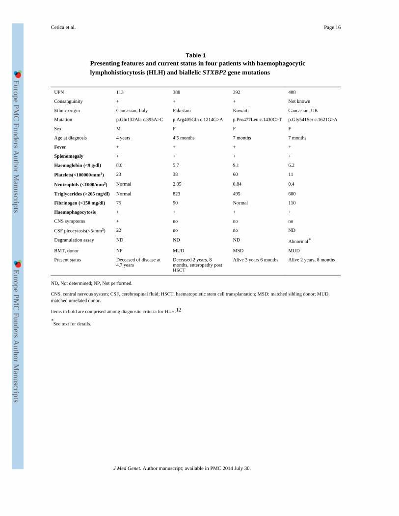
The clinical and laboratory features of the four patients with STXBP2 mutations fit the
diagnostic criteria for HLH (fever, splenomegaly, cytopenia, hypertriglyceridaemia or
hypofibrinogenaemia, and haemophagocytosis)1213 and are summarised in table 1. Their age
at onset of the disease was 4 months in one patient, 7 months in two, while one patient had a
later onset at 4 years; however, the disease course of this patient with later onset was very
Cetica et al. Page 6
J Med Genet. Author manuscript; available in PMC 2014 July 30.
Europe PM
C Funders A
uthor Manuscripts
Europe PM
C Funders A
uthor Manuscripts
aggressive and the child died within 6 months of progressive disease. The remaining 24
patients in whom no STXBP2 mutation was found had a median age of 14 months, with a
range of <1 month to 17 years.
Among 451 cases enrolled over more than 20 years in our HLH Registry1 and evaluable for
this parameter, the median age at diagnosis was 12 months (range, 1 day to 61 years); 24%
were diagnosed within 3 months of age, 39% within 6 months, 63% within 2 years, and 76%
within 5 years of age.
All patients received HLH directed therapy, according to current and local regimens. One
patient died of reactivation 7 months after the diagnosis, while the remaining three
underwent HSCT: two of them are alive and well, while one died of complications.
DISCUSSION
Identification of a genetic marker is of paramount importance in HLH.1128 This may allow
the attending physician to recognise the patient as a candidate for early HSCT from the best
available donor, as well as allowing the screening of family donors for HLH. HSCT has
been recognised as the only available therapeutic approach with the potential to cure
FHL.5–10
Current knowledge on FHL related genes allows the identification of a large proportion of
cases. Yet, this proportion is variable according to the different ethnic groups and
geographic regions. The only large genotype–phenotype study available so far has been
conducted by the Histiocyte Society in FHL-2 and led to identification of significant
associations between specific mutations and ethnic groups. Some mutations were found
more commonly: c.1122G→A (p.Trp374X), associated with Turkish origin; c.50delT
(p.Leu17fsX22) associated with African/African American origin; and c.1090–91delCT
(p.Leu364fsX), so far identified only in Japanese patients.21 A similar study is ongoing but
has not yet been completed for FHL-3. The contribution of FHL-4 appears to be quite
limited so far, with the majority of patients reported from a single geographic area.2324
Thus, additional genetic defects were expected in order to assign a defect to the remaining
families, accounting for a variable proportion included between about 20%—the proportion
observed among Italian patients (M Aricò, unpublished data,2930) and 60–70%—as
observed among native German patients—of the total population (U zur Stadt, unpublished
data). Current knowledge suggests that the population of patients with an unassigned defect
is not clinically distinguishable from those belonging to the currently recognised genetic
subgroups (ie, FHL-2 and FHL-3).
Following identification of STXBP2 as a novel FHL related gene, thus becoming FHL-5,24
we decided to screen the series of patients referred to our registry for a genetic study. We
found that only a minority of such patients, 14%, belong to the FHL-5 subset of this disease.
The presenting features of these patients appear largely comparable to those included in the
remaining subgroups, in particular FHL-2 and FHL-3. Thus, it appears unlikely that FHL-5
may be suspected on the basis of the initial clinical picture.
Cetica et al. Page 7
J Med Genet. Author manuscript; available in PMC 2014 July 30.
Europe PM
C Funders A
uthor Manuscripts
Europe PM
C Funders A
uthor Manuscripts
Overall, a total of 25 patients with FHL-5 are known so far, including the present four.
Among the observed mutations, three appear to be more frequent: the c.1430C→T
(p.Pro477Leu) mutation, previously reported in seven homozygous patients of Arabian
origin2425 and now in a Kuwaiti family; and the c.12471G→C (p.Val417LeufsX126)
mutation, reported in nine patients of different geographic origin, six of them in a
homozygous state. Furthermore, the p.Ile232del mutation was reported in two Turkish
patients. In addition, we now also report the c.1214G/A (p.Arg405Gln) mutation, which was
previously found in a Turkish patient,24 in our patient UPN 388 of Pakistani origin.
We have identified two novel mutations. The c.395A→C (p. Glu132Ala) was found in
homozygosity in a consanguineous family from southern Italy, and the Gly541Ser mutation
identified in homozygosity in a Caucasian patient of English origin. The pathogenic value of
this latter mutation is supported by evidence of defective protein and by defective
cytotoxicity by T lymphocytes. Furthermore, both mutations affect the binding of syntaxin
11. A similar impairment in the binding of syntaxin 3 was reported for mutations
p.Arg405Pro and p.Gly541Glu by Riento et al.31
Screening of genetic mutations in all the FHL related genes, the gold standard for
diagnosing the different subgroups of FHL, is time consuming and not available to most
clinical centres. To address this issue, our group has originally introduced the evaluation of
expression of CD107 by stimulated cytotoxic cells26; a defect in this function may herald a
defect in Munc13-4 protein, as well as in syntaxin11. Defective degranulation impairs the
delivery of effector proteins to the target cells, hampering cellular cytotoxicity; the latter is
the pathogenic mechanism of FHL in its different subsets. Lack of functional STXBP2 is
associated with defective degranulation and defective cellular cytotoxicity, as documented in
the present case UPN 408. Yet, compared to patients with FHL-3 due to a defect in
Munc13-4, the degranulation defect in patients with FHL-5 may be different. In fact, based
on our present finding of a single patient with homozygous Gly541Ser mutation, upon
activation these cells may be able to rescue their degranulation capacity, as observed in the
classical degranulation assay as described by Marcenaro et al.26 It may be of interest that
cells from some patients with FHL-5 may behave normally in the standard CD107 assay
performed on activated NK cells, but may show an impaired degranulation capacity when
either resting NK2425 or overnight IL-2 incubated PBMC are tested and, as we describe
here, stimulating NK cells with PHA and T lymphocytes by anti-CD3. Long term activated
CTL revealed some defect of degranulation. From this point of view, patients with FHL-5
might appear to be closer to patients with FHL-4 (with syntaxin 11 deficiency)32 than to
those with FHL-3. These results are consistent with the previous report that degranulation is
at least partially restored by culture of cells in IL-2.2425
In conclusion, mutations of STXBP2 resulting in defective Munc18-2 protein and the clinical
HLH phenotype underline the genetic heterogeneity of the disease, although apparently
accounting for a minority of cases. Patients with the clinical phenotype, defined by the
diagnostic criteria, and unassigned genetic defect are the best candidates for mutation
analysis of this gene. Patients with FHL-5 apparently may be not detected by the
degranulation assay which is very effective in detecting patients with FHL-3. It is possible
that modifications of this method may allow detection of some patients with a more subtle
Cetica et al. Page 8
J Med Genet. Author manuscript; available in PMC 2014 July 30.
Europe PM
C Funders A
uthor Manuscripts
Europe PM
C Funders A
uthor Manuscripts
degranulation defect, thus facilitating the screening process. Accumulation of additional
cases reported from other groups worldwide might enable definition of its distribution
among ethnic groups and genotype–phenotype correlations. Based on current initial
knowledge, FHL-5 patients should receive the standard therapy for HLH followed by
HSCT. Genetic studies of patients with FHL are still necessary in an attempt to identify
additional disease related genes, with the aim of assigning a familial marker to the remaining
unassigned patients and families.
Acknowledgments
The authors are grateful to the following sources of funding: European Grant ‘CureHLH’ HEALTH-F2-2008-201461, ‘Antonio Pinzino—Associazione per la Ricerca sulle Sindromi Emofagocitiche (ARSE), ‘Noi perVoi per il Meyer Onlus’; the Fördergemeinschaft Kinderkrebszentrum Hamburg e.V. Some of this work wasundertaken at GOSH/UCL Institute of Child Health which received a proportion of funding from the Department ofHealth’s NIHR Biomedical Research Centres funding scheme. GMG is funded by the Wellcome Trust.
REFERENCES
1. Aricò M, Janka G, Fischer A, Henter JI, Blanche S, Elinder G, Martinetti M, Rusca MP.Hemophagocytic lymphohistiocytosis. Report of 122 children from the International Registry. FHLstudy group of the histiocyte society. Leukemia. 1996; 10:197–203. [PubMed: 8637226]
2. Henter JI, Samuelsson-Horne A, Aricò M, Egeler RM, Elinder G, Filipovich AH, Gadner H,Imashuku S, Komp D, Ladisch S, Webb D, Janka G, Histocyte Society. Treatment ofhemophagocytic lymphohistiocytosis with HLH-94 immunochemotherapy and bone marrowtransplantation. Blood. 2002; 100:2367–73. [PubMed: 12239144]
3. Janka GE, Schneider EM. Modern management of children with haemophagocyticlymphohistiocytosis. Br J Haematol. 2004; 124:4–14. [PubMed: 14675403]
4. Mahlaoui N, Ouachee-Chardin M, de Saint Basile G, Neven B, Picard C, Blanche S, Fischer A.Immunotherapy of familial hemophagocytic lymphohistiocytosis with antithymocyte globulins: asingle-center retrospective report of 38 patients. Pediatrics. 2007; 120:e622–8. doi:10.1542/peds.2006-3164. [PubMed: 17698967]
5. Jabado N, de Graeff-Meeder ER, Cavazzana-Calvo M, Haddad E, Le Deist F, Benkerrou M,Dufourcq R, Caillat S, Blanche S, Fischer A. Treatment of familial hemophagocyticlymphohistiocytosis with bone marrow transplantation from HLA genetically nonidentical donors.Blood. 1997; 90:4743–8. [PubMed: 9389690]
6. Durken M, Horstmann M, Bieling P, Erttmann R, Kabisch H, Loliger C, Schneider EM, HellwegeHH, Kruger W, Kroger N, Zander AR, Janka GE. Improved outcome in haemophagocyticlymphohistiocytosis after bone marrow transplantation from related and unrelated donors: a single-centre experience of 12 patients. Br J Haematol. 1999; 106:1052–8. [PubMed: 10520013]
7. Horne A, Janka G, Maarten Egeler R, Gadner H, Imashuku S, Ladisch S, Locatelli F, MontgomerySM, Webb D, Winiarski J, Filipovich AH, Henter JI, Histiocyte Society. Haematopoietic stem celltransplantation in haemophagocytic lymphohistiocytosis. Br J Haematol. 2005; 129:622–30.[PubMed: 15916685]
8. Baker KS, Filipovich AH, Gross TG, Grossman WJ, Hale GA, Hayashi RJ, Kamani NR, Kurian S,Kapoor N, Ringdén O, Eapen M. Unrelated donor hematopoietic cell transplantation forhemophagocytic lymphohistiocytosis. Bone Marrow Transplant. 2008; 42:175–80. [PubMed:18454181]
9. Cooper N, Rao K, Goulden N, Webb D, Amrolia P, Veys P. The use of reduced-intensity stem celltransplantation in haemophagocytic lymphohistiocytosis and Langerhans cell histiocytosis. BoneMarrow Transplant. 2008; 42(Suppl 2):S47–50. [PubMed: 18978744]
10. Cesaro S, Locatelli F, Lanino E, Porta F, Di Maio L, Messina C, Prete A, Ripaldi M, Maximova N,Giorgiani G, Rondelli R, Aricò M, Fagioli F. Hematopoietic stem cell transplantation forhemophagocytic lymphohistiocytosis: a retrospective analysis of data from the Italian Association
Cetica et al. Page 9
J Med Genet. Author manuscript; available in PMC 2014 July 30.
Europe PM
C Funders A
uthor Manuscripts
Europe PM
C Funders A
uthor Manuscripts

of Pediatric Hematology Oncology (AIEOP). Haematologica. 2008; 93:1694–701. [PubMed:18768529]
11. Aricò M, Allen M, Brusa S, Clementi R, Pende D, Maccario R, Moretta L, Danesino C.Haemophagocytic lymphohistiocytosis: proposal of a diagnostic algorithm based on perforinexpression. Br J Haematol. 2002; 119:180–8. [PubMed: 12358924]
12. Henter JI, Elinder G, Ost A. Diagnostic guidelines for hemophagocytic lymphohistiocytosis. TheFHL Study Group of the Histiocyte Society. Semin Oncol. 1991; 18:29–33. [PubMed: 1992521]
13. Henter JI, Horne A, Aricò M, Egeler RM, Filipovich AH, Imashuku S, Ladisch S, McClain K,Webb D, Winiarski J, Janka G. HLH-2004: Diagnostic and therapeutic guidelines forHemophagocytic lymphohistiocytosis. Pediatr Blood Cancer. 2007; 48:124–31. [PubMed:16937360]
14. Perez N, Virelizier JL, Arenzana-Seisdedos F, Fischer A, Griscelli C. Impaired natural killeractivity in lymphohistiocytosis syndrome. J Pediatr. 1984; 104:569–73. [PubMed: 6368780]
15. Aricò M, Nespoli L, Maccario R, Montagna D, Bonetti F, Caselli D, Burgio GR. Naturalcytotoxicity impairment in familial haemophagocytic lymphohistiocytosis. Arch Dis Child. 1988;63:292–6. [PubMed: 3355209]
16. Schneider EM, Lorenz I, Muller-Rosenberger M, Steinbach G, Kron M, Janka-Schaub GE.Hemophagocytic lymphohistiocytosis is associated with deficiencies of cellular cytolysis butnormal expression of transcripts relevant to killer-cell-induced apoptosis. Blood. 2002; 100:2891–8. [PubMed: 12351400]
17. Farquhar J, Claireaux A. Familial haemophagocytic reticulosis. Archives of Disease in Childhood.1952; 27:519–25. [PubMed: 13008468]
18. Ohadi M, Lalloz MR, Sham P, Zhao J, Dearlove AM, Shiach C, Kinsey S, Rhodes M, Layton DM.Localization of a gene for familial hemophagocytic lymphohistiocytosis at chromosome 9q21.3–22 by homozygosity mapping. Am J Hum Genet. 1999; 64:165–171. [PubMed: 9915955]
19. Dufurcq-Lagelouse R, Jabado N, Le Deist F, Stéphan JL, Souillet G, Bruin M, Vilmer E, SchneiderM, Janka G, Fischer A, de Saint Basile G. Linkage of familial hemophagocyticlymphohistiocytosis to 10q21-22 and evidence for heterogeneity. Am J Hum Genet. 1999; 64:172–9. [PubMed: 9915956]
20. Stepp SE, Dufourcq-Lagelouse R, Le Deist F, Bhawan S, Certain S, Mathew PA, Henter JI,Bennett M, Fischer A, de Saint Basile G, Kumar V. Perforin gene defects in familialhemophagocytic lymphohistiocytosis. Science. 1999; 286:1957–9. [PubMed: 10583959]
21. Trizzino A, zur Stadt U, Ueda I, Risma K, Janka G, Ishii E, Beutel K, Sumegi J, Cannella S, PendeD, Mian A, Henter JI, Griffiths G, Santoro A, Filipovich A, Aricò M, Histiocyte Society HLHStudy group. Genotype-phenotype study of familial haemophagocytic lymphohistiocytosis due toperforin mutations. J Med Genet. 2008; 45:15–21. [PubMed: 17873118]
22. Feldmann J, Callebaut I, Raposo G, Certain S, Bacq D, Dumont C, Lambert N, Ouachee-ChardinM, Chedeville G, Tamary H, Minard-Colin V, Vilmer E, Blanche S, Le Deist F, Fischer A, deSaint Basile G. Munc13-4 is essential for cytolytic granules fusion and is mutated in a form offamilial hemophagocytic lymphohistiocytosis (FHL3). Cell. 2003; 115:461–73. [PubMed:14622600]
23. Zur Stadt U, Beutel K, Kolberg S, Schneppenheim R, Kabisch H, Janka G, Hennies HC. Mutationspectrum in children with primary hemophagocytic lymphohistiocytosis: molecular and functionalanalyses of PRF1, UNC13D, STX11, and RAB27A. Hum Mutat. 2006; 27:62–8. [PubMed:16278825]
24. zur Stadt U, Rohr J, Seifert W, Koch F, Grieve S, Pagel J, Strauss J, Kasper B, Nürnberg G, BeckerC, Maul-Pavicic A, Beutel K, Janka G, Griffiths G, Ehl S, Hennies HC. Familial HemophagocyticLymphohistiocytosis Type 5 (FHL-5) Is Caused by Mutations in Munc18-2 and Impaired Bindingto Syntaxin 11. Am J Hum Genet. 2009; 85:482–92. [PubMed: 19804848]
25. Côte M, Ménager MM, Burgess A, Mahlaoui N, Picard C, Schaffner C, Al-Manjomi F, Al-HarbiM, Alangari A, Le Deist F, Gennery AR, Prince N, Cariou A, Nitschke P, Blank U, El-Ghazali G,Ménasché G, Latour S, Fischer A, de Saint Basile G. Munc18-2 deficiency causes familialhemophagocytic lymphohistiocytosis type 5 and impairs cytotoxic granule exocytosis in patientNK cells. J Clin Invest. 2009:40732. doi: 10.1172/JCI40732. [PubMed: 19884660]
Cetica et al. Page 10
J Med Genet. Author manuscript; available in PMC 2014 July 30.
Europe PM
C Funders A
uthor Manuscripts
Europe PM
C Funders A
uthor Manuscripts
26. Marcenaro S, Gallo F, Martini S, Santoro A, Griffiths GM, Aricò M, Moretta L, Pende D. Analysisof natural killer-cell function in familial Hemophagocytic lymphohistiocytosis (FHL): defectiveCD107a surface expression heralds Munc13-4 defect and discriminates between genetic subtypesof the disease. Blood. 2006; 108:2316–23. [PubMed: 16778144]
27. Clark RH, Stinchcombe JC, Day A, Blott E, Booth S, Bossi G, Hamblin T, Davies EG, GriffithsGM. Adaptor protein 3-dependent microtubule-mediated movement of lytic granules to theimmunological synapse. Nat Immunol. 2003; 4:1111–20. [PubMed: 14566336]
28. Notarangelo LD, Sorensen R. Is it necessary to identify molecular defects in primaryimmunodeficiency disease? J Allergy Clin. Immunol. 2008; 122:1069–73. [PubMed: 18992927]
29. Clementi R, zur Stadt U, Savoldi G, Varotto S, Conter V, De Fusco C, Notarangelo LD, SchneiderM, Klersy C, Janka G, Danesino C, Aricò M. Six novel mutations in the PRF1 gene in childrenwith haemophagocytic lymphohistiocytosis. J Med Genet. 2001; 38:643–6. [PubMed: 11565555]
30. Santoro A, Cannella S, Bossi G, Gallo F, Trizzino A, Pende D, Dieli F, Bruno G, Stinchcombe JC,Micalizzi C, De Fusco C, Danesino C, Moretta L, Notarangelo LD, Griffiths GM, Aricò M. NovelMunc13-4 mutations in children and young adult patients with haemophagocyticlymphohistiocytosis. J Med Genet. 2006; 43:953–60. [PubMed: 16825436]
31. Riento K, Kauppi M, Keranen S, Olkkonen VM. Munc18-2, a functional partner of syntaxin 3,controls apical membrane trafficking in epithelial cells. J Biol Chem. 2000; 275:13476–83.[PubMed: 10788461]
32. Bryceson YT, Rudd E, Zheng C, Edner J, Ma D, Wood SM, Bechensteen AG, Boelens JJ, CelkanT, Farah RA, Hultenby K, Winiarski J, Roche PA, Nordenskjöld M, Henter JI, Long EO,Ljunggren HG. Defective cytotoxic lymphocyte degranulation in syntaxin-11 deficient familialhemophagocytic lymphohistiocytosis (FHL4) patients. Blood. 2007; 15:1906–15. [PubMed:17525286]
Cetica et al. Page 11
J Med Genet. Author manuscript; available in PMC 2014 July 30.
Europe PM
C Funders A
uthor Manuscripts
Europe PM
C Funders A
uthor Manuscripts
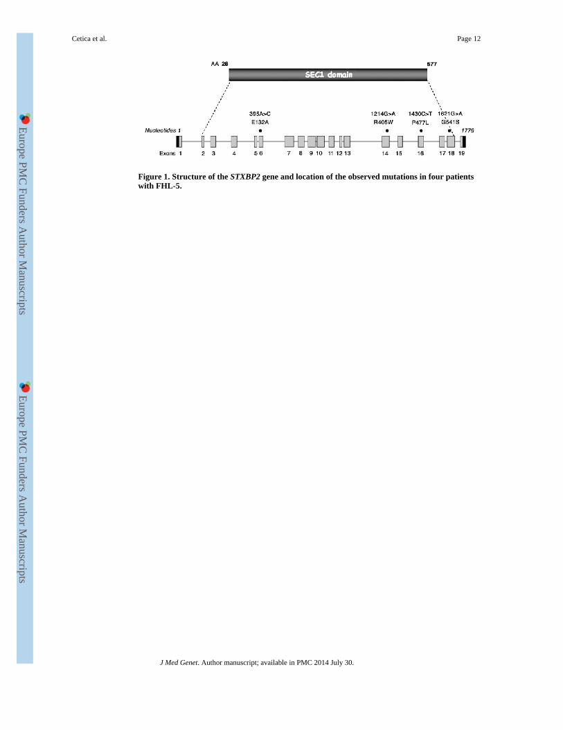
Figure 1. Structure of the STXBP2 gene and location of the observed mutations in four patientswith FHL-5.
Cetica et al. Page 12
J Med Genet. Author manuscript; available in PMC 2014 July 30.
Europe PM
C Funders A
uthor Manuscripts
Europe PM
C Funders A
uthor Manuscripts

Figure 2. Co-immunoprecipitation analysis.Upper panel: Neither the negative control 1 (lane 1) without FLAG-Stx11 nor the negative
control 2 (lane 2) without EGFP-STXBP2 show unspecific binding to the IP antibody
coupled agarose beads. EGFP-STXBP2 WT (lane 3) as well as both EGFP-STXBP2
polymorphic variants (lane 4/lane 5) co-immunoprecipitate with FLAG-Stx11, whereas
EGFP-STXBP2 Gly541Ser (lane 6) and Glu132Ala (lane 7) show an impaired bingeing to
FLAG-Stx11. The middle panel shows the corresponding input control of the EGFP-
STXBP2 proteins. The lower panel presents the IP controls of each approach which reveal
approximately equal amounts of FLAG-Stx11 after immunoprecipitation with the capture
anti-FLAG-M2 murine IgG1 monoclonal antibody.
Cetica et al. Page 13
J Med Genet. Author manuscript; available in PMC 2014 July 30.
Europe PM
C Funders A
uthor Manuscripts
Europe PM
C Funders A
uthor Manuscripts

Figure 3. Flow cytometry evaluation of CD107 expression of patient UPN 408.Peripheral blood mononuclear cells (PBMC) from patient and healthy control cultured for
24 h with IL-2 were analysed for CD107 expression by NK cells (gate CD3− CD56+) upon
stimulation with phytohaemagglutinin (PHA) (panel A), or by CD3+ CD8+ lymphocytes
stimulating with anti-CD3 mAb (panel B). Activated and expanded lymphocytes were
analysed for CD107 expression by NK cells upon co-culture with K562 (panel C).
Cetica et al. Page 14
J Med Genet. Author manuscript; available in PMC 2014 July 30.
Europe PM
C Funders A
uthor Manuscripts
Europe PM
C Funders A
uthor Manuscripts
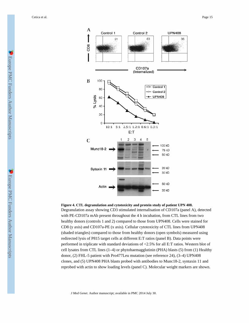
Figure 4. CTL degranulation and cytotoxicity and protein study of patient UPN 408.Degranulation assay showing CD3 stimulated internalisation of CD107a (panel A), detected
with PE-CD107a mAb present throughout the 4 h incubation, from CTL lines from two
healthy donors (controls 1 and 2) compared to those from UPN408. Cells were stained for
CD8 (y axis) and CD107a-PE (x axis). Cellular cytotoxicity of CTL lines from UPN408
(shaded triangles) compared to those from healthy donors (open symbols) measured using
redirected lysis of P815 target cells at different E:T ratios (panel B). Data points were
performed in triplicate with standard deviations of <2.5% for all E:T ratios. Western blot of
cell lysates from CTL lines (1–4) or phytohaemagglutinin (PHA) blasts (5) from (1) Healthy
donor, (2) FHL-5 patient with Pro477Leu mutation (see reference 24), (3–4) UPN408
clones, and (5) UPN408 PHA blasts probed with antibodies to Munc18-2, syntaxin 11 and
reprobed with actin to show loading levels (panel C). Molecular weight markers are shown.
Cetica et al. Page 15
J Med Genet. Author manuscript; available in PMC 2014 July 30.
Europe PM
C Funders A
uthor Manuscripts
Europe PM
C Funders A
uthor Manuscripts
Europe PM
C Funders A
uthor Manuscripts
Europe PM
C Funders A
uthor Manuscripts
Cetica et al. Page 16
Table 1Presenting features and current status in four patients with haemophagocyticlymphohistiocytosis (HLH) and biallelic STXBP2 gene mutations
UPN 113 388 392 408
Consanguinity + + + Not known
Ethnic origin Caucasian, Italy Pakistani Kuwaiti Caucasian, UK
Mutation p.Glu132Ala c.395A>C p.Arg405Gln c.1214G>A p.Pro477Leu c.1430C>T p.Gly541Ser c.1621G>A
Sex M F F F
Age at diagnosis 4 years 4.5 months 7 months 7 months
Fever + + + +
Splenomegaly + + + +
Haemoglobin (<9 g/dl) 8.0 5.7 9.1 6.2
Platelets(<100000/mm3) 23 38 60 11
Neutrophils (<1000/mm3) Normal 2.05 0.84 0.4
Triglycerides (>265 mg/dl) Normal 823 495 600
Fibrinogen (<150 mg/dl) 75 90 Normal 110
Haemophagocytosis + + + +
CNS symptoms + no no no
CSF pleocytosis(<5/mm3) 22 no no ND
Degranulation assay ND ND ND Abnormal*
BMT, donor NP MUD MSD MUD
Present status Deceased of disease at4.7 years
Deceased 2 years, 8months, enteropathy postHSCT
Alive 3 years 6 months Alive 2 years, 8 months
ND, Not determined; NP, Not performed.
CNS, central nervous system; CSF, cerebrospinal fluid; HSCT, haematopoietic stem cell transplantation; MSD: matched sibling donor; MUD,matched unrelated donor.
Items in bold are comprised among diagnostic criteria for HLH.12
*See text for details.
J Med Genet. Author manuscript; available in PMC 2014 July 30.
Copyright © 2022 FDOKUMEN