Study of nano-scale diffusion in thin films and multilayers
8
Hyperfine Interact (2008) 182:23–30 DOI 10.1007/s10751-008-9722-9 Study of nano-scale diffusion in thin films and multilayers Ajay Gupta · Sujoy Chakravarty · Parasmani Rajput · Mukul Gupta · Rudolf Rüffer Published online: 4 October 2008 © Springer Science + Business Media B.V. 2008 Abstract It is shown that x-ray reflectivity and x-ray fluorescence under standing wave conditions can be used to study atomic diffusion with an accuracy of a fraction of a nanometer. Both the techniques can be made isotope selective by making use of nuclear resonance scattering from a Mössbauer active isotope. The techniques have been used to study self-diffusion of Fe in amorphous and nano-crystalline alloys of FeZr and FeN. The observed correlation of the activation energy E with pre- exponent factor D 0 confirms that in amorphous FeZr alloy diffusion takes place via collective motion of a group of atoms. Even in nanocrystalline alloys it is found that atomic diffusion occurs mainly through grain boundaries which are amorphous or highly disordered in nature. Keywords Diffusion · FeZr · Thin films and multilayers 1 Introduction Thin films and multilayes form an important class of nanostructured materials. A large variety of new materials have been developed using such structures: multilayers consisting of alternate layers of a high-Z and a low-Z elements like W/Si, Pt/C are used as x-ray mirror and monochromators; magnetic multilayers like Fe/Cr exhibit A. Gupta (B ) · S. Chakravarty · P. Rajput UGC-DAE Consortium for Scientific Research, University Campus, Khandwa road, Indore 452017, India e-mail: [email protected] M. Gupta UGC-DAE Consortium for Scientific Research, Mumbai Centre, R-5 SHED, BARC, Mumbai 400 085, India R. Rüffer European Synchrotron Radiation Facility, BP 220, 38043 Grenoble Cedex, France
-
Upload
independent -
Category
Documents
-
view
0 -
download
0
Transcript of Study of nano-scale diffusion in thin films and multilayers

Hyperfine Interact (2008) 182:23–30DOI 10.1007/s10751-008-9722-9
Study of nano-scale diffusion in thin filmsand multilayers
Ajay Gupta · Sujoy Chakravarty · Parasmani Rajput ·Mukul Gupta · Rudolf Rüffer
Published online: 4 October 2008© Springer Science + Business Media B.V. 2008
Abstract It is shown that x-ray reflectivity and x-ray fluorescence under standingwave conditions can be used to study atomic diffusion with an accuracy of a fractionof a nanometer. Both the techniques can be made isotope selective by making useof nuclear resonance scattering from a Mössbauer active isotope. The techniqueshave been used to study self-diffusion of Fe in amorphous and nano-crystalline alloysof FeZr and FeN. The observed correlation of the activation energy E with pre-exponent factor D0 confirms that in amorphous FeZr alloy diffusion takes place viacollective motion of a group of atoms. Even in nanocrystalline alloys it is found thatatomic diffusion occurs mainly through grain boundaries which are amorphous orhighly disordered in nature.
Keywords Diffusion · FeZr · Thin films and multilayers
1 Introduction
Thin films and multilayes form an important class of nanostructured materials. Alarge variety of new materials have been developed using such structures: multilayersconsisting of alternate layers of a high-Z and a low-Z elements like W/Si, Pt/C areused as x-ray mirror and monochromators; magnetic multilayers like Fe/Cr exhibit
A. Gupta (B) · S. Chakravarty · P. RajputUGC-DAE Consortium for Scientific Research, University Campus,Khandwa road, Indore 452017, Indiae-mail: [email protected]
M. GuptaUGC-DAE Consortium for Scientific Research, Mumbai Centre,R-5 SHED, BARC, Mumbai 400 085, India
R. RüfferEuropean Synchrotron Radiation Facility, BP 220, 38043 Grenoble Cedex, France
24 A. Gupta et al.
giant magnetoresistance and are used as magnetic sensors in read/write heads and inmemory devices. Magnetic thin films are also extensively used in tunnel magneto-resistance devices. Any changes in the interfacial region between two layers insuch nanostructures can greatly influence their physical properties [1–3]. Typicalwidth of the interface in such multilayers is of the order of a nanometer. In thiscontext, nanoscale diffusion across the interfaces in multilayers has acquired a greatsignificance, as it can modify their properties wantonly or un-wantonly. In addition tothe interdiffusion at the interfaces, self-diffusion of the constituent species in a thinfilm is equally important, as it governs the structural transformations which affect allthe physical properties of such films [4].
Since the typical diffusion lengths of interest in the case of thin films and multilay-ers are of the order of a nanometer or less, the conventional depth profiling techniquelike SIMS, RBS, radioactive tracer techniques are not suitable. However, someof the x-ray based techniques for depth profiling, e.g., x-ray reflectivity and x-rayfluorescence under standing wave conditions can be conveniently used for studyingatomic diffusion at nanometer scale [5, 6]. In general, x-rays cannot differentiatebetween two isotopes of an element, and therefore cannot be used for depth profilingof an isotopic marker layer (as required for studying self-diffusion). However, inspecific cases, isotope selectivity can be achieved by making use of nuclear resonancescattering of x-rays from a Mössbauer active species (e.g. 57Fe). In that case, it ispossible to do depth profiling of the Mössbauer isotope and hence to study self-diffusion of the corresponding element [7, 8].
2 Diffusion studies using nuclear resonance reflectivity
X-ray reflectivity from compositionally modulated multilayers has been used in theliterature for measuring small diffusivity across the interfaces down to values of10−23 cm2/s [9]. However, the technique can only be applied to study interdiffusionbetween the species which have large contrast in x-ray scattering. The techniqueconsists in monitoring the intensity of the satellites of the (000) forward scattering.As the compositional modulation depth decreases due to interdiffusion, the intensityof the satellite peaks goes down. The diffusivity D(T) is related to the relative changein the intensity of the satellite peaks through the relation [10]:
In(
I (t)I0
)= −8π2n2 D (T) t
λ2, (1)
where I0 is the intensity of the nth order Bragg peak at t = 0, t is the annealing timeat temperature T, and λ is the bilayer period.
This technique, in general, cannot be used to study self-diffusion of a givenconstituent, as x-ray scattering cannot differentiate between two isotopes of thesame element. However, x-ray scattering can be made isotope selective in case theenergy of the x-rays is tuned to the nuclear transition energy of Mössbauer activeisotope (e.g., 57Fe). In such a case, total scattering amplitude from the Mössbauerisotope increases significantly due to additional contribution from nuclear resonancescattering. In the presence of nuclear resonance scattering, the total scatteringamplitude of an atom must be written as a sum of the electronic scattering and

Study of nano-scale diffusion in thin films and multilayers 25
nuclear resonance scattering amplitudes. As a result, the index of refraction of thelayer material is written as:
n =√√√√1 + λ2
0
π
∑i
σi fi ≈ 1 + λ20
2π
∑i
σ(
f ei + f n
i
)(2)
Where f ei and f n
i are respectively the electronic and nuclear scattering amplitudes,and σ i is the atomic density of the species i. The nuclear scattering amplitude is givenfor instance in reference [11] and is proportional to the enrichment of the resonantnuclei of species i. For the Mössbauer transition of 57Fe nuclei (M1 transition), inabsence of a magnetic splitting, and for a polycrystalline sample, the nuclear forwardscattering amplitude can be written as:
f n = λ0
4π
fLM
1 + α
2 j1 + 1
2 j0 + 1
Ax − i
(3)
Where fLM is the Lamb–Mössbauer factor, j0 and j1 are the spin quantum numbersof ground and excited states respectively, α is the coefficient for internal conversion,x = (E − ω)
/0, A denotes the inhomogeneous broadening, E is the quadru-
pole splitting and 0 the natural line width. The nuclear part of the refractive indexshows a resonance behaviour and is significant only within a narrow energy range ofthe order of μeV around the nuclear transition energy. However, in this energy rangethe Mössbauer isotope presents a strong scattering contrast.
The film used for self-diffusion measurement of Fe has the structure:substrate/Fe70Zr30 4 nm/[57Fe70Zr303 nm/Fe70Zr30 4 nm]×10 [7]. The alternate layershave the same chemical composition but differ in the abundance of the Mössbaueractive isotope (e.g., 57Fe). While the Fe70Zr30 layer has natural abundance (2.5%) of57Fe, the layer 57Fe70Zr30 is enriched in 57Fe to 30%. X-rays diffraction shows thatthe film is amorphous in nature. ID22N beamline of ESRF was used for measuringboth electronic and nuclear pats of the reflectivity. The storage ring operated in16-bunch mode providing short pulses of x rays (duration ∼100 ps) every 176 ns.The radiation from the undulator source, optimized for the 14,413 eV transition in57Fe, was filtered by a double Si (111) reflection followed by a high-resolution nestedmonochromator, providing a bandpass of 4 meV [12]. The scattered radiation wasdetected using fast avalanche photodiodes (time resolution ∼1 ns). The nuclear andelectronic parts of the signal were separated by making use of the fact that nucleartransitions are delayed in time due to finite life time of the Mössbauer excited state(140 ns in case of 57Fe isotope) [12]. Thus, photons detected within a few ns of theincident x-ray pulse constitute the prompt signal due to electronic scattering, whilethose detected in an interval of 10–160 ns after the incident x-ray pulse correspondto nuclear resonance scattering.
Figure 1 gives the nuclear part of the reflectivity (nuclear resonance reflectivity) asa function of isochronal annealing temperature. Annealing time at each temperaturewas kept fixed at 30 min. Nuclear resonance reflectivity (NRR) exhibits a well definedBragg peak corresponding to the isotopic periodicity of the multilayer. On the otherand, the electronic part of the reflectivity (not shown) exhibits only the Kiessig fringescorresponding to the total thickness of the film. Monitoring the height of the Braggpeak in NRR as a function of annealing temperature and time can yield the diffusivityof Fe in the film. It may be noted that due to large nuclear resonance scattering cross-

26 A. Gupta et al.
Fig. 1 Nuclear resonancereflectivity of the57Fr70Zr30/Fe70Zr30 multilayertaken at the Mössbauerresonance energy of 57Fe(14.413 keV). Inset showsthe electronic reflectivityof the same multilayer
0.4 0.6 0.8 1.0 1.2
513K483K
455K423K
qz(nm-1)
300K
Fig. 2 Arrhenius behaviorof diffusion coefficient withisochronal annealingtemperature for57Fr70Zr30/Fe70Zr30multilayer
0.0021 0.0022 0.0023 0.0024
-51.0
-50.4
-49.8
-49.2ln [D0 (m2/s)] = -39±1
E = 0.42±0.05 eV
ln [D
(m
/s
)]
1/T (K-1)
2
section, the dynamical effects may become appreciable, modifying the intensity of theBragg peak [13]. However, in the present case such effects are relatively week dueto the fact that the abundance of 57Fe in 57Fe70Zr30 layer is only 30%. The valuesof diffusion coefficient as obtained from the height of the Bragg peak at differenttemperatures are used to calculate activation energy E and pre-exponential factorD0 using the relation D = D0 exp
(−E/
KBT)
(Fig. 2). The activation energy forself-diffusion of Fe in the present system comes out to be 0.42 ± 0.04 eV and pre-exponential factor D0 = exp (−39 ± 1). This value is substantially lower as comparedto that in Fe-Zr metallic glasses of similar composition [14]. This low activationenergy can be attributed to the fact that the present results pertain to amorphousfilm which would have a higher density of defects, and that the measurements havebeen done in the un-relaxed state of the system.

Study of nano-scale diffusion in thin films and multilayers 27
Fig. 3 Schematic diagram ofthe experimental geometry formeasuring x-ray reflectivityand x-ray fluorescence understanding wave conditions.Detector D1 is a NaIscintillation detector, whileD2 is an energy dispersivedetector with an energyresolution of 200 eV
substrate
Au
Si
Si
Si
Si
D1D2
3 Diffusion studies using x-ray standing waves
A typical arrangement using x-ray standing waves for depth profiling of marker layeris shown in Fig. 3. Standing waves are generated using total external reflection ofx-rays from a layer of high Z material (e.g., Au) [6, 7, 17]. The incident and thereflected x-ray amplitudes interfere with each other to form standing waves abovethe surface of totally reflected layer. The system to be studied (e.g., Substrate/Cr20 nm/Au 60 nm/Si 12.5 nm/W 3 nm/Si 12.5 nm/Fe 3 nm/Si 12.5 nm/Ti 3 nm/Si12.5 nm) is deposited on top of the high Z layer [15]. The separation between twosuccessive nodes and antinodes is given by: d = λ
/(2 sin θ), θ being the angle of
incidence and λ being the wavelength of x-rays [16]. Thus, as the angle of incidentincreases, various antinodes move down. A peak in fluorescence from the M (M =Ti, Fe, W) marker layer is observed whenever an antinode coincides with the positionof the marker layer. Detailed fitting of the fluorescence pattern as a function ofq = 4π sin θ
/λ, gives the concentration profile of the marker layers. Thus, in a single
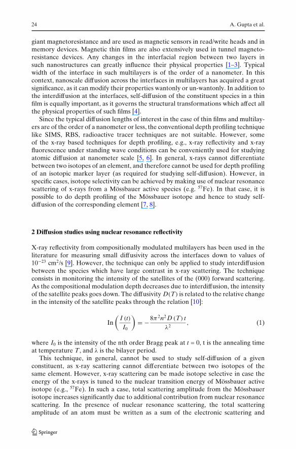
measurement one gets concentration profiles of all the three marker layers. Diffusionof M in Si induced by irradiation with high energy heavy ion (120 MeV Au ions) hasbeen studied using this technique [17]. Figure 4 gives the fluorescence pattern of oneof the marker layer (namely Fe) before and after an irradiation to a fluorescence of1 × 1013 ions/cm2. The corresponding concentration profiles of Ti, Fe, W are shownin the inset of Fig. 4. The diffusion lengths as obtained using the relation:
L2d = σ 2 − σ 2
0 , (4)
where σ 0 and σ are the standard deviation of the concentrationprofile before andafter irradiation, come out to be 2.8, 2.7, and 1.4 nm respectively for Ti, Fe and Wmarker layers. These results are in agreement with the predictions of thermal spikemodel according to which Ti is highly sensitive to electronic energy loss while W isalmost insensitive to the same, and Fe has intermediate sensitivity [17].
As in the case of x-ray reflectivity, the x-ray standing wave technique can alsobe made isotope sensitive by making use of nuclear resonance scattering, and thuscan be used for the study of self-diffusion in chemically homogeneous films usinga single isotopic marker layer (e.g., 57Fe). The multilayer used for depth profilingof 57Fe isotope in a compositionally homogeneous film of FeN has the nominal
28 A. Gupta et al.
Fig. 4 a Fe-fluorescence ofpristine and irradiated samplesAu/Si/W/Si/Fe/Si/Ti/Si as afunction of q. The continuouscurves represent fit to the data.b Concentration profiles inpristine and irradiated samplesas obtained from simultaneousfitting of x-ray reflectivity andx-ray fluorescence data.Surface of the Au layer hasbeen taken at the origin
0.02 0.04 0.06 0.08 0.10 0.120
1
20
1
2
PristineF
e F
luore
scence
Conce
ntr
atio
n (
%)
q (Å-1)
Depth (Å)
Irradiated
0-600 -500 -400 -300 -200 -100 0
20
40
60
80
100 Pristine
Irradiated
Ti Fe W
a
b
structure: substrate/Pt 70 nm/FeN 20 nm/57FeN 4 nm/FeN 20 nm (Gupta et al., tobe published). X-ray diffraction measurements show that the film is nanocrystallinewith grain size of 5 nm. The ID18 beamline of ESRF, Grenoble was used for studyingnuclear resonance scattering and fluorescence under standing wave conditions fromthis structure. Figure 5 gives the nuclear reflectivity and fluorescence from the as-deposited multilayer as well as multilayer annealed at temperatures of 200C and300C for 60 min. Significant changes in the peak intensities take place with thermalannealing which can be explained by broadening of the depth profile of the 57Femarker layer as a result of self-diffusion of Fe. One can see that there is a one to onecorrespondence between nuclear reflectivity and nuclear fluorescence. A detailedfitting of nuclear reflectivity data has been done to obtain the concentration profileof the 57Fe marker layer, which is shown in the inset of Fig. 5. Typical error barsin the width of concentration profiles are ±0.2 nm. The diffusion length is obtained

Study of nano-scale diffusion in thin films and multilayers 29
Fig. 5 Nuclear reflectivity ofmultilayer Pt/FeN/57FeN/FeNin pristine state as well as afterannealing at 200C and 300C.Curves are shifted verticallywith respect to each other forthe sake of clarity. Continuouscurves represent best fit tothe data obtained using thecomputer code [14]. Insetshows the concentrationprofiles of 57Fe layer asobtained from the fitting ofnuclear reflectivity data ofpristine (—), 200C annealed(- - - - -), and 300C annealed(......) samples
2 4 6 8 100
1
2
3
1
2
3
1
2
3
pristine
θ (mrad)
200 0C
rela
tive
inte
nsi
ty
5 10 15 20 250.0
0.5
1.0
con
cen
tra
tion
depth (nm)
300 0C
using (4) which in the case of one-dimensional diffusion, is related to diffusivity D(T)
through the relation L2d = 2D(T)t. Diffusivity at the two temperatures of 200C and
300C comes out to be 1.0 × 10⁻²² and 3.7 × 10⁻²² m2/s respectively. These valuesare well below the grain boundary diffusion in polycrystalline Fe [18], however arecomparable to diffusivity in nanocrystalline FeZr [19], where diffusion occurs mainlythrough grain boundaries which are amorphous in nature. These results demonstratethat nuclear resonance reflectivity from isotopic marker layers under standing waveconditions can be used to study self-diffusion with an accuracy which is roughlyan order of magnitude better than that obtainable using conventional techniqueslike SIMS.
4 Conclusions
In conclusion, x-ray reflectivity and fluorescence under standing wave conditionshave been used for measuring atomic diffusion in thin films with sub-nanometeraccuracy. Self-diffusion of Fe has been measured by making use of nuclear resonancescattering from 57Fe, which results in strong scattering contrast between differentisotopes. In amorphous FeZr films the activation energy is found to be substantiallylower as compared to that in Fe-Zr metallic glasses of similar composition. This lowactivation energy can be attributed to a higher density of defects in thin films. Theobserved correlation of the activation energy E with pre-exponent factor D0 confirmsthat in amorphous FeZr alloy diffusion takes place via collective motion of a group of

30 A. Gupta et al.
atoms. Even in nanocrystalline alloys it is found that atomic diffusion occurs mainlythrough grain boundaries which are amorphous or highly disordered in nature.
Acknowledgements Partial support from Indo-French Center for Promotion of Advanced researchis thankfully acknowledged.
References
1. Phang, Y.H., Savage, D.E., Kariotis, R., Lagally, M.G.: X-ray diffraction measurement of par-tially correlated interfacial roughness in multilayers. J. Appl. Phys. 74, 3181–3188 (1993)
2. Gupta, A., Paul, A., Chaudhari, S.M., Phase, D.M.: Effect of interface roughness on GMR inFe/Cr multilayers. J. Phys. Soc. Jpn. 69, 2182–2187 (2000)
3. Kim, W.-S., Andrä, W., Kleemann, W.: Influence of interfaces on the perpendicular magneticanisotropy in Tb/Fe multilayers. Phys. Rev. B 58, 6346–6352 (1998)
4. Sharma, P., Gupta, A.: Effect of preparation condition on the soft magnetic properties ofFeCuNbSiB thin films. J. Magn. Magn. Mater. 288, 347–353 (2005)
5. Ghose, S.K., Dev, B.N., Gupta, A.: Resonance enhancement of x-rays and fluorescence yieldfrom marker layers in thin films. Phys. Rev. B 64, 233403 (2001)
6. Gupta, A., Rajput, P., Saraiya, A., Reddy, V.R., Gupta, M., Bernstorff, S., Amenitsch, H.: Depthprofiling of marker layers using x-ray waveguide structures. Phys. Rev. B 72, 075436 (2005)
7. Gupta, A., Gupta, M., Chakravarty, S., Rüffer, R., Wille, H.-C., Leupold, O.: Fe diffusion inamorphous and nanocrystalline alloys studied using nuclear resonance reflectivity. Phys. Rev. B72, 014207 (2005)
8. Rennhofer, M., Sepiol, B., Sladecek, M., Kmiec, D., Stankov, S., Vogl, G., Kozlowski, M.,Kozubski, R., Vantomme, A., Meersschaut, J., Rüffer, R., Gupta, A.: Self-diffusion of iron inL10-ordered FePt thin films. Phys. Rev. B 74, 104301 (2006)
9. Wang, W.-H., Bai, H.Y., Zhang, M., Zhao, J.H., Zhang, X.Y., Wang, W.K.: Interdiffusion innanometer-scale multilayers investigated by in situ low-angle x-ray diffraction. Phys. Rev. B 59,10811–10822 (1999)
10. Greer, A.L., Spaepen, F.: Diffusion. In: Chang, L.L., Giessen, B.C. (eds.) Synthetic ModulatedStructures, pp. 419–486. Academic, New York (1985)
11. Hannon, J.P., Trammell, G.T., Mueller, M., Gerdau, E., Rüffer, R., Winkler, H.: Grazing-incidence antireflection films. III. General theory for pure nuclear reflections. Phys. Rev. B 32,6363–6373 (1985)
12. Rüffer, R., Chumakov, A.I.: Nuclear resonance beamline at ESRF. Hyperfine Interact. 97–98,589–604 (1996). See also http://www.esrf.fr/exp_facilities/ID18/
13. Andreeva, M.: Nuclear resonant reflectivity data evaluation with the <<REFTIM>> program.Proceedings of this conference
14. Faupel, F., Frank, W., Macht, M.-P., Mehrer, H., Naundorf, V., Rätzke, K., Schober, H.R.,Sharma, S.K., Teichler, H.: Diffusion in metallic glasses and supercooled melts. Rev. Mod. Phys.75, 237–280 (2003)
15. Gupta, A., Rajput, P., Meneghini, C.: Depth-resolved x-ray absorption fine structure study ofFe/Si interfaces using x-ray standing waves. Phys. Rev. B 76, 195401 (2007)
16. Bedzyk, M.J., Bommarito, G.M., Schildkraut, J.S.: X-ray standing waves at a reflecting mirrorsurface. Phys. Rev. Lett. 62, 1376–1379 (1989)
17. Wang, Z.G., Dufour, Ch., Paumier, E., Toulemonde, M.: The Se sensitivity of metals under swift-heavy-ion irradiation: a transient thermal process. J. Phys. Condens. Matter 6, 6733–6750 (1994)
18. Tanimoto, H., Farber, P., Würschum, R., Valiev, R.Z., Schaefer, H.-E.: Self-diffusion in high-density nanocrystalline Fe. Nanostruct. Mater. 12, 681–684 (1999)
19. Gupta, A., Gupta, M., Pietsch, U., Ayachit, S., Rajagopalanl, S., Balamurgan, A.K., Tyagi, A.K.:Iron self-diffusion in nanocrystalline FeZr thin films. J. Non-Cryst. Solids 343, 39–47 (2004)