
Structure-function studies on - human granulocyt€-rrâcrophage
391
Structure-function studies on human granulocyt€-rrâcrophage colony-stimulating factor Timothy Robert Hercus, B.Sc. (Hons.) A thesis submitted for the degree of Doctor of Philosophy of the University of Adelaide (Faculty of Science) Division of Human Immunology Institute of Medical and Veterinary Science Department of Microbiology and Immunology University of Adelaide Adelaide, South Australia /\**"ot e *{' 1ctt5 ¿1' t(.cttk August 1994
-
Upload
khangminh22 -
Category
Documents
-
view
3 -
download
0
Transcript of Structure-function studies on - human granulocyt€-rrâcrophage
Structure-function studies onhuman granulocyt€-rrâcrophage
colony-stimulating factor
Timothy Robert Hercus, B.Sc. (Hons.)
A thesis submitted for the degree of Doctor of Philosophy of the
University of Adelaide (Faculty of Science)
Division of Human Immunology
Institute of Medical and Veterinary Science
Department of Microbiology and Immunology
University of Adelaide
Adelaide, South Australia
/\**"ot e *{' 1ctt5
¿1' t(.cttk
August 1994
Contents
Summary
Declaration
Acknowledgments
Chapter 2 Materials and Methods
2.1 Abbreviations
2.2 Chemicals, reagents and consumables
2.3 Radiochemicals and radiolabelted reagents
2.4 Enzymes
2.5 Bacterial strains and genotypes
2.6 Cloning and expression vectors
2.7 Cloned DNA sequences
2.8 Standard solutions and bacterial media
2.9 Molecular weight standards
2.10 Methods2.10.1 Processing oligonucleotides2.10.2 The polymerase chain reaction2.10.3 Maintenance and transformation of E. colí2.10.4 M13 mutagenesis protocol2.10.5 Preparation of M13 and plasmid DNA2.10.6 DNA sequencing
2.10.7 Transfection of COS and CHO cells
2.10.E Osmotic shock protocol2.10.9 Preparation and use of immunoaffinity columns
I
IY
10
t2t4t415
t6l6t718
t919
22
22
25
27
30
32
33
34
v
Chapter L lntroduction
1.1. Haemopoiesis and the colony stimulating factors I1.2 Granulocyte-macrophage colony-stimulating factor 2
1.3 Project aims g
2.1 0.102.1 0.1 I2.10.122.10.132.t0.142.10.152.10.162.to.t7
Radioiodination of recombinant cytokines
Quantification of GM-CSF
Electrophoresis of proteins
Visualisation of proteins after electrophoresis
Protein sequencing
Purilication of neutrophils and monocytes
GM-CSF biological activity assays
GM-CSF receptor binding assays
36
36
37
40
42
43
44
46
4950
5151
52s2
58
60
6262
65
67
69
7070
Chapter 3 Structure-function studies on human GM'CSF:Identification of two potential receptor bindingsites
3.1 Introduction3.1.1 Hydropathy analysis of human GM-CSF
3.2 Methods3.2.1 Protocol for the identification of functionally
important regions of GM-CSF
3.2 Results3.3. I Analysis of residues in the predicted first
cr-helix of GM-CSF'
3.3.2 Analysis of residues in the predicted fourtha-helix of GM-CSF
3.3.3 Mutagenesis of the disulphide-bondedcysteine residues of GM-CSF
3.4 Discussion3.4.1 Identification of functionally important residues
in human GM-CSF'
3.4.2 The structure of human GM-CSF'
3.4.3 A model for the interaction of GM'CSF withthe two chains of the GM-CSF receptor complex
Chaptef 4 Saturation mutagenesis of residues in the firstcr-helix of GM-CSF
4.1 Introduction
4.2 Results4.2.1 A potential problem for site-directed mutagenesis
in the first cr-helix of GM-CSF
4.2.2 A more efficient mutagenesis protocol' OCM 7l
4.2.3 Preparation of plasmid constructs for OCM of thefirst a-helix of GM-CSF
OCM of the first a-helix of GM-CSF
The biological activity of GM-CSF mutantsThe binding of GM-CSF mutants to the low (o) and
high (ap") aflinity GM-CSF receptors
72
73
7475
76
80E1
8384
86E7
8995
97
4.3 Discussion
Chapter 5 The expression and purification of GM-CSFresidue 21 analogues from an E. coli secretion
system
5.1 Introduction5.1. 1 Expression of heterologous proteins in E. colí
5.2 ResultsMutagenesis of pshGM-CSF
Expression of wild type GM-CSF and analogues
Improving the expression of the E21R analogue
Purification of GM-CSF
Quantification of purified GM-CSF
5.3 Discussion
4.2.44.2.54.2.6
s.2.t5.2.25.2.35.2.4s.2.3
Chapter 6 The biotogical activities and receptor bindingproperties of E. colí-derived residue 2lanalogues: Identification of GM-CSF antagonists
6. I Introduction 10 I6.2 Results 102
6.2.1 Activity of E. coli-derived wild type GM-CSF 102
6.2.2 Comparison of the activity of eco GM-CSF and 103eco E21R
6.2.3 Receptor binding characteristics of eco E21R 104
6.2.4 Comparison of eco E21R and cho E21R 105
6.2.5 Analysis of eco E21R for antagonism of GM-CSF 108mediated proliferation
6.2.6 Eco E21R antagonism of neutrophil activation 109
6.2.7 Activity of multiple residue 21 analogues 110
6.2.E Analysis of eco E21K for antagonistic activity 111
6.3 Discussion ll26.3.1 The properties of GM-CSF residue 21 analogues ll2
Potential role of carbohydrate in cho E21R
Clinical relevance of GM-CSF antagonists
Chapter 7 Final Discussion
Appendices
Bibliography
Publications
6.3.26.3.3
115
It7
tt9
a avrt
Summary
The aim of the work described in this thesis was to study the structure-function
properties of the human cytokine, granulocyæ-macrophage colony-stimulating factor
(GM-CSÐ in order ûo generate molecules with novel biological properties. The approach
used was to generate mutated forms of GM-CSF cDNA, express mutant proteins and
examine their biological activity and receptor binding properties.
At the time the work for this thesis commenced the structure of GM-CSF had not
yet been determined. However, the predicæd structure of GM-CSF was a bundle of four
alpha helices containing hydrophilic regions. Initial studies using COS and CHO cell
expression systems focussed on residues within the first predicted helix of GM-CSF that
contribute to a prominent hydrophilic peak. Deletion analysis and substitution
mutagenesis of residues 20 and 2l indicated that Glu2l was a functionally significant
residue as charge reversal mutations at this position reduced GM-CSF activity 300-fold.
A variety of amino acid substitutions at position 21 indicated a hierarchy of tolerance
with Glu,Asp > Asn > Ala > Ser > Gln > Lys,Arg. The results indicated that Glu2l was
essential for multiple GM-CSF activities including cell proliferation and mature cell
activation.
The GM-CSF receptor comprises a GM-CSF specific, low affinity receptor ü
chain (GMRø) and a p chain (p.) that does not by itself detectably bind GM-CSF but
confers high affinity binding when co-expressed with the cr chain and is required for
signal transduction. The p6 chain is shared with the IL-3 and IL-5 receptors. Receptor
binding studies with residue 2l analogues indicated that Glu2l is essential for binding to
the high affinity (GMRcrps) but not the low affinity (GMRø) receptor. The results
identified the presence of two functional domains of GM-CSF required for either GMRa
I
or GMRcrpç interaction and demonstrated that GM-CSF stimulation of both proliferation
and mature cell activation are mediated through high afhnity receptors.
The functional role of other residues in the first alpha helix was examined in light
of the critical role deærmined for Glu2l. Oligonucleotide cassette mutagenesis (OCM)
was developed to generate large numbers of mutants for expression in COS cells.
Residues 14 and l7 to 28 of the first alpha helix of human GM-CSF were subjected to
extensive substitution mutagenesis. Mutation of most amino acids buried in the
hydrophobic core did not significantly impair biological activity or receptor binding apart
from a modest decrease in biological activity observed with mutation of residues Ala22
and Leu26. Mutation of llel9 produced a marked decrease in biological activity and
receptor binding, probably as a result of structural perturbations. Mutation of amino
acids locaæd on the surface of the first helix did not significantly impair biological
activity or receptor binding, with the notable exception of Glu2l (discussed above). The
conclusion was that residue 2l is the only signifrcant Pc chain contact on the surface of
the first helix.
The carboxy terminus of GM-CSF contains a prominent hydrophilic peak centred
over the fourth alpha helix with a number of charged amino acids conserved across
several species. The importance of these residues wÍts examined by charge reversal
mutagenesis and indicated a role in GM-CSF biological activity for Aspl12. Binding
studies showed a reduction in high- and low-aff,rnity binding for the D112R analogue. In
this respect residue 112 appears to be functionally distinct from residue 2l and is
apparently involved in binding to the GMRct chain.
An E coli expression system was used to produce larger quantities of GM-CSF
residue 2l analogues for detailed receptor binding studies and to enable structural
characterisation. The analogues which included hydrophobic (Ala, Phe), hydrophilic
(Gln) and basic (His, Lys, Arg) substitutions at position 21, proved refractive to
expression but techniques were devised that enabled their expression and purification.
u
The E coli-denved GM-CSF residue 2l analogues displayed a range of biological
activities, yet they all exhibited low affinity binding cha¡acteristics on both GMRcr and
GMRøpç, similar to CHO cell-derived analogues. The E2lR and E2lK charge reversal
analogues, were surprisingly devoid of activity. The lack of activity from the E- colï
derived E21R analogue was in marked contrast to the activity observed for the CHO cell-
derived E2lR analogue. These results demonstraæd that although in some cases low
afhnity binding can lead to biological signalling, in other cases such as with the E21R
and E21K analogues, low affinity binding can be dissociaæd from receptor activation
and biological signalling.
The E colí-denved E21R and E21K analogues, which bind GMRct, chain without
eliciting a functional response, were tested for antagonistic activity. Both analogues were
effective antagonists of the GM-CSF-mediated proliferation of leukaemic cells and the
GM-CSF-mediated release of superoxide anions from neutrophils. The antagonism was
effective against glycosylated and non-glycosylaæd forms of GM-CSF and was specific
for GM-CSF in that no antagonism of Il-3-mediated leukaemic cell proliferation or
TNF-cr- mediaæd neutrophil superoxide production was observed.
On the basis of the observations presented in this thesis, a model for the interaction
of GM-CSF with the receptor complex is proposed that should also be applicable to
related cytokines, IL-3 and IL-5 in particular. The critical feature of this model is that
GM-CSF contains two functionally distinct receptor binding sites, one for the GMRa
chain and another for the pç chain. The GMRct chain binding site includes residues from
the fourth helix and in particular the Aspll2 residue. ft" Þ" chain binding site includes
only Glu2l of ttre first helix. A model to account for the antagonism of GM-CSF activity
is presented which proposes that the antagonist sequesters the available GMRa chain
into complexes unable to associate with the Þc chain and therefore prevents wild type
GM-CSF interaction with the GM-CSF receptor complex. Possible clinical implications
for GM-CSF antagonists are discussed.
lll
Acknowledgments
I would like to thank Professor Mathew Vadas for allowing me to pursue my
studies in the Division of Human Immunology at the Instituæ of Medical and Veterinary
Science and for his support and interest in this project
I am extremely grateful for the friendly and relaxed supervision provided by my
immediate supervisors, Associate Professor Angel Lopez and Dr. Frances Shannon.
Thei¡ guidance in preparing this thesis was also extremely helpful.
I would also like to acknowledge the words of encouragement I received from Dr.
Debbie Morris in 1982 which had a marked influence on my tertia¡y studies.
I would like to acknowledge my fellow workers in the Division of Human
Immunology, particularly those who have contributed to the work presenæd in this
thesis. Thankyou to Bronwyn Camba¡eri for her willing and enthusiastic participation
and particularly for her affinity with tissue culture procedures. Thankyou also to Sue
Beltrame, Michelle Parsons, Betty Zacharakis, Rosa Katsikeros and Julie Halsall for
their assistance. Thankyou to Jo Woodcock and Mara Dottore for their invaluable
participation in receptor binding studies and to Chris Bagley and Craig Gaunt for their
collaborative work with E coli expression of GM-CSF. Thanks also to Chris Bagley for
reading through my thesis. Thankyou to Heath Suskins and Julie Phillips for
oligonucleotide synthesis, Frank Stomski for help with rabbits and Joe Wrin and Sun
Qiyu for help with monoclonal antibodies. Thankyou to Dr. J.Cebon and Louis Fabri of
the Melbourne Tumour Biology Branch, Ludwig Instituæ for Cancer Research for
collaboration with the purification of GM-CSF analogues and to Dr. R.Simpson of the
Joint Proæin Structure Laboratory, Ludwig Institute for Cancer Researcl/Waltar &.Blizr
v
Hall Institute for Medical Research for amino acid analysis and protein sequencing.
Thanþou to my fellow students Steph Dunn, Jeff Ba¡bara and Greg Ryan who helped
me keep track of my sanity and Peter Bardy who made me realise how much more
demanding life could have been. Thankyou also to other members of the division for
friendly and stimulating discussions over the years including, Simon Barry, Læeanne
Coles, Dianne Favier, Roy Himes, Elizabeth Kuczek and Fil Occhiodoro,
Thanks also to Cindy Ralph and Brian Walker for their efforts at keeping the lab
tidy and stocked with tips, pipettes and glassware and to the IMVS photographic section
for their assistance and high quality work.
Finally I would like to thank all my family for their encouragement with special
thanks to my wife, Kim Williams, whose understanding nature and incredible support
over the years has been greatly appreciated.
vl
Chapter 1
Introduction
1. 1 Haemopoiesis and the colony stimulatingfactors
Haemopoiesis is the process of blood cell production that under normal conditions
is required to maintain the sæady-state levels of maturc blood cells. A typical feature of
blood cells is their short life-span.In the human adult.erythrocytes survive for only a few
weeks, and every hour approximaæly I x 1010 erythrocytes die and must be replaced.
The process of haemopoiesis is also flexible and responds to situations of haemopoietic
stress such as blood loss or infection. All of the mature blood cells generated by the
process of haemopoiesis originate from a population of self-renewing, pluripoæntial
stem cells found predominantly in the bone marrow of adults. During haemopoiesis these
stem cells proliferate and differentiate via a number of committed intermediate
progenitors and eventually undergo terminal differentiation into the multiple components
of the haemopoietic system. These include cells of the lymphoid, myeloid,
megakaryocyte and erythroid lineages (reviewed in Mercalf, 1985; Whetton and Dexter,
1986).
Over many years, in vitro cell culture techniques were developed to study the
process of haemopoiesis and established that the clonal proliferation and differentiation
of haemopoietic progenitor cells was dependent upon the presence of soluble peptide
growth factors. These growth factors were termed the haemopoietic colony-stimulating
factors and are members of the larger family of regulatory molecules called cytokines.
Members of the haemopoietic growth factor family, which now number more than 20,
exhibit considerable functional overlap as well as functional pleiotropy by promoting cell
proliferation, survival, differentiation commiûnent and the functional acúvation of mature
cell responses.
-1-
kritialty four haemopoietic colony stimulating factors were identified in both human
and murine systems that were able to stimulate the formation of colonies of the
granulocyæ-macrophage lineage (reviewed in Mercalf, 1986; Cla¡k and Kamen,1987;
Sieff, 1987; Morstyn and Burgess, 1988). Two of these factors, G-CSF and M-CSF'
are relatively lineage specific giving rise to colonies of granulocytes and macrophages
respectively. The other two factors are less lineage specific with GM-CSF able to
generate colonies of both granulocytes and macrophages and IL-3, also known as multi-
CSF, able to generate colonies of many different lineages. The development of other
lineages requires the presence of appropriate growth factors such as erythropoietin for
erythrocyte development and IL-2 which is a lymphocyte growth factor (Clark and
Kamen, 1987).
1.2 Granulocyte-macrophage colony'stimulatingfactor
The initial characterisation of the biological activities of haemopoietic growth
factors such as GM-CSF used material purified from natural sources. The advent of
recombinant material enabled detailed characterisation of the in vitro and in vivo
biological activities of GM-CSF. Experiments in virro using purified native or
recombinant GM-CSF have demonstrated that GM-CSF is a pleiotropic cytokine able to
stimulate both the producúon of different haemopoietic lineages and the effector function
of mature myeloid cells (reviewed in Clark and Kamen,1987; Gasson, 1991). Human
GM-CSF stimulates the formation of colonies of the granulocyte-macrophage lineage
(Sieff et al., 1985; Tomonaga et al., 1986; Metcalf et al., 1986) as well as the
proliferation of leukaemic cell lines (Hoang et aI., 1986; Begley et al., 1987b) and
differentiation of the HL-60 cell line (Tomonaga et a1.,1986; Begley et aI., 1987a)-
Human GM-CSF is also able to stimulate the functional activation of mature cells of the
granulocyte-macrophage lineage. Thus GM-CSF is able to enhance macrophage
-2-
activation (Grabstein et a1.,1986), adherence (Gamble et a1.,1989; Elliott et a1.,1990)'
HIV-I production (Koyanagi et al., 19SS) and cytokine production (Bender et al.,
1993). Neutrophits (Gasson et aL.,1984; Weisbart et aL.,1985), eosinophils (Vadas er
at., 1983; Lopez et al., 1986) and basophits (Haak-Frendscho et aI-, 1988) are all
functionally activated by GM-CSF incubation. The surface expression of adhesion
molecules on neutrophils and monocytes (Griffin et a1.,1990) and the adhesion of
neutrophils to endothelial cells (Gamble et a1.,1990) a¡e also enhanced by GM-CSF-
The biological activities attributed to GM-CSF in vitro have in general been
observable in vivo following recombinant GM-CSF administration in animal models or
human patients. Recombinant human GM-CSF has been used in vivo to stimulate
haemopoiesis in primates (Donahue et a1.,1986b) or humans following chemotherapy
(Antman et a1.,1988; Brandt et al., 1988; Socinski et al., 1988) or bone mafrow
transplantation (Nemunaitis ¿r aI.,l99I) and in situations of myelodysplasia (Vadhan-
Raj er al., 1987; Ganser et al., 1989). In patients with AIDS, administration of
recombinant GM-CSF enhances neutrophil production and function (Baldwin et aI.,
le88).
In contrast to these therapeutic applications for recombinant GM-CSF, a number of
studies have implicated in vivo GM-CSF activity in disease conditions that include
chronic inflammation and leukaemia. The presence of elevated levels of GM-CSF
(V/illiamson et o1.,1988; Xu et aI., 1989; Alvaro-Gracia et al., 1989) and activated
neutrophils (Emery et o1.,1988) in the synovial fluid of patients with rheumatoid arthritis
suggests ttrat GM-CSF plays a pathological role in rheumatoid a¡thritis. The expression
of GM-CSF by activated eosinophils (Moqbel et a1.,1991) and the presence of GM-CSF
in bronchoalveolar lavage fluids from allergen-challenged atopic subjects (Kato et al.,
lgg2) and asthmatic subjects (Broide et a1.,1992), suggested an important role for GM-
CSF in allergic inflammation. The over-expression of GM-CSF in transgenic mice
carrying additional copies of the murine GM-CSF gene produces an accumulation of
macrophages in the eyes and striated muscles leading to blindness, muscle damage and
-3-
premature death (Lang et al.,l9S7). Myeloid leukaemia's such as AML (Hoang et al.,
1986; Begley et a1.,1987b), ALL (Freedman et a1.,1993) and CMMoL (Everson et al.,
1989) have demonstrated a proliferative response to paracrine GM-CSF as have other
non-haemopoietic tumours such as certain small cell lung carcinomas (Baldwin et al.,
1989), osæogenic sarcoma's (Dedhar et a1.,1988) and certain colon adenocarcinoma's
(Berdel et a1.,1989). Certain GM-CSF responsive AML populations are also able to
express GM-CSF suggesting the possibility of an autocrine response to GM-CSF
(Young and Griffin, 1986; Young et a1.,1987, 1988). The observation that factor-
independent proliferation of an ALL cell line able to secrete and respond to GM-CSF was
abolished by a GM-CSF neutralising MoAb is consistent with an autocrine response to
GM-CSF (Freedman et a1.,1993).
GM-CSF is expressed from many different cell types including T-cells,
macrophages, fibroblasts and endothelial cells (reviewed in Gasson, 1991). GM-CSF
expression from T-cells is induced by factors such as lectin (Wong et a1.,1985a), anti-
CD28 MoAb (Lindstein et a1.,1989), HTLV (Chan et a1.,1986) and IL-l (Herrmann ef
ø/., 1988) while expression from fibroblasts and endothelial cells is induced by the
inflammatory stimuli TNF-o (Munker et al., 1986; Broudy et al., 1986) and IL-l
(Bagby et a1.,1986; Broudy et al., 1987: Kaushansky et aI., 1988a). Human bone
marrow stromal fibroblasts are able to produce GM-CSF (Charboard et a1.,1991) in
response to factors in human serum as well as the inflammatory stimuli TNF-cr and IL-l
(Guba et al., 1992). Bacterial LPS increases the expression of GM-CSF from
macrophages (Sieff et o1.,1988), osteoblasts (Horowitz et aI., 1989) and endothelial
cells (Seelentag et a1.,1987). Stimulation of GM-CSF expression in the lungs and the
presence of GM-CSF colony stimulating activity in the serum, has also been observed in
mice following treaünent with TNF-c¡ or TNF-p (Kaushansþ et aI.,l988b).
Under normal conditions GM-CSF is undetectable in the circulation and
constitutive GM-CSF expression by populations of normal cells has not been observed
(Chan et al., 1986). These observations suggest that in vivo, GM-CSF is likely to
-4-
function in response to stress conditions such as blood loss or infection rather than as a
regulator of steady-state processes. This idea is supported by the observation that GM-
CSF-deficient mice exhibit no major perturbation of haemopoiesis up to 12 weeks of age
but do develop abnormal lungs, frequently infected with opportunistic bacterial and
fungal organisms and displaying a pathology similar to some forms of the human
disorder, alveolar proteinosis (Stanley et aI., 1994; Lieschke et aI., 1994). This
highlights the functional redundancy that exists within the haemopoietic system but also
indicates that GM-CSF activity is clearly required in vivo for optimum functioning of the
immune system.
Clones encoding the cDNA for human GM-CSF have been isolated from libraries
made with human T-cell line mRNA as well as human peripheral blood T-lymphocyte
mRNA (Wong et aI.,l985a; Lee et al., 1985; Cantrell et al., 1985). The cDNA for
human GM-CSF encodes a 144 amino acid precursor protein and includes a 17 residue
signal peptide that is cleaved off to yield a mature protein of 127 amino acids (Fig.
I.2.1). The amino acid sequence of GM-CSF from a number of different species has
been deduced from the cloned GM-CSF cDNA. The other species for which GM-CSF
sequences have been obtained include gibbon, bovine, canine, ovine, rat and murine
GM-CSF. Comparison of the seven GM-CSF sequences shows that residues at 44
positions (35V") are absolutely conserved across all species while residues at 68
positions (54Vo) are conserved across atleast six of the seven species (Fig. 1.2.2). The
conserved residues are spread throughout the entire sequence with only one region, from
residues 28 to 36, devoid of highty conserved residues. Despite the conservation of
sequence, murine and human GM-CSF exhibit absolute species specificity (Metcalf er
aI.,1986: Maliszewski et al-,1988) while others such a.s human, bovine and canine GM-
CSF exhibit partial cross reactivity (Maliszewski et al., 1988; Nash ¿/ al., l99I). The
reasons for the differences in species-specifîcity exhibited by GM-CSF are unknown but
a¡e almost certainly a consequence of differences in the amino acid sequence. All of the
GM-CSF sequences contain at least one potential N-linked glycosylation site (Asn-X-
Thr/Ser) and four absolutely conserved Cys residues (Fig. 1.2.2).In human GM-CSF
5
Figure 1.2.1 Sequence of the human GM-CSF cDNA.
The human GM-CSF cDNA sequence (Wong et a1.,1985a), is flanked by
EcoRI and HindIII restriction enzyme sites. The cDNA encodes a single open
reading frame of 144 amino acids that includes a 17 amino acid signal peptide and
127 amino acid mature protein sequence. The sequence also contains a short, 8
nucleotide, 5'untranslated sequence and a significantly longer, 227 nucleotide, 3'
untranslated sequence.
EcoRI(l) PsrI(22) Psü(54)l-o 20 30 40 50 60
GAAETçCGC TGGÀGGATGTGCCTGCÀgAGC C TGC TGC TCTIGGGCAC TGÎGGCçTGçÀCC
METITpLeuG lnSe rleuLeuleuLeuG lyThrVa lAIaCy s S e r
7o 80 90 100 110 L20ÀTCTCTGCÀCC CGCCCGCTCGCCCAGCCCCÀGCACGCAGCCCTGGGÀGCÀTGTGAÀTGCCI leSerAlaProAlaArgSerProSerProSerThrGlnProTrpG luHi sVa lÀsnAla
110
130 140 150 160 L70 180ÀTCCAGGAGGCCCGGCGTCTCCTGAÀCCTGAGTAGÀGACÀCTGCTGCTGAGÀTGAATGAÀI 1 eGlnGluÀlaArgArgleuleuAsnLeuSerArgÀspThrAlaAlaGluMet AsnGlu
20 30
L90 2oo 2L0 220 230 240ACÀGTAGAÀGTCATCTCAGAÀATGTIIIIGÀCCTCCAGGAGC CGACCTGC C TACAGAC CCGC
ThrValGluVa I I leSerGluMet PheÀspleuGlnGluProThrCysLeuGlnThrArg40 50
250 260 270 280 290 300CTGGAGCTGTACAÀGCAGGGCCTGCGGGGCAGCCTCACCAÀGCTCAÀGGGCC CCTTGAC C
LeuGlulJeu$rrLysGlnGlyIJeuArgGlySerLeuThrLysLeulysGlyProLeuThr60 70
310 320 330 340 350 350ATGATGGC CÀGC CACTACAÀGCAGCACTGC CCTCCAÀCCCCGGAAÀCTTC CTGTCCAAC C
MetMetAlaSerH i s$rrLysGlnH i sCys ProProThrProGluThrSerCysAIaThr80 90
370 380 390 400 410 420CAGÀC TATCACCT:IITGAÀÀGT¡ITCAAÀGAGÀÀCCTGÀÀGGACIIIPTC TGC.ITGTCATCCC C
GlnThr I leThrPheGluSerPheLysGluAsnleuLysAspPheLeuLeuVa 1 I 1 e Pro100 l-10
430 440 450 450 470 480TTTGACTGCT'GGGAGCCAGTC CAGGAGTGAGAC CC'GCCAGATGAGGCT€GCCAÀGC CGEG
PheÀspCysTrpGluProVa lGlnGluEnd]-20
490 500 510 520 530 540GAGC TGC TC TC TC ATGÀÀACÀÀçAG C TAGAÀAC TC AC,GATGGTCATC TTGGÀGGGAC C AA
550 560 570 580 590 500GGGGTGGGCCACAGC CATGGTGGGÀGTGGC CTC'GACCTGC C C TC'GGCCACACTGAC C CTG
5r-o 620 630 640 650 660ÀTACÀGGC AÎGGC AGAÀGAATGGGAATATTTTATAC TGAC AGÀÀATCAGTAATÀTTTATA
¡1¡¿¡(682)67 0 680
TATTTATASIITTTACGTCGCGME
Figure 1.2.2 Alignment of the GM-CSF protein sequencefrom seYen different species.
The sequence of GM-CSF cDNA from seven different species has been
deærmined and the amino acid sequence deduced. The largest GM-CSF comprises
L27 amlno acid with only bovine GM-CSF (126 amino acids) and murine GM-CSF (124 amino acids) differing in size. Sequences were aligned from the amino
terminus of the mature protein and residues conserved in at least 6 of the seven
sequences boxed. The sequence of GM-CSF exhibits conservation in all 7 species
at 44 positions (357o) and conservation in at least 6 species at 68 positions (54Vo).
The location of potential N-linked glycosylation sites a¡e marked with a "V" and
the sites (N X T/S) italicised while the position of the conserved Cys residues are
indicaæd with an asterisk.
The cDNA for rat and ovine GM-CSF was cloned by PCR using primers
derived from murine and bovine GM-CSF respectively. Rat GM-CSF cDNA was
amplified using primers designed from residues I to 7 of mature murine GM-CSF,
and the 3' untranslated region of murine GM-CSF (Smith et al., 1994)- Ovine
GM-CSF cDNA was amplified using primers designed from the amino terminus ofthe bovine GM-CSF signal peptide and residues I 18 to 126 of bovine GM-CSF(O'Brien et aL.,1991). As a result, the actual sequence of rat GM-CSF residues Ito 7 and ovine GM-CSF residues ll9 to 127 have yet to be deærmined so the
published sequence in this region is included in lower case only.
GM-CSF sequences; human (Wong et al., 1985a), gibbon (Wong et aI.,
1985b), canine (Nash et aL,l99l), bovine (Maliszewski er ø/.,1988), ovine(O'Brien et a1.,1991), rat (Smith et a1.,I994), murine (Gough et a1.,1984, 1985).
Rcyc'ovl!lYIIIH
Htsts
¡lffHH
-tl*¡t¡lr¡¡rt
rd H 0c'c'l¿
I
''þlÈÈ
ÈÈ
lrÈÈ
ÈÈ
ÈÈ
oeúúúúú
¡ì¡l<F
rHâf¡l
r¡tHX
QF
¡r¡lÉ¡
zzE
-EE
Ev'
l¡¡trì >Þ
Þf¡l
,
<<
Hâ<
rr¡¡¡H
ts>F
rt¡Flts
Êââââ<
>úú2.t2Í1
úù
.au2u2 % %
E >
htÈÈ
ÈQ
ââ
úúraoçt2eZúú)¡¡F
¡>¡¡¡
h¡ H f¡l f¡l fd f¡l Ê
l
¡lj¡ì¡l¡l>>
fr¡r¿9rdriÉ
lc'otr¡ c'c,c'vF
¡ r¡ È att ?t2 Å
Ía
âââÀÀ
øca
h k tr tu9.qr
>>
>l¿
EH
Hh¡ r¡¡ f¡¡ hl îA
Z Z
.t) (n câ v2 tt2 o v)r>
>>
>H
>
E
FtsF
Hts
F¡F
¡¡l¡ì>¡rl
ÈÈ
Ê.Ø
(û<<
*9(,()(,
lvx>?zY
l:¡¡l>¡r¡¡lJ
*Ycae0Y
Y
)racav2erâZZ
999(,(,(,oúúQ
Oúúú
i¡lF¡¡l!¡F
¡¡l
(¡(¡()(,9(¡(Jcl€lr¡l Z
OQ
O
E(JU
()()()(J9tâqùØ
qÀÀ
E E
T E
EZ
>A
c,c'IIEiH
lVX
r¡¡l€tC)
.À ça zÍ1
u2 q u)
>>
74HH
ââir¡khkf¡É
<l¡lF
¡Fl:liF
¡
rhhfE
Ê.kf¡
âââHâ9H
tgvtlv11lr¡¡¡F
lr¡F¡¡l
ZZ
ZÀ
ZZ
q)rÈ
É¡H
r¡¡h¡llâllV
!lÉ--
h ç2.9 lz l\ l\
orâqtZv2À
ÀÊ
ttr¡!4Vlri<
E4Í|.Z
rrFF
Er
HH
rrH[{ts
€r'¡e cllr'¡ gv t>
>E
EE
-¡-ê
Í1 -Zf¡rH
>>
oN
t
flx¡{E
rFtsts
tt2cÀ>
<>
>>
ÈÈ
¡¡FÈ
Èts
0tt2E42ra a-
ÀÀ
ÈÀ
À
Êr
tt2u2u)ùQq¡tl
úúúúú rú
<Q
Í1 [r E{ - F
*¡\l\l\\êt\¡\çôI(\ôIe{êI(\ê¡É
ÈtÉ
FlÉ
r<
r¡¡(9lv¡gl? úoo
ÊrÈ
È
ÈÊ
rÊr
rirdxr¡¡ {¡vv
@*
r¿f¿
xfIlHâr¡¡
ËåË
ËË
¡Ë
t
ååËË
Ë¡Ë
ËË
ËË
Ë¡Ë
ËåË
ËË
-ËË
åËË
Ë¡Ë
ËåË
ËË
¡ËË
åËË
Ë¡Ë
âââEâ
11 lt h,
lz. l\
ÈÈ
ÊrÈ
ÈÊ
r
oo
¡l¡l:¡:lFl:l
úúúúúúE
iFE
rtstrtsF
titstrdoa
zzz
9(J99(J(J(JF
EitsE
rtstsÈ
ÀÈ
ÈÈ
n c. c.þl* n
ErtsF
tr¡<E
iÈ
ÈÀ
ÀÈ
È
F.
3tsHfr¡ Ê
¡F
¡ fr¡ Êl hl
o(J(J(J()(J
tsErH
F¡-¡-
:f:lrl>l¡
ttàtzF
¡¡¡¡¡¡¡¡¡¡l
r¡>¡¡lF
¡¡¡¡¡
txvvl
¡¡ F¡ ¡¡ r¡ ¡l
TT
IHH
t*ÈË
rLçztr.l-. l\
()O(J
r¡9OÈ
lÈ ¡È
the four Cys residues are involved in two disulphide bonds between Cys5a/Cys96 and
between Cys88/Cys12l lSchrimsher ¿f al., 1987).
Naturally occurring human GM-CSF is extensively modified by the addition of
both N- and O-linked carbohydrate (Lusis et a1.,1981; Donatrue et al., 1986a). The
carbohydrate is not required for biological activity (Kaushansþ et a1.,1987) and actually
reduces the specific activity of GM-CSF (Moonen et a1.,1987; Cebon et al., 1990),
probably as a result of a reduced affrnity for the GM-CSF receptor (Cebon et a1.,1990).
The role of the carbohydrate moieties in GM-CSF activity remains unclear although rr
vivo studies have shown that the effective half-life of GM-CSF in the serum of rats
(Donahue et aI.,1986a) or humans (Denzlinger et a1.,1993) is greatly enhanced by the
presence of carbohydrate on GM-CSF.
At the beginning of this study the structure of human GM-CSF and many of the
other cytokines had been predicted from molecular modelling studies to be based around
a bundle of four cr-helices (Parry et al., 1988, 1991) (Fig. 1.2.3). The predicted
structure of GM-CSF was consistent with circula¡ dichroism measurements on purified
human GM-CSF which indicated a high content of a-helical structure (Wingfield et al.,
1988). The three-dimensional structure for one of the cytokines, human IL-2, had been
determined by x-ray crystallography (Brandhuber et aL.,1987) and was in agreement
with the models proposed by Parry et al., (1988, 1991). Although the original structure
misinterpreted the location and connectivity of the helical regions within the IL-2
molecule, the essentially helical nature of n -2 was correctly identified. Alignment of the
amino acid sequences of GM-CSF and IL-2 demonstrated a low but significant level of
sequence identity, indicating that these cytokines may be structurally homologous
(Schrader et a1.,1986). During the course of this study, the three-dimensional structure
of human GM-CSF was published (Diederichs ¿r al.,l99l; Walter et al.,1992a) and
revealed a bundle of four cr-helices with considerable structural homology to IL-2
(Rozwarski et aI., L994). The empirically determined structure of human GM-CSF
displayed some significant differences when compared with the predicæd structure
-6-
Figure 1.2.3 The predicted structure of human GM-CSF.
A model for the structure of human GM-CSF w¿¡s proposed by Party et al,
(1988) based on an analysis of the GM-CSF amino acid sequence with a number
of algorithms predictive for protein secondary structure. The key feature of the
GM-CSF fold illustrated in this topology diagram, is the presence of four c-helices
(A-D) in an up-down-up-down arrangemenl
l-1 cr helices A,B,C & D.
N C-r¡-l2l
s4-,
96
EE
although as described in Chapær 3, these differences did not effect the work presented
here.
Human GM-CSF exerts its biological effects by binding to receptors located on the
surface of responsive cell types. Receptors capable of specifîcalty binding 1251-1¿6s11sd
human GM-CSF with high (K¿=10-50pM) or low (K¿-1-10nM) affinity have been
detected on a va¡iety of haemopoietic cells. These include bone marrow cells, primary
myeloid leukaemia's and leukaemic cell lines (Gearin g et a1.,1989; Chiba et a1.,1990) as
well as neutrophils, eosinophils, monocytes (Gasson et al., 1986; Park ¿r al., 1986;
DiPersio et aI., 1988; Baldwin et aI.,1989; Elliott et aL.,1989) and basophils (I-opez et
aI., L990a, b, 1991). Similar high (K¿=20-60pM) and low (K¿=0.8-1.2nM) affînity
receptors have also been detected tbr murine GM-CSF binding to bone marrow cells,
myeloid cell lines and neutrophils (Walker and Burgess, 1985). Certain non-
haemopoietic cells such as small cell lung carcinoma lines (Baldwin er ø/., 1989) and
endothelial cells (Bussolino ¿r a1.,1989) express high affinity GM-CSF receptors while
purified human placental cell membranes (Gearing et a1.,1989), melanoma cell lines
(Baldwin et aL.,1991) and the simian COS cell line (Baldwin era1.,1989), express low
affinity GM-CSF receptors. The receptor binding studies suggested that cell types which
are functionally responsive to GM-CSF are able to bind GM-CSF with high-affinity
(Gasson, 1991).
The specific binding of GM-CSF to certain cell types can be at least partially
competed for by the heterologous ligands IL-3 and IL-5. This cross-competition of
specific binding has been observed for GM-CSF and IL-3 on KG-l cells (Park et aI.,
1989a; Gesner et al., 1989), acute nonlymphocytic leukaemia (Park et al., 1989b),
eosinophils (Lopez et a1.,1989) and monocytes (Park et a1.,1989a; Elliott et a1.,1989)
and for GM-CSF, IL-5 and IL-3 on basophils (Lopez et al., 1990a) and eosinophils
(Lopez et a1.,1989, 1991). These results indicated that the receptors for GM-CSF, IL-3
and IL-5 are closely associated on the surface and suggested the existence of a common
-7 -
receptor or at least a common receptor subunit that is shared by the receptors for these
cytokines (Gearing et a1.,1989; reviewed in LoWz et aI.,l992a).
Receptor crosslinking experiments identified a number of potential receptor
components, suggesting that the GM-CSF receptor is a multichain complex. A GM-
CSF-specific receptor component with a molecular mass of 84-kDa was observed on
neutrophils, myeloid cell lines (DiPersio et al., 1988), CML cells, small cell lung
carcinoma lines and COS cells (Baldwin et a1.,1989) and additional receptor components
with molecular masses of 135-kDa and 100-kDa have been detected on neutrophils and
the myeloid cell lines U937 and TF-l (Chiba et a1.,1990). A cDNA clone encoding a
GM-CSF binding protein was isolated from a placental cDNA library which, when
transfected into COS cells, enabl"r 1251-6¡4-CSF to bind with low affinity (K¿=2-8nM)
(Gearing et aL.,1989). Cross-linking studies estimated the molecular mass of the cloned
low affinity receptor to be 85-kDa (Gearing et a1.,1989) in good agreement with the
cross-linking studies on a number of different cell types- A second GM-CSF receptor
chain was isolated which does not detectably bind GM-CSF by itself but when co-
expressed with the cloned low affinity GM-CSF receptor forms a high-affinity GM-CSF
receptor (Hayashida et a\.,1990). Cross-linking studies estimated the molecular mass of
the second GM-CSF receptor chain to be 120-kDa and also indicated the presence of a
complex containing the 80-kDa and 120-kDa receptor components in the presence of
GM-CSF (Hayashida et a1.,1990). The low-affinity GM-CSF binding protein has been
ærmed the cr subunit (GMRa) of the GM-CSF receptor while the second, affinity
converting protein has been termed the B subunit. Thus the low-affinity GM-CSF
receptor comprises the GMRc chain alone while the biologically active, high-affinity
receptor comprises a GMRap complex (Fig. 1.2.a). The discovery that the GM-CSF
receptor p chain is shared by the receptors for IL-3 (Kitamura et aI.,l99lb) and IL-5
(Tavernier et a1.,1991) led to this protein being termed the common p subunit (Þ"). The
existence of a receptor subunit that is shared by GM-CSF, IL-3 and IL-5 may account
for some of the overlapping biological activities displayed by these c¡okines and for the
cross-competition of receptor binding.
-8-
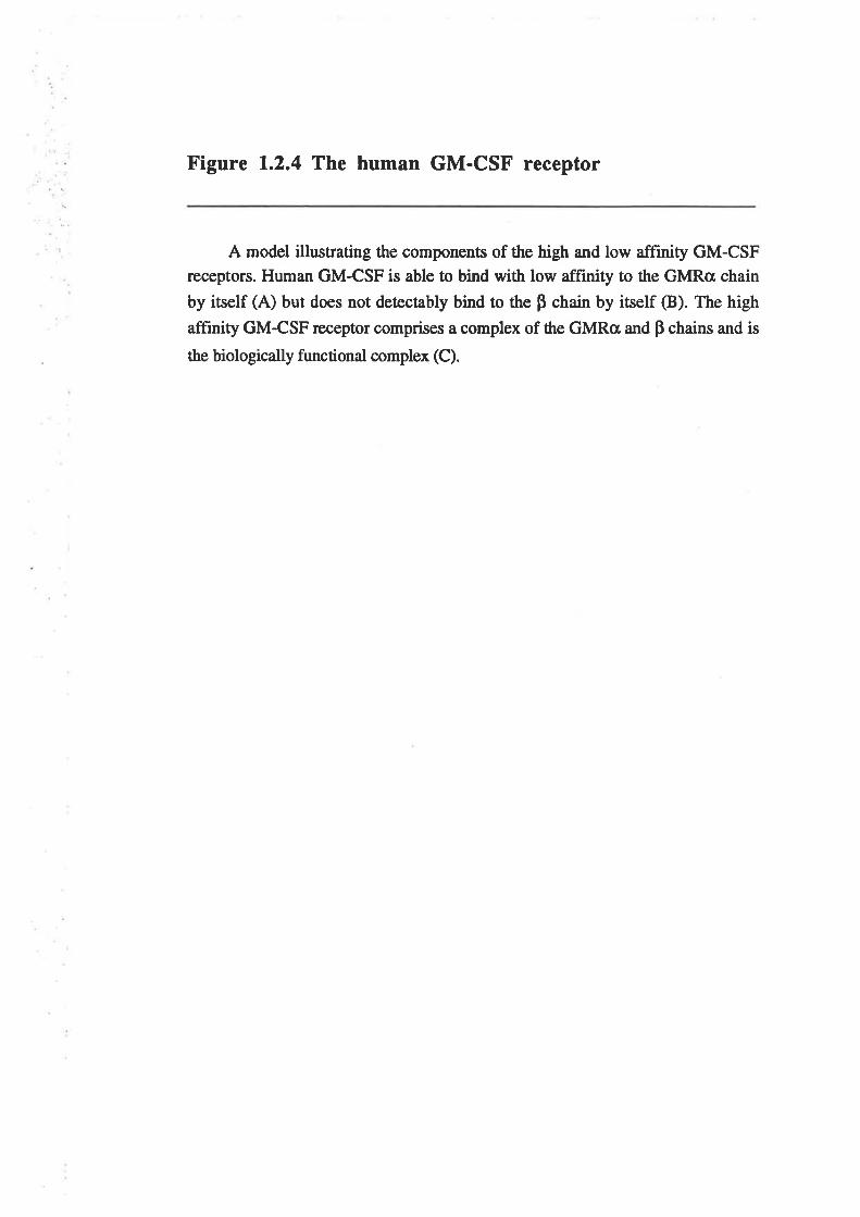
Figure 1.2.4 The human GM-CSF receptor
A model illustrating the components of the high and low affinity GM-CSFreceptors. Human GM-CSF is able to bind with low affinity to the GMRc¡ chain
by itself (A) but does not detectably bind to the p chain by itself (B). The high
affinity GM-CSF receptor comprises a complex of the GMRc¡ and p chains and is
the biologically functional complex (C).
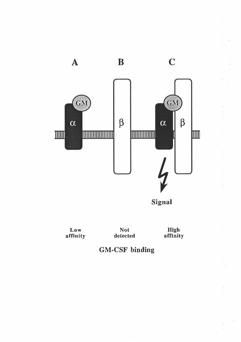
CBA
iSignaI
Lowaffinity
Notdetected
GM-CSF binding
Highaffinity
B B
Although the biological actions of GM-CSF were well described at the beginning
of this study, very little detailed information was available concerning the structure-
function properties of this cytokine and as a result, the mechanism of GM-CSF action
was very poorly understood. Structure-function information can be obtained using many
different approaches although many of the most powerful æchniques utilise recombinant
DNA technology. This enables the generation of protein analogues with precisely defined
alterations. Analysis of the structural and functional properties of the protein analogues
identifies regions and specific residues that a¡e sensitive ûo mutation and that a¡e therefore
either structurally or functionally important in the wild type protein.
1 .3 Project aims
The aim of this project was to study the structure-function properties of human
GM-CSF and specifically to identify regions and residues that are important for the
functional interaction of human GM-CSF with the high- and low-affinity GM-CSF
receptor. Functionally important regions on the surface of GM-CSF are identified by
comparing the biological activity and receptor binding properties of mutated GM-CSF
analogues with wild type GM-CSF. The generation of human GM-CSF analogues with
alæred biological and receptor binding activities might also give rise to analogues with
clinically useful properties such as the ability to antagonise GM-CSF function or
selectively inæract with restricted cell populations.
-9-
Chapter 2
Materials & Methods
2.L Abbreviations
Standard abbreviations were as described for The Journal of Biological Chemistry,
1993, volume 268, pages 750-753. Non-standa¡d abbreviations are lisæd below.
ALL acute lymphoblastic leukaemia
AI\4L acute myeloid or myeloblastic leukaemia
AU absorbance units
Fc common beta chain of the GM-CSF, IL-3 and IL-5 receptors
BCIG 5-bromo-4-chloro-3-indolyl-p-D-galactoside
bp base pair
BSA bovine serum albumin
CAPS 3-cyclohexylamino-l-propanesulfonic acid
CML chronic myeloid leukaemia
CMMoL chronic myelomonoc¡ic leukaemia
CNTF ciliary neurotophic factor
DAB 3,3'diaminobenzidine
DMEM Dulbecco's Modifred Eagle's medium
DTT dithiothreitol
EDso effective dose,507o
EPO erythropoietin
FCS foetal calf serum
FMLP N-formyl methionyl leucyl phenylalanine (f-Met-Leu-Phe)
G-CSF granulocyte colony-stimulating factor
GH growth hormone
GHbp growth hormone binding protein
GM-CSF granulocyæ-macrophage colony-stimulating factor
GMRcr GM-CSF receptor alpha chain
HBSS Hank's balanced salt solution
HFBA heptafluorobutyric acid
HIV human immunodeficiency virus
HP-SEC high performance size exclusion chromatography
I{TLV human T-cell leukaemia virus
ICso inhibitory concentration,50Vo
IL inærleukin
IL-3Ra IL-3 receptor alpha chain
- 10-
IPTG
LIF
LPS
Kd
kDa
mA
M-CSF
MoAb
Mr
NEM
NP.4O
ocMOSM
PAGE
PBS
PCR
PEG
PVP
RP-HPLC
RIA
RNase ARSV-LTR
S.A.
SCF
SEM
sv40TEMED
TFA
TNF
TMACI
Tween 20
USP
isopropyl þD-thiogalactoside
leukaemia inhibitory factor
lipopolysaccharide
dissociation constant
kilodalton
milliamps
macrophage colony-stimulating factor
monoclonal antibody
relative molecula¡ mass
N-ethyl morpholine
Nonidet P-40
oligonucleotide cassette mutagenesis
oncostatin Mpolyacrylamide gel electrophoresis
phosphaæ buffered saline
polymerase chain reaction
polyethylene glycol 8,000
polyvinyl pyrrolidone
reversed phase high performance liquid chromatography
radioimmunoassay
ribonuclease Arous sarcoma virus long terminal repeat
specific activity
stem cell facûor
standard error of the mean
simian virus 40
N,N,N',N'-tetramethyl-ethene-diamine
trifluoroacetic acid
tumour necrosis factor
tetramethylammonium chloride
polyoxyethylene (2O)-sorbiøn monolaurate
universal sequencing primer
- 11-
2.2 Chemicals, reagents and consumables
Standard chemicals were obtained from Ajax Chemicals (Auburn, NSW), BDH
Chemicals (Poole, UK) and Sigma Chemical Company (Sr Louis, MO)
Ethanolamine: Ajæ< Chemicals, Auburn, NSW
TMACT: Aldrich Chemical Company Inc., Milwaukee,'WI.
Centricon-lO, Centriprep-lO, Diaflo YM10 membrane: Amicon, Danvers, MA.
PEG, TFA Spectrosol, thioglycolic acid, Tween 20, urea : BDH Chemicals, Poole, UK.
Acrylamide, bisacrylamide, broad range biotinylated SDS/PAGE calibration standards,
Coomassie brilliant blue R-250, Econo columns, anti-mouse immunobeads, anti-rabbit
immunobeads, anti-sheep immunobeads, Econo-Pac Q columns, Macro-Prep Q anion
exchange support, TEMED : Bio-Rad, Richmond, CA.
Chondroitin sulphate : Calbiochem, La Jolla, CA.
BSA, penicillin : Commonwealth Serum Laboratories, Melbourne, Victoria.
Gentamicin, heparin : Delta West, Bentley, Western Australia.
E. colïdeived TNF-a : Genentech, South San Francisco, CA.
CHO cell-derived human GM-CSF, E. coli-denved human IL-3, sheep anti-GM-CSF
polyclonal antisera: gift from Dr. S. Clark, Genetics Institute, Cambridge, MA.
Aga¡, caesium chloride, DMEM, foetal bovine serum, G-418 (Geneticin), Ham's F12
nutrient mixture, peptone, RPMI, yeast extract : Gibco Laboratories, Glen lVaverly,
Victoria-
Yeast-derived human GM-CSF : gift from Dr. Linda Park, Immunex Corporation,
Seattle, WA.
-12-
E. cotidenved human GM-CSF : gift from Dr. George Morstyn, Melbourne Tumour
Biology branch, Ludwig Instituæ for Cancer Resea¡ch, Melbourne, Vic.
Isopropanol ChromAR HPLC : Mallinckrodt Specialty Chemicals Co., Pa¡is, KY-
Acetonitrile LiChrosolv, amido black: Merck, Dormstadt, FRG.
Imobilon PVDF membrane, PLGC 10,000 Mr regenerated cellulose membrane :
Millipore Coqporation, Bedford, MA-
Lymphoprep : Nycomed, Oslo, NorwaY.
Tryptone : Oxoid, Basingstoke, UK.
CM-sepharose CL-68 columns, CNBr-activated Sepharose 4B, Dextran T-500,
dideoxyribonucleotide triphosphates, deoxyribonucleotide triphosphates, NP-40,
ribonucleotide triphosphates, low molecular weight protein standards for SDS/PAGE'
Sephadex G-100, Sephadex G-25 PD-10 columns, Sephacryl 5-200 : Pharmacia,
Uppsala, Sweden.
BCIG,IPTG : Promega Corporation, Madison, WI.
Nitrocellulose : Schleicher and Schuell, Dassel, FRG
Agarose, ampicillin, BSA, chloramphenicol, cytochrome-c, DTT, EDTA, ethidium
bromide, ficoll, FMLP, HFBA, NEM, PVP, salmon spenn DNA, SDS, ætracycline,
Tris base, tunicamycin : Sigma Chemical Company, St Louis, MO.
Avidin DH, biotinylated rabbit anti-sheep IgG immunoglobulin : Vector Laboratories,
Burlingame, CA.
No. 6 filter paper, 3MM : Whatman International Ltd. Maidstone, U.K.
-13-
2.3 Radiochemicals and radiolabelled reagents
Reagents were obtained from the following sources:
[o-32P]dATP (3,000 Cilmmol), [a-3sS]dATP (1,500 Ci/mmol), [t-32p]Rrp (4,000
Ci/mmol) : BRESATEC, Adelaide, S.A.
Na251gúo (200 Cilmmol), Nal25¡ (2,500 Ci/mmol), l25I-GM-csF cHo cell-derived(98pCi/pg) : DuPont Australia, Melbourne, Vic.
l3H]thymi¿ine (6.7 Cilmmol) : ICN, Costa Mesa, CA.
2.4 Enzymes
All restriction enzymes were purchased from New England Biolabs @everly, MA.) or
Pharmacia (Uppsala, Sweden).
Other enzymes were obtained from the sources lisæd;
Calf inæstinal phosphatâse : Boehringer Mannheim, Mannheim, FRG
T4 DNA ligase, T4 polynucleotide kinase : New England Biolabs, Beverly, MA.
Thermus aquaticns DNA polymerase : The Perkin Elmer Corporation, Norwalk, CT.
E. coli DNA polymerase I Klenow fragment, T7 DNA polymerase, T4 DNA ligase, T4
polynucleotide kinase : Pharmacia, Uppsala, Sweden.
Lysozyme, ribonuclease A : Sigma Chemical Company, St. Louis, MO.
Sequenase sequencing kit : United States Biochemical Coqporation, Cleveland, OH.
-t4-
Biotinylaæd horseradish peroxidase H : Vector Laboratories, Burlingame, CA.
2.5 Bacterial strains and genotypes
A81899 thr-1 leuB6 thi-l argE3 his G4 proA2lon-l lacYl galK2
mtl-l xyl-5 ua-L4 strA31 tsx-33 À supBl4
Gift from Dr. Robert Kastelein, DNAX ResearchInstitute, Palo Alto, CA.
(Greenberg et al., 1988)
B178htrA63 degP
Gift from Dr. Renato Moreno, Department of Microbiologyand Immunology, University of Adelaide, S.A.
BB4 F'[proAB+ Iacl9lacZLMl5 Tn10 Tetrl, supF58 supE44
hsdR514 (r.- m.-) galK2 galT22 trpR55 metBl tonA À-
À(argJac)U169
(Stratagene Inc., La Jolla, CA.)
B.L2I F- ompT hsdSs (rs-ms-)
E. coli B strain so is probably deficient in lon protease
Gift from Dr. Renato Moreno, Department of Microbiologyand Immunology, University of Adelaide, S.A.
(Studier and Moffatt, 1986; Studier et a1.,1990)
8299 F' lac lonÂ100 Tn10 Tef sEA thi araD139
Gift from Dr. Justin Dibbens, Department of Biochemistry,University of Adelaide, S.A.
MC1061 araD139 Â (a¡aABC-leu)761s galEl5 galK16 Â (lac) X74
rpsl. (Strr) galU thi hsdR2 (r*- m.+) mcrA mcrBl
(New England Biolabs, Beverly, MA.)
JM101 F'traD36 laclg  (lacZ) Ml5 proA+B+ / supE thi
 (lac-proAB)
(New England Biolabs, Beverly, MA.)
-15-
TOPP 1,2,4,5,6
TOPP 3
F[proAB+ laclilacZ^ Ml5 Tn10 Tef ] riflF [proAB+ lacliLacZ^ M15 Tn10 Tetr ] rifr kanr
(Stratagene Inc., La Jolla, CA.)
2.6 Cloning and expression Yectors
M13mp19 was purchased from New England Biolabs, Beverly, MA.
pGEX-2T was purchased from Pharmacia, Uppsala, Sweden.
pIN-III-OmpH3 (Lundell et al., l99O) was a gift from Dr. Robert Kastelein, DNAX
Research Institute, Palo Alto, CA.
pJL4 (Gough et a1.,1985) was a gift from Dr. Nicholas Gough, Melbourne Tumour
Biology branch, Ludwig Instituæ for Cancer Research, Melbourne, Vic.
pRc/CMV was purchased from Invitrogen Corporation, San Diego, CA.
pRSVN.O7 was a gift from Dr. Allan Robbins, Department of Biochemistry, University
of Adelaide, S.A
2.7 Cloned DNA sequences
Human GM-CSF cDNA (Wong et a1.,1985a) was a gift from Dr. Steve Clark, Genetics
Institute, Cambridge, MA.
Human GM-CSF synthetic cDNA (Shanafelt and Kastelein, 1991a) in pIN-trI-OmpH3
was a gift from Dr. Robert Kastelein, DNAX Resea¡ch Institute, Palo Alto, CA.
Human GM-CSF receptor alpha chain cDNA (Gearing et a1.,1989) in tEH3 was a gift
from Dr. Nicos Nicola, The Walter and Eliza Hall Institute, Melbourne, Vic.
-16-
2.8 Standard solutions and bacterial media
Denha¡dt's solution (1x)
Dextran
IxDSS
IxHBSS
HEPES buffered saline
24mglml IPTG
6xNET
PAT buffer
PBS (lx)
PTB buffer
lOmg/ml RNase A(DNase free)
3M Sodium acetate
SSC (lx)TAE buffer (lx)TBE buffer (lx)
TE
TEG
TES
2.8.1 Standard solutions
Coupling buffer
2tmglmlBCIG
Binding medium
Blotto solution
0.5M NaCl,0.1M Na2CO3,0.1M NaHCO3 pH8.3.
Dissolved in dimethyl formamide and stored at-20"C.
RPMI 1640, 10mM HEPES, 0.5Vo (wlv) BSA and0.17o (wlv) sodium azide
1xPBS, 0.05Vo (v/v) Tween20,27o (ilv) BSA and57o (wlv) Skim milk powder.
0.027o (ilv) Ficoll, 0.02Vo (w/v) BSA, 0.027o (w/v) PVP.
57o (wlv) Dextan T-500 in I x PBS.
900mM NaCl, 90mM sodium citaæ pH7.4,0.57o (wlv) SDS, 0.02Vo (ilv) Ficoll, 0.02Vo (ilv) BSA,0.027o (w/v) PVP.
l36mM NaCl, 5.4mM KCl,4.2mM NaHCO3,0.4mM KH2PO4, 0.3mM Na2HPOa.
20mM HEPES, 137mM NaCl, 5mM KCl, pH7.0
Dissolved in sterile MiniQ and sûored at -20oC.
900mM NaCl, 90mM Tris-HCl pH7.6,9mM EDTA,0.5Vo (vlv) NP-40, 0.02Vo (w/v) Ficoll,0.02Vo (dv) BSA, 0.02Vo (w/v) PVP.
lxPBS, O.lVo (wlv) sodium azide,0.0lVo (v/v) Tween 20.
136mM NaCl, 2.7mM KCl, 8mM Na2HPO4,l.5mM KH2POa.
lxPBS, 0.057o (v/v) Tween20,0.57o (w/v) BSA.
lOmg/ml bovine pancreatic RNase A in 10mM Tris-HCI pH7.5, 15mM NaCl.Incubated for 15'at 100oC,cooled slowly to 25oC and aliquots stored at -20oC.
3M sodium acetate adjusted to pH4.6-5.5 with glacialacetic acid
150mM NaCl, 15mM sodium citrate pH7.4.
40mM Tris,20mM acetic acid,0.9mM EDTA
50mM Tris,42mM boric acid, lmM EDTA.
10mM Tris-HCl pH7.5,0.1mM EDTA.
25mM Tris-HCl pH8.0, 10mM EDTA,50mM glucose. Autoclaved.
25mM Tris-HCl pH8.0, 10mM EDTA,l57o (wlv) sucrose. Autoclaved.
3M TMACI,50mM Tris-HClpH8.0,0.17o (w/v) SDS2mM EDTA.
TMAC1 solution
-17 -
TS l0mM Tris-HCl pH8.0,20Vo (wlv) sucrose. Autoclaved.
2.8.2 Bacterial media
0.7Vo aflu 170mM NaCl, t.07o (wlv) tryptone, O.7Vo (wlv) agarAutoclaved.
L-agar
Luria broth (L-broth)
Minimal medium
Minimal agar
SOC medium
Terrific broth
2xYl
L-broth, 1.57o (wlv) agar. Autoclaved.
170mM NaCl, 0.57o (wlv) yeast extract,l.ÙVo (wlv) tryptone or peptone. Autoclaved.
60mM KzHPO¿, 33mM KH2PO4,7.6mM (NH¿)zSO¿, 1.7mM sodium citrate.After autoclaving, final concentrations of 1.7mM MgSOa,
14.8¡rM Thiamine and0.2Vo (w/v) glucose were added.
l.SVo (wlv) agar. After autoclaving, all components ofminimal medium were added.
10mM NaCl,2.3mM KCI,O.SVo (w/v) yeast extract,2.0Vo (wlv) tryptone, or peptone. Afær autoclaving, finalconcentrations of 10mM MgCl2, l0mM MgSO¿ and0.367o (ilv) glucose were added.
2.4Vo (w/v) tryptone,O.47o laving, a finalconce pH7.0 was added.
85mM NaCl, L.ÙVo (wlv) yeast extract,I.67o (wlv) tryptone or peptone. Autoclaved.
2.9 Molecular weight standards
EcoRI digested bacteriophage SPP-I DNA molecular weight markers were
obtained from BRESATEC (Adelaide, SA). Approximate fragment sizes in Kbp are:
8.51, 7.35, 6.11, 4.84,3.59,2.8r, 1.95, 1.86, 1.51, 1.39, 1.16, 0.98, 0.72,0.48,
0.36.
HpaII digested pUC19 DNA molecular weight markers were obtained from
BRESATEC (Adelaide, SA). Fragment sizes in base pairs are: 501, 489,404,33L,242,
190, 147, 1 11, I L0, 6',1, 34, 34, 26.
Low molecular weight protein standards for SDS/PAGE from Pharmacia
(Uppsala, Sweden).
Phosphorylase b (rabbit muscle) 94kDa
Albumin (bovine serum) 67kDa
Ovalbumin (hen egg white) 43kDa
-18-
Ca¡bonic anhydrase (bovine er¡hrocyæ)
Trypsin inhibitor (soybean)
a-Lactalbumin (bovine milk)
30kDa
20.lkDal4.4kDa
Broad range, biotinylated SDS/PAGE standards from Bio-Rad (Richmond, CA).
Biotinylated myosin (rabbit muscle) 200kDa
Biotinylaædþgalactosidase(E.coli) 116.5kDa
Biotinylaæd phosphorylase B (rabbit muscle) 97.4þ'Ða
Biotinylaæd albumin (bovine serum) 66.21<Da
Biotinylated ovalbumin (hen egg white) 45kDa
Biotinylated ca¡bonic anhydrase B (bovine erythrocyte) 3lkDa
Biotinylatedtrypsininhibitor(soybean) 21.5kDa
Biotinylated lysozyme (hen egg white) 14.4kDa
Biotinylatedaprotinin(bovinepancreas) 6.5kDa
2.L0 Methods
2.10.1 Processing oligonucleotides
2.10.14 Synthesis, cleavage and deprotection
Oligonucleotides were synthesised on an Applied Biosystems model38lA DNA
synrhesiser (Applied Biosystems, Foster City, CA) by Heath Suskin and Julie Phillips.
Following trityl-off synthesis, oligonucleotides were cleaved from the support by
incubating in 700p1 of ammonium hydroxide for 15' at room temperature. The cleavage
reaction was repeated twice and the pooled fractions of crude oligonucleotide deprotected
by incubation at 56oC overnight. Oligonucleotides prepared using FOD (fast
oligonucleotide deprotection) chemistry were deprotected by incubation at 56oC fot 2
hours or at room temperature overnight.
The mixture containing crude oligonucleotide was thoroughly mixed with 30mls of
water-saturated butanol and centrifuged at 15,000 rpm, 4oC for 10'in a Beckman JA-20
rotor @eckman Instruments, Palo Alto, CA). The pelleted oligonucleotide was dried and
-19-
resuspended in 5ffipl of MilliQ. As an alternative, oligonucleotide was recovered by
lyophilisation and resuspended in 500p1of MilliQ. Oligonucleotides were quantified by
measuring the ODzeonm and assuming lAU = 33pglml.
2.10.18 PAGE PurifÏcation
Full length oligonucleotides were purified from the crude synthesis mixture by
polyacrylamide gel electrophoresis. Typically gels contained 207o (wlv) acrylamide with
an acrylamide:bisacrylamide ratio of 30:1, 8M urea, IxTBE buffer and were polymerised
by adding final concentrations of 0J7o (v/v) TEMED and O.l%o (wlv) ammonium
persulphaæ. For oligonucleotides larger than 40 nucleotides in length, the concentration
of acrylamide was reduced to IO7o. Gels were pre-electrophoresed at 350 volts for at
least 30' in IxTBE buffer. Samples were prepared in a load buffer containing final
concentrations of 4O7o (vtv) deionised formamide, lmM EDTA, O-0027o (w/v)
bromophenol blue, 0.0027o (ilv) xylene cyanole and incubated at 100oC for 5' before
loading onto the gel. Gels were electrophoresed at 400 to 500 volts and then stained for
15' in a solution of lOpg/ml ethidium bromide. Gels were visualised using ultra violet
light, and the full length oligonucleotides excised and eluted in 5mls of TE at 37oC
overnight. Eluates were then filtered through a 0.45pM cellulose acetate filter and DNA
precipitaæd by adding 500p1of 3M sodium acet¿te pH5.5, 12.5mls of I00Vo ethanol and
incubating at -2O"C overnight. Purifred oligonucleotides were pelleted by centrifugation
at 15,000 rpm, 4oC for 30' in a JA-20 rotor, washed with 5mls 70Vo (vlv) ethanol and
centrifuged at 15,000 rpm,4oC for 15'in a JA-20 rotor. The oligonucleotide pellets were
dried, resuspended in TE and quantified.
Oligonucleotides were also separated from un-incorporated T-32p-^ltp following
phosphorylation (2.10.1C) by polyacrylamide gel electrophoresis. Following
electrophoresis, as described above, gels were carefully removed from the glass plates,
wrapped in cling film and autoradiographed. The 32P-labelled, full length
oligonucleotides, identified from the autoradiograph, were excised and eluted at 37oC
overnight in lml of TE.
-20 -
2.10.1C 5' phosphorylation of oligonucleotides
Oligonucleotides were 5' phosphorylated with y-32p-¡,tp for use as probes. A
20pl reaction containing 100 to 500ng of purihed oligonucleotide, 50mM Tris-HCl pH
7.4, lmglml BSA, lOmM MgCl2, 5mM DTT, 20¡rCi T-32p-Rtp and 20 units of T4
polynucleotide kinase was incubated at37oC for 45'. Un-incorporated y-32p-ATP was
separated from 5' phosphorylated oligonucleotides by electrophoresis on an 8M
ttreal2}To acrylamide gel (2. I 0. 1B).
Oligonucleotides were 5' phosphorylated with ATP for use in the M13
mutagenesis procedure (2.10.4). A 20¡rl reaction containing 100ng of purified
oligonucleotide, 50mM Tris-HCl pH 7.4, lmg/ml BSA, 10mM MgCl2, 5mM DTT,
lmM ATP and 20 units of T4 polynucleotide kinase was incubated at 37oC for 45'.
Enzyme was inactivated by incubation at 65oC for 5'.
2.10.lD Preparation of oligonucleotide fragments for OCM
Complimentary oligonucleotides were 5'phosphorylated with ATP and annealed to
form double stranded fragments used in the OCM procedure. A 40ttt reaction containing
500ng of each of the purified, complementary oligonucleotides, 50mM Tris-HCl pH7.4,
lmg/ml BSA, 10mM MgCl2, 5mM DTT, lmM ATP and 20 units of T4 polynucleotide
kinase was incubaæd at 37oC for 60'. The mixture was then incubaæd at l00oc for 5'
and allowed 15' to cool to room temperature, allowing the complementary
oligonucleotides to anneal. Volume was adjusted to 100p1 with MilliQ and reaction
extracted once with 100p1 of buffer-saturated phenol:chloroform:iso-amyl alcohol
(25:24:l), vortexed vigorously and centrifuged for 5' in a micro-centrifuge. DNA was
precipitated by adding lOpl of 3M sodium acetate pH5.5 and 250p1 of l00Vo ethanol to
the aqueous phase and incubating at -70oC for 30'. Centrifugation for 15'in a micro-
centrifuge at4"C pelleted the DNA which was washed with 500¡rl of 70Vo (v/v) ethanol,
dried and resuspended in 40pl of TE buffer. The procedure for fragments composed of
more than two oligonucleotides was essentially the same with a 2x reaction volume for
four oligonucleotides and a 3x reaction volume for six oligonucleotides.
-2r -
2.10.2 The polymerase chain reaction
A standard PCR protocol was used to amplify deflrned DNA fragments with
terminal restriction enzyme siæs. The typical 50¡tl reaction contained 5pM of each of the
purified oligonucleotide primers, lOmM Tris-HCl pH8.4, 50mM KCl, 1.5mM MgC12,
O.OlVo (v/v) glycerol, 200pM dATP, 200pM dGTP, 200¡tM dCTP, 200pM dTTP, 1
unit of f¿q DNA polymerase and lng of æmplaæ DNA. The reaction mix was overlayed
with 100p1 of paraffin oil and subjecæd to 25 to 30 cycles of PCR. The standard PCR
profile consisæd of a f incubation at94"C to denature the DNA, a 2' incubation at 52oC
ûo anneal the primers and templaæ and a f incubation atl2oC for prlmer extension.
A modified PCR protocol was used to screen bacærial colonies for the prcsence of
plasmids carrying inserts in the correct orientation (Sandhu et a1.,1989). This same
protocol was also used to screen M13 plaques for conectly orientaæd inserß. The typical
50pl reaction contained all the components of the standard reaction without polymera.se
and with DNA templaæ obtained from a colony or plaque. The reaction mix was
overlayed with l00pl of paraffin oil, incubated at 100oC for 10' and cooled slowly to
50oC. One ¡rl of Taq DNA polymerase, diluæd to 0.5 units/¡rl in 20mM Tris-HCl pH
8.4, l00mM KCl, 200mg/ml gelatin, was added and the reaction mix subjected to 25
cycles of the standard PCR profile.
2.10.3 Maintenance and transformation of E. coli
2.10.34 Maintenance of strains
The genotypes of the E coli strains used during the course of the work described
in this thesis are listed in Section 2.5. Glycerol stocks of all strains were prepared by
mixing 500p1 of fresh overnight culture with 500p1 of SOVo (v/v) glycerol and storing at
-700c.
ABl899, BL2l and MC1061 were maintained on L-agar plates and incubated at
37"C.
B178htrA63 was maintained on L-agar plates and incubaæd at room temperature.
-22-
BB4, 8299 and TOPP strains were maintained on L-agar plaæs containing
lOpg/ml tefacycline and incubated at 37t.
JM10l was maintained on minimal agar and incubaæd at37oC.
2.10.38 Transformation oÍ E. coli made competent with
calcium chloride
1. Transformation of MCl061
A fiesh single colony of MC1061 was inoculated into lOmls L-broth and incubaæd
at37"C overnight The following day,500td of the overnight culture was added to 50mls
of L-broth and incubated at37oC with shaking, to an ODooon- of 0.4 to 0.6. The culture
was chilled on ice for 30'and the cells pelleûed by centrifugation at 5,000 rpm,4oC for 5'
in a JA-20 rotor. The cells were gently resuspended in 25mls of ice-cold, sterile 100mM
MgCl2 and then pelleæd again by centrifugation at 5,000 rpm, 4oC for 5'in a JA-20
rotor. The cells were gently resuspended in 2mls of ice-cold, sterile 100mM CaCl2 and
incubated on ice for I hour.
Competent MC106l were gently resuspended and 200p1 aliquots mixed with I to
20¡rl of DNA and incubated on ice for 30'. Mixture was then heat shocked at 42"C for 2'
and left at room temperature for 5'. One ml of L-broth was added and the mixture
incubated at 37"C for 30'. Cells were pelleæd by centrifugation for I' in a micro-
centrifuge at room temperature and lml of the supernatant discarded. The bacterial pellet
was resuspended in the remaining supernatant, spread onto L-agar plates containing
l0Opg/ml ampicillin and incubated at 37oC overnight.
The procedure described for MC1061 was also effective at transforming the
ABl899, BL2L, BB4 and E299 strains. The B178htrA63 strain was also transformed
using this procedure but the bacteria were cultured at room temperature and the heat
shock was performed at37"C.
-23 -
2. Transformation of JM101
A fresh single colony of JM101 was inoculated into lOmls minimal media and
incubated at 37oC overnight. The following day, 500p1 of the overnight culture was
added to 50mls of 2xYT broth and incubaæd at37oC, with shaking, to an OD66gnn of
0.4 to 0.6. The culture was chilled on ice for 30'and the cells pelleted by centrifugation
at 5,000 rpm,4oC for 5'in aJA-20 roûor. The cells were gently resuspended in 25mls of
ice-cold, sterile 100mM CaCl2 and then pelleted again by centrifugation at 5,000 rpm,
4oC for 5'in a lA-20 rotor. The cells were gently resuspended in 2mls of ice-cold, sterile
100mM CaClz and incubated on ice for I hour.
Competent JMl01 were gently resuspended and 200p1 aliquots mixed with I to
20pl of DNA and incubated on ice for 30'. Cells were heat shocked at 42"C for 2' and
added to 3mls of 0.7Vo agar seeded 1:100 with a log-phase culture of JMl01. The
mixture was plated on minimal media plaæs and incubated at 37oC overnight. For blue-
white colour selection, 20pl of 24mglml IPTG and 20¡rl of 20mglml BCIG was added to
the plating mixture.
2.10.3C Transformation of E. colí by electroporation
This procedure was specifically used to transform all six of the TOPP E. coli
strains which exhibit a very low transformation efficiency as a result of their non-Kl2
background butcan be used for all strains of E. colí.
A fresh single colony of TOPP E. coli was inoculated into 20mls of L-broth with
l0pg/ml tetracycline and incubated at37"C overnight. The following day, 10mls of the
overnight culture was added to lL of L-broth with l0pg/ml tetracycline and incubaæd at
37"C, with shaking, to an ODomn", of 0.6. The culture was chilled on ice for 30'and the
cells pelleted by centrifugation at 5,000 rpm, 4oC for l0' in a Beckman JA-10 rotor
(Beckman Instruments, Palo Alto, CA). Pelleted bacteria were then washed by gentle
resuspension in 250mls of ice-cold lmM HEPES, l07o (v/v) glycerol followed by a
second wash with l25mls. Bacteria were pelleted by centrifugation at 5,000 rpm,4oC for
-24 -
l0'in a JA-10 roûor. Pelleæd bacteria were then washed by gentle resuspension in 50mls
of ice-cold lÙVo (vlv) glycerol followed by a second wash with 25mls. Bacteria were
pelleted by centrifugation at 5,000 rpm, 4oC for 10' in a JA-20 rotor. Finally, bacæria
\ryere resuspended in lml l07o (vlv) glycerol and zlO¡tl aliquots stored at -70oC.
An aliquot of electrocompetent cells was thawed and mixed with lpl of DNA in
low salt buffer. The mixture of cells plus DNA was then placed in a chilled 2mm cuvette
and pulsed at 25pF and 2,500 volts with a resistance of 200O using a Bio-Rad Gene
Pulser (Richmond, CA).Typical time constants were between 4.1 and 4.9. Immediately
afær the pulse, lml of SOC medium at room temperaturc was added to the cuvette and
the cells incubated at 37"C for 30'. Cells were pelleæd by centrifugation for f in a
micro-centrifuge at room temperaturc and lml of the supernatant discarded. The bacærial
pellet was resuspended in the remaining supernatant, spread onto L-agar plates
containing lOpg/ml tetracycline, 100pg/m1ampicillin and incubaæd at 37oC overnight.
2.10.4' M13 mutagenesis protocol
The standard protocol for the mutagenesis of genes carried by the M13
bacteriophage was based on the procedure described by 7-oller and Smith (1984).
2.10,4^ The mutagenesis reaction
A standa¡d reaction comprised 100ng of single stranded M13 DNA containing the
GM-CSF cDNA, 5ng of each 5'-phosphorylated oligonucleotide (2.10.1C), USP and
the mutagenic primer, 60mM Tris-HCl pH7.5, 9mM MgCl2 and 67mM NaCl in a
volume of 15p1. The mixture was incubated at 65oC for 5' and cooled at room
temperature for 5'. The reaction mixture was increased to a final volume of 50pl
containing 50pM of dATP, dGTP, dCTP and dTTP, lmM ATP, lmM DTT, 2 units of
E. coli DNA I polymerase Klenow fragment, I unit of T4 DNA ligase and incubated at
room temperature overnight. Competent JM101 were then transformed (2.10.38) with
0.5, I and 2pl aliqouts of the mutagenesis reaction, plated on minimal media and
incubaæd at 37 oC overnight.
-25-
2.10.48 Screening the mutagenesis
1. Plaque lifa.
The following day the plates were chilled at 4oC for at last 30' and duplicate lifts,
90" for the first and 5' for the second, taken from one of the transformation plates
containing approximately 2ffi plaques. Nitrocellulose or nylon membranes were used
although nylon membranes were reserved for mutagenic oligonucleotides with TmoC
>65oC. Filærs were air dried and nitrocellulose filters baked in a vacuum oven at 80oC
for 2 hours while nylon filærs were cross-linked with ultra violet light for 5'.
2. Prehybridisation.
Nitrocellulose filters were prehybridised in 6xNET while nylon filters were
prehybridised in IxDSS with approximaæly 50mls of prehybridisation solution for each
pair of filærs. Sonicaæd salmon sperm DNA at l0mg/ml, was incubated at l00oC for 5',
snap-chilled on ice and added to the prehybridisation solution at a f,rnal concentration of
lOpg/ml just before use. Prehybridisation and hybridisation was done at 37oC when
using oligonucleotides < 25-mers and at 42oC when using oligonucleotides > 24-mers.
3. Hybridisation.
Hybridisation was performed in the same buffer and at the same temperature as the
prehybridisaúon. Hybridisation mix was prepared in n+lmls where n is the number of
filærs being probed. The eluted, 5' phosphorylated, oligonucleotide probe (2.10.1C)
was adjusted to 500pg/ml with sonicaæd salmon sperm DNA, incubated at 100oC for 5',
snap-chilled on ice and added to the hybridisation solution. Filærs were sandwiched
between pieces of V/hatman IMM hlter paper, soaked in hybridisation mix and incubated
overnight.
4. Washing filærs.
Filters were washed at room temperature for 5', twice in 6xSSC, 0.17o (w/v) SDS
and then once in TMACI solution. Filters were then washed twice at the appropriaæ
TmoC-A for 30'in TMACI solution. The TmoC of an oligonucleotide is a function of
-26 -
length in the TMACI solution (Wood et al., 1985). Deductions of 2oC per base
substitution and 5-10oC for an insertion/deletion, enabled the æmperature at which a
mutagenic oligonucleotide melts from the wild type sequence (TmoC-À) to be calculaæd.
Filters were blotted dry and autoradiographed. Plaques that aligned to duplicating
positive signals were picked and single stranded DNA prepared (2.10.5A) for sequence
analysis (2.10.6).
2.10.5 Preparation of M13 and plasmid DNA
2.10.54 Small scale isolation of phage M13, single stranded DNA
This procedure was used to isolaæ the single stranded DNA from M13 clones. An
Ml3 plaque w͡s inoculated into 2mls 2xYT, seeded 1:100 with an overnight culture of
JM10l, and incubated at 37oC for 6 hours. Bacteria were pelleted by centrifugation of
1.5mls of culture for 10' in a micro-centrifuge, lml of the supernatant transferred to a
fresh tube and mixed with 270p1 of a solution containing 2.5M NaCl and 207o (wlv)
PEG. After a 15' incubation at room temperature, centrifugation for 10' in a micro-
centrifuge was used to pellet M13 phage. The phage pellet was r€suspended in 100¡tl of
TE and extracted twice with l00pl of buffer-saturated phenol:chloroform:iso-amyl
alcohol (25:24:1), vortexed vigorously and centrifuged for 5' in a micro-centrifuge.
DNA was precipitated by adding l0¡rl of 3M sodium acetztß pH5.5 and 250p1 of t00%o
ethanol to the final aqueous phase and incubating at -20"C overnight. The DNA was
pelleæd by centrifugation for l5' in a micro-centrifuge at 4oC, washed with 500p1 of
707o (vlv) ethanol, dried, resuspended in 20pl of TE buffer and stored at -20oC.
2.10.58 smatl scale isolation of plasmid DNA and phage M13
replicative form DNA
The procedure for isolating plasmid DNA from bacteria wa.s based on the alkaline
lysis procedure described by Birnboim and Doly (1979). Using suitable antibiotic
selection, usually 100pg/ml ampicillin, a single, fresh colony of E. coli ha¡bouring the
appropriaæ plasmid was inoculated into 2mls L-broth + ampicillin and incubaæd at37"C
overnight. The same procedure was also used to isolate the double stranded, replicative
-27 -
form DNA of phage M13. An M13 plaque was inoculated into 2mls 2xYT, seeded 1:100
with an overnight culture of JM101, and incubaæd at 37oC for 6 hours.
Bacteria were pelleæd by centrifugation of 1.5mls of culture for 10' in a micro-
centrifuge and the petlet resuspended in 100¡rls TES containing 2mglml lysozyme. Afær
a 30'incubation on ice, 200¡rls of a solution containing 0.2M NaOH and l7o SDS was
added, mixed in gently and incubated on ice. After 5', 150pls of ice-cold 3M sodium
acetate pH4.6 was added, mixed in gently and incubated on ice for 40'. Cellular and
chromosomal debris was pelleted by centrifuging twice for 15'in a micro-centrifuge at
4oC. The supernarant was then incubated with 5¡rl of l0mg/ml RNase A (DNase fiee) at
37oC for 30' and extracted twice with 400p1 of buffer-saturated phenol:chloroform:iso-
amyl alcohol (25:24:1). Mixture was vigorously vortexed, centrifuged for 5' in a
microcentrifuge and lml of 1007o ethanol added to the final aqueous phase. DNA was
precipitated at -70oC for 30'or at -20oC ovemight then pelleæd by centrifugation for 15'
in a micro-centrifuge at 4oC, washed with 800p1 of 707o (v/v) ethanol, dried,
resuspended in 20pl of TE buffer and stored at -20oC.
2.10.5C Large scale isolation of plasmid DNA
Large scale preparation of plasmid DNA used alkaline lysis followed by banding of
covalently closed circular DNA in a caesium chloride gradient. Using suitable antibiotic
selection, usually 100pg/ml ampicillin, a single, fresh colony of E. coli harbouring the
appropriate plasmid was inoculated into 10mls L-broth + ampicillin and incubaæd at
37oC overnight. The following day 5ml of overnight culture was added to 500mls of L-
broth containing 100¡rg/ml ampicillin in a 2L conical flask. The culture was incubaæd at
37oC with shaking to an ODoggno' of 0.8 to 1.0 before being adjusæd to a final
concenrration of lO¡rg/ml chloramphenicol, 2mM MgCl2 (Frenkel and Bremer, 1986)
and the incubation at37"C continued overnight
Bacteria were pelleted by centrifugation at 5,000 rpm, 4oC for 10'in a JA-10 rotor,
vigorously resuspended in 5mls of TEG containing 2mglml lysozyme. After a 30'
-28 -
incubation on ice, l0mls of fresh 0.2M NaOH,17o SDS was added, mixed in gently and
incubaæd on ice. After 10', 6.25mls of ice-cold 3M sodium acetate pH4.6 was added,
mixed in gently and incubated on ice for 40'. Cellular and chromosomal debris was
pelleæd by centrifuging twice at 16,000 rpm, 4oC for 30' in a JA-20 rotor. The final
supernatant was extracted twice with 20mls of buffer-saturated phenol:chloroform:iso-
amyl alcoh ol (25:24;l), vortexed and centrifuged at 10,000 rpm, 20oC for l0' in a JA-20
roror. The DNA was precipitaæd by adding 50mls of l00%o ethanol and incubating at
-2}"Cfor 2 to 16 hours. DNA was pelleæd by centrifugation at 15,000 rpm, 4oC for 30'
in a JA-20 rotor and resuspended in 5mls of TE. Protein and RNA were precipitated by
adding 5mls of 5M ammonium acetate, incubating on ice for 30' and pelleting debris by
centrifugation at 15,000 rpm,4oC for 10'in a JA-20 rotor. DNA in the supernatant was
precipitated by adding 12.5mls of I00Vo ethanol and incubating at -zO"C for 2 to 16
hours. DNA was pelleæd by centrifugation at 15,000 rpm, 4oC for 30'in a JA-20 rotor,
washed with 5mls 707o ethanol and centrifuged at 15,000 rpm, 4oC for 10'in a JA-20
rotor
The DNA was resuspended in 2.3mls of TE, 2.459 of optical grade caesium
chloride dissolved in and 60pl of l0mg/ml ethidium bromide added. The solution of
DNA w¿s loaded into a 13x32mm Quick-seal tube, sealed and centrifuged overnight at
65,000 rpm, 20oC in a Beckman TLA 100.3 rotor (Beckman Instruments, Palo Alto,
CA). The lower band, containing covalently closed circular plasmid DNA, was collected
using a25 gauge needle and the ethidium bromide removed by extracting three times
with lml of MilliQ-saturated butanol. The DNA was precipitated by adding 6mls of
Millie, 700p1 of 3M sodium acetate pH5.5, l8mls of l00Vo ethanol and incubating at
-2}"Covernight. DNA was pelleted by centrifugation at 15,0(X) rpm,4oC for 30'in a JA-
20 rotor, washed with 5mls 707o ethanol and centrifuged at 15,000 rpm, 4oC for 10'in a
IA-20 roror. The DNA was resuspended in 400p1 TE and re-precipitated by adding
400p1 of 3M sodium acetate pH5.5, lml of l00%o ethanol and incubating at -20oC for
30'. DNA was pelleted by centrifugation for 15' in a micro-centrifuge at 4oC, washed
-29 -
with 800p1 of 707o (v/v) ethanol, dried and resuspended in 300¡tl of TE buffer. The
DNA was quantified by measuring the ODzeo¡¡m and assuming lAU = 50ttdml.
2.10.6 DNA sequencing2.10.6^ Denaturation of double stranded DNA for sequencing
This method was based on a protocol described by Chen and Seeburg (1985).
To 2-5pg of double stranded DNA in a volume of less than 5pl, was added 40¡rl of
denaturation buffer containing 200mM NaOH and 2mM EDTA. Afær incubation at 37oC
for 30', 4¡tl2lll ammonium acetate pH4.5 and 125p1 1007o ethanol were added and
DNA precipitated at -70oC for25'. DNA was pelleted by centrifugation for 15'in a
micro-centrifuge ^t
4oC, washed with lml of.TOVo (v/v) ethanol, dried and resuspended
in 10pl of annealing mix (2.10.68).
2.10.68 Sequencing with Sequenase
DNA was sequenced by the chain termination method originally described by
Sanger et aI. (1977), using a Sequenase kit (United Staæs Biochemical Corporation,
Cleveland, OH). The method was used to sequence single- and double-stranded DNA
using either [cr-32P]dATP or [cr-355]dATP.
The annealing reaction contained 40mM Tris-HCl pH7.5, 20mM MgCl2, 50mM
NaCl, 10ng of purified or 100ng of crude primer and 3-5pg of single-stranded DNA
template in a final volume of 10p1. DNA was denatured at 65oC for 5'and cooled slowly
to room temperature. For double-stranded DNA the denatured DNA (2.10.64) was
resuspended in l0¡rl of annealing mix which contained 40mM Tris-HCl pH7.5, 20mM
MgCl2, 50mM NaCl and l0ng of purified or l00ng of crude primer. Template and
primer were annealed at 45oC for 20'.
The labelling reaction contained 6.3mM DTT, 190nM dGTP, 190nM dCTP,
190nM dTTP, 5 to 10pCi ¡ø-32P1OATP or ¡6¡-35S10ATP and 1 unit of Sequenase
-30-
enzyme added to the annealing reaction with a frnal volume of l6pl. The labelling
reactions was incubated at l5t for 5'.
Reactions werc terminated by mixing 3.5¡rl from the labelling reaction with 2.5p1
of each of the A,G,C,T termination mixes in tubes pre-warmed to 45oC. The ærmination
mixes contained 80pM of dATP, dGTP, dCTP and dTTP as well as 8pM of the
appropriaæ dideoxynucleotide triphosphate. Termination reactions were incubated at
45oC for 5'. Reactions were stopped by adding 4¡rl of stop solution containing95Vo (vlv)
formamide, 20mM EDTA, 0.05Vo (ilv) bromophenol blue,0.057o (w/v) xylene cyanole
and placing at -20oC until electrophoresed.
Modifications of the standard protocol that enabled the sequence close to the primer
to be deærmined included using 1:10 or 1:20 dilutions of the labelling mix, reduced
incubation times (3') for the labelling reaction and the use of Mn buffer. The Mn buffer
contains l50mM sodium isocitrate, 100mM MnCl2 and lpl was included in the standard
labelling reaction for short sequencing runs.
2.10.6C Electrophoresis of the sequencing reactions
The sequencing reactions from 2.10.68 were analysed on 400mm x 4(X)mm x
lmm sequencing gels. Gels contained 6Vo acrylanride with an acrylamide:bisacrylamide
ratio of 19:1,8M urea, IxTBE buffer and were polymerised by adding final
concentrations of 0.IVo (vlv) TEMED and O.l%o (ilv) ammonium persulphate. Gels
were pre-electrophoresed at 35mA for at least 30' in IxTBE buffer prior to loading.
Samples, denatured at 100oC for 5', were lciaded at 1-3pl per well, depending on the
comb used and electrophoresed at 35m4. Standard conditions included the addition of a
final concentration of 333mM sodium acetate to the buffer in the lower tank during
electrophoresis to compress the 5' sequence and running the bromophenol blue to the
end of the gel. The gel was then fixed in 3L of l0%o (vlv) methanol, l07o (vlv) acetic
acid for 30', transferred to Whatman 3MM, covered in cling film and dried under
vacuum for thour at 80oC. Once dry the gel was autoradiographed.
-31 -
2.10.7 Transfection of COS and CHO cells
COS and CHO cells were maintained by Bronwyn Cambareri who also routinely
performed the tansfection of plasmid constructs into COS cells.
2.10.7L Transient transfection of COS cells
COS cells were maintained in DMEM containing l0mM HEPES, 0.2Vo (wlv)
sodium bicarbonate, 1.7mM glutamine, 10.5pg/ml penicillin, lA¡tglml gentamicin and
supplemented with l07o (vlv) FCS. Cells were usually grown to 50-707o confluence in
150cm2 canted neck flasks containing 35mls of medium, before being ha¡vested for
transfection. Medium was aspirated off and cells rinsed briefly in 0.8Vo (ilv) NaCl,
0.027o (w/v) EDTA before adding 3mls of 0.8Vo (wlv) trypsin in 0.87o (w/v) NaCl,
0.02Vo (w/v) EDTA and incubating at 37oC for l'. Cells were detached by "knocking",
l5mls of fresh DMEM added immediaæly and cells collected by centrifugation at 1,600
rpm for 5'. Harvested cells were counted, pelleæd by centrifugation at 1,600 rpm for 5'
and resuspended in IxHBSS supplemenæd with 6mM glucose to I x 107 cells/ml.
Plasmid constructs were introduced into COS cells by electroporation (Chu et aI.,
1987) using a Bio-Rad Gene Pulser (Richmond, CA). For each transfection, 20pg of
plasmid DNA, 25pg of sonicated salmon sperm DNA and 50¡rl of FCS were mixed in
4mm electroporation cuvettes chilled on ice before adding 500p1 of resuspended COS
cells. For mock transfections, 20pl of HEPES buffered saline, 6mM glucose was
substituæd for the plasmid DNA. The mixture was electroporated at 300V and 250pF,
left at room temperature for l0' and plated out in petri dishes with lOmls of DMEM.
Afær a 24 hour incubation at37oc, the medium was replaced with FCS-free DMEM and
the incubation continued for a further 72 hours. Finally the condition medium was
harvesæd and assayed for GM-CSF.
2.10.78 Transfection of CHO cells and cloning permanently
transfected cell lines
CHO cells were maintained in Ham's Fl2 nutrient mixture containing 10.5¡rg/ml
penicillin, lfi¡tglml gentamicin and supplemented with IOVo (v/v) FCS. CHO cells were
-32-
grown ¡a 80Vo confluence in 150cm2 canted neck flasks containing 35mls of medium and
harvested using essentially the procedure described for COS cells. The ha¡vested CHO
cells were washed twice in cold 1 x PBS before being resuspended in IxPBS to 6.3 x
lN cellVml.
For electroporation, lOpg of plasmid DNA wa.s mixed with 800p1 of CHO cells in
chilled 4mm electroporation cuvettes and incubated on ice for l0'. Mock transfections
contained 10pl of IxPBS substituted for the plasmid DNA. The mixture was
elecuoporaæd at 1300V and 25pF using a Bio-Rad Gene Pulser (Richmond, CA) and
left at room temperature for 10' before mixing with 30mls of Ham's F12 nutrient
mixture. Transfected CHO cells were plated out in 6 x 60mm gridded petri dishes and
afær a 24 hour incubation at 37oC, the medium was replaced with selective Ham's F12
containing 350pg/ml G-418.
The selective medium was replaced every 72 hours until all cells in the mock
transfection were dead. Individual colonies of G-418 resistant CHO cells were cloned by
placing cloning rings over the colonies and harvesting the transfected CHO cells with a
f incubatio n at37"C in 2ü)pls of 0.8Vo (w/v) trypsin in 0.87o (w/v) NaCl ,0.02Vo (wlv)
EDTA. Cloned CHO cells were grown in selective Ham's F12 containing 350pg/ml G-
418. A number of colonies for each transfection wete cloned and the conditioned
medium for each cell line assayed for GM-CSF using an RIA (2.10.114). Transfected
CHO cell lines expressing the highest level of GM-CSF mutant analogue were selected
and used to produce analogues for purihcation.
2.10.8 Osmotic shock Protocol
The procedure for isolating periplasmic proteins was based on a protocol described
by Koshland and Botstein (1980). Using suitable antibiotic selection, usually 100pg/ml
ampicillin, a single, fresh colony of E. coli harbouring the appropriate GM-CSF
expression construct was inoculated into lOmls L-broth + ampicillin and incubaæd at
37oC overnight. The following day the overnight culture was pelleted at 3,000 rpm for
-33 -
10' and the cell pellet washed twice in fresh L-broth ûo remove accumulated pJactamase.
The entire, washed overnight culture was then added to lL of L'broth containing
lO0pg/ml ampicillin in a 2L conical flask. The culture was incubated at 37oC with
shaking to an OD660nm of 0.8 to 1.0. IPTG was then added to a final concentration of
0.1mM and the culture incubated at room temperature for a further 3 hours or left
overnight.
Cells were then pelleted by centrifugation at 5,000 rpm, 4oC for 20' in a JA-10
rotor. The pellet was then resuspended in 5mls TS buffer with aggressive pipening and
transferred to Oakridge tubes before adding 250p10.25M ice-cold EDTA and incubating
the mixture on ice for 30'. Tubes were filled with ice-cold MiltiQ, mixed rapidly and
incubated on ice for a further 30'. Cellular debris was pelleæd by centrifugation at
12,000 rpm and 4oC for 15' in a JA-20 rotor. The supernatant was transferred to a clean
tube and centrifuged at 19,500 rpm and 4oC for 15'. The final supernatant containing
periplasmic contents, was filtered through Whatman No. 6 filær paper and adjusæd to
O.lVo (wlv) sodium azide. Preparations of periplasmic proteins were stored at4oC forup
to a few weeks but normally GM-CSF was purified from this crude preparation
immediaæly.
2.10.9 Preparation and use of immunoaffinity columns
2.10.9^ Preparation of MoAb from ascites fluid
Asciæs fluid containing crude anti-GM-CSF monoclonal antibodies, was generaæd
from BALB/c mice. Solids were removed and MoAb precipitaæd by slowly adding
660p1 of saturated ammonium sulphate solution per ml of asciæs fluid. The mixture was
stirred for 30' at room temperature, pelleted by centrifugation for 10' in a micro-
centrifuge and the pellet washed twice with a 507o saturated ammonium sulphate
solution. The precipitaæd monoclonal antibody was resuspended in MilliQ to twice the
volume of the original ascites fluid, dialysed l:10,000 (final) against fresh coupling
buffer and concentrated in Centricon-l0 mini concentrators to 5-lOmg/ml. Coomassie
-34-
stained SDS/PAGE indicated that the preparations typically contained 80-90Vo
immunoglobulin.
2.10.98 Coupling MoAb to CNBr.activated Sepharose
Partially purified monoclonal antibodies were coupled to CNBr-activated
Sepharose 48 according to the manufacturer's protocol. Freeze-dried CNBr-activated
Sepharose 48 was swollen in lmM HCI for 15'and washed in several changes of lmM
HCl. One gram of freeze-dried Sepharose swelled to a gel volume of 3.5mls. Swollen
gel was then washed briefly with coupling buffer and mixed with monoclonal antibody
for 2 hours at room temperature. The gel was drained of coupling mixture and the liquid
assayed for uncoupled proæin which was usually <5Vo of the starting maærial for a
successful coupling. The gel was then mixed with 10 volumes of blocking agent for 2
hours at room temperature. Typical blocking agents were 200mM glycine, 1.0M Tris-
HCI or l.0M ethanolamine all at pH8.0.
The gel was washed with 5 cycles of alærnating high pH and low pH buffers to
remove uncoupled protein using 5 volumes of buffer for each wash. Coupling buffer
was used for the high pH washes and 100mM sodium acetate, 500mM NaCl pH4.0 used
for the low pH washes. Gel was then washed 5 úmes with 10 volumes of coupling
buffer. Finally gel was washed 5 times with 10 volumes of PAT buffer containing 1 x
PBS, 0.17o (w/v) sodium azide, 0.017o (v/v) Tween 20. The immunoaffinity matrix was
stored in PAT buffer at4oC where it was stable for at least 12 months.
2.10.9C Immunoaffinity chromatography
Immunoaffinity matrix was used to purify GM-CSF mutant analogues expressed
from COS or CHO cells or E. coli. Aliquots of immunoaffinity matrix, usually 0.5-
1.0mls, were mixed with crude GM-CSF overnight and the following day the matrix
was collected in an Econo column. Alternatively the immunoaffinity matrix was packed
into an Econo column and crude GM-CSF passed over the column 2-3 times. For ease of
handling, the volume of crude GM-CSF either in COS or CHO cell conditioned medium
-35-
or E. coli osmotic shock preparations, was always less than 50mls. Crude samples were
concentrated from as much as lL using a stirred-cell apparatus (Amicon, Danvers, MA)
with a 10,000 Mr cutoff membrane.
The immunoaffinity matrix with bound GM-CSF was then washed 5 times with l0
volumes of PAT buffer. Typically, the specifically bound GM-CSF was eluted using 4 x
500p1 aliquots of l00mM glycine, 100mM NaCl, 0.027o (ilv) sodium azide pH3.0. For
some immunoaffrnity matrices milder conditions werc sufficient to eluæ bound GM-CSF
so 100mM sodium acet¿te, 100mM NaCl, 0.027o (w/v) sodium ande pH5.0 was used
instead. The eluted GM-CSF was immediately neutralised by adding 1.0M Tris-HCl
pHl0.0. The immunoaffinity matrix was then washed 5 times with l0 volumes of PAT
buffer and stored in PAT buffer at4"C.
2.10.10 Radioiodination of recombinant cytokines
The radioiodination of GM-CSF was performed by Mara Dottore or Dr. J.
Woodcock. Recombinant GM-CSF produced in CHO cells, yeast or E. coli, was
radioiodinated by the iodine monochloride method as previously described (Contreras er
al., L983).Iodinaæd GM-CSF was separated from ¡¡¿1251 by chromatography with a
Sephadex G-25 PD-10 column equilibrated in I x PBS, 0.02Vo (v/v) Tween 20 and
stored at 4oC for up to I month. For use in receptor binding experiments, non-protein-
associated radioactivity and Tween 20 were removed by cation exchange
chromatography on a 300¡rl CM-Sepharose CL-6B column and the iodinaæd GM-CSF
stored at 4oC for up to 5 days. Radioiodinated GM-CSF retained at least 907o of wild
type biological activity compared with unlabelled GM-CSF.
2.l0.ll Quantification of GM-CSF
2.10.114 Radioimmunoassay
A modification of the radioimmunoassay described in Lopez et al (I992c) was used
to quantify crude GM-CSF analogues in COS (2.10.7A) or CHO (2.10.7B) cetl
condiúoned medium and osmotic shock supernatants (2.10.8). Briefly, tOpt 125I-Gtvt-
-36-
CSF (0.lng) was mixed with 50pl crude GM-CSF sample and 50pl of suitably diluted
polyclonal anti-GM-CSF serum and incubated at room temperature for 2 to 4 hours. The
serum was either sheep anti-GM-CSF diluted 1:40,000 (final) or rabbit anti-GM-CSF
diluted 1:4,000 (final). An equal volume of reconstituted anti-sheep or anti-rabbit
Immunobeads was then added and the incubation continued for a further 2 ¡o 4 hours.
The beads were then pelleted, washed ¡ryice in I x PBS containng0.L%o (w/v) BSA and
counted in a gamma counter (Cobra Auto Gamma, Packard Instrument Company,
Meriden, CÐ. The amount of GM-CSF in the samples was calculated from a standard
curve constructed with known amounts of wild type GM-CSF.
2.10.118 High performance size exclusion chromatography
Wild type GM-CSF and mutant analogues purified from E coli were directly
quantified by high performance size exclusion chromatography (HP-SEC). All HPLC
applications used a Bio-Rad 1350 dual pump HPLC with a series 600 gradient controller
and either a Bio-Rad model 1306 UV monitor @io-Rad, Richmond, CA) or a Waters
490E programmable multiwavelength detector using Maxima 820 software (Waters,
Milford, MA). For HP-SEC samples were chromatographed on a 7.5 x 300mm
Ultraspherogel SEC 3000 column (Beckman Instruments, San Ramon, CA) using a
mobile phase containing 0.lM sodium phosphaæ pH7.0, 0.1M sodium sulphaæ and
0.057o (w/v) sodium azide run at O,Smls/min. The GM-CSF absorbance peak was
integrated and the amount of protein determined using a calculaæd extinction coeffìcient
of 0.95 AU.mll.mg-l or 1.1 AU.ml-t.mg-l for the P6YY analogue.
2.10.12 Electrophoresis of proteins
2.IO.I2A SDS/PAGE
The procedure for fractionating proteins by SDSÆAGE was based on the system
described by Laemmti (1970). A30Vo (ilv) acrylamide stock solution was used for all
protein gels and contains an acrylamide:bisacrylamide ratio of 36:1. Separating gels were
composed of 12.5 or líVo (w/v) acrylamide, O.IVo (wlv) SDS, 375mM Tris-HCl pH8.8
and were polymerised by adding final concentrations of 0.l25%o (v/v) TEMED and
-37 -
0.0257o (w/v) ammonium persulphate. Stacking gels were composed of 4.8Vo (w/v)
acrylamide,O.lTo (ilv) SDS, 125mM Tris-HCl pH6.8 and were polymerised by adding
flrnal concentrations of 0.2Vo (v/v) TEMED and 0.06Vo (w/v) ammonium persulphate.
Samples to be analysed by SDS/PAGE under reducing conditions rvere prepared in a
load buffer containing final concentrations of 3lmM Tris-HCl pH6.8, 0.67o (w/v) SDS,
6.l%o (vlv) glycerol, 0.0l%o (w/v) bromophenol blue and 57o (vlv) p-mercaptoethanol.
Prior to loading on the gel samples were incubaæd at 100oC for 5'. For analysis of
samples under non-reducing conditions, the p-mercaptoethanol was omitted from the
sample. Gels were elecuophoresed at 15 to 30mA for standard 160xl60mm gels in a
buffer composed of 83mM Tris, 192mM glycine and0.l%o (Øv) SDS.
2.10.128 High pH native PAGE
The procedure described by Goldenberg (1990) was used for native PAGE of GM-
CSF. The separating gel was composed of l07o (w/v) acrylamide, 375mM Tris-HCl
pH8.9 and0.067o (v/v) TEMED and was polymerised by adding ammonium persulphaæ
to a final concentration of 0.057o (w/v). The stacking gel was composed of 4.8Vo (wlv)
acrylamide, 60mM Tris-POa pH 6.9 and0.06Vo (v/v) TEMED and was polymerised by
adding ammonium persulphate to a final concentration of O.0l27o (w/v). Samples were
prepared in a load buffer containing hnal concentrations of lïVo (v/v) glycerol and
0.017o (Øv) bromophenol blue. Gels were electrophoresed at l0 to 20mA for standard
160x160mm gels in a buffer composed of 5mM Tris base and 38mM glycine.
2.10.12C Transverse urea gradient PAGE
Transverse urea gradient PAGE was performed using a protocol described by
Creighton (1979). The gradient, which was formed perpendicular to the direction of
electrophoresis, contained a gradient of urea from 0M to 8M with a compensatory
gradient of acrylamide from lïVo to 87o. Two acrylamide solutions were prepared
containing either 0M or 8M urea. The 0M solution contained l07o (wlv) acrylamide,
375mM Tris-HCl pH8.9, 0.06Vo (v/v) TEMED and 0.047o (w/v) ammonium persulphaæ
while the 8M solution contained 87o (wlv) acrylamide, 8M urea, 375mM Tris-HCl
-38-
pH8.9, O.O6Vo (v/v) TEMED and O.MVo (w/v) ammonium persulphate. Using llmls of
each solution a linea¡ gradient from 0M to 8M urea was formed using a gradient former
and pumped into the bottom of a gel casting apparatus containing nvo 80 x 80 x lmm gel
cassettes at SmVmin. A solution of 507o (v/v) glycerol, O.I7o (w/v) methyl green
pumped at 3mUmin was used to ensure that all of the acrylamide solution enters the gel
cassette. Once set, the gel was rotated by 90o for electrophoresis. Samples were prepared
in a load buffer containing final concentrations of l07o (v/v) glycerol and 0.017o (wlv)
bromophenol blue. Gels were electrophoresed for 400 volt/hours at 50 volts and at 4oC
in a buffer composed of 5mM Tris base and 38mM glycine.
2.10.12D Western transfer, Towbin protocol
The electrophoretic transfer of proteins fractionaæd by SDS/PAGE to nitrocellulose
was performed using one of two methods. The original method was based on the
protocol of Towbin et aI. (1979). Following electrophoresis, the gel was placed on three
sheets of Whatman 3MM soaked in transfer buffer containing 20mM Tris base, 150mM
glycine, 0.17o (wtv) SDS and20Vo (v/v) methanol. A sheet of nitrocellulose soaked in
transfer buffer was placed over the gel and all air bubbles carefully squeezed out. Three
more sheets of Whatman 3MM soaked in t¡ansfer buffer were then placed on top. Two
Scorchbrite pads soaked in transfer buffer were placed on either side of the sandwich and
the entire assembly supported in a plastic grid which was then placed in the transfer tank.
The assembly was completely immersed in transfer buffer and transfer performed at25Y
o/n or 60V for 4 hours if a cooling coil was used. Protein migration was towards the
anode so the nitrocellulose sheet faced the anode while the gel faced the cathode.
2.10.128 Western transfer, semi-dry protocol
The second method used a different, semi-dry apparatus that enabled faster
transfer. Immediately prior to transfer, the graphite anode was washed with MilliQ. A
stack of nine sheets of Whatman 3MM, trimmed to match the gel and soaked in transfer
buffer containing 50mM Tris base,40mM glycine, 0.03757o (w/v) SDS and 20Vo (vlv)
methanol, was then assembled. Alternatively the Towbin transfer buffer used in the
-39-
original transfer method can be used. A sheet of nitrocellulose, trimmed to match the gel
and soaked in transfer buffer was then placed on the Whatman 3MM and following
electrophoresis, the gel was placed on the nitrocellulose and all air bubbles carefully
squeezed out. Another stack of nine sheets of Whatman 3MM, trimmed to match the gel
and soaked in transfer buffer was then placed on the gel. The graphite cathode was then
rinsed with MilliQ and placed on top. Transfer was performed for 60 to 90' at 0.8m4 x
surface area, which was typically 160m4.
2.10.13 Visualisation of proteins after electrophoresis
2.10.134 Coomassie blue staining
Following SDS/PAGE or native PAGE electrophoresis, the gel was stained for 30'
in a solution of.0.l7o (w/v) Coomassie Brilliant blue R-250,307o (v/v) methanol, I07o
(v/v) acetic acid. Staining solution was filtered through \Vhatman 3MM prior to use and
used at most three times. Gel was destained in307o (v/v) methanol,l0%o (v/v) acetic acid
although afær initial de-staining, native PAGE gels and urea gradient gels were destained
in57o (v/v) methanol,5Vo (v/v) acetic acid.
2.10.138 Silver staining
The procedure for silver staining SDS/PAGE gels was based on a protocol
described by Morrissey (1981) but did not include fîxing in lÙVo glutaraldehyde.
Because silver staining is approximately 40x more sensitive than Coomassie blue
staining, the apparatus used for SDS/PAGE was cleaned thoroughly to remove
contaminating proteins. Protein molecula¡ weight standards were diluted 4Gfold because
of the increased sensitivity of silver staining. The following protocol was based on a
standa¡d 160x160mm gel and volumes were doubled for the 320x160mm gels.
Following electrophoresis the gel was fixed using 500mls of 507o (v/v) methanol,
I\Vo (vtv) acetic acid for 30' followed by 5ü)mls of 57o (v/v) methanol,TVo (v/v) acetic
acid for 30'. Throughout the procedure the gel was gently agitaæd. Gel was then soaked
-40-
overnight in MilliQ with a 30'soak the following day in fresh MilliQ. Alternatively the
gel was soaked in several changes of MilliQ for 1 to 2 hours.
Washed gel was then soaked in 200mls of Spg/ml DTT for 30' followed
immediaæly by 200mls of 0.17o (ilv) silver nitraæ for 30'. Gel was then briefly rinsed
with MilliQ and then twice with a small volume (50mls) of developer solution,
containing 37o (wlv) sodium carbonate and 0.02Vo (v/v) formaldehyde, before soaking
gel in developer solution until appropriately stained. The developer solution was
discarded and the reaction stopped with SVo (v/v) acetic acid. The gel could be stored in
SVo acettc acid for months if sealed in a plastic bag.
2.10.13C Amido Black staining
Proæin molecular weight standards transferred to nitrocellulose by Wesærn blot
transfer, were stained with amido black for I' in a solution of 0.17o (ilv) Amido black,
45Vo (vlv) methanol, lOTo (vlv) acetic acid. The nitrocellulose was destained in 457o
(v/v) methanol,lÙVo (v/v) acetic acid.
2.10.13D Western blot staining
Following transfer of proæins to nitrocellulose (2.10.12D,E) the track containing
non-biotinylated, protein molecular weight standards was cut off and stained with amido
black (2.10.13C). Non-specific protein binding sites on the remainder of the
nitrocellulose filær were blocked by incubation at 4oC overnight in Blotto solution. The
filter was then probed with sheep anti-GM-CSF polyclonal antibody diluted 1:2,000
(frnal) in PTB buffer and incubated in a sealed plastic bag at 4oC for 3 hours. The filter
was then washed in four changes of PTB, 5' each wash with agitation. The filter was
then probed with biotinylated, rabbit anti-sheep immunoglobulin diluted 1:lü) in PTB
and incubated in a sealed plastic bag at 4oC for I hour. Filter was then washed and
probed with a conjugate of avidin and biotinylated horseradish peroxidase diluted
1:1,000 in PTB and incubated in a sealed plastic bag at 4oC for t hour. Filter was then
washed and placed in substrate solution and stain allowed to develop. Substrate solution
-41 -
\ilas prepared by dissolving 50mg DAB in lml lM HCI and adding to 100mls of MilliQ.
A small amount of solid Tris base was then added to adjust to pH7.0 and immediaæly
prior to use H2O2 was added to 0.0187o (v/v) hnal. When the filær was appropriaæly
stained, development was halæd by placing in a solution of 0.57o (ilv) sodium ande-
The filær was photographed immediaæly as discolouration occurred rapidly, <1 hour.
2.10.14 Protein sequencing
This is a method from Richard Simpson's laboratory (Ludwig Instituæ for Cancer
Research, Melbourne, Vic). The separating gel was composed of I57o (wlv) acrylamide,
750mM Tris-HCl pH8.8, O.IVo (wlv) SDS and was polymerised by adding final
concentrations of 0.067o (v/v) TEMED and 0.097o (w/v) ammonium persulphate. The
separating gel was pre-electrophoresed at 20mA overnight in a buffer composed of
375mM Tris-HCl pH8.8, O.l%o (wlv) SDS and 0.077o (v/v) thioglycolic acid. Søcking
gel was composed of 4.7Vo (w/v) acrylamide, 123mM Tris-HCl pH6.8, O.LVo (wlv)
SDS and was polymerised by adding final concentrations of.0.LVo (v/v) TEMED and
0.03Vo (w/v) ammonium persulphate. Samples were prepared in a load buffer containing
f,rnal concentrations of 31mM Tris-HCl pH6.8, 0.67o (wlv) SDS, 6.lVo (vlv) glycerol,
0.0I7o (w/v) bromophenol blue and 57o (vlv) p-mercaptoethanol. Prior to loading on the
gel samples were incubated at 100oC for 5'. The gel was electrophoresed at 10 to 35mA
in a buffer composed of 50mM Tris, 192mM glycine, 0.IVo (wlv) SDS. The cathode
buffer also contained}.ffi7%o (v/v) thioglycolic acid.
Following electrophoresis the gel was soaked for 20' in transfer buffer containing
10mM CAPS pHl1, l07o (vtv) methanol and placed on three sheets of V/hatman 3MM
soaked in transfer buffer. Two sheets of Imobilon PVDF membrane, wet in methanol for
5' and soaked in transfer buffer for 15', were placed over the gel and all air bubbles
carefully squeezed out. Three more sheets of Whatman 3MM soaked in transfer buffer
were then placed on top. Two Scotchbrite pads soaked in transfer buffer were then
placed on either side of the sandwich and the entire assembly supported in a plastic grid
-42-
which was then placed in the transfer tank. The assembly was completely immersed in
transfer buffer and transfer performed at 90V for 45'.
Following transfer, the membrane ura.s washed in MilliQ for 5'and then stained in
a solution of O.IVo (w/v) Coomassie Brilliant Blue R-250,507o (v/v) methanol for 5'.
The membrane was then destained in 50Vo (vlv) methanol, lÙVo (vlv) acetic acid for l5',
washed in MilliQ for 5' and blotted dry. The GM-CSF band was then excised ready for
sequence determination.
2.10.15 Purification of neutrophils and monocytes
Neutrophils were typically purified by Bronwyn Cambareri while monocytes were
purified by Julie Halsall, Michelle Parsons or Betty Zacharakis.
2.10.154 Purification of human neutrophils
Neutrophils were prepared from the peripheral blood of normal donors as
previously described (Vadas et al., L979). Fresh blood was collected in 50ml tubes
containing 1,000 units of heparin, mixed with a 1/3 volume of Dextran solution and left
at room temperature for 30'. The buffy coat was collecæd and washed with 50mls of
supplemented RPMI 1640 medium containing l0mM HEPES and O.lVo (w/v) BSA.
Cells were pelleæd by centrifugation at 1,500 rpm, 25t for 5'in a benchtop centrifuge.
Supernatant was discarded and the cell pellet resuspended in l0mls of supplemented
RPMI 1640 medium, carefully underlaid with 10mls of Lymphoprep and centrifuged at
1,800 rpm, 25oC for 30' in a Beckman JS-4.2 rotor (Beckman Instruments, Palo Alto,
CA) with minimal deceleration. Supernatant was discarded and cell pellet resuspended in
5mls of 0.2Vo (wlv) NaCl and mixed gently for 45" to lyse erythrocyæs. Cell suspension
was made isotonic by adding 5mls of l.6%o (wlv) NaCl before mixing with 40mls of
supplemented RPMI 1640 medium. Cells were pelleæd by centrifugation at 1,500 rpm,
25oC for 5', the supernatant discarded and cells resuspended in supplemented RPMI
1640 medium to I x 107 cellVml. The neutrophil purity was typically >95Vo.
-43-
2.10.158 Purification of human monocytes
Monocyæs were purified from the peripheral blood of normal donors, obtained
from the Adelaide Red Cross Transfusion Service, as previously described (Elliott et aI.,
1990). Whole blood was diluæd 1:1 in IxHBSS and carefully underlaid with 0.5 x
volumes of Lymphoprep and centrifuged at 1,800 rpm, 25oC for 25' in a Beckman JS-
4.2 rotor (Beckman Instruments, Palo Alto, CA) with minimal deceleration.
Mononuclea¡ cells were collected, washed twice in HBSS, centrifuged at 1,000 rpm for
5' to remove platelets and resuspended in elutriation buffer consisting of HBSS, 0.17o
(11y'v) glucose, 0.017o (w/v) EDTA and}.lVo (v/v) FCS. Monocytes were purified in a
Beckman J-6M/E elutriator (Beckman Instruments, Palo Alto, CA) using the Sanderson
chamber, a flow rate of 12 mVmin and a constant rotor speed of 2,050 rpm' Cells
remaining in the chamber after 30'were collected, washed twice in HBSS and used
immediately.The monocyte purity was typically >957o-
2.10.16 GM-CSF biological activity assays
2.10.164 Cellular proliferation assays
Cellular proliferation assays were routinely performed by Bronwyn Cambareri.
Primary cells from patients with CML or AML were selected for their ability to
incorporare l3H]thymi¿ine in response to GM-CSF and were maintained in RPMI 1640
containing L\Vo (vtv) FCS, 10.5pg/ml penicillin, l4¡rglml gentamicin, 1.7mM glutamine
and 0.23Vo (w/v) NaHCO3. The erythroleukaemic cell line TF-l, incorporates
[3H]thymidine in response to GM-CSF or IL-3 (Kitamura et al., 1989) and was
maintained in RPMI 1640 containing I07o (v/v) non-heat inactivated FCS, 10.5pg/ml
penicillin, |4pglmt gentamicin, 1.7mM glutamine, 0.23Vo (w/v) NaHCO3 and 2nglml
GM-CSF.
Haemopoietic cell proliferation assays were performed essentially as described
(Kitamura et al.,1989). Briefly, 100p1 of cells at I x ld cellVml in supplemented RPMI
1640, was incubated with 100p1 of growth factor in 96-well plates for 48 hours (TF-1)
or 72 hours (CML, AML) at37oC. Cells were then pulsed with lpCi/wel [3t{t}rymidine
-44-
for 5 hours at37oC, harvested with a Skatron automated cell harvester (Lier, Norway)
and cell associaæd radioactivity counted in a Packard TriCarb liquid scintillation counter
(Meriden, CT.).
The percent relative activity for GM-CSF analogues was calculated as described
(Hercus et al.,l994b). The equation for an asymmetric sigmoid curve that fitted the
dose-response for each GM-CSF analogue was obtained using Fig.P (Biosoft,
Cambridge, UK). From this equation, the concentration of GM-CSF analogue required
to elicit a biotogical response equivalent ûo the EDsO response of wild type GM-CSF was
calculated. The percent relative activity of wild type GM-CSF was defined as lfi)% and
that of the GM-CSF analogues calculated using the formula,
wild type GM-CSF ED50, (ng/ml) x 100
analogue dose equivalent to wild type EDso response, (ng/ml)
Using the log19 value of the percentage relative activity, the means and standard
deviations were calculated for each analogue and then expressed as percent relative
activity.
2.10.168 Neutrophil superoxide release assay
The production of superoxide anions by neutrophils was measured as previously
described (Lopez et a1.,1986) and the assays were performed by Bronwyn Cambareri.
Purified neutrophils at I x 107 cells/ml in supplemented RPMI 1640 medium, were
mixed with 0.5 volumes of medium or cytokine stimulus, pre-incubated at 37oC for 45'
and then placed on ice. A reaction mixture was prepared on ice containing 650p1 of
supplemented RPMI 1640 medium, lü)pl of FMLP at lpM in medium, lü)pl of fresh
cytochrom e-c at L2.4 mglml in medium and finally 150p1 of pre-incubated neutrophils at
6.7 x 10⁶ cells/ml. Samples were vortexed and incubated at37"C for l5', chilled to 4oC
and centrifuged at 2,500 rpm at 4oC for 5'. The supernatant was transferred to plastic
disposable cuvettes and the absorbance at 550nm measured using a Beckman DU-50
-45-
spectrophotometer (Beckman Instruments, San Ramon, CA). A blank sample was
always included for calibration of the spectrophotometer where the neutrophils were pre-
incubated in medium alone and no FMLP was included in the reaction mixture.
Superoxide production was measured by the reduction of cytochrome-c using an
extinction coefficient of 21. lmM-l.
2.10.16C Monocyte adherence assay
Monocyæ adherence was measured as previously described (Elliott et a1.,1990)
and the assays were performed by Julie Halsall or Betty Zacha¡akis. Purifred monocytes
(1 x lAz¡ were resuspended in lml of elutriation buffer and incubated at 37oC for 40'
with 500pCi of Na25tçtO¿. Cells were then washed three times in RPMI and
resuspende d to 2 x10⁶ cells/ml in culture medium consisting of RPMI, 107o (v/v) FCS,
lO.5pg/ml penicillin, I{¡tglml gentamicin and 0.27o (Wv) sodium bicarbonate.
Monocytes were aliquoted into 96-well plates at2x 105 cells/well, mixed with culture
medium or GM-CSF in a total volume of 100p1and incubated at room temperature for2l
hours. At harvest, samples of culture supematant were collected to assess spontaneous
5lCr release and wells carefully washed three times with RPMI at 37"C. Residual
adherent cells were lysed in 10mM Tris, l50mM NaCl and I7o (v/v) NP-40 at 37oC for
30' and lysates transferred to tubes and counted in a gamma counter (Cobra Auto
Gamma, Packard Instrument Company, Meriden, CT). The percent adhesion was
calculated using the formula,
residual adherence (cpm) x 100
total cpm added - spontaneously released cpm
2.10.17 GM-CSF receptor binding assays
Receptor binding experiments were routinely performed by Mara Dottore.
2.10.17 ^
Receptor binding on neutrophils
Competition for binding to the GM-CSF high affinity receptor used freshly
purified human neutrophils which only express this type of receptor (Gasson et al.,
-46-
1986). Neutrophils were resuspended in binding medium consisting of RPMI 1640
supplemented with 10mM HEPES, O.SVo (wlv) BSA znd 0.17o (ilv) sodium azide.
Typically, equal volumes (50ltl) of 2-4 x 10⁶ neutrophils, 100-200p¡4 1251-6¡4-CSF
and different concentrations of wild type GM-CSF or GM-CSF analogues were mixed in
siliconised glass tubes for 2-3 hours at room temperature.
The cell suspensions were then overlaid on a 200p1 cushion of cold FCS,
centifuged at room temperature and the tip of each tube conøining the visible cell pellet
cut off and the radioactivity counted in a gamma counter (Cobra Auto Gamma, Packard
Instrument Company, Meriden, CT). Typically, the maximum specifically bound counts
was between 5,000 and 10,000 cpm. Data from receptor binding experiments were
analysed using the EBDA/LIGAND programs (Munson and Rodbard, 1980) obtained
from Biosoft (Cambridge, UK).
2.10.17u_ Receptor binding on monocytes
Competition for binding to high and low affTnity GM-CSF receptors used freshly
purified human monocytes which express both types of receptor (Elliott et aL.,1989).
Equal volumes (50p1) of 2 x 10⁶ monocytes, 125I-6M-CSF at different concentrations
and a 100-fold excess of wild type GM-CSF to establish non-specific binding or a 35-
fold excess of the E2lR analogue, were mixed in siliconised glass tubes for 16 hours at
4oC. The cell suspensions were then overlaid on a 200p1 cushion of cold FCS,
centrifuged at room temperature and the tip of each tube conøining the visible cell pellet
cut off and the radioactivity counted in a gamma counter. Specific counts were
determined by subtracting the counts obtained in the presence of a 100-fold excess of
wild type GM-CSF. Typically, the maximum specifically bound counts was between
25,000 and 30,000 cpm. Results are expressed in the form of equilibrium binding data
and Scarchard transformation of these data as described (Scarchard, 1949).
-47-
2.10.17C Receptor binding on A9lC7 cells
Competition for binding to the cloned low affrnity GM-CSF receptor also used the
A9tC7 CHO cell line which express 2to 5 x 10⁵low affinity GM-CSF binding sites per
cell (Hercus et a1.,1994b). A9lC7 CHO cells were maintained in 150cm2 canted neck
flasks containing 35mls of medium consisting of Ham's Fl2 nutrient mixture,
10.5pg/ml penicillin, lâ¡tglml genta¡nicin, L07o (v/v) FCS and 350pg/ml G-418. A9lC7
cells grown to 807o confluence, were detached from flasks by incubating in IxPBS
containing 40mM EDTA and lOOpg/ml chondroitin sulfate for 5-10', washed and
resuspended in binding medium. Typically, equal volumes (50p1) of 6 x L05 A9tC7
CHO cells, 500p¡4 1251-6¡4-CSF and different concentrations of wild type GM-CSF or
GM-CSF analogues were mixed in siliconised glass tubes for 2-3 hours at room
temperature. Celt suspensions were spun through an FCS cushion, bound 1251-6¡4-ç5¡
counted and data analysed as described (2.10.17A). Typically, the ma¡rimum specifically
bound counts was between 5,000 and 10,000 cpm.
-48-
Chapter 3
Structure-function studies onhuman GM-CSF:
Identification of two potentialreceptor binding sites
3. 1 Introduction
At the commencement of this project to study human GM-CSF, little information
was available regarding the structure-function relationships of GM-CSF or other
members of the cytokine family. Two preliminary structure-function studies had
identified residues within GM-CSF that appeared to be important for activity. The
generation of a series of deletion and clusûered point mutations of murine GM-CSF,
revealed four regions comprising residues l1-15, 24-37,47-49 and 81-89, that were
required for a functional murine GM-CSF molecule (Gough et al., 1987). Chemical
synthesis of truncated human GM-CSF analogues demonstrated that the structural
inægrity of residues 14-25, coincident with the predicted first cr-helix and residues in the
C-terminus, were critical for biological activity (Clark-Lewis et aL.,1938). These studies
demonstrated that residues in the N- and C-ærmini of both human and murine GM-CSF
were important for GM-CSF activity.
Structure-function studies of other relaæd cytokines suggested that residues in the
first and last a-helices are required for biological activity. Deletion and substitution
mutagenesis studies on murine lL-2 (Zurawski ¿r al., 1986; Zurawski and Zurawski,
1988) and human IL-2 (Ju et a1.,1987) revealed the presence of residues in the N- and
C-ærminus critical for biological activity. These biologically important regions coincide
with regions that were predicted to form the first and last cr-helices of IL-2 (Zurawski
and Zwawski, 1988). Homologue-scanning and alanine-scanning mutagenesis studies
on human growth hormone identified three regions that modulaæ the binding of growth
hormone to the growth hormone receptor from human liver (Cunningham et aL.,1989;
Cunningham and Wells, 1989). These studies, in general, point to the existence of
functionally important regions in the N- and C- ærmini of these cytokines.
-49-
3.1.1 Hydropathy analysis of human GM-CSF
The primary structure of human GM-CSF was analysed for regions likely to be
available for interaction with the GM-CSF receptor based on physical properties rather
than structure-function studies. To this end the hydropathy (hydro = wâter, pathy -feeling for) proflrle of human GM-CSF was analysed using three different hydropathy
scales (Fig. 3.1.1). Hydrophilic regions which exhibit a relatively high affrnity for water
are likely to occur on the surface of a protein where they are closely associated with
antigenic determinants (Hopp and Woods, 1981). The presence of these regions on the
surface of a protein also indicates that they are likely to be available for interaction with
receptor molecules. Analysis of the hydropathy plots of human GM-CSF, revealed the
presence of three hydrophilic peaks (Fig. 3.1.1). Two of the hydrophilic peaks are
associated with the predicæd a-helices between residues 15 to 28 and residues97 to Il7
(Fig. 1.2.3).
Helical wheel plots of the first and fourth predicted alpha helices (Fig. 3.1.2)
indicaæd that these helices are amphipathic with a hydrophobic face that exhibits a very
high degree of sequence conservation from species to species (Fig. 1.2.2). The
conserved and hydrophobic nature of this region suggests that is likety to contribute to
the hydrophobic core of GM-CSF. Residues on the hydrophilic surfaces of these helices,
which ænd to exhibit less overall sequence conservation, are likely to be located on the
surface of GM-CSF and are candidates for interaction with receptor molecules.
Mutational analysis of these residues should provide information on which residues are
required for the interaction of GM-CSF and its receptor.
On the basis of the hydropathy analysis and the available structure-function data on
the cytokines, hydrophilic regions of the first and fourth predicted a-helices of GM-CSF
were t¿¡rgeted for mutagenesis.
-50-
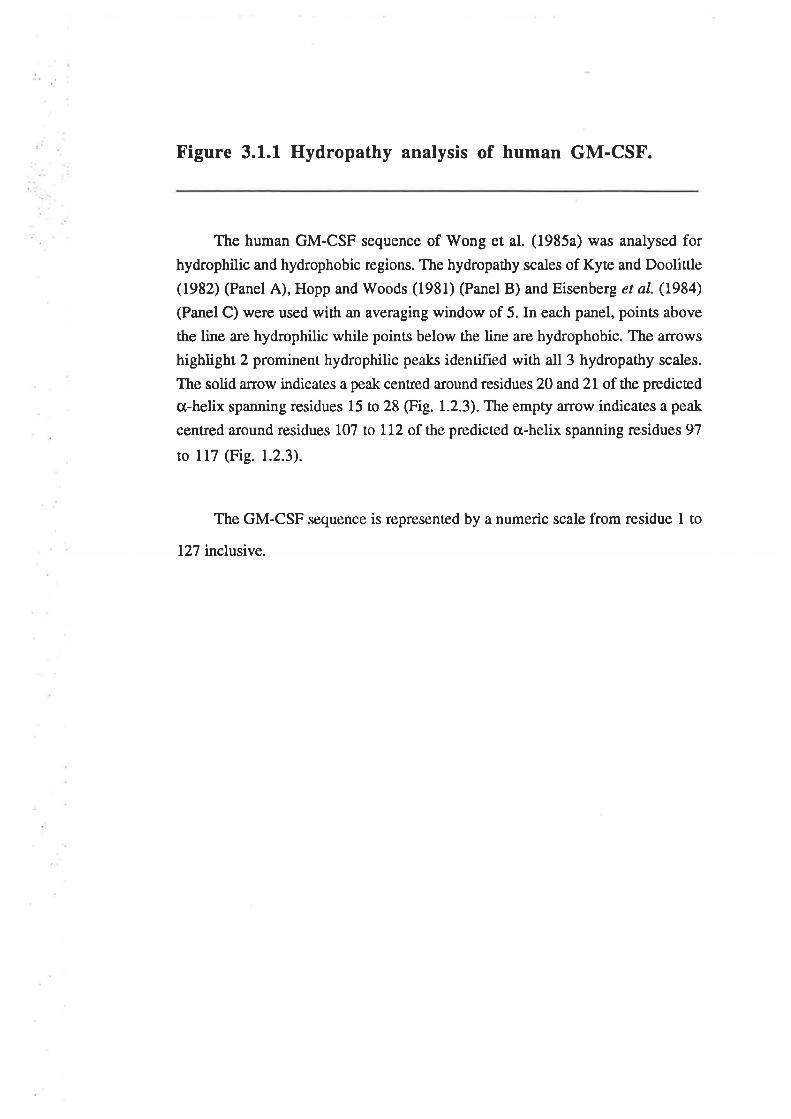
Figure 3.1.1 Hydropathy analysis of human GM-CSF.
The human GM-CSF sequence of Wong et al. (1985a) was analysed for
hydrophilic and hydrophobic regions. The hydropathy scales of Kyæ and Doolittle
(1982) (Panel A), Hopp and Woods (1981) (Panel B) and Eisenberg et aI. (1984)
(Panel C) were used with an averaging window of 5. In each panel, points above
the line are hydrophilic while points below the line are hydrophobic. The arrows
highlight 2 prominent hydrophilic peaks identified with all 3 hydropathy scales.
The solid a¡row indicates a peak centred around residues 20 and 21 of the predicæd
cr-helix spanning residues 15 to 28 (Fig. 1.2.3). The empty arrow indicates a peak
centred around residues 107 to ll2 of the predicted cr-helix spanning residues 97
to l17 (Fig. 1.2.3).
The GM-CSF sequence is represented by a numeric scale from residue 1 to
127 inclusive.
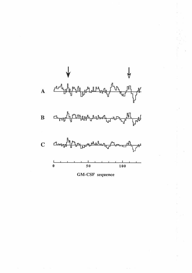
ütA
B rt_ l"Ï1fi n nJl" lt n ll"-r'l- n[h l'\ rlruu u LJUI u- " -- -l*rJ -"' \l lfl
C 4^---J\ fuLn" r,, nl[.njl,* .n * nf\'uu g u 1¡¡lu -- "k' grrJ F
-Þ
0 50 100
GM-CSF sequence
Figure 3.1.2 The amphipathic nature of the first and
fourth cr-helices of human GM-CSF.
Helical wheel plots (Schiffer and Edmundson, 1967) of residues within the
first and fourth predicted cr-helices, illustrate the amphipathic nature of these
helices. The first residue of the proposed helix is marked with an asterisk and
subsequent residues are affanged with l00o of clockwise rotation around the helical
axis between each residue. The helix is viewed from the initiating residue and is
projected into the page. Residues are identified using the single letter code while
the hydrophilic residues are also identified with their position number.
Panel A) The first predicted cr-helix from residues 15 to 28 inclusive. The
residues in the hydrophobic face, His15, Alal8,Ile19, ALa22, Leu25 and Leu26 are
completely conserved between the GM-CSF from seven different species (Fig.
r.2.2).
Panel B) The fourth predicted cr-helix from residues 97 to 117 inclusive.
The residues in the hydrophobic face, Gln99, p¡e103, p¡"106, ¡"o110, p¡"l13,
¡sr114 and Leull7 arc highly conserved between the GM-CSF from seven
different species (Fig. 1.2.2).
l) = Hydrophilic residues
O = Hydrophobic residues
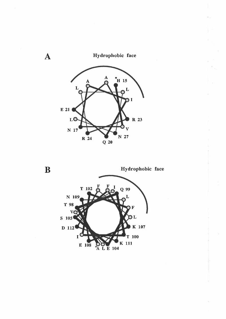
A Hydrophobic face
*H15
Q20
I
LElO4
L
I
R23
V
N27
Hydrophobic face
QeeL
F'
L
K 107
T 100
K 111
AA
L
ß, 2l
B
L
N17
N 109
T98
s 105
D ttzI
R24
T 102
E lOE
3.2 Methods
3.2.1 Protocol for the identification of functionallyimportant regions of GM-CSF
A protocol was devised for the generation of mutated GM-CSF cDNA's and
expression and analysis of the mutant GM-CSF protein (Fig. 3.2.1). The protocol
contained three fundamental procedures;
A. Mutagenesis
The oligonucleotide-directed mutagenesis procedure of Zoller and Smith (1984)
provided a simple and efficient method for generating a range of insertion, deletion or
substitution mutations throughout the entire GM-CSF cDNA (Fig. 1.2.1). Deletion
analogues are described with a A symbol followed by the position of deleted residues,
thus 414-18 represents a GM-CSF analogue with a deletion of residues 14 to 18,
inclusive. Substitution analogues are described using the single letter amino acid code
where the wild type residue and residue number are listed, followed by the mutant
residue, thus E21A represents a GM-CSF analogue where Glu2l is replaced by Al¿21.
B. Expression of GM-CSF mutant analogues
Expression of mutated GM-CSF cDNA's using a suitable plasmid expression
vector was achieved by transfection of COS cells. COS cell-derived human GM-CSF is
functionally indistinguishable from native human GM-CSF isolated from the HTLV-
transformed T-lymphocyte cell line, Mo (Wong et al., 1985a) or the U5637 bladder
cancer cell line (Metcalf et al-, 1986). Transient expression in COS cells also enabled
rapid analysis of biological activity. GM-CSF analogues in COS cell conditioned
medium were quantified by means of a radioimmunoassay (RIA) using a rabbit
polyclonal anti-GM-CSF antibody. The level of expression of the GM-CSF analogues
-51 -
Figure 3.2.1 Protocol for the generation and analysis ofhuman GM-CSF analogues.
Human GM-CSF cDNA (Fig. 1.2.1) cloned into the M13mp19 vector was
mutated by the method of Zoller and Smith (1984) and plaques screened for
specific binding to the mutagenic oligonucleotide using TMACI (Wood et al.,
1985). The presence of the correct mutation was confirmed by chain termination
sequencing (Sanger et al., t977). Double stranded (replicative form) DNA was
prepared from Ml3 clones and the GM-CSF cDNA fragment excised with EcoRI
and HindIII. The GM-CSF fragment was incubated with Klenow and deoxy
nucleotides and the resulting blunt end fragment cloned into SmaI digested pJL4
(Appendix 4.3.2.1). The orientation of the GM-CSF fragment in the resulting
pJGM series of plasmids was determined by PstI digestion, utilising the
asymmetric PstI siæs located at the 5'end of the GM-CSF cDNA (Fig. 1.2.1). The
presence of the correct mutation in the pJGM plasmids was also confirmed by
chain termination sequencing of the mutated region.
GM-CSF was exprcssed from COS cells after electroporation with the pJGM
plasmids (Chu er al.,1987). Serum-free conditioned medium was collecæd afær
incubation for 72 hours and the GM-CSF concentration determined by
radioimmunoassay. The biological activity of GM-CSF mutant analogues was
typically assessed in a GM-CSF dependent cell proliferaúon assay. The ability of
GM-CSF mutant analogues to bind the GM-CSF receptor on human neutrophils
was assessed in competitive binding experiments using 125¡-6¡4-ç5¡.
M13.GM
v
v
Human GM.CSFcDNA inM13mp19
Mutagenesisof GM-CSF
Subclone mutantGM-CSF cDNA
into pJL4expression vector
Transfect COS cellswith pJGM plasmid
Harvest conditionedmedia
auantifywith
GM.CSFRIA
\ /
I.-.-rjI
l00Vo
Assay GM-CSFmutant for
biological activity andreceptor binding
M13.GMmut¡nt
PJGMwild type
PJGMmutant
NOVo
WT Mut
was also confirmed by Western blot analysis using a sheep polyclonal anti-GM-CSF
antibody.
C. Biological activity and receptor binding of GM-CSF analogues
The biological activity of wild type GM-CSF and mutant analogues were typically
compared in a GM-CSF-dependent, cell proliferation assay. Primary leukaemic cells
from a patient with CML that incorporate l3l{thymidine in response to GM-CSF
(Mercalf, 1984) were used for proliferation assays. The ability of wild type GM-CSF
and mutant analogues to stimulate mature cell function was also compared by analysing
the GM-CSF mediated release of superoxide anions from neutrophils (Weisbart et ø1.,
1985) or the GM-CSF mediated adherence of monocytes to cultured endothelial cells or
plastic surfaces (Gamble et a1.,1989: Elliott et a1.,1990). The binding of wild type GM-
CSF and analogues to the high affinity (GMRcrpç) receptor of neutrophils (Gasson er
a1.,I986;Park et a1.,1986) was compared in competition assays using l2SI-GM-CSF.
Crude wild type GM-CSF produced by transfected COS cells and quantified by
RIA, displayed essentially identical activity comp¿¡.red with purified, CHO cell-derived
GM-CSF in CML cell proliferation and neutrophil superoxide anion rclease assays (data
not shown). Conditioned media from mock transfected COS cells failed to stimulate
CML proliferation or neutrophil superoxide anion release (data not shown).
3 .3 Results
3.3.1 Analysis of residues in the predicted first s-helixof GM-CSF
The initial mutants (Table 3.3.1) were designed to delete regions of the amino
terminus of GM-CSF, specifically the predicted first cr-helix and the residues responsible
for the prominent hydrophilic peak in this region (Fig. 3.1.1).
-52-
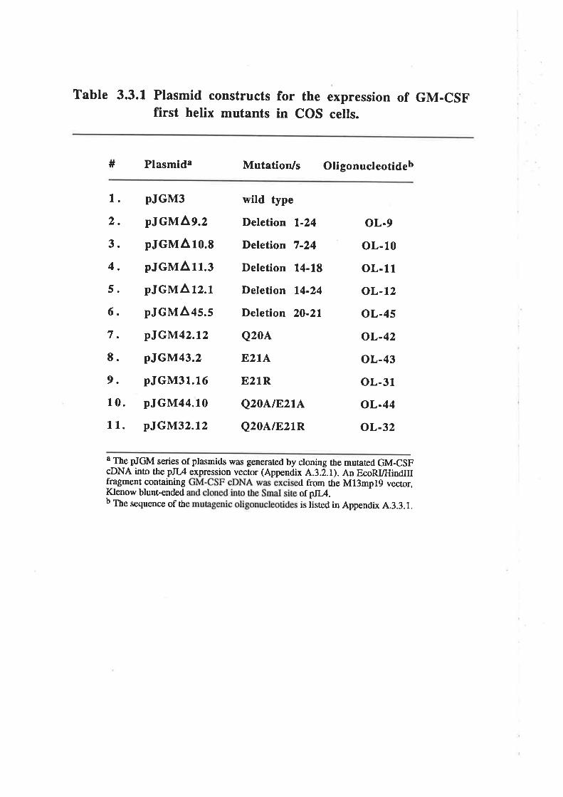
Table 3.3.1 Plasmid constructs for the expression of GM-CSFfirst helix mutants in COS cells.
# Plasmida Mutation/s Oligonucleotideb
1.
2.
3.
4.
5.
6.
7.
E.
9.
10.
11.
oL-9
oL-10
oL-11
oL-12
oL-45
oL-42
oL-43
oL-31
oL-44
oL-32
pJGM3
pJGMAg.z
pJGMA ro.s
pJGMA rr.rpJGMA rz.r
pJGMAls.s
pJGM42.tz
pJGM43.2
pJGM3r.16
pJGM44.10
pJGM32.t2
wild type
Deletion l-24
Deletion 7-24
Deletion 14-18
Deletion 14-24
Deletion 20-21
Q20A
E2l^E21R
Q20A.tE,2tL
Q20A/E21R
a The pJGM series of plasmids was generated by cloning the mutated GM-csFcDNA into the pJ[,4 expression vecror (Appendix A.3.z.l). An EcoRI/HindIII{qagment containing d from the Ml3mplg vecror,Klenow blunt-ended otpIIA.b The sequence of th is iisted in Appendix A.3.3.1.
A. Analysis of residues at the N-terminus of GM-CSF and their role
in biological activity
The mutanß Al-24 and A7-24 expressed very low levels of GM-CSF protein in
the conditioned medium of transfecæd COS cells (Fig. 3.3.14) and were not tested for
biological activity. This probably reflected degradation of unfolded or poorly secreted
GM-CSF analogues within COS cells. ttre At¿-24 mutant expressed moderate levels of
GM-CSF while rhe A14-18 and AZO-21 mutants expressed levels of GM-CSF proæin
similar to that observed for wild type GM-CSF. The wild type GM-CSF and deletion
analogues that were expressed in sufficient quantities, were titrated for their ability to
stimulate the dose-dependent proliferation of CML cells (Fig. 3.3.18). rWild type GM-
CSF was able to stimulate [3H]thymidine incorporation by CML cells with an ED56
value of O.2nglml but the deletion mutants were devoid of measurable activity except for
the À14-18 analogue which, with an EDso value of l.0ng/ml, exhibited a modest
decrease (S-fold) in activity. The deletion of residues 20 and 21 also abolished the ability
of GM-CSF to stimulate bone maffow colony formation, neutrophil superoxide anion
release and neutrophil- and eosinophil-mediaæd antibody-dependent cytotoxicity (Lopez
et al.,l992c). The observation that within the first predicted cr-helix, deletion of residues
14 to 18 produced a modest decrease in activity but that deletion of residues 20 and2l
abolished activity, suggested a more important functional role for the latter two residues.
B. Analysis of the role of residues 20 and 2l in GM-CSF activity
The loss of activity observed with the AZ}-ZL analogue may be attributable to
disruption of the amphipathic first helix and associated structural perturbations of the
analogue. Thus the role of residues 20 and 21 in GM-CSF activity was examined further
by alanine substitution mutagenesis since this may be expected to cause less structural
disruptions than deletion mutagenesis (fable 3.3.1). Alanine substitutions were used to
examine the contribution of residues 20 and 2l to GM-CSF biological activity and reduce
the hydrophilic nature of the first predicted cr-helix. The activity of the Q20A analogue
(EDso 0.4nglml) was comparable with wild type GM-CSF (ED5s 0.2nglml) in
stimulating the proliferation of CML cells while the E21A analogue (EDso l.3ng/ml)
exhibited a 6-fold reduction in activity (Fig. 3.3.2A). In neutrophil superoxide anion
-53-
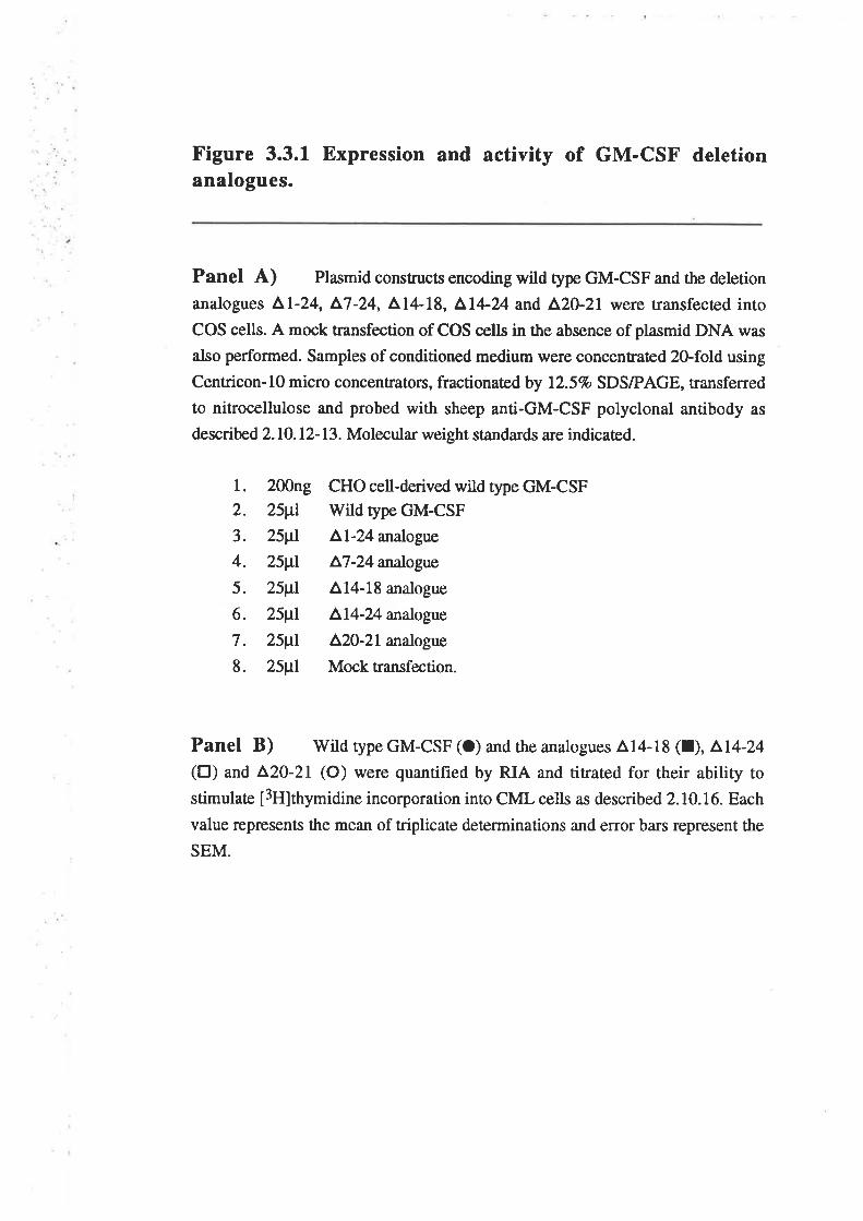
Figure 3.3.1 Expression and activity of GM-CSF deletionanalogues.
Panel A) Plasmid constructs encoding wild type GM-CSF and the deletion
analogues ^l-24, ^7-24,
Al4-18, ^l+24
and A2O-21 were transfecæd into
COS cells. A mock transfection of COS cells in the absence of plasmid DNA was
also performed. Samples of conditioned medium were concentated 2O-fold using
Centricon-10 micro concentrators, fractionated by 12.5% SDS/PAGE, transferred
to nitrocellulose and probed with sheep anti-GM-CSF polyclonal antibody as
described 2.10.12-13. Molecular weight standards a¡e indicaæd.
l. 200ng CHO cell-derived wild type GM-CSF2. 25¡tl Wild type GM-CSF
3. 25¡tl Ll-24 analogue
4. 25¡tl ^7-24
analogue
5. 25¡tI 414-18 analogue
6. 25¡tl ^14-24
analogue
7. 25¡tl ^20-2L
analogue
8. 25¡tl Mock transfection.
Panel B) Wild type GM-CSF (O) and the analogues 414-18 (l), ^L4-24(tr) and
^20-21 (O) were quantified by RIA and titrated for their ability to
stimulate [3H]thymidine incorporation into CML cells as described 2.10.16. Each
value represents the mean of triplicate determinations and error bars represent the
SEM.
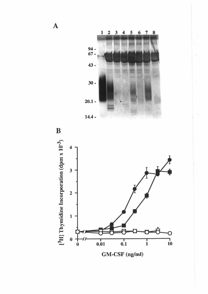
At2 3 4 5 67 t
0.01 0.1
9467
*
43
30-
20.1 -
14.4 -
4
B
(?¡I
t-lx-FIÈEtto.-+¡GILot¡Lro9ãIFiq)ãI.-E.-ãFI>ì--Er
l*l¡r(')
3
2
1
00 10I
GM-CSF (nglml)
Figure 3.3.2 Activity of GM-CSF analogues withsubstitutions at residues 20 and 21.
Wild type GM-CSF and the analogues 4,20-21, Q204, E2lA, E21R,
Q20NE2LA and Q20AÆ21R were expressed in COS cells, quantified by RIA and
titraæd for their ability to stimulate [3lilthymidine incorporation into CML cells.
Each value represents the mean of triplicate determinations and error ba¡s represent
the SEM.
Panel A). Wild type GM-CSF (a) and the analogues AZ}-ZL (O), Q20A(A) and E21A (A).
Panel B). Wild type GM-CSF (O) and the analogues E2IA (A), E21R (O),
Q20NEztA(o) and Q20A/E21R (tr).
t3ffl
fny
mid
ine
Inco
rpor
atio
n (d
pm x
10'
s) E
Ët3
ffl f
nym
idin
e In
corp
orat
ion
(dpm
x 1
0'3)
>
U¡
)it¡l¿
NC
tr
O
U
tì.)
5o\
ælj
lrO
ì.)o
lr li
o ¡à a o (t) frl
FI
oe t- )
OP
7tr
)l ¡ o U) lrj - R.
=l
)j
release and neutrophil- and eosinophil-mediaæd antibody-dependent cytotoxicity, the
Q20A analogue was indistinguishable from wild type GM-CSF while the E21A analogue
exhibiæd a 4-fold reduction in activity (Lopez et aI.,l992c). The reduction in biological
activity of the E2lA analogue compared with wild type GM-CSF suggested ttrat Glu2l
was important for GM-CSF activity.
The significance of the highly conserved acidic residue at position 21 (Fig. 1.2.2)
was examined more closely by generating the E2lR analogue (Table 3.3.1) which,
unlike the E2lA analogue, retained the hydrophilic nature of this region but reversed the
charge (data not shown). Dose-dependent proliferation of CML cells was observed with
wild type GM-CSF (EDso 0.17nglml) and the E2IA analogue (EDso 5.7ng/ml) but rhe
E21R analogue was unable to stimulate proliferation of CML cells up to a concentration
of lOng/ml (Fig. 3.3.28). This observation confirmed the critical role of Glu2l in GM-
CSF activity and demonstrated that the acidic nature of this highly conserved residue is
important.
The Q20A mutation, which appeared to have no affect on GM-CSF activity (Fig.
3.3.2A) was coupled with the mildly deleterious E2IA mutarion and the highly
deleterious E2lR mutation (Table 3.3.1). Interestingly rhe double substitution
Q20NE2IA analogue displayed a marked decrease in biological activity compared ro the
single substitution E2lA (Fig. 3.3.28) and failed to stimulate a proliferative response
equivalent to the EDso response of wild type GM-CSF up to a concentration of lOng/ml.
However, comparison of the EDzs values suggested a 270-foß decrease in activity for
the Q20A/E2lA analogue compared with wild type GM-CSF. In the same assay, rhe
EzlA analogue exhibited a 3O-fold decrease in activity compared with wild type GM-
CSF (Fig. 3.3.28). The difference in activity of the EzlA and Q20A/E2lA analogues
suggested that the Q20A mutation has an effect on GM-CSF activity but only when
Glu2l was also mutated. The Q20A/82lR analogue was unable to stimulate proliferation
of CML cells up to a concentration of l0ng/ml (Fig. 3.3.28).
-54-
C. Expression of GM-CSF residue 20 and 2l analogues frompermanently transfected CHO cell lines
The GM-CSF residue 20, 2l analogues were expressed and purified from
permanently transfecæd CHO cell lines to enable a more detailed analysis of the effects of
the mutations on the multiple GM-CSF activities and receptor binding. Mutated GM-CSF
cDNA's were subcloned from the pJGM series of plasmids into the pRSVN.07
expression vector (Appendix A.3.3.2) and the resulting constructs transfected into CHO
cells. Following selection with geneticin and cloning, cell lines were obtained that
expressed each of the GM-CSF analogues (fable 3.3.2).
In collaboration with Dr J.Cebon and Louis Fabri (Melbourne Tumour Biology
Branch, Ludwig Institute for Cancer Research, Melbourne, Vic), the GM-CSF
analogues expressed from CHO cell lines were immunoaffinity purified using the anti-
GM-CSF monoclonal antibody, LMM 111 (Cebon et al., 1988). Affinity purified GM-
CSF analogues were subjected to reversed phase HPLC which separated three distinct
species of GM-CSF (Fig. 3.3.3). This pattern was similar to that observed with the
reversed phase fractionation of GM-CSF purif,red from activated human T-lymphocytes
(Cebon et a1.,1990). The species fractionated by reversed phase HPLC represent three
size classes of glycosylated GM-CSF with high Mr species eluting first, followed by
intermediate and low Mr species (Cebon et al., 1990). Donahue et al (I986a) have
reported that the three size classes of CHO cell-derived GM-CSF are attributable to
different degrees of occupancy of the two sites for asparagine-linked (N-linked)
carbohydraæ at Asn27 and Asn37. High Mr species contain GM-CSF in which both N-
linked sites are occupied, intermediate Mr species contain GM-CSF in which either site is
occupied while the low Mr GM-CSF is devoid of N-linked carbohydrate (Donahue et aI.,
1986a).
Fractions containing the first two glycosylation species of GM-CSF analogue to
elute from the reversed phase column were combined and in collaboration with Dr
R.Simpson (Joint Protein Structure Laboratory, Ludwig Institute for Cancer
-55-
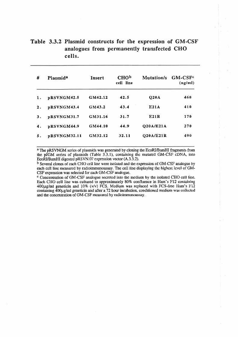
Table 3.3.2 Plasmid constructs for the expression of GM-CSFanalogues from permanently transfected CHOcells.
# Plasmida Insert cHobcell line
Mutation/s GM-CSFc(ng/ml)
Ia
3
4
5
pRSVNGM42.5
pRSVNGM43.4
pRSVNGM3l.7
pRSVNGM44.e
pRSVNGM32.ll
GM42.t2 42.5
cM43.2 43.4
GM31.16 3r.7
GM44.10 44.9
GM32.t2 32.11
Q20A
E2IA
E21R
Q2OAtF,2tL
Q20A/821R
460
410
t70
270
490
a The pRSVNGM series of plasmids was generated by cloning the EcoRI/Ba¡nHI fragments fromthe pJGM series of plasmids (Table 3.3.1), containing the mutated GM-CSF cDNA, intoEcoRVB amHI digesæd pRS VN. 07 expression vector (A'. 3. 3. 2).b Several clones of each CHO cell line were isolated and the expression of GM-CSF anatogue byeach cell line measured by radioimmunoassay. The cpll line displaying the highest level of GM-CSF expression was selected for e¿ch GM-CSF analogue.c Concentration of GM-CSF analogue secreted into the medium by the isolated CHO cell line.Each CHO cell line was cultured to approximately 807o confluence in Ham's F12 conøining400pg/ml geneticin and lOTo (v/v) FCS. Medium was replaced with FCS-free Ham's F12containing 400pg/ml geneticin and after a72bot¡r incubation, conditioned medium was collectedand the concenEation of GM-CSF measured by radioimmunoassay.
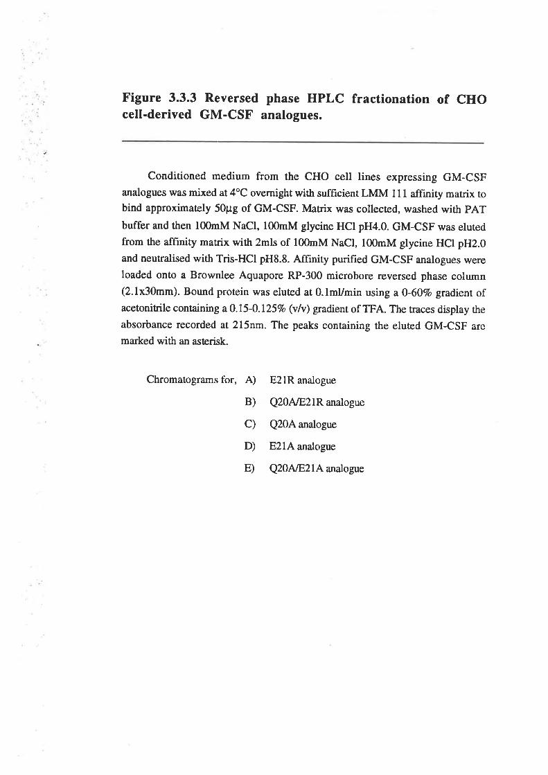
Figure 3.3.3 Reversed phase HPLC fractionation of CHOcell-derived GM-CSF analogues.
Conditioned medium from the CHO cell lines expressing GM-CSFanalogues was mixed at 4oC overnight with sufficient LMM l1 I affinity matrix tobind approximaæly 50pg of GM-csF. Matrix was collecred, washed with pAT
buffer and then l(nmM NaCl, l00mM glycine HCI pH4.0. GM-csF was eluted
from the aff,rnity matrix with 2mls of l00mM NaCl, l00mM glycine HCI pH2.0
and neutralised with Tris-HCl pH8.8. Affînity purified GM-CSF analogues were
loaded onto a Brownlee Aquapore RP-300 microbore reversed phase column(2.1x30mm). Bound protein was eluted at 0.lmUmin using a 0-60Vo gradienr ofacetonitrile containing a 0.15-0.125Vo (vlv) gradient of TFA. The traces display the
absorbance recorded at 2l5nm. The peaks containing the eluæd GM-CSF are
marked with an asterisk.
Chromatograms t'or, A)
B)
c)
D)
E)
E2lR analogue
Q20AÆ2IR analogue
Q20A analogue
E2lA analogue
Q20NE2LA analogue
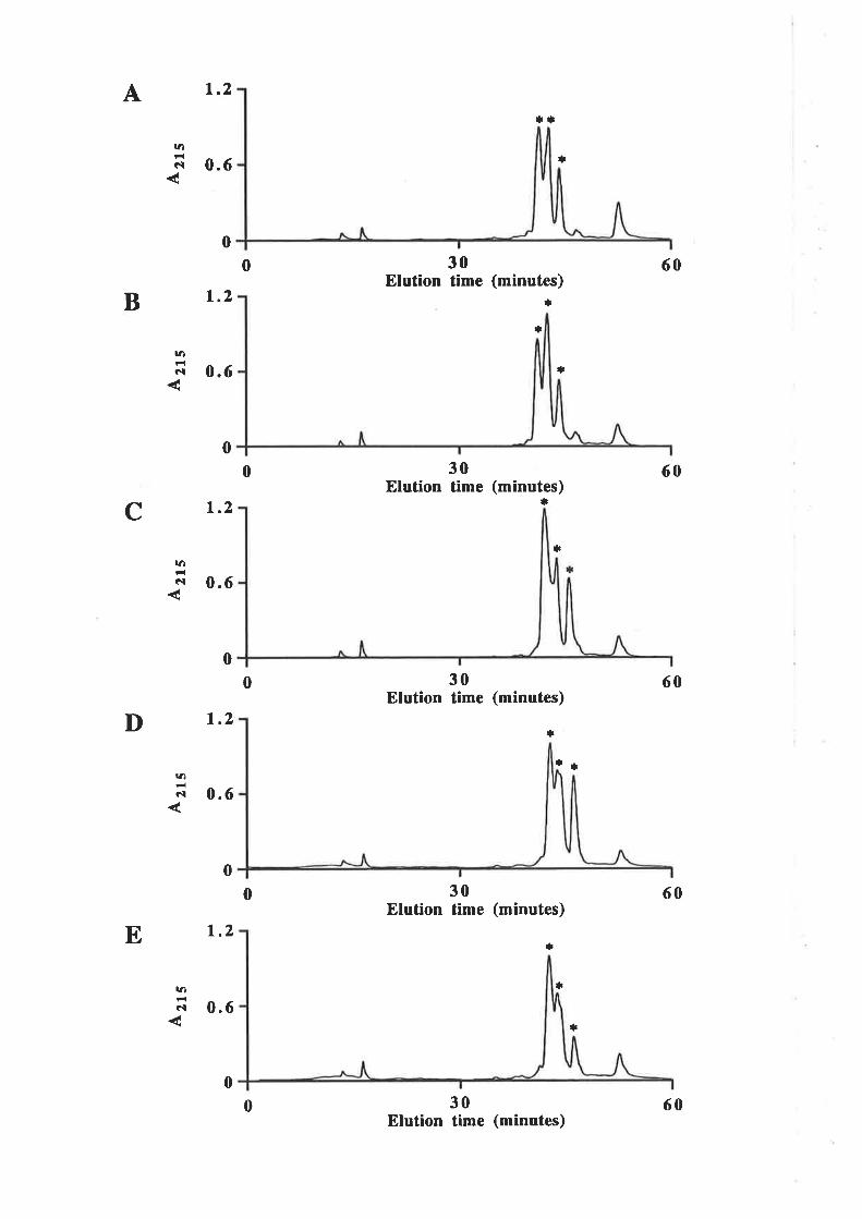
A t.2
0.6
B 1.2
0.6
C 1.2
0.6
1.2
E 1.2
*t
+
30Elution time (minutes)
r¡
*
r¡
30Elution time (minutes)
*
*
30Elution time (minutes)
*
*
30Elution time (minutes)
tl
*
30Elution time (minutes)
la
GI
0
ta
GI
|a
cl
ra
c-l
¡a
Cì
0
0
0
0
0
60
60
60
60
0
D
:t
0.6
0.6
0
0
0
*
60
Researct/Walter & Eliza Hall Instituæ for Medical Research, Melbourne, Vic), quantifred
by amino acid analysis. The use of amino acid analysis avoided the potential problems
associated with immunological quantification where mutant analogues do not necessarily
bind with wild type affinity to the antibody leading to an inaccurate estimate of mutant
analogue concentration. However, there was no significant difference in the
concentation of ttre GM-CSF analogues determined by amino acid analysis or by using
the standa¡d RIA procedure (data not shown). Comparison of the purified GM-CSF
analogues by silver stained SDS/PAGE (Fig. 3.3.aA) and rvestern blot analysis (Fig.
3.3.48) showed very similar molecular weight heterogeneity for the analogues compared
with the CHO cell-derived wild type GM-CSF obtained from Genetics Instituæ. The
level of glycosylation was deliberaæly balanced as much as possible because of the
known influence of carbohydrate on the specific activity of GM-CSF (Moonen et al.,
1987; Kaushansky et a1.,1987; Cebon et a1.,1990). Table 3.3.3 summarises the yield
for each of the purified GM-CSF analogues.
D. Activity of CHO cell-derived GM-CSF residue 20 and 2lanalogues
The purified, CHO cell-derived GM-CSF analogues were compared for their
ability to stimulate proliferation of CML cells (Fig. 3.3.5). Wild type GM-CSF and the
purified analogues stimulated dose-dependent proliferation of CML cells with EDSo
values ranging from 0.15 ng/ml for wild type up to 40 ng/ml for the Q20A/E21R
analogue (Table 3.3.4). The hierarchy of biological activity for the purified analogues
was similar to that observed with crude, COS cell-derived analogues (Fig. 3.3.2). The
purified E2lR analogues did exhibit biological activity but only at significantly higher
concentrations than previously tested with COS cell-derived material (Fig. 3.3.2) and are
20G to 270-fold less active than wild type GM-CSF.
The purified GM-CSF analogues were also compared for their ability to stimulate
mature cell function (Fig. 3.3.6). Vfild type GM-CSF and the purified analogues
stimulated the dose-dependent production of superoxide anions by neutrophils (Fig.
3.3.64) with ED5g values ranging from 0.33 ng/ml for the Q20A analogue up to 330
-56-
Figure 3.3.4 Purified GM-CSF residue 20121 analogues.
GM-CSF analogues were expressed from permanently transfecæd CHO cell
lines, immunoaffinity purified and fractionated by reversed phase HPLC (Fig.
3.3.3). Fractions containing high molecular weight GM-CSF, similar in mobility
when compared with CHO cell-derived wild type GM-CSF, were pooled and
quantified by amino acid analysis. Samples of wild type GM-CSF and analogues
were fractionated by 12.5% SDS/PAGE and stained with silver (Panel A) or
transferred to nitrocellulose and probed with sheep anti-GM-CSF polyclonal
antibody (Panel B). Molecular weight standards are indicated.
1. 200ng
2. 200ng
3. 200ng
4. 200ng
5. 2ü)ng
6. 200ng
7. 200ng
Wild type GM-CSF
E21R analogue
Q20AÆ21R analcigue
Q20A analogue
E2lA analogue
Q2ONE2IA analogue
Wild type GM-CSF
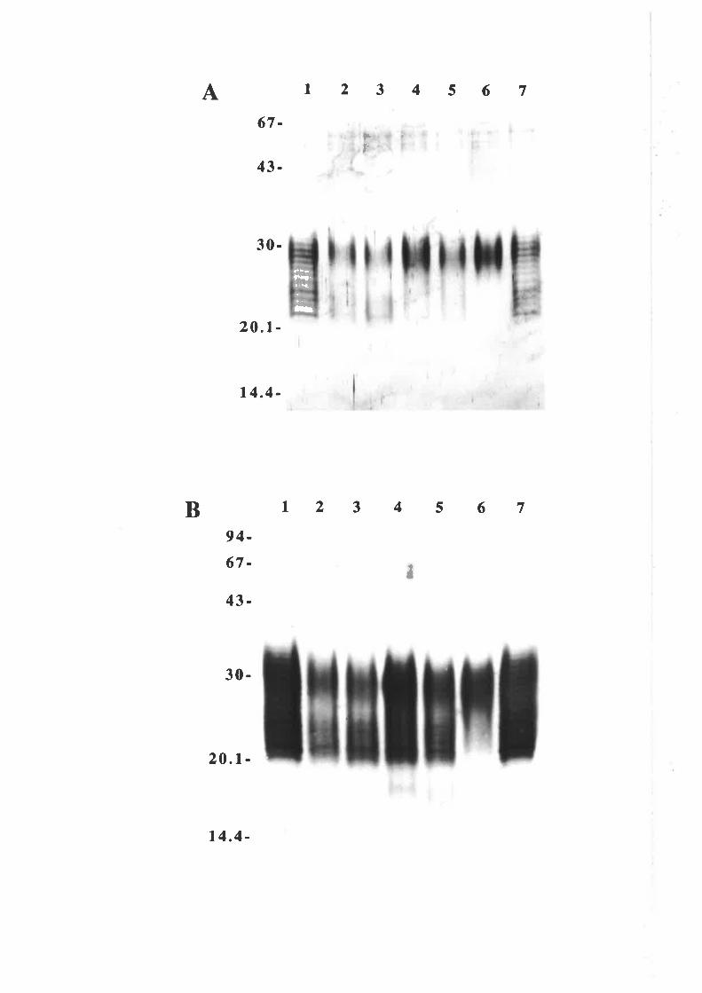
A t23456767-
43-
30-
20.t -
14.4 -
Þr
a
B 1234567
¡
94-67-
43-
30-
20 .1-
lÉçl'
t
14.4-
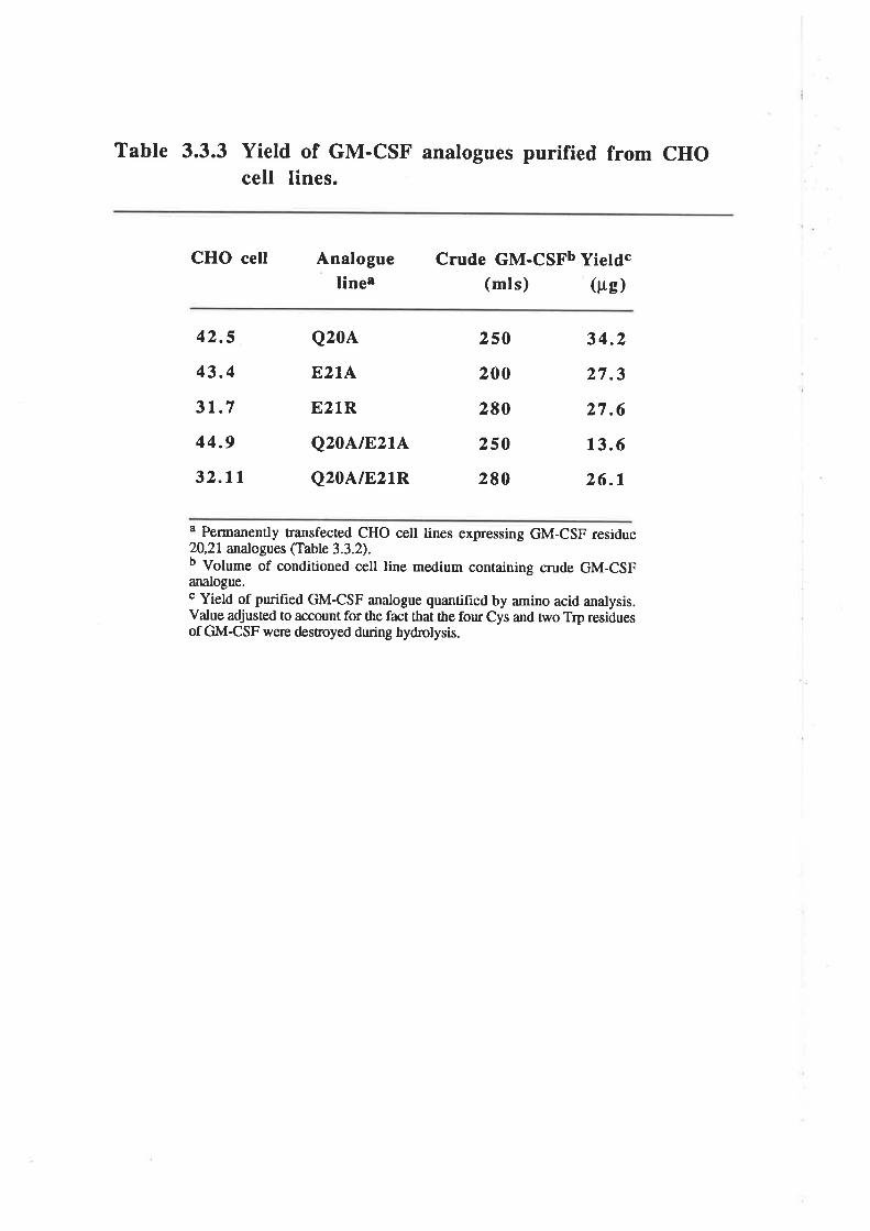
Table 3.3.3 Yield of GM-csF analogues purified from cHocell Iines.
CHO cell Analogueliner
Crude GM-CSFb Yietdc(mls) (trg)
42.5
43.4
31.7
44.9
32.11
Q20A
EzIA
E21R
Q20AtE.ztA
Q20A/E21R
250
200
2E0
250
280
34.2
27.3
27.6
13.6
26.1
a Pennanently transfected CHO cell lines expressing GM-CSF residue20,21 analogues (Table 3.3.2).b Volume of conditioned cell line medium containing crude GM-CSFanalogue.c Yield of purified GM-CSF analogue quantified by amino acid analysis.Value adjusted to account for the fact that the fou¡ Cys and two Trp residuesof GM-CSF were desroyed during bydrolysis.
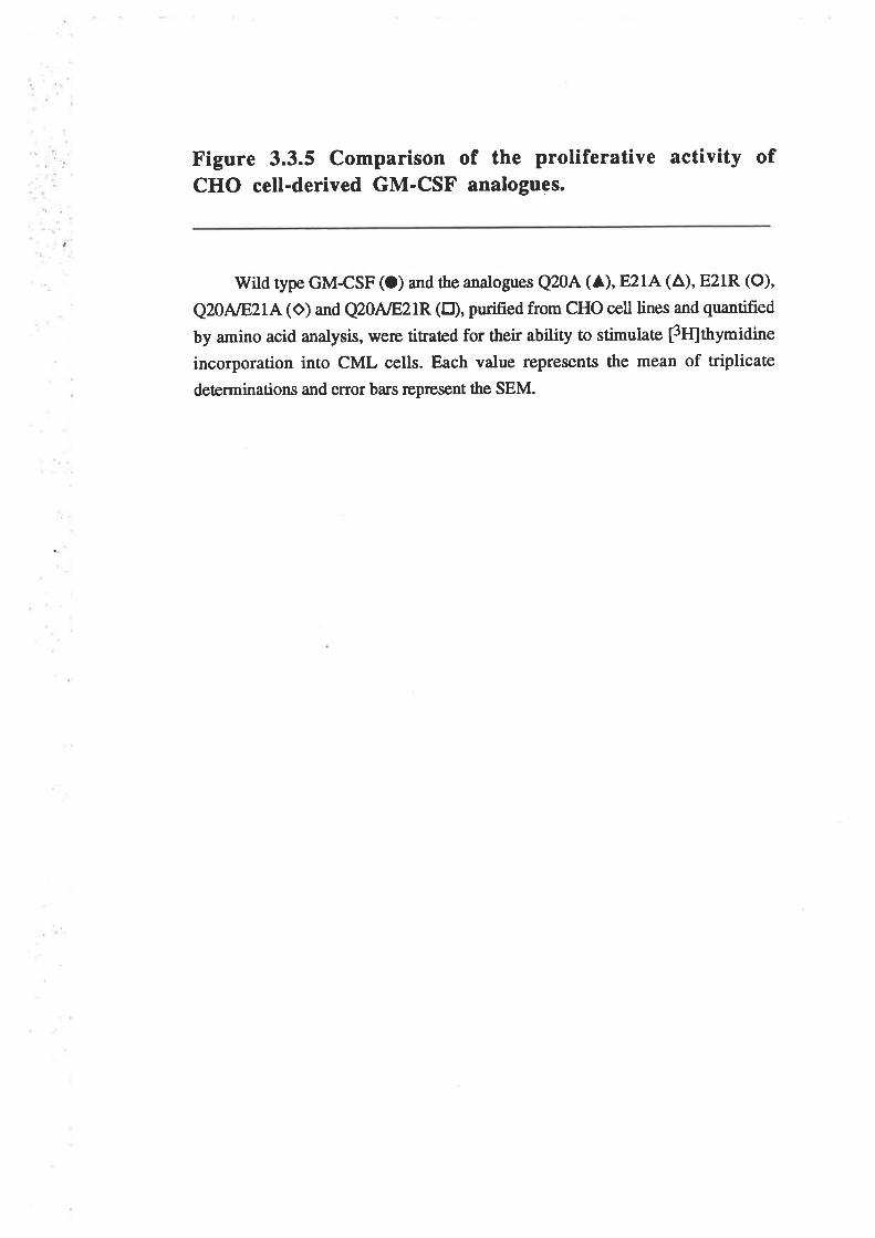
Figure 3.3.5 Comparison of the proliferative activity ofCHO cell-derived GM-CSF analogues.
Wild type GM-CSF (O) and the analogues Q20A (A), E21A (A), E21R (O)'
Q2DNE2LA(O) and Q20AÆ2IR (tr), purified from CHO cell lines and quantified
by amino acid analysis, were titraæd for their ability to stimulate ¡3tt¡thymidine
incorporation into CML cells. Each value represents the mean of triplicate
deærminations and error ba¡s represent the SEM.
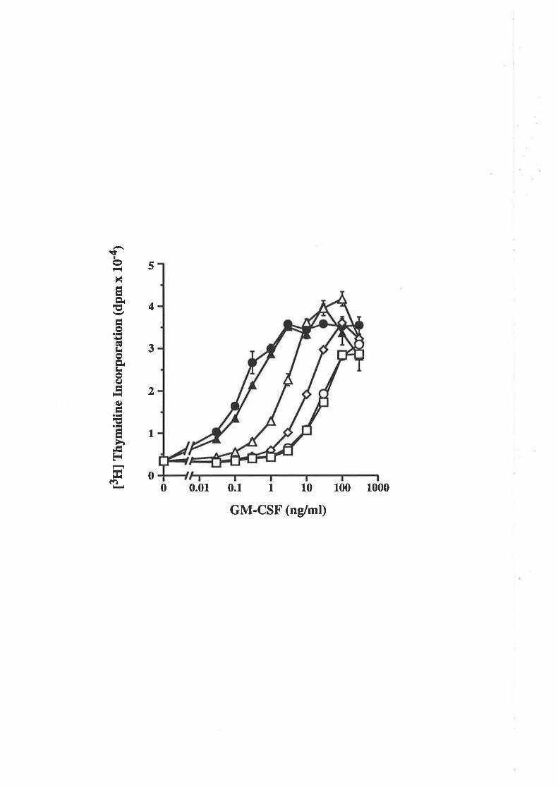
t3ffl
fny
mid
ine
Inco
rpor
atio
n (d
pm x
10'
a)
uì.)
(,5
Ur
lr lr ¡¿ l.¿
o ? tl I o u) r=j
E J q.
Figure 3.3.6 Comparison of the CHO cell-derived GM-CSF analogues ability to stimulate mature cell function.
Vfild type GM-CSF (O) and the analogues Q20A (A), E21A (A), E21R (O),
Q20NE2IA (O) and Q20AÆ21R (E), were titraæd for their ability to stimulate the
production of superoxide anions from human neutrophils (A) or to stimulaæ the
adherence of human monocytes as described 2.10.16 (B). Each value represents
the mean of riplicaæ determinations and error bars represent the SEM.
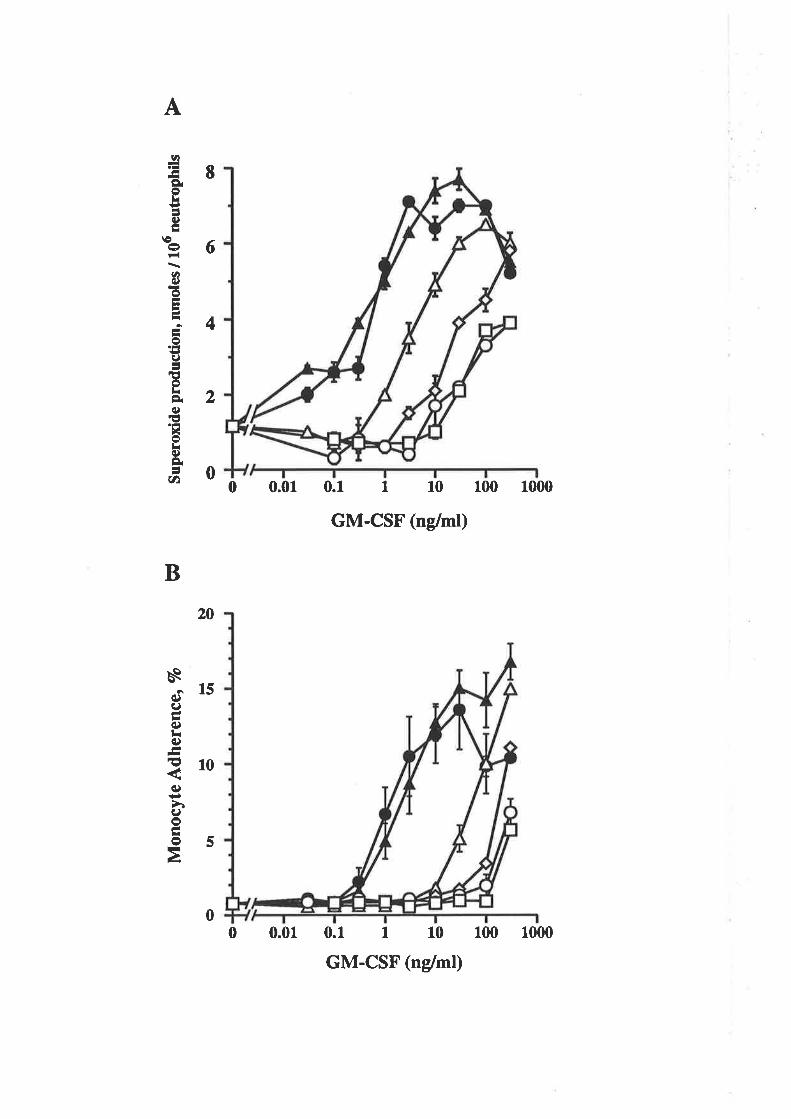
Mon
ocyt
e A
dher
ence
, V
o
Þ.
llut
out
æì.¡
tÉS
uper
oxid
e pr
oduc
tion,
nm
oles
/ 1
06 n
eutr
ophi
ls >
ì.¡5
o\
o )i Þ¡
ll li l¿ o Þ¡
6) ? rì I o rt) frj
E ) oc¡ -I
l¿ )¡ )i )r )¡ o Þr o
o z ¡ì I ô u) |at
I oe E
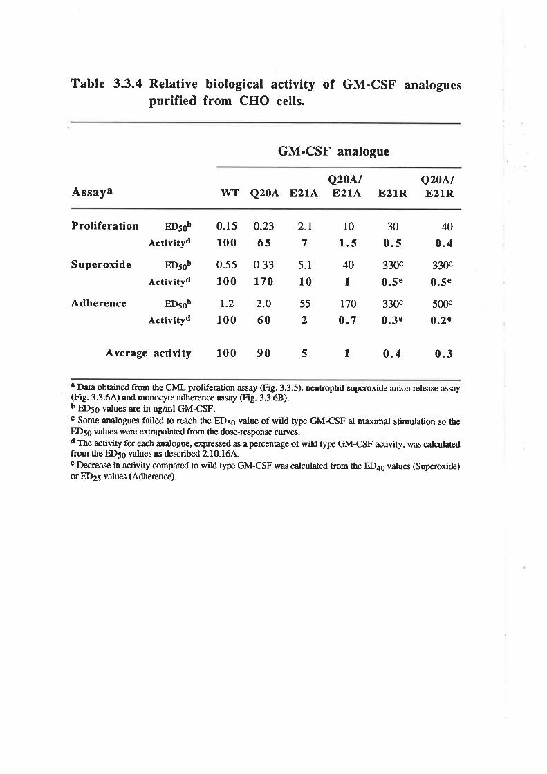
Table 3.3.4 Relative biological activity of GM-CSF analoguespurified from CHO cells.
GM-CSF analogue
AssayaQ20A/
wT Q20A E,2lA E'2tAQ20A/
E21R E21R
Proliferation
Superoxide
Adherence
EDsob
Activityd
EDsob
Activityd
EDsob
Activityd
0.15
100
0.55
100
1.2
100
0.23
65
0.33
170
2.0
60
2.1
7
5.1
10
55
2
10
1.5
40
I170
o.7
30
330c
0.5e
330c
0.3e
40
0.4
330c
0.5e
5mc
o.2e
0.5
Average activity 100 90 5 t 0.4 0.3
I Data obtained from the CML proliferation assay (Fig. 3.3.5), neutrophil superoxide anion release assay(Fig. 3.3.64) and monocyte adherence assay (Fig. 3.3.68).b Ð56 values are in ng/ml GM-CSF.c Some analogues failed to reach the ED5g value of wild type GM-CSF at maximal stimulation so theED56 values werc extrapolated from the dose-response curves.d The activity for each analogue, expressed as apercentage of wild type GM-CSF activity, was calculatedfrom the ED56 values as described 2.10.164.e Decrease in activity compared to wild type GM-CSF was calculated from the ED4q values (Superoxide)or ED25 values (Adherence).
ng/ml for the E21R and Q20A/E2IR analogues (Table 3.3.4). Wild type GM-CSF and
the purif,red analogues also stimulated the dose-dependent adherence of monocytes to
plastic (Fig. 3.3.68) with EDso values ranging from 1.2 ng/ml for wild type up to 500
ng/ml for the Q20NE2LR analogue (Table 3.3.4). The E21R and Q20A/E21R analogues
\Ã,ere not able to maximally stimulate neutrophil or monocyte activation at the
concentrations tested.
The hierarchy of biological activity for the GM-CSF analogues w¿rs very similar in
both proliferation and functional activation assays. The average decrease in GM-CSF
activity for each of the single amino acid substitutions was, Q204, l.3-fold, 821A,24-
fold and E21R, 250-fold (Table 3.3.4).
E. Receptor binding characteristics of CHO cell-derived GM-CSF'
residue 20 and 21 analogues
The purif,red, CHO cell-derived GM-CSF analogues were compared for their
ability to compete ¡ot 1251-6¡4-CSF binding to the high afhnity recepror (GMRupç) of
human neutrophils (Fig. 3.3.7). Compared with wild type GM-CSF, the Q20A analogue
was 3-fold less effective and the E21A analogue 40-fold less effective at competing for
125I-GM-CSF binding. The Q20A/8214, E21R and Q20A/E21R analogues were all at
least 100-fold less effective at competing for the binding o¡ 1251-6¡4-CSF than wild type
GM-CSF. Thus, the binding of the GM-CSF residue 2012I analogues to the GM-CSF
high affinity receptor (GMRapç) paralleled the biological activity of these analogues on
neutrophils, monocytes and leukaemic cells. However, these results did not indicaæ ifbinding to the low affinity receptor (GMRa) was also affecæd.
The question of low affinity receptor binding was addressed with the E21R
analogue which exhibited the lowest level of biological activity and weakest interaction
with the high affrnity receptor. The purified E2lR analogue was ÍNsessed for the ability
to compete ¡or 125¡-6M-CSF binding to the low and high affinity receptor of human
monocytes (Elliott et a1.,1989). A binding curve ¡o. 125¡-6¡4-CSF was performed in the
presence of a 35-fold excess of the E21R analogue (Fig. 3.3.8A). The E21R analogue
-57 -
Figure 3.3.7 Comparison of the binding of the CHO cell-derived GM-CSF analogues to the GM-CSF receptor onhuman neutrophils.
The binding of wild type GM-CSF (a) and the analogues Q20A (A), E21A
(A), E21R (O), Q20A/E214 (O) and Q20AÆ2IR (tr) to cells expressing the high
(GMRap.) affrnity GM-CSF receptors $,¡rs comp¿¡red. Binding experiments were
performed as described2.l0.l1. Unlabelled wild type GM-CSF or analogues rvere
titrated against 70pM 1251-6¡4-gSF and 4 x 10⁶ neutrophils per tube. The values
are expressed as a percentage of 6" 125J-6M-CSF bound in the absence ofcompetitor and a¡e the means of duplicate deærminations.
125¡
-ç¡4
-CS
F b
ound
(7o
orc
ontr
ol)
o.50
\æä
ocêê
oo
c l¡
6) ZP
tl ¡r
I ô U)
faj ¡ ¿ z rl
lr
Figure 3.3.8 Binding of the CHO cell-derived E2lRanalogue to the low affinity and high affinity GM-CSFreceptors of human monocytes.
Binding curve o¡ 125¡-6¡4-CSF to purified monocytes in the presence orabsence of the E2lR analogue (Panel A). lvild type 125¡-6¡{-CSF in rhe absence
(O) or presence (O) of a 35-fold excess of the E2lR analogue was mixed with 2 x10ó monocytes per tube. The values are expressed as specific counts bound and a¡e
the means of duplicate deærminations. Specific counts were determined bysubtracting the counts obtained in the presence of a 100-fold excess of wild type
GM-CSF at each concentration o¡ 1251-6M-CSF.
Scatchard analysis of binding data from Panel A, showing the competition
¡or 125¡-6M-CSF binding in the presence (O) or absence (O) of the E21R
analogue (Panel B).
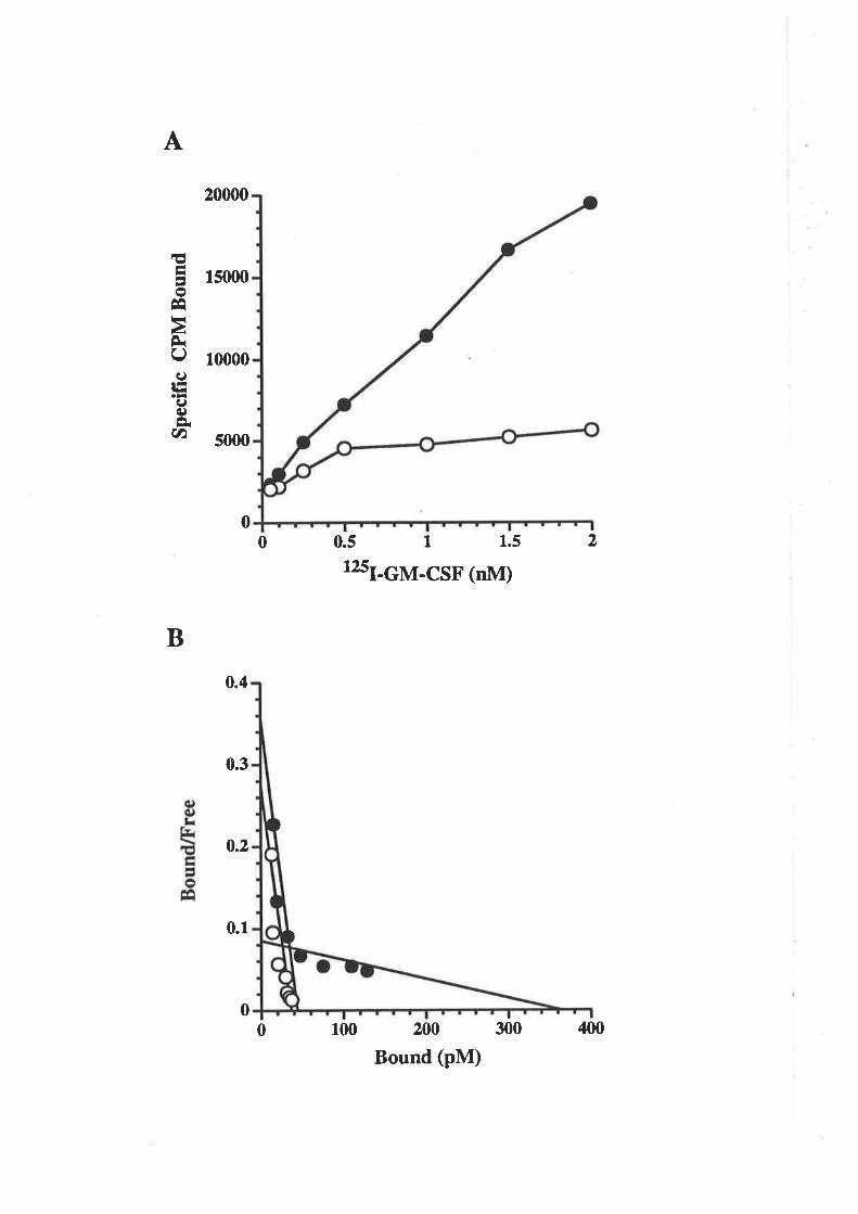
A
0
20000
15m0
10000
5000
EI)o
ÊqttàÈQItãI(¡)È(t)
0.4
0.3
0.2
0.1
0.5 I12sI-cM-csF (nM)
200
Bound (pM)
1.5 20
B
00 400300100
slightly inhibited the binding of low concentrarions of 1251-6¡4-çSF but strongly
inhibited the binding of high concentrations of l25I-GM-CSF. Scatchard transformarion
of the data showed that the E21R analogue was able to effectively compete for the low
affinity binding o¡ 125¡-6¡4-CSF but only slightly reduced the high affrnity binding (Fig.
3.3.88). This result indicated that the E2lR analogue was able to preferentially interact
with the low affrnity but not the high afhnity receptor.
To further characærise the interaction of the E2lR analogue with the low affinity
receptor, competitive binding experiments were performed using COS cells expressing
the cloned low affinity receptor cr-chain (Gearing et a1.,1989). In collaboration with Dr.
N.Nicola and Meredith Layton (The V/alær and Eliza Hall Institute, Parkville, Vic) the
ability of wild type GM-CSF and the E2lR analogue to compete for the binding o¡ 1251-
GM-CSF to COS cells expressing the cloned human GM-CSF receptor cr-chain was
compared (Fig. 3.3.94). The results indicaæd that the ability of wild type GM-CSF and
the E2lR analogue to the compete ¡or 125J-6¡4-CSF binding to the low affrnity recepror
was indistinguishable. In parallel experiments the E2lR analogue was 300-fold less
potent than wild type GM-CSF at competing ¡or 125¡-6M-CSF binding ro rhe high
affinity receptor of neutrophils (Fig. 3.3.98). These results show that the reduced
biological activity of the residue 2l analogues was associaæd with reduced binding to the
high affrnity receptor (GMRop.) but not the low afhnity receptor (GMRa).
3.3.2 Analysis of residues in the predicted fourtha-helix of GM-CSF
Using the experience gained with mutagenesis of residues in the first helix,
charged residues on the hydrophilic surface of the predicæd fourth cr-helix were targeted
for mutagenesis. The residues Lys107, 61o108, Lysllt and Aspl12, which exhibit a high
degree of conservation amongst the seven known GM-CSF sequences (Fig. 1.2.2) and
may therefore be important for GM-CSF function, were subjected to charge reversal
mutagenesis (Table 3.3.5). The mutation encoding the El04A analogue arose as the
result of an error during PCR amplification of the GM-CSF cDNA and was retained to
-58-
Figure 3.3.9 Comparison of the binding of the wild typeGM-CSF and the E21R analogue to COS cells transfectedwith the human GMRa chain and neutrophils.
The binding of wild type GM-CSF (a) and the E2lR (O) analogue to cells
expressing the low (GMRcr, Panel A) or high (GMRøpç, Panel B) affinity GM-
CSF receptors w¿ts compared. Wild type GM-CSF or E2lR analogue were titraæd
against 6nM of E. colïdeing¿ 125¡-6M-CSF and 1 x 10ó pGMR138 (Gearing er
at.,1989) transfected COS cells per tube (A) or 2nM of E. coti-deiusd 125¡-6lvl-
CSF and 1.5 x 10⁶ neutrophils per tube (B). The values are expressed as a
percentage of the l25I-GM-CSF bound in the absence of competitor and are the
means of duplicate determinations.
125I
-GM
-CS
F bo
und
(vo
orco
ntro
l) E
É12
5I-G
M-C
SF
boun
d (V
o or
cont
rol)
Þl¡
l.)È
O\æ
OO
OO
OO
b.)5
O\æ
OO
OE
l¡ o
o tr Fr lr )l )i l¡
o z Ê I o u) lrt E z rl
)¡
o Zrr
tl I o U)
frj
Fre ? È
lr
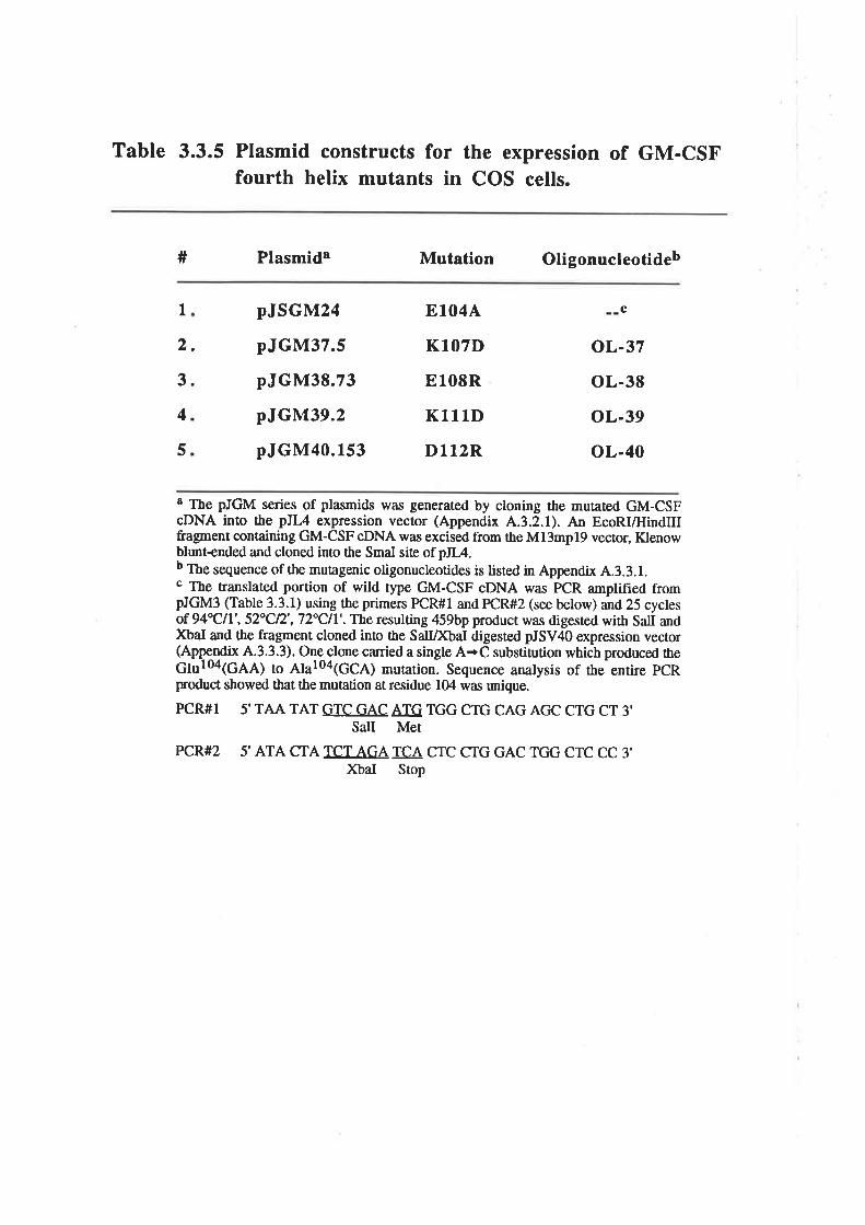
Table 3.3.5 Plasmid constructs for the expression of GM-GSFfourth helix mutants in COS cells.
# Plasmida Mutation Oligonucleotideb
I2.
3.
4
5
pJSGM24
pJGM37.5
pJGM38.73
pJGM39.2
pJGM40.153
ElO4A
KlO7D
El08R
Kl1lD
D112R
c
oL-37
oL-38
oL-39
oL-40
a The pJGM series of plasmids was generated by cloning the mutated GM-CSFcDNA into the pJL4 expression vector (Appendix
^.3.2.1). An EcoRI/HindIII
fragment containing GM-CSF cDNA was excised from the M13mp19 vector, Klenowblunt+nded and cloned into the SmaI siæ of pJI/.b The sequence of the mutagenic oligonucleotides is listed in Appendix 4.3.3.1.c The translated portion of wild type GM-CSF cDNA was PCR amplified frompJGM3 (Table 3.3.1) using the primen PCR#I and PCR#2 (see below) and 25 cyclesof 94"Cll', 52"Cn' , 72"Cll' . The resulting 459bp product was digesæd with Salt andXbaI and the fragment cloned into the SalIDftaI digested pJSV4O expression vector(Appe-ndix 4.3.3.3). One-clone canied a single A+C substitution which produced rheGlul04(GAA) to AhloalGCA) mutation. Sequence analysis of the entire pCRproduct showed tbat the mutation at residue 104 was unique.
PCR#I 5'TAA TAT GTC GAC ATG TGG CTG CAG ACIC CTG CT 3'SalI Met
PCR#2 5' ATA CTA TCT AGA TCA CTC CTG GAC TGG CTC CC 3'XbaI Stop

enable the role of the frfth charged residue in the predicted fourth helix to be assessed
(Table 3.3.5).
A. Analysis of the role in biological activity of charged residues in
the predicted fourth helix of GM-CSF
The wild type GM-CSF and fourth cr-helix analogues were titrated for the ability to
stimulaæ the proliferation of CML cells (Fig. 3.3.10). The fourth helix analogues were
all able ûo stimulate dose-dependent proliferation of CML cells with EDso values ranging
from 0.42nglml for the Kl1lD analogue to 6.4nglml for the Dl12R analogue compared
with 0.56ng/ml for wild type GM-CSF. The effect of substitutions affecting the charged
residues of the fourth predicted helix on GM-CSF biological activity appeared less
dramatic than observed for Glu2l in the first predicted helix (Fig. 3.3.28). However, the
El08R and Dl12R analogues exhibited a 4- and 1l-fold decrease in biological activity,
respectively, which suggested that Glu108 and Aspll2 are important for GM-CSF
activity.
B. Receptor binding characteristics of the E108R and D112R
analogues
The El08R and Dl12R analogues were compared for the ability to compete for
125¡-6¡4-çSF binding to the high affinity receptor (GMRap") of human neutrophils.
Compared with wild type GM-CSF the El08R analogue was two-fold less effective and
the D112R analogue six-fold less effective at competing for l25I-GM-CSF binding to
neutrophils (Fig. 3.3.114). Thus the residue 108 and 112 analogues exhibit a modest
reduction in binding to the high affinity receptor (GMRap.) that is similar to the
rcduction in biological activity of these analogues.
The ability of the El08R and Dl l2R analogues to compete ¡ot 125¡-6M-CSF
binding to the low affinity receptor (GMRcr) of the A9lC7 CHO cell line (Hercus et aI.,
1994b) was compared. The A9lC7 cell line is a G-4l8-resistant clone of CHO cells,
stably transfected with the human GMRcr chain cDNA (Gearing et al., 1989) and
isolaæd by Dr J.V/oodcock and Mara Dottore in our laboratory. Scatchard analysis
-59-
Figure 3.3.10 Activity of GM-CSFsubstitutions of the charged residuesfourth a-helix.
analogues within the predicted
Wild type GM-CSF (O) and the analogues E1044 (A), K107D (l), El08R(A), Kl1lD (a) and Dl12R (O) were expressed in COS cells, quantified by RIA
and titraæd for the abilþ ûo stimulate ¡3¡t¡thymidine incorporation into CML cells.
Each value represents the me¿n of tiplicaæ deærminations and error bars represent
the SEM.
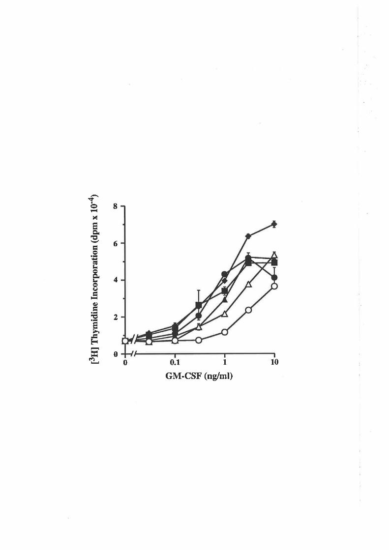
t3ffl
fny
mid
ine
Inco
rpor
atio
n (d
pm x
10'
a)
ì.¡5
o\æ
ôA I o U)
at E R.
ÉF
¡
Figure 3.3.11 Comparison of the binding of wild typeGM-CSF and the analogues, 8108R and D112R, toneutrophils and CHO cells transfected with the humanGMRcr chain.
The binding of wild type GM-CSF (O) and the E108R (A) and Dll2R (O)
analogues to cells expressing the high (GMRap", Panel A) or low (GMRø, Panel
B) affinity GM-CSF receptors was compared. Binding experiments on thepermanently transfected CHO cell line, A9lC7, expressing the human GMRa chain
(Hercus et al., 1994b), were performed as described 2.10.17. Unlabelled wildtype GM-CSF or analogues were titrated against 150pM of CHO cell-derive¿ 1251-
GM-CSF and 1 x 10ó neutrophils per tube (A) or 500pM of CHO cell-derivedl25I-GM-CSF and 5 x ld A9tC7 cells per tube (B). The values are expressed as a
percentage of the l25I-GM-CSF bound in the absence of competitor and are the
means of duplicaæ deærminations.
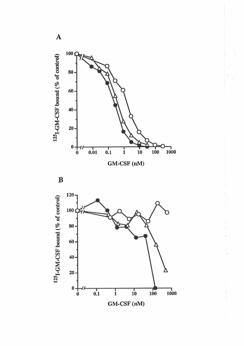
125I
-GM
-CS
F bo
und
(Vo
ofco
ntro
l)
r¿òo
\epä
ËO
OO
OO
OO
r.r.
t.l.l.
A.¡
125I
-GM
-CS
F bo
und
(vo
ofco
ntro
l)
ì.)àg
\æä
oooo
e
EË
c F¡ c F¿ o
o 7 rl ¡ o u) rrt
I I ? È
¡¡
o I o u) ,-t 5o z rì
tl o ll
demonstrated from 2 to 5 x 10⁵ binding siæs for I25I-GM-CSF per cell. The E108R
analogue was four-fold less effective at competing for l25I-GM-CSF binding to rhe
A9lC7 cell line and the D1l2R analogue, which did not compete ¡or 125J-6M-CSF
binding, was at least 2O-fold less effective (Fig. 3.3.1lB).
These results demonstrated a role ¡sr 61u108 and particularly for Aspll2 in GM-
CSF biological activity and in the inæraction of GM-CSF with its receptor complex. The
lack of detectable interaction between the Dl12R analogue and the low affinity receptor
suggests that the Aspll2 residue plays a signif,rcant role in the interaction of GM-CSF
with the GMRa chain.
3.3.3 Mutagenesis of the disulphide-bonded cyste¡neresidues of GM-CSF
Human GM-CSF, which is sensitive to sulphydryl reducing agents (Clark-Lewis
et aI., 1988), contains four cysteine residues that are known to be connected by
disulphide bonds between Cys54-Cys96 and Cys88-Cys121 (Schrimsher er at.,1987).
Alignment of the amino acid sequences of human GM-CSF and the structurally related
cytokine, human IL-2, showed that Cys54, Cys96 and Cysl2l of GM-CSF align with the
three Cys residues of lL-2 (Schrader et al., 1986). Importantly, only two of the Cys
residues of human IL-2 are critical for activity (Iu et al., L987). These results suggested
that one of the disulphide bonds within human GM-CSF may be redundant and not
required for biological activity. Mutants were designed in which pairs of disulphide-
bonded Cys residues were substituted with Ala to prevent the formation of either one or
both of the disulphide bonds (Table 3.3.6). In the case of the second disulphide bond
between Cys88-Cys121, the Cys residues were also substituted with Ser which may
represent a more conservative substitution.
A. Expression of GM-CSF Cys mutants
The Cys mutants of GM-CSF failed to express any detectable proæin as measured
by the standa¡d RIA (data not shown). However, rvestern blot analysis of the transfected
-60-
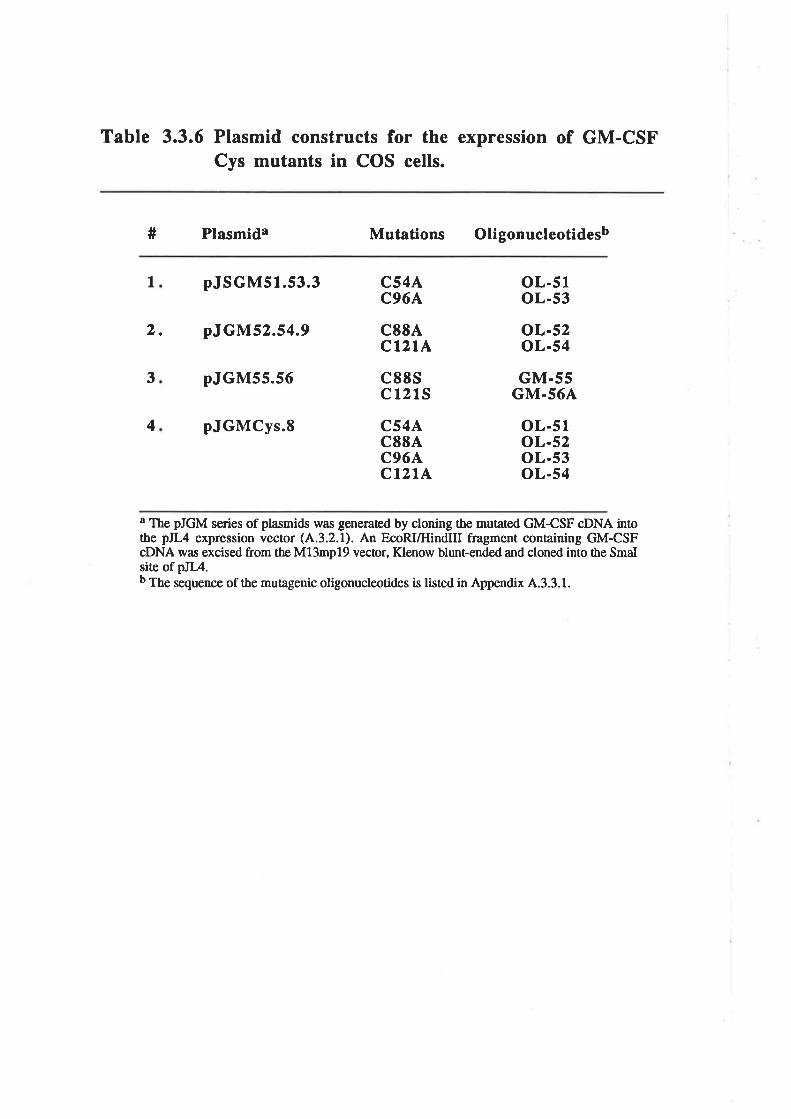
Table 3.3.6 Plasmid constructs for the expression of GM-CSFCys mutants in COS cells.
# Plasmida Mutations Oligonucleotidesb
I pJSGM51.53.3
2. pJGM52.54.9
pJGM55.56
pJGMCys.S
3
4
c54Ac96A
CEsAct2t^c88Sc121S
c54Ac88Ac96Acl2t^
oL-51oL-53
oL-s2oL-54
GM.55GM-56A
oL-51oL-s2oL-53oL-54
I The pJGM series of plasmids was generated by cloning the mutâted GM-CSF cDNA intothe pJL4 expression vector (4.3.2.1). An EcoRI/HindIII fragment containing GM-CSFcDNA was excised from the M13mp19 vector, Klenow blunt-ended and cloned into the SmaIsiæ of pJI/.b The sequence of the mutâgenic oligonucleotides is listed in Appendix 4.3.3.1.
COS cell conditioned media, indicaæd that the Cys analogues expressed at a level
comparable with wild type GM-CSF expression (Fig. 3.3.124). Thus it appeared that
the polyclonal antibody used in the RIA for GM-CSF was unable to recognise GM-CSF
lacking one or both of the disulphide bonds. This was later confirmed using chemically
reduced GM-CSF (Fig. 5.2.16).
B. Activity of GM-CSF CYs mutants
The GM-CSF Cys mutants were quantified by RIA using the sheep anti-GM-CSF
polyclonal antibody which is able to recognise chemically reduced GM-CSF in Vy'estern
btots (data not shown). The concentrations of the GM-CSF Cys mutants deærmined by
this modifîed RIA were consistent with the amounts of GM-CSF protein detected by
V/estern blot (Fig. 3.3.I2A). V/ild type GM-CSF and the Cys mutants were titrated for
the ability to stimulate the proliferation of CML cells (Fig. 3.3.128). Dose-dependent
proliferation of CML cells was observed with wild type GM-CSF, ED5g 0.lng/ml, but
the GM-CSF Cys mutants were unable to stimulate the proliferation of CML cells.
The increase in the apparent molecula¡ weight of the Cys mutants compared to wild
type GM-CSF by SDS/PAGE (Fig. 3.3.124), indicated that these analogues were
hyperglycosylated. As glycosylation is known to influence the specific activity of GM-
CSF (Moonen et aI., 1987: Kaushansky et a1.,1987) it was possible that the loss of
activity observed for the Cys mutants was attributable to hyperglycosylation. In an
attempt to address this possibility, COS cells transfected with the Cys mutant constructs
(Table 3.3.6), were incubated with the drug tunicamycin which inhibits the addition of
N-linked carbohydrate (Donahue et a1.,1986a). However, the level of GM-CSF Cys
mutant expression from tunicamycin treated COS cells was too low for analysis of the
analogues biological activity (data not shown). The expression of non-glycosylated GM-
CSF Cys mutants from E coli may be a more efficient alternative to the tunicamycin
treatment of transfecæd COS cells.
-61 -
Figure 3.3.12 Expression and activity of GM-CSF Cysmutants.
Plasmid constructs encoding wild type GM-CSF and the Cys mutants
C54,964, C88,1214, C88,l2lS and C54,88,96,121A were transfecæd into COS
cells. A mock transfection of COS cells in the absence of plasmid DNA was also
performed. Samples of conditioned medium were concentrated 2O-fold using
Centricon-lO micro concentrators, fractionated by 12.57o SDS/PAGE, transferred
to nit¡ocellulose and probed with sheep anti-GM-CSF polyclonal antibody (Panel
A). Molecular weight standa¡ds are indicated.
1. 200ng
2. l5¡rl
3. 15pl
4. l5pl5. l5pl6. 15pl
7. 15pl
CHO cell-derived wild type GM-CSF
Wild type GM-CSF
C54,964 analogue
C88,l2lA analogue
C88,121S analogue
C54,8 8,96, I2L A analogue
Mock
Wild type GM-CSF (O) and the analogues C54,964 (l), C88,l2lA ( ),
C88,121S (À) and C54,88,96,121A (O) were quantified by RIA using the sheep
anti-GM-CSF polyclonal antibody and titrated for their ability to stimulate
[3H]thymidine incorporation into CML cells (Panel B). Each value represents the
mean of triplicate deærminations and error bars represent the SEM.
A
9467
1234567FlEttFF<
-?
-II
0.1 1
GM-CSF (nglml)
43
30-
20.t -
t4.4 -
1
B
(r)¡OFIx-LLÈE-Lo
oÉ+acÉLoÈLo9ÁI-e)/I.-€.--LL>ìa-E<
)lÈ('¡
0
15
5
00 10 100
3.4 Discussion
The work presented in this chapter describes the analysis of human GM-CSF
analogues mutated in the predicæd fust and fourth ct-helices. Mutation of the Glu2l and
Asp112 residues demonstrated an important role for these residues in the inæraction of
GM-CSF with its receptor and for the biological activities of GM-CSF. These residues
differed in that murarion of Aspllz (D112R) influenced binding to the GMRa chain but
mutarion of Glu2l (E2lR) did not. On the basis of the results obtained with a number of
GM-CSF mutant analogues and a consideration of the GM-CSF structure, a model for
the interaction of GM-CSF with its receptor is proposed.
3.4.1 ldentification of functionally important residues
in human GM-CSF
A. The role of residue 21
Deletion analysis and single amino acid substitutions of residues in the N-terminus
demonstrated that Glu21, predicted to be exposed on the surface of the first a-helix (Fig.
3.1.2A), was critical for GM-CSF function. Mutagenesis of Glu2l significantly reduced
the ability of GM-CSF to stimulate both proliferation (Fig. 3.3.5) and mature cell
acrivarion (Fig. 3.3.6) and also decreased the ability of GM-CSF to bind the high affinity
(GMRcrÊc) receptor of neutrophils (Fig. 3.3.7). Significantly, while the E2lR analogue
displayed greatly reduced biological activity and binding to the high affinity receptor, its
binding to the low aff,rnity (GMRa) receptor was essentially the same as wild type (Fig.
3.3.9). These results linked the binding of GM-CSF to the high affrnity (GMRcrÞc)
receptor with multiple GM-CSF activities and also implicated Glu2l in the interaction of
GM-CSF with the Bç chain.
-62-
The ability of the E21R analogue to trigger a biological response at high but not
low concentrations, indicated that the low affinity receptor (GMRcr) may be able to
transmit a proliferative signal. This possibility w¿rs supported by the observation that
high concentrations (in the low affrnity binding range) of human GM-CSF were required
to stimulaæ l3H]thymi¿ine incorporation in monocyæs (Elliott et a1.,1989) and the
proliferation of murine FDC-PI cells transfected with the human GMRa (Metcalf et aI.,
1990). In contrast" the murine Il-2-dependent T-cell line, CTLL2, does not proliferaæ in
response to human GM-CSF when transfected with the human GMRc alone (Kitamura
et al.,l991a). This indicaæd that the GMRcr alone is insufficient to allow proliferation.
The proliferation of FDC-PI cells transfecæd with the human GMRcr may be the result
of a functional interaction between the human GMRcr and the murine Êc, as CTLL2 cell
lines co-transf'ected with the human GMRct and human pç or murine pç do proliferate in
response to human GM-CSF (Kitamura et aL.,1991a). These results demonstrated that
both GMRcr and Fc are required for a functional response to GM-CSF and therefore the
activity of the E2lR analogue must presumably be mediated through residual interaction
with the pç chain.
The Gh20 residue did not appear to be critical for GM-CSF activity (Fig. 3.3.5 &
3.3.6). However, the slightly impaired ability of the Q20A analogue to compete for the
high affinity binding o¡ l2s1-6¡4-CSF (Fig. 3.3.7) and the fact that the double mutant
Q20NE2IA was less active than E21A alone (Fig. 3.3.5 &.3.3.6), suggested that Gln20
may also contribute to GM-CSF activity. It is possible that the proximity of Gln20 and
Glu2l ailows these residues to interact with a similar region of the GM-CSF receptor
complex. Alternatively these residues may interact with slightly different regions of the
receptor complex but the relatively minor influence of Gh20 is normally masked by the
more pronounced influence of Glu2l on receptor interaction.
B. The role of residue 112
Analysis of the charged residues predicted to be exposed on the surface of the
fourth a-helix of human GM-CSF, showed that the conserved acidic residues 61o108
-63-
and Aspl12 were important for maximal GM-CSF activity (Fig.3.3.10). The other
charged residues in the predicted fourth helix, 61u104, Lys107 and Lyslll, did not
appear to be critical for GM-CSF activity (Fig. 3.3.10). Binding studies demonstrated
that mutation of Aspl12 significantly reduced the binding of GM-CSF !o the high affînity
receptor (GMRcrp.) and low affinity receptor (GMRa) GM-CSF. In fact no binding to
the GMRc chain was observed with the Dl12R analogue (Fig. 3.3.11B) although, must
presumably occur at higher concentrations than was used as this analogue did bind to the
high affinity receptor of neutrophils (Fig. 3.3.114). As the GMRcr chain is common to
both high and low affinity receptors, the receptor binding properties of the D112R
analogue suggested that the Aspl12 residue may interact with the GMRa chain.
C. The fïrst and fourth cr-helices of GM-CSF are implicated as
functionally important by a number of different studies
The results described in this chapter indicated that certain residues in the first and
fourth cr-helices have important roles in the biological and receptor binding activity of
human GM-CSF. A number of different experimental approaches have also attributed
functional significance to these regions in both human and murine GM-CSF.
Fine deletion analysis revealed functionally important regions spanning residues
18-22 and 94-115 of murine GM-CSF (Shanafelt and Kastelein 1989) and residues 20-
2l and 89-120 of human GM-CSF (Shanafelt et al., 1991a). Interspecies chimeras of
human and murine GM-CSF, which exploited the lack of cross-reactivity between these
two cytokines, identified residues 17-31 as being important for GM-CSF activity
(Kaushansky et aI., 1989; Shanafelt et al., 1991b). The mapping of GM-CSF
neutralising monoclonal antibodies suggested that Arg2z in the first predicted s-helix
(Brown et al., 1990) and residues lI0-127 at the C-terminus (Nice et al., 1990;
Kanakura et e1.,1991) paficipate in the interaction of GM-CSF and its receptor. Finally,
the substitution of individual amino acids demonstrated a functional role for residues 14,
15,20 and 2l of murine GM-CSF (Shanafelt et al., 1991b; Meropol et al., 1992:
-64-
Shanafelt and Kastelein 1992) and residues 20 and 23 of human GM-CSF (Kaushansþ
et a1.,1989, Shanafelt et al.,l99lb).
The data presented in this chapter is therefore consistent with a number of
structure-function studies that identified functionally important regions in the first and
fourth cr-helices and most signihcantly, identified two residues (Gluzt, Asp112¡ that are
critical for GM-CSF activity and receptor binding. It is worth noting that the fuñctionally
important Glu2l residue, which appears to be conserved amongst many of the cytokines
(Shanafelt et a1.,1991b) as well as in all species of GM-CSF for which sequence is
available (Fig. 1.2.2), is not specifically identified as important by studies that utilised
interspecies chimeras.
D. The role of the two disulphide bonds in GM-CSF activity
The absence of detectable activity for any of the GM-CSF Cys mutants showed
that both of the disulphide bonds were required for GM-CSF activity. In this respect
human and murine GM-CSF differ as the second disulphide bond of murine GM-CSF
can be eliminated by mutagenesis of Cys85 and Cys118 with no apparent influence on
GM-CSF activity (Shanafelt and Kastelein, 1989). This is despite the absolute
conservation of the four Cys residues in GM-CSF from the seven species of GM-CSF
examined (Fig. t.2.2). One possible explanation for this difference is that murine GM-
CSF requires only the one disulphide linkage to form a stable structure while human
GM-CSF requires both disulphides. GM-CSF, lL-2 and IL-4 exhibit highly
superimposable three dimensional structures, particularly within the helical regions, yet
display a remarkable diversity and number of disulphide bonds (Wlodawer et aL.,1993).
3.4.2 The structure of human GM-CSF
The main feature of the predicted structure of human GM-CSF, described in
Chapter l, was the presence of a bundle of four a-helices (Parry et al., t988; Fig.
1.2.3).Initially the topology for these helices was proposed as classical antiparallel, up-
down-up-down but a later model proposed a three-down-one-up topology (Parry et
-65-
al.,lggt). During the course of this work the three dimensional structure of recombinant
human GM-CSF was solved by means of x-ray crystallography (Diederichs et al.,l99l;
Walær et al.,l992a). The key features of the GM-CSF fold were the presence of four cr-
helices (Fig. 3.4.14) with an unusual two-up-two-down arrangement previously
observed for porcine growth hormone. Another feature of the GM-CSF fold was the
presence of two short p strands, one in the loop connecting helix A and B and the other
in the loop connecting helix C and D (Fig. 3.4.14). At least five cytokines have been
demonstrated to adopt a GM-CSF-like fold including M-CSF,IL-Z,IL-4 and IL-5 which
all exhibit a conserved structural core despite the low level of amino acid sequence
identity ¿rmongst these cytokines (reviewed in Wlodawer et a1.,1993; Rozwarski et al.,
1994). Other cytokines such as lL-3,[L-i and SCF are predicæd to adopt a GM-CSF-
like fold (Rozwarski et aI., 1994). Two other cytokines, G-CSF and GH, possess a
similar topology but with longer helices (Kaushansþ and Karplus, 1993).
Comparison of the GM-CSF structure determined by x-ray crystallography with
the structure proposed by molecular modelling, showed only limited alignment of
secondary structure components (Fig. 3.4.18). Overall, the predicted structure
accurately identified helices A & D but misplaced helices B & C and failed to correctly
anticipate the unusual topology of the GM-CSF fold. Significantly, the functionally
important residues identihed in this chapter, Glu2l and Aspll2, are located on distinct
and separaæ regions of the GM-CSF surface (Fig. 3.a.2). When viewed along the axis
of the helical bundle, the side chains of residues Glu2l and Aspll2 project at
approximately 1800 from each other while the functional groups are separated by at least
20,4, and a number of intervening residues.
The results presented in this chapter assumed that the amino acid substitutions for
the non-Cys mutants, identihed specific contact points for ligand:receptor interaction. An
alternative possibility is that the substitutions introduced structural perturbations that
were responsible for the reduced receptor interaction and biological activity. The location
of Glu2l and Aspl12 on the surface of GM-CSF suggests that their substitution by other
-66-
Figure 3.4.1 Topology of the three-dimensional structureof GM-CSF determined by x-ray crystallography and acomparison with that predicted by molecular modelling.
The topology of the GM-CSF fold shown in panel A was derived from
Diederichs et al. (L99L), The key feature of the GM-CSF fold is the presence offour cr-helices (A-D) in a two-up-two-down arrangement. In addition to the cr-
helices, the long overhand loops, which connect helices A&B and helices C&D,contain short regions of p-strand (I & II). The disulphide bonded Cys residues are
indicaæd.
The predicted and x-ray determined secondary structure elements of human
GM-CSF are compared in panel B. The key features of the predicted structure
(Parry et al., 1988) are shown in the upper figure (i) and those of the x-ray
structure (Diederichs et al.,1991) in the lower figure (ii). Regions shown to be
important for biological activity and receptor binding are indicated with arrows.
l-1 cr helices A,B,C & D
I pstrandsl&IL
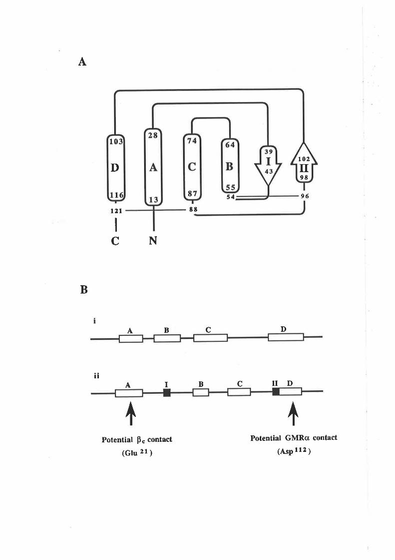
A
B
54
B
Potential p" contact
(Glu zt ¡
96
D
ND
Potential GMRcI contact
(AsP r12 ¡
t2l 8t
NC
l¡
cA
IA B C
^
ô
to2
98tr
10
D
I
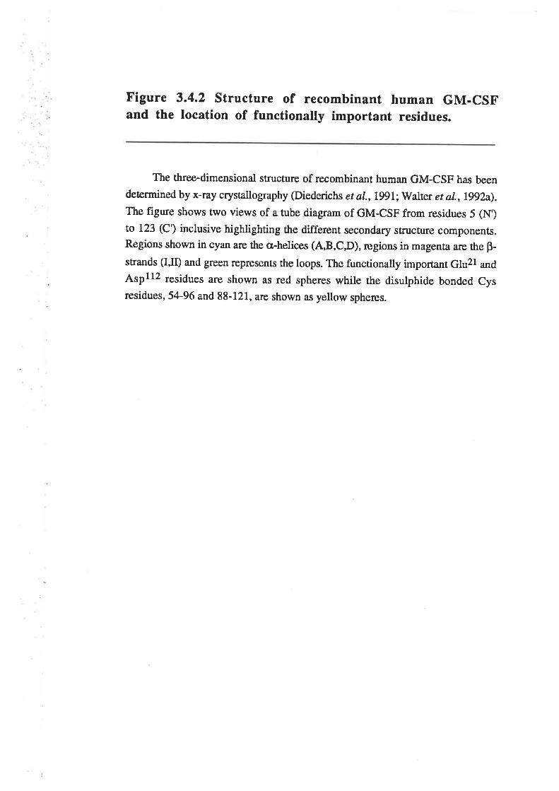
Figure 3.4.2 structure of recombinant human GM-csFand the location of functionally important residues.
The three-dimensional structure of recombinant human GM-CSF has been
determined by x-ray crystallography (Diederichs er al.,lgl;Walter et al.,l992a).The figure shows two views of a tube diagram of GM-CSF from residues 5 (N')to 123 (C') inclusive highlighting the different secondary structure components.Regions shown in cyan are the a-helices (A,B,C,D), regions in magenta are the p-
stands (I,II) and green represents the loops. The functionally important Glu2l and
Atpll2 residues are shown as red spheres while the disulphide bonded cysresidues, 54-96 and 88-121, are shown as yellow spheres.
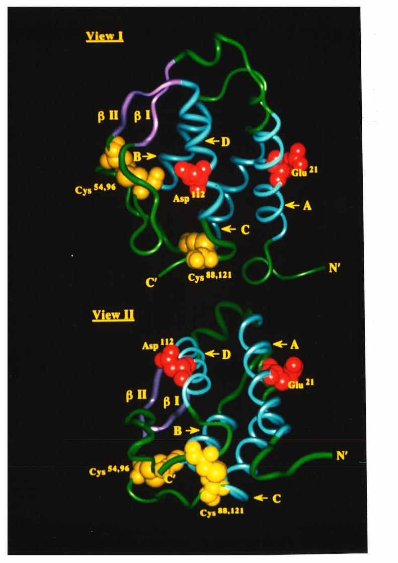
-
amino acids compatible with an cr-helical structure, is not expected to perturb the tertiary
structure. The wild type binding of the E2lR analogue to the GMRo chain provides
evidence that the binding site for this receptor has not been adversely influenced by the
mutation of residue 21. Indirect evidence that the mutations have not affected the gross
tertiary structure of GM-CSF, comes from two observations. The first is that western
blot analysis of GM-CSF analogues revealed no evidence of hyperglycosylation that has
previously been associated with mutations affecting GM-CSF folding (LaBranche et aI.,
1990; Altmann et al., 1991). The second is that the polyclonal rabbit anti-GM-CSF
serum used for RIA quantification of the GM-CSF mutants described in this thesis is
conformation-sensitive and unable to recognise unfolded GM-CSF (Fig. 5.2.16).
Ultimately the question of mutation-induced structural changes may need to be resolved,
particularly for those analogues with reduced receptor interaction, using techniques such
as native gel electrophoresis, x-ray crystallography or nuclear magnetic resonance
spectroscopy.
3.4.3 A model for the interaction of GM-CSF with thetwo chains of the GM-CSF receptor complex
The consequences of charge-reversal mutations at Glu2l and Aspl12 together with
the position occupied by these residues on the surface of GM-CSF, suggests that these
residues interact with two different components of the GM-CSF receptor complex. The
fact that the E21R analogue retained wild type binding to the low affinity receptor
(GMRo) but not the high aftìnity receptor (GMRoBs), indicated that Glu2l probably
participates in the interaction of GM-CSF with the Bç chain. In contrast, the D112R
analogue exhibited reduced interaction with both the low and high affinity receptor and as
the Aspl12 residue is spatially removed from Glu2l in the three-dimensional structure,
suggested that it may participate in the interaction of GM-CSF with the GMRcr chain. On
the basis of the data described in this chapter, a model for the interaction of GM-CSF and
its receptor complex is proposed (Fig. 3.4.3). The model proposes two distinct receptor
interaction sites on the surface of GM-CSF. The first site interacts with the GMRo chain
-67 -
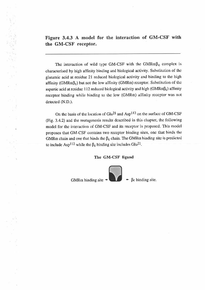
Figure 3.4.3 A model for the interaction of GM'CSF withthe GM-CSF receptor.
The interaction of wild type GM-CSF with the GMRcrp" complex is
characterised by high affinity binding and biological activity. Substitution of the
glutamic acid at residue 21 reduced biological activity and binding to the high
affinity (GMRcrp") but not the low affinity (GMRø) receptor. Substitution of the
aspartic acid at residue I 12 reduced biological activity and high (GMRclp") affinity
receptor binding while binding to the low (GMRa) affinity receptor was not
detected (N.D.).
On the basis of the location of Glu2l and Aspl12 on the surface of GM-CSF
(Fig.3.4.2) and the mutagenesis results described in this chapter, the following
model for the interaction of GM-CSF and its receptor is proposed. This model
proposes that GM-CSF contains trwo receptor binding sites, one that binds the
GMRa chain and one that binds the p" chain. The GMRcr binding site is predicted
to include Aspl1z while ü" F" binding siæ includes Glu21.
The GM-CSF ligand
GMRcr binding site - * pc binding site.
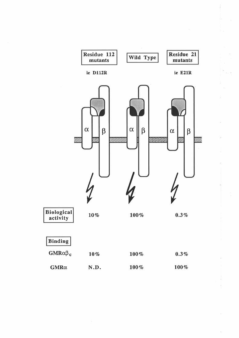
Biologicalactivity
Binding
GMRap
Residue ll2mutants
ie D112R
l07o
c l0Vo
N.D.
Wild Type
1007o
l00Vo
l00Vo
Residue 21mutants
ie E21R
0.3Vo
0.37o
l00Vo
IúI
GMRcr
c[c[ c[pp B'f//Å 'flrt'r/////////Å
and may include 61u108 and Aspll2 while the second site interacts with ù" F" chain and
includes Glu2l.
The E21R analogue which identified two functionally distinct receptor binding sites
also suggested a mechanism whereby GM-CSF antagonists might be generated. Since
binding to GMRa md F. is required for high affinity binding and biological activity
(Kitamura et al.,l991a), analogues totally devoid of interaction with ps but retaining the
inæraction with GMRcr, may be able to antagonise the activity of wild type GM-CSF.
The model shown in Figure 3.4.3 illustrates the essential features of the interaction of
GM-CSF and its receptor complex and also identifies regions to be mutated in order to
elucidate which residues on the surface of GM-CSF make functionally relevant contacts
with the two chains of the GM-CSF receptor complex.
-68-
Chapter 4
Saturation mutagenesis of residuesin the first cr-helix of GM-CSF
4.1 Introduction
The results presented in Chapter 3 identified residues 2l and 112 as being
important for GM-CSF biological activity. Residue ll2, on the surface of the fourth a-
helix, poæntially interacts with the GMRct chain while residue 21, on the surface of the
first a-helix, w¿rs identified as playing an important role in the interaction of GM-CSF
with the pç chain of the GMRupç receptor complex. The fact that mutation of these
residues reduced but did not abolish biological activity suggested that additional residues
were likely to be involved in the interaction of GM-CSF with the GMRø md F. chains
of the GM-CSF receptor complex.
Mutation of residue 21 significantly reduced the biological activity of GM-CSF
(Figures 3.3.5 &.3.3.6) as well as the ability of GM-CSF to bind the. high affinity
(GMRap") receptor but not the low affinity (GMRcr) receptor (Figures 3.3.7 & 3.3.9).
The identification of all functionally important ps chain contact residues on GM-CSF
may enable the generation of a GM-CSF analogue devoid of pç chain interaction but still
capable of binding the GMRcL chain. A GM-CSF analogue with these receptor binding
properties may act as a GM-CSF antagonist and block wild type GM-CSF function. In
an attempt to identify other functionally important residues which may interact with the
pç chain, residues in the vicinity of Glu2l and predicted to be locaæd on the solvent
exposed surface of the first q-helix were specihcally targeted for mutagenesis. As the
solvent exposed residues could only be predicted prior to the determination of the three-
dimensional structure of GM-CSF (Fig. 3.1.2), a number of buried residues were also
targeted for mutagenesis. The first cr-helix of human GM-CSF spans amino acids 13 to
28 with Trp13, Glu14, Asn17, Gln20, Glu21, Arg23, AtE24, Asn27 and Leu28 showing a
high degree of solvent accessibility (Diederichs et al.,l99l). These residues were
targeted for mutagenesis with multiple substitutions apart from Trpl3 which has been
-69-
shown to be dispensable for GM-CSF activity (Clark-Lewis et a1.,1983). The Ala18,
1e19, Ala22,Leu25 and Leu26 residues which are located in the vicinity of Glu21, were
also targeted for mutagenesis although they show low solvent accessibility (Diederichs ¿r
aI.,L99l).
The mutagenesis procedure described in Chapær 3 was not considered suitable for
the saturation mutagenesis required to thoroughly investigate the functional role of
residues in the first a-helix of GM-CSF, so an alternative procedute was pursued. The
results presented in this chapter describe the development of an efficient protocol for
generating large numbers of substitution mutants within the first a-helix of GM-CSF and
analysis of the functional consequence of these mutations.
4.2 Results
4.2.1 A potential problem for site-directed mutagenesis
in the first g-helix of GM-CSF
During the mutagenesis of residues in the first helix, it became apparent that
mutagenesis at some positions was particularly inefficient with only I-5Vo of plaques
hybridising to the mutagenic oligonucleotide (data not shown). Close examination of the
GM-CSF cDNA sequence showed that a region spanning residues 20 to 26 had the
porential to form a stable hairpin loop structure in mRNA (Fig.a.2.1A). Using the values
of Freier et al. (1986), the hairpin loop has a calculated stability of ÂG" = -24.3kllmol
at37"C. A similar structure might also exist in the single stranded, anti-sense strand of
the cDNA used for the mutagenesis procedure although the stability of such a structure is
not known. From these observations it seemed possible that the presence of secondary
structural features might be responsible for the low mutagenesis efficiency of this region,
perhaps by preventing the mutagenic oligonucleotides from annealing to the æmplate-
Two silent mutations were introduced to disrupt the potential hairpin loop structure
-70-
Figure 4.2.1 A potential hairpin loop in the GM,CSFcDNA may influence mutagenesis efficiency.
The sequence of the GM-CSF cDNA anti-sense strand used for mutagenesis,
was examined for potential secondary structure in the region encoding the first cl,-
helix. A hairpin loop involving the codons for amino acids 20 to 26 (A) was found
which may influence the annealing of mutagenic oligonucleotides to this region.
The same region of the GM-CSF mRNA, equivalent to the sense strand of the
cDNA, also appears able to form a hairpin loop with a calculated stability of AG" =-24.3kIlmol at 37'C (Freier et a1.,1986)
The CpC doublet at positions l4l and 142 of the GM-CSF cDNA sense
strand (Fig. 1.2.1), which corresponds to a GpG in the anti-sense strand, wâs
substituted for GpT (ApC in the anti-sense strand) using the OL-256oligonucleotide (Appendix A.4.2.1). This mutarion markedly reduced the
calculated stability of the potential hairpin loop in the mRNA strand (AG" = +5.9
kJ/urol at 37"C) and suggested lhat a hairpin loop structure was unlikely ro form inthis region of the DNA anti-sense strand @).
A wild type GM-csF
anti-sense cDNA
c
3'CGG
18
TAG19
TTG27
s' GCC AUC
Alal t ller e
AA.C CUG 3'
Asn27 Leu 2E
AAC CUG 3'
Asn27 Leu 2E
mRNA
c-GT -Ac=Gc=GT -AG=C
cGccc
cG
cA
G
G
G
cC=GG-UG=CA:UtJr- L'G=CA-UC=G
<<
CGGCCc=GG-Uõ-^tJr: L,A:U
GG
GAC 5'
2E
B "Hairpin" mutant GM-CSF
anti-sense cDNA
ccCAT:AG=C
Ac l, = - 24.3 kJ/mol
mRNA
G-UA-UC=G
CcG
cA
G
G
G
cc=GT -A r<
3' CGG
18
TAG19
TTG27
GAC 5'
2t5' GCC AUC
Atal8 Ilel e
oAC 37 = + 5.9 kJ/mol
lvithout altering the amino acid sequence (Figure 4.2.18). These mutations markedly
reduced the calculated stability of the potential hairpin loop in the mRNA strand (AG" =
+5.9 kJ/mol at 37oC) and suggested that a hairpin loop structure was unlikely to form in
this region of the DNA anti-sense strand.
Mutagenesis experiments were performed with the "hairpin minus" and wild type
DNA templates and mutagenic oligonucleotides binding to the region of the haþin loop
but not including the residues mutated to disrupt the potential hairpin loop structure.
These experiments showed a 2- to 5- fold increase in mutagenesis efficiency for the
"hairpin minus" DNA template when compared to wild type DNA æmplate (data not
shown). The plasmid containing the "hairpin minus" GM-CSF cDNA, pJGM256.3,
expressed essentially the same level of GM-CSF as the pJGM3 plasmid encoding wild
type GM-CSF cDNA when transfecæd into COS cells (data not shown).
Despiæ the modest improvements in mutagenesis efficiency, the entire procedure
was considered to be too slow and complex for the generation of large numbers of
mutants, so alternatives were examined.
4.2.2 A more efficient mutagenesis protocol, OCM
Altemative site-di¡ected mutagenesis protocols, based on the method of Zoller and
Smith (1984), have been developed that by selecting against the template or wild type
strand, produce mutagenesis efficiencies of 50 to 90Vo. The use of DNA templates
grown in dut ung strains of E coli and which as a result contain a low level of
deoxyuridine in place of thymine, enables selection against the template strand in ung+
strains of. E. coli (Kunkel, l9S5). Another approach is to use the second primer to
introduce an observable phenotypic change into the mutagenesis template such as
alæration of p-galactosidase activity (Setzer et aL.,1990) or antibiotic resistance (Lewis
and Thompson, 1990). The latter approach has the advantage that the antibiotic resistance
provides a convenient selection for mutated plasmids. The asymmetric polymerase chain
-71-
reaction has also been used to introduce siæ specific mutations (Perrin and Gilliland,
1990).
These methods, whilst presumably more efficient than method of Tnller. and Smith
(1984), were all considered to be too complex. However, a simpler alternative that
appeared to be ideally suiæd to the task of saturation mutagenesis was oligonucleotide
cassette mutagenesis (OCM). In essence OCM is a simple cloning strategy where the
region to be mutated is deleæd using suitable unique restriction enzyme siæs and replaced
with a fragment formed with appropriately mutated oligonucleotides (Lo et aL.,1984). As
mutagenesis can be performed with the target gene cloned into the appropriate expression
vector, DNA manipulations are minimised. In addition, the use of redundant or
randomised oligonucleotides, enables the OCM procedure to conveniently generate
libraries of mutations (Matæucci and Heyneker, 1983; Oliphant et a1.,1986; Oliphant and
Struhl, 1989).
4.2.3 Preparation of plasmid constructs for OCM of thefirst cr-helix of GM-CSF
The GM-CSF cDNA sequence shown in Figure 1.2.I, contained no unique
restriction enzyme siæs that conveniently spanned the first cr-helix. Reverse translation
of the encoded amino acid sequence indicaæd that two SalI restriction'sites and a unique
XbaI restriction site could be introduced into the GM-CSF cDNA without altering the
amino acid sequence and spanning residues 11 to 28 @ig. 4.2.21\). The pJL4 expression
vector, used to express the wild type and mutated GM-CSF cDNA's from transfected
COS cells (Appendix A.3.2.1), contains SalI and XbaI sites in the polylinker. These
sites were first destroyed by digestion with BamHI and SalI, incubation with Klenow
and religation of the blunt-ended fragment to produce the BamHI-, XbaI-, SalI-, pJL5
expression vector (Appendix A.4.2.2).Conventional site-directed mutagenesis was then
used to introduce the SalI and XbaI restriction sites into the GM-CSF oDNA in pairs.
The mutated cDNA's were then sub-cloned into the pJL5 expression vector to generate
the pJGMV series of plasmids (Fig. 4.2.28). The pJGMV series of plasmids and the
-72-
Figure 4.2.2 The generation of vectors for OCM of thefirst a-helix of GM-CSF.
Nucleotides 6l to 180 of the human GM-CSF cDNA sequence a¡e illustrated
in Panel A (from Figure 1.2.1) along with the three potential restriction enzyme
sites spanning amino acids I I to 28. The mutations required to generate the
restiction siæ are underlined and do not alter the amino acid sequence.
The three poûential restriction enzyme sites were introduced into the GM-CSF
cDNA in pairs by siæ-directed mutagenesis with the oligonucleotides MI-2,-3,-5
(Appendix 4.4.2.I) and the mutated cDNA fragments blunt cloned into the SmaI
siæ of the pJL5 expression vector (Appendix A.4.2.2). The pJGMV series ofplasmids encoded wild type GM-CSF and carried pairs of restriction enzyme sites
which flanked part or all of the GM-CSF cDNA sequence that encoded amino acids
1l to 28 (Panel B).
satl(e0)GICçuAc
. ATCTCTGCÀCCCGCCCGCTCGCCCÀGCCCCÀGCACGCÀGCCCTGGGAGCATGTGAÀTGCC
. I leSerAlaProAlaArgSerProSerProSerThrGlnProTrpGluH i sValAsnAlaL10
saII(134) xbal(lso)GÎCCAC ISTAGÀ
ÀTCCAGGAGGCCCGGCGTCTCCTGAÀCCTGÀGTÀGÀGÀCACTGCTGCTGÀGATGAATGAÀ . .
I IeGlnGtuÀlaArgArgLeuLeuAsnLeuSerÀrgÀspThrÀla}laGluMetÀsnGlu . .
20 30
A
B
Vector
pJGMVI --
pJGMV2 --
-__T___-| .43bp ll-22
.60bp 11-2E
Map Insertsize
Aminoacids
pJGMV3..$-. 17bp 26-2E
ô ôSalI (e0) SalI ( 134
xbal(1so)
pJGM3 plasmid encoding wild type GM-CSF cDNA, expressed similar levels of GM-
CSF proæin when transfected into COS cells (data not shown).
4.2.4 OCM of the fïrst q,-helix of GM-CSF
Mutagenesis of the first cr-helix of GM-CSF was achieved by replacing the SalI
and/or XbaI defined cDNA fragments with double-stranded oligonucleotide cassettes
encoding mutated cDNA fragments. Fragments cloned into the pJGMVI (SalI90lSa¡t34¡
vector were randomly orientated so a PCR protocol was devised to enable colonies to be
screened for plasmids with the conectly orientated SalI fragment. In conrast" fragments
cloned into the pJGMV2 (Sa[90XU¿150¡ and pJGMV3 (Sa[t34¿ç6¿ls0¡ vectors were
always correctly orientated because of the non-compatible SalI/XbaI sites used.
The oligonucleotide cassettes were generated in a number of different ways which
emphasised the utility of OCM for generating a range of mutations (Fig. 4.2.3).Initially
the cassettes used were fragments composed of two complementary, 5' phosphorylated
oligonucleotides encoding single amino acid changes. Larger fragments were usually
formed ¿ìs two separate fragments with overlapping 6bp single-stranded regions to enable
annealing and ligation. A feature of the multiple fragment oligonucleotide cassettes was
that usually only one fragment contained the mutated region and so the other, wild type
fragment, could be coupled with different mutated fragments. Mutations were also
int¡oduced at multiple sites which enabled the generation of double-mutant analogues.
This could also be used to introduce entirely different sequences from other cytokines
such as murine GM-CSF or human IL-3. The use of redundant oligonucleotides enabled
libraries of recombinant plasmids to be generated with each plasmid containing only one
of the potential sequences.
The oligonucleotide cassette mutagenesis procedure was used to generate 62
substitution mutants at 13 of the 16 residues within the first cr-helix of GM-CSF (Table
4.2.1). In conjunction with substitutions generated by conventional site-directed
-73-
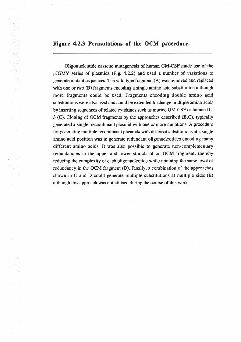
Figure 4.2.3 Permutations of the OCM procedure.
Oligonucleotide cassette mutagenesis of human GM-CSF made use of the
pJGMV series of plasmids (Fig. 4.2.2) and used a number of variations to
generate mutant sequences. The wild type fragment (A) was removed and replaced
with one or two @) fragments encoding a single amino acid substitution although
more fragments could be used. Fragments encoding double amino acid
substitutions were also used and could be extended to change multiple amino acids
by inserting sequences of related cytokines such as murine GM-CSF or human IL-
3 (C). Cloning of OCM fragments by the approaches described (B,C), typically
generated a single, recombinant plasmid with one or more mutations. A procedure
for generating multiple recombinant plasmids with different substitutions at a single
amino acid position was to generate redundant oligonucleotides encoding many
different amino acids. It was also possible to generate non-complementary
redundancies in the upper and lower strands of an OCM fragment, thereby
reducing the complexity of each oligonucleotide while retaining the same level ofredundancy in the OCM fragment (D). Finally, a combination of the approaches
shown in C and D could generate multiple substitutions at multiple sites (E)
although this approach was not utilised during the course of this work.
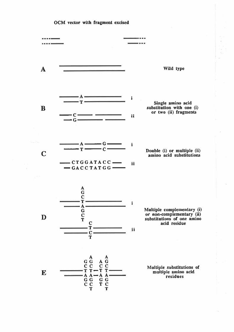
A
OCM vector with fragment excised
-C--G
a-G-
-T-c--CTGGATACC--GACCTATGG-
Wild type
Single amino acidsubstitution with one (i)
or two (ii) fragments
Double (i) or multiple (ii)¡mino acid substitutions
Multiple complementary (i)or non-complementary (ii)substitutions of one amino
acid residue
Multiple substitutions ofmultiple amino acid
residues
I
B
c
It
It
AGcTAGcT c
TcT
-
-
AAGG AGcc ccT T-T TAA-AAGG GGcc rc
TT
E
I
D
ll
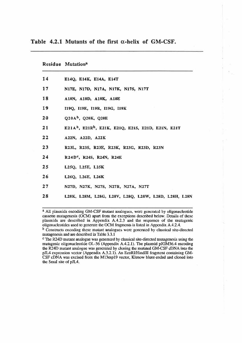
Table 4.2.1 Mutants of the first s-helix of GM-CSF.
Residue Mutationa
t4
l718
19
20
2l22
23
24
25
26
27
28
814Q, 814K, 8144, E14T
N17E, N17D, N174, N17K, N17S, N17T
418N, A18D, 418K, A18E
I19Q, 1198, r19R, I19G, I19K
q2oAb, e2oK, e2oE
E2rAb, E21Rb, E21K, 821e, E21s, E2rD, 821N, E21T
422N, 422D, A22K
R23L, R23S, R23E, R23K, R23G, R23D, R23N
R24Dc, R24S, R24N, R24E
L25Q, L2SE,, L25K
L26Q, L26E,, L26K
N27D, N27K, N27S, N27R, N27A, N27T
L2EK, L2EM, L28G, L28V, L28Q, L28W, L28D, L28H, L28N
a All plasmids encoding GM-CSF mutânt analogues, were generated by oligonucleotidecassette mutagenesis (OCM) apart from the exceptions described below. Details of theseplasmids a¡e described in Appendix A.4.2.3 and the sequence of the mutagenicoligonucleotides used to generate the OCM fragments is listed in Appendix A.4.2.4.b Constructs encoding these mutânt analogues were generated by classical site-directedmutagenesis and are described in Table 3.3.1c The R24D mutant analogue was generated by classical site-directed ¡¡¡lqgen€sis using themutagenic oligonucleotide OL-36 (Appendix A.4.2.1'). The plasmid pJGM36.4 encodingthe RZlD mutant analogue $,as generated by cloning the mutated GM-CSF cDNA into thepJL4 expression vector (Appendix A.3.2.1). An EcoRVfIindIII fragment containing GM-CSF cDNA was excised from the M13mpl9 vector, Klenow blunt-ended and cloned intothe SmaI site of pJL4.
mutagenesis, a total of 65 single amino acid substitution mutants werc generated in the
first a-helix, with 3 to 9 substitutions at each position mutated.
4.2.5 The biological activity of GM-CSF mutants
GM-CSF analogues were expressed from COS cells after transfection with the
appropriate plasmid and the concentration of GM-CSF in the conditioned medium
determined by RIA. In general the GM-CSF analogues were all expressed at levels
similar to that obtained with wild type GM-CSF. However, analogues mutated at residue
22 expressed quite poorly, yielding approximately lÙVo of the levels observed for wild
type GM-CSF and suggesting that the mutations may have had a deleærious effect on
GM-CSF folding or secretion. Western blot analysis showed a similar degree of
molecular weight heterogeneity for the majority of the analogues compared with wild
type GM-CSF (data not shown). Analogues mutated at residue 27 showed a reduced
level of glycosylation, consistent with the loss of the Asn27, N-linked glycosylation site
(Donahue et a1.,1986a). The GM-CSF analogues were then titrated for the ability to
stimulate t¡glmymi¿ine incorporation into CML cells.
The data from CML proliferation assays was analysed using the Fig.P program to
obtain the equation for an asymmetric sigmoid curve that fit the data. Using this
equation, the concentration of GM-CSF analogue required to elicit a biological response
equivalent to the half maximal response (EDso) of wild type GM-CSF was calculated.
The relative biological activity of each GM-CSF analogue was then expressed as a
percentage of wild type activity where wild type was defined as 1007o. At least two
determinations for the activity of each analogue were made from independent COS cell
transfections. The relative biological activity of GM-CSF analogues mutated at solvent
exposed residues (Fig.4.2.4/\) and buried residues (Fig.4.2.aB) are shown. Biological
activity data for a number of the GM-CSF analogues included in Table 4.2.1 was not
available or for a number of reasons was not included. The A22D analogue was very
poorly expressed such that analysis of the biological activity of this analogue was not
possible. The E2lR, EzlK and E2lT analogues were devoid of measurable biological
-74-
Figure 4.2.4 Relative biological activity of GM-CSF firsta-helix analogues.
Plasmid constructs encoding GM-CSF analogues were transfecæd into COS
cells, quantified by RIA and titraæd for the ability to stimulate [3H]thymidineincorporation into CML cells. Data for the E2lR analogue was derived from Figure
3.3.5 using purified, CHO cell-derived E2lR. The relative biological activity ofGM-CSF analogues is shown for solvent exposed residues (Panel A) and buried
residues (Panel B). Columns represent the variation in potency from wild type
GM-CSF (l007o) for each of the GM-CSF analogues, using the mean from at least
two experiments in which full titrations were performed. Poæncy was defined as
the concentration of GM-CSF analogue required to elicit a response equivalent to
the 50Vo maximal response (EDSo) induced by wild type GM-CSF and is
expressed as a percentage of the wild type GM-CSF concentration. For certain
analogues with low activity, relative potency was determined at a level equivalent
to 257o of the maximal response (EDzs) for wild type GM-CSF. Ba¡s indicate the
single-sided .standard deviation from all assays. GM-CSF analogues are iclentified
in one letter code under the wild type residue in three letter code with the residue
number.
001
+)
+lIcÉ
frrC')O
!l(Åoq)
IcÉ(¡)Lr¡tq)C')lré)
Êr
A
B
>ì.È¡
Jc)c\l
t\cn
t)¡lrÅ(,
q)
+¡c!€)L--tq)I¡i()È
1000
10
1
0.1
1000
10
1
Gtu14 A.nl7 GIn2o Gru2l Ar Lrga Arn27 """"QTA ADKSTE KAE DNASQR LGND EDSN ATRDSK DNK
Alal8 Ilel9 AJtz Leu25 Leu26
NKED GAEKR NK EK AEK
00I
v
0.1
activity although with the E21R analogue this is the result of insufficient GM-CSF
protein in the COS cell conditioned medium. The concentrated and purified E21R
analogue expressed from CHO cells displays biological activity, albeit some 3ü)-fold
lower than wild type GM-CSF (Frg. 3.3.5). The GM-CSF analogues mutated at residue
28 alt exhibiæd essentially wild type biological activity although L28Q, L28H, L28W,
L28G, L28M and L28V were not included in Figure 4.2.4A for the sake of simplicity.
The analysis of GM-CSF analogues mutated at solvent exposed residues of the
fîrst cr-helix, indicated that Glu2l was the only residue that contribuæd significantly to
the biological activity of GM-CSF. Residues Asn17, Gh20, /lrgn and Arg24 made only
minor contributions as assessed by the GM-CSF-dependent proliferation of primary
human CML cells. The hierarchy of residue sensitivity to mutation and the fold decrease
in activity (in parentheses) for the most deleærious mutation at each residue was,
61uzt(220) >> Arg2312¡ = ch20(1.9) > ArgzaqLT) > AsnlT(1.5) > Asn27(1.1) >
Glul4(l) (Fig. 4.2.a4). The analysis of GM-CSF analogues mutated at buried residues
of the first cr-helix, indicated that these residues are in general, slightly less tolerant to
substitution. The hierarchy of residue sensitivity to mutation and the fold decrease in
activity (in parentheses) for the most deleterious mutation at each residue **, ils19(330)
>I.¡9v26(7.7) > Ì+ta226.7) > Leu2s(1.6)= {l¿18(1.6) > Leu28(0.9) (Fig. a.z.aB).
Interestingly, certain analogues such as El4K, El4Q, N174, 418N, R23L, R24E,
L25F,, most residue 27 mutants and L28D, consistently generated higher biological
activity than wild type GM-CSF.
4.2.6 The binding of GM-CSF mutants to the low (cr)
and high (crF") affinity GM-CSF receptors
Representative first a-helix GM-CSF mutants were compared for their ability to
recognise the high affinity (GMRcrÞc) receptor of human neutrophils (Fig. 4.2.5). A 15-
fold excess of each of the representative analogues was used to compete for the binding
of 150pM l25I-GM-CSF to human neutrophils. Analogues with a signihcant reduction in
biological activity also demonstrated a significantly reduced ability to compete for the
-75-
Figure 4.2.5 Binding of GM-CSF first a-helix analoguesto the high affinity (GMRaps) receptor.
The binding of wild type GM-CSF and the first c¡-helix analogues to cells
expressing the high affinity (GMRcrp") GM-CSF receptor was compared.
Neutrophils (4 x 10⁶) were incubaæd at room temperature for 2.5 hours in the
presence of 150pM I25I-GM-CSF and a 15 fold excess of wild type GM-CSF or
analogue. Non-specific binding was determined in the presence of a 100-fold
excess of unlabelled wild type GM-CSF. The percentage competition of each
analogue ¡ot 125¡-6¡4-CSF binding w¿ls deærmined from the mean of duplicaæs
and is expressed relative to the competition of wild type GM-CSF taken as IÙOVo.
Panel A shows the binding of GM-CSF analogues mutated at solvent exposed
residues and panel B the binding of analogues mutated at buried residues.
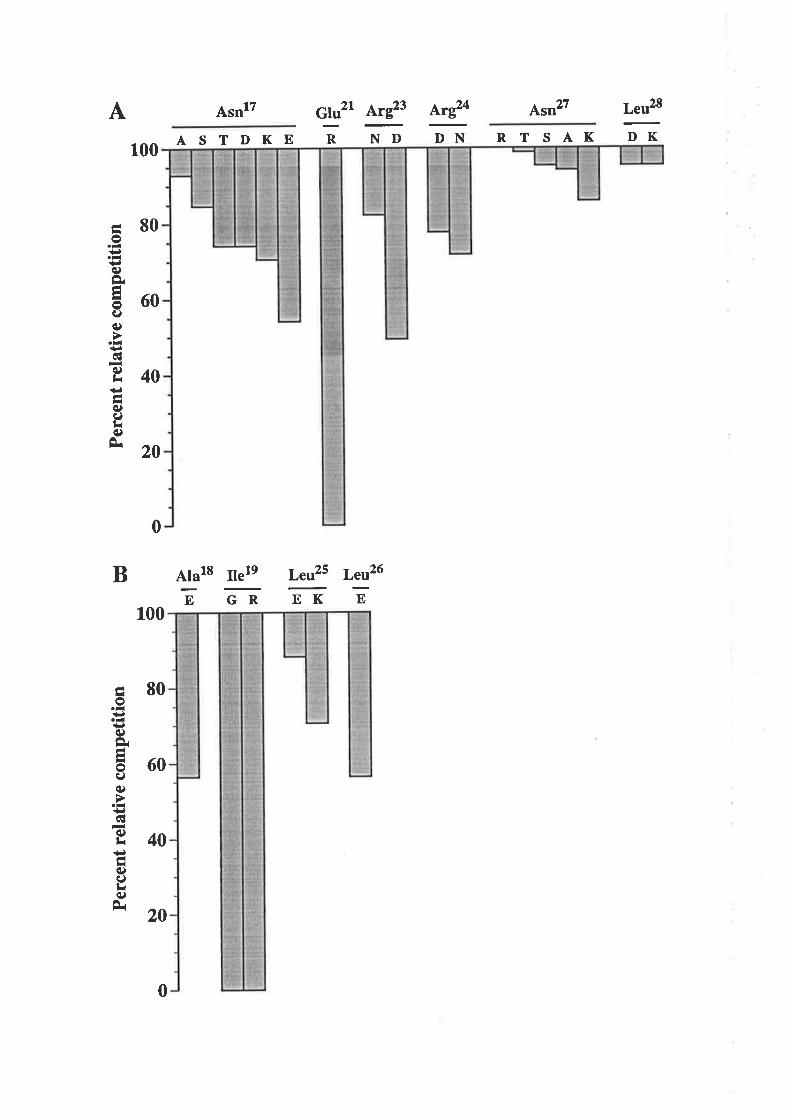
Per
cent
rel
ativ
e co
mpe
titio
n
\JÈ
O\C
AO
OO
E
tÉlr O
l¡ e
Per
cent
rel
ativ
e co
mpe
titio
n
ò.)5
0\a
êèO
O
.rF
- æ
ol=
rl'e
EIF
xlou
rlE,' ì.¡
tâ Ë> ah
Èt
=i
ñ Irl ,l?
szl
ì"lt ul
Ì=
l\
ÞlF
^l=
u(h
l.) {
F Ê çn ñ
binding of I2SI-GM-CSF. Muration of the surface residue Glu2l (Fig. 4.2.54) and the
buried residue Ilel9 Gig. 4.2.58) caused the most signif,rcant reduction in ability to
compete for receptor-binding.
Representative analogues were titraæd for the ability to compete ¡ot 1251-6¡4-ç5¡
binding to the high affinity (GMRap") receptor of human neutrophils or to the low
affinity (GMRcr) receptor of the A9tC7 CHO cell line (Hercus et al., 1994b). The
competitive binding curyes for representative analogues a¡e illustrated (Fig. 4.2.6) nd
the derived K¿ values for all analogues tested, shown in Table 4.2-2. Analogues mutated
at posirions 17,23,24,27 and 28 bound GMRop" with essentially wild type affinity
although the N27D analogue displayed a modest (3-fold) increase in affinity, consistent
with the biological activity observed for this analogue. As previously shown in Chapter
3, muration of residue 21 reduced binding to GMRap" without having a significant
effect on the binding to GMRcr, confirming the selective interaction of this residue with
ft" Þ. chain of the GM-CSF receptor. Mutation of residue 19 had a modest effect on
binding to both GMRcrp. (<2-fold) and GMRa (4-fold) which, along with the buried
nature of this residue, suggests that the mutation may lead to structural perturbations-
4.3 Discussion
A new mutagenesis procedure (OCM) was developed to enable the efficient
saturation mutagenesis of residues within the first cr-helix of human GM-CSF- Once
established, the OCM procedure was able to rapidly generate large numbers of single
amino acid substitutions within the first a-helix and should be useful in the analysis of
other potential receptor binding regions. A logical extension of the oligonucleotide
cassette mutagenesis approach uses oligonucleotides with redundancies at multiple sites
to generate libraries containing hundreds or even thousands of different GM-CSF
analogues (Fie. 4.2.38).
-76-
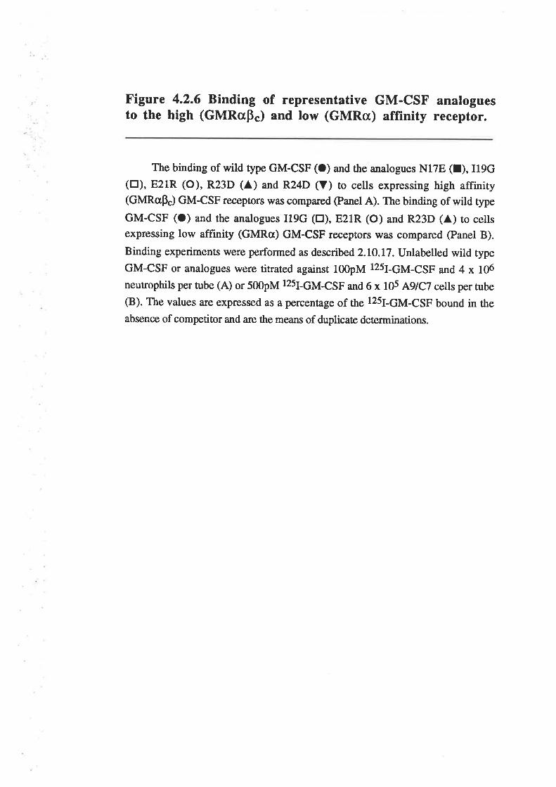
Figure 4.2.6 Binding of representative GM-CSF analoguesto the high (GMRcrÞ") and low (GMRa) affïnity receptor.
The binding of wild type GM-CSF (O) and the analogues N17E (t), I19G(tr), E21R (O), R23D (A) and R24D (Y) to cells expressing high affinity(GMRøp") GM-CSF recepûors w¡ts compared @anel A). The binding of wild type
GM-CSF (O) and the analogues I19G (tr), E2lR (O) and R23D (A) to cellsexpressing low affinity (GMRa) GM-CSF receptors was compared (Panel B).
Binding experiments were performed as described2.I0.l7. Unlabelled wild type
GM-CSF or analogues were titrated against 100pM 125¡-6¡4-çSF and 4 x 10⁶
neutrophils per rube (A) or 500pM 1251-6¡4-çSF and 6 x 10s AgtCT cells per tube
(B). The values are expressed as a percentage of the 125¡-6¡4-çSF bound in the
absence of competitor and are the means of duplicaæ deærminations.
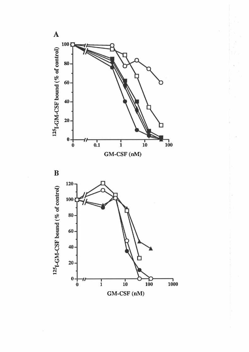
125I
-GM
-CS
F bo
und
(vo
ofco
ntro
l) E
É
Hlr
l.)õg
\æC
l.)oo
oooo
l2sI
-GM
-csF
boun
d (7
o or
cont
rol)
ctså
eg)¡ o
ê
l¡
o ? ¡l I AË
ã1
v
v rà¡¿
o ¡à I CI v)F
)r1
I rà
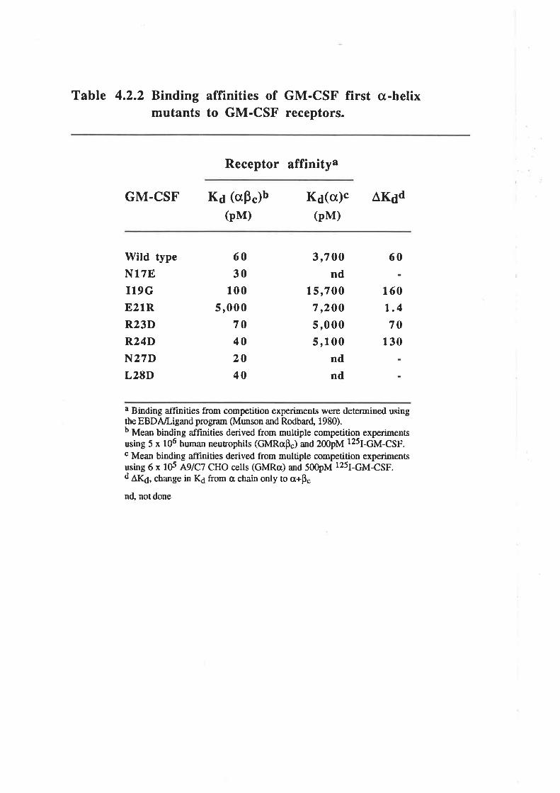
Table 4.2.2 Binding affinities of GM-CSF first o-helixmutants to GM-CSF receptors.
Receptor affinitya
GM.CSF K¿ (aÞ")b(pM)
K¿(o)c ÂK¿d(pM)
Wild type
N17EI19GE21R
R23D
R24D
N27DL28D
60
30100
5,00070402040
3,700nd
15,7007,20O
5,0005,100
ndnd
60
160
1.470
130
a Binding affinities from competition experiments were determined usingthe EBDA/Ligand program (Munson and Rodba¡d, 1980).b Mean binding affinities derived from multiple competition experimentsusing 5 x 10⁶ hunan neuuophils (GMRaÞ") and 200pM 125¡-6¡4-65p.c Mean binding affinities derived from multiple competition experimentsusing 6 x td aglCz CHO cells (GMRa) and 500pM 12s¡-6¡4-65p.d
^Kd, change in K¿ from a chain only to cr+p"
nd, not done
The results presented in this chapær establish the relative, individual contributions
to biological activity of residues of the first cr-helix of human GM-CSF. Substitution
mutågenesis identified residue 21 as the only solvent exposed residue where mutation
had a significant effect on GM-CSF biological activity or receptor interaction. These
results emphasise the critical role played by Glu2l in the inæraction of GM-CSF with the
pç chain of its receptor complex and hence the biological activity of GM-CSF. The
remaining solvent exposed residues of the first cr-helix tolerated a range of substitutions
with little or no effect on the biological activity of GM-CSF. Thus individually, the
solvent exposed residues of the first cr-helix other than Glu2l, do not make critical
contributions to the interaction of GM-CSF and its rcceptor.
The absence of a critical contribution does not preclude the majority of the solvent
accessible residues in the first a-helix from a role in the interaction with the GM-CSF
receptor. Analogues mutated at residue 27 exhibiæd a reduced level of glycosylation but
consistently higher biological activity (Fig.4.2.4A), suggesting that the presence of N-
linked carbohydrate at this position influenced receptor binding. The fact that by itself,
the Q20A substitution did not markedly influence GM-CSF activity is in contrast with the
influence of this mutation when combined with the EzlA substitution (Fig. 3.3.5). In a
similar fashion, the wild type activity of the murine GM-CSF analogues, K14A and
K204, suggested no functional role for residues 14 and 20 whereas the marked
reduction in activity of the K14A,K20A analogue indicated that both residues do play a
role in murine GM-CSF activity (Meropol et a1.,1992). Residue 20 of human GM-CSF
has been implicated in the interaction of human GM-CSF with the murine Bs chain
(Shanafelr et al.,199lb) while residue 23 of human GM-CSF has been implicated in
receptor interaction by substitution mutagenesis (Kaushansky et aL.,1989) and by the
identification of this residue in the binding epitope of a GM-CSF neutralising MoAb
(Brown et a1.,1990). These observations are consistent with the results reported in this
chapter and in most cases, differences are probably attributable to differences in
experimental approach. In summary, of the residues located on the surface of the first cr-
helix of human GM-CSF, only residu e 2L appeats to play a critical role in the inæraction
-77 -
of GM-CSF with its receptor although it is likely that other residues such as 14, 20 and
23 also make a minor contribution. Although not pursued during the course of this work,
it is possible that the mutation of some or all of these residues, in the context of
deleterious residue 21 substitutions, may generate GM-CSF analogues devoid of
detectable biological activity but retaining inæraction with the GMRcr chain. Analogues
with these properties may be able to function as GM-CSF antagonists.
The buried residues targeted for mutagenesis, Ala18, Ile19, AIa22, Leu25 and
l-eu26, a¡e all absolutely conserved in the species for which the GM-CSF sequence has
been determined (Fig. 1.2.2) which suggests a potential structural role for these
residues. However, with the exception of Ile19, mutation of the buried residues in the
first a-helix had only a minor influence on GM-CSF activity. This finding suggests that
the neutral or charged, hydrophilic side chains of the substituæd residues where able to
be accommodated within the protein core or that mutation induced perturbations were
minimal. The analogues AlSN and L25E actually exhibiæd a slightly enhanced biological
activity. Surprisingly, the residue 22 analogues which were poorly expressed, perhaps
suggestive of some gross defect in protein folding and/or secretion, exhibited only a
modest loss of function. In contrast, the sensitivity of Ilel9 to mutagenesis indicaæs that
the nature of the side chain at this position may be more structurally constrained. Close
examination of the side chain environment for lle19, indicated that the side chain occupies
a highly constricted pocket within the protein core. The analysis of analogues mutated at
buried residues is complicated by the possibility of the mutations causing structural
perturbations leading to a loss of function. Despiæ absoluæ conservation of these buried
residues amongst the seven cloned species of GM-CSF (Fig. 1.2.2), analogues mutated
at four of the five buried residues within the first cr-helix, exhibited a relaúve biological
activity equal to at least lÙVo of wild type (Fig. a.2.48). Whilst this represents a
significant reduction in biological activity, the level of residual activity indicated a
surprising degree of mutational tolerance amongst many of the buried residues. This
demonstrates that absolute sequence conservation is not indicative of the functional status
-78-
of a residue even though conserved, buried residues are considered to be structurally
important.
Analysis of the relative biological activity of all first helix analogues identified 8
residues where at least one analogue displayed biological activity enhanced by 50 to
L5O7o (Fig. 4.2.4). These included all of the solvent accessible residues apart from
Gh20 and Glu2l, and the buried residues AlalS and Leu25. Whilst the influence of the
individual substitutions may be minor, it is possible that a single GM-CSF analogue
incorporating all 8 enhancing substitr.rtions will give rise ûo an analogue with significantly
enhanced biological activity.
The observation that the E2lR analogue retains residual biological activity,
presumably mediaæd through üt Þ" chain, suggests that additiond Ê" chain cont¿cts
exist. The lack of obvious receptor binding candidates in the first cr-helix and the
possible role of the fourth cr-helix in binding the GMRa chain suggests that alærnative
siæs may be involved with receptor interaction. A number of studies have indicated that a
region encompassing the third a-helix of GM-CSF, is involved in the biological activity
of human and murine GM-CSF (Kaushansky et al., 1989; Kanakura et al., 1991;
Shanafelt et a1.,1991a). Intriguingly, the first and third a-helices of human GM-CSF a¡e
neighbours in the three-dimensional structure and residues from the third a-helix are in
close proximity to Glu21 in the first cr-helix (Diederichs et a1.,1991). Thus residues on
the surface of the third a-helix may form part of the pç chain receptor binding site and
analysis of the solvent accessible residues in this region may identify those which are
functionally important.
-79 -
Chapter 5
The expression and purification ofGM-CSF residue 2l analogues
using an E. coli secretion system
5.1 Introduction
The results presenæd in Chapær 3 of this thesis clearly demonstrated the critical
nature of Glu2l in the high affinity binding of GM-CSF to its receptor complex
GMRap. and in the biological activities mediaæd by GM-CSF (Lopez et al.,I992c).
Binding experiments suggested that the Glu2l residue interacts with the pç chain, but not
the a chain, of the high affinity receptor complex. An alternative explanation is that
mutations of Glu2l perturb the tertiary structure of GM-CSF leading to an impaired
interaction with receptor chains and concomitant loss of function. The aim of the work
described in this chapter and Chapter 6 was to generate human GM-CSF residue 21
analogues in E. coli to enable more extensive characterisation of their biological and
receptor binding properties, particularly receptor binding in the low affinity range. To
address the possibility that structural perturbations are responsible for the loss of activity
it was inænded to implement a preliminary analysis of the biophysical properties of these
analogues.
The expression of human GM-CSF analogues in a prokaryotic system offers two
clear advantages over the eukaryotic systems used previously. Firstly, E. coli represents
a cheaper and far more robust expression system that should enable the economical
production of large quantities of recombinant human GM-CSF analogues. Secondly, the
absence of carbohydrate modifications, which are not required for GM-CSF activity
(Kaushansky et aL.,1987; Moonen et aL.,1987), means that the purified GM-CSF is
expected to be a single, homogenous protein which simplifies biophysical
cha¡acterisation. The three dimensional structure of GM-CSF has been determined by x-
ray crystallography using E. colïdenved, recombinant human GM-CSF (Diederichs ør
aI.,l99l; Walter et aI.,I992a).
-80-
Naturally occurring human GM-CSF and recombinant human GM-CSF produced
in eukaryotic cells such as COS and CHO are extensively modified by the addition of
both N- and O-linked carbohydrate (Wong et al., 1985a; Donahue et al., 1986a;
Kaushansky et oI., 1987; Moonen et al., 1987; Cebon et al., 1990). It has been
conclusively demonstrated that this ca¡bohydrate is not required for activity by analysis
of a range of human GM-CSF analogues lacking some or all forms of carbohydrate
modification. Non-glycosylated GM-CSF has been produced in eukaryotic cells either by
enzymatically removing the carbohydrate (Moonen et al., L987) or by mutagenesis of the
siæs of carbohydrate addition (Kaushansky et a1.,1987), by peptide synthesis (Clark-
Lewis et a1.,1988) or by expression in prokaryotic cells (Burgess et a1.,1987). Analysis
of the non-glycosylaæd GM-CSF indicaæs a significantly higher specific activity relative
to glycosylated GM-CSF (Moonen et al., 1987: Cebon et a1.,1990) due to a higher
affinity for the GM-CSF receptor (Cebon et aI., 1990). Exactly what role the
carbohydrate might play in the activity of GM-CSF is not clea¡ although in vivo studies
indicaæ that carbohydrate influences pharmacokinetic parameters such as serum clearance
times (Donahue et a1.,1986a; Denzlinger et aL.,1993). It was therefore considered that
GM-CSF Glu2l mutants produced in E. colí and lacking carbohydraæ offered a suitable
system for more detailed analysis of the effects of these mutations on GM-CSF activity.
5.1.1 Expression of heterologous prote¡ns in E. coli
There are many different systems for expressing heterologous proteins in E. coli
but they can be divided into three categories that differ principally in the form of the
expressed protein although are not mutually exclusive.
A. Inclusion body
Expression of heterologous proteins as cytoplasmic inclusion bodies is probably
the most common approach and under optimal conditions may yield gr¿ìms of crude,
insoluble, heterologous protein per litre of bacærial culture. Human GM-CSF has been
produced as cytoplasmic inclusion bodies, accumulating to l07o of the total cellular
protein (Burgess et a|.,1987) however other attempts have been unsuccessful (Libby er
-81 -
aL.,1987; Greenberg et a1.,1988). The heterologous protein usually carries additional
residues on the N-terminus, such as an initiating methionine, and must be solubilised and
correctly re-folded before use. The high density of inclusion bodies assists the
preliminary purification of heterologous protein but final purification relies on
conventional or affinity chromatography.
B. Fusion protein
Fusion of heærologous proteins with specific protein marker sequences may yield
as much as 100 milligrams of crude, soluble fusion protein per liue of bacærial culture
and a variety of systems utilising this approach are commercially available. The most
useful feature of these systems is the ability to purify the fusion protein using recognition
features endowed by the protein marker sequences. The pGEX system (Pharmacia,
Uppsala, Sweden) produces fusion proteins with the 26-kDa glutathione S-transferase of
Schistosomn japonicum that can then be purified using glutathione agarose beads. The
pMAL system (New England Biolabs, Beverly, MA.) produces fusion proteins with the
40-kDa maltose-binding protein of Escherichia coli that can be purified using amylose
resin. Once purified the fusion protein can be enzymatically cleaved with sequence
specific proteases such as thrombin or factor Xa to release the desired protein although
the cleavage process ænds to be inefficient and expensive.
C. Secretion
Secretion of heterologous proteins is probably the least common approach and may
yield optimally only 5 milligrams of crude, soluble, heterologous protein per litre of
bacærial culture as observed for human IL-4 (Lundell et a1.,1990). Biologically active
human GM-CSF has been successfully expressed by secretion expression systems
(Libby et al., L987:' Greenberg et a1.,1988). High level expression (207o of total cellula¡
protein) of a secreted heterologous protein, B-lactamase, has been reported although the
high level of expression leads to an accumulation of insoluble heterologous protein in the
periplasm (Ghrayeb et a1.,1984). Heterologous proteins a¡e direcæd to the periplasm or
extracellular environment by a bacterial signal peptide, which is cleaved during
-82-
translocation releasing the heærologous protein which must be purified by conventional
or affinity chromatography. The advantages offered by the secretion system are that the
heterologous protein is likely to be soluble and correctly folded and if engineered
appropriaæly the secreted protein will contain an authentic amino terminus.
Generating soluble human GM-CSF analogues offers the possibility of analysing
the activity of crude protein preparations as well avôiding potential problems that might
be associated with the correct refolding of mutant proûeins from inclusion bodies. Thus
the expression of soluble human GM-CSF analogues from E coli either as fusion
proteins with the advantage of simplifred purification protocols or as a secreted protein
with an authentic amino terminus, was examined. Production of wild type GM-CSF and
E2lR using the fusion system, pGEX-2T failed to generate any detectable fusion
protein. It is possible that human GM-CSF is refractile to expression in this manner
although this seems surprising as ovine GM-CSF (Dr T. Focaretta, personal
communication) has been successfully expressed using the pGEX system. However, the
production of human GM-CSF in an E. coli secretion system was successful. This
chapær describes the methods developed for the production and purification of human
GM-CSF and mutant analogues from E. coli .
5.2 Results
An E coli expression system that can secrete biologically active murine and human
GM-CSF (Greenberg et a1.,1988; Shanafelt and Kastelein, 1989) was obtained from
Drs A.B.Shanafelt and R.A.Kastelein (DNAX Research Institute, Palo Alto, CA). The
plasmid pshGM-CSF (Fig. 5.1.14) contained a synthetic human GM-CSF cDNA (Fig.
5.1.18) cloned into the E. coli expression vector pIN-III-OmpH3 (Lundell et aL.,1990),
a derivative of pIN-trI-OmpA2 (Ghrayeb et a1.,1984).
-83-
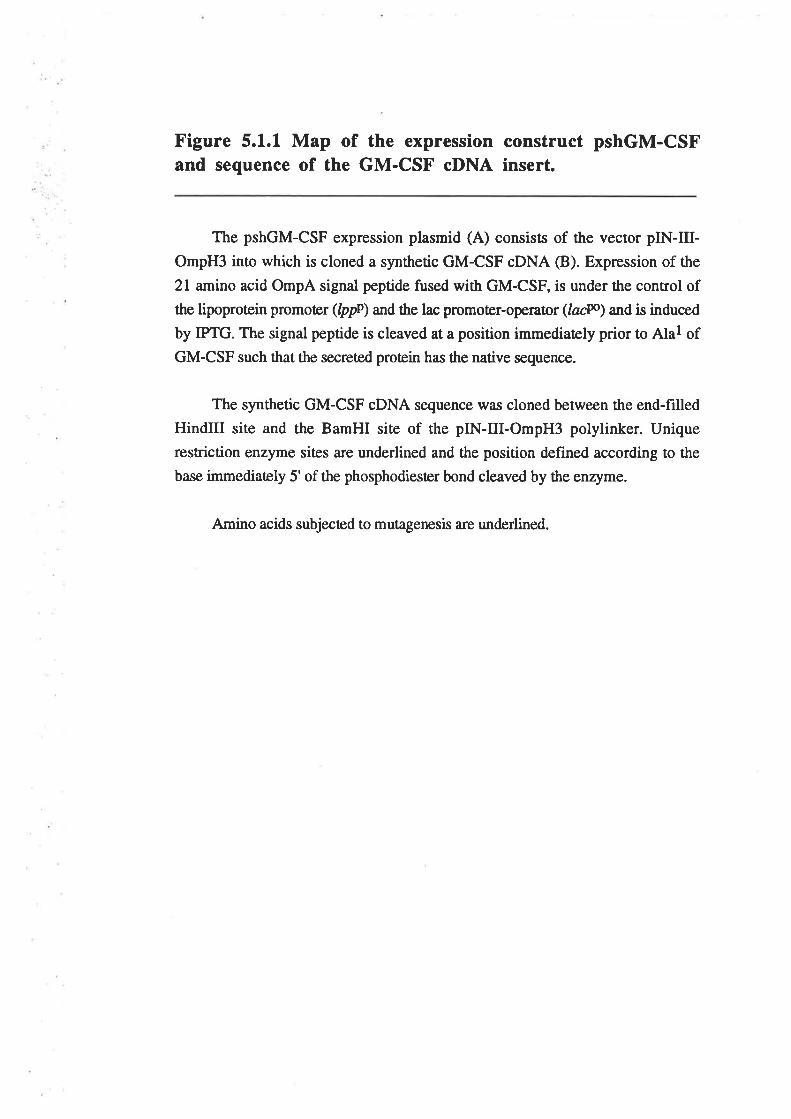
Figure 5.1.1 Map of the expression construct pshGM-CSFand sequence of the GM-CSF cDNA insert.
The pshGM-CSF expression plasmid (A) consists of the vector pIN-III-
OmpH3 into which is cloned a synthetic GM-CSF cDNA @). Expression of the
2l amino acid OmpA signal peptide fused with GM-CSF, is under the control ofthe lipoproûein promoter (Ippo\ and the lac promoter-operaûor (lacw) and is induced
by IPTG. The signal peptide is cleaved at a position immediately prior to Alal ofGM-CSF such that the secreted protein has the native sequence.
The synthetic GM-CSF cDNA sequence was cloned between the end-filled
HindIII site and the BamHI site of the pIN-III-OmpH3 polylinker. Unique
restriction enzyme sites a¡e underlined and the position defined according to the
base immediaæly 5' of the phosphodiester bond cleaved by the enzyme.
Amino acids subjected to mutagenesis a¡e underlined.
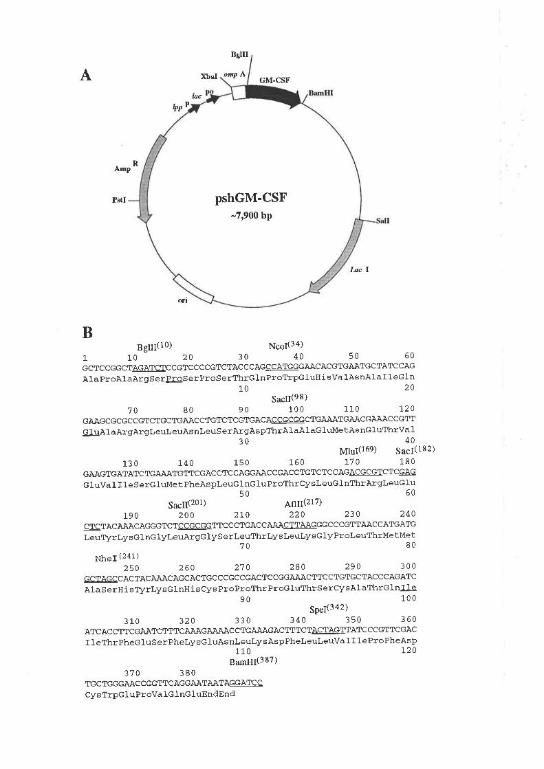
BsUI
A XbaI omp ÃGM-CSF
hclpp
p
Anp
Pstl
Itc I
ott
BBg[(10) ¡çs¡(34)
1 10 20 30 40 50 60
GCTCCGGCT@!ccGTccccGTcTACCCAGWGAACACGTGAATGCTATCCAGAlaProAlaArgSerþSerProSerThrGlnProTrpGluHi sValAsnAlaI leGln
10 20
sacll(e8)7o 80 90 100 110 t20
GAAGCGCGC CGTC TGC TGAÀC CTGTCTCGTGAC AC CGCGGC TGA.AATGAACGAAAC C GTT
G IuAl aArgArgLeuLeuAs nLeuS erArgAspThrAl aAlaGluMe tAs nGluThrVa 1
30 40¡41¡¡(169) 5¿ç¡(182)
l-3 o 1-40 1-50 160 1-7 0 180GAAGTGATATC TGAAATGTTCGAC C TC CAGGAÀC CGAC C TGTCTC C AGAeGegIC TCGAG
GluVa I I 1 eS erGluMet PheAspleuGlnGluProThrCysLeuGlnThrArgleuGlu50 60
5¿çg(201) ¡¡1¡(217)19 0 2oO 21,0 220 230 240
cTc TACAAACAGGGTCT@TTC C CTGAC CAAÀC TIlAAGGGC C CGTTìAÀCC ATGATG
LeuTlrLysGlnGlyIJeuArgGlyS e rLeuThrlysLeuLysGlyProLeuThrMetMeb70 8o
¡¡.1(241)250 260 270 280 290 300
GC.IAGCCACTACA.AACAGCACTGCCCGCCGACTCCGGAAÀCTTCCTGTGCTACCCAGATCAlaSerH i sTlzrlysGlnH i sCys ProProThrProGluThrsercysAlaThrGlnIte
90 1ooSPeI(342¡
3l-o 320 330 340 350 360ATCAC C TTCGAÀTCTTTC AÀAG AAÄÀC CTGAAAGAC TTTCTACTAGTTATC CCGTTCGAC
I I eThrPheG luSerPhelysGluAs nl,eulry sAspPheleuleuVa I I 1 e ProPheAsp110 l2o
gu¡¡¡(387)370 380
TGC TGGGAAC C GGTTC ÀGGAÀTAÀTASGÀTSC
Cys TrpGluProVa IG 1nG luEndEnd
R
pshGM-CSF-7,900 bp
5.2.1 Mutagenesis of pshGM-CSF
The plasmid pshGM-CSF encodes wild type GM-CSF with codon usage
optimised for E coli expression but incorporating a number of unique restriction enzyme
siæs. The pshGM-CSF plasmid was mutated to encode a number of GM-CSF mutant
analogues. The synthetic GM-CSF cDNA within the pshGM-CSF plasmid contains
several unique restriction enzyme siæs (Fig 5.1.1) that are suitable for oligonucleotide
c¿rssette mutagenesis (OCM). One of the major target regions for mutagenesis was the
frrst helix. Residues of the first helix (13-28) a¡e situaæd within a 64bp fragment defined
by a unique NcoI siæ (position 34) and one of two SacII siæs (position 98). To allow
ørgeting of the NcoVSacII fragment by OCM, the plasmid pSGMVI was created in
which the second SacII siæ of pshGM-CSF (position 201) was removed. This was
achieved by replacing the 35bp SacVAflII fragment with the SGMVI.U/L (Table 5.2.1)
fragment which alærs the codon usage of Leuffi and A1967 and thereby eliminates the
Sactr siæ (position 201).
OCM has been used to construct a number of plasmids encoding mutated forms of
human GM-CSF (Table 5.2.1). The constituent oligonucleotides were 5'phosphorylated
and annealed to generate the OCM fragment which was ligated into the appropriaæly
digesæd plasmid (Table 5.2.1).
1. Plasmid pSGM6.2
This plasmid encodes the P6YY analogue of GM-CSF where Pro6 is replaced by
two tyrosine residues and was generated by OCM of the 24bp BgIII/NcoI fragment
(Table 5.2.1). To facilitate receptor binding experiments the P6YY analogue of GM-CSF
was designed to enable high specific activity labelling *¡¡t 1251. This mutation was not
expected to influence the activity of GM-CSF and was expected to be accessible for
labelling.
Continuation of the work described in Chapter 3 required the generation of mutant
analogues in which Glu2l was substituted by a range of amino acids that included Arg,
-84-
Table 5.2.1 B. colí GM-CSF expression plasmids.
# Plasmid Mutation/s fnserta Vectorb
1. pshGM.CSFc wild type
pSGMVI
pSGM6.2
pSGM10.1/21.1
pSGM21.1
pSGM21.2
wild type
P6YY
TlOI/E21R
E21R
E21A
synthetic humanGM-CSF cDNA
vt.atL6.2 UtL
Nco21.1U/LSac21.U/L
#4 NcoI/BamIIId
pIN-III-OmpH3
#1 SacVAflII
#1 BgIII/NcoI
#2 NcoVSacII
#1 NcoI/BamIII
#2 BgIIUSacII
2
3
4
5
6 Bgl/Nco.U/LNco21.2UlL
Sac21.U/L
7. pSGM21.3 E21Q Bgl/Nco.U/LNco21.34U/L
Sac21.U/L
#2 BgIIVSacII
t. pSGM21.4 E2IK Bgl/Nco.U/LNco21.34UlL
Sac21.U/L
#2 BgIIUSacII
9. pSGM21.5 E2IIJ Bgl/Nco.U/LNco21.5U/6L
Sac21.U/L
#2 BgIIUSacII
10. pSGM21.6 E21F Bgl/Nco.U/LNco21.5U/6L
Sac2l.U/L
#2 BgIIUSacII
11. pSGM100 IlOOT Nhe2.U/L100.u/L
Spe2.U/L
#2 NheVSpeI
12. pSGMzl.lll00 E21R/I1007 Nhe2.U/L100.u/L
Spe2.U/L
#5 NheVSpeI
a The insert DNA was generated by 5' phosphorylating and annealing an equimolar a¡nount of theoligonucleotides that constituted an OCM fragment. The sequence of tbe oligonucleotides, oneencoding the upper süand (IÐ and lhe other encoding the lower strand (L), is listed in Appendix4.5.2.t.b The vector describes which of the plasmids (#) listed in this øble was rhe recipient of the OCMfragment and the enzymes used to digest the plasmid.c The deøils of the parental pshGM-CSF plasmid, including the location of unique restrictionenzyme sites, are described in Figure 5.1.18.d The insert used to generate the pSGM2l.l plasmid was in fact obøined by NcoI/BamHIdigesrion of the pSGMl0.ll2l.l plasmid.
Lys, His, Gln, Ala and Phe. This range of substitutions included basic, neutral and
hydrophobic side chains which should generate analogues with different biological and
receptor binding activities.
2a. Plasmid pSGM21.1
This plasmid encodes the E21R analogue of GM-CSF and proved particularly
difficult to construct. Initially the NcoVSacII fragment of pSGMVI was targeûed by
OCM and the plasmid pSGM10.1l2I.l carrying the E21R mutation was isolated (Iable
5.2.1). This plasmid also harboured an unexpected mutation, Tl0I, the origins of which
are unknown and its location within the BglIVNcoI fragment of the vector, outside the
region targeted for mutagenesis, surprising. However, the presence of the unique NcoI
site between the T10I and E2lR mutations enabled the NcoI/BamHI fragment from
pSGM10.1 l2l.I to be cloned into NcoI/BamHI digesæd pshGM-CSF to generate the
correcr psGM2l.l plasmid (Iable 5.2.I).
2b. Plasmids pSGM2l.2 through to pSGM21.6
These plasmids encode various analogues of GM-CSF mutated at residue 2l and
were generated by OCM of the 88bp BgIIVSacII fragment (Table 5.2.1). This approach
was very much more successful than that used to generate the pSGM2l.1 plasmid.
3. Plasmids pSGM100 and pSGM21.1/100
During the course of work examining the activity of E2lR, it became necessary to
examine the role of an IleÆhr polymorphism at residue 100 (Wong et a1.,1985a). The
¡1s100 residue was replaced by'¡X¡100 in both wild type GM-CSF and E21R. The
plasmids pSGMIOO and pSGM2l.llrc} encode the I100T analogues of wild type GM-
CSF and E21R respectively and were generated by OCM of the 101bp NheVSpeI
fragment (Table 5.2.I).
-85-
5.2.2 Expression of wild type GM-CSF and analogues
In pilot expression studies the plasmid pshGM-CSF was transformed into four E
coli strains, MC1061, JMl01, BB4 and E;299 and the level of GM-CSF expression into
the periplasm comp¿ued by Coomassie blue stained SDS/PAGE and western blots
probed with a GM-CSF specific polyclonal antibody. MC1061, BB4 and JM101 are
standard laboratory strains although JM101 has been reported to give high level
expression of soluble human GM-CSF (Greenberg et al., 1988) while E,299 is a lon
strain, dehcient in intracellular protease activity. The conclusion from the pilot study was
that GM-CSF was expressed at approximately the same level in all four strains of E. coli.
In addition to the GM-CSF deæcæd in the periplasm, GM-CSF was also detected in the
culture supernatant, particularly from the JMl0l and BB4 clones. The presence of GM-
CSF in the culture supernatånt may be the result of complete translocation across the cell
membranes (Sakaguchi et aI., 1988) or as a consequence of cell death prematurely
releasing the contents of the periplasm.
On the basis of these results, larger scale expression of wild type GM-CSF was
attempted with 2 litre cultures of the JM10l and BB4 clones. Periplasmic extracts rvere
prepared from pelleted cells while the culture supernatant was pooled and total proæin
precipitaæd with ammonium sulphate. Coomassie stained SDS/PAGE revealed a band of
approximately 15kDa consisænt with the expected size of non-glycosylaæd human GM-
CSF in both periplasmic and culture supernatânt samples (Fig. 5.2.14). Western blot
analysis with a GM-CSF specific polyclonal antibody indicated that there was a
significant amount of GM-CSF in the periplasm as well as the pooled culture supernatant
(Fig. 5.2.1B). The periplasmic and supernatant forms appeared to be full length as they
were the same size as an E coli-denved recombinant human GM-CSF produced in
inclusion bodies (Gift from Dr G.Morstyn, Ludwig Institute for Cancer Research,
Parkville, Vic.). RIA quantihcation of the GM-CSF in the periplasm indicaæd a yield of
3mg per litre with JMlOl and 2.5mg per litre with BB4. The culture supernatant
contained 0.75mg of GM-CSF per litre. This amount of GM-CSF was considered
-86-

Figure 5.2.1 Expression of GM-CSF into the periplasmand culture medium by JM101 and BB4
JMl0l and BB4 clones harbouring the plasmid pshGM-CSF, were cultured
at3ToCin2L L-broth plus antibiotic, to an OD6¡¡¡¡¡¡ of 0.9 before adding IPTG to
O.lmM and incubating at room temperature overnight. Cells were pelleæd and
periplasmic contents isolated by osmotic shock (as described 2.10.8) and
concentrated in a Centriprep-I0 to a final volume of 7mls. The pooled culturesupematants \ilere subjecæd to ammonium sulphate precipitation (65Vo saturation).
The pelleted maærial was resuspended in 60mls MilliQ, dialysed 1:800,000 (frnal)
against PBS, 0.01vo (vlv) Tween 20 and 0.02vo (w/v) sodium azide and
concentrated to a final volume of 4.5mls in a stirred-cell using a 10,000 Mr cutoffmembrane.
Panel A) Samples were fractionaæd by l1Vo SDS/PAGE and the gel stained withCoomassie brilliant Blue. Molecular weight standards a¡e indicated while the
position of GM-CSF migration is indicated "Þ".
l. 2wr2. 20¡tl
3. 2Wr
JM I 0 l/pshGM-CSF periplasmic contents
BB4/pshGM-CSF periplasmic contents
Pooled culture supernatants (prior to concentration)
Panel B) Samples were fractionated by lívo SDS/PAGE, transferred tonitrocellulose and probed with sheep anti-GM-CSF polyclonal antibody. Molecular
weight standa¡ds are indicaæd.
60ngII2
3
4
5pl
std
lpt
E. coli recombinant human GM-CSF
JM lOl/pshGM-CSF periplasmic contents
BB4/pshGM-CSF periplasmic contents
Pooled culture supernatants (concentrated)
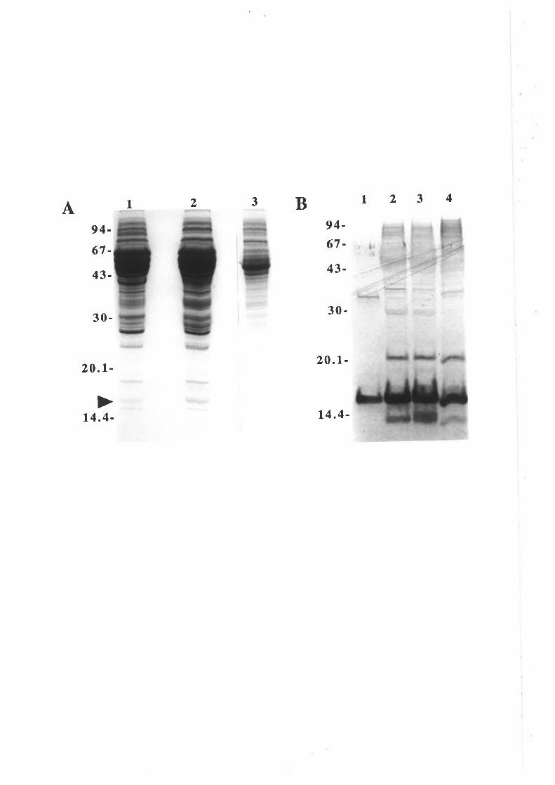
A 2I 3B 1234
Þ¡t¡.a
94-
67-
43-
30-
20.t-
94-67-
43-
30-
20.1-
14.4-
tr
14.4-
insufhcient ûo warrant hanresting as the periplasmic material is much easier to obtain and
is more concentrated.
Having established that the secretion expression system was able to produce
adequate levels of wild type GM-CSF, the expression of the GM-CSF mutant analogues
was examined. Initial expression work centred around obtaining the P6YY analogue for
radiolabelling and the E21R analogue for receptor binding studies. The P6YY analogue
expressed at about the same level as wild type GM-CSF but the E2lR analogue appeared
to be particularly refractile to expression. MC1061 harbouring the plasmid pSGM21.1,
which encodes 821R, yielded no detectable E2lR by either silver stained SDS/PAGE
(Fig. 5.2.2A) or western blot analysis with GM-CSF specific polyclonal antibody (Fig.
5.2.28). This was confirmed by RIA which estimated lpg E2lR per litre of culture
which represents a relative yield of 0.027o compared to wild type GM-CSF.
5.2.3 Improving the expression of the E21R analogue
A number of approaches were examined to improve the yield of the E21R
analogue. Mutant constructs generated by means of OCM were normally only sequenced
through the cassette and flanking restriction sites. However, the entire GM-CSF cDNA
of pSGM2l.l was sequenced which confirmed that the E21R mutation was the only
mutation present in this plasmid.
The role that culture conditions might play in E21R expression by MCl061 was
assessed by varying media composition, media pH, buffering agents and induction
conditions. The conclusion was that there was no signifîcant expression of E2lR from
MC1061 under any of the conditions tested.
The next variable examined was to deærmine if E2lR expression could be detected
by using different strains of E. coli, particularly protease dehcient strains. The strains
tested were;
-87 -
Figure 5.2.2 Expression of E21R in MCl06l
Mc106l clones harbouring the plasmids pIN-III-ompH3 (expression
vector), pSGMVI (wild type GM-CSF) or pSGM2l.l (E2lR) were culrured at37oC in lL L-broth plus antibiotic, to an oDoo0¡¡¡ of 0.7 before adding IprG toO.lmM and incubating at room temperature overnight. Cells were pelleæd and
periplasmic contents isolated by osmotic shock
Panel A) Samples were fractionated by 15% SDS/PAGE and the gel srained withsilver. Molecular weight standards a¡e indicaæd.
Panel B) Samples were fractionaterl hy l57o SDS/PAGE, transferred tonitrocellulose and probed with sheep anti-GM-CSF polyclonal antibody. Molecularweight standa¡ds are indicaæd.
1. 300ng
2. 10pl
3. lOpl
4. lOpl
l. l00ng2. l0¡rl3. 10pl
4. lOpl
E. coli recombinant human GM-CSFMC 106 l/pIN-trI-OmpH3 periplasmic conrenrs
MC 106 1/pSGMV I periplasmic contenrs
MC 106 l/pSGM2 l. I periplasmic contents
E. coli recombinant human GM-CSFMC 106 1/pIN-trI-OmpH3 periplasmic conrents
MC I 06 l/pSGMV I periplasmic contenrs
MC 106 l/pSGM2 l. I periplasmic conrents
1234BA 1234f, - -"'=-.ll
94-
67-
43-
94-67-
14.4-
43-
30.
ts{
20.1-
30.
20.1-
14.4- tI
1. TOPP strains
The six TOPP strains which are non-Kl2 strains of E. coli that can be useful for
the expression of proteins that are otherwise difficult or impossible to express. The
genotypes of the TOPP sEains have not been characterised.
2. lon strains
T1,rc8299 and AB 1899 /on strains a¡e dehcient in intracellular protease activity and
some eukaryotic proteins are stabilised in these strains.
3. degP strain
The Bl78htrA63 degP strun is defective in protease activity associated with the
periplasmic space.
4. ornpT strain
The BL21 ompT strun is defective in an outer membrane protease and is probably
also defrcient in intracellular protease activity (lon).
In summary, some twelve strains of E. coli were examined for their ability to
express the E2lR analogue and in no case did the expression of this analogue approach
the 3 to 5 milligrams of GM-CSF per litre of culture that was typical for wild type GM-
CSF. Western blot analysis with a GM-CSF specific polyclonal antibody, indicaæd a
low level of E2lR expression by BL2l (Fig. 5.2.38). This was confirmed by RIA
which estimated 40ttg of E21R per litre of culture which represents a yield of. lVo
compared with wild type GM-CSF. The BL2l strain was subsequently used for
expression of all GM-CSF residue 21 analogues as most expressed at a low level when
compared to wild type GM-CSF. The lowest yield, as a percentage of wild type
expression, was obtained with the charge reversal analogues E2lR (I.77o) and E2lK
(2.17o), while the highest yield was obtained with the more conservative substitutions
E2lA (2lVo) and E21Q QlVo). The TIOUE2lR analogue expressed at consistently
higher levels than the E21R analogue with a yield of approximately lÙVo compared to
-88-
Figure 5.2.3 Expression of E21R in the protease deficientstrains B178htrA63 and BL2l
B178htrA63 clones harbouring the plasmids pshGM-csF (wild rype GM-CSF) or pSGM2l.l (E2lR) were cultured at room temperature in lL L-broth plus
antibiotic, to an OD6g6¡¡¡ of 0.8 before adding IPTG to 0.lmM and incubating fora further 3 hours at room temperature. BL2l clones harbouring the plasmidspshGM-CSF or pSGM21.1 were cultured at37"C in lL Lbroth plus antibioric, toan OD6gg¡¡¡¡ of 0.85 before adding IPTG to 0.lmM and incubating for a further 3
hours at room temperature. Cells were pelleted and periplasmic contents isolaæd byosmotic shock.
Panel A) Samples were fractionated by líVo SDS/PAGE and the gel stained withsilver. Molecular weight standards are indicaæd.
E. coli recombinant human GM-CSFB l78htrA63/pshGM-CSF periplasmic conrents
B I 78htrA63/pSGM2 l. I periplasmic conrenrs
BL2 l/pshGM-CSF periplasmic conrenrs
BL2 l/pSGM2 1. I periplasmic contents
Panel B) Samples were fracrionated by l57o SDS/PAGE, transferred tonitrocellulose and probed with sheep anti-GM-CSF polyclonal antibody. Molecularweight standards a¡e indicated.
I2
3
4
5
200ng
4r4il2ltl2yd
l. 200ng
2. lOpl
3. 10pl
4. l0¡rl
5. lOpl
E. coli recombinant human GM-CSFB I 78htrA63/pshGM-CSF periplasmic contents
B l78htrA63/pSGM2 l. I periplasmic conrents
BL2 l/pshGM-CSF peripla.smic conrenrs
BL2 l/pSGM2 1. I periplasmic contents
12 3 4 s B 12 3 4 5A
67-43.
20.1-
14.4-
l-t'-
tf rt<
200.0-97 .4-66.2-
45-
14.4-
6.5 -
I
fËrËl-l-
30. Ë-!-,
Ft'-;
TI
31-
512
-r-l-t
fr
wild type GM-CSF (data not shown). In addition, batches of wild type GM-CSF and
analogues carrying the Il00T substitution were expressed in BLzl (discussed in Chapær
6) so that any differences in the activity of analogues relative to wild type, could not be
attributed to differences arising as a result of expressing GM-CSF in different strains of
E. coli.
5.2.4 Purification of GM-CSF
A number of different protocols for the purification of wild type GM-CSF and
analogues were investigaæd and are described below.
A. Gel filtration chromatography of wild type GM-CSF
Analysis of the crude GM-CSF preparations by Coomassie stained SDS/PAGE
(Fig. 5.2.14) indicated that the bulk of the contaminating proteins in both periplasmic
and supernatant preparations are significantly larger than GM-CSF. This observation
suggested that gel hltration chromatography might provide a significant purifrcation step
and in fact has been used to purify human GM-CSF prepared from inclusion bodies
(Burgess et al., 1987). Ten milligrams of total protein from the pooled culture
supernatant of JM101 and BB4 harbouring pshGM-CSF (Fig. 5.2.IA,lane 3) was
fractionated using a Sephadex G-100 gel filtration column and fractions analysed by
silver stained SDS/PAGE. A GM-CSF peak was detected eluting at approximately
56.5mls which falls between the calibration standa¡ds chymotrypsin, 25kDa (51.5m1s)
and RNase A, 13.7kDa (58.9m1s) (data not shown). This indicaæd that the GM-CSF
was behaving as a monomeric protein as has been reported (Wingfield et ø1., 1988).
Fractions containing GM-CSF were then pooled and fractionated by reversed phase
HPLC using a 0-60Vo gradient of acetonitrile to elute bound proteins. A GM-CSF peak
was detected eluting at approximately 45Vo acetonitrile (data not shown). Figure 5.2.4
shows the results of this purihcation protocol with the final product judged >95Vo GM-
CSF by silver stained SDS/PAGE.
-89-
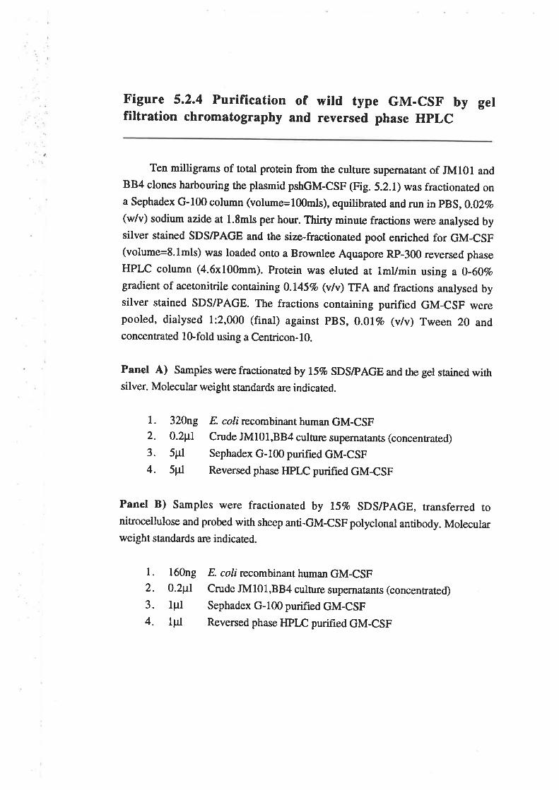
Figure 5.2.4 Purification of wild type GM-CSF by gelfiltration chromatography and reversed phase HPLC
Ten milligrams of total protein from the culture supernatant of JM10l andBB4 clones harbouring the plasmid pshGM-CSF (Fig. 5.2.I) was fracrionared ona Sephadex G-100 column (volume=l00mls), equilibraûed and run in p$S,0.02vo
(Øv) sodium azide at 1.8mls per hour. Thirfy minute fractions were analysed bysilver stained SDS/PAGE and the size-fractionated pool enriched for GM-CSF(volume=8.lmls) was loaded onto a Brownlee Aquapore RP-300 reversed phase
HPLC column (4.6x100mm). Protein was eluted at lmUmin using a 0-60vogradient of acetonitrile conøining 0.l45Vo (v/v) TFA and fractions analysed bysilver stained SDS/PAGE. The fractions containing purified GM-CSF werepooled, dialysed l:2,000 (final) against pBS, 0.0lvo (vlv) Tween 20 andconcentrated lG,fold using a Centricon-IO.
Panel A) Samples were fractionated by 15% SDS/PAGE and the gel stained withsilver. Molecula¡ weight standa¡ds a¡e indicatecl.
l. 32Ûng E. coli recombinant human GM-CSF2. 0.2p1 Crude JM101,BB4 culture supernatants (concentrated)
3. 5trl Sephadex G-100 purified GM-CSF4. sttl Reversed phase HPrc purified GM-CSF
Panel B) Samples were fractionated by lívo SDS/PAGE, transferred tonitrocellulose and probed with sheep anti-GM-CSF polyclonal antibody. Molecularweight standa¡ds are indicaæd.
l. l60ng2. 0.2¡tl3. lpl4. lpl
E. coli recombinant human GM-CSFCrude JM l0 l,BB4 culture supernatants (concentrated)
Sephadex G-100 purified GM-CSF
Reversed phase HPLC purifred GM-CSF
A 1234
¡.¡t¡d67-
43-
30- H
20.1-
14.4-
B 1234
ffi
:a *"ü
94-
67-
43-
30-
20.1-
14.4-
a
The amino terminus of this purified GM-CSF was sequenced to confirm that
cleavage of the OmpA signal peptide generated the appropriate amino terminus. A sample
of purified wild type GM-CSF was fractionated by l57o SDS/PAGE and transferred to
Imobilon PVDF membrane in the absence of glycine, stained with Coomassie blue and
the GM-CSF band excised and sequenced by Richard Simpson (Ludwig Institute for
Cancer Research, Pa¡kville, Vic.). The sequence that was obtained, Ala Pro Ala Arg Ser
Pro Ser Pro Ser Thr Gln Pro Trp Glu matched precisely the amino terminus of mature
human GM-CSF (Fie. 5.1.18).
B. Anion exchange chromatography of wild type GM-CSF
A commonly used first step in protein purification is ion exchange
chromatography. Therefore conditions for the purification of GM-CSF by ion exchange
chromatography were examined. Human GM-CSF prepared in E coli has an
experimentally determined p/ of 5.3, similar to that predicted from the amino acid
composition (Schrimsher et aI., 1987). Efficient binding of GM-CSF to the anion
exchange resin Macro-Prep Q was observed at pH10.5 and was reversible by lowering
the pH to 7.0 and increasing the concentration of NaCl (Chris Bagley personal
communication). Fourteen milligrams of total protein from the periplasm of MC106l
harbouring pSGMVI (Fig. 5.2.2, lane 3), was fractionated by anion exchange
chromatography. Analysis of the eluted protein by silver stained SDSÆAGE (Fig.
5.2.54) indicated a GM-CSF peak at fraction 13, eluting in approximately 100mM NaCl
(Fig. 5.2.54 lane l1). The fractions containing GM-CSF were then pooled and
fractionated by reversed phase HPLC using a 30-507o gradient of acetonitrile to elute
bound proteins (Fig. 5.2.58). The HPLC chromatogram contained a very prominent
peak of absorbance at approxim ately 45Vo acetonitrile which was co-incident with the
peak of GM-CSF eluting from the column as determined by SDS/PAGE analysis of the
eluted proteins (data not shown). The final product of this purification protocol was
judged >95Vo GM-CSF by silver stained SDS/PAGE (Fig. 5.2.5C).
-90-
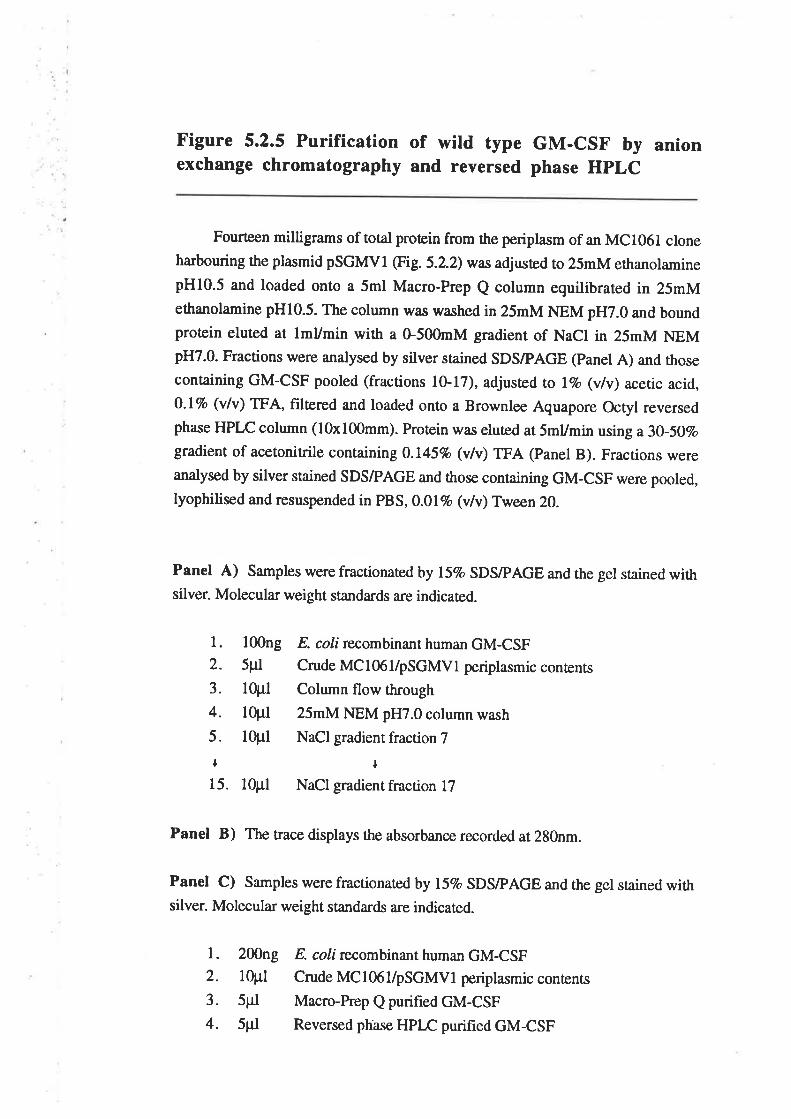
Figure s.2.5 Purification of wild type GM-csF by anionexchange chromatography and reversed phase HPLC
Fourteen milligrams of total protein from the periplasm of an MC106l cloneharbouring the plasmid pSGMVI (Fig. 5.2.2) was adjusted to 25mM ethanolaminepH10.5 and loaded onto a 5ml Macro-Prep Q column equilibrated in 25mMethanolamine pHl0.5. The column w¿¡s washed in 25mM NEM pH7.0 and boundprotein eluted at lmUmin with a G500mM gradient of NaCl in 25mM NEMpH7.0. Fractions were analysed by silver stained SDS/PAGE (Panel A) and rhose
containing GM-CSF pooled (fractions lG.l7), adjusæd to lvo (v/v) acetic acid,0.17o (vlv) TFA, filæred and loaded onto a Brownlee Aquapore Octyl reversedphase HPLC column (l0xl00mm). Proæin was eluted at SmUmin using a30-5OVogradient of acetonitrile containing 0.145% (v/v) TFA (Panel B). Fractions wereanalysed by silver stained SDS/PAGE and those containing GM-CSF were pooled,lyophilised and resuspended in PBS,0.0l% (v/v) Tween 20.
Panel A) Samples were fractionaæd by l5Vo SDS/PAGE and the gel stained withsilver. Molecula¡ weight standards are indicaæd.
Panel B) The trace displays the absorbance recorded at 280nm.
Panel C) Samples were fractionaæd by líVo SDS/PAGE and the gel stained withsilver. Molecula¡ weight standards are indicated.
l. l00ng2. 5pl
3. lOpl
4. l0pl5. l0¡rl
¡
15. lOpl
l. 200ng
2. lOpl
3. spl
4. 5pl
E. coli recombinant human GM-CSFCrude MCl06l/pSGMVl periplasmic conrenrs
Column flow through
25mM NEM pH7.0 column wash
NaCl gradient fraction 7
¡
NaCl gradient fraction l7
E. coli recombinant human GM-CSF
Crude MC I 06 l/pSGMV I periplasmic contents
Macro-Prep Q purified GM-CSF
Reversed ph'ase HPLC purihed GM-CSF
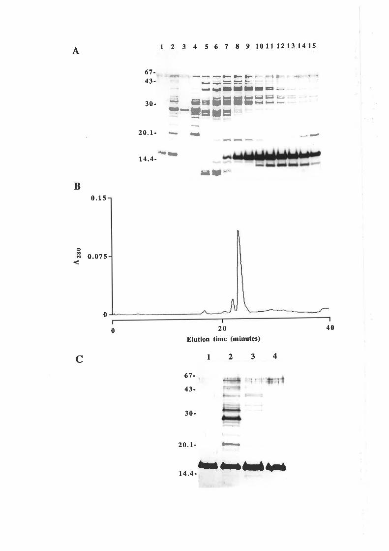
A | 2 3 4 5 6 7 8 I l0lllzlt 1415
.¡rÉ67-4t-
30-
20.1.
t4.4-
___f_>-Þ--->
E_
F - È{ J ¡t. I '
-: ¡¡=tE.¡E==---!=r=- -
¡¡)It¡lI¡=.
---
¡¡ ¡t *-
--
=l
B
o(!CI
0.r5
0.075
0
20
Elution time (minutes)
400
C 123467-
43-
30-
20.1- È-
..ïbT5r4.4-
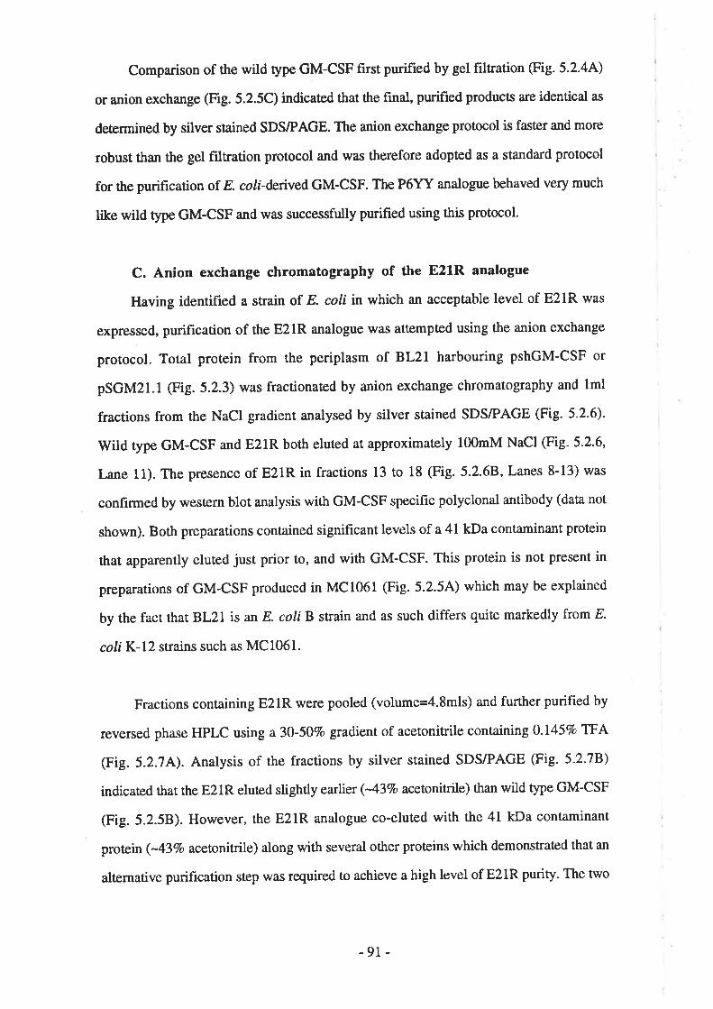
Comparison of the witd type GM-CSF first purified by gel filtration (Fie. 5.2.44)
or anion exchange (Fig. 5.2.5C) indicated that the final, purified products a¡e identical as
deærmined by silver stained SDS/PAGE. The anion exchange protocol is faster and more
robust than the gel filtration protocol and was therefore adopted as a standard protocol
for the purification of. E. coli-derived GM-CSF. The P6YY analogue behaved very much
tike wild type GM-CSF and w¿u¡ successfully purified using this protocol.
C. Anion exchange chromatography of the E21R analogue
Having identified a strain of. E. coli in which an acceptable level of E21R was
expressed, purification of the E21R analogue was attempted using the anion exchange
protocol. Total protein from the periplasm of BL21 harbouring pshGM-CSF or
pSGM21.1 (Fig. 5.2.3) was fractionated by anion exchange chromatography and lml
fractions from the NaCl gradient analysed by silver stained SDS/PAGE (Fig. 5-2.6)'
Wild type GM-CSF and E2lR both eluted at appfoximaæly l00mM NaCl (Fig- 5.2.6'
Lane 11). The presence of E21R in fractions 13 to 18 (Fig. 5.2.68, Lanes 8-13) was
confirmed by western blot analysis with GM-CSF specific polyclonal antibody (data not
shown). Both preparations contained significant levels of a 41 kDa contaminant protein
that apparently eluted just prior to, and with GM-CSF. This protein is not present in
preparations of GM-CSF produced in MC1061 (Fig. 5.2.5A) which may be explained
by the fact that BL}L is an E coli B strain and as such differs quite markedly from E
coliK-I2 strains such as MC1061.
Fractions containing E2lR were pooled (volume=4.8mls) and further purified by
reversed phase HPLC using a30-507o gradient of acetonitrile containing 0.1457o TFA
(Fig. 5.2.74). Analysis of the fractions by silver stained SDS/PAGE (Fig. 5.2.78)
indicaæd that the E21R eluted slightly earlier (-43Vo acetonitrile) than wild type GM-CSF
(Fig. 5.2.58). However, the E21R analogue co-eluted with the 41 kDa contaminant
protein (-43Vo acetonitrile) along with several other proteins which demonstrated that an
alærnative purification step was required to achieve a high level of E21R purity. The two
-91 -
Figure 5.2.6 Purification of wild type GM-csF and E2rRby anion exchange chromatography
crude wild type GM-csF from the periplasm of BL2l harbouring pshGM-cSF and crude E2lR from the periplasm of BL2l harbouring pSGM21.l (Fig.
5.2.3) were purified by anion exchange chromatography. Protein was loaded onto
a 5ml Macro-Prep Q column in 25mM ethanolamine pHl0.5 and the columnwashed in 25mM NEM pH7.0. Bound protein was eluted at lmUmin using a 0-500mM gradient of NaCl in 25mM NEM pH7.0 and lml fractions collecæd.Samples were fractionated by líVo SDS/PAGE and the gel stained with silver.Molecular weight standards a¡e indicaæd.
Panel A) Fractionation of 18 milligrams total protein from B,Lzl harbouringpshGM-CSF.
Panel B) Fractionation of I I milligrÍrms total protein from BL2l harbouringpSGM2l.l.
l. 200ng
2. 5Fl
3. l0pl4. l0pls. 10pl
6. l0pl7. 10pl
t16. 10pl
17. lOpl
18. l0pl
E. coli recombinant human GM-CSFCrude periplasmic contents
Column flow through
25mM NEM pH7.0 column wash
NaCl gradient fraction l0NaCl gradient fraction l lNaCl gradient fraction 12
+
NaCl gradient fraction 2lNaCl gradient fraction 22
NaCl gradient fraction 23
A
67.
43-
30-
lr20.1 - )|.
fl¡-
-
B
67-43-
30-
20.1-
t 2 3 4 5 6 7 E 9 10lttz 1314 1516t7 18
ET , ¡lrr{}rI : : L¡-a-:É-r-
s'-.I-!t
.ID -¡
If -t- -r.-,-rIIT F
-a-ra
-
123456789101112131415161718
rËIIII -ÇrlaE-¡
- ''{ Þ' S4et
E-
.-
.E
tt -- -¡ J)--l?Ç -' - - é
r4.4-
-
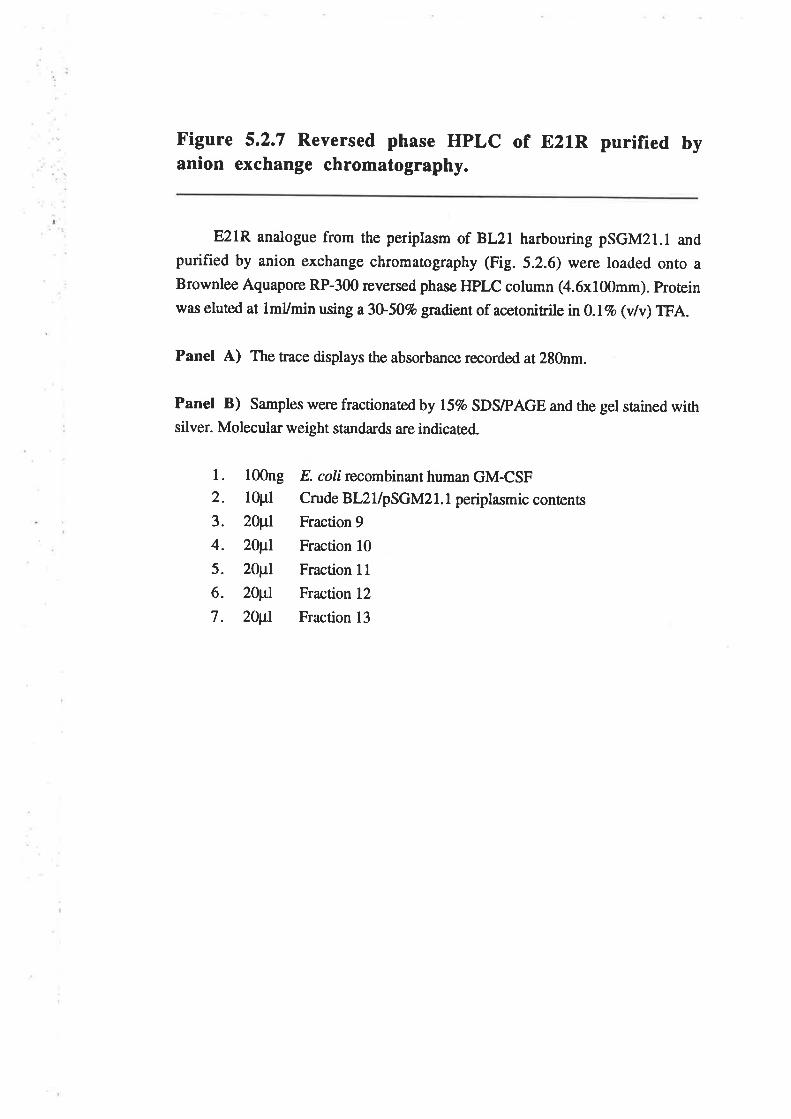
Figure 5.2.7 Reversed phase HPLC of B21R purified byanion exchange chromatography.
E21R analogue from the periplasm of BL2l harbouring pSGM2r.l and
purified by anion exchange chromatography (Fig. 5.2.6) were loaded onto aBrownlee Aquapore RP-300 reversed phase HPLC column (4.6x100mm). protein
was eluted at lmVmin using a 3U50% gradient of acetonitrilen}.lvo (v/v) TFA.
Panel A) The trace displays the absorbance recorded at 280nm.
Panel B) Samples were fractionated by l57o SDS/PAGE and the gel stained withsilver. Molecular weight standards are indicated.
l. l00ng2. lOpl
3. 20¡tl
4. 2Wr5. 20¡tl
6. zur7. 20¡tl
E. coli recombinant human GM-CSFCrude BL2 1/pSGM2 l. I periplasmic conrents
Fraction 9
Fraction l0Fraction I IFraction 12
Fraction 13
A
OoCI
0.6
0.3
0
0 20Elution time (mlnutes)
1'
40
4 56 7rFTF{t ? 3B 4
or'l ;¡I*tÊl43- I
30- -_=
rl!?Ç
la
¡
20.1-
t4.4. - -r
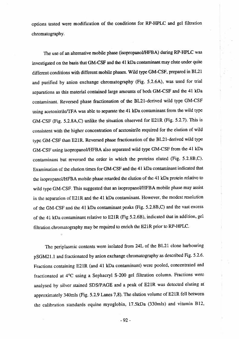
options tested \r,ere modification of the conditions for RP-HPLC and gel filtration
chromatography.
The use of an alæmative mobile phase (isopropanol/HFBA) during RP-HPIÆ was
investigated on the basis that GMCSF and the 41 kDa contaminant may elute under quiæ
different conditions with different mobile phases. lvild type GM-CSF, prepared in BL21
and purified by anion exchange chromatography (Fig. 5.2.64), was used for t¡ial
separations as this material contained large amounts of both GM-CSF and the 4l kDa
contaminant. Reversed phase fractionation of the Bl2l-derived wild type GM-CSF
using acetonitrile/TFA was able to separate the 41 kDa contaminant from the wild type
GM-CSF (Fig. 5.2.84,C) unlike the situation observed for E21R (Fig. 5.2.7). This is
consistent with the higher concentration of acetonitrile required for the elution of wild
type GM-CSF than E21R. Reversed phase fractionation of the Bl2l-derived wild type
GM-CSF using isopropanoyHFBA also separated wild type GM-CSF from the 41 kDa
contaminant but reversed the order in which the proteins eluted (Fig. 5.2'88,C).
Examination of the elution times for GM-CSF and the 4l kDa contaminant indicaæd that
the isopropanoUHFBA mobile phase reta¡ded the elution of the 41 kDa protein relative to
wild type GM-CSF. This suggested that an isopropanolÆIFBA mobile phase may assist
in the separation of E21R and the 4l kDa contariinanL However, the modest resolution
of the GM-CSF and the 41 kDa contaminant peaks (Fig. 5.2.8B,C) and the vast excess
of the 41 kDa contaminant relative to E2lR (Fig 5.2.6B), indicaæd that in addition, gel
filtration chromatography may be required to enrich the E21R prior Ûo RP-HPLC.
The periplasmic contents were isolated from 24L of the BL21 clone harbouring
pSGM2l.l and fractionated by anion exchange chromatography as described Fig. 5.2-6-
Fractions containing E21R (and 41 kDa contaminant) were pooled, concentrated and
fractionated at 4oC using a Sephacryl 5-200 gel filtration column. Fractions were
analysed by silver stained SDS/PAGE and a peak of E21R was detected eluting at
approximaæly 340mls (Fig. 5.2.9 Lanes 7,8). The elution volume of E21R fell between
the calibration standards equine myoglobin, 17.5kDa (330mls) and vitamin 812,
-92 -

Figure 5.2.8 Determining RP-HPLC conditions required toseparate GM-CSF from the 4lkDa contaminant
Samples of wild type GM-CSF from the periplasm of BL21 and purified by
anion exchange chromatography (Fig. 5.2.64), were loaded onto a BrownleeAquapore RP-300 reversed phase HPLC column (4.6x100mm). Protein was
eluted at lmUmin using a30-50Vo gradient of acetonitilein0.l%o (v/v) TFA (A) or
a 30-50Vo gradient isopropanol n 0.13% (v/v) HFBA (B).
Panel A and B) The traces display the absorbance recorded at 280nm. The
peaks representing GM-CSF and the 4lkDa contaminant are labelled.
Panel C) Samples were fractionated by I5Vo SDS/PAGE and the gel stained withsilver. Molecular weight standards a¡e indicated.
l.2.
3.
4.
5.
6.
7.
8.
9.
10
l00ng
5pl
20pl
20pl
20¡tl
2Wr2Opl
20¡tl
20¡tl
20¡tl
E. coli recombinant human GM-CSF
MacrePrep Q purified GM-CSF
3U50Vo aceto¡itrile gradienf fraction 11
fraction 12
fraction 13
3o - Svvoooo-n* or e ru¿i"",, Tî.tlî lr1
fraction 13
fraction 14
fraction 15
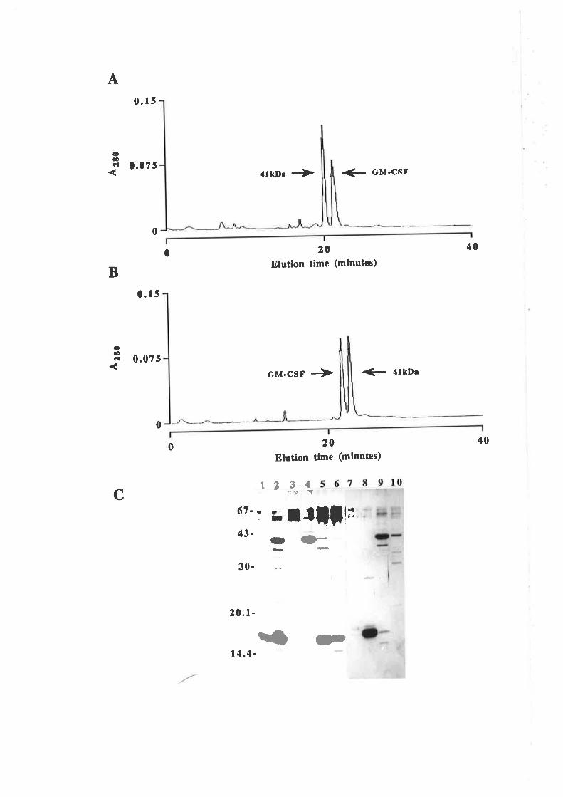
A0.r5
ooCI 0.075
B0.15
0.075
0
0
4lkD¡ à + GM-CSF
20Etution time (mlnutes)
GM.CSF + € trto¡
20Elution tlmc (mlnutes)
123_4-Tr !
67.. t; l,flfln
400
oOCI
400
C
43-
30-
20.1-
O (D-
{ t)'14.4-
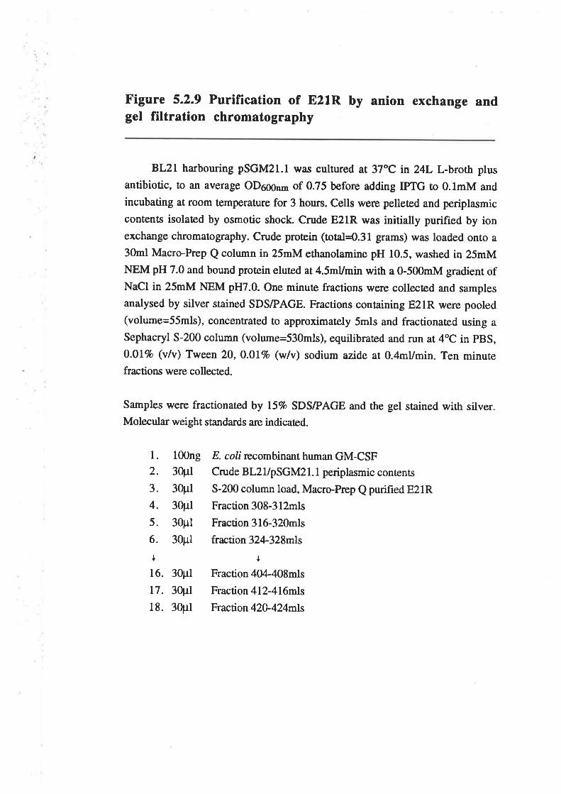
Figure 5.2.9 Purification of E21R by anion exchange andgel filtration chromatography
BL2l harbouring psGM2l.l was cultured at 3zoc in 24L L-broth plus
antibiotic, to an average oD666nm of 0.75 before adding IPTG to 0.lmM and
incubating at room temperature for 3 hours. Cells were pelleted and periplasmic
contents isolated by osmotic shock. Crude E2lR was initially purified by ionexchange chromatography. Crude protein (total=O.3l grams) was loaded onto a30ml Macro-Prep Q column in 25mM ethanolamine pH 10.5, washed in 25mMNEM pH 7.0 and bound protein eluted at 4.5mUmin with a 0-500mM gradient ofNaCl in 25mM NEM pH7.0. One minute fractions were collected and samples
analysed by silver stained SDS/PAGE. Fractions containing E21R were pooled(volume=55mls), concentrated to approximately 5mls and fractionated using asephacryl s-200 column (volume=530mls), equilibraæd and run at 4oc in pBS,
0.ol% (v/v) Tween 20, 0.0lvo (w/v) sodium azîde ar 0.4mumin. Ten minurefractions were collected.
Samples were fractionated by ISVo SDS/PAGE and the gel stained with silver.Molecular weight standards are indicated.
l. 100ng
2. 30pl
3. 30pl
4. 30pl
s. 30pl
6. 30pl
l16. 30pl
17. 30¡rl
18. 30pl
E. coli recombinant human GM-CSFCrude BL2 l/pSGM2l.l periplasmic contenrs
5-200 column load, Macro-Prep Q purified E21R
Fraction 308-312mls
Fraction 316-320mls
fraction 32L328mls
IFraction 404-408mls
Fraction 412-4l6mls
Fraction 42o.424mls
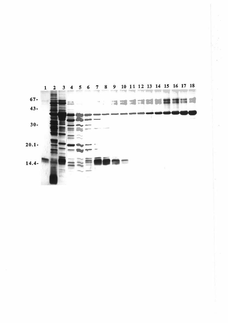
t 2 3 4 s 6 7 8I 10 ll 12 13 14 15 16 l7 18ÇÇ F --'- -:! -*" r{ç -{ç rq *'''q¡'FÇJ I!'|'ÇÇ;r-,:ß.È- ::t Il I rl¡l; â67-
43-
30-
20.1-
14.4-
l--
t--ìræ-
Ort-tt---(-l-jo-
--
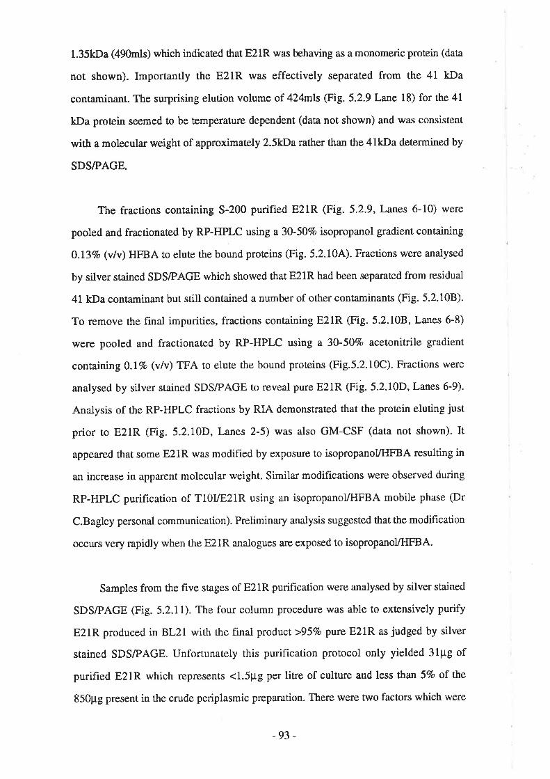
1.35kDa (490mls) which indicaæd that E21R was behaving as a monomeric protein (data
not shown). Importantly the E21R was effectively separated from the 41 kDa
contaminant. The surprising elution volume of 424mls (Fig. 5.2.9 Lane l8) for the 41
kDa protein seemed to be temperature dependent (data not shown) and was consistent
with a molecula¡ weight of approximately 2.5kDa rather than the 4lkDa deærmined by
SDS/PAGE.
The fractions containing 5-200 purified E21R (Fig.5.2.9, Lanes 6-10) were
pooled and fractionated by RP-HPLC using a 30-50Vo isopropanol gradient containing
0.l3Vo (v/v) HFBA to eluæ the bound proteins (Fig. 5.2.104). Fractions were analysed
by silver stained SDS/PAGE which showed that E2lR had been separated from residual
41 kDa contaminant but still contained a number of other contaminants (Fig. 5.2.108).
To remove the final impurities, fractions containing E2lR (Fig. 5.2.108, Lanes 6-8)
were pooled and fractionated by RP-HPLC using a 30-50Vo acetonitrile gradient
conraining O.IVo (vlv) TFA to elute the bound proteins (Fig.5.2.10C). Fractions were
analysed by silver stained SDS/PAGE to reveal pure E2lR (Fig.5.2.10D, Lanes 6-9).
Analysis of the RP-HPLC fractions by RIA demonstrated that the proæin eluting just
prior to E2lR (Fig. 5.2.10D, Lanes 2-5) was also GM-CSF (data not shown). It
appeared that some E2lR was modified by exposure to isopropanol/HFBA resulting in
an increase in apparent molecular weight. Similar modifications were observed during
RP-HPLC purification of T10VE21R using an isopropanol/HFBA mobile phase (Dr
C.Bagley personal communication). Preliminary analysis suggested that the modihcation
occurs very rapidly when the E21R analogues are exposed to isopropanol/HFBA.
Samples from the fîve stages of E2lR purification were analysed by silver stained
SDS/PAGE (Fig. 5.2.1l). The four column procedure was able to extensively purify
E21R produced in BL21 with the final product >957o pure E2lR as judged by silver
stained SDS/PAGE. Unfortunately this purification protocol only yielded 3lpg of
purified E2lR which represents <1.5pg per litre of culture and less than 5Vo of the
850¡rg present in the crude periplasmic preparation. There were two factors which were
-93 -
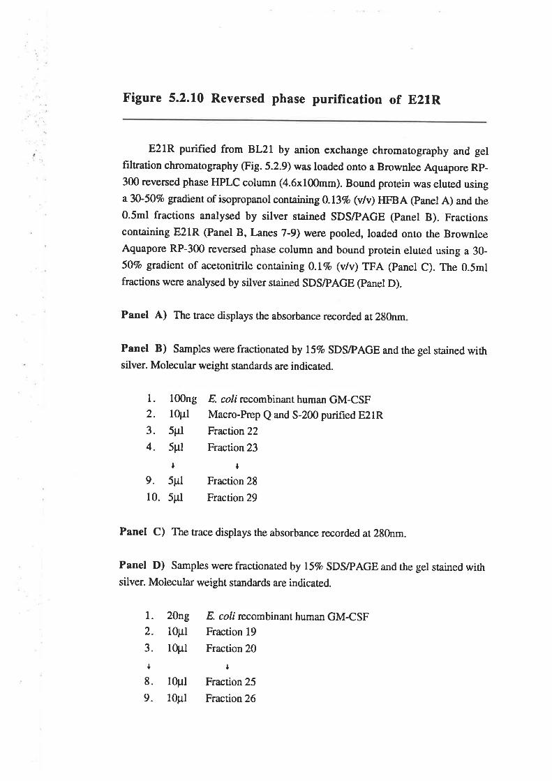
Figure 5.2.10 Reversed phase purification of E2lR
E2lR purified from BL2l by anion exchange chromatography and gelfiltration chromatography (Fig. 5.2.9) was loaded onto a Brownlee Aquapore Rp-300 reversed phase HPLC column (4.6x100mm). Bound protein was eluted usingr30-507o gradient of isopropanol containin g0.l3vo (v/v) HFBA @anel A) and the0.5m1 fractions analysed by silver stained SDS/PAGE (Panel B). Fractionscontaining E2lR (Panel B, Lanes 7-9) werc pooled, loaded onto the BrownleeAquapore RP-300 reversed phase column and bound protein eluted using a 30-50vo gradient of acetonitrile conraining O.l%o (vlv) TFA (panet C). The 0.5m1
fractions were analysed by silver stained SDS/PAGE (panel D).
Panel A) The trace displays the absorbance recorded at 280nm
Panel B) Samples rvere fractionaæd by ISVo SDS/PAGE and the gel stained withsilver. Molecula¡ weight sranda¡ds a¡e indicaæd.
Panel C) The trace displays the absorbance recorded at 280nm.
Panel D) Samples were fractionated by l5%o SDSÆAGE and the gel stained withsilver. Molecula¡ weight standa¡ds are indicaæd.
l. l00ng2. lOpl
3. 5pI
4. 5pl
t9. 5pl
10. 5pl
l. 20ng
2. l0pl3. lOpl
I8. 10pl
9. 10¡rl
E. coli recombinant human GM-CSFMacro-Prep Q and 5-200 purified E2lRFraction22
Fraction 23
IFraction 28
Fraction 29
E. colí recombinant humarn GM-CSFFraction 19
Fraction 20
¡
Fraction 25
Fraction 26
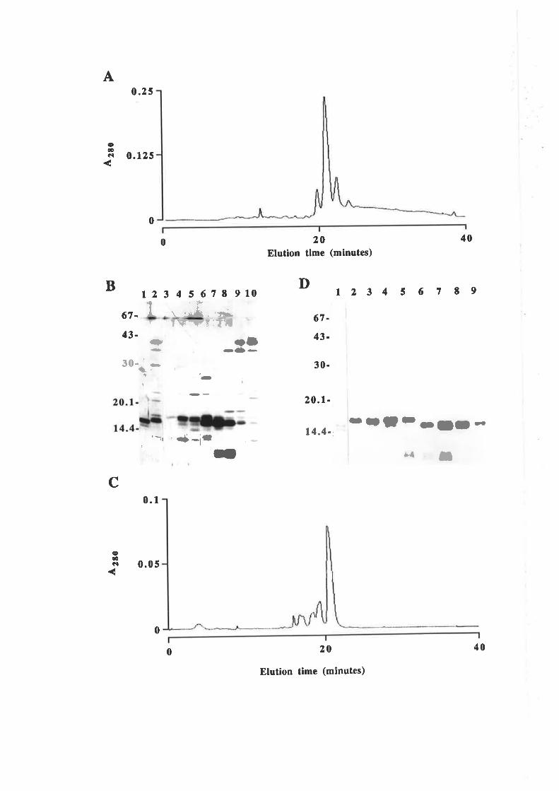
A
cæCI
0.25
0.125
0
0
12345678910
20Elutlon tlme (minutes)
D
67-
43-
30-
20.1-
t4.4-,
20
Elution time (minutes)
123456789
40
B
c
67.
43-
if 'P
t ', *Å . 'il;"Y - Y\r!lF?:TTrD
30.,' -t.
OI-al -
(-
'-, .+-ir -tlt-ll-ri"l , II
.ÐOcl
0.1
0.05
0
0 40
Figure 5.2.11 Purification of E21R from BLZI
E2lR was purihed from 24L of Bl2llpsGM2l.l periplasmic isolare byanion exchange chromatography and gel filuation chromatography (Fig. 5.2.9) and
then two rounds of reversed phase purification (Fig. 5.2.10). Samples from each
stage of the purification process wer€ fractionaæd by líVo SDS/PAGE and the gel
stained with silver. Molecular weight standards are indicaæd.
l. l00ng E. colí recombinanr human GM-CSF2. 20¡tl Crude BL2llpSGMzl.l periplasmic conrenrs
3. zqtJ Macro-Prrep Q purified E2lR4. 20¡tl 5-200 purified E2lR5. zpJ Isopropanol/I{FBA RP-HPLC purifred E2lR6. zpl AceûonitrildTFA RP-HPLC purifred E2lR
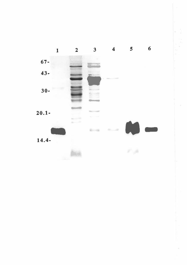
631
r2
É
45
67-
43-
30-
20.1-
14.4-
-
Þ
(b
*.-
rr r-{)
-S.' {a I
k
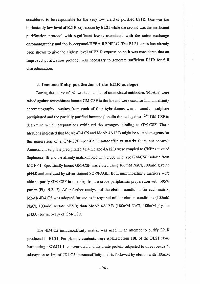
considered to be responsible for the very low yield of purified E21R. One was the
intrinsically low level of E2lR expression by BL21 while the second was the inefficient
purification protocol with significant losses associated with the anion exchange
chromatography and the isopropanol/HFBA RP-HPLC. The BL21 strain has already
been shown to give the highest level of E21R expression so it was considered that an
improved purification protocol was necessary to generate sufficient E21R for full
characterisation.
4. fmmunoaffinity purification of the E21R analogue
During the course of this work, a number of monoclonal antibodies (MoAbs) were
raised against recombinant human GM-CSF in the lab and were used for immunoaffinity
chromatography. Asciæs from each of four hybridomas was ammonium sulphate
precipitaæd and the partially purified immunoglobulin titrated against I25I-GM-CSF to
determine which preparations exhibited the strongest binding to GM-CSF. These
titrations indicated that MoAb 4D4.C5 and MoAb 4AL2.B might be suitable reagents for
the generation of a GM-CSF specific immunoaffinity matrix (data not shown).
Ammonium sulphaæ precipitated 4D4.C5 and 4Al2.B were coupled to CNBr activated
Sepharose-4B and the afhnity matrix mixed with crude wild type GM-CSF isolated from
MC106l. Specifically bound GM-CSF wa.s eluted using 100mM NaCl, 100mM glycine
pH4.0 and analysed by silver stained SDS/PAGE. Both immunoaffinity matrices were
able to purify GM-CSF in one step from a crude periplasmic preparation with >957o
purity (Fig. 5.2.12). After further analysis of the elution conditions for each matrix,
MoAb 4D4.C5 was adopted for use as it required milder elution conditions (100mM
NaCl, l00mM acetate pH5.0) than MoAb 4Al2.B (100mM NaCl, 100mM glycine
pH3.0) for recovery of GM-CSF.
The 4D4.C5 immunoaffinity matrix was used in an attempt to purify E21R
produced in 8L21. Periplasmic contents were isolated from 10L of the BL2I clone
harbouring pSGM21.1, concentrated and the crude protein subjected to three rounds of
adsorption to lml of 4D4.C5 immunoaffinity matrix followed by elution with 100mM
-94-
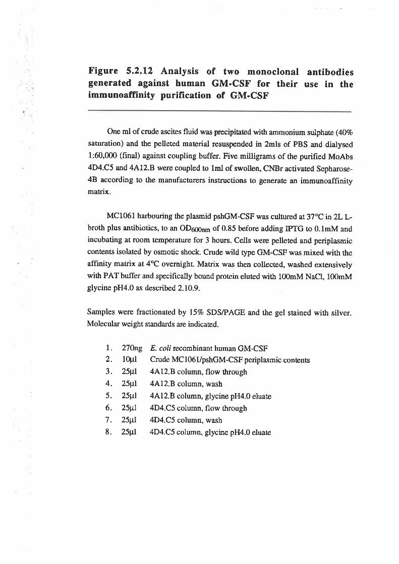
Figure 5.2.12 Analysis of two monoclonal antibodiesgenerated against human GM-CSF for their use in theimmunoaffinity purification of GM-CSF
One ml of crude ascites fluid was precipitated with ammonium sulphaæ(4O7o
saturation) and the pelleted maærial resuspended in 2mls of PBS and dialysed
l:60,000 (final) against coupling buffer. Five milligrams of the purified MoAbs4D4.C5 and 4412.8 were coupled to lml of swollen, CNBr activated Sepharose-
4B according to the manufacturers instructions to generate an immunoaffinitymatrix.
Mc1061 harbouring the plasmid pshGM-csF was cultured at37"cin2LL-broth plus antibiotics, to an oD6ggnm of 0.85 before adding IPTG to 0.lmM and
incubating at room temperature for 3 hours. Cells were pelleæd and periplasmic
contents isolaæd by osmotic shock. Crude wild type GM-CSF was mixed with the
affrnity matrix at 4"C overnight. Matrix was then collecæd, washed extensively
with PAT buffer and specifically bound protein cluted with l00mM NaCl, lfi)mMglycine pH4.0 as described 2.10.9.
Samples were fractionated by líVo SDS/PAGE and the gel stained with silver.Molecular weight standards are indicated.
l. 270ng E. coli recombinant human GM-CSF2. 10pl Crude MCl06l/pshGM-CSF periplasmic conrenrs
3. 25¡tl 4Al2.B column, flow through
4. 25¡tl 4Al2.B column, wash
5. 25¡tl 4Al2.B column, glycine pH4.0 eluate
6. 25¡tl 4D4.C5 column, flow through
7 . 25¡tl 4D4.C5 column, wash
8. 25¡tI 4D4.C5 column, glycine pH4.0 eluate
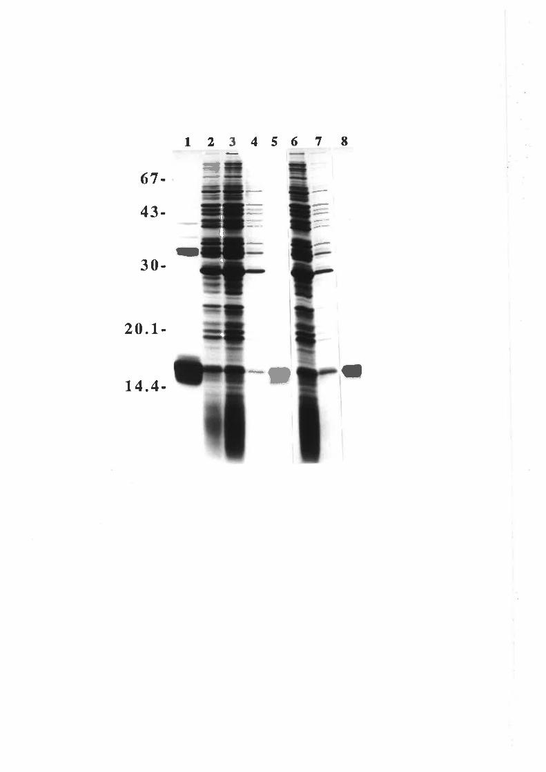
4 5 6 7 I12I
=t67-
43-
30-
20.1-
14.4-
II
e-,
a
II
I
t
NaCl, 100mM acetate pH5.0. Analysis of the eluates by silver stained SDS/PAGE
indicaæd that the one step purif,rcation yielded E21R at >95Vo purity as w¿ts observed
with wild type GM-CSF (Fig. 5.2.12). The eluates were pooled and fractionated by RP-
HPLC using a30-507o gradient of acetonitrile. Analysis of the RP-HPLC chromatogram
confirmed that the E21R was essentially free of protein contaminants after the
immunoaffinity purification (Fig. 5.2.13A). Comparison of the wild type GM-CSF and
E21R purifications indicaæd the final products a¡e both >95Vo pure as judged by silver
srained SDS/PAGE (Fig. 5.2.138). The two column procedure yielded 112pg of
purifred E21R which represented 11¡rg per litre of culture and approximately 50% of the
E21R presenr in the crude periplasmic preparation. It should be noted that although the
GM-CSF was >957o pure after aff,rnity chromatography, the final purification by RP-
HPLC was essential to remove non-protein E. colí contaminants such as LPS. The
activation of myeloid cells by factors such as LPS may mask or complicate the analysis
of the activity of GM-CSF analogues.
The protocol of immunoaffînity chromatography and RP-HPLC was used to purify
all subsequent GM-CSF analogues expressed in the BLzl strain of E. coli. Table 5.2.2
summarises the yields for the purification of all GM-CSF analogues from BL2I-
5.2.3 Quantification of purified GM-CSF
It is essential that estimates of the concentration of the purified protein be accurate
to enable comparisons of the relative activities of wild type GM-CSF and mutant
analogues. Throughout this work crude and purified GM-CSF has been quantified by
means of a radioimmunoassay (RIA). However as w¿ts mentioned in Chapter 3, a
potential problem with immunologically based protocols is the assumption that the
antibody displays equal recognition of the protein used to generate the standard curve and
the protein of unknown concentration. Clea¡ly this may not be the case when quantifying
GM-CSF mutant analogues, using wild type GM-CSF to generate a standard curve. To
some extent this problem is avoided by using polyclonal antibodies which recognise a
-95-
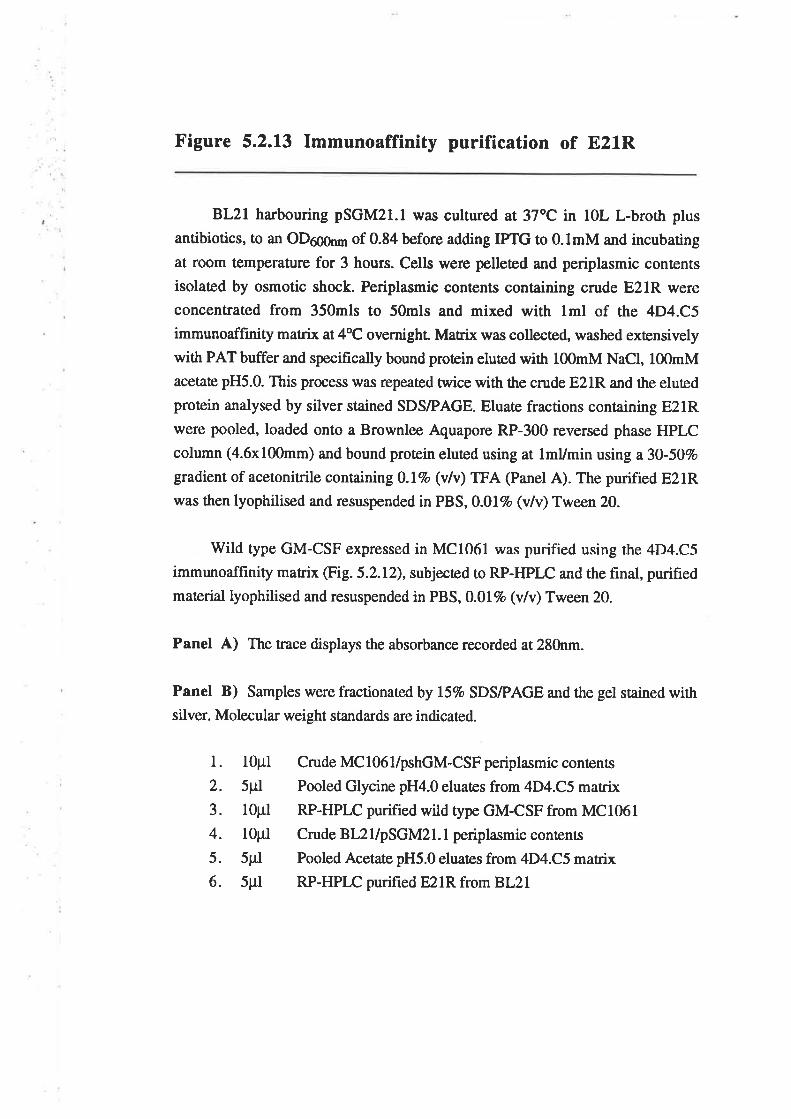
Figure 5.2.13 Immunoaffinity purification of E21R
BL2l harbouring pSGM2l.l was cultured, at 37oC in lOL L-broth plus
antibiotics, to an OD6g6¡¡¡ of 0.84 before adding IPTG to 0.lmM and incubating
at room temperature for 3 hours. Cells were pelleæd and periplasmic contents
isolaæd by osmotic shock. Periplasmic contents containing crude E2lR were
concentrated from 350mls to 50mls and mixed with lml of the 4D4.C5
immunoaffinity matrix at4oC overnight Matrix was collected, washed extensively
with PAT buffer and specifrcally bound protein eluted with 100mM NaCl, l00mMacetate pH5.0. This process w¿ls repeated twice with the crude E2lR and the eluted
protein analysed by silver stained SDS/PAGE. Eluate fractions containing E2lRwere pooled, loaded onto a Brownlee Aquapore RP-300 reversed phase HPLC
column (4.6x100mm) and bound protein eluted using at lmUmin using a30-50Vo
gradient of acetonitrile containingD.lvo (v/v) TFA (Panel A). The purified E2lRwas then lyophilised and resuspended in PBS,0.0l7o (v/v) Tween 20,
Wild type GM-CSF expressed in MC1061 wa.s purified using the 4D4.C5
immunoaffinity matrix @ig.5.2.L2), subjected to RP-HPLC and the final, purified
material lyophilised and resuspended in PBS, 0.017o (v/v) Tween 20.
Panel A) The trace displays the absorbance recorded at 280nm.
Panel B) Samples were fractionated by l57o SDS/PAGE and the gel stained with
silver. Molecula¡ weight standa¡ds are indicaæd.
l. lOpl
2. 5pl
3. lOpl
4. l0pl5. 5pl
6. 5Fl
Crude MC 106 l/pshGM-CSF periplasmic contents
Pooled Glycine pH4.0 eluates from 4D4.C5 matrix
RP-HPLC purified wild type GM-CSF from MC106l
Crude BL2|lpSGM2l. I periplasmic contents
Pooled Acetate pH5.0 eluates from 4D4.C5 matrix
RP-HPLC purified E2lR from BL21
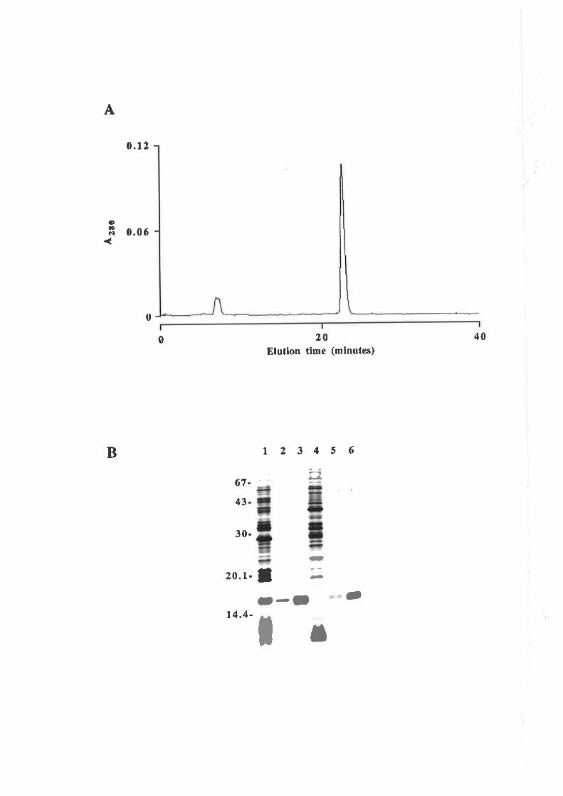
A
(Ðæcl
0.12
0.06
0
0 20Elution time (minutes)
t 234 5 6
r>.lla¡
ls)
-
40
B
67-
43-
30-
t4.4-
20.1- IO-() O--
I I
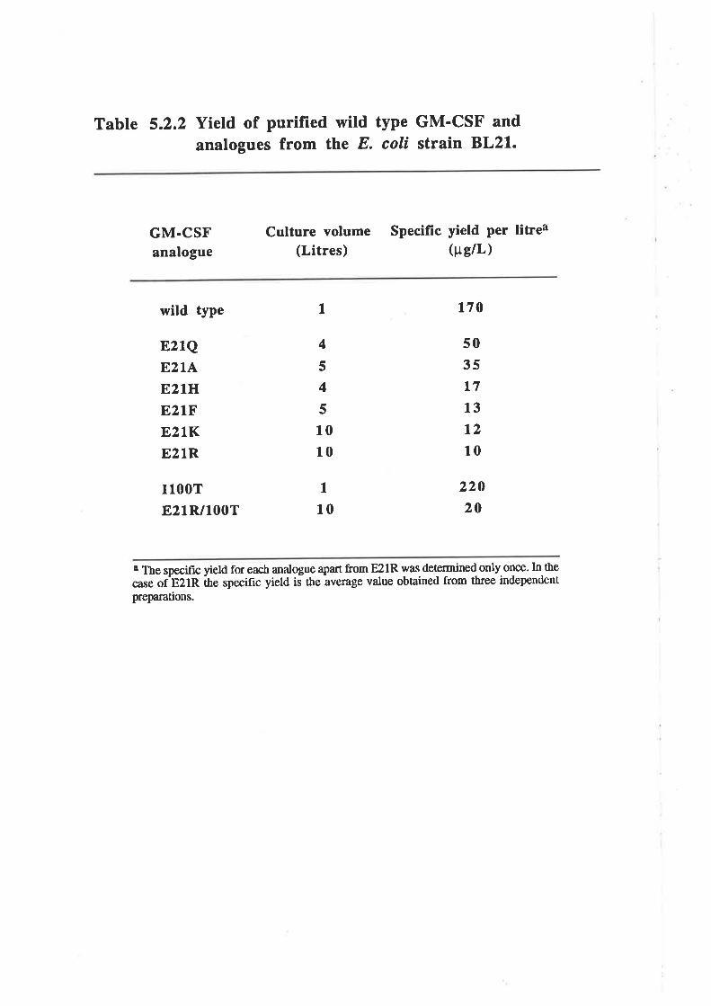
Table 5.2.2 Yield of purified wild type GM'CSF and
analogues from the E coli strain BL21-
GM.CSFanalogue
Culture volume(Litres)
Specific yietd per litrea(ttg/L)
wild type
E21Q
EzIAE'2IIJE21FE21KE21R
IlOOTE21R/1007
1
4
5
4
5
10
10
I10
170
5035l713
t210
22020
I The specificcase of E2lRpreparations.
yield for each analogue apart ftom E21R was deternined only once. In the
ihe specific yield is the average value obtained from three independent
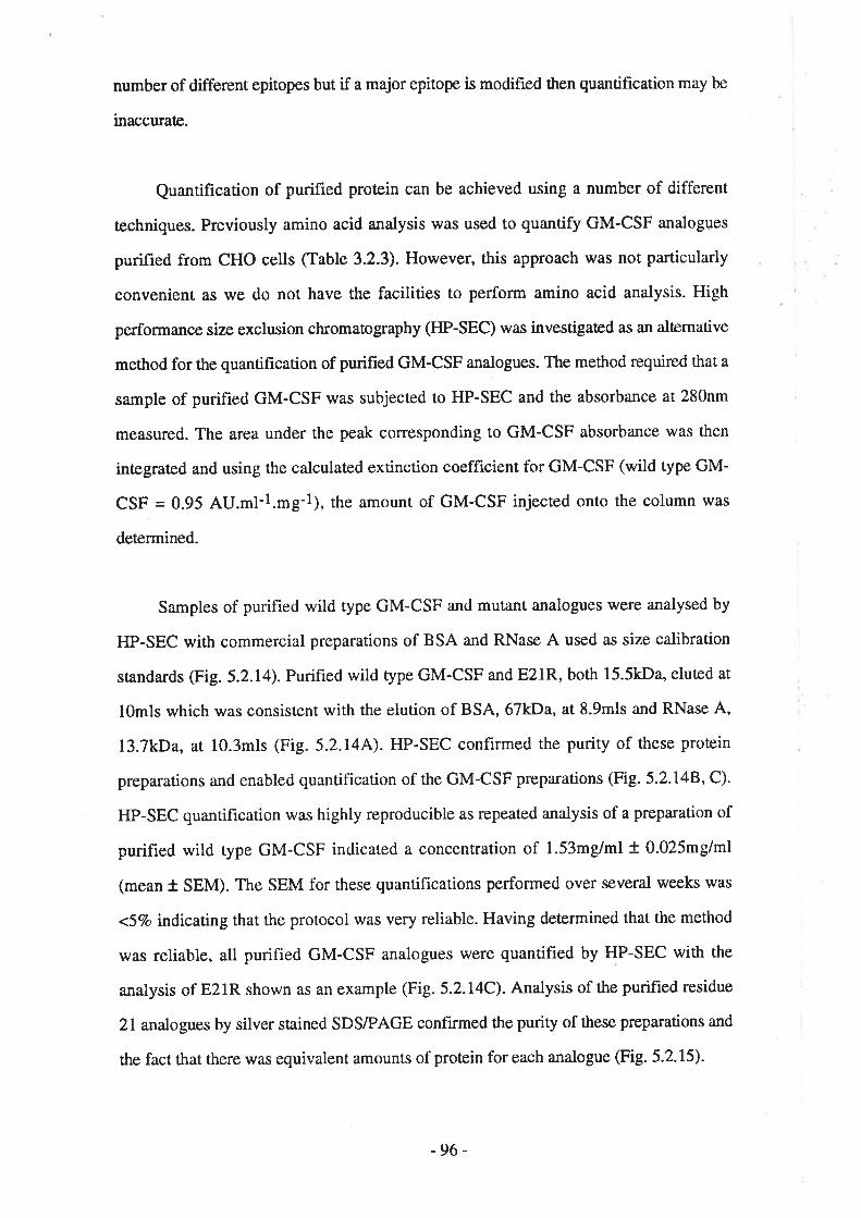
number of different epitopes but if a major epitope is modifred then quantification may be
inaccurate.
Quantification of purified protein can be achieved using a number of different
æchniques. Previously amino acid analysis was used to quantify GM-CSF analogues
purified from CHO celts (Table 3.2.3). However, this approach was not particularly
convenient as we do not have the facilities to perform amino acid analysis. High
performance size exclusion chromatography ([IP-SEC) was investigated as an alærnative
method for the quantification of purifred GM-CSF analogues. The method required that a
sample of purified GM-CSF was subjected to HP-SEC and the absorbance at 280nm
measured. The area under the peak corresponding to GM-CSF absorbance was then
integrated and using the calculaæd extinction coefficient for GM-CSF (wild type GM-
CSF = 0.95 AU.ml-l.mg-1), the amount of GM-CSF injected onto the column was
deærmined.
Samples of purified wild type GM-CSF and mutant analogues were analysed by
HP-SEC with commercial preparations of BSA and RNase A used as size calibration
standa¡ds (Fig. 5.2.14). Purified wild type GM-CSF and E21R, both 15.5kDa, eluted at
l0mls which was consistent with the elution of BSA, 67kDa, at 8.9mls and RNase A,
l3.7kDa, ar lO.3mls (Fig. 5.2.144). HP-SEC confirmed the purity of these protein
preparations and enabled quantification of the GM-CSF preparations (Fig. 5.2.148, C).
HP-SEC quantification was highly reproducible as repeated analysis of a preparation of
purified wild type GM-CSF indicated a concentration of 1.53mg/ml + 0.025m9/ml
(mean t SEM). The SEM for these quantifications performed over several weeks was
<5Vo indícating that the protocol was very reliable. Having determined that the method
was reliable, all purified GM-CSF analogues twere quantified by HP-SEC with the
analysis of E2lR shown as an example (Fig. 5.2.14C). Analysis of the purified residue
21 analogues by silver stained SDS/PAGE confirmed the purity of these preparations and
the fact that there was equivalent amounts of protein for each analogue (Fig. 5.2.15).
-96-
Figure 5.2.14 HP-SEC analysis of purified GM-CSF
HP-sEc was used to quantify samples of purified GM-csF using aBeckman ultraspherogel SEC3000 (7.5x300mm) run ar O.Smls/min in 0.1Msodium sulphate, 0.lM sodium phosphaæ pH7.0, O.os% (ilv) sodium ande.Samples in a total volume of 100p1 were loaded and the absorbance at 280nm
recorded. Inægration of the a¡ea under the peak (AU.ml) and use of the calculaæd
GM-CSF extinction coefficient (0.95 AU.ml-l.mg-1) enabled rhe amount of GM-CSF in the original sample to be calculated.
Panel A) The trace displays the absorbance recorded at 280nm for the HP-SECof a sample containing 75¡rg BSA and 5Qpg of RNase A.
Panel B) The trace displays the absorbance recorded at 280nm for the HP-SECof a sample containing 20pl of purified wild rype GM-CSF. Inægration of the peak
indicated 2¡tg of GM-CSF.
Panel C) The trace displays the absorbance recorded at 280nm for the HP-SECof a sample containing l0¡rl of purifìed E21R. Inægration of the peak indicated4.6tte of E2lR.
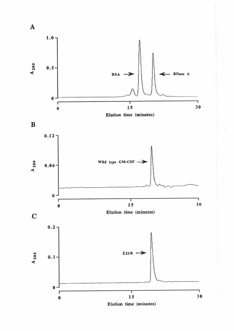
A
æGI
1.0
0.5
0
BSA + + RNsse A
0 15
Elution time (minutes)
30
B
cæêl
o.l2
0.06l{ild type GM-CSF +
0
0 15
Elution time (minutes)
E21R ->
30
c
0ûel
0.2
I
15
Elution time (minutes)
0
0 30
Figure 5.2,15 Purified GM-CSF residue 21 analogues
Human GM-CSF analogues mutated at residue 2l were expressed in 8L21,purified by immunoaffinity chromatography and RP-HPLC and quantified by Hp-sEC. The wild type GM-csF was expressed in Mc1061 and purified by anionexchange chromatography and RP-HPLC. Samples were fractionatedby I5Vo
SDS/PAGE and the gel stained with silver. Molecular weight standards are
indicated.
l. 340ng
2. 400ng
3. 410ng
4. 4l0ng
5. 415ng
6. 400ng
7. 440ng
tüild type GM-CSF
E2IAE2lQ
EzIFEzTH
EZIKE2IR
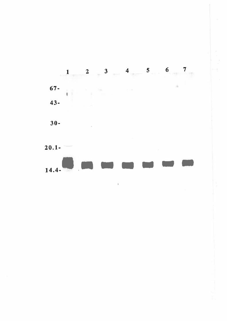
3456 712
67-
43-
30-
20.1-
14.4- ;TIIIII
ô
I

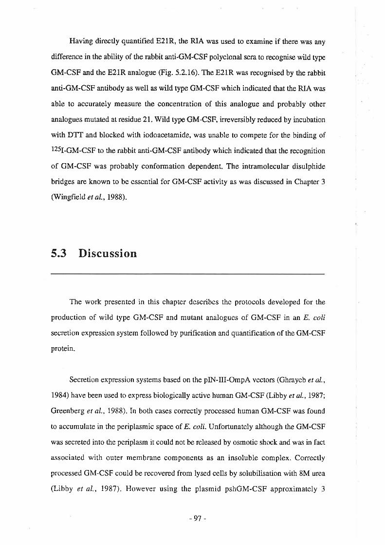
Having directly quantified E2lR, the RIA was used to examine if there was any
difference in the ability of the rabbit anti-GM-CSF polyclonal sera to recognise wild type
GM-CSF and the E21R analogue (Fig. 5.2.16). The E21R was recognised by the rabbit
anti-GM-CSF antibody as well as wild type GM-CSF which indicaæd that the RIA was
able to accurately measure the concentration of this analogue and probably other
analogues mutated at residue 21. Wild type GM-CSF, irreversibly reduced by incubation
with DTT and blocked with iodoacetamide, was unable to compete for the binding of
I25I-GM-CSF to the rabbit anti-GM-CSF antibody which indicaæd that the recognition
of GM-CSF was probably conformation dependent. The intramolecular disulphide
bridges are known to be essential for GM-CSF activity as was discussed in Chapter 3
(Wingfield et aI., 1988).
5.3 Discussion
The work presented in this chapter describes the protocols developed for the
production of wild type GM-CSF and mutant analogues of GM-CSF in an E. coli
secretion expression system followed by purification and quantification of the GM-CSF
protein.
Secretion expression systems based on the pIN-trI-OmpA vectors (Ghrayeb et al.,
1984) have been used to express biologically active human GM-CSF (Libby et aL.,1987;
Greenberg et al.,1988). In both cases correctly processed human GM-CSF was found
to accumulate in the periplasmic space of E. coli. Unfortunately although the GM-CSF
was secreted into the periplasm it could not be released by osmotic shock and was in fact
associated with outer membrane components as an insoluble complex. Correctly
processed GM-CSF could be recovered from lysed cells by solubilisation with 8M urea
(Libby et aI., 1987). However using the plasmid pshGM-CSF approximately 3
-97 -
Figure 5.2.16 Rabbit anti-GM-CSF polyclonal antibodyshows equal response to wild type GM-CSF and E21R butdoes not recognise chemically reduced GM-CSF
A radioimmunoassay using the standard rabbit anti-GM-CSF polyclonal
antisera was performed using titrations of wild type GM-CSF (a), E21R (O) and
reduced and blocked wild type GM-CSF (r) quantified by HP-SEC. Duplicate
titrations of GM-CSF competitor were mixed with rabbit anti-GM-CSF polyclonal
antibody (final l:4,000) *¿ 125¡-ÇM-CSF (O.lng/tube) and incubated at 4"C
overnight. Rabbit antibody was then precipiøæd using anti-rabbit immunobeads
and counæd.
The chemically reduced and blocked GM-CSF lvas prepared as described
below. Wild type GM-CSF, purified by anion exchange chromatography and RP-
HPLC and resuspended in PBS, was incubated with a 100-fold molar excess of
reducing agent (DTT) at25"C for 30'. A lO-fold mola¡ excess (with respect to
DTT) of thc blocking agcnt, frcsh iodoaueüamidc, was atltled and thc incubation
continued for 5'. Mixture was then adjusæd ¡o L% (v/v) acetic acid and 0.17o (vlv)
TFA, filæred and loaded onto a Brownlee Aquapore Octyl reversed phase HPLC
column (10xl00mm). Protein was eluted at SmlVmin using a3O-507o gradient ofacetonitrile containing O.lVo (vlv) TFA and fractions analysed by silver stained
SDS/PAGE. Fractions containing GM-CSF were pooled, lyophilised and
resuspended to 130¡tg/ml in PBS containing a final concentration of 800mM urea,
0.I7o (wlv) sodium azide and 0.17o (vlv) Tween 20. The reduced and blocked
GM-CSF was quantif,red by HP-SEC.
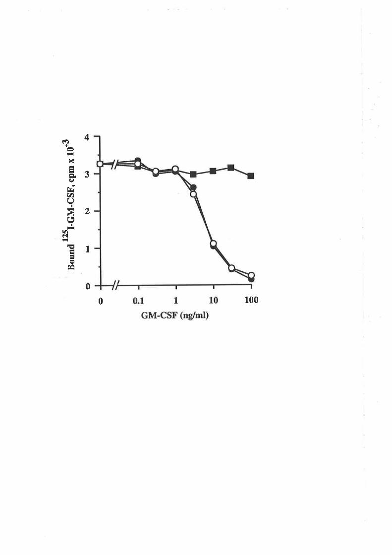
Bou
nd t"
I-G
M-c
sF,
cpm
x 1
o'3
(¡5
l.)F
¡o
c FÀ O

milligram of soluble GM-CSF per litre of culture was obtained using a standard osmotic
shock protocol (Koshland and Boaæin, 1980). Libby et al. (1987) reporæd a yield of 20
milligram of secreted, insoluble GM-CSF per litre of culture so it is quite possible that
after osmotic shock releases soluble GM-CSF some 907o of. the expressed GM-CSF
remains as an insoluble complex with outer membrane components. Cell pellets were not
examined for the presenc€ of insoluble GM-CSF a.s the aim was to generate soluble GM-
CSF. Despite the poæntial loss of insoluble GM-CSF, a yield of 3 milligrams soluble
GM-CSF per litre of culture was quiæ useable. Wild type GM-CSF was purified using
either gel filtration chromatography or anion exchange chromatography as the first
purification step, followed by reversed phase HPLC with the final product judged >957o
pure by silver stained SDS/PAGE (Fig. 5.3.1). Quantification of the purified wild type
GM-CSF by HP-SEC indicated a yield of 500pg per litre of culture which represented a
recovery of approxim ately 177o. Amino terminal sequence analysis of the purified GM-
CSF indicaæd that the OmpA signal peptide w¿¡s appropriately cleaved to leave GM-CSF
with the correct amino terminus.
The E21R analogue was of particular interest (see Chapter 3) and so, was the hrst
residue 21 analogue to be expressed in E. coli. Major problems were encountered with
very low levels of E2lR expression and then difficulties with the purification protocol
had to be negotiated. During the course of this work, 12 strains of E. coli were screened
for the ability to express E21R with only one, BL21, expressing any E21R. The highest
level of E21R expression amounted to just L.\Vo of. the expression obtained with wild
type GM-CSF. All the residue 2l substitutions examined expressed poorly in BL21
relative to wild type GM-CSF but the more conservative substitutions such as Ala or Gln
expressed considerably better than the charge reversal substitutions Arg and Lys-
purification of GM-CSF from BL2l was complicated by the presence of a 41 kDa
contaminant that co-purified with GM-CSF at most stages of the conventional
purification protocol. Eventually the problems associated with this contaminant were
overcome by developing an immunoaffinity purifrcation protocol used to purify all the
residue 21 analogues (Fig. 5.3.1).
-98-
Figure 5.3.1 Protocols for the purification of secreted,soluble human GM-CSF
Human GM-CSF expressed as a soluble product in the periplasm E. coli wa,s
purified by either conventional chromatography or affinity chromarographydepending on the strain used for expression. The purification was compleæd byRP-HPLC and the >95% pure GM-CsFquantified by HP-SEC.
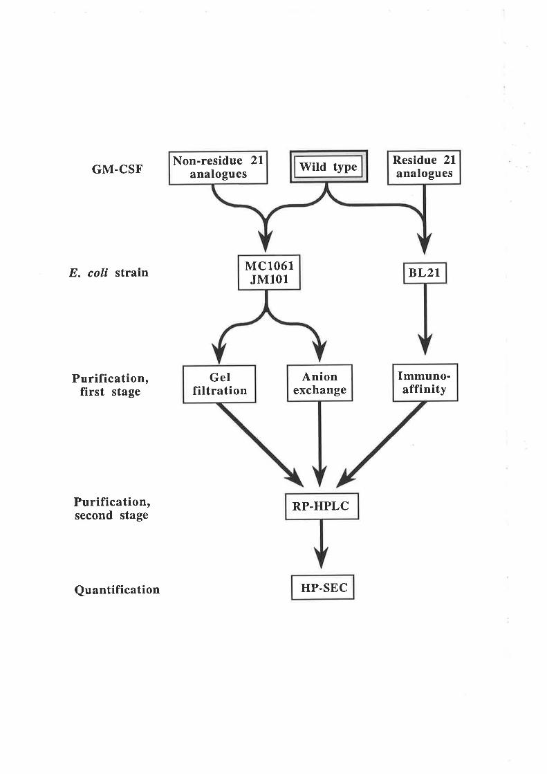
GM.CSF
E. coä strain
Purification,first stage
Purification,second stage
Quantification HP.SEC
BL2I
Immuno-affinity
Anionexchange
Wild typeNon-residue 2lanalogues
Residue 21analogues
MC1,061JM101
Gelfiltration
RP.HPLC

A key question that remains unanswered is why do residue 2l substitutions in
general and charge reversal mutations in particular, reduce the level of GM-CSF
expressed. One possible explanation for the low level of E2lR expression was that E21R
was being proæolysed within the E colí. Multiple strains of E. coli were examined for
E21R expression with a particular emphasis on strains exhibiting some deficiency in
protease activity. The strains examined exhibiæd dehciencies in intracellular protease
activity (lon),periplasmic protease activity (deSP) or outer membrane protease activity
(ompT). TTre ompT strain, BLzl, was able to express the E21R analogue but with a
relative yield of only L%o compared with wild type GM-CSF. The fact that none of the
protease defrciencies were able to support wild type levels of E21R expression suggested
that the problem was not attributable, at least directly, to bacterial protease activity.
Alærnatively the protease activity associated with low level E2lR expression remained
active in all the strains of E. coli that were examined.
A second and potentially related explanation for the low level of E21R expression
was that the mutation was structurally unfavourable and interfered with proæin folding.
The Glu2l residue is conserved in all species of GM-CSF examined (Fig. 1.2.2) and a
structurally conserved acidic amino acid is found in the hrst a-helix of most of the four
helix cyrokines (Shanafelt et a1.,1991b). The fact that this motif appe¿¡rs to play a critical
role in the activity of GM-CSF (Shanafelt et al.,199lb; Lopez et al., 1992c),IL-2
(Zurawski and Zurawski, 1989) and IL-3 (Barry et al., 1994) does not preclude it from
also having a structural role. Molecular modelling studies suggested that a number of
different amino acids may be accommodated at position 21 without generating any
destabilising steric clashes although a cluster of potentially destabilising positive charges
may result from the inuoduction of a positive charge at this position (Dr C.Bagley
personal communication). Additionally, the analogues E21A and E21R were efficiently
expressed using the CHO cell expression system (Table 3.2.3). Despite these
observations, the serendipitous mutation Tl0I enhanced the expression of the E21R
analogue. Expression of the double mutant T10UE21R was about ten-fold higher than
the single mutant, E2lR. The enhanced expression brought about by a compensatory
-99 -

mutation at a different location, suggested that there might be a structural basis for the
low expression of the E2lR analogue. The properties of this analogue will be discussed
further in Chapter 6 but so far the only observable effect of the T10I mutation appears to
be on the expression level of the E21R analogue.
The observation that secreted GM-CSF can become associated in an insoluble
complex with membrane components (Libby et al., 1987; Greenberg et aI., 1988)
suggested another explanation. Mutation of residue 2L may enhance the association of
GM-CSF with membrane components forming an insoluble aggregate with a
concomitant loss of soluble GM-CSF from the periplasm. The effectiveness of the E
coliB strain, BL2l, in the expression of E2lR may be the result of a weaker association
between E21R and outer membrane components leading to a higher proportion of soluble
E2lR in the periplasm.
An alærnative explanation, not examined during the course of this work, is that the
mutations at residue 21 disrupt the transcriptional or translational machinery thereby
lowering expression of the various analogues. This explanation does not seem likely
given that the single amino acid substitutions involve a residue 41 amino acids
downstream of the initiating methionine.
This chapter has described the bacterial expression and purification of wild type
GM-CSF and a number of GM-CSF analogues mutated at residue 21. Whilst expression
of high yields of the residue 21 analogues \'/as not ¿ts successful as wild type, sufficient
purihed maærial was obtained and accurately quantified to enable detailed analysis of the
properties of these analogues. Chapter 6 will describe the analysis of the biological
activity and receptor binding properties of the GM-CSF residue 2l analogues.
- 100-

Chapter 6
The biological activities andreceptor binding properties of
E. coli-derived GM-CSF residueanalogues: Identification of
GM-CSF antagonists
2L

lil:{)
/l?
A
6.1 Introduction
The results presented in Chapærs 3 and 4 showed that the Glu2l residue in the first
cr-helix of GM-CSF plays a crucial role in biological activity. Alignment of the predicted
amino acid sequence of GM-CSF from seven different species illustrated the absolute
conservation of the Glu2l residue (Fig. I.2.2). Acidic residues structurally equivalent to
the Glu2l of human GM-CSF are also present in many of the four cr-helix cytokines
(Shanafelt et a|.,1991b). With at least two of these cytokines, murine IL-2 (Zurawski
and Zurawski, 1989) and human IL-3 (Barry et al., t994), considerable functional
significance is attached to the acidic nature of this residue. This evidence suggests that
the conserved acidic residue in the four cr-helix cytokines may display a conserved
functional role in the interaction of ligand and receptor, perhaps through the formation of
a salt bridge.
An alternative explanation, perhaps equally consistent with the highly conserved
nature of the residue, is that the conserved acidic residue plays an important structural
role in the four a-helix cytokines. Mutation of this residue may perturb or destabilise the
tertiary structure leading to an impaired interaction with receptor and concomitant loss of
function. The altemative explanations for the function of the conserved acidic residue are
not mutually exclusive as this residue may serve both structural and functional roles. The
results presented in Chapter 5 showed that mutation of Glu2l strongly influenced the
level of GM-CSF expression using an E. coli secretion system. The fact that protease
deficient strains of. E. coli were only partially able to enhance the expression of residue
2l mutants and that a second mutation at residue 10 was also able to enhance expression
of residue 2l mutants, suggested that Glu2l may be a structurally important residue'
Interestingly, mutation of Glu21 did not appear to influence GM-CSF expression using
the COS or CHO cell expression systems described in Chapter 3.
-101 -

The aim of the work described in this chapter was to analyse the biological and
receptor binding properties of human GM-CSF residue 21 analogues generated using an
E. colí secretion expression system, as described in Chapter 5. This also enabled the
influence of carbohydrate on GM-CSF activity to be examined in the context of GM-CSF
analogues with mutations at residue 21. In preliminary experiments designed to
determine a possible structural role for Glu21, the influence of residue 2l mutations on
the stability of GM-CSF was examined.
6.2 Results
6.2.1 Activity of E. coli-derived wild type GM-CSF
Wild type GM-CSF expressed in E. coli (eco GM-CSF) was compared with wild
type GM-CSF expressed in CHO cells (cho GM-CSF) for the ability to stimulate the
proliferation of TF-l cells. The TF-l cell line is a stable, erythroleukaemia cell line,
dependent upon GM-CSF, IL-3 or erythropoietin for proliferation (Kitamura et aI.,
1939). GM-CSF from either source was able to stimulate the dose-dependent
proliferation of TF-l cells with EDSO values of 0.01ng/ml for the E. coli-denved material
and O.3ng/ml for the CHO cell-derived maærial (Figure 6.2.1). Thus eco GM-CSF is
3Q-fold more active than cho GM-CSF as previously reported (Moonen et a1.,1987).
This indicated that GM-CSF expressed by the pIN-trI-Omp secretion system in E. coli is
a fully active form of non-glycosylated GM-CSF.
The analogue designed to facilitate high specific activity labelling *i¡¡t 1251, P6YY,
exhibited wild type biological activity in TF-l proliferation assays (data not shown). As
the substitution at residue 6 did not interfere with biological activity, it was considered
unlikely that the substitution would influence receptor binding. ImportantlY; under
identical labelling conditions *¡¡6 1251, the P6YY mutant analogue was consistently
labelled to a higher specific activity than wild type GM-CSF, S.A. = 6lpCi/FB and 13.5
-t02-
Figure 6.2.1 comparison of the biological activity of eco
GM-CSF and cho GM-CSF.
Eco GM-CSF (f) and cho GM-CSF (O) were titrated for their ability tostimulate ¡3qtnymiOine incorporation into TF-l cells as described (2.10.16). Each
value represents the mean of triplicaæ determinations and error bars represent the
SEM.
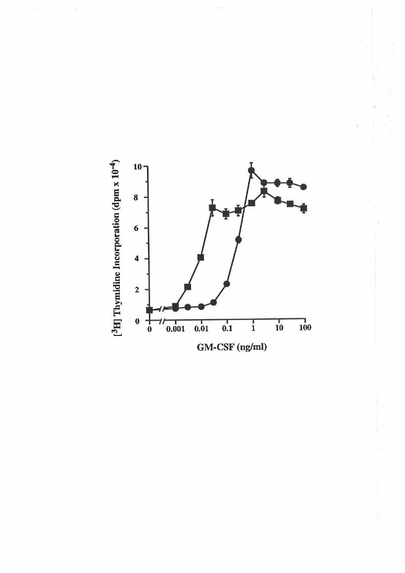
t3ril
rny
mid
ine
Inco
rpor
atio
n (d
pm x
tO
'a)
ì.)5
o\æ
)i
l¿ Fr
)l
o 7 rà ¡ o u) )l, I J oe E I -
pCi/pg, respectively (Dr J.Woodcock, personal communication). On the basis of wild
type biological activity and high specific activity labelling, I25I-GM P6YY was used
routinely in the receptor binding assays and radioimmunoassays described in this
chapter.
6.2.2 Comparison of the activity of eco GM'CSF and
eco E21R
The E coli-denved wild type GM-CSF and E21R analogue were then compared
for their ability to stimulate the proliferation of TF-l cells (Flg. 6.2.21\) or to stimulate
the production of superoxide anions by human neutrophils (Fig. 6-2.28). Quite
surprisingly the E2lR analogue was devoid of activity in both assays. Wild type GM-
CSF stimulated the dose-dependent proliferation of TF-l cells with an ED5g of
0.02nglml whereas a 50,000-fold higher concentration of E21R (1,000 ng/ml) was
complerely ineffective. Similarly, wild type GM-CSF stimulated the dose-dependent
release of superoxide anions from neutrophils with an EDSO of 0.07ng/ml whereas a
14,000-fold higher level of E2lR (1,0(Ðng/ml) was completely ineffective. No activity
was detected for eco E2lR purified by either conventional chromatographic techniques or
by immunoafhnity chromatography (data not shown)-
The absence of activity for the E. coli-denved E2lR analogue (eco E2lR) was in
contrasr ro the activity exhibited by the CHO cell-derived E2lR analogue (cho E2lR)
which was able ro stimulate the proliferation of CML cells (Fig. 3.3.5), the release of
superoxide anions from neutrophils (Fig. 3.3.6A) and the adherence of monocytes (Fig.
3.3.68) (Lopez et oI., 1992c). In all of the activities examined, cho E2lR was
approximately 300-fold less potent than cho GM-CSF. Why does the eco E21R analogue
exhibit a complere loss of biological activity when the cho E2lR analogue exhibited
reduced, but readily detectable activity?
-103-
Figure 6.2.2 comparison of the biorogical activity of eco
GM-CSF and eco E21R.
Eco GM-CSF (f) and eco E2lR (tr) were titrated for their ability tostimulaæ [3tlJthymidine incorporation into Tp-l cells (Panel A) or to stimulare theproduction of superoxide anions from human neutrophils as described (2.10.16)(Panel B). Each value represents the mean of triplicaæ deærminations and errorbars represent the SEM.
4
3
2
A
ç¡
t-{x-tLÈE-Ioi¿GI¡roÈLoc)-h-c)-I€LI
ùEr
¡¡l(f¡
B
tt)
ùÈoLûl
e)L
\ot-{
tt)q)
oILI
Io:aIEokÈq)Exotr(L)
Èct)
1
00 0.001 0.01 0.1 I 10 100 1000
GM-CSF (ng/mt)
0.1 1 10 100 1000
30
25
20
15
10
5
0
GM-CSF (nglmt)
0 0.01
6.2.3 Receptor binding characteristics of eco E21RA. Binding of eco E21R to the high (GMRaÞ") and low (GMRa)
affinity receptor
One immediate explanation for the lack of activity exhibiæd by eco E21R was that
it was unable to interact with the GM-CSF receptor. Therefore binding of eco E21R to
the high affrnity receptor (GMRap.) of human neutrophils (Gasson et a\.,1986; Park et
al., 1986) or the low affinity receptor (GMRc) of the A9lC7 cell line (Hercus et al.,
1994b) was analysed. Competitive binding studies on human neutrophils expressing the
GMRcrp" complex indicated that eco GM-CSF and eco E21R were able to bind with
affinities of 7pM and 770pM respectively (Fig. 6.2.3A). Competitive binding studies on
the A9/C7 cell line expressing the GMRcr chain indicated that eco GM-CSF and eco
E2lR were able to bind with affinities of 2,700pM and 1,l00pM respectively (Fig.
6.2.38). Thus the eco E2lR exhibited a reduced affinity for the GMRap. complex on
human neutrophils but a slightly enhanced affinity for the cloned GMRcr chain.
B. Comparative binding of eco E21R and cho E21R to the high
(GMRap") and low (GMRa) aflinity receptor
Eco E21R and cho E21R were compared in parallel for the ability to bind the high
affinity (GMRcrB") or low affinity (GMRa) GM-CSF receptor. The competitive receptor
binding curves are shown in Figure 6-2-4 and the derived K¿ values in Table 6.2.1.
Using human neutrophils both eco E21R (Fig.6.2.44) and cho E2lR (Fig. 6.2.48)
exhibit an 80- ro 100-fold reduction in affinity compared with their respective wild type
proteins (Iable 6.2.1).In contrast using the CHO cell line A9lC7, both eco E21R (Fig.
6.2.4C) and cho E2lR (Fig.6.2.4D) exhibited affinities similar to the corresponding
wild type protein (Table 6.2.I).
These results indicated that eco E21R, like cho E21R, was able to recognise the
GMRcr chain with wild type affinity but that the interaction with GMRøp" appeared to
be reduced.
- 104-
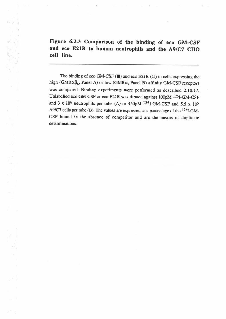
Figure 6.2.3 comparison of the binding of eco GM-csFand eco E2lR to human neutrophils and the l¡9tc7 cHocell line.
The binding of eco GM-csF (r) and eco E2lR (tr) to cells expressing thehigh (GMRc,p., Panel A) or low (GMRcr, panel B) affinity GM-csF receprors
was compared. Binding experiments were performed as described 2.10.17.unlabelled eco GM-CSF or eco E21R was titraæd against l00pM l2sI-GM-csF
and 3 x 10⁶ neutrophils per rube (A) or 450pM l2sJ-6¡4-çSF and 5.5 x 10⁵
A9lC7 cells per tube (B). The values are expressed as a percentage of the 125¡-6¡4-
CSF bound in the absence of competitor and are the means of duplicatedeterminations.
A
-o¡rt¡Ioc)
€Ho
Èa
€-LraoÊf¡ict)O
¡
ÅC,
a
rneìr-'l
80
6,0
001 GMRapc
0 0.01 0.1 I 10 100 1000
GM-CSF (nM)
GMRcr
0.01 0.1 1 10 100 1000
40-
20-
0
100
80
60
40
20
B
o¡rIIoI
CH0
èe
E
¡turt)O
¡tsrà(,
¡H
|f¡d
00
GM-CSF (nM)
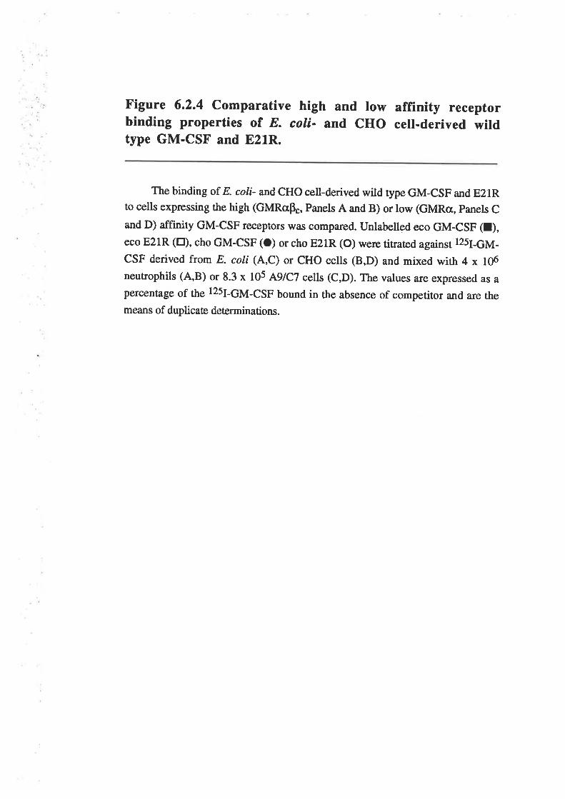
Figure 6.2.4 comparative high and low affinity receptorbinding properties or E. coli- and cHo cell-derived wildtype GM-CSF and 821R.
The binding or E. coli- andcHo cell-derived wild type GM-csF and E21Rto cells expressing the high (GMRap", Panels A and B) or low (GMRcr, panels C
and D) affinity GM-csF receprors \r,as compared. unlabelled eco GM-csF (t),eco E21R (tr), cho GM-csF (o) or cho E2lR (o) were titraæd against 12s¡-6¡4-
csF derived from E. coli (A,c) or cHo cells (B,D) and mixed with 4 x 10⁶
neutrophils (A,B) or 8.3 x 10⁵ A9tc7 cells (c,D). The values are expressed as apercentage of the l25I-GM-CSF bound in the absence of competitor and are themeans of duplicate deærminations.
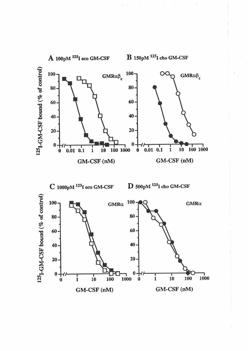
A rooprvr 25r ""o
GM-csF B rsopvt 12sJ dro GM-csF
-otr9toI
t¡ios€Å
I5o¡tf.'tC)(J
Il3à(,I
-rt¡c|F{
1 100
80
60
40
20
0
00
80
60
40
20
100
80
60
40
20
0
00
80
60
40
20
0
1
GMRoPc
GMRcr
1 10 100 1000 0
GM-CSF (nM)
0 0.01 0.1 I 10 100 1000
GM-CSF (nM)
GMRo
1 10 100 1000
GM-CSF (nM)
GMRctPc
00 0.01 0.1 1 10 100 1000
GM-CSF (nM)
C rooopvt 12sr eco GM-csF D sooplt 12sr cho GM'csF
-oLi+)hoI
€HosEaIao¡
frr(t)U
¡Hà(J
IìnôtF{ 0
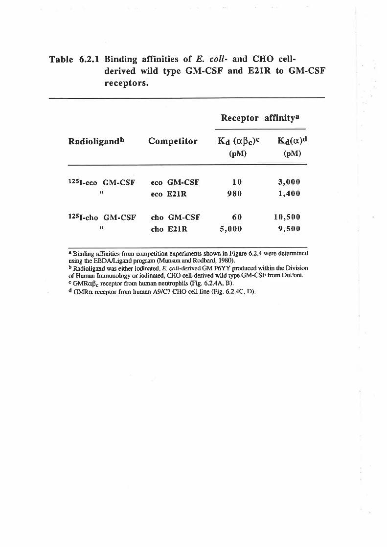
Tabte 6.2.1 Binding affinities of E. coli- and CHO cell-derived wild type GM-CSF and E21R to GM-CSFreceptors.
Receptor affinitya
Radioligandb Competitor Ka (crF")"(pM)
K¿(c)d(pM)
125¡-""o GM-CSF eco GM-CSF
eco E21R
cho GM-CSF
cho E21R
125¡-s¡r GM-CSF
It
tt
10
980
60
5,000
3,0001,400
10,5009,500
8 Binding afünities from competition experiments shown in Figure 6.2.4 we¡e detemtinedusing the EBDA/Ligand progran (Munson and Rodbard, 1980).b Radiotigand was either iodinated, E. coti-denved GM P6YY produced within the Divisionof Human Immunology or iodinated, CHO cell-derived wild type GM-CSF from DuPont.c GMRcp" receptor from human neutrophils (Fig.6.2.44, B).d GURo receptor from human A9lC7 CHO cell line @g. 6.2.4C,D).
6.2.4 Comparison of eco E21R and cho E21R
The eco E21R and cho E21R mutant analogues were analysed to try and determine
what factors may be responsible for the different biological activities of these analogues.
There a¡e two known differences between the eco E21R and cho E21R preparations. The
first is a single amino acid change due to a polymorphism at residue 100 (Wong et aI.,
1985a). The second and more obvious difference is the fact that the CHO cell-derived
material is heavily glycosylated (Fig. 3.3.4).
A. The influence of a polymorphism at residue 100 on wild type
GM-CSF and E21R activitY
The GM-CSF çDNA clone used for CHO cell expression (GM-CSF(T¡r100¡,
Wong et a1.,1985a) and E. coli expression (GM-CSF(Ib100),LeI et al.,l9S5) have a
single amino acid difference due to a polymorphism at residue 100. The fact that the
homologue of residue 100 in IL-3 has been implicated in receptor binding (Lopez et al-,
1992b) suggested that this residue might exert some influence on GM-CSF activity.
Thus the 1¡1¡100 substitution was introduced into eco GM-CSF and eco E21R and the
biological activity of the resulting analogues æsted in a TF-l proliferation assay (Fig.
6.2.5). Wild type GM-CSF carrying either 11e100 or 1¡¡100, stimulated the dose-
dependent proliferation of TF-1 cells with an ED5g of 0.013ng/ml whereas a 77,000-fold
higher level of E2lR (1,000ng/ml) carrying either ¡s100 ot 1¡¡100, was completely
ineffective. Quite clearly the I1ü)T substitution had no detectable effect on eco GM-CSF
or eco E21R activity and therefore this single amino acid difference could not account for
the different biological activities of eco E2lR and cho E21R.
B. The influence of glycosylation on the biological activity of the
E21R analogue
GM-CSF produced in eukaryotic cells, such as COS or CHO cells, is
heterogeneously modified by the addition of carbohydrate (Figures 3.3.14, 3.3.4). The
degree of GM-CSF glycosylation has been demonstrated to influence both biological
activity and receptor binding with non-glycosylated GM-CSF exhibiting higher specific
-105-
Figure 6.2.5 Comparing the effect of the residue 100polymorphism on eco GM-CSF and eco E21R biologicalactivity.
Eco GM-CSF (l), eco I100T (A), eco E21R (tr) and eco E21M100T (A)were titrated for their ability to stimulate [3H]thymidine incorporation into TF-lcells. Each value represents the mean of triplicaæ deærminations and error bars
represent the sEM.
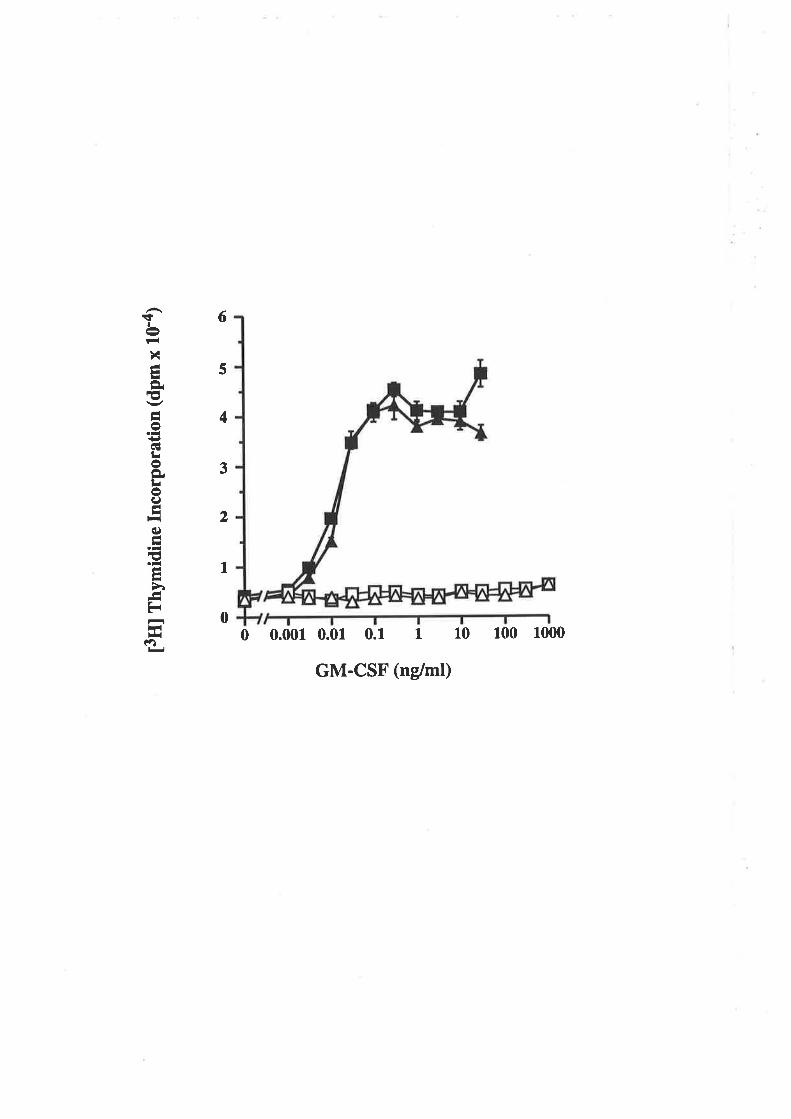
t3H
l rny
mid
ine
Inco
rpor
atio
n (d
pm x
10-
4)
Fr
ì.)(,
5U
ro\
ll li )¡ )¿ ll
o z ¡l I o u) lll I - o€I
activity and higher receptor affrnity than high Mr, glycosylaæd GM-CSF (Moonen et aI.,
1987; Cebon et a1.,1990). During the purification of the CHO cell-derived GM-CSF
analogues (Fig. 3.3.3), low Mr species were excluded because the CHO cell-derived
wild type GM-CSF was devoid of these forms (Fie. 3.3.4). It seemed possible that
some of the low Mr species of cho E2lR might be devoid of activity as is the non-
glycosylated eco E21R.
Cho E2lR was affrnity purifred from the conditioned medium of the CHO cell line,
31.7, expressing the E2lR analogue (table 3.3.2). The affinity purified cho E21R was
then fractionated by RP-HPLC and the eluted glycoprotein analysed by silver stained
SDS/PAGE. Seven fractions of cho E2lR were identified (Fig. 6.2.6A) comprising
three size classes of E21R and with molecular weights ranging from 17kDa to 33kDa
(Table 6.2.2).The size heterogeneity of CHO cell-derived GM-CSF has been attributed
to different degrees of occupancy of the two sites for Nlinked carbohydrate (Donahue ef
at.,I986a). Fractions 1&2 contain high Mr E2lR, fractions 3,4&5 contain intermediate
Mr E2lR while fractions 6&7 contain low Mr E2lR. An eighth fraction was created by
pooling equal aliquots of fractions I to 5 (Fig. 6.2.6A, Lane 9). This pooled fraction
displayed a similar degree of molecular weight heterogeneity as the CHO cell-derived
wild type GM-CSF obtained from Genetics Institute (Fig. 6.2.6A, Lane 1) and was
devoid of the low Mr forms. The fractions of cho E2lR were individually quantified by
RIA and a total of 200pg identified in the eight fractions. Analysis of the fractions by
silver stained SDS/PAGE (Fig. 6.2.64) suggested that the quantification was probably
reliable. Differences may be attributable to silver staining of the carbohydrate, which is
mosr noticeable when comparing fractions containing high Mr GM-CSF (Fig' 6-2.64,
Lane2) with those fractions containing low Mr GM-CSF (Fig. 6.2.6A, Lane 8).
The fractions of cho E2lR were tested for biological activity in a TF-l proliferation
assay (Fig.6.2.68). Wild type GM-CSF and the pooled fraction of cho E2lR, fraction
8, stimulated the dose-dependent proliferation of TF-1 cells with EDSO values of 0-15
and 130 ng/ml, respectively. This demonstrated that cho E2lR is 900-fold less active
-106-
Figure 6.2.6 Fractionation of cho E21R and biologicalactivity of the variably glycosylated species.
Conditioned medium (volume4,700mls) from the CHO cell line 31.7,
expressing cho E21R, was concentrated to a f,rnal volume of 270mls in a stirred-
cell using a 10,000 Mr cutoff membrane. Concentrated conditioned medium was
mixed with lml of fresh 4D4.C5 immunoaffinity matrix at 4oC overnight. Matrixwas collected, washed with PAT buffer and specifically bound protein eluæd withl00mM NaCl, l00mM sodium acetate, 0.027o (w/v) sodium azide, pH5.0. This
process u,as repeated several times and the eluæd protein analysed by silver stained
SDS/PAGE. Eluates containing cho E2lR were pooled, adjusæd to l%o (v/v) acetic
acid and O.lVo (vlv) TFA and loaded onto a Brownlee Aquapore RP-300 reversed
phase HPLC column (4.6x100mm). Bound protein was eluted using a 30-50vo
gradient of acetonitrile containin g0.t7o (v/v) TFA. Fractions containing cho E2lRwere lyophilised and resuspended in PBS. Following RIA quantification, the
variably glycosylated fractions of cho E2lR were analysed by silver stained
SDS/PAGE (A) and the biological activities of each compnred (B).
Panel A) Samples were fractionated by L5Vo SDSÆAGE and the gel stained withsilver. Approximately 400ng of each GM-CSF sample was loaded per track.
Molecular weight standards are indicaæd.
l. CHO cell-derived recombinant human GM-CSF
2. cho E2lR, fraction 1
3. cho E2lR, fraction 2
4. cho E2lR, fraction 3
5. cho E21R, fraction 4
6. cho E2lR, fraction 5
7 . cho E2lR, fraction 6
8. cho E2lR, fraction 7
9. cho E2lR, fraction 8 (Pool of fractions I to 5)
Panel B) Cho GM-CSF (O) and the cho E2lR fractions I (tr), 2 (a),3 (L), 4(A), 5 (O), 6 (O),7 (V) and 8 (O) were titrated for their ability to stimulate
[3H]thymidine incorporation into TF-1 cells. Each value represents the mean oftriplicate determinations and error bars represent the SEM.
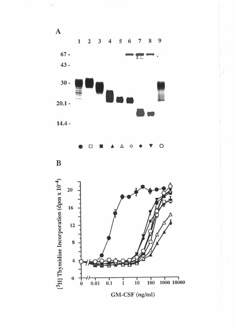
A
67-
43-
20.1 -
14.4 -
12 3 4 567 89
-Er,
1..lr
20
B
çI
F{
XÅ7ÈE¡od¡roÈLoI
q)
E
ÉrH*{
ra
t6
t2
I
4
o I A^Oa v o
0 0.01 0.1 1 10 100 10m 100000
GM-CSF (ng/ml)
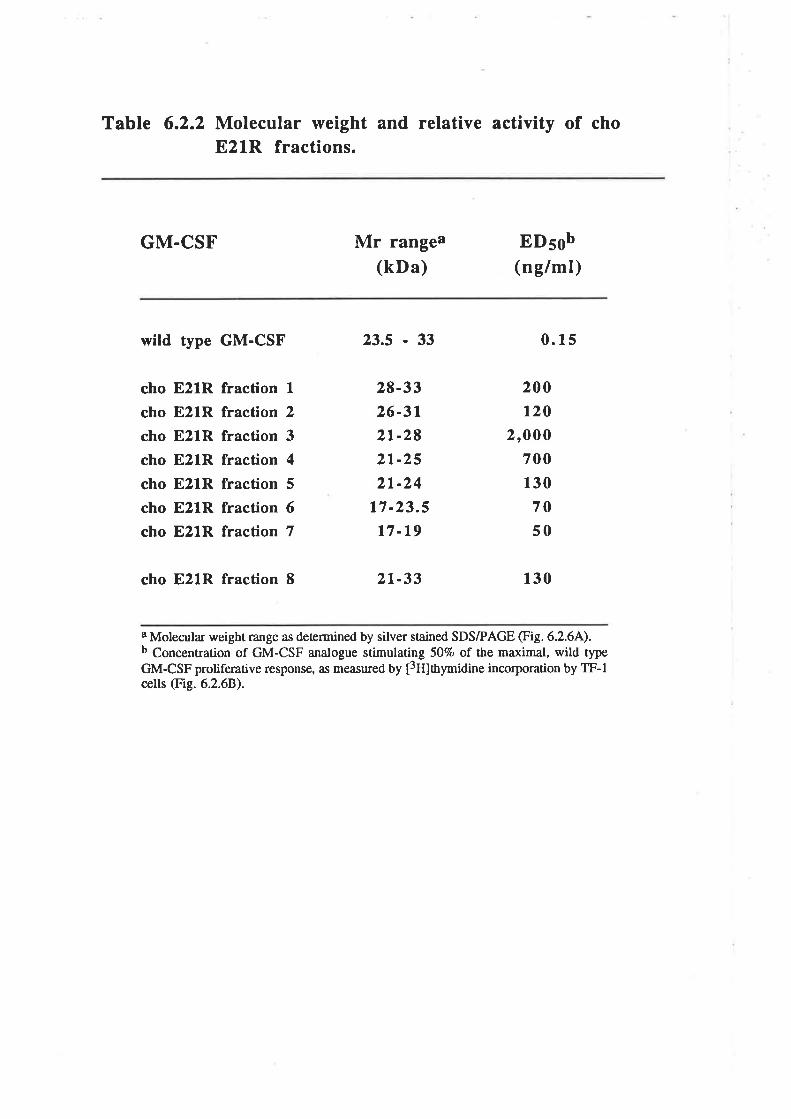
Table 6.2.2 Molecular weight and relative activity of choE21R fractions.
GM.CSF Mr rangea(kDa)
ED5sb(ng/ml)
wild type GM-CSF 23.5 - 33 0.15
cho
cho
cho
cho
cho
cho
cho
E21R
E21R
E21R
E21R
E21R
E21R
E21R
fraction Ifraction 2
fraction 3
fraction 4
fraction 5
fraction 6fraction 7
28-3326-312t-282t-252t-24
t7 -23.5
t7 -19
200120
2,000700130
7050
cho E21R fraction 8 2t-33 130
I Molecular weight mnge Íìs deærrrined by silver stained SDS/PAGE (Fig. 6.2.64).b Concentration of GM-CSF analogue stimulating SOVo of the maxinal, wild typeGM-CSF proliferative response, as me¿su¡ed Uy t3Hlt¡ymidine incorporation by TF-lcells (Fig. 6.2.68).
than wild type in agreement with previous estimates of the activity of this mutant (Fig.
3.3.5). The ED56 values of the different fractions of cho E21R ranged from 5Ong/ml for
cho E21R fraction 7 up to 2,000ng/ml for cho E21R fraction 3 (Table 6.2.2).
Intermediate Mr 821R, fractions 3&4, exhibited the lowest specific activity (Table
6.2.2). Most importantly the low Mr fraction of cho E2lR, fraction 7, which previously
had not been analysed, exhibited the highest specific activity (Fig. 6.2.68, Table 6.2.2).
The low Mr of this fraction indicated that it is likely to be devoid of N-linked
carbohydraæ (Donahue et al.,l986a; Kaushansky et a1.,1987). In this respect the cho
E21R behaves in the same manner as wild type GM-CSF where the N-linked
carbohydrate appears to play a significant role in reducing the specific activity of
glycosylated GM-CSF (Moonen et a1.,1987: Cebon et a1.,1990).
C. Stability of eco GM-CSF and eco E21R
In order to deærmine what effect the charge reversal has on GM-CSF folding,
transverse urea gradient gel electrophoresis was used to analyse eco GM-CSF and eco
E21R (Fig.6.2.7). From these gels it was clear that both eco GM-CSF and eco E2lR
displayed a reduced electrophoretic mobility as the protein unfolded with increasing
concentrations of urea. The smooth mobility transition from the folded to the unfolded
state is indicative of a rate of unfolding and folding that is fast compared with the time of
electrophoresis (Hollecker and Creighton, 1982). The midpoint of the unfolding
transition occurred at about 4M urea for eco GM-CSF and 2.5M urea for eco E2lR. The
stability of the folded protein (ÀG'(HzO)) was estimated using a procedure described by
Hollecker and Creighton (1982) to be -43 kJ/mol for eco GM-CSF and -21kJlmol for
eco E21R. This observation demonstrated that the folding of eco E2lR was significantly
less stable than for eco GM-CSF but most importantly that the eco E2lR was folded in
the absence of urea. This suggested that the highly conserved Glu2l residue might play
an important role in stabilising the folded state of human GM-CSF, presumably through
some form of electrostatic interaction.
-107-
Figure 6.2.7 Comparison of the stability of eco GM-CSFand eco E21R.
Eco GM-CSF (Panel A) and eco E2lR (Panel B) were subjected to
transverse urea gradient PAGE as described 2.10.L2. Fifteen micrograms ofpurified GM-CSF was fractionated on gels cast as an identical pair and stained wittr
Coomassie blue as described 2.10.13. The direction of electrophoresis (- * f) and
orientation of the urea gradient (0* 8M) a¡e indicated.

A
0 lUrea
'¡
Ij +
B
-
+
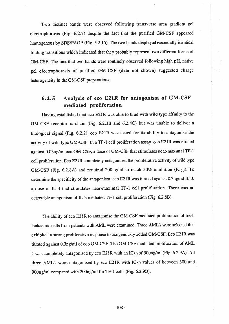
Two distinct bands were observed following transverse urea gradient gel
electrophoresis (Fig. 6.2.7) despite the fact that the purified GM-CSF appeared
homogenous by SDS/PAGE (Fig. 5.2.15).The two bands displayed essentially identical
folding transitions which indicaæd that they probably represent two different forms of
GM-CSF. The fact that two bands were routinely observed following high pH, native
gel electrophoresis of purified GM-CSF (data not shown) suggested charge
heterogeneity in the GM-CSF preparations.
6.2.5 Analysis of eco E21R for antagonism of GM'CSFmediated proliferation
Having established that eco E21R was able to bind with wild type affinity to the
GM-CSF receptor s chain (Fig. 6.2.38 and 6.2.4C) but was unable to deliver a
biological signal (Fig. 6.2.2), eco E21R was tested for its ability to antagonise the
activity of wild type GM-CSF. In a TF-l cell proliferation assay, eco E2lR was titrated
against 0.03ng/ml eco GM-CSF, a dose of GM-CSF that stimulates near-maximal TF-l
cell proliferation. Eco E21R completely antagonised the proliferative activity of wild type
GM-CSF (Fig. 6.2.84) and required 200ng/ml to reach 507o inhibition (IC5s). To
determine the specificity of the antagonism, eco E2lR was titrated against O.3nglml IL-3,
a dose of IL-3 that stimulates near-maximal TF-l cell proliferation. There was no
detectable antagonism of IL-3 mediated TF-l cell proliferation (Fig. 6.2.88).
The ability of eco E2lR to antagonise the GM-CSF mediaæd proliferation of fresh
leukaemic cells from patients with AML were examined. Three AML's were selected that
exhibited a strong proliferative response to exogenously added GM-CSF. Eco E2lR was
útrared against O.3ng/ml of eco GM-CSF. The GM-CSF mediated proliferation of AML
I was completely antagonised by eco E2lR with an IC5g of 500ng/ml (Fig. 6.2.94). AI
three AML'S were antagonised by eco E2lR with ICso values of between 300 and
900ng/ml compared with 200ng/ml for TF-1 cells (Fig. 6.2.98).
-108-
Figure 6.2.8 Eco E21R antagonises GM-csF but not IL-3mediated proliferation of TF-l cells.
Eco GM-CSF (t), eco E21R (tr) and IL-3 (A) were titrated for their abilityto stimulate l3H]thymidine incorporation into TF-l cells. In antagonisticexperiments, eco E21R (o) was titrated against 0.03ng/ml eco GM-CSF (panel A)or O.3ng/ml IL-3 (Panel B). Each value represents the mean of triplicatedeærminations and error bars represent the SEM.
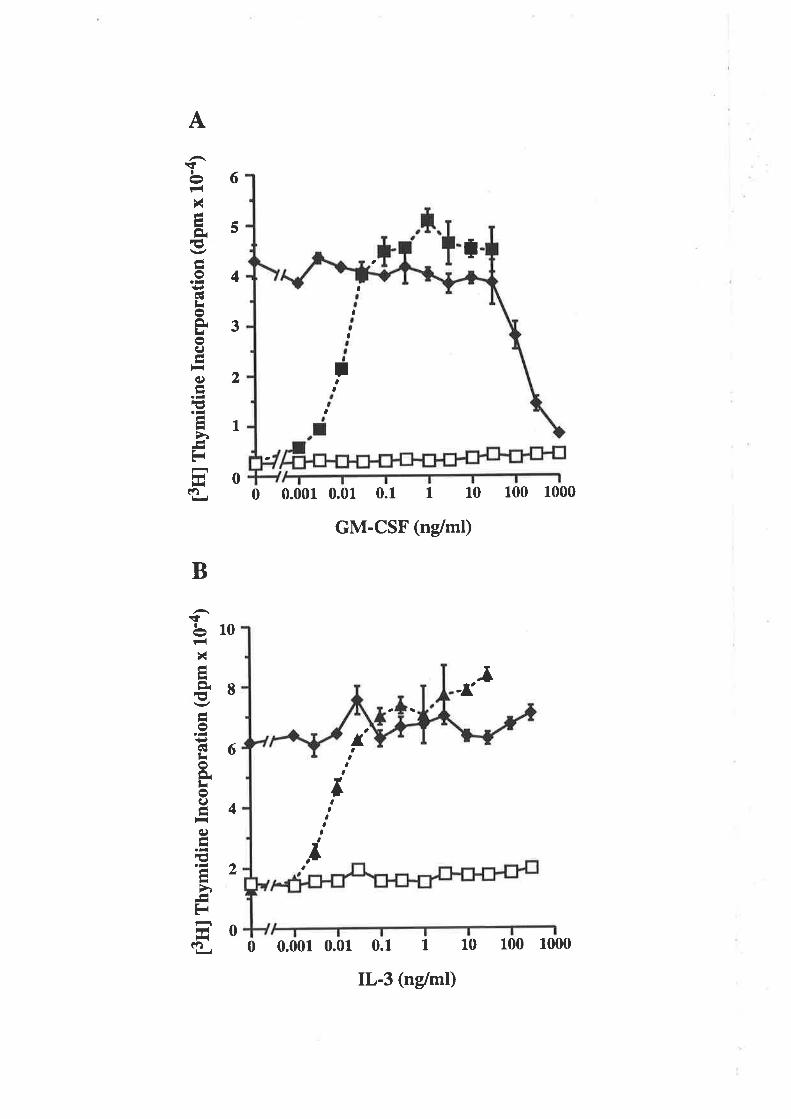
[3ff]
ffr
ymid
ine
Inco
rpor
atio
n (d
pm x
tg'a
) eg
t3
ffl f
nym
idin
e In
corp
orat
ion
(dpm
x 1
0-4)
Þæ
o\
¡r l¿ l¡ ll
l{ ¡ (, oe
ì.)A
o z ¡ì ¡ o U)
tat
J oe
¡
l.)r¡
A(,
lo\
Þ.
t)t
II
I
I a
Jrtl
Figure 6.2.9 Eco E21R antagonises GM-csF mediatedproliferation of AML cells.
Primary AML cells were selected for their ability to incorporate
[3H]thymidine in response to GM-CSF. Panel A shows the incorporation of[3FrJthymidine by AML #1 in response ro eco GM-csF (r) and eco E2rR (tr). Inantagonistic experiments, eco E2lR (o) was titraæd against O.3ng/ml eco GM-CSF. Each value r€presents the mean of triplicaæ deærminations and error bars
represent the SEM.
In panel B the responsiveness to eco GM-CSF and sensitivity to eco E21Rantagonism of 3 AML isolates and the TF-l cell line a¡e compared.
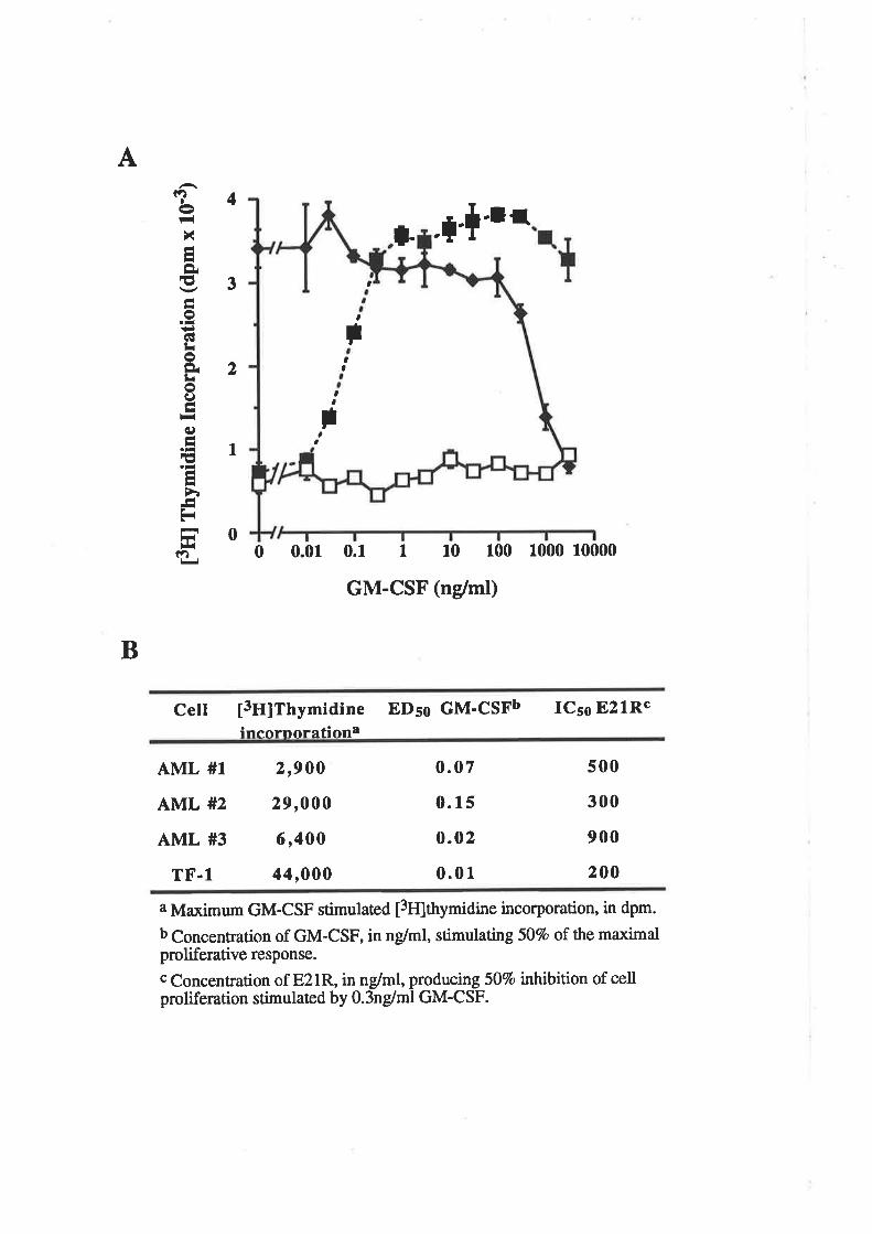
A
aa
4
3
2
1
11Iet-r(
xÉHÈE-to.-t¡GIlioÈLoI-L-(¡)Á
I
-ÅÁI
ÅF{
)¡iÉ)
l. .l'+'lra
aIt
a
B
st
a
00 0.01 0.1 I 10 100 1000 10000
GM-CSF (ng/ml)
Cell 13ff¡tnymidine EDso GM-CSFb ICso E2lRcineornorationa
21900
29,000
6,400
44,000
aMaximum GM-CSF stimulaæd [3H]thymi¿ine incorporation, in dpm.
b Concentration of GM-CSF, in ng/ml, stimulating 507o of the maximalproliferative response.
c Concentration of E2lR, in nglml, producing 507o inhibition of cellproliferation stimulated by O.3ng/ml GM-CSF.
AML #1
AML #2
AML #3
TF-1
0.07
0. 15
0.02
0.01
s00
300
900
200
GM-CSF isolated from in yivo sources is variably but quite extensively
glycosylated (Lusis et al., 1981; Donahue et aI., 1986a: Cebon et aI., 1990). The
biologicat activity and receptor binding properties of GM-CSF are influenced by the
degree of gtycosylaúon (Cebon et a1.,1990) so the ability of eco E2lR to antagonise the
activity of glycosylaæd GM-CSF produced in ye¿Nt or CHO cells was assessed. Eco
E21R was titrated against concentrations of the different forms of wild type GM-CSF
that stimulate near-maximal TF-l cell proliferation. Eco E21R antagonised the
proliferative activity of all forms of wild type GM-CSF (Fig. 6.2.L0) with IC5g values of
eco GM-CSF, 600ng/ml, yeast GM-CSF, 700ng/ml, and cho GM-CSF, 90ng/ml- The
yerrst GM-CSF used in this assay, although partialty glycosylated, appeared to display
the same level of biological acúvity as the eco GM-CSF (Fig. 6.2.104 and B). This is
consistent with the fact that this preparation of yeast GM-CSF lacked N-linked
carbohydrate (Cosman et a1.,1988) and therefore would be expected to exhibit higher
specific activity than GM-CSF modified by the addition N-linked carbohydrate (Moonen
et al., 1987; Cebon et al., 1990). Interestingly, the IC5g value with cho GM-CSF
(9gng/ml) represented a 9O-fold excess of eco E21R but the ICso with eco GM-CSF
(600ng/ml) represented a 6,000-fold excess.
The effect of the residue 100 polymorphism on eco E21R antagonism was
examined in a TF-1 cell proliferation assay. Eco E2lR and ecoE2lMlOOT were titrated
against 0.03ng/ml eco GM-CSF. Both forms of eco E2lR were able to completely
antagonise the proliferative activity of eco GM-CSF with IC5g values of eco E2lR,
60ng/ml and eco E2lR/I100T, l(X)ng/ml (Fig. 6.2.11). Thus the I100T substitution has
no effect on the antagonistic activity of eco E21R'
6.2.6 Eco E21R antagonism of neutrophil activation
Since GM-CSF is a strong stimulator of inflammatory cells, the ability of eco
E21R to antagonise GM-CSF-mediated stimulation of superoxide production by human
neutrophils was examined. Eco E21R was titrated against 1.Ong/ml eco GM-CSF, a dose
that stimulates near-maximal superoxide release from neutrophils. Eco E21R completely
-109-
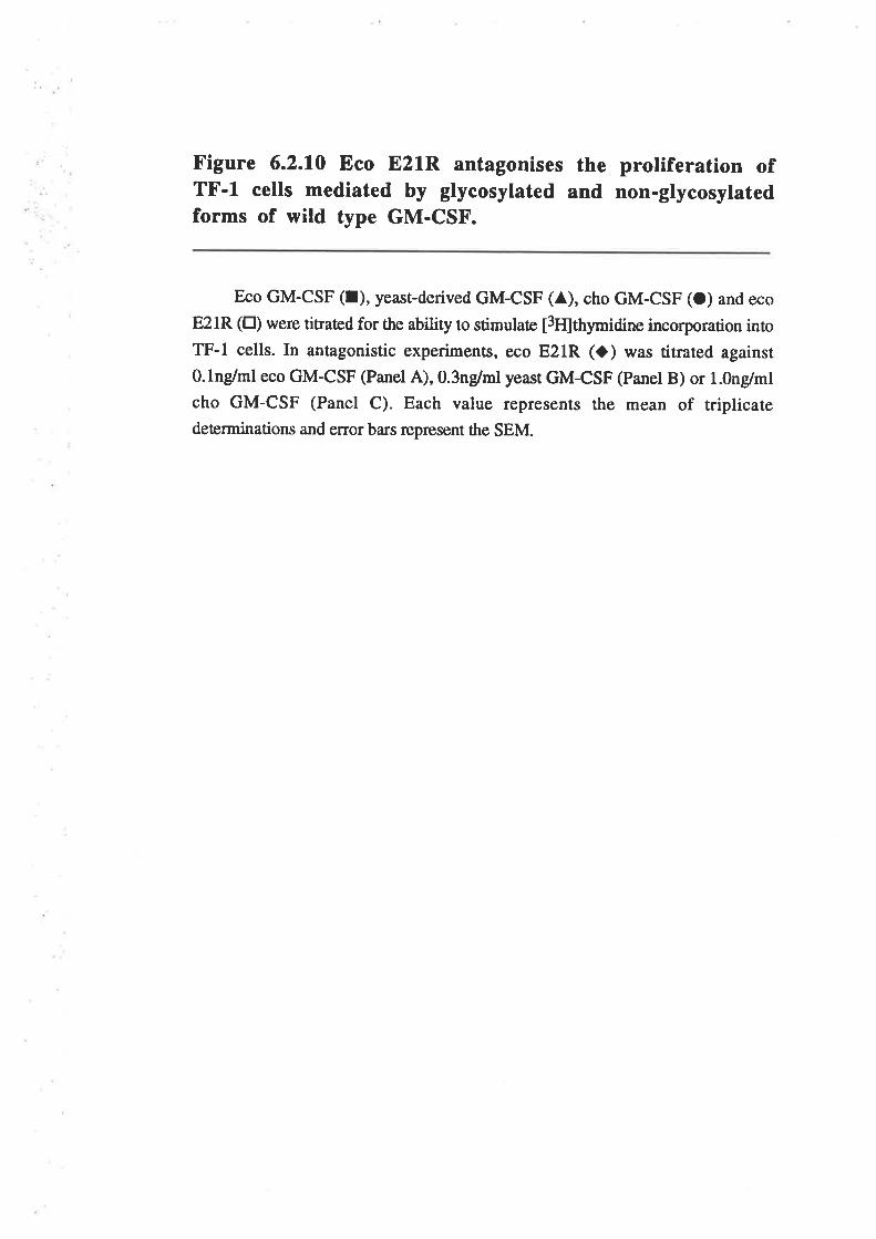
Figure 6.2.10 Eco E21R antagonises the proliferation ofTF-l cells mediated by glycosylated and non-glycosylatedforms of wild type GM-CSF.
Eco GM-CSF (I), yeast-derived GM-CSF (A), cho GM-CSF (O) and eco
E2lR (tr) were titrated for the ability to stimulate t3flttrymidne incorporation into
TF-l cells. In antagonistic experiments, eco E21R (l) was titrated against
O.lng/ml eco GM-CSF (Panel A),0.3ng/ml yeasr GM-CSF (Panel B) or l.Ong/ml
cho GM-CSF (Panel c). Each value represents the mean of rriplicatedeærminations and error bars represent the SEM.
t3ril
rny
mid
ine
Inco
rpor
atio
n (d
pm x
10'
4)
oE
É
cþ50
\æË
)ll¿
êþòc
¡¿5o
\æ¡H
H êtr'è
c¡¡5
o\æ
¡¿llF
clJS
f,-.
-...1
.a_
-aa-
aa-
aìa-
_-__
)l c li l-. Þ¡ |¡
o z È I o U)
F!
I ) oe I
}l ll tr li c Þr o li o
ú
II
¡¿ l¿ l¿ l¿ )r Fr O
'oO
.
ù I o
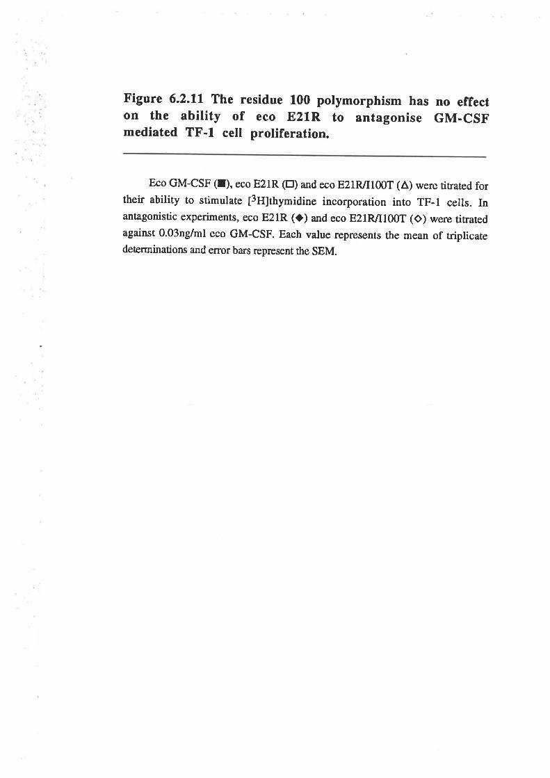
Figure 6.2.11 rhe residue 100 polymorphism has no effecton the ability of eco E21R to antagonise GM-csFmediated TF-l cell proliferation.
Eco GM-csF (t), eco E2tR (tr) and eco E2IR/IIOOT (a) were tirared fortheir ability to srimulate ¡3H;thymidine incorporarion into TF-l cells. Inantagonistic experiments, eco E21R (o) and eco E2IR/I100T (o) were titratedagainst 0.03ng/ml eco GM-CSF. Each value represents the mean of triplicatedeærminations and error bars represent the SEM.
t3rr
l rny
mid
ine
Inco
rpor
atio
n (d
pm x
t0'4
)
5ll
ì.)(, r.
o ll )i ll
o z )l I o U) ãt I ) oq I
II
-fr
f
antagonised the GM-CSF-mediated release of superoxide from neutrophils (Fig.
6.2.12A) with an IC5g of 300ng/ml. To determine the specificity of the antagonism, eco
E2lR was titrated against 3.Ong/ml TNF-c¿, a dose that stimulate near-maximal
superoxide release from neutrophils. There was no detectable antagonism of TNF-a-
mediated release of superoxide from neutrophils (Fig. 6.2.128).
The ability of eco E21R to antagonise the stimulation of superoxide production by
neutrophils mediaæd by glycosytaæd GM-CSF was examined. Eco E2lR was titrated
against 3.0ng/ml cho GM-CSF, a dose that stimulates near-muimal superoxide release
from neurrophils. Eco E21R completely antagonised the GM-CSF-mediated release of
superoxide from neutrophils (Fig. 6.2.13) with an ICso of 9Ong/ml-
6.2.7 Activity of multiple residue 2l analogues
The results presented in this chapter clearly established that the E21R substitution
can totally abolish GM-CSF agonism without affecting the binding affinity for the
GMRcx, chain. Previous work, using a COS cell expression system, suggested that other
substitutions at position 2l would have less dramatic consequences (Figures 3.3.5 and
4.2.4).
A. Functional activity of residue 21 analogues
Eco GM-CSF and the E coli-denved residue 21 analogues, E214, 821Q, E2lF,
E2LH, E2lK and E2lR were compared for their ability to stimulate the proliferation of
TF-l cells. Eco GM-CSF stimulated dose-dependent TF-l cell proliferation with an
EDSO of Q.Qlng/ml while the eco E21R and E21K analogues were completely
ineffective. The other analogues stimulate dose-dependent TF-1 cell proliferation with
EDso values of O.2nglml for eco E}IA and E21Q, 6nglml for eco EzlF and l0ng/ml for
eco E2lH (Fig. 6.2.14). The fold decrease in activity for the eco E21A analogue
compared with eco GM-CSF was similar to that observed for the cho E21A analogue
compared with cho GM-CSF (Fig. 3.3.5). These results illustrated the fact that many
- 110-
Figure 6.2.12 Eco E21R antagonises GM-csF but notTNFa mediated stimulation of human neutrophils.
Eco GM-csF (l), eco E2lR (tr) and rNF-a (a) were titraæd for theirability to stimulate the production of superoxide anions from human neutrophils. Inantagonistic experiments, eco E2lR was titraæd against l.0ng/ml (a) of eco GM-CSF (Panel A) or 3.Ong/ml (a) of TNF-cr (panel B). Each value represenß the
mean of triplicaæ deærminations and error bars represent the SEM.
Sup
erox
ide
prod
uctio
n, n
mol
es /
106
neu
trop
hils
tÉS
uper
oxid
e pr
oduc
tion,
nm
oles
/ 10
6 ne
utro
phils
>
Ur
Fr
lr \a
êu¡o
UI
HB
bJe(
,lc
T¡ I ¡ l.
o o F (Þ oe
l.
c l¿ o l¿ l¡ c
'l'l
o F¡ o ,\ I o I oe
t'l
t
Figure 6.2.13 Eco E21R antagonises the activation ofhuman neutrophils mediated by glycosyrated wild typeGM.CSF.
cho GM-csF (a) and eco E21R (tr) were titraæd for their ability tostimulaûe the production of superoxide anions from human neutrophils. Inantagonistic experiments, eco E2lR was titraæd against 3.Ong/ml (o) of cho GM-CSF. Each value represents the mean of triplicaæ determinations and error barsrepresent the SEM.
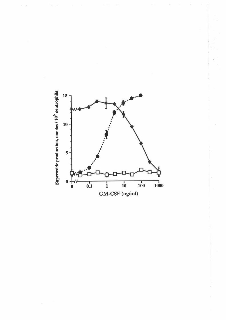
o ¡
Sup
erox
ide
prod
uctio
n, n
mol
es /
106
neu
trop
hils
ll oll U
r.
I .
.¡.
. I
rì(
t I aO
'..(,I I
ì b
t
II
t
)i )r l¡ o
o z ¡à ¡ o U)
tal
qt I
Èìr
.....
Figure 6.2.14 comparison of the biological activity of eco
GM-CSF and the E. colí-derived, residue 21 analogues.
Eco GM-CSF (t), eco E2lA (A), eco E21Q (O), eco E2lF (O), eco EZLH(V), eco E21K (A) and eco E21R (tr) were titrated for their ability to stimulare
¡l¡f¡mymidine incorporation into TF-l cells. Each value represents the mean oftriplicate deærminations and error bars represent ttre SEM.
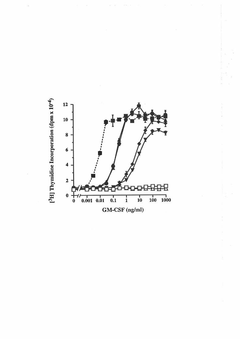
r
0 0.001 0.01 0.1 I 10 100 1000
GM-CSF (ndml)
tt2
10
I,,
a,,,aI
aa
6
4
2
çIct-lN-ILÈ€Lo:¡6lhoC¡.¡roIhtlq)t
r3LL
ùEr
r*lÉì
aaaIIaaI,a
I
a
0
amino acids are toleraæd at residue 21, albeit with a reduction in biological activity, but
that charge reversal has a dramatic influence on GM-CSF activity.
B. Receptor binding of residue 21 analogues
The binding of eco GM-CSF and the residue 21 analogues to the high affinity
(GMRcrÞc) and low affinity (GMRcr) GM-CSF receptor was comparcd. The competitive
receptor binding curves are shown in Figure 6.2.15 and the derived Kd values in Table
6.2.3. Using human neutrophils which express the high affînity receptor (GMRclÞ") all
the residue 21 analogues exhibit a 100- to 23Gfold reduction in affinity compared to eco
GM-CSF (Fig.6.2.15A and Table 6.2.3).In contrast using the CHO cell line A9lC7
which expresses the low affinity receptor (GMRcr), eco GM-CSF and the residue 21
analogues all exhibit similar affînities (Fig. 6.2.15B and Table 6.2.3). Thus despite their
considerable differences in biological activity (Fig.6.2.14), the binding of the E. coli-
derived GM-CSF residue 21 analogues to the high and low affrnity rcceptors are virtually
indistinguishable from each other.
6.2.8 Analysis of eco E21K for antagonistic activity
The fact that eco E2lK was able to bind with wild type affinity to the GM-CSF
receptor a chain (Fig. 6.2.15C) but was unable to deliver a biological signal (Fig.
6.2.14) suggested thar it would posses antagonistic activity like the eco E2lR analogue.
In a TF-l proliferation assay, eco E21K and eco E2lR were titrated against 0.03ng/ml
eco GM-CSF and 3.0ng/ml cho GM-CSF. Both analogues completely antagonised the
proliferative activity of eco GM-CSF (Fig. 6.2.164) with IC5s values of 150ng/ml for
eco E2lK and 200ng/ml for eco E2lR. Similarly the proliferative activity of cho GM-
CSF was anragonised (Fig. 6.2.168) with IC5g values of 500ng/ml for eco E2lK and
350ng/mt for eco E2lR.
In the neutrophil activation assay, eco E21K and eco E21R were titrated against
1.Ong/mt eco GM-CSF or 3.Ong/ml cho GM-CSF. Both analogues completely
anragonised the superoxide release mediated by eco GM-CSF (Fig. 6.2.16C) with ICso
- 111-
Figure 6.2.15 comparison of the binding of eco GM-csFand the B. coli-derived residue 2l analogues to humanneutrophils and the A9lC7 CHO cell line.
The binding of eco GM-CSF (f), eco EzlA (A), eco E2le (O), eco E2tF(o), eco EzlH (V), eco EzlK (a) and eco E2lR (tr) to cells expressing the high(GMRap", Panel A) or low (GMRcr, Panel B) affinity GM-csF receprors was
compared. Unlabelled eco GM-CSF or analogues were titrated against 100pMl2s¡-6¡4-gSF and 3 x 10⁶ neutrophils per tube (A) or 450pM l2s¡-6¡4-çSF and
5.5 x 10⁵ A9lC7 cells per tube (B). The values are expressed as a percenrage of the1251-6¡4-çSF bound in the absence of competitor and are the means of duplicatedeterminations.
0 0.01
T.0.1 1 10
GM-CSF (nM)
GMRaP"
100 1000
GMRcr
IIItI¡ttt¡I¡I
100
EO
60
40
20
A
-otr--toIttoÈq
EtI=oå
t¡'{(r)t)
Ittà(,¡
H|ir¡ôlr-{
B
-oL+lLoI
+ioBa
EÁI-o¡?(t)(J
ar-À()a
tÍ¡ñtF(
It¡II¡I
r.I
0
¡
100
80
60
40
20
0
I
0.1 1 10
GM-CSF (nM)100 10000 0.01
Table 6.2.3 Binding affinities of eco GM-CSF and the residue
2l analogues.
Receptor affinitya
GM.CSF K¿ (gÞ")b(pM)
K¿(cr)"(pM)
Wild type
E,2tA^
E21Q
E21FE2IIJE.2IKE21R
7
730940
1,6001,0601,060
730
1,800820
1,3001,400
8001,20o1,000
I Binding affinities from competition experiments shown in Figure 6.2.15
were determined using the EBDA/Ligand program (Munson and Rodbard,
1980).b Mean binding affinities derived from two competition experiments on
human neutropnin (GMRap") using lOOpM 1251-6¡4-ç5F and differentconcentrations of residue 21 analogues.c Mean binding affinities derived from th¡ee competitio_n experimenfs on
the CHO cell line AgtCT (GMRd) using 500pM 12s1-6¡4-ç5F anddifferent concenEations of residue 2 1 analogues.
Figure 6.2.16 Eco E2lK and E2lR antagonise theproliferation of TF-l cells and the activation of humanneutrophils mediated by glycosylated and non-glycosylatedforms of wild type GM-CSF.
Eco GM-CSF (I), cho GM-CSF (O), eco E21R (tr) and eco E2lK (A)were tiuated for the ability ûo stimulate [3t!]t]rymidine incorporation into TF-l cells(Panels A and B) and for the ability to stimulate the production of superoxideanions from human neutrophils (Panels C and D). In anøgonistic experiments, eco
E2lR (o) and eco E2lK (o) were titraæd against 0.03ng/ml eco GM-csF (panel
A)' 3.Ong/ml cho GM-csF (Panel B), l.Ong/ml eco GM-csF (paner c) or3.0ng/ml cho GM-CSF. Each value represents the mean of riplicaæ determinations
and error bars represent the SEM.
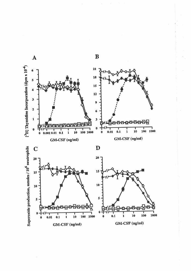
Sup
erox
ide
prod
uctio
n, n
mol
es /
106
neu
trop
hils
[3U
] fn
ymid
ine
Incu
rpor
atio
n (d
pmx
104)
ëaB
O
gsì\)
l|cu
r-\J
\¡
(,
"'-I-
--._
_-
orro
\\oB
IJÞ
¡\)
\)(,
lælr
ta
13.
èutc
,\
EÉ
ê -ôc ?'
êÈ
t¡¡ O
cu)
;-fã oa
(3 o
ô ZP
¡ o (t) ãtË
CC
I r¡ ê ê
ê c
o ? ìì I o (t) trt
ce
(3 co 7 ¡l ¡ o (t) ta
1
R.
êl,r l,r
ê ê ê
values of 150ng/ml for eco E2lK and eco 82lR. Similarly eco E2lK and eco E2lR
completely antagonised the superoxide release mediaæd by cho GM-CSF (Fig. 6.2.16D)
with IC5g values of 200ng/ml for eco EzlK and 8Ong/ml for eco E2lR.
Thus the eco E21R and E21K GM-CSF analogues, which are devoid of
measurable agonistic activity, are equally effective antagonists of GM-CSF mediaæd
activity.
6.3 Discussion
The work presented in this chapter describes the analysis of E coli-denved wild
type GM-CSF and analogues mutated at residue 21. The somewhat unexpected finding
that the E21R analogue was devoid of activity when the same analogue produced in CHO
cells, was biologically active (Chapær 3) was examined in some detail. The absence of
biological activity in certain GM-CSF analogues carrying basic amino acids at residue 21
(Fig.6.2Ja) but the retention of wild type binding affinity for the GM-CSF receptor
alpha chain (Fig.6.2.15) resulted in the generation of GM-CSF specific antagonists
(Fig.6.2.16). The potential clinical application of GM-CSF antagonists are discussed.
6.3.1 The properties of the GM'CSF residue 21
analogues
A. Receptor binding and biological activity
The results described in Chapter 3 indicated that residue 2l of human GM-CSF is
specifically involved in the binding to the pç chain of the GM-CSF receptor. The results
with the E. coli-denved residue 21 analogues support a model of GM-CSF interaction
with its receptor that emphasises the role of residue 2l and suggests a possible role for
carbohydraæ in the pç chain interaction.
-tr2-
The GM-CSF residue 21 analogues E214, E21Q, E21F and E21H all exhibiæd
significantly reduced biological activity (Fig. 6.2.14) yet the binding affinity of these
analogues for human neutrophils expressing GMRaps appeared to consist of low
affrnity binding only, equivalent to the binding affrnity observed for the A9lC7 cell line
expressing the GMRa chain (Tabte 6.2.3). Thus despite the fact that the E2lA, E2lQ,
EzlF and E21H analogues were able to deliver a biological signal, presumably through
interaction with the pç chain, this inæraction did not result in higher affinity binding to
GMRaBç than GMRcr alone. As the binding afhnity of the residue 21 analogues for the
GMRcr chain was essentially unalæred (Table 6.2.3), the biological activity probably
reflects the absolute effect of the mutations on Þc interaction. The possibility that the
analogues signal through binding to the GMRc¡ chain alone, does not account for the
different biological potencies of these analogues. The data strongly support a model
where Glu2l of GM-CSF directly interacts with p" and that mutations at this residue
disturb this interaction (Fig. 6.3.1). The lack of a direct correlation between binding
afhnity and biological activity as observed for the residue 2l analogues is a result of the
interaction of residue 2l with the critical signalling component (Þ.) rather than the
primary binding component (GMRcr) of the GMRcrp" receptor complex. A region of the
pç chain that specifîcally inclu¿s5 l{ir367, has been shown to be important for high
affinity binding of GM-CSF (Lock et al., 1994). The ability of the ps chain mutant
Y3654/H3674/I3684 to partially restore the high affinity binding of eco E2lR
(Woodcock et a1.,1994) provides additional evidence for a direct interaction between
GM-CSF GIu2l md Þ..
The exact mechanism of GM-CSF receptor activation is not known but appears to
involve the low affinity binding of GM-CSF to GMRcr and functional association of this
complex with pç, leading to high affinity binding of GM-CSF and signalling (Nicola ør
al., 1993; Goodall et al., 1993; Hoang et al., 1993; Miyajima et al., 1993). Recent
studies indicate that GMRcr md Þ" may exist as a preformed complex in the absence of
ligand (Frank Stomski, personal communication; Ronco et a1.,1994), demonstrating that
association of GMRcr and pç alone is insufficient for receptor activation. Considerable
-113-
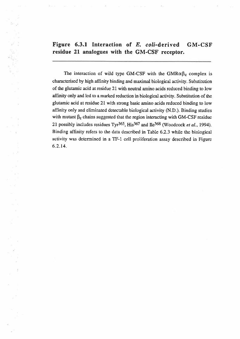
Figure 6.3.1 Interaction of E. colí-derived GM-CSFresidue 21 analogues with the GM-CSF receptor.
The interaction of wild type GM-CSF with the GMRcrp. complex is
characterised by high affinity binding and maximal biological activity. Substitution
of the glutamic acid at residue 21 with neutral amino acids reduced binding to lowafftnity only and led to a marked reduction in biological activity. Substitution of the
glutamic acid at residue 21 with strong basic amino acids reduced binding to lowaffinity only and eliminaæd deæctable biological activity (N.D.). Binding studies
with mutant p" chains suggested that the region interacting with GM-CSF residue
2l possibly includes residues Tyr365, ¡¡is367 ¿¡1¿ ns368 (Woodcock et a1.,1994).
Binding affinity refers to the data described in Table 6.2,3 while the biological
activity was determined in a TF-l cell proliferation assay described in Figure
6.2.14.
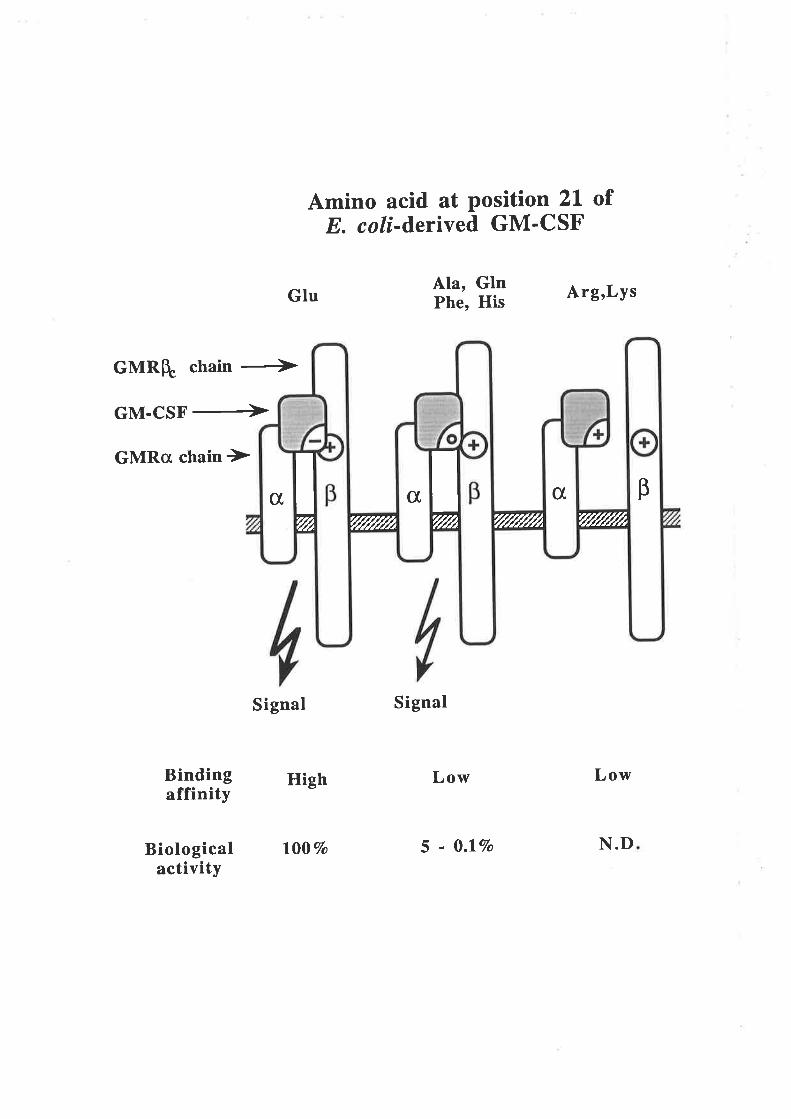
Amino acid at Position 2l ofE. coli-d,erived GM'CSF
Ala, GlnPhe, His Arg,Lys
GMRqc chain Ð
GM-CSF+
GMRo chain )
Signal
Bindingaffinity
Signal
Low
5 - 0.lVo
Glu
High
l00Vo
Low
N.DBiologicalactivity
c[c[ct, BI't/Å
I 'f/l////rtT//////fr rll¿ '{//////Á
evidence indicates that the low affinity binding of GM-CSF to a GMRaps complex is
sufficient to trigger a biological response perhaps through an allosæric interaction' Thus'
human GM-CSF binds with low affinity to murine cell lines expressing the endogenous
murine pç chain and the transfected human GMRg chain and elicits a proliferative
fesponse (Metcalf et a1.,1990; Kitamura et a1.,1991a). Similarly the cho E2lR (Fig'
6.2.68) and eco EzlA, E21Q, E21F and E21H (Fig. 6-2.14) analogues, which lack
high affinity binding, were able to elicit a proliferative response- In all these cases the
reduced binding affinity was correlated with a reduction in biological potency' In a most
unusual example however, the murine GM-CSF E2l|analogue binds with low affinity
only yet triggers a full biological response with wild type potency (Shanafelt and
Kastelein, lggz).In contrast, the eco E21R and E2lK analogues were unable to deliver
a biological signal yet exhibited binding afhnities for the GMRøpç receptor that were
indistinguishable from the eco E214, E21Q, E2lF and E21H analogues (Fig' 6'2'15)'
This demonstrated that low affinity binding does not necessarily lead to receptor
activation and suggested that the eco E2lR and E2lK analogues were completely unable
to interact with the pç chain. This was also demonstrated by the inability of eco E21R to
antagonise the IL-3 mediated proliferation of TF-l cells (Fig. 6-2-8) or to cross-compete
¡or 1251-¡¡-3 binding (data not shown).
B. Structural ProPerties
Transverse urea gradient gel electrophoresis was used to obtain an estimate of the
srabiliry of wild type GM-CSF and the E2lR analogue expressed in E- coli (Fig' 6'2'7)'
The stability of the fotded protein (ÀG"(Hzo)) was estimated to be -43 kJ/mol for eco
GM-CSF and -21 kJ/mol for eco E21R with folding:unfolding transitions at urea
concenrrations of 4M (eco GM-CSF) and 2.5M (eco E21R). Thus the urea gradient gels
provided qualitative evidence that the substitution of arginine at residue 21 reduced the
folded stability of GM-CSF. However, it must be emphasised that in the absence of
denarurant the E2rR analogue did appear to adopt a forded conformarion (Fig. 6-2-7)-
The esrimate of eco GM-CSF stability (-43 kJ/mol) differs from that previously reported
for E coli-deived wild type GM-CSF where AGo(H2O) = -24-Zk[lmol (Wingfield et al''
-lt4-
1988). This presumably reflects a difference in data analysis as the 4M folding transition
observed for eco GM-CSF is similar to the 4.lM transition reported by Wingfield et al.
(1e88).
The reduced stability of the eco E21R analogue does not imply gross structural
differences and is unlikely to account for the dramatic loss of function. Two observations
suggested that the tertiary stn¡cture of the E21R analogue was closely relaæd to that of
wild type GM-CSF. Firsrly, all residue 21 analogues bind with wild type affinity to the
GMRcI chain (Fig. 6.2.158 and Table 6.2.3). Secondly, the conformation sensitive
polyclonal antibody used for RIA quantification, exhibiæd full recognition of the eco
E21R analogue (Fig. 5.2.16).
6.3.2 Potential role of carbohydrate in cho E21R
The main difference between the model described in Figure 6.3.1 and the previous
model (Fig. 3.a.3) is that the E coli-denved, residue 2l chatge reversal mutants were
unable to signal. The eco E21R and cho E2lR analogues exhibit significant differences
in biological activity (Fig.6.2.2 vs Fig. 6.2.68) despite essentially identical receptor
binding properties (Fig.6.2.4). The only known difference between the two analogues
is the presence of extensive carbohydrate modifications in the cho E21R. Fractionation of
cho E2lR by reversed phase HPLC was able to separate a number of glycosylation
variants of the E2lR analogue (Fig. 6.2.64), all able to stimulate proliferation of TF-l
cells (Fig. 6.2.68). The fact that cho E21R which apparently lacks N-linked
carbohydrate exhibited the highest biological activity (Fig. 6.2.68, Table 6.2-l)
suggested that rhe presence of O-linked carbohydrate may play a role in the biological
activity of cho E21R. If such a role does exist for the O-linked carbohydraæ of cho
E21R, then iS removal either enzymatically or by mutation, should eliminate the
biological activiry of the cho E21R analogue. Interestingly, in the context of the E2lA
substitution, the presence of ca¡bohydraæ did not appear to influence the relative activity
of the analogue as both eco E21A (Fig.6.2.14) and cho EZIA (Fig. 3.3.5) exhibited an
activity of approximately 5 to l\Vo of the respective wild type GM-CSF molecules.
-115-
At least two mechanisms can be envisaged to account for the potential role of
carbohydrate in the biological activity of the cho E21R analogue (Fig. 6.3.2). The first
model proposes that the substitr¡tion of basic amino acids at residue 21 removes a critical
contact between GM-CSF and the pç chain. The presence of carbohydrate moieties on
cho E2lR, possibly O-linked, enables direct contact between GM-CSF and the pç chain
in the absence of an interaction utilising the Glu2l of GM-CSF (Opdenakker et al.,
1993). A carbohydrate mediated pç chain contact is clearly not required for the activity of
non-glycosylated wild type GM-CSF (Fig. 6.2.1), may or may not occur with
glycosylated wild type GM-CSF and is not measurable in binding experiments (Table
6.2.2).In this last respect, direct carbohydrate contact would generate a signal but with
no affinity conversion and be simila¡ to the situation described in Figure 6.3.1 for ¡he E.
coli-denved GM-CSF analogues able to signal but exhibiting only low affinity binding.
The second model proposes that the substitution of basic amino acids at residue 2l
introduces a charge repulsion between GM-CSF and the pç chain that completely
abolishes pç chain contact. The presence of carbohydrate may be able to mask the charge
repulsion and thereby enable interaction between GM-CSF mutant analogues such as cho
E21R and the pç chain. The potential of carbohydrate to mask charge interactions may
also function with wild type GM-CSF to mask favourable charge interactions with ü. Þ"
chain and thereby be partially responsible for the reduced activity of glycosylated GM-
CSF compared with non-glycosylated GM-CSF (Fig- 6-2.1)-
An alternative explanation is that the carbohydrate influences the stability of the
E2lR analogue. In general, the presence of covalently attached carbohydrate increases
the overall dynamic stability of a protein (Opdenakker et a1.,1993) although it has been
demonstrated that carbohydrate does not increase the stability of wild type human GM-
CSF (Wingfield et ¿1., 1988). Eco E21R adopted a folded conformation in the absence of
denaturant but was approximately 50Vo less stable than eco GM-CSF (Fig. 6.2.7)- It is
possible though, that in the context of the E21R substitution, carbohydrate might exert a
stabilising influence on the protein, perhaps by nullifying destabilising electrostatic
inreractions introduced by the basic amino acid (Hollecker and Creighton, 1982)- This
- 116-

Figure 6.3.2 Models for the role of carbohydrate in thebiological activity of the cho E21R analogue.
Models proposed to account for the possible role of carbohydraæ in the
biological activity of the CHO cell-derived E2lR analogue. In model one the
carbohydraæ moieties on cho E21R are able to bind th" 9. chain and this leads to
signalling. In model two the carbohydraæ moieties on cho E2lR mask a charge
repulsion of the p. chain that enables cho E21R to inæract wittr the pç chain leading
to signalling. Fractionation of the various glycosylation species of cho E2lRindicaæs that the relevant carbohydrate moieties may be O-linked (Fig. 6.2.6).
ECO GM.CSF >\
eco E21R >\
GMRcx, chain
E. coli-derivedGM-CSF
E. coli-derivedE21R
GMRqc chain
Model 2
carbohydrateshieldscharge
a
/ \
-Z
Model L
carbohydratecontact
@
+
p
ct,p
CHO cell-derived E21'R
question could be addressed by deærmining the stability of the cho E21R analogue using
the transverse urea gradient gel electrophoresis method.
6.3.3 Clinical relevance of GM'CSF antagonists
In vítro and iz vivo studies have demonstraæd that GM-CSF is a pleiotropic
cytokine that stimulates both the production of different haemopoietic lineages (Clark and
Kamen, 1987;Gasson, 1991), and the effector function of mature myeloid cells (Lopez
et a1.,1983, 1986; Grabsæin et a1.,1986; Metcalf et al., 1986, 1987:' Gasson et al.,
1984; Sieff ¿r aI., I98!;Haak-Frendscho et a1.,1988). These properties have lead to the
in vivo use of GM-CSF to stimulate haemopoiesis following chemotherapy (Antman ¿r
¿1., 1988; Brandt et a1.,1988; Socinski et al., 1988), bone marrow transplantation
(Nemunaiti s et a1.,1991; Peters et a1.,1993; Rabinowe et a1.,1993) and in situations of
myelodysplasia (Vadhan-Raj et a|.,1987; Ganser et al-, 1989). In patients with AIDS
GM-CSF stimulates myelopoiesis (Groopma;n et aI., 1987) as well as enhancing
neuüophil function (Baldwin et a1.,1988).
It is also apparent that GM-CSF may play a role in the pathogenesis of several
disease conditions, in particular chronic inflammation and leukaemia. Transgenic mice
carrying additional copies of the murine GM-CSF gene display elevated levels of GM-
CSF and an accumulation of macrophages in the eyes and striated muscles leading to
blindness, muscle damage and premature death possibly as a result of GM-CSF-
mediated macrophage activation (Lang et a1.,1987). In humans the presence of elevated
levels of GM-CSF in the bronchoalveolar lavage of atopic patients (Kato et a1.,1992)
and in the synovial fluid of patients with rheumatoid arthritis (Williamson ¿t aI.,1988;
Xa et al., L989; Alvaro-Gracia et a1.,1989) suggest that GM-CSF plays a pathological
role in some inflammatory diseases. This idea is supported by the observations that
patients with rheumatoid arthritis possess activated neutrophils in the synovial fluid
(Emery et a1.,1988) and an increase in the level of circulating monocytes expressing the
GM-CSF receptor (Field and Clinton, 1993). GM-CSF also stimulates the production of
the neutrophil chemoattractant IL-8 (McCain et at., 1993) and, in conjunction with
-tL7-
lipopolysaccharide, the monocyte chemoattractant macrophage inflammatory protein lcr
(Kasama et a1.,1993) thus regulating the loçalisation of leukocytes at the site of
inflammation.
Studies on Alvfl (Hoang et a1.,1986; Begley et a1.,1987b), CMMoL @verson ef
aI.,l9g9) and ALL (Freedman et a1.,1993) have demonstrated a proliferative response
to paracrine GM-CSF.In certain cases of AML (Young and Griffîn, 1986; Young et al',
Ig87,1938) and ALL (Freedman et al., 1993), GM-CSF mRNA or protein has been
deæcæd suggesting the possibility of an autocrine fesponse to GM-CSF. The expression
of GM-CSF by AML blast cells appears to be regulated by IL-l (Delwel et al-,1989;
Russell, lgg2) as is the expression of other cytokines such as G-CSF and IL-6 which
are also able to stimulate AML proliferation (Bradbury et al-,1990). In addition, non-
haemopoietic tumours such as certain small cell lung carcinomas (Baldwin et a1.,1989)'
osteogenic sarcomas (Dedhar et a1.,1983) and certain colon adenocarcinomas (Berdel et
a1.,1989) aberrantly proliferate in response to GM-CSF'
The ability of the GM-CSF analogues eco E21R and eco EzlK to specifically
inhibit the GM-CSF mediated activation of neutrophils in vitro (Fig.6-2-17 D) and
leukaemic cell proliferation (Fig.6.2.178), suggests a potential therapy for disea'ses
exacerbated by GM-CSF. The clinical potential of these GM-CSF antagonists may be
enhanced by generating more potent, second generation antagonists, which exhibit
higher affinity for the GMRcr chain, Phage display has been used to select a variant of
human growth hormone with 15 substitutions that exhibits a 400-fold increase in affinity
for the growth hormone binding protein (Lowman et al-,1991; Lowman and Wells'
lgg4). This approach should be suitable for the selection GM-CSF variants with
enhanced affinity for the GMRcr chain. The results shown in Chapter 3 which identify
rhe fourth cr-helix of GM-CSF as a potenrial GMRct chain binding site (Fig- 3.3.11)'
suggest a region where appropriate mutations might enhance the affinity of GM-CSF for
the GMRg chain.
-118-
Chapter 7
Final Discussion
The aim of the work described in this thesis was to study the structure-function
properties of the human cytokine GM-CSF and specifically to identify regions involved
in the interaction of GM-CSF with its receptor complex. The biological and receptor
binding properties of analogues generated by mutagenesis of the GM-CSF cDNA were
examined and the functional role of the wild type sequences deduced. Using this
approach, two spatially distinct recepûor binding sites were identified that are required for
maximal biological activity and interaction with the two chains, GMRa and pç, of the
GM-CSF receptor. This information paved the way for the generation of the frrst GM-
CSF specific antagonists.
GM-CSF is a pleiotropic cytokine able to súmulate both the production of different
haemopoietic lineages and the effector function of mature myeloid cells (reviewed in
Gasson, 1991). In vivo, recombinant GM-CSF has been used to stimulate haemopoiesis
following chemotherapy (Antm an et a1.,1988; Brandt et al., 1988; Socinski ¿r ¿/., 1988)
or bone marrow transplantation (Nemunaitis et a1.,1991) and to enhance neutrophil
production and function in AIDS patients (Baldwin et a1.,1988). A number of studies
have also suggested that endogenous GM-CSF activity may be involved in disease
conditions such as chronic inflammation and leukaemia. Thus GM-CSF has been
implicated as a pathological agent in diseases such as rheumatoid arthritis (V/illiamson et
a/., 1988; Xu et al-, 1989; Alvaro-Gracia et o1.,1989), allergy (Kato et a1.,1992) nd
asrhma (Broide et aI., 1992). GM-CSF is also able to stimulate the proliferation of
certain leukaemias (Hoang et aI.,1986; Begley et al-,1987b) and there is evidence that
GM-CSF may function as an autocrine growth factor for leukaemic cell proliferation
(Young and Griffin, 1986; Young et aI-,1987, 1988; Freedman et a1.,1993).
Human GM-CSF functions by binding to receptors located on the surface of
responsive cells. Since the commencement of the work described in this thesis,
considerable information about the cytokine receptors has become available. The GM-
CSF receptor was first cloned from a human placental cDNA library, is expressed in a
variety of haemopoietic cells that bind GM-CSF and when expresòed in COS cells is able
- 119-
to bind GM-CSF with low affinity only (Gearing et a1.,1989). This chain is the low
affinity receptor a chain (GMRa). A soluble form of the human GM-CSF receptor has
been cloned from chorio-carcinoma cells and appears to be a splicing variant of the
membrane bound GMRa chain (Raines et a1.,1991). A second subunit of the GM-CSF
receptor was cloned from the TF-l cell line and while unable to detectably bind GM-CSF
when expressed by itself, was able to contribute to the high affinity binding of GM-CSF
when co-expressed with the GMRa chain (Hayashida et al., 1990). Cloning of low
afhnity receptors for IL-3 (IL-3Rcr) and IL-5 (L-SRct) revealed that the second chain of
the GM-CSF receptor was also able to contribute to the high affinity binding of IL-3 and
IL-5 when co-expressed with the appropriate receptor cr chain (Kitamura et a1.,1991b;
Tavernier et a1.,1991). The presence of a common, affinity converting p chain (F") in
the GM-CSF (GMRøpc),IL-3 (IL-3Rapç) and IL-5 (L-SRcrÞc) high affrnity receptors
explains the cross-competition observed for these cytokines on human cells (Elliott et aI.,
1989; Lopez et al., 1989, 1990a, 1991, 1992a). The GM-CSF receptor chains are
members of the type I cytokine receptor superfamily which includes the receptors for IL-
3,IL-4,IL-5, IL.6,TL.1,IL-9, GM.CSF, G-CSF, EPO, GH, LIF, OSM, CNTF ANd
the Þ and 1 subunits of the IL-2 receptor (reviewed in Miyajima et aI., 1993). The
extracellular, ligand binding domains exhibit little sequence homology apart from four
higtrly conserved cysteine residues and a Trp-Ser-X-Trp-Ser (WSXV/S) motif (Miyajima
et a1.,1993). Structurally, the extracellula¡ domain is predicted to contain cytokine
receptor modules of approximately 2ü) amino acids that form two discrete domains
containing seven p strands folded into anti-parallel p sandwiches with a topology similar
to the immunoglobulin constant domains (Bazan, 1990b). Another feature of the
cytokine receptors is the lack of sequence homology in the cytoplasmic domain apart
from a few motifs such as the RLFP motif (Miyajima et aI., 1993) and the absence of
intrinsic tyrosine kinase activity (Kaczmarski and Mufti, l99l). Figure 7.1 illustrates
some of the features of the type I cytokine receptor structure as found in the GM-CSF,
IL-3 and IL-5 receptors.
-120-
Figure 7.1 The structure of the receptors for GM-CSF,IL-3 and IL-s.
The structural features of the type I cytokine receptors for GM-csF, IL-3and IL-5. The receptor p" chain comprises two cytokine receptor modules, each
containing two anti-parallel p. sandwich domains while the receptor cr chains each
contain a single cytokine receptor module together with an N-terminal domain
shown in black. Within each cytokine receptor module the four conserved cysteine
residues are shown as horizontal lines while the WSXV/S motif is represenæd as a
grey box. The receptor cr chains contain short cytoplasmic domains with a
conserved RLFP motif shown as a dashed horizontal line while the Ê. chain
contains a significantly larger cytoplasmic domain.
This figure was derived from Miyajima et al., L993.
GM.CSFreceptor
IL.3receptor
IL.5receptor
cpct,c[ cpGMR
c[ cp
a¡a¡¡¡alI
.(\\È-I\NÈNI\
I
{I
The three-dimensional structure of several cytokines, including human GM-CSF,
was determined during the course of the work described in this thesis. The most striking
structural feature of human GM-CSF is the presence of four s-helices with an unusual
two-up-two-down topology (Fig. 3.4.14). Elucidation of the three-dimensional
structures for other members of the cytokine family revealed considerable structural
simila¡ities with GM-CSF. Cytokines with a GM-CSFJike fold (Diederichs et aI.,I99t;
Walter et al.,l992a) are G-CSF (Hill ør aI.,1993), M-CSF (Pandit et al-,1992),IL-2
(Brandhuber et a1.,1987; revised McKay, L992:Bazaî, 1992),IL-4 (Powets et aI.,
1992; Smith et al.,1992; Walær et al., 1992b),IL-5 (Milburn ¿t al-, 1993) and growth
hormone (Abdel-Meguid et al., 1987; de Vos et al., 1992). Members of this family
contain a core structure composed of four c-helices arranged in a two-up-two-down
topology that is unique to this family. Comparison of the structures suggested that, based
on the length of their cr-helices, the helical cytokines be divided into two sub-families,
the short-chain cytokines which include GM-CSF, M-CSF, IL-2,IJ'-4 and IL-5, and the
long chain cytokines which include G-CSF and growth hormone. The alignment of
predicted structural features in other cytokines indicates that IL-3,IL-7 and SCF are also
likely to be members of the short-chain helical cytokine family (Rozwarski et al-,1994)-
Comparison of the position of the cr-carbon atoms amongst the members of the short
chain cytokine family demonstrated that approximately half of the residues of each
cytokine are maintained in a common three-dimensional position forming a conserved
structural core (Wlodawer et al., 1993; Rozwarski et al., 1994). This structural
homology exists despiæ the very low level of amino acid sequence identity displayed by
these cytokines which within the conserved structural core is as low as ll%o for GM-
CSF and IL-2. The srrucrural homology and unique topology of the short-chain helical
cytokines suggests a common ancestry for these proteins. The remarkable similarity in
the exon structure of the genes encoding the helical cytokine family of proteins lends
support to the hypothesis that the members of this family have diverged from a common
ancestral protein (Bazan, 1990a; Rozwarski et aI-,1994)-
-t2L-
The data presented in this thesis identifies two residues of human GM-CSF,
located in the first (Glu2l) and fourth (Aspttz¡ cr-helices, that are critical for the
functional interaction of GM-CSF with either üt F" or GMRcr chains, respectively, of
the GM-CSF receptor complex. Mutagenesis within the first cr-helix of human GM-CSF
identifred residue 21 as the only solvent exposed residue that was by itsell critical for
activity. Receptor binding studies with residue 21 analogues indicaæd that the Gtu2l of
wild type GM-CSF was essential for binding to the high affrnity (GMRoFc) but not the
low affinity (GMRcr) receptor and thus identified Glu2l æ a Þ" chain contact residue.
This observation also indicaæd that a second functional domain must exist which is
required for GMRc¡ chain interaction and demonstrated that GM-CSF stimulation of both
proliferation and mature cell activation are mediated through high affinity receptors'
Mutagenesis within the fourth cr-helix identified a role for residue 112 in GM-CSF
biological activity. Binding studies showed that Aspl12 of wild type GM-CSF was
importanr for high (GMRcrÞc) and low (GMRcr) affinity binding and thereby identified
Aspllz as a potential GMRq chain contact residue.
The identification of functionally important residues in the first cr-helix of human
GM-CSF is in agreement with a number of structure-funcúon studies of human and
murine GM-CSF (Clark-Lewis et a|.,1988; Kaushansky et a|.,1989; Shanafelt and
Kasælein, 1989; Shanafelt et a|.,1991a, b, 1992:' Meropol et oI., 1992). Amongst these
studies a region that includes residues 20 and 21 is consistently identified as being
important for both human and murine GM-CSF function. The work described in this
thesis clearly identified the critical nature of residue 2l for human GM-CSF function.
Mutagenesis studies with murine GM-CSF indicaæd that mutation of residue 21 had only
a marginal effect on GM-CSF function but that mutation of residue 21 in conjunction
with mutations at residue 14 or 20, had a pronounced effect on murine GM-CSF activity
and high affinity receptor interaction (Meropol et aI., 1992: Shanafelt and Kastelein,
lgg2). Interestingly, rhe murine GM-CSF analogue E2lA exhibits near wild type
biotogical activity in absence of high affinity binding (Meropol et el.,1992; Shanafelt
and Kastelein, 1992). The properties of the murine E2lLanalogue are quite unlike those
- 122-
of the human E2lA analogue described in this thesis where loss of high affinity binding
(Fig. 3.3.?) is paralleted by a loss of biological activity (Fig. 3.3.5). The near wild type
biological activity of murine EzIAin the absence of high affinity binding prompted the
suggestion that ligand binding to GMRc chain induces a conformational change in
GMRcr that is required for receptor activation but not necessarily required for high
affinity binding (Shanafelt and Kastelein, 1992). Such a mechanism is not inconsistent
with the properties of GM-CSF analogues described in this thesis however, the only
GM-CSF analogue for which the postulation of such a mechanism appears to be
necessary is the murine B2lA analogue. Functional differences between murine and
human GM-CSF analogues with mutations in the hrst cr-helix may also be a result of
species specificity. This possibility is at least consistent with the fact that the critical
regions in the first a-helix of human and murine GM-CSF were also identified using
human:murine GM-CSF chimeras, a method that is expected to reveal regions of species
specificity (Kaushansþ et al.,1989; Shanafelt et a1.,1991b).
The Glu2l residue is absolutely conserved in GM-CSF from seven different
species (Fig. L.2.2)and an acidic residue is conserved at a structurally similar position in
the first cr-helix of many structurally related cytokines (Shanafelt et al', 1991b;
Rozwarski et al.,lgg4). Sequence alignment of the first cr-helix of GM-CSF and IL-5
together with the first predicted cr,-helix of IL-3 (Goodall et al., 1993; Rozwarski et al-,
Igg4) identified glutamic acid residues in the first cr-helix of IL-3 (Gtuzz¡ and IL-5
(Gturl¡ that aligned with Glu2l of GM-CSF. Mutagenesis of GIu22 in IL-3 greatly
reduced the biological activity and high affinity feceptor (IL-3RcrÞc) binding of IL-3 but
did not affect binding to the low affinity IL-3 feceptor (IL-3Ra) (Barry et al-, 1994)'
Dfuect evidence for the role of Glul3 in IL-5 activity is not available although Shanafelt sr
¿/. (1991b) have demonstrated that the hrst predicted cl-helix of murine IL-5 is able to
mediate a functional interaction with the murine ps chain, AIC2B. Thus GM-CSF,IL-3
and IL-5 interaction with the pç chain involves residues in the first cr-helix and at least in
the case of GM-CSF and IL-3, the conserved glutamic acid plays a critical role in this
interaction. A critical receptor binding site has also been identified fot tr--2 and IL-4 that
-t23-
includes a conserved acidic residue in the first o-helix (Cottins et a1.,1988; Zurawski
and Zurawski, 1989; Zurawski et al., L993; Kruse et a1.,1993)'
Scanning-mutagenesis of residues locaæd on the surface of the first a-helix
revealed the unique functional sensitivity of residue 21 (Fig. 4.2.4). Despiæ tolerating a
variety of different amino acids without drastically influencing biological activity, a
number of the surface exposed residues within the hrst ø-helix are likely to contribute to
GM-CSF activity. Residue 20 was identified as functionally important for human GM-
CSF activity but only in the context of analogues mutated at residue 21 (Fig- 3.3-5 &
3.3.6). The observation of a functional role for residue 20 in murine GM-CSF also
appears to be dependent upon residue 2l mutation (Meropol et al., 1992). Mut¿tion of
residue 20 in murine GM-CSF affects binding to both GMRcr and GMRapç, suggesting
that this residue is a potential GMRcr chain contact (Shanafelt and Kasælein, 1992)-
Inærestingly, mutation of residue2l nhuman IL-3, which is structurally homologous to
GM-CSF residue 20, reduced the biological activity and binding to both IL-3Rcr and IL-
3Rapç (Barry et aI., Lgg4). Thus, as was observed for murine GM-CSF, adjacent
residues in the first a-helix of IL-3 appear to be required for the interaction with the c[
md Þ" chains of the IL-3 receptor complex. This indicaæs that residue 20 of human GM-
CSF may also contribute to GMRcr chain binding although we have no direct evidence
for this. The loss of biological activity and receptor binding observed for murine GM-
CSF analogues mutated at residues 14 and 21 indicated that residue 14 is also
functionally important (Merop ol et al., L992) and contributes to F" chain binding
(Shanafelt and Kastelein,lgg2). Thus despite tolerating a variety of substitutions, in the
context of analogues mutated at residue 21 a functional role for residue 14 of human
GM-CSF is also likely, perhaps via interaction with the GMRø chain. Mutations at
residue 23 had only a modest influence on human GM-CSF activity (Fig. 4.2'4;
Kaushansky et a|.,1989) however, the fact that this residue contributes to the binding
epitope of a neutralising MoAb suggests that it is likely to be in close proximity to the
receptor binding site (Brown et aI., 1990). Glycosylated GM-CSF possess a lower
specific acriviry (Fig. 6.2.1) and receptor affinity (Table 6.2.I) than non-glycosylated
-r24-
GM-CSF. Analogues mutated at residue 27, one of two N-linked glycosylation sites,
exhibit reduced glycosylation and are consistently more active than wild type GM-CSF
(Fig. 4.2.4) suggesting that the N-linked carbohydrate associated with residue 27 may
influence receptor binding.
Initial examination of residues within the fourth a-helix of human GM-CSF
identified two that contribute to GM-CSF activity and one, residue 112, that appears to
be important for interaction with the GMRa chain (Fig. 3.3.11). The identification of
functionally important residues within the fourth cr-helix of human GM-CSF is
consistent with a number of studies that have identified this region as being important for
the biological activity of both human and murine GM-CSF (Shanafelt and Kastelein,
1989; Nice ¿r al., !990; Seelig et a1.,1990; Kanakura et al., 1991; Shanafelt et al.,
1991a). The C-terminus of murine GM-CSF, including the fourth cr-helix, has been
demonstrated to be important for interaction with the murine GMRcr chain (Shanafelt er
at.,l99lb). Despite being identified as a functionally important region, only one other
study has identified a specific residue within the fourth cr-helix that contributes to GM-
CSF function. Thus residue 102 is important for the activity of murine GM-CSF
(Shanafelt et al.,199la) and is structurally homologous to residue 105 of human GM-
CSF which is located on the same face of the fourth a-helix as residue 112 (Fig. 3.I.2).
Residues in the fourth a-helix are also important for the activity of the structurally and
functionally related cytokines, IL-3 and IL-5. The use of interspecies chimeras
demonstrated that residues spanning the fourth cr-helix of IL-5 are important for
biological activity and receptor binding (McKenzie et a1.,1991) while a human IL-3
analogue mutated in the region of the predicted fourth cr-helix (D10lA,K116V), exhibits
increased biological activity and a lS-fold enhanced binding to the IL-3Ra chain (Lopez
et aI.,1992b).
On the basis of the data presented in this thesis, a model describing the interaction
of GM-CSF with its receptor is proposed (Fig. 7.2) that incorporates information
presented in earlier models described in this thesis (Figures 3.4.3 and 6.3.1). In the
-r25-
Figure 7.2 Model of the functionally important receptorcontacts on the surface of GM-CSF.
The work presented in this thesis identified residues in the first and fourth cr-
helices of GM-CSF that are important for the biological activity and receptorbinding properties of GM-CSF. Residue 21 is critical for pç binding while residue
I 12 and to a lesser extent residue 108, appear likely to be GMRc¿ chain contacts.
Residue 20 is also functionally important although the receptor chain through
which the influence of this residue is mediated was not identifîed. The possible
influence of carbohydrate at residue 27 (shown in grey) on binding to the GMRcr
chain is included.
27^Glu2
ln2o
Signal
current model, residues in the first and fourth cr-helices of GM-CSF interact with the
GMRcr, chain while residues in the first a-helix also interact with the pç chain. The
critical roles of Asp112, for interaction with the GMRa chain, and Glu21, for interaction
with the pç chain, are indicated. In addition residue 108 is likely to contact the GMRcr
chain although it has only a very modest influence on biological activity while residue 20
may be able to interact with either the GMRcI chain or the pç chain or both. The possible
influence of Nlinked glycosylation at residue2T on biological activity and binding to the
GMRcr chain is also indicaæd.
Although the work described in this thesis targeted residues in the first and fourth
cr-helix, a number of other studies have identified the third cr-helix of GM-CSF as
importanr for biological activity (Kaushansky et al., 1989; Nice er al., 1990; Brown ¿t
al.,I99O: Kanakura et a1.,1991; Shanafelt et a1.,1991a). Within the third cr-helix of
human GM-CSF, residue 78 has been identified as important for biological activity
although only in the context of analogues mutated at residue 80 (Shanafelt et a1.,1991a)
The location of the third a-helix and its proximity to the first a-helix in the three-
dimensional structure indicates that contact with the receptor pç chain is the most likely
function of this region (Fig.7 .2).
Structure-function studies of GM-CSF have utilised a number of different methods
such a.s epitope mapping of neutralising MoAbs, deletion and substitution mutagenesis
and inærspecies chimeras in an attempt to identify the regions and amino acids that define
the functional inæraction of GM-CSF with its receptor. Typically the receptor interacúng
regions are identified by a loss of function or receptor binding, although the loss of
function may be a consequence of structural perturbations rather than a direct effect on
the receptor interacting regions. Unfortunately, all of the methods used can potentially
introduce structural perturbations which may therefore limit the conclusions that can be
drawn from the functional data. Thus the binding of a neutralising MoAb may sterically
hinder or structurally perturb the receptor binding site when bound at a distant and
functionally irrelevant site on the ligand. Deletion or substitution mutagenesis may also
-126-
perturb the protein structure and hence influence function although substitution
mutagenesis is the method least likely to introduce structural changes. Finally,
inærspecies chimeric analysis is essentially a multiple substitution approach but as this
usually involves substitution of residues buried in the protein core, is considerably more
likely to perturb protein folding. An additional problem with chimeric analysis is that
functionally impoftant residues that a¡e conserved amongst species specifîc cytokines
will not be identified and Glu2l of GM-CSF is an example of this problem. The
functionally important regions of human and murine GM-CSF identified using the
methods described above afe summarised in Table 7.1- The functionally important
regions span residues 14-31 and 37-127 and includes 86Vo of the mature human GM-
cSF sequence! At least two factors are likely to contribute to the broad range of residues
implicated in GM-CSF funcrion. The first is the lack of precision mapping the binding
epitopes of neutralising MoAbs which include residues 23 and 40-127 ' This is
highlighred by the fact that residues 78-94 and 110-127 contain the epitopes for both
neutralising and non-neutralising MoAbs (Kanakura et a|.,1991). The second factor is
that regions identihed by loss of biological activity or receptof binding as functionally
important, may in fact be structurally important and not part of the receptor binding site'
comparison of all the structure-function data enables a GM-CSF consensus
receptor binding site to be identihed. The consensus receptor binding surface for GM-
CSF contains regions identified as functionally important by at least three different
techniques (Table 7.1). Data from human and murine GM-CSF studies was pooled for
this comparison. The consensus receptor binding surface comprises residues in the first,
third and fourth q-helices and appears likely to represent the minimum receptor binding
surface of human and murine GM-CSF (Fig. 7.3). Additional receptor contacts by
residues outside of the first, third and fourth a-helix consensus are quite likely (Table
7.1) but will require further investigation before inclusion in a more comprehensive
receptor binding surface for GM-CSF. By comparison with the model describing the
inæraction of GM-CSF and its receptor complex (Fig. 7.2), Face I, comprising residues
-t27-
Table 7.1 Structure'function studies on GM'CSF
1. Epitope mapPing of MoAbs
Human 86-93, tl2-L27
Human 23,77-94
Human ll0-I27Human 40-77,78-94,110-127 neutralising
| -16, 7 8-9 4, ll0'127 non-neutralising
2. Deletion analysis
Murine 11-15, 24-37, 47-49,81-89
Human 14-25
Murine 18-22,34-41,52-61,94-ll5
Murine LI8-124
Human 20-21,55-60, 77-82,89-120
Human 20-21
3. Substitution mutagenesis
Murine 20 P" contact
Murine 21, 56,60, 63, 107
Murine 14,20,21
Murine 14,21GMRcrP. binding
20 GMRcr and GMRcTPç binding
Human 20,21
2l Pç contact
Human 2l P" contact
Human 21,108,112112 GMRcr and GMRaPç binding
4. Interspecies chimeric GM'CSF
Hum/lvlur 2I-3I,78-94
Human 43,78,80, 122
Murine 92,98, I02Murine 17-22 B" contact
103-124 GMRcr contact
Nice ¿l a1.,1990
Brown et aI.,1990
Seelig etaI.,1990
Kanakura et aI-,l99l
Gough et oI.,1987
Clark-Lewis et al., 1988
Shanafelt and Kastelein, 1989
LaBranche et aI.,1990
Shanafelt etal., l99la
Lopez et al.,I992c
Shanafelt et a1.,1991b
Alrnann et al.,I99L
MeroPol et a1.,1992
Shanafelt and Kastele\n, 1992
Lopez et al.,1992c
Hercus et al.,1994a
Hercus et o1.,1994b
Kaushansky et aL.,1989
Shanafelt et al.,l99laI
Shanafelt et al.,199lb
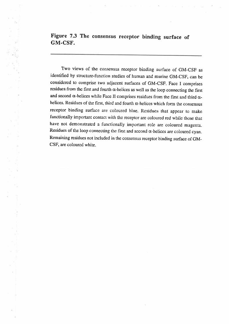
Figure 7.3 The consensus receptor binding surface ofGM.CSF.
Two views of the consensus receptor binding surface of GM-csF as
identified by structure-function studies of human and murine GM-CSF, can beconsidered to comprise two adjacent surfaces of GM-CSF. Face I comprisesresidues from the first and fourth a-helices as well as the loop connecting the firstand second a-helices while Face II comprises residues from the first and third 6¿-
helices. Residues of the first, third and fourth a-helices which form the consensus
receptor binding surface are coloured blue. Residues that appear to makefunctionally important contact with the receptor are coloured red while those thathave not demonstrated a functionally important role are coloured magenta.Residues of the loop connecting the first and second a-helices are coloured cyan.Remaining residues not included in the consensus receptor binding surface of GM-CSF, are coloured white.
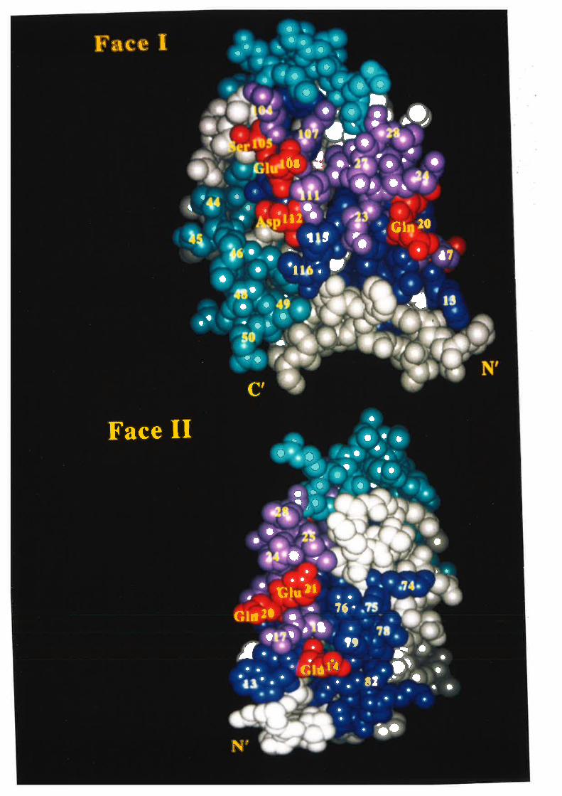
l. t aa¡l¡ aåtce o' a t oa
I
attala I
a 'r,fü\ o Irt'a t ¡ ,),aj O
n) ü
0r{ a.a¡a
I oa *r lra
aoa
Iil II
a
'1.a
a
a ll2 '. Gl 20aa rt ,t
a aa1o I i
a e aa l. a
aa
o
J
.ta
a
O
.a
OJ
ol
tf
c t
Face IIot
oa
f.tro
a
a.ta
acO
It ta
a¡
aa
tI
a. ¡a¡Oa
Glu lla aa
Glr 2l r¡ 76.tI
t , I a
t ta tt f.
üì.ìaa aa a lI'
.'.'ttO¡ a o
a
a
a
ao
\,
aaa
'Glû
'ao
a
of the first and fourth cr-helices, is the GMRa chain binding site while Face II,
comprising residues of the first and third a-helices, is the Pc chain binding siæ.
Human growth hormone represents the only member of the cytokine family for
which the structure of the hormone:receptor complex has been resolved (de Vos et al.,
1992). Analysis of the interaction between growth hormone and its binding protein
provides a paradigm for understanding the hormone:receptor interactions of other
members of the cytokine family. Growth hormone has a core structure composed of four
c¡-helices and although the helices are longer than those found in the short-chain helical
cytokines, the unusual connectivity of the helical cytokines is preserved (Abdel-Meguid
et a1.,1987; de Vos et aI., 1992). The extracellular portion of the growth hormone
binding protein consists of two domains, each composed of seven p strands in an
antiparallel sandwich and with a topology identical to the D2 domain of CD4 and the
chaperone protein PapD (de Vos et aI., 1992). The structure of the growth hormone
binding protein is very similar to that previously predicted for members of the cytokine
receptor family including the GHbp (Bazan, 1990b).
Biophysical measurements and mutagenesis studies identified two receptor binding
sites on GH with site I comprising residues from the first and fourth a-helices together
with residues from the loop between the first and second cr-helices and site 2 comprising
residues from the first and third o-helices (Cunningham et a\.,1989; Cunningham and
Wells, 1989; Cunningham et a\.,1991). The only receptor binding region identihed on
growth hormone but not included in the receptor binding surface of GM-CSF (Fig. 7.3)
is the loop between the first and second cr-helices (residues 28-55). The binding epitope
of a neutralising MoAb maps to residues 40-77 of human GM-CSF (Kanakura et aI-,
1991), overlapping the loop between the fîrst and second cr-helices so that this region
may also form part of the receptor binding surface for GM-CSF. These studies also
demonstrated that growth hormone binds to two molecules of the GHbp to form a
GH:(GHbp)2 complex (Cunningham et aI., 1991). Analysis of the crystallised
GH:(GHbp)2 complex, demonstrated that the binding of growth hormone to its receptor
-r28-
buried 1230 L2 through the site 1 interaction and 900 Å2 through the siæ 2 interaction
(de Vos et aI., 1992).Interaction between the membrane-proximal domains of the two
receptor molecules buried a furttrer 500 Å2. The majority of amino acids identified in GH
or the GHbp as functionally important by mutagenesis studies, are located in the
hormone:receptor interface and are classified as direct binding determinants (de Vos er
al., 1992).Interestingly all of the amino acids specifically identified as important or at
least poæntially involved in GM-CSF receptor binding and biological activity, residues
14,20,2I,23,27,78,105, 108 and lI2, aligned with amino acids in growth hormone
that contact the two molecules of the growth hormone binding protein (Goodall et al.,
1993). Formation of the GH:(GHbp)2 complex occurs through a sequential binding
mechanism (Cunningham et al., I99l). Thus growth hormone binds molecule I of
GHbp through site I and this complex is then able to bind molecule 2 of GHbp through
site 2 and an interaction of the membrane proximal domains of GHbpl and GHbp2. It
has been proposed that the binding of growth hormone to the second receptor molecule
through site 2 is stabilised by the interaction of the membrane-proximal domains of the
t\r/o receptor molecules (de Vos ¿r a1.,1992). An analogue of growth hormone in which
the second receptor binding site is mutated but the first binding site remains active, was
unable to dimerise the GHbp and functioned as an antagonist of wild type growth
hormone activity (Fuh ør al., 1992). The properties of the GH antagonist closely
resemble the properties of the GM-CSF antagonists described in Chapter 6.
A model illustrating the interaction of GM-CSF with the high affinity GM-CSF
receptor complex (Fig. 7.a) has been generated using the GH:(GHbp)Z complex as a
starting point for molecular modelling (model generated by Dr C.Bagley). In this model
GM-CSF is located in a binding groove formed by the juxtaposition of the GMRc¡ and
pç chains and with residues from both domains of the cytokine receptor modules
contributing to the ligand binding sites. By comparison with the interaction of growth
hormone and its binding protein, Face I of GM-CSF binds the GMRcr, chain and is the
equivalent of the site I interaction with GHbpl while Face II of GM-CSF binds the pç
chain and is the equivalent of the siæ 2 interaction with GHbp2. The model illustrates the
-129-
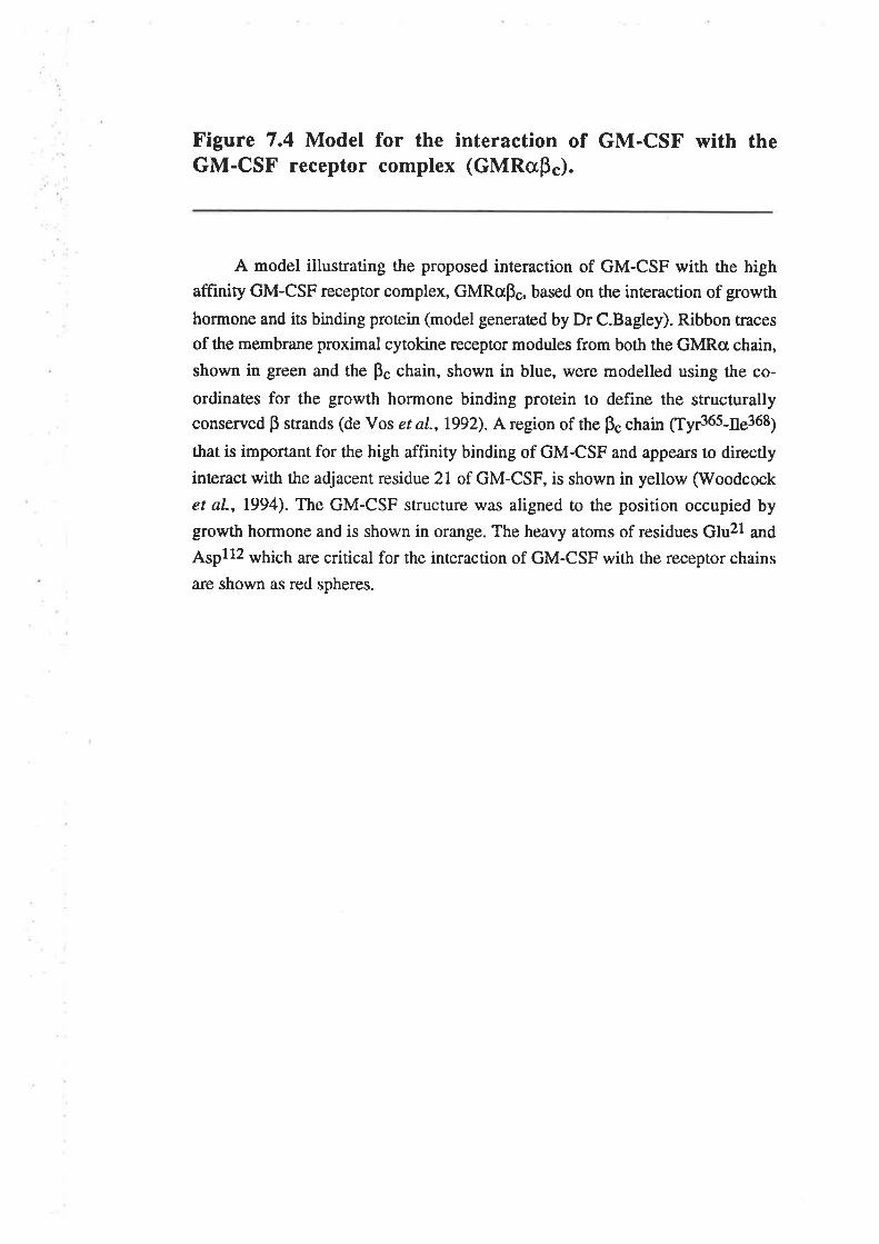
Figure 7.4 Model for the interaction of GM-CSF with theGM-CSF receptor complex (GMRaps).
A model illustrating the proposed interaction of GM-CSF with the highaffinity GM-CSF receptor complex, GMRcpç, based on the interaction of growth
hormone and its binding protein (model generated by Dr C.Bagley). Ribbon traces
of the membrane proximal cytokine receptor modules from both the GMRo chain,
shown in green and the ps chain, shown in blue, were modelled using the co-
ordinates for the grotwth hormone binding protein to define the structurallyconserved p strands (de Vos et a1.,1992). A region of the pç chain ¡fyr365-ns368¡
that is important for the high affinity binding of GM-CSF and appears to directly
interact with the adjacent residue 2l of GM-CSF, is shown in yellow (Woodcock
et al., 1994). The GM-CSF structure was aligned to the position occupied by
growth hormone and is shown in orange. The heavy atoms of residues Glu2l and
Aspllz which are critical for the interaction of GM-CSF with the receptor chains
are shown as red spheres.
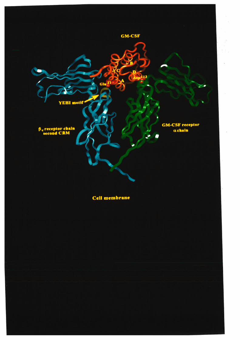
GM-CSF
tt ,/
YB
l" s- t
¡¡t a
3c¡¡ !r '2
l
- aa
YEHI Dollf ö
a
-p. rcccplor cbrl¡
¡cco¡d CnMIr GM-CSF rccePlor
ccbrlea- a
a
I a
Ccll mcmbr¡nc
relative proximity of residues within GM-CSF to the rcceptor chains and also indicates
which regions of the receptor molecules might directly participate in ligand binding.
Features of the receptor chains that cannot be studied using this model a¡e the influence
of the N-terminal domain of the GMRcr chain or the second cyûokine receptor module of
ú" F" chain on GM-CSF binding.
A region of the GM-CSF recepûor Fc chain including ¡¡it367, has been identified as
critical for the high affinity binding of GM-CSF as well as for the mediation of maximal
biological activity (Lock et al., L994). This region of the pç chain is located adjacent to
Glu2l of GM-CSF (Fig. 7.4) and also participates in the high affinity binding of IL-3
and IL-5 to their respective receptor complexes while mutations at residues Tyr3ó5,
¡¡i5367 ¿¡16 ils3ó8 of the ps chain a¡e able to complement the binding defects of the GM-
CSF E2lR analogue and partially restore high affinity binding (Woodcock et a1.,1994)-
Thus it appears likely that residue 21 of human GM-CSF directly interacts with the pç
chain of the GMRapç receptor complex through residues in the vicinity 6¡ 65367'
GM-CSF expressed from cultured human lymphocytes (Donahue et a1.,1986a;
Cebon et al., 1990) and recombinant GM-CSF expressed from eukaryotic cells
(Kaushansky et a|.,19S7) is extensively glycosylaæd. Glycosylation is not required for
the activity of wild type GM-CSF and in fact reduces the specific biological activity
(Moonen et a|.,1987; Fig. 6.2.1) and the high- (GMRap") and low-affinity (GMRa)
receptor binding (Cebon et aI., 1990; Table 6.2.t) of GM-CSF. In general, the relative
biological activity and receptor binding properties of glycosylated and, E. coli-denved,
non-glycosylated GM-CSF residue 21 analogues were comparable. However, the non-
glycosylated E21R and E21K analogues were devoid of activity and yet were able to
bind the GMRcr chain with wild type low affinity. This demonstrated that low affinity
binding can be dissociated from receptor activation and biological signalling. The lack of
activity for the non-glycosylated E2lR analogue was in contrast to the reduced but
measurable activity observed for the glycosytaæd E2lR analogue. It has not been
determined why the two analogues exhibit different levels of biological activity although
- 130-
it appears that the O-linked carbohydrate may be sufficient to impart agonistic activity to
the glycosylaæd E21R analogue (Fie. 6.2.6).
The work presented in Chapter 6 described the properties of the first GM-CSF
antagonist (Hercus et aI., 1994a). The E coli-deived GM-CSF analogues, E21R and
E2lK, were devoid of biological activity and yet exhibited wild type binding affinity for
the low affinity receptor (GMRo). These analogues were demonstrated to be effective
anøgonists of the GM-CSF-mediaæd proliferation of leukaemic cells and the GM-CSF-
mediated stimulation of neutrophil superoxide release (Fig. 6.2.16). The specificity of
this antagonism was illustrated by the fact that eco E21R was unable to antagonise the
IL-3 mediated proliferation of TF-1 cells (Fig. 6.2.88) or the TNF-cr-mediated
stimulation of neutrophil superoxide release @ig.6.2.128). Importantly, the eco E2lR
and E21K analogues were also effective antagonists of glycosylated GM-CSF species
(Fig. 6.2.17) suggesting the potential use of these antagonists in vivo to block the
activity of endogenous, glycosylated GM-CSF. The receptor binding properties of the
GM-CSF antagonists (Table 6.2.3) suggested that these analogues block wild type GM-
CSF activity by sequestration of all of the available GMRa chains into complexes that
are unable to interact with the Bç chain (Fig. 7.5). In the absence of unoccupied GMRa
chains, the wild type GM-CSF is prevented from binding and hence eliciting a biological
r€sponse. The structural and functional similarities displayed by GM-CSF, IL-3 and IL-5
suggests that functionally similar antagonists of both IL-3 and IL-5 may be generated by
charge-reversal mutation of the conserved acidic residue in the first ct-helix. The fact that
the IL-3 E22F. analogue exhibited biological activity, albeit at a 20,000-fold reduced
potency, demonstrated that in the case of IL-3, residues other than the conserved acidic
residue make functionally relevant contacts with the Bç chain and may need to mut¿ted in
conjunction with residue22to generate an antagonistic analogue (Barry et a1.,1994).
Cytokine analogues with antagonistic properties have also been described for other
members of the cytokine family. Thus an analogue of human IL-6 with wild type affinity
for the IL-6Rcr chain but impaired binding to gp130, which is functionally equivalent to
-131 -
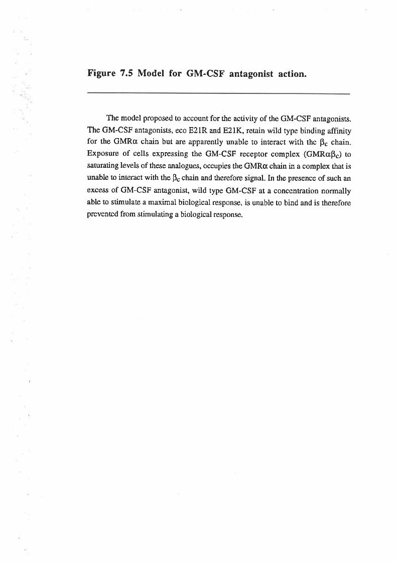
Figure 7.5 Model for GM-CSF antagonist action.
The model proposed to account for the activity of the GM-CSF antagonists.
The GM-CSF antagonists, eco E2lR and E2lK, retain wild type binding affinityfor the GMRa chain but are apparently unable to interact with the pç chain.
Exposure of cells expressing the GM-CSF recepror complex (GMRcp") tosaturating levels of these analogues, occupies the GMRa chain in a complex that is
unable to interact with the pç chain and therefore signal. In the presence of such an
excess of GM-CSF antagonist, wild type GM-CSF at a concentration normallyable to stimulate a maximal biological response, is unable to bind and is therefore
prevented from stimulating a biological response.
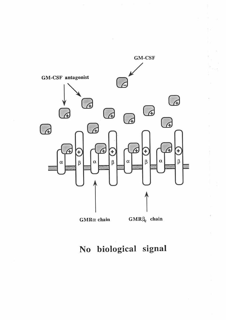
GM.CSF
GM-CSF antagonist
\
GMRcr chain GMRH. chain
No biotogical signal
c[c[c[c[ ppppFf/l///¿rtllffr .F¿r//f//¿r{////Å
the GM-CSF receptor pç chain, is able to antagonise wild type IL-6 function (Savino er
at., t994).In a similar manner, analogues of murine lL'2 (Zurawski ¿f al', 1990;
Zurawski and Zurawski, 1992) or human IL-4 (Kruse et al., 1992) with impaired
binding to the yç chain shared by the receptors for IL-2 and IL-4 (Kondo et a1.,1993;
Russell et a1.,1993), are specific antagonists of the appropriaæ wild type cytokine
function. The recurring theme amongst the cytokine antagonists is one of selective
interaction with the ligand binding but not the receptor activation components of a multi-
subunit recepûor comPlex.
A feature of the GM-CSF antagonists was that the typical IC5g values of around
200ng/ml, were considerably higher than the l5ng/ml (lnM) required to achieve 507o
occupancy of the GMRa chain (Fig. 6.2.15). The IC5g value presumably reflects the
level of GMRcr chain occupancy by antagonist required to block 507o of. the wild type
GM-CSF signalling. The relatively high IC5g values for the GM-CSF antagonists
indicaæs thar approximaæly 90vo ofthe GMRcr chain needs to be occupied by antâgonist
for effective blocking of the signal from wild type GM-CSF. The required excess of
antagonist was consistent with reports that maximum biological effects of colony
stimulating factors can be exerted with steady state receptor occupancy levels as low as
107o (Nicola et a|.,1988). Although the antagonists and wild type GM-CSF exhibit
simila¡ affinities for the GMRcr chain (fable 6.2.3), the ability of wild type GM-CSF to
interact with the Bç chain and form a high affinity complex is probably critical for the
biological signalling mediated by low concentrations of wild type GM-CSF. GM-CSF
antagonists with enhanced GMRcx, chain binding, may function as better antagonists and
exhibit lower IC59 values by competing more effecúvely with wild type GM-CSF for the
GMRcr chain.
The effectiveness of the GM-CSF antagonists is dependent upon their affinity for
the GMRa chain and it seems likely that pç chain defective antagonists for IL-3 and IL-5
would be similarly influenced. The affinity of IL-3 for the IL-3Ra chain (K¿=30nM) is
considerably lower than the affinity of GM-CSF for the GMRcr chain (K¿=lnM)' This
-r32-
suggests that an IL-3 antagonist using the mechanism described for GM-CSF (Fig. 7.5)
would possibly require significantly higher concentrations of IL-3 antagonist to achieve
equivalent IC5¡ responses. Thus a typical IC5g value for the IL-3 antagonists might be
expecred to be 6,0(X)ng/ml compared with 200ng/ml for the GM-CSF antagonists. The
somewhat higher afhnity of IL-5 for the IL-SRct chain (Ko=0'5nM) suggests that an IL-
5 antagonist using the mechanism described for GM-CSF (Fig. 7.5) might exhibit ICso
values of around l00ng/ml.
The excess of antagonist over agonist required for 50Vo inhibition of biological
activity exhibited considerable variation. The values range from 5,000-fold excess to
achieve 507o inhibition of eco GM-CSF mediated TF-l cell proliferation to 50-fold
excess to achieve 507o inhibition of cho GM-CSF mediated neutrophil superoxide release
(Table 7.2). This probably reflects differences in the EDso values of wild type GM-CSF
in these biological assays which range from 0.017ng/ml for eco GM-CSF stimulation of
TF-l cell proliferation to 0.53ng/ml for cho GM-CSF stimulation of neutrophil
superoxide release (Table 7.2).Tlte difference can parúally be accounted for by the effect
of the carbohydrate on GM-CSF activity as eco GM-CSF exhibited a 5- to lO-fold higher
potency (lower EDSO) than cho GM-CSF. However, there are real differences in the
concengation of GM-CSF required to trigger receptor activation in different cell types as
there is a 3- to S-fold difference in the EDso values of neutrophil activation and TF-l cell
proliferation (fable 7.2) using the same form of GM-CSF. One possible explanation for
this difference in the ED59 values is that the proliferative response of TF-l cells and the
release of superoxide anions by neutrophils occur through different pathways, utilising
different signalling components and with different receptor occupancy thresholds.
Alternatively, the presence of different forms of the receptor molecules with cytoplasmic
domain variants (Crosier et a|.,1991) or the presence of additional signalling chains in
the GM-CSF receptor complex of certain cell types, might explain the differences in
EDSO values. The presence of a third receptor chain has been demonstrated for theIL-2
receptor complex (Takeshita et a1.,1992).
-133-
Tabte 7.2 Comparison of ED5¡ values for wild type
GM-CSF and the antagonist excess at IC5¡, for
TF-l cells and neutroPhils.
Neutrophilsuperoxide release
TF.1cell proliferation
A) EDso GM-CSF (ng/ml)a
eco GM-CSF 0.09 t o.ozu
n=10
cho GM-CSF 0.53 t o.oz¿
n=12
B) Fold excess of antagonist (E21R or E21K)c
eco GM-CSF 514 t ¡so
n=5
0.017 + 0.004
n=13
0.16 I 0.05
n=4
4,950 + t,l.2o
n=6
cho GM-CSF 50d
n=2
120 x tcn=3
â The ED5s values for wild type GM-CSF derived from E. coli
cells (cho GM-CSÐ was determined in the TF-l cell proliferation(eco GM-CSF) or CHOassay and the neutroPhil
su¡reroxide anion release assaY.b Error values are the standard error of the mean (SEM).c The antagonist excess for eco E2lR and eco E2lK was deteflnined at the IC59 value
against Uot¡ eco GM-CSF and cho GM-CSF in the TF-l cell proliferation assay and the
nãutropnit superoxide anion release assay. Tbe values are expressed as an excess of*t^gonirt ovór agonist and the data from both antagonists Pooled as the two analogues are
essentially equipotent GM-CSF antagonists.d No SEIr,l calculated because only two daø poins.
Apa¡t from the clinical potential, the generation of GM-CSF analogues devoid of
interaction with the pç chain but retaining wild type affinity for the GMRcr chain
provides an opportunity to specifically examine the functional role of the GMRa chain. It
has recently been demonstrated that GM-CSF binding to the human GMRc¡ chain
expressed on Xenopus laevis oocytes, caused activation of 2-deoxyglucose transport
through endogenous glucose transporters (Ding et al., 1994).Importantly, GM-CSF is
also able to stimulate 2-deoxyglucose uptake in neutrophils under conditions that
prevented the tyrosine phosphorylation normally associated with GM-CSF receptor
activation thereby directly implicating the GMRø chain in glucose transport. The GM-
CSF antagonist provides the ideal reagent for specifically testing the role of the GMRo
chain in activating glucose transport in cell types such as neutrophils that also express the
pç chain.
It is now evident that GM-CSF activity is not essential for steady state
haemopoiesis as mice homozygous for a disrupted GM-CSF gene develop a normal
haemopoietic population (Dranoff et a\.,1994; Stanley et aL.,1994). However, GM-CSF
deficient mice do develop a non-fatal lung condition, similar to forms of alveolar
proteinosis, characterised by an accumulation of lipids and surfactant protein (Dranoff et
al., 1994) and the presence of opportunistic bacterial and fungal infections (Stanley er
al., 1994) within the alveoli. M-CSF modulates the consequences of GM-CSF
deficiency as mice deñcient in both GM-CSF and M-CSF develop a much more severe
lung disease that is often fatal (Lieschke et al.,1994). The in vivo role of the GMRcr
chain might be better understood by exposing GM-CSF-deficient mice to GM-CSF
analogues that specif,rcally bind to the GMRcr chain only. It is therefore of great interest
that a murine GM-CSF antagonist has been developed that is functionally similar to the
human GM-CSF antagonists described in this thesis (R. Kastelein personal
communication). Thus the functional role of the GMRcr chain in GM-CsF-deficient mice
might be studied by intra venous administration of the murine GM-CSF antagonist or by
generating transgenic mice, deficient in wild type GM-CSF but expressing the murine
GM-CSF antagonist.
-r34-
The work discussed in this chapter describes some of the structure-function
properties of GM-CSF and other members of the cytokine family. It is now quite
apparent that the principal features of cytokine structure and function are remarkably
similar amongst a family of proteins exhibiting very little amino acid homology and a
vast range of biological activities. This functional similarity amongst the helical cytokines
should assist in the identification of functionally important regions and enable the
generation of antagonistic analogues for all the members of the helical cytokine family.
-135-
Appendices
Appendix 4.3.2.1 Map of the pJL4 expression vector.
The pJL4 expression vector (Gough et a1.,1985) was used to transiently
express wild type GM-CSF and analogues from COS cells. COS cells are amonkey kidney cell line transformed by SV4O. The presence of the SV40 origin ofreplication enables the pJL4 plasmid to replicate in COS cells. Expression of GM-
CSF sequences cloned into the polylinker, is under the control of the SV4O laæ
promoter.
Restriction siæs in the polylinker that are not unique are ma¡ked with an
asterisk.

51417 bp
Late
Early
pJL4
Polylinker*
Eco RI
Ava I
sv40orl
Pst I
Pst I
Hlnd III
RAmp
Pst I
on
Htnd trI
T-antigenHind Itr
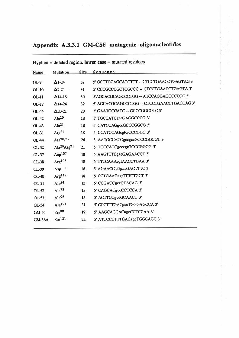
Appendix 4.3.3.1 GM-CSF mutagenic oligonucleotides
Hyphen = deleted region,lower case = mutated residues
Name fi nn Size Secrrence
oL-9
oL-10
ol.-l1
oL-12
oL45
oL42
oL43
oL-31
oL4/'
oL-32
oL-37
oL-38
oL-39
oL40
oL-51
oL-52
oL-53
oL-54
GM-55
GM.56A
^r-uÃt-uAr+re
^t+?AAzuzt
Ala2o
Ala2l
ArEzL
¡y3'20,2L
¡620¡¡921
AsPl0z
Argloe
AsplllArgllz
Ala54
Ala88
Ala96
4¡¿121
Ser88
5"¡121
32
31
30
32
20
18
18
l8
24
2t
18
18
18
18
15
15
15
2t
t9
22
5' GCCTGCAGCATCTCT - CTCCTGAACCTGAGTAG 3'
5' CCCGCCCCICTCC'CCC'- CTCCTGAACCTGAGTA 3'
S'AGCACCICAGCCCTGG -- ATCCAGGAC'C¡CCCGG 3'
5' AGCACGCAGCCCTC'G -- CTCCTGAACCTGAGTAG 3'
5'GAATGCCATC .- CICCCGC'CGTC 3'
5' TGCCATCgccGAGGCCCG 3'
5' CATCCAGgccGCCCCTGCG 3'
5' CCATCCAGcgtGCCCCTCC 3'
5' AATGCCATCgccgccGCCCGGCGT 3'
5' TGCCATCgcccgtGCCCGGCG 3'
5' AAGTTTCgacGAGAACCT 3'
5' TTTCAAAcgTAACCTGAA 3'
5' AGAACCTGgacGACTTTC 3'
5' CCTGAAGcgTTTTCTGCT 3'
5'CCGACCgccCTACAG 3'
5'CAGCACgccCCTCCA 3'
5'ACTTCCgccGCAACC 3'
5' CCCTTTGACgccTGGGAGCCA 3'
5' AAGCAGCACagcCCTCCAA 3'
5' ATCCCCTT'IGACagcTGGGAGC 3'
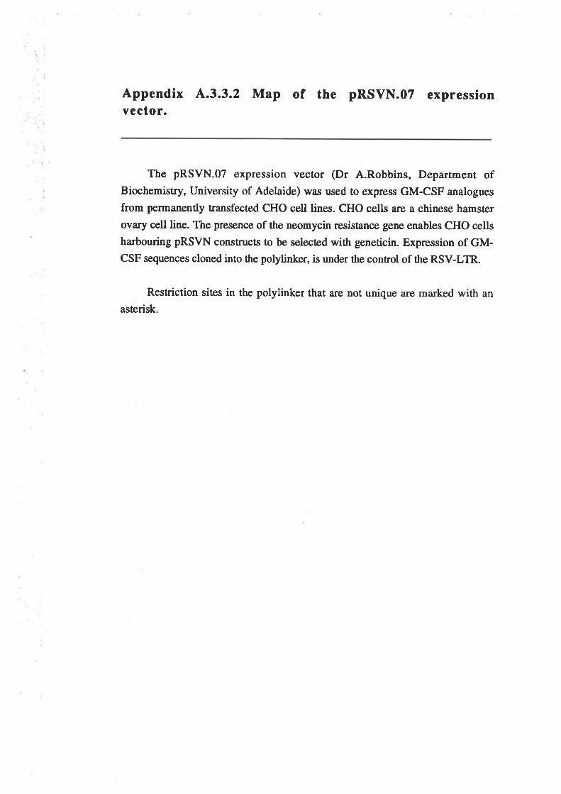
Appendix 4.3.3.2 Map of the pRSVN.07 expressionYector.
The pRSVN.O7 expression vector (Dr A.Robbins, Department ofBiochemistry, University of Adelaide) was used to express GM-CSF analogues
from permanently transfected CHO cell lines. CHO cells are a chinese hamster
ovary cell line. The presence of the neomycin resistance gene enables CHO cells
harbouring pRSVN constructs to be selected with geneticin. Expression of GM-CSF sequences cloned into the polylinker, is under the control of the RSV-LTR.
Restriction siæs in the polylinker that are not unique are marked with an
asterisk.
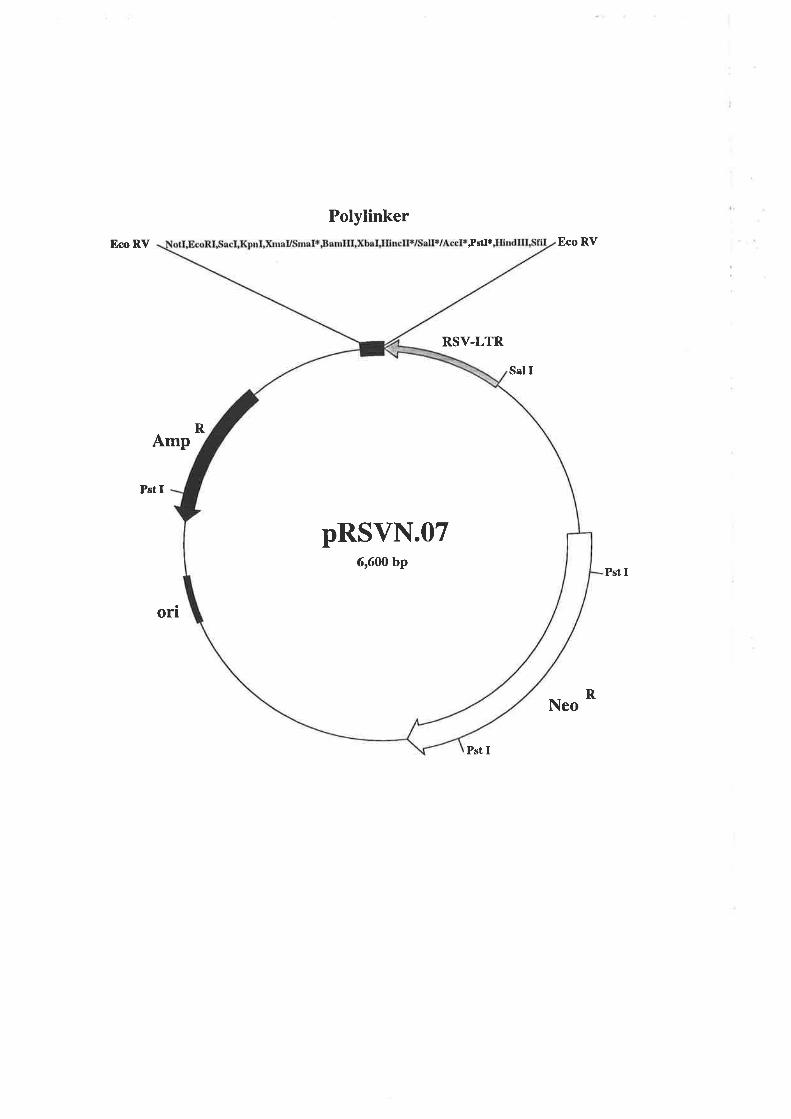
PolylinkerEco RV Istr*
RSV-LTR
Pst I
Eco RV
RNeo
Sal I
RAmp
Pst I
Pst I
orr
pRSVN.076,6fi) bp
Appendix 4.3.3.3 Map of the pJSV4O expression vector.
The pJSV4O expression vector was created by cloning an SV40 poly (A)-addition into the pJL4 expression vector (A.3.2.1) and was used to transiently
express wild type GM-CSF and analogues from COS cells. The properties of thisplasmid are essentially the same as for the pJL4 expression vector.
An EcoRVHpaI fragment containing the SV40 polyadenylation sequence
from the pSV2CAT plasmid (Gorman et aI., 1982), was subcloned intoSmaVEcoRI digested pBluescript SK(-) (Phillips et aL.,1989). Subsequenrly, rhe
SV40 polyadenylation sequence was isolated as an XbaI/EcoRI fragment and
cloned into XbaVEcoR[ digestedpllA to generate the pJSV4O expression vector.
Restriction sites in the polylinker that are not unique are marked with an
asterisk.
Polylinker* PstI*BamHI SpeIXbaISalVAccI
BamHI sv40ori Hind III
SV40 Poly A
PstI
EcoRI 1
R
Hind Itr
Hind Itr
T-antigen
amp
Pst I
Pst I
orlAva I
pJSV406,295bp
LateEarly
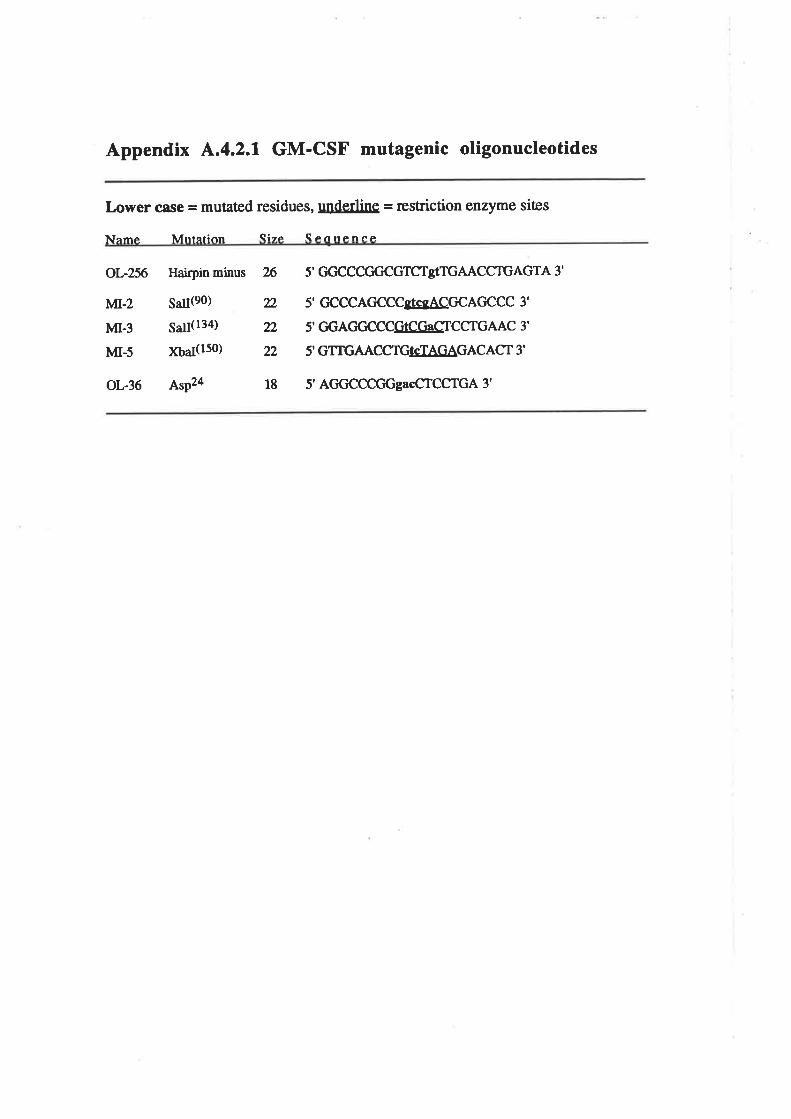
Appendix ^.4.2.1
GM-CSF mutagenic oligonucleotides
Lower ci¡se = mutated residues, underline = restriction enzyme sites
Name Mrrtâtion Size Scnrrence
oL-zft
MT-z
MI.3
MI-5
oL-36
Haþin minus 26
saßeo) 22
5¿¡(134) 22
¡ç5¿(ls0) 22
AsP24 18
5' GGCCCGGCGTCTgTTGAACCTTGAGTA 3'
5' CCCCAGCCCgTcgACGCAGCCC 3'
5' GGAGGCCCGSCC.gCTCCTGAAC 3'
5' GTTGAACCTGIgIAGAGACACT 3'
5' AGGCCCCTGgacCTCCTGA 3'
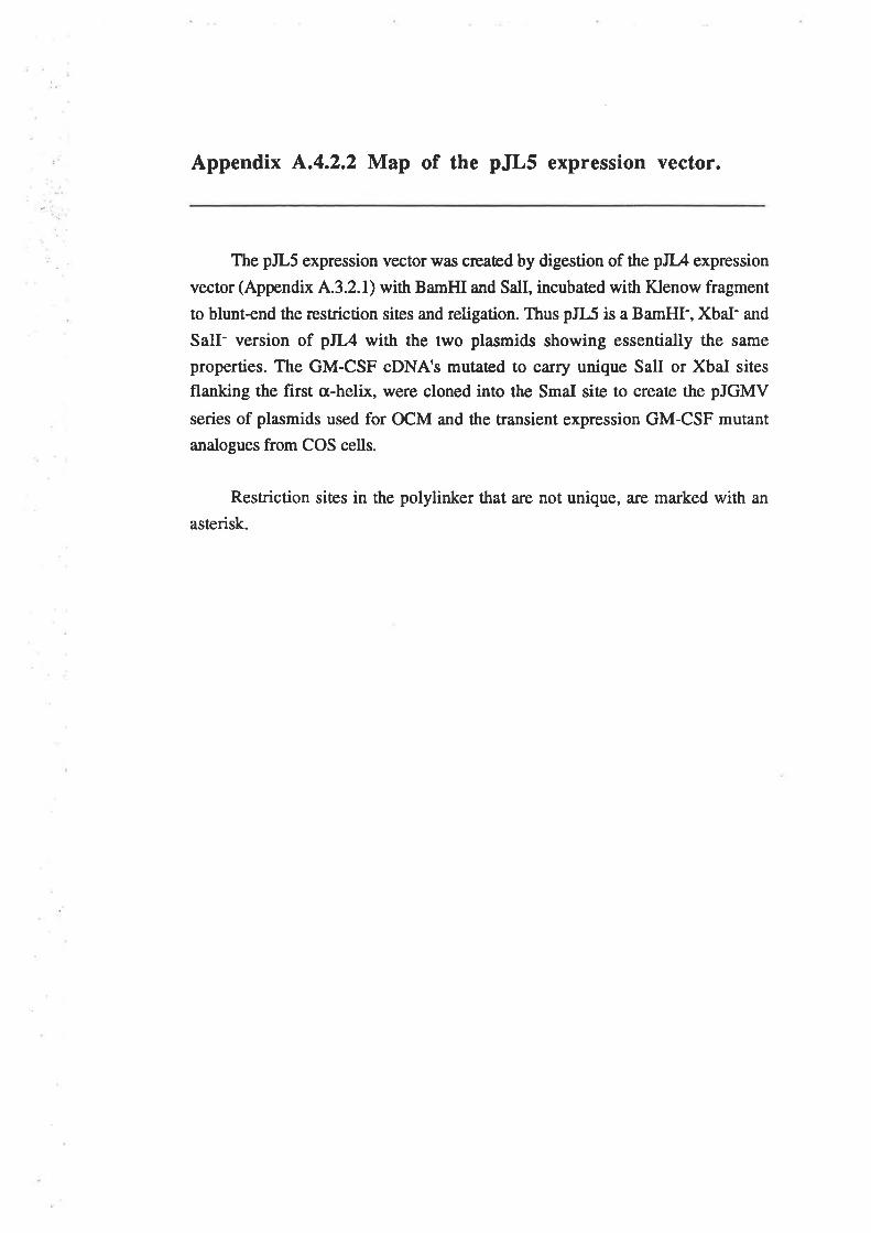
Appendix þt.4.2.2 Map of the pJLs expression vector.
The pJL5 expression vector was created by digestion of the pJL4 expression
vector (Appendix 4.3.2.1) with BamHI and SalI, incubaæd with Klenow fragment
to blunt-end the restriction siæs and religation. Thus pJL5 is a BamHI-, XbaI- and
SalI- version of pJL4 with the two plasmids showing essentially the same
properties. The GM-CSF cDNA's mutaæd to carry unique SalI or XbaI sites
flanking the first cr-helix, were cloned into the SmaI siæ to create the pJGMV
series of plasmids used for OCM and the transient expression GM-CSF mutant
analogues from COS cells.
Restriction siæs in the polylinker that are not unique, are marked with an
asterisk.
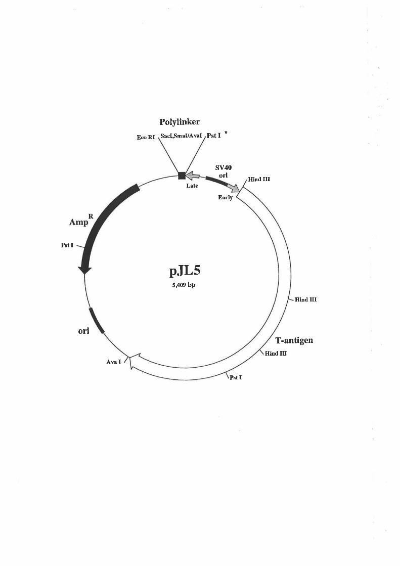
Eco RI PstI *PolylinkerSacI,SmaVAval
sv40orl Hhd IIT
Pst I
on
Hlnd III
T-antigenHind III
Ava f
Pst I
5,409 bp
Late
Early
pJLs
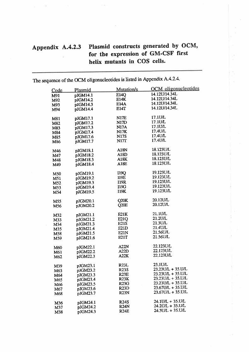
Appendix L.4.2.3 Plasmid constructs generated by OCM'for the expression of GM'CSF firsthelix mutants in COS cells.
The sequence of the OCM oligonucleotides is lisæd in Appendix A'4'2'4'
M91M92M93M94
M81M82M83M84M85M8ó
M46iù-l,47
M48M49
M50M51M52M53M54
pJGM17.1pJGM17.2pJGM17.3pJGMl7.4pIGM17.6pJGM17.7
Er4QEI4KEI4AE14T
N17ENITDN17ANl7KNl75N17T
AISNA18DA18KA18E
r19QI19EII9RI19GII9K
Q20KQ20E
E2IKE21QE21SE21DE21NF2IT
pIGM14.1pJGM14.2pJGM14.3pJGM14.4
t4.lzvll4.34Lt4.lzul14.34Lt4.t2vll4.34L14.t2ul14.34L
n.lurLtT.tulLlT.llJILt7.4ull-t7.4UlLt7.4UlL
pJGM18.1pJGM18.2pJGM18.3pJGM18.4
t8.t23UlLt8.723VtLt8.t23VlLt8.t23UlL
t9.lz3ulLt9.tz3ulL19.t23UlLt9.tz3ulLt9.t23UlL
pJGMl9.lpJGM19.2pJGM19.3pJGM19.4pIGM19.5
M55M5ó
pJGM2O.1pJGM2O.2
pIGÌtlZZ.lpIGM2Z.2pIGM22.3
20.t2ulL20.t2utL
M32M33M34M35M58M59
M60M6llltl62
M39M63M64M65M66tv'd67
M68
pJGM21.1ptGM2l.2pJGM21.3pJGM2l.4pJGM2l.5pJGM21.6
2t.tvlL2t.2UlL2t.3VlL2t.4VlL2t56ViI-2t.56UtL
pJGM23.1pIGM23.2pJGM23.3pJGMZ3.4pJGM23.5pJGM23.6pIGMZ3.7
pJGMZ.lpJGItl?A.2pJGM24.3
22.t23UlL22.L23UlL22.l23VtL
?+.lUtL + 35.U/LZ4.}U|L + 35.U/L24.3UlL + 35.U/L
23.tUtL23.23VtL + 35.U/L23.23V1L + 35.UlL23.23rJtL + 35.U/L23.23VtL + 35.U/L23.67UtL + 35.U/L23.67UtL + 35.U/L
A22lrqA2?ÐA22K
R24SR24NR21E
R23LR23SR23ER23KR23GR23DR23N
M36Iùl37M38
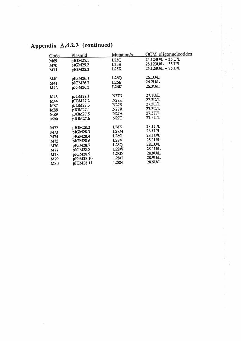
Appendix ^.4.2.3
(continued)
M69M70M71
pJGM25.1ptGM25.2pJGM25.3
pJGM26.1pIGM26.2pJGM26.3
L25QT25Er25K
L2ßQL26EL26K
25.l23UlL + 35.U/L25.l23UtL + 35U/L25.l23UlL + 35.UlL
26.tVß,26.2VlL26.3vlL
M40M4lIùl42
M43MUM87M88M89M90
Iù.d72tsl73}ù'[74M75tM76It1Á77
M78M79M80
pJGM27.1pJGM27.2pIGM27.3pIGM27.4pJGM27.5pIGtllZ7.6
pJGM28.2pJGM28.3pJGM28.4pJGM28.6pJGM28.7pJGM28.8pJGM28.9pJGM28.10pJGM28.1l
N27DN27KN27SN27RN27AN27T
T2ßKL28ML28GL28VL28QL28WL28DL28HL28N
27.tU/I-27.zUtL27.3UIL27.3UtL27.sV/I.27.sutr,
28.lUtL28.tUlL28.lVlL28.rrJlL28.trJIL28.lUlL28.9UtL28.9UlL28.9UtL
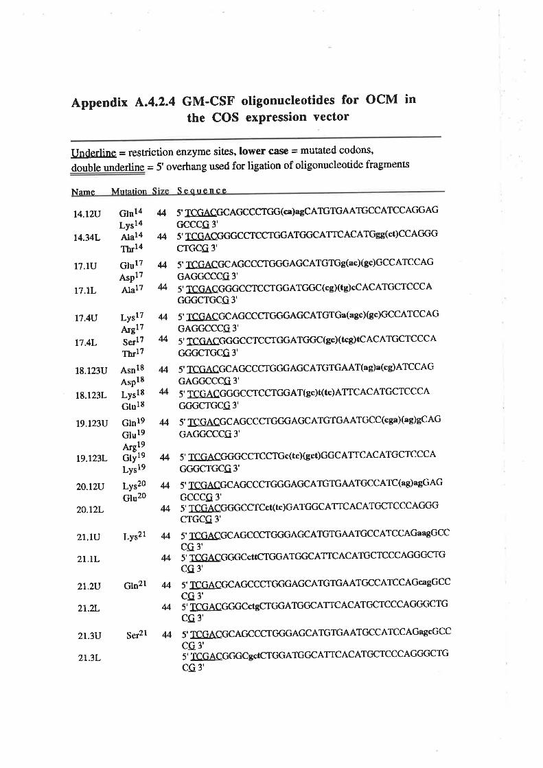
Appendix Ã.4.2.4 GM-CSF otigonucleotides for ocM inthe COS exPression vector
Underline = restriction enzyme sites, lower case = mutated codons,
double underline = 5' overhang used for ligation of oligonucleoúde fragments
Sen¡renceNeme Mntnfion S
t4.tzv
14.34L
17.lU
t7.lL
17.4U
17.4L
18.123U
t8.t23L
t9.t23V
t9.t23L
20.12u
20.tzL
2t.lv
2t.tL
2t.2U
2t.2L
2t.3U
21.3L
Glnl4 4LY3l4Alal4 MThrl4
GlulT 4AsPlT
Alal7 M
LyslT 4ArglTSerl7 MThrlT
AsnlS 4A.pl8Ly518 4Gluls
Glnl9 4Glul9Afgl9Gtyte 4Ly.19
Lys2o 4Glu2o
4
Ly52l 4
Gln2l M
M
4
5' TCGACGCAGCCCTGG(ca)agCATGTGAATGCCATCCAGGAGGCCCG 3'
5' TCGACGCTGCCTCCTC'GATGCICATrc AC ATGss(ct) CC AGGG
C"TGCG 3'
5' TCGACGCAGCCCTGGGAGCATGTGg(acXgc)GCCATCCAGGAGGCCCG3'5' TCGACGGGCCTCCTGGATGGC(cgXtg)cCACATGCTCCCAccc'crccc 3'
5' TCGACCCAGCCCTGGGAGCATGTGa(agc)(gc)GCCATCCAGGACTGCCCG3'
5' TCGACGGGCCTCCTGGATGGC(gcXIcg)TCACATGCTCCCAGC'GCTGCG3'
5' TCGACGCAC'CCCTGGGAGCATGTGAAT(a g)a(cs)ATCCAG
GAGGCCCG3'
5' TCGACC'GGCCTCCTGGAT(gc)t(tc)ATICACATGCTCCCAC'GC¡CTC¡CG 3'
5' TCGACGCAGCCCTGC'GACCATGTGAATGCC(cgaXag)gCAGGAGGCCCG3'
5' TCGACGCTGCCTCCTGc(tc)Gct)GGC ATTCAC ATGCTCCCA
C'GC¡CTGCG 3'
5' TCGACGCAGCCCTGGGACCATGTGAATC'CCATC(a g)agGAG
GCCCG 3'
5' TCGACGGGCCTCcT(tc)GATGGCATTC ACATGCTCCCAGGG
CTGCG 3'
5' TCGACCIC AGCCCTCCGACCATGTGAATGCCATCCAGaagGCCcG 3'5'-TCGACIGGC¡CcttCTGGATC'CCATTCACATGCTCCCAGGCrCTGcG3'
5' rcGACGC ACCCCTC'GGAGCATGTGAATCCCATCCAGca gGCC
CG 3's-ICGAccccccÇGTGGATGGCATTCACATCcTcccACTcTc'crGCG 3'
5'TCGACGCACCCCTC'C'GAGCATGTGAATGCCATCCAGagcCCCcG 3'5'-1çç4CGGGC gcTCTGGATGGCATIC AC ATCCTCCCAGGC'CTG
cG 3'
ser2l 4
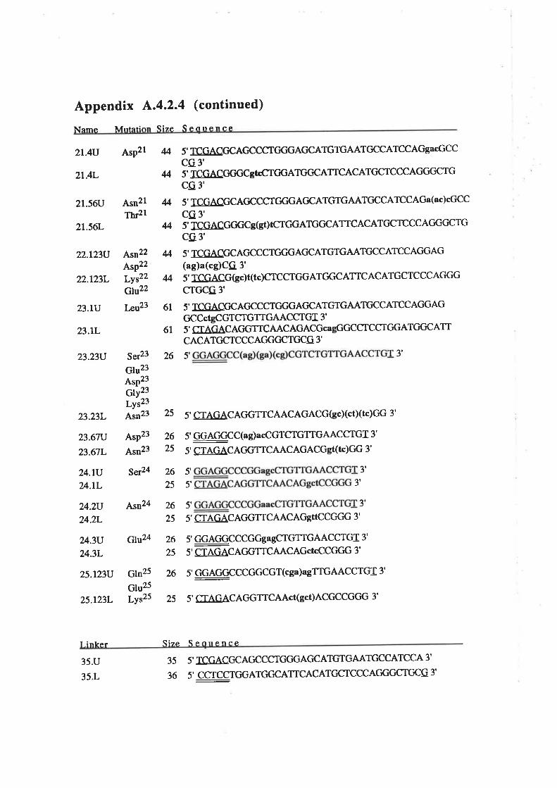
Appendix ^.4.2.4
(continued)
Mntntinn Size S eflrrenceNnme
2t.4U
2t.4L
2r.56lJ
21.56L
22.123U
22.123L
23.tV
23.tL
23.23n
AsP2l
Asn2lThr2l
Asn22AsP22
Lyt22Glu22
I,eu23
4 5' TCGACCCAGCCCTC'GGAC'CATGTGAATGCCATCCAGgacGCCcQ3,
4 5' TCGACGGGCgTTCTGGATGCICATrcACATGCTCCCAC'GC'CTGcca'
4 5' TCGACCCAGCCCTGC'GACCATGTGAATGCCATCCAGa(ac)cCCCcQ 3',
Ms'TCGACC'CCTgGOtCTGGATGCCATTCACATGCTCCCAGGCCTGcG 3'
4 5' TCGACGCAGCCCTCTCTGACCATGTGAATCCCATCCACTGAG(ag)a(cg)CQ 3'
M 5' TCGACG(gc)t(tc)CTCCTGGATC'GCATICACATGCTCCCAGGGcrc'cc 3'
61 5' TCGACGCAGCCCTGCTGACICATGTGAATGCCATCCACIGAGGCCcTgCGTCTGTIGAACCTGI 3'
61 5' CTAGACAC'GTTCAACAGACGcagGGCCTCCTC'GATGC'CATIC ACATC¡CTCCCAC'GCICTC¡CG 3'
ser23 26
Glu23AsP23Gty2tLYs23
Asn23 2523.23L
23.67U
23.67L
24.tUu.lL
24.2U
24.2L
24.3U
u.3L
25.t23U
5
5' CTAGACAGGTTCAACAGACGGcXcTXTc)GG 3'
5' GG,AGGCC(ag)acCGTCTGTIGAACCTGI 3'
5' CTAGACAGGT-ICAACAGACGgt(tc)GG 3'
5' 3'
5' 3'
3
5' CTAGACAGGTICAACAGgTTCCGGG 3'
5' çGAGGçCCGGga gCTGTIGAACCTGI 3'
5' CTAGACACTGTTCAACAGcTcCCGGG 3'
5' GGAGGCCCGGCGT(cga)agTTGAACCTGI 3'
5' CTAGACAGGTTCAAcT(gct)ACGCCGCTG 3'
3'
25.123L
Arp23
Asn23
Ser24
Asn24 5',
Glu24
Gln25 26
Glu25Lyr25 25
2625
26
25
26
25
26
25
Sizc S ênrrpnaêI inkcr
35.U
35.L
3 5 5' TCGACCCACCCCTCTC'GACCATGTGAATGCCATCCA 3'
36 5' CCTCCTC'GATGGCATICACATCICTCCCAGGC'CTGCG 3'
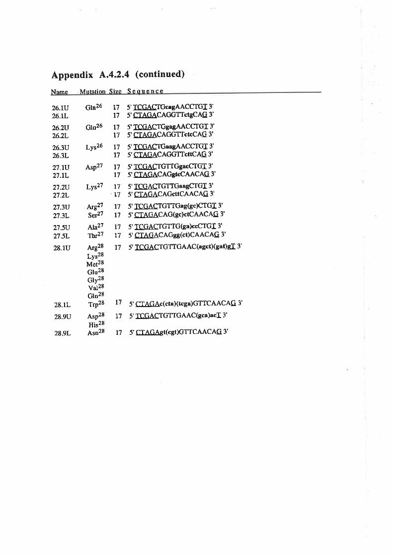
Appendix ^.4.2.4
(continued)
Mntetion Size S enllenceName
26.\V?Á.lL
26.2r126.2L
26.3U26.3L
27.|V27.|L
27.2U27.2L
273U27.3L
27.5u
27.5L
28.lU
Gln26
Glu26
LY526
AsP2z
LYs27
¡ÚjE27
Ser27
^1a21TW27
Arg28
LY528Met28Glu28Gtyzeval28Gln28TrP28
AsP28
His28Asn28
t7l7
t7l7
t7t7
t7t7
t7t7
l7t7
t7t7
t7
5' TCGACTGc¡gAACCTGI 3'
5' CTAGACACTGTTctgCAG 3'
5' TCGACTGgagAACCTG] 3'
5' CTAGACAGGTTcTcCAG 3'
5' TCGACTGaagAACCTGI 3'
5' CTAGACAGGTTcTTCAG 3'
5' TCGACTGTTGgacCTGf 3'
5' CTAGACAGgTcCAACAG 3'
5' TCGACTGTTGaagCTGI 3'
5' CTAGACAGcTTCAACAG 3'
5' TCGACTGTTGag(gc)CTGf 3'
5' CTAGACAG(gc)ctCAACAG 3'
5' TCGACTGTTG(ga)ccCTGI 3'
5' CTAGACAGSS(ct)CAACAG 3'
5' -CGACTGTTGAAC(agct)(gat)gf 3'
28.1L
28.9U
28.9L
l7 5' CIAGAc(cta)(tcga)GTTCAACAG 3'
17 5' TCGACTGTTGAAC(gca)acI 3'
t7 5' C[AGAgt(cgt)GTTCAACAG 3'
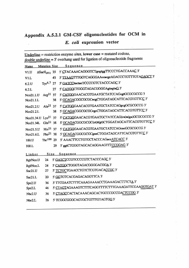
appendix 4.5.2.1 GM-CSF oligonucleotides for ocM inE. colí exPression vector
Underline = restriction enzyme sites, lower case = mutated codons,
double underling = 5' overhang used for ligation of oligonucleotide fragments
Senrren P.Nnme M¡rration Si
Vl.U ÂSacII@s¡¡ 35
vl.L 43
6.2.v Ty{'1 27
6.2.L 27
Nco2l.l.U /u;gzr 37
Nco21.1.L 38
Nco21.2U lttazl 37
Nco21.2.L 38
Nco21.34U Lys2t 37
Nco2l.34l Gm21 38
Nco21.5.U His2l 37
Nco21.6.L Pne2l 38
100.U Thrloo 29
100.L 29
5' CTACAAACACTGGTCTgcgEgTTCCCTGACCAAAC 3'
5' TTAAGTTTGGTCAGGGAAcc¡cgcAGACCCTGTTTGTAGAGCI 3'
5' GATCCtact¡cTCCCCGTCTACCCAGC: 3'
5' CATGGCTC'GGTAGACGCTC'GAgtagtaG 3'
5' CATGGGAACACGTGAATGCTATCCAGcgTGCGCGCCG 3
5' GCAGACGCCGCGC¡cgCTGGATAGCATTACGTGTTCC 3'
5' CATGGGAACACGTGAATGCTATCCAC'gcgGCGCGCCG 3'
5' GCAGACGGCGCCICcgcCTCIGATACIC ATrc ACGTGTTCC 3'
5' CATGGGAACACGTGAATCCTATCCAG(ca)a(ga)GCGCGCCG 3'
5' GCAGACGGCGCGC(cI)tGI)CTGGATACCATTCACGTGTICC 3'
5' e ATGGGAACACGTGAATGCTATCCAGc¡cGCCCCCCG 3'
5' GCAGACGCCCTCGCgaaCTGGATAGCATTCACGTGTICC 3'
5' AAACTTCCTGTCCTACCCAGaccATCACC 3'
5' seTCTGGGTAGCACAGGAAGTTI.CCGGAG 3'
Qcnrrc.nceI.inke- I Size-
24 5' GATSICCGTCCCCGTCTACCCAC'C 3'
24 5' CATGGCTCTGGTAGACCTGGGACCTGA 3'
27 5' TCTGCTGAACCTGTCTCGTGACACC@ 3'
20 5'GGiTGTCACGAGACAGGTTCA 3'
36 5' TTCGAATCTITCATÀAGÀLtu{CCTGAuL{GACTTTCTA 3'
M 5' CTAGTAGÀfu{GTCTTTCACIGTTTTCTTTGAAAGATTCGAAGGTGAT 3'
36 5' CTAGCCACTACAAACAC'CACTGCCCGCCGACTCCGG 3'
26 5' TCGGCC'CTGCAGTCCTGTTTGTAGTGG 3'
BgVNco.U
Bgl/Nco.L
Sac2l.U
Sac21.L
Spe2.U
Spe2.L
Nbe2.U
Nhe2.L

Bibliography
Abdel-Meguid, S.S., Shieh, H.-S., Smith, W.W., Dayringer, H.E-, Violand, B'N' and
Bentle, L.A. (1987). Three-dimensional structure of a genetically engineered variant of
porcine growth hormone. Proc. Natl. Acad. Sci. USA 84,6434'6437.
Altmann, S.W., Johnson, G.D. and Prystowky, M.B. (1991). Single proline
substitutions in predicted a-helices of murine granulocyte-macrophage colony-
stimulating factor result in a loss in bioactivity and alæred glycosylation. J. Biol' Chem'
266,5333-5341.
Alvaro-Gracia, f.M., Zvíflfle\ N.J. and Firestein, G.S' (1939)' Cytokines in chronic
inflammatory arthritis IV. Granulocyte-macrophage colony-stimulating factor-mediaæd
induction of class II MHC antigen on human monocytes: A possible role in rheumatoid
arthritis. J. Exp. Med- 170, 865-875.
Antman, K.S., Griffin, J.D., Elias, 4., Socinski, M.4., Ryan, L', Cannistra' S'A''
Oette, D., Whitley, M., Frei trI, E. and Schnipper, L.E' (1988). Effect of recombinant
human granulocyte-macrophage colony-stimulating factor on chemotherapy-induced
myelosuppression. N. Eng. J. Med. 319, 593-598'
Bagby, G.C., Dinarello, c.4., wallace, P., Wagner, c., Hefeneider, S. and Mccall, E'
(19g6). Interleukin 1 stimulates granulocyte macrophage colony-stimulating activity
release by vascular endothelial cells. J. Clin. Invest. 78, 1316-1323'
Baldwin, G.c., Gasson, J.C., Quan, S.G., Fleischmann, J., WeiSba¡t, R., oette, D',
Mitsuyasu, R.T. and Golde, D.W. ( 1983). Granulocyte-macrophage colony-stimulating
factor enhances neutrophil function in acquired immunodeficiency syndrome patients'
Proc. Natl. Acad. Sci. USA 85,2763-2766'
Baldwin, G.C., Gasson, J.C., Kaufman, S.E., Quan, s'G., williams, R'E', Avalos,
B.R., Gazdar, A.F., Golde, D.W. and DiPersio, J.F. (1939). Nonhematopoietic tumour
cells express functional GM-CSF receptors. Blood 73,1033-1037 '
Baldwin, G.C., Golde, D.'w., widhopf, G.F., Economou' J. and Gasson, J'C'
(1991). Identification and characterization of a low-affinity granulocyte-macfophage
colony-stimulating factor receptor on primary and cultured human melanoma cells' Blood
78, 609-615.
Barry, S.C., Bagley, C.J., Phillips, J., Dottore, M', Cambareri' B'' Moretti' P"
D'Andrea, R., Goodall, G., Shannon, M.F., Vadas, M.A. and Lopez, A-F. (1994)'
Two contiguous residues in human inærleukin-3, Asp2l and Glu22, selectively interact
vu
with the a- and p-chains of its receptor and participaæ in function. J. Biol. Chem. 269,
8488-8492.
BrzÀn,J.F. (1990a). Haemopoietic receptors and helical cytokines. Immunol. Today 11,
350-354.
Baz.an, J.F. (1990b). Structural design and molecular evolution of a cytokine receptor
superfamily. Proc. Natl. Acad. Sci. USA 87,6934-6938-
BazÀÍt, J.F. (1992). Unraveling the structure of IL-2. Science 257 ,4t0-412
Begley, C.G., Metcalf, D. and Nicola, N.A. (1987a). Purified colony stimulating
factors (G-CSF and GM-CSF) induce differentiation in human HL60leukemic cells with
suppression of clonogenicity. Int. J. Cancer 39, 99-105.
Begley, C.G., Metcalf, D. and Nicola, N.A. (1987b). Primary human myeloid
leukaemia cells: Comparative responsiveness to proliferative stimulation by GM-CSF or
G-CSF and membrane expression of CSF receptors. Leukemia 1, 1-8.
Bender, A., Amantr, fI., Jägér, R., Nain, M. and Gemsa, D. (1993)' Effect of
granulocyte/macrophage colony-stimulatin g factor on human monocytes infected with
influenza A virus. J. Immunol. 151, 5416-5424-
Berdel, W.E,, Danhauser-Riedl, S., Sæinhauser, G. and V/inton, E.F. (1989)- Various
human hematopoietic growth factors (Interleukin-3, GM-CSF, G-CSF) stimulate clonal
growth of nonhematopoietic tumour cells. Blood 73, 80-83.
Birnboim, H.C. and Doly, J. (1979)- A rapid alkaline extraction procedure for screening
recombinant plasmid DNA. Nucleic Acids Research 7,1513-L523.
Bradbury, D., Bowen, G., Kozlowski, R., Reilly, I. and Russell, N. (1989).
Endogenous interleukin-1 can regulate the autonomous growth of the blast cells of acute
myeloblastic leukaemia by inducing autocrine secretion of GM-CSF. Leukemia 4' 4+47 -
Brandhuber, 8.J., Boone, T., Kenney, W.C. and McKay, D.B. (1987). Three-
dimensional structure of interleukin-2. Scienc e 238, 17 07 -I7 09.
Brandt, S.J., Peters, W.P., Atwater, S.K., Kurtzberg, J., Borowitz, M.J., Jones,
R.8., Shpall, E.J., Bast, R.C., Gilbert, c.J. and oette, D.H. (1988). Effect of
recombinant human granulocyte-macrophage colony-stimulating factor on hematopoietic
vlll
reconstitution after high-dose chemotherapy and autologous bone marrow
transplantation. N. Engt. J. Med. 318, 869-876.
Broide, D.H., paine, M.M. and Firestein, G.S. (1992). Eosinophils express interleukin
5 and granulocyte macrophage-colony-stimulating factor mRNA at sites of allergic
inflammation in asthmatics. J. Clin. Invest- 90, t4L4-1424'
Broudy, v.c., Kaushansky, K., Segal, G.M., Harlan, J.M. and Adamson, J.W.
(1986). Tumour necrosis factor type a stimulates human endothelial cells to produce
granulocyte/macrophage colony-stimulating factor. Proc. Natl. Acad. Sci. USA 83'
7467-747r.
Broudy, V.C., Kaushansky, K., Harlan, J.M. and Adamson, J'V/. (1987). Interleukin
1 stimulates human endothelial cells to produce granulocyte-macrophage colony-
stimulating factor and granulocyte colony-stimulating factor. J. Immunol. 139, 464-468.
Brown, C.B., Hart, C.E., Curtis, D.M., Bailey, M.C. and Kaushansky, K. (1990).
Two neutralizing monoclonal antibodies against human granulocyæ-macrophage colony-
stimulating factor recognize the receptor binding domain of the molecule. J. Immunol.
144,2184-2189.
Burgess, 4.W., Begley, C'G., Johnson, G-R', Lopez, A'F', Williamson' D'J''
Mermod, J.J., Simpson, R.J., Schmitz, A. and Delamarter, J-F. (1987). Purification
and properties of bacterially synthesized human granulocyte-macrophage colony-
stimulating factor. Blood 69, 43-5L -
Bussolino, F., wang, J.M., Defilippi, P., Turrini, F., Sanavio, F., Edgell, c--J.s.,
Aglietta, M., Arese, P. and Mantovani, A. (1989). Granulocyte- and granulocyte-
macrophage colony-stimulating factors induce human endothelial cells to migrate and
proliferaæ. Nature 337, 47 L-473-
Cantrell, M.A., Anderson, D., Cerretti, D.P., Price, V., McKereghan, K., Tushinski,
R.J., Mochizuki, D.Y., Larsen, 4., Grabstein, K., Gillis, S. and Cosman, D. (1985)'
Cloning, sequence, and expression of a human granulocyte/macrophage colony-
stimulating facror. Proc. Natl. Acad. sci. USA 82,6250-6254.
Cebon, J., Dempsey, P., Fox, R., Kannourakis, G', Bonnem, E', Burgess' A'W' and
Morstyn, G. (1988). Pharmacokinetics of human granulocyte-macrophage colony-
stimulating factor using a sensitive immunoassay. Blood 72,134[,-1347 '
D(
Cebon, J., Nicola, N., Ward, M., Gardner, I., Dempsey, P', Layton' J'' Dührsen' LJ''
Burgess, 4.W., Nice, E. and Morstyn, G. (1990). Granulocyte-macrophage colony
stimulating factor from human lymphocyæs. J. Biol. Chem' 265,M83'M9|
chan, J.Y., Slamon, D.J., Nimer, S.D., GOlde, D.W. and Gasson, J.C. (1986)'
Regulation of expression of human granulocyte/macrophage colony-stimulating factor'
Proc. Natl. Acad. Sci. USA 83' 8669-8673'
Charboard, P., TamaYo, E., Saeland, S', Duvert, V', Poulet' J'' Gown' A'M' and
Herve, P. (1991). Granulocyte-macrophage colony-stimulating factor (GM-CSF) in
human long-term bone marrow cultures: Endogenous production in the adherent layer
and effect of exogenous GM-CSF on granulomonopoiesis. Blood 78,1230-1236'
Chen, E.Y. and Seeburg, P.H. (1985). Supercoil sequencing: A fast and simple method
for sequencing plasmid DNA. DNA 4,165-170'
Chiba, S., Tojo, 4., Kitamura, T., Urabe, A', Miyazono' K' and Takaku' F' (1990)'
characterization and molecular features of the cell surface receptor for human
granulocyte-macrophage colony-stimulating factor, Leukaemia 4, 29 -36 -
chu, G., Hayakawa, H. and Berg, P. (1987). Electroporation for the efficient
transfection of mammalian cells with DNA Nucleic Acids Resea¡ch 15, 1311-1326'
Clark, S.C. and Kamen, R. (1937). The human hematopoietic colony-stimulating
factors. Science 236,1229-L237 -
Clark-Lewis, I., Lopez, A.F-, To, L-B', Vadas, M'A" Schrader' J'W'' Hood' L'E'
and Kent, S.B.H. (1988). Structure-function studies of human granulocyte-macrophage
colony-stimulating factor. J- Immunol. 141, 881-889'
Collins, L., Tsien, W.-H., Seals, C., Hakimi, J', WebeÍ' D" Bailon' P'' HoskingS' J''
Greene, W.C., Toome, V. and Ju, G. (1988)' Identification of specific residues of
human interleukin 2 thataffect binding to rhe 70-kDa subunit (p70) of the inærleukin 2
receptor. Proc. Natl. Acad. Sci- USA 85,7709-7713'
Contreras, M.4., Bale, W.F. and Spar, I.L. (1983). Iodine monochloride (ICl)
iodination techniques' Methods Enzymol' 92, 277 -292'
cosman, D., Deeley, M-C., Price, v., Klinke, R., cleVenger, 'w'., Hemenway, T',
Anderson, D., sassenfeld, H. and urdal, D.L. (1988). Human interleukin-3 and
x
granulocyte-macrophage colony-stimulating factor: Siæ-specific mutagenesis and
expression in yeast. 20th IABS Congress on cytokines: Laboratory and clinical
evaluation. Develop. biol. Standa¡d. 69, 9-13.
Creighton, T.E. (1979). Electrophoretic analysis of the unfolding of proteins by urea. J.
Mol. Biol. 129, 235-264.
Crosier, K.E., Wong, G.G., Mathey-Prevot, B., Nathan, D.G. and Sieff' C. (1991). A
functional isoform of the human granulocyte/macrophage colony-stimulating factor
receptor has an unusual cytoplasmic domain. Proc. Natl. Acad. Sci. USA 88,774'
7748.
Cunningham, B.C., Jhurani, P., Ng, P. and Wells, J.A. (1989). Receptor and antibody
epitopes in human growth hormone identified by homolog-scanning mutagenesis.
Science 243,1330-1336
Cunningham, B.C. and rWells, J.A. (1989). High-resolution epitope mapping of hGH-
receptor interactions by alanine-scanning mutagenesis. Science 244 1081-1085
Cunningham, B.C., Ultsch, M., De Vos, 4.M., Mulkerrin, M.G., Clauser, K.R. and
Wells, J.A. (1991). Dimerization of the extracellular domain of the human growth
hormone receptor by a single hormone molecule. Science 254,82L-825
Dedhar, S., Gaboury, L., Galloway, P. and Eaves, C. (1988). Human granulocyte-
macrophage colony-stimulating factor is a growth factor active on a variety of cell types
of nonhemopoietic origin. Proc. Natl. Acad. Sci. USA 85,9253-9257.
Delwel, R., van Buitenen, C., Salem, M-, Bot, F., Gillis, S-, Kaushansky, K',
Altrock, B. and Löwenberg, B. (1989). Interleukin-1 stimulates proliferation of acute
myeloblastic leukaemia cells by induction of granulocyte-macrophage colony-stimulating
factor release. Blood 74,586-593.
Denzlingef, C., Tetzloff, W., Gerhartz, H.H., Pokorny, R., Sagebiel, S., Haberl, C.
and Wilmanns, W. (1993). Differential activation of the endogenous leukotriene
biosynthesis by two different preparations of granulocyte-macrophage colony-
stimulating factor in healthy volunteers. Blood 81,2007-2013-
de Vos, A,M., Ultsch, M. and Kossiakoff, A.A. (1992). Human growth hormone and
extracellular domain of its receptor: crystal structure of the complex. Science 255,306'
3r2.
xl
Diederichs, K., Boone, T. and Karplus, P.A. (1991). Novel fold and putative receptor
binding siæ of granulocyte-macrophage colony-stimulating factor. Science 254,1779-
t782.
Ding, D.X.-H., Rivas, C.I., Heaney, M.L., Raines, M-4., Vera, J-C. and Golde,
D.W. (1994). The a subunit of the human granulocyte-macrophage colony-stimulating
factor rcceptor signals for glucose Eansport via a phosphorylation-independent pathway'
Proc. Natl. Acad. Sci. USA 9L,2537-2541-
DiPersio, J., Billing, P., Kaufman, S., Eghtesady, P., rWilliams, R.E. and Gasson,
I.C. (1988). Characterization of the human granulocyte-macrophage colony-stimulating
factor receptor. I. Biot. Chem. 263, t834-1841.
Donahue, R.E., Wang, E.4., Kaufman, R.J., Foutch, L-, Leary, 4.C., Witek-
Giannetti, J.S., Metzger, M., Hewick, R.M., Steinbrink, D.R., Shaw, G', Kamen, R'
and Clark, S.C. (1986a). Effects of N-linked carbohydrate on the in vivo properties of
human GM-CSF. Cold Spring Harbour Symposia on Quantitative Biology 51, 685-692.
Donahue, R.E., Wang, E.4., Stone, D.K., Kamen, R., Wong, G.G', Sehgal, P-K',
Narhan, D.G. and Clark, S.C. (l9S6b). Stimulation of haematopoiesis in primates by
continuous infusion of recombinant human GM-CSF. Nature 321,872-875.
Dranoff, G., Crawford, 4.D., Sadelain, M., Ream, B-, Rashid, A', Bronson, R'T',
Dickersin, G.R., Bachurski, C.J., Mark, E.L., Whitsett, J.A. and Mulligan, R.C.
(1994). Involvement of granulocyte-macrophage colony-stimulating factor in pulmonary
homeostasis. Science 264, 7 l3-7 16.
Eisenberg, D., Schwarz, 8., Komaromy, M. and wall, R. (1984). Analysis of
membrane and surface protein sequences with the hydrophobic moment plot. J. Mol.
Biol. 179, 125-142.
Elliott, M.J., Vadas, M.4., Eglinton, J.M., Park, L.S., To, L.8., Cleland, L'G',
Clark, S.C. and Lopez, A.F. (1939) Recombinant human interleukin-3 and granulocyte-
macrophage colony-stimulating factor show common biological effects and binding
characteristics on human monocytes. Blood 74,2349-2359-
Elliott, M.J., Vadas, M.A., Cleland, L.G., Gamble, J.R. and Lopez, A.F. (1990). IL-3
and granulocyte-macrophage colony-stimulating factor stimulate two distinct phases of
adhesion in human monocytes. J. Immunol. 145, 167-176-
xll
Emery, P., Lopez, 4.F., Burns, G.F. and Vadas, M.A. (1988). Synovial fluid
neutrophils of patients with rheumatoid arthritis have membrane antigen changes that
rcflect activation. Annal. Rheumatic Diseases 47,3+39.
Everson, M.P., Brown, C.B. and Lilly, M.B. (1989). Interleukin-6 and granulocyte-
macrophage colony-stimulating factor are candidate growth factors for chronic
myelomonocytic leukaemia cells. Blood 74, L472-1476.
Field, M. and Clinton, L. (1993). Expression of GM-CSF receptor in rheumatoid
arthritis. Lancet 342, 1244.
Freedman, M.H., Grunberger, T., Correa, P., Axelrad, 4.4., Dube, I.D. and Cohen,
A. (1993). Autocrine and paracrine growth control by granulocyte-monocyte colony-
stimulating factor of acute lymphoblastic leukemia cells. Blood 81, 3068-3075.
Freier, S.M., Kierzek, R., Jaeger, J.4., Sugimoto, N., Caruthers, M.H., Neilson, T.
and Turner, D.H. (1986). Improved free-energy par¿rmeters for predictions of RNA
duplex stability. Proc. Natl. Acad. Sci. USA 83,9373-9377.
Frenkel, L. and Bremer, H. (1936). Increased amplification of plasmids pBR322 and
pBR327 by low concentrations of chloramphenicol. DNA 5,539-5M-
Fuh, G., Cunningham, 8.C., Fukunaga, R., Nagata, S., Goeddel, D.V' and Wells,
J.A. (1992). Rational design of potent antagonists to the human growth hormone
receptor. Science 256, 1677 -1680.
Gamble, J.R., Elliott, M.J., Jaipargas, E., Lopez, A.F. and Vadas, M.A. (1989)'
Regulation of human monocyte adherence by granulocyte-macrophage colony-
stimulating factor. Proc. Natl. Acad. Sci. USA 86,7169-7173.
Gamble, J-R., Rand, T.H., Lopez,4.F., Clark-Lewis, I. and Vadas, M-A- (1990)'
Heterogeneity of recombinant granulocyte-macrophage colony-stimulating factor-
mediated enhancement of neutrophil adherence to endothelium. Exp. Hematol. 18, 897-
902.
Ganser, 4., Völkers, B., Greher, J., Ottmann, O.G., Walther, F-, Becher, R.,
Bergmann, L., Schulz,G. and Hoelzer, D. (1989). Recombinant human granulocyte-
macrophage colony-stimulating factor in patients with myelodysplastic syndromes - a
phase Vtr trial. Blood 73,31-37.
xlrt
Gasson, J.C., Weisbart, R.H., Kaufman, S.8., Clark, S.C., Hewick, R.M., Vy'ong,
G.G. and Golde, D.W. (1984). Purified human granulocyte-macrophage colony-
stimulating factor : direct action on neutrophils. Science 226,1339-1342.
Gasson, J.C., Kaufman, S.E., Weisbart, R.H., Tomonaga, M. and Golde, D.W.
(1936). High-affinity binding of granulocyte-macrophage colony-stimulaúng factor to
normal and leukemic human myeloid cells. Proc. Natl. Acad. Sci. USA 83,669-673-
Gasson, J.C. (1991). Molecular physiology of granulocyte-macrophage colony-
stimulating factor. Blood 77, ll3l-Ll45 -
Gearing, D.P., King, J.4., Gough, N.M. and Nicola, N.A. (1989). Expression cloning
of a receptor for human granulocyte-macrophage colony-stimulating factor. EMBO J. 8'
3667-3676.
Gesner, T., Mufson, R.4., Turner, K.J. and Clark, S.C. (1989). Identification through
chemical cross-linking of distinct granulocyte-macrophage colony-stimulating factor and
interleukin-3 receptors on myeloid leukemic cells, KG-1. Blood 74,2652-2656-
Ghrayeb, J., Kimura, H., Takahara, M., Hsiung, H-, Masui, Y' and Inouye, M'
(1984). Secretion cloning vectors in Escherichia coli. EMBO J.3,2437-2442-
Goldenberg, D.P. (1990). In Protein structure, a practical approach. Editor Creighton,
T.E. IRL Press, Oxford, 225-250.
Goodall, G.J., Bagley, c.J., vadas, M.A. and Lopez, A.F. (1993). A model for the
interaction of the GM-CSF, IL-3 and IL-5 receptors with their ligands. Growth Factors
8, 87-97.
Gorman, C.M., Moffat, L.F. and Howard, B.H. (1982). Recombinant genomes which
express chloramphenicol acetyltransferase in mammalian cells. Mol. Cell. Biol. 2, lÙM-
1051.
Gough, N.M., Gough, J., Metcalf, D., Kelso, 4., Grail, D., Nicola, N'A', Burgess,
A.V/. and Dunn, A.R. (1984). Molecular cloning of cDNA encoding a murine
haematopoietic growth regulator, granulocyte-macrophage colony stimulating factor.
Nature 309,763-767.
xlv
Gough, N.M., Metcalf, D., Gough, J., Grail, D. and Dunn, A.R. (1985). Structure and
expression of the 6RNA for murine granulocyte-macrophage colony stimulating factor.
EMBO J.4,645-653.
Gough, N.M., Grail, D., Gearing, D.P. and Metcall D. (1987). Mutagenesis of murine
granulocyte/macrophage colony-stimulating factor reveals critical residues near the N
terminus. Eur. J. Biochem. 169' 353-358'
Grabstein, K.H., Urdal, D.L., Tushinski, R.J., Mochizuki, D'Y., Price, V.L.,
Cantrell, M.A., Gillis, S. and Conlon, P.I. (1986). Induction of macrophage
tumoricidal activity by granulocyûe-macrophage colony-stimulating factor. Science 232,
506-508.
Greenberg, R., Lundell, D., Alroy, Y., Bonitz, S., Condoû, R., Fossett¿, J', Frommer,
8., Gewaitr, K., Katz, M., Leibowitz, P.J., Narula, S.K,, Kastelein, R- and van
Kimmenade, A. (1988). Expression of biologically active, mature human granulocyte-
macrophage colony stimulating factor with an E. coli secretory expression system- Curr.
Microbiol. 17, 321-332.
Griffin, J.D., Spertini, O., Ernst, T.J., Belvin, M.P., Levine, H.B', Kanakura, Y. and
Tedder, T.F. (1990). Granulocyte-macrophage colony-stimulating factor and other
cytokines regulate surface expression of the leukocyte adhesion molecule-l on human
neutrophils, monocytes, and their precursofs. J. Immunol. 145, 576-584.
Groopman, J:E., Mitsuyasu, R.T., Deleo, M.J., Oette, D.H. and Golde, D.W. (1987)'
Effect of recombinant human granulocyte-macrophage colony-stimulating factor on
myelopoiesis in the acquired immunodeficiency syndrome. N. Engl' J. Med. 317 ' 593-
598.
Guba, S.C., Sartor, C.I., Gottschalk, L.R., Ye-Hu, J., Mulligan, T' and Emerson,
S.G. (1992). Bone marrow stromal fibroblasts secrete interleukin-6 and granulocyte-
macrophage colony-stimulating factor in the absence of inflammatory stimulation:
Demonstration by serum-free bioassay, enzyme-linked immunosorbent assay' and
reverse transcriptase polymerase chain reaction. Blood 80, 1190-1198'
Haak-Frendscho, M., Arai, N., Arai, K.-i., Baeza, M.L., Finn, A. and Kaplan, A.P'
(1988). Human recombinant granulocyte-macrophage colony-stimulating factor and
interleukin 3 cause basophil histamine release. J. Clin. InvesL 82,I7-20'
xv
Hayashida, K., Kitamura, T., Gorman, D.M., Arai, K.-i., Yokota, T. and Miyajima' A'
(1990). Molecula¡ cloning of a second subunit of the receptor for human granulocyte-
macrophage colony-stimulating factor (GM-CSF): Reconstitr¡tion of a high-affinity GM-
CSF receptor. Proc. Natl. Acad. Sci. USA 87' 9655-9659.
Hercus, T.R., Bagley, C.J., Cambareri, 8., Dottore, M., Woodcock, J'M., Vadas,
M.4,, Shannon, M.F. and Lopez, A.F. (1994a). Specific human granulocyte-
macrophage colony-stimulating factor antagonists. Proc. Natl. Acad. Sci. USA 91,
5838-5842.
Hercus, T.R., Cambareri,8., Dottorg, M., Woodcock, J., Bagley, C.J., Vadas, M.4.,
Shannon, M.F. and Lopez, A.F. (1994b). Identification of residues in the first and
fourth helices of human GM-CSF involved in biological activity and in binding to the s
and P chains of its receptor. Blood 83, 3500-3508.
Herrmann, F., Oster, W., Meuer, S.C., Lindemann, A' and Merælsmann, R.H' (1988)'
Inærleukin 1 stimulates T lymphocytes to produce granulocyte-macrophage colony
stimulating factor. J. Clin. Invest. 81, 1415-1418.
Hill, C.P., Osslund, T.D. and Eisenberg, D. (1993). The structure of granulocyte-
colony-stimulating factor and its relationship to other growth factors. Proc. Natl- Acad.
Sci. USA 90, 5167-5171.
Hoang, T., Nara, N., Wong, G., Clark, S., Minden, M.D. and Mcculloch, E'4.
(1986). Effecrs of recombinant GM-CSF on the blast cells of acute myeloblastic
leukaemia. Blood 68, 313-316.
HOang, T., De Léan,4., Haman, 4., Beauchemin, V., Kitamura, T. and Clark, S.C.
(1993). The structure and dynamics of the granulocyte macrophage colony-stimulating
factor receptor defined by the ternary complex model. J. Biol. Chem- 268, 11881-
l 1887.
Hollecker, M. and Creighton, T.E. (1982). Effect on protein stability of reversing the
charge on amino groups. Biochim. Biophys- Acta.70l, 395-404'
Hopp, T. p. and Woods, K. R. (1981). Prediction of protein antigenic determinants
from amino acid sequences. Proc. Natl. Acad. Sci. USA 78,3824-3828.
Horowitz, M.C., Coleman, D.L., Flood, P.M., Kupper, T.S. and Jilka, R.L. (1989).
parathyroid hormone and lipopolysaccharide induce murine osteoblast-like cells to
xvl
secrete a cytokine indistinguishable from granulocyte-macrophage colony-stimulating
factor. J. Clin. Invest. 83, L49'157.
Ju, G., Collins, L., Kaffka, K.L., Tsien, W-H., Chizzonite, R., Crowl, R., Bhatt., R.
and Kilian, P.L. (1987). Structure-function analysis of human interleukin-2. J. Biol.
Chem. 262,5723-5731.
Kaczma¡ski, R.S. and Mufti, G.J. (1991). The cytokine receptor superfamily. Blood
Reviews 5, L93-203.
Kanakura, Y., Cannistra, S.4., Brgwn, C.8., Nakamura' M., Seelig, G-F-, Prosise,
'W.W., Hawkins, J.C., Kaushansky, K. and Griffin, J.D. (1991). Identification of
functionally distinct domains of human granulocyte-macrophage colony-stimulating
factor using monoclonal antibodies. Blood 77,1033-1043-
Kasama, T., Strieter, R.M., Standiford, T.J., Burdick, M.D. and Kunkel, S-L' (1993)'
Expression and regulation of human neutrophil-derived macrophage inflammatory
protein lcr,. J. Exp. Med. 178,63-72.
Kato, M., Liu, M.C., Stealey, B.4., Friedman, 8., Lichtenstein, L.M., Permutt, S.M.
and Schleimer, R.P. (Igg2). Production of granulocyte/macrophage colony-stimulating
factor in human airways during allergen-induced laæ-phase reactions in atopic subjects.
Lymphokine Cytokine Res. Lt, 287 -292-
Kaushansky, K., O'Hara, P.J., Hart, C.E., Forstrom, J.lù/. and Hagen, F.S. (1987).
Role of carbohydrate in the function of human granulocyte-macrophage colony-
stimulatin g factor. B iochemis try 26, 4861 - 4867 -
Kaushansky, K., Lin, N. and Adamson, J.W. (1988a). Interleukin I stimulates
fibroblasts to synthesize granulocyte-macrophage and granulocyte colony-stimulating
factors. J. Clin. Invest. 8I,92-97.
Kaushansky, K., Broudy, v.c., Harlan, J.M. and Adamson, J.V/. (1988b). Tumour
necrosis factor-cr and tumour necrosis factor-p (lymphotoxin) stimulate the production of
granulocyûe-macrophage colony-stimulating factor, macrophage colony-stimulating
factor, and IL-1 in vivo. J. Immunol' 141, 3410-3415-
Kaushansky, K., Shoemaker, S.G., Alfaro, S. and Brown, C. (1989). Hematopoietic
activity of granulocyte/macrophage colony-stimulating factor is dependent upon two
xvtl
distinct regions of the molecule: Functional analysis based upon the activities of
interspecies hybrid growth factors. Proc. Natl. Acad. Sci. USA 86,I213-L217.
Kaushansky, K. and Karplus, P.A. (1993). Hematopoietic growth factors:
Understanding functional diversity in structural terms. Blood 82,3229-3240-
Kitamura, T., Tange, T., Terasawa, T-, Chiba, S-, Kuwaki, T', Miyagawa, K', Piao,
Y.-F., Miyazono, K., Urabe, A. and Takaku, F. (1989). Establishment and
characterization of a unique human cell line that proliferates dependently on GM-CSF,
IL-3, or erythropoietin. J. Cell. Physiol- 140, 323'334-
Kitamura, T., Hayashida, K., Sakamaki, K., Yokota, T., Arai, K.-i. and Miyajima, A'
(1991a). Reconstitution of functional receptors for human granulocyte-macrophage
colony-stimulating factor (GM-CSF): Evidence that the protein encoded by the AIC2B
cDNA is a subunit of the murine GM-CSF receptor. Proc. Natl. Acad. Sci. USA 88'
s082-5086.
Kitamura, T., Sato, N., Arai, K.-i. and Miyajima, A. (1991b). Expression cloning of
the human IL-3 receptor cDNA reveals a shared p subunit for the human IL-3 and GM-
CSF receptors. Cell 66, LI65-1174.
Kondo, M., Takeshita, T., Ishii, N,, Nakamura, M., Watanabe, S., Arai, K--i. and
Sugamura, K. (1993). Sharing of the interleukin-2 (lL-z) receptor y chain between
receptors forIL-2 and IL-4. Science 262,1874-1877 -
Koyanagi, Y., O'Brien, W.4., Zhao, J.Q., Golde, D-W., Gasson, J'C' and Chen'
I.S.y. (1938). Cytokines alter production of HIV-l from primary mononuclear
phagocytes. Science 241, 167 3-167 5 -
Koshland, D. and Botstein, D. (1980). Secretion of beta-lactamase requires the carboxy
end of the protein. Cell 20,749-760-
Kruse, N., Tony, H.-P. and Sebald, W. (1992). Conversion of human interleukin-4
into a high afflrnity antagonist by a single amino acid replacement. EMBO J. 11, 3237-
3244.
Kruse, N., Shen, B.-J., Arnold, s., Tony, H.-P., Müller, T. and Sebald, w. (1993).
Two distinct functional sites of human interleukin 4 are identifîed by va¡iants impaired in
either receptor binding or receptor activation. EMBO I- L2,5L2l-5I29-
xvul
Kunkel, T.A. (1935). Rapid and efficient site-specific mutagenesis without phenotypic
selection. Proc. Natl. Acad. Sci. USA 82,488-492-
Kyre, J. and Doolirtle, R. F. (1982). A Simple method for displaying the hydropathic
character of a protein. J. Mol. Biol. 157, 105-132-
LaBranche, C.C., Cla¡k, S.C., Johnson, G.D., Ornstein, D., Sabath, D.E., Tushinski,
R., Paetkau, V. and Prystowsky, M.B. (1990). Deletion of carboxy-terminal residues of
murine granulocyte-macrophage colony-stimulating factor results in a loss of biologic
activity and alæred glycosylation. Arch. Biochem. Biophys. 276,153-159.
Laemmli, U.K. (1970). Cleavage of structural proteins during the assembly of the head
of bacæriophage T4. Nature 227 ,680-685.
Lang, R.A., Metcalf, D., Cuthbertson, R.4., Lyons, I-, Stanley, E-, Kelso, A',
Kannourakis, G., Williamson, D.J., Klintworth, G.K., Gonda, T.J., Dunn, A'R'
(1987). Transgenic mice expressing a hemopoietic growth factor gene (GM-CSF)
develop accumulations of macrophages, blindness, and a fatal syndrome of tissue
damage. Cell 51, 675-686.
Lee, F., Yokota, T., Otsuka, T., Gemmell, L., Larson' N-, Luh, J., Arai, K-i' and
Rennick, D. (1985). Isolation of cDNA for a human granulocyte-macrophage colony-
stimulating factor by expression in mammalian cells. Proc. Natl. Acad. Sci. USA 82'
4360-4364.
Lewis, M.K. and Thompson, D.V. (1990). Efficient site directed in vitro mutagenesis
using ampicillin selection. Nucleic Acids Resea¡ch I8,3439-3M3-
Libby, R.T., Braedt, G., Kronheim, S.R., March, C.J., Urdal, D.L., Chiaverotti,
T.4., Tushinski, R.J., Mochizuki, D'Y., Hopp, T.P. and Cosman, D' (1987)'
Expression and purihcation of native human granulocyte-macrophage colony-stimulating
factor from an Escherichia coli secretion vector- DNA 6,221-229.
Lieschke, G.J., Stanley, E., Grail, D., Hodgson, G., Sinickas, V., Gall, J.A.M.,
Sinclair, R.A. and Dunn, A.R. (1994). Mice lacking both macrophage- and granulocyte-
macrophage colony-stimulating factor have macrophages and co-existent osteopetrosis
and severe lung disease. Blood 84,27-35.
xlx
Lindsæin, T., June, C.H., Ledbetter, J.4., Stella, G. and Thompson, C.B' (1989)'
Regulation of lymphokine messenger RNA stability by a surface-mediaæd T cell
activation pathway. Science 2M, 339 -343 -
Lo, K.-M., Jones, S.S., Hackett, N.R. and Khorana, H.G. (1984). Specific amino acid
substitutions in bacterioopsin: Replacement of a restriction fragment in the struc$ral gene
by synthetic DNA fragments containing altered codons. Proc. Natl. Acad. Sci- USA 81'
2285-2289.
Lock, P., Metcalt D. and Nicola, N.A. (1994). Histidine-367 of the human common p
chain of the receptor is critical for high-affinity binding of human granulocyte-
macrophage colony-stimulating factor. Proc. Natl. Acad. Sci. USA 91,252-256'
Lopez,A.F., Nicola, N.A-, Burgess, 4.W., Metcalf, D', Battye, F'L'' Sewell' W'A'
and Vadas, M. (1983). Activation of granulocyte cytotoxic function by purified mouse
colony-stimulating factors. J- Immunol. I3l, 2983-2988'
Lopez,A.F., Williamson, D.J., Gamble, J.R', Begley, C'G', Harlan' J'M'' Klebanoff'
S.J., V/altersdorph, 4., Wong, G., Clark, S.C. and Vadas, M.A. (1986). Recombinant
human granulocyte-macrophage colony-stimulating factor stimulates in vitro mature
human neutrophil and eosinophil function, surface receptor expression, and survival' J'
Clin. Invest. 7 8, 1220-1228.
Lopez,A.F., Eglinton, J.M., Gillis, D-, Park, L'S', Clark, S' and Vadas' M'A' (1989)'
Reciprocal inhibition of binding between interleukin 3 and granulocyte-macrophage
colony-stimulating factor to human eosinophils. Proc. Natl. Acad. Sci. USA 86'7022-
7026.
Lopez,A.F., Eglinton, J.M-, Lyons, A.B-, Tapley, P'M', To, L'B'' Park' L'S" Clark'
S.C. and Vadas, M.A. (1990a). Human interleukin-3 inhibits the binding of
granulocyte-macrophage colony-stimulating factor and interleukin-5 to basophils and
strongly enhances their functional activity. J. Cell. Physiol. 145,69-77 '
Lopez,4.F., Lyons, A.B., Eglinton, J.M-, Park, L'S', To, L'B'' Clark' S'C' and
vadas, M.A. (1990b). Specific binding of human interleukin-3 and granulocyte-
macrophage colony-stimulating factor to human basophils' J' Allergy Clin' Immunol'
85, 99-102.
Lopez, A.F., Vadas, M.4., Woodcock, J-M., Milton, S'E', Lewis' A'' Elliott' M'J''
Gillis, D., Ireland, R., Olwell, E. and Park, L.S. (1991). Interleukin-S, interleukin-3'
XX
and granulocyte-macrophage colony-stimulating factor cross-compete for binding to cell
surface receptors on human eosinophils. J. Biol. Chem' 266,24741-24747 '
Lopez, A.F., Elliott, M.J., V/oodcock, J. and vadas, M.A. (1992a). GM-CSF, IL-3
and IL-5: Cross-competition on human haemopoietic cells. Immunology Today 13'495-
500.
Lopez, A.F., Shannon, M.F., Barry, S', Phillips, J'A'' Cambareri' B'' Dottore' M''
Simmons, P. and vadas, M.A. (1992b). A human interleukin 3 analog with increased
biological and binding activities. Proc. Natl. Acad. Sci' USA 89, 11842-11846'
Lopez,A.F., Shannon, M.F., Hercus, T', Nicola, N'A'' Cambareri' B" Dottore' M''
Layton, M.J., Eglinton, L. and vadas, M.A. (1992c). Residue 21 of human
granulocyte-macrophage colony-stimulating factor is critical for biological activity and
for high but not low affinity binding. EMBO J' 11, 909-916'
Lowman, H.B., Bass, S.H., Simpson, N. and wells, J.A. (1991). Selecting high-
affinity binding proteins by monovalent phage display' Biochemistry 30, 10832-10838'
Lowman, H.B. and Wells, J.A. (1994). Affinity maturation of human growth hormone
by monovalent phage display. J. Mol. Biol' 234, 564-578'
Lundell, D., Greenberg, R., Alroy, Y', Condofl, R" Fossetta' J'D'' Gewain' K''
Kastelein, R., Lunn, C.A., Reim, R., Shah, C', van Kimmenade' A' and Narula' S'K'
(1990). Cytoplasmic and periplasmic expression of a highly basic protein' human
inærleukin 4,in Escherichia coli. J.Indust. Microbiol' 5,215-228'
Lusis, 4.J., Quon, D.H. and Golde, D.W. (1981). Purification and charactenzation of a
human T-lymphocyte-derived granulocyte-macrophage colony-stimulating factor' Blood
57, L3-2r.
Maliszewski, C.R., Schoenborn, M.A., Cerretti, D.P., Wignall, J'M'' Picha' K'S''
cosman, D., Tushinski, R.J., Gillis, S. and Baker, P-E. (1988). Bovine GM-CSF:
Molecular cloning and biological activity of the recombinant protein. Mol' Imm' 25,843-
850.
Matteucci, M.D. and Heyneker, H.L. (1933). Targeted random mutagenesis: the use of
ambiguously synthesized oligonucleotides to mutagenize sequences immediately 5'of an
ATGinitiationcodon.NucleicAcidsRes.ll,3|I3-3L2I.
xxl
McCain, R.W., Dessypris, E.N. and Christman, J.W. (1993). Granulocyte-macrophage
colony-stimulating factor stimulates human polymorphonuclear leukocytes to produce
inærleukin-8 in vitro. Am. J. Respir- Cell. Mol. Biol. 8, 28-34-
McKay, D.B. (1992). Unraveling the structufe of IL-2. Science 257,412-413
McKenzie, A.N.J., Barry, S.C., Strath, M. and Sanderson, C.J. (1991). Structure-
function analysis of interleukin-S utilizing mouse/Ìruman chimeric molecules- EMBO J.
10,1193-1199.
Meropol, N.J., Altmann, S.W., Shanafelt, 4.8., Kastelein, R.4., Johnson, G.D. and
prystowsky, M.B. (lgg2). Requirement of hydrophilic amino-terminal residues for
granulocyte-macrophage colony-stimulating factor bioactivity and receptor binding. J.
Biol. Chem . 267, 14266-14269 -
Metcalf, D. (1984). The hemopoietic colony stimulating factors. Elsevier, Amsterdam.
Metcalf, D. (19S5). The granulocyte-macrophage colony-stimulating factors. Science
229, L6-22.
Metcalf, D. (1986). The molecular biology and functions of the granuloc¡e-macrophage
colony-stimulating factors. Blood 67, 257 -267 -
Metcalf, D., Begley, c.G., Johnson, G.R., NiCOla, N.4., vadas, M.4., LOpez, A.F',
williamson, D.J., 'Wong, G.G., Clark, s.c. and wang, E.A. (1986). Biologic
properties in vitro of a recombinant human granulocyte-macrophage colony-stimulating
factor. Blood 67, 37 -45.
Metcalf, D., Begley, C.G., Williamson, D.J., Nice, E.C., Delamarter, J., Mermod, J.,
Thatcher, D. and Schmidt, A. (1987). Hemopoietic responses in mice injected with
purified recombinant murine GM-CSF. Exp. Hematol- 15, 1-9'
Metcalf, D., Nicola, N.4., Gearing, D.P. and Gough, N.M. (1990). Low-affinity
placenta-derived receptors for human granulocyte-macrophage colony-stimulating factor
can deliver a proliferative signal to murine hemopoietic cells. Proc. Natl. Acad. Sci.
u.s.A. 87, 4670-4674.
Milburn, M.V., Hassell, 4.M., Lambert, M.H., Jordan, S.R., Proudfoot, A'E'I',
Graber, p. and'Wells, T.N.C. (1993). A novel dimer configuration revealed by the
crystal structure ^tz.4L,resolution
of human inærleukin-S. Nature 363,172-176-
xxll
Miyajima, 4., Mui, A.L.-F., Ogorochi, T. and Sakamaki, K. (1993). Receptors for
granulocyte-macrophage colony-stimulating factor, interleukin-3, and interleukin-5.
Blood 82,1960-1974.
Moonen, P., Mermod, J.J., Ernst, J.F., Hirschi, M. and Delamarter, J.F. (1987)'
Increased biological activity of deglycosylated recombinant human
granulocyte/macrophage colony-stimulating factor produced by yeast or animal cells'
Proc. Natl. Acad. Sci. U.S.A- 84,428-M3t-
Moqbel, R., Hamid, Q., Ying, s., Barkans, J., Hartnell, 4., Tsicopoulos, 4.,
Wardlaw, A.J. and Kay, A.B. (1991). Expression of mRNA and immunoreactivity for
the granulocyte/macrophage colony-stimulating factor in activated human eosinophils. J.
Exp. Med. 174,749-752.
Morrissey, J.H. (1981). Silver stain for proteins in polyacrylamide gels: A modif,red
procedure with enhanced uniform sensitivity. Anal. Biochem- 117,307-310'
Morstyn, G. and Burgess, A.W. (1988). Hemopoietic growth factors: A review- Cancer
Research 48, 5624-5637 .
Munker, R., Gasson, J., Ogawa, M. and Koeffler, H.P' (1986). Recombinant human
TNF induces production of granulocyte-monocyte colony-stimulating factor- Nature
323,79-82.
Munson, P.J. and Rodbard, D. (1980). LIGAND: A versatile computerized approach for
characærization of tigand-binding systems. Anal. Biochem. I07, 220-239'
Nash, R.4., Schuening, F., Appelbaum, F., Hammond, W'P', Boone' T'' Morris'
C.F., Slichter, S.J. and Storb, R. (1991). Molecular cloning and in vivo evaluation of
canine granulocyte-macrophage colony-stimulating factor. Blood 78,930-937 -
Nemunaitis, J., Rabinowe, S.N., Singer, J.'W., Bierman, P'J., Vose, J-M., Freedman,
4.S., Onetto, N., Gillis, S., Oette, D., Gold, M., Buckner, C'D', Hansen, J'A'' Ritz'
J., Appelbaum, F.R., Armitage, J.O. and Nadler, L.M. (1991). Recombinant
granulocyte-macrophage colony-stimulating factor after autologous bone marrow
transplantation for lymphoid cancer. N. Engl. J. Med. 324,1773-L778.
Nice, E., Dempsey, P., Layton, J., Morstyn, G., Cui, D', Simpsotr, R', Fabri, L' and
Burgess, A. (1990). Human granulocyte-macrophage colony-stimulating factor (hGM-
xxlll
CSÐ: Identihcation of a binding siæ for a neutralizing antibody. Growth Factors 3, 159-
t69.
Nicola, N.A., Peterson, L., Hilton, D.J. and Metcalf, D. (1988). Cellular processing of
murine colony-stimulating factor (Multi-CSF, GM-CSF, G-CSF) receptors by normal
hemopoietic cells and cell lines. Growth Factors l,4l-49'
Nicola, N.4., \üycherley, K., Boyd, 4.W., Layton, J'E', Cary, D' and Metcalf' D'
(1993). Neutralizing and nonneutralizing monoclonal antibodies to the human
granulocyte-macrophage colony-stimulating factor receptor cr-chain. Blood 82,1724'
173L.
O'Brien, P.M., Rothel, J.S., Seow, H. and Wood, P.R. (1991). Cloning and
sequencing of the çDNA for ovine granulocyte-macrophage colony-stimulating factor
(GM-CSF). Imm. Cell Biol. 69,51-55.
Oliphant, 4.R., Nussbaum, A.L. and Struhl, K. (1986). Cloning of random-sequence
oligodeoxynucleotides. Gene 44, 177'183-
Oliphant, A.R. and Struhl, K. (1989). An efficient method for generating proteins with
alæred enzymatic properties: Application to p-lactamase. Proc' Natl- Acad. Sci. USA 86'
9094-9098.
Opdenakker, G., Rudd, P.M., Ponting, C.P. and Dwek, R.A. (1993). Concepts and
principles of glycobiology' FASEB I.7,1330-1337 '
pandit, J., Bohm, 4., Jancarik, J., Halenbeck, R., KOthS, K. and Kim, S. (1992)-
Three-dimensional structure of dimeric human recombinant macrophage colony-
stimulating factor. Science 258, L358-I362-
Park, L.S., Friend, D., Gillis, S. and Urdal, D.L. (1986). Characterization of the cell
surface receptor for human granulocyte-macrophage colony-stimulating factor. J' Exp'
Med. 164,251-262.
Park, L.S., Friend, D., Price, V., Anderson, D., Singer, J., Prickett, K.S. and Urdal,
D.L. (1989a). Hererogeneity in human interleukin-3 receptors. J. Biol. chem.264'
5420-5427.
park, L.S., Waldron, P.E., Friend, D., sassenfeld, H.M., Price, v., Anderson, D.,
Cosman, D., Andrews, R.G., Bernstein, I.D. and Urdal, D.L. (19S9b)' Interleukin-3'
xxlv
GM-CSF and G-CSF receptor expression on cell lines and primary leukaemia cells:
Receptor heterogeneity and relationship to growth factor responsiveness. Blood 74' 56-
65.
parry, D.A.D, Minasian, E. and Leach, S.J. (1988). Conformational homologies among
cytokines: Interleukins and colony stimulating factors. J. Mol. Recogn. 1, 107-110'
parry, D.A.D, Minasian, E. and Leach, S.J. (1991). Cytokine conformations: Predictive
studies. J. Mol. Recogn. 4,63'75-
peters, vy'.P., Rosner, G., Ross, M., Vredenburgh, J., MeiSenberg,8., Gilbert, c' and
Kurtzberg, J. (1993). Comparative effects of granulocyte-macrophage colony-
stimulating factor (GM-CSF) and granulocyte colony-stimulating factor (G-CSF) on
priming peripheral blood progenitor cells for use with autologous bone marrow after
high-dose chemotherapy. Blood 81' 1709-1719.
Perrin, S. and Gilliland, G. (1990). Site-specific mutagenesis using asymmetric
polymerase chain reaction and a single mutant primer. Nucleic Acids Research I8,7433-
7438.
Phillips, J.4., Lopez, A.F., Milton, S.E., Vadas, M.A. and Shannon, M.F' (1989)'
Synthesis and expression of the gene encoding human interleukin-3. Gene 84' 501-507.
Powers, R., Ga¡rett, D.S., March, C.J., Frieden, E.A., Gronenborn, A.M. and ClOre,
G.M. (lgg2). Three-dimensional solution structure of human interleukin-4 by
multidimensional heteronuclear magnetic resonance spectroscopy. Science 256,1673-
1677.
Rabinowe, S.N., Neuberg, D-, Bierman, P.J., Vose, J'M', Nemunaitis' J'' Singer'
J.'W., Freedman, 4.S., Mauch, P., Demetri, G', Onetto, N', Gillis' S'' Oette' D''
Buckner, D., Hansen, J.4., Ritz, J., Armitage, J'O', Nadler, L'M' and Appelbaum'
F.R. (1993). Long-term follow-up of a phase III study of recombinant human
granulocyte-macrophage colony-stimulating factor after autologous bone marrow
transplantation for lymphoid malignancies. Blood 81, 1903-1908.
Raines, M.4., Liu, L., Quan, S.G., Joe, V., DiPersio, J.F. and GOIde, D.w. (1991).
Identification and molecular cloning of a soluble human granulocyæ-macrophage colony-
stimulating factor receptor. Proc. Natl. Acad. Sci. USA 88, 8203-8207'
xxv
Ronco, L.V., Silverman, S.L., Wong, S-G., Slamon, D.J., Park, L'S' and Gasson,
J.C. (1994). Identification of conserved amino acids in the human granulocyte-
macrophage colony-stimulating factor receptor cr subunit critical for function. J. Biol.
Chem. 269,277-283.
Rozwarski, D.A., Gronenborn, A-M', Clore, G.M., Bazan, J'F', Bohm, A',
Wlodawer,4., Hatada, M. and Karplus, P.A. (1994). Structural comparisons Íìmong
the short-chain helical cytokines. Stn¡cture 2,159-173.
Russell, N.H. (1992). Autocrine growth factors and leukaemic haemopoiesis. Blood
Reviews 6,149-156.
Russell, s.M., Keegan, 4.D., Harada, N., Nakamura, Y., NOguChi, M., Leland, P.,
Friedmann, M.C., Miyajima, 4., Puri, R.K., Paul, W.E. and Leonard, W.J. (1993)'
Interleukin-2 receptor l chain: A functional component of the interleukin-4 receptor.
Science 262, l88}-l 883.
Sakaguchi, M., Yamanishi, K., Ohmoto, Y., Kamogashira, T. and Hirai, Y. (1988)'
Extracellular secretion of human granulocyte-macrophage colony-stimulating factor in
Es cherichia c oli. Agric. B iol. Chem . 52, 2669 -267 2 -
Sandhu, G.S., Precup, J.W. and Kline, B.c. (1989). Rapid one-step characterization of
recombinant vectors by direct analysis of transformed Escherichio coli colonies.
Bioæchniques 7, 689-690.
Sanger, F., Nicklen, S. and Coulson, A.R. (1977). DNA sequencing with chain-
terminating inhibitors. Proc. Natl. Acad. Sci. USA 74,5463-5467.
Savino, R., Lahm, 4., Salvati, 4.L., Ciapponi, L', Sporeno, E', Altamura' S''
paonessa, G., Toniatti, C. and Ciliberto, G. (1994). Generation of interleukin-6 receptor
antagonists by molecular-modeling guided mutagenesis of residues important for gpl30
acúvation. EMBO J. 13, 1357-1367 -
Scatchard, G. (1949). The attraction of proteins for small molecules and ions. Ann. NY
Acad. Sci.51, 660-672.
Schiffer, M. and Edmundson, A. B. (1967). Use of helical wheels to represent the
structures of proteins and to identify segments with helical potential. Biophys. J.7 ' l2l'135.
xxvl
schrader, J.w., Ziltener, H.J. and Leslie, K.B. (1986). Structural homologies among
the hemopoietins. Proc. Natl. Acad. Sci. USA 83,2458'2462.
schrimsher, J.L., Rose, K., simona, M.G. and winfield, P. (1987). characterization
of human and mouse granulocyte-macrophage-colony-stimulating factors derived from
Escherichiø coli. Biochem' I - 247 , 195- 199'
Seelentag, W., Mermod, J.-J., Montesano, R. and Vassalli, P. (19S7)' Additive effects
of interleukin I and tumour necrosis factor-cr on the accumulation of the three
granulocyte and macrophage colony-stimulating factor mRNAs in human endothelial
cells. EMBO J. 6, 2261-2265-
Seelig, G.F., Prosise, w.w., Scheffler, J.E., Nagabhushan, T.L. and Trotta, P.P'
(1990). Evidence for a direct involvement of the carboxyl terminus of human
granulocyte-macrophage colony stimulating factor in receptor binding. J. cell' Biochem'
14C,246. (SuPPl.)
Setzer, D.R., Hmiel, R.M. and Liao, S. (1990)- A simple vector modification to
facilitate oligonucleotide-directed mutagenesis. Nucleic Acids Research 18,4175-4178'
Shanafelt, A.B. and Kastelein, R.A. (1989). Identification of critical regions in mouse
granulocyte-macrophage colony-stimulating factor by scanning-deletion analysis' Proc'
Natl. Acad. Sci. USA 86,4872-4876'
Shanafelt, 4.8., Johnson, K.E. and Kasrelein, R.A. (1991a). Identification of critical
amino acid residues in human and mouse granulocyte-macrophage colony-stimulating
facror and their involvement in species specificity. J. Biol. Chem' 266,138M-13810'
Shanafelt, 4.8., Miyajima, 4., Kitamura, T. and Kastelein, R.A- (1991b). The amino-
terminar herix of GM-csF and IL-5 governs high affinity binding to their receprors.
EMBO J. 10, 4105-4112.
Shanafelt, A.B. and Kastelein, R.A. (1992). High affinity ligand binding is not essential
for granulocyte-macrophage colony-stimulating factor receptor activation. J- Biol' Chem'
267,25466-25472.
Sieff, C.A., Emerson, S.G., Donahue, R'E', Nathan, D'G'' Wang' E'A'' Wong' G'G'
and Clark, S.C. (1935). Human recombinant granulocyte-macrophage colony-
stimulating factor: A multilineage hematopoietin. Science 230, ll7 1-117 3'
xxvll
Sieff, C.A. (1987). Hematopoietic growth factors. J. Clin' Invest' 79, L549-1557 '
Sieff, C.4., Niemeyer, C.M., Mentzer, S.J. and Faller, D'V' (1988)' Interleukin-1'
tumour necrosis factor, and the production of colony-stimulating factors by cultured
mesenchymal cells. Blood 7 2, 1316-1323'
Smith, L.J., Redfield, C., Boyd, J', Lawrence' G'M'P'' Edwards' R'G" Smith'
R.A.G. and Dobson, c.M. (lgg2). Human Inærleukin 4: The solution structure of a
four-helix bundle protein. J. Mol. BîoL'224,899-904'
Smith, L.R., Lundeen, K.4., Dively, J'P', Carlo' D'J' and Brostoff' S'W' (1994)'
Nucleotide sequence of the l-ewis rat granulocyte-macrophage colony stimulating factor'
Immunogenetics 39, 80.
Socinski, M.4., Cannistra, S.A', Elias, A', Antman' K'H'' Schnipper' L' and Griffin'
J.D. (1988). Granulocyte-macrophage colony stimulating factor expands the circulating
haemopoietic progenitor cell compartment in man. Lancet" 1194-1198'
Stanley, E., Lieschke, G.J., Grail, D', Metcalf' D'' Hodgson' G'' Gall' J'A'M''
Maher, D.W., Cebon, J., Sinickas, V' and Dunn' A'R' (1994)' Granulocyte/
macrophage colony-stimulating factor-deficient mice show no major perturbation of
hematopoiesis but develop a characteristic pulmonary pathology' Proc' Natl' Acad' Sci'
usA 91, 5592-5596.
Studier, F.W. and Moffatt, B.A. (1986). Use of bacteriophage T7 RNA polymerase to
direct selective high-level expression of cloned genes. J' Mol' Biol' 189' 113-130'
Studier, F.W., Rosenberg, 4.H., Dunn, J.J. and Dubendorff, J'w' (1990)' Use of T7
RNA polymerase to direct expression of cloned genes' Methods Enzymol' 185' 60-89'
Takeshita, T., ASao, H., Ohtani, K., Ishii, N', Kumaki, S', Tanakâ' N'' Munakata' H''
Nakamura, M. and Sugamura, K. (1992). Cloning of the y chain of the human IL-2
receptor. Science 257, 37 9-382'
Tavernier, J., Devos, R., Cornelis, S', Tuypens' T'' Van der Heyden' J'' Fiers' W' and
Plaetinck, G. (1991). A human high affinity interleukin-5 receptor (ILSR) is composed
of an IL-5 specific cr chain and a p chain shared with the receptor for GM-CSF' Cell66'
1 175-1 1 84.
xxvul
Tomonaga, M., Golde, D.W. and Gasson, J.C. (1986). Biosynthetic (recombinant)
human granulocyte-macrophage colony-stimulating factor: Effect on normal bone
maffow and leukaemia cell lines. Blood67,3l-36.
Towbin, H., Staehelin, T. and Gordon, I. (1979). Electrophoretic transfer of proteins
from polyacrylamide gels to nitrocellulose sheets: Procedures and some applications.
Proc. Natl. Acad. Sci. USA 76,4350-4354.
Vadas, M.4., David, J.R., Butterworth, 4,8., Pisani, N.T. and Siongok, T.A. (1979).
A new method for the purification of human eosinophils and neutrophils, and a
comparison of the ability of these cells to damage schistosomula of Schistosoma
mantsoni. J. Immunol. 122, 1228-1236.
Vadas, M.4., Nicola, N.A. and Metcalf, D. (1983). Activation of antibody-dependent
cell-mediated cytotoxicity of human neutrophils and eosinophils by separate colony-
stimulating factors. J. Immunol. 130, 795-799.
Vadhan-Raj, S., Keating, M., LeMaistre,4., Hittelman, W.N., McCredie, K., Trujillo,
J.M., Broxmeyer, H.E., Henney, C. and Gutterman, J.U. (1987). Effects of
recombinant human granulocyte-macrophage colony-stimulating factor in patients with
myelodysplastic syndromes. N. Engl. J. Med. 3I7,1545-1552-
Walker, F. and Burgess, A.W. (1985). Specific binding of radioiodinated granulocyte-
macrophage colony-stimulating factor to hemopoietic cells. EMBO I- 4,933-939-
Walter, M.R., Cook, W.J., Ealick, S.E., Nagabhushan, T.L., Trotta, P.P. and Bugg,
C.E. (1992a). Three-dimensional structure of recombinant human granulocyte-
macrophage colony-stimulating factor. J. Mol. Biol. 224, 1 075- 1085.
Vy'alter, M.R., Cook, Zhao,8.G., Cameron, R.P., W.J., Ealick, S-E., Walter, R'L',
Reichert, P., Nagabhushan, T.L., Trotta, P.P. and Bugg, C.E. (1992b). Crystal
structure of recombinant human interleukin-4. J. Biot. Chem' 267,2037I-20376-
Weisbart, R.H., Golde, D.W., Clark, S.C., Wong, G.G. and Gasson, J.C. (1985)
Human granulocyte-macrophage colony-stimulating factor is a neutrophil activator
Nature 314,361-363.
Whetton, A.D. and Dexter, T.M. (1986). Haemopoietic growth factors. TIBS II,207-
2tt.
xxD(

Williamson, D.J., Begley, C.G., Vadas, M.A. and Metcalf, D. (1988)' The deæction
and initial characærization of colony-stimulating factors in synovial fluid. Clin. exp.
Immunol. 72,67'73.
wingfietd, P., Graber, P., Moonen, P., Craig, S. and Pain, R.H. (1988). The
conformation and stability of recombinant-derived granulocyte-macrophage colony
stimulating factors. Eur. I. Biochem. t73,65'72'
Wlodawer, A., Pavlovsky, A. and Gustchina, A- (1993)' Hematopoietic cytokines:
Similarities and differences in the structures, with implications for receptor binding.
Protein Science 2, 137 3'1382.
Vy'ong, G.G., Witek, J.S., Temple, P.A', V/ilkens, K'M', Leary' A'C'' Luxenberg'
D.P., Jones, S.S., Brown, E.L., Kay, R.M-, Orr, E'C', Shoemaker' C'' Golde' D"W''
Kaufman, R.J., Hewick, R.M., Wang, E.A, and clark, s.c. (1985a). Human GM-
CSF: Molecular cloning of the complementary DNA and purification of the natural and
recombinant proteins. Science 228, 810-815.
Wong, G.G., Witek, J.S., Temple, P-4., Wilkens, K'M', l-eary' A'C'' Luxenberg'
D.P., Jones, S.S., Brown, E'L., Kay, R.M., Orr, E'C', Shoemaker' C'' Golde' D"W''
Kaufman, R.J., Hewick, R.M., Clark, S.C. and Vy'ang, E'A' (1985b)' Molecular
cloning of human and gibbon T-cell-derived GM-CSF cDNAs and purification of the
natural and recombinant human proteins, in Feramisco, J., Ozanne, B' and Stiles' C'
(eds): Growth Factors and Transformation. New York, Cold Spring Harbour
Laboratory, p235-241.
Wood, W.I., Gitschier, J., Lasky, L.A. and Lawn, R.M. (1985)' Base composition-
independent hybridization in tetramethylammonium chloride: A method for
oligonucleotide screening of highly complex gene libraries. Proc- Natl. Acad' Sci' USA
82,1585-1588.
'Woodcock, J.M., Zacharakis, 8., Plaetinck, G., Bagley, C.J., Qiyu, S., Hercus, T'R',
Tavernier, J. and Lopez, A.F. (1994). Three residues in the common p chain of the
human GM-CSF,IL-3 and IL-5 receptors are essential for GM-CSF and IL-5 but not IL-
3 high affinity binding and interact with Glu2l of GM-CSF' (EMBO J' in press)
Xu, W., Firestein, G.S., Taetle, R., Kaushansky, K. and zvAifler, N.J. (1989).
Cytokines in chronic inflammatory arthritis IL Granulocyte-macrophage colony-
stimulating factor in rheumatoid synovial effusions. J' Clin. Invest. 83, 876-882-
xxx
Young, D.C. and Griffin, J.D. (1986). Autocrine secretion of GM-CSF in acute
myeloblastic leukaemia. Blood 68, 1178-1181.
young, D.C., Wagner, K. and Griffin, J.D. (1937). Constitutive expression of the
granulocyte-macrophage colony-stimulating factor gene in acuæ myeloblastic leukaemia.
J. Clin. Invest. 79, 100-106.
Young, D.C., Demetri, G.D., Ernst, T.J,, Cannistra, S.A. and Griffin, J.D. (1988). In
vitro expression of colony-stimulating factor genes by human myeloblastic leukaemia
cells. Exp. Hematol. L6,378-382.
Zoller, M.J. and Smith, M. (1984). Oligonucleotide-directed mutagenesis: A simple
method using two oligonucleotide primers and a single-stranded DNA template. DNA 3,
479-488.
Zurawski, S.M., Mosmann, T.R., Benedik, M. and Zurawski, G' (1986). Alterations in
the amino-terminal third of mouse interleukin 2: Effects on biological activity and
immunoreactivity. J. Immunol 137, 3354'3360 -
Zurawski, S.M. and Zurawski, G. (1988). Identification of three critical regions within
mouse interleukin 2 by fine structural deletion analysis. EMBO J. 7, 1061-1069'
Zurawski, S.M. and Zurawski, G. (1989). Mouse interleukin-2 structure-function
studies: substitutions in the first cr-helix can specifically inactivate p70 receptor binding
and mutations in the fifth cr-helix can specihcally inactivate p55 receptor binding. EMBO
J. 8,2583-2590.
Zurawski, S.M., Imler, J.-L. and Zurawski, G. (1990). Partial agonist/antagonist
mouse interleukin-2 proteins indicate that a third component of the receptor complex
functions in signal transduction. EMBO J. 9,3899-3905.
Zurawski, S.M. and Zurawski, G. (1992). Receptor antagonist and selective agonist
derivatives of mouse interleukin-2. EMBO J. 11, 3905-3910'
Zurawski, S.M., Vega (Jr), F., Doyle, E.L-, Huyghe, B. Flaherty, K', McKay, D'B'
and Zurawski, G. (1993). Definition and spatial location of mouse interleukin-2 residues
that interact with its heterotrimeric receptor. EMBO J. 12,5lI3-5119-
xxxl
Publications
Publications arising from this thesis
Lopez,A,F., Shannon, M.F., Hercus, T', Nicola' N'A'' Cambareri' B'' Dottore' M''
Layton, M.J., Eglinton, L. and Vadas, M'A' (1992c)' Residue 2l of human
granulocyte-macrophage colony-stimulating factor is critical for biological activity and
for high but not low affinity binding' EMBO J' 11' 909-916'
Hercus, T.R., Bagley, C-J., Cambareri, B', Dottore' M'' Woodcock' J'M" Vadas'
M.A., Shannon, M.F. and Lopez, A.F. (1994a). Specific human granulocyte-
macrophage colony-stimulating factor antagonists' Proc' Natl' Acad' Sci' USA 91'
5838-5842.
Hercus, T.R., Cambareri, B., Dottore, M.,'Woodcock, J., Bagley, C'J', Vadas'
M.4., Shannon, M.F. and Lopez, A.F. (1994b)- Identification of residues in the first
and fourth helices of human GM-CSF involved in biological activity and in binding to
the cr and P chains of its receptor. Blood 83, 3500-3508'
Woodcock,J.M.,Zacharakis,B.,Plaetinck,G.,Bagley,C'J',Qiyu'S''Hercus'T.R., Tavernier, J. and Lopez, A.F. (1994). Three residues in the common p chain of
the human GM-CSF, IL-3 and IL-5 receptors are essential for GM-CSF and IL-5 but not
IL-3 high affrnity binding and interact with Glu2l of GM-CSF- (EMBO J' in press)
Lopez, S.F., Shannon, M.F., Hercus, T., Nicola, N.A., Cambareri, B., Dottore, M.,
Layton, M.J., Eglinton, L. and Vadas, M.A. (1992) Residue 21 of human granulocyte-
macrophage colony-stimulating factor is critical for biological activity and for high
but not low affinity binding.
Embo Journal, v. 11 (3), pp. 909–916, March 1992
NOTE: This publication is included in the print copy of the thesis
held in the University of Adelaide Library.
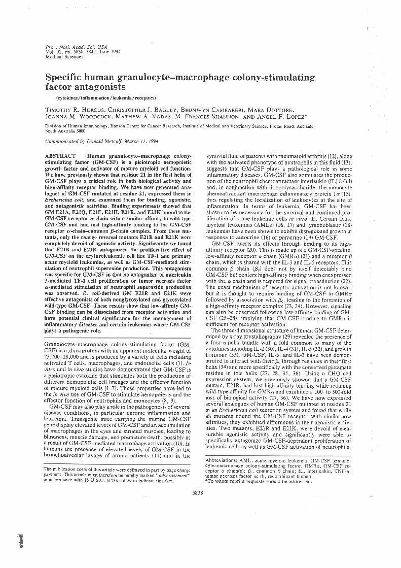
Proc. Notl. Acad. Sci. USAVol. 91, pp 5838-5842, June 1994Medical Sciences
Specific human granulocyte-macrophage colony-stimulatingfactor antagonists
(cytokines/inllammation /leukemia/receptors)
TluorHv R. H¡,ncus, CunrsropHen J. B¡,clev, BRoNwyN Cevne.neRr, Man¡ Dorronr,JoenNn M. Wooococx, M.trHew A. Ve.oe,s, M. Fnn¡¡c¿s SH¡,N1.¡on, e¡lp ANcer- F. Lop¡,2*
Division of Human Immunology, Hanson Centre for Cancer Research, Institute of Medical and Veterinary Science, F¡ome Road, Adelaide,South Ausrrâlia 5000
Communicated by Donald Metcalf, March Il, 1994
ABSTRACT Human granulocyte-macrophage colony-stimulating factor (GM-CSF) is a pleiotropic hemopoieticgrowth factor and activator of mature myeloid cell function,We have previously shown that residue 2l in the first helix ofGM-CSF plays a critical role in both biological activity andhigh-aflinity receptor binding, We have now generated ana-logues of GM-CSF mutated at residue 21, expressed them inEscherichia coli, and examined them for binding, agonistic,and antagonistic activities. Binding experiments showed thatGM E2lA, E21Q, E21F, E21H, 821R, and E21K bound to theGM-CSF receptor a chain with a similar affrnity to wild-typeGM-CSF and had lost high-affinity binding to the GM-CSFreceptor a-chain+ommon p-chain complex. From these mu-tants, only the charge reversal mutånts E21R and E21K werecompletely devoid of agonistic activit¡'. Significantly we foundthat E21R and E21K antagonized the proliferative effect ofGM-CSF on the erythroleukemic cell line TF-l and primarl,acute myeloid leukemias, as well as GM-CSF-mediated stim-ulation of neutrophil superoxide production. This antagonismwas specific for GM-CSF i¡ that no antagonism of interleukin3-mediated TF-l cell proliferation or tumor necrosis factora-mediated stimulation of neutrophil superoxide productionwas observed, E. coli-òerived GM E21R and E2lK wereeffective antagonists of both nonglycosylated and glycosylatedwild-type GM-CSF. These results show that low-affrnity GM-CSF binding can be dissociated from receptor activation andhave potential clinical significance for the management ofin{lammatory diseases and certain leukemias where GM-CSFplays a pathogenic role,
Granulocyte-macrophage colony-stimulating factor (GM-CSF) is a glycoprotein with an apparent molecular weight ol23,000-28,000 and is produced by avariety ofcells includingactivated T cells, macrophages, and endothelial cells (1). 1zvitro and i¿ vivo studies have demonstrated that GM-CSF isa pleiotropic cytokine that stimulates both the production ofdifferent hemopoietic cell lineages and the effector functionof mature myeloid cells (1-7). These properties have led tothe in vivo use of GM-CSF to stimulate hemopoiesis and theeffector function of neutrophils and monocytes (8, 9).
GM-CSF may also play a role in the pathogenesis of severaldisease conditions, in particular chronic infiammation andleukemia. Transgenic mice carrying the murine GM-CSFgene display elevated levels of GM-CSF and an accumulationol macrophages in the eyes and striated muscles, leading toblindness, muscle damage, and premature death, possibly asa result of GM-CSF-mediated macrophage activation (10). Inhumans the presence of elevated levels of GM-CSF in thebronchoalveolar lavage of atopic patients (1I) and in the
The publication costs ofthis article were defrayed in part by page chargepayment. This article must therefore be hereby marked ,, odvertisement',in accordance with 18 U.S.C. $l?34 solely 1o indicare this facr
synovial fluid of patients with rheumatoid arthritis (12), alongwith the activated phenotype ofneutrophils in this fluid (13).suggests that GM-CSF plays a pathological role in someinflammatory diseases. GM-CSF also stimulates the produc-tion ofthe neutrophil chemoattractant interleukin (lL) 8 (14)and, in conjunction with lipopolysaccharide, the monocytechemoattractant macrophage inflammatory protein 1o (15),thus regulating the localization of leukocytes at the site ofinflammation. In terms of leukemia, GM-CSF has beenshown to be necessary for the survival and continued pro-liferation of some leukemic cells in yito (1). Certain acutemyeloid leukemias (AMLs) (16, f7) and lymphoblastic (18)leukemias have been shown to exhibit disregulated growth inresponse to autocrine (16) or paracrine (19) GM-CSF.
GM-CSF exerts its effects through binding to its high-affinity receptor (20). This is made up of a GM-CSF-specific,low-affinity receptor a chain (GMRa) (21) and a receptor pchain, which is shared u/ith the IL-3 and IL-5 receptors. Thiscommon B chain (B") does not by itself detectably bindGM-CSF but confers high-aflinity binding when coexpressedwith the a chain and is required for signal transduction (22).The exact mechanism of receptor activation is not known,but it is thought to require binding of GM-CSF to GMRafollowed by association with É", leading to the formation ofa high-affinity receptor complex (23, 24). However, signalingcan also be observed following low-affinity binding of GM-CSF (25-28), implying that GM-CSF binding to GMRa issuffi cient for receptor activation,
The three-dimensional structure of human GM-CSF deter-mined by x-ray crystallography (29) revealed the presence ofa four-a-helix bundle with a fold common to many of thecytokines including IL-2 (30), IL-4 (3i), IL-5 (32), and growthhormone (33). GM-CSF, IL-5, and IL-3 have been demon-strated to interact with their p. through resjdues in their firstheiix (34) and more specifically with the conserved glutamateresidue in this helix (2'l ,28,35, 36), Using a CHO cellexpression system, we previously sbowed that a GM-CSFmutant, E21R, bad lost high-aflinity binding while rerainingwild-type affrnity for GMRø and exbibited a 100- to 300-foldloss of biological activity (27, 36). We have now expressedsevEiál ãnalogues of human GM-CSF mutated at residue 21in an Escherichi¿ coli secretion system and found that whileall mutants bound the GM-CSF receptor with similar lowaffinities, they exhibited differences in their agonistic activ-ities. Two mutants, E21R and E21K, were devoid of mea-surable agonistic activity and significantly were able tospecifically antagonize GM-CSF-dependent proliferation ofleukemic cells as well as GM-CSF activatjon of neutrophils.
Abbreviations: AML, acute myeloid leukemia; GM-CSF, granulo-cyte-macrophage colony-stimulating factor: GMRa, GM-CSF re-ceplor a chain(s); p", common p chain; IL, interleukin: TNF-a,tumor Decrosis factor a; rh, recombinan( human.*To whom reprint requests should be addressed.
T
5838
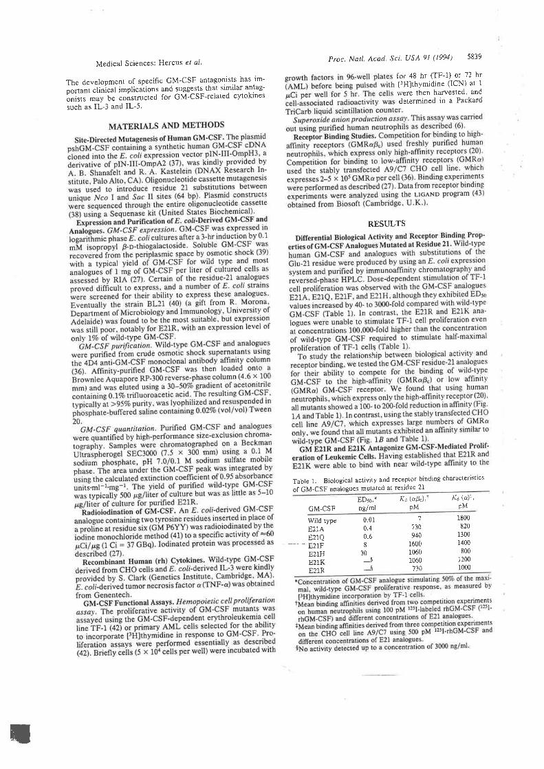
Medical Sciences: Hercus el al'
The development of specific GM-CSF antagonists has im-
ñ;"", clinical implications and suggests that similar antag-
ãnirt. muy be constructed for GM-CSF-related cytokines
such as IL-3 and IL-5.
Proc. Narl. Acad. Sci. USA 91 (1994) 5839
for 48 hr (TF-l) or 72 hr
l]Hlthymidine (lCN) at Iwere then harvested. and
determined in a Packard
RESULTS
Table 1. Biological activity and receptor binding characteristics
of GM-CSF analogues mutated at residue 21
GM-CSFEDso,*nglml
X¿ (aÉ"),rpM
K¿ (a)Ì,pM
Wild type 1800
820
1300
1400
800
1200
1000
7
730
940
1600
r0601060
730
0.010.40.68
30_t
_$
E,zTAE21Q
---- E21FE21HE21KE2lR
il
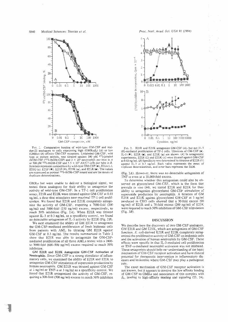
5840 Medical Sciences: Hercus ¿l ¿/. Proc. Natl. Acad. Sci. USA 9l (1994)
100
80
60
40
6 A
0 0.001 0.01 0.1 1 10 100 1000
B
a
5
4
3
2
ES
Ð.EE.E-ôþ-ØO
o-
IcX
ÊÀEao
oÈoOÉ
o
!E
F
0
0 0.01 0.1 1 10 100 1000
6
2
0
r00
80
60
40
20
0
To
1
0 0.01 0.1 I 10 100 1000
GM-CSF competitor, nM
Frc. 1. Comparative binding of wild+ype GM-CSF and resi-due-21 analogues to cells expressing high (GMRaÉJ (A) or lou'(GMRc) (8) affinity GM-CSF receptors. Unlabeled GM-CSF, wildtype or mutant protein, was titrated against 100 pM 125l-labeled
rbGM-CSF (125I-rbGM-CSF) and 3 x 10⁶ neutrophils per tube in áor 500 pM r2sl-rbGM-CSF and 5.5 x 705 A9/C7 cells per tube in LSymbols represent competition by wild-type GM-CSF (.), E2IA (tr),E2IQ (^), E2iF (a). E21H (v), E21K (^), and E21R (r). The valuesare expressed as percent 1251-rhGM-CSF bound and are the means ofduplicate determinations.
GMRa but were unable to deliver a biological signal, wetested these analogues for their ability to antagonize theactivity of wild-type GM-CSF. In a TF-l cell proliferationassay, E21R and E21K were titrated against GM-CSF at 0.03ng/ml, a dose tbat stimulates near-maximal TF-1 cell prolif-eration. We found that E2lR and E21K completely antago-nize the activity of GM-CSF, reguiring a 7000-fold (200
ng/ml) and 5000-fold (150 ng/ml) excess, respectively, toreach 50Vo inhibition (Fie. 2A). When E21R was titratedagainst IL-3 at 0.3 ng/ml, as a specificity control, we foundno detectable antagonism of IL-3 activity by E21R (Fie. 28).
We next examined the ability of GM E21R to antagonizethe GM-CSF-mediated proliferation of fresh leukemic cellsfrom patients with AML by titrating GM E21R againstGM-CSF at 0.3 ng/ml. The results summarized in Table 2
show tbat E21R was able to antagonize the GM-CSF-mediated proliferation of all three AMLS tested with a 2000-to 3000-fold (600-900 ng/ml) excess required ro reach 50Vo
inhibition.GM E21R and E2lK Antagonize GM-CSF A,ctivation of
Neutrophils. Since GM-CSF is a strong stimulator of inflam-matory cells, we examined the ability of E21R and E21K toantagonize GM-CSF stimulation of superoxide production byhuman neutrophils. GM E21R was titrated against GM-CSFat 1 ng/ml or TNF-a at 3 ng/ml as a specificity conrrol. Wefound that E21R antagonized tbe activity of GM-CSF, re-quiring a 300-fold (300 nglml) excess to reach50Vo inhibition
Flc. 2. E21R and E21K antagonize GM-CSF (,.4)- but not IL-3(8)-mediated proliferation of TF-l cel)s. Titrations of GM-CSF (.),IL-3 (O), E21R (¡), and E21K (¡) are shown. (24) In antagonisticexperiments, E21R (o) and E21K (¿) were titraled against GM-CSFat 0.03 ng/ml. (8) Specificity was determined by titration of E21R (O)against IL-3 at 0.3 ng/ml. Each value represents the mean oftriplicate determinations, and error bars represent the SEM.
(Fig. 3A). However, there was no detectable antagonism ofTNF-a even at a 10,000-fold excess.
To determine whether this antagonism could also be ob-served on glycosylated GM-CSF, which is the form thatprevails in vivo (44), we tested E21R and E21K for theirability to antagonize glycosylated GM-CSF stimulation ofsuperoxide production by neutrophils, A titration of GME21R and E2IK against glycosylated GM-CSF at 3 ng/mlproduced in CHO cells sho\ped that a 3O-fold excess (90
nglml) of E21R and a 7O-fold excess (200 nglml) of E21Kwere required to reach 50% inhibition of GM-CSF stimulation(Fie. 38).
DISCUSSION
We describe here the discovery of two GM-CSF analogues,-CM E21R and GM E21K, which are antagonists of GM-CSFfunction. E. coli-derived E21R and E21K completely antag-onized the proliferative activity of GM-CSF on leukemic cellsand the activation of human neutrophils by GM-CSF. Theseeffects \rere speci-fic in that ll--3-mediated cell proliferationor TNF-o-mediated neutrophil activation was not inhibited.These antagonists should beip our understanding ofthe basicmechanism of GM-CSF receptor activation and have clinicalpotential for therapeutic intervention in inflammatory dis-eases and leukemias where GM-CSF may play a pathogenicrole.
The exact mechanism of GM-CSF receptor activation isnot known, but it appears to involve the low affrnity bindingof GM-CSF to GMRo and association of this complex withB., leading to high-affrnity binding and signaling (23,24).
0 0 0.i I 10 i00 1000 10000
Cltokine. ng/ml
O
.I
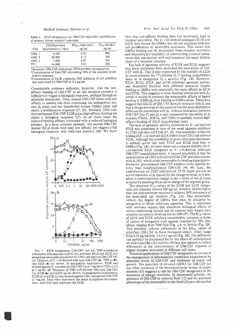
[3H]ThymidineCell incorporation,+ dpm
Medical Sciences: Hercus el al.
Table 2. E2IR antagonizes the GM-CSF-dependent proliferationof primary human myeloid ieukemias
Proc. Natl. Acod. Sci. USA 9l (/994) 5841
here that low-afhnity binding does not necessarily lead toreceptor activation. 'lhe E. colí-derived analogues E21R and
E2lK fully bound the GMRa but did not stimulate leukemiccell proliferation or neutrophil activation. This shows thatGMRa binding can be dissociated from receptor activationand illustrates the feasibility of constructing cytokine antag-onists that can interact with and sequester the major bindingchain of a receptor complex.
The lack of agonistic activity of E2lR and E21K suggeststhat these mutations have abolished the interaction of GM-CSF with Ê.. This is also supported by the inabiÌity of E21Rto cross-compete for r2sllabeled IL-3 binding (unpublisheddata) or to antagonize IL-3 activity (Fig, 2B). However,E214, 821Q, E21F, and E21H exhibited agonistic activit)'and stimulated function with different potencies despitebinding to GMRa with essentially the same affrnity as E21Rand E21K. This suggests a weak residual interaction with 8".which is unable to enhance the measurable affrnity of ligandbinding to GMRaB. over binding to GMRa alone, These datasuggest that Glu-21 of GM-CSF directly interacts with B" andthat a charge reversal at this position has the most deleteriouseffect on the interaction \,/ith B". A direct interaction betweenGM-CSF Glu-21 and É" is also supported by the ability of B.mutants Y3654, H3674, and 13684 to partially restore high-affinity binding of E21R (unpublished data).
The lack of agonistic activity exhibited by E. coliderivedE21R was surprising in view of the weak agonism exhibitedby CHO cell-derived E21R (27, 36), One possibility is that thefolding of E. collderived E21R differs from CHO cell-derivedE2lR. Although the possibility of gross structural alterationsis unlikely given that both E21R and E21K bind fully toGMRø (Fig. 1.8), we have observed a reduced stability for E.coii-derived E21R compared to E. col¡'-derived wild-typeGM-CSF (unpublished data). A second possibility is that thecarbohydrate of CHO celÌ-derived GM-CSF provides contactwith É" (45), which is not measurable by binding experiments.However, glycosylated GM-CSF exhibits lower specifìc ac-
tivity than nonglycosylated GM-CSF (44, 46). Last, the
carbohydrate on CHO cell-derived E21R might prevent an
active repulsion of p. caused by the charge reversal, or it ma1'
allow a conformational change in the a chain or the B. chainto occur by masking the positive charge ofthe arginine group.
The observed ICso values of the E21R and E21K antago-nists are typically around 200 ng/ml, which is l0-fold higherthan the concentration required to achieve 50Vo occupancy ofthe neutrophil ap" receptor (Fig. 1,4)' This presumabl¡'reflects the degree of GMRa that must be occupied byantagonist to block wild-type signaling. This is consistentwith previous reports that maximum biological effects ofcolony-stimulating factors can be exerted with steady-statereceptor occupancy levels as low as IïVo (47) ' The IC5¡ valuesof E21R and E21K exhibit a considerable variation in termsof excess of antagonist over agonist required for 50% inht-bition, ranging from 7000-fold (Fie. ZA) to 30-fold (Fig. 38)'This probably reflects di-fferences in the ED¡o values ofwild-type GM-CSF in these biological assays, which range
-lrom 0.01 nglml (Fig .2A)tol nglml (Fig' 38). The differencecan partialJy be accounted for by the effect of carbohydrateon wild-type GM-CSF activity (44) but also appears to reflectdifferences in the concentration of GM-CSF required 1o
vivo are inerbated bY
tumor ceilgrowth, The detection of elevated mRNA for GM-CSF butnot other cytokines in the bronchoalveolar lavage of atopic
gonists in thearthritis' thethe activated
ovide another
EDso (GM-CSF)î,nglml
ICso (E21R)+,
ng/ml
AML 1
AML 2
AML 3
TF-1
6,40029,000
2,90044,000
0.020.150.070.01
900
300
500
200
*Maximum GM-CSF-stimulated [3H]thymidíne incorporation.Tconcentration of GM-CSF stimulating 50Vo of lhe maximal prolif-erauve response.
tConcentration of E21R producing 50% inhibition of cell prolifera-tion stimulated by GM-CSF at 0.3 ng/ml.
Considerable evidence indicates, however, that the low-affrnity binding of GM-CSF to an aB" receptor complex is
sufficient to trigger a bioiogical response, perhaps through an
allosteric interaction. Thus, human GM-CSF binds with lowaffinity to murine cell lines expressing the endogenous mu-rine p" chain and the transfected human GMRa chain and
elicits a proliferative response (25,26). Similarly CHO cell-derived human GM-CSF E21R lacks high'affrnity binding yetelicits a biological response (27). In all these cases thereduced binding affrnity correlated with a reduced biologicalpotency. ln a most extreme example, the murine GM-CSFmutant E21A binds with only low affrnity yet triggers a fullbiological response with wild-type potency (28). We show
A
0 0.01 0.1 1 10 r00 1000 10000
B
0 0.1 I 10 100 1000
C)'tokine, ng/ml
Frc, 3. E21R anøgonizes GM-CSF- but not TNF-a-mediatedstimulation of hùman neutrophils (,4), and both E21R and E2IK alsoantagonize neutrophil stimulation by CHO cell-derived GM-CSF (B).(,4.) Titrations of E. colí-denved wild+ype GM-CSF (¡), TNF-ø (O),and E21R (r) are shown. In antagonistic experiments, E21R wastitrared against E. colËderived GM-CSF (o) at 1 ng/ml or TNF-a (Ô)at 3 ng/ml. (B) Titrations of CHO cell-derived wild-type GM-CSF(o), E21R (r), and E21K (¡) are shown. In antagonistic experiments,E21R (o) or E21K (^) was titrated against CHO cell-derived GM-CSFat 3 ng/ml. Eacb value represents the mean of triplicate determina-tions, and error bars represent the SEM.
2Eè
Ð
èõEçc
.9o
EoÀ
!'tooÀ=ln
15
10
5
15
10
5
I
5842 Medical Sciences: Hercus et ol
scenario for therapeutic intervention with GM-CSF antâgo-nists. The demonstration that E21R and E21K inhibit theGM-CSF-mediated growth of leukemic cells ir vi¡ro raisesthe possibility that tbese analogues may also be effectiveantagonists of leukemic cell proliferation mediated by GM-CSF produced in either a paracrine or autocrine manner invivo (16, 19).
We thank M. Parsons. B. Zacharakis, and C. Gaun( for theirexcellent technical assistance. This work was suppoled by theNational Health and Medical Research Council of Australia and byNational Institutes of Health Grant CA 45822. C.I.B. is a recipientof a C. J. Martin fellowship from the National Health and MedicalResearch Council of Australia.
1. Mctcalf, D. (1984) The Hemopoieric Colony Stimuloting Fac-rors (Elsevier, Amsterdam).
2. Vadas, M. ,4'., Nicola, N. A. & Metcalf, D. (1983),f. lm¡nunol.t}o, 795-:799.
3. Lopez, A. F., Nicola, N.4., Burgess, A. W., Metcalf, D.,Battye, F. L., Sewell, W. A. & Vadas, M. (1983) J. Immunol.13t,2983-2988.
4. Gasson, J. C., Wejsbart, R. H., Kaufman, S. E., Clark, S. C.,Hewick, R. M., Wong, G. G, & Golde, D. W. (1984) Science226, 1339-1342.
5. Metcalf. D., Begley, C. G., Johnson, G. R., Nicola, N.A'.,Vadas, M. ,4.., Lopez, A. F., Williamson, D. J., Wong, G, G.,Clark, S. C. & Wang, E. A. (1986) Blood 67,37-45.
6. Lopez,A. F., Williamson, D. J., Gamble,J. R.,Begley, C. G.,Harlan, J. M., Klebanoff, S., \{altersdorph, ,{., Wong, G.,Cla¡k, S. C. & Vadas, M. A. (1986) J. Clin. Invest. TE, 1220-7228.
7. Metcalf, D., Begley, C. G., Williamson, D. J., Nice, E. C.,Delamarter, J., Mermod, J., Thatcher, D. & Schmidt, A.(1987) Exp. Hemotol.15, I-9.
8. Groopman, J. E., Mitsuyasu, R. T., Deleo, M. J., Oette,D. H. & Golde, D. W. (1987) N. Ensl. J. Med.317,593-598.
9. Baldwin, G. C., Gasson, J. C-, Quan, S. G., Fleischmann, J.,Weisbart, R., Oette, D., Mitsuyasu, R. T. & Golde, D. W.(1988) Proc. NatL. Acod.,Sci. US,4 85,2763-2?66.
10 Lang, R.,4'., Metcalf, D., Cuthbertson, R.,4'., Lyons, L,Stanley, E., Kelso, .4'., Kannourakis, G., Williamson, D. J.,Klintworth, G. K., Gonda, T. J. & Dunn, A. R. (1987) C¿ll 51,675-686.Kato, M., Liu, M. C., Stealey, B. 4., Friedman, 8., Lichten-stein, L. M., Permutt, S. & Schleimer, R. P. (1992) Lympho-kine Cytokine Res. ll, 281¿92.Williamson. D. J., Begley, C. G., Vadas, M. A. & Metcalf, D.(1988) C/tn. Exp. Immunol. 72, 6'7-73.Emery, P., Lopez, A. F., Burns, G. F. & Vadas, M. A. (1988)Ann. Rheum. Dis. 47, 34-39.McCain, R. W., Dessypris, E. N. & Christman, J. W. (1993)Am. J. Respir. Cell Mol. Biol.8,28-34.Kasama, T., Strieter, R. M., Standiford, T. J., Burdick, M. D.& Kunkel, S. L. (1993) J. Exp. Med. 178,63-'72.Young, D. C. & Griffin, J. D. (1986) Blood 68,1178-1181.Russell, N. H. (1992) Blood Rev.6, 149-156.Freedman, M. H., Grunberger, T., Correa, P., Axelrad, A. 4.,Dube, I. D. & Cohen, A. (1993) Blood 8r,3068-3075.Metcalf, D. (1986) Blood 67,257-267.Gasson. J. C., Kaufman, S. E., Weisbart, R. H., Tomonaga,
Proc. Natl. Acad. Sci. USA 9l (1994)
M. & Golde, D. W. (f986) Proc. Natl. Acad. Sci. US,{ 83,669-6"t3.
21. Gearing. D. P., King, J ,4.., Gough, N. M. & Nicola, N A.(1989) EMBO J. 8, 3667-1676
22. Hayashida, K., Kitamura, T., Gorman, D M., Arai, K.-1.,Yokota, T. & Miyajima, A. (1990) Proc. Natl. Acad. Scì. USA87,9655-9659.
23. Nicola, N.4., Wycherley, K., Boyd, A. W., Layton, J. E.,Cary, D. & Metcalf, D. (1993) BloodE2,I'124-7731.
24. Miyajima, ,4'., Mui, A. L,-F,, Ogorochi, T. & Sakamaki. K.(1993) Blood 82, 1960-19"14.
25. Metcalf, D., Nicola, N.,4'., Gearing, D. P. & Gough, N M.(1990) Proc. Natl. Acad. Sci. USA 87,4670-4674.
26. Kitamura, T., Hayashida, K., Sakamaki, K., Yokota, T., Arai.K.-1. & Miyajima, A. (199i) Proc. Natl. Acad. Sci. US,4 E6,
5082-5086.27. Lopez, A. F., Shannon, M. F., Hercus, T., Nicola, N.,A.,
Cambareri, B., Dottore, M., Layton, M. J., Eglinton, L. &Vadas, M. A. (1992) EMBO J. 11, 909-916
28. Shanafelr, A. B. & Kastelein, R. A. (1992)./. Biol. Chem.267,2s466-254'72.
29. Diederichs, K. , Boone, T. & Karplus, P . A. (1991) Science 254,1779-1'182.
30. McKay, D. B. (1992) Science 257,412-413.3r. Smjth, L. J., Redfield, C., Boyd, J., Lawrence, G. M. P.,
Edwards, R. G., Smith, R. A. G. & Dobson, C. M. (1992) J.MoL BìoL.2U,899-9M.
32. Milburn, M. V.. Hassell, A. M., Lambert, M. H., Jordan,S. R., Proudfoot, A. E. I., Graber, P. & Wells, T. N. C. (1993)
N a tu r e (Londo n) 363, 77 2-7'1 6.33. de Vos, A. M., Ultscb, M. & Kossiakoff, A. A. (1992) Science
25s,306-312.34. Shanafelt, A. 8,, Miyajima, ,4.., Kitamura, T. & Kastelein,
R. A. (1991) EMBO J. r0, 4105-4112.35. Barry, S. C., Bagley, C. J., Phillips, J., Dottore, M., Cam-
bareri,8., Moretti, P., D'Andrea, R., Goodall, G. J., Shannon,M. F., Vadas, M. A. & Lopez, A. F. (1994) J. B ioL. C he m. 269,8488-8492.
36 Hercus, T. R., Cambareri, 8., Dotlore, M., Woodcock. J.,Bagley, C. J., Vadas, M. ,4',, Shannon, M. F. & Lopez, A. F,(I94) Blood, in press.
37. Ghrayeb, J., Kimura, H., Takahara, M., Hsiung. H.. Masui, Y.& Inouye, M. (1984) EMBO J.3,2437-2442.
38. Chen, E. Y. & Seeburg, P. H. (1985) DNA 4, 165-7'1039. Koshland, D. & Botstein, D. (1980) Cell 20,149-160.40. Studier, F. W. & Moffatt, B. A. (1986) J. Mol. Biol. lE9,
113-130.41. Contreras, M. 4., Bale, W. F. & Spar, I. L. (7983) Methods
Enzymol.92, Z"l1¿97.42. Kitamura, T., Tange, T., Terasawa, T., Chiba, S., Kuwaki, T.,
Miyagawa, K., Piao, Y.-F., Miyazono, K., Urabe, A. &Takaku, F. (1989) J. Cell. PhysioL.140,323-334.
43. Munson. P. J. & Rodbard, D. (19E0) AnaL. Biochem. 101,220-239.
44. Cebon, J., Nìcola, N., Ward, M., Ga¡dner, I., Dempsey, P.,Layton, J,, Dührsen, U., Burgess, A. W., Nice, E. &Morstyn,G. (19m) J. Biol. Chem. 26s, 4483-4491.
45. Opdenakker, G., Rudd, P. M., Ponting, C. P. & Dwek, R. A.(t993) FASEB J. 7, 1330-1337
46, Kaushansky, K., O'Hara, P J., Hart, C. E., Forstrom, J. W.___& Hagen, F. S. (1987) Biochemistry 26,4861-4E6'1 .
47. Nicola, N. ,4'., Peterson, L., Hilton, D. J & Metcalf, D. (1988)Growth Factors l, 4I-49.
11.
12.
13.
14.
15.
16.71 .
18.
1920
it
Hercus, T., Cambareri, B., Dottore, M., Woodcock, J., Bagley, C.J., Vadas, M.A.,
Shannon, M.F. & Lopez, A.F. (1994) Identification of residues in the first and fourth
helices of human granulocyte-macrophage colony-stimulating factor involved in
biologic activity and in binding to the alpha and beta chains of its receptor.
Blood, v. 83(12), pp. 3500-3508
NOTE:
This publication is included as paper 3 in the print copy
of the thesis held in the University of Adelaide Library.
(EMBO J. in press, 1994)
Three residues in the common p chain of the human GM-CSF' IL'3 and
IL-5 receptors are essential for GM-CSF and IL'5 but not IL'3 high
affinity binding and interact rvith Glu2l of GM-CSF'
Woodcock, 1.M., Zacharakis, 8., Plaetinck, G.#, Bagley, C'J', Qiyu, S', Hercus' T'R"
Tavernier, J.# and LoPez, A.F.
Keywords: Affinity conversion / cytokines / mutants / st¡ucture.
Running title: Molecula¡ basis for the affinity conversion by Fc'
Division of Human Immunology, Hanson Centre for Cancer Research, Institute of Medical
and Veterinary Science, Frome Road, Adelaide, South Aust¡alia 5000.
#Roche Resea¡ch Gent, Plateau sf:aat 22, 9000 Gent, Belgium'
Address reprint requests to Dr. Angel Lopez, Division of Human Immunology, Institute of
Medical and Veterinary Science, Box 14 Rundle Mall Post Office, Adelaide, South
Australia 5000.
Phone + 6l-8-228 747l,Fax +61-8-228 7538
Communicated bY Walter Fiers.
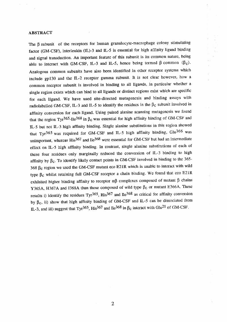
ABSTRÄCT
The F subunit of the receptors for human granulocyte-macrophage colony stimulating
factor (GM-CSF), interleukin (IL)-3 and IL-5 is essential for high affinity ligand binding
and signal transduction. An important feature of this subunit is its common nature, being
able to interact with GM-CSF, IL-3 and IL-5, hence being termed p common (ps).
Analogous common subunits have also been identified in other receptor systems which
include gp130 and the lL-2 receptor gamma subunit. It is not clear however, how a
common receptor subunit is involved in binding to all ligands, in particular whether a
single region exists which can bind to all ligands or distinct regions exist which are specific
for each ligand. We have used site-directed mutagenesis and binding assays with
radiolabelled GM-CSF ,lL-3 and IL-5 to identify the residues in the Bç subunit involved in
affinity conversion for each ligand. Using paired alanine scanning mutagenesis we found
that the region Tyr365-Ile368 in Þc was essential for high affinity binding of GM-CSF and
IL-5 but not IL-3 high affinity binding. Single alanine substitutions in this region showed
that Tyr365 was required for GM-CSF and IL-5 high affinity binding, 61u366 \Ã/¿t
unimportant, whereas ¡y¡s367 ¿¡¿ ¡1s368 were essential for GM-CSF but had an intermediate
effect on IL-5 high affinity binding. In contrast, single alanine substitutions of each of
these four residues only marginally reduced the conversion of IL-3 binding to high
affinity by Fc. To identify likely contact points in GM-CSF involved in binding to the 365-
36g pc region we used the GM-CSF mutant eco E21R which is unable to interact with wild
type Þc whilst retaining full GM-CSF receptor a chain binding. We found that eco E21R
exhibited higher binding affinity to receptor aB complexes composed of mutant B chains
Y3654, H367A and 13684 than those composed of wild type Fc or mutant E3664. These
results i) identify the residues Tyr365, g¡5367 ¿n6 ns368 as critical for affinity conversion
by Þc, ii) show that high affinity binding of GM-CSF and IL-5 can be dissociated from
IL-3, and iii) suggest that tyr36S, ¡¡¡s367 ¿¡¿ ¡1s368 in ps interact with Glu2l of GM-CSF'
2
INTRODUCTION
The human cytokines granulocyte-macrophage colony-stimulating factor
(GM-CSF), interleukin-3 (IL-3) and interleukin-S (L-5) are important regulators of
haemopoiesis and inflammation (reviewed in Metcalf, 1991 and Lopez et a1.,1992b). The
regulatory effects of these cytokines are mediated by high affinity cell surface receptors
that afe known to comprise at least an c chain and a p chain. The F chain (Hayashida et al"
1990) and a chains of the GM-CSF (Gearing et a1.,1989), IL-3 (Kitamura, et al., 1991b)
and IL-5 (Tavernier et a1.,1991, Murata et ø1., 1992) receptors have been cloned and the
receptor complexes reconstituted and characterized (Hayashidt et al., 1990' Kitamvla' et
at., l99lb and Tavernier et a1.,1991). These experiments showed that whilst both the s
and B receptor subunits are required for signalling (Kitamurt et al., l99la, Kitamuta et al.,
1992, Takakt et a1.,1993) they subserve distinct functions in terms of binding. Thus the o
chains are ligand-specific and bind their cognate ligand with low affinity only (Kd = 10-
9-tO-7Vf). On the other hand the p subunit is common (Þc) to the GM-CSF, IL-3 and IL-5
receptors and although it does not detectably bind ligand by itself, it acts as an affinity
converter allowing high affinity (10-10y¡ binding when co-expressed with an cr chain.
The role of Bç in mediating affinity conversion and signal transduction by different
ligands is similar to that of gp130 and the lL-2 teceptor 1 chain' Thus, gp130 provides
high affinity binding for leukaemia inhibitory factor, IL-6, IL-11, oncostatin M and ciliary
neurotrophic factor (Akira et al., 1993) and is important for function. similarly, the T
chain of t¡¡elL-2 receptor increases the binding affinity of lL-2 (Takeshita et al., 1992),
IL-4 (Russ ell et al., 1993) and lL-7 (Noguchi et al., 1993) when co-expressed with the
respective ligand specific receptor chains and contributes to signalling. It is not clear
however, how a single subunit in all these receptor complexes can increase the binding
affinity of several different ligands. InterestinglY, the pç subunit increases the affinity of
GM-CSF, IL-3 and IL-5 binding to different degrees when co-expressed with the respective
a chain. Thus ps increases IL-5 binding affinity approximately 2-5 fold (Tavernier et al',
1991 and Tavernier et a.1., lgg2), GM-CSF binding affinity 20-100 fold (Hayaslida et al"
1990) and IL-3 binding affinity 500-1000 fold (Kitamutr et al', 1991b and Kitamura ¿f
al., lgg2) raising the possibility that more than one region is involved in conferring high
affinity binding on GM-CSF,lL-3 and IL-5.
Structurally fre Þc and cr chains of the GM-CSF, IL-3 and IL-5 receptors belong to
the family of cytokine receptors that includes receptors for growth hormone (GH)'
erythropoieti n, [L-2,IL-4, IL-6 and lL-7 (Gearing et û1., 1989, Bazan, l99O and D'Andrea
et al.,l9g9). Members of this family share conserved structural motifs in the extracellular
portion which can provide useful information when appropriately aligned. Growth
3
hormone (GH) binds to a homodimeric receptor. The soluble form of this receptor, growth
hormone binding protein (GHbp) has been crystallised in a complex with GH and the three
dimensional structure elucidated (de Vos et al., 1992). We have aligned the amino acid
sequence of Fc to the GHbp and used information from the GH'(GHbp)2 crystals to
hypothesize regions in ps which may be involved in ligand binding (Goodall et al., 1993).
Of these regions we have targetted the region between residues 361-370 because it aligns
with a region in GHbp (163-171) that is known to bind GH (de Vos ¿l al., 1992). Thts
region also aligns with residues of the mouse IL-3 receptor p chain AIC2A, and the human
IL-2 receptor p chain that are known to be involved in ligand binding (Wang et al., 1992
and Imler et al., lggz).In addition recent studies (Lock et al., 1994) showed 11¿1 ¡¡i5367 6¡
ps is important for GM-CSF high affinity. It is not clear however, whether this region is
involved in IL-3 and IL-5 as well as GM-CSF binding, nor the identity of complementary
regions in the ligands involved.
A candidate common motif for the binding of human GM-CSF, IL-3 and IL-5 to
ps is represented by the conserved acidic residue Glu2l in GM-CSF, Glu22 in IL-3 and
Glu13 in IL-5. The mutation of this Glu selectively impairs high affinity binding for
human GM-CSF (Lopez et al., 1992a, Shanafelt et a1.,1992),IL-3 (Barry et al., 1994) and
IL-5 (unpublished observations). In particula¡, substitution of Glu2l of human GM-CSF
for Arg and expression in E.coli causes complete loss of high affinity binding as well as
signalling and results in a specific antagonist (Hercus et al.,l994a). We have now used this
molecule to test the hypothesis that the conserved Glu in GM-CSF,IL-3 and IL-5 interacts
with the 361-370 region of Fc.
We show here using alanine mutagenesis in conjunction with binding studies with
all three known ligands, that residues Tyr365, ¡¡¡s367 ¿n¿ ¡1s368, located in the membrane
proximal domain of the extracellula¡ portion of the ps subunit are involved in high affinity
binding. Importantly, these residues are essential for GM-CSF and IL-5 but not for IL-3
high affinity binding implying that an additional region may be involved in IL-3 high
afflnity binding and suggesting a structural explanation for the large effect of the pç
subunit in increasing IL-3 binding affinity. Finally, by using the GM-CSF mutant eco
E21R that is deficient in Bs interaction (Hercus et al., 1994a), we propose a model where
ûre Fc region 365-368 interacts with Glu2l in the first helix of GM-CSF.
4
RESI'LTS
Identification of residues of the human p chain critical for high affinity receptor
formation; differential effects of alanine-scanning mutants on GM'CSF,IL-s and IL-3
high alfinity receptor formation
In order to identify residues of ps which may play a role in affinity conversion we
focussed on a region (358-373) which aligns with a region in the membrane proximal
domain of GHbp that is involved in GH binding (de Vos et al., 1992). The two subunits of
GIIbp bind GH in a non-equivalent manner and a¡e therefore denoted GHbpI and GHbpII.
The region 163-17l of GHbp is particularly attractive since it is involved in the binding of
GH to GHbpII (de Vos et al., 1992).In this respect GHbpII is tunctionally analogous to
tne ps in that GH does not bind to GHbpII unless it is already bound to GHbpI
(Cunningham and Wells, 1989). In addition, the region 358-373 in ps aligns with regions
in the mouse IL-3 receptor AIC2A and the human IL-2 receptor p chain which have been
shown to be important in ligand binding (Fig. 1) (Wang et al., 1992 and Imler et al.,
lgg2). This region of Þc is predicted to lie in or close to a loop between two p strands
(Bazan, 1990).
Alanine-scanning mutagenesis was carried out across the region 361-370 in order
to identify residues of the p chain required for affinity conversion and in particular their
involvement with each ligand. Alanine substitutions were used to minimize conformational
effects of mutation across this region (Cunningham and Wells, 1989). Initially, residues
were substituted with alanines in pairs (Fig. 1). The mutant p chains M36INK362A,
M363A/R364A, y365AÆ3664, IJ367/./13684 and D369NH3704 were co-expressed on
COS cells with either the GM-CSF, IL-3 or IL-5 receptor a chain and binding of the
appropriate ligand determined in saturation binding studies. In the absence of ps the
binding affinities of the transfected cr chains alone ranged from 2-12 rMl for GM-CSF,20-
100 nM for IL-3 and 0.8-1.0 nM for IL-5 (Tables I and II). These values are consistent
with previously determined affinities (Gearing et al., 1989, Kitam:urr et al-, l99lb,
Tavernier et a1.,1991 and Murata et al., 1992). The Þ chain mutants M3614/K3624,
M3634/R3644 and D361NH370A had no significant effect on GM-CSFor IL-3 high
affinity binding (Fig. 2 and Table I). However, the mutants Y3654/E3664 and
H367N13684 had a profound effect on high affinity conversion by Fc Gig. 2 and Table
I). Both mutants completely abolished GM-CSF high affinity binding. In contrast, the
mutant 1.365py¡;3664 did not significantly reduce IL-3 high affinity binding whilst the
mutant H367N13684 caused a reduction in high affinity ftom 110 pM (wil<l type ps) to
530 pM which was statistically significant (Table I). In the case of IL-5, the mutant
M361A/K3624 did not affect high affinity binding whilst the mutants M3634/R3644 and
5
D361NH370A exhibited a small loss of IL-5 high affinity binding (Fig. 2 and Table I).
The mutants Y365A/E3664 and H367N1368A on the other hand caused the greatest loss
of IL-5 high affinity binding (Fie.2 and Table I).
The loss of high affinity binding associated with mutant p chains Y3654/83664
and H3674/I3684 was not due to lack of expression on the transfected cells. The cell
surface expression of mutant p chains was monitored by flow cytometry using anti-p chain
monoclonal antibodies 3D7 and 4F3 (see Materials and Methods), and CRS-I (Watanabe et
at., 1992). Antibodies 3D7 and 4F3 both specifically precipitate a protein of 120 Kd from
permanently transfected surface-labelled CHO cells corresponding in size to the p chain
(Fig. 3A). Fig. 3B shows a typical result where the anti-p chain antibody 4F3 stained COS
cells transfected with mutant p chain H367 NI368A as well as cells transfected with wild
type P chain. Fig. 38 also shows staining for GM-CSF receptor a chain expression on these
transfectants with monoclonal antibody 8G6. In IL-5 studies surface expression of mutant
B chains was confirmed by affinity crosslinking. Fig. 3C shows the result of crosslinking
l25y1y-5 to COS transfectants expressing IL-5 receptor a chain alone and with wild type
or mutant p chain Y3654Æ3664. The protein bands detected by the irreversible
crosslinking o¡ 125¡-¡¡-5 correspond in size, after subtraction of the molecular weight of
an IL-5 monomer (the gel was run under reducing conditions), to the IL-5 receptor a
chain (60 kD) and Bc (120 kD).
In order to elucidate the contribution of individual residues to high affinity
binding, single alanine substitutions were made at positions 365-368. In each case the
effect of these substitutions was determined after cotransfection with either GM-CSF' IL-3
or IL-5 receptor cr chain in saturation and competition binding studies. As above, cell
surface expression of mutant p chains was confirmed by flow cytometry (data not shown).
Results showed that alanine substitution of residues Tyr365, gi5367 ¿¡¿ ¡1s368 led to the
complete loss of high affinity GM-CSF binding, whereas alanine substitution s¡ 61¡366 ¡¿¿
no significant effect on high affinity GM-CSF binding (Fig. 4, Table II). In contrast to
GM-CSF, the effects of individual substitutions at residues 365-368 on IL-3 high affinity
binding were small, resulting in only a four fold decrease in affinity relative to the wild-
type receptor in each case (Table II). Alanine substitutions at Tyr365 , ¡¡¡5367 fl1¿ ¡1s368
caused significant reductions in IL-5 high affinity binding whereas substitution ¿1 Ç1¡366
had no significant effect.
partial restoration of binding of the GM-CSF analogue eco E21R to mutant p chains.
We have previously shown that the GM-CSF analogue eco E21R is deficient in
binding to ps and signalling while retaining wild type binding to the GM-CSF receptor CI,
chain (Hercus ¿f at., 1994a). In order to ascertain whether the conserved Glu motif of
6
GM-CSF, IL-3 and IL-5 is involved in direct intefaction with the 365-3ó8 region of the p
chain we óxamined the ability of the eco E21R to compete for wild-type GM-CSF binding
on mutant p chains. Competition binding studies were carried out on COS cells
cotransfected with GM-CSF receptor a chain and wild-type or mutant p chain. Radio-
iodinated wild-type GM-CSF binding was determined using 100 pM 125¡-6¡4-çSF in the
presence of an increasing amount of eco E2lR analogue. Characterization of eco E21R
binding showed that this mutant was able to compete with wild-type GM-CSF with equal
potency for binding to low affinity o chain receptors but exhibited a 100 fold reduction in
its ability to compete for binding to high affinity crp receptors (Fig. 5A). Significantly, eco
E2lR competed more effectively on transfectants expressing p chains with alanine
substitutions at position 365,367 and 368 compared to the wild-type p chain or the p chain
mutant E3664 (Fig. 5B). This suggests that the mutant p chains Y3654, H367A and 13ó84
are able to interact with eco E21R and that the substitutions made in Bs complement in part
the charge reversal substitution made in GM-CSF. Comparison of the effective dose for
SOVo competition (ED5g) exhibited by eco E21R showed that eco E21R bound to GM-CSF
receptor ü chain plus Y3654, H3ó74 and 13684 complexes 5-7 times better than to
E,3664 or wild-type ps complexes (Table III).
7
DISCUSSION
We show here that the common p chain of the GM-CSF, IL-3 and IL-5 receptors
contains three residues, Tyr365, ¡¡¡s367 ¿n¿ ¡1s368 which are involved in GM-CSF and IL-5
high affinity binding. However, whilst alanine substitution of these three residues abolishes
GM-CSF and IL-5 high affinity binding, they affect only marginally IL-3 high affinity
binding. These findings raise the possibility that other common subunits of receptor
complexes such as gp130 and the lL-2llL-4llL-7 receptor gamma chain may also have
common regions and unique regions recognized by different ligands. In addition, using
the human GM-CSF antagonist eco E21R which has selectively lost its ability to bind to Bs
and signal (Hercus et al., 1994a) we show partial binding complementation on pç mutants
1'3654, H3674 and 13684 suggesting that the conserved Glu in the first helix of GM-CSF
directly interacts with the 365-368 region of Fc.
By using alanine substitution mutagenesis we have defined the region 365-3ó8 in
ps and more specifically Tyr365, g¡s367 ¿¡6 ¡¡s368 as being essential for GM-CSF high
affinity binding. The region between Tyr365 ¿¡6 ¡1s368 aügns with a region in the second
cytokine receptor domain of the GHbp (Goodall et al., 1993) which has been shown to
contact GH after GH binds to GHbpI (de Vos et al, 1992), and with a region in AIC2A
(Wang et al., 1992) and IL-2R p (Imler et aI., 1992) important for mouse lL-3 and lL-2
binding respectively. By analogy with these molecules and from structural predictions
(Bazan, 1990 and Goodalt et a1.,1993) this region is predicted to lie in the loop between
tlre B' and C' beta strands of the second cytokine receptor module (CRM) of ps (Fig. 6).
This suggests that rather than forming a discrete contact point for interaction, this region
forms a binding surface on which several residues contribute to ligand interaction. It is
noteworthy ft¿1 61u366 does not appear to contribute to ligand interaction suggesting that
the side chain of this residue is orientated away from the proposed binding surface.
The loss of affinity converting activity associated with mutant p chains Y3654,
H367A and 13684 is neither the result of the lack of surface expression nor due to gross
conformational changes as judged by the following criteria. Firstly, t¡ansfected cells were
all stained with monoclonal anti-B chain antibodies. Three different antibodies were used
and the staining seen with each antibody was equivalent in all cases confirming cell surface
expression and suggesting that the mutations did not introduce gross conformational
changes. Secondly, affinity cross-linking with 125ç1y-5 to cells expressing receptor c, and
y365¡y83664 Þc which bind IL-5 with reduced affinity resulted in cross-linking of two
proteins which corresponded in size to the IL-5 receptor cr and B chains. Thirdly, mutant p
chains which were deficient in affinity converting GM-CSF and IL-5 binding still conferred
high affinity binding on IL-3.
I
In order to identify corresponding residues in GM-CSF likely to interact with the
365-368 region of ps we focussed on the first helix of GM-CSF which has been shown to
be important for high affinity binding (Shanafelt et aL, 1991, Lopez et al., 1992a). Of
several GM-CSF helix A mutants we have produced, we used the GM-CSF mutant eco
E21R because mutations at Glu2l lead to a selective loss of GM-CSF-Pc interaction (Lopez
et al., 1992r and Shanafelt and Kastelein, 1992) and Glu2l is the main residue in the first
helix of human GM-CSF that interacts with ps (Hercus et al., 1994b). We found that the
GM-CSF mutant eco E21R competed better ¡ot 125¡-6¡4-CSF binding on cells expressing
the mutant p chains that affect high afflnity GM-CSF binding, namely Y3654, H3674 and
136gA. No effect was observed on E3664 consistent with the lack of .effect of this
substitution in direct binding studies. These results raise the possibility that residues Tyr365,
¡¡¡s367 ¿¡6 ns368 directly interact with GM-CSF and more specifically with Glu21 (Frg. 6).
It is interesting to note that individual substitution of each residue, Tyr365, ¡¡¡s367 ¿¡6
¡1s368 is sufficient to abrogate GM-CSF high affinity binding. The mechanism by which
these residues contribute to GM-CSF high affinity binding is not exactly known. It is
possible that the mutations alter the overall structure of the high affinity binding site,
however this is unlikely in view of the fact that all the mutant receptors retained high
affinity conversion for IL-3. This argues against severe structural alterations' It is more
likely, however, that residues 365 - 368 act cooperatively to recognize helix A of GM-CSF.
In this case the conúibution of each residue to the ligand-binding determinant is dependent
on the presence of the appropriate neighbouring residue. This cooperativity is also likely
to explain the slight differences in affinity between the double and single mutants in IL-3
and IL-5 high affinity binding. Our results are consistent with the recent report 6¡ ¡1i5367
of ps being important for high affinity GM-CSF binding (Lock et al., 1994).Interestingly,
in contrast to our findings, Tyr365 was not found to be important in that study. This
difference may be due to the nature of the substitution made; phenylalanine was substituted
for Tyr36S. This subsitution retains the hydrophobicity of this residue and therefore may
not interfere with salt bridge formation. Residue ¡1e368 was not investigated in that paper
(Lock et al., 1994).
In the case of IL-5 the results were less clea¡ than with GM-CSF and IL-3 due Ûo the
relatively small (2-5 fold) increase in binding provided by Fc over the IL-5 receptor ø
chain alone. Nevertheless alanine substitutions revealed that Tyr365 was essential for
affinity conversion by Þc. In contrast, the substitution of 61¡366 alone did not reduce IL-5
binding and may have slightly attenuated the effect of Y3654 in the double mutant.
Individual alanine substitutions at positions 367 and 368 resulted in modest decreases in
binding but together resulted in the abolition of detectable high affinity binding' Thus, as
with GM-CSF binding, residues Tyr365, gis367 ¿n6 ¡1s368 are important for the high
affinity binding of IL-5.
9
rwe found that in contrast to GM-CSF and IL-5, high affinity binding of IL-3 was
not abolished by mutation of residues Tyr365, ¡1¡s367 ¿¡¿ ¡1s368. In fact only a four fold
reduction in affinity was observed (Flg. 2 and 4, Tables I and II) which is a relatively small
effect when taking into account the approximate 500 fold increase in IL-3 binding affinity
provided by Þc (Kitamura et a1.,1991b and Table II). This suggests that an additional or
alternative region in ps contributes to IL-3 high affinity binding which may explain why
ps is able to confer such a large increase in affinity on tlre IL-3 receptor c[ chain relative to
GM-CSF and IL-5. This notion is supported by two other sets of evidence. Firstly, whilst
the GM-CSF eco E21R mutant has completely lost its ability to interact with pç and signal,
behaving as an antagonist (Hercus et al,1994a), the analogous IL-3 mutant eco E22R is a
weak agonist (Barry et al., 1994) suggesting residual interaction with ps. Secondly, we
found that the monoclonal antibody 4F3 partially inhibits IL-3 but not GM-CSF high
affinity binding and stimulation of TF-l cell proliferation (unpublished results). Alignment
of ps with the GHbp suggest that residues 309-315, 333-343 and 414-424 in the loops
between p strands E and F, G and A', and F' and G' respectively, may be involved and it
would be of interest to test these hypotheses experimentally'
Finally, our results illustrate that GM-CSF and IL-5 high affinity binding can be
dissociated from IL-3 high affinity binding by discrete mutations it F. raising the
possibility that differences in ligand specificity may also be found in other common
receptor subunits such as gpl30 and the IL-2 receptor l chain. The identification of ligand
binding regions and determination of their specificity in these common subunits may allow
the selective blocking of cytokines with appropriately designed compounds'
10
MATERIALS AND METHODS
Mutagenesis of p chain and expression plasmid constructs'
The cDNA for the human p chain was cloned by PCR from cDNA prepared from
the KMT-2 cell line (Barry et aI., lg94). The 2.8 Kb CDNA encoding the p chain was
subcloned as an EcoRl-XbaI fragment into pSelect (Promega, Sydney, NSW). Mutations
were introduced into the p chain cDNA using oligonucleotide-directed mutagenesis
(Altered-sites, Promega). Using this method, a second oligonucleotide which restores
ampicillin resistance to template DNA was used in combination with the mutagenic
oligonucleotide and mutants selected for ampicillin-resistance in a repair deficient strain of
E. coli. The mutagenic oligonucleotides used were 3O-mers and were synthesised on an
Applied Biosystems 3814 DNA Synthesiser (Burnwood, VIC). The mutations were
confirmed by nucleotide sequencing and the mutant p chain cDNAs subcloned into the
eukaryotic expression vector pcDNAl (Invitrogen, San Diego, CA)'
The IL-3 receptor a chain was cloned by PCR from KMT-2 cells (Barry et al',
lgg4). The GM-CSF receptor o chain was kindly provided by Dr. N. Nicola (Walter and
Eliza Hall Institute, Melbourne Victoria) and the IL-5 receptor cr chain was obtained as
described previously (Tavernier et a1.,1991). All the receptor cr chains were cloned into the
eukaryotic expression vector pcDMS (Invitrogen) for t¡ansfection.
COS celts and DNA transfection.
COS cells were maintained in RPMI-1640 medium supplemented with 57o vlv fetal
calf serum (FCS). DNA constructs were int¡oduced into COS cells by electroporation using
a Bio-Rad Gene pulser (North Ryde, NSW). Routinely 2 x lO7 cells in 0'8 ml ice-cold PBS
were co-transfected with 10 g of GM-CSF or IL-3 receptor cr chain DNA in pcDMS and
25 mg of pcDNAl vector containing the .wild type or mutated p chain cDNA at 500 F and
300 V. For IL-5 studies cells were transfected using DEAE-dextran as described previously
(Tavernier et al.,l99l). Binding assays and antibody staining were carried out on cells 2-3
days after transfection.
GM-CSF,IL-3 and IL-S
Recombinant human GM-CSF and IL-3 were produced in E. coli. Wild-type
GM-CSF and the analogue eco E21R were expressed in the periplasmic space of E. coli'
from a synthetic human GM-CSF cDNA in the expression vector pIN-III-OmpH3 and
purified as described elsewhere (Hercus et al., 1994a). Recombinant IL-3 was expressed
similarly from a native cDNA carrying the mutation F133Y to improve radio-iodination'
The CDNA was cloned into the pFLAG vector (International Biotechnologies inc',
Newhaven, CT) which adds an octapeptide sequence to the N-terminus to facilitate affinity
11
purification (Barry et al., 1994). Recombinant human IL-5 was produced in S9 cells as
detailed (Tavernier et al., 1989\.
Radiolabelling cytokines
Recombinant human GM-CSF and hIL-3 were radio-iodinated by the iodine mono-
chlòride method (Contreras et al., 1983) to a specific activity of approximately 10 mCi/mg
and 36 mCi/mg respectively. Iodinated protein was separated from iodide ions on a
Sephadex G-25 column (Pharmacia, North Ryde, NSW) eluted with PBS containing 0.O2Vo
v/v Tween 20 and stored ú 4oC for up to 4 weeks. Prior to use, radio-iodinated protein was
buffer exchanged on a carboxymethyl-Sepharose CL-68 column loaded at pH 2.6 in 10
mM citrate-phosphate buffer and eluted in binding medium consisting of RPMI 1640
supplemented with l0 mM HEPES, 0.5 Vo w/v bovine serum albumin and 0.1 7o wlv
sodium azide. Recombinant hIL-5 was iodinated with the Iodogen reagent (Pierce
Chemicals, Rockford, IL), as described (Plaetinck et a1.,1990).
Binding assays
For binding studies, transfected COS cells were detached by treatment with RPMI
containing 40mM EDTA, 2N glml chondroitin sulphate and O.lVo w/v sodium azide and
resuspended in binding medium (as above). Typically binding assays were performed with
0.5-l x 106 cells in 150 I of binding medium containing radioligand in the presence or
absence of unlabelled cytokine in a glass tube under shaking conditions at 24"C fot 2-3
hrs. In saturation studies cells were incubated in the presence of increasing concentrations
of radioligand. Routinely a concentfation range of 10 pM - 8 nM was ut"¿ 6¡ 125¡-
GM-CSF o, 125¡-¡¡-3 and 10 pM - 5 nM o¡ 125¡-¡¡-5. Non-specific binding was
determined in assays in which high concentrations of radioligand were used by adding 1 M
of the unlabelled cytokine. The non-specific binding component for data points obtained
at lower radioligand concentrations was obtained by interpolation. For competition studies
cells were incubated with 100 pM radioligand in the presence of increasing concentration
of competitor. After incubation cell-associated radioligand was separated from free
radioligand by overlaying the cell suspension on a 0.2 ml cushion of Fcs or phthalate oil
(Plaetinck et al., 1990) and centrifuging for 10 sec at maximum speed in a micro-
centrifuge. The visible cell pellet was removed by cutting and radioactivity was determined
using the Cobra 5010 gamma counter (Packard, Meriden, CT). Dissociation constants were
calculated using the EBDA and LIGAND programs (Munson and Rodba¡d, 1980, Biosoft,
Cambridge, U.K.). Where indicated multiple data files were co-analysed to obtain more
accurate estimates. For GM-CSF and IL-3 data files were modelled on one-site and two-site
fits and the better fit determined from the LIGAND program. Only where significantly
preferred (p<0.05) was a two-site model used to determine K¡ values. In order to obtain
more reliable estimates of the high affinity binding component of the IL-3 binding
t2
isotherms, a fixed value (50nM) was introduced for the dissociation constant for the low
affinity binding site. In the case of IL-5 only a single class of receptor affinity was
identified in all binding experiments. Statistical significance between K¡ values obtained
with wild type P and mutant Fc wæ determined using the non-paired "t" test. EDSO values
were obtained using the Fig.P program (Biosoft, Cambridge, U.K').
Ligand-independent monitoring of receptor cr and p chain expression
To monitor cell surface expression of transfected GM-CSF and IL-3 receptor cr
chains and mutant p chains in a ligand-independent manner we generated specific
monoclonal antibodies (MAb) and examined their binding to COS cell transfectants. Anti-
p chain monoclonal antibodies 4F3 and 3D7 were raised in a mouse immunized with COS
cells co-transfected with IL-3 receptor a chain and p chain. The splenocytes from this
mouse were fused with NS-l myeloma cells and the resulting hybridoma supernatants
screened on CHO cells permanently expressing GM-CSF receptor cr chains and p chains
(ASs). Antibodies 4F3 and 3D7 both specifically precipitated a protein of 120 kD from
surface labelled A8 cells (Fig. 3A). These two monoclonals and CRS-I (Watanabe et al.,
lgg2), a rat monoclonal antibody reactive against the human p chain (kindly donated by
Drs. R. Kastelein and A. Miyajima, DNAX, Palo Alto, CA) were used to stain p chain
transfected COS cells by flow cytometry. AnIi-GM-CSFR and anti-IL-3R a chain
monoclonal antibodies were obtained essentially in the same \ryay except that hybridoma
supernatants were screened on permanently transfected CHO cells; A9lC7 for GM-CSF
receptor cr chain and F6 for IL-3 receptor s chain (Korpelainen et al., 1993). Thre
appropriate anti-a chain monoclonal antibodies were used to stain COS transfectants for
receptor cr chain expression; 8G6 for GM-CSF receptor cr chain and 6Hó for IL-3 receptor
a chain. These antibodies both precipitated a cell surface protein of the appropriate size
from the relevant CHO cell line (data not shown). In IL-5 studies cell surface expression of
the IL-5 receptor o chain and mutant p chains was confirmed by affinity cross-linking
(Plaetinck et al., 1990).
ACKNOWLEDGEMENTS
lùVe would like to thank Mara Dottore for her excellent technical assistance, Annette
Davies for secreta¡ial assistance and Drs G. Goodall, M.A. Vadas, M. F. Shannon and X.
Van Ostade for criticizing the manuscript. This work was funded by the National Health
and Medical Research Council of Aust¡alia. CJB is a recipient of a C.J.Martin fellowship
from the NH&MRC of Australia.
13
REFERENCES
Akira,S., Taga,T. and Kishimoto,T' (1993) Adv. Immunol. 54,1-78.
Barry,S.C., Bagley,c.J., Phillips,J., _DottOre,M., Cambarreri,B., Moretti.P., D'fndrea,R.,Cooáfuf,C.i., Sñanïon,liLf., VáOas,M.A.and Lopez,A.F. (1994) J. BioI. Chem., 269,8488-8492.
Bazan,J.F. (1990) Proc. Natl. Acad. Sci. USA, 87, 6934-6938'
Contreras,M.A., Bale,W.F., and Spar,I.L. (1983) Methods Enrymol., 92,217-292.
Cunningham,B.C. and Wells,I.A. (1989) Science, 244, 1081-1085'
D'Andrea,A.D., Fasman,G.D., and Lodish,H.F. (1989) Cell, 58, 1023-1024'
de Vos,A.M., Ultsch,M.and Kossiakoff,A.A' (1992) Sc ience, 255, 3A6-312'
Gearing,D.P., King,J.A., Gough,N.M.and Nicola,N.A. (1989) EMBO J., 8,3667-3676.
Goodall,G.J., Bagley,C.J., Vadas,M.A. and Lopez,A.F. (1993) Growth Factors, 8,87-97.
Hayashida,K., Kitamura,T., Gorman,D-.M., Atai,K., YOkOta,T., and Miyajima,A' (1990)
Prõc. Natt. Acad. Sci. USA, 87,9655-9659.
Hercus,T.R., Bagley,c.J., cambareri,B., DottOre,M., _WOodcock,J.M., Vadas,M.A.,Snonttoi,Vt.n. anO-I-ópez,A.F. (1994a) Proc. NatI. Acad. Sci. USA, 91,5838-5842.
Hercus,T.R., Cambareri,B., Dottore,M., W-oo-d-c^o^c\,J.,^ Bagley,C'J', Vadas,M'A',Shannon,M.F. and Lopez,A.F. (1994b) Blooc, 83, 3500-3508'
Imler,J., Miyajima,A., andZwavtski,G. (1992) EMBO J., lL,20/'7-2053'
Itoh,N., Yonehara,S., Schreurs,J., Gorman,D,M., Maruyama,K., Ishii,A., YahafA,I., Afai,K.and Miyajima,A. (1990) Science, 247, 324-327 '
Kitamura,T., Hayashida,K., Sakamaki,K., YOkOta,T., Afai,K., and Miyajima,A. (1991a)
Proc. Natl. Acad. Sa. USA , 88, 5082-5086.
Kitamura,T., Sato,N., Arai,K., and Miyajima,A' (199Ib) Cell, 6,1165-1174'
Kitamura,T., and Miyajima,A. (1992) Blood, 80, 84-90.
Korpelainen,E.I., Gamble,J.R., Smith,W.B., Goodallr9.I., QiYu'!'.' -W-oodl:ock,J'M',Dotöore,M., Vadas,M.A., and Lopez,A.F. (1993) Proc. Natl. Acad' SCi- USA, 90, lll37-11141.
Leung,D.W., Spencer,S.A., CachilleS,Q,, Hammondt ^R.G., Collins,c., Henzel,w.J.,
Barnaiâ,R., Watêrs,M.J. and Wood,W.I. (1987\ Nature, 330,537-543'
Lock,P., Metcalf,D. and Nicola,N. (1993) Proc. Natl. Acad. Sci. USA, 91,252-256'
Lopez,A.F., Shannon,M.F., He1gqs.T.,l.{icola,N.A., Cambareri,B., Dottore,M., Layton,M.J.,
Egiinton,L., and Vadas ,M.A. (1992a) EMBO J., 11,909-91ó.
Lopez,A.F., Elliott,M.J., Woodcock,J. and vadas,M.A. (1992b) Immunology Today, 13,
495-500.
Metcalf,D. (1991) Science, 254, 529-533.
t4
Munson,P.J, and Rodbard,D. (1980) Anal. Biochem., 107,220-239'
Murata,Y., Takaki,s., Migita,M., Kikuchi,Y., Tominaga,A., and Takatsu,K. (1992)
J.Exp.Med., t7 5, 341-351.
Noguchi,M., Nakamura,Y., Ruqs-ell,s.M., Ziegler,s.F., Tsang,M., Cao,X. and Leonard,w.J'(1993) Science, 262, 1877 -1880.
Plaetinck,G., Van der Heyden,J., Tavernier,J., Fache,I., Ttrypens,T., Fischkoff,S', Fiers,W',and Devos,R. (1990) J.Exp. Med., 172,683-191.
Russell,S.M., Keegan,A.D., Harada,N', Nakamura,Y., -\-99-u9lt]lY"^ Leland,P',F¡edmahn,M.ô., Miyãjima,A.,.Puri,R.K., Paul,W.E. and Leona¡d W.J' (1993) Science, 262,
1 880- I 883.
Shanafelt, 4.8., Miyajima,A., Kitamura,T. and Kastelein,R'4. (1991) EMBO J., 1O,4105-
4112.
shanafelt,A.B. and Kastelein,R .A (1992) t. BioI. Chem.,267, 25466-25472.
Takaki,S., Murata,Y., Kitamura,T., Miyajima,A., Tominaga,A', and Takatsu'K' (1993) J'
Exp. Med., 177, 1523-1529.
Takeshita,T., ASao,H., Ohtâni,K.,Ishii,N., Kumaki,S., Tanaka,N., Munakata,H., Nakamura,M'
and Sugamuru,K. (1992) Science, 257, 379-382.
Tavernier,J., DevOS,R., Van der Heyden,J., Hauquier,G- Pauden,R., FaChe,I., Kawashima,E',
Vanoeterôtirove,J.,'Contreras,R', aird Fiers,W. (1989) DNA , 8, 491-501'
Tavernier,J., Devos,R., cornelis,s., Tuypens, T., Van der Heyden,J., Fiers,w., and
Plaetinck,G. (1991) Cell, 66, 1175-1184.
Tavernier,J., Tuypens,T., Plaetinck,G., Verhee,A., Fiers,W., and Devos,R. (1992)Proc.Natl.Acad.Scl. UsA, 89, 7o4l-7o45.
wang,H., ogorochi,T., Afai,K., and Miyajima,A. (1992) J. Biol. Chem., 267,979-983.
Watanabe,Y., Kitamura,T., Hayashida,K., and Miyajima,A. (1992) Blood, tO,2215-2220'
Zurawski,s.M., vega,F., Doyle,E.L., Huyghe,B., Flaherty,K., McKay,D.B. and Zurawski,G.(1993) EMBO J., 12,5113-5119.
15
Figure Legends
Fig 1 Identification of residues in the human p chain important for high affinity GM-CSF'
IL-3 and IL-5 binding by alanine scanning mutagenesis. Top: Schematic representation of
the extracellular domain of the human B chain showing two cytokine receptor modules
(CRMI & 2) (Goodxll et al., 1993) and the conserved features of the cytokine receptor
family (Bazan 1990). Bottom: The amino acid sequences corresponding to the proposed
B'- C' loop of CRM2 of the human p chain (ps) (Hayashida et al., l99O), growth hormone
binding protein (GIIbp) (Leung et al., lg87), AIC2A (Itoh ør al., l99O) and hIL2Rp (Imler
et a1.,1992) arc aligned and previously identified regions that play a role in ligand binding
a¡e underlined (de Vos et al., 1992, Wang et al., 1992, Imler et al., 1992). Mutant forms of
the human p chain which were made to identify ligand binding sites are also shown either
as double alanine substitutions (left) or single alanine substitutions (right).
Fig 2 Double alanine substitution in the ps region 365-368 abolishes high affinity binding
of GM-CSF and IL-5 but not of IL-3. Scatchard t¡ansformation of saturation binding
studies *i¡, 125¡-6M-CSF (top¡, 125¡-IL-3 (middle) and l25y1y-5 (bottom) were carried
out on COS cells expressing either the GM-CSFRa, IL-3Ra or IL-5Rcx, chains together with
wild type 0 or mutant (.) Fc. The range of ligand concentrations was; 10 pM - 8 nM for
125¡-6¡4-çSF and 1251-11--3, and l0 pM - 5 nM for 125¡1y-5. In each case specific
binding was determined in the presence of lM unlabelled ligand. Each point is the mean of
two replicates using lx 106 COS cells. The broken line indicates the high and low affinity
binding components using the wild type pç, and the solid line represents the line of best fit
for the different pç mutants as determined using the LIGAND programme (Biosoft,
Cambridge, U.K.). A representative experiment is shown and the Kd values derived from
these and other similar experiments are shown in Table I.
Fig 3 Ligand-independent determination of pç expression on transfected COS cells. (A)
Chuacterization of anti-ps MAbs by immuno-precipitation of radio-iodinated surface
proteins from A8 CHO cells expressing p chain with MAbs 4F3 and 3D7 or no MAb (-ve).
Briefly, 107 CHO cells were surface-labelled with 125 6y the lactoperoxidase method and
after washing incubated in lysis buffer containing protease inhibitors for 60 min' at 4oC on
a rotating mixer. The mixture wæ then centrifuged at 10,000 g for 15 min. at 4oC. and the
lysate supernatant "precleared" with l00l anti-mouse Ig-Sepharose beads for 18h at 4oC'
Following centrifugation the lysate supernatant was mixed with 59 of MAb for lh at 4oC
followed by 1001 of anti-mouse Ig Sepharose beads. The beads were then washed, boiled,
and the supernatants run on a 7.5Vo SDS-PAGE. The gel was visualized by
phosphorimager (Molecular Dynamics, Sunnyvale, CA). (B) COS cells transfected with
GM-CSF receptof a chain and wild-type or mutant p chain cDNAs were stained with anti-s
16
(8G6) and anti-p chain (4F3) monoclonal antibodies and cell surface expression of these
proteins detected by flow cytometry. (C) Chemical cross-llnking of 125¡-L-5 to COS cells
expressing IL-5 receptor cr chain alone and with wild-type or Y365êJÌF-366,{ mutant p
chain.
Fig.4 The effect of single alanine substitution of residues Tyr365, 61u366, 9¡53ó7 ¿¡¿ ¡s368
of the human p chain on high affinity GM-CSF, IL-3 and IL-5 binding. Scatchard
transformation of saturation binding studies *¡gt 125¡-6M-CSF, 125¡-¡¡-3 un¿ 125¡-¡¡-5
were carried out as described in Fig. 2. A representative experiment is shown and the
derived Kd values of these and other experiments shown in Table II.
Fig. 5 Alanine substitution at Tyr365, ¡¡¡s367 ¿nfl ¡s368 in ps improve the ability of eco
E2lR to compere for wild-typ" 125¡-6¡4-CSF binding to COS cells expressing GM-CSF
receptor cr and mutant p chains. (A) Competition of eco E21R and wild-type GM-CSF
binding ¡o. 125¡-6¡4-CSF at 100 pM on COS cells expressing GM-CSF receptor a chain
alone (a) or cr and wild type B chains (aÞ). (B) Competition of eco E21R ¡o¡ 125¡-
GM-CSF binding at 100 pM on COS cells expressing GM-CSF receptor cr chain and
mutant or wild type P chains.
Fig. 6 Schematic diagram of the interaction between GM-CSF and the extracellula¡
portions of the cr- and p-chains of its receptor. GM-CSF is represented as a four helix
bundle in which the helices a¡e labelled A, B, C, and D. The seven B-strands of each
receptor domain are viewed as barrels. The cytokine receptor modules (CRMI & 2) ue
each comprised of two domains (I & II). The loops of domain II of CRM2 project towards
helix A of GM-CSF. The insert shows the close positioning and putative interaction
between Glu2l in helix A of GM-CSF and the B'- C'loop on domain II of CRM2. Residues
Tyr365 (y), Its36Z 1H) and ¡1s368 (I) which are predicted to be involved in interaction with
GM-CSF Glu2l are outlined in bold circles. The scheme is based on a superposition of
receptor seQuences onto the homologous GHbp structure and the GM-CSF structure onto
that of GH . In particular, residue Glu21 of GM-CSF is aligned with residue Argl9 of GH, a
residue that interacts with the B'- C' loop of GHbpII (de Vos et al., 1992).
l7
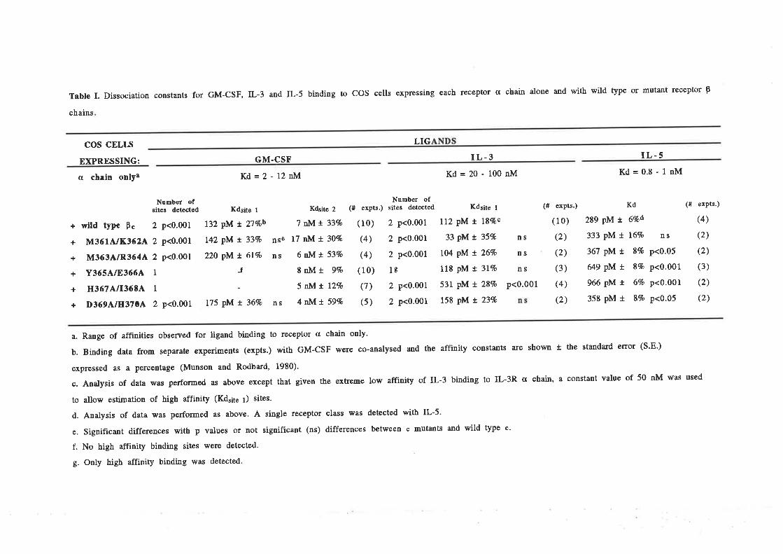
Table I. Dissociation constants for GM-CSF, IL-3 and IL-5 binding to COS cells expressing each receptor a chain alone and with wild tyPe or mutant receptor þ
chains
LIGcos cELIs
EXPRESSING:
a chain onlya
+ wild type Þ"
+ M3614/11362^
+ M3634/R3644
+ Y365A/83664
+ H3671¡lI36tA
+ D3694/H3704
GM.CSF
Kd=2-12nM
IL-3Kd=20-100nM
Kd
2S9 pM t 6qod
333 pM t 16%
367 pM t 84o
649 pM t 84o
966 pM L 64o
358 pM L 87o
IL-5l(d=0.8-1nM
(# expts.)
(4)
ns (2)
p<0.05 (2)
p<0.001 (3)
p<0.001 (2)
p<0.05 (2)
Nunber ofsites detected Kdsite I
2 p<0.001 132 PM t 27%ob
2 p<0.001 L42 PM t 334o
2 p<0.001 220 PM x 614o
1J1-2 p<0.001 175 PM t 36%
Ktrsite z (# exprs.) .,[Ì'låi*iju Kdsite l
7 nl,Ã! 334o (10) 2 p<0.001 112 pM t l84oc
nse l7 nM r 30% (4) 2 p<0.001 33 pM t 35%
ns 6nl,Ãt. 53Vo (4) 2 p<0.001 104 pM t 264o
8 nM t 94o (10) lc 118 PM x 3t%o
5 tñIx lzEo (7) 2 p<0'001 531 pM t 287o
ns 4nMlr. 5gEo (5) 2 p<0.001 158 pM t 234o
p<0.001
(# expts.)
( 10)
(2)
(2)
(3)
(4)
(2)
ns
ns
ns
a. Range of aff,rnities observed for ligand binding to receptor a chain only.
b. Binding data from separate experiments (expts.) with GM-CSF were co-analysed and the affinity constants a¡e shown t the standa¡d enor (S'E')
expressed as a percentage (Munson and Rodba¡d' 1980).
c. Analysis of data was perforured as above except that given the extreme low affinity of IL-3 binding to IL-3R a chain, a constant value of 50 nM was used
to allow estimation of high affinity (Kdsite t) sites.
d. Anaþsis of data was perforrred as above. A single receptor class was detected with IL-5.
e. Significant differences with p values or not significant (ns) differences between c mutants and wild type c'
f. No high affinity binding sites lvere detected.
g. Only high affinity binding was detected.
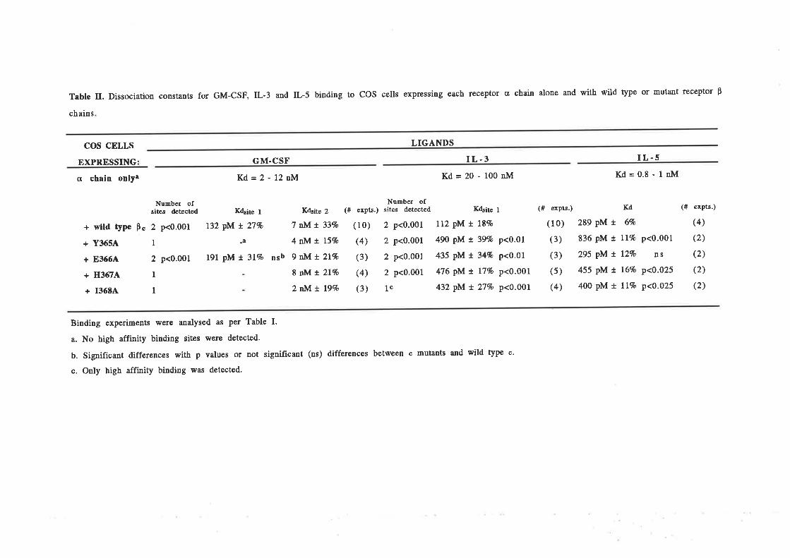
Table II. Dissociation constants for GM-CSF, IL-3 and IL-5 binding to COS cells expressing each receptor a chain alone and with wild type or mutant receptor p
chains
cos cELLs
EXPRESSING
LIGANDS
GM.CSF
cr chain onlya Kd=2 - 12nM Kd=20-100nM Kd=0.8-1nM
Number ofsites detected
+ wlld type Þ" 2 p<0.ü)l
+ Y3654 I
+ 83664 2 p<0.001
+ H36ílA. I
+ 13684 1
K4ite t
I32 pM t 27%
_a
191 pM t 3L4o
I L.3
K4site 2 (# exprs.) .tTå'i"J,*å Krbite r
7 rNIt 33Vo (10) 2 p<0.001 112 pM t L8%
4 nli/^t L54o (4) 2 p<0.001 490 pM t 397o p<0.01
nsb 9 r:M^t 2l7o (3) 2 p<0.001 435 pM t 344o p<0.01
8 nNlr.2L4o (4) 2 p<0.001 476 pM r' L7% p<0-001
2 tNI r. 19% (3) lc 432 pM t 27Vo p<0.001
I L.5
(f expts.) Kd
(10) 289 pM t 6%
(3) 836 pM r. Ll% p<0.001
(3) 295 pM t I24o ns
(5) 455 pM r. 16% p<0.025
(4) 400 pM t tL%o p<0.025
(# expts.)
(4)
(2)
(2)
(2)
(2)
Binding experiments were analysed as per Table I.
a. No high affinity binding sites were detected.
b. Significant differences with p values or not significant (ns) differences between c mutants and wild type c.
c. Only high affinity binding was detected'
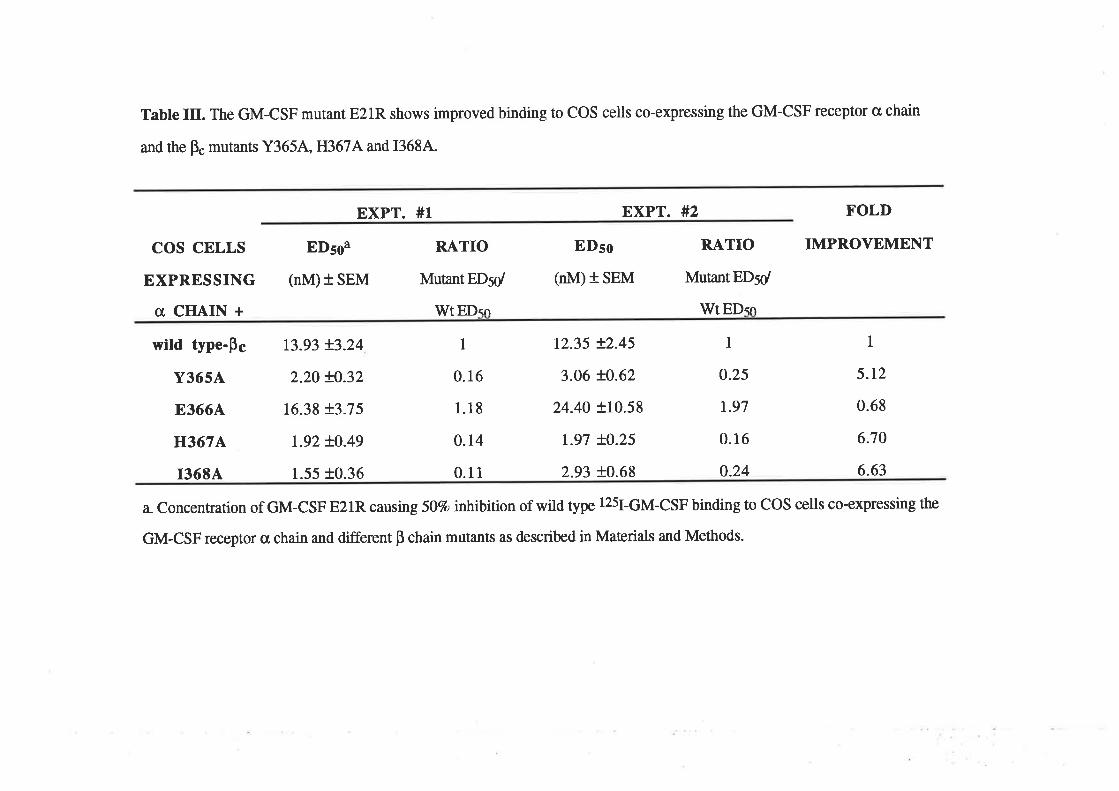
Table Itr. The GM-CSF mutant E21R shows improved binding to COS cells co-expressing the GM-CSF receptor o chain
and the p" mutants Y3654, H367A and 13684.
EXPT. #1 EXPT. #2 FOLD
cos cELLs
EXPRESSING
a CIIAIN +
wild type-pg
Y3654
E3664
H367A,
I3684
EDsoa
(nM) + 5p¡4
t3.93 +3.24
2.20 +0.32
16.38 +3.75
1.92 +0.49
1.55 +0.36
RATIO
Mutant EDsd
wr EI)<n
I
0.16
1.18
0.14
0.11
EDso
(nM) + SEM
12.35 +2.45
3.06 +0.62
24.40 +10.58
r.97 +0.25
2.93 +0.68
RATIO
Mutant EDsd
WtEDso
1
0.25
1.97
0.16
0.24
IMPROVEMENT
1
5.r2
0.68
6.70
6.63
a. Concentration of GM-CSF E21R causing 507o irútibirion of wild type 125¡-6M-CSF binding to COS cells co-expressing the
GM-CSF receptor cr chain and different p chain mutants as described in Materials and Methods.
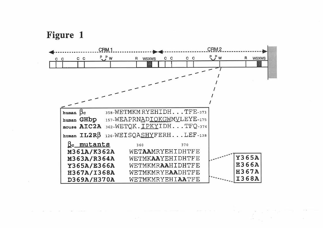
Figure 1
cc PP w RwR
I
cc cc
¿¿¿ I-- I
¿¿¿¿-¿ I
HIIIT Ii\Ìlll
¡sg-WETMKMRYEHIDH. . . TFE-¡z¡GHbp rsz-WEAPRNADIOKGWMI¿LEYE-rzsAIC2A ¡oz-WETQK. IPKYIDH. . . TFQ-¡ze
IIT2RB rzo-WEISQASHY.FERH. . . LEF-r¡e
mutant s 3 60 37 o
M3 6 LA/ K3 62AM3 6 3A/ R3 64AY365AlE366AH367Alr368AD369A/H370A
WETAAMRYEHIDHTFEWETMKAAYEHI DHTFEWETMKMRAÀHIDHTFEWETMKMRYE.AÀDHTFEWETMKMRYEHIAATFE
B"
human
human
mouae
hunan
Y365A8366AH3 67A1368A
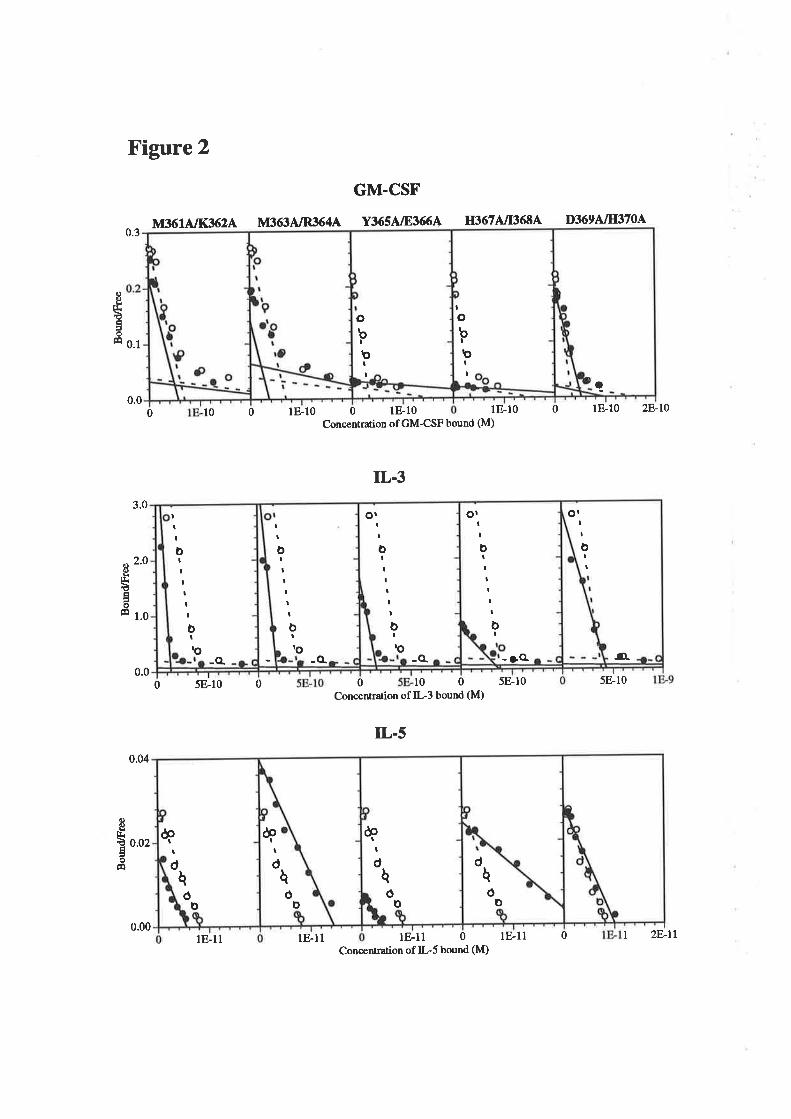
obt
r)II
IobI
þtI
a
Il¡rèâoÉ
Figure 2
GM.CSF
M3614/I(3624 l\,Ít634/R3644 Y3654/8366A'0.3
0.1
0.00
Concentration of GM-CSF bound (M)
3.0
r001810018-10
H3674/r36tA D3694/Ir370A
1E-10 0 lE-10 2E l0
IL.3
2.0Ie'octáoÉ 1.0
0.0
0.04
0.02
0 5E-10 0 0 t0 0 5E-10
Concentration of IL3 bound (M)
IL-5
5E-10
{)oEÉao
9q
1F-11 0 lE-ll 0
-o-
ÒIt
o¡II
oIIIIII
bI
- r(L
orII
oIItItI
b¡
-o.ro
tI
III
oIIItI¡
o
-o.
òIrO
ItI
oIIIIII
ò
-a
I
þ
q
Id
ôo
dq
0Þ
&I
Þ
t
dhó
t
d
æI
h0
o
0.001E-I1 lE-11
Concentralion of IL5 bound (M)I 2Ell
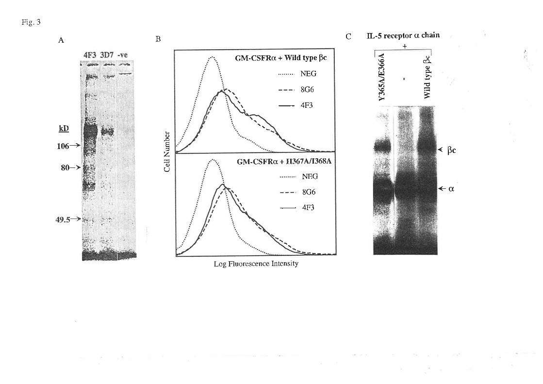
Fig.3
A B C IL-5 recePtor cr chain
+4F3 3D7 -ve
-,'1:| '#ÛÉ +*¡ ¡¡rùo(ñq)
É
\oú)r¿
tfl\ot)
KD
106->
80-->
49.5+ilw¡l ,,,:',i:
o-oÉ
za)U
<- Bc
<-G-ì.
Log Fluorescence IntensitY
tl
GM-CSFRcr + Wild tyPe Pc
NEG
8G6
4F3\
GM-CSFRcI + H3674/I3684
NEG
8G6
4F3\\\

Figure 4GM.CSF
Y3654 E36640.3
0.1
0 1.5E-10 0 1.5E-10 3E-10Concenhation of
IL.31.5
.0
0.5 1.0
0.0 0.0
0 l.5E-9 0 1.5E-9 3E-9 0 1E-9Concenhation of IL-3 bound (M)
IL-5
o.o2
0.01
0.00
H3674
0 1E-10GM-CSF bound (M)
5E-I2bound (M)
0 2E-9
I36840.9
o.2g 0'6
l¡.€âotr o.¡
0.0
2.OI
0.0
0.03
o.o2
0.01
0 lE-10
()o.FËáo
Êe
Il¡.€ÉoÉ
0 5E-12 0
I
Io
_o_
I
IIIro
o
I
tII
roII
o
o
III
o
o
I
roI
t oo
a
o
aa-o-
o
I
I
¡OI
I
o
a
oa
bt
o
I
0III
t
I
I o
to
ò.o
a Io
ò.o
It
oa\
tot
%
ol
I
a
ò.o
Or
rO
I
\o.
ò.o
Or
rO
\o.
0.005E-12 1E-11 0Concenhation of IL-5
o 5E-12 1E-11
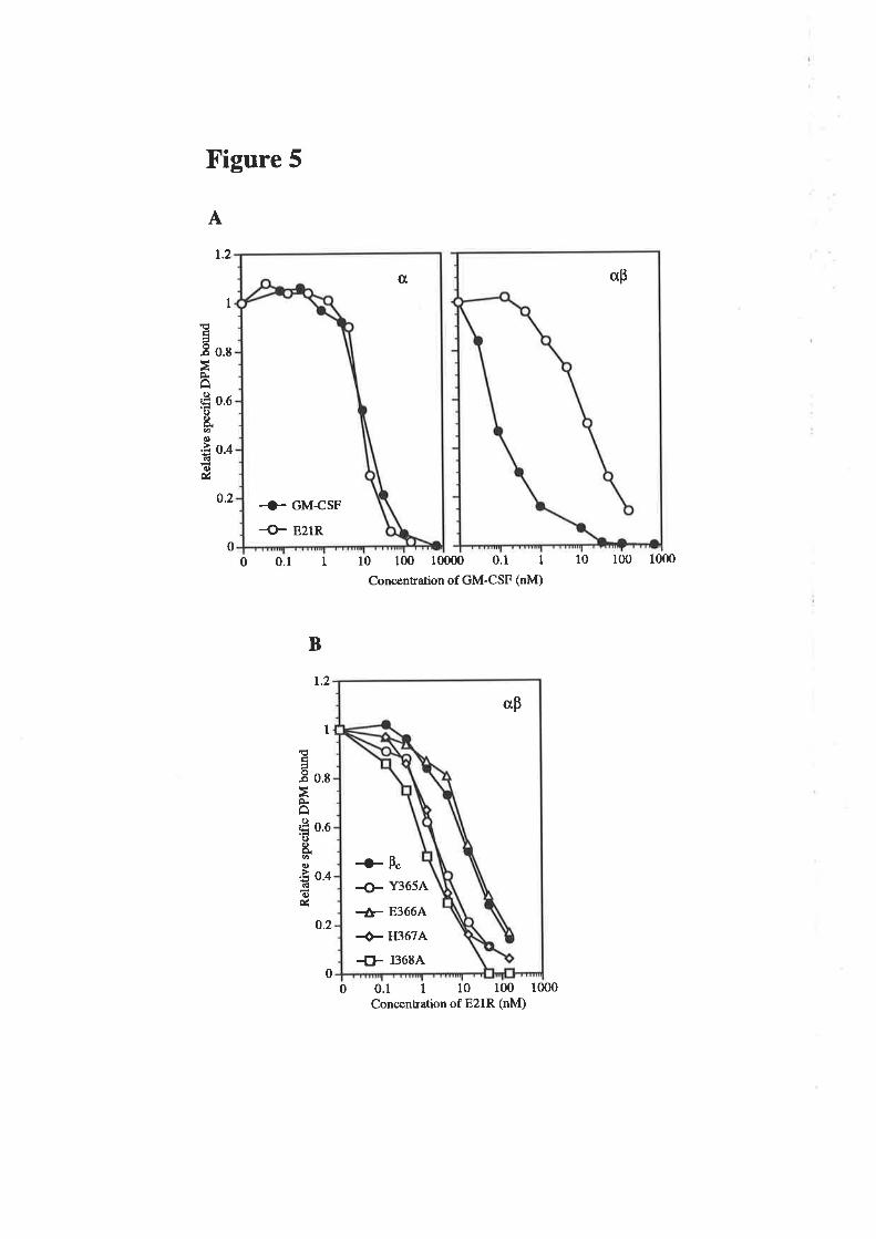
Figure 5
A
1.2
1
ۃa-B o.s
ÈA
'Ë o.eotøo.È o.¿dc)ú
0.2
o00.1 1 10 100 10000 0.1 I
Concentration of GM-CSF (nM)
0.1 1 10 100 1000Concentration of E21R (nM)
10 100 1000
B
r.2
I
Éa-B o.e
FâË o.ooE.().È o.¿6l6)ú
0.2
00
0,
+ GM-CSF
+ E21R
ap
ap
{)- Y3654
+- E3664
+ H3674
{- 13684
-O- Þ"
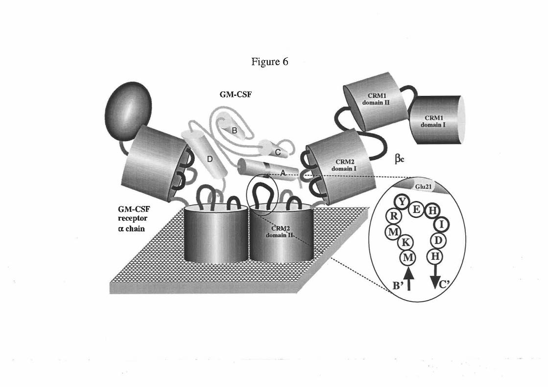
Figure 6
GM.CSF
GM.CSFreceptora chain