Structure and Function of the ABCD1 Variant Database - MDPI
13
Citation: Mallack, E.J.; Gao, K.; Engelen, M.; Kemp, S. Structure and Function of the ABCD1 Variant Database: 20 Years, 940 Pathogenic Variants, and 3400 Cases of Adrenoleukodystrophy. Cells 2022, 11, 283. https://doi.org/10.3390/ cells11020283 Academic Editors: Joseph G. Hacia and William B. Rizzo Received: 17 November 2021 Accepted: 12 January 2022 Published: 14 January 2022 Publisher’s Note: MDPI stays neutral with regard to jurisdictional claims in published maps and institutional affil- iations. Copyright: © 2022 by the authors. Licensee MDPI, Basel, Switzerland. This article is an open access article distributed under the terms and conditions of the Creative Commons Attribution (CC BY) license (https:// creativecommons.org/licenses/by/ 4.0/). cells Article Structure and Function of the ABCD1 Variant Database: 20 Years, 940 Pathogenic Variants, and 3400 Cases of Adrenoleukodystrophy Eric J. Mallack 1,2 , Kerry Gao 1 , Marc Engelen 3 and Stephan Kemp 3,4, * 1 Department of Pediatrics, Division of Child Neurology, Weill Cornell Medical College, NewYork-Presbyterian Hospital, New York, NY 10065, USA; [email protected] (E.J.M.); [email protected] (K.G.) 2 Department of Pediatrics, Memorial Sloan Kettering Cancer Center, New York, NY 10065, USA 3 Department of Pediatric Neurology, Emma Children’s Hospital, Amsterdam University Medical Centers, Amsterdam Neuroscience, University of Amsterdam, 1105 AZ Amsterdam, The Netherlands; [email protected] 4 Laboratory Genetic Metabolic Diseases, Department of Clinical Chemistry, Amsterdam University Medical Center, Amsterdam Gastroenterology Endocrinology Metabolism, University of Amsterdam, 1105 AZ Amsterdam, The Netherlands * Correspondence: [email protected] Abstract: The progressive neurometabolic disorder X-linked adrenoleukodystrophy (ALD) is caused by pathogenic variants in the ABCD1 gene, which encodes the peroxisomal ATP-binding trans- porter for very-long-chain fatty acids. The clinical spectrum of ALD includes adrenal insufficiency, myelopathy, and/or leukodystrophy. A complicating factor in disease management is the absence of a genotype–phenotype correlation in ALD. Since 1999, most ABCD1 (likely) pathogenic and benign variants have been reported in the ABCD1 Variant Database. In 2017, following the expansion of ALD newborn screening, the database was rebuilt. To add an additional level of confidence with respect to pathogenicity, for each variant, it now also reports the number of cases identified and, where available, experimental data supporting the pathogenicity of the variant. The website also provides information on a number of ALD-related topics in several languages. Here, we provide an updated analysis of the known variants in ABCD1. The order of pathogenic variant frequency, overall clustering of disease-causing variants in exons 1–2 (transmembrane domain spanning region) and 6–9 (ATP-binding domain), and the most commonly reported pathogenic variant p.Gln472Argfs*83 in exon 5 are consistent with the initial reports of the mutation database. Novel insights include nonran- dom clustering of high-density missense variant hotspots within exons 1, 2, 6, 8, and 9. Perhaps more importantly, we illustrate the importance of collaboration and utility of the database as a scientific, clinical, and ALD-community-wide resource. Keywords: adrenoleukodystrophy; peroxisome; ABC transporter; newborn screening; genetics; diagnosis; mutation; variants of uncertain significance; ABCD1 1. Introduction The ABCD1 gene is located on Xq28, covers 19.9 kb and contains 10 exons [1,2]. It encodes the ABCD1/ALD protein of 745 amino acids, an ATP-binding cassette transmem- brane half-transporter required for the import of coenzyme A-activated very-long-chain fatty acids (VLCFAs; >C22:0) into peroxisomes for degradation [3,4]. Pathogenic variants in the ABCD1 gene lead to the accumulation of VLCFAs in plasma and tissues, including adrenal glands, testes, the central nervous system, and the subsequent development of X-linked adrenoleukodystrophy (ALD) [5]. ALD has an estimated birth prevalence of 1 in 16,000 births [6,7]. The disease is characterized by a striking clinical spectrum and is unpredictable in disease course and severity, with no established genotype–phenotype relationship [8,9]. About 50% of affected boys will develop primary adrenal insufficiency Cells 2022, 11, 283. https://doi.org/10.3390/cells11020283 https://www.mdpi.com/journal/cells
-
Upload
khangminh22 -
Category
Documents
-
view
5 -
download
0
Transcript of Structure and Function of the ABCD1 Variant Database - MDPI
�����������������
Citation: Mallack, E.J.; Gao, K.;
Engelen, M.; Kemp, S. Structure and
Function of the ABCD1 Variant
Database: 20 Years, 940 Pathogenic
Variants, and 3400 Cases of
Adrenoleukodystrophy. Cells 2022,
11, 283. https://doi.org/10.3390/
cells11020283
Academic Editors: Joseph G. Hacia
and William B. Rizzo
Received: 17 November 2021
Accepted: 12 January 2022
Published: 14 January 2022
Publisher’s Note: MDPI stays neutral
with regard to jurisdictional claims in
published maps and institutional affil-
iations.
Copyright: © 2022 by the authors.
Licensee MDPI, Basel, Switzerland.
This article is an open access article
distributed under the terms and
conditions of the Creative Commons
Attribution (CC BY) license (https://
creativecommons.org/licenses/by/
4.0/).
cells
Article
Structure and Function of the ABCD1 Variant Database:20 Years, 940 Pathogenic Variants, and 3400 Cases ofAdrenoleukodystrophyEric J. Mallack 1,2 , Kerry Gao 1, Marc Engelen 3 and Stephan Kemp 3,4,*
1 Department of Pediatrics, Division of Child Neurology, Weill Cornell Medical College, NewYork-PresbyterianHospital, New York, NY 10065, USA; [email protected] (E.J.M.); [email protected] (K.G.)
2 Department of Pediatrics, Memorial Sloan Kettering Cancer Center, New York, NY 10065, USA3 Department of Pediatric Neurology, Emma Children’s Hospital, Amsterdam University Medical Centers,
Amsterdam Neuroscience, University of Amsterdam, 1105 AZ Amsterdam, The Netherlands;[email protected]
4 Laboratory Genetic Metabolic Diseases, Department of Clinical Chemistry, Amsterdam University MedicalCenter, Amsterdam Gastroenterology Endocrinology Metabolism, University of Amsterdam,1105 AZ Amsterdam, The Netherlands
* Correspondence: [email protected]
Abstract: The progressive neurometabolic disorder X-linked adrenoleukodystrophy (ALD) is causedby pathogenic variants in the ABCD1 gene, which encodes the peroxisomal ATP-binding trans-porter for very-long-chain fatty acids. The clinical spectrum of ALD includes adrenal insufficiency,myelopathy, and/or leukodystrophy. A complicating factor in disease management is the absence ofa genotype–phenotype correlation in ALD. Since 1999, most ABCD1 (likely) pathogenic and benignvariants have been reported in the ABCD1 Variant Database. In 2017, following the expansion ofALD newborn screening, the database was rebuilt. To add an additional level of confidence withrespect to pathogenicity, for each variant, it now also reports the number of cases identified and,where available, experimental data supporting the pathogenicity of the variant. The website alsoprovides information on a number of ALD-related topics in several languages. Here, we provide anupdated analysis of the known variants in ABCD1. The order of pathogenic variant frequency, overallclustering of disease-causing variants in exons 1–2 (transmembrane domain spanning region) and6–9 (ATP-binding domain), and the most commonly reported pathogenic variant p.Gln472Argfs*83 inexon 5 are consistent with the initial reports of the mutation database. Novel insights include nonran-dom clustering of high-density missense variant hotspots within exons 1, 2, 6, 8, and 9. Perhaps moreimportantly, we illustrate the importance of collaboration and utility of the database as a scientific,clinical, and ALD-community-wide resource.
Keywords: adrenoleukodystrophy; peroxisome; ABC transporter; newborn screening; genetics;diagnosis; mutation; variants of uncertain significance; ABCD1
1. Introduction
The ABCD1 gene is located on Xq28, covers 19.9 kb and contains 10 exons [1,2]. Itencodes the ABCD1/ALD protein of 745 amino acids, an ATP-binding cassette transmem-brane half-transporter required for the import of coenzyme A-activated very-long-chainfatty acids (VLCFAs; >C22:0) into peroxisomes for degradation [3,4]. Pathogenic variantsin the ABCD1 gene lead to the accumulation of VLCFAs in plasma and tissues, includingadrenal glands, testes, the central nervous system, and the subsequent development ofX-linked adrenoleukodystrophy (ALD) [5]. ALD has an estimated birth prevalence of 1in 16,000 births [6,7]. The disease is characterized by a striking clinical spectrum and isunpredictable in disease course and severity, with no established genotype–phenotyperelationship [8,9]. About 50% of affected boys will develop primary adrenal insufficiency
Cells 2022, 11, 283. https://doi.org/10.3390/cells11020283 https://www.mdpi.com/journal/cells
Cells 2022, 11, 283 2 of 13
by the age of 10 [10]. In childhood, 30–35% of boys will develop a fatal cerebral inflam-matory disease (cerebral ALD) with a 50–60% total lifetime risk of developing cerebraldisease [6,11,12]. In adulthood, virtually all male patients and >80% of affected womendevelop a chronic progressive myelopathy over their lifetime [10,13–15].
Since 1999, most ABCD1 (likely) pathogenic and benign variants have been reported inthe ALD mutation database. The database was initiated as a collaborative effort between thePeroxisomal Diseases Laboratory at the Kennedy Krieger Institute led by Hugo Moser, MD,and the Laboratory Genetic Metabolic Diseases at the Amsterdam UMC in the Netherlands,the initial findings of which were published in 2001 [16]. The aims of the database wereto facilitate ABCD1 variant analysis, collect and catalogue variants in the ABCD1 gene,improve the analysis of pathogenic variants identified in ALD, facilitate the reportingof novel variants, and make the information overall more accessible by maintaining thedatabase on the internet (original URL: www.x-ald.nl) [16]. The primary aim of this studywas to provide an updated analysis of the known variants in ABCD1 as catalogued over thepast 20 years. In addition, we aimed to highlight the database as a resource for physicians,scientists, and patient families; to detail the role of the database in the era of newbornscreening for ALD; and, perhaps most importantly, to encourage widespread collaboration,use, and continued data transparency of the database.
2. ABCD1 Variant Database2.1. ABCD1 Variant Analysis, 2001: Initial Results
The first iteration of the ALD Mutation Database [16] reported four polymorphismsand 406 total mutations. Of the 406, 234 (58%) were non-recurrent and 47 were previouslyunpublished novel pathogenic variants. Missense mutations constituted the most frequentpathogenic variants (55.9%), followed by frame shift (27.1%), nonsense (9.1%), small aminoacid insertions/deletions (3.9%), and large deletions (3.9%).
Pathogenic variants in the ABCD1 gene clustered most often in the transmembranedomain (40%, exons 1 and 2), followed by the ATP-binding domain (30%, exons 6–9), andexon 5 (14%). Non-nested amplification of all exons in the ABCD1 gene using the Xq28optimized primer sets developed by Boehm et al. [17] revealed 67% of known mutationsclustered into four amplicons covering 40% of the coding region: amplicon 1b in exon1 (amino acids (aa) 75–188; 20%); amplicon 1c in exon 1 (aa 177–300; 20%); amplicon8/9 (exons 8 and 9, aa 594–664; 14%); and amplicon 5 (exon 5, aa 465–496; 13%) [17]. Apathogenic variant hotspot was identified in exon 5 accounting for 10.3% of all mutationsin the database, c.1415_16delAG (p.Gln472Argfs*83), resulting in a premature stop codonat position 554 [18].
Analysis of missense pathogenic variants revealed a non-random distribution acrossthe gene. Missense mutations were found at higher-than-expected frequency in exon 1(amino acids 100–300), which code for four segments of the transmembrane domain, and athigher-than-expected frequency in exons 6–9 (amino acids 497–664), which code for theATP-binding domain.
2.2. Evolution of the Database: Experimental Data and Variant Reclassification
The identification of the ALD gene in 1993 enabled large-scale genetic testing by di-agnostic laboratories and research groups. Between 1993 and 1999, over 40 large-scalepathogenic variant screening studies were published. Unfortunately, two different nu-cleotide numbering systems were used to annotate variants. The first used the first nu-cleotide of the cDNA as number +1. The second, and correct system, uses the A of theinitiator methionine (ATG) codon as +1. Early versions of the ALD Mutation Databaseincluded both numbering systems for each variant. From 2003 onward, all variants havebeen reported according to the correct numbering system only. Due to this discrepancy, itmay be difficult to align newly identified pathogenic variants with cases from those earlyreports in which the wrong numbering system was used as the nucleotide numbers do notmatch: they are off by 386. Therefore, reported pathogenic variants from early publica-

Cells 2022, 11, 283 3 of 13
tions may need to be realigned against the second numbering system. For example, thecorrect annotation of C696T, R104C [19] is (696 − 386 = 310) c.310C > T, p.Arg104Cys. TheABCD1 Variant Database reports all (likely) pathogenic and benign variants in conformitywith the nomenclature recommended by the Human Genome Variation Society (HGVS;https://varnomen.hgvs.org/) [20]. All variants, including those already published, areannotated using the Alamut Visual software package with transcript NM_000033.3 onGRCh37 (hg19) as the reference sequence.
In 2017, following the expansion of ALD newborn screening in the U.S., the ABCD1Variant Database became part of Adrenoleukodystrophy.info. This name better reflectsthe broader focus on ALD. The website also moved to a new domain name: https://adrenoleukodystrophy.info/.
The ABCD1 Variant Database was also rebuilt. Instead of merely reporting whethera variant was already reported in literature or shared by diagnostic laboratories, the newversion contains additional information: it lists the number of cases reported for eachpathogenic variant. An ALD case is defined as an individual with clinical signs andsymptoms related to ALD and a biochemical or genetic confirmation. Where available inthe literature, experimental data were extracted supporting the pathogenicity of a particularvariant (a Western blot, peroxisomal beta-oxidation, etc.). For example, the pathogenicvariant p.Ser606Leu has been reported in 37 ALD cases. Other ALD-related researchstudies using patient cell lines revealed that this pathogenic variant reduced the amount ofdetectable ALD protein to 25% that of control cells (Database Ref 97) [21]. Studies usingexperimental cell lines showed that the pathogenic variant affects ATP-binding capacity(Database Refs 194 and 244) [22,23], resulting in deficient VLCFA beta-oxidation (DatabaseRef 261) [24].
Information from publicly available databases, such as the genome AggregationDatabase (gnomAD, https://gnomad.broadinstitute.org/), was also included [25]. In someinstances, a variant that was reported to be pathogenic could be correctly reclassified asbenign. For example, p.Gly608Asp was reported as the pathogenic variant in an ALD malepatient (Database Ref 48) [26], but data from gnomAD show its frequency to be 124/162246alleles (X:153008483 G/A), confirming that this is not a pathogenic variant.
In addition to the ABCD1 Variant Database, the website provides information on anumber of ALD-related topics. The website pages are written by ALD researchers andphysicians with expertise on that topic. A number of these pages are available in Spanish,French, German, and Dutch. Interestingly, some of the most visited pages are in Spanish,which indicates that there is an unmet need for information written by experts in otherlanguages. This motivates us to continue adding translations; however, this is a relativelyslow process as the database is maintained without funding. Hence, we are fully dependenton collaborators with a biology background/medical training willing to translate content.Parallel to the expansion of ALD newborn screening, there has been a steady increase invisitors with an annual average of 30,000 visits before 2014 to over 150,000 since 2019. Therealso has been a shift in the devices that visitors use to visit the website. As of 2020, themajority of visitors have used a mobile phone to visit the website.
2.3. ABCD1 Variant Analysis, 2021: Materials and Methods
ABCD1 variant data were analyzed and annotated in RStudio (v.1.2.5033) using theBioconductor package trackViewer [27,28]. Dandelion plots were generated to depictthe distribution and density of variants by chromosome position across the ABCD1 geneaccording to assembly GRCh37 (hg19) (Figure 1). The height of each dandelion reflects thedensity of variants at that position. The size of the chromosome visualized per plot wascontrolled by coding the ratio of the chromosome segment size to the maxgaps variable:a ratio of 8 for visualization of the whole gene (19,893 bp), 5 for visualization of the openreading frame of exon 1 in its entirety (900 bp, Figure 2b), and 2 for comparison acrosscomparably sized segments of the gene (100–300 bp each): exons 2, 6, 8, and 9. A multiplesequence alignment of ABCD1 proteins from 54 different species (from Danio rerio to
Cells 2022, 11, 283 4 of 13
human) was created using ClustalW [29,30] (see Supplemental Figure 1 for the multiplesequence alignment). The WebLogo program (http://weblogo.threeplusone.com/) wasused to generate a graphic representation of the evolutionary conservation of each aminoacid within the ABCD1 protein [31].
Cells 2022, 11, 283 4 of 14
reading frame of exon 1 in its entirety (900 bp, Figure 2b), and 2 for comparison across comparably sized segments of the gene (100–300 bp each): exons 2, 6, 8, and 9. A multiple sequence alignment of ABCD1 proteins from 54 different species (from Danio rerio to human) was created using ClustalW [29,30] (see Supplemental Figure 1 for the multiple sequence alignment). The WebLogo program (http://weblogo.threeplusone.com/) was used to generate a graphic representation of the evolutionary conservation of each amino acid within the ABCD1 protein [31].
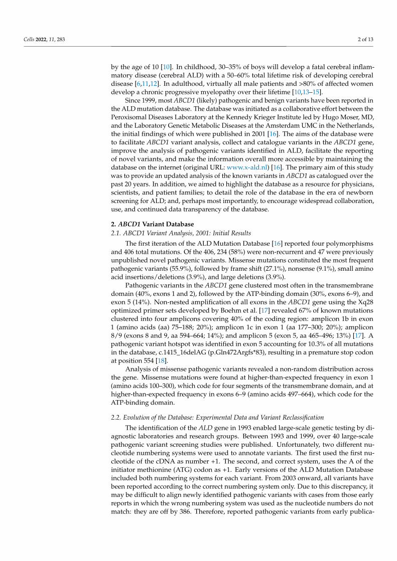
Figure 1. Dandelion plot illustrating variant density in ABCD1 (density is indicated by height of dandelion). (A) All variants (pathogenic, benign, VUS, and synonymous) in the ABCD1 gene (open circles). The highest variant density is in exon 6 followed by exon 1. (B) Only pathogenic variants in the ABCD1 gene (red pins) are shown. The highest event burden and pathogenic variant density are in exon 1 followed by exon 6. (C) All pathogenic variants in exon 1.
Figure 1. Dandelion plot illustrating variant density in ABCD1 (density is indicated by height ofdandelion). (A) All variants (pathogenic, benign, VUS, and synonymous) in the ABCD1 gene (opencircles). The highest variant density is in exon 6 followed by exon 1. (B) Only pathogenic variants inthe ABCD1 gene (red pins) are shown. The highest event burden and pathogenic variant density arein exon 1 followed by exon 6. (C) All pathogenic variants in exon 1.
Cells 2022, 11, 283 5 of 13Cells 2022, 11, 283 5 of 14
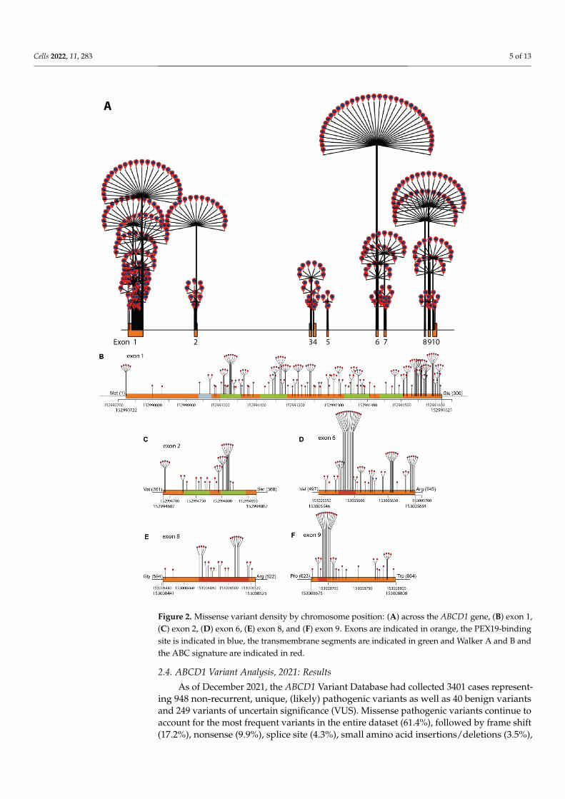
Figure 2. Missense variant density by chromosome position: (A) across the ABCD1 gene, (B) exon 1, (C) exon 2, (D) exon 6, (E) exon 8, and (F) exon 9. Exons are indicated in orange, the PEX19-binding site is indicated in blue, the transmembrane segments are indicated in green and Walker A and B and the ABC signature are indicated in red.
Figure 2. Missense variant density by chromosome position: (A) across the ABCD1 gene, (B) exon 1,(C) exon 2, (D) exon 6, (E) exon 8, and (F) exon 9. Exons are indicated in orange, the PEX19-bindingsite is indicated in blue, the transmembrane segments are indicated in green and Walker A and B andthe ABC signature are indicated in red.
2.4. ABCD1 Variant Analysis, 2021: Results
As of December 2021, the ABCD1 Variant Database had collected 3401 cases represent-ing 948 non-recurrent, unique, (likely) pathogenic variants as well as 40 benign variantsand 249 variants of uncertain significance (VUS). Missense pathogenic variants continue toaccount for the most frequent variants in the entire dataset (61.4%), followed by frame shift(17.2%), nonsense (9.9%), splice site (4.3%), small amino acid insertions/deletions (3.5%),
Cells 2022, 11, 283 6 of 13
large deletions (2.6%), and benign variants (1.2%). The distribution is generally consistentacross the 948 non-recurrent variants (Table 1).
Table 1. Variant Counts and Frequencies in ABCD1 variants.
Total Uniquen % n %
All ABCD1 variants in the database 3401 948 28%Missense pathogenic variants 2087 61.4% 411 43.4%Nonsense pathogenic variants 336 9.9% 116 12.3%Frame shift pathogenic variants 585 17.2% 262 27.6%Amino acid insertions/deletions 119 3.5% 52 5.5%Splice site pathogenic variants 145 4.3% 43 4.5%One or more exons deleted 89 2.6% 24 2.5%Benign variants 40 1.2% 40 4.2%
Pathogenic variants in ABCD1 cluster most often in the transmembrane domain (46%,exons 1–2), followed by the ATP-binding domain (35%, exons 6–9), followed by exons 3and 4 (9%) and exon 5 (7%). The most frequent pathogenic variant continues to be the 2 bpdeletion in exon 5 p.Gln472Argfs*83 (n = 170, 5.0% of all pathogenic variants), followedby missense pathogenic variants p.Arg554His (n = 70, 2.1%), p.Arg660Trp (n = 60, 1.8%),p.Arg617His (n = 57, 1.7%), and p.Arg518Gln (n = 54, 1.6%). All pathogenic variant typesare homogenously distributed spatially across ABCD1. Eighty-nine large exon deletionsare reported, also distributed across the entire gene, the most frequent of which are exon8–10 del (n = 21), exon 7–10 del (n = 16), and exon 3–10 del (n = 12). Entire ABCD1gene deletions have also been reported. However, complete deletion of ABCD1 extendsinto the promoter region of the upstream neighboring gene (BCAP31). The combineddeletion or dysfunction of both ABCD1 and BCAP31 (initial name DXS1375E [32]) causescontiguous ABCD1 DXS1357E deletion syndrome (CADDS). CADDS is characterized bysevere intrauterine growth retardation, neonatal hypotonia, failure to thrive, severe globaldevelopmental delay, and liver dysfunction, leading to early death [33].
The density of all variants (pathogenic, benign, synonymous, and VUS combined)in the database are visualized in Figure 1. Overall, variant density is highest in exon 6,followed by exon 1 (Figure 1a). The highest density of pathogenic variants occurs in exon1 followed by exon 6, with comparable densities in exons 2, 8, and 9 (Figure 1b). Exon1 contains the overall highest pathogenic variant burden distributed in the latter 2/3 ofthe exon (amino acids 100–300), studded with multiple high-density hotspots (Figure 1c).Benign variants and variants of uncertain significance (VUS) are homogenously distributedacross ABCD1 with no variation in density (data not shown).
The density of missense variants across the whole gene and across multiple exonsis plotted in Figure 2. Missense variants cluster across chromosome segment 152991000–152991621 (exon 1, amino acids 100–300, Figure 2b), and in the chromosome segment152994751–152994822 (exon 2, amino acids 315–346, Figure 2c), which encodes the trans-membrane domain. Notable high-density missense hotspots are located in the ATP-bindingdomain encoded by segments in exon 6 (chromosome position 153005575–153005610, aminoacids 497–545, Figure 2d), exon 8 (chromosome position 153008470–153008520, amino acids604–620, Figure 2e), and in exon 9 (chromosome position 153008685–153008710, aminoacids 623–664, Figure 2f).
Cells 2022, 11, 283 7 of 13
3. ABCD1 Variant Interpretation: 2001 vs. 2021
The order of (likely) pathogenic variant frequency, overall nonrandom clusteringof disease-causing variants in exons 1–2 (transmembrane domain) and exons 6–9 (ATP-binding domain), and the most commonly reported pathogenic variant p.Gln472Argfs*83on exon 5 are consistent with the initial reports of the mutation database [16]. A few novelinsights are afforded by the updated analysis and are secondary to the ability to mapvariants onto the gene with higher resolution. The clusters of missense variants in exons 6and 9 are not equally distributed; rather, they form high-density hotspots in both exons.Given that VUS, synonymous, and benign variants appear to be equally distributed acrossall exons (Figure 1a), these observations imply that: (1) these segments of the protein areintolerant to variation, and (2) a VUS that occurs in either of the hotspots on exon 6 or 9 mayhave a higher likelihood of being pathogenic. The same logic applies to VUSs that appear inexon-1-segment amino acids 100–300, chromosome interval 152994751–152994822 (aminoacids 315–346) on exon 2, and exon 8 (amino acids 594–622). At the functional level, the high-density hotspots on exons 6 and 9 identified independently via the bioinformatics analysisof missense variants map to two specific motifs within the ATP-binding domain: Walker Aand B and the ABC signature (Figure 2d–f) [4]. Early experimental data revealed that thehelical domain between the Walker A and B motifs undergoes conformational change whenbound to ADP, indicating their role in the transduction of free energy during ATP-hydrolysisin the ALD protein [34,35]. Further work demonstrated the role of the Walker A and B motifsin forming the ATP bindings site sandwich required for homodimerization of the ALDprotein, and the subsequent power stroke of ATP-dependent substrate transport [36,37].This highlights the potential for accurate variant curation and analysis of the databaseto identify functionally important segments of the gene (Figure 3). In the case of exons6, 8, and 9, the functional data provide further validation that novel variants in thesehigh-density clusters have a higher likelihood of being pathogenic.
Cells 2022, 11, 283 8 of 13Cells 2022, 11, 283 8 of 14
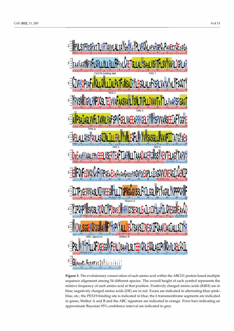
Figure 3. The evolutionary conservation of each amino acid within the ABCD1 protein based mul-tiple sequence alignment among 54 different species. The overall height of each symbol represents the relative frequency of each amino acid at that position. Positively charged amino acids (KRH) are in blue; negatively charged amino acids (DE) are in red. Exons are indicated in alternating blue–pink–blue, etc.; the PEX19-binding site is indicated in blue; the 6 transmembrane segments are indi-cated in green; Walker A and B and the ABC signature are indicated in orange. Error bars indicating an approximate Bayesian 95% confidence interval are indicated in grey.
Figure 3. The evolutionary conservation of each amino acid within the ABCD1 protein based multiplesequence alignment among 54 different species. The overall height of each symbol represents therelative frequency of each amino acid at that position. Positively charged amino acids (KRH) are inblue; negatively charged amino acids (DE) are in red. Exons are indicated in alternating blue–pink–blue, etc.; the PEX19-binding site is indicated in blue; the 6 transmembrane segments are indicatedin green; Walker A and B and the ABC signature are indicated in orange. Error bars indicating anapproximate Bayesian 95% confidence interval are indicated in grey.
Cells 2022, 11, 283 9 of 13
4. ABCD1 Variant Interpretation in the Era of ALD Newborn Screening4.1. Newborn Screening for ALD
Adrenal crises and the onset of cerebral ALD confer significant morbidity and mortalityin childhood without proper disease monitoring and treatment [7,38]. Before the age of10 years, one out of two boys diagnosed with ALD will develop adrenal insufficiency andone out of three boys will develop cerebral ALD [8,10]. Historically, boys who presentedwith neurologically symptomatic cerebral ALD translated to extensive disease on MRIand poor hematopoietic stem cell transplant outcomes [11,39–42]. Early-stage treatment ofchildhood cerebral ALD with hematopoietic stem cell transplantation and, more recently,gene therapy can arrest disease progression if performed in the window of opportunitywhen the MRI is abnormal but neurological symptoms are not yet apparent [7,38,43–50].The implementation of newborn screening for ALD in the United States and the Netherlandshas provided the opportunity to identify patients at birth [7,51–56]. Asymptomatic boysare monitored for adrenal insufficiency [57] and by MRI [58] to detect early cerebral lesions,with the aim of intervention in the narrow window prior to symptom onset [41,57,58].
4.2. Asymptomatic Diagnosis Requires a Platform to Resolve Variants of Uncertain Significance
Newborn screening gives rise to the clinical situation where patients, without a clearfamily history of disease, are identified asymptomatically at birth with a novel variant orVUS. This is clearly different from the more classical situation prior to newborn screeningwhere index patients presented with signs and symptoms of disease, and a diagnosis wasmade based on confirmatory biochemistry and genetics. A family history would then betaken to identify other family members at risk of ALD, or to diagnose other symptomaticfamily members. In newborn screening, or population screening, patients are also identifiedthat have C26:0-lysoPC levels above the upper level of the reference range, but below thelower level of the disease range. The identification of a novel variant or VUS creates adilemma: is the VUS a benign change or a pathogenic variant? The diagnosis of index casesidentified by newborn screening requires the demonstration of elevated VLCFAs and anaccurate interpretation of the genetic variant in ABCD1. In the case of a VUS, biochemicaland functional analyses of patient fibroblasts can aid the interpretation of the variant aseither a (likely) benign or pathogenic change [59].
Taken together, the ABCD1 Variant Database, open collaboration, and the results ofthis study serve as a platform to aid in the resolution of VUSs identified during newbornscreening. This is in addition to other publicly available variant archives (e.g., ClinVar: https://www.ncbi.nlm.nih.gov/clinvar/; Human Genome Variation Society: http://www.hgvs.org/locus-specific-mutation-databases). Medical professionals can report the biochemicalevidence or functional studies supportive of a VUS’s pathogenicity, map the variant ontothe gene to visualize whether it exists within a pathogenic variant cluster or hotspot, andassess the conservation of the potentially affected amino acid (Figure 3). For example, theVUS c.970C > T (p.Arg324Cys) within the exon 2 missense cluster was resolved in thisway. The patient developed elevated VLCFAs and an elevated ACTH, was diagnosed withpartial adrenal insufficiency before 2 years old, and is now proactively treated with stressdose steroids. Moving forward, the database provides a wealth of input to understand theeffect of missense variants on protein structure and folding, as emerging presently withneural-network-based protein modeling such as AlphaFold [60].
4.3. The Clinical and Collaborative Importance of the ABCD1 Variant Database
The facilitation of a correct interpretation of a variant’s pathogenicity in ABCD1 ishighly important. The accurate genetic diagnosis of ALD obligates the patient to rigorousmonitoring protocols for adrenal insufficiency [57] and cerebral ALD by MRI [58], requiringmultiple medical subspecialty evaluations early in life. If an early-stage cerebral lesionis identified in a boy with neurologically asymptomatic ALD [61], he will be referredfor hematopoietic stem cell transplantation or gene therapy, if applicable. The former isassociated with its own degree of morbidity and mortality [62], and both require the patient
Cells 2022, 11, 283 10 of 13
undergo chemotherapy [48]. Lastly, the identification of a familial gene mutation may leadto a life-altering diagnosis of ALD in extended family members.
Despite ALD being an X-linked disorder, a genetic diagnosis is especially important forwomen. It is becoming increasingly recognized that the majority of women are not merelycarriers, but may develop some degree of myeloneuropathy leading to gait, bowel, andbladder dysfunction [14,15,63]. In terms of family planning, identification of index boys innew families with newborn screening has uncovered the expected maternal inheritancepattern of the pathogenic variant. This has caused young families to pursue prenatal geneticdiagnosis and in vitro fertilization of unaffected embryos. This process, while necessaryto prevent further propagation of the disease to future generations, imposes significantmedical, emotional, and financial distress on new families trying to have more children [64].
From a collaborative standpoint, the downstream effects have been multiple. Updatingrelevant clinical data to novel VUSs and pre-existing reported missense pathogenic variantshas allowed physicians across the world to network quickly. This was the case for twonewborn screening patients born in two different states who were identified with the samereported VUS c.970C > T (p.Arg324Cys) above. Rapid reporting of pathogenicity andsharing of information via the ABCD1 variant database have enhanced the care for thepatients, their families, and their treating physicians. Lastly, the database has allowedfamilies to take an active part in their child’s care by being a resource for researching theirchild’s variant and learning about ALD through well-curated information.
5. Conclusions
A unique resource such as the ABCD1 Variant Database can only be maintainedthrough open collaboration and the sharing of (de-identified) information. If well-maintained,it helps the ALD community at large. From a scientific perspective, the database providesa backbone for realignment of older reported variants, a resource for the integration ofexperimental data and large datasets, a platform for variant classification, and a wayto shed light on functionally important segments of the protein. Clinically, it helps toaddress challenges posed by novel variants in the setting of newborn screening. Forthe ALD community, the database provides an opportunity to collaborate, translate, andtransmit well-curated disease information to both non-expert physicians and familiesaffected by ALD.
Supplementary Materials: The following supporting information can be downloaded at: https://www.mdpi.com/article/10.3390/cells11020283/s1. Figure S1 to Structure and function of theABCD1 variant database: 20 years, 940 pathogenic variants and 3400 cases of adrenoleukodystrophy.
Author Contributions: Conception and design of the study, E.J.M. and S.K.; acquisition and analysisof the data, E.J.M., K.G. and S.K.; drafting a significant portion of the manuscript or figures, E.J.M.,M.E. and S.K.; S.K. is the corresponding author and guarantor of this article. All authors have readand agreed to the published version of the manuscript.
Funding: This research was funded by the National Institutes of Health, grant number K23NS118044to EM; the Netherlands Organization for Scientific Research, grant number 016.196.310 to ME; andthe Netherlands Organization for Health Research and Development, grant number 543002004 to SK.
Institutional Review Board Statement: This study was conducted using published, publicly avail-able data. No human subjects were enrolled. This study is therefore exempt from ethics/institutionalreview board approval and informed consent was not applicable.
Data Availability Statement: Following publication, any data not published within this article willbe shared on request from any qualified investigator.
Acknowledgments: To the collaborators, physicians, laboratory scientists, and families who con-tributed to this database, we thank you. We thank Marcel Mattijssen (for funding the hosting of theinitial website (1999–2003); Ted van Geest (GoedGedaan) for his continued help with the websiteand sponsoring and hosting the website (since 2012); Johannes Berger, Wouter van Ballegoij, VirginieBonnamain, Gabor Linthorst, Charles Peters, Gerald Raymond, Rachel Salzman, Steven Steinberg,
Cells 2022, 11, 283 11 of 13
Björn van Geel, and Paul Watkins for their scientific contributions to the website; and a big thank youto: Cyntia Amorosi, Alfried Kohlschütter, Nerea Montedeoca Vázquez, and Elise Saunier Vivar forgraciously volunteering to translate the website content to Spanish, German, and French.
Conflicts of Interest: Eric Mallack, Kerry Gao, Marc Engelen, and Stephan Kemp have no financialrelationships or conflict of interest relevant to this article to disclose.
References1. Mosser, J.; Douar, A.M.; Sarde, C.O.; Kioschis, P.; Feil, R.; Moser, H.; Poustka, A.M.; Mandel, J.L.; Aubourg, P. Putative X-linked
adrenoleukodystrophy gene shares unexpected homology with ABC transporters. Nature 1993, 361, 726–730. [CrossRef]2. Sarde, C.O.; Mosser, J.; Kioschis, P.; Kretz, C.; Vicaire, S.; Aubourg, P.; Poustka, A.; Mandel, J.L. Genomic organization of the
adrenoleukodystrophy gene. Genomics 1994, 22, 13–20. [CrossRef] [PubMed]3. Mosser, J.; Lutz, Y.; Stoeckel, M.E.; Sarde, C.O.; Kretz, C.; Douar, A.M.; Lopez, J.; Aubourg, P.; Mandel, J.L. The gene responsible
for adrenoleukodystrophy encodes a peroxisomal membrane protein. Hum. Mol. Genet. 1994, 3, 265–271. [CrossRef]4. Kemp, S.; Theodoulou, F.L.; Wanders, R.J. Mammalian peroxisomal ABC transporters: From endogenous substrates to pathology
and clinical significance Correspondence. Br. J. Pharmacol. 2011, 164, 1753–1766. [CrossRef] [PubMed]5. Moser, A.B.; Kreiter, N.; Bezman, L.; Lu, S.; Raymond, G.V.; Naidu, S.; Moser, H.W. Plasma very long chain fatty acids in 3,000
peroxisome disease patients and 29,000 controls. Ann. Neurol. 1999, 45, 100–110. [CrossRef]6. Bezman, L.; Moser, H.W. Incidence of X-linked adrenoleukodystrophy and the relative frequency of its phenotypes. Am. J. Med.
Genet. 1998, 76, 415–419. [CrossRef]7. Moser, A.B.; Jones, R.O.; Hubbard, W.C.; Tortorelli, S.; Orsini, J.; Caggana, M.; Vogel, B.H.; Raymond, G.V. Newborn screening for
X-linked adrenoleukodystrophy. Int. J. Neonatal Screen. 2016, 2, 15. [CrossRef] [PubMed]8. van Karnebeek, C.D.M.; Richmond, P.A.; van der Kloet, F.; Wasserman, W.W.; Engelen, M.; Kemp, S. The variability conundrum
in neurometabolic degenerative diseases. Mol. Genet. Metab. 2020, 131, 367–369. [CrossRef]9. Kemp, S.; Huffnagel, I.C.; Linthorst, G.E.; Wanders, R.J.A.; Engelen, M. Adrenoleukodystrophy—Neuroendocrine pathogenesis
and redefinition of natural history. Nat. Rev. Endocrinol. 2016, 12, 606–615. [CrossRef] [PubMed]10. Huffnagel, I.C.; Laheji, F.K.; Aziz-Bose, R.; Tritos, N.A.; Marino, R.; Linthorst, G.E.; Kemp, S.; Engelen, M.; Eichler, F. The Natural
History of Adrenal Insufficiency in X-Linked Adrenoleukodystrophy: An International Collaboration. J. Clin. Endocrinol. Metab.2019, 104, 118–126. [CrossRef] [PubMed]
11. Moser, H.W.; Loes, D.J.; Melhem, E.R.; Raymond, G.V.; Bezman, L.; Cox, C.S.; Lu, S.E. X-Linked adrenoleukodystrophy:Overview and prognosis as a function of age and brain magnetic resonance imaging abnormality. A study involving 372 patients.Neuropediatrics 2000, 31, 227–239. [CrossRef] [PubMed]
12. Engelen, M.; Kemp, S.; de Visser, M.; van Geel, B.M.; Wanders, R.J.; Aubourg, P.; Poll-The, B. X-linked adrenoleukodystrophy(X-ALD): Clinical presentation and guidelines for diagnosis, follow-up and management. Orphanet J. Rare Dis. 2012, 7, 51.[CrossRef]
13. Huffnagel, I.C.; van Ballegoij, W.J.C.; van Geel, B.M.; Vos, J.M.B.W.; Kemp, S.; Engelen, M. Progression of myelopathy in maleswith adrenoleukodystrophy: Towards clinical trial readiness. Brain 2019, 142, 334–343. [CrossRef] [PubMed]
14. Engelen, M.; Barbier, M.; Dijkstra, I.M.E.; Schür, R.; de Bie, R.M.A.; Verhamme, C.; Dijkgraaf, M.G.W.; Aubourg, P.A.; Wanders,R.J.A.; van Geel, B.M.; et al. X-linked adrenoleukodystrophy in women: A cross-sectional cohort study. Brain 2014, 137, 693–706.[CrossRef]
15. Huffnagel, I.C.; Dijkgraaf, M.G.W.; Janssens, G.E.; van Weeghel, M.; van Geel, B.M.; Poll-The, B.T.; Kemp, S.; Engelen, M. Diseaseprogression in women with X-linked adrenoleukodystrophy is slow. Orphanet J. Rare Dis. 2019, 14, 30. [CrossRef] [PubMed]
16. Kemp, S.; Pujol, A.; Waterham, H.R.; van Geel, B.M.; Boehm, C.D.; Raymond, G.V.; Cutting, G.R.; Wanders, R.J.A.; Moser, H.W.ABCD1 mutations and the X-linked adrenoleukodystrophy mutation database: Role in diagnosis and clinical correlations. Hum.Mutat. 2001, 18, 499–515. [CrossRef] [PubMed]
17. Boehm, C.D.; Cutting, G.R.; Lachtermacher, M.B.; Moser, H.W.; Chong, S.S. Accurate DNA-based diagnostic and carrier testingfor X-linked adrenoleukodystrophy. Mol. Genet. Metab. 1999, 66, 128–136. [CrossRef] [PubMed]
18. Kemp, S.; Ligtenberg, M.J.; van Geel, B.M.; Barth, P.G.; Wolterman, R.A.; Schoute, F.; Sarde, C.O.; Mandel, J.L.; Van Oost, B.A.;Bolhuis, P.A. Identification of a two base pair deletion in five unrelated families with adrenoleukodystrophy: A possible hot spotfor mutations. Biochem. Biophys. Res. Commun. 1994, 202, 647–653. [CrossRef] [PubMed]
19. Ligtenberg, M.J.; Kemp, S.; Sarde, C.O.; van Geel, B.M.; Kleijer, W.J.; Barth, P.G.; Mandel, J.L.; van Oost, B.A.; Bolhuis, P.A.Spectrum of mutations in the gene encoding the adrenoleukodystrophy protein. Am. J. Hum. Genet. 1995, 56, 44–50.
20. Higgins, J.; Dalgleish, R.; den Dunnen, J.T.; Barsh, G.; Freeman, P.J.; Cooper, D.N.; Cullinan, S.; Davies, K.E.; Dorkins, H.; Gong,L.; et al. Verifying nomenclature of DNA variants in submitted manuscripts: Guidance for journals. Hum. Mutat. 2021, 42, 3–7.[CrossRef] [PubMed]
21. Zhang, X.; De Marcos Lousa, C.; Schutte-Lensink, N.; Ofman, R.; Wanders, R.J.; Baldwin, S.A.; Baker, A.; Kemp, S.; Theodoulou,F.L. Conservation of targeting but divergence in function and quality control of peroxisomal ABC transporters: An analysis usingcross-kingdom expression. Biochem. J. 2011, 436, 547–557. [CrossRef] [PubMed]
Cells 2022, 11, 283 12 of 13
22. Gärtner, J.; Dehmel, T.; Klusmann, A.; Roerig, P. Functional characterization of the adrenoleukodystrophy protein (ALDP) anddisease pathogenesis. Endocr. Res. 2002, 28, 741–748. [CrossRef]
23. Roerig, P.; Mayerhofer, P.; Holzinger, A.; Gärtner, J. Characterization and functional analysis of the nucleotide binding fold inhuman peroxisomal ATP binding cassette transporters. FEBS Lett. 2001, 492, 66–72. [CrossRef]
24. Takahashi, N.; Morita, M.; Maeda, T.; Harayama, Y.; Shimozawa, N.; Suzuki, Y.; Furuya, H.; Sato, R.; Kashiwayama, Y.; Imanaka,T. Adrenoleukodystrophy: Subcellular localization and degradation of adrenoleukodystrophy protein (ALDP/ABCD1) withnaturally occurring missense mutations. J. Neurochem. 2007, 101, 1632–1643. [CrossRef]
25. Karczewski, K.J.; Francioli, L.C.; Tiao, G.; Cummings, B.B.; Alföldi, J.; Wang, Q.; Collins, R.L.; Laricchia, K.M.; Ganna, A.;Birnbaum, D.P.; et al. The mutational constraint spectrum quantified from variation in 141,456 humans. Nature 2020, 581, 434–443.[CrossRef] [PubMed]
26. Dvorakova, L.; Storkanova, G.; Unterrainer, G.; Hujova, J.; Kmoch, S.; Zeman, J.; Hrebicek, M.; Berger, J. Eight novel ABCD1 genemutations and three polymorphisms in patients with X-linked adrenoleukodystrophy: The first polymorphism causing an aminoacid exchange. Hum. Mutat. 2001, 18, 52–60. [CrossRef] [PubMed]
27. Lawrence, M.; Gentleman, R.; Carey, V. rtracklayer: An R package for interfacing with genome browsers. Bioinformatics 2009, 25,1841–1842. [CrossRef] [PubMed]
28. Ou, J.; Zhu, L.J. trackViewer: A Bioconductor package for interactive and integrative visualization of multi-omics data. Nat.Methods 2019, 16, 453–454. [CrossRef] [PubMed]
29. Sievers, F.; Wilm, A.; Dineen, D.; Gibson, T.J.; Karplus, K.; Li, W.; Lopez, R.; McWilliam, H.; Remmert, M.; Söding, J.; et al. Fast,scalable generation of high-quality protein multiple sequence alignments using Clustal Omega. Mol. Syst. Biol. 2011, 7, 539.[CrossRef] [PubMed]
30. Madeira, F.; Park, Y.M.; Lee, J.; Buso, N.; Gur, T.; Madhusoodanan, N.; Basutkar, P.; Tivey, A.R.N.; Potter, S.C.; Finn, R.D.; et al.The EMBL-EBI search and sequence analysis tools APIs in 2019. Nucleic Acids Res. 2019, 47, W636–W641. [CrossRef] [PubMed]
31. Crooks, G.E.; Hon, G.; Chandonia, J.-M.; Brenner, S.E. WebLogo: A sequence logo generator. Genome Res. 2004, 14, 1188–1190.[CrossRef] [PubMed]
32. Mosser, J.; Sarde, C.O.; Vicaire, S.; Yates, J.R.; Mandel, J.L. A new human gene (DXS1357E) with ubiquitous expression, located inXq28 adjacent to the adrenoleukodystrophy gene. Genomics 1994, 22, 469–471. [CrossRef]
33. Corzo, D.; Gibson, W.; Johnson, K.; Mitchell, G.; LePage, G.; Cox, G.F.; Casey, R.; Zeiss, C.; Tyson, H.; Cutting, G.R.; et al.Contiguous deletion of the X-linked adrenoleukodystrophy gene (ABCD1) and DXS1357E: A novel neonatal phenotype similar toperoxisomal biogenesis disorders. Am. J. Hum. Genet. 2002, 70, 1520–1531. [CrossRef] [PubMed]
34. Morita, M.; Kurisu, M.; Kashiwayama, Y.; Yokota, S.; Imanaka, T. ATP-binding and -hydrolysis activities of ALDP (ABCD1)and ALDRP (ABCD2), human peroxisomal ABC proteins, overexpressed in Sf21 cells. Biol. Pharm. Bull. 2006, 29, 1836–1842.[CrossRef] [PubMed]
35. Kashiwayama, Y.; Morita, M.; Kamijo, K.; Imanaka, T. Nucleotide-induced conformational changes of PMP70, an ATP bindingcassette transporter on rat liver peroxisomal membranes. Biochem. Biophys. Res. Commun. 2002, 291, 1245–1251. [CrossRef][PubMed]
36. Kawaguchi, K.; Morita, M. ABC Transporter Subfamily D: Distinct Differences in Behavior between ABCD1-3 and ABCD4 inSubcellular Localization, Function, and Human Disease. Biomed. Res. Int. 2016, 2016, 6786245. [CrossRef]
37. Smith, P.C.; Karpowich, N.; Millen, L.; Moody, J.E.; Rosen, J.; Thomas, P.J.; Hunt, J.F. ATP binding to the motor domain from anABC transporter drives formation of a nucleotide sandwich dimer. Mol. Cell 2002, 10, 139–149. [CrossRef]
38. Raymond, G.V.; Aubourg, P.; Paker, A.; Escolar, M.; Fischer, A.; Blanche, S.; Baruchel, A.; Dalle, J.H.; Michel, G.; Prasad, V.; et al.Survival and Functional Outcomes in Boys with Cerebral Adrenoleukodystrophy with and without Hematopoietic Stem CellTransplantation. Biol. Blood Marrow Transpl. 2019, 25, 538–548. [CrossRef] [PubMed]
39. Beckmann, N.B.; Miller, W.P.; Dietrich, M.S.; Orchard, P.J. Quality of life among boys with adrenoleukodystrophy followinghematopoietic stem cell transplant. Child Neuropsychol. 2018, 24, 986–998. [CrossRef]
40. Ashwal, S.; Michelson, D.; Plawner, L.; Dobyns, W.B. Quality Standards Subcommittee of the American Academy of Neurologyand the Practice Committee of the Child Neurology Society Practice parameter: Evaluation of the child with microcephaly (anevidence-based review): Report of the Quality Standards Subcommittee of the American Academy of Neurology and the PracticeCommittee of the Child Neurology Society. Neurology 2009, 73, 887–897. [CrossRef] [PubMed]
41. Mahmood, A.; Raymond, G.V.; Dubey, P.; Peters, C.; Moser, H.W. Survival analysis of haematopoietic cell transplantation forchildhood cerebral X-linked adrenoleukodystrophy: A comparison study. Lancet Neurol. 2007, 6, 687–692. [CrossRef]
42. Pierpont, E.I.; Nascene, D.R.; Shanley, R.; Kenney-Jung, D.L.; Ziegler, R.S.; Miller, W.P.; Gupta, A.O.; Lund, T.C.; Orchard, P.J.;Eisengart, J.B. Neurocognitive benchmarks following transplant for emerging cerebral adrenoleukodystrophy. Neurology 2020, 95,e591–e600. [CrossRef]
43. Peters, C.; Charnas, L.R.; Tan, Y.; Ziegler, R.S.; Shapiro, E.G.; DeFor, T.; Grewal, S.S.; Orchard, P.J.; Abel, S.L.; Goldman, A.I.; et al.Cerebral X-linked adrenoleukodystrophy: The international hematopoietic cell transplantation experience from 1982 to 1999.Blood 2004, 104, 881–888. [CrossRef]
44. Moser, H.W.; Moser, A.B.; Smith, K.D.; Bergin, A.; Borel, J.; Shankroff, J.; Stine, O.C.; Merette, C.; Ott, J.; Krivit, W.; et al.Adrenoleukodystrophy: Phenotypic variability and implications for therapy. J. Inherit. Metab. Dis. 1992, 15, 645–664. [CrossRef]
Cells 2022, 11, 283 13 of 13
45. van Geel, B.M.; Bezman, L.; Loes, D.J.; Moser, H.W.; Raymond, G.V. Evolution of phenotypes in adult male patients with X-linkedadrenoleukodystrophy. Ann. Neurol. 2001, 49, 186–194. [CrossRef]
46. Shapiro, E.; Krivit, W.; Lockman, L.; Jambaque, I.; Peters, C.; Cowan, M.; Harris, R.; Blanche, S.; Bordigoni, P.; Loes, D.; et al.Long-term effect of bone-marrow transplantation for childhood-onset cerebral X-linked adrenoleukodystrophy. Lancet 2000, 356,713–718. [CrossRef]
47. Pierpont, E.I.; Eisengart, J.B.; Shanley, R.; Nascene, D.; Raymond, G.V.; Shapiro, E.G.; Ziegler, R.S.; Orchard, P.J.; Miller, W.P.Neurocognitive trajectory of boys who received a hematopoietic stem cell transplant at an early stage of childhood cerebraladrenoleukodystrophy. JAMA Neurol. 2017, 74, 710–717. [CrossRef] [PubMed]
48. Eichler, F.; Duncan, C.; Musolino, P.L.; Orchard, P.J.; De Oliveira, S.; Thrasher, A.J.; Armant, M.; Dansereau, C.; Lund, T.C.; Miller,W.P.; et al. Hematopoietic stem-cell gene therapy for cerebral adrenoleukodystrophy. N. Engl. J. Med. 2017, 377, 1630–1638.[CrossRef] [PubMed]
49. Moser, A.B.; Fatemi, A. Newborn Screening and Emerging Therapies for X-Linked Adrenoleukodystrophy. JAMA Neurol. 2018,75, 1175–1176. [CrossRef]
50. Cartier, N.; Hacein-Bey-Abina, S.; Bartholomae, C.C.; Veres, G.; Schmidt, M.; Kutschera, I.; Vidaud, M.; Abel, U.; Dal-Cortivo, L.;Caccavelli, L.; et al. Hematopoietic stem cell gene therapy with a lentiviral vector in X-linked adrenoleukodystrophy. Science 2009,326, 818–823. [CrossRef]
51. Vogel, B.H.; Bradley, S.E.; Adams, D.J.; D’Aco, K.; Erbe, R.W.; Fong, C.; Iglesias, A.; Kronn, D.; Levy, P.; Morrissey, M.; et al.Newborn screening for X-linked adrenoleukodystrophy in New York State: Diagnostic protocol, surveillance protocol andtreatment guidelines. Mol. Genet. Metab. 2015, 114, 599–603. [CrossRef] [PubMed]
52. Barendsen, R.W.; Dijkstra, I.M.E.; Visser, W.F.; Alders, M.; Bliek, J.; Boelen, A.; Bouva, M.J.; van der Crabben, S.N.; Elsinghorst, E.;van Gorp, A.G.M.; et al. Adrenoleukodystrophy Newborn Screening in the Netherlands (SCAN Study): The X-Factor. Front. cellDev. Biol. 2020, 8, 499. [CrossRef] [PubMed]
53. Wiens, K.; Berry, S.A.; Choi, H.; Gaviglio, A.; Gupta, A.; Hietala, A.; Kenney-Jung, D.; Lund, T.; Miller, W.; Pierpont, E.I.; et al. Areport on state-wide implementation of newborn screening for X-linked Adrenoleukodystrophy. Am. J. Med. Genet. A 2019, 179,1205–1213. [CrossRef]
54. Lee, S.; Clinard, K.; Young, S.P.; Rehder, C.W.; Fan, Z.; Calikoglu, A.S.; Bali, D.S.; Bailey, D.B.; Gehtland, L.M.; Millington, D.S.;et al. Evaluation of X-Linked Adrenoleukodystrophy Newborn Screening in North Carolina. JAMA Netw. Open 2020, 3, e1920356.[CrossRef] [PubMed]
55. Hall, P.L.; Li, H.; Hagar, A.F.; Jerris, S.C.; Wittenauer, A.; Wilcox, W. Newborn Screening for X-Linked Adrenoleukodystrophy inGeorgia: Experiences from a Pilot Study Screening of 51,081 Newborns. Int. J. Neonatal Screen. 2020, 6, 81. [CrossRef]
56. Matteson, J.; Sciortino, S.; Feuchtbaum, L.; Bishop, T.; Olney, R.S.; Tang, H. Adrenoleukodystrophy Newborn Screening inCalifornia Since 2016: Programmatic Outcomes and Follow-Up. Int. J. Neonatal Screen. 2021, 7, 22. [CrossRef]
57. Regelmann, M.O.; Kamboj, M.K.; Miller, B.S.; Nakamoto, J.M.; Sarafoglou, K.; Shah, S.; Stanley, T.L.; Marino, R. Adrenoleukodys-trophy: Guidance for adrenal surveillance in males identified by newborn screen. J. Clin. Endocrinol. Metab. 2018, 103, 4324–4331.[CrossRef]
58. Mallack, E.J.; Turk, B.R.; Yan, H.; Price, C.; Demetres, M.; Moser, A.B.; Becker, C.; Hollandsworth, K.; Adang, L.; Vanderver,A.; et al. MRI surveillance of boys with X-linked adrenoleukodystrophy identified by newborn screening: Meta-analysis andconsensus guidelines. J. Inherit. Metab. Dis. 2021, 44, 728–739. [CrossRef]
59. van de Stadt, S.I.W.; Mooyer, P.A.W.; Dijkstra, I.M.E.; Dekker, C.J.M.; Vats, D.; Vera, M.; Ruzhnikov, M.R.Z.; van Haren, K.; Tang,N.; Koop, K.; et al. Biochemical Studies in Fibroblasts to Interpret Variants of Unknown Significance in the ABCD1 Gene. Genes2021, 12, 1930. [CrossRef]
60. Jumper, J.; Evans, R.; Pritzel, A.; Green, T.; Figurnov, M.; Ronneberger, O.; Tunyasuvunakool, K.; Bates, R.; Žídek, A.; Potapenko,A.; et al. Highly accurate protein structure prediction with AlphaFold. Nature 2021, 596, 583–589. [CrossRef]
61. Liberato, A.P.; Mallack, E.J.; Aziz-Bose, R.; Hayden, D.; Lauer, A.; Caruso, P.A.; Musolino, P.L.; Eichler, F.S. MRI brain lesions inasymptomatic boys with X-linked adrenoleukodystrophy. Neurology 2019, 92, e1698–e1708. [CrossRef]
62. Henig, I.; Zuckerman, T. Hematopoietic stem cell transplantation-50 years of evolution and future perspectives. RambamMaimonides Med. J. 2014, 5, e0028. [CrossRef] [PubMed]
63. Corre, C.S.; Grant, N.; Sadjadi, R.; Hayden, D.; Becker, C.; Gomery, P.; Eichler, F.S. Beyond gait and balance: Urinary and boweldysfunction in X-linked adrenoleukodystrophy. Orphanet J. Rare Dis. 2021, 16, 14. [CrossRef] [PubMed]
64. Eugster, A.; Vingerhoets, A.J. Psychological aspects of in vitro fertilization: A review. Soc. Sci. Med. 1999, 48, 575–589. [CrossRef]