Structure-activity studies of lGnRH-III through rational amino acid substitution and NMR...
10
Eleni V. Pappa, 1 Aikaterini A. Zompra, 1 Zoi Diamantopoulou, 2 Zinovia Spyranti, 1 George Pairas, 1 Fotini N. Lamari, 1 Panagiotis Katsoris, 2 George A. Spyroulias, 1 Paul Cordopatis 1 1 Department of Pharmacy, University of Patras, Patras 26504, Greece 2 Department of Biology, University of Patras, Patras 26504, Greece Received 3 April 2012; revised 31 May 2012; accepted 2 July 2012 Published online 17 July 2012 in Wiley Online Library (wileyonlinelibrary.com). DOI 10.1002/bip.22123 This article was originally published online as an accepted preprint. The ‘‘Published Online’’ date corresponds to the preprint version. You can request a copy of the preprint by emailing the Biopolymers editorial office at biopolymers@wiley. com INTRODUCTION T he decapeptide gonadotropin-releasing hormone-I (GnRH-I; pGlu-His-Trp-Ser-Tyr-Gly-Leu-Arg-Pro- Gly-NH 2 ) has a pivotal role in orchestrating and maintaining normal reproductive events by regulat- ing the secretion of pituitary gonadotropin hor- mones. 1 In addition, GnRH-I and many of its analogs exhibit an antiproliferative effect on various cancer cells, which can be exerted by indirect or direct ways. 2–4 Chronic administra- tion of GnRH analogs desensitizes the pituitary gonadotroph cells, results in arrest of gonadotropin secretion, and thereby suppresses ovarian and testicular function (chemical castra- tion). Thus, chemical castration is a therapeutic approach of hormone-dependent tumors such as prostate and breast can- cer. Thousands of synthetic peptide analogs of GnRH have been synthesized since its discovery, and several of them (agonists or antagonists) have been used in breast and/or prostate cancer therapy. 4 In addition, GnRH analogs also exert anticancer activity by directly affecting the hormone- dependent and -independent cancer cells, an effect which Structure–Activity Studies of lGnRH-III Through Rational Amino Acid Substitution and NMR Conformational Studies Additional Supporting Information may be found in the online version of this article. Correspondence to: Paul Cordopatis, Laboratory of Pharmacognosy and Chemistry of Natural Products, Department of Pharmacy, University of Patras, GR-265 04 Patras, Greece; e-mail: [email protected] or Eleni V. Pappa, Laboratory of Phar- macognosy and Chemistry of Natural Products, Department of Pharmacy, University of Patras, GR-265 04 Patras, Greece; e-mail: [email protected] ABSTRACT: Lamprey gonadotropin-releasing hormone type III (lGnRH-III) is an isoform of GnRH isolated from the sea lamprey (Petromyzon marinus) with negligible endocrine activity in mammalian systems. Data concerning the superior direct anticancer activity of lGnRH-III have been published, raising questions on the structure– activity relationship. We synthesized 21 lGnRH-III analogs with rational amino acid substitutions and studied their effect on PC3 and LNCaP prostate cancer cell proliferation. Our results question the importance of the acidic charge of Asp 6 for the antiproliferative activity and indicate the significance of the stereochemistry of Trp in positions 3 and 7. Furthermore, conjugation of an acetyl-group to the side chain of Lys 8 or side chain cyclization of amino acids 1-8 increased the antiproliferative activity of lGnRH-III demonstrating that the proposed salt bridge between Asp 6 and Lys 8 is not crucial. Conformational studies of lGnRH-III were performed through NMR spectroscopy, and the solution structure of GnRH-I was solved. In solution, lGnRH-III adopts an extended backbone conformation in contrast to the well-defined b-turn conformation of GnRH-I. # 2012 Wiley Periodicals, Inc. Biopolymers (Pept Sci) 98: 525–534, 2012. Keywords: Lamprey GnRH-III; lGnRH-III analogs; solution structure GnRH-I; solution structure lGnRH-III; antiproliferative activity V V C 2012 Wiley Periodicals, Inc. PeptideScience Volume 98 / Number 6 525
-
Upload
independent -
Category
Documents
-
view
0 -
download
0
Transcript of Structure-activity studies of lGnRH-III through rational amino acid substitution and NMR...

Structure–Activity Studies of lGnRH-III Through Rational Amino AcidSubstitution and NMR Conformational Studies
Eleni V. Pappa,1 Aikaterini A. Zompra,1 Zoi Diamantopoulou,2 Zinovia Spyranti,1 George Pairas,1
Fotini N. Lamari,1 Panagiotis Katsoris,2 George A. Spyroulias,1 Paul Cordopatis1
1Department of Pharmacy, University of Patras, Patras 26504, Greece
2Department of Biology, University of Patras, Patras 26504, Greece
Received 3 April 2012; revised 31 May 2012; accepted 2 July 2012
Published online 17 July 2012 in Wiley Online Library (wileyonlinelibrary.com). DOI 10.1002/bip.22123
This article was originally published online as an accepted
preprint. The ‘‘Published Online’’ date corresponds to the
preprint version. You can request a copy of the preprint by
emailing the Biopolymers editorial office at biopolymers@wiley.
com
INTRODUCTION
The decapeptide gonadotropin-releasing hormone-I
(GnRH-I; pGlu-His-Trp-Ser-Tyr-Gly-Leu-Arg-Pro-
Gly-NH2) has a pivotal role in orchestrating and
maintaining normal reproductive events by regulat-
ing the secretion of pituitary gonadotropin hor-
mones.1 In addition, GnRH-I and many of its analogs exhibit
an antiproliferative effect on various cancer cells, which can
be exerted by indirect or direct ways.2–4 Chronic administra-
tion of GnRH analogs desensitizes the pituitary gonadotroph
cells, results in arrest of gonadotropin secretion, and thereby
suppresses ovarian and testicular function (chemical castra-
tion). Thus, chemical castration is a therapeutic approach of
hormone-dependent tumors such as prostate and breast can-
cer. Thousands of synthetic peptide analogs of GnRH have
been synthesized since its discovery, and several of them
(agonists or antagonists) have been used in breast and/or
prostate cancer therapy.4 In addition, GnRH analogs also
exert anticancer activity by directly affecting the hormone-
dependent and -independent cancer cells, an effect which
Structure–Activity Studies of lGnRH-III Through Rational Amino AcidSubstitution and NMR Conformational Studies
Additional Supporting Information may be found in the online version of this
article.
Correspondence to: Paul Cordopatis, Laboratory of Pharmacognosy and Chemistry
of Natural Products, Department of Pharmacy, University of Patras, GR-265 04
Patras, Greece; e-mail: [email protected] or Eleni V. Pappa, Laboratory of Phar-
macognosy and Chemistry of Natural Products, Department of Pharmacy,
University of Patras, GR-265 04 Patras, Greece; e-mail: [email protected]
ABSTRACT:
Lamprey gonadotropin-releasing hormone type III
(lGnRH-III) is an isoform of GnRH isolated from the sea
lamprey (Petromyzon marinus) with negligible endocrine
activity in mammalian systems. Data concerning the
superior direct anticancer activity of lGnRH-III have
been published, raising questions on the structure–
activity relationship. We synthesized 21 lGnRH-III
analogs with rational amino acid substitutions and
studied their effect on PC3 and LNCaP prostate cancer
cell proliferation. Our results question the importance of
the acidic charge of Asp6 for the antiproliferative activity
and indicate the significance of the stereochemistry of Trp
in positions 3 and 7. Furthermore, conjugation of an
acetyl-group to the side chain of Lys8 or side chain
cyclization of amino acids 1-8 increased the
antiproliferative activity of lGnRH-III demonstrating
that the proposed salt bridge between Asp6 and Lys8 is not
crucial. Conformational studies of lGnRH-III were
performed through NMR spectroscopy, and the solution
structure of GnRH-I was solved. In solution, lGnRH-III
adopts an extended backbone conformation in contrast to
the well-defined b-turn conformation of GnRH-I.
# 2012 Wiley Periodicals, Inc. Biopolymers (Pept Sci) 98:
525–534, 2012.
Keywords: Lamprey GnRH-III; lGnRH-III analogs;
solution structure GnRH-I; solution structure lGnRH-III;
antiproliferative activity
VVC 2012 Wiley Periodicals, Inc.
PeptideScience Volume 98 / Number 6 525

could be mediated through the GnRH receptors (GnRHRs)
highly expressed in those cells.
The discovery of lGnRH-III (pGlu-His-Trp-Ser-His-Asp-
Trp-Lys-Pro-Gly-NH2), a native GnRH analog isolated from
sea lamprey (Petromyzon marinus)5 that stimulates the
release of estradiol and progesterone in the adult female sea
lamprey,6,7 has stimulated further research on their potential
medicinal uses. Functional activity of lGnRH-III has been
found in the hypothalamus of several (mammalian) species,
including rats, cows, and sheep, and it was originally suggested
that lGnRH-III might act as a mammalian Follicle-Stimulating
Hormone (FSH) releasing factor.8–11 However, the data con-
cerning the effect of lGnRH-III on the selective secretion of
FSH are inconsistent.12–14 While questions concerning the en-
docrine activity of lGnRH-III are yet not answered, studies
concerning the direct anticancer activity of lGnRH-III have
been published in the last decade.15–18 lGnRH-III differs in the
sequence 5-8 from the mammalian GnRH (GnRH-I)5 and is a
weak agonist of the mammalian GnRHR. The ability of
lGnRH-III to inhibit proliferation of cancer cells, combined
with the weak GnRH-I agonistic activity, makes it an excellent
starting compound for the development of peptide analogs
with high and selective anticancer activity. Currently, new
GnRH analogs have been synthesized to enhance the anti-
cancer potency of native lGnRH-III.5,13,16,17 Those studies have
shown that modifications in positions 5-8 decrease the anti-
cancer activity of lGnRH-III. Systematic replacement of resi-
dues 5-8 of lGnRH-III by Ala19 showed that mutation of Asp6
or Trp7 resulted in the loss of antiproliferative effect probably
due to an ionic interaction between these residues, which
might stabilize the biologically active conformation of lGnRH-
III. In addition, the importance of indole rings of Trp3,7 was
also proposed.16 However, further information on the struc-
ture–activity relationships of lGnRH-III is still lacking.
To further investigate the structure–activity relationship
of lGnRH-III, we synthesized 21 new lGnRH-III analogs and
studied their direct antiproliferative effect on prostate cancer
cells. The analogs were arranged into three groups: (A) ana-
logs with single amino acid changes in position 6, (B) ana-
logs with modifications in positions 3 and/or 7, (C) analogs
with amino acid changes in positions 1, 5, 8 and cyclic ana-
logs (Table I). In particular, we studied the importance of the
Asp in position 6 by incorporation of several amino acids
[Asn, Asp(OMe), Glu, Gln]. DTrp and L/D-1,2,3,4-tetrahy-
droisoquinoline-3-carboxylic acid (Tic) were inserted in
positions 3 and/or 7 to further investigate the role of the
Table I Yield of Synthesis and Physicochemical Properties of lGnRH-III Analogs
Peptide Code Sequence Yielda (%)
HPLCb
tR (min) MH+obsd/(calcd)c
lGnRH-III pGlu1-His2-Trp3-Ser4-His5-Asp6-Trp7-Lys8-Pro9-Gly10-NH2 76.1 7.09 1260.33/1259.33
Group A I [Asn6]-lGnRH-III 69.8 7.12 1258.92/1258.35
II [Asp6(OMe)]-lGnRH-III 54.8 7.58 1274.02/1273.36
III [Glu6]-lGnRH-III 71.1 7.23 1273.66/1273.36
IV [Gln6]-lGnRH-III 73.1 7.18 1272.10/1272.37
Group B V [DTrp3]-lGnRH-III 70.7 7.27 1259.70/1259.33
VI [DTrp7]-lGnRH-III 58.6 7.32 1260.23/1259.33
VII [DTrp3, DTrp7]-lGnRH-III 67.9 7.20 1259.83/1259.33
VIII [DTrp3, DTrp7]-lGnRH-III-NHEt 78.3 7.19 1287.92/1287.38
IX [Tic3, Tic7]-lGnRH-III 73.2 7.14 1205.55/1205.28
X [DTic3, DTic7]-lGnRH-III 77.8 7.23 1205.58/1205.28
XI [DTrp3, DTic7]-lGnRH-III 74.4 6.93 1231.97/1232.31
XII [DTic3, DTrp7]-lGnRH-III 78.5 6.83 1231.94/1232.31
Group C XIII [NMeGlu1]-lGnRH-III 81.3 7.73 1292.11/1291.37
XIV [cyclo(Glu1, Lys8)]-lGnRH-III 45.2 7.86 1276.17/1275.33
XV [cyclo(Ac-Glu1, Lys8)]-lGnRH-III 53.1 9.61 1318.14/1317.37
XVI [e-N-Ac-Lys8]-lGnRH-III 80.8 7.96 1302.23/1301.37
XVII [Glu1, e-N-Ac-Lys8]-lGnRH-III 67.1 7.21 1320.19/1319.38
XVIII [Ac-Glu1, e-N-Ac-Lys8]-lGnRH-III 80.4 8.11 1362.33/1361.42
XIX [Lys5, His8]-lGnRH-III 81.0 7.31 1259.71/1259.33
XX [cyclo(Glu1, Lys5), His8]-lGnRH-III 39.1 7.43 1276.08/1275.33
XXI [cyclo(Ac-Glu1, Lys5), His8]-lGnRH-III 43.7 8.60 1318.21/1317.37
a Yields were calculated on the basis of the amino acid content of the resin. All peptides were at least 98% pure.b For elution conditions, see Materials and Methods section.c Data obtained by ESI-MS, observed (obsd) and calculated (calcd) m/z values of MH+.
526 Pappa et al.
Biopolymers (Peptide Science)

indole ring of Trp and its stereochemistry. In the last group,
the importance of the amino acids in positions 1 and 8 are
mainly studied. The utility of backbone cyclization has been
well established in peptides, and it has been demonstrated to
increase biological activity and selectivity20,21 since they are
usually more stable in metabolism than the parent linear
molecules.22 Therefore, cyclopeptides are of great importance
and interest both in pharmaceutical and chemical respect.
According to these considerations, we also synthesized 1-8
and 1-5 side chain cyclic peptides and studied their antiproli-
ferative activity on prostate cancer cells.
Moreover, until today, some sporadic attempts have been
done to study the solution conformation of lGnRH-III, but
there is still no NMR 3D model available. According to the
results of a previous molecular dynamics simulation study by
Lovas and coworkers,23 lGnRH-III has an extended helical con-
formation from residues 2 through 7, which is stabilized by
intramolecular hydrogen bonds and slightly polar interactions.
However, that conformational model has not been confirmed by
other studies.17 The determination of the conformational char-
acteristics of lGnRH-III is a crucial step for in depth study of
structure–activity relationship. In that direction, we studied the
solution structure of lGnRH-III through NMR spectroscopy,
and a conformational model of this decapeptide is presented.
Moreover, the solution structure of GnRH-I (PDB 1YY1) was
studied under the same experimental conditions to elucidate the
conformational differences between GnRH-I and lGnRH-III.
MATERIALS AND METHODS
Peptide SynthesisThe linear and the cyclic peptides (Table I) were rationally designed
and synthesized manually. 9-Fluorenylmethoxycarbonyl (Fmoc)-pro-
tected amino acids and peptide reagents were obtained from CBL
(Patras, Greece), Bachem (Bubendorf, Switzerland), and Novabio-
chem (Laufelfingen, Switzerland). All solvents and reagents used for
solid-phase synthesis were of analytical quality. Purification of crude
peptides was achieved by semipreparative high-performance liquid
chromatography (HPLC) (Mod.10 AKTA, Amersham Biosciences,
Piscataway, NJ) coupled to a UV/Vis detector from Amersham Phar-
macia Biotech on a Supelcosil C18 (5lm particle size, 8 mm 3 250
mm, Sigma-Aldrich, St Louis, MO). Analytical HPLC (Waters, Malva,
Milford, CT) equipped with a Waters C18 column (symmetry, 3.5
lm, 4.6 mm 3 75 mm) produced single peaks with at least 98% of
the total peak integrals (integrated with Empower software). Electro-
spray ionization-mass spectrometry (ESI-MS, Micromass-Platform LC
instrument, Waters Micromass Technologies, Milford, MA) was in
agreement with the expected mass. The physicochemical properties of
the new analogs are summarized in Table I.
Linear Peptides. Sieber amide resin (0.45 mmol/g capacity) was
used as a solid support for the linear peptides. All peptides were
synthesized manually following a Fmoc strategy. First, the Fmoc
protecting group on the resin was removed by treatment with 20%
piperidine/N,N-dimethylformamide (DMF) and then, the coupling
reaction was performed by using threefold molar excesses of acti-
vated Fmoc-amino acids throughout the synthesis. The amino acids
were activated essentially either in situ using diisopropylcarbodii-
mide/1-hydroxy-benzotriazol (HOBt) in DMF for 2.5 h or with
O-benzotriazol-1-yl-1,1,3,3-tetramethyluronium tetrafluoroborate,
HOBt, and N,N-diisopropylethylamine (DIEA) (2.45:3:6). The
Fmoc deprotection step was accomplished by treatment with 20%
piperidine in DMF or by 2% 1,8-diazabicyclo[5.4.0]undec-7-ene
(DBU) in DMF containing 2% piperidine for 2–3 min24 to prevent
the potential migration of Dde and DMab groups to other func-
tional groups after the incorporation of Fmoc-DLys(Dde)-OH or/
and Fmoc-Glu(DMab)-OH. Regarding analogs XVI, XVII, and
XVIII after the removal of the DMab and/or Dde protecting groups
by 2% hydrazine in DMF (3 3 2 min), on-resin acetylation of the
Na-amino group of Glu1 (peptide XVIII) and of Ne-amino group of
Lys8 (peptides XVI, XVII and XVIII) was performed by 0.5M Ac2O
in DMF for 1 h. The resin was washed manually with DMF,
dichloromethane (DCM), and diethyl ether successively to remove
the solvents excess. The peptidyl resin was treated with the splitting
mixture trifluoroacetic acid (TFA)/DCM/anisole/1,2-ethanedithiol
(EDT)/H2O (92:3:2:1:2, v/v/v/v/v), for 3 h to remove the protecting
groups and the peptide from the resin. The resin was filtered off,
and the solution was concentrated. Peptides were isolated by precip-
itation with cold diethyl ether, centrifuged, dissolved in water with a
few drops of acetic acid, and lyophilized. Purification of crude pep-
tides was achieved by semipreparative HPLC (Mod.10 AKTA, Amer-
sham Biosciences) on Supelcosil C18 (5 lm particle size, 8 mm 3
250 mm, Sigma-Aldrich) Analytical HPLC (Waters, Malva)
equipped with a Waters C18 column (symmetry, 3.5 lm, 4.6 mm 3
75 mm) produced single peaks with at least 98% of the total peak
integrals (integrated with Empower software). ESI-MS (Micromass-
Platform LC instrument, Waters Micromass Technologies) was in
agreement with the expected mass. The physicochemical properties
of the new analogs are summarized in Table I.
Cyclic Peptides. The precyclic peptides (analogs XIV, XV, XX,
XXI) were synthesized on a Sieber amide resin (0.45 mmol/g
capacity) utilizing a standard Fmoc solid-phase synthesis protocol
described above. Fmoc-protected peptidyl resins were treated with
2% DBU in DMF containing 2% piperidine for 2–3 min. Boc-
protecting group was inserted on the free terminal amino group of
the XIV and XX after treatment with (Boc)2O (3 equiv.)/DIEA (3
equiv.) in DMF (1 h, RT) while, on-resin N-terminal acetylation of
the peptides XV and XXI was performed by using 0.5M Ac2O in
DMF for 1 h. After that step, Dde and DMab protecting groups
were removed by using 2% hydrazine in DMF (3 3 2 min). Then,
the amide bond between c-carboxyl group of Glu1 and the e-amino
group of Lys in position 8 (analogs XIV, XV) or in position 5 (ana-
logs XX, XXI) was carried out on the resin, using a mixture of
PyBoP/HOBt/DIEA (1.5 equiv./1.5 equiv./2.0 equiv, 3 3 4 h, RT-
608C), monitoring the formation of the lactam bridge by Kaiser
test. Cleavage from the resin and deprotection of the amino acid
side chains of the cyclopeptides were performed as reported above.
The resin was filtered off, and the solution was concentrated. The
crude product was precipitated with cold diethyl ether, centrifuged,
and lyophilized. Cyclopeptides were analyzed by analytical RP-
HPLC and characterized using the procedure described above. The
Structure–Activity Studies of lGnRH-III 527
Biopolymers (Peptide Science)

physicochemical properties of the new analogs are summarized in
Table I.
All peptides were purified by semipreparative HPLC on a RP C-
18 support using a linear gradient from 20% to 35% of solvent A
(0.1% TFA/H2O) and solvent B (0.1% TFA/acetonitrile) for 15 min
at a 2 mL/min flow rate and UV detection at 215 and 280 nm.
Eluted peptides were lyophilized immediately. The purity of each
peptide was verified by an analytical using a reversed-phase C18 col-
umn at a 1 mL/min flow rate with the same solvent system as in the
preparative HPLC. The molecular weight of the peptides was con-
firmed by using ESI-MS. The HPLC chromatogram showed that the
purity of the peptides was >98%, while ESI-MS showed the correct
molecular ion for the peptide.
Cell Culture and Proliferation AssayThe human prostate cancer epithelial cell lines PC3 and LNCaP
(ATCC, American Type Culture Collection) were grown in RPMI-
1640 medium supplemented with 10% fetal bovine serum (FBS),
100 U/mL penicillin, and 100 lg/mL streptomycin. Cultures were
maintained in 5% CO2 and 100% humidity at 378C. Cell culture
reagents were from BiochromKG (Seromed, Germany).
Slow-growing LNCaP and fast-growing PC3 cells were seeded
into 48-well culture plates at 1 3 104 cells/well. After a 24-h incuba-
tion period, adherent cells were treated with the peptides in 2.5%
(v/v) FBS-RPMI-1640. At the end of the incubation period, cells
were fixed with methanol and stained with 0.5% crystal violet in
20% methanol for 20 min.25 After gentle rinsing with water, the
retained dye was extracted with 30% acetic acid, and the absorbance
was measured at 590 nm.
Comparison of mean values among groups was done using
ANOVA and the unpaired Student t-test. Homogeneity of variance
was tested by Levene’s test. Each experiment included at least tripli-
cate measurements for each condition tested. All results are
expressed as the mean 6 SD of at least three independent experi-
ments. Values of P less than 0.05 were taken to be significant (*P <0.05, **P < 0.01, ***P < 0.001).
NMR SpectroscopySamples of synthetic GnRH-I and 1GnRH-III in deuterated di-
methyl sulfoxide (DMSO-d6) were used for NMR studies. Data were
acquired in a wide range of temperatures from 298 K up to 343 K on
a Bruker Avance 400 MHz spectrometer. 1H 1D NMR spectra were
recorded using spectral width of 12–17 ppm with or without presatu-
ration of the H2O signal. 1H-1H 2D Total Correlation Spectroscopy
(TOCSY)26,27 were recorded using the MLEV-17 spin lock sequence
using sm ¼ 80–100 ms, and 1H-13C HSQC28,29 spectra with 200.791
ppm spectral width in F1. 1H-1H TPPI Nuclear Overhauser Effect
Spectroscopy (NOESY)30,31 spectra were acquired using mixing time
sm ¼ 400 ms applying water suppression during the relaxation delay
and mixing time. All 2D spectra were acquired with 10.014 ppm spec-
tral width, consisting of 2K data points in the F2 dimension, 16–32
transients, and 512–1024 complex increments in the F1 dimension.
Raw data were multiplied in both dimensions by a pure cosine-
squared bell window function and Fourier-transformed to obtain
2048 3 2048 real data points. A polynomial baseline correction was
applied in both directions. For data processing and spectral analysis,
the standard Bruker software (XWinNMR 3.5) and XEASY pro-
gram32 (ETH, Zurich) were used.
NOE ConstraintsIntensities of dipolar connectivities were mainly extracted by
NOESY maps acquired at 298 K and 400 ms of mixing time. 409
and 299 NOESY cross-peaks were assigned in both dimensions for
GnRH-I and 1GnRH-III peptides, respectively, in DMSO-d6. The
number of unique cross-peaks was 198 and 150 (20 and 15 NOEs
per residue for GnRH-I and 1GnRH-III, respectively). Their inten-
sities were converted into upper limit distances through CALIBA.33
Sequential constraints, number and range of NOEs are illustrated in
Supporting Information Figures S1 and S2.
Structure Calculations and RefinementThe NOE-derived structural information extracted from the analysis
of NOESY spectra acquired in DMSO-d6 solutions under identical
experimental conditions for both peptides was introduced to
DYANA34,35 software for structure calculation. 113 and 87 NOE
constraints for GnRH-I and 1GnRH-III, respectively, were found
meaningful and used in DYANA calculations. A force constant of
133.76 kJ mol�1 A2 is applied for the distance constraints. The 20
models’ ensembles of both GnRH peptides are illustrated in Figures
4 and 5 (Figures are generated with MOLMOL36). Structural calcu-
lations have been performed on IBM RISC6000 and xw4100/
xw4200 HP Linux workstations.
RESULTS
Synthesis of Peptides
The application of Fmoc/tBu solid phase strategy combined
with the appropriate side chain protecting groups resulted in
the appropriate peptides with good yields of 40–81% (Table
I). The preparation of side chain to side chain-cyclized pep-
tides through lactam bridge formation requires orthogonal
protecting groups for side chain amino and carboxylate func-
tionalities. Our approach to acetylate or protect by the Boc
protecting group the N-terminal amino group of the pro-
tected precyclic peptide was effective to prevent the potential
formation of Na-pyroglutamyl chain-terminated peptides,
often favored during the deprotection step of Dmab group.37
Effect on Prostate Cancer Cell Proliferation
To study the anticancer activity of lGnRH-III and lGnRH-III
analogs, we tested their effect on the anchorage-dependent
proliferation of the well-established prostate carcinoma cell
lines, LNCaP and PC3, that mimic different stages on the
progression of prostate cancer. LNCaP cells are androgen-
sensitive and express a large number of GnRHRs,16,17 and
PC3 cells are highly malignant, androgen-insensitive, and
express lower number of GnRHrs.6,38,39 lGnRH-III inhibited
the proliferation of LNCaP cells in a concentration-depend-
ent manner having a maximal effect (21.5% reduction) at the
528 Pappa et al.
Biopolymers (Peptide Science)

concentration of 20 lM. All analogs of lGnRH-III were tested
at 20 lM concentration.
Analogs Modified in Position 6 (Group A)
[Asn6]-lGnRH-III (analog I) and [Glu6]-lGnRH-III (analog
III) did not have significant inhibitory effect on LNCaP and
PC3 cell proliferation whereas, the antiproliferative effect of
analog [Gln6]-lGnRH-III (IV) was similar to that of lGnRH-
III. Substitution of Asp in position 6 by Asp(OMe) (peptide
II) resulted in the most effective analog of that group (Figure
1); the inhibition effect on LNCaP cell proliferation, was
higher compared to lGnRH-III (32.1 6 0.9%, P < 0.001).
Furthermore, while lGnRH-III was ineffective on PC3 cell
proliferation, analog [Asp6(OMe)]-lGnRH-III had significant
(27.7 6 2.1%, P < 0.001) antiproliferative effect.
Analogs with Modifications in Position 3 and/or 7
(Group B)
Analog [DTrp3]-lGnRH-III (V) had increased antiprolifera-
tive activity on LNCaP cells and significant (P < 0.001) effect
on PC3 cell proliferation compared to lGnRH-III, while sub-
stitution of Trp7 by DTrp (analog VI) dramatically decreased
the effect (no significant effect on both cell lines). Simultane-
ous incorporation of DTrp in positions 3 and 7 (analog VII)
decreased the antiproliferative effect induced by analog V
whereas, further modification of the C-terminal amide in
analog [DTrp3, DTrp7]-lGnRH-III-NHEt (peptide VIII)
increased it. Although analog [Tic3, Tic7]-lGnRH-III (IX)
caused lower inhibition of LNCaP cell proliferation than that
of lGnRH-III and significant (P < 0.01) effect on PC3 prolif-
eration, incorporation of DTic in positions 3 and 7 (analog
X) led to increased antiproliferative effect on both cancer cell
lines (Figure 2). The antiproliferative effect of [DTrp3,
DTic7]-lGnRH-III (analog XI) and [DTic3, DTrp7]-lGnRH-
III (analog XII) was higher than that of lGnRH-III. In detail,
analog XI inhibited the proliferation of LNCaP and PC3 cells
by 36.5 6 0.9% (P < 0.001) and 32.4 6 4.0% (P < 0.001),
respectively, and peptide XII, by 36.7 6 0.7% (P < 0.001)
and 31.6 6 2.7% (P < 0.001), respectively.
Analogs with Amino Acid Changes in Positions 1, 5, 8
and Cyclic Compounds (Group C)
The antiproliferative effect of [NMeGlu1]-lGnRH-III (analog
XIII) on LNCaP cells was 30% lower than that of lGnRH-III
whereas it had no effect on PC3 cell proliferation. Cyclic ana-
logs [cyclo(Glu1, Lys8)]-lGnRH-III (XIV) and [cyclo(Ac-
Glu1, Lys8)]-lGnRH-III (XV) had increased inhibitory effect
on cell proliferation compared to lGnRH-III; their effect on
LNCaP cells was 36.0 6 2.0 (P < 0.001) and 35.861.0 (P <
0.001), respectively, while on PC3 cells the observed effect
was 33.8 6 2.0 (P < 0.001) and 30.6 6 3.0 (P < 0.001),
respectively (Figure 3). Acetylation of Ne amino group of
Lys8 (analog XVI) led to significant increase in antiprolifera-
tive effect on both cell lines compared to lGnRH-III. Interest-
ingly, further modifications on that analog in position 1
(analogs XVII and XVIII) decreased the effect on LNCaP cell
proliferation compared to [e-N-Ac-Lys8]-lGnRH-III (Figure
3). On PC3 cell line, analog XVII had similar and XVIII had
higher effect than analog with simple acetylation of Ne amino
group of Lys8. Analog XIX {[Lys5, His8]-lGnRH-III} resulting
FIGURE 1 Effect of lGnRH-III and analogs I–IV on the prolifera-
tion of LNCaP and PC3 cells at a final concentration of 20 lM, after
144 h incubation for LNCaP cells and 96 h for PC3 cells. The cell
number was estimated as described under Materials and Methods
section. Results are presented as % inhibition relative to the control
and are the mean 6 SEM of three independent experiments. ***P <0.001.
FIGURE 2 Effect of lGnRH-III and analogs V–XII on the prolif-
eration of LNCaP and PC3 cells at a final concentration of 20 lM,
after 144 h incubation for LNCaP cells and 96 h for PC3 cells. The
cell number was estimated as described under Materials and Meth-
ods section. Results are presented as % inhibition relative to the
control and are the mean 6 SEM of three independent experiments.
**P < 0.01; ***P < 0.001.
Structure–Activity Studies of lGnRH-III 529
Biopolymers (Peptide Science)

after an exchange of positions between the two basic amino
acids, His and Lys, had similar effect to lGnRH-III on LNCaP
and PC3 proliferation. The antiproliferative effect on LNCaP
cells of 1-5 cyclic analogs XX and XXI was lower than that of
lGnRH-III and evidently decreased compared to counter-
parts 1-8 cyclic analogs (XIV and XV, Figure 3).
NMR StudiesResonance Assignment. TOCSY maps of GnRH-I and
1GnRH-III peptides were first analyzed to assign the individ-
ual spin patterns of amino acids through scalar connectivities
(Figure 4). Sequential, medium-, and long-range connectiv-
ities were identified from NOESY maps acquired with
sm ¼ 400 ms. Proton chemical shift peptides are reported in
Supporting Information Tables S1 and S2.
As far as the GnRH-I peptide is concerned, NH��NH con-
nectivities of the type (i,i + 1) are detected for the region span-
ning the residues His2-Leu7 while NH��NH (i,i + 2) NOE
cross-peaks are detected for the residue pairs His2-Ser4, Trp3-
Tyr5, Tyr5-Leu7, and Arg8-Gly10. On the other hand, Ha��NH
connectivities of the type (i,i + 1) are observed for the frag-
ments pGlu1-Tyr5 and between the residues Leu7-Arg8, while
Hb��NH type (i,i + 1) connectivities have been observed for
the N-terminal tetra-peptide segment (pGlu1-Ser4) and for the
Tyr5-Gly6 pair. Moreover, Ha��NH (i,i + 2) NOEs are identi-
fied among amino acids almost all over the peptide sequence.
More than 30 medium-range NOEs were observed in the
NOESY maps acquired with sm ¼ 400 ms. The most character-
istics among them are Hb��NH type connectivities between
amino acids Ser4-Gly6 and protons of the aromatic ring of His2
with all backbone protons of Ser4 as well as NOEs between
Leu7-Gly10 and Tyr5-Arg8. Long-range NOE signals involve the
amide proton of N-terminal Serine at position 4 of the peptide
sequence and the C-terminal Proline at the position 9 (four
NOEs) as well as the amine protons of the CO-NH2 terminal
group (one NOE). These data provide a solid experimental evi-
dence for the spatial proximity of the peptide’s termini.
FIGURE 3 Effect of lGnRH-III and analogs XIII–XXI on the prolif-
eration of LNCaP and PC3 cells at a final concentration of 20 lM, af-
ter 144 h incubation for LNCaP cells and 96 h for PC3 cells. The cell
number was estimated as described under Materials and Methods sec-
tion. Results are presented as % inhibition relative to the control and
are the mean 6 SEM of three independent experiments. ***P < 0.001.
FIGURE 4 Characteristics 1H-1H TOCSY fingerprint regions of GnRH-I and 1GnRH-III pep-
tides recorded at 400 MHz NMR and in DMSO-d6 at 298 K.
530 Pappa et al.
Biopolymers (Peptide Science)
Although the 1GnRH-III exhibits a lower number of NOE
cross-peaks compared to the GnRH-I, the number of identi-
fied NOEs is satisfactory, and 1GnRH-III can be defined by a
single well-determined structure. The NOE pattern involving
NH��NH connectivities, spanning the residues His2-Lys8, for
this peptide is rather similar to these of the GnRH-I analog,
while Ha��NH and Hb��NH (i,i + 1) type connectivities have
been identified all over the peptide sequence except for Pro9.
Both long- and medium-range NOEs involving backbone pro-
tons present in GnRH-I were totally absent in the 1GnRH-III
analog. Only two Hb��Hb (i,i + 2) type connectivities among
Trp3 and His5 as well as one NOE cross-peak between the
eNH of Lys8 and the Hb of Asp6 have been assigned. Moreover,
the interaction among the aromatic rings of His5 and Trp7 is
manifested by the existence of two medium-range NOEs
between the He proton of His5 and the Hd proton of Trp7.
Structure Calculation and Conformational Analysis
The NOE-derived structural information extracted from the
analysis of NOESY spectra acquired in DMSO under identical
experimental conditions (sm ¼ 400 ms) was introduced to
DYANA for structure calculations. The average target function
for the DYANA family of 20 calculated models was found 9.2
3 10�2 6 1.7 3 10�2 A2 for GnRH-I and 0.36 6 1.64 3 10�3
A2 for 1GnRH-III, respectively. No consistent violations
existed at the final DYANA run, and no constraint violation
was found larger than 0.20 A. The RMSD values for the
GnRH-I 20 models’ ensembles were calculated as 0.52 6 0.24
A (BB) and 0.96 6 0.19 A (HA). Similarly, the 1GnRH-III
DYANA 20 models’ ensembles exhibit pairwise RMSD values
for all residues 0.70 6 0.18 A (BB) and 1.52 6 0.60 A (HA).
3D Solution Models
Despite the fact that a typical Ha��NH (i,i + 3) connectivity
has not been observed for the fragment Ser4-Arg8, the pres-
ence of the Ha��NH and NH��NH (i,i + 2) connectivities—
together with the absence of Ha��NH (i,i + 1) type NOEs
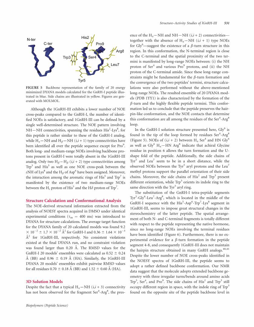
for Gly6—suggest the existence of a b-turn structure in this
region. In this conformation, the N-terminal region is close
to the C-terminal and the spatial proximity of the two ter-
mini is manifested by long-range NOEs between: (i) the NH
proton of Ser4 and various Pro9 protons, and (ii) the NH
proton of the C-terminal amide. Since these long-range con-
straints might be fundamental for the b-turn formation and
the convergence of the two peptides’ termini, structure calcu-
lations were also performed without the above-mentioned
long-range NOEs. The resulted ensemble of 20 DYANA mod-
els (PDB 1YY1) is also characterized by the formation of the
b-turn and the highly flexible peptide termini. This confor-
mation led us to conclude that the peptide preserves the hair-
pin-like conformation, and the NOE contacts that determine
this conformation are all among the residues of the Ser4-Arg8
loop.
In the GnRH-I solution structure presented here, Gly6 is
found in the tip of the loop formed by residues Ser4-Arg8
(Figure 5). NOEs of (i,i + 2) between Ha Ser4 and HN Gly6
as well as Gly6 Ha��HN Arg8 indicate that achiral Glycine
residue in position 6 allows the turn formation and the U-
shape fold of the peptide. Additionally, the side chains of
Tyr5 and Leu7 seem to be in a short distance, while the
observed NOEs between the Tyr5 aryl protons and the Leu7
methyl protons support the parallel orientation of their side
chains. Moreover, the side chains of His2 and Trp3 present
different orientation, while Trp3 orients its indole ring to the
same direction with the Tyr5 aryl ring.
The substitution of the GnRH-I tetra-peptide segments
Tyr5-Gly6-Leu7-Arg8, which is located in the middle of the
GnRH-I sequence with the His5-Asp6-Trp7-Lys8 segment in
1GnRH-III, seems to impose great structural changes in the
stereochemistry of the latter peptide. The spatial arrange-
ment of both N- and C-terminal fragments is totally different
with respect to the peptide representing the native hormone,
since no long-range NOEs involving the terminal residues
have been identified (Figure 6). Furthermore, there is no ex-
perimental evidence for a b-turn formation in the peptide
segment 4-8, and consequently 1GnRH-III does not maintain
the hairpin structure obtained in many GnRH analogs.40,41
Despite the lower number of NOE cross-peaks identified in
the NOESY spectra of 1GnRH-III, the peptide seems to
adopt a rather defined backbone conformation. Our NMR
data suggest that the molecule adopts extended backbone ge-
ometry with three irregular turns/bends around amino acids
Trp3, Ser4, and Pro9. The side chains of His2 and Trp3 still
occupy different region in space, with the indole ring of Trp3
found on the opposite site of the peptide backbone level in
FIGURE 5 Backbone representation of the family of 20 energy
minimized DYANA models calculated for the GnRH-I peptide illus-
trated in blue. Side chains are illustrated in yellow. Figures are gen-
erated with MOLMOL.
Structure–Activity Studies of lGnRH-III 531
Biopolymers (Peptide Science)

respect to the His2 side chain and pointing towards the
indole ring of Trp7. Moreover, the aromatic ring of His5 is
found within NOE distance with Trp7 indole ring supporting
the parallel orientation of their side chains.
DISCUSSIONThe ability of lGnRH-III to inhibit proliferation of cancer
cells, combined with the weak GnRH-I agonistic activity,
makes it an excellent starting compound for the development
of peptide analogs with enhanced selectivity and anticancer
activity. In this study, we report the synthesis of 21 new
lGnRH-III analogs and their impact on the proliferation of
two different prostate cancer cell lines. Moreover, to investi-
gate the conformational resemblance or diversity in GnRH
family, we studied the conformational properties of lGnRH-
III and GnRH-I through NMR spectroscopy and their solu-
tion models are presented herein.
In our previous studies,41 we have shown that GnRH-I
does not exhibit significant antiproliferative activity on
LNCaP and PC3 cells even at the concentration of 100 lM.
Results of this study provide experimental evidence that the
inhibitory effect of lGnRH-III, at the concentration of 20
lM, on LNCaP cell proliferation was 21.5 6 0.9% while, on
PC3 cell it was negligible. The absence of high affinity bind-
ing sites on PC3 cells42 and the lower number of GnRHRs
expressed on them39 compared to LNCaP cells, may be
related to the lack of responsiveness of PC3 cells following
lGnRH-III treatment. However, many of the new analogs of
lGnRH-III presented on this study had significant antiproli-
ferative effect on both cancer cell lines. Our observations
confirm the results of other studies that additional mecha-
nisms may mediate the biological effects of GnRH analogs
on cancer cells which have not yet been clarified.42
In lGnRH-III, the position 6 is occupied by Asp, whereas
mammalian GnRH (GnRH-I) bears Gly at this position.
Although Gly6 is often replaced by D-amino acids to enhance
the biological activity of GnRH-I analogs, previous studies
report that removal of the Asp6 in lGnRH-III resulted in
complete loss of biological activity.19,43 To further investigate
the significance of this position, we substituted Asp6 by Asn,
Asp(OMe), Glu, and Gln. Incorporation of Asp(OMe)
instead of Asp6 significantly increased the antiproliferative
activity of lGnRH-III on LNCaP cell line. Furthermore,
Asp(OMe)-analog was the only analog of that group, with
significant effect on PC3 proliferation. Insertion of Gln was
well tolerated, while no antiproliferative activity on LNCaP
cells was observed when Glu or Asn substitutes Asp6. These
findings suggest that neither the acidic function of the side
chain of Asp6 nor the putative salt bridge (Asp6-Lys8) are
crucial for the antiproliferative activity of lGnRH-III, and we
presume that different intramolecular interactions, that
deserve further investigation, may occur and preserve the
bioactive conformation.
According to Heredi-Szabo et al.,19 indole rings of Trp3,7
are crucial for the anticancer activity, probably due to inter-
actions of these residues with the N-terminal which stabilizes
the biological active conformation of lGnRH-III. In our
study, the antiproliferative effect of the analog with DTrp in
position 3 was significantly higher than lGnRH-III whereas
DTrp7 analog was ineffective. Introduction of DTrp instead
of Trp3,7 decreased the anticancer activity while, combined
modification on the C-terminal resulted in a significant
increase of the anticancer potency in both cancer cell lines.
Tic is a conformationally restrained imino acid that has been
incorporated to GnRH antagonists as local structure deter-
minant.44 In addition, we have previously shown that substi-
tution of Trp3 in GnRH-I by L- or D-Tic increased the anti-
proliferative effect of the analogs.45 In this study, results
show that substitution of Trp3,7 by Tic decreases the antipro-
liferative effect of lGnRH-III on LNCaP cells and slightly
improves the effect on PC3 cancer cells. In contrast, the in-
hibitory activity of the analogs [DTic3,7]-lGnRH-III (X),
[DTrp3, DTic7]-lGnRH-III (XI), and [DTic3, DTrp7]-
lGnRH-III (XII) was significantly higher. Based on these
findings, we presume that introduction of DTic, although
lacks the indole ring, preserved the biologically active confor-
mation of lGnRH-III. Furthermore, the importance of the
stereochemistry of the residues in positions 3 and 7 is also
indicated.
Conjugation of an acetyl-group via the side chain of Lys8
significantly increases the antiproliferative activity of lGnRH-
III suggesting that the putative salt bridge between Asp6 and
Lys8 in lGnRH-III, proposed in an earlier structure activity
study16 and demonstrated by our findings, is not important
for the anticancer activity. Similar elevation of the antiproli-
ferative effect was observed by analogs with side chain
FIGURE 6 Backbone representation of the family of 20 energy
minimized DYANA models calculated for the 1GnRH-III peptide
illustrated in blue. Side chains are illustrated in cyan. Figures are
generated with MOLMOL.
532 Pappa et al.
Biopolymers (Peptide Science)

cyclization of amino acids 1-8 (peptides XIV, XV) probably
due to a more constrained conformation. On the other hand,
although exchanging places between the weakly basic His5
and the more basic Lys8 did not alter the anticancer activity
of the parent hormone, simultaneous 1-5 side chain cycliza-
tion in analogs XX and XXI resulted in lower antiprolifera-
tive effect.
In our attempt to elucidate conformational characteristics
of lGnRH-III, the solution model was determined through
NMR spectroscopy in DMSO, a solvent widely used for solu-
bility reasons and is an acceptable medium of mimicking a
prototypical hydrophobic environment. Furthermore, the 3D
solution model of GnRH-I was also determined to shed light
on the conformational differences between the two peptides.
The obtained results show substantially different conforma-
tional features between the 1GnRH-III and the GnRH-I
(Figure 7). According to our NMR study, the network of se-
quential NOE connectivities present in GnRH-I spectra sug-
gest the existence of b-turn structure for the Ser4-Arg8 seg-
ment. Long-range NOE signals between the N- and C-ter-
mini provide solid experimental evidence for the spatial
proximity of the two termini. On the other hand, 1GnRH-III
does not exhibit the GnRH-I conformation (U-shape). In
spite of earlier publications suggesting the existence of some
helical properties,23 such backbone conformation cannot be
supported by our results. In contrast, the 3D models of
1GnRH-III can be characterized by the extended backbone
conformation forming a kink around Asp6 and two more
turns/bends around Trp3 and Pro9. The present lGnRH-III
models are in agreement with Mezo et al.,17 where a relatively
ordered extended like backbone conformation is proposed
for the central region of the 1GnRH-III peptide.
The substitution of the small and flexible residue Gly in
position 6 of the GnRH-I sequence with Asp6 in 1GnRH-III
does not favor the formation of the b-type conformation in
the 4-8 segment of the peptide sequence. Moreover, the sub-
stitution of Leu7 with the bulky Trp7 differentiates remark-
ably the conformation of the peptide. This also becomes evi-
dent by the observed NOE interactions between His5 and
Trp7, which do not favor the spatial proximity of the two ter-
mini. As a consequence, the hydrophobic core present in
GnRH-I, is lost in 1GnRH-III.
Furthermore, the proposed ionic interaction between
Asp6-Lys8 residues16 is in agreement with our findings. Trp7
changes its orientation pointing away from Lys8 and conse-
quently no NOE interaction is found between their side
chains (as in the Leu7-Arg8 case in GnRH-I), and Asp6 is
found to be within NOE distance with Lys8 giving rise to one
interaction between eNH of Lys8 and Hb of Asp6.
In conclusion, we synthesized 21 new lGnRH-III analogs
and the structure–activity studies show that several of
them had similar or higher activity than that of the parent
hormone. Analogs II, V, VIII, X, XI, XII, XIV, XV, and XVI
had higher antiproliferative activity than lGnRH-III on
LNCaP cells and significant activity on PC3 cell. The modi-
fications of those vary significantly but their inhibitory
effect is almost the same, hampering thus the identification
of one particular ‘‘lead compound.’’ However, we have
studied ‘‘key’’ positions of lGnRH-III, providing new infor-
mation regarding the structure–activity relationship of
lGnRH-III. Our observations are illustrating that lGnRH-
III is a promising molecule for the development of peptide
analogs with increased and potentially selective anticancer
activity.
REFERENCES1. Millar, R.P.; Pawson, A.J.; Morgan, K.; Rissman, E.F.; Lu, Z.L.
Front Neuroendocrinol 2008, 29, 17–35.
2. Morgan, K.; Stewart, A.J.; Miller, N.; Mullen, P.; Muir, M.;
Dodds, M.; Medda, F.; Harrison, D.; Langdon, S.; Millar, R.P.
Cancer Res 2008, 68, 6331–6340.
3. Santen, R.J.; Manni, A.; Harvey, H. Breast Cancer Res Treat
1986, 7, 129–145.
4. Limonta, P.; Moretti, R.M.; Marelli, M.M.; Motta, M. Front
Neuroendocrinol 2003, 24, 279–295.
5. Sower, S.A.; Chiang, Y.C.; Lovas, S.; Conlon, J.M. Endocrinol-
ogy 1993, 132, 1125–1131.
6. Nozaki, M.; Ominato, K.; Gorbman, A.; Sower, S.A. Gen Comp
Endocrinol 2000, 118, 57–67.
7. Tobet, S.A.; Nozaki, M.; Youson, J.H.; Sower, S.A. Cell Tissue
Res 1995, 279, 261–270.
8. Yu, W.H.; Karanth, S.; Sower, S.A.; Parlow, A.F.; McCann, S.M.
Proc Soc Exp Biol Med 2000, 224, 87–92.
9. Dees, W.L.; Hiney, K.K.; Sower, S.A.; Yu, W.H.; McCann, S.M.
Peptides 1999, 20, 1503–1511.
10. McCann, S.M.; Karanth, S.; Mastronardi, C.A.; Dees, W.L.;
Childs, G.; Miller, B.; Sower, S.; Yu, W.H. Arch Med Res 2001,
32, 476–485.
FIGURE 7 Superimposition of the backbone of the two hor-
mones, lGnRH-III is illustrated in yellow while GnRH-I in blue. The
calculated RMSD of both peptides for the segment 1-6 found to be
0.815A.
Structure–Activity Studies of lGnRH-III 533
Biopolymers (Peptide Science)

11. Yu, W.H.; Karanth, S.; Walczewska, A.; Sower, S.A.; McCann,
S.M. Proc Natl Acad Sci USA 1997, 94, 9499–9503.
12. Sower, S.A. J Great Lakes Res 2003, 29 (suppl 1), 50–65.
13. Kovacs, M.; Seprodi, J.; Koppan, M.; Horvath, J.E.; Vincze, B.;
Teplan, I. J Neuroendocrinol 2002, 14, 1–14.
14. Amstalden, M.; Zieba, D.A.; Garcia, M.R.; Stanko, R.L.; Welsh,
T.H., Jr.; Hansel, W.H. Reproduction 2004, 127, 35–43.
15. Lovas, S.; Palyi, I.; Vincze, B.; Horvath, J.; Kovacs, M.; Mezo, I.
J Pept Res 1998, 52, 384–389.
16. Mezo, I.; Lovas, S.; Palyi, I.; Vincze, B.; Kalnay, A.; Turi, G.;
Vadasz, Z.; Seprodi, J.; Idei, M.; Toth, G.; Gulyas, E.; Otvos, F.;
Mak, M.; Horvath, J.E.; Teplan, I.; Murphy, R.F. Med Chem
1997, 40, 3353–3358.
17. Mezo, G.; Czajlik, A.; Manea, M.; Jakab, A.; Farkas, V.; Majer,
Z.; Vass, E.; Bodor, A.; Kapuvari, B.; Boldizsar, M.; Vincze, B.;
Csuka, O.; Kovacs, M.; Przybylski, M.; Perczel, A.; Hudecz, F.
Peptides 2007, 28, 806–820.
18. Lovas, S.; Palyi, I.; Vincze, B.; Horvath, J.; Kovacs, M.; Mezo, I.;
Toth, G.; Teplan, I.; Murphy, R.F. J Pep Res 1998, 52, 384–389.
19. Heredi-Szabo, K.; Lubke, J.; Toth, G.; Murphy, R.F.; Lovas, S.
Peptides 2005, 26, 419–422.
20. Iwai, H.; FEBS Lett 1999, 459, 166–172.
21. Gilon, C.; Mang, C.; Lohof, E.; Friedler, A.; Kessler, H. Synthesis
of Peptides and Peptidomimetics, Goodman, M.; Toniolo, C.;
Moroder, L.; Felix, A., Eds.; Georg Thieme Verlag: Stuttgart,
2003; pp 422–529.
22. Liu, C.F.; Rao, C.; Tam, J.P. Peptides: Frontiers of peptide sci-
ence. Tam, J.P.; Kaumaya, P.T.P.; Eds.; Kluwer Academic Pub-
lishers: Dordrecht, The Netherlands, 1999; pp 235–237.
23. Watts, C.R.; Mezei, M.; Murphy, R.F; Lovas, S. J Biomol Struct
Dyn 2001, 18, 733–748.
24. Augustyns, K.; Kraas, W.; Jung, G. J Pept Res 1998, 51, 127–133.
25. Nagakawa, O.; Fujiuchi, Y.; Fuse, H.; Saiki, I. Urology 2003, 62,
553–558.
26. Braunschweiler, L.; Ernst, R.R. J Magn Reson 1983, 53,
521–528.
27. Bax, A.; Davis, D.G. J Magn Reson 1985, 65, 355–360.
28. Bax, A.; Grzesiek, S. Acc Chem Res 1993, 26, 131–138.
29. Bothner-By, A.A.; Stephens, R.L.; Lee, J.C.; Warren, C.D.; Jean-
loz, R.W. J Am Chem Soc 1984, 106, 811–813.
30. Marion, D.; Wuthrich, K. Biochem Biophys Res Commun 1983,
113, 967–974.
31. Jeener, J.; Meier, B.; Bachmann, P.; Ernst, R.R. J Chem Phys
1979, 71, 4546–4553.
32. Eccles, C.; Guntert, P.; Billeter, M.; Wuthrich, K.J. Biomol NMR
1991, 1, 111–130.
33. Guntert, P.; Braun, W.; Wuthrich, K. J Mol Biol 1991, 217,
517–530.
34. Guntert, P.; Mumenthaler, C.; Wuthrich, K. J Mol Biol 1997,
273, 283–298.
35. Wuthrich, K.; Billeter, M.; Braun, W. J Mol Biol 1983, 169,
949–961.
36. Koradi, R.; Billeter, M.; Wuthrich, K. J Mol Graph 1996, 14,
51–55, 29–32.
37. Johnson, T.; Liley, M.; Cheeseright, J.T.; Begum, F.J. Chem Soc
Perkin Trans 1 2000, 16, 2811–2820.
38. Alimirah, F.; Chena, J.; Basrawalab, Z.; Xina, H.; Choubey, D.
FEBS Lett 2006, 580, 2294–2300.
39. Kraus, S.; Levy, G.; Hanoch, T.; Naor, Z.; Seger, R.M. Cancer
Res 2004, 64, 5736–5744.
40. Tare, R.S.; Oreffo, R.O.; Sato, K.; Rauvala, H.; Clarke, N.M.;
Roach, H.I. Biochem Biophys Res Commun 2002, 298,
324–332.
41. Pappa, E.V.; Zompra, A.A.; Spyranti, Z.; Diamantopoulou, Z.;
Pairas, G.; Lamari, F.N.; Katsoris, P.; Spyroulias, G.A.; Cordopa-
tis, P. Biopolymers 2011, 96, 260–272.
42. Ravenna, L.; Salvatori, L.; Morrone, S.; Lubrano, C.; Cardillo,
MR.; Sciarra, F.; Frati, L.; Di Silverio, F.; Petrangeli, E. J Androl
2000, 21, 549–557.
43. Kovacs, M.; Vincze, B.; Horvath, J.E.; Seprodi, J. Peptides 2007,
28, 821–829.
44. Koerber, S.C.; Ibea, M.; Hagler, A.T.; Rivier, C.L; Rivier, J.E. Bio-
chem Biophys Res Commun 1992, 187, 1035–1041.
45. Zompra, A.A.; Magafa, V.; Lamari, F.N.; Nock, B.; Maina, T.;
Spyroulias, G.A.; Karamanos, N.K.; Cordopatis, P. J Pept Res
2005, 1, 57–64.
534 Pappa et al.
Biopolymers (Peptide Science)


![Synthesis and Conformational Studies on [3.3.3]Metacyclophane Oligoketone Derivatives, and Their Metal Ion Recognition.](https://static.fdokumen.com/doc/165x107/6337d1c7a42190c2190e7bc8/synthesis-and-conformational-studies-on-333metacyclophane-oligoketone-derivatives.jpg)