Structural, transport, magnetic, and dielectric properties of La1− x Te x MnO3 (x= 0.10 and 0.15)
,
Materials Chemistry and Physics 113 (2009) 749–755
Contents lists available at ScienceDirect
Materials Chemistry and Physics
journa l homepage: www.e lsev ier .com/ locate /matchemphys
Structural, optical and magnetic properties of nanocrystalline Zn0.9Co0.1O-baseddiluted magnetic semiconductors
Y. Kalyana Lakshmia, K. Srinivasa, B. Sreedharb, M. Manivel Rajac, M. Vithald, P. Venugopal Reddya,∗
a Department of Physics, Osmania University, Hyderabad 500007, Indiab I&PC Division, Indian Institute of Chemical Technology, Hyderabad 500007, Indiac Defence Material Research Laboratory, Hyderabad 500058, Indiad Department of Chemistry, Osmania University, Hyderabad 500007, India
a r t i c l e i n f o
Article history:Received 1 December 2007Received in revised form 28 May 2008Accepted 3 August 2008
PACS:75.50.Pp74.25.Gz75.30.Hx75.60.Ej
a b s t r a c t
With a view to understand the influence of nano size on various properties of cobalt-doped ZnO-baseddiluted magnetic semiconductors, a series of materials were prepared by the citrate gel route. The phaseand morphology studies have been carried out by X-ray diffraction and transmission electron microscopyrespectively. All the samples of the present investigation are found to have hexagonal wurtzite structureand crystallite sizes are found to vary from 25 nm to 65 nm. From the optical absorption measurementsit has been observed that upon doping with cobalt, the energy band gap is found to shift towards lowerenergy side (red shift) while it shifts towards higher energy side (blue shift) when the crystallite size isincreased continuously. It has been observed from the XPS results that oxidation state of Cobalt is +2 andthat the difference in binding energies of Co 2p3/2 and Co 2p1/2 is found to increase continuously withincreasing crystallite size. Finally, all the samples are found to exhibit room temperature ferromagnetism
Keywords:Diluted magnetic semiconductorNOM
and the specific magnetization decreases with increasing crystallite size.Published by Elsevier B.V.
1
acnbmsmsoasoaeei
(
pdtfZarnntnh
0d
anostructuresptical propertiesagnetic properties
. Introduction
Diluted Magnetic semiconductors (DMS) have recently attractedgreat deal of attention due to possibility of manipulating
harge and spin degrees of freedom. They exhibit unique mag-etic, magneto-optical and magneto-electrical effects and cane exploited as spintronic devices [1]. Efforts are going on toeet some of the deficiencies of these materials such as intrin-
ic ferromagnetism, high ferromagnetic Curie-temperature, largeagnetization and a precise controllable spin properties, etc. In
earch of a new DMSs, lead the attention towards wide band gapxide-based DMSs such as Zn1−xTMxO, Ti1−xTMxO2, Sn1−xTMxO2nd Hf1−xTMxO2 (TM: Co, Ni, Mn, Fe) [2–5], etc., because of their
table ferromagnetism near or above room temperature. Amongxide-based DMSs, ZnO doped with small amount of Co2+ havettracted considerably. Moreover, ZnO has a large exciton bindingnergy of 60 meV, with wide band gap energy (∼3.3 eV) resulting infficient excitonic emission at room temperature as well as suitablen making transparent devices.∗ Corresponding author. Tel.: +91 40 27682287; fax: +91 40 27090020.E-mail addresses: [email protected], [email protected]
P.V. Reddy).
2
bicsh(wo
254-0584/$ – see front matter. Published by Elsevier B.V.oi:10.1016/j.matchemphys.2008.08.021
It is well known that nano size has great influence on theerformance of several material systems. Moreover, the role ofimensionality in shaping the spin-polarized electronic struc-ure of nanocrystalline DMSs is important in understanding theirerromagnetic behavior [6–12]. Apart from this, nanocrystallinen0.9Co0.1O may be exploited for ferro-fluids, magnetic recording,nd biomedical applications because quantum confinement mayesult in intriguing magnetic properties. As the structural, mag-etic and electronic properties of Zn0.9Co0.1O are sensitive to theano size of the materials, an effort has been made to understandhe influence of nano size on their structural, optical and ferromag-etic behavior and the results of such an investigation are presentedere.
. Experimental procedure
The nanocrystalline Zn0.9Co0.1O samples were prepared using citrate gel routey taking the corresponding starting materials as nitrates. More details are givenn an earlier publication [9]. In this method, first the nitrates were converted into
itrates and by adding suitable chemicals, pH was adjusted to 6.5. After getting aolution on slow evaporation, a gelating reagent ethylene glycol was added andeated at about 180 ◦C to get a gel. After a few more steps dry fluffy porous massprecursor), was calcined at 250 ◦C for 3 h. Finally, the samples in the form of pelletsere sintered at 300 ◦C, 400 ◦C, 500 ◦C and 600 ◦C temperatures for 3 h in order tobtain the materials with varying crystallite size.
7 mistry
PaaTg2tdmdto(trflacwoia
3
3
taFsishhwtt
naRtstTt
iIec
3
Trs
smaciuasot
3
ptmwlFaCibpom
FZ
50 Y.K. Lakshmi et al. / Materials Che
The crystal structure data was determined using X-ray diffraction (XRD) usinghillips (Xpert) diffractometer with Cu K� radiation (� = 1.5406 Å) by recording at2� value of 20–80◦ in a step of 0.02◦ . Transmission electron micrographs (TEM)
nd selected area electron diffraction (SAD) patterns were recorded using a Phillipsecnai G2 FEI F12 electron microscope. With a view to determine the optical bandap, the optical absorption spectra measurements were undertaken in the range of00–800 nm using a GBC Cintra 10e UV–Vis-DRS spectrometer. For this purpose, allhe samples were palletized by mixing the sample powder with KBr. As the pow-ers are not highly transparent, KBr was used as a reference for the absorbanceeasurements. In order to make the base line corrections for the band gap values
ue to free excitons near the band gap, the authors recorded the absorbance spec-ra of all the Co-doped samples using ZnO pellet as a reference. The thicknessesf all the pellets were found to be about 3 mm. X-ray photoelectron spectroscopicXPS) measurements were also performed on a Kratos X-ray photoelectron spec-rometer. The X-ray excitation energy was 1253.6 eV (Mg K�), and the spectra wereecorded with a pass energy of 80 eV. The angle between the detector and the X-rayux direction was constant and equal to 90◦ , while the measurements were madet an electron take off angle of 70◦ . The calibration of the spectrometer and theharging correction were verified by determining the binding energy of C 1s levelith a graphite substrate (at 285 eV). Finally, in order to determine ferromagnetism
f these samples, magnetization measurements were undertaken using a vibrat-ng sample magnetometer (Model: DMSADE-1660 MRS) at room temperature bypplying 15 kOe magnetic field.
. Results and discussion
.1. X-ray diffraction measurements
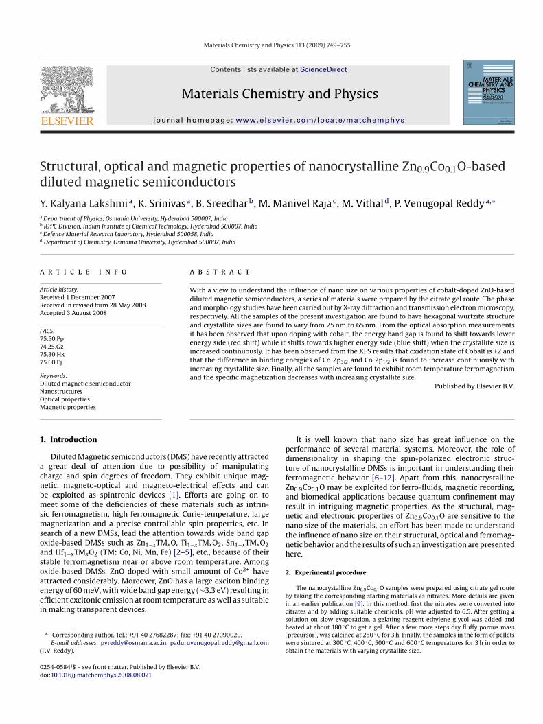
The XRD patterns of Zn0.9Co0.1O samples sintered at differentemperatures along with undoped ZnO sample sintered at 600 ◦Cnd prepared under similar experimental conditions are shown inig. 1(a). All the patterns are found to have hexagonal wurtzitetructure, without any additional impurity phases thereby indicat-ng that the wurtzite structure might have not affected due to theubstitution of Cobalt. Further, as no excess peaks were detected, itas been concluded that all the starting organic precursors mightave been completely decomposed. The diffraction peaks alongith their relative intensities are found to be in agreement with
hose from JCPDS [card no. 36-1451] and the patterns were indexedo ZnO.
The XRD data were also analyzed by Reitveld refinement tech-ique and using the data, lattice parameters (Table 1) were obtainednd are found to be in agreement with the reported ones [7].eitveld refined pattern of one of the samples viz; Zn0.9Co0.1O sin-
ered at 300 ◦C is shown in Fig. 1(b). Further, the average crystalliteize values of all the samples were estimated using peak broadeningechnique (Table 1) and are found to be in the range of 25–65 nm.he lattice parameter “a” obtained from the Reitveld data indicatehat there is no systematic variation.tCTti
ig. 1. XRD pattern for (a) undoped and Co-doped Zn0.9Co0.1O nanocrystalline powders sintn0.9Co0.1O powder sintered at 300 ◦C.
and Physics 113 (2009) 749–755
However, “c” parameter is found to decrease continuously withncreasing nano size of the materials except in the case of ZCO-600.n fact a similar behavior was reported earlier [7]. It is also inter-sting to note that c/a value of these materials is found to decreaseontinuously.
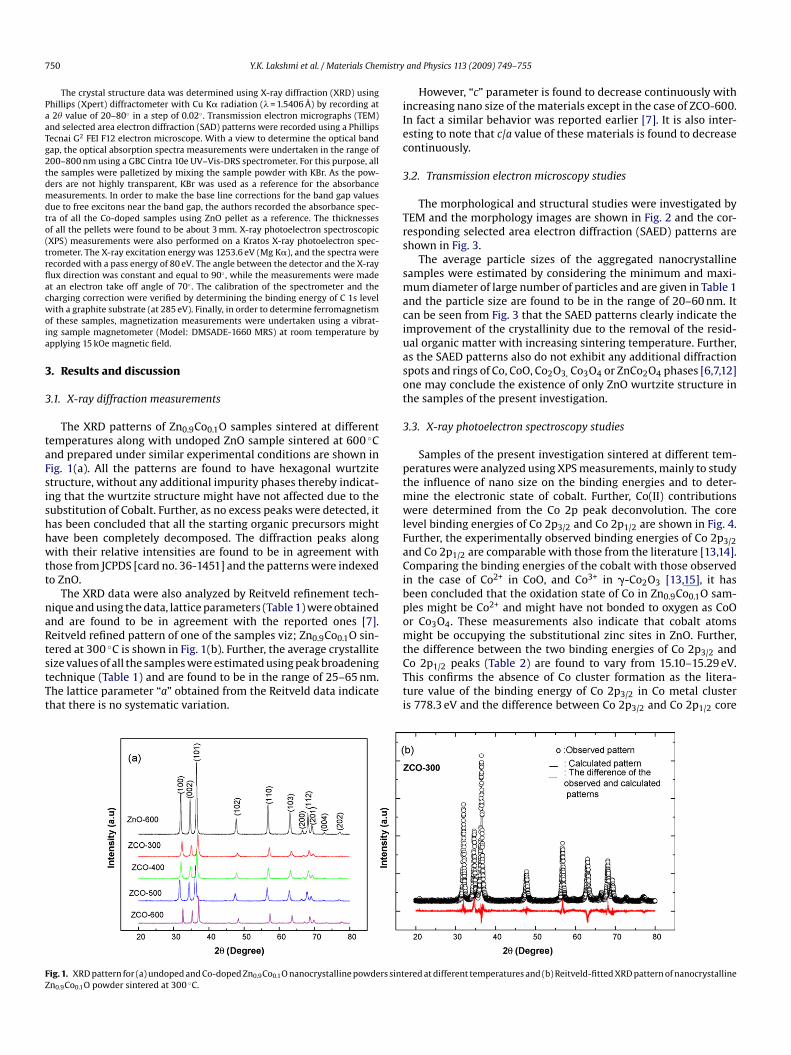
.2. Transmission electron microscopy studies
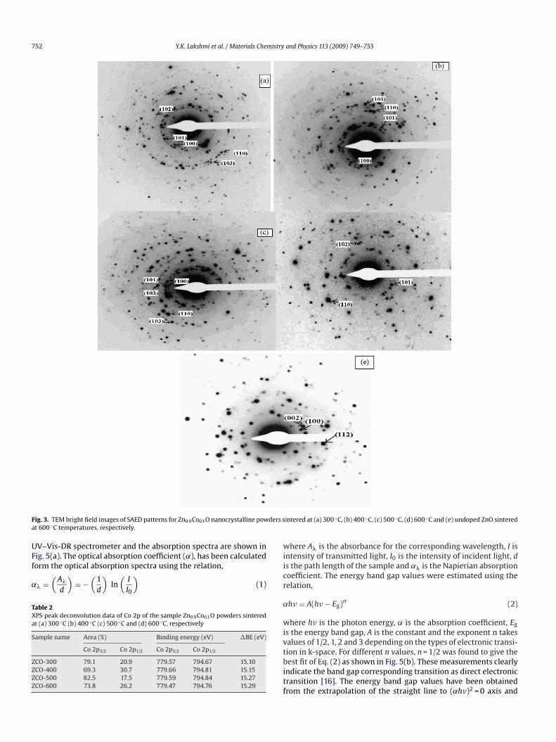
The morphological and structural studies were investigated byEM and the morphology images are shown in Fig. 2 and the cor-esponding selected area electron diffraction (SAED) patterns arehown in Fig. 3.
The average particle sizes of the aggregated nanocrystallineamples were estimated by considering the minimum and maxi-um diameter of large number of particles and are given in Table 1
nd the particle size are found to be in the range of 20–60 nm. Itan be seen from Fig. 3 that the SAED patterns clearly indicate themprovement of the crystallinity due to the removal of the resid-al organic matter with increasing sintering temperature. Further,s the SAED patterns also do not exhibit any additional diffractionpots and rings of Co, CoO, Co2O3, Co3O4 or ZnCo2O4 phases [6,7,12]ne may conclude the existence of only ZnO wurtzite structure inhe samples of the present investigation.
.3. X-ray photoelectron spectroscopy studies
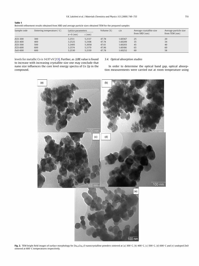
Samples of the present investigation sintered at different tem-eratures were analyzed using XPS measurements, mainly to studyhe influence of nano size on the binding energies and to deter-
ine the electronic state of cobalt. Further, Co(II) contributionsere determined from the Co 2p peak deconvolution. The core
evel binding energies of Co 2p3/2 and Co 2p1/2 are shown in Fig. 4.urther, the experimentally observed binding energies of Co 2p3/2nd Co 2p1/2 are comparable with those from the literature [13,14].omparing the binding energies of the cobalt with those observed
n the case of Co2+ in CoO, and Co3+ in �-Co2O3 [13,15], it haseen concluded that the oxidation state of Co in Zn0.9Co0.1O sam-les might be Co2+ and might have not bonded to oxygen as CoOr Co3O4. These measurements also indicate that cobalt atomsight be occupying the substitutional zinc sites in ZnO. Further,
he difference between the two binding energies of Co 2p3/2 ando 2p1/2 peaks (Table 2) are found to vary from 15.10–15.29 eV.his confirms the absence of Co cluster formation as the litera-ure value of the binding energy of Co 2p3/2 in Co metal clusters 778.3 eV and the difference between Co 2p3/2 and Co 2p1/2 core
ered at different temperatures and (b) Reitveld-fitted XRD pattern of nanocrystalline
Y.K. Lakshmi et al. / Materials Chemistry and Physics 113 (2009) 749–755 751
Table 1Reitveld refinement results obtained from XRD and average particle sizes obtained TEM for the prepared samples
Sample code Sintering temperature (◦C) Lattice parameters Volume (%) c/a Average crystallite sizefrom XRD (nm)
Average particle sizefrom TEM (nm)a = b (nm) c (nm)
ZCO-300 300 3.2511 5.2137 47.78 1.60367 25 20Z 47.74Z 47.61Z 47.9Z 47.7
ltnc
Fs
CO-400 400 3.2525 5.2108CO-500 500 3.2495 5.2058CO-600 600 3.2574 5.2179nO-600 600 3.2530 5.2130
evels for metallic Co is 14.97 eV [13]. Further, as �BE value is foundo increase with increasing crystallite size one may conclude thatano size influences the core level energy spectra of Co 2p in theompound.
3
t
ig. 2. TEM bright field images of surface morphology for Zn0.9Co0.1O nanocrystalline powintered at 600 ◦C temperatures respectively.
1.60209 35 341.60203 45 40
6 1.60186 65 608 1.60252 60 58
.4. Optical absorption studies
In order to determine the optical band gap, optical absorp-ion measurements were carried out at room temperature using
ders sintered at (a) 300 ◦C, (b) 400 ◦C, (c) 500 ◦C, (d) 600 ◦C and (e) undoped ZnO
752 Y.K. Lakshmi et al. / Materials Chemistry and Physics 113 (2009) 749–755
F ders sa
UFf
˛
TXa
S
ZZZZ
w
ig. 3. TEM bright field images of SAED patterns for Zn0.9Co0.1O nanocrystalline powt 600 ◦C temperatures, respectively.
V–Vis-DR spectrometer and the absorption spectra are shown in
ig. 5(a). The optical absorption coefficient (˛), has been calculatedorm the optical absorption spectra using the relation,� =(
A�
d
)= −
(1d
)ln
(I
I0
)(1)
able 2PS peak deconvolution data of Co 2p of the sample Zn0.9Co0.1O powders sinteredt (a) 300 ◦C (b) 400 ◦C (c) 500 ◦C and (d) 600 ◦C, respectively
ample name Area (%) Binding energy (eV) �BE (eV)
Co 2p3/2 Co 2p1/2 Co 2p3/2 Co 2p1/2
CO-300 79.1 20.9 779.57 794.67 15.10CO-400 69.3 30.7 779.66 794.81 15.15CO-500 82.5 17.5 779.59 794.84 15.27CO-600 73.8 26.2 779.47 794.76 15.29
iicr
˛
wivtbitf
intered at (a) 300 ◦C, (b) 400 ◦C, (c) 500 ◦C, (d) 600 ◦C and (e) undoped ZnO sintered
here A� is the absorbance for the corresponding wavelength, I isntensity of transmitted light, I0 is the intensity of incident light, ds the path length of the sample and ˛� is the Napierian absorptionoefficient. The energy band gap values were estimated using theelation,
h� = A(h� − Eg)n (2)
here h� is the photon energy, ˛ is the absorption coefficient, Eg
s the energy band gap, A is the constant and the exponent n takesalues of 1/2, 1, 2 and 3 depending on the types of electronic transi-
ion in k-space. For different n values, n = 1/2 was found to give theest fit of Eq. (2) as shown in Fig. 5(b). These measurements clearlyndicate the band gap corresponding transition as direct electronicransition [16]. The energy band gap values have been obtainedrom the extrapolation of the straight line to (˛h�)2 = 0 axis and

Y.K. Lakshmi et al. / Materials Chemistry and Physics 113 (2009) 749–755 753
Fig. 4. XPS spectra of Co of Zn0.9Co0.1O nanocrystalline powders sintered at (a) 300 ◦C (b) 400 ◦C (c) 500 ◦C and (d) 600 ◦C, respectively.
F Co-dou
aracar
(
TT
S
ZZZZZ
ig. 5. (a) The optical absorption spectra and (b) plot of (˛h�)2 versus h� for thendoped ZnO sintered at 600 ◦C.
re given in Table 3 [Baseline corrections have eliminated free car-
ier contributions to determining optical band gaps]. The UV–visbsorption spectra of the nanocrystalline undoped ZnO powdersalcined at 600 ◦C having average crystallite size ∼50 nm show anbsorption edge observed at ∼3.2117 ± 0.03 eV (� = 380 nm). Theeduction in the band gap compared with bulk ZnO band gapb[6sa
able 3he optical band gaps with corresponding crystallite sizes for the prepared samples along
ample name Crystallite size from XRD (nm) Band gap Eg (eV)
CO-300 25 3.053CO-400 35 3.071CO-500 45 3.091CO-600 66 3.116nO-600 60 3.211
ped ZnO samples sintered at 300 ◦C, 400 ◦C, 500 ◦C and 600 ◦C temperatures and
∼3.35 eV) might be due to the enhanced band bending at the grain
oundaries [17,18] and this value is in agreement with the reports7,17,19,20]. It is interesting to note that the band gap value of ZnO-00 is found to shift from 3.211 eV to 3.11 ± 0.02 eV due to theubstitution of cobalt, clearly indicating a red shift of the band edgend a similar shift was reported earlier [6,7,21]. The observed redwith Ms and Hc values
Saturation magnetization (emu g−1) Coercive field Hc (Oe)
0.128585 163.2970.084370 130.6580.049671 104.6460.0479365 255.774– –

754 Y.K. Lakshmi et al. / Materials Chemistry and Physics 113 (2009) 749–755
tered
sTfici
dfprtsmbstlIpaa
3
p
apcnr(Timibrcwotos
4
Fig. 6. Room temperature B–H curves of Zn0.9Co0.1O samples sin
hift may be due to induced band gap renormalization effect [22].he band gap shrinkage and free carrier screening are responsibleor the band gap renormalization effect. The s–d and p–d exchangenteractions give rise to a negative and a positive correction to theonduction-band and valence-band edges, respectively, resultingn shrinkage of the band gap.
The optical band gap plots of Zn0.9Co0.1O samples sintered atifferent temperatures are shown in Fig. 5(b). The estimated directundamental optical band gap (Eg) values for the Zn0.9Co0.1O sam-les listed in Table 3. These results are in agreement with theeported literature [23–26]. It can be observed from the figures thathere is a shift in the absorption edge to higher energy side (bluehift) with increasing crystalline size and the origin of this blue shiftay be explained using Burstein-Moss effect [27,28]. Further, it has
een argued that the change in band gap values influences the localtructural environment of Co-doped ZnO lattice. In order to inves-igate the modifications in the electronic structure, the slope of theinear part of (˛h�)2 versus h� plot (Fig. 5(b)) has been determined.t has been observed that there is a small change in the slope of thelot and may be attributed to the changes in the conduction-bandnd valence-band edges, which itself might have arisen due to theddition of cobalt.
.5. Magnetic properties
Properties of nanocrystalline Zn0.9Co Fig. 6 shows, room tem-erature magnetization versus magnetic field (B–H) curves at an
coifa
at 300 ◦C, 400 ◦C, 500 ◦C and 600 ◦C temperatures, respectively.
pplied field of 15 kOe for the samples sintered at different tem-eratures. It can be seen from Fig. 6 that all the samples exhibitinglear magnetic hysteresis loops with low coercivties and the mag-etization values are well saturated for the all samples indicatingoom temperature ferromagnetism. The saturation magnetizationMs) and coercive field (Hc) of all the samples are given in Table 3.he observed magnetic behavior may be attributed to the exchangenteraction between the localized magnetic dipole moments of the
agnetic ions (the localized d-spins of Co ions) and the free delocal-zed charge of current carriers (holes or electrons from the valenceand). The impurities/defects play a major source of these free car-iers, and strongly related to the oxygen vacancies and processingonditions. It can be seen that Ms values are found to be decreaseith increasing crystallite size and this may be due to the trapping
f free charge carriers in the grain boundaries leading to the varia-ion of charge carriers occupying states. The variation in the valuesf charge carriers in turn influences the magnetic ordering in theamples thereby decreasing the values of saturation magnetization.
. Conclusions
Nanocrystalline Zn0.9Co0.1O materials were synthesized using
itrate gel route and the crystallite sizes are found to be in the rangef 25–65 nm. It has been concluded from the XPS studies that Co isncorporated in ZnO host with an oxidation state of Co2+ withoutorming cobalt clusters and might have not bonded to oxygen eithers CoO or as Co3O4. It has been found that the optical band gap of
mistry
tmlCteistt
A
vp
R
[[
[[
[[
[
[
[[
[[
[
[
[[25] J.H. Kim, H. Kim, D. Kim, S.G. Yoon, W.K. Choo, Solid State Commun. 131 (2004)
Y.K. Lakshmi et al. / Materials Che
hese materials is increasing with increasing crystallite size of theaterials. From optical studies, a small change in the slope of the
inear part of (˛h�)2 versus h� also implies the presence of strongoulomb interaction between Co, Zn and O sites, and supportshe observed ferromagnetism in these samples. All the samplesxhibited room temperature ferromagnetism and decreases withncreasing crystallite size of the materials. As the high value ofaturation magnetization is exhibited by a sample with lowest crys-alline size, one may conclude that nano size might be beneficial tohe ferromagnetism in the samples of the present investigation.
cknowledgement
The authors are very much thankful to DRDO New Delhi, for pro-iding financial assistance and research facilities through researchroject.
eferences
[1] S.A. Wolf, D.D. Awschalom, R.A. Buhrman, J.M. Daughton, S. von Molnar, M.L.Roukes, A.Y. Chtchelkanova, D.M. Treger, Science 294 (2001) 1488.
[2] K. Ueda, H. Tabata, T. Kawai, Appl. Phys. Lett. 79 (2001) 988.[3] Y. Matsumoto, M. Murakami, T. Shono, T. Hasegawa, T. Fukumura, M. Kawasaki,
P. Ahmet, T. Chikyow, S.Y. Koshihara, H. Koinuma, Science 29 (2001) 854.
[4] H. Kimura, T. Fukumura, H. Koinuma, M. Kawasaki, Appl. Phys. Lett. 80 (2001)94.[5] I.B. Shim, C.S. Kim, J. Magn. Magn. Mater. 272 (2004) e1571.[6] S. Maensiri, P. Laokul, S. Phokha, J. Magn. Magn. Mater. 305 (2006) 381–387.[7] S. Maensiri, J. Sreesongmuang, C. Thomas, J. Klinkaewnarong, J. Magn. Magn.
Mater. 301 (2006) 422.
[
[[
and Physics 113 (2009) 749–755 755
[8] S. Deka, R. Paricha, P.A. Joy, Chem. Mater. 16 (2004) 1168.[9] Y. Kalyana Laskmi, K. Raju, P. Venugopal Reddy, NSTI 2007 Conference Proceed-
ings, vol. 4, 2007, pp. 118–121.10] J. Cui, U. Gibson, Phys. Rev. B 74 (2006) 045416.11] P. Lomments, F.S. Philippe, C. de Mello Donega, A. Meijerink, L. Piraux,
S. Michotte, S. Matefi-Tempfli, D. Poelman, Z. Hens, J. Lumin. 118 (2006)245.
12] J.J. Wu, S.C. Liu, M.H. Yang, Appl. Phys. Lett. 85 (2004) 1027.13] J.F. Moulder, W.F. Stickle, P.E. Sobol, K.D. bomben, Handbook of X-ray Photo-
electron Spectroscopy, PerkinElmer Corporation, Eden Praine, 1992.14] H.J. Lee, S.Y. Jeong, C.R. Cho, C.H. Park, Appl. Phys. Lett. 81 (2001) 4020.15] C.D. Wagner, W.M. Riggs, L.E. Davis, J.F. Moulder, Handbook of X-ray Photo-
electron spectroscopy, PerkinElmer Corporation, Physical Electronics Division,1979, p. 78 (edited by G. E. Mulenberg PerkinElmer Co.).
16] N.F. Mott, E.A. Davis, Electronic Processes in Non-Crystalline Materials, 2nd ed.,Clarendon Press, Oxford, 1979, pp. 272–275.
17] S. Dutta, S. Chattopadhyay, M. Sutradhar, A. Sarkar, M. Chakrabarti, D. Sanyal,D. Jana, J. Phys. Condens. Matter 19 (2007) 236218.
18] V. Srikant, D.R. Clarke, J. Appl. Phys. 81 (1997) 6357.19] R.E. Marotti, D.N. Guerra, C. Bello, G. Machado, E.A. Dalchiele, Sol. Energy Mater.
Sol. Cells 82 (2004) 85.20] D. Maouche, P. Ruterana, L. Louail, Phys. Lett. A 365 (2007) 231.21] M. Bouloudenine, N. Viart, S. Coils, A. Dinia, Appl. Phys. Lett. 87 (2005)
052501.22] J.D. Ye, S.L. Gu, S.M. Zhu, S.M. Liu, Y.D. Zheng, R. Zhang, Y. Shi, H.Q. Yuand, Y.D.
Ye, J. Cryst. Growth 283 (2005) 279.23] R.B. Bylsma, W.M. Becker, J. Kossut, U. Debska, D. Yoder-short, Phys. Rev. B33
(1986) 8207.24] S. Venkataprasad bhat, F.L. Deepak, Solid State Commun. 135 (2005) 345.
677.26] Z.-B. Gu, M.-H. Lu, J. Wang, C.-L. Du, C.-S. Yuan, D. Wu, S.-T. Zhang, Y.-Y. Zhu,
S.-N. Zhu, Y.-F. Chen, Thin Solid Films 515 (2006) 2361.27] P.V. Kamat, N.M. Dimitrijevic, A.J. Nozik, J. Phys. Chem. 93 (8) (1989) 2873.28] I. Hamberg, C.G. Granqvist, Phys. Rev. B 30 (1984) 3240.
Copyright © 2022 FDOKUMEN