Structural insights into the catalytic mechanism of sphingomyelinases D and evolutionary...
13
Biochem. J. (2010) 425, 513–522 (Printed in Great Britain) doi:10.1042/BJ20091167 513 Structural insights into the catalytic mechanism of Trypanosoma cruzi GPXI (glutathione peroxidase-like enzyme I) Shreenal PATEL*, Syeed HUSSAIN*, Richard HARRIS*, Sunita SARDIWAL*, John M. KELLY†, Shane R. WILKINSON‡, Paul C. DRISCOLL*§ and Snezana DJORDJEVIC* 1 *Institute of Structural and Molecular Biology, Division of Biosciences, University College London, London WC1E 6BT, U.K., †Department of Infectious and Tropical Diseases, London School of Hygiene and Tropical Medicine, London WC1E 7HT, U.K., ‡School of Biological and Chemical Sciences, Queen Mary University of London, London E1 4NS, U.K., and §Division of Molecular Structure, Medical Research Council National Institute for Medical Research, The Ridgeway, Mill Hill, London NW7 1AA, U.K. Current drug therapies against Trypanosoma cruzi, the causative agent of Chagas disease, have limited effectiveness and are highly toxic. T. cruzi-specific metabolic pathways that utilize trypanothione for the reduction of peroxides are being explored as potential novel therapeutic targets. In the present study we solved the X-ray crystal structure of one of the T. cruzi enzymes involved in peroxide reduction, the glutathione peroxidase-like enzyme TcGPXI (T. cruzi glutathione peroxidase-like enzyme I). We also characterized the wild-type, C48G and C96G variants of TcGPXI by NMR spectroscopy and biochemical assays. Our results show that residues Cys 48 and Cys 96 are required for catalytic activity. In solution, the TcGPXI molecule readily forms a Cys 48 –Cys 96 di- sulfide bridge and the polypeptide segment containing Cys 96 lacks regular secondary structure. NMR spectra of the reduced TcGPXI are indicative of a protein that undergoes widespread conformational exchange on an intermediate time scale. Despite the absence of the disulfide bond, the active site mutant proteins acquired an oxidized-like conformation as judged from their NMR spectra. The protein that was used for crystallization was pre- oxidized by t-butyl hydroperoxide; however, the electron density maps clearly showed that the active site cysteine residues are in the reduced thiol form, indicative of X-ray-induced reduction. Our crystallographic and solution studies suggest a level of structural plasticity in TcGPXI consistent with the requirement of the atypical two-cysteine (2-Cys) peroxiredoxin-like mechanism implied by the behaviour of the Cys 48 and Cys 96 mutant proteins. Key words: Chagas disease, enzyme mechanism, NMR spectros- copy, peroxidase, X-ray crystallography. INTRODUCTION Across the tropics over 20 million people are infected by the protozoan parasites Trypanosoma brucei, Trypanosoma cruzi and Leishmania, the causative agents of African sleeping sickness, Chagas disease and leishmaniasis respectively [1]. With little prospect of protective vaccines for these diseases, drugs are the only viable option to ameliorate the effects of these infections. For Chagas disease, treatment is based on the nitroheterocyclic drugs nifurtimox and benznidazole, which require enzyme-mediated activation to render them trypanocidal. However, use of these compounds is problematic; they can cause serious side effects and parasites refractory to drug treatment are commonly encountered [2,3]. The search for new therapeutics and potential therapeutic targets in T. cruzi is underpinned by extensive research on basic metabolic systems that are essential for parasite infectivity and viability, focusing predominantly on those pathways that are unique to T. cruzi. One of the major differences between trypanosomes and their mammalian hosts is that the former utilize trypanothione (N 1 ,N 8 -bisglutathionylspermidine) as their low-molecular-mass cytoplasmic reductant. In T. cruzi, trypanothione is maintained in its reduced form (dihydrotrypanothione) by the NADPH-dependent flavoenzyme trypanothione reductase. Dihydrotrypanothione subsequently provides reducing equivalents to hydroperoxides via several possible routes, including via GSH and GPXs (glutathione- dependent peroxidases), tryparedoxin (a thioredoxin-like protein) and peroxiredoxins, or ascorbate and ascorbate-dependent haemoperoxidase (see Supplementary Figure S1 available at http://www.BiochemJ.org/bj/425/bj4250513add.htm) [4]. The crystal structures of some of the components of the T. cruzi antioxidant pathway have been determined, including that of trypanothione reductase (PDB code 1AOG/1BZL) [5,6] and the typical two-cysteine (2-Cys) cytosolic peroxiredoxin (PDB code 1UUL) [7]. The peroxiredoxin and GPX family of enzymes have been extensively characterized in various organisms including in mammals and Trypanosoma [4,7–14]. However, the discovery of peroxidases in trypanosomes with sequence similarities to mammalian glutathione-dependent phospholipid hydroperoxidases was unexpected, as no glutathione-reductase activity is present in these organisms [9]. Furthermore, GSH concentrations in trypanosomes are reportedly rather low [17]. Considering the low apparent affinity of trypanosomal GPXs for GSH (the K m is in the millimolar range), the physiological function of these enzymes was difficult to ascertain [15]. Furthermore, it was demonstrated that one of these enzymes, TcGPXI (T. cruzi glutathione peroxidase-like enzyme I), can be reduced by T. cruzi tryparedoxin I, resulting in an 8–15- fold higher peroxidase activity compared with that obtained in a GSH-dependent reaction [16]. An even higher enhancement in activity with these different electron donors was observed Abbreviations used: CSI, chemical shift index; DTT, dithiothreitol; GPX, glutathione-dependent peroxidase; HSQC, heteronuclear single-quantum coherence; PtGPX5, Populus trichocarpaxdeltoides thioredoxin peroxidase 5; RNAi, RNA interference; TbPxII, Trypanosoma brucei peroxidase II; TbPxIII, Trypanosoma brucei peroxidase III; TcGPXI, glutathione peroxidase-like enzyme I; TEV, tobacco etch virus. 1 To whom correspondence should be addressed (email [email protected]). The chemical shift assignments for oxidized TcGPXI will appear in the BioMagResBank under accession number 16518. The atomic co-ordinates for TcGPXI structure will appear in the PDB under ID 3E0U. c The Authors Journal compilation c 2010 Biochemical Society www.biochemj.org Biochemical Journal
Transcript of Structural insights into the catalytic mechanism of sphingomyelinases D and evolutionary...

Biochem. J. (2010) 425, 513–522 (Printed in Great Britain) doi:10.1042/BJ20091167 513
Structural insights into the catalytic mechanism of Trypanosoma cruzi GPXI(glutathione peroxidase-like enzyme I)Shreenal PATEL*, Syeed HUSSAIN*, Richard HARRIS*, Sunita SARDIWAL*, John M. KELLY†, Shane R. WILKINSON‡,Paul C. DRISCOLL*§ and Snezana DJORDJEVIC*1
*Institute of Structural and Molecular Biology, Division of Biosciences, University College London, London WC1E 6BT, U.K., †Department of Infectious and Tropical Diseases, LondonSchool of Hygiene and Tropical Medicine, London WC1E 7HT, U.K., ‡School of Biological and Chemical Sciences, Queen Mary University of London, London E1 4NS, U.K., and§Division of Molecular Structure, Medical Research Council National Institute for Medical Research, The Ridgeway, Mill Hill, London NW7 1AA, U.K.
Current drug therapies against Trypanosoma cruzi, the causativeagent of Chagas disease, have limited effectiveness and arehighly toxic. T. cruzi-specific metabolic pathways that utilizetrypanothione for the reduction of peroxides are being explored aspotential novel therapeutic targets. In the present study we solvedthe X-ray crystal structure of one of the T. cruzi enzymes involvedin peroxide reduction, the glutathione peroxidase-like enzymeTcGPXI (T. cruzi glutathione peroxidase-like enzyme I). We alsocharacterized the wild-type, C48G and C96G variants of TcGPXIby NMR spectroscopy and biochemical assays. Our results showthat residues Cys48 and Cys96 are required for catalytic activity. Insolution, the TcGPXI molecule readily forms a Cys48–Cys96 di-sulfide bridge and the polypeptide segment containing Cys96
lacks regular secondary structure. NMR spectra of the reducedTcGPXI are indicative of a protein that undergoes widespread
conformational exchange on an intermediate time scale. Despitethe absence of the disulfide bond, the active site mutant proteinsacquired an oxidized-like conformation as judged from their NMRspectra. The protein that was used for crystallization was pre-oxidized by t-butyl hydroperoxide; however, the electron densitymaps clearly showed that the active site cysteine residues are inthe reduced thiol form, indicative of X-ray-induced reduction.Our crystallographic and solution studies suggest a level ofstructural plasticity in TcGPXI consistent with the requirement ofthe atypical two-cysteine (2-Cys) peroxiredoxin-like mechanismimplied by the behaviour of the Cys48 and Cys96 mutant proteins.
Key words: Chagas disease, enzyme mechanism, NMR spectros-copy, peroxidase, X-ray crystallography.
INTRODUCTION
Across the tropics over 20 million people are infected by theprotozoan parasites Trypanosoma brucei, Trypanosoma cruzi andLeishmania, the causative agents of African sleeping sickness,Chagas disease and leishmaniasis respectively [1]. With littleprospect of protective vaccines for these diseases, drugs are theonly viable option to ameliorate the effects of these infections. ForChagas disease, treatment is based on the nitroheterocyclic drugsnifurtimox and benznidazole, which require enzyme-mediatedactivation to render them trypanocidal. However, use of thesecompounds is problematic; they can cause serious side effects andparasites refractory to drug treatment are commonly encountered[2,3]. The search for new therapeutics and potential therapeutictargets in T. cruzi is underpinned by extensive research onbasic metabolic systems that are essential for parasite infectivityand viability, focusing predominantly on those pathways thatare unique to T. cruzi. One of the major differences betweentrypanosomes and their mammalian hosts is that the formerutilize trypanothione (N1,N8-bisglutathionylspermidine) as theirlow-molecular-mass cytoplasmic reductant.
In T. cruzi, trypanothione is maintained in its reduced form(dihydrotrypanothione) by the NADPH-dependent flavoenzymetrypanothione reductase. Dihydrotrypanothione subsequentlyprovides reducing equivalents to hydroperoxides via severalpossible routes, including via GSH and GPXs (glutathione-
dependent peroxidases), tryparedoxin (a thioredoxin-like protein)and peroxiredoxins, or ascorbate and ascorbate-dependenthaemoperoxidase (see Supplementary Figure S1 availableat http://www.BiochemJ.org/bj/425/bj4250513add.htm) [4]. Thecrystal structures of some of the components of the T. cruziantioxidant pathway have been determined, including that oftrypanothione reductase (PDB code 1AOG/1BZL) [5,6] and thetypical two-cysteine (2-Cys) cytosolic peroxiredoxin (PDB code1UUL) [7].
The peroxiredoxin and GPX family of enzymes havebeen extensively characterized in various organisms includingin mammals and Trypanosoma [4,7–14]. However, thediscovery of peroxidases in trypanosomes with sequencesimilarities to mammalian glutathione-dependent phospholipidhydroperoxidases was unexpected, as no glutathione-reductaseactivity is present in these organisms [9]. Furthermore, GSHconcentrations in trypanosomes are reportedly rather low [17].Considering the low apparent affinity of trypanosomal GPXsfor GSH (the Km is in the millimolar range), the physiologicalfunction of these enzymes was difficult to ascertain [15].Furthermore, it was demonstrated that one of these enzymes,TcGPXI (T. cruzi glutathione peroxidase-like enzyme I), canbe reduced by T. cruzi tryparedoxin I, resulting in an 8–15-fold higher peroxidase activity compared with that obtained ina GSH-dependent reaction [16]. An even higher enhancementin activity with these different electron donors was observed
Abbreviations used: CSI, chemical shift index; DTT, dithiothreitol; GPX, glutathione-dependent peroxidase; HSQC, heteronuclear single-quantumcoherence; PtGPX5, Populus trichocarpaxdeltoides thioredoxin peroxidase 5; RNAi, RNA interference; TbPxII, Trypanosoma brucei peroxidase II; TbPxIII,Trypanosoma brucei peroxidase III; TcGPXI, glutathione peroxidase-like enzyme I; TEV, tobacco etch virus.
1 To whom correspondence should be addressed (email [email protected]).The chemical shift assignments for oxidized TcGPXI will appear in the BioMagResBank under accession number 16518. The atomic co-ordinates for
TcGPXI structure will appear in the PDB under ID 3E0U.
c© The Authors Journal compilation c© 2010 Biochemical Society
www.biochemj.org
Bio
chem
ical
Jo
urn
al

514 S. Patel and others
for the orthologous enzyme TbPxIII (Trypanosoma bruceiperoxidase III), consistent with an emerging view that many ofthe cysteine-dependent GPX-like molecules utilize thioredoxin-like proteins for regeneration of their reduced state [17]. RNAi(RNA interference) experiments identified TbPxIII as essentialfor the viability of this parasite [18,19]. Although similarRNAi experiments could not be performed in T. cruzi, as thisorganism lacks components of the RNAi pathway, increasingthe level of TcGPXI through overexpression led to an enhancedresistance to H2O2 and t-butyl hydroperoxide confirming theimportant role of these enzymes in maintaining pathogen viability[15–16,20].
In the present paper we report a combined NMR and X-raycrystallographic analysis of TcGPXI. Structural studies andenzymatic assays of the wild-type enzyme, as well as TcGPXIproteins where residues Cys48, Cys77 and Cys96 have beenseparately mutated, provide strong evidence for the requirementof both Cys48 and Cys96 for enzymatic activity. The resultssuggest that TcGPXI adopts an intramolecular disulfide bondin the oxidized state thus utilizing a mechanism resemblingthat of the atypical 2-Cys peroxiredoxin enzymes [21]. Theresults also further reinforce the functional similarities of TcGPXIto thioredoxin-dependent peroxiredoxins despite the significantsequence similarity to glutathione-dependent peroxidases thatutilize a single selenocysteine mechanism [9]. Comparativeanalysis of the crystal structure of TcGPXI and the NMRspectroscopic results with the recently reported structures forthe homologous T. brucei proteins [22,23] suggests that thisfamily of enzymes exhibits significant structural plasticity thatis intrinsic to the catalytic mechanism. In addition, our structuralanalysis allowed us to consider the putative ligand-binding sitesin TcGPXI, thereby identifying surfaces that could be targetedwith drug-like molecules to block the protein function.
EXPERIMENTAL
Expression and purification of TcGPXI
PCR-amplified DNA coding sequence for the wild-type TcGPXIresidues 14–177 (EMBL accession number CAC85914) wasligated into NarI- and HindIII-digested pProEX expression vector(Invitrogen). The resultant recombinant protein contained an N-terminal His6 tag, and a TEV (tobacco etch virus) protease-cleavage site. Following cleavage with TEV protease, additionalplasmid-derived glycine and alanine residues remained at theN-terminus of the TcGPXI construct. Single cysteine residuemutations were introduced by site-directed mutagenesis using theQuikChangeTM site-directed mutagenesis kit (Stratagene). Wild-type TcGPXI and TcGPXI mutant proteins were overexpressedin Escherichia coli Rosetta (DE3) pLysS cells at 30 ◦C. ForNMR studies uniform 15N- and 15N/13C-labelling was obtainedby growing cells in M9 media containing ([15N]H4)2SO4 and[13C6]glucose (Cambridge Isotope Laboratories) as the solenitrogen and carbon sources respectively. Soluble TcGPXI proteinwas purified by immobilized metal ion-affinity chromatographyon Ni-NTA (Ni2+-nitrilotriacetate) agarose resin (Novagen). Forcrystallization experiments the proteins were further purified bysize-exclusion chromatography (using a Superdex 100 column;GE Healthcare) and dialysed into buffer (10 mM Tris/HCl, pH 7.4,containing 200 mM NaCl). For NMR experiments, dialysis wascarried out in 50 mM sodium phosphate buffer, pH 6.5, containing300 mM NaCl. Samples of the reduced protein were obtained byaddition of 10 mM DTT (dithiothreitol).
NMR spectroscopy
Two-dimensional 1H,15N-HSQC (heteronuclear single-quantumcoherence) and three-dimensional triple-resonance NMR[HNCO, HNCA, HN(CO)CA, CBCA(CO)NH, HNCACB,HN(CA)CO, HA(CA)NH and HA(CACO)NH] spectra wererecorded at a 1H frequency of 500, 600 and 800 MHz on VarianUnityplus, Varian INOVA or Bruker Avance III spectrometersat 25 ◦C using protein samples at a concentration of 0.3 –1 mM. NMR data were processed using NMRPipe [24] andAzara software packages and the spectra were analysed usingthe program ANSIG [25]. Secondary structure prediction on thebasis of Cα, Cβ, CO and Hα chemical shifts of oxidized wild-typeTcGPXI was obtained using the CSI (chemical shift index) [26]program.
Enzyme assays
Wild-type TcGPXI, and the mutant C48G, C77G andC96G TcGPXI proteins, were freshly prepared underreducing conditions. The enzymatic activity was measured induplicate reactions by following the oxidation of NADPHspectrophotometrically (A340) at 30 ◦C as described previously[9,16]. For the glutathione-dependent assay, the standard reactionmixture in 100 mM Tris/HCl, pH 7.4, contained 0.5 mM EDTA,0.2 mM β-NADPH, 1 mM NaN3, 3 mM reduced glutathione,0.1% (v/v) Triton X-100, 1.4 units of Saccharomyces cerevisiaeglutathione reductase and 2.2 μM TcGPXI. The reaction wasinitiated by the addition of t-butyl hydroperoxide substrate (3.6–50 μM) and followed for 2 min. The basal A340 level was obtainedin the absence of substrate. Tryparedoxin-dependent enzymaticassays were carried out in 50 mM Hepes, pH 8.0, containing0.5 mM EDTA, 0.2 mM β-NADPH, 0.5 μM Crithidia fasciculatatrypanothione reductase, 20 μM trypanothione, 2 μM TcGPXI,1 μM recombinant T. cruzi tryparedoxin I and the substrate t-butylhydroperoxide (0.5–100 μM). All components except NADPH,trypanothione reductase and the TcGPXI were added to create anassay blank. NADPH was added and the absorbance monitoredfor one minute prior to the addition of trypanothione reductaseto obtain a background reading. The enzyme reaction was theninitiated by addition of TcGPXI and the A340 followed for 2 mins.Kinetic parameters were obtained by linear regression to theEadie–Hoftsee representation of the Michaelis–Menten equation[27].
Structure determination of oxidized TcGPXI
A 5-fold excess of t-butyl hydroperoxide was added to thepurified wild-type TcGPXI protein and left at room temperature(22 ◦C) for 2 h. The solution was then applied to a Superdex 100size-exclusion chromatography column for further purification.Crystallization was performed using the hanging-drop vapourdiffusion method at 20 ◦C, with protein concentrated to 15–20 mg/ml. TcGPXI crystals grown from unbuffered 1.8–2.0 Mammonium sulphate were submerged into cryoprotectant solutioncontaining 20% (v/v) glycerol prior to data collection. X-raydiffraction data were collected in-house (using rotating anodeRigaku RU-H3R) at 100 K to 2.3 Å (1 Å = 0.1 nm) resolutionusing a R-AXISIV image plate detector system and Osmicmirrors. Data were collected for 180◦ over 1◦ oscillations anddiffraction images were scaled, and the data merged and indexedusing the program d*TREK [28] in the CrystalClear softwaresuite (http://www.rigaku.com/software/crystalclear.html). Struc-ture determination was carried out by molecular replacementusing MOLREP (CCP4 package) [30] with an edited homology
c© The Authors Journal compilation c© 2010 Biochemical Society

Structure and mechanism of TcGPXI 515
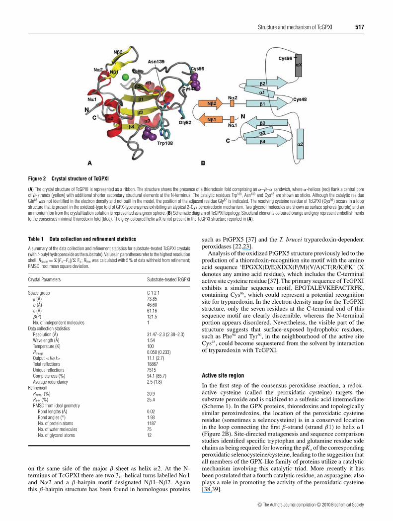
Figure 1 Two-dimensional 1H,15N-HSQC spectra of wild-type TcGPXI
1H,15N-HSQC spectra of 13C,15N-labelled TcGPXI in the oxidized (black) and reduced (red)forms. The protein samples were prepared in a buffer containing 50 mM sodium phosphate,pH 6.5, 300 mM NaCl. The reduced form additionally contains 10 mM DTT.
model of TcGPXI based on the template structure of oxidizedpoplar PtGPX5 (Populus trichocarpaxdeltoides thioredoxinperoxidase 5; 42% sequence identity to TcGPXI; PDB code2P5R). The initial model did not include any surface loops orpoorly structured regions.
Structure refinement was carried out using the maximum-likelihood restrained method with simple scaling in theREFMAC/CCP4 program [31] and simulated annealing torsionangle dynamics with the maximum likelihood function inCNS (Crystallography & NMR System) [32]. Real-spacerefinement/manual-fitting of the electron density map and model-building were carried out using the COOT tool [33].
For the prediction of potential ligand- or protein-partner-binding sites the TcGPXI crystal co-ordinates (edited to removesolvent water and other ligands present in the refined model)were submitted to the Q-SiteFinder server (http://www.modelling.leeds.ac.uk/qsitefinder/) [34].
RESULTS
NMR spectroscopy of oxidized TcGPXI
The two-dimensional 1H,15N-HSQC spectrum of purified wild-type TcGPXI showed cross-peaks that were well-dispersed, ofapproximately uniform line-width and relatively homogenousintensity. The spectrum contains in total approx. 170 cross-peaks, with a maximum of 154 peaks from backbone amidegroups (i.e. a total of 164 residues including ten prolineresidues). Complete analysis of the triple-resonance spectrarecorded for a 13C,15N-labelled sample yielded the definitiveassignment of 148 (96% completed amide resonance assignment)amino acid residues in the 1H,15N-HSQC spectrum of wild-type TcGPXI [Figure 1, Supplementary Figure S2 (available athttp://www.BiochemJ.org/bj/425/bj4250513add.htm)]. We wereunable to assign peak positions for the amide groups of theCys48, Asn78, Thr97, Gly113, Ser114 and Lys134 residues in the two-dimensional 1H,15N-HSQC spectrum, but overall a total of 99%of the 13Cα chemical shifts, 91% of the 13Cβ chemical shifts,97% of the 13CO chemical shifts and 99 % of the 1Hα chemicalshifts were assigned.
Detection of disulfide bonds in wild-type TcGPXI
It has been shown previously that the chemical shift values ofCα and Cβ resonances can be used to predict the redox stateof cysteine residues [35,36]. When surveyed over NMR resonanceassignments for proteins with known three-dimensional structurethe chemical shift ranges for the Cα and Cβ of a reducedcysteine were found to be 59.3 +− 3.2 p.p.m. and 28.4 +− 2.4 p.p.m.respectively, whereas the chemical shift ranges for Cα and Cβ ofan oxidized cysteine were 55.5 +− 2.5 p.p.m. and 40.7 +− 3.8 p.p.m.respectively. Wild-type TcGPXI has three cysteine residues:Cys48, designated the catalytic cysteine [9], Cys77 and Cys96.As no amide resonance could be identified for Cys48 in the 1H,15N-HSQC spectrum, the i-1 correlations connected to the NHcross peak of Gly49 were examined. For Cys77 the Cα (58.1 p.p.m.)and Cβ (29.9 p.p.m.) chemical shifts were consistent with that of areduced, thiol state. However, for Cys48 (Cα 56.4 p.p.m.; Cβ 42.0p.p.m.) and Cys96 (Cα 56.3 p.p.m.; Cβ 43.3 p.p.m.) the chemicalshifts were consistent with that of the oxidized state, suggestingthe presence of a disulfide bond between Cys48 and Cys96. Wesurmise that the TcGPXI protein became oxidized during thepurification and remained in that state throughout the NMR dataacquisition.
NMR-based secondary structure prediction for oxidized TcGPXI
The individual chemical shifts for the Cα, Cβ, CO and Hαatoms of the oxidized sample of TcGPXI were analysed using theCSI program [26] to assess the secondary structure composition.The CSI prediction for oxidized wild-type TcGPXI contains fiveβ-strands and four α-helices (see Supplementary Figure S3 avail-able at http://www.BiochemJ.org/bj/425/bj4250513add.htm).The following regions were identified as β-strands: Gln20–Ala23, Leu39–Ala45, Phe70–Phe75, Ser142–Ile145 and Val150–Phe155.The regions interpreted as α-helical are: Ser32–His34, Gly53–Gly67, Pro118–Thr126 and Val161–Lys166. Thus the CSI predictionfor TcGPXI yields a β–α–β–α–β–α–β–β–α compositionthat is consistent with a three-dimensional thioredoxin fold.Interestingly, the region Ala88–Phe99, which is identified as helicalin the three-dimensional structures of the reduced forms ofhomologous enzymes (PtGPX5 and TbPxII), but as an extendedloop in the oxidized state of the same proteins [22,23,37],is predicted by the CSI analysis to adopt a non-regular loopconformation in the solution structure of TcGPXI.
NMR spectroscopy of reduced TcGPXI
Although possessing essentially similar overall chemical shiftdispersion to the spectrum of the oxidized protein, thetwo-dimensional 1H,15N-HSQC spectrum of reduced TcGPXIcontained fewer cross-peaks, with apparently broader lines inthe central, random-coil region and more variable peak-to-peakintensity variation (Figure 1). In order to obtain the sequence-specific resonance assignment of reduced TcGPXI two triple-resonance experiments, HNCO and HN(CO)CA, were acquired.Although these two experiments are the most sensitive of thetriple-resonance experiments, analysis of the HN(CO)CA datashowed few Cα correlations in the three-dimensional spectrumoutside of the more intense cross-peaks in the random-coil regionof the 1H,15N-HSQC spectrum. This characteristic, which musthave arisen from accelerated transverse relaxation rates for amajor fraction of the NMR resonances, limited the utility ofperforming further triple-resonance NMR experiments. Instead,the assignment of the reduced TcGPXI protein was carried
c© The Authors Journal compilation c© 2010 Biochemical Society
516 S. Patel and others
out by comparison of the two-dimensional 1H,15N-HSQC andthree-dimensional HNCO spectra with those obtained for theoxidized protein. Assignments were transferred by inspection forthose NH cross-peaks that were well resolved from other amidecross-peaks, and that overlaid with assigned cross-peaks in theoxidized 1H,15N-HSQC spectrum. For cross-peaks where therewas ambiguity in the assignment through the direct comparisonwith the oxidized TcGPXI spectrum, the closest match of thecorresponding carbonyl carbon chemical shifts was chosen, unlessmore than one candidate cross-peak lay within 1 p.p.m. ofthe carbonyl carbon shift. This protocol resulted in tentativesequence-specific backbone assignment of the reduced wild-typespectrum for 86 out of the 148 residues assigned in the oxidizedspectrum. Mapping assigned amide resonances in the reducedTcGPXI 1H,15N-HSQC spectrum upon the secondary structureprediction of the oxidized protein reveals that those residues thatwere not assigned were located in the predicted loop regionssurrounding the active site cysteine residues. The appearanceof the reduced TcGPXI spectrum is thus consistent with a pic-ture in which the overall folding topology of TcGPXI ismaintained in the reduced state, but with an active site regiondemonstrating dynamic flexibility and chemical exchange onthe intermediate (μs–ms) timescale. A similar observation wasrecently reported for the reduced state of the closely homologousTbPxIII protein [22].
NMR spectroscopy of TcGPXI cysteine mutants
In an attempt to produce a model of the reduced TcGPXI structurethat would be more generally tractable to NMR spectroscopicstudies, site-directed mutagenesis to replace Cys48 or Cys96 withglycine residues was performed. We expected that in the absenceof the disulfide bridge these mutant proteins would adopt amore reduced-like conformation. In contrast, superposition of thetwo-dimensional 1H,15N-HSQC spectra of the C96G and C48Gmutants with reduced and oxidized wild-type TcGPXI spectrashows that the mutants appear more similar to the oxidizedwild-type TcGPXI (see Supplementary Figure S4 availableat http://www.BiochemJ.org/bj/425/bj4250513add.htm). Consid-ering that the reduced protein showed clear indications ofincreased flexibility from conformational-exchange-induced line-broadening it appears that the mutation of the TcGPXI active sitecysteine residues distinctly favours an oxidized-like conformationof the active site. Moreover this observation indicates that theoxidized-like conformation might be accessible to the reducedwild-type TcGPXI.
Enzyme assays
The catalytic activity of the recombinant wild-type TcGPXIprotein, which was used for the crystallization and NMRspectroscopy studies, was confirmed by enzyme assays utilizingboth tryparedoxin- and glutathione-dependent reactions (see Sup-plementary Figure S5 available at http://www.BiochemJ.org/bj/425/bj4250513add.htm). In the tryparedoxin-linked assay, usingt-butyl hydroperoxide as the substrate in a concentration rangeof 3–100 μM, the Eadie–Hofstee plot for the wild-type TcGPXIreaction yielded values of Vmax = 1300 +− 160 nmol · min−1 · mg−1,corresponding to a kcat = 24 +− 3 min−1 and Km = 51 +− 10 μM,with kcat/Km of 0.5 +− 0.2 min−1 · μM−1. These values areessentially equivalent to those reported for GPXI and cumenehydroperoxide under similar conditions [16]. As expected,activity in the glutathione-dependent assay for the reactionof wild-type TcGPXI with t-butyl hydroperoxide was much
lower with Vmax = 360 +− 20 nmol · min−1 · mg−1, correspondingto kcat = 6.6 +− 0.3 min−1 and Km = 52 +− 4 μM, with kcat/Km =0.13 +− 0.02 min−1 · μM−1. Although the assay was carried outat conditions identical with those in Wilkinson et al. [9] theapparent reaction rate and Km were both higher than reportedpreviously. Considering the possible experimental errors whenmeasuring such a low activity and taking into account that theestimated Km value corresponds well with that reported for the T.brucei orthologue, TbPxIII (47 μM) [17], the discrepancy withthe parameters determined previously was not pursued further.
In contrast with the wild-type TcGPXI protein the C48G andC96G mutant enzymes exhibited no activity in either assay format.On the other hand, conservative mutation of Cys77 (C77G andC77S) did not significantly perturb the catalytic function (resultsnot shown).
Protein crystallization
Wild-type TcGPXI was initially crystallized from the unbufferedammonium sulphate solution. These crystals took a long timeto grow, sometimes up to 2 months, and despite testing ofa range of additives it proved impossible to find conditionsfor reproducible crystal growth for the protein in this form.The parallel NMR investigation revealed that TcGPXI has apronounced tendency to oxidize by forming the Cys48–Cys96
disulfide. This observation suggested that the crystals obtainedprobably contained the oxidized form even though the proteinhad been purified under nominally reducing conditions. Mostof the crystals obtained at this stage grew in clusters. X-raydiffraction was performed using one of the individual crystals;however, the data were of poor quality and only to 3 Åresolution. In order to more carefully control the oxidationprocess a t-butyl hydroperoxide-treated protein sample was usedfor crystallization. Under these conditions, large, prism-shapedcrystals (∼0.2 mm) grew reproducibly within 2 days.
Description of the X-ray crystal structure of substrate-treatedTcGPXI
The X-ray structure of substrate-oxidized TcGPXI wasdetermined by molecular replacement using data at a resolution of2.3 Å and refined to a crystallographic Rfactor and Rfree of 20.9% and25.4% respectively (Table 1). Following iterative refinement allresidues were clearly identified in the difference electron densitymap except for Gln83–Glu93, which were not included in the finalmodel (Figure 2A). Additionally, two molecules of glycerol andan ammonium ion were identified in the electron density map.The final model contains 75 water molecules.
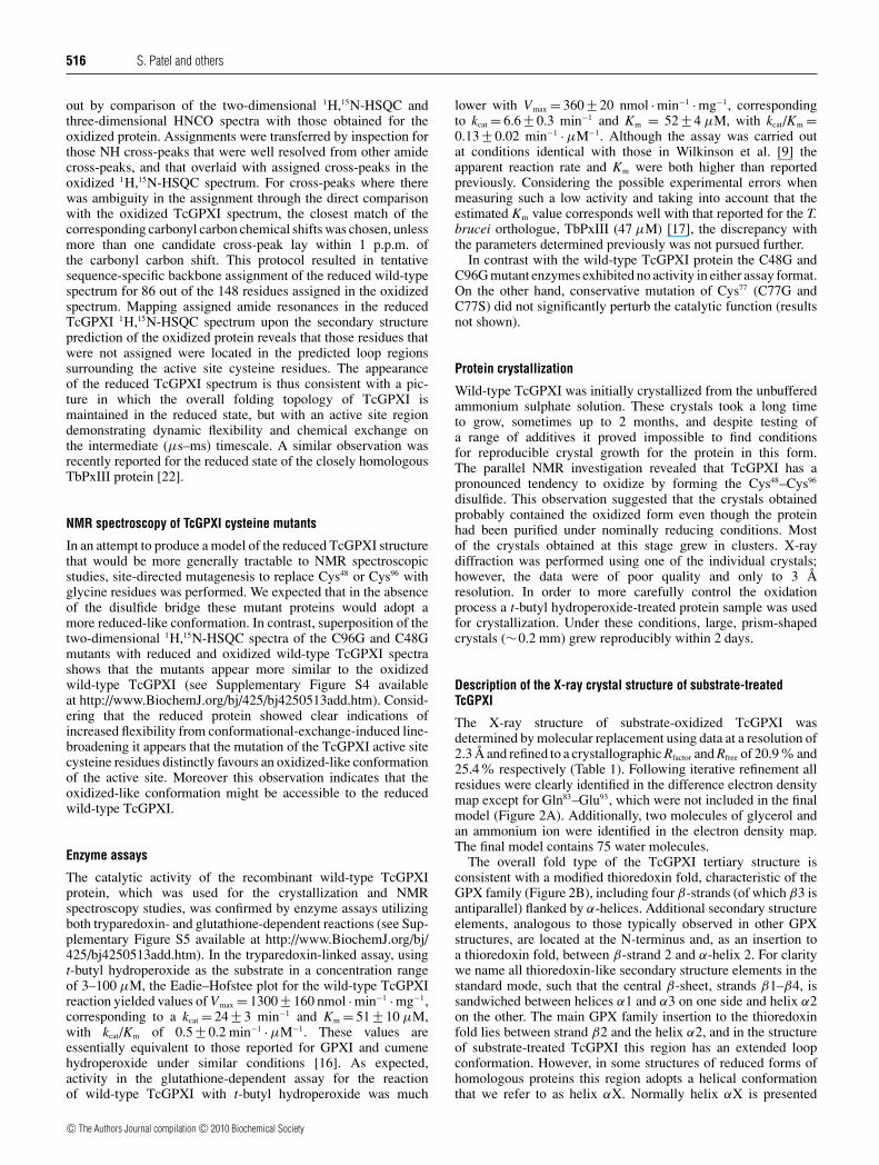
The overall fold type of the TcGPXI tertiary structure isconsistent with a modified thioredoxin fold, characteristic of theGPX family (Figure 2B), including four β-strands (of which β3 isantiparallel) flanked by α-helices. Additional secondary structureelements, analogous to those typically observed in other GPXstructures, are located at the N-terminus and, as an insertion toa thioredoxin fold, between β-strand 2 and α-helix 2. For claritywe name all thioredoxin-like secondary structure elements in thestandard mode, such that the central β-sheet, strands β1–β4, issandwiched between helices α1 and α3 on one side and helix α2on the other. The main GPX family insertion to the thioredoxinfold lies between strand β2 and the helix α2, and in the structureof substrate-treated TcGPXI this region has an extended loopconformation. However, in some structures of reduced forms ofhomologous proteins this region adopts a helical conformationthat we refer to as helix αX. Normally helix αX is presented
c© The Authors Journal compilation c© 2010 Biochemical Society
Structure and mechanism of TcGPXI 517
Figure 2 Crystal structure of TcGPXI
(A) The crystal structure of TcGPXI is represented as a ribbon. The structure shows the presence of a thioredoxin fold comprising an α–β–α sandwich, where α-helices (red) flank a central coreof β-strands (yellow) with additional shorter secondary structural elements at the N-terminus. The catalytic residues Trp138, Asn139 and Cys48 are shown as sticks. Although the catalytic residueGln83 was not identified in the electron density and not built in the model, the position of the adjacent residue Gly82 is indicated. The resolving cysteine residue of TcGPXI (Cys96) occurs in a loopstructure that is present in the oxidized-type fold of GPX-type enzymes exhibiting an atypical 2-Cys peroxiredoxin mechanism. Two glycerol molecules are shown as surface spheres (purple) and anammonium ion from the crystallization solution is represented as a green sphere. (B) Schematic diagram of TcGPXI topology. Structural elements coloured orange and grey represent embellishmentsto the consensus minimal thioredoxin fold (blue). The grey-coloured helix αX is not present in the TcGPXI structure reported in (A).
Table 1 Data collection and refinement statistics
A summary of the data collection and refinement statistics for substrate-treated TcGPXI crystals(with t -butyl hydroperoxide as the substrate). Values in parentheses refer to the highest resolutionshell. R factor = �|F o–F c|/� Fo; R free was calculated with 5 % of data withheld from refinement;RMSD, root mean square deviation.
Crystal Parameters Substrate-treated TcGPXI
Space group C 1 2 1a (A) 73.85b (A) 46.60c (A) 61.16β(◦) 121.5No. of independent molecules 1
Data collection statisticsResolution (A) 31.47–2.3 (2.38–2.3)Wavelength (A) 1.54Temperature (K) 100Rmerge 0.050 (0.233)Output <I/σ I> 11.1 (2.7)Total reflections 18867Unique reflections 7515Completeness (%) 94.1 (85.7)Average redundancy 2.5 (1.8)
RefinementR factor (%) 20.9Rfree (%) 25.4RMSD from ideal geometry
Bond lengths (A) 0.02Bond angles (◦) 1.93No. of protein atoms 1187No. of water molecules 75No. of glycerol atoms 12
on the same side of the major β-sheet as helix α2. At the N-terminus of TcGPXI there are two 310-helical turns labelled Nα1and Nα2 and a β-hairpin motif designated Nβ1–Nβ2. Againthis β-hairpin structure has been found in homologous proteins
such as PtGPX5 [37] and the T. brucei tryparedoxin-dependentperoxidases [22,23].
Analysis of the oxidized PtGPX5 structure previously led to theprediction of a thioredoxin-recognition site motif with the aminoacid sequence ‘EPGXX(D/E)XIXX(F/M)(V/A)CT(R/K)FK’ (Xdenotes any amino acid residue), which includes the C-terminalactive site cysteine residue [37]. The primary sequence of TcGPXIexhibits a similar sequence motif, EPGTALEVKEFACTRFK,containing Cys96, which could represent a potential recognitionsite for tryparedoxin. In the electron density map for the TcGPXIstructure, only the seven residues at the C-terminal end of thissequence motif are clearly discernible, whereas the N-terminalportion appears disordered. Nevertheless, the visible part of thestructure suggests that surface-exposed hydrophobic residues,such as Phe94 and Tyr50, in the neighbourhood of the active siteCys48, could become sequestered from the solvent by interactionof tryparedoxin with TcGPXI.
Active site region
In the first step of the consensus peroxidase reaction, a redox-active cysteine (called the peroxidatic cysteine) targets thesubstrate peroxide and is oxidized to a sulfenic acid intermediate(Scheme 1). In the GPX proteins, thioredoxins and topologicallysimilar peroxiredoxins, the location of the peroxidatic cysteineresidue (sometimes a selenocysteine) is in a conserved locationin the loop connecting the first β-strand (strand β1) to helix α1(Figure 2B). Site-directed mutagenesis and sequence comparisonstudies identified specific tryptophan and glutamine residue sidechains as being required for lowering the pKa of the correspondingperoxidatic selenocysteine/cysteine, leading to the suggestion thatall members of the GPX-like family of proteins utilize a catalyticmechanism involving this catalytic triad. More recently it hasbeen postulated that a fourth catalytic residue, an asparagine, alsoplays a role in promoting the activity of the peroxidatic cysteine[38,39].
c© The Authors Journal compilation c© 2010 Biochemical Society

518 S. Patel and others
Scheme 1 Peroxidase reaction
The peroxidatic and resolving cysteine residues are labelled with (P) and (R) respectively. Theproduct of the first step of reaction is sulfenic acid and the second step of the reaction leadsto formation of a disulfide bond. In the atypical 2-Cys mechanism (P) and (R) are on the samepolypeptide chain. In the context of the present paper the reductant is tryparedoxin or glutathione.
In the crystal structure of TcGPXI Cys48, which clearlyrepresents the peroxidatic cysteine, Trp138 and Asn139 are allwell-resolved in the electron density map. However, the putativecatalytic glutamine residue is localized in the loop region that isonly partially built due to the disorder reflected in the diffuseelectron density and thus Gln83 is absent in the model. Theposition of Gln83 can be estimated from the position of theadjacent surface-exposed residue Gly82, which is separated by9.2 Å (Cα–Cα distance) from the surface-exposed residue Trp138
(Figure 3). Overall, the active site appears open and solventexposed. Crucially, in the conformation of the active site weobserve that the Cys48 residue is significantly (>9 Å) removedfrom the residues proposed to be participating in its activation,Trp138, Gln83 and Asn139.
The second step of the consensus peroxidase reactionmechanism involves an attack by a resolving cysteine residueside chain on the sulfenic acid intermediate of the peroxidaticcysteine residue, and hence formation of a disulfide bond, whichis subsequently reduced by an appropriate electron donor. Ourmutagenesis results suggest that in TcGPXI Cys96 adopts therole of the resolving cysteine residue. In the crystal structurethis residue is located in an extended loop region followingthe strand β2. The conformation of the loop containing Cys96
places this residue in close proximity to Cys48, in a positionapparently competent to form the disulfide bond. This interactionis reminiscent of the proposed catalytic mechanism of atypical
2-Cys peroxiredoxins where the peroxidatic and resolvingcysteines are found on the same polypeptide chain.
In the observed TcGPXI crystal structure the active site cysteineresidues are reduced
As the TcGPXI protein was revealed by our NMR investigationto be readily oxidized to the Cys48–Cys96 disulfide-bonded form,and the protein formed well-ordered crystals following oxidationwith a peroxide substrate, we expected the electron density tobe consistent with the presence of a disulfide bond between thetwo cysteine side chains. Indeed, the absence of the helix αXin our structure, consistent with the secondary structure analysisbased upon chemical shifts, together with the similarity of thegeneral active site conformation to oxidized states of homologousstructures, suggest that it is the oxidized conformation of TcGPXIthat is amenable to crystallization.
Although the structure was solved by the molecularreplacement method using a structure of the oxidized form ofPtGPX5, to minimize model bias all the loop regions were deletedfrom the search model and the loops were subsequently builtthrough the iterative process of interpretation of the electrondensity map and refinement. Any model of the TcGPXI structurethat contained disulfide-linked Cys48–Cys96 showed an increasein the Rfree value and a relatively poor fit to the map, with clearnegative electron density between the two sulfur atoms in thedifference map. Refinement of the TcGPXI structure without theexplicit disulfide bond resulted in improved refinement statisticsand a model that fitted well into the electron density. The structureof the whole of the active site was additionally refined by severalrounds of calculation of the omit map for the separate regionsof the two loops containing the active site cysteine residues.We conclude that even though the crystallized protein was pre-oxidized by treatment with substrate, and hence overall theresulting model is indicative of a typical oxidized conformation, itappears that subsequently to crystal formation the disulfide bondwas reduced due to the effect of X-ray radiation. In the active siteconformation observed in the TcGPXI electron density (Figure 3),the peroxidatic cysteine residue, Cys48, and resolving cysteine
Figure 3 Structure of the TcGPXI active site
(A) Electron density for the weighted 2F o–F c map in the region of the active site residues is shown, viewed at 1.0 σ . Catalytic residues Cys48, Cys96, Trp138 and the Gly82 residue adjacent to catalyticresidue Gln83 are highlighted. Both cysteine residues are shown to exist in their reduced thiol form. (B) Close-up view of the redox-active cysteine residues of TcGPXI.
c© The Authors Journal compilation c© 2010 Biochemical Society

Structure and mechanism of TcGPXI 519
Figure 4 Glycerol molecule-binding sites in the active site region of TcGPXI
(A) Ribbon diagram of TcGPXI molecule with the two glycerol molecules shown explicitly as yellow stick models. The top-four-ranked potential ligand-binding sites from the Q-site finder analysisare represented as clusters of methyl probes (spheres). (B) Detailed view of ligand probe clusters and the residues surrounding them.
residue, Cys96, are in a close proximity; the distance between thesulfur atoms of the Cys48 and Cys96 side chains is 4.7 Å and onlysmall changes in side-chain torsion angles would be sufficient forthe two residues to form a disulfide bond. The crystal packingarrangement apparently constrains the two cysteine residues toremain in close proximity to one another. The phenomenon of X-ray induced reduction has been observed for other protein crystals,for example in the case of DsbG disulfide isomerase [40] and inthe radiation-cleaved structures of the N-terminal domain of theAhpF protein [41] and tryparedoxin [42]. Therefore although theactive site cysteine residues of TcGPXI are clearly in the reducedthiol state according to the electron density, the close proximityof these residues and the overall active site structure are morerepresentative of the oxidized protein conformation.
Glycerol molecules and putative ligand-binding sites
Two glycerol molecules associated with the surface of TcGPXIwere identified from the difference electron density map. Oneof the glycerol molecules is located between the peroxidatic andresolving cysteine residues near residue Tyr50, which flanks theactive site. The position of this glycerol molecule, so close tothe catalytic cysteine residues, is indicative of a crevice on themolecular surface that could otherwise be engaged in interactionwith tryparedoxin or another electron donor. The second glycerolmolecule is positioned on the surface region near residue Trp138
(Figure 4). The binding of this glycerol molecule is stabilizedthrough interaction with the side chains of Trp138 and Arg154, aswell as the backbone amide of Lys137. Trp138 is implicated incatalysis; the face of the indole that is opposite the glycerol-binding site lines the region postulated to comprise the activesite in the reduced conformation of TcGPXI, together with theperoxidatic Cys48, Asn139 and the putative catalytic residue Gln83.Interestingly, the side chain of Trp138 adopts a different rotamer
conformation in the structures of the homologous T. brucei[22] and poplar proteins [37]. Specifically, in an NMR solutionstructure of TbPxIII [22] the corresponding tryptophan side chainoccupies the space equivalent to the second glycerol-binding sitein TcGPXI.
Prediction of potential ligand-binding regions on the structureof oxidized TcGPXI was carried out with Q-SiteFinder [34]. Q-SiteFinder uses the interaction energy between the protein anda simple van der Waals probe to locate energetically favourablebinding sites. Clusters of favourable probe sites are ranked basedon their likelihood of identifying a binding site according tothe sum total binding energies for each cluster. When appliedto TcGPXI, the top three ranked regions for potential ligandinteraction were found in surface cavities around the peroxidaticCys48 and resolving Cys96 residues. Two out of the top threepotential ligand-interaction sites correspond to regions of thecrystal structure populated by glycerol molecules. Figure 4illustrates the location of putative ligand probes placed withinthe top four most probable ligand-binding regions. The potentialligand probe clusters designated I, II and III have a volume of222 Å3, 174 Å3 and 123 Å3 respectively. Potential binding site IIIis the most distant from the redox-active cysteine residues, it iswithin the space adjacent to Trp138 where a glycerol molecule isalso observed in the crystal structure of TcGPXI. Ligand probesite II is located near Cys48, in an extended cavity delineated byTrp138 and Asn139 on one side and Tyr50, Ala52 and Gly53 on theother. This region extends further and connects to the predictedfourth site (174 Å3), which reaches towards the missing loop in thestructure. The ‘missing’ part of the loop region between Gly82 andPhe94 might further contribute to the distinctive appearance of thispocket. The top-ranked putative binding site I partially overlapswith the second glycerol molecule in the TcGPXI crystal structure.This site comprises the surface pocket lined by Cys48 and Cys96,polar residues Thr51 and Thr97, hydrophobic residues Tyr50, Tyr55,Phe99, and charged residues Arg98 and Lys100. Interestingly, the
c© The Authors Journal compilation c© 2010 Biochemical Society

520 S. Patel and others
corresponding charged residues from this pocket (Lys97 and Lys99)were identified, through mutagenesis studies, as contributing tothe tryparedoxin interaction with TbPxIII [22].
Each of the top-ranked putative binding sites occupies arelatively small volume corresponding to potential ligandscomprised of approximately ten non-hydrogen atoms. Closeexamination of the identified sites, however, reveals that thetop two sites connect and extend towards site four, thus greatlyenlarging the potential space for binding of a drug-like molecule.The proximity of the predicted ligand-binding sites to the redox-active cysteine residues legitimizes targeting of these surfaces inthe design of potential inhibitors that would either stabilize theoxidized conformation of TcGPXI or interfere with tryparedoxinbinding.
DISCUSSION
Conformational changes associated with oxidation of GPX-likeenzymes utilizing the atypical 2-Cys mechanism
Over the last two years it has been demonstrated that thioredoxin-dependent poplar PtGPX5 and TbPxIII, which are closelyrelated to glutathione peroxidase-type enzymes, utilize a catalyticmechanism that involves formation of an intramolecular disulfidebridge, commonly associated with atypical 2-Cys peroxiredoxinproteins [43]. X-ray crystallographic investigation of PtGPX5revealed extensive conformational changes that accompany thetransition between the reduced and the oxidized states of theprotein. The reduced conformation of PtGPX5 exhibited atopology previously observed in other members of this structuralfamily, such as bovine GPXI ([44]; PDB 2F8A) and humanGPX1–4 (PDB 2F8A, 2HE3, 2R37, 2GS3 and 2OBI respectively),which includes helix αX between the strand β2 and helix α2 of acanonical thioredoxin fold. Unlike the selenocysteine members ofthe family, the PtGPX5 sequence contains an additional cysteineresidue within the helix αX region and it is this residue thatprovides the role of the resolving cysteine in the 2-Cys catalyticmechanism. In the reduced PtGPX5 structure, the two cysteineresidues are >15 Å apart; in the oxidized structure the tworesidues are brought closer by a change in conformation thatincludes unwinding of the C-terminal end of helix αX [37]. Theseresults point to the capacity for proteins in this family to undergosignificant conformational plasticity and dynamic exchange thatmust underpin the 2-Cys mechanism and ‘Ping Pong’ interactionswith the peroxide substrate and upstream reductant.
Recently there have been extensive investigations into thefunctional and structural properties of both TbPxIII and its closehomologue TbPxII (Trypanosoma brucei peroxidase II). Melcherset al. [22] obtained NMR solution structures of the oxidizedand reduced form of TbPxIII. Although the NMR spectrumof the reduced form of TbPxIII is characterized by significantchemical shift differences with respect to the oxidized protein anddisappearance of several cross-peaks due to exchange broadening,the derived three-dimensional solution structures of the oxidizedand reduced forms of the protein are essentially the same. AnX-ray crystal structure of the oxidized form reveals a structurethat correlates well with that obtained by solution NMR methods.Similar to the X-ray crystal structure of TcGPXI we obtainedin the present study, even the reduced form of TbPxIII showsthat the two active site cysteine residues of TbPxIII are in closeproximity and the reduced GPX-type helix αX is clearly absent. Inaddition, in the reduced active site the peroxidatic cysteine residueis not found to interact with the residues that were previouslysuggested to be involved in stabilizing the thiolate nucleophile[38,39]. Owing to a lack of unambiguous distance restraints to
the surrounding residues, the exact orientation of several residuesin the active site was not well-determined by the NMR data,although it was postulated that the active site cysteine residueactivation might occur through interaction of the side chain withthe backbone [22]. It was therefore proposed that the catalyticmechanism of TbPxIII differs fundamentally from that of anyof the previously characterized enzymes and that the T. bruceienzyme does not undergo such a large conformational changeas described for PtGPX5. Independent work by Alphey et al.[23] provided an alternative view into the structure of the T.brucei enzymes when they described an X-ray crystal structureof the reduced form of the TbPxII, an enzyme whose amino acidsequence differs from TbPxIII only at a single residue (threoninecompared with asparagine) at a position remote from the activesite. Whereas the cores of the TbPxII and TbPxIII structures super-pose closely, the reduced TbPxII active site conformation differssignificantly from that of the reduced TbPxIII NMR structure.Specifically, in the reduced form of TbPxII the resolving cysteineresidue is positioned very distantly from the peroxidatic cysteineresidue, and the consensus helix αX is present. Furthermore,the peroxidatic cysteine is engaged in a network of interactionswith the tryptophan, glutamine and asparagine residues that werepreviously identified as forming a catalytic tetrad [38,39]. Thuson the basis of the crystal structure of reduced TbPxII, Alpheyet al. [23] declared that the catalytic mechanism of the T. bruceienzyme(s) is similar to that described for PtGPX5 [37].
Structural plasticity of T. cruzi GPXI
The apparently contradictory reports either provide support for,or explicitly dismiss, the occurrence of a large conformationalchange in an atypical 2-Cys peroxidase mechanism for theTbPxII/III enzymes. It is highly unlikely that the discrepancycan be attributed to the single amino acid difference between thetwo proteins. The present study on TcGPXI, which exhibits 72%sequence identity with TbPxIII, suggests that both Cys48 and Cys96
are required for the reaction mechanism. The NMR spectrumof the oxidized state is consistent with a stable fold in whichthe loops containing these residues are tethered by the disulfidecross-link. The NMR spectrum of reduced TcGPXI displayssignificant line-broadening and loss of resonances for the majorityof residues within the regions surrounding the active site. Theseresults demonstrate that the reduced state of TcGPXI can adoptmultiple conformations that exchange on the μs–ms timescaleindicating the presence of an equilibrium between conformationalsub-states separated by one or more thermal activation barriers.In their solution NMR studies, Melchers et al. [22] reportedthat the reduced form of TbPxIII also displayed a spectrumconsistent with a μs–ms timescale exchange. It is tempting toargue that in both cases the spectra reveal that the reducedstates of these proteins can sample a range of conformationsthat spans the type of arrangements observed in the crystalstructures of reduced PtGPX5 and TbPxII, in which the helixαX is formed and the active site cysteine residues are separatedby >10 Å, as well as conformations in which the resolvingcysteine residue can approach the peroxidatic cysteine residue,as it must when acting to resolve the sulfenic acid intermediatestate in the reaction pathway. In this context, the conformationreported in the present paper for the crystal structure of TcGPXI,which we attribute to X-ray-induced modification of crystals of‘oxidized’ protein, represents a snapshot of an extreme of theconformational landscape for the reduced state of the protein.Notably, in the TcGPXI structure the crystallographic two-foldaxis is very close to the active site and interactions between
c© The Authors Journal compilation c© 2010 Biochemical Society

Structure and mechanism of TcGPXI 521
symmetry-related molecules presumably provide constraints onthe conformation of the extended sequence region containing theresolving cysteine, limiting the potential of the protein to undergomore significant rearrangement upon reduction of the disulfidebond. Moreover, the finding that the conformational exchange-broadening of the NMR spectrum of reduced TcGPXI is largelyrelieved by mutation of either Cys48 or Cys96 to glycine is furtherevidence that the ‘untethered’ protein has an intrinsic capacityto explore different configurations. The fact that the spectrum ofeach of the mutants appears rather similar to that of the oxidizedwild-type protein perhaps suggests that in the conformationalequilibrium, which must exist for the reduced protein, the energylandscape is biased in favour of conformations that resemblethe oxidized protein, a conclusion that would be consistent withthe observation that the NMR-derived solution structure of thereduced TbPxIII protein (despite the presence of conformationalexchange) emerges with an overall conformation similar to that ofthe oxidized state [22]. Extrapolation of this analysis leads to theprediction that the conformations of reduced PtGPX5 and TbPxII‘trapped’ by crystallization represent the opposite extremes ofthe underlying conformational equilibrium, possibly stabilizedby specific crystal packing forces.
Concluding remarks
The present results provide a platform upon which to build a moreextensive study of TcGPXI. To this end, we have found that intitrations of the Cys→Gly mutant forms of TcGPXI with t-butylhydroperoxide, no changes in the 1H,15N chemical shifts occureven at a 1.5 mM concentration (results not shown). Althoughfurther exploration of this observation is needed, it appears thatthe protein conformations favoured by the Cys→Gly mutants,which might be closer to that of the oxidized protein, exhibita very poor affinity for the peroxide substrate. It is anticipatedthat a truly reduced-type conformation would bind the substratewith an affinity comparable with that of the Km (approx. 50 μM).Poor affinity for the substrate of an oxidized-type conformationis consistent with the Ping Pong reaction mechanism that isdisplayed by TcGPXI and the T. brucei homologues [16,17].Moreover, we analysed the molecular surface of the oxidized-type conformation in the TcGPXI crystal structure and identifiedsurface cavities that could be targeted for therapeutic intervention.Structure-led design of small molecules that preferentially bindthe specific conformation might provide a novel avenue forinhibiting the peroxidase activity of this enzyme by interferingwith the interaction of the upstream reducing agent.
AUTHOR CONTRIBUTION
Shreenal Patel carried out main body of the experimental work as a part of her Ph.D.thesis. Syeed Hussain was closely involved in enzymatic assays and analysis of kineticparameters; he also obtained initial crystals of the wild-type GPXI. Richard Harris wasinvolved in NMR spectroscopy data acquisition and processing. Sunita Sardiwal assistedin production of mutant proteins and protein purification. John Kelly and Shane Wilkinsonoriginally cloned and characterized GPXI and provided assistance with activity assaysand with their expertise in T. cruzi biology. Paul Driscoll provided leadership in the NMRaspect of the project, including in-depth interpretation of data. Snezana Djordjevic wasthe principal investigator on the project and provided X-ray crystallographic expertise.
ACKNOWLEDGEMENTS
We thank Dr Claire Bagneris (School of Crystallography, Birkbeck College, London, U.K.)for the initial work on recombinant expression of TcGPXI and Dr Gary Parkinson (LondonSchool of Pharmacy, London, U.K.) for assistance with collection and interpretation ofX-ray data. We gratefully acknowledge the staff of the Medical Research Council National
NMR Centre at Mill Hill for access to the 800 MHz spectrometer. We thank to Dr JohnChristodoulou (Institute of Structural and Molecular Biology, University College London,London, U.K.) for helpful discussion.
FUNDING
This work was supported by the Medical Research Council, U.K. [grant number G0401079].The Medical Research Council file reference for P. C. D. is U117574559.
REFERENCES
1 Stuart, K., Brun, R., Croft, S., Fairlamb, A., Gurtler, R. E., McKerrow, J., Reed, S. andTarleton, R. (2008) Kinetoplastids: related protozoan pathogens, different diseases.J. Clin. Invest. 118, 1301–1310
2 Krauth-Siegel, R. L., Bauer, H. and Schirmer, R. H. (2005) Dithiol proteins as guardians ofthe intracellular redox milieu in parasites: old and new drug targets in trypanosomes andmalaria-causing plasmodia. Angew. Chem. Int. Ed. Engl. 44, 690–715
3 Castro, J. A., de Mecca, M. M. and Bartel, L. C. (2006) Toxic side effects of drugs used totreat Chagas disease (American trypanosomiasis). Hum. Exp. Toxicol. 25, 471–479
4 Castro, H. and Tomas, A. M. (2008) Peroxidases of trypanosomatids. Antioxid. RedoxSignal. 10, 1593–1606
5 Zhang, Y., Bond, C. S., Bailey, S., Cunningham, M. L., Fairlamb, A. H. and Hunter, W. N.(1996) The crystal structure of trypanothione reductase from the human pathogenTrypanosoma cruzi at 2.3 A resolution. Protein Sci. 5, 52–61
6 Bond, C. S., Zhang, Y., Berriman, M., Cunningham, M. L., Fairlamb, A. H. and Hunter,W. N. (1999) Crystal structure of Trypanosoma cruzi trypanothione reductase in complexwith trypanothione, and the structure-based discovery of new natural product inhibitors.Structure 7, 81–89
7 Pineyro, M. D., Pizarro, J. C., Lema, F., Pritsch, O., Cayota, A., Bentley, G. A. and Robello,C. (2005) Crystal structure of the tryparedoxin peroxidase from the human parasiteTrypanosoma cruzi. J. Struct. Biol. 150, 11–22
8 Rhee, S. G., Chae, H. Z. and Kim, K. (2005) Peroxiredoxins: a historical overview andspeculative preview of novel mechanisms and emerging concepts in cell signaling. FreeRadical Biol. Med. 38, 1543–1552
9 Wilkinson, S. R., Meyer, D. J. and Kelly, J. M. (2000) Biochemical characterization of atrypanosome enzyme with glutathione-dependent peroxidase activity. Biochem. J. 352,755–761
10 Flohe, L., Budde, H. and Hofmann, B. (2003) Peroxiredoxins in antioxidant defense andredox regulation. Biofactors 19, 3–10
11 Tetaud, E., Giroud, C., Prescott, A. R., Parkin, D. W., Baltz, D., Biteau, N., Baltz, T. andFairlamb, A. H. (2001) Molecular characterisation of mitochondrial and cytosolictrypanothione-dependent tryparedoxin peroxidases in Trypanosoma brucei. Mol.Biochem. Parasitol. 116, 171–183
12 Alphey, M. S., Bond, C. S., Tetaud, E., Fairlamb, A. H. and Hunter, W. N. (2000) Thestructure of reduced tryparedoxin peroxidase reveals a decamer and insight into reactivityof 2-Cys-peroxiredoxins. J. Mol. Biol. 300, 903–916
13 Declercq, J. P., Evrard, C., Clippe, A., Stricht, D. V., Bernard, A. and Knoops, B. (2001)Crystal structure of human peroxiredoxin 5, a novel type of mammalian peroxiredoxin at1.5 A resolution. J. Mol. Biol. 311, 751–759
14 Seo, M. S., Kang, S. W., Kim, K., Baines, I. C., Lee, T. H. and Rhee, S. G. (2000)Identification of a new type of mammalian peroxiredoxin that forms an intramoleculardisulfide as a reaction intermediate. J. Biol. Chem. 275, 20346–20354
15 Wilkinson, S. R. and Kelly, J. M. (2003) The role of glutathione peroxidases intrypanosomatids. Biol. Chem. 384, 517–525
16 Wilkinson, S. R., Meyer, D. J., Taylor, M. C., Bromley, E. V., Miles, M. A. and Kelly, J. M.(2002) The Trypanosoma cruzi enzyme TcGPXI is a glycosomal peroxidase and can belinked to trypanothione reduction by glutathione or tryparedoxin. J. Biol. Chem. 277,17062–17071
17 Hillebrand, H., Schmidt, A. and Krauth-Siegel, R. L. (2003) A second class of peroxidaseslinked to the trypanothione metabolism. J. Biol. Chem. 278, 6809–6815
18 Wilkinson, S. R., Horn, D., Prathalingam, S. R. and Kelly, J. M. (2003) RNA interferenceidentifies two hydroperoxide metabolizing enzymes that are essential to the bloodstreamform of the African trypanosome. J. Biol. Chem. 278, 31640–31646
19 Schlecker, T., Schmidt, A., Dirdjaja, N., Voncken, F., Clayton, C. and Krauth-Siegel, R. L.(2005) Substrate specificity, localization, and essential role of the glutathioneperoxidase-type tryparedoxin peroxidases in Trypanosoma brucei. J. Biol. Chem. 280,14385–14394
20 Wilkinson, S. R., Temperton, N. J., Mondragon, A. and Kelly, J. M. (2000) Distinctmitochondrial and cytosolic enzymes mediate trypanothione-dependent peroxidemetabolism in Trypanosoma cruzi. J. Biol. Chem. 275, 8220–8225
21 Schlecker, T., Comini, M. A., Melchers, J., Ruppert, T. and Krauth-Siegel, R. L. (2007)Catalytic mechanism of the glutathione peroxidase-type tryparedoxin peroxidase ofTrypanosoma brucei. Biochem. J. 405, 445–454
c© The Authors Journal compilation c© 2010 Biochemical Society

522 S. Patel and others
22 Melchers, J., Diechtierow, M., Feher, K., Sinning, I., Tews, I., Krauth-Siegel, R. L. andMuhle-Goll, C. (2008) Structural basis for a distinct catalytic mechanism in Trypanosomabrucei tryparedoxin peroxidase. J. Biol. Chem. 283, 30401–30411
23 Alphey, M. S., Konig, J. and Fairlamb, A. H. (2008) Structural and mechanistic insightsinto type II trypanosomatid tryparedoxin-dependent peroxidases. Biochem. J. 414,375–381
24 Delaglio, F., Grzesiek, S., Vuister, G. W., Zhu, G., Pfeifer, J. and Bax, A. (1995) NMRPipe: amultidimensional spectral processing system based on UNIX pipes. J. Biomol. NMR 6,277–293
25 Kraulis, P. J. (1989) ANSIG: a program for the assignment of protein protontwo-dimensional NMR spectra by interactive computer graphics. J. Mag. Res. 24,627–633
26 Wishart, D. S. and Sykes, B. D. (1994) The 13C chemical-shift index: a simple method forthe identification of protein secondary structure using 13C chemical-shift data. J. Biomol.NMR 4, 171–180
27 Hofstee, B. H. J. (1959) Non-inverted versus inverted plots in enzyme kinetics. Nature184, 1296–1298
28 Pflugrath, J. W. (1999) The finer things in X-ray diffraction data collection. ActaCrystallogr. D Biol. Crystallogr. 55, 1718–1725
29 Reference deleted30 Vagin, A. and Teplyakov, A. (1997) MOLREP an automated program for molecular
replacement. J. Appl. Cryst. 30, 1022–102531 Murshudov, G. N., Vagin, A. A. and Dodson, E . J. (1997) Refinement of macromolecular
structures by the maximum-likelihood method. Acta Crystallogr. D Biol. Crystallogr. 53,240–255
32 Rice, L. M. and Brunger, A. T. (1994) Torsion angle dynamics: reduced variableconformational sampling enhances crystallographic structure refinement. Proteins 19,277–290
33 Emsley, P. and Cowtan, K. (2004) Coot: model-building tools for molecular graphics. ActaCrystallogr. D Biol. Crystallogr. 60, 2126–2132
34 Laurie, A. T. and Jackson, R. M. (2005) Q-SiteFinder: an energy-based method for theprediction of protein–ligand-binding sites. Bioinformatics 21, 1908–1916
35 Sharma, D. and Rajarathnam, K. (2000) 13C NMR chemical shifts can predict disulfidebond formation. J. Biomol. NMR 18, 165–171
36 Wang, C., Chen, J., Yin, S. and Chuang, W. (2006) Predicting the redox state andsecondary structure of cysteine residues in proteins using NMR chemical shifts. Proteins63, 219 –226
37 Koh, C. S., Didierjean, C., Navrot, N., Panjikar, S., Mulliert, G., Rouhier, N., Jacquot,J. P., Aubry, A., Shawkataly, O. and Corbier, C. (2007) Crystal structures of a poplarthioredoxin peroxidase that exhibits the structure of glutathione peroxidases: insightsinto redox-driven conformational changes. J. Mol. Biol. 370,512–529
38 Tosatto, S. C., Bosello, V., Fogolari, F., Mauri, P., Roveri, A., Toppo, S., Flohe, L., Ursini, F.and Maiorino, M. (2008) The catalytic site of glutathione peroxidases. Antioxid. RedoxSignal. 10, 1515–1526
39 Maiorino, M., Ursini, F., Bosello, V., Toppo, S., Tosatto, S. C., Mauri, P., Becker, K.,Roveri, A., Bulato, C., Benazzi, L. et al. (2007) The thioredoxin specificity of DrosophilaGPx: a paradigm for a peroxiredoxin-like mechanism of many glutathione peroxidases. J.Mol. Biol. 365, 1033–1046
40 Heras, B., Edeling, M. A., Schirra, H. J., Raina, S. and Martin, J. L. (2004) Crystalstructures of the DsbG disulfide isomerase reveal an unstable disulfide. Proc. Natl. Acad.Sci. U.S.A. 101, 8876–8881
41 Roberts, B. R., Wood, Z. A., Jonsson, T. J., Poole, L. B. and Karplus, P. A. (2005) Oxidizedand synchrotron cleaved structures of the disulfide redox center in the N-terminal domainof Salmonella typhimurium AhpF. Protein Sci. 14, 2414–2420
42 Alphey, M. S., Gabrielsen, M., Micossi, E., Leonard, G. A., McSweeney, S. M., Ravelli,R. B., Tetaud, E., Fairlamb, A. H., Bond, C. S. and Hunter, W. N. (2003) Tryparedoxinsfrom Crithidia fasciculata and Trypanosoma brucei: photoreduction of the redox disulfideusing synchrotron radiation and evidence for a conformational switch implicated infunction. J. Biol. Chem. 278, 25919–25925
43 Wood, Z. A., Schroder, E., Harris, R. J. and Poole, L. B. (2003) Structure, mechanism andregulation of peroxiredoxins. Trends Biochem. Sci. 28, 32–40
44 Epp, O., Ladenstein, R. and Wendel, A. (1983) The refined structure of the selenoenzymeglutathione peroxidase at 0.2 nm resolution. Eur. J. Biochem. 133, 51–69
Received 30 July 2009/2 November 2009; accepted 3 November 2009Published as BJ Immediate Publication 3 November 2009, doi:10.1042/BJ20091167
c© The Authors Journal compilation c© 2010 Biochemical Society

Biochem. J. (2010) 425, 513–522 (Printed in Great Britain) doi:10.1042/BJ20091167
SUPPLEMENTARY ONLINE DATAStructural insights into the catalytic mechanism of Trypanosoma cruzi GPXI(glutathione peroxidase-like enzyme I)Shreenal PATEL*, Syeed HUSSAIN*, Richard HARRIS*, Sunita SARDIWAL*, John M. KELLY†, Shane R. WILKINSON‡,Paul C. DRISCOLL*§ and Snezana DJORDJEVIC*1
*Institute of Structural and Molecular Biology, Division of Biosciences, University College London, London WC1E 6BT, U.K., †Department of Infectious and Tropical Diseases, LondonSchool of Hygiene and Tropical Medicine, London WC1E 7HT, U.K., ‡School of Biological and Chemical Sciences, Queen Mary University of London, London E1 4NS, U.K., and§Division of Molecular Structure, Medical Research Council National Institute for Medical Research, The Ridgeway, Mill Hill, London NW7 1AA, U.K.
Figure S1 The trypanosomal oxidative defence mechanism
In the oxidative defence system of T. cruzi reduction of hydroperoxides occurs via the transfer of reducing equivalents from NADPH and dihydrotrypanothione (T[SH]2) to either: (i) GSH and GPXenzymes; (ii) ascorbate (ASC) and ascorbate-dependent peroxidase (APX); (iii) tryparedoxin (TXN) and cytosolic/mitochondrial peroxiredoxins (CPX/MPX) or GPX enzymes (present in cytosol,glycosomes or endoplasmic reticulum).
1 To whom correspondence should be addressed (email [email protected]).The chemical shift assignments for oxidized TcGPXI will appear in the BioMagResBank under accession number 16518. The atomic co-ordinates for
TcGPXI structure will appear in the PDB under ID 3E0U.
c© The Authors Journal compilation c© 2010 Biochemical Society

S. Patel and others
Figure S2 NMR sequence specific assignments of oxidised TcGPXI
Two-dimensional 1H,15N- HSQC spectrum of 13C, 15N-labelled TcGPXI in 50 mM sodium phosphate, pH 6.5, containing 300 mM NaCl. Sequence-specific backbone assignments were made for allresidues except Cys48, Asn78, Thr97, Gly113, Ser114 and Lys134. The side chain NH group of the lone tryptophan is denoted Wε138.
c© The Authors Journal compilation c© 2010 Biochemical Society

Structure and mechanism of TcGPXI
Figure S3 The chemical shift index for the secondary structure predictionof oxidised TcGPXI
Secondary chemical shift values for: (A) �Cα–�Cβ , (B) �CO and (C) �Hα. (D) CSIsecondary structure prediction for each residue based on Cα, Cβ , CO and Hα. β-strandregions are: Gln20–Ala23 (β1); Leu39–Ala45 (β2); Phe70–Phe75 (β3); Ser142–Ile145 (β4); andVal150–Phe155 (β5). Regions assigned α-helical are Ser32–His34 (α1); Gly53–Gly67 (α2);Pro118–Thr126 (α3); Val161–Lys166 (α4). Blank regions indicate the absence of a cross-peakor a residue that was not assigned.
Received 30 July 2009/2 November 2009; accepted 3 November 2009Published as BJ Immediate Publication 3 November 2009, doi:10.1042/BJ20091167
Figure S4 Superposition of two-dimensional 1H,15N-HSQC spectra of theC48G and C96G mutant and wild-type TcGPXI
Superposition of two-dimensional 1H,15N-HSQC spectra of the C48G mutant TcGPXI (magenta)with the C96G mutant (blue) and wild-type protein (black) in 50 mM sodium phosphate, pH 6.5,containing 300 mM NaCl. All three spectra overlap significantly.
Figure S5 Kinetic investigation of wild-type TcGPXI using the glutathione-dependent and tryparedoxin-dependent antioxidant pathway
Eadie–Hoftsee plot constructed from values obtained for the kinetic investigation of wild-typeTcGPXI in (A) glutathione-dependent and (B) tryparedoxin-dependent assay format.
c© The Authors Journal compilation c© 2010 Biochemical Society