Modeling nucleotide evolution: A heterogeneous rate analysis
Upload
journalexpertsCategory
view
3download
0
1
Differential Expression of Cyclic Nucleotide Phosphodiesterases 4 in Developing Rat Lung
Running Title: PDE4 in developing rat lung
Emmanuel Lopez 1,2,3,4
, Pierre-Henri Jarreau 1,2,3,4
, Elodie Zana 1,3
, Marie-Laure Franco-
Montoya 3,5
, Thomas Schmitz 1,2,3,6
, Danièle Evain-Brion 1,3
, Jacques Bourbon 3,5
, Christophe
Delacourt 3,5
, Céline Méhats1,3,*
1UMR 767 Inserm-Paris Descartes, Paris, France;
2Université Paris Descartes, Faculté de
Médecine, Paris, France; 3PremUP, Paris, France;
4AP-HP, Groupe hospitalier Cochin-Saint
Vincent de Paul, Service de Médecine Néonatale de Port-Royal, Paris, France; 5Inserm U955,
Créteil, France; 6AP-HP, Groupe hospitalier Cochin-Saint Vincent de Paul, Service
d’Obstétrique, Maternité de Port-Royal, Paris, France.
* Corresponding author: Céline Méhats, UMR 767 Inserm-Paris Descartes, Hôpital Saint
Vincent de Paul, 82 Avenue Denfert-Rochereau, 75014 Paris, France.
E-mail address: [email protected]
Phone: 33 1 40 48 82 38, Fax: 33 1 40 48 83 94
Statement of financial support: This work was supported by grants from la Chancellerie des
Universités de Paris (Legs Poix 2006) and from Air Liquide Foundation.
hal-0
0526
901,
ver
sion
1 -
16 O
ct 2
010
Author manuscript, published in "Developmental Dynamics 239, 9 (2010) 2470-8" DOI : 10.1002/dvdy.22374
2
Abstract
During the perinatal period, lungs undergo changes to adapt to air breathing. The genes
involved in these changes are developmentally regulated by various signaling pathways,
including the cyclic nucleotide cAMP. As PDE4s are critical enzymes for regulation of cAMP
levels, the objective of this study was to investigate PDE4s ontogeny in developing rat lung
during the perinatal period. Pulmonary PDE4 activity, PDE4A-D and PDE4B and PDE4D
variants expression levels, PDE4B and PDE4D protein levels, and PDE4Ds localization in
distal lung were determined. PDE4 activity increased towards term, dropped at birth, and
increased thereafter to reach a plateau at the end of the second week of life. PDE4B2 and
PDE4D long forms demonstrated a pattern of expression that increased markedly at birth.
After birth, PDE4D were expressed in alveolar epithelial and mesenchymal cells. The study
therefore evidenced striking variations in expression patterns among the PDE4 family that
differed from changes in global PDE4 activity.
Key words: lung development, phosphodiesterases 4, cyclic AMP
hal-0
0526
901,
ver
sion
1 -
16 O
ct 2
010
3
Introduction
Successful transition to air breathing at birth involves dramatic changes in lung
physiology, including differentiation of alveolar type II cells, clearance of pulmonary fluid,
decrease of pulmonary vascular resistance, and surfactant release (Cardoso, 2001). Defects in
these physiological changes may affect neonates and can be responsible for neonatal
respiratory disorders, which represent the leading cause of hospitalization in neonatal
intensive care units. Preterm infants may develop bronchopulmonary dysplasia (BPD), a
disease mainly related to impairment of alveolarization and microvascular development.
Identification of the molecular and physiological processes controlling perinatal lung
development and alveolarization would be promising to explore and develop new therapeutic
approaches to cure or prevent respiratory disorders.
Lung adaptation to air breathing and lung development are controlled by a large number
of factors. Many of these factors involve signaling pathways involving cAMP (Mendelson,
2000). Earlier studies have focused on developmental changes in cAMP/PKA pathway as key
controllers of the actions of catecholamines or other hormones in perinatal lungs.
Manipulation of the cAMP pathway facilitated surfactant release and reduced surface tension
in experimental models (Aeberhard et al., 1984; Odom et al., 1987; Fisher et al., 1991).
Ontogenic variations in PKA and some of its protein substrates have been described: the
phosphorylation pattern and the expression of specific phosphatase or adenylate cyclase
activities changed during lung development (Whitsett et al., 1982a; Whitsett et al., 1982b;
Whitsett et al., 1983; Whitsett et al., 1985; Acarregui et al., 1998).
Cyclic nucleotide phosphodiesterases (PDE) represent a superfamily of enzymes that
hydrolyze cAMP. Eleven families of PDE have been described in mammals, and selective
inhibitors of specific families are now used or proposed to treat various disorders including
cardiovascular disease, erectile dysfunction, pulmonary arterial hypertension, and chronic
hal-0
0526
901,
ver
sion
1 -
16 O
ct 2
010
4
pulmonary disorders (Bender and Beavo, 2006). Among these families, the PDE4 family has
been extensively investigated over recent decades for its potential as a new pharmacological
target in asthma and chronic obstructive pulmonary disease in adults (Boswell-Smith and
Spina, 2007; Spina, 2008).
PDE4s are encoded by four genes designated A through D, and encompass more than
20 different proteins produced through differential promoter activation (Conti et al., 2003).
Because of this diversity, each PDE4 protein is now thought to have a specific, non-redundant
function in controlling cAMP metabolism within physiological processes (Conti et al., 2003).
The ontogeny and precise role of PDE4s in the perinatal developing lung and alveolarization
have not yet been investigated.
Rat pups are a common experimental model to study normal or pathological distal
lung development. They are born at the saccular phase of lung development and
alveolarization is an entirely postnatal event, starting on the fourth postnatal day and ending at
day 21. The potential of PDE4 inhibition was evaluated in an experimental model of BPD in
rat pups and demonstrated that PDE4 inhibition may impair alveolarization (Mehats et al.,
2008). Although a global impact via a decrease of somatic growth is certainly involved in this
deleterious effect, the possibility of a specific involvement of PDE4 proteins in lung
development must also be considered.
The present study investigated global PDE4 activity and specific PDE4 protein
expression patterns in fetal and newborn rats. Specific isoforms of PDE4B and PDE4D varied
profoundly during distal lung development, suggesting a role for these proteins in perinatal
lung physiology.
hal-0
0526
901,
ver
sion
1 -
16 O
ct 2
010
5
Results
PDE4 activity in lung during fetal and early postnatal life
At fetal day 17 (F17, term F22), PDE4 activity was 14±2 pmol/min/mg and increased
significantly towards term, doubling at F21 (28±3 pmol/min/mg) (Figure 1). After birth,
PDE4 activity decreased transiently (16±2 pmol/min/mg) before increasing sharply from P7
(44±7 pmol/min/mg), reaching a plateau at P14 (60±9 pmol/min/mg). No gender effect was
observed (at P1 males: 18±1 pmol/min/mg, n=5; females: 17±4 pmol/min/mg, n=6; other data
not shown).
Pulmonary PDE4 mRNA levels during fetal and early postnatal life
Developmental changes in mRNA levels of the four PDE4 genes were investigated
using generic primers covering all known variants derived from each gene. Data are expressed
as Ct, which means that a variation of 1 cycle represents a 2-fold increase or decrease and
that a low Ct indicates high expression. At F17, PDE4A and PDE4B mRNAs were the most
abundant forms, with 8-fold lower and 1,000-fold lower expression of PDE4D and PDE4C,
respectively (Table 2). PDE4C remained the PDE4 with the lowest expression at all stages
examined, although PDE4C mRNA increased significantly after F21 (Figure 2). PDE4A
mRNA expression levels did not change significantly either during saccular and alveolar
stages, or at birth. PDE4B and PDE4D mRNAs rose at F21, peaked on P1 to reach 25- and
60-fold their F17 levels, respectively, then decreased 2- to 3-fold at the beginning of
alveolarization and decreased further at P21 (Figure 2).
PDE4B and PDE4D protein levels in developing rat lung
Subsequent studies focused on PDE4B and PDE4D variant protein expression. Using
PDE4B-specific antibodies that recognized the common C-terminus of all PDE4B isoforms, a
hal-0
0526
901,
ver
sion
1 -
16 O
ct 2
010
6
single immunoreactive signal was observed with an apparent molecular mass of 71 kDa
(Figure 3). The signal was faint at F17 and F19, increased slightly at F21 and was strong from
P1 to P21. Using PDE4D-specific antibodies that recognized the common C-terminus of all
PDE4D isoforms, four immunoreactive signals of 110, 105, 100, and 90-kDa, respectively,
were detected with a differential pattern of expression during the developmental stages of the
lungs. While the 110 kDa signal decreased and was faint after birth, 105 and 100 kDa signals
were both more intense around P1 and P3, and the 90 kDa signal was mainly observed after
birth. Immunoreactive bands of short-form PDE4D variants were not detected in any of the
developmental stages investigated.
PDE4B2 and long-form PDE4D mRNA levels in lung during fetal and early postnatal
life
Real-time PCR showed that PDE4B2 mRNA expression followed the same pattern as
that of PDE4B detected with generic primers (Table 2). Among PDE4D long-form variants,
PDE4D9 was the most abundant at F17, followed by PDE4D8, PDE4D3, PDE4D5, PDE4D4,
and PDE4D7, and the most abundant form was expressed 15- to 16-fold higher than the least
abundant form.
PDE4D3 mRNA levels increased 8-fold at birth and decreased at P7 (Figure 4 and
Table 2). PDE4D4 mRNA fell sharply after F17, representing a 1,000-fold decrease, and
remained low during all of the following stages of development. PDE4D5 increased sharply at
birth, reaching 8-fold the F17 levels, and subsequently decreased to a level not statistically
different from that observed at F17. PDE4D7 and PDE4D8 mRNA levels increased 15- and 7-
fold respectively from F17 to F21. Their levels remained high from F21 to P7 with a peak at
P1 for PDE4D8. PDE4D9 mRNA levels increased 6-fold at birth as compared to F17 and
decreased thereafter.
hal-0
0526
901,
ver
sion
1 -
16 O
ct 2
010
7
PDE4Ds localization during distal lung development
Antibodies directed against PDE4B were not suitable for immunohistochemical
assessment of their localization in the distal developing lung.
PDE4Ds were present at all stages of lung development investigated (F20, P1, P4, and
P14) Before birth, immunolabeling was punctate and ubiquitous with no evidence of
expression specificity in epithelial or mesenchymal cells (Figure 5); no pro-SP-C signal was
detected at this stage. Labeling was the strongest at P1 when PDE4D proteins were mainly
localized in the alveolar wall in thin, elongated cells with the morphologic appearance of
alveolar type I cells as well as in mesenchymal cells. A few cells that stained for pro-SP-C,
therefore likely to be alveolar type II cells, displayed some stains for PDE4D (Figure 6). At
P4 and P14, the signal was very weak and mostly elongated (data not shown)..
hal-0
0526
901,
ver
sion
1 -
16 O
ct 2
010
8
Discussion
The present study investigated the ontogeny of members of the PDE4 family during
perinatal lung development in rat pups. Global PDE4 activity varied considerably during this
period. This variation was associated with changes of the expression patterns of mostly
PDE4B and PDE4D genes. In particular, PDE4D displayed a complex pattern with several
isoforms being expressed at different phases of development. Immunofluorescence study of
their localization in the distal lung suggested predominant expression in the walls of the
alveoli with the most robust signal detected just after birth.
Previous studies have described changes in cAMP levels and protein kinase (PKA)
activity during rat lung perinatal development (Whitsett et al., 1983; Acarregui et al., 1998).
The only way to inactivate cAMP within cells is via hydrolysis by PDE, and among the 11
families of PDEs, PDE4 is thought to play a key role by modulating cAMP levels for many
physiological events. The present study showed that global PDE4 activity increased towards
the term of gestation, dropped at birth, then increased sharply to reach the highest level at the
end of alveolarization. Global PDE4 activity in whole lung homogenates is the sum of the
activities of several isoforms expressed from four genes, and only accounts for the total
capacity of PDE4 enzymes to hydrolyze cAMP under the particular experimental conditions
used and in the absence of external stimulation. However these variations of capacity to
hydrolyze cAMP at time of key processes in lung development strongly suggest a role for
PDE4s in both the preparation for air breathing and alveolarization.
It has been postulated that genes that regulate these processes should be differentially
expressed during strictly defined period of times during perinatal life (Boucherat et al., 2007).
Strategies based on longitudinal evaluation of gene expression in this period have identified
significant changes in the expression of several genes involved in these processes (Clerch et
al., 2004; Foster et al., 2006; Boucherat et al., 2007). This strategy was applied to the PDE4
hal-0
0526
901,
ver
sion
1 -
16 O
ct 2
010
9
family by measuring steady-state levels of mRNAs for the four PDE4 genes, PDE4A-D, by
real-time PCR. Because each gene expressed more than one variant, PDE4 mRNAs were
initially examined by using generic primers allowing simultaneous amplification of all
products of a particular gene. PDE4A mRNA steady-state levels were the highest among the
various PDE4 in whole lung, and did not vary throughout the period investigated. PDE4C
mRNA steady-state levels were the lowest of all PDE4 genes. Although PDE4C expression
gradually increased during the first three weeks of life, this expression remained about 1,000-
fold lower than that of the other PDE4 genes at the end of alveolarization, suggesting that this
gene did not play a major role in perinatal distal lung development. As measurements
performed with whole lung tissue may hide strictly regulated expression in a few subsets of
specialized cells, a role for these genes in perinatal lung development cannot be strictly
excluded, but given the limited knowledge about the organization of the PDE4A and PDE4C
variants and the absence of suitable biochemical tools to study them, the expression of these
two genes was not investigated further.
Steady-state levels of PDE4B and PDE4D mRNAs presented the same pattern of
expression: increasing just before birth, reaching levels at birth almost 30- to 60-fold higher
than the levels at day 17 of fetal life, respectively, and slowly decreasing towards the end of
alveolarization. This pattern infers global regulation of PDE4B and PDE4D expression during
the perinatal period and suggests a role in lung development, especially shortly after birth.
Western blot analysis of PDE4B and PDE4D expression was performed to provide further
information on the expression of these two genes. Although some variants displayed similar
migration on SDS-PAGE, this criterion allowed us to first circumvent some specific variants
of these genes. Only one immunosignal was detected for the PDE4B gene with an apparent
molecular weight of 71 kDa, which may correspond to the PDE4B2 variant, the only variant
currently described to present this migration pattern in the rat (Iona et al., 1998). Real-time
hal-0
0526
901,
ver
sion
1 -
16 O
ct 2
010
10
PCR measurements of PDE4B2 mRNA steady-state demonstrated an expression pattern
similar to that obtained with generic pattern of PDE4B, implying that only PDE4B2 is
expressed in the developing lung. The signal of this variant increased markedly from birth in
terms of both mRNA and protein and remained strong at the protein level throughout the
postnatal period, suggesting a role in adaptation to air breathing. PDE4B2 is expressed in
several tissues with high expression in inflammatory cells (Wang et al., 1999). The invasion
of macrophages into airspaces shortly after birth (van Rees et al., 1991) might account for this
increase on the 1st day of life but PDE4B2 expression may also be expressed in developing
lung structures. Unfortunately, PDE4B antibodies available at the time of the study were not
suitable to further localize this protein within distal lung (data not shown).
The expression pattern of PDE4D was much more complex during the perinatal
period. Four apparent signals above 90 kDa were observed in western blots that might
represent the six long forms of PDE4D described to date, namely PDE4D3, PDED4,
PDE4D5, PDE4D7, PDE4D8, and PDE4D9. According to the migration size of recombinant
PDE4 in SDS-PAGE and the expression pattern of the mRNAs, the 110 kDa signal may be
PDE4D4, the 90 kDa signals may be PDE4D3, PDE4D8, and PDE4D9, and the 100 and 105
kDa signals may be PDE4D5 and PDE4D7. Accordingly, the pattern of expression of these
proteins reflected the pattern of expression of their respective mRNAs detected by
quantitative PCR : PDE4D4 expression fell at P21, PDE4D3 (90 kDa), PDE4D5 (100 kDa),
PDE4D8 (90 kDa), and PDE4D9 (90kDa) had a peak of expression at birth, that, however,
dropped at P7 but remained significantly different from the expression at F20, at the exception
of PDE4D5 expression. PDE4D4, PDE4D7, and PDE4D8 are selectively transcribed in a
limited number of tissues, while PDE4D3, PDE4D5, and PDE4D9 are present in most tissues
(Richter et al., 2005). PDE4D4 was absent in adult rat lung, consistent with its absence at the
end of alveolarization. The fact that its expression was highest and fell sharply after F17
hal-0
0526
901,
ver
sion
1 -
16 O
ct 2
010
11
suggested a role in late fetal development. The increase of PDE4D7 and PDE4D8 mRNAs
just before birth and the relative stability of their expression during the alveolarization period,
as well as their restricted expression within the organism suggest a more specific role in distal
lung development. The relatively wide expression of PDE4D3, PDE4D5, and PDE4D9 within
the organism infers they have a role in ubiquitous functional pathways shared by several
tissues. These isoforms show a peak of expression at birth. Given the role of PDE4 in
hormonal pathways (Mehats et al., 2002), one may speculate that at birth, hormonal
regulations of postnatal lung physiology begin to be tightly regulated by selective PDE4s.
Notably, clearance of fetal lung fluid is initiated secondary to beta-adrenergic-stimulated Na
transport near term or at birth (Finley et al., 1998). Because PDE4D5 and PDE4D8 have been
recently implicated in regulation of beta2-, and beta1-adrenergic signaling in ventricular
cardiac myocytes (Richter et al., 2008), they might facilitate beta-adrenergic signaling after
the surge of catecholamines during labor.
Attempts to localize PDE4D expression within specific cell types in the distal lung did
not allow assignment of a selective variant to one particular cell type, but demonstrated robust
expression in alveolar walls on the first day of life. The elongated appearance of staining
suggested an anchored localization, consistent with a discrete function within alveolar cells.
Although PDE4D isoforms are more abundant at P1, P3, P7 and P14 than at F20 in whole
lung homogenates, the signals in the distal lung was faint at P4 and P14, suggesting a main
site of PDE4D protein expression in the proximal lung and in bronchial tubes. Indeed PDE4D
has been reported expressed in the smooth muscle of bronchi in adult lungs (Mehats et al.,
2003). Further studies with antibodies directed against selective variants and manipulating
expression tools in specific pulmonary epithelial and mesenchymal cells will help to decipher
the role of these PDE4 in developing lungs.
hal-0
0526
901,
ver
sion
1 -
16 O
ct 2
010

12
In conclusion, the present study identified complex patterns of expression of PDE4s in
perinatal lung development. We have previously reported impaired alveolarization in rat pups
chronically treated with rolipram (Mehats et al., 2008), and selective inhibition of discrete
PDE4 isoforms could prevent this deleterious effect. Further investigations will be designed to
characterize PDE4s functions. This might provide new therapeutic targets for the prevention
and care of neonatal respiratory disorders.
hal-0
0526
901,
ver
sion
1 -
16 O
ct 2
010
13
Materials and methods
Reagents
All reagents were from Sigma-Aldrich (L’Isle d’Abeau, France) unless otherwise
indicated.
Animals and lung tissue sampling
Time-dated pregnant Sprague-Dawley rats were purchased from Charles River (Saint-
Germain sur l’Arbresle, France). Animal procedures complied with the rules of the Guide for
Care and Use of Laboratory Animals and were approved by our Institutional Committee on
Animal Use and Care. Lungs were collected between fetal day 17 and postnatal day 21. Rat
pups were killed by an intraperitoneal overdose of sodium pentobarbital (70 mg/kg, Ceva,
Libourne, France). Fetuses were retrieved by cesarean section from pentobarbital-anesthetized
pregnant females. Lungs were immediately frozen in liquid nitrogen and kept at -80°C until
RNA extraction or biochemical analyses. For immunofluorescence studies, lungs were gently
extracted, fixed through a polyethylene tracheal cannula with OCT Tissue Tek (Sakura
Finetek, Zoeterwoude, The Netherlands) mixed with PBS, and stored at -80°C until frozen
sections were cut.
RNA isolation and reverse transcription
Total RNAs were extracted from frozen lung tissue or cell cultures using the RNeasy
Plus Mini kit (Qiagen, Valencia, CA) according to the manufacturer’s instructions. RNAs
were reverse-transcribed into cDNA using 1µg of total RNA, Superscript III Reverse
Transcriptase, and oligo-dT primers (Invitrogen, Cergy-Pontoise, France) according to the
manufacturer’s protocol.
hal-0
0526
901,
ver
sion
1 -
16 O
ct 2
010
14
PDE4 activity, mRNA expression, protein measurements and localization
PDE4 activity
Lungs were homogenized in ice-cold hypotonic buffer (100 mM Tris-HCl pH 7.4), 2
mM MgSO4, 2 mM EDTA, 50% glycerol, and 1 mM 2-β-mercaptoethanol supplemented with
P2714 protease inhibitor cocktail, Sigma) using an all-glass homogenizer. Aliquots of
homogenates were assayed for cAMP-PDE activity according to the method of Thompson
and Appleman (Thompson et al., 1974). PDE activities were measured with 1 µM acAMP as
substrate (GE Healthcare, Amersham, Buckinghamshire, UK). PDE4 activity was defined as
the fraction of cAMP PDE activity inhibited by 10 µM rolipram. Protein concentrations were
determined using the protein assay kit from Bio-Rad (Bio-Rad Laboratories, Hercules, CA)
with BSA as standard.
PDE4 mRNA expression level
Similar amounts of the resulting first-strand cDNA were used as template for real-time
PCR which was performed by Genoscreen (Lille, France). Primers were designed by
Genoscreen and the primer sequences are given in Table 1 for the 4 genes and for the specific
variants studied. Real-time PCR was performed on an ABI Prism 7000 device (Applied
Biosystems) by Genoscreen. Relative expression was determined using the ΔΔCt (threshold
cycle) method of normalized samples (ΔCt), in relation to the expression of 18S RNA at F17
as a calibrator. This calibrator was identical throughout all PCR runs (Livak and Schmittgen,
2001).
Measurement of PDE4 proteins by semiquantitative Western blot analysis
Samples (30 µg protein/lane) were boiled in Laemmli buffer, subjected to
electrophoresis on 10% SDS-PAGE, and blotted onto Hybond-P transfer membrane (GE
Healthcare, Amersham). Membranes were blocked in 0.1% Tris Buffered Saline-Tween 20
containing 5% nonfat milk. Polyclonal rabbit anti-PDE4B antibody (1:2,500) (K118, kindly
hal-0
0526
901,
ver
sion
1 -
16 O
ct 2
010
15
provided by Prof. Marco Conti (Iona et al., 1998)), and monoclonal mouse anti-PDE4D
antibody (1:10,000) (61D10E, ICOS Corp., Seattle WA) were used (Bolger et al., 1997).
Second-step HRP-conjugated antibodies (1:7,000) were purchased from Santa Cruz
Biotechnology, Inc. (Santa Cruz, CA) and visualized by using ECL detection reagents (GE
Healthcare Amersham). Labeling with beta-actin antibody (A2066, Sigma; 1:2,000) was used
as a loading control. Densitometry data for band intensities in different sets of experiments
was generated by analyzing the gel images on the Image J software (Version 1.33, USA).
PDE4 localization by immunofluorescence and confocal microscopy
Lungs fixed and stored as described above were cut into twenty-micrometer-thick
tissue sections with a Jung CM3000 cryostat (Leica Microsystems GmbH, Wetzlar,
Germany). Sections were fixed in 4% paraformaldehyde, permeabilized in 0.2% Triton, and
saturated with 50mM NH4Cl. They were then incubated in 1% PBS-BSA-10% donkey serum
to block nonspecific binding. The primary antibody, a mouse monoclonal anti-PDE4D
antibody (M3S1, provided by Prof. Marco Conti (Iona et al., 1998)) diluted to 1:65 in PBS
was added and incubated overnight at 4°C. The secondary antibody, an Alexa 488-conjugated
donkey anti-mouse antibody (Molecular Probes, Invitrogen), diluted to 1:200, was added for 1
h. Sections were also incubated with an anti-pro-SP-C primary antibody (C-19, Santa Cruz
Biotechnology), diluted to 1:200, then a Rhodamine-conjugated donkey anti-goat secondary
antibody (TRITC: Jackson ImmunoResearch, Suffolk, UK), diluted to 1:200. After washing in
PBS, nuclei were counter-stained with TOPRO 3 (Molecular Probes) diluted to 1:1000.
Normal mouse IgG was used as negative control. Images were recorded on a Leica TCS SP2
confocal microscope (Leica Microsystems) equipped with a ×63 oil-immersion objective
(Numerical Aperture=1.32). The three channels were acquired sequentially with the following
excitation and emission parameters: 488 nm, 500–540 nm for Alexa Fluor 488, 543 nm, 555–
hal-0
0526
901,
ver
sion
1 -
16 O
ct 2
010
16
615 nm for Rhodamine, and 633 nm, 645–750 nm for TOPRO-3. Gains were adjusted to
avoid saturation pixel intensities. The three channels were color-coded, green, red, and blue,
and merged.
Statistical analysis
Values are expressed as mean ± SE, unless otherwise indicated. Multiple comparisons of
mean values were performed by nonparametric Kruskal-Wallis tests and two-group
comparisons were performed by a Mann-Whitney U test. A value of p< 0.05 was considered
to be significant. All calculations were performed with Graph Pad Prism software (San Diego,
CA, USA).
hal-0
0526
901,
ver
sion
1 -
16 O
ct 2
010

17
Acknowledgments
We are grateful to Josette Badet and Michelle Breuiller-Fouché for advice and
assistance in immunofluorescence and to Nadine Segond and Bruno Saubamea for assistance
in confocal microscopy.
hal-0
0526
901,
ver
sion
1 -
16 O
ct 2
010
18
References
Acarregui MJ, Brown JJ, Penisten ST. 1998. Cyclic AMP-dependent protein kinase (PKA)
gene expression is developmentally regulated in fetal lung. Biochim Biophys Acta
1402:303-312.
Aeberhard EE, Scott ML, Barrett CT, Kaplan SA. 1984. Effects of cyclic AMP analogues and
phosphodiesterase inhibitors on phospholipid biosynthesis in fetal type II pneumocytes.
Biochim Biophys Acta 803:29-38.
Bender AT, Beavo JA. 2006. Cyclic nucleotide phosphodiesterases: molecular regulation to
clinical use. Pharmacol Rev 58:488-520.
Bolger GB, Erdogan S, Jones RE, Loughney K, Scotland G, Hoffmann R, Wilkinson I, Farrell
C, Houslay MD. 1997. Characterization of five different proteins produced by alternatively
spliced mRNAs from the human cAMP-specific phosphodiesterase PDE4D gene. Biochem
J 328 ( Pt 2):539-548.
Boswell-Smith V, Spina D. 2007. PDE4 inhibitors as potential therapeutic agents in the
treatment of COPD-focus on roflumilast. Int J Chron Obstruct Pulmon Dis 2:121-129.
Boucherat O, Franco-Montoya ML, Thibault C, Incitti R, Chailley-Heu B, Delacourt C,
Bourbon JR. 2007. Gene expression profiling in lung fibroblasts reveals new players in
alveolarization. Physiol Genomics 32:128-141.
Cardoso WV. 2001. Molecular regulation of lung development. Annu Rev Physiol 63:471-
494.
Clerch LB, Baras AS, Massaro GD, Hoffman EP, Massaro D. 2004. DNA microarray analysis
of neonatal mouse lung connects regulation of KDR with dexamethasone-induced inhibition
of alveolar formation. Am J Physiol Lung Cell Mol Physiol 286:L411-419.
hal-0
0526
901,
ver
sion
1 -
16 O
ct 2
010
19
Conti M, Richter W, Mehats C, Livera G, Park JY, Jin C. 2003. Cyclic AMP-specific PDE4
phosphodiesterases as critical components of cyclic AMP signaling. J Biol Chem 278:5493-
5496.
Finley N, Norlin A, Baines DL, Folkesson HG. 1998. Alveolar epithelial fluid clearance is
mediated by endogenous catecholamines at birth in guinea pigs. J Clin Invest 101:972-981.
Fisher AB, Arad I, Dodia C, Chander A, Feinstein SI. 1991. cAMP increases synthesis of
surfactant-associated protein A by perfused rat lung. Am J Physiol 260:L226-233.
Foster JJ, Goss KL, George CL, Bangsund PJ, Snyder JM. 2006. Galectin-1 in secondary
alveolar septae of neonatal mouse lung. Am J Physiol Lung Cell Mol Physiol 291:L1142-
1149.
Iona S, Cuomo M, Bushnik T, Naro F, Sette C, Hess M, Shelton ER, Conti M. 1998.
Characterization of the rolipram-sensitive, cyclic AMP-specific phosphodiesterases:
identification and differential expression of immunologically distinct forms in the rat brain.
Mol Pharmacol 53:23-32.
Livak KJ, Schmittgen TD. 2001. Analysis of relative gene expression data using real-time
quantitative PCR and the 2(-Delta Delta C(T)) Method. Methods 25:402-408.
Mehats C, Andersen CB, Filopanti M, Jin SL, Conti M. 2002. Cyclic nucleotide
phosphodiesterases and their role in endocrine cell signaling. Trends Endocrinol Metab
13:29-35.
Mehats C, Franco-Montoya ML, Boucherat O, Lopez E, Schmitz T, Zana E, Evain-Brion D,
Bourbon J, Delacourt C, Jarreau PH. 2008. Effects of phosphodiesterase 4 inhibition on
alveolarization and hyperoxia toxicity in newborn rats. PLoS ONE 3:e3445.
Mehats C, Jin SL, Wahlstrom J, Law E, Umetsu DT, Conti M. 2003. PDE4D plays a critical
role in the control of airway smooth muscle contraction. Faseb J 17:1831-1841.
hal-0
0526
901,
ver
sion
1 -
16 O
ct 2
010
20
Mendelson CR. 2000. Role of transcription factors in fetal lung development and surfactant
protein gene expression. Annu Rev Physiol 62:875-915.
Odom MJ, Snyder JM, Mendelson CR. 1987. Adenosine 3',5'-monophosphate analogs and
beta-adrenergic agonists induce the synthesis of the major surfactant apoprotein in human
fetal lung in vitro. Endocrinology 121:1155-1163.
Richter W, Day P, Agrawal R, Bruss MD, Granier S, Wang YL, Rasmussen SG, Horner K,
Wang P, Lei T, Patterson AJ, Kobilka B, Conti M. 2008. Signaling from beta1- and beta2-
adrenergic receptors is defined by differential interactions with PDE4. Embo J 27:384-393.
Richter W, Jin SL, Conti M. 2005. Splice variants of the cyclic nucleotide phosphodiesterase
PDE4D are differentially expressed and regulated in rat tissue. Biochem J 388:803-811.
Spina D. 2008. PDE4 inhibitors: current status. Br J Pharmacol 155:308-315.
Thompson WJ, Brooker G, Appleman MM. 1974. Assay of cyclic nucleotide
phosphodiesterases with radioactive substrates. Methods Enzymol 38:205-212.
van Rees EP, van der Ende MB, Sminia T. 1991. Ontogeny of macrophage subpopulations
and Ia-positive dendritic cells in pulmonary tissue of the rat. Cell Tissue Res 263:367-373.
Wang P, Wu P, Ohleth KM, Egan RW, Billah MM. 1999. Phosphodiesterase 4B2 is the
predominant phosphodiesterase species and undergoes differential regulation of gene
expression in human monocytes and neutrophils. Mol Pharmacol 56:170-174.
Whitsett JA, Hull W, Dion C, Lessard J. 1985. cAMP dependent actin phosphorylation in
developing rat lung and type II epithelial cells. Exp Lung Res 9:191-209.
Whitsett JA, Machulskis A, Noguchi A, Burdsall JA. 1982a. Ontogeny of alpha 1- and beta-
adrenergic receptors in rat lung. Life Sci 30:139-145.
Whitsett JA, Matz S, Darovec-Beckerman C. 1983. cAMP-dependent protein kinase and
protein phosphorylation in developing rat lung. Pediatr Res 17:959-966.
hal-0
0526
901,
ver
sion
1 -
16 O
ct 2
010

21
Whitsett JA, Noguchi A, Moore JJ. 1982b. Developmental aspects of alpha- and beta-
adrenergic receptors. Semin Perinatol 6:125-141.
hal-0
0526
901,
ver
sion
1 -
16 O
ct 2
010
22
Figure legends
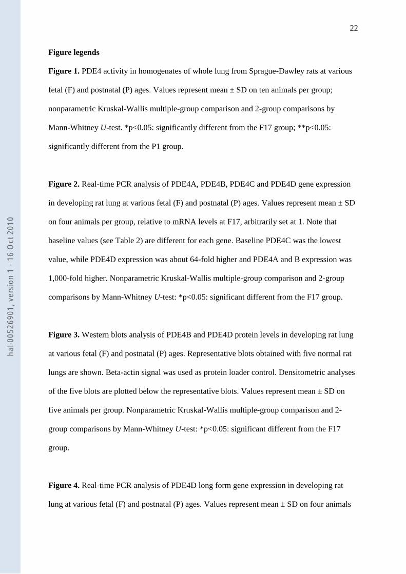
Figure 1. PDE4 activity in homogenates of whole lung from Sprague-Dawley rats at various
fetal (F) and postnatal (P) ages. Values represent mean ± SD on ten animals per group;
nonparametric Kruskal-Wallis multiple-group comparison and 2-group comparisons by
Mann-Whitney U-test. *p<0.05: significantly different from the F17 group; **p<0.05:
significantly different from the P1 group.
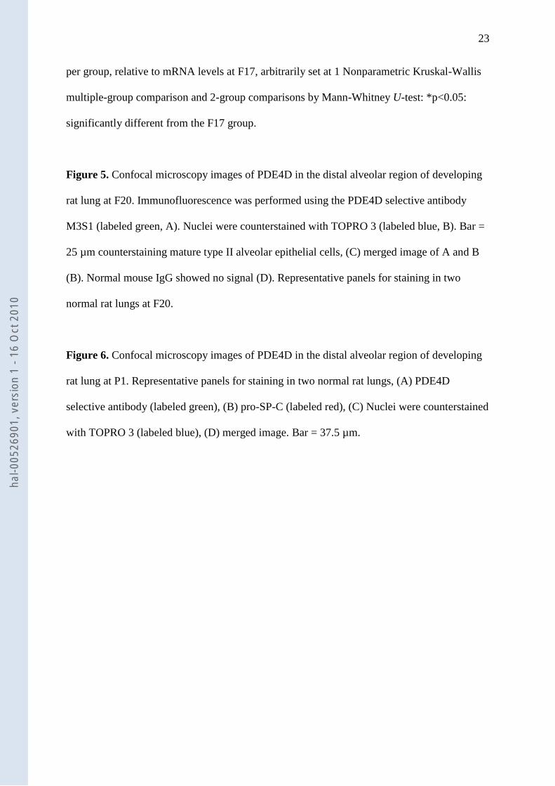
Figure 2. Real-time PCR analysis of PDE4A, PDE4B, PDE4C and PDE4D gene expression
in developing rat lung at various fetal (F) and postnatal (P) ages. Values represent mean ± SD
on four animals per group, relative to mRNA levels at F17, arbitrarily set at 1. Note that
baseline values (see Table 2) are different for each gene. Baseline PDE4C was the lowest
value, while PDE4D expression was about 64-fold higher and PDE4A and B expression was
1,000-fold higher. Nonparametric Kruskal-Wallis multiple-group comparison and 2-group
comparisons by Mann-Whitney U-test: *p<0.05: significant different from the F17 group.
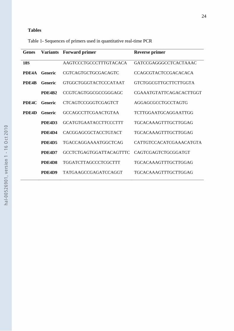
Figure 3. Western blots analysis of PDE4B and PDE4D protein levels in developing rat lung
at various fetal (F) and postnatal (P) ages. Representative blots obtained with five normal rat
lungs are shown. Beta-actin signal was used as protein loader control. Densitometric analyses
of the five blots are plotted below the representative blots. Values represent mean ± SD on
five animals per group. Nonparametric Kruskal-Wallis multiple-group comparison and 2-
group comparisons by Mann-Whitney U-test: *p<0.05: significant different from the F17
group.
Figure 4. Real-time PCR analysis of PDE4D long form gene expression in developing rat
lung at various fetal (F) and postnatal (P) ages. Values represent mean ± SD on four animals
hal-0
0526
901,
ver
sion
1 -
16 O
ct 2
010
23
per group, relative to mRNA levels at F17, arbitrarily set at 1 Nonparametric Kruskal-Wallis
multiple-group comparison and 2-group comparisons by Mann-Whitney U-test: *p<0.05:
significantly different from the F17 group.
Figure 5. Confocal microscopy images of PDE4D in the distal alveolar region of developing
rat lung at F20. Immunofluorescence was performed using the PDE4D selective antibody
M3S1 (labeled green, A). Nuclei were counterstained with TOPRO 3 (labeled blue, B). Bar =
25 µm counterstaining mature type II alveolar epithelial cells, (C) merged image of A and B
(B). Normal mouse IgG showed no signal (D). Representative panels for staining in two
normal rat lungs at F20.
Figure 6. Confocal microscopy images of PDE4D in the distal alveolar region of developing
rat lung at P1. Representative panels for staining in two normal rat lungs, (A) PDE4D
selective antibody (labeled green), (B) pro-SP-C (labeled red), (C) Nuclei were counterstained
with TOPRO 3 (labeled blue), (D) merged image. Bar = 37.5 µm.
hal-0
0526
901,
ver
sion
1 -
16 O
ct 2
010
24
Tables
Table 1- Sequences of primers used in quantitative real-time PCR
Genes Variants Forward primer Reverse primer
18S AAGTCCCTGCCCTTTGTACACA GATCCGAGGGCCTCACTAAAC
PDE4A Generic CGTCAGTGCTGCGACAGTC CCAGCGTACTCCGACACACA
PDE4B Generic GTGGCTGGGTACTCCCATAAT GTCTGGCGTTGCTTCTTGGTA
PDE4B2 CCGTCAGTGGCGCCGGGAGC CGAAATGTATTCAGACACTTGGT
PDE4C Generic CTCAGTCCGGGTCGAGTCT AGGAGCGCCTGCCTAGTG
PDE4D Generic GCCAGCCTTCGAACTGTAA TCTTGGAATGCAGGAATTGG
PDE4D3 GCATGTGAATACCTTCCCTTT TGCACAAAGTTTGCTTGGAG
PDE4D4 CACGGAGCGCTACCTGTACT TGCACAAAGTTTGCTTGGAG
PDE4D5 TGACCAGGAAAATGGCTCAG CATTGTCCACATCGAAACATGTA
PDE4D7 GCCTCTGAGTGGATTACAGTTTC CAGTCGAGTCTGCGGATGT
PDE4D8 TGGATCTTAGCCCTCGCTTT TGCACAAAGTTTGCTTGGAG
PDE4D9 TATGAAGCCGAGATCCAGGT TGCACAAAGTTTGCTTGGAG
hal-0
0526
901,
ver
sion
1 -
16 O
ct 2
010
25
Table 2- Analysis of PDE4 gene expression in whole lung tissue at various fetal (F) and
postnatal (P) ages: mean Delta Ct
F17 F19 F21 P1 P7 P14 P21
PDE4A 11.3±0.8 11.4±1.5 9.4±0.1 9.5±1.4 9.7±1.0 10.1±0.5 10.5±0.3
PDE4B 11.3±0.6 10.8±0.7 8.0±0.4 6.8±0.8 8.5±1.2 9.0±0.4 10.8±0.4
PDE4C 21.6±1.3 20.3±1.3 19.2±0.6 18.4±0.7 18.2±1.0 17.0±0.6 15.3±0.3
PDE4D 14.8±1.0 14.5±0.8 10.9±0.6 9.1±1.3 8.7±1.5 10.8±0.2 12.0±0.4
Isoforms
PDE4B2 12.6±0.6 11.9±0.9 9.1±0.3 7.9±0.4 9.4±1.2 9.6±0.5 11.5±0.4
PDE4D3 14.0±1.1 15.2±0.6 12.5±0.2 11.1±0.6 11.9±0.9 12.8±0.1 12.2±0.2
PDE4D4 15.2±0.0 25.3±2.9 21.6±3.9 17.6±1.8 17.7±3.7 16.7±1.4 18.7±1.1
PDE4D5 15.1±0.7 16.9±1.4 14.0±0.6 12.1±0.6 13.7±1.2 14.8±0.2 14.8±0.6
PDE4D7 15.9±0.9 16.1±0.6 11.8±0.4 11.4±0.2 11.7±1.4 12.3±0.7 11.9±0.6
PDE4D8 13.7±0.3 13.2±0.6 10.7±0.3 10.1±0.7 10.9±0.7 11.6±0.3 11.4±0.3
PDE4D9 11.5±0.6 11.9±0.5 10.6±0.7 9.1±0.8 9.4±0.8 10.1±0.4 10.5±0.2
Data are presented as mean ± SEM of data obtained from at least three animals at each age.
Delta Ct was calculated by subtracting Sample Ct to 18S Ct of matching age. Note: a decrease
in the Delta Ct value represents an increase in mRNA expression and a variation of 1 cycle
means a two-fold increase or a decrease.
hal-0
0526
901,
ver
sion
1 -
16 O
ct 2
010
Copyright © 2022 FDOKUMEN