Structural Insights into Maize Viviparous14, a Key Enzyme in the Biosynthesis of the Phytohormone...
11
Structural Insights into Maize Viviparous14, a Key Enzyme in the Biosynthesis of the Phytohormone Abscisic Acid W Simon A.J. Messing, a Sandra B. Gabelli, a Ignacia Echeverria, a Jonathan T. Vogel, b Jiahn Chou Guan, b Bao Cai Tan, b Harry J. Klee, b Donald R. McCarty, b and L. Mario Amzel a,1 a Department of Biophysics and Biophysical Chemistry, Johns Hopkins University, School of Medicine, Baltimore, Maryland 21205 b Department of Horticultural Sciences, University of Florida, Gainsville, Florida 32611 The key regulatory step in the biosynthesis of abscisic acid (ABA), a hormone central to the regulation of several important processes in plants, is the oxidative cleavage of the 11,12 double bond of a 9-cis-epoxycarotenoid. The enzyme viviparous14 (VP14) performs this cleavage in maize (Zea mays), making it a target for the rational design of novel chemical agents and genetic modifications that improve plant behavior through the modulation of ABA levels. The structure of VP14, determined to 3.2-A ˚ resolution, provides both insight into the determinants of regio- and stereospecificity of this enzyme and suggests a possible mechanism for oxidative cleavage. Furthermore, mutagenesis of the distantly related CCD1 of maize shows how the VP14 structure represents a template for all plant carotenoid cleavage dioxygenases (CCDs). In addition, the structure suggests how VP14 associates with the membrane as a way of gaining access to its membrane soluble substrate. INTRODUCTION Abscicic acid (ABA) is a key plant hormone involved in a broad spectrum of growth and development processes, including seed maturation and dormancy (Phillips et al., 1997; Finkelstein et al., 2002), shoot and root growth (Sharp, 2002), and responses to drought (Zeevaart and Creelman, 1988; Hasegawa et al., 2000) and nutrient depletion (Gomez-Cadenas et al., 2000) (Figure 1). For example, in conditions of drought, increases in ABA levels lead to restructuring of the cell cytoskeleton and subsequent closure of stomata (Schwartz et al., 1994), biosynthesis of osmo- lytes (glycine, betaine, etoine, sugar alchohols such as glycerol, and complex sugars) (Hasegawa et al., 2000), and transcription of stress response genes (Bray, 2002). In maize (Zea mays), the world’s most productive cereal crop (United Nations, 2007), vivaparous14 (VP14), a 9-cis-epoxyca- rotenoid dioxygenase (NCED), catalyzes the rate-limiting step in ABA biosynthesis (Tan et al., 1997; Qin and Zeevaart, 1999)—the oxidative cleavage of the 11,12 carbon-carbon double bond of either 9-cis-violaxanthin or 9-cis-neoxanthin (Schwartz et al., 1997). The C15 aldehyde, xanthoxin, is oxidized and converted through two subsequent reactions to the biologically active ABA (Figure 1) (Taylor et al., 2000; Schwartz et al., 2003a). In condi- tions of environmental stress, induction of transcription of the NCED gene(s) follows within 15 to 30 min of the onset of the stress (Tan et al., 1997; Qin and Zeevaart, 1999; Thompson et al., 2000). Thus, NCEDs, including VP14, are key regulators that determine ABA levels (Schwartz et al., 2003a), which in turn control ABA-regulated processes. This involvement makes VP14 an attractive target for the development of novel chemical agents and genetic modifications to control ABA levels and improve plant productivity, performance, and architecture (Cutler and Krochko, 1999; Qin and Zeevaart, 1999) (Figure 1). Studies of the mutant vp14-2274 also implicate VP14 in the synthesis of strigolactones (Matusova et al., 2005), signaling molecules in- volved in shoot branching (Gomez-Roldan et al., 2008; Umehara et al., 2008), presymbiotic growth of arbuscular mycorrhizal fungi (Besserer et al., 2006), and parasitic weed germination (Hauck et al., 1992; Siame et al., 1993). Recently, progress was made in identifying downstream pro- cesses involving the ABA receptors and their partners, including the determination of the structure and the mechanism of activa- tion of the ABA receptor (Miyazono et al., 2009; Nishimura et al., 2009). These results, along with those studies into the physio- logical effects of ABA, laid a foundation for a more complete understanding of ABA physiology. Yet, although a mechanism has been proposed for the committed step in ABA biosynthesis, the dioxygenase cleavage of the 9-cis-carotenoid double bond (Taylor et al., 2000; Schwartz et al., 2003b), the structure and the nature of the determinants of specificity of the dioxygenase remained unknown. Using the structure of VP14, determined here to 3.2-A ˚ resolution, we identified amino acid residues that help position the substrate and determine which bond is cleaved. These residues were com- pared and contrasted with those present in the broadly defined family of plant carotenoid cleavage dioxygenases (CCDs) (von Lintig and Vogt, 2004), which process a wide variety of carotenoid substrates and are involved in the biosynthesis of several plant hormones (Gomez-Roldan et al., 2008; Umehara et al., 2008; Vogel et al., 2008). We tested the importance of these residues by studying the effects of mutations of residues identified by homol- ogy with VP14 in a distantly related family member, maize CCD1. Furthermore, the structure led to the identification of a structural 1 Address correspondence to [email protected]. The author responsible for distribution of materials integral to the findings presented in this article in accordance with the policy described in the Instructions for Authors (www.plantcell.org) is: L. Mario Amzel ([email protected]). W Online version contains Web-only data. www.plantcell.org/cgi/doi/10.1105/tpc.110.074815 The Plant Cell, Vol. 22: 2970–2980, September 2010, www.plantcell.org ã 2010 American Society of Plant Biologists
-
Upload
johnshopkins -
Category
Documents
-
view
0 -
download
0
Transcript of Structural Insights into Maize Viviparous14, a Key Enzyme in the Biosynthesis of the Phytohormone...
Structural Insights into Maize Viviparous14, a Key Enzyme inthe Biosynthesis of the Phytohormone Abscisic Acid W
SimonA.J.Messing,a SandraB.Gabelli,a IgnaciaEcheverria,a JonathanT. Vogel,b JiahnChouGuan,b BaoCai Tan,b
Harry J. Klee,b Donald R. McCarty,b and L. Mario Amzela,1
a Department of Biophysics and Biophysical Chemistry, Johns Hopkins University, School of Medicine, Baltimore, Maryland 21205b Department of Horticultural Sciences, University of Florida, Gainsville, Florida 32611
The key regulatory step in the biosynthesis of abscisic acid (ABA), a hormone central to the regulation of several important
processes in plants, is the oxidative cleavage of the 11,12 double bond of a 9-cis-epoxycarotenoid. The enzyme viviparous14
(VP14) performs this cleavage in maize (Zea mays), making it a target for the rational design of novel chemical agents and
genetic modifications that improve plant behavior through the modulation of ABA levels. The structure of VP14, determined
to 3.2-A resolution, provides both insight into the determinants of regio- and stereospecificity of this enzyme and suggests a
possible mechanism for oxidative cleavage. Furthermore, mutagenesis of the distantly related CCD1 of maize shows how
the VP14 structure represents a template for all plant carotenoid cleavage dioxygenases (CCDs). In addition, the structure
suggests how VP14 associates with the membrane as a way of gaining access to its membrane soluble substrate.
INTRODUCTION
Abscicic acid (ABA) is a key plant hormone involved in a broad
spectrum of growth and development processes, including seed
maturation and dormancy (Phillips et al., 1997; Finkelstein et al.,
2002), shoot and root growth (Sharp, 2002), and responses to
drought (Zeevaart and Creelman, 1988; Hasegawa et al., 2000)
and nutrient depletion (Gomez-Cadenas et al., 2000) (Figure 1).
For example, in conditions of drought, increases in ABA levels
lead to restructuring of the cell cytoskeleton and subsequent
closure of stomata (Schwartz et al., 1994), biosynthesis of osmo-
lytes (glycine, betaine, etoine, sugar alchohols such as glycerol,
and complex sugars) (Hasegawa et al., 2000), and transcription of
stress response genes (Bray, 2002).
In maize (Zea mays), the world’s most productive cereal crop
(United Nations, 2007), vivaparous14 (VP14), a 9-cis-epoxyca-
rotenoid dioxygenase (NCED), catalyzes the rate-limiting step in
ABA biosynthesis (Tan et al., 1997; Qin and Zeevaart, 1999)—the
oxidative cleavage of the 11,12 carbon-carbon double bond of
either 9-cis-violaxanthin or 9-cis-neoxanthin (Schwartz et al.,
1997). The C15 aldehyde, xanthoxin, is oxidized and converted
through two subsequent reactions to the biologically active ABA
(Figure 1) (Taylor et al., 2000; Schwartz et al., 2003a). In condi-
tions of environmental stress, induction of transcription of the
NCED gene(s) follows within 15 to 30 min of the onset of the
stress (Tan et al., 1997; Qin and Zeevaart, 1999; Thompson et al.,
2000). Thus, NCEDs, including VP14, are key regulators that
determine ABA levels (Schwartz et al., 2003a), which in turn
control ABA-regulated processes. This involvement makes VP14
an attractive target for the development of novel chemical agents
and genetic modifications to control ABA levels and improve
plant productivity, performance, and architecture (Cutler and
Krochko, 1999; Qin and Zeevaart, 1999) (Figure 1). Studies of
the mutant vp14-2274 also implicate VP14 in the synthesis of
strigolactones (Matusova et al., 2005), signaling molecules in-
volved in shoot branching (Gomez-Roldan et al., 2008; Umehara
et al., 2008), presymbiotic growth of arbuscular mycorrhizal
fungi (Besserer et al., 2006), and parasitic weed germination
(Hauck et al., 1992; Siame et al., 1993).
Recently, progress was made in identifying downstream pro-
cesses involving the ABA receptors and their partners, including
the determination of the structure and the mechanism of activa-
tion of the ABA receptor (Miyazono et al., 2009; Nishimura et al.,
2009). These results, along with those studies into the physio-
logical effects of ABA, laid a foundation for a more complete
understanding of ABA physiology. Yet, although a mechanism
has been proposed for the committed step in ABA biosynthesis,
the dioxygenase cleavage of the 9-cis-carotenoid double bond
(Taylor et al., 2000; Schwartz et al., 2003b), the structure and
the nature of the determinants of specificity of the dioxygenase
remained unknown.
Using the structure of VP14, determined here to 3.2-A resolution,
we identified amino acid residues that help position the substrate
and determine which bond is cleaved. These residues were com-
pared and contrasted with those present in the broadly defined
family of plant carotenoid cleavage dioxygenases (CCDs) (von
Lintig and Vogt, 2004), which process a wide variety of carotenoid
substrates and are involved in the biosynthesis of several plant
hormones (Gomez-Roldan et al., 2008; Umehara et al., 2008; Vogel
et al., 2008). We tested the importance of these residues by
studying the effects of mutations of residues identified by homol-
ogy with VP14 in a distantly related family member, maize CCD1.
Furthermore, the structure led to the identification of a structural
1 Address correspondence to [email protected] author responsible for distribution of materials integral to thefindings presented in this article in accordance with the policy describedin the Instructions for Authors (www.plantcell.org) is: L. Mario Amzel([email protected]).WOnline version contains Web-only data.www.plantcell.org/cgi/doi/10.1105/tpc.110.074815
The Plant Cell, Vol. 22: 2970–2980, September 2010, www.plantcell.org ã 2010 American Society of Plant Biologists
motif in the sequence of many plant CCDs that is ideally suited for
membrane association, placing these enzymes in close proximity
with their lipid-soluble carotenoid substrates.
RESULTS
Overall Structure
The structure of VP14 (residues 84 to 604 out of 604), determined
bymultiple wavelength anomalous dispersion (MAD; Hendrickson,
1991), was refined to an Rwork/Rfree of 23.2/28.4% (Table 1; see
Methods). VP14 folds as a seven-blade b-propeller with four
a-helical inserts that form an a-helical domain on top of the
b-propeller (Figure 2). Five of the seven blades of the b-propeller
have four locally connected antiparallel strands, while the first
and seventh blades have five antiparallel strands. In blades one
and seven, the N terminus provides the outermost b-strand
(residues 132 to 137 in blade 1; residues 140 to 143 in blade 7),
and the C terminus provides the innermost b-strand to blade
one. As suggested by Kloer and Schulz (2006), the b-propeller
portion of the structure may be a conserved characteristic
throughout the CCD family of enzymes, as it is present in both
the prokaryotic apocarotenoid 15,15’-oxygenase (ACO) (Kloer
et al., 2005) and the eukaryotic VP14.
Catalytic Iron
As in other b-propeller structures, a long tunnel surrounded by
the blades runs through the center of the structure from one end
to the other. The Fe2+ required for dioxygenase activity is located
inside the tunnel on the central axis of the b-propeller. It is bound
octahedrally by four His residues located on the innermost
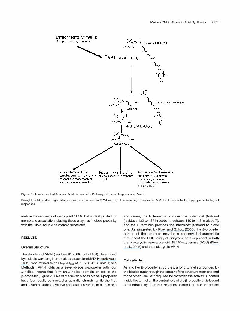
Figure 1. Involvement of Abscicic Acid Biosynthetic Pathway in Stress Responses in Plants.
Drought, cold, and/or high salinity induce an increase in VP14 activity. The resulting elevation of ABA levels leads to the appropriate biological
responses.
Maize VP14 in Abscicic Acid Synthesis 2971
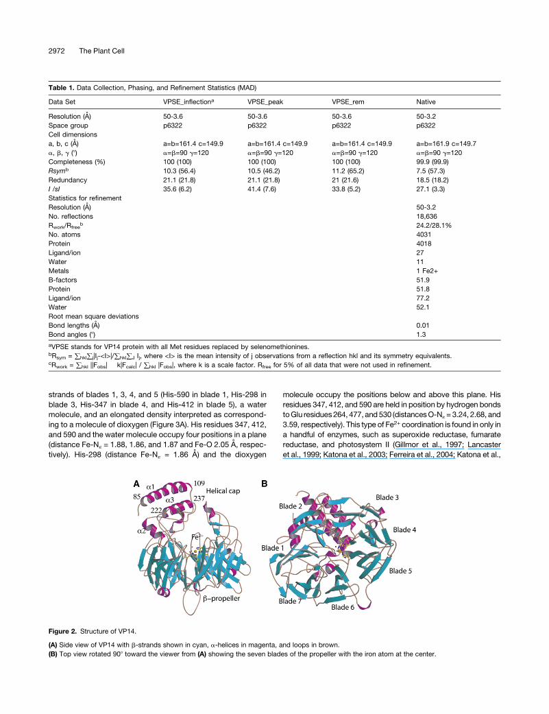
strands of blades 1, 3, 4, and 5 (His-590 in blade 1, His-298 in
blade 3, His-347 in blade 4, and His-412 in blade 5), a water
molecule, and an elongated density interpreted as correspond-
ing to a molecule of dioxygen (Figure 3A). His residues 347, 412,
and 590 and the water molecule occupy four positions in a plane
(distance Fe-N« = 1.88, 1.86, and 1.87 and Fe-O 2.05 A, respec-
tively). His-298 (distance Fe-N« = 1.86 A) and the dioxygen
molecule occupy the positions below and above this plane. His
residues 347, 412, and 590 are held in position by hydrogen bonds
toGlu residues264, 477, and530 (distancesO-N«=3.24, 2.68, and
3.59, respectively). This type of Fe2+ coordination is found in only in
a handful of enzymes, such as superoxide reductase, fumarate
reductase, and photosystem II (Gillmor et al., 1997; Lancaster
et al., 1999; Katona et al., 2003; Ferreira et al., 2004; Katona et al.,
Table 1. Data Collection, Phasing, and Refinement Statistics (MAD)
Data Set VPSE_inflectiona VPSE_peak VPSE_rem Native
Resolution (A) 50-3.6 50-3.6 50-3.6 50-3.2
Space group p6322 p6322 p6322 p6322
Cell dimensions
a, b, c (A) a=b=161.4 c=149.9 a=b=161.4 c=149.9 a=b=161.4 c=149.9 a=b=161.9 c=149.7
a, b, g (8) a=b=90 g=120 a=b=90 g=120 a=b=90 g=120 a=b=90 g=120
Completeness (%) 100 (100) 100 (100) 100 (100) 99.9 (99.9)
Rsymb 10.3 (56.4) 10.5 (46.2) 11.2 (65.2) 7.5 (57.3)
Redundancy 21.1 (21.8) 21.1 (21.8) 21 (21.6) 18.5 (18.2)
I /sI 35.6 (6.2) 41.4 (7.6) 33.8 (5.2) 27.1 (3.3)
Statistics for refinement
Resolution (A) 50-3.2
No. reflections 18,636
Rwork/Rfreeb 24.2/28.1%
No. atoms 4031
Protein 4018
Ligand/ion 27
Water 11
Metals 1 Fe2+
B-factors 51.9
Protein 51.8
Ligand/ion 77.2
Water 52.1
Root mean square deviations
Bond lengths (A) 0.01
Bond angles (8) 1.3
aVPSE stands for VP14 protein with all Met residues replaced by selenomethionines.bRsym = )hkl)j|Ij-<I>|/)hkl)J Ij, where <I> is the mean intensity of j observations from a reflection hkl and its symmetry equivalents.cRwork = )hkl ||Fobs| � k|Fcalc| / )hkl |Fobs|, where k is a scale factor. Rfree for 5% of all data that were not used in refinement.
Figure 2. Structure of VP14.
(A) Side view of VP14 with b-strands shown in cyan, a-helices in magenta, and loops in brown.
(B) Top view rotated 908 toward the viewer from (A) showing the seven blades of the propeller with the iron atom at the center.
2972 The Plant Cell
2007). The presence of Fe2+ was confirmed by soaking crystals
with orthophenanthroline, which turned the crystals a deep red
(Cavallini et al., 1969), indicating the presence of Fe2+.
Mechanism
The putative oxygen molecule is coordinated to the iron in an
angular end-on geometry as originally proposed by Pauling
(1964), forming an Fe-O-O angle of 1318 (proximal oxygen
distance Fe-Op = 2.04 A; distal oxygen Fe-Od = 2.97 A). The
identity of the dioxygen was confirmed by calculating several
types of omit maps. Attempts to refine the structure with a water
molecule/hydroxide ion instead of dioxygen resulted in large ex-
cess positive density in the Fobs-Fcalc maps. Although other inter-
pretations of this density are possible, all our tests suggest that
molecular oxygen is the most likely ligand at this position from a
crystallographic as well as from a mechanistic point of view.
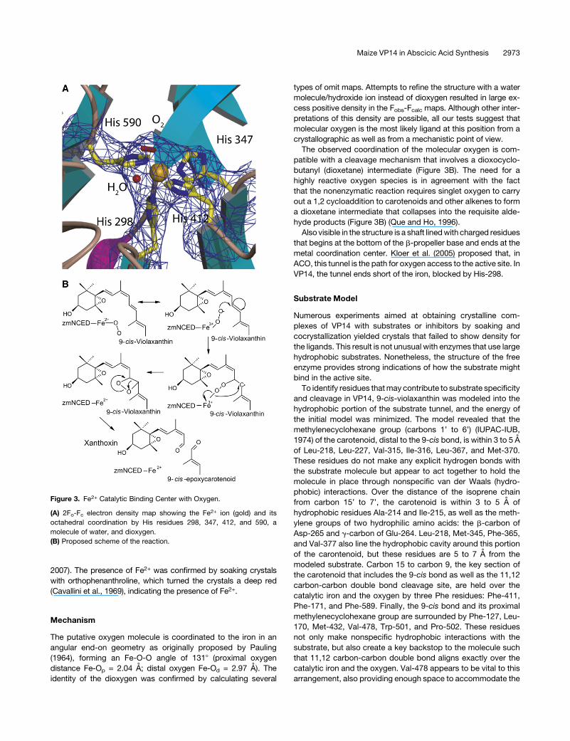
The observed coordination of the molecular oxygen is com-
patible with a cleavage mechanism that involves a dioxocyclo-
butanyl (dioxetane) intermediate (Figure 3B). The need for a
highly reactive oxygen species is in agreement with the fact
that the nonenzymatic reaction requires singlet oxygen to carry
out a 1,2 cycloaddition to carotenoids and other alkenes to form
a dioxetane intermediate that collapses into the requisite alde-
hyde products (Figure 3B) (Que and Ho, 1996).
Also visible in the structure is a shaft linedwith charged residues
that begins at the bottom of the b-propeller base and ends at the
metal coordination center. Kloer et al. (2005) proposed that, in
ACO, this tunnel is the path for oxygen access to the active site. In
VP14, the tunnel ends short of the iron, blocked by His-298.
Substrate Model
Numerous experiments aimed at obtaining crystalline com-
plexes of VP14 with substrates or inhibitors by soaking and
cocrystallization yielded crystals that failed to show density for
the ligands. This result is not unusual with enzymes that use large
hydrophobic substrates. Nonetheless, the structure of the free
enzyme provides strong indications of how the substrate might
bind in the active site.
To identify residues thatmay contribute to substrate specificity
and cleavage in VP14, 9-cis-violaxanthin was modeled into the
hydrophobic portion of the substrate tunnel, and the energy of
the initial model was minimized. The model revealed that the
methylenecyclohexane group (carbons 1’ to 6’) (IUPAC-IUB,
1974) of the carotenoid, distal to the 9-cis bond, is within 3 to 5 A
of Leu-218, Leu-227, Val-315, Ile-316, Leu-367, and Met-370.
These residues do not make any explicit hydrogen bonds with
the substrate molecule but appear to act together to hold the
molecule in place through nonspecific van der Waals (hydro-
phobic) interactions. Over the distance of the isoprene chain
from carbon 15’ to 7’, the carotenoid is within 3 to 5 A of
hydrophobic residues Ala-214 and Ile-215, as well as the meth-
ylene groups of two hydrophilic amino acids: the b-carbon of
Asp-265 and g-carbon of Glu-264. Leu-218, Met-345, Phe-365,
and Val-377 also line the hydrophobic cavity around this portion
of the carontenoid, but these residues are 5 to 7 A from the
modeled substrate. Carbon 15 to carbon 9, the key section of
the carotenoid that includes the 9-cis bond as well as the 11,12
carbon-carbon double bond cleavage site, are held over the
catalytic iron and the oxygen by three Phe residues: Phe-411,
Phe-171, and Phe-589. Finally, the 9-cis bond and its proximal
methylenecyclohexane group are surrounded by Phe-127, Leu-
170, Met-432, Val-478, Trp-501, and Pro-502. These residues
not only make nonspecific hydrophobic interactions with the
substrate, but also create a key backstop to the molecule such
that 11,12 carbon-carbon double bond aligns exactly over the
catalytic iron and the oxygen. Val-478 appears to be vital to this
arrangement, also providing enough space to accommodate the
Figure 3. Fe2+ Catalytic Binding Center with Oxygen.
(A) 2Fo-Fc electron density map showing the Fe2+ ion (gold) and its
octahedral coordination by His residues 298, 347, 412, and 590, a
molecule of water, and dioxygen.
(B) Proposed scheme of the reaction.
Maize VP14 in Abscicic Acid Synthesis 2973

methyl group protruding from carbon 9. The absence of basic
and acidic residues in proximity to the scissile bond further
supports the dioxetane-intermediate mechanism.
The Helical Domain and Membrane Penetration
The a-helical domain of VP14 is dominated by two antiparallel
a-helices, a1 and a3, which are formed by amino acids 88 to 108
and 222 to 237 (Figure 2A). Analysis of the surface properties of
these two helices reveals a hydrophobic patch of 2226 A2 formed
by 25 hydrophobic residues (Leu-86, Phe-87, Ala-90, Ala-91,
Ala-93, Ala-94, Leu-95, Ala-97, Phe-98, Gly-101, Phe-102, Val-
103, Val-106, Leu-107, Pro-110, Ile-224, Ala-225, Leu-227, Ala-
228, Leu-229, Tyr-231, Ala-232, Ala-234, Ala-235, and Gly-237)
that may penetrate the membrane beyond the head groups of
the membrane phospholipids and interact with fatty acid resi-
dues in the membrane interior (Figure 4A). With VP14 in this
position, a crown of positive, neutral, and negatively charged
residues (Asn-85, Gln-88, Arg-89, Asp-97, Glu-101, Asn-106,
Arg-110, Tyr-232, Asp-241, and Arg-373) interact with the
charged surface of the membrane directly or through water-
mediated contacts. If helix a1 is removed, an area of positive
surface charge is exposed, and the hydrophobic patch is dis-
rupted. This observation explains why in genetic and cell bio-
logical studies, deleting or disrupting this first helix results in a
loss of interaction of VP14 with the thylakoid membrane and loss
of ABA biosynthesis (Tan et al., 2001).
To investigate whether these helices actually could penetrate
the membrane, we implemented a modification of the computa-
tional/structural approach developed by Mosberg and coworkers
(Lomize et al., 2006) to calculate the DGtransfer of membrane
penetration. Using this procedure, we found for VP14 a maximal
membrane penetration of 7 A with a DGtransfer = 28 kcal/mol for
VP14 (Figures 4B and 4C). As a positive control, we calculated
DGtransfer for carnitine palmitoyltransferase 2 (CPT-2) and obtained
a value of 29.7 kcal/mol, in good agreement with the reported
value of DGtransfer = 28.6 (kcal/mol) (Lomize et al., 2006).
The feature proposed for the interaction of CPT-2 with the
membrane consists of two antiparallel helices, in a similar
arrangement to that found in VP14. It was proposed that in
CPT-2, these two helices penetrate the membrane, allowing the
enzyme to extract a lipophilic substrate (Rufer et al., 2007).
Interestingly, VP14 helix a3 is adjacent to the opening to the
substrate tunnel, which is lined by hydrophobic aromatic resi-
dues. These observations, coupled with the favorable DGtransfer,
show how VP14 could use a mechanism of substrate accessi-
bility similar to that proposed for CPT-2. Penetration of the
membrane by the hydrophobic patch of VP14 would place the
substrate tunnel in close proximity to carotenoids in the thylakoid
membrane. Importantly, 9-cis-neoxanthin and 9-cis-violaxanthin,
the primary substrates of VP14, are 10 timesmore abundant in the
thylakoid membrane than in other plant membranes (Parry and
Horgan, 1991). Thismechanismmay be consistent throughout the
CCD family; a hydrophobic patch, albeit significantly smaller, was
observed in the prokaryotic ACO structure (Kloer et al., 2005) (nine
residues covering 800 A2 versus 25 residues and 2226 A2 in VP14).
VP14 as the Prototype
Comparison of the VP14 sequence with those of other plant
NCEDs shows 60 to 85% identity across the plant kingdom—
from close relatives such as Oryza sativa to distant relatives
such as that ofCitrus clementina andArabidopsis thaliana (Figure
5). In addition to similarities in the b-propeller, these sequences
show high conservation of the two long antiparallel a-helices that
we propose are involved in membrane penetration.
VP14 shows an overall lower, but significant, sequence identity
(58 to 16%) with Z. mays CCD1 (Zm CCD1) and the complete
family of CCD enzymes of Arabidopsis (see Supplemental Figure
1 online), especially the conservation of key structural elements.
Figure 4. VP14 Helical Domain and Its Interaction with the Thylakoid Membrane.
(A) VP14 is colored according to its electrostatic surface potential and is labeled with key residues depicting the hydrophobic patch used for membrane
penetration (red represents negatively charged and blue represents positively charged residues). Key hydrophobic residues from helices a1 and a3 are
shown in yellow below the surface (e.g., Phe-87).
(B) Electrostatic surface potential representation of VP14 penetrating the membrane surface, which is represented by a plane.
(C) Same as (B) with the protein shown using a ribbon diagram.
2974 The Plant Cell
This conservation is interesting because members of the CCD
family, which encompasses the NCED family, process a more
diverse array of carotenoid substrates. For example, maize CCD1
cleaves multiple linear and cyclic carotenoids (Vogel et al., 2008),
whereas Arabidopsis CCD7/MAX3 is involved in the synthesis of
strigolactones, novel plant signalingmolecules that regulate shoot
branching (Booker et al., 2004; Gomez-Roldan et al., 2008;
Umehara et al., 2008). The significant level of sequence identity
between VP14, NCEDs, and the CCDs family in plants makes the
VP14 structure an ideal prototype of all these enzymes. VP14 also
belongs to a more broadly defined family of CCDs present in
bacteria, plants, and animals (von Lintig and Vogt, 2004). These
enzymes range from the apocarotenoid-15,15’-oxygenaseofSyn-
echocystis spp to the human b-carotene 15,15’-monooxygenase,
the enzyme responsible for the production of retinol (vitamin A)
(von Lintig and Vogt, 2000; von Lintig et al., 2001).
Modeling of a Distant VP14 Relative: Regions Responsible
for Specificity Differences
Using the coordinates of the VP14 substrate model as a tem-
plate, we constructed a homology model of Zm CCD1. This
enzyme was chosen because, in contrast with VP14, its sub-
strate is commercially available, permitting extensive inves-
tigation of the effects of selective mutations (see below). A
comparison of the VP14 structure and the Zm CCD1 model
suggests that the differences in substrate specificity reside in
three key regions of the structures.
The first involves the substitution of a Val residue (Val-478) on
the innermost strand of blade 6 of VP14 by a Phe (Phe-409) in Zm
CCD1. As was observed in the model of VP14 subtrate, this Val
creates a small cleft in VP14 that accommodates the protruding
methyl group on carbon 9 of the isoprene chain and allows the
11,12 bond to fit over the Fe2+ (Figure 6). The presence of a Phe
in Zm CCD1 interferes with this arrangement. This structural
difference is highlighted when comparing the sequence of VP14
with those of Zm CCD1 and of the complete family of CCDs from
Arabidopsis (At). At NCEDs 2, 3, 5, 6, and 9 have either an Ala or
Ile at this position, whereas At CCDs 4, 1, 7, 8, and Zm CCD1
have a Phe or Met (see Supplemental Figure 1 online). This is in
agreement with the known activity of these proteins: while At
NCEDs 2, 3, 5, 6, and 9 carry out 9-cis-carotenoid cleavage (Iuchi
et al., 2001; Tan et al., 2003), At CCDs 4, 1, 7, and 8 andZmCCD1
are neither involved in ABA biosynthesis nor in the cleavage of
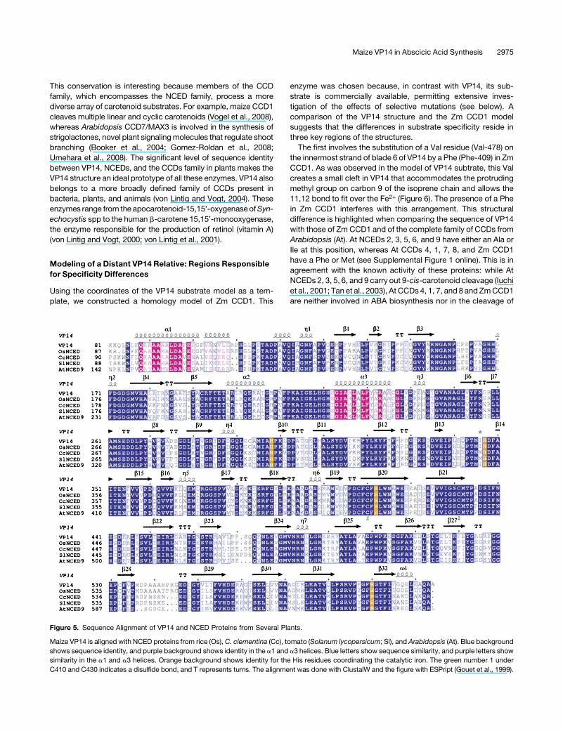
Figure 5. Sequence Alignment of VP14 and NCED Proteins from Several Plants.
Maize VP14 is aligned with NCED proteins from rice (Os), C. clementina (Cc), tomato (Solanum lycopersicum; Sl), and Arabidopsis (At). Blue background
shows sequence identity, and purple background shows identity in the a1 and a3 helices. Blue letters show sequence similarity, and purple letters show
similarity in the a1 and a3 helices. Orange background shows identity for the His residues coordinating the catalytic iron. The green number 1 under
C410 and C430 indicates a disulfide bond, and T represents turns. The alignment was done with ClustalW and the figure with ESPript (Gouet et al., 1999).
Maize VP14 in Abscicic Acid Synthesis 2975
other 9-cis-carotenoid (Sorefan et al., 2003; Booker et al., 2004;
Vogel et al., 2008).
The second region involves a loop on the back side of the
substrate pocket (residues 499 to 503 in VP14 and 432 to 434 in
Zm CCD1) (Figure 6) and a substitution of Leu-170 in VP14 by a
Trp (Trp-104) in Zm CCD1. In VP14, this loop and the Leu form a
pocket that can accommodate the second ring of the carotenoid
in such a way that the 11,12 carbon-carbon double bond of the
9-cis-epoxycarotenoid is close to the Fe2+. This arrangement is
not possible in Zm CCD1.
The third region, a feature conserved in the two enzymes, is
comprised of three Phe residues (Phe-171, Phe-411, and Phe-589
in VP14 and Phe-105, Phe-343, and Phe-532 in Zm CCD1)
important for caging the substrate over the catalytic iron. Com-
parison of the structures of VP14modeled with a 9-cis-carotenoid
and ACO with substrate reveals that the same three regions
account for their differences in substrate specificity.
CCD1Mutational Studies
To test the model of Zm CCD1, we mutated to Ala residues
indentified to be important for determining specificity and mea-
sured the activity of the mutated proteins toward a b-apo-8’-
carotenal. Substitution of Phe-105, Phe-343, and Phe-533 of
CCD1 by Ala decreased activity to <10, 15, and 30% of the wild
type, respectively (Figure 7). As these experiments were carried
with high substrate concentrations (72 mM versus a Km of 0.98
mM for the wild type), this result suggests that these effects are a
consequence of a reduction in kcat. Detailed kinetic analysis of
the F343Amutant showed a 90%decrease in Vmax and threefold
increase in Km (Table 2) compared with the wild type that under
saturation conditions (72 mM versus a Km mutant of 8.9 mM)
predicts an 11% residual activity, which is in good agreement
with the 15% of wild-type activity observed in the high substrate
concentration experiments. These results are compatible with a
90% reduction inVmax for P105A and a 67% reduction for P533A.
These results underscore the role of these three residues in
catalysis, probably by holding the substrate over the active site.
Substitution I147A (Ile-215 in VP14) also produced a reduction
in activity to 15% of that of the wild type. This corresponds
well with the models, as Ile-147 is spatially next to Phe-105 and
may be contributing to caging the substrate as well.
Mutations M276A and F296A in CCD1 (Met-345 and Phe-365
in VP14) did not result in a significant drop in activity. These
results are also explained by the models, which show that these
residues are 5 to 7 A from the substrate on the opposite side of
the hydrophobic cavity. Mutation of Phe-409 and Phe-533 to Ala
(Val-478 and Phe-589 in VP14) resulted in a reduction of activity
to 30 and 15%of that of thewild type, reinforcing the observation
that the back side of the pocket is important for creating contours
to ensure that the C11,C12 carbon-carbon double bond aligns
over the catalytic site (Figures 6 and 7). The effect of these
mutations on the activity of CCD1 validates the use of VP14 as a
template for mapping important residues in the substrate tunnel
on distant plant CCDs and for providing a rationale for under-
standing their substrate specificity.
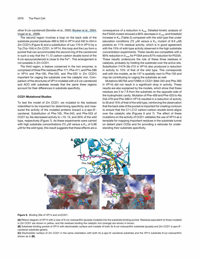
Figure 6. Binding Site of VP14 and zmCCD1.
(A) Ribbon diagram of VP14 with a view of 9-cis-violoxanthin (purple) modeled into the substrate binding pocket. Residues equivalent to those mutated
in Zm CCD1 are shown in yellow, and His residues binding the catalytic iron (orange) are shown in brown.
(B) Substrate binding pocket of VP14 with electrostatic surface and models of both its 9-cis-violoxanthin substrate (purple) and Zm CCD1 b-apo-8’-
carotenal substrate (green).
(C) Electrostatic surface of Zm CCD1 in the same orientation with both its b-apo-8’-carotenal substrate and the VP14 substrate 9-cis-violoxanthin
shown as in (B).
2976 The Plant Cell
DISCUSSION
VP14 catalyzes the first committed step in the biosynthesis of
ABA in maize: cleavage of the 9-cis-violoxanthin 11,12 double
bond by incorporation of the two oxygen atoms of O2 into the two
products. The structure of VP14 determined here is fully compat-
ible with the dioxetane mechanism suggested by the uncatalyzed
reaction performed with singlet oxygen. The end-on Fe2+-O2
species is in resonance with Fe3+-O2.2, a radical superoxide
ideally suited to attack the double bond resulting in a radical
species that can react with the second oxygen to form the
dioxetane species that spontaneously produces the two alde-
hydes. The absence of basic and acidic residues in the proximity
of the scissile bond argues against mechanisms that would
require proton transfer steps.
VP14 has two domains: a seven-blade b-propeller domain and
a second domain comprised largely of a-helices. The b-propeller
portion of the structure, present in both the prokaryotic ACO and
the eukaryotic VP14 (Kloer et al., 2005), is a conserved charac-
teristic of the CCD family of enzymes, while the helical domain
may be a structural feature that differentiates plant carotenoid-
cleaving dioxygenases from other members of the family. Our
analysis of this domain, in conjunction with previous biochemical
studies (Tan et al., 2001), provides a possible mechanism by
which the enzyme might penetrate the surface of the thylakoid
membrane and extract substrate through a tunnel that extends
into the catalytic center.
The broader CCD family plays a vital role in the biosynthesis of
other key signaling molecules that regulate growth and devel-
opment, including strigolactone-related compounds implicated
in control of branching (Booker et al., 2004; Gomez-Roldan et al.,
2008). Structural comparison of VP14 and a model of CCD1
combined with mutagenesis analysis revealed differences in the
active sites, indicating that Phe-171, Phe-411, and Val-478 in
VP14 and their equivalent residues in CCD1 are important for
discriminating between the 9-cis-carotenoid substrate and other
carotenoids. TheVP14structure reported here canbe considered
a template for all related plant CCDs and provides a blueprint for
studies of function in this key family of enzymes. Our studies also
show how our VP14 substrate model may be useful not only for
the rational design of novel chemical agents but also for engi-
neering of genetically modified organisms for the purpose of
controlling ABA levels, an obvious target for crop improvement
(Cutler and Krochko, 1999; Qin and Zeevaart, 1999).
METHODS
Expression of Partial VP14
Using the original full-length Zea mays vp14 gene in a pGEX-2T vector
(Pharmacia), the DNA sequence representing amino acid residues 75 to
604 (residues 1 to 75 are a signal sequence and were not included in the
construct) was subcloned byPCR into pMal-c2x-hv (NewEnglandBiolab)
using BamHI and EcoRI restriction enzymes (59 primer, 59-GCGGCG-
ACTGGATCCAGGAAAGCGGAGGGC-39; 39primer, T7 terminator). M15-
(pREP) cells (Qiagen) were transformed with this construct and grown
aerobically in 2xTY broth (per liter = 16 g Bacto-tryptone, 10 g yeast
extract, and 5 gNaCl) at 158C to anA600 of;0.6. Expression was induced
with 1 mM isopropyl-b-D-thiogalactopyranoside, cells were grown for
16 h, and the cell pellet was frozen at2808C. All mutants and constructs
were expressed in this manner, except for selenomethionine-substituted
protein. Selenomethionine-substituted protein in which all Met residues
were replaced with selenomethionines was expressed as discussed by
Hendrickson (Leahy et al., 1994).
Purification
Cell pellets were resuspended in 20mMNaH2PO4, pH 7.5, 500mMNaCl,
15 mM imidazole, 5 mM b-mercaptoethanol buffer (Ni2+ affinity binding
buffer), lysed via microfluidization, and centrifuged at 20,000g for 30 min
at 48C. The soluble cytoplasmic fraction was then purified with a Ni2+
affinity column, using an imidazole gradient of 10 to 250mMas the eluant.
The N-terminal tag was cleaved overnight using 100 mL of 1 mg/mL TEV
protease to 50 mL Ni2+ affinity column elution at room temperature in a
buffer of 50 mM Tris-HCl, 50 mM NaCl, and 1 mM Tris 2-carboxyethyl
phosphine, pH 7.5. VP14 in the buffer from the TEV digestion was further
purified on an anion exchange column (source 15Q beads from GE
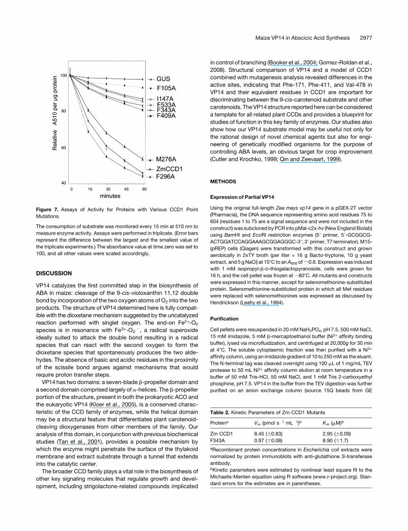
Figure 7. Assays of Activity for Proteins with Various CCD1 Point
Mutations.
The consumption of substrate was monitored every 15 min at 510 nm to
measure enzyme activity. Assays were performed in triplicate. (Error bars
represent the difference between the largest and the smallest value of
the triplicate experiments.) The absorbance value at time zero was set to
100, and all other values were scaled accordingly.
Table 2. Kinetic Parameters of Zm CCD1 Mutants
Proteina Vm (pmol s�1 mL�1)b Km (mM)b
Zm CCD1 8.45 (60.83) 2.95 (60.09)
F343A 0.97 (60.08) 8.90 (61.7)
aRecombinant protein concentrations in Escherichia coli extracts were
normalized by protein immunoblots with anti-glutathione S-transferase
antibody.bKinetic parameters were estimated by nonlinear least square fit to the
Michaelis-Menten equation using R software (www.r-project.org). Stan-
dard errors for the estimates are in parentheses.
Maize VP14 in Abscicic Acid Synthesis 2977
Healthcare) using an NaCl gradient of 0 to 1 M as the eluant. Undigested
protein was eliminated on a second Ni2+ affinity column using the same.
The flow-through was loaded onto a 26/60 Sephacryl S-200 column
(GE Healthcare Life Sciences) using a buffer of 50 mM Tris-HCl, 50 mM
NaCl, and 1 mM Tris 2-carboxyethyl phosphine, pH 7.5 (crystallization
buffer) to elute the protein. The protein was then concentrated to 10mg/
mL using a Vivaspin 20 concentrator (Sartorius Stedim Biotech). Pro-
duction of the protein with a selenomethionine substitution followed
the same protocol.
Crystallization
VP14 was crystallized at 208C by vapor diffusion in hanging drops of 2 mL
of protein at 10 mg/mL in crystallization buffer with an equal volume of a
reservoir solution of 0.1 M Na-HEPES, pH 7.5, 1.5 M Li2SO4, 2%
hexanediol, and 4% dioxane. Single crystals belonging to P6322 space
group grew in 1 to 5 d and contained onemonomer in the asymmetric unit
Surface Site Mutagenesis for KE381AA
Because the initial crystallization attempts did not lead to diffracting
crystals, the VP14 sequence was analyzed using the Surface-Entropy-
Reduction prediction (SERp) server (Goldschmidt et al., 2007). The
program predicted Lys-381 and Glu-382 to be in loop regions of the
protein and recommended mutation of these residues to Ala to improve
the possibility of crystal contacts. These residues were mutated to Ala
using the following primer (coding mismatch underlined): 59-GCCCG-
TGGTGCTGGACGCGGCGAAGACGTCGCGGTT-39. The wild-type VP14
gene in the pMal-c2x-hv vector was used as the template for the PCR to
generate the double mutant, following the standard QuickChange pro-
cedure for site-directed mutagenesis (Stratagene). These mutations lead
to diffracting crystals.
Data Collection, Structure Determination, and Refinement
Data to a resolution of 3.6 A were collected from selenomethionine-
substituted crystals at the Brookhaven National Laboratory beamline X6A
and processed with the HKL suite (Otwinowski and Minor, 1997). MAD
phases (Hendrickson, 1991) were calculated using the program SOLVE
(Z score of 36.37, and figure of merit of 0.36 at 3.92 A) and were improved
by density modification with the program RESOLVE (Terwilliger, 2003).
The model was built interactively with the program O (Jones et al., 1991;
Jones and Kjeldgaard, 1997) and was refined using REFMAC 5.0 (Col-
laborative Computational Project, Number 4, 1994) with individual re-
strained B-factors and a solvent correction using the Babinet option.
Anisotropic refinement using translation, libration, and screw rotation
(TLS) of rigid bodies was performed using each domain as a TLS group
(CCP4, 1994). Refinement was monitored using R-free calculated with
5% of the data set aside for cross validation (Brunger, 1997). Draw-
ings were prepared with PYMOL (DeLano, 2002), MOLSCRIPT (Kraulis,
1991), ISIS/DRAW 2.4 (MDL Information Systems, www.mdli.com), and
ESPRIPT (Gouet et al., 1999).
Calculation of the DG of Transfer into the Membrane
DGtransfer, the free energy of insertion of the protein into the membrane
from an aqueous solution, was calculated as described by previously
(Lomize et al., 2006). Solvation parameters for atoms were obtained from
Lomize et al. (2006). The transfer energy for VP14 at different levels of
membrane penetration was then calculated by considering the protein as
a rigid body andmoving it into themembrane in 0.2-A steps. At each step,
the protein was tilted in 0.58 increments in x and y coordinate planes, with
a limit of658. The optimal position and orientation was found as the result
of the described grid search (Lomize et al., 2006).
Modeling of a VP14 9-cis-Carotene Complex
The VP14 9-cis-carotene model complex was built by manually docking
the carotenoid into the active site using the Molecular Operating Envi-
ronment (MOE; Chemical Computing Group). The coordinates of the
resulting model were energy minimized locally (9-cis-carotene and res-
idues within 6 A were allowed to move subject to tether) with the suite
LigEx of MOE. The final model showed no unfavorable interactions.
Model of Maize CCD1
The three-dimensionalmodel of ZmCCD1was constructedwith theMOE
programusing the structure of VP14 as a template. Following alignment of
the two sequences, the homology modeling algorithm was used to build
and energy minimize a model of CCD1. The scoring of the model was
done using a MOE function that calculates a pseudoenergy of folding as
the –kTlog (probability).
Site-Directed Mutagenesis CCD1
Mutations of CCD1 were made using the QuickChange Lighting
site-directed mutagenesis kit (Stratagene) following the manufacturer’s
instructions. Recombinant proteins were expressed using pGEX2T-
ZmCCD1 (Vogel et al., 2008). Mutagenic primers were designed using
the QuickChange Primer Design Program (http://www.stratagene.com/
qcprimerdesign). The following purified primers were used, and modified
codons are underlined and the nucleotide changes are indicated in bold:
F296A-sense, 59-TTATGGACCTCCCTTTATTGGCCCGACCAAAGGAA-
ATGGTG-39; F296A-antisense, 59-CACCATTTCCTTTGGTCGGGCCAA-
TAAAGGGAGGTCCATAA-39; F296L-sense, 59-TTATGGACCTCCC-TTT-
ATTGTTACGACCAAAGGAAATGG-39; F296L-antisense, 59-CCATT-
TCCTTTGGTCGTAACAATAAAGGGAGGTCCATAA-39; F343A-sense,
59-ATCAGATGGTTTCAACTCCCTAATTGTTTCATAGCCCTAATGCTAA-
TGCTT-39; F343A-antisense, 59-AAGCATTAGCATTATGGGCTATGAAA-
CAATTAGGGAGTTGAAACCATCTGAT-39. Mutations were confirmed by
complete DNA sequencing of the resulting mutant ccd1 genes.
Recombinant Protein Expression and Extraction of Maize CCD1
pGEX2T-ZmCCD1 and mutant plasmids were cotransformed into BL21
(DE3) competent cells (Clontech) with the pGRO7 chaperone (Takara
Bio). After 21 h of induction at 208C, protein was extracted using the
B-PER reagent (Pierce) and quantified using the Bio-Rad protein reagent.
Lysate was stored on ice at 48C.
Immunoblot Analysis of CCD1
Protein extracts were resolved by SDS-PAGE using 10%Ready Gel Tris-
HCl (Bio-Rad), and immunoblot analysis and estimation of CCD1 con-
centration by densitometry was performed as previously described
(Vogel et al., 2008).
CCD1 Enzyme Assays
Cleavage of b-apo-8’-carotenal (Sigma-Aldrich) by CCD1 was assayed
as described (Vogel et al., 2008) with the following modifications. Sub-
strate dilutions were made from a stock solution (50 mM) prepared in
buffer containing 1% Tween 20. Triplicate assays were initiated by
addition of 100 mg protein extract followed by measurement of absor-
bance at 510 nm. Production of C17 dialdehyde was confirmed by HPLC
as described (Vogel et al., 2008). Absorbance was measured at 30-s
intervals for up to 6 min. Reaction velocities were calculated from the
slope of absorbance change in the linear range determined for each
mutant protein. Kinetic parameters were estimated by nonlinear least-
square fit to the Michaelis-Menten equation using R software.
2978 The Plant Cell
Activity assays in Figure 7 were performed with 72 mM b-apo-8’-
carotenal and ;300 mg total protein after normalization of relative
recombinant protein based on densitometry of immunoblots. To compare
the various mutations, the absorbance value at time zero was set to 100
and all other values were scaled accordingly. All measurements were
performed in triplicate with errors bars shown in the figure. Analysis of the
completed assays via HPLC revealed that the change in absorbance
matched the disappearance of substrate and the production of the C17
product.
Accession Numbers
Sequence data from this article can be found in the GeneBank database
under the following accession numbers: Z. mays VP14 (AAB62181.2) and
CCD1 (AAZ22349.1),Oryza sativa NCED (NP_001050765.1), Citrus clem-
entinaNCED (ABA43901.1),Solanum lycopersicumNCED (CAB10168.1),
andArabidopsis thalianaNCED9 (NP_177960.1). Atomic coordinates and
structure factors are deposited in the Protein Data Bank with accession
code 3NPE.
Supplemental Data
The following material is available in the online version of this article.
Supplemental Figure 1. Sequence Alignment of VP14 with CCDs
from Arabidopsis thaliana and Zm CCD1.
ACKNOWLEDGMENTS
Use of beamline X6A and X25 of the National Synchrotron Light Source
(Brookhaven National Laboratory) and the SGX mail-in program of the
Advanced Photon Source (Argonne National Laboratory) are gratefully
acknowledged. These studies were supported by Grant MCB-0450465
to L.M.A. from the National Science Foundation and by Grant 0749266
to H.J.K. and D.R.M from the National Science Foundation.
Received April 15, 2010; revised June 28, 2010; accepted September 6,
2010; published September 30, 2010.
REFERENCES
Besserer, A., Puech-Pages, V., Kiefer, P., Gomez-Roldan, V., Jauneau,
A., Roy, S., Portais, J.C., Roux, C., Becard, G., and Sejalon-Delmas,
N. (2006). Strigolactones stimulate arbuscular mycorrhizal fungi by
activating mitochondria. PLoS Biol. 4: e226.
Booker, J., Auldridge, M., Wills, S., McCarty, D., Klee, H., and
Leyser, O. (2004). MAX3/CCD7 is a carotenoid cleavage dioxygenase
required for the synthesis of a novel plant signaling molecule. Curr.
Biol. 14: 1232–1238.
Bray, E.A. (2002). Abscisic acid regulation of gene expression during
water-deficit stress in the era of the Arabidopsis genome. Plant Cell
Environ. 25: 153–161.
Brunger, A.T. (1997). Free R value: Cross-validation in crystallography.
Methods Enzymol. 277: 366–396.
Cavallini, D., Cannella, C., Barboni, E., Fiori, A., and Marcucci, M.
(1969). Interaction of cysteamine oxygenase with o-phenanthroline.
Eur. J. Biochem. 11: 360–363.
Collaborative Computational Project, Number 4. (1994). The CCP4
suite: Programs for protein crystallography. Acta Crystallogr. D Biol.
Crystallogr. 50: 760–763.
Cutler, A.J., and Krochko, J.E. (1999). Formation and breakdown of
ABA. Trends Plant Sci. 4: 472–478.
DeLano, W.L. (2002). The PyMOL Molecular Graphics System. (San
Carlos, CA: DeLano Scientific).
Ferreira, K.N., Iverson, T.M., Maghlaoui, K., Barber, J., and Iwata, S.
(2004). Architecture of the photosynthetic oxygen-evolving center.
Science 303: 1831–1838.
Finkelstein, R.R., Gampala, S.S., and Rock, C.D. (2002). Abscisic acid
signaling in seeds and seedlings. Plant Cell 14 (suppl.): S15–S45.
Gillmor, S.A., Villasenor, A., Fletterick, R., Sigal, E., and Browner, M.F.
(1997). The structure of mammalian 15-lipoxygenase reveals similarity
to the lipases and the determinants of substrate specificity. Nat. Struct.
Biol. 4: 1003–1009.
Goldschmidt, L., Cooper, D.R., Derewenda, Z.S., and Eisenberg, D.
(2007). Toward rational protein crystallization: A Web server for the
design of crystallizable protein variants. Protein Sci. 16: 1569–1576.
Gomez-Cadenas, A., Mehouachi, J., Tadeo, F.R., Primo-Millo, E.,
and Talon, M. (2000). Hormonal regulation of fruitlet abscission
induced by carbohydrate shortage in citrus. Planta 210: 636–643.
Gomez-Roldan, V., et al. (2008). Strigolactone inhibition of shoot
branching. Nature 455: 189–194.
Gouet, P., Courcelle, E., Stuart, D.I., and Metoz, F. (1999). ESPript:
Analysis of multiple sequence alignments in PostScript. Bioinformatics
15: 305–308.
Hasegawa, P.M., Bressan, R.A., Zhu, J.K., and Bohnert, H.J. (2000).
Plant cellular and molecular responses to high salinity. Annu. Rev.
Plant Physiol. Plant Mol. Biol. 51: 463–499.
Hauck, C., Muller, S., and Schildknecht, H. (1992). A germination
stimulant for parasitic flowing plants from Sorghum bicolor, a genuine
host plant. J. Plant Physiol. 139: 474–478.
Hendrickson, W.A. (1991). Determination of macromolecular structures
from anomalous diffraction of synchrotron radiation. Science 254: 51–58.
Iuchi, S., Kobayashi, M., Taji, T., Naramoto, M., Seki, M., Kato, T.,
Tabata, S., Kakubari, Y., Yamaguchi-Shinozaki, K., and Shinozaki,
K. (2001). Regulation of drought tolerance by gene manipulation of
9-cis-epoxycarotenoid dioxygenase, a key enzyme in abscisic acid
biosynthesis in Arabidopsis. Plant J. 27: 325–333.
IUPAC-IUB (1974). Nomenclature of carotenoids. In Biochemisty (Lon-
don: Butterworths), pp. 406–431.
Jones, T.A., and Kjeldgaard, M. (1997). Electron-density map inter-
pretation. Methods Enzymol. 277: 173–208.
Jones, T.A., Zou, J.Y., Cowan, S.W., and Kjeldgaard, M. (1991). Im-
proved methods for building protein models in electron density maps and
the location of errors in these models. Acta Crystallogr. A 47: 110–119.
Katona, G., Andreasson, U., Landau, E.M., Andreasson, L.E., and
Neutze, R. (2003). Lipidic cubic phase crystal structure of the pho-
tosynthetic reaction centre from Rhodobacter sphaeroides at 2.35A
resolution. J. Mol. Biol. 331: 681–692.
Katona, G., Carpentier, P., Niviere, V., Amara, P., Adam, V., Ohana,
J., Tsanov, N., and Bourgeois, D. (2007). Raman-assisted crystal-
lography reveals end-on peroxide intermediates in a nonheme iron
enzyme. Science 316: 449–453.
Kloer, D.P., and Schulz, G.E. (2006). Structural and biological aspects
of carotenoid cleavage. Cell. Mol. Life Sci. 63: 2291–2303.
Kloer, D.P., Ruch, S., Al-Babili, S., Beyer, P., and Schulz, G.E. (2005).
The structure of a retinal-forming carotenoid oxygenase. Science 308:
267–269.
Kraulis, J. (1991). MOLSCRIPT: A program to produce both detailed
and schematic plots of protein structure. J. Appl. Cryst. 24: 946–950.
Lancaster, C.R., Kroger, A., Auer, M., and Michel, H. (1999). Structure
of fumarate reductase from Wolinella succinogenes at 2.2 A resolu-
tion. Nature 402: 377–385.
Leahy, D.J., Erickson, H.P., Aukhil, I., Joshi, P., and Hendrickson,
Maize VP14 in Abscicic Acid Synthesis 2979
W.A. (1994). Crystallization of a fragment of human fibronectin:
Introduction of methionine by site-directed mutagenesis to allow
phasing via selenomethionine. Proteins 19: 48–54.
Lomize, A.L., Pogozheva, I.D., Lomize, M.A., and Mosberg, H.I.
(2006). Positioning of proteins in membranes: A computational ap-
proach. Protein Sci. 15: 1318–1333.
Matusova, R., Rani, K., Verstappen, F.W., Franssen, M.C., Beale,
M.H., and Bouwmeester, H.J. (2005). The strigolactone germination
stimulants of the plant-parasitic Striga and Orobanche spp. are
derived from the carotenoid pathway. Plant Physiol. 139: 920–934.
Miyazono, K., et al. (2009). Structural basis of abscisic acid signalling.
Nature 462: 609–614.
Nishimura, N., Hitomi, K., Arvai, A.S., Rambo, R.P., Hitomi, C.,
Cutler, S.R., Schroeder, J.I., and Getzoff, E.D. (2009). Structural
mechanism of abscisic acid binding and signaling by dimeric PYR1.
Science 326: 1373–1379.
Otwinowski, Z., and Minor, W. (1997). Processing of x-ray diffraction
data collected in oscillation mode. In Methods in Enzymology, Volume
276: Macromolecular Crystallography, Part A, C.W. Carter, Jr. and
R.M. Sweet, eds (New York: Academic Press), pp. 307–326.
Parry, A.D., and Horgan, R. (1991). Carotenoids and abscisic acid
(ABA) biosynthesis in higher plants. Physiol. Plant. 82: 320–326.
Phillips, J., Artsaenko, O., Fiedler, U., Horstmann, C., Mock, H.P.,
Muntz, K., and Conrad, U. (1997). Seed-specific immunomodulation
of abscisic acid activity induces a developmental switch. EMBO J. 16:
4489–4496.
Qin, X., and Zeevaart, J.A. (1999). The 9-cis-epoxycarotenoid cleavage
reaction is the key regulatory step of abscisic acid biosynthesis in
water-stressed bean. Proc. Natl. Acad. Sci. USA 96: 15354–15361.
Que, L., Jr., and Ho, R.Y. (1996). Dioxygen activation by enzymes with
mononuclear non-Heme iron active sites. Chem. Rev. 96: 2607–2624.
Rufer, A.C., Lomize, A., Benz, J., Chomienne, O., Thoma, R., and
Hennig, M. (2007). Carnitine palmitoyltransferase 2: Analysis of
membrane association and complex structure with a substrate ana-
log. FEBS Lett. 581: 3247–3252.
Schwartz, A., Wu, W.H., Tucker, E.B., and Assmann, S.M. (1994).
Inhibition of inward K+ channels and stomatal response by abscisic
acid: An intracellular locus of phytohormone action. Proc. Natl. Acad.
Sci. USA 91: 4019–4023.
Schwartz, S.H., Qin, X., and Zeevaart, J.A. (2003a). Elucidation of the
indirect pathway of abscisic acid biosynthesis by mutants, genes, and
enzymes. Plant Physiol. 131: 1591–1601.
Schwartz, S.H., Tan, B.C., Gage, D.A., Zeevaart, J.A., and McCarty,
D.R. (1997). Specific oxidative cleavage of carotenoids by VP14 of
maize. Science 276: 1872–1874.
Schwartz, S.H., Tan, B.C., McCarty, D.R., Welch, W., and Zeevaart,
J.A. (2003b). Substrate specificity and kinetics for VP14, a carotenoid
cleavage dioxygenase in the ABA biosynthetic pathway. Biochim.
Biophys. Acta 1619: 9–14.
Sharp, R.E. (2002). Interaction with ethylene: Changing views on the
role of abscisic acid in root and shoot growth responses to water
stress. Plant Cell Environ. 25: 211–222.
Siame, B., Weerasuriya, Y., Wood, K., Ejeta, G., and Butler, L. (1993).
Isolation of strigol, a germination stimulant for Striga asiatica, from
host plants. J. Agric. Food Chem. 41: 1486–1491.
Sorefan, K., Booker, J., Haurogne, K., Goussot, M., Bainbridge, K.,
Foo, E., Chatfield, S., Ward, S., Beveridge, C., Rameau, C., and
Leyser, O. (2003). MAX4 and RMS1 are orthologous dioxygenase-like
genes that regulate shoot branching in Arabidopsis and pea. Genes Dev.
17: 1469–1474.
Tan, B.C., Cline, K., and McCarty, D.R. (2001). Localization and
targeting of the VP14 epoxy-carotenoid dioxygenase to chloroplast
membranes. Plant J. 27: 373–382.
Tan, B.C., Joseph, L.M., Deng, W.T., Liu, L., Li, Q.B., Cline, K., and
McCarty, D.R. (2003). Molecular characterization of the Arabidop-
sis 9-cis epoxycarotenoid dioxygenase gene family. Plant J. 35:
44–56.
Tan, B.C., Schwartz, S.H., Zeevaart, J.A., and McCarty, D.R. (1997).
Genetic control of abscisic acid biosynthesis in maize. Proc. Natl.
Acad. Sci. USA 94: 12235–12240.
Taylor, I.B., Burbidge, A., and Thompson, A.J. (2000). Control of
abscisic acid synthesis. J. Exp. Bot. 51: 1563–1574.
Terwilliger, T.C. (2003). SOLVE and RESOLVE: Automated structure
solution and density modification. Methods Enzymol. 374: 22–37.
Thompson, A.J., Jackson, A.C., Parker, R.A., Morpeth, D.R.,
Burbidge, A., and Taylor, I.B. (2000). Abscisic acid biosynthesis in
tomato: regulation of zeaxanthin epoxidase and 9-cis-epoxycarotenoid
dioxygenase mRNAs by light/dark cycles, water stress and abscisic
acid. Plant Mol. Biol. 42: 833–845.
Umehara, M., Hanada, A., Yoshida, S., Akiyama, K., Arite, T., Takeda-
Kamiya, N., Magome, H., Kamiya, Y., Shirasu, K., Yoneyama, K.,
Kyozuka, J., and Yamaguchi, S. (2008). Inhibition of shoot branching
by new terpenoid plant hormones. Nature 455: 195–200.
United Nations (2007). Food and Agriculture Organization of the United
Nations. http://faostat.fao.org/site/291/default.aspx (February 5, 2008).
Vogel, J.T., Tan, B.C., McCarty, D.R., and Klee, H.J. (2008). The
carotenoid cleavage dioxygenase 1 enzyme has broad substrate
specificity, cleaving multiple carotenoids at two different bond posi-
tions. Journal Biol. Chem. 283: 11364–11373.
von Lintig, J., and Vogt, K. (2000). Filling the gap in vitamin A research.
Molecular identification of an enzyme cleaving beta-carotene to
retinal. J. Biol. Chem. 275: 11915–11920.
von Lintig, J., and Vogt, K. (2004). Vitamin A formation in animals:
Molecular identification and functional characterization of carotene
cleaving enzymes. J. Nutr. 134: 251S–256S.
von Lintig, J., Dreher, A., Kiefer, C., Wernet, M.F., and Vogt, K.
(2001). Analysis of the blind Drosophila mutant ninaB identifies the
gene encoding the key enzyme for vitamin A formation invivo. Proc.
Natl. Acad. Sci. USA 98: 1130–1135.
Weiss, J.J. (1964). Nature of the iron–oxygen bond in oxyhæmoglobin.
Nature 203: 182–183.
Zeevaart, J.A.D., and Creelman, R.A. (1988). Metabolism and phys-
iology of abscisic acid. Annu. Rev. Plant Physiol. Plant Mol. Biol. 39:
439–473.
2980 The Plant Cell