Structural implications of the C-terminal tail in the catalytic and stability properties of...
20
electronic reprint Acta Crystallographica Section D Biological Crystallography ISSN 1399-0047 Structural implications of the C-terminal tail in the catalytic and stability properties of manganese peroxidases from ligninolytic fungi Elena Fern´ andez-Fueyo, Sandra Acebes, Francisco J. Ruiz-Due˜ nas, Mar´ ıa Jes ´ us Mart´ ınez, Antonio Romero, Francisco Javier Medrano, Victor Guallar and Angel T. Mart´ ınez Acta Cryst. (2014). D70, 3253–3265 This open-access article is distributed under the terms of the Creative Commons Attribution Licence http://creativecommons.org/licenses/by/2.0/uk/legalcode , which permits unrestricted use, distribution, and reproduction in any medium, provided the original authors and source are cited. Acta Crystallographica Section D: Biological Crystallography welcomes the submission of papers covering any aspect of structural biology, with a particular emphasis on the struc- tures of biological macromolecules and the methods used to determine them. Reports on new protein structures are particularly encouraged, as are structure–function papers that could include crystallographic binding studies, or structural analysis of mutants or other modified forms of a known protein structure. The key criterion is that such papers should present new insights into biology, chemistry or structure. Papers on crystallo- graphic methods should be oriented towards biological crystallography, and may include new approaches to any aspect of structure determination or analysis. Papers on the crys- tallization of biological molecules will be accepted providing that these focus on new methods or other features that are of general importance or applicability. Crystallography Journals Online is available from journals.iucr.org Acta Cryst. (2014). D70, 3253–3265 Fern´ andez-Fueyo et al. · Manganese peroxidases
-
Upload
independent -
Category
Documents
-
view
3 -
download
0
Transcript of Structural implications of the C-terminal tail in the catalytic and stability properties of...

electronic reprint
Acta Crystallographica Section D
BiologicalCrystallography
ISSN 1399-0047
Structural implications of the C-terminal tail in the catalyticand stability properties of manganese peroxidases fromligninolytic fungi
Elena Fernandez-Fueyo, Sandra Acebes, Francisco J. Ruiz-Duenas, MarıaJesus Martınez, Antonio Romero, Francisco Javier Medrano, VictorGuallar and Angel T. Martınez
Acta Cryst. (2014). D70, 3253–3265
This open-access article is distributed under the terms of the Creative Commons Attribution Licencehttp://creativecommons.org/licenses/by/2.0/uk/legalcode, which permits unrestricted use, distribution, andreproduction in any medium, provided the original authors and source are cited.
Acta Crystallographica Section D: Biological Crystallography welcomes the submission ofpapers covering any aspect of structural biology, with a particular emphasis on the struc-tures of biological macromolecules and the methods used to determine them. Reportson new protein structures are particularly encouraged, as are structure–function papersthat could include crystallographic binding studies, or structural analysis of mutants orother modified forms of a known protein structure. The key criterion is that such papersshould present new insights into biology, chemistry or structure. Papers on crystallo-graphic methods should be oriented towards biological crystallography, and may includenew approaches to any aspect of structure determination or analysis. Papers on the crys-tallization of biological molecules will be accepted providing that these focus on newmethods or other features that are of general importance or applicability.
Crystallography Journals Online is available from journals.iucr.org
Acta Cryst. (2014). D70, 3253–3265 Fernandez-Fueyo et al. · Manganese peroxidases

research papers
Acta Cryst. (2014). D70, 3253–3265 doi:10.1107/S1399004714022755 3253
Acta Crystallographica Section D
BiologicalCrystallography
ISSN 1399-0047
Structural implications of the C-terminal tail in thecatalytic and stability properties of manganeseperoxidases from ligninolytic fungi
Elena Fernandez-Fueyo,a
Sandra Acebes,b Francisco J.
Ruiz-Duenas,a Marıa Jesus
Martınez,a Antonio Romero,a
Francisco Javier Medrano,a*
Victor Guallarb,c* and Angel T.
Martıneza*
aCentro de Investigaciones Biologicas, CSIC,
Ramiro de Maeztu 9, 28040 Madrid, Spain,bJoint BSC–CRG–IRB Research Program in
Computational Biology, Barcelona
Supercomputing Center, Jordi Girona 29,
08034 Barcelona, Spain, and cICREA, Passeig
Lluıs Companys 23, 08010 Barcelona, Spain
Correspondence e-mail:
[email protected], [email protected],
The genome of Ceriporiopsis subvermispora includes 13
manganese peroxidase (MnP) genes representative of the
three subfamilies described in ligninolytic fungi, which share
an Mn2+-oxidation site and have varying lengths of the
C-terminal tail. Short, long and extralong MnPs were
heterologously expressed and biochemically characterized,
and the first structure of an extralong MnP was solved. Its C-
terminal tail surrounds the haem-propionate access channel,
contributing to Mn2+ oxidation by the internal propionate, but
prevents the oxidation of 2,20-azino-bis(3-ethylbenzothiazoline-
6-sulfonate) (ABTS), which is only oxidized by short MnPs
and by shortened-tail variants from site-directed mutagenesis.
The tail, which is anchored by numerous contacts, not only
affects the catalytic properties of long/extralong MnPs but is
also associated with their high acidic stability. Cd2+ binds at
the Mn2+-oxidation site and competitively inhibits oxidation
of both Mn2+ and ABTS. Moreover, mutations blocking the
haem-propionate channel prevent substrate oxidation. This
agrees with molecular simulations that position ABTS at an
electron-transfer distance from the haem propionates of an
in silico shortened-tail form, while it cannot reach this position
in the extralong MnP crystal structure. Only small differences
exist between the long and the extralong MnPs, which do not
justify their classification as two different subfamilies, but they
significantly differ from the short MnPs, with the presence/
absence of the C-terminal tail extension being implicated in
these differences.
Received 10 July 2014
Accepted 16 October 2014
PDB references: MnP6, 4czn;
MnP6–Mn2+, 4czo; 4czp;
MnP6–Cd2+, 4czq; 4czr
1. Introduction
Fungal degradation of lignin, a key step in carbon recycling
in land ecosystems, has been described as an ‘enzymatic
combustion’ (Kirk & Farrell, 1987), in which high redox-
potential haem peroxidases – the so-called lignin peroxidases
(LiPs), manganese peroxidases (MnPs) and versatile perox-
idases (VPs) – play a central role (Ruiz-Duenas & Martınez,
2009). Recent evolutionary studies (Ruiz-Duenas et al., 2013)
show that MnPs evolved from fungal generic peroxidases
(similar to plant peroxidases) by developing an Mn-binding
site formed by three acidic residues near the haem propio-
nates, as shown in the first MnP crystal structure (Sundara-
moorthy et al., 1994). MnPs then gave rise to VPs by
incorporating an exposed catalytic tryptophan and finally to
LiPs by loss of the VP Mn-oxidation site, with the presence of
the exposed tryptophan being characteristic of both LiP
(Piontek et al., 1993; Poulos et al., 1993) and VP (Perez-Boada
et al., 2005) crystal structures, and the latter also conserving
the above Mn-binding site. Owing to their ancestral origin,
MnPs have been reported from many lignin-degrading
electronic reprint
basidiomycetes (the so-called white-rot fungi) in the orders
Polyporales and Agaricales, a distribution that is being
enlarged through genomic studies.
After deciphering the first basidiomycete genome
(Martinez et al., 2004), that of the model white-rot fungus
Phanerochaete chrysosporium, a number of genomes of other
wood-decaying basidiomycetes have been sequenced at the
Joint Genome Institute (JGI; Floudas et al., 2012; Ruiz-
Duenas et al., 2013), looking for biotechnological tools that
will allow the complete utilization of plant biomass in ligno-
cellulose biorefineries (Ragauskas et al., 2006). In these
transformations, lignin removal or modification is a pre-
requisite for the production of cellulose, fermentable sugars
and other products. Therefore, lignin-degrading organisms
and enzymes are the biocatalysts of choice for the initial
processing of plant feedstocks in biomass routes to substitute
for the current petrochemical routes in the development of a
sustainable bioeconomy (Martınez et al., 2009).
A recent survey of over 30 fungal genomes by Floudas et al.
(2012) (i) provided evidence for the involvement of LiPs, VPs
and MnPs in lignin degradation, since the corresponding genes
are present in all sequenced white-rot fungi and are absent
from all brown-rot fungi (that depolymerize cellulose without
degrading lignin), and (ii) recognized three MnP subfamilies
defined by the length of the predicted C-terminal tail (C-tail).
The above correlation between the presence of ligninolytic
peroxidase genes and the wood-rotting decay pattern is still
valid, although Riley et al. (2014) have recently reported the
absence of these genes in the genomes of two atypical white-
rot fungal species. The first-described MnP from P. chrysos-
porium has largely been characterized (Gold et al., 2000),
being representative of the long MnPs (357-residue mature
protein). This was the third peroxidase for which a crystal
structure has been solved (after cytochrome c peroxidase and
LiP), and several high-resolution structures are currently
available (Sundaramoorthy et al., 2005, 2010). Extralong MnPs
(366 residues) were described as having a longer C-tail and a
higher thermostability (Li et al., 2001), but no crystal struc-
tures are available. Finally, Mn-independent activity on 2,20-azino-bis(3-ethylbenzothiazoline-6-sulfonate) (ABTS) and
some phenols has been reported for some short MnPs (Giar-
dina et al., 2000; Steffen et al., 2002), and the first crystal
structure of a short MnP (the 337-residue Pleurotus ostreatus
MnP) has recently been solved (Fernandez-Fueyo et al., 2014).
The genome of Ceriporiopsis subvermispora was sequenced
as a selective lignin degrader of interest for industrial
delignification (Scott et al., 1998). A large expansion in the
number of MnP genes was found, to give a total of 13, which is
more than in any other known fungal genome (Fernandez-
Fueyo, Ruiz-Duenas, Ferreira et al., 2012). Interestingly, this
genome includes representatives of the three MnP subfamilies
(Floudas et al., 2012), a fact that has never been reported for
any other genome to date. This enabled comparison of the
three subfamilies without the interference from the evolu-
tionary history of the species that can be produced when genes
from different species (occupying different habitats) are
compared. With this purpose, C. subvermispora extra-long,
long and short MnP genes were heterologously expressed and
the kinetic and stability properties of the enzymes were
determined. At the same time, the first crystal structure of an
extralong MnP was solved from this fungus and was compared
with the known structures of other MnPs. This structure and
an in silico shortened-tail form were used in substrate diffu-
sion and electronic coupling simulations to explain the
differences in the oxidized substrates. Finally, several directed
variants were produed at the C-tail and the haem-propionate
channel. The final goal is to obtain a structural and functional
validation of the three MnP subfamilies and to study the
involvement of the C-tail in the different enzyme properties
that are observed.
2. Materials and methods
2.1. Genome sequencing and inventory of MnP genes
The genome of C. subvermispora was sequenced at the JGI
in a project coordinated by Daniel Cullen (FPL, USDA,
Madison, USA) and Rafael Vicuna (Pontificia Universidad
Catolica, Santiago, Chile). The results of gene annotation (a
total of 12 125 models) are available at the JGI portal (http://
genome.jgi.doe.gov/Cersu1/Cersu1.home.html). The inventory
of C. subvermispora haem-peroxidase genes was obtained
as described by Fernandez-Fueyo, Ruiz-Duenas, Miki et al.
(2012). The manual annotation process included, among other
steps, confirmation of the presence of characteristic residues at
the haem pocket and putative substrate-oxidation sites after
homology modelling.
2.2. Heterologous expression of coding sequences
The coding sequences of the C. subvermispora gene models
49863, 50686, 117436, 157986 and 124076 corresponding to
the MnP5, MnP6, MnP10, MnP12 and MnP13 isoenzymes,
respectively, were designed with Escherichia coli preferred
codon usage and were synthesized by ATG:biosynthetics. The
five genes were cloned in the pET-23a(+) vector (Novagen)
for expression and directed mutagenesis. E. coli DH5� was
used for plasmid propagation, and peroxidases were produced
in E. coli BL21 (DE3) pLysS cells, folded in vitro and purified
(Fernandez-Fueyo, Ruiz-Duenas, Miki et al., 2012). The
molecular masses from SDS–PAGE matched those from the
predicted protein sequences, and the electronic absorption
spectra showed the Soret and other bands characteristic of the
peroxidase resting state.
2.3. Directed mutagenesis
Stop mutations were introduced into the MnP6 (G348stop)
and MnP10 (G344stop) coding sequences by polymerase chain
reaction (PCR) using the expression plasmids pET-23a-50686
and pET-23a-117436 (see above) as templates (Fernandez-
Fueyo, Ruiz-Duenas, Miki et al., 2012). The 50-CC CTC ATT
CCT CAC TGC TCC GAC TAA CTC GAG AAC TGC-30
and 50-CCC CAT TGC AAC TAA GGC CAA GAC TGC CC-
30 direct primers (the mutated codons are shown in italics) and
reverse primers bearing the complementary sequences were
research papers
3254 Fernandez-Fueyo et al. � Manganese peroxidases Acta Cryst. (2014). D70, 3253–3265
electronic reprint

used, respectively. 11 additional variants (E35L, E39L, G82L,
D85L, D179V, E35L/E39L, G82L/D85L, D85L/D179V, E35L/
E39L/G82L, E35L/E39L/D85L and E35L/E39L/D179V) were
obtained by mutating the following residues (located around
the haem-propionate channel) in the previously obtained
MnP6 short variant (with the G348stop mutation): E35L
(GAA for CTA), E39L (GAG for CTG), G82L (GGT for
CTT), D85L (GAC for CTC) and D179V (GAC for GTC).
2.4. Kinetics of Mn2+ and ABTS oxidation and of inhibition byCd2+
Absorbance changes during substrate oxidation in 0.1 M
tartrate (at different pH values) were recorded using a UV-160
spectrophotometer (Shimadzu). The oxidation of Mn2+ was
followed at pH 5 by the formation of the Mn3+–tartrate
complex ("238 = 6.5 mM�1 cm�1). ABTS oxidation was
followed at pH 3.5 by the formation of the ABTS cation
radical ("436 = 29.3 mM�1 cm�1). All reactions were carried
out at 25�C (using �0.01 mM enzyme) and were initiated by
the addition of 0.1 mM H2O2. Means and standard errors
for the apparent affinity constant (Km) and enzyme turnover
(kcat) were obtained by nonlinear least-squares fitting to the
Michaelis–Menten model. Constant fitting to the normalized
equation v = (Vmax/Km)[S]/(1 + [S]/Km), where v is the reaction
rate and [S] is the substrate concentration, yielded the cata-
lytic efficiency as Vmax/Km values, which were converted into
kcat/Km, and corresponding standard errors.
The apparent kinetic constants of MnP6 short-variant
oxidation of (i) Mn2+ at pH 5.0 in the presence of 0–1 mM
Cd2+ and (ii) ABTS at pH 4.5 in the presence of 0–30 mM
Cd2+ were determined as above to investigate inhibitory
effects. Inhibition constants (Ki) were obtained from inverse
plots.
research papers
Acta Cryst. (2014). D70, 3253–3265 Fernandez-Fueyo et al. � Manganese peroxidases 3255
Table 1Crystallographic data-collection and refinement statistics.
Values in parentheses are for the last resolution shell.
MnP6 MnP6 + Mn2+MnP6 + Mn2+
(anomalous) MnP6 + Cd2+MnP6 + Cd2+
(anomalous)
Data collectionSpace group P43212 P43212 P43212 P43212 P43212Unit-cell parameters (A) a = b = 108.6,
c = 68.8a = b = 108.5,
c = 68.5a = b = 108.7,
c = 68.2a = b = 108.8,
c = 68a = b = 108.8,
c = 68.4Wavelength (A) 0.933400 0.980110 1.741350 0.980110 1.922240Resolution range (A) 50.00–1.20 (1.27–1.20) 50.00–1.20 (1.27–1.20) 50.00–1.90 (2.01–1.90) 50.00–1.20 (1.27–1.20) 50.00–1.90 (2.10–1.98)Total No. of reflections 1124979 1410599 576129 1055455 311398No. of unique reflections 128076 127081 60358 128714 47444Rmerge (%) 8.2 (119.6) 8.3 (174.2) 5.5 (34.5) 4.7 (128.9) 2.9 (6.4)Completeness (%) 99.9 (99.3) 99.5 (97.2) 97.9 (87.4) 99.8 (98.9) 87.4 (33.4)hI/�(I)i 18.7 (1.8) 14.2 (1.0) 25.2 (2.9) 19.2 (1.1) 42.6 (7.3)Multiplicity 8.8 (8.6) 11.1 (7.1) 9.5 (3.5) 8.2 (4.9) 6.6 (1.4)Solvent content (%) 50.77 50.77 50.77 50.77 50.77Matthews coefficient (A3 Da�1) 2.50 2.50 2.50 2.50 2.50Subunits in asymmetric unit 1 1 1 1 1Wilson B factor (A2) 10.5 13.7 26.1 15.1 21.8
RefinementResolution range (A) 50.0–1.20 50.0–1.20 50.0–1.90 50.0–1.20 50.0–1.98Working reflections 127105 126815 60357 127474 47437Rwork/Rfree (%) 13.4/15.1 13.9/15.4 16.7/19.1 15.1/16.2 15.9/19.1Protein atoms (non-H) 2701 2713 2701 2715 2701Haem groups 1 1 1 1 1Ca2+ ions 2 2 2 2 2Water molecules 595 491 296 445 333Na+ ions 1 0 0 0 0Mn2+ ions 0 2 2 0 0Cd2+ ions 0 0 0 3 3Mean B factors (A2)
Protein atoms (non-H) 12.56 18.86 28.17 18.86 22.53Haem group 8.89 11.49 23.42 14.75 18.76Ca2+ 7.90 10.99 21.96 13.06 15.65Water molecules 25.78 28.24 33.04 30.42 31.46Na+ ions 12.28 — — — —Mn2+ ions — 20.51 39.05 — —Cd2+ ions — — — 20.42 45.60
Deviations from idealityR.m.s.d., bond lengths (A) 0.008 0.009 0.007 0.008 0.007R.m.s.d., angles (�) 1.244 1.371 1.079 1.366 1.231
Ramachandran plot statistics (%)Preferred 98.08 98.08 97.53 97.26 96.70Allowed 1.65 1.92 2.47 2.74 3.30Outliers 0.27 0.00 0.00 0.00 0.00
PDB code 4czn 4czo 4czp 4czq 4czr
electronic reprint

2.5. pH and temperature stabilities and the optimal pH
To study the effect of pH on stability, the enzymes were
dissolved in Britton–Robinson (B&R) buffer pH 2–9 and kept
at 4�C for 24 h. Activity was estimated by the oxidation of
Mn2+ (1.5 mM) in 0.1 M tartrate pH 5 at 25�C. The reaction
was initiated by the addition of 0.1 mM H2O2 as described
above. The highest activity immediately after mixing (at
any pH) was taken as 100%, and the percentage of residual
activity at the different pH conditions was referred to this
value.
To study the effect of temperature on enzyme stability,
the enzymes in 10 mM tartrate pH 5 were incubated at 5�C
intervals in the range 25–70�C. Residual activity was estimated
as described above. Activity immediately after mixing at 25�C
was considered as 100%, and the percentage of residual
activity was estimated referring to this value. Temperature
stability was presented as the 10 min T50, i.e. the temperature
at which 50% of the activity was lost after a 10 min incubation.
The same experiments were performed in the presence of
1 mM CaCl2, 1 mM MnSO4, 1 mM CdSO4 and 0.5 mM MnSO4
plus 0.5 mM CdSO4, using the extralong MnP6 and its short
variant, to evaluate the effect of these cations on the pH and
thermal stabilities.
The optimal pH values for substrate oxidation were deter-
mined by measuring the oxidation of Mn2+ (1.5 mM) and
ABTS (5 mM) in 0.1 M tartrate pH 3–6 and B&R buffer
pH 2–6, respectively, as described above.
2.6. Crystallization and refinement
Crystallization trials were carried out at 22�C by the sitting-
drop vapour-diffusion method. 96-well MRC2 plates (with
50 ml reservoir solution) and commercially available screens
from Emerald Bio and Jena Biosciences were used. The drops
consisted of 0.2 ml protein solution (10 mg ml�1 in 10 mM
sodium tartrate pH 5.0) and 0.2 ml reservoir solution. Crystals
suitable for X-ray data collection were obtained in 0.1 M
sodium acetate pH 4.6, 18% PEG 4000. Crystals were soaked
with 5 mM MnCl2 or CdCl2 in the crystallization solution to
obtain the protein–metal complexes.
For X-ray data collection, crystals were transferred into a
cryosolution containing the well condition and 20%(w/v)
glycerol and were cooled in liquid nitrogen prior to data
collection. X-ray diffraction intensities were collected on the
X06DA beamline at the Swiss Light Source, Villigen, Swit-
zerland, the BL21-XALOC beamline at ALBA, Barcelona,
Spain and the PROXIMA1 beamline at SOLEIL, Gif-sur-
Yvette, France at 100 K. Diffraction data were indexed, inte-
grated, merged and scaled using XSCALE and XDS (Kabsch,
2010). Data-collection statistics are shown in Table 1.
The structure was solved by molecular replacement using
the crystal structure of P. chrysosporium MnP (PDB entry
3m5q; Sundaramoorthy et al., 2010) as the search model and
the AutoMR program implemented in the PHENIX package
(Adams et al., 2010). The final models were obtained by
consecutive rounds of refinement performed with the
PHENIX package followed by manual building performed
with Coot (Emsley & Cowtan, 2004) using �A-weighted
2Fo � Fc and Fo � Fc electron-density maps. Solvent molecules
were introduced into the structure automatically in the
refinement as implemented in the PHENIX package and were
visually inspected. A total of 5% of the reflections was used to
calculate the Rfree value throughout the refinement process.
The root-mean square (r.m.s.) deviation for the common C�
residues of the extralong MnP when compared with the long
MnP used as a model (PDB entry 3m5q) is 0.62 A for 352
residues, with 76% sequence identity for a total of 361 resi-
dues. The structures were validated using MolProbity (Chen
et al., 2010). Data-collection, refinement and final model
statistics are summarized in Table 1. Figures were produced
with PyMOL (Schrodinger) and DeepView/Swiss-PdbViewer
(http://www.expasy.org/spdbv). The coordinates and structure
factors have been deposited in the Protein Data Bank under
accession codes 4czn (MnP6), 4czo (MnP6–Mn2+), 4czp
(MnP6–Mn2+, anomalous), 4czq (MnP–Cd2+) and 4czr (MnP–
Cd2+, anomalous).
The final model of the noncomplexed protein consisted of
366 amino acids, one haem group, two Ca2+ ions and one Na+
ion. This includes two extra amino acids at the N-terminus
of the protein that came from the cloning stages; the last
C-terminal residue (Pro365) was not visible in the electron-
density map. The final models for the Mn and Cd complexes
consisted of 367 amino acids, one haem group, two Ca2+ ions
and two Mn2+ ions (Mn complex) or three Cd2+ ions (Cd
complex). The extra residue in the latter models corresponded
to the last Pro365, which became visible in the electron-
density maps with the metals bound to the protein.
2.7. Computational simulations
The coordinates for the C. subvermispora extralong MnP6
were taken from PDB entry 4czn. The terminal proline (which
was not visible in this crystal structure) was added. A trun-
cated MnP6 structure was obtained by removing the last 18
residues. The protonation states were assigned assuming that
the system was at pH 3.0 (to take into account the shift in the
pKa of acidic residues owing to the presence of the dianionic
substrate ABTS) with the Protein Preparation Wizard from
Schrodinger. Therefore, the haem propionates and some
residues in its access channel (Glu35, Glu39 and Asp179) were
protonated. Partial charges for the haem and ABTS moieties
were obtained by quantum-mechanics/molecular-mechanics
(QM/MM) calculations at the B3LYP(lacvp*)/OPLS2005 level
of theory with Qsite (v.5.7; Schrodinger). Electronic couplings
were obtained at the same level of QM/MM theory by the
fragment charge difference (FCD) approach (Voityuk, 2012)
using the e-coupling server (http://ecouplingserver.bsc.es).
The FCD method calculates the electronic coupling of adia-
batic states by diagonalization of the Hamiltonian using the
charge difference operator. More details of the implementa-
tion of the FCD approach in proteins have recently been given
by Wallrapp et al. (2013).
A 100 ns molecular-dynamics (MD) simulation was carried
out with AMBER for both extralong MnP6 and its in silico
research papers
3256 Fernandez-Fueyo et al. � Manganese peroxidases Acta Cryst. (2014). D70, 3253–3265
electronic reprint

shortened-tail form. The proteins were placed in an ortho-
rhombic box of TIP3P water with an ionic strength of 0.15 M.
A time step of 2.0 fs was used throughout the simulations in
combination with the SHAKE algorithm to constrain bond
lengths involving H atoms. Nonbonded interactions were
explicitly evaluated for distances below 9 A. The particle mesh
Ewald method was employed to treat long-range electrostatic
interactions. Constant pressure and temperature (NPT
ensemble) were maintained by weakly coupling the system to
an external bath at 100 kPa and 300 K using the Martyna–
Tobias–Klein barostat and the Nose–Hoover thermostat,
respectively.
The PELE (Protein Energy Landscape Exploration) soft-
ware (Borrelli et al., 2005) was used to study ABTS diffusion.
This software uses a Monte Carlo scheme to describe the
protein–ligand conformational dynamics. It is capable of
reproducing their induced fit, including ligand migration and
cavity search, in a computationally efficient manner (typically
requiring no more than 24 h). At each iteration the algorithm
performs (i) translation and rotation of the ligand, (ii) protein
perturbation following a combination of normal modes, (iii)
side-chain prediction and (iv) overall minimization. Subse-
quently, the final structure is accepted or rejected based on a
Metropolis criterion (for more details, see https://pele.bsc.es).
In this study, the ligand first freely explored the whole protein
surface and was then forced to explore the area close to the
haem propionates by using a cutoff distance of 20 A between
the haem CHA atom and the ABTS centre of mass.
research papers
Acta Cryst. (2014). D70, 3253–3265 Fernandez-Fueyo et al. � Manganese peroxidases 3257
Figure 1Crystal structure of extralong MnP compared with short and long MnPs. (a, b) Superimposition of extralong MnP6 from C. subvermispora (PDB entry4czn; green), long MnP1 from P. chrysosporium (PDB entry 3m5q; light brown) and short MnP4 from P. ostreatus (PDB entry 4bm1; light blue) with thethree Mn2+-binding residues as CPK-coloured sticks, Na+ and Fe3+ as orange spheres and the two Ca2+ ions as yellow spheres (see Fig. 3a for the Mn2+-binding site). (c) Superimposition of the C-tail extension and neighbouring loop in extralong MnP with the same regions of long and short MnPs. (d–f )Surfaces of C. subvermispora extralong MnP (PDB entry 4czo with Mn2+) and the above long and short MnPs, respectively, showing the main (yellowcircles) and propionate (white circles) haem-access channels and the long and extralong C-tails in red [the final residue, in light brown, corresponds toPro365 in (d), Ala357 in (e) and Ser337 in ( f )].
electronic reprint

3. Results
3.1. The first crystal structure of an extra-long MnP (compared with short and longMnPs)
We have solved the crystal structure of
the extralong MnP6 from C. subvermispora
after its heterologous expression (Table 1).
The overall folding is similar to that of other
fungal peroxidases, with the structure
divided into two domains by the haem group
(Fig. 1a, green; Supplementary Fig. S11). A
metal-binding site is located near the haem
propionates, being occupied by Na+ in the
recombinant enzyme (PDB entry 4czn) and
coordinating one Mn2+ ion in PDB entry
4czo (from crystals soaked in metal-ion
solution). The upper domain is mainly
helical with a first structural Ca2+ ion,
whereas the lower domain contains �-
helices and a non-ordered region stabilized
by a second Ca2+ ion. The most significant
feature is the extralong tail described below
in the C-terminal region (Fig. 1b). Most of
the tail is well defined (Fig. 2a) and only the
last residue (Pro365) that was not visible in
the electron-density map and the one before
(Ser364) showed significant disorder. The B
factors for the residues forming the C-tail
(Gly348–Pro365) are comparable to the
average for the protein, except for Ser364,
which showed a higher value, and the last
Pro365, which did not show any electron
density in the metal-free form of the protein
(Fig. 2b). The presence of Mn2+ or Cd2+
bound to the protein seems to stabilize the
conformation of the last two residues of the
C-tail, suggesting that the metal ions might stabilize the
enzyme (as described below). A comparison of the experi-
mental B factors of the C-tail residues with those obtained in
the MD calculations is shown in Fig. 2(b). It can be observed
that those obtained in the aqueous-medium simulation show
higher values than those from the crystal medium. The extra
residues that are only present in the extralong forms show a
higher mobility in both approaches.
The first difference from other MnP crystal structures (Fig.
1a, arrow) concerns the surroundings of the distal Ca2+, where
an exposed loop exists in short MnP (light blue) compared
with long (light brown) and extralong (green) MnPs. The four
extra residues (Gly32 and Pro54–Leu56 in P. ostreatus MnP4)
are conserved in other short MnPs but are absent in all long
and extralong forms examined (Supplementary Fig. S2). A
second difference lies in the loop below the Mn2+-binding site
(Figs. 1b and 1c, green arrows). In short MnPs this loop is
small and does not protrude from the surface of the protein,
while it includes seven extra residues in some extralong and all
of the long MnPs and four more residues in all of the other
extralong MnPs (Supplementary Fig. S2). The last difference
concerns the C-tail itself (Figs. 1b and 1c, red arrows), with 14
extra residues in long MnPs with respect to short MnPs and
with 18–23 extra residues in extralong MnPs (Supplementary
Fig. S2), including C. subvermispora MnP6 (the Gly348–
Ser364 tail extension of which with respect to short MnPs is
indicated in red in Supplementary Fig. S1; Pro365 is not
present in PDB entry 4czn but is present in PDB entries 4czo
and 4czq that include Mn2+ and Cd2+ ions, respectively). At
the beginning of the tail, the long/extralong MnPs present one
extra disulfide bridge that forms a turn pointing towards the
surface. The tail goes up to near the top of the protein and
then turns down towards the region between the Mn2+-binding
site (haem-propionate channel) and the main haem-access
channel. The last two differences result in a protrusion below
the Mn2+-binding site that causes some access restrictions in
long and extralong MnPs. The differences in the C-tail position
research papers
3258 Fernandez-Fueyo et al. � Manganese peroxidases Acta Cryst. (2014). D70, 3253–3265
Figure 2MnP C-terminal tail. (a) Omit map of the C-tail of C. subvermispora extralong MnP6. The2Fo � Fc map was calculated by deleting residues Gly346–Pro365 and is shown at 1�. (b)Comparison of the B factors from the crystal structure with those obtained in MD calculations(for water medium).
1 Supporting information has been deposited in the IUCr electronic archive(Reference: MN5072).
electronic reprint

in extralong, long and short MnPs are clearly shown in Figs.
1(d), 1(e) and 1( f).
3.2. Detailed description of the extralong C-tail
Fig. 3 provides a close-up view of the C-tail in the extralong
MnP crystal structure, including the interactions that lock it
in place. At the base, Glu350 makes a hydrogen bond to the
main-chain N atom of Lys236, and the extra disulfide bridge
between Cys345 and Cys352 forces the main chain to go
around in a loop. Then, several solvent molecules (not shown
in the figure) connect Ser354 to the main-chain N atom of
Glu35 involved in Mn2+ cation binding. The remaining resi-
dues form three interaction patches (two hydrophobic and one
hydrophilic). The hydrophobic patches involve Val355/Pro357
and Pro360/Ala361 in the tail and four residues of the protein
body in each case, while the hydrophilic patch involves
Gln356, Pro360, Ala361 and Asp363 in the
tail and five residues in the protein body
(Fig. 3). The main chain of Pro360 and
Ala361 makes contacts with the side chains
of Glu76 and Asp363, which also interact
with three more residues directly or through
a network of four solvent molecules.
All long and extralong MnPs (Supple-
mentary Fig. S2) present a highly conserved
sequence in the common part of the tail,
while the last part does not show this
conservation. This also happens with the
residues that interact with the tail. Thus, six
of the eight protein-body residues involved
in the hydrophobic patches in C. subver-
mispora MnP6 are conserved, one (Val28) is
substituted by isoleucine or leucine,
conserving the hydrophobic character, and
only the position of Met27 presents a
hydrophilic residue. The residues involved
in the hydrophilic patches are also highly
conserved. Thus, three of the four tail resi-
dues participating in these interactions in
C. subvermispora MnP6 are conserved and
only one (Asp363) varies, although in most
cases residues with side chains capable of
hydrogen bonding are present. Among the
five protein-body residues forming part of
this interaction, three are strictly conserved,
while alanine can substitute for two of them
(Glu76 and Asp84). In summary, there is a
high degree of conservation among all long
and extralong MnPs concerning the residues
that interact to lock the C-tail in place.
3.3. Catalytic properties of native MnPs andtheir short directed mutants
All C. subvermispora native (i.e. wild-type
recombinant) MnPs oxidize Mn2+, but the
long and extralong MnPs showed a higher catalytic efficiency
(>5000 s�1 mM�1) than the short MnPs (�750 s�1 mM�1)
(Table 2). The difference resulted in lower Km values (from
116 to 9–11 mM) when the length of the C-tail increases,
suggesting a higher affinity for Mn2+. The Mn3+ generated by
MnPs can oxidize ABTS. However, the short MnP also exhi-
bits Mn-independent activity on ABTS, as advanced in other
studies, which is absent for the long and extralong forms
(Table 2).
To investigate the connection between the C-tail length and
the catalytic properties of MnP, short variants of extralong
MnP6 (with an 18-residue shortened tail) and long MnP10
(with a 14-residue shortened tail) were produced by directed
mutagenesis. The two variants possessed the ability to oxidize
ABTS that was absent from the parent enzymes, with the
MnP6 short variant showing the highest catalytic efficiency
(Table 2). On the other hand, the efficiency in oxidizing Mn2+
research papers
Acta Cryst. (2014). D70, 3253–3265 Fernandez-Fueyo et al. � Manganese peroxidases 3259
Figure 3Extralong C-terminal tail (Cys345–Ser364). (a, b) Two stereoviews of the C. subvermisporaMnP6 (PDB entry 4czn) C-terminal tail with waters as red spheres and dashed lines forinteractions smaller than 3.5 A, showing (i) the first hydrophobic patch involving Val355 andPhe357 at the extralong tail, together with Met27, Val28, Pro92 and Phe93 in the protein body,(ii) the second hydrophobic patch involving Pro360 and Ala361, together with Pro71, Pro75,Val87 and Iso91, and (iii) the hydrophilic patch involving Gln356, Pro360, Ala361 and Asp363,together with Glu76, Asp84, Asp85, Asn88 and Asn89 (although they are not in close contact,the side chain of Gln356 is wedged in between the side chains of Asp85 and Asn88).
electronic reprint

decreased when the C-tail was shortened, in agreement with
the result obtained for the short MnP13.
Optimal oxidation of Mn2+ is produced at pH 5 for short
MnP13, pH 4.5 for short-tail variants and pH 4 for native long
and extralong MnPs. However, ABTS oxidation by the short
MnP13 and the short variants was maximal at pH 4.0 and 3.5,
respectively (long and extralong MnPs had no detectable
activity at these pH values).
3.4. pH and thermal stabilities
All of the C. subvermispora native MnPs are relatively
stable at pH 3, retaining over 75% activity after 24 h (Fig. 4a).
However, strong differences were observed at pH 2, with
the long and extralong MnPs maintaining over 60 and 90%
activity, respectively, while the short MnP was very quickly
(1 min) fully inactivated. Shortening the C-tail affects the pH
stability, and the two short variants lost the stability at pH 2
characteristic of the native forms. With regard to temperature,
a rough inverse correlation was found between the length of
the C-tail and the thermal stability of the native forms
(Fig. 4b), with the short MnP being more thermostable (T50 ’55�C) than the other native MnPs (T50 ’ 38–45�C). Short-
ening the C-tails resulted in variants with lower T50 values.
3.5. Cation binding and its effect on stability and ABTSoxidation
The recombinant MnPs include two structural Ca2+ ions
that are incorporated during in vitro folding, but have never
been in contact with Mn2+ or Cd2+. Addition of any of these
cations confers alkaline stability to both the native MnP6 and
research papers
3260 Fernandez-Fueyo et al. � Manganese peroxidases Acta Cryst. (2014). D70, 3253–3265
Figure 4pH and temperature stabilities. (a) pH stability after incubating theC. subvermispora extralong, long and short MnPs and their short variants(obtained by partial removal of the C-tail) in the pH range 2–8 for 24 h at4�C (the results at pH 4 and pH 6 were similar to those obtained at pH 5).(b) Temperature stability after incubating the same MnPs and variants inthe 25–55�C range for 10 min at pH 5 (the corresponding T50 values areincluded).
Table 2Kinetics of substrate oxidation by short, long and extralong MnPs from the C. subvermispora genome, two directed variants at the C-tail and 11additional directed variants at the haem-propionate channel of the MnP short variant.
Mn2+ ABTS
Km (mM) kcat (s�1)kcat/Km
(s�1 mM�1) Km (mM) kcat (s�1)kcat/Km
(s�1 mM�1)
Native MnPsMnP13 (short) 116 � 12 88 � 2 756 � 65 2440 � 200 93 � 3 38 � 2MnP5 (long) 50 � 7 327 � 15 6600 � 700 0 0 —MnP10 (long) 59 � 9 331 � 20 5600 � 500 0 0 —MnP6 (extralong) 9 � 2 83 � 5 9540 � 653 0 0 —MnP12 (extralong) 11 � 1 61 � 0.8 5560 � 325 0 0 —
Short-tail variants†MnP6 short variant 16 � 2 50 � 1 3030 � 390 116 � 6 29 � 1 251 � 11MnP10 short variant 34 � 3 118 � 4 3500 � 360 3430 � 370 20 � 1 5.8 � 0.4
Propionate-channel variants‡E35L 0 0 — 118 � 13 64 � 2 546 � 51E39L 0 0 — 250 � 12 30 � 1 120 � 16G82L 46 � 5 30 � 1 651 � 46 1290 � 150 100 � 4 77 � 5D85L 107 � 14 19 � 1 179 � 18 45 � 8 4 � 0 91 � 15D179V 0 0 — 288 � 12 76 � 1 263 � 13E35L/E39L 0 0 — 80 � 9 34 � 1 417 � 38G82L/D85L 0 0 — 0 0 —D85L/D179V 0 0 — 35 � 3 35 � 1 977 � 60E35L/E39L/G82L 0 0 — 844 � 121 98 � 5 116 � 12E35L/E39L/D85L 0 0 — 243 � 17 14 � 3 59 � 3E35L/E39L/D179V 0 0 — 146 � 17 2 � 0 15 � 1
† Variants with an 18-residue shortened C-tail. ‡ Variants at the haem-propionate channel of the MnP6 short variant.
electronic reprint

its short variant (Figs. 5a and 5c, respectively), and a T50 higher
by over 10�C was also obtained (Figs. 5b and 5d, respectively).
Binding of Mn2+ and Cd2+ (with only the first being a MnP
substrate) was confirmed by anomalous difference electron-
density maps of the MnP6–Mn2+ and MnP6–Cd2+ complexes.
The first map (Fig. 6a) showed two Mn2+ ions, one at the
predicted Mn-oxidation site, coordinated by Glu35, Glu39,
Asp179, the internal (with respect to the main haem-access
research papers
Acta Cryst. (2014). D70, 3253–3265 Fernandez-Fueyo et al. � Manganese peroxidases 3261
Figure 5Effect of cations on the stability and activity of extralong MnP6 and its short variant. (a, b) Effect of 1 mM Ca2+ (green), Mn2+ (dark blue) or Cd2+ (cyan)and 0.5 mM Mn2+ + 0.5 mM Cd2+ (red) compared with a control without the above cations (magenta) on the stability of MnP6 after incubation in the pHrange 2–8 for 24 h at 4�C (a) and in the 25–55�C range for 10 min at pH 5 (b). (c, d) Effect of cations on the pH (c) and thermal (d) stability of the MnP6short variant assayed as described for (a) and (b). (e, f ) Inverse plots of Mn2+ oxidation at pH 5.0 by the MnP6 short variant in the presence of 0 mM(black), 0.1 mM (red), 0.25 mM (green), 0.5 mM (blue) and 1.0 mM Cd2+ (magenta) (e) and ABTS oxidation at pH 4.5 in the presence of 0 mM (black),5.5 mM (red), 15 mM (green) and 30 mM Cd2+ (blue) ( f ), revealing competitive inhibition in both cases.
electronic reprint

channel) propionate of haem and two (not shown) water
molecules, and the second in a neighbouring position at 6.7 A
from the external propionate. Cd2+ binding was observed at
the same two locations, plus a third position at 10.7 A from the
external propionate (Fig. 6b).
Cd2+ inhibition of Mn2+ and ABTS oxidation by the MnP6
short variant was investigated. The inverse plots show a
common ordinate intercept, indicating that Cd2+ is a compe-
titive inhibitor not only of Mn2+ oxidation (Fig. 5e) but also of
ABTS oxidation (Fig. 5f).
3.6. Directed mutagenesis at the haem-propionate channel
To confirm the ABTS oxidation site, a battery of mutants
were prepared at the haem-propionate channel of the MnP6
short variant that (i) reduced the negative charge and (ii)
introduced bulkier residues (Table 2). Substituting the above
Mn2+-binding residues in single, double, triple or ‘mixed’ (with
Gly82 and/or Asp85) variants always resulted in the complete
loss of Mn-oxidation activity. Interestingly, the single and
double mutations did not reduce, but increased, the ability to
oxidize ABTS.
However, the most interesting mutations were those in
the G82L/D85L variant, a combination that blocks the haem-
propionate channel (Fig. 7). The individual variants already
showed a decrease in the ABTS oxidation efficiency, but the
double mutation resulted in complete loss of activity towards
both ABTS and Mn2+ (although the Mn2+-binding residues
remained unchanged). Finally, a near-fourfold increase in
ABTS oxidation efficiency was produced when the D85L and
D179V mutations were combined in a double variant.
3.7. Molecular simulation of ABTS access to haem
Additional evidence for ABTS oxidation at the haem-
propionate channel was provided by dynamic ligand diffusion
using PELE, and other molecular-simulation tools, on the
crystal structure of extralong MnP6 and an in silico shortened-
tail form. Firstly, a 100 ns MD simulation was performed to
map the conformational plasticity in the haem-propionate
channel. We observed that Gluh39 and Asph179 (protonated
under ABTS-oxidation pH conditions)
adopt different conformations, resulting
in opening/closing of the channel
(Supplementary Fig. S3).
A first free exploration of ABTS
diffusion on the whole enzyme was
performed with PELE (Supplementary
Figs. S4a and S4b). The main energy
minimum was identified at the entrance
to the haem-propionate channel (site I),
where more detailed local explorations
were performed (Fig. 8a). For the shor-
tened-tail form (red dots), the structures
with the best interaction energy have
ABTS closer to the active site. In
contrast, ABTS diffusion on extralong
MnP (blue dots) failed to identify a
structure with good interaction energy,
and ABTS always occupied much more
distant positions. Using QM/MM tech-
niques, the electronic coupling was
calculated at the best positions
predicted, being one order of magnitude
higher for the shortened-tail MnP than
for the extralong MnP (Fig. 8a, red and
blue text, respectively).
The presence and dynamics of the
C-tail hinders the entrance of the ligand
into the haem-propionate channel
(Supplementary Figs. S4c). The tail also
limits the mobility of the residues at the
entrance of the channel in the extralong
enzyme. The PELE results showed that
the better binding energies in the
shortened-tail form correspond to
longer Gluh35–Asph179 distances,
research papers
3262 Fernandez-Fueyo et al. � Manganese peroxidases Acta Cryst. (2014). D70, 3253–3265
Figure 7Haem-propionate channel in the C. subvermispora MnP6 short variant. (a) Channel entranceshowing the haem cofactor (semitransparent surface, channel residues as van der Waals spheres andhaem as CPK-coloured sticks; PDB entry 4czn). (b) Double G82L/D85L substitution closing thechannel (in silico mutations).
Figure 6Cation binding on extralong MnP. (a, b) Anomalous difference electron-density maps showing twoMn2+ (a) and three Cd2+ (b) ions in the proximity of haem (and six acidic residues) in the crystalstructures of metal complexes of extralong MnP6 from C. subvermispora (PDB entries 4czp and4czr, respectively).
electronic reprint
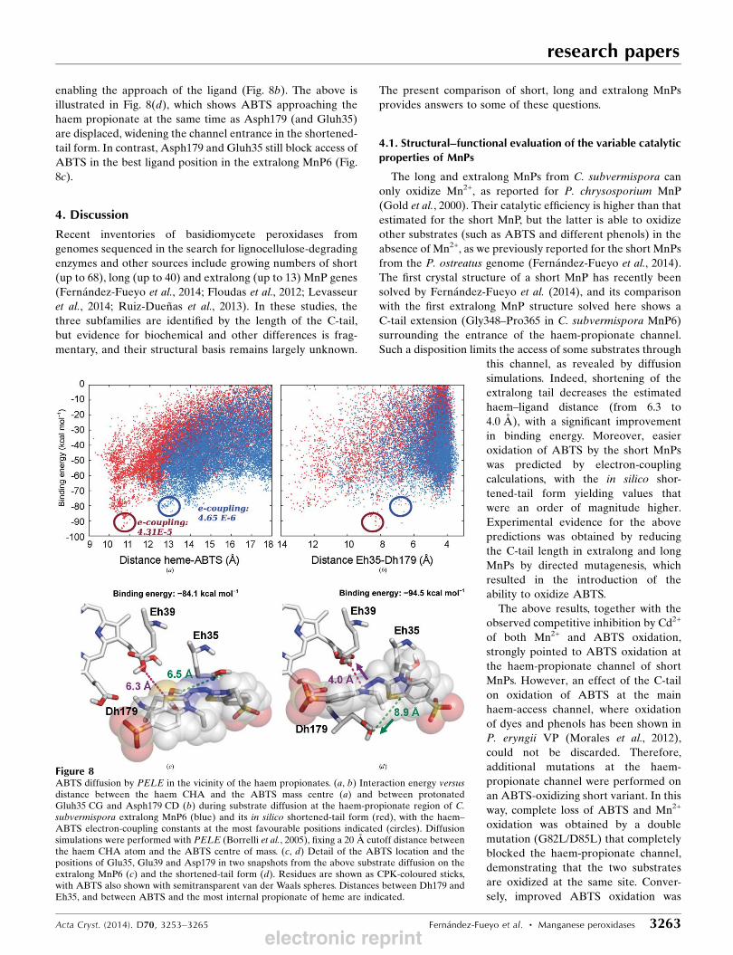
enabling the approach of the ligand (Fig. 8b). The above is
illustrated in Fig. 8(d), which shows ABTS approaching the
haem propionate at the same time as Asph179 (and Gluh35)
are displaced, widening the channel entrance in the shortened-
tail form. In contrast, Asph179 and Gluh35 still block access of
ABTS in the best ligand position in the extralong MnP6 (Fig.
8c).
4. Discussion
Recent inventories of basidiomycete peroxidases from
genomes sequenced in the search for lignocellulose-degrading
enzymes and other sources include growing numbers of short
(up to 68), long (up to 40) and extralong (up to 13) MnP genes
(Fernandez-Fueyo et al., 2014; Floudas et al., 2012; Levasseur
et al., 2014; Ruiz-Duenas et al., 2013). In these studies, the
three subfamilies are identified by the length of the C-tail,
but evidence for biochemical and other differences is frag-
mentary, and their structural basis remains largely unknown.
The present comparison of short, long and extralong MnPs
provides answers to some of these questions.
4.1. Structural–functional evaluation of the variable catalyticproperties of MnPs
The long and extralong MnPs from C. subvermispora can
only oxidize Mn2+, as reported for P. chrysosporium MnP
(Gold et al., 2000). Their catalytic efficiency is higher than that
estimated for the short MnP, but the latter is able to oxidize
other substrates (such as ABTS and different phenols) in the
absence of Mn2+, as we previously reported for the short MnPs
from the P. ostreatus genome (Fernandez-Fueyo et al., 2014).
The first crystal structure of a short MnP has recently been
solved by Fernandez-Fueyo et al. (2014), and its comparison
with the first extralong MnP structure solved here shows a
C-tail extension (Gly348–Pro365 in C. subvermispora MnP6)
surrounding the entrance of the haem-propionate channel.
Such a disposition limits the access of some substrates through
this channel, as revealed by diffusion
simulations. Indeed, shortening of the
extralong tail decreases the estimated
haem–ligand distance (from 6.3 to
4.0 A), with a significant improvement
in binding energy. Moreover, easier
oxidation of ABTS by the short MnPs
was predicted by electron-coupling
calculations, with the in silico shor-
tened-tail form yielding values that
were an order of magnitude higher.
Experimental evidence for the above
predictions was obtained by reducing
the C-tail length in extralong and long
MnPs by directed mutagenesis, which
resulted in the introduction of the
ability to oxidize ABTS.
The above results, together with the
observed competitive inhibition by Cd2+
of both Mn2+ and ABTS oxidation,
strongly pointed to ABTS oxidation at
the haem-propionate channel of short
MnPs. However, an effect of the C-tail
on oxidation of ABTS at the main
haem-access channel, where oxidation
of dyes and phenols has been shown in
P. eryngii VP (Morales et al., 2012),
could not be discarded. Therefore,
additional mutations at the haem-
propionate channel were performed on
an ABTS-oxidizing short variant. In this
way, complete loss of ABTS and Mn2+
oxidation was obtained by a double
mutation (G82L/D85L) that completely
blocked the haem-propionate channel,
demonstrating that the two substrates
are oxidized at the same site. Conver-
sely, improved ABTS oxidation was
research papers
Acta Cryst. (2014). D70, 3253–3265 Fernandez-Fueyo et al. � Manganese peroxidases 3263
Figure 8ABTS diffusion by PELE in the vicinity of the haem propionates. (a, b) Interaction energy versusdistance between the haem CHA and the ABTS mass centre (a) and between protonatedGluh35 CG and Asph179 CD (b) during substrate diffusion at the haem-propionate region of C.subvermispora extralong MnP6 (blue) and its in silico shortened-tail form (red), with the haem–ABTS electron-coupling constants at the most favourable positions indicated (circles). Diffusionsimulations were performed with PELE (Borrelli et al., 2005), fixing a 20 A cutoff distance betweenthe haem CHA atom and the ABTS centre of mass. (c, d) Detail of the ABTS location and thepositions of Glu35, Glu39 and Asp179 in two snapshots from the above substrate diffusion on theextralong MnP6 (c) and the shortened-tail form (d). Residues are shown as CPK-coloured sticks,with ABTS also shown with semitransparent van der Waals spheres. Distances between Dh179 andEh35, and between ABTS and the most internal propionate of heme are indicated.
electronic reprint
obtained by reducing the negative charge at the haem-
propionate channel owing to reduced electrostatic repulsion
with the anionic ABTS. Interestingly, the oxidation of a
substrate other than Mn2+ in the haem-propionate channel has
been described in ascorbate peroxidase (Mandelman et al.,
1998). Mn2+ oxidation by MnPs (Sundaramoorthy et al., 2010)
and VPs (Ruiz-Duenas et al., 2007) implies the closure and
opening of the haem-propionate channel (by reorientation of
the ion-binding side chains) for Mn2+ entering near the haem
propionate and release of the Mn3+ formed, respectively.
However, PELE simulations show that ABTS oxidation by
short MnP requires much more significant reorganizations
enabling the approach and oxidation of this bulky substrate.
4.2. Structural–functional evaluation of the variable stabilityproperties of MnPs
The C. subvermispora long and especially extralong MnPs
are exceptionally stable at acidic pH, while the short MnP is
quickly inactivated, in agreement with the results for other
short MnPs (Fernandez-Fueyo et al., 2014). On the other hand,
only slight differences in thermal stability were observed
between long and extralong MnPs, contrasting with the higher
thermostability reported for the Dichomitus squalens extra-
long MnPs compared with the P. chrysoporium long MnPs (Li
et al., 2001). In fact, we found the highest thermal stability for
the C. subvermispora short MnP, with a T50 value in the same
range as those of the P. ostreatus short MnPs (43–57�C;
Fernandez-Fueyo et al., 2014). Interestingly, removal of the
C-tail does not result in improved thermostability; on the
contrary, a 5–7�C decrease in T50 was observed, revealing that
the presence of the tail does not always result in decreased
MnP stability. This is explained by the numerous interactions
with the protein body observed in the extralong MnP crystal
structure, including three hydrophobic/hydrophilic patches.
Therefore, more factors than just the tail length affect the
thermal stability of MnPs.
Up to three Mn2+ or Cd2+ ions were identified in the
anomalous difference electron-density maps of extralong
MnP6–metal complexes. One of them occupied the Mn-
oxidation site, in agreement with directed-mutagenesis results,
and the others were located at neighbouring positions. Mn2+
binding at the oxidation site closes the lower part of the haem-
propionate access channel owing to the reorientation of the
Glu35 and Glu39 side chains, while the upper part of this
channel remains open, being the site for binding of the second
metal ion (near Asp85). Finally, the third metal ion in the
MnP6–Cd2+ complex binds to one of the last residues of the
C-tail (Asp363). Similar binding has been described for
P. chrysosporium MnP (Sundaramoorthy et al., 2005),
although the third site does not exist owing to the shorter
C-tail. Metal binding would contribute to haem fixation on the
protein, resulting in improved enzyme stability, as reported for
the P. chrysosporium and D. squalens MnPs (Mauk et al., 1998;
Youngs, Sundaramoorthy et al., 2000) and as confirmed here
for both short and long MnPs. Moreover, Cd2+ binding at the
Mn-oxidation site enabled us to describe ABTS oxidation at
the same site, as shown by competitive inhibition results.
On the other hand, MnP stabilization by Ca2+ agrees with the
general effect found in eukaryotic peroxidases (from the
superfamily of plant–fungal–prokaryotic peroxidases), where
addition of this cation reverts previous unfolding (for example
as caused by alkaline conditions; George et al., 1999; Youngs,
Moenne-Loccoz et al., 2000).
4.3. Evolutionary relationships of basidiomycete peroxidases
The relationships between C. subvermispora haem perox-
idases, a total of 26 proteins including one cytochrome c
peroxidase, and those from other basidiomycetes, a total of up
to 376 sequences, have been analyzed by Fernandez-Fueyo,
Ruiz-Duenas, Miki et al. (2012). The phylogram obtained (see
Supplementary Fig. S5) shows a cluster for the basidiomycete
members of the superfamily of plant–fungal–prokaryotic
peroxidases (almost 200 sequences). In this cluster, three main
groups, corresponding to (i) short-MnPs, VPs and LiPs (with
the latter forming a separate subgroup), (ii) long and extra-
long MnPs and (iii) generic peroxidases, are defined (together
with several unclustered sequences). The separate clustering
of short MnPs, and the intermixed presence of long and
extralong MnPs, reveals a separate origin for the short MnP
subfamily, while the long and extralong MnP types would not
constitute two separate groups from an evolutionary point of
view. The evolutionary relatedness between long and extra-
long MnPs would be on the basis of the previously discussed
biochemical and structural similarities between these two
peroxidase types, while the short MnPs would represent a
distant branch more related to VPs, with which they share
biochemical and structural characteristics.
5. Conclusions
The evolutionary analysis, together with the different
biochemical and structural properties, including the presence
of a 14–22-residue shorter C-tail in short MnPs, supports their
classification as a different subfamily. In contrast, the long and
extralong forms cluster together in the peroxidase phylogram,
with their catalytic and stability properties showing only slight
differences, and present similar structures with conserved
C-tails (in spite of their different lengths). These modest
differences do not justify maintaining them as two separate
subfamilies in genome-annotation and other peroxidase
studies. Interestingly, in the above comparison we demon-
strate for the first time that substrates other than Mn2+ can be
oxidized at the Mn-oxidation site of some MnPs. The present
work provides a structural basis for the different catalytic and
stability properties characterizing different MnPs and the
involvement of the C-tail extension in some of them. In this
way, we contribute to better structural and functional under-
standing of the enzymes involved in lignin attack, which is a
key issue for the industrial use of plant biomass in ligno-
cellulose biorefineries.
This work was supported by Spanish projects BIO2011-
26694, CTQ2010-18123, BFU2011-24615 and CSD2009-00088
research papers
3264 Fernandez-Fueyo et al. � Manganese peroxidases Acta Cryst. (2014). D70, 3253–3265
electronic reprint

and the EU projects KBBE-2013-3-613549 and ERC-2009-
Adg25027. Sequencing of the C. subvermispora genome by
JGI was supported by the US DOE under Contract DE-AC02-
05CH11231. FJR-D acknowledges a Ramon y Cajal contract.
The authors thank the staff of Synchrotron SOLEIL (France),
ALBA (Spain) and Swiss Light Source (Switzerland).
References
Adams, P. D. et al. (2010). Acta Cryst. D66, 213–221.Borrelli, K. W., Vitalis, A., Alcantara, R. & Guallar, V. (2005). J.Chem. Theory Comput. 1, 1304–1311.
Chen, V. B., Arendall, W. B., Headd, J. J., Keedy, D. A., Immormino,R. M., Kapral, G. J., Murray, L. W., Richardson, J. S. & Richardson,D. C. (2010). Acta Cryst. D66, 12–21.
Emsley, P. & Cowtan, K. (2004). Acta Cryst. D60, 2126–2132.Fernandez-Fueyo, E., Ruiz-Duenas, F. J., Ferreira, P. et al. (2012).Proc. Natl Acad. Sci. USA, 109, 5458–5463.
Fernandez-Fueyo, E., Ruiz-Duenas, F. J., Martınez, M. J., Romero, A.,Hammel, K. E., Medrano, F. J. & Martınez, A. T. (2014).Biotechnol. Biofuels, 7, 2.
Fernandez-Fueyo, E., Ruiz-Duenas, F. J., Miki, Y., Martınez, M. J.,Hammel, K. E. & Martınez, A. T. (2012). J. Biol. Chem. 287, 16903–16916.
Floudas, D. et al. (2012). Science, 336, 1715–1719.George, S. J., Kvaratskhelia, M., Dilworth, M. J. & Thorneley, R. N. F.
(1999). Biochem. J. 344, 237–244.Giardina, P., Palmieri, G., Fontanella, B., Rivieccio, V. & Sannia, G.
(2000). Arch. Biochem. Biophys. 376, 171–179.Gold, M. H., Youngs, H. L. & Gelpke, M. D. (2000). Met. Ions Biol.Syst. 37, 559–586.
Kabsch, W. (2010). Acta Cryst. D66, 125–132.Kirk, T. K. & Farrell, R. L. (1987). Annu. Rev. Microbiol. 41, 465–501.Levasseur, A. et al. (2014). BMC Genomics, 15, 486.Li, D., Youngs, H. L. & Gold, M. H. (2001). Arch. Biochem. Biophys.385, 348–356.
Mandelman, D., Jamal, J. & Poulos, T. L. (1998). Biochemistry, 37,17610–17617.
Martınez, A. T., Ruiz-Duenas, F. J., Martınez, M. J., del Rıo, J. C. &Gutierrez, A. (2009). Curr. Opin. Biotechnol. 20, 348–357.
Martinez, D., Larrondo, L. F., Putnam, N., Gelpke, M. D., Huang, K.,Chapman, J., Helfenbein, K. G., Ramaiya, P., Detter, J. C., Larimer,
F., Coutinho, P. M., Henrissat, B., Berka, R., Cullen, D. & Rokhsar,D. (2004). Nature Biotechnol. 22, 695–700.
Mauk, M. R., Kishi, K., Gold, M. H. & Mauk, A. G. (1998).Biochemistry, 37, 6767–6771.
Morales, M., Mate, M. J., Romero, A., Martınez, M. J., Martınez, A. T.& Ruiz-Duenas, F. J. (2012). J. Biol. Chem. 287, 41053–41067.
Perez-Boada, M., Ruiz-Duenas, F. J., Pogni, R., Basosi, R.,Choinowski, T., Martınez, M. J., Piontek, K. & Martınez, A. T.(2005). J. Mol. Biol. 354, 385–402.
Piontek, K., Glumoff, T. & Winterhalter, K. (1993). FEBS Lett. 315,119–124.
Poulos, T. L., Edwards, S. L., Wariishi, H. & Gold, M. H. (1993). J.Biol. Chem. 268, 4429–4440.
Ragauskas, A. J., Williams, C. K., Davison, B. H., Britovsek, G.,Cairney, J., Eckert, C. A., Frederick, W. J., Hallett, J. P., Leak, D. J.,Liotta, C. L., Mielenz, J. R., Murphy, R., Templer, R. &Tschaplinski, T. (2006). Science, 311, 484–489.
Riley, R. et al. (2014). Proc. Natl Acad. Sci. USA, 111, 9923–9928.Ruiz-Duenas, F. J., Lundell, T., Floudas, D., Nagy, L. G., Barrasa, J. M.,
Hibbett, D. S. & Martınez, A. T. (2013). Mycologia, 105, 1428–1444.Ruiz-Duenas, F. J. & Martınez, A. T. (2009). Microb. Biotechnol. 2,
164–177.Ruiz-Duenas, F. J., Morales, M., Perez-Boada, M., Choinowski, T.,
Martınez, M. J., Piontek, K. & Martınez, A. T. (2007). Biochemistry,46, 66–77.
Scott, G. M., Akhtar, M., Lenz, M. J. & Swaney, R. E. (1998).Environmentally Friendly Technologies for the Pulp and PaperIndustry, edited by R. A. Young & M. Akhtar, pp. 341–384. NewYork: Wiley.
Steffen, K. T., Hofrichter, M. & Hatakka, A. (2002). Enzyme Microb.Technol. 30, 550–555.
Sundaramoorthy, M., Gold, M. H. & Poulos, T. L. (2010). J. Inorg.Biochem. 104, 683–690.
Sundaramoorthy, M., Kishi, K., Gold, M. H. & Poulos, T. L. (1994). J.Biol. Chem. 269, 32759–32767.
Sundaramoorthy, M., Youngs, H. L., Gold, M. H. & Poulos, T. L.(2005). Biochemistry, 44, 6463–6470.
Voityuk, A. A. (2012). Phys. Chem. Chem. Phys. 14, 13789–13793.Wallrapp, F. H., Voityuk, A. A. & Guallar, V. (2013). PLoS Comput.Biol. 9, e1002990.
Youngs, H. L., Moenne-Loccoz, P., Loehr, T. M. & Gold, M. H.(2000). Biochemistry, 39, 9994–10000.
Youngs, H. L., Sundaramoorthy, M. & Gold, M. H. (2000). Eur. J.Biochem. 267, 1761–1769.
research papers
Acta Cryst. (2014). D70, 3253–3265 Fernandez-Fueyo et al. � Manganese peroxidases 3265electronic reprint

Acta Cryst. (2014). D70, doi:10.1107/S1399004714022755 Supporting information
Volume 70 (2014)
Supporting information for article:
Structural implications of the C-terminal tail in the catalytic and stability properties of manganese peroxidases from ligninolytic fungi
Elena Fernández-Fueyo, Sandra Acebes, Francisco J. Ruiz-Dueñas, María Jesús Martínez, Antonio Romero, Francisco Javier Medrano, Victor Guallar and Angel T. Martínez

Acta Cryst. (2014). D70, doi:10.1107/S1399004714022755 Supporting information, sup-1
The Supporting Information consists of five supplementary figures showing stereo view of the
general structure of extralong MnP6 (Figure S1), multiple alignment of extralong, long and short
MnP sequences from different basidiomycetes (Figure S2), mobility of Mn2+
-binding residues in the
in silico shortened-tail form of C. subvermispora MnP6 as shown by MD (Figure S3), ABTS
binding and C-tail and mobility as shown by PELE (Figure S4), and phylogram of C.
subvermispora and other basidiomycete peroxidases (Figure S5).
Figure S1. Stereo view of general structure of extralong MnP. A ribbon model of C. subvermispora extralong MnP6
(4CZN) is shown with helices in green, short strands in dark red, and C-tail extension (Gly348-Ser364 being absent
from short MnP) in red (Pro365 is not included in 4CZN, but it is present in structures including a metal ion). Residues
involved in the binding of Mn2+
(Glu35, Glu39 and Asp179) and forming disulphide bridges are shown as CPK sticks,
Ca2+
ions as yellow spheres, and Na+ ion at the metal-binding site as orange sphere (see Figure 3A for Mn
2+ binding).

Acta Cryst. (2014). D70, doi:10.1107/S1399004714022755 Supporting information, sup-2
Figure S2. Multiple alignment of extralong, long and short MnPs sequences. C. subvermispora extralong
(MnP6/MnP12), long (MnP5/MnP10) and short (MnP13) MnPs, extralong MnPs from Phlebia radiata (GenBank
CAC85963) and D. squalens (JGI 169843), long MnP from P. chrysosporium (JGI 66512), and short MnPs from P.
radiata (GenBank Q96TS6), D. squalens (JGI 169526) and P. ostreatus (JGI 1099081) are included. Extra residues in
long and extralong MnPs are in magenta and red, respectively. Conserved residues include: i) eight cysteines (cyan); ii)
nine Ca2+
ligands (green); iii) two active site histidines (dark gray); and iv) three acidic residues (red) forming the Mn2+
-
oxidation site. Numbering starts at the first residue of the mature protein (red asterisk). Symbols below indicate full
conservation of the same (*) or equivalent residues (:) and partial residue conservation (.).
*
MnP-12 (157986) --MAFPALLALVALAASVRAAPAS--SSAVCSDGT-IVSNAVCCDFIPLAQDLQSMVLQN 31
DICSQ MnP3 (169843) MAFKLLSIVSLVALATVASAAPS----RTVCSDGT-VVPDSVCCEFLPLAEALQTQVLMG 31
PHLRA MnP2 (CAC85963) MAFNFAAILAFVSLAAVTSAAPS----KTTCSNGV-VVPDAVCCDFVPLASALQSEVLMG 31
MnP-6 (50686) --MSFATLLAIVSLAAIATAAPT-----AVCSDGT-RVSNAVCCDFVSLGQDLQSMVLQG 31
MnP-10 (117436) --MAFTSFVALAALVGIASAAPT-----TICPDGT-RVSNHACCAFIPLAEDLQKTIFMN 31
PHACH MnP1 (66512) --MAFKSLIAFVALAAAVRAAPT-----AVCPDGT-RVSHAACCAFIPLAQDLQETIFQN 31
MnP-5 (49683) --MAFTSLLALSALVAVSRAAPT-----AVCSDGT-RVSNSACCAFIPLAQDLQETLFMN 31
DICSQ MnP6 (169524) --MVFKALLIS-VLAAFQITKGAL-IRRATCSDGT-VVANSACCVLIPVIQDIQENLFDG 31
PHLRA MnP3 (Q96TS6) --MAFKQLLT--AISIVSVANAAL-TRRVACPDGVNTATNAVCCSLFAVRDLIQDQLFDG 32
MnP-13 (124076) --MAFKPLAALVALLSVSIAHGAI-TRRVTCPDGVNTVANAACCPLFAVRDDIQESLFDG 32
PLEOS MnP4 (1099081) MVNSFHSLLSTIALALLVPSVLAVPAHRAKCSKGR-TASNDACCVWFDVLDDIQENLFDG 31
: : : : : . *..* ... .** . : . :* :: .
MnP-12 (157986) -DCGEDAHEIIRLTFHDAIPIS---RSLGPSAGGGADGSMLIFPLVEPEFFASNGIDDSV 87
DICSQ MnP3 (169843) -DCGEDTHELLRLTFHDAIAIS---RSN-ASAGGGADGSMLIFPTVEPAFFANLGIADSV 87
PHLRA MnP2 (CAC85963) -DCGEDAHELVRLIFHDAIAIS---QSMGPSAGGGADGSMLIFPTVEPAFFPNLGIADSV 87
MnP-6 (50686) -DCGEDAHEIIRLTFHDAVAIS---RKLGPSAGGGADGSMLLFPLVEPEFAASNGIDDSV 87
MnP-10 (117436) -DCGEDAHEVIRLTFHDAVAIS---RKLGPKAGGGADGSMLLFPTVEPNFSANNGIDDSV 87
PHACHMnP1 (66512) -ECGEDAHEVIRLTFHDAIAIS---RSQGPKAGGGADGSMLLFPTVEPNFSANNGIDDSV 87
MnP-5 (49683) -DCGEDAHEVIRLTFHDAVAIS---RSQGPSAGGGADGSMLLFPTVEPNFSANNGIDDSV 87
DICSQ MnP6 (169524) GECGEEVHESLRLTFHDAIGISPAIAATGVFGGGGADGSIILFEDIEPNFHANNGVDEII 91
PHLRA MnP3 (Q96TS6) GECGEEVHESLRLTFHDAIGISPTIASTGVFGGGGADGSIAIFAEIETNFHANNGVDEII 92
MnP-13 (124076) GKCGEEVHESLRLTFHDAIGFSPSLTAQGKFGGGGADGSIAIFESIETGYHANLGIDEII 92
PLEOS MnP4 (1099081) GECGEEVHESLRLTFHDAIGFSPALTRQGKFGGGGADGSIMLFSDIETNFAANNGVDDIV 91
.***:.** :** ****: :* .*******: :* :*. : .. *: : :
MnP-12 (157986) NNLIPFLSSHPTISAGDLVQFAGAVALSNCPGAPRATFFAGRPNATAPAIDGLIPEPQDN 147
DICSQ MnP3 (169843) NNLIPFLSQFPSISAGDLVQFAGAVAITNCPGAPQLEFLAGRPNATAPAIDGLIPEPQDN 147
PHLRA MnP2 (CAC85963) NNLIPFLSQFPTISAGDLVHFAGAVAISNCPGAPQLEFLAGRPNATAPAIDGLIPEPQDD 147
MnP-6 (50686) NNLIPFLSLHPTISAGDLVQFAGAVALSNCPGAPRVQFLAGRPNHTIAAIDGLIPEPQDN 147
MnP-10 (117436) NNLIPFMARHPTVSAGDLVQFAGAVALSNCPGAPRLEFLAGRPNHTIAAIDGLIPEPQDD 147
PHACHMnP1 (66512) NNLIPFMQKHNTISAADLVQFAGAVALSNCPGAPRLEFLAGRPNKTIAAVDGLIPEPQDS 147
MnP-5 (49683) NNLIPFLAKHP-VSAGDLVQFAGAIALTNCPGAPQLEFLAGRPNHTIAAVDGLIPEPQDD 146
DICSQ MnP6 (169524) DEQKPIIAKHN-ITTADFIQLAGAIGVSNCPGAPQLNVFIGRPDATQPAPDKTVPEPFDS 150
PHLRA MnP3 (Q96TS6) GEQAPFIQMTN-MTTADFIQFAGAVGVSNCPGAPALPVFVGRPDATQPAPDKTVPEPFDT 151
MnP-13 (124076) NEQATFILKHN-MTAGDFIQFAGAVGVSNCPGAPQLEFLLGRPAATAPAPDKTVPEPFDT 151
PLEOS MnP4 (1099081) EQQKPIAIKHQ-VSFGDFIQFAGAVGSSNCAGGPRIQFLAGRSNVTKPSPDHLVPEPFDS 150
: .: :: .*::::***:. :**.*.* .: **. * .: * :*** *
MnP-12 (157986) VTSILAR----FDDAGGFTPFEVVSLLASHTIARADKVDPTLDAAPFDTTPFTFDTQFFL 203
DICSQ MnP3 (169843) ITKILAR----FDDAGGFTPFEVVSLLASHTIARADHVDPTLDAAPFDSTPFTFDTQIFL 203
PHLRA MnP2 (CAC85963) VTKILAR----FKDAGNFSPAEVVALLASHSIARADHVDPTLDAAPFDSTPFDFDTQVFL 203
MnP-6 (50686) VTSILER----FDDAGGFTPFEVVSLLASHTIARADKVDPTLDAAPFDTTPFTFDSQIFL 203
MnP-10 (117436) VTKILER----FDDAGGFTPFEVVSLLASHTVARADKVDETIDAAPFDSTPFTFDTQVFL 203
PHACHMnP1 (66512) VTKILQR----FEDAGGFTPFEVVSLLASHSVARADKVDQTIDAAPFDSTPFTFDTQVFL 203
MnP-5 (49683) VTKILAR----FDDAGGFSPFEVVSLLASHTVARADKVDETIDAAPFDSTPFTFDTQVFL 202
DICSQ MnP6 (169524) VDSILARFQDAFSDVGGFTPAEVVALLASHTIAAADHVDPSIPGTPFDSTPELFDTQFFI 210
PHLRA MnP3 (Q96TS6) VDSILAR----FADAGGFSSAEVVALLASHTIAAADHVDPSIPGTPFDSTPEIFDTQFFI 207
MnP-13 (124076) VDSILAR----FGDAG-FSPQEVIALLASHSVAAADHVDPAIPGTPFDSTPSDFDPQIFI 206
PLEOS MnP4 (1099081) VTSILAR----MGDAG-FKPDEVVALLASHSVAAQDTIDPKLAGHPFDSTPSDFDSQFFV 199
: .** * : *.* *.. **::*****::* * :* : . ***:** **.*.*:
MnP-12 (157986) ETLLKGVGFPGTDDNVGEVASPLPLGDTSTGGNDTGMMRLQSDFVLARDERTACFWQSFV 263
DICSQ MnP3 (169843) DVLLKGVGFPGLNNNTGEVSSPLPLSN----GTDVGELRLQSDFGLAHDPRTACFWQGFV 259
PHLRA MnP2 (CAC85963) EVLLKGVGFPGLANNTGEVSSPLPVTD----GTDVGELRLQSDFALARDERTACAWQSFV 259
MnP-6 (50686) EVLLKGVGFPGLDNNTGEVSSPLPLGDTSTGGKDTGLMRLQSDFALAHDPRTACFWQGFV 263
MnP-10 (117436) EVLLKGVGFPGTDNNTGEVASPLPKGS----GNDTGEMRLQSDFALARDPRTACFWQGFV 259
PHACHMnP1 (66512) EVLLKGVGFPGSANNTGEVASPLPLGS----GSDTGEMRLQSDFALAHDPRTACIWQGFV 259
MnP-5 (49683) EVLLKGVGFPGTDNNTGEVASPLPLTS----GNDTGEMRLQSDFALARDSRTACFWQGFV 258
DICSQ MnP6 (169524) ETQLRGTLFPGTGGNQGEAQSALA-----------GELRLQSDSELARDSRTACEWQSFV 259
PHLRA MnP3 (Q96TS6) ETQLRGILFPGTGGNQGEVESPLH-----------GEIRLQSDSELARDSRTACEWQSFV 256
MnP-13 (124076) EVQLRGTLFPGTGGNQGEVESPYP-----------GEIRLQSDHNLARDSRTACFWQSLA 255
PLEOS MnP4 (1099081) ETLLKGTLIPGDSLHKGQVKSPLP-----------GEFRLQSDELLARDSRTSCEWQSFI 254
:. *:* :** : *:. *. * :***** **:* **:* **.:
MnP-12 (157986) NQQDLMAESFKAAFFKLSLLGHNQADLVDCSEVVPIPVPGDGKPATFPATTGPQDLQLTC 323
DICSQ MnP3 (169843) NEQEFMAQSFKAAMAKLAVLGHNADDLVDCSAVVPKPKLAVAVSAAFPATKGPDDLELSC 319
PHLRA MnP2 (CAC85963) NEQEAMATAFKNAVKKLAVLGHNRNDLVDCSAVVPVPKPATGTPATFPASTGPQDLELTC 319
MnP-6 (50686) DQQEFMSQSFASAFAKLAVLGHNTDDLIDCSEVVPVPKPAVDKPTTFPATTGPQDLELSC 323
MnP-10 (117436) DEQEFMAESFKAAMAKLAILGHNRASLTDCSDVVPIPRPAVKKPASFPATTGPKDLELTC 319
PHACHMnP1 (66512) NEQAFMAASFRAAMSKLAVLGHNRNSLIDCSDVVPVPKPATGQPAMFPASTGPQDLELSC 319
MnP-5 (49683) NEQEFMAQSFKAAMSKLAVLGHSRSDLIDCSDVIPTPKPAVNKPATFPASTGPKDLELSC 318
DICSQ MnP6 (169524) NNQAKLQSAFKAAFRRMSILGHDESSLIDCSDVVPVP-PAPASDAHFPAGQTIDDVEQAC 318
PHLRA MnP3 (Q96TS6) NNQAKIQSAFKAAFRKMTILGHSESSLIECSEVIQTP-PALEGNAHLPAGQTMNDIEQAC 315
MnP-13 (124076) NNQQQMQAQFKAAMAKLAVLGQDVSQMVDCSDVIPVP-LPPATTPHIPAGLSQNNIEQAC 314
PLEOS MnP4 (1099081) SNPNSMVPKFERAMAKMATLGQNPKKLIDCSEVIPVP-RGRVKQPTLPAGKTIKDIEASC 313
.: : * *. ::: **:. .: :** *: * . :** .::: :*
MnP-12 (157986) TAERFPTLSVDPGATETLVPHCSDGGENCPSVQFAGPATGFNGTD-- 368
DICSQ MnP3 (169843) NTSRFPNLPIDHGTQEALIPHCSDGSMSCTTVQFDGPALSFGDNNSS 366
PHLRA MnP2 (CAC85963) TTEPFPTLSTAPGAQQTLIPHCSDGTMTCNSVQFDGPATNFGGADDS 366
MnP-6 (50686) LAERFPTLSVDPGAQETLIPHCSDGLENCTSVQFSGPATDSP----- 365
MnP-10 (117436) RAERFPTLTVDRGAVQALIPHCSNGGQDCPSVQFDGPA--------- 357
PHACHMnP1 (66512) PSERFPTLTTQPGASQSLIAHCPDGSMSCPGVQFNGPA--------- 357
MnP-5 (49683) FAERFPTLPVTPGATQTLIPHCSNGGEDCPTVQFTGPA--------- 356
DICSQ MnP6 (169524) ASTPFPTLPTDPGPASSVAPVPPS----------------------- 342
PHLRA MnP3 (Q96TS6) ATTPFPSLSADPGPATSVAPVPPS----------------------- 339
MnP-13 (124076) ATAAFPTLPIDVGPQTSIPPVPGS----------------------- 338
PLEOS MnP4 (1099081) RKAPFPRLPTDKGTFTSILPVPSS----------------------- 337
** *. *. :: . .

Acta Cryst. (2014). D70, doi:10.1107/S1399004714022755 Supporting information, sup-3
Figure S3. Mobility of Mn
2+-binding residues in the MnP6 in silico shortened-tail form as shown by MD. Distances
(in Å) between heme-CG and protonated E35-HE2 (green), E39-HE2 (blue) and D179-HD2 (red) during 100 ns MD
simulation (300 K) on the shortened-tail form of C. subvermispora MnP6.

Acta Cryst. (2014). D70, doi:10.1107/S1399004714022755 Supporting information, sup-4
Figure S4. PELE molecular simulations on C. subvermispora MnP6. (A, B) Preliminary exploration on ABTS
diffusion showing three minima in heme-ABTS distance vs binding energy (A), and location of the three sites at the
entrance of the heme-propionate channel at 6.2 Å from the heme (I), near the distal Ca2+
binding site at 14.3 Å from the
heme (II), and the main heme access channel at only 4.8 Å but with the worst binding energy (III). (C) Mobility of the
extralong tail preventing ABTS approach, as shown by superimposition of extralong MnP6 (blue ribbon, and heme) and
one ABTS position (orange sticks) selected from the PELE diffusion on the in silico shortened-tail form, the latter being
in collision with the extralong tail whose mobility is indicated in dark blue (the end of the shortened-tail variant is in
orange).
B C
A

Acta Cryst. (2014). D70, doi:10.1107/S1399004714022755 Supporting information, sup-5
Figure S5. Phylogram of basidiomycete peroxidases (GenBank and genomes). The position of 25 sequences from C.
subvermispora genome is shown (underlined) including one short, seven long and five extralong MnPs, two LiPs and
one generic peroxidase (all in Class II of plant-fungal-prokaryotic peroxidase superfamily), and nine members of the
heme-thiolate peroxidase (HTP) superfamily. Clusters where C. subvermispora sequences are not included were
collapsed, and prokaryotic peroxidases are not included. The three main peroxidase groups, Class II, HTP and dye-
decolorizing peroxidases (DyP), and the number of sequences in each of them (parentheses) are indicated, together with
the scaffold (sc) and JGI references of the C. subvermispora gene models (parentheses). Additionally, MnP-atypical
corresponds to a MnP with only two acidic residues at the oxidation site; TC-LiP corresponds to an unusual LiP from T.
cervina; and CERRI MnP-short corresponds to a Ceriporiopsis rivulosa MnP (GenBank BBB83813) clustering together
with C. subvermispora short MnP13. Adapted from Fernandez-Fueyo et al. (2012).
A
B
C
D
E
F
G
H
MnP13-short (sc7,124076)
VP/MnP-shortLiP2 (sc20,118677)LiP1 (sc20,99382)
MnP-short
MnP-atypical
MnP-short/MnP-atypical
MnP-longMnP-shortGPMnP9-long (sc6,114076)
MnP7-long (sc6,105539)MnP8-long (sc6,114036)MnP1-extralong (sc11,116608)MnP3-extralong (sc11,139965)MnP6-extralong (sc6,50686)MnP12-extralong (sc11,157986)
MnP5-long (sc6,49863)MnP4-long (sc6,94398)MnP10-long (sc14,117436)MnP-longMnP2-extralong (sc6,50297)MnP11-long (sc19,143390)MnP-longGPGP1 (sc3,112162)
HTP
HTP9 (sc3,122198)HTP2 (sc3,81391)HTP3 (sc7,114787)
HTP4 (sc7,114799)
HTP5 (sc7,115079)
HTP8 (sc2,121474)
HTP6 (sc8,115379)HTPHTP1 (sc2,80799)HTPHTP7 (sc16,118102)
MnP-short
MnP-short
VP/MnP-short
LiP/VP
GP
MnP-extralong
MnP-long
GP
DyP
HTP
HTP
HTP
HTP
HTP
HTP
MnP-short/VP/TC-LiP
HTP (133)
Class II (196)
DyP (47)
CERRI MnP-short (ABB83813)