A tabu thresholding algorithm for arc crossing minimization in bipartite graphs
Upload
independentCategory
view
5download
0
Structural characterization of an intermolecularRNA±RNA interaction involved in the transcriptionregulation element of a bipartite plant virusRichard H. Guenther, Tim L. Sit, Hanna S. Gracz1, Michael A. Dolan1,
Hannah L. Townsend1, Guihua Liu1, Winnell H. Newman1, Paul F. Agris1 and
Steven A. Lommel*
Department of Plant Pathology, Box 7616 and 1Department of Molecular and Structural Biochemistry, Box 7622,North Carolina State University, Raleigh, NC 27695, USA
Received January 13, 2004; Revised March 9, 2004; Accepted April 14, 2004
ABSTRACT
The 34-nucleotide trans-activator (TA) located withinthe RNA-2 of Red clover necrotic mosaic virus foldsinto a simple hairpin. The eight-nucleotide TA loopbase pairs with eight complementary nucleotides inthe TA binding sequence (TABS) of the capsidprotein subgenomic promoter on RNA-1 and trans-activates subgenomic RNA synthesis. Short syn-thetic oligoribonucleotide mimics of the RNA-1TABS and the RNA-2 TA form a weak 1:1 bimolecu-lar complex in vitro with a Ka of 5.3 3 104 M±1. Ka
determination for a series of RNA-1 and RNA-2mimic variants indicated optimum stability isobtained with seven-base complementarity. Thermaldenaturation and NMR show that the RNA-1 TABS8mers are weakly ordered in solution while RNA-2TA oligomers form the predicted hairpin. NMR diffu-sion studies con®rmed RNA-1 and RNA-2 oligomercomplex formation in vitro. MC-Sym generatedstructural models suggest that the bimolecular com-plex is composed of two stacked helices, one beingthe stem of the RNA-2 TA hairpin and the otherformed by the intermolecular base pairing betweenRNA-1 and RNA-2. The RCNMV TA structural modelis similar to those for the Simian retrovirus frame-shifting element and the Human immunode®ciencyvirus-1 dimerization kissing hairpins, suggesting aconservation of form and function.
INTRODUCTION
RNA viruses employ a diverse array of RNA motifs for theregulation of gene expression through interactions with othernucleic acids and proteins. The genomes of positive (+)stranded RNA viruses behave as mRNAs within host cells to
allow the expression of viral proteins. However, many viralgenomes are polycistronic and the expression of inaccessibleopen reading frames (ORFs) is facilitated by multiplestrategies (1,2). Subgenomic RNA (sgRNA) synthesis is acommon mechanism for the generation of viral RNAs thatbehave as monocistronic mRNAs for internal and 3¢ co-terminal ORFs (3). Essentially, there are two proposedmechanisms for the generation of (+) stranded sgRNAs bythe virally encoded RNA polymerase (i) internal initiation oftranscription from the full length minus (±) strand copy of thegenomic RNA (4) and (ii) premature termination during (±)strand synthesis followed by independent replication of thistruncated RNA template (5). For both models, the presence ofa distinct subgenomic promoter element on the (±) strand or aterminator on the (+) strand, is required for the initiation oftranscription (1).
Recently, it has been determined that for several RNAviruses, RNA synthesis requires long-distance cis or transRNA±RNA interactions (6). It is suggested that viruses thatutilize these interactions generate sgRNA via the prematuretermination mechanism. The base pairing of upstream cis-acting elements to the subgenomic promoter was found to beessential for sgRNA synthesis in Potato virus X, Tomato bushystunt virus (TBSV) and Flock house virus (7±9). The situationis taken to its extreme for the bipartite genome of Red clovernecrotic mosaic virus (Fig. 1A) (RCNMV; genusDianthovirus, family Tombusviridae) where expression ofthe sgRNA from RNA-1 requires the base pairing of asequence element in trans in RNA-2 (10). The 34-nucleotidetrans-activator (TA) element from RNA-2 is predicted to forma stable hairpin structure with an eight-nucleotide looppossessing complementarity to an eight-nucleotide elementimmediately upstream of the sgRNA transcription start site onRNA-1, referred to as the trans-activator binding site (TABS)(Fig. 1B). Mutagenesis demonstrated that complementarybase pairing between the two elements is essential for sgRNAtranscription but the nature of this RNA±RNA tertiarystructure has not been elucidated.
*To whom correspondence should be addressed. Tel: +1 919 515 6992; Fax: +1 919 515 7716; Email: [email protected] addresses:Michael A. Dolan, Tripos Inc., St Louis, MO 63144, USAGuihua Liu, Department of Chemistry, China Medical University, Shenyang, China
Nucleic Acids Research, 2004, Vol. 32, No. 9 2819±2828DOI: 10.1093/nar/gkh585
Nucleic Acids Research, Vol. 32 No. 9 ã Oxford University Press 2004; all rights reserved
Published online May 20, 2004
Determination of the tertiary structures of RNAs, particu-larly RNA±RNA complexes is dif®cult. Only a few examplesof high-resolution non-duplex bimolecular RNA±RNA struc-tures such as the Human immunode®ciency virus-1 (HIV-1)kissing hairpins and dimerization signal (11,12) and inter-actions within the ribosome complex (13) have been elucid-ated and provide limited examples of structural variation.While bimolecular RNA interactions generally involve loop±loop interactions (14), it is likely that a range of structuraldiversity exists in bimolecular RNA±RNA complexes. Manystructures may prove to be similar to unimolecular tertiaryinteractions, particularly those of similar function. Here wereport direct biophysical evidence for the formation of a weak,yet stable bimolecular complex between the RCNMV RNA-2TA element and the RNA-1 subgenomic promoter. We furtherpropose structural models for this interaction that exhibitconservation in both structure and function with other viral cisand trans RNA interactions.
MATERIALS AND METHODS
RNA oligomer preparation
Synthetic oligomers used in the study were obtained from theNorth Carolina State University Nucleic Acids Facility andfrom Dharmacon, Inc. (Lafayette, CO). Isotopically labeledRNA for the NMR studies was obtained by T7 transcription ofa plasmid template containing the 21-nucleotide RCNMVRNA-2 sequence (Fig. 1A) cloned into pUC18 and terminatedat the XbaI site. The transcription reaction was performed at37°C for 8 h using a T7-MEGAshortscript High YieldTranscription Kit (Ambion, Austin, TX). For the isotopiclabeling, 80 mM uniformly labeled 13C and 15N GTP sodiumsalt (Isotec, Miamisburg, OH) was substituted for the kit's75 mM GTP solution. Each oligomer was puri®ed by anionexchange HPLC using a Machery-Nagel 250/10 NucleogenDEAE 60-7 column (Duren, Germany). The purity of eacholigomer was con®rmed by denaturing polyacrylamide gelelectrophoresis (PAGE). The RNA-1 oligomers representingthe TABS are designated R1 with a superscript denoting thelength, e.g. R18 is the eight-nucleotide oligomer (nucleotides2356±2363) used in preliminary NMR studies. Four R18
oligomers, used in PAGE and NMR experiments have anadditional + or ± superscript denoting the nucleotide shift fromthe central sequence (nucleotides 2356±2363). The RNA-2oligomer is similarly designated R220 (nucleotides 761±780)while mutant versions are identi®ed by a superscript denotingmutations in the U:G base pair. The RNA-2 transcript wasdesignated R221*. The sequences of the oligomers are shownin Figure 1A and Table 1.
Thermal denaturation
Thermal denaturation of the oligomers was monitored by UVabsorbance (at 260 nm) over a temperature range of 5±90°C(1°C min±1) using a Cary 3 Spectrophotometer (Varian, PaloAlto, CA). The denaturation pro®le of each oligomer wasconducted in triplicate at three concentrations, 0.5, 5 and50 mM in 10 mM Na phosphate, 100 mM NaCl, pH 7.0. Both 2and 10 mm cuvettes were used in order to maintain properoptical response. Variation of Tm was less than 1°C for allconcentrations. Samples were thermally denatured and then
renatured to determine transition reversibility. In all cases, adifference of less than 1°C in Tm was observed betweendenaturation and renaturation with no hysteresis detected.
Polyacrylamide gel electrophoresis assay
Denaturing PAGE (7 M urea, 9 mM Tris±borate, 0.25 mMEDTA in 20% polyacrylamide) was conducted to assess thepurity of the synthetic oligomers and to analyze thecomponents of the bimolecular complex. Non-denaturingPAGE (9 mM Tris±borate in 20% polyacrylamide run on ice)was used to investigate and characterize parameters affectingcomplex formation. After electrophoresis, gels were stainedwith ethidium bromide (EtBr), visualized by UV transillumin-ation and images recorded on a Bio-Rad Gel Doc 2000 imager
Figure 1. Genome organization, expression strategy and model of RNA-1±RNA-2 interaction facilitating dianthovirus RNA-1 sgRNA synthesis.(A) The bipartite RNA genome of Red clover necrotic mosaic virus(RCNMV) is depicted with open rectangles representing ORFs. RNA-1ORFs p27 and p88 are involved in viral replication while the capsid protein(CP) is the virion structural protein. RNA-2 encodes the movement protein(MP) required for cell-to-cell movement of the viral RNA. p88 is expressedby a ±1 ribosomal frameshifting event (±1 FS) of p27. The CP is expressedfrom a sgRNA generated during the replication of the virus. The RNA-1sequence near the sgRNA transcription start site (denoted with an arrow) isshown aligned with comparable regions from Sweet clover necrotic mosaicvirus (SCNMV) and Carnation ringspot virus (CRSV), the two otherspecies comprising the Dianthovirus genus. The RNA-2 sequence of the TAregion is shown below RNA-2 with the corresponding aligned regions fromSCNMV and CRSV. The shaded sequences represent the complementaryregions between RNA-1 and RNA-2 involved in trans-activation of tran-scription. The nucleotide sequences for the RCNMV oligomers used in thisstudy are depicted below their respective genomic sequences. The nucle-otide positions of the various sequences are noted. (B) Model of interactionpreviously proposed (10).
2820 Nucleic Acids Research, 2004, Vol. 32, No. 9

(Bio-Rad Laboratories, Hercules, CA). The intensities of theRNA complex bands were quanti®ed using Bio-Rad QuantityOne software. Over the concentration ranges used, it wasdetermined that EtBr intercalation reported a linear measure ofRNA concentration. Apparent association constants (Ka
values) were calculated based on one site binding with theaid of PRISM software (GraphPad Software, San Diego, CA).
NMR analysis
Samples were prepared by solubilizing puri®ed RNA in 0.2 mlof 90% H2O/10% D2O phosphate buffer [100 mM NaCl,10 mM NaPO4 buffer, pH 6.9 and 0.1 mM EDTA d4(Cambridge Isotope Laboratories, Andover, MA)] to give~0.5 mM RNA. Samples were equilibrated with multiplebuffer exchanges in Centricon spin columns (Millipore,Milford, MA) and placed in NMR microtubes (Shigemi,Allison Park, PA, stock # BMS005V). The NMR spectra forthe 1:1 complex of R18+1:R221* were also collected at 25°C in100% D2O phosphate buffer [100 mM NaCl, 10 mM NaPO4
buffer, pH 6.9 and 0.1 mM EDTA d4]. Bruker AVANCE500 MHz spectrometer with an Oxford Narrow Bore Magnet(Bruker NMR, Billerica, MA) and a 5 mm 1H/BB (109Ag-31P) inverse Triple-Axis Gradient Probe (ID500±5EB,Nalorac Cryogenic Corp., Martinez, CA) was used for allNMR experiments. The 2D NOESY spectra were acquiredusing a Watergate-water suppression pulse sequence (15) withmixing times of 120 and 250 ms and a recycle delay of 5.2 s.Data sets with 2048 complex points in t2 and 512 complexpoints in t1 were acquired with 5000 Hz sweep widths in bothdimensions and 128 scans per slice. All spectra wereprocessed with a combination of exponential and sine-skewedfunctions and zero-®lled to 2 K 3 2 K data points with anexponential weighting function or shifted sine-bell function toresolve overlapped imino protons using Bruker XWINNMRsoftware. The longitudinal eddy current (LED) pulse sequencewas used in all PFGSE-NMR diffusion experiments (16).
The gradient pulse interval, D and gradient duration, d, were32 and 4 ms, respectively. The settling time was 6 ms and therelaxation 1 s. Typically, 2048 scans were collected for eachspectra. The pulsed-®eld gradient was applied in the ydirection. The gradient strength in a series of 1H experimentswas incremented from 28 G cm±1 to about 56 G cm±1 in sixincrements. Accurate diffusion coef®cients were obtained forsix (three samples and three standard samples) from theslopes of ln (A/A0) versus g2, determined by least-squaresregression.
Computer modeling
Structures were generated using MC-Sym software (17). Forthis study, scripts were written to generate structural models
that both satis®ed experimental constraints while also retain-ing, as close as possible, A-form RNA. In an effort to explorethe full range of structures that could be constructed, anattempt was made to model by the step-wise addition of basescomprising the R1 oligomer to the R2 hairpin loop beginningwith the 5¢ terminus and in a separate run, from the 3¢terminus. This approach was used to eliminate the potentialbias of the software. The MC-Sym scripts and constraints usedin the modeling are available upon request. Output structureswere energy minimized by 300 steps of steepest descentgradient using B biomolecular modeling package (N.White,http://www.scripps.edu/~nwhite/B) with the AMBER force®eld parameters. Ribbon representations were created usingMOLMOL software version 2K.2 (18). Analysis of the RNAstructures was made with MC-Annotate (19). Structuralcomparisons were based on visual alignment and then the ®tof selected backbone phosphates. The root mean squaredeviation (RMSD) calculated for comparison of RCNMV toSimian retrovirus-1 (SRV-1) and HIV-1 RNA structures wasfor 11 stem and eight loop phosphates.
Biological activity of RCNMV RNA-2 mutants
Oligomers were synthesized that incorporated site-speci®cmutations in the RCNMV RNA-2 TA region. Complementaryoligomers were annealed and cloned into the TBSV expres-sion vector pHST2 as previously described (10) and thenassayed on Nicotiana benthamiana plants for the ability totrans-activate expression of GFP. Mutations were made at theend of the stem before the loop region such that the U:G basepair was changed to either a U:A (construct TA-M22) or a G:Ubase pair (construct TA-M23). RNA transcripts were synthe-sized in vitro and co-inoculated with an RCNMV RNA-1construct (R1SG1) containing the green ¯uorescent protein(GFP) as a reporter gene onto N.benthamiana plants. sGFPexpression was assayed 2±3 days post-inoculation (dpi) (10).
RESULTS
R18 and R220 form a 1:1 bimolecular complex
Non-denaturing PAGE was used to evaluate the interaction ofthe model oligomers representing the RNA-1 TABS and theRNA-2 TA (R18 and R220, respectively). When R18 and R220
were mixed and subjected to PAGE, an additional band wasobserved in the gel (Fig. 2A, lanes 3±7). When this band wasexcised from the gel, and re-electrophoresed under denaturingconditions, it was found to contain both R18 and R220 (Fig. 2B,lane 3) in the form of a complex. The formation of thiscomplex band did not require magnesium ions. To determinethe stoichiometry of the complex, multiple titrations of bothcomponent oligomers were subjected to PAGE and theintensities of the complex bands formed were determined bydensitometry. The density values were analyzed with Prismsoftware. The data best ®t a single site binding model,indicating a 1:1 stoichiometry for the complex. The apparentKa of the complex is 5.3 6 0.5 3 104 M±1.
Phylogenetic comparison of the TA elements for the threespecies comprising the Dianthovirus genus, all three of whichtrans-activate RCNMV sgRNA synthesis in vivo (10), indi-cated that a minimum complementarity of six bases issuf®cient for viral activity (Fig. 1A). A series of oligomers
Table 1. The apparent complex association constants, Ka, for a series ofRCNMV RNA-1 oligomers binding to R220
Oligomer name Sequencea Shift Apparent Ka (M±1)
R18+2 GGCGAUCA +2 <1 3 104
R18+1 GGGCGAUC +1 5.0 3 105
R18 GGGGCGAU 0 5.3 3 104
R18±1 AGGGGCGA ±1 5.3 3 104
R18±2 GAGGGGCG ±2 1.4 3 104
aNucleotides complementary to R220 are indicated in bold typeface.
Nucleic Acids Research, 2004, Vol. 32, No. 9 2821

that shifted an eight-nucleotide frame of the RNA-1 TABSelement was synthesized and PAGE assayed to determine howcomplementarity affected complex formation. A ®xed lengthof eight nucleotides was chosen to maintain the dynamics ofthe assay. From these experiments, it was determined that astable R1:R2 complex formed with six-base complementarity(Table 1). But the most stable complex formed with the sevencomplementary bases found in R18+1. The PAGE assay wasalso used to con®rm that R221*, a labeled in vitro transcript,formed a complex with the R18+1 oligomer.
Biophysical characterization of the complex components
The RNA oligomers were characterized by thermal denatura-tion over a range of 0.5±50 mM. R220 exhibited a bi-phasicdenaturation pro®le with a Tm of 59°C (data not shown). Theconcentration-independent Tm indicated that R220 was aunimolecular structure. This is consistent with the mfoldweb server prediction that the region of RNA-2, containingthis conserved TA sequence, forms a simple hairpin structurecomposed of a six-base pair stem and an eight-nucleotide loop(10). The imino region of the R220 2D NOESY (Fig. 3A),exhibited six well separated resonances consistent withWatson±Crick base paired imino protons. A signal for aterminal base paired imino proton was not observed due to thedynamics of the terminal base pair (20). Two resonances at12.0 and 10.9 p.p.m. possessed strong NOE cross-peaksattributed to a U:G base pair. Unlike typical Watson±Crickbase pairs that have a single imino proton, a U:G base pair hastwo imino protons with diagnostic strong cross-peaks(Fig. 3A). The assignment of the U:G base pair was furthercon®rmed in the 2D 1H1-15N spectra of R221*, an isotopicallylabeled transcript (Fig. 3B). The stem imino resonances, withthe exception of the terminal base pair, were assigned based on2D 1H-1H NOE connectivities using the U:G base pair as astarting point (21). In contrast, the thermal denaturationpro®les for the R1 oligomers exhibited only a limited increasein UV absorbance with increased temperature (data notshown) and the NMR spectra for R18+1 lacked featuresconsistent with an ordered structure (Fig. 3C). Thermaldenaturation of two additional mutant R2 sequences was
also performed (Table 2). Changing the base pairings of thenucleotides at the top of the stem from U:G to U:A resulted inan increase of 4°C in the Tm, consistent with mfold web serverpredictions of comparable stability for the two sequences.Changing the pairing from U:G to G:U also resulted in a 20°Cincrease in Tm. While this oligomer formed a unimolecularstructure based on concentration-independent Tm, the oligo-mer likely formed a different secondary structure than the six-base paired stem and eight-nucleotide loop of the TA.Structure prediction with mfold web server suggests that thesequence likely forms a more stable hairpin structure with asix-nucleotide loop, a ®ve-base pair stem and four unpaired
Figure 2. PAGE characterization of a complex formed when R18 and R220
are mixed. (A) Non-denaturing PAGE analysis. A titration of 30 mM ofR220 with increasing amounts of R18 (15±120 mM) gives rise to an addi-tional band. Lane 1, 30 mM R220; lane 2, 30 mM R18 (stains weakly); lanes3±7, 30 mM R220 with 15, 30, 60, 90 and 120 mM of R18, respectively. Theband in lane 7 was extracted and subjected to denaturing electrophoresis.(B) Denaturing PAGE was used to determine that the additional bandsformed under non-denaturing conditions are comprised of R18 and R220.Lane 1, R220; lane 2, R18; lane 3, excised band of R18 and R220 complex.
Figure 3. NMR spectra of RCNMV oligomers. (A) An expanded contourplot for the imino region of the NOESY spectrum of R220 with 120 ms mix-ing time at 15°C. Six imino resonances are observed. The two resonanceswithin the dashed box are assigned to the U6:G15 base pair that closes thestem of the hairpin. (B) 1H-15N spectra of R221* at 20°C identify thoseresonances that arise from labeled G residues. (C) The imino region of the1D proton spectra for R18+1 at 10°C.
2822 Nucleic Acids Research, 2004, Vol. 32, No. 9

nucleotides. NMR analysis also supports this as the preferredconformation (data not shown).
NMR diffusion rate and exchange regime of the R1:R2complex
Diffusion experiments were performed to con®rm that thecomplex formed in PAGE conditions also formed under NMRconditions. The NMR studies were conducted with R18+1 sincethis formed a complex with the highest Ka. When R18+1 andR221* were mixed at a ratio of 1:1, the diffusion rate shiftedfrom 1.89 3 10±6 to 1.56 3 10±6 cm2 s±1 (Table 3). Thedecrease in mobility was attributed to the formation of a 29-nucleotide RNA complex. Formation of this complexdecreased the signal-to-noise ratio, in the NMR spectra,4-fold for the exchangeable protons. This decrease was notobserved in the non-exchangeable signals. Loss of sensitivityarose from a change from the fast exchange for the R2 hairpinto an intermediate exchange regime for the complex. Attemptsto shift back to a fast exchange by altering the temperature,pH, and addition of mono and divalent counter ions, wereunsuccessful. Similar conversion to intermediate exchangeregimes for other bimolecular RNA complexes with compar-able weak association constants have previously been reported(22).
NMR of the complex
The NMR proton spectra for the R18+1:R220 complex, at 20°C,were notably different from those of the uncomplexed R220
(Fig. 4A and B). The imino resonances in the complex werebroadened as compared to those of uncomplexed R220. Thecomplex's imino region exhibited ®ve relatively narrow, wellresolved, resonances and several broad resonances. Thenarrow signals at 14.1, 13.4, 12.8 and 12.3 (p.p.m.), wereassigned to U18, G5, G4 and G2, respectively, in the stem ofthe RNA-2 hairpin. This assignment was based on con-nectivity in the 2D NOESY for the R18+1:R220 complex and
the 2D 1H1-15N spectra of the R18+1:R221* complex (Fig. 4Band C). The retention of these resonances in the 2D 1H1-15Nspectra of the R18+1:R221* complex constitutes de®nitiveevidence for the persistence of the R2 hairpin stem in the
Table 2. Thermal stability, Tm, apparent association constants, Ka, for RNA2 mimics and correlation ofin vitro complex formation with in vivo trans-activation for selected RCNMV RNA-2 stem sequencemutations
Oligomer RNA-2 stemtermination
Tm Ka of complex(M±1)
Viralconstructa
sGFPproductionb
R220 U:G 59.0 5.3 3 104 TA-WT +++R220U:A U:A 63.8 5.3 3 104 TA-M22 +++R220G:U G:U 79.2 2.4 3 104 TA-M23 +/±
aCorresponding TA mutant constructs in Tomato bushy stunt virus vector pHST2 (see Materials and Methods).bsGFP production from co-inoculation with R1SG1 on N.benthamiana plants measured 2±3 dayspost-inoculation: +++, wild-type level; +/±, <10%.
Table 3. Values of NMR diffusion experiments for selected oligomers
Oligomer (size) Diffusion coef®cient(10±6 cm2 s±1)
Control (12mer) 2.38 6 0.02R221* (21mer) 1.87 6 0.05Control (28mer) 1.52 6 0.031:1 R18+1:R221* (complex) 1.56 6 0.02
Figure 4. NMR spectra of the RCNMV complex. (A) The imino region ofthe 1D proton spectra for R220 and 1:1 complex of R18+1:R220 at 10°C.(B) The NMR, 2D NOESY spectra of the imino region for a 1:1 complex ofR18+1:R220 at 10°C, with 250 ms mixing time. The connectivities of thestem backbone protons of R220 hairpin are observed in the complex. (C) Theimino region of the 1H-15N spectra for a 1:1 complex of R18+1:R221* at20°C.
Nucleic Acids Research, 2004, Vol. 32, No. 9 2823

complex and excludes the possibility that the R2 hairpin iscompletely relaxed upon complex formation. The remainingwell separated, resonance at 12.6 p.p.m. cannot be de®nitelyassigned, but certainly does arise from one of the labeledR221* G residues. Although this 12.6 p.p.m. signal does nothave connectivity to other imino signals, it likely represents aG:C base pair based on the 2D NOESY cross-peaks.Comparison of the R220 (Fig. 3A) and the R18+1:R220
complex (Fig. 4B) 2D NOESY spectra indicated that thecomplex lacked the clear and characteristic U:G base paircross-peaks prominent in the R220 hairpin (boxed region in
Fig. 3A). This observation is consistent with the absence of theG6 N1 proton in the 1H1-15N spectra of the R18+1:R221*complex (Fig. 4C). Collectively, these results indicate thatformation of the RNA-1:RNA-2 complex results in a partialrelaxation of the RNA-2 hairpin stem and the concomitant lossof the U:G base pair closing the stem. Additional noteworthydistinctions between the R220 and complex NOESY spectrawere observed in other regions as well. Figure 5 compares thespectra in three regions; the imino to aromatic (Fig. 5A),aromatic and H1¢ (Fig. 5B), and the sugar to aromatic(Fig. 5C). While detailed interpretation of the data from these
Figure 5. Comparison of selected regions of the water NOESY spectra for RCNMV R220 and the R220:R18+1 complex. (A) In the region of the spectra charac-teristic of imino to aromatic interactions, a number of resolved cross-peaks are observed for both spectra. Using a color coded overlay of the two spectra, newresonances are observed (red) while most are unchanged (green) and others are absent (uncolored). (B) In the aromatic and H1¢ region, again notabledifferences can be observed between the two spectra. Following the same coloring scheme, a number of new resonances are observed (red) while most reson-ances appear unaffected (green) and several are not observed (uncolored). (C) Differences are also seen in the sugar to aromatic region. While this region hassigni®cant overlap, formation of the complex results in a noticeable increase in the complexity of this region. The broadening of resonances observed forexchangeable protons is not observed in this region.
2824 Nucleic Acids Research, 2004, Vol. 32, No. 9

regions is not possible without full isotopic labeling, severalgeneral observations can be made. Complex formation doesnot affect a majority of the NOESY cross-peaks for the non-exchangeable protons. A number of new cross-peaks arise as aconsequence of complex formation. The number of new cross-peaks exceeded the number of peaks no longer observed. Theshift of the complex's exchange regime had limited effecton NOESY cross-peaks associated with non-exchangeableprotons.
Computer modeling of the complex
It was hoped that NMR experiments would provide enoughrestraints for a high resolution structural determination. Butthe intermediate exchange regime of the complex precludedthat option. Instead, an alternative approach using theavailable NMR data as constraints for MC-Sym softwarecalculations was employed to generate three-dimensionalRNA structures consistent with this study's experimentalresults. One of the preliminary modeling results was thedetermination that if the R2 stem was constrained to have ®vebase pairs, as observed in NMR experiments, the MC-Symalgorithm was unable to generate models with seven or eightintermolecular base pairs. This result suggests that the nativecomplex must contain fewer than the eight intermolecularbase pairs initially indicated by the RCNMV sequence andmutation analysis. Supporting evidence for the complexhaving fewer than eight base pairs is the phylogeneticsequence comparison of all Dianthoviruses. In Carnationringspot virus, six-base complementarity is observed betweenthe TA and TABS. To better survey the structural possibilitiesthat the data gathered in this study re¯ect, the MC-Symalgorithm was used to generate models for two separate sets ofconstraints. The ®rst family of models (Model A) wasgenerated with constraints re¯ecting experimental results for
the most stable bimolecular complex identi®ed by PAGE andsubsequently investigated in the NMR experiments. Thesemodels have ®ve intermolecular base pairs (Fig. 6A). Byconstraining the complex to having only ®ve intermolecularbase pairs, MC-Sym could generate models both with andwithout a constrained G:U base pair. The second family ofmodels (Model B) was generated incorporating phylogeneticand PAGE data which collectively added the additional sixintermolecular base pair (Fig. 6B) constraint. MC-Sym wasonly able to generate multiple structures for this constraint setwhen the G:U base pair constraint was removed, consistentwith NMR results that indicate formation of the complexresults in the relaxing of the G:U base pair.
Within each set of models, the maximum RMSD betweenany two structures, for all heavy atoms, was no greater than0.2 AÊ . Therefore, the mean structure for both families wasgenerated and used as representative structures (Fig. 6A andB). A general comparison of the two families ®nds they have asimilar global structure. Both families have two stackedhelices consisting of (i) the stem of the R2 hairpin and (ii) ashort helix formed by base pairing between the R2 loop andR1. In both families, the stem region of R2 retains ®ve basepairs and the intermolecular helix contains a stretch of G:Cbase pairs that provides a core of thermodynamic stability. Inboth models, R2 nucleotides 7 and 8 are unpaired to allow aturn of the backbone that allows formation of the inter-molecular base pairing. The similarity of the models can beseen in the secondary structures drawn for both families(Fig. 6). Despite their general similarities, there are notabledifferences between the two families. While the Model Afamily maintain A form RNA helical properties for bothhelical regions, the presence of an additional base pair in theModel B family results in a more compact intermolecularhelix. The additional base pair, which is located in the hinge
Figure 6. Models of RCNMV R1:R2 bimolecular complex generated by MC-Sym. The backbone ribbon of RNA1 is green and RNA2 is cyan. (A) View ofthe mean structure Model A family constrained to have ®ve intermolecular base pairs and a secondary structure representation. (B) View of the mean structureModel B family constrained to have six intermolecular base pairs and a secondary structure representation.
Nucleic Acids Research, 2004, Vol. 32, No. 9 2825

region between the two helices, results in a change in the anglebetween the two helical regions. The change is from about 60°for the Model A family to greater than 145° for Model B.
Closing base pair of the RNA-2 stem is biologicallyimportant
It is reported that in ribosomal and other functional RNAs U:Gbase pairs are not randomly distributed but are frequentlylocated at the termination of hairpins in regions of bimolecularinteraction (23). To determine whether the U:G in theRCNMV RNA-2 TA is important for either structure orfunction, two mutant constructs were created within RCNMV.These constructs were assayed in vitro for binding by PAGEand in vivo for their ability to trans-activate sgRNA synthesisin plants from an RCNMV RNA-1 construct (R1SG1)containing the green ¯uorescent protein (GFP) as a reportergene (10). The ®rst construct contained a single G ® Amutation that converted the U:G to a U:A base pair (R220U:A
and TA-M22). PAGE found the Ka unaffected by the G ® Atransition and co-inoculation of R1SG1 with TA-M22 resultedin wild-type expression of GFP at 2±3 dpi (Table 2). Thesecond construct contained a double mutation that transposedthe U:G base pair to a G:U (R220G:U and TA-M23). Thetransposition to a G:U base pair resulted in a 2-fold reductionin the Ka for complex formation and co-inoculation of R1SG1with TA-M23 resulted in a severe reduction in GFP expressionin vivo, 2±3 dpi (Table 2).
DISCUSSION
The aim of this study was to structurally characterize therather unique RNA interaction involved in the regulation ofRCNMV sgRNA synthesis. Using a variety of experimentalapproaches, we developed a model system to characterize thestructure and dynamics of the RNAs involved in this process.The in vitro formation of a bimolecular RNA±RNA complexin the absence of protein supports a hypothesis that theunderlying mechanism is an RNA process. However, theaccessory role of host or viral proteins in this RNA±RNAinteraction cannot be excluded. The data collected in thisstudy indicate that the formation of the bimolecular complexinvolves structural changes to the RNA-2 hairpin. The mostsigni®cant structural change observed in NMR experiments isthe breaking of the U:G base pair at the top of the stem ofRNA-2 in order to facilitate intermolecular base pairing in thecomplex. A number of experimental observations suggest thatthe identities of the nucleotides that close the stem areimportant for the optimum structure and function of the TAelement. Mutagenesis of these nucleotides in the viral reportersystem from U:G to G:U was suf®cient to nearly abolishactivity. The thermal denaturation measurements, for theRNA-2 mimics, show that changes to the nucleotides closingthe hairpin stem can result in a >20°C increase in Tm. Since thenative U:G RNA-2 mimic had the lowest experimental Tm, itwould have the lowest barrier for relaxing the closing basepair. Relaxing of the U:G base pair during complex formationappears important in the formation of the intermolecular basepairs, as observed in the NMR experiments as well as in modelbuilding. The signi®cance of a G:U closing the stem of a loopmay not be unique to RCNMV. In Escherichia coli, U:G andG:U base pairs are not randomly distributed but are most
frequently found at the termination of hairpins near regions ofprotein and RNA interactions (24). The increased frequency ofthese pairings in functional regions is likely due to the uniquenetwork of hydrogen bonds in U:G and G:U base pairs thatallows greater conformational ¯exibility compared to othercanonical and wobble pairings (25). The type of structureformed by the RCNMV TA element may be an example of abroader RNA motif that utilizes a U:G base pair to facilitatetransient long range, structural interactions.
The data gathered in this study indicate that while thecomplex is stable, it displays dynamic properties consistentwith a weak complex. The association constant, determinedexperimentally for the RCNMV complex (Ka = 5.3 3104 M±1), is considered weak when compared to otherbiological, bimolecular interactions, being nearly two ordersof magnitude lower than those of the highly ef®cient phagetranscription regulation complexes (26). Yet, it is still within abiologically relevant range as it is less than an order ofmagnitude different from other RNA complexes possessingfunctional biological activity (26). The weak association of theRNAs makes formation of the complex an infrequent event,which may be functionally advantageous. By necessity,complex formation in RCNMV cannot be too favorable, atleast early in the virus infection cycle, since this would lead tothe prevention of full-length, genomic RNA synthesis. Thislow af®nity property is likely employed as a temporalregulatory mechanism, based on an equilibrium effect. Thatis, the interaction is not favored until one or both componentsare at a suf®cient concentration to drive the reaction towardscomplex formation. This situation occurs later in the viralreplication cycle when the accumulation of both RNAs is attheir greatest. This would also ensure that transcription of thecapsid protein sgRNA does not occur until there is a thresholdamount of both progeny viral genomic RNAs to beencapsidated by capsid protein. The direct RNA±RNAinteraction provides a means of communication between thetwo viral RNAs to ensure that both are present in the same cellfor the continuation of a productive infection.
Previous computer modeling of nucleic acids has generatedstructures that closely ®t those structures obtained by experi-mental techniques. For example the model generated for thef29 packaging motor closely matched the structure obtainedby biochemical studies (27). A modeling approach can beparticularly valuable when working with nucleic acids that aredynamic or do not lend themselves to biochemical structuraldeterminations. The evaluation of the two models generated inthis study with MC-Annotate (19) found that the Model Afamily possessed key structural features consistent with apseudoknot, in contrast to the Model B family which lackedpseudoknot features. Visual comparison of the backbonestructure of Model A to RNA pseudoknot structures in theProtein Data Base con®rms the general similarity (28). A morethorough comparison ®nds the greatest similarity to thosepseudoknots involved in viral ±1 ribosomal frameshifting(29). The structural similarities include (i) two stacked helicalregions, (ii) 5±6 loop nucleotides base paired, (iii) G:C richpairing in the long distance/intermolecular helical region and(iv) a limited number of unpaired loop nucleotides. TheRCNMV TA TABS pseudoknot model now provides for astructural basis for interpretation of the functional activity fora series of genetic mutants in the RCNMV TA element
2826 Nucleic Acids Research, 2004, Vol. 32, No. 9
(T.L.Sit, K.A.Turner and S.A.Lommel, unpublished data).This further supports these models as reasonable structures forthe TA complex.
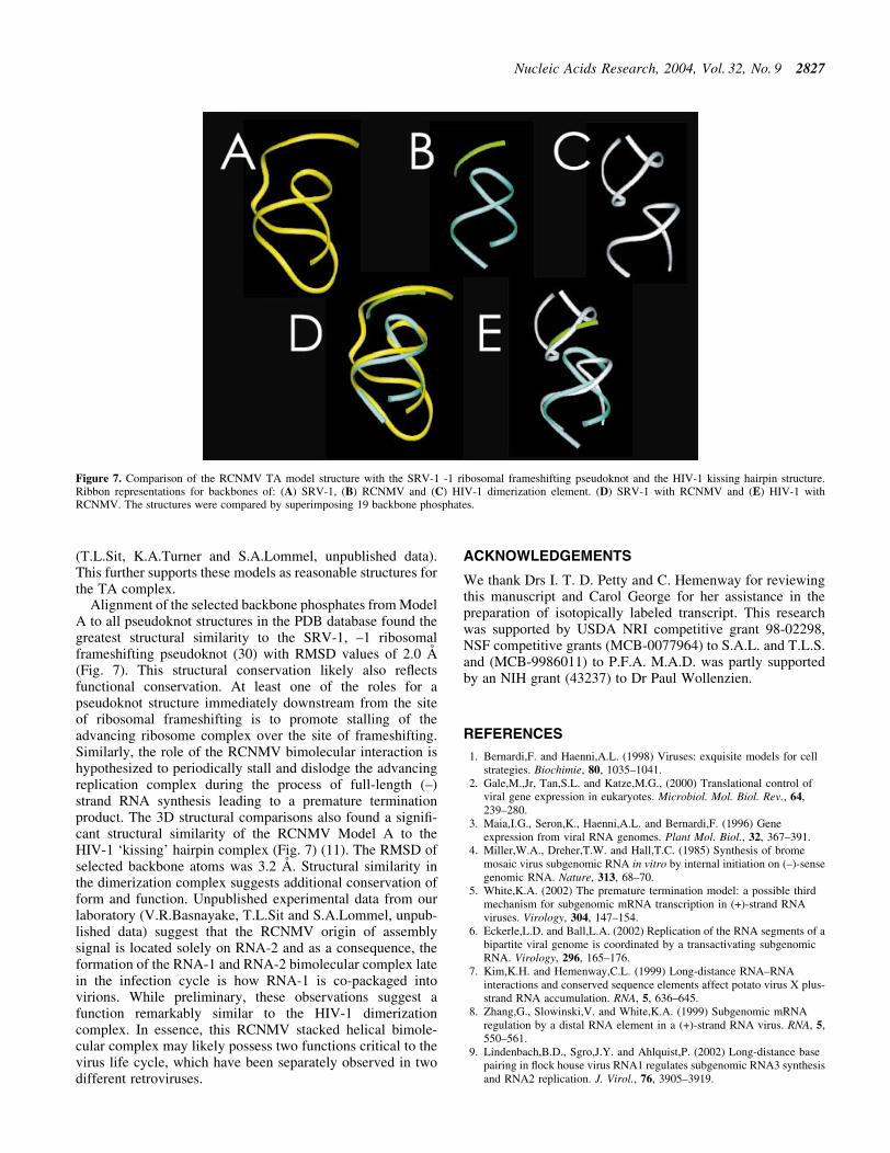
Alignment of the selected backbone phosphates from ModelA to all pseudoknot structures in the PDB database found thegreatest structural similarity to the SRV-1, ±1 ribosomalframeshifting pseudoknot (30) with RMSD values of 2.0 AÊ
(Fig. 7). This structural conservation likely also re¯ectsfunctional conservation. At least one of the roles for apseudoknot structure immediately downstream from the siteof ribosomal frameshifting is to promote stalling of theadvancing ribosome complex over the site of frameshifting.Similarly, the role of the RCNMV bimolecular interaction ishypothesized to periodically stall and dislodge the advancingreplication complex during the process of full-length (±)strand RNA synthesis leading to a premature terminationproduct. The 3D structural comparisons also found a signi®-cant structural similarity of the RCNMV Model A to theHIV-1 `kissing' hairpin complex (Fig. 7) (11). The RMSD ofselected backbone atoms was 3.2 AÊ . Structural similarity inthe dimerization complex suggests additional conservation ofform and function. Unpublished experimental data from ourlaboratory (V.R.Basnayake, T.L.Sit and S.A.Lommel, unpub-lished data) suggest that the RCNMV origin of assemblysignal is located solely on RNA-2 and as a consequence, theformation of the RNA-1 and RNA-2 bimolecular complex latein the infection cycle is how RNA-1 is co-packaged intovirions. While preliminary, these observations suggest afunction remarkably similar to the HIV-1 dimerizationcomplex. In essence, this RCNMV stacked helical bimole-cular complex may likely possess two functions critical to thevirus life cycle, which have been separately observed in twodifferent retroviruses.
ACKNOWLEDGEMENTS
We thank Drs I. T. D. Petty and C. Hemenway for reviewingthis manuscript and Carol George for her assistance in thepreparation of isotopically labeled transcript. This researchwas supported by USDA NRI competitive grant 98-02298,NSF competitive grants (MCB-0077964) to S.A.L. and T.L.S.and (MCB-9986011) to P.F.A. M.A.D. was partly supportedby an NIH grant (43237) to Dr Paul Wollenzien.
REFERENCES
1. Bernardi,F. and Haenni,A.L. (1998) Viruses: exquisite models for cellstrategies. Biochimie, 80, 1035±1041.
2. Gale,M.,Jr, Tan,S.L. and Katze,M.G., (2000) Translational control ofviral gene expression in eukaryotes. Microbiol. Mol. Biol. Rev., 64,239±280.
3. Maia,I.G., Seron,K., Haenni,A.L. and Bernardi,F. (1996) Geneexpression from viral RNA genomes. Plant Mol. Biol., 32, 367±391.
4. Miller,W.A., Dreher,T.W. and Hall,T.C. (1985) Synthesis of bromemosaic virus subgenomic RNA in vitro by internal initiation on (±)-sensegenomic RNA. Nature, 313, 68±70.
5. White,K.A. (2002) The premature termination model: a possible thirdmechanism for subgenomic mRNA transcription in (+)-strand RNAviruses. Virology, 304, 147±154.
6. Eckerle,L.D. and Ball,L.A. (2002) Replication of the RNA segments of abipartite viral genome is coordinated by a transactivating subgenomicRNA. Virology, 296, 165±176.
7. Kim,K.H. and Hemenway,C.L. (1999) Long-distance RNA±RNAinteractions and conserved sequence elements affect potato virus X plus-strand RNA accumulation. RNA, 5, 636±645.
8. Zhang,G., Slowinski,V. and White,K.A. (1999) Subgenomic mRNAregulation by a distal RNA element in a (+)-strand RNA virus. RNA, 5,550±561.
9. Lindenbach,B.D., Sgro,J.Y. and Ahlquist,P. (2002) Long-distance basepairing in ¯ock house virus RNA1 regulates subgenomic RNA3 synthesisand RNA2 replication. J. Virol., 76, 3905±3919.
Figure 7. Comparison of the RCNMV TA model structure with the SRV-1 -1 ribosomal frameshifting pseudoknot and the HIV-1 kissing hairpin structure.Ribbon representations for backbones of: (A) SRV-1, (B) RCNMV and (C) HIV-1 dimerization element. (D) SRV-1 with RCNMV and (E) HIV-1 withRCNMV. The structures were compared by superimposing 19 backbone phosphates.
Nucleic Acids Research, 2004, Vol. 32, No. 9 2827

10. Sit,T.L., Vaewhongs,A.A. and Lommel,S.A. (1998) RNA-mediatedtrans-activation of transcription from a viral RNA. Science, 281,829±832.
11. Chang,K.Y. and Tinoco,I.,Jr (1997) The structure of an RNA `kissing'hairpin complex of the HIV TAR hairpin loop and its complement.J. Mol. Biol., 269, 52±66.
12. Mujeeb,A., Clever,J.L., Billeci,T.M., James,T.L. and Parslow,T.G.(1998) Structure of the dimer initiation complex of HIV-1 genomic RNA.Nat. Struct. Biol., 6, 432±436.
13. Ban,N., Nissen,P., Hansen,J., Moore,P.B. and Steitz,T.A. (2000) Thecomplete atomic structure of the large ribosomal subunit at 2.4 AÊ
resolution. Science, 289, 905±920.14. Brunel,C., Marquet,R., Romby,P. and Ehresman,C. (2002) RNA loop±
loop interactions as dynamic functional motifs. Biochimie, 84, 924±944.15. Piotto,M., Saudek,V. and Sklenar,V. (1992) Gradient-tailored excitation
for single-quantum NMR spectroscopy of aqueous solutions. J. Biomol.NMR, 6, 661±665.
16. Gibbs,S.J. and Johnson,C.S.,Jr (1991) A PFG NMR experiment foraccurate diffusion and ¯ow studies in the presence of eddy currents.J. Magn. Reson., 93, 395±402.
17. Major,F., Turcotte,M., Gautheret,D., Lapalme,G., Fillion,E. andCedergren,R. (1991) The combination of symbolic and numericalcomputation for three-dimensional modeling of RNA. Science, 253,1255±1260.
18. Koradi,R., Billeter,M. and WuÈthrich,K. (1996) MOLMOL: a program fordisplay and analysis of macromolecular structures J. Mol. Graph., 14,51±55.
19. Gendron,P., Lemieuxs,S. and Major,F. (2001) Quantitative analysis ofnucleic acid three-dimensional structures. J. Mol. Biol., 308, 919±936.
20. Varani,G. and Tinoco,I.,Jr (1991) RNA structure and NMR spectroscopy.Q. Rev. Biophys., 24, 479±532.
21. Kolk,M.H., van der Graaf,M., Wijmenga,S.S., Pleij,C.W., Heus,H.A. andHilbers,C.W. (1998) NMR structure of a classical pseudoknot: interplayof single- and double-stranded RNA. Science, 280, 434±438.
22. Wemmer,D.E. and Williams,P.G. (1994) Use of nuclear-magneticresonance in probing ligand-macromolecular interactions. MethodsEnzymol., 239, 737±767.
23. Masquida,B. and Westhof,E. (2000) On the wobble GoU and relatedpairs. RNA, 6, 9±15.
24. Szymanski,M., Barciszewska,M.Z., Erdmann,V.A. and Barciszewski,J.(2000) An analysis of G-U base pair occurrence in eukaryotic 5S rRNAs.Mol. Biol. Evol., 8, 1194±1198.
25. Giese,M.R., Betschart,K., Dale,T., Riley,C.K., Rowan,C., Sprouse,K.J.and Serra,M.J. (1998) Stability of RNA hairpins closed by wobble basepairs. Biochemistry, 37, 1094±1100.
26. Wagner,E.G. and Simons,R.W. (1994) Antisense RNA control inbacteria, phage and plasmids. Annu. Rev. Microbiol., 48, 713±742.
27. Hoeprich,S. and Guo,P. (2002) Computer modeling of three-dimensionalstructure of DNA-packaging RNA (pRNA) monomer, dimer andhexamer of Phi29 DNA packaging motor. J. Biol. Chem., 277,20794±20803.
28. Berman,M., Westbrook,J., Feng,Z., Gilliland,G., Bhat,T.N., Weissig,H.,Shindyalov,I.N. and Bourne,P.E. (2000) The Protein Data Bank. NucleicAcids Res., 28, 235±242.
29. Hilbers,C.W., Michiels,P.J. and Heus,H.A. (1999) New developments instructure determination of pseudoknots. Biopolymers, 48, 137±153.
30. Michiels,P.J., Versleijen,A.A., Verlaan,P.W., Pleij,C.W., Hilbers,C.W.and Heus,H.A. (2001) Solution structure of the pseudoknot of SRV-1RNA, involved in ribosomal frameshifting. J. Mol. Biol., 310,1109±1123.
2828 Nucleic Acids Research, 2004, Vol. 32, No. 9
Copyright © 2022 FDOKUMEN