The Nature of the Intramolecular Charge Transfer State in Peridinin

Charles University in Prague
Faculty of Science
Chemistry:
Modeling of Chemical Properties of Nano- and Biostructures
Boris Fackovec
Intramolekularnı a intermolekularnı interakce vproteinech
Intramolecular and intermolecularinteractions in proteins
DIPLOMA THESIS
Supervisor: RNDr. Jirı Vondrasek, CSc.
Prague 2012

Prohlasuji, ze jsem zaverecnou praci zpracoval samostatne a ze jsem uvedl vsechnypouzite informacnı zdroje a literaturu. Tato prace ani jejı podstatna cast nebylapredlozena k zıskanı jineho nebo stejneho akademickeho titulu.
I declare that I carried out this master thesis independently, and only with thecited sources, literature and other professional sources.
I understand that my work relates to the rights and obligations under the ActNo. 121/2000 Coll., the Copyright Act, as amended, in particular the fact thatthe Charles University in Prague has the right to conclude a license agreementon the use of this work as a school work pursuant to Section 60 paragraph 1 ofthe Copyright Act.
In ........ date ............ signature of the author

Nazev prace: Intra a intermolekularni interakce v proteinech
Autor: Boris Fackovec
Katedra: Katedra fyzikalnı a makromolekulove chemie
Vedoucı diplomove prace: RNDr. Jirı Vondrasek, CSc.
Abstrakt: Volna energie sbalenı proteinu predstavuje pozoruhodnou rovnovahumezi stabilizujıcımi a destabilizujıcımi nekovalentnımi interakcemi. V teto pracistabilizujıcı energii rozkladame na fyzikalne smysluplne prıspevky, ktere by jsmenasledne dokazali slozit do konzistentnıho transferabilnıho obrazu teplotnı stabil-ity. Empirickym potencialem vypocteme interakcnı energie mezi fragmenty klasi-fikovanymi na zaklade jejich fyzikalnıch vlastnostı na setu 1200 neredundantnıchstruktur z PDB databaze. Vysledkem prace je lepsı pochopenı vztahu mezi in-terakcnımi energiemi vypoctenymi metodami teoreticke chemie a prıspevky jed-notlivych interakcı na teto rovnovaze.
Klıcova slova: protein, aminokyselina, interakcnı energie, stabilita proteinu, ter-modynamika proteinu, molekularnı modelovanı, bioinformatika, biofyzika
Title: Intramolecular and intermolecular interactions in proteins
Author: Boris Fackovec
Department: Department of Physical and Macromolecular Chemistry
Supervisor: RNDr. Jirı Vondrasek, CSc., Insitute of Organic Chemistry andBiochemistry, AS CR, vvi.
Abstract: Folding free energy of a protein is a delicate balance between stabi-lizing and destabilizing non-covalent itneractions. In this work, we decomposefolding free energy into physically meaningful contributions, in which we aim tofind general trends. Empirical potential is used to calculate interaction ener-gy between all protein fragments, which are classified based on their dominantterm in multipolar expansion. Calculations are done using 1200 non-redundantstructures from PDB database. Based on general trends found in interactionsbetween these fragments, we attempt to better understand relationships betweeninteraction energies calculated using computational chemistry methods and theircorresponding free energy contributions on stabilization.
Keywords: protein, amino acid, interaction energy, protein stability, protein ther-modynamics, molecular modeling, bioinformatics, biophysics

Acknowledgement
I would like to thank to my supervisor dr. Jirı Vondrasek for introducing me tothe field and for his devoted supervision and support before and during the workon the thesis. Many thanks are owed to my colleagues at the Institute of OrganicChemistry and Biochemistry for enriching discussions. Vital power for my workwas supplied by my beloved girlfriend, mother, grandmother and sister.
4
Contents
1 Introduction 41.1 Experimental studies of protein structure and stability . . . . . . 4
1.1.1 Biological context . . . . . . . . . . . . . . . . . . . . . . . 41.1.2 Calorimetric studies . . . . . . . . . . . . . . . . . . . . . 51.1.3 NMR and X-ray . . . . . . . . . . . . . . . . . . . . . . . . 81.1.4 Biophysical and chemical studies . . . . . . . . . . . . . . 81.1.5 Availability of experimental data . . . . . . . . . . . . . . 9
1.2 Theoretical investigations of protein structure and stability . . . . 101.2.1 Framework of protein modeling . . . . . . . . . . . . . . . 101.2.2 All-atom models of proteins . . . . . . . . . . . . . . . . . 111.2.3 Simplified models of proteins . . . . . . . . . . . . . . . . . 131.2.4 Solvation . . . . . . . . . . . . . . . . . . . . . . . . . . . . 131.2.5 Denatured state . . . . . . . . . . . . . . . . . . . . . . . . 15
1.3 Stability models . . . . . . . . . . . . . . . . . . . . . . . . . . . . 161.3.1 Driving force of protein folding . . . . . . . . . . . . . . . 161.3.2 Free energy partitioning . . . . . . . . . . . . . . . . . . . 161.3.3 Contribution of interactions in native state . . . . . . . . . 171.3.4 Energy functions for structure prediction . . . . . . . . . . 181.3.5 Energy functions for mutants . . . . . . . . . . . . . . . . 19
1.4 Intramolecular interactions in native state . . . . . . . . . . . . . 191.4.1 Covalent and non-covalent interactions in native state . . . 191.4.2 Distance scaling . . . . . . . . . . . . . . . . . . . . . . . . 201.4.3 Classification in quantum chemistry . . . . . . . . . . . . . 201.4.4 Classification in structural biology . . . . . . . . . . . . . . 211.4.5 Contribution of interactions to stability . . . . . . . . . . . 22
1.5 Mathematical representation of protein structure and stability . . 231.5.1 Representation of protein organization by matrices . . . . 231.5.2 Geometry representation . . . . . . . . . . . . . . . . . . . 241.5.3 Energy representation . . . . . . . . . . . . . . . . . . . . 25
1.6 Aims of the thesis . . . . . . . . . . . . . . . . . . . . . . . . . . . 261.7 Organisation of the thesis . . . . . . . . . . . . . . . . . . . . . . 27
2 Methods 282.1 Structures of proteins . . . . . . . . . . . . . . . . . . . . . . . . . 28
2.1.1 Structure set selections . . . . . . . . . . . . . . . . . . . . 282.1.2 Structure preparation . . . . . . . . . . . . . . . . . . . . . 28
2.2 Energy calculations . . . . . . . . . . . . . . . . . . . . . . . . . . 282.2.1 Fragmentation . . . . . . . . . . . . . . . . . . . . . . . . . 282.2.2 Interaction energy matrix . . . . . . . . . . . . . . . . . . 292.2.3 Residue interaction energy . . . . . . . . . . . . . . . . . . 302.2.4 Solvent accessible surface area . . . . . . . . . . . . . . . . 302.2.5 Molecular dynamics . . . . . . . . . . . . . . . . . . . . . . 30
2.3 Parametrization of models . . . . . . . . . . . . . . . . . . . . . . 31
1

3 Results 333.1 Classification of amino acids . . . . . . . . . . . . . . . . . . . . . 333.2 Residue interaction energy . . . . . . . . . . . . . . . . . . . . . . 333.3 Domain size . . . . . . . . . . . . . . . . . . . . . . . . . . . . . . 343.4 Interaction energy distributions . . . . . . . . . . . . . . . . . . . 343.5 Definition of residue-residue contact . . . . . . . . . . . . . . . . . 353.6 Interaction energy balance in proteins . . . . . . . . . . . . . . . . 35
3.6.1 Derivation of the model . . . . . . . . . . . . . . . . . . . 363.6.2 Interpretation of the scaling factors . . . . . . . . . . . . . 383.6.3 Reliability of the model . . . . . . . . . . . . . . . . . . . 403.6.4 Applications . . . . . . . . . . . . . . . . . . . . . . . . . . 40
3.7 Dynamic properties of interaction energy sums . . . . . . . . . . . 41
4 Conclusion 42
Bibliography 43
2

Preface
Protein folding problem has been attracting researchers for more than 80 years.Despite incredible work done by theoretical and experimental groups, we still donot understand balance of the driving forces of protein folding. It seems now,that we have fundamental understanding and that only quantitative performanceof our models is not sufficient for the problem to be solved. Probably the mostpromising research directions are studies of energy landscape topology, identifi-cation of errors in interaction energy calculations and decomposition of stability.
Although the thesis is called ”Intramolecular and intermolecular interactionsin proteins”, only intramolecular interactions in globular proteins are addressed,since protein-protein interactions have been studied by Jirı Kysilka, PhD studentin the group of Dr. Vondrasek. In our group, interaction energies between aminoacids were calculated at the highest level of accuracy few years ago. In this time,there is only limited space for improvement of description of particular pairwiseinteractions. More challenging and urgent problem became the connection of theinteraction energies and their free energy contribution to protein stability.
The main contribution of this work is a novel treatment of solvent effectsand development of stability model and method for optimization of its parame-ters.
Introduction presents an essential view on protein folding problem integrat-ing statistical mechanics, biophysical experimentation and molecular modeling.Attention was paid to discuss in details terms ambiguously defined in literature.Phenomena studied by one of the mentioned disciplines were formulated in a wayunderstandable by researchers from different fields.
3

1. Introduction
Proteins are linear biomacromolecules synthesized in ribozomes by linking aminoacids by peptide bonds. They can fold into stable 3D structure at certain condi-tions. Foldable proteins have 2 forms - native and denatured state.
Definition 1. Native state of a protein is an ensemble of microstates which arevery similar in structure. This state is the most stable at native conditions.
Definition 2. Denatured state of a protein is an ensemble of microstates whichare higher in energy but much more numerous than those of native state ensemble.Denatured state is more stable than native state in presence of denaturant, whichcan be chemical agent.
In order to prevent ambiguities this work strictly distincts between denaturedand unfolded state.
Definition 3. Unfolded state is in this work defined as a random coil - a theoret-ical construct of heteropolymer chain in which non-covalent interactions betweenconstituent amino acids are negligible.
In the first subsection of Introduction, only certain observations easy to inter-pret without complex models are briefly overviewed. Emphasis is put on stabilitystudies, but other relevant experimental observations are briefly mentioned. Inthe next section, the facts are complemented by the most influential theories ofprotein folding which formed the field. As the most important result in thiswork is represented by the stability model, previous stability models and addi-tivity principles are discussed. Subsection 1.4 provides three different views onintramolecular interactions - view of statistical physics, quantum chemistry andstructural biology. Link between interactions and protein stability adressed byprevious studies is referenced throughout the whole work. As we propose newdefinitions of residue-residue contact and interaction energy matrices are usedthroughout the work, section 1.5 introduces the matrix representation of proteinorganization.
1.1 Experimental studies of protein structure
and stability
1.1.1 Biological context
Proteins constitute about 40 % of dry weight of human organism [1], in whichthey perform the most important tasks because of their structural variability, theirfunctional and binding specificity and for their ability to adopt special propertieslike enzymatic activity or even fluorescence. Proteins are evolutionary optimizedfor general well-being of an organism, i.e. not causing illnesses by misfolding,being able to resist short- or long-term adverse conditions and catalyze reactionsto increase adaptability and compatibility of the whole metabolic network of anorganism to its environment. For successful accomplishment of the mentioned
4

properties, proteins need to have stability in some specified thermodynamic in-terval and to fold in order of nanoseconds to minutes.
Proteins are synthesized in ribosomes in process of translation where a select-ed linear information from DNA is (after transcription and splicing) translatedinto sequence of amino acids. Simultaneously with synthesis of the chain, thesynthesized N-terminus of the new protein starts to fold into 3D structure [2].It is generally accepted that function of a protein is determined by its structure.However, about 30% of proteins are unstructured [3, 4]. These intrinsically disor-dered proteins were overlooked by protein biophysics community in the past butrecently have become increasingly popular[5]. The process of folding is in cellssometimes promoted by chaperones inhibiting misfolding of nascent polypeptides.In eukaryotic organisms, translation is usually followed by posttranslational mod-ification changing chemical character of proteins. After this process, loss of abilityto refold is usual and the structure with minimum energy can change substan-tially.
Protein folding problem in the natural environment is a complex problemof molecular biology. Number of relevant factors in protein folding is immense.The cellular environment can be imagined as a dense soup with concentrationof proteins in the cell being in order of 300 g/l with no aggregation. Effects ofmacromolecular crowding on protein structure and function have been recentlyextensively reviewed by Zhou [6] and Elcock [7].
1.1.2 Calorimetric studies
Necessary reduction of protein folding problem came to a simplified formulationwhich remains a challenge for physicists:
Definition 4. Protein folding problem concerns about thermodynamics and ki-netics of transformation of a protein chain composed of 20 types of amino acidsfrom denatured to native state in a buffered water solutions at low protein andbuffer / salt concentration.
In this thesis, only foldable globular proteins are studied; disordered, mem-brane and fibrilar proteins are not subject of this study. In 1961, Anfinsen pro-posed a hypothesis based on his in vitro refolding studies of ribonuclease [8].
Hypothesis 1. (Anfinsen) Structure of a protein is uniquely determined by itssequence and environmental conditions (solvent composition, temperature etc).The native state structure represents global free energy minimum ensemble atthese conditions.
Pitfalls of the original formulation by Anfinsen are discussed in works of Govin-darajan [9] and Ben-Naim [10]. Throughout the rest of this work, we assume va-lidity of thermodynamic hypothesis. We can now define thermodynamic stabilityof a protein.
Definition 5. Protein stability is defined as the Gibbs free energy differencebetween native and denatured state of a protein at defined conditions (temperature,pressure, solvent composition, pH etc.). Negative free energy value means higherstability of native state.
5

In the rest of the text, shorter term ’free energy’ will be used instead of Gibbsfree energy (or free enthalpy). The difference between Helmholtz free energy andGibbs free energy is in liquids such negligible that the two terms can be usedinterchangeably.
In 1976, Pfeil and Privalov proved [11] that calorimetrically determined en-thalpy, entropy and free energy represent real thermodynamic potentials specify-ing the states of a protein. Differential scanning calorimetry (DSC) is thereforethe source of experimental data with the most straightforward interpretation, soit is no surprise that DSC has become standard methodology of biophysics. Heatcapacity of a small sample of dissolved protein is recorded as a function of tem-perature. The DSC is considered to be very precise; the least precise variable isprobably protein concentration [12] (error is estimated to be under 2%). A typi-cal output curve of DSC experiment of protein denaturation is shown on Figure1.1.
Figure 1.1: Left - curve of DSC measurement of pancreatic ribonuclease A(RNase) and sperm whale myoglobin (Mb). Peak maxima correspond to meltingtemperatures. Dashed lines denote extrapolated heat capacities. Adapted fromref. [13]. Right - DSC experiment of defatted bovine serum albumine, a proteinconsisting of 2 domains. Adapted from ref. [14]
Protein presents a thermodynamically discrete macroscopic system i.e. it canbe divided into domains each having only 2 accessible states [15]. Temperaturedependence of stability of 1 domain can be well described by 3 parameters (seeEquation 1.1 and the following text).
∆G = ∆H − T∆S = ∆H0 +∫ T
Tm
∆CpdT − T
(
∆H0
Tm+∫ T
Tm
∆Cp
TdT
)
(1.1)
Tm - unfolding (or melting) temperature or transition midpoint is temperatureat which ∆ G = 0. It is the highest point of the DSC curve. Tm can be easilymeasured by other techniques and its value is known for most of proteins.
∆H0 - enthalpy of unfolding is the heat release accompanying unfolding attemperature Tm. Unfolding of a protein is always endothermic process, i.e. energy
6

Table 1.1: Correlation between thermodynamics quantities of globular proteinsand chain length. First two columns contain correlated quantities (N - proteinsize = number of amino acids, ∆H,∆S,∆Cp and Tm are defined above. Numbersin parentheses after quantities are temperatures at which the quantities weremeasured. Third column contains Pearson correlation coefficient between thevariables in first two columns. Data in line 1 to 5 are obtained on set of 65proteins [12], data in line 6 and 7 are obtained using datase of 3224 proteins[18].Measurements at inconsistent conditions. Data in columns 8 and 9 on set of 19proteins [19]
N ∆H(374K) 0.922N ∆H(333K) 0.789N ∆S(385K) 0.920N ∆S(333K) 0.759N ∆Cp 0.862∆H(298K) 298 K ∆S(298K) 0.991∆H(298K) ∆G(298K) 0.600Tm ∆G(298K)/N 0.830Tm ∆H(298K)/N 0.810
must be supplied to increase entropy. It can be determined by DSC as area underthe peak in Cp/T plot. An alternative method of determination of ∆ H can bederived from van’t Hoff equation
∆H = kBT2m
dlnK
dT|Tm
(1.2)
where kB is Boltzmann constant, and K (equilibrium constant) is ratio ofconcentration of folded and unfolded form. Comparison of calorimetric ∆H andvan’t Hoff ∆H can be used to assess cooperativity of folding [16].
∆Cp - heat capacity difference between native and denatured state. In 1979,Privalov [17] discovered that linear extrapolation of experimental data yields con-vergence of both specific enthalpy and entropy of unfolding to common values atapproximately the same temperature. Independent studies of liquid hydrocarbonsdissolution showed that at this temperature (112 ◦C) entropy of hydrocarbonfransfer to water becomes zero.
In 80’s and 90’s, extensive calorimetric measurements of globular proteinswere performed. Experimental correlation between thermal data and proteinchain length can be found in Table 1.1.
Briefly, folding enthalpy very well correlates with protein size. Each aminoacid contributes almost 2 kcal/mol (about 8 kJ/mol) to folding enthalpy. Sta-bility of all proteins lies between 0 and -20 kcal/mol despite good correlation ofunfolding ethalpy and protein size. This inconsistency is due to strong compensa-tion of enthalpy term by opposing entropy. The enthalpy-entropy compensation[20, 18, 21] is about 91% [18]. Energy of contributing interactions is two ordersof magnitude higher than free energy of folding at standard conditions. Proteinscan be denatured by heat or cold.
7
1.1.3 NMR and X-ray
In 1958, Kendrew et al published first X-ray structure of protein [22]. It wasfound that unlike DNA, protein chains are folded into globules, with hydropho-bic residues being buried inside and hydrophillic ones being exposed on proteinsurface. On average, approximately 83% of hydrophobic residues are buried com-pared to 63% for polar and 54% for charged ones [23]. This fact indicated thathydrophobic effect plays significant role in protein folding. Packing of hydropho-bic residues in protein interiors has ratio of void space similar to aliphatic crystalsrather than liquids. Dense packing of hydrophobic core has been identified as adeterminant of protein stability.
Secondary structure had been discovered even before first X-ray structure ofprotein appeared [24]. Nowadays, structures are classified hierarchically by twomajor classification schemes (CATH [25]) and SCOP [26]). An interesting fact isthat although the number of structures determined per year continually increases,the number of new folds discovered per year decreases since 2004 (SCOP) or 2007(CATH).
Other general structural features have been observed in proteins and theiroccurence has been correlated with thermal stability, mostly by comparison ofstructures of homologous proteins with significantly different thermal stability.In these studies, an important assumption is made:
Hypothesis 2. Native state of a protein can be characterized by one structure.
Crystallography provides an averaged structure which is probably the bestrepresenative one. Some examples of common structural features in proteins arepairing of oppositely charged amino acid sidechains [27, 28], and aromatic clus-ters [29]. Structural features like polar contacts or volume have been correlatedwith protein stability. Some structural studies can provide information aboutdenatured state. It has also been shown [30, 31] that hydrophobic clusters canbe present in denatured state of proteins.
Native structures are essential for most of the theoretical protein thermody-namics studies. As an efficient application of NMR methods is limited to smallproteins, X-ray crystallography remains the main source of data. The assump-tion that structures of proteins in crystals are identical to those in solutions iswidely accepted and firmly based [32]. Unfortunately, X-ray crystallography can-not always determine rotameric position of asparagine, glutamine and histidinewhich are ex post modeled [33]. Studies by Higman et al. [34] assessing X-raystructures by NMR experiments show that in the studied protein, at ξ2 or ξ3 in atleast one glutamine or asparagine residue (respectively) is incorrect. Quality ofstructure of flexible regions is usually very poor. Although methods for structuredetermination are subject of continual development, determination of a proteinnative structure remains an expensive and long process.
1.1.4 Biophysical and chemical studies
Apart from high temperatures, unfolding of a protein can be caused by chemicalagents called denaturants. Concentrated acids were the first known denaturants.High stability dependence on pH led into conclusions that ion pairing is the mainforce responsible for protein structure. Other denaturants (guanidinium chloride,
8

urea, etc.) are used to measure stability of proteins by measuring concentrationratio of denatured and native state using spectral probes [35] and extrapolatingto zero concentration. Slopes of stability dependence on denaturant concentra-tion, called m-values [36], determined by this method well correlate with solventaccessible surface area difference of native and unfolded state. Recent theoriesabout chemical denaturation suggest that denaturants change structure and low-er free energy of denatured state rather than increasing energy of native state.Therefore, one should be careful when considering m-values to derive models ofprotein thermal stability [37, 38].
Hydrodynamic methods like dynamic light scattering are used for determi-nations of gyration radius. Scaling of hydrodynamic radius with chain length iscompared with theoretical predictions based on simplified models of denaturedor unfolded state. Pressure perturbation calorimetry [39] enables measurementof temperature dependence of thermal expansion coefficient [40, 41] and so helpsto understand hydration of folded and denatured proteins.
Mutational studies are invaluable tools of studying effects of single aminoacid sidechain on protein stability or folding rate. Alanine screening measurescontribution of a sidechain to stability by recording stability change upon itsreplacement by alanine. Double mutant cycles are used to measure interactionbetween two sidechains.
Atomic force microscopy enables studying thermodynamics and kinetics ofmechanical unfolding of a single molecule. Such experiments can be directlyreproduced by simulations. Pathways of protein folding have been studied byNMR [42] and protein folding process can be observed real-time in vivo usingfluorescent labeling [43]. Information about transition state ensemble can beobtained by φ and ψ value analyses [44].
1.1.5 Availability of experimental data
Up to now, more than 80,000 structures have been deposited to PDB publiclyopen protein database [45] and the number of structures still increases. About24,000 protein structures out of 80,000 represent non-redundant proteins (70%sequence identity removed, 16,000 if 30% removed), 11,500 of which have beenresolved by X-ray crystallography at resolution better than 2 A.
Thermodynamics data are collected in ProTherm [46] database. Althoughdetermination of native structure is more laborous than DSC measurements, fullthermodynamics characterization of proteins is spare. Out of about 25,000 en-tries in ProTherm database, only about 100 represent ∆G values measured byDSC on proteins with structure deposited in PDB at consistent conditions (pH7). Similarly, about 100 entries represent consitently measured ∆H by DSC onproteins with published structure. Most of the data represent stabilities of substi-tution mutants, so the corresponding structure must be modeled for theoreticalstudies. Stability changes upon amino acid replacement for same systems mea-sured at same conditions published by different research groups are not equal,but correlate with Pearson coefficient 0.86 [47]
9

1.2 Theoretical investigations of protein struc-
ture and stability
1.2.1 Framework of protein modeling
Models of proteins have been developed simultaneously with experimental studiesto interpret observations and to answer some theoretical questions. Developmentin integration of theory and experiments is reviewed in references [48, 49, 50].Aims of the protein models were usually to explain folding times which are sur-prisingly low [51], to find the dominant force of protein folding or to study phasediagrams of proteins. For structure prediction, models leading to energy func-tion for structures or to protein folding kinetics are of principal interest. Thelatter interest is motivated by the fact that finding a global minimum on energylandscape is NP-complete problem [52], though the nature can fold a protein inmiliseconds to seconds. Structure prediction algorithm might efficiently simulatenatural folding process.
Protein folding occurs on a broad scale of times and lengths. Proteins arecomposed of tens to thousands of amino acids, i.e. hundreds to tens of thousandsof atoms. Diamaters of folded proteins vary from units to tens of nanometers.Helix-coil transition occurs in order of microseconds, folding in order of mil-icesonds to seconds. The limitations of model complexity imposed by proteinsize and complexity of their configurational space severely limits the maximumaccuracy. It is in principle possible to run accurate quantum mechanics simula-tions, where nuclei are treated as quantum objects. However, even single pointquantum mechanics calculations are prohibitively expensive. All-atom empiricalforce field model is the highest accuracy of microstate description that allowssufficient sampling to some extent. Folding simulations from extended state tonative state have been performed only for few small proteins. On the other side,the most simplified models are simply theories with no analytical solution. Theirsolution can be found by simple simulations, e.g. averaging of all states of a HPlattice model.
Pure theory with analytical solutions to model equations is rare because thereis myriad of possible sequences, each having its own properties. General poly-mer theory is well developed but applicability on proteins is limited. Analyticalmodel of entropy for a cross-linking polymer was proposed by Vorov et al [53].Some purely theoretical models for proteins have been proposed by Dill’s group.Wide variety of simplified model was reviewed [54]. Models for entropy of proteinensembles is digestedly reviewed in review by Dill and Stigter [55]. Statisticalmechanics of warm and cold unfolding has been studied by Hansen et al. [56].Excellent review of protein models and theories of protein folding, their assump-tions and experimental validations was written by Shakhnovich [57]. More recentreview on theoretical studies of proteins focused on molecular transfer model andunfolding was written by Thirumalai et al. [58]. Another review on the topicfocused on Markov state model has been recently published by Bowman et al.[59].
Models of proteins can be characterized by 3 main features.
1. Space in which a protein resides. Continuous space is more realistic whilelattices enable sampling of all possible protein configurations.
10

2. Particle representation of the protein chain. In all-atom models, each nu-cleus is represented by one particle, in coarse-grained models, small groupsof atoms are represented by beads. A residue is in many models represent-ed by 1 or 2 spheres or a points on a grid. Each amino acid can have itsown paremeters or amino acids can be grouped like in HP model[60], wherethere are only 2 types of amino acids.
3. Potential energy form or force field assigns energy to a given protein struc-ture based on mutual position of residues and displacement from equilibriumpositions. In lattice models, residue-residue interaction energy is evaluat-ed based on connectivity on lattice while in continuous models, geometricdistance or mutual orientation are considered.
Direct simulations of protein folding using all-atom models are rare. Implicitsolvent models are usually applied to decrease computational cost. Recently, 1ms simulation of BPTI in explicit solvent [61] was performed and boldly present-ed. Large independent simulation running on different computers was pioneeredby folding@home 1 project. Other related notable projects are bluegene2, po-em@home3, protein structure initiative4 and dynameomics5.
Standard view on proteins emphasizes native states and usually simplifiessolvation and denatured state. Therefore, these two important but less developedaspects of protein modeling are discussed in separate sections.
1.2.2 All-atom models of proteins
Despite the technical and methodology development, accurate quantum dynamicsstudies are limited to systems with few electrons. Born-Oppenheimer approxima-tion, i.e. treating nuclei as classical objects, allows substantial simplification withreasonable error. Electronic structure problem for given coordinates of nuclei ismostly solved by variational or perturbation wavefunction theories or densitiyfunctional theories. Wide scale of methods has been developed [62] but generally,the time needed for accurate calculations increases at least with third power ofnumber of atoms or base functions. Accurate methods with linear scaling derivedfrom the mentioned methods imposing a distance constraint on orbital correla-tion are in vigorous development and might be in near future useful for proteinmodeling.
In proteins, stability in order of tens kcal/mol is a balance of thousands ofresidue-residue and residue-solvent interactions whose strength is in order ofunits of kcal/mol. Since random error propagates with square root of numberof residues and systematic error linearly with number of residues each interactionmust be calculated to level of sub-chemical accuracy and systematic errors elimi-nated to level of ’sub-sub-chemical’ accuracy (0.01 kcal/mol)[63]. Such accuracyis unreachable even by very expensive benchmark ab initio quantum mechanicscalculations. Therefore, empirical force fields seem to be good tradeoff between
1http://folding.stanford.edu/2http://www.research.ibm.com/journal/sj/402/allen.pdf3http://boinc.fzk.de/poem/4http://www.nigms.nih.gov/Research/FeaturedPrograms/PSI/5http://www.dynameomics.org/
11

accuracy and speed. They preserve most important features, like mutual orienta-tion while energy can be calculated in a short time allowing sampling of millionsof microstate in hours of computational time. Review of force fields used forall-atom protein simulations can be found in [64]. Potential energy is composedof additive contribution of the form
U = Ubonds + Uangles + Utorsions + Ucoulomb + UvdW (1.3)
Ubonds =∑
bonds
1
2kb(l − l0)
2 (1.4)
Uangles =∑
angles
1
2ka(θ − θ0)
2 (1.5)
Utorsions =∑
torsions
1
2Vt[1 + cos(nω − γ)] (1.6)
Ucoulomb =i<N∑
i=1
j<N+1∑
j=i+1
332qiqjrij
(1.7)
UvdW =i<N∑
i=1
j<N+1∑
j=i+1
4 · ǫi,j
(
σijrij
)12
−
(
σijrij
)6
(1.8)
Where kb and ka are force constants of bond stretching and angle bendingrespectively. l0 and θ0 are equilibrium values of bond lengths and bond angles.Torsion angle potentials are given as a sum of cosines.
Non-bonding interactions are calculated for every pair of atoms in the sys-tem. rij is distance between atoms, σij and ǫij are calculated for every pair fromparticular atom parameters ǫ and σ. Is partial charges qi are given in units ofelementary charge, Ucoulomb is in kcal/mol.
Empirical force fields fail to describe bond breaking, so the protonation stateof each basic or acidic site must be determined before simulation. Dispersion en-ergy is surprisingly well described in force fields by Lennard-Jones potential [65],whereas non-additive induction forces remain a challenge. Until polarizable forcefields are sufficiently developed, induction energy significantly contributing to hy-drogen bonding will remain the weak point. It seems that backbone parametersare mostly biased towards helical conformations [66] as non-bonding backboneparameters are often fitted to reproduce secondary structure [67].
Empirical force fields has been used for simulations of protein folding [68] anddynamics, free energy perturbation calculations of stability change upon aminoacid replacement [69]. Native state ensemble can be very well sampled [70] butsampling of all-atom models of denatured state remains a challenge. It is alsoworth to mention popular model systems. Some proteins are extraordinarily suit-able for simulations for their small size or available mutational data, for exampleBPTI, villin headpiece, SH3 domain, lysozyme, GroEL, rubredoxins etc.
12

Figure 1.2: Left - schematic picture of folding funnel. Adapted from ref. [80].Right - phase diagram of protein constructed using lattice model. Adapted fromref. [81]
1.2.3 Simplified models of proteins
By simplified models I hereby mean all models which cannot reproduce all thedesired features of all-atom models. One approach of their construction is coarse-graining, a process in which atoms are grouped to be represented by one sphereor elipsoid. Coarse-grained force fields like MARTINI [71] are widely used forsimulations of membrane proteins, biomolecular complexes and for sampling de-natured state ensemble.
Many valuable simple models provided deeper insight into protein folding.They fill the gaps of time scales unreachable by experiments and link experi-ments and more accurate simulations to theory. Go model [72] is often calleda perfect gas model for protein folding. It possesses smooth energy landscapewhere reaction coordinate of protein folding can be easily defined. In 90s, lat-tice models were extensively used to study whole configrational space of proteins.Phase diagrams and folding funnels of proteins (see Figure 1.2) were studied. Dis-tance constraint model (DCM) was constructed to provide a rapid and accurateestimate of conformational entropy with minimal computational cost [73, 74].Excellent review by Kolinski and Skolnick [75] summarizes the most significantoutputs of reduced protein models. Simple or minimalist models of proteins arealso reviewed in [54, 76, 77, 78, 79].
1.2.4 Solvation
In vitro experiments which are usually benchmarks for theoretical models aredone in low-concentrated solutions of buffers in water. Interactions contributingto thermodynamics of protein folding can be therefore divided into 3 groups
• protein-protein interactions
• protein-solvent interactions
13

• solvent-solvent interactions
It is generally accepted, that solvent plays important or even critical role in pro-tein folding. Many puzzles of protein thermodynamics result from contraintuitivefeatures of solvent effects. For its importance, water is sometimes called the 21stamino acid [82].
Mechanism of solvation of polar species is more intuitive than solvation ofnon-polar ones. Free energy of polar particle transfer from vacuum to water isalmost equal to enthalpy. It is because of strength of electrostatic interactionsbetween solvent and partially strongly charged solute, which exceed water-waterinteractions. Unlike polar hydration, hydrophobic hydration causes density ofwater to decrease and heat capacity of the system to increase 6. Entropy of hy-drophobic hydration is 0 for many solutes at about 400 K [83]. Hydrophobic effecthas been a subject of debates since its discovery [84, 85, 86, 87]. In 1959, Kauz-mann proposed that it is the driving force of protein folding. This indication ofbiological relevance contributed to considerable attention it attracted. Entropiccontribution to hydrophobic effect seems to prevail for small and enthalpic con-tribution for large solutes. Recent studies suggest that shape of the solute is alsoimportant [88].
In unfolded state, only protein-solvent non-covalent interactions, backbonetorsion and some self-avoiding residue-residue potential, like hard spheres, playrole. Free energy of burying a hydrophobic group has been studied extensivelyexperimentally and theoretically. Independence of hydration of backbone andsidechain usually is supposed. However, it has been found that even group ad-ditivity is unjustified in this case [89]. While hydration energies of sidechainanalogues are measured to high level of accuracy, data for capped amino acidsare sparse.
Polarizable all-atom water is the most precise used model of solvent in simula-tions. However, as mentioned above, simulations using polarizable force fields arerare. Many water models for empirical potential simulations have been developed.Their main parameter is number of sites for partial charges which correspondsto their computational cost and boundaries of their quality from 3-site models(TIP3P, SPC/E) to n-site models (TIP4P, TIP4P/2005; TIPnP, n=5,6...). Fur-ther simplification leads to coarse-grained water models.
Explicit modeling significantly increases number of degrees of freedom. There-fore, simplified model have been devised. In review [90], Warshel et al. presenthierarchy of solvation models. At the top of the hierarchy, the most accurateand expensive microscopic models are followed by simplified microscopic onesand macroscopic ones are at the bottom. In 1976, Warshel and Levitt proposedmodeling solvent by explicit grid of Langevin dipoles [91] (LD model). Poisson-Boltzmann (PB) models solve Poisson-Boltzmann equation, which is simply lin-earized Poisson equation 7 assumed Boltzmann’s distribution. They are stronglydependent on dielectric constant used. Even more simplified treatment of elec-torstatics, Generalized Born (GB) model, was proposed in 1997 [92]. Actually,
6Great source of strange properties of water and hydration is website of LondonSouth Bank University ”water structure and science” created by Dr Martin Chaplinhttp://www.lsbu.ac.uk/water/
7Poisson equation is an alternative formulation of Coulomb law.
14

GB model stands for simple Coulomb law with distance-dependent dielectric con-stant [90]. The relationship between simplified microscopic models (like LD orBDL models) and macroscopic models (PB, GB) was established by Papazyanand Warshel [93]. Briefly, macroscopic models disregard structure of water andeven extended Poisson model cannot describe physical discreteness of the solvent.Explicit and implicit solvend models are often combined to improve descriptionof solvation shell without significantly increasing degrees of freedom. In suchtreatments, small amount of water molecules representing few hydration shellsare added and reaction field represents the bulk water.
1.2.5 Denatured state
Definition of protein stability implies that two states of proteins should be inves-tigated and thermodynamically characterized. However, low attention was paidto structure and energetics of denatured state as it was usually identified withunfolded state. Unfolded state can be very well described by polymer theories [94]because residue-residue interactions are negligible and solvation of amino acids isindependent.
Hypothesis 3. Denatured state ensemble is identical to unfolded state ensemble.
While structure of native state can be experimentally determined, structureand therefore energetics of denatured state ensemble is poorly understood [95].Initial approximations of denatured state by unfolded were supported by scaling ofhydrodynamic radii of urea and GdCl unfolded proteins with chain length. How-ever, mechanism of denaturation by chemical agents seems to be much differentfrom thermal denaturation. Apart from that, the highly concentrated solutionsof denaturants are far from natural environment of proteins. Several studies haveshown that denatured state is much more compact than unfolded state [96, 97].NMR methods suggest that denatured state contains hydrophobic clusters [31]and large amounts of residual secondary structures [98]. Hypothesis 3 can betherefore confidently refused.
Modeling denatured state ensemble consisting of huge number of poorly foldedstructures remains a challenge for molecular modeling. First, it seems that back-bone parameters of current force fields are biased towards helical structures andwould probably fail to describe protein structures being far from folded ones [66].Second, ensemble constitutes a complex subset of configurational space, whichis very difficult to sample. Simplified models have been used to study compactdenatured states [99] and recently to study dominant forces in denatured stateensembles [100].
Denatured state did not receive attention it deserved due to difficulties in itsproper modeling. Moreover, denatured state is irrelevant for structure predictionas it is same for all decoys as well as for the native state.
15

1.3 Stability models
1.3.1 Driving force of protein folding
In 1959 in his seminal paper [84]8, Kauzmann suggested that hydrophobic effectmight be this force. Strong enthalpy-entropy compensation observed in proteins iscommon for solvation of hydrophobic species. Urea denaturation experiments byTanford [102] supported theory of identification of unfolded and denatured statesand by other properties of proteins like Privalov puzzle and burial of hydrophobicresidues observed in experimentally determined native structures.
In 1984, Dill [60] derived a model based on the abovementioned assumption,which predicted stability as a function of fraction of hydrophobic residues. Ac-cording to the model, at least 42% of residues in a protein must be hydrophobicin order to the protein be stable.
However, it has been found that hydrophobic effect is not the only stabiliz-ing force, indeed its contribution to protein stability was estimated to 60% byPace et al. [103]. Recent studies show that loss of van der Waals interactionsupon unfolding might be even more important than the effect of hydrophobic sur-face exposition [104, 105, 106, 107]. Comparison of fusion enthalpy of benzene orpropane (up to 1 kcal/mol) is not far from the values of average enthalpy contribu-tion per residue (up to 2 kcal/mol). Compactness of denatured states underminestheories emphasizing role of hydrophobic effect and supports the view of proteinunfolding as a hydrophobic crystal fusion rather than oil droplet dissolution.
1.3.2 Free energy partitioning
In early 90’s models of stability decomposition into contributions of amino acidswere proposed. Such models assume additivity of free energy.
Hypothesis 4. Stability can be decomposed into free energy contributions ofgroups af amino acids.
The hypothesis 4 is valid only if energy contributions are independent, i.e. ifsum of interaction energies of the components is zero in each microstate (Equation1.9).
E(x1, x2, ...) =∑
i
Ei(xi) (1.9)
Therefore, even though additivity of potential energy perfectly applies for asingle microstate, which is the case of empirical potentials, additive treatment ofensemble average quantities like free energy and even enthalpy inevitably intro-duces an error. While entropy is almost negligible in thermochemistry, entropyis very important in protein folding. Additivity of potential energy can be safe-ly used for studying native state if the hypothesis 2 is accepted. Justificationof free energy decomposition in protein stability studies has been vigorously de-bated [108, 109]. Dill suggested that the non-additivity problem is particularlysignificant for energy component decomposition [110].
8according to Pace [101] the most important paper ever published on protein stability
16

As mentioned before, each residue contributes approximately 2 kcal/mol tofolding enthalpy [12]. Also heat capacity change upon unfolding can be welldecomposed into contributions of amino acids (see Table 1.1). Such partitioning istoo rough for protein stability as it disregards residue-residue interactions. Sincevalue of typical interaction energy is the same magnitude as protein stability,an average contribution of amino acids to stability would be inaccurate. Suchcontributions for an amino acid type would involve
• average free energy of burial from denatured state to native state, providedthat residues are more exposed to water in denatured state
• average free energy contribution of residue-residue interaction of this aminoacid in denatured state
• average free energy contribution by conformational entropy of folding
• average free energy contribution of residue-residue interaction of this aminoacid in native state
Mutation studies have shown that such contribution correlates with buried area[111]. Remarkable model by Ghosh and Dill [112] enables estimate of stabilitydependence on pH and temperature just from sequence.
1.3.3 Contribution of interactions in native state
1-body decompositions cannot discriminate native state from decoys, since theyneglect interactions in native state. Considerable non-additivity in double mutantcycles [113] also corroborates significance of residue-residue contacts. Therefore,much attention was paid to stabilization by intramolecular interactions. Themethods mostly identify structural biology features and assign them universalvalues established by previous studies. One of the first free energy partitionings byPonnuswamy and Gromiha [114] comprises hydrophobic, electrostatic, hydrogenbonding, disulfide, van der Waals, entropic and non-entropic (in denatured state)free energy contributions (Equation 1.10).
∆G = GF −GU = (Ghy +Gel +Ghb +Gss +Gvw)F − (Gen +Gne)U (1.10)
Ghy was calculated from solvent accessible surface areas of amino-acids calcu-lated by previous study [115] and solvation parameters. Each identified surfaceion pair [116] was assigned 1 kcal/mol and each buried one 3 kcal/mol. Ghb wascalculated from protein chain length assuming number of hydrogen bonds is 0.73times number of residues and each hydrogen bonds was expected to contribute1 kcal/mol. Each disulfide bridge was assigned stabilization of 2.3 kcal/mol, GU
was calculated from chain length and Ghb as 1.2N+0.5Ghb. van der Waals energywas calculated from chain length as Gvw = 8.885 + 0.1413N kcal/mol. Averagecontribution of hydrogen bonding, hydrophobic burial and van der Waals forceswas similar, while contribution of electrostatics was small. Fitting 6 parametersto 14 experimental values led to expected correlation.
More recent study integrating stabilizing energy contributions by Pace et al.[117] also relies on structural biology definitions of interactions.
17

Table 1.2: A rough estimate of the contribution of various forces to the confor-mational stability of RNase T1 [117]
Energy term ∆G / [kcal/mol]
Destabilizing:Conformational entropy: -177Peptide groups buried: -81Polar groups buried: -28Total destabilizing: -286
Stabilizing:Histidine ionization: 4Disulfide bonds: 7
Hydrophobic groups buried: 94Hydrogen bonding: 166Total stabilizing: +271
Sum:∆G estimate: -15
∆G experimental: +9
Assuming that denatured state is identical to unfolded, one can easily improvedescription of folding enthalpy by adding contribution of native state interactions.Lazaridis, Archontis and Karplus [118] decomposed stabilizing enthalpy into con-tribution using physics-based empirical force field. Nowadays, stability modelsare studied by groups developing energy functions for protein structures.
1.3.4 Energy functions for structure prediction
Potential energy function gives energy of one microstate. From this value, freeenergy of ensemble of states close to the representative one can be calculatedonly by sampling over the ensemble. Such sampling is extremely computational-ly expensive, especially if explicit water model is used. In structure prediction,energy function needs to be evaluated fast from a single structure. Free energyfunction enabling comparison of any pair of ensembles by comparison of represen-tative structures are called energy functions for protein structure (EF). Energyfunctions for structure prediction must deal with the mentioned error propaga-tion problem but need not deal with denatured state as all decoys and the nativestate share the same denatured state ensemble.
The energy fucntions can be classified into 4 groups
• Physical effective energy functions (PEEF) are based on fundamental anal-ysis of the interactions stabilzing proteins Their parameters have physicalmeaning and do not depent on any data set.
• Statistical effective energy functions (SEEF) are derived from known proteinstructures. They are less sensitive to errors of atom displacement.
• Empirical effective energy functions (EEEF) are fitted to experimental sta-bility measurements. They are fitted to data they are intended to reproduce.
18

For structure prediction, sampling algorithm applying an energy function isneeded. Energy functions are tested biennally at CASP competitions. Rosettaprogram [119], which is particularly successful in these competitions, appearedin 1999 at IIIth CASP. In Rosetta, simulation annealing in torsion space repre-sentation is performed to sample structures evaluated using statistical potential.Review of global optimization methods was published by Wales and Scheraga[120]. Review of structure prediction methods with emphasis on energy functionshas been published by Prentiss et al [121].
1.3.5 Energy functions for mutants
Energy functions for predction of stability change upon amino acid replacementmust deal with change both in native and denatured state but do not face theproblem of enormous error propagation. They can be classified into 4 groups[122]:
• First principle methods. Free energy is calculated using detailed atomicmodels.
• Statistical potential.
• Force fields combined with empirical parameters fitted to experimental data.These methods are most relevant to the work presented in this thesis.
• Machine learning methods.
The mentioned energy functions have been integrated to various softwarepackages or web services. Eris [123] by Dokholyan group uses Medusa forcefield and was tested against 595 mutants. In Medusa force field, free energychange upon mutation is calculated as a sum of 8 energy terms including van derWaals interactions, sidechain and backbone hydrogen bonding, solvation energyand internal degrees of freedom. Electrostatics is omitted. Scoring function ofEris comprises 8 empirical parameters. FoldX is based on FoldX energy function[124] comprising 10 empirical parameters.
Performance of stability predictors upon mutation is evaluated in recent articleby Potapov et el. Prediction performances assessed by Potapov et al. [47] canbe underestimated; for example FoldX stability predictions disregard pH. If datawith different pH were omitted, correlation would probably be better. Also asmentioned above, Pearson correlation coefficient of experimental data publishedby different groups is 0.86. Therefore, best Pearson correlation coefficient ofpredicted and experimental data is about 0.55.
1.4 Intramolecular interactions in native state
1.4.1 Covalent and non-covalent interactions in native state
Protein folding is driven by weak interaction interactions, particularly non-covalentinteractions and torsional strains. Strong interactions, called also stiff degrees offreedom (SDoFs), confine configurational space of all the protein ensembles intoa narrow complex subspace, which is difficult to sample. There are three types
19

of SDoFs in non-polarizable empirical force fields. First, Lennard-Jones repulsonterm defines the shape of residues (”lego set”). Second, covalent bonds whichremain stable and their lengths oscillate a little around their equilibrium values.Nevertheless, their energy increase with a disturbance is such stiff that they donot significantly contribute to energy.
However, covalent sidechain bridges are interesting for protein stability re-search since they strongly bridge long-ranged residues (in sequence), so protein isno longer a linear polymer. Anfinsen in his experiments [8] showed that proteinscan fold to correct native structures even if S-S bridges are broken. Effect of S-Sbridges on protein stability is still not understood. Thornton suggested that theycontribute 2.3 kcal/mol [125] to protein stability per S-S bridge. Beside disulfidebonds, other covalent sidechain bridges has been found for example sulfiliminebonds which have been recently studied by Oncak et al. [126].
Ionic bonds are so strong in terms of bonding energy that vaporization tem-peratures of ionic species are very high. Therefore, one may expect that pairs ofions with opposing charges will contribute to stiff, or strong, degrees of freedom.However, as mentioned above, solvation decreases actual bonding energy of ionicspecies to low values. Second, energy of ionic bond decreases slowly with distancewhich allows more flexibility and increases opposing entropic effect.
Pitzer (torsional) strain can be classified as soft degree of freedom. Rotamersare direct consequence of torsional energy barriers which are usually in order of5 kcal/mol.
1.4.2 Distance scaling
From statistical mechanics point of view, the most important features of an in-teraction are additivity distance scaling Distance scaling simplifies PES, charac-terizes density of states, and therefore effect of entropy. Distance dependence ofnon-covalent interaction energy can be expanded into polynome in 1
r
IE =∞∑
i=1
ai r−i (1.11)
Non-covalent interactions can be naturally classified into short-ranged andlong-ranged. Contribution of distant short-ranged interactions vanish
4π∫
∞
0IE r2dr <∞ (1.12)
which is true for all interactions IE ∝ r−n for n > 3. Long-ranged inter-actions (n ≤ 3) vanish only in presence of opposite interactions, for eqample inionic crystals. These sums converge slowly in original (or direct or standard 3D)space but rapidly in reciprocal space. Therefore, Ewald summation [127] andaugmented Ewald summation [128] is used in molecular modeling for treatmentof electrostatics. Errors of these methods are discussed in [129] where correctionformulas for charge distribution are proposed and in [130] and [131].
1.4.3 Classification in quantum chemistry
Interaction energy of systems A and B si defined as
20

Definition 6. Interaction energy between systems A and B is defined as
EA...B = EAB − (EA + EB)
where EAB is energy of system involving both system A and B, EA energy ofsystem A and EB energy fo system B.
This definition serves also as a standard scheme for its calculation. Quantumchemistry methods calculate ground state energy of the system defined by posi-tions of nuclei (Born-Oppenheimer approximation), number of electrons and theirspin. The energy is in order of 38 a.u. per heavy atom (carbon), while interactionenergies are in order of 10−3 a.u. per heavy atom and stabilities of proteins arein order of 10−5 a.u. Interaction energies of pairs of small biologically relevantmolecules can be calculated using ”golden standard” of quantum chemistry, i.e.the best feasible method which is believed to reach level of sub-chemical accuracy(0.1 kcal/mol interactions), CCSD(T)/CBS. 9 Computational cost of these inter-action energy calculations increases with 7th power of number of basefunctions(equivalent to number of heavy atoms). Calculations involving up to 30 heavyatoms are feasible [62].
An alternative method of calculation is Symmetry-adapted perturbation the-ory (SAPT) in which interaction Hamiltonian is treated as perturbation of sumHamiltonian. Interaction energy is obtained as energy perturbation. First pertur-bation order represents electrostatic energy and second order represents inductionand dispersion energy.
EIE = E(1)elst + E
(1)exch + E
(2)ind + E
(2)disp + δHF (1.13)
SAPT calculations [132] are very computationally expensive compared to theiraccuracy (7th power with number of basefunctions). Therefore, DFT-SAPTmethod [133] based on density functional theory (DFT) was proposed, whichscales with 5th power of number of basefunctions. From quantum chemistrypoint of view, each interaction can be decomposed and classified according to itsdominant term.
1.4.4 Classification in structural biology
In biology, some types intramolecular interactions between amino acids have beengiven names. Oppositely charged ion pairs separated less than about 4 A arecalled salt bridges. Hydrogen bond is defined by IUPAC as an attractive interac-tion between a hydrogen atom from a molecule or a molecular fragment X–H inwhich X is more electronegative than H, and an atom or a group of atoms in thesame or a different molecule, in which there is evidence of bond formation [134].In proteins, it can be realized between backbones, polar sidechains and chargedsidechains (pairwise, i.e. 5 possibilities, since charged-charged sidechain form saltbridges). Another type of interactions, cation - π between charged and aromaticsidechains are recognized by wide community. Other non-specific dispersive in-teractions are called van der Waals bonds. Non-specific electrostatic interactionsremain nameless for biologists.
9Coupled cluster of second order and perturbative treatment of triple excitations. For accu-rate calculations, zero point vibration energy must be calculated, basis set superposition treatedand energy extrapolated to complete basis set limit.
21

In 2009, Berka [135] decomposed representative sidechain interaction energiesusing SAPT method. He showed that induction and dispersion energy signifi-cantly contribute especially to hydrogen bonds. It is therefore worth to discussquality of their description in cheap methods.
For studies of biomolecules, sampling is so important that decrease of de-scription quality for each microstate is tolerated, since entropy term estimatefrom single structure using rigid rotor harmonic oscillator (RRHO) approxima-tion is insufficient. It has been found that empirical force fields are in surprisingagreement with benchmark (CCSD(T)) calculations[65]. If one is not interestedin quantum effects (ZPVE, tunneling, resonance), empirical force fields providegood approximation of true potential.
Reluctance of biomolecular modeling community to polarizable force fieldsis an unfortunate paragon of conservative attitude in science. Computationalcost increase of in simulations using always stable predictor-corrector integrator[136, 137] with introduction of polarization through Drude model would be muchsmaller than computational cost increase with introduction of explicit solvent.Also implementation would be straightforward and no iteration would be needed.
1.4.5 Contribution of interactions to stability
Free energy of an interaction is a collective property and cannot be determinedfrom single microstate. However, methods of estimation of entropic term (forexample RRHO) have been proposed to prevent computationaly expensive sam-pling. Free energy contribution of a native state residue-residue interaction canbe experimentally determined by double mutant cycles.
Relationship between interaction energy and free energy contribution of aninteraction to protein stability remains unsolved. Most usual approach in struc-tural biology is definition of interaction type based on some orientational anddistance thresholds and assigning a unique constant value to each interactiontype [138, 117]. Interaction of the same type, which is stronger in terms of inter-action energy retains same strength in terms of free energy. Weighting of thesearbitrary values by distance or mutual contact surface area [139]. Another ap-proach is using a dielectric constant which is equivalent to defining function forfree energy as
FE = IELJ +IEel
εr(1.14)
where FE is free energy of an interaction, IELJ is its Lennard-Jones compo-nent, IEel its Coulombic component and εr dielectric constant of environment.Dielectric constant close to 80 (value for water) describes interaction which isfully solvated after breakage, so no persistence of contacts in denatured state isassumed. If effect of denatured state is not included, an improved method of en-tropy estimate would well stand for IE → FE mapping. It is plausible to assumethat interaction with lower interaction energy value is more stabilizing than an-other interaction of the same type (for example arginine - glutamate) with higherinteraction energy value.
Hypothesis 5. Transformation of interaction energies pertaining to the sameclass into their free energy contributions to protein stabilization is monotonic. In
22

mathematical expression:
IE1 ≥ IE2 ⇒ FE1 ≥ FE2
where IE1 and IE2 are pairwise interaction energies and FE1 and FE2 arecorresponding free energy contributions to protein stability.
which is a weaker assumption than using dielectric constant. Monotonicity ofIE → FE mapping is surely approximative but justifiable in most cases.
1.5 Mathematical representation of protein struc-
ture and stability
1.5.1 Representation of protein organization by matrices
Internal organization of a protein of N residues can be well represented by N × Nmatrix having in each field aij an association measure of ith and jth residues insequence. The association measure is a real number which can vary from distancebetween Cα atoms, through correlation of residues’ motion in a simulation to in-teraction energy, depending on the property studied. Such matrix is symmetricand represents a graph, vertices corresponding to residues and edges correspond-ing to their interactions. Advantage over simple listing or summing interactionsis inclusion of sequence information, therefore vertices cannot be arranged in dif-ferent order. Such graphs are small-world networks, known from other fields ofphysics [140].
It is generally accepted that proximity in 3D structure implies interaction inenergy and function means. However, such assumption is unjustified. Distanceis actually a very rough measure of communication of a residue pair. For identi-fication of most stabilizing residues or allosteric communication network studies,an actual energetic measure might be more beneficial than just mutual distance.Introduction of physical character of residue-residue interactions is promising forexample in revealing relationship between protein flexibility and stability. Possi-ble applications to protein stability studies are discussed in [141].
Residue-residue pairwise association measures are usually simplified to con-tacts, boolean quantities being 1 if the measure exceeds some arbitrary predefinedthreshold and being 0 otherwise. The loss of accuracy is compensated by 2 advan-tages. First, contact matrices are sparse, the number of contacts scales roughlylinearly with number of residues. Second, contacts are additive, whereas mutualdistance is not additive and interaction energies between 2 hydrophobic residuesand between 2 charged residues are not comparable. Contacts are often usedto describe topology of proteins by identification of ”long-range” contacts. Thisrange means sequential distance and has nothing common with long-ranged in-teractions defined in section 1.4.2. Plaxco and Baker [142] have found correlationbetween topology measured by contact order (Equation 1.15) and
CO =N∑ ∆Sij
L N(1.15)
where ∆Sij is separation of residues i and j (in contact), N is number ofcontacts and L is number of residues. Threshold for contact definition has been
23

optimized by Gromiha [143] to maximize the correlation. Other measures ofprotein topology have been proposed [140].
It is worth to remind that in all mentioned studies, only one structure repre-senting native state ensemble is studied as validity of hypothesis 2 is assumed.
1.5.2 Geometry representation
Distance matrices (DMs) are intended to represent protein geometry in a wayuseful for particular studies. DMs are standard structure representation in struc-ture determination studies by NMR. Contact maps derived from DMs have beenextensively used in structural biology and bioinformatics for structure alignmentby DALI [144], MatAlign, by combinatorial extension of the optimal path or us-ing contact map overlap[145]. Distance-based contacts are also essential for foldrecognition [146].
There are many posible definitions of distance-based contacts. First, there isa variety of measures of proximity of two amino acids. Geometry of each residueis usually reduced to one point and distance between such two points is measured.Such approach is faster than averaging distances between all pairs of atoms andno advantage of the latter has been found. The point representing a residue canbe for example Cα atom, Cβ atom or average position of all heavy atoms. Second,cutoff value is arbitrary. The following definitions are used.
• 5.4 A separation of Cα atoms reproduces length of one turn in alpha-helix.
• 6 A is separation at which occurence of oppositely charged ion pairs becomesuncorrelated [147].
• 4 A is separation of atoms at which most Lennard-Jones interactions cease.
• 11 A separation between Cβ atoms is optimal for reproduction of structurefrom contact maps [148].
• 8 A separation of Cα atoms is reported by Gromiha and Selvaraj [143] asoptimal definition of contact for folding rate prediction (Figure 1.5.2).
• 7 A separation between Cα atoms was used in elastic network model studiesby Kundu et el [149].
• 8 A, 10 A and 21 A cutoffs are used in automated class assignment [146]depending on the types of interacting secondary structures.
24

Figure 1.3: Optimization of cutoff value to maximize correlation between foldingrate (logarithm of folding rate) and long-range contact order. Abscissa - Cαdistance cutoff in A. Ordinate - correlation coefficient. Adapted from ref. [143].
Vendruscolo and Domany [150] studied protein dynamics in contact map spaceand came to conclusion that searching contact map space is more efficient thansearching space of possible as changing few contacts in contact matrix corre-sponds to a large move in conformational space[151, 152]. Contact map can bealso reduced to vector from which protein structure can be recovered [153]. For-mulation of protein folding as mapping from sequence space to space of principaleigenvectors of contact maps is probably its most simplified formulation. 10 Theauthors (Vendruscolo and Domany) predict that introduction of an energy func-tion discriminating the native state from decoys might make the contact mapsearch applicable for structure prediction. However, such potentials are very dif-ficult to construct. As mentioned above, contribution of an interaction highlydepends on proximity and mutual orientation of the pair of residues. Error ofenergy assigned to a contact, which completely neglects orientation and distance,is such high that error of its sum is magnitudes higher than folding free energy.Khatun et al. argue that it is impossible to reach experimental accuracy usingsimple contact potentials [154].
1.5.3 Energy representation
Physical interactions between residues can be introduced by three different ways.First, normal mode analysis [155] of proteins can provide measure of dynamiccorrelation for each residue pair. Flexibility of proteins can be studied by wellestablished and simple Gaussian network model [156] or by Distance constraintmodel [73]. Mean square fluctuations calculated using GNM surprisingly wellcorrelate with experimental temperature factors [149]. Flexibility of proteins isof interest not only for allostery studies but also stability studies. For structure
10mapping from space of N-digit numbers in base 20 to space of N-dimensional vectors ofpositive real numbers. Representation of structure by 2N-dimensional real vector of φ and ψangles is inconvenient since any physically meaningful energy function is extremely sensitive toerrors in some torsional angles.
25

prediction, fast configurational entropy estimates of higher accuracy than simpleRRHO [157] are desirable. GNM can be used to identify residues critical forconformational transitions in protein structures [158].
Second approach of including energetics is based on quasi-chemical approxi-mation. The potential proposed by Miyazawa and Jernigan [159, 160] comprises210 parameters - energies corresponding to probabilities of proximity of particularpairs of residues. They are well suited for lattice models.
Third approach is calculation of physical interaction energy between eachpair of residues. In 2008, our group [161] introduced interaction energy matrix(IEM) concept for identification of most stabilizing residues. Interaction energiesbetween sidechains were calculated using GB solvent model. Sum of interactionenergies of one residue with all the others was proposed as a measure of itscontribution to overall stability of Trp-cage miniprotein. Key residues for stabilityare the ones with highest interaction with all the other residues. Unfortunately,we still do not know, how to exploit sequence information contained in IEMs toimprove estimate of free energy from interaction energy.
Methods of computational chemistry are well developed and enable calcula-tions of interaction energies on level of sub-chemical accuracy (0.1 kcal/mol).Berka et al calculated benchmark interaction energies of representative inter-actions between sidechain analogues and decomposed them using DFT-SAPT[135, 162]. It seems that fragmentation of a protein and then calculation ofpairwise interaction energies of contacting fragments could lead to accurate de-scription of protein energetics scaling roughly linearly with protein size [163].Unfortunately, utilization of quantum mechanics methods requires artifical frag-mentation schemes like Cα representation of sidechains [135, 162]. Contributionof 3-body interactions could also be high enough to introduce substantial er-rors. Moreover, entropy estimates using RRHO approximation are inaccurate forbiomolecules. As mentioned before, even sub-chemical accuracy is insufficient foraccurate calculation of protein stability. Nevertheless, identification and dimin-ishing the most significant errors can in future lead to accurate energy functionsfor protein structures.
1.6 Aims of the thesis
The main aims of this thesis are to
1. propose treatment for proper modeling of solvent effects on intramolecularinteractions
2. characterize distributions of residue-residue interaction energies, eventuallypropose a model for the distributions
3. propose a unified treatment for all interactions in interaction energy matri-ces
4. discuss contribution of particular interaction energies to protein stability
5. study relationship between sidechain interaction energies and secondarystructure
26
1.7 Organisation of the thesis
Thesis is based on 2 attached papers (Appendix 1 and Appendix 2) and yet un-published data which are in stage of preparation for publishing. Results publishedin the 2 papers are not duplicated in the Results section. Instead, they are com-plemented, summarized and re-evaluated or just briefly summarized. Methodssection contains methodology details of work presented in the Results section.Methodology of work presented in attached papers and only reviewed in Resultssection can be found in the corresponding papers.
• Paper in Appendix 1 was published in form of peer-reviewed open accessbook chapter [164]. In the paper, we present results of our studies of inter-action energy distributions. Proper modeling of solvent effects is addressed,relationship between interaction energies and secondary structure contentis studied.
• Paper in Appendix 2 was submitted to Journal of Physical Chemistry B.In the paper 2, we propose new definition of residue-residue contact basedon interaction energy calculations. Thresholds were set independently foreach interaction energy type. Classification of sidechains is justified and alittle different from those in Paper 1.
• The core of Results section is model of stability decomposition into 1-bodyand 2-body free energy contributions, which has not been prepared in theform of paper yet.
27

2. Methods
2.1 Structures of proteins
2.1.1 Structure set selections
Structures were selected from PDB open database to represent wide variety ofstructural information. For all the studies presented in sections 3.1 to 3.6, thesame basic structure set of 1358 protein structures was used. We selected onlyprotein molecules with one chain, no ligands, resolved by the X-ray crystallogra-phy method at a minimum resolution of 2.0 A. We also omitted structures witha 70% sequence identity and higher. 1531 structures were returned by ”advancedsearch” in PDB database (download Jan 31, 2011). Unfortunately, inconsistencesin structures such as missing backbone atoms or more residues forced us to omit173 structures. We selected 1358 structures, in which we had higher trust. Thecharacteristics of the structure set are illustrated in Figure 1 of Appendix 1.
In Appendix 1, we also address the question of the residue selectivity forsecondary structure motifs. We therefore constructed additional structure setsfrom structures of the mentioned set of 1358 structures based on their size andsecondary structure content. As we have found that average RIE strongly dependson protein size, the set for secondary structure - RIE relationship had to beconsistent in chain length distributions. We selected 99 structures to each set,their properties are summarizes in Appendix 1, section 2.1.1. Structure setsfor size - RIE relationship studies were not equal in number of structures, seeAppendix 1, section 2.3.
2.1.2 Structure preparation
Amino acids lacking sidechain heavy atoms were turned into glycines. If backboneatoms were missing, the structure was ommited (therefore only 1358 structures).Hydrogens were added by pdb2gmx procedure implemented in GROMACS pack-age (version 4[165]) at pH 5.5 (histidines were always double protonated andtherefore charged) and optimized as the whole structure (not pairwise).
2.2 Energy calculations
2.2.1 Fragmentation
In previous works by Berka et al. [135, 162] and in my bachelor thesis[166], Cαrepresentation was widely used. In this representation, only sidechain interactionis interesting and backbone atoms are replaced by methyl groups, so methane rep-resents glycine, ethane represents alanine and so on. This fragmentation is usedin Appendix 1 to show that OPLS RIE distribution are in perfect agreement withAmber03 distributions. Interaction energy calculation using this fragmentation isstraightforward and discussed in [135]. Pitfall of this fragmentation is arbitraryCα modification of existing force field and disregarding backbones interactions.The fragmentation enabled comparison of force fields with higher level methods.
28

In Appendix 1, backbone interactions were included. A protein containing Nresidues of which M (M < N) were glycins was fragmented into N backbone andN-M sidechain fragments, since glycine possesses only backbone atoms.
Figure 2.1: fragmentation of amino acids into backbone (BB, left) and sidechain(SC, right). Cα atom is assigned to backbone.
In Appendix 1, sidechain fragments were classified as follows. charged sidechains(abbreviated to CH - asp, glu, lys, arg, his), polar sidechains (abbr. PO - asn,gln, thr, ser) and non-polar sidechains (abbr. NP - ala, leu, ile, val, pro, cys,met, phe, tyr, trp). In Appendix 2, fragmentation scheme was slightly changedto reflect similarities of interaction energy distributions. The only change wasclassification of tyr and trp as polar (instead of non-polar in Appendix 1).
2.2.2 Interaction energy matrix
Interaction energy was calculated between each pair of fragments which werenot covalently bound as sum of Lennard-Jones and Coulombic terms (Equations2.1 and 2.2) for each pair of atom, one from each residue. A protein having Nresidues, of which M are glycines has 2N2− 4MN +M2/2− 3N +3/2M +1−Ointeraction energies, where O is number of S-S bridges. No covalent bondingterms were used.
Ucoulomb =i<N∑
i=1
j<N+1∑
j=i+1
332qiqjrij
(2.1)
UvdW =i<N∑
i=1
j<N+1∑
j=i+1
4 · εi,j
(
σijrij
)12
−
(
σijrij
)6
(2.2)
where rij is distance between atoms, σij and εij are calculated for every pairfrom particular atom parameters ε and σ. If partial charges of atoms is given inunits of elementary charges and distance is given in nm, Ucoulomb is in kcal/mol.
Parameters were taken from GROMACS implementation of OPLS [167] forcefield. The only change made was in proline, where partial charge of N atom waschanged from -0.14 to -0.07 and of CD atom changed from to maintain electroneu-trality of fragments. To examine robustness against force field used, we performedthe calculations in Appendix 2 using CHARMM27 [168] force field, which alsoenables fragmentation with electroneutral (or properly charged in case of charged
29

sidechains) fragments. Amber force field was not used in this fragmentation sinceany attempt to neutralize fragments leads to significant modification of force field.
The classification of the amino-acid atoms in four groups resulted in ten typesof mutual interactions - BB-BB, BB-CH, BB-PO, BB-NP, CH-CH, CH-PO, CH-NP, PO-PO, PO-NP, NP-NP. Interaction energies were collected in 10 separateinteraction energy matrices of size N × N . It is guaranteed that no interactionenergy is counted twice, so the sum of all of the matrices provides the interactionenergy between the corresponding residues, but the matrices are symmetric andeach interaction energy is twice recorded in corresponding matrix.
2.2.3 Residue interaction energy
In order to compare the residual energy content, we have introduced a residueinteraction energy (RIE) characteristic for each residue. The definition and cal-culation is similar as in [161] with the only difference that our RIE values areclassified into 7 types. In other words, NP-NP RIE of a residue is sum of allnumbers in line (or column) corresponding to the residue in NP-NP matrix. Itis by definition 0 for all polar and charged amino acids. Therefore, sidechain-sidechain IEMs contain many zero values, while matrices containing interactionsof backbones have exactly zero values only for covalently bound fragments.
RIE distributions can be done for set of all residues from all the proteinsor for one protein only. In Appendix 1, mostly the latter is presented. It isalso averaged throughout the set of proteins to get average distribution of RIE.Details of averaging can be found in section 2.1.4 of Appendix 1. Calculation ofRIE distributions and HCIE curves (histograms of IE distributions multiplied byIE) is described in detail in Appendices 1 and 2 respectively.
2.2.4 Solvent accessible surface area
Stability decomposition model described in section 3.6 uses a simplistic macro-scopic solvation model. Surface was classified as non-polar, polar and chargedbased on partial charge of atom. Non-polar surface was defined as surface ofatoms with absolute value of partial charge between 0 and 0.35 1, polar surfacebetween 0.35 and 0.65 and very polar (or charged) surface over 0.65. These val-ues were chosen as the values dividing the distribution of surface polarity (Figure2.2.4) into well-defined regions. The calculation is performed using g sas routineimplemented in GROMACS package.
2.2.5 Molecular dynamics
For examination of hypothesis 2, structure of Fusarium Solani Cutinase (PDBID3QPA) was selected for molecular dynamics study of IEMs in native state en-semble. 3QPA is very suitable, since it is resolved at 0.85 A resolution, containsboth helices and β-sheets and all types of residue-residue interactions. Positionof hydrogens in the raw structure were optimized using OPLS/AA force field inexplicit solvent (SPC/E).
1if not stated, charges are given in units of elementary charge, i.e. 1.602E-19 C.
30

Figure 2.2: Average distribution of surface polarity of the proteins in our structureset. As non-polar surface definition shifts to higher partial charge (parameter ofg sas routine), its area grows.
0
0.2
0.4
0.6
0.8
1
0 0.1 0.2 0.3 0.4 0.5 0.6 0.7 0.8 0.9
nonp
olar
sur
face
rat
io
nonpolar surface definition / [elementary charge]
Then, the structure was equilibrated for 200 ps and then simulated for 3.8ns using leap-frog integrator implemented in GROMACS package. Temperaturewas kept using V-rescale thermostat by Bussi et al. [169] and pressure usingBerendsen barostat. Periodic boundary conditions were used and Particle MeshEwald treatment for long-range electrostatics.
All equilibrations and consecutive simulations were performed at 6 differenttemperatures. For each run, 190 IEMs were calculated on snapshots (no opti-mization) from simulation after every 20 ps. IEM was also calculated for singleX-ray structure for comparison.
2.3 Parametrization of models
Parametrization of a model of the form
A p = 0 (2.3)
comprising n parameters (scaling factors) to best reproduce m experimentaldata (A is of size m × n, m >> n) is non-trivial. Least squares method cannotbe used as overdetermined system has single solution p = 0. If none of thecontributions is dominant, setting one of the parameters to 1 biases the result.We are interested only in relative numbers of scaling factors.
Here we propose solution to the problem using Courant-Fischer-Weil min-maxtheorem, from which it follows that expression
xTATAx
xTx(2.4)
reaches minimum in x if x is eigenvector of A corresponding to lowest eigen-value. Particularly in calculations presented in section 3.6, matrix A of size
31

1188 × 33 is multiplied by its transpose to yield matrix B of size 33 × 33, fromwhich x is calculated as the eigenvector of size 33 corresponding to lowest eigen-value. Calculation were done using Octave interpreted language, routine eig()implemented in GNU Octave 3 was used for calculation of eigenvectors.
32

3. Results
3.1 Classification of amino acids
In this work we propose a natural fragmentation of all-atom force field modelof globular protein. Amino acids are divided into backbones and sidechains andsidechains are classified into 3 groups - charged, polar and non-polar. Aim ofthe sidechain classification is grouping interactions with similar properties, suchas distance scaling (and therefore entropy contribution) and similar interactionswith water.
We assume validity of monotonicity of IE → FE mapping for IEs in particularclass. Despite lacking structural knowledge of denatured state ensemble anddisregarding dynamics and cooperativity of bonds, such hypothesis seems closeto reality. Validity of this hypothesis is assumed in variety of works. It is usuallyassumed, that all fragments should be in one group with a slightly modifiedscaling scheme (Equation 1.14).
Backbones were selected because their interactions form special structural pa-terns and hydrogen bonds between backbone atoms are abundant in comparisonto sidechain polar-polar hydrogen bonds. Classification of sidechain fragmentsinto groups was examined in Appendix 2, where similarity in distribution of in-teraction energies was required.
3.2 Residue interaction energy
In Appendix 1, we propose residue interaction energy (RIE) as a measure of con-tribution of an amino acid to overall stability of the protein. Residue interactionenergy (RIE) is a property of 1 amino acid which can be calculated from the cor-responding IEMs as the sum of all IEs in which the amino acid takes part. Themethod is similar to the work of Bendova et al. [161]. The difference is in treat-ment of solvent by classification of RIEs in accord with 10 types of contributinginteractions resulting in 7 RIE values for each amino acid. The only exception isglycine which lacks sidechain and therefore has only 4 RIEs.
Distribution of RIE can be easily characterized by median or average. Unfor-tunately, we could not propose a physical model of distributions since interactionsshould be scaled and all types merged, since their distribution is not indepen-dent. At least, we can propose mathematical description of the RIE distributioncurves involving few parameters (see [166]). From distributions of RIE, it can beconcluded that there are residues with significantly strong residue-residue inter-actions which significantly contribute to stabilization.
Following our hypothesis that IEs of particular type are comparable and canbe summed, RIE is a measure of contribution of particular amino acid to stabilityof the protein. RIE distribution profiles in proteins are studied.
33
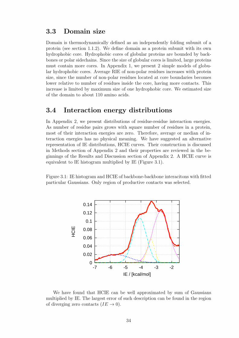
3.3 Domain size
Domain is thermodynamically defined as an independently folding subunit of aprotein (see section 1.1.2). We define domain as a protein subunit with its ownhydrophobic core. Hydrophobic cores of globular proteins are bounded by back-bones or polar sidechains. Since the size of globular cores is limited, large proteinsmust contain more cores. In Appendix 1, we present 2 simple models of globu-lar hydrophobic cores. Average RIE of non-polar residues increases with proteinsize, since the number of non-polar residues located at core boundaries becomeslower relative to number of residues inside the core, having more contacts. Thisincrease is limited by maximum size of one hydrophobic core. We estimated sizeof the domain to about 110 amino acids.
3.4 Interaction energy distributions
In Appendix 2, we present distributions of residue-residue interaction energies.As number of residue pairs grows with square number of residues in a protein,most of their interaction energies are zero. Therefore, average or median of in-teraction energies has no physical meaning. We have suggested an alternativerepresentation of IE distributions, HCIE curves. Their construction is discussedin Methods section of Appendix 2 and their properties are reviewed in the be-ginnings of the Results and Discussion section of Appendix 2. A HCIE curve isequivalent to IE histogram multiplied by IE (Figure 3.1).
Figure 3.1: IE histogram and HCIE of backbone-backbone interacitons with fittedparticular Gaussians. Only region of productive contacts was selected.
0
0.02
0.04
0.06
0.08
0.1
0.12
0.14
-7 -6 -5 -4 -3 -2
HC
IE
IE / [kcal/mol]
We have found that HCIE can be well approximated by sum of Gaussiansmultiplied by IE. The largest error of such description can be found in the regionof diverging zero contacts (IE → 0).
34

3.5 Definition of residue-residue contact
Geometric definition of residue-residue contact disregards strength of interactionbetween residues. Success of models decomposing stability into 1-body contribu-tions neglecting native state interactions [112] indicates that energy is so balancedthat only significantly strong interactions play role. In Appendix 2, we proposesuch definition based on HCIE curves as an interaction energy value dividingregions of significantly strong (we call them productive) contacts from the weak(we call them bulk) interactions.
In addition to assuming validity of hypotheses 1, 2, 4 and 5, we assume thatpeaks in HCIE curves correspond to interaction patterns and they can be used todiscriminate between productive and bulk interactions. A HCIE curve containsmultiple stationary points, so we employed additional criteria to identify the onecorresponding to optimum contact definition. Contact number (average numberof contacts per residue) for productive contacts was expected to be more restric-tive than the one for geometric contacts. Therefore, optimum contact definitionwere expected to result in contact numbers in order of units. At most 3 stationarypoints were possible candidates for a productive contact definition (see Appendix2), so we suppose that the selection of the contact definition resulting in con-tact numbers consistent with current understanding of contacts However, thereis a possibility that our view was biased by current recognition of residue-residueinteraction strength.
We have shown that using different force field (CHARMM) leads to very sim-ilar contact matrices. From comparison of contact matrix similarities at fixedaverage contact numbers (Table 2 in Appendix 2), it is obvious that contact def-inition based on interaction energies is less ambiguous than any geometry-baseddefinition. We have also shown that thermostable proteins contain higher num-ber of contacts defined based on interaction energy than do mesostable proteins.Contact definition might find its application in flexibility models, identification ofaminoacids key to stability and in identification of domains as clusters of produc-tive contacts. The view of cooperatively folding domains as clusters of energy-based contact is plausible because of entropic effects connected with non-covalentbonding of coupled bonding sites [170].
3.6 Interaction energy balance in proteins
This project was motivated by successful application of the interaction energymatrix concept on prediction of the melting temperatures for sequence variantsof haloalkane dehalogenases based on change of summed interaction energies ofuncharged sidechains calculated in native state structures. We could not includecharged residues, since their interaction energies are incomparable to interactionenergies between uncharged residues and from the beginning, we did not trustusing dielectric constant.
35

Figure 3.2: Correlation between melting temperatures and interaction energydifferences between mutants and wild type haloalkane dehalogenase.
45
50
55
60
65
70
-40 -30 -20 -10 0 10 20 30
Tm
/ [o
C]
energy difference wt->mutant / [kcal/mol]
The correlation was, surprisingly, better than the one obtained acknowledgedweb applications (e.g. FoldX). This result might be only a unique property ofthe tested system. However, interaction energy matrix approach to stabilityprediction from structure has a lot of space for further improvement.
3.6.1 Derivation of the model
We hereby propose a model of protein stability decomposition into separate freeenergy terms based on physical considerations. The model assumes additivityof free energy contributions (hypothesis 1.9) and linear dependence of force fieldinteraction energy of a residue-residue interaction and its free energy contributionto overall stability. Free energy of folding is formally expanded into 1-body and2-body terms.
∆Gfold =N∑
i=1
∆G(1)i +
N∑
i=1
N∑
j=1
∆G(2)ij + δ∆G(3) (3.1)
where N is number of residues in the protein. 1-body terms represent stabilitycontributions of single amino acids and 2-body terms represent pairwise stabilitycontributions. These terms are calculated from native structure. Thermodynamiccycle (Figure 3.3) provides a physically intuitive picture of stability contributions.
36

Figure 3.3: Thermodynamic cycle divides protein unfolding into 3 steps - desol-vation of native state, unfolding invacuo and solvation of denatured state.
• ∆G1 - Solvation free energy in the native state.
Using this macroscopic solvation model, contribution is 1-body and canbe approximated by a linear function of and non-polar solvent accessiblesurface areas of the native state.
• ∆G2 - Folding free energy in vacuo.
1-body free energy contribution comes from configurational entropy changeand internal energy change, e.g. torsional strain.
2-body free energy contribution is a difference of residue-residue interactionenergies between native and denatured state corrected for entropic effects.Assuming that proportion of persistence of interactions of a particular typein denatured state is universal for all proteins, this free energy contributioncan be calculated as a sum of interaction energies of that type multipliedby a universal scaling factor.
• ∆G3 - Solvation free energy of the denatured state.
Denatured state is approximated by an unfolded state with presence ofresidual native state contacts. Macroscopic model of solvation leads to 1-body contributions of amino acids to solvation of unfolded state.
To summarize, 1-body contributions comprise solvation of the native and de-natured state and configurational entropy. The latter two can be calculated assums of amino acids of particular type multiplied by universal scaling factorsrepresenting their characteristic solvation energies and configurational entropies.In addition to the mentioned, 1-body contribution of an amino acid also involvesaverage interaction of the amino acid in the native state. Solvation of the nativestate can be calculated as a linear transformation of surface areas (see methodssection 2.2.4). 2-body interactions comprise residue-residue interactions reducedby their persistence in denatured state and opposing entropic effect. The reduc-tion is modeled by linear transformation of interaction energies to free energycontributions, which is quite stronger assumption than hypothesis 5. Stability ofa protein can be written as
∆Gfold =20∑
i=1
ai ni +10∑
j=1
IEj bj +3∑
k=1
SASk ck (3.2)
37

where i’s denote amino acid types, j’s denote interaction energy types andk’s surface types. ni is number of residues of type i (dimensionless), IEj sumof interaction energies of type j (in kcal/mol) and SASk area of native statesurface of type k (in A2). There are 33 scaling factors, 20 ai’s (in kcal/mol), 10bj (dimensionless) and 3 ck (in kcal/(molA2) ).
3.6.2 Interpretation of the scaling factors
Scaling factors calculated using Courant-Fischer-Weyl theorem (see section 2.3)to best represent stabilites of 1188 proteins are summarized in Table 3.1. First, itis worth to remind that their values are relative to each other, therefore one scalingfactor must be set to arbitrary value. Largest 2-body scaling factor was set to 1.Such selection of scale leads to 1-body scaling factors in the order of magnitude ofsolvation free energies (units to decades of kcal/mol). Expected values of 2-bodyscaling factors are between 0 and 1, since they originate in entropic compensationof interaction energies and persistence of interactions in denatured state. Boththe mentioned effects are destabilizing while interaction energies are stabilizing.In Table 3.1, destabilizing terms are negative and stabilizing terms positive.
Highest stabilizing contribution (42%) comes from solvation of native state.This result might seem surprising, but average value of this free energy con-tribution (700 kcal/mol) accords with usual solvation free energies of proteins.Contribution of surface solvation to stability also decreases with increasing pro-tein size, as expected. High contribution comes also from interactions betweenbackbones and non-polar sidechains. This term is independent of protein size andseems to be a result of overestimated Lennard-Jones parameters of backbones.As this type of interactions seems to be non-specific, their effect on fold selectionshould be lower.
Surprisingly low are scaling factors of charged residues. If solvation wasthe main force compensating stabilization by interactions of charged sidechains,high compensating values of both 1-body and 2-body contributions would be ex-pected. Three interpretations of the surprisingly low values obtained by modelparametrization appear. First, energy of charged interactions might be incor-rectly calculated at force field level. Inaccurate distributions of partial chargeson charged sidechains can damage quality of close-ranged interaction energiesbetween charged sidechains. However, long-range interactions between chargedresidues remain unaffected, since they are not force field dependent. Second, hy-pothesis 2 can be invalid for charged interactions. In each protein, dynamics ofresulting in disagreement between native state ensemble average sum of charged-charged interactions and the sum calculated for single structure from PDB.
Third explanation seems to be the most interesting one. In Appendix 2, weshow that 92% of energy contribution by attractive interactions between chargedsidechains is compensated by repulsive interactions. Low values of scaling factorscorresponding to charged residues suggest that very similar compensation mighttake place in the denatured state, so that positions of charged sidechains arecorrelated to decrease energy. In other words sort of Madelung constant of nativeand denatured state is very similar. This effect can be confirmed by interactionenergy matrix analyses of protein unfolding simulations.
38

Table 3.1: Scaling factors obtained using the prioposed stability model. Tableis divided into 2 subtables, 1-body contributions are in the left one and 1-bodynative state solvation contributions in the right subtable at the bottom. 2-bodycontributions are stabilizing and can be found in the right subtable (first tenrows). First column of each subtable is name of the scaling factor, second is itsvalue, third is its standard deviation and fourth columns contain contribution ofthe corresponding scaling factor to protein stability. Destabilizing cintributionsare marked by minus sign and in the left subtable. Units are as mentioned above.
scaling value stdev contrib scaling value stdev contribfactor in % in % factor in % in %GLY -4.9 2.4 -4.0 BB-BB 0.31 8.9 16.2ALA -6.6 1.8 -5.8 BB-CH 0.10 22 3.4VAL -7.3 1.8 -6.1 BB-PO 0.57 7.1 9.2ILE -7.4 1.9 -4.8 BB-NP 0.78 7.0 17.6CYS -7.8 2.2 -1.3 CH-CH 0.012 86 0.7LEU -8.2 1.6 -8.6 CH-PO 0.051 51 0.5MET -9.2 1.9 -1.7 CH-NP 0.15 24 0.7PHE -10.3 1.7 -4.7 PO-PO 0.75 8.8 1.5PRO -16.7 1.8 -8.8 PO-NP 1.00 6.5 5.1THR -7.9 1.6 -4.9 NP-NP 0.16 29 1.1SER -8.2 1.5 -5.5TYR -13.8 1.3 -5.4 SASNP 0.43 13 6.3ASN -14.8 1.0 -7.3 SASPO 6.2 4.3 6.8TRP -16.1 1.4 -2.5 SASCH 19.9 1.0 30.9GLN -17.0 0.90 -7.4LYS -4.6 2.7 -3.5HIS -5.3 2.9 -1.3ASP -5.9 3.3 -3.9GLU -6.6 3.2 -5.3ARG -12.3 1.3 -7.2
39
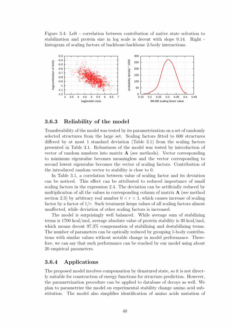
Figure 3.4: Left - correlation between contribution of native state solvation tostabilization and protein size in log scale is decent with slope 0.14. Right -histogram of scaling factors of backbone-backbone 2-body interactions.
-1.2
-1.1
-1
-0.9
-0.8
-0.7
-0.6
-0.5
-0.4
-0.3
3 3.5 4 4.5 5 5.5 6 6.5 7
log(
cont
ribut
ion
of S
AS
)
log(protein size)
0
50
100
150
200
250
300
0.15 0.2 0.25 0.3 0.35 0.4 0.45
prob
abili
ty d
ensi
ty *
100
0
BB-BB scaling factor value
3.6.3 Reliability of the model
Transferability of the model was tested by its parametrization on a set of randomlyselected structures from the large set. Scaling factors fitted to 600 structuresdiffered by at most 1 standard deviation (Table 3.1) from the scaling factorspresented in Table 3.1. Robustness of the model was tested by introduction ofvector of random numbers into matrix A (see methods). Vector correspondingto minimum eigenvalue becomes meaningless and the vector corresponding tosecond lowest eigenvalue becomes the vector of scaling factors. Contribution ofthe introduced random vector to stability is close to 0.
In Table 3.1, a correlation between value of scaling factor and its deviationcan be noticed. This effect can be attributed to reduced importance of smallscaling factors in the expression 2.4. The deviation can be artificially reduced bymultiplication of all the values in corresponding column of matrix A (see methodsection 2.3) by arbitrary real number 0 < r < 1, which causes increase of scalingfactor by a factor of 1/r. Such treatment keeps values of all scaling factors almostunaffected, while deviation of other scaling factors is increased.
The model is surprisingly well balanced. While average sum of stabilizingterms is 1700 kcal/mol, average absolute value of protein stability is 30 kcal/mol,which means decent 97.3% compensation of stabilizing and destabilizing terms.The number of parameters can be optically reduced by grouping 1-body contribu-tions with similar values without notable change in model performance. There-fore, we can say that such performance can be reached by our model using about20 empirical parameters.
3.6.4 Applications
The proposed model involves compensation by denatured state, so it is not direct-ly suitable for construction of energy functions for structure prediction. However,the parametrization procedure can be applied to database of decoys as well. Weplan to parametrize the model on experimental stability change amino acid sub-stitution. The model also simplifies identification of amino acids mutation of
40

which might increase stability, since residues can be ranked according to theirstabilization effect from most destabilizing to most stabilizing ones.
The model also attempts to solve the question of stabilization of proteins byparticular types of interactions. Understanding forces contributing to stability iscrucial for systematic improvement of energy functions. The presented model isnot purely empirical; particular contributions have physical meaning which canbe further studied.
3.7 Dynamic properties of interaction energy sums
Applicability of hypothesis 2 is interesting for all structural studies. We haveundertaken a short study to examine dynamical behavior of interaction energysums, since we assume validity of hypothesis 2 in most of the presented work.Description of native state by simulation is more realistic. Moreover, it might beuseful in prediction of entropy and average IEs from single structure. In Table3.2, interaction energy sums calculated for a single X-ray structure are comparedto average sums obtained from IEM analysis of snapshots from simulation (seemethods section 2.2.6). Differences between static and dynamic description aresmall in case of interaction energy types with high scaling factors in Table 3.1.
Table 3.2: Comparison of dynamic and static interaction energy matrix descrip-tion of protein 3QPA. First column contains interaction energy types, secondcolumn contains sum of interaction energies of that type calculated using a singlestructure, third contains average sum of theinteraction energies thourgout sim-ulation and fourth gives standard deviation of value in the third column. Allvalues are in kcal/mol.
IE type X-ray dynamics stdevBBBB -668 -719 12BBCH -957 -586 20BBPO -351 -293 14BBNP -358 -349 7.2CHCH -1813 -1450 58CHPO -336 -212 15CHNP -82.9 -77.5 4.8POPO -42.2 -43.4 5.2PONP -93.7 -78.7 3.3NPNP -99.1 -96.3 3.3
41
4. Conclusion
In the presented thesis, connection between pairwise interaction energies and theirfree energy contribution to stability was studied.
Aims listed in section 1.6 were addressed as follows
1. Solvent effect and compensation of interaction energies by persistence indenatured state is modeled by fragmentation of proteins and classificationof fragments based on their multipolar character. Instead of using an empir-ical dielectric constant, we propose comparison of interaction energies withthe determined characteristic values or linear scaling of interaction energiesusing the proposed empirical aprameters.
2. In Paper 1 and Paper 2, distributions of residue interaction energies and in-teraction energies (respectively) are studied. Interaction energy histogramshas been found to be composed of Gaussian-shaped peaks correspondingto particular interaction patterns. A model explaining this observation wasdeveloped and its description can be found in Paper 2.
3. In Paper 2, we present a method of constructing matrices of productivecontacts from native state geometry. We hypothesize that such contactsare additive quantities. We have not succeed in exploiting the sequenceinformation contained in contact matrices yet.
4. We have proposed a model integrating all interaction energies into proteinstability. The model seems to be well balanced and its further improvementis possible.
5. In Paper 1, we have found no correlation between secondary structuresand sidechain interaction energies. The correlation was found only betweensecondary structure content and interactions between backbones, which isnot surprising. Secondary structure propensities could not be correlatedwith positions of peaks in BB-BB RIE distributions.
There is a lot of space for future studies. First, we want to launch a webapplication for calculation of interaction energy matrices and construction of ourcontact matrices. We hope that results of our work will be useful for structuralbiologists in finding structural and functional features in protein structures. Sec-ond, we want to study entropic effect of contact separation in sequence. Third, weplan to use the energy balance in construction of energy functions for predictionof stability change upon amino acid replacement. Last, but not least, we want tomodify the methodology to avoid problems arising from difference between nativestate ensemble and single X-ray structure from database.
42
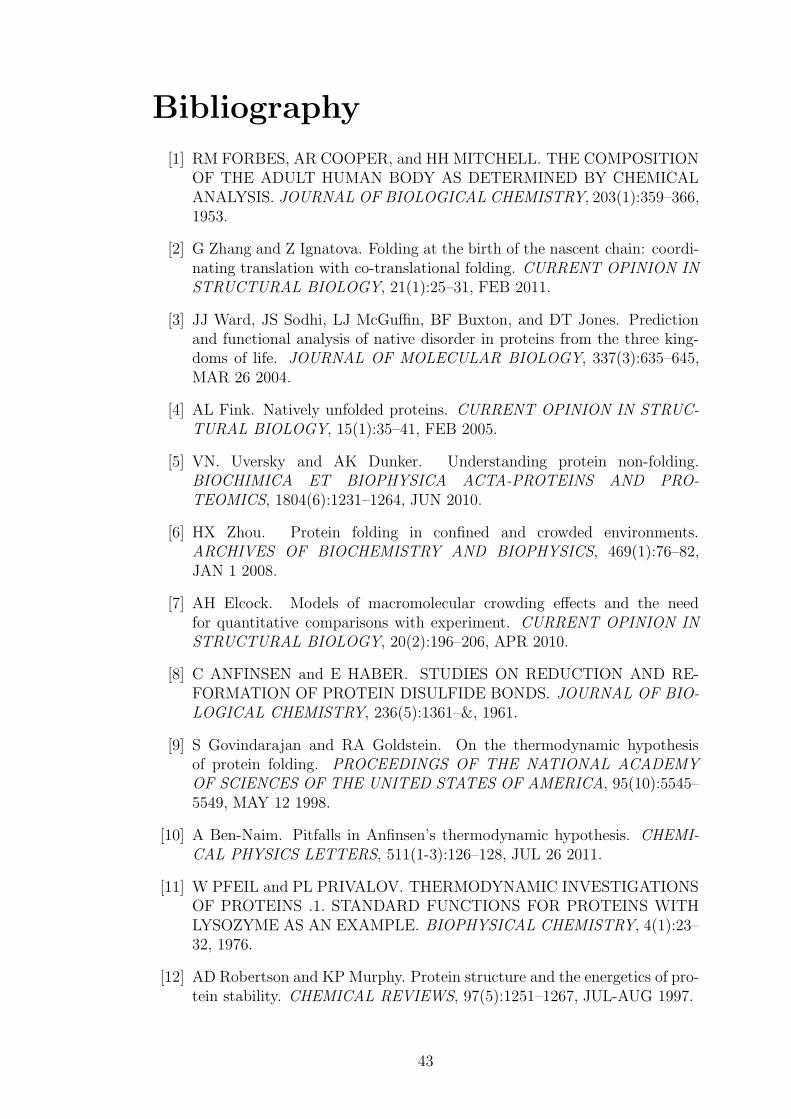
Bibliography
[1] RM FORBES, AR COOPER, and HH MITCHELL. THE COMPOSITIONOF THE ADULT HUMAN BODY AS DETERMINED BY CHEMICALANALYSIS. JOURNAL OF BIOLOGICAL CHEMISTRY, 203(1):359–366,1953.
[2] G Zhang and Z Ignatova. Folding at the birth of the nascent chain: coordi-nating translation with co-translational folding. CURRENT OPINION INSTRUCTURAL BIOLOGY, 21(1):25–31, FEB 2011.
[3] JJ Ward, JS Sodhi, LJ McGuffin, BF Buxton, and DT Jones. Predictionand functional analysis of native disorder in proteins from the three king-doms of life. JOURNAL OF MOLECULAR BIOLOGY, 337(3):635–645,MAR 26 2004.
[4] AL Fink. Natively unfolded proteins. CURRENT OPINION IN STRUC-TURAL BIOLOGY, 15(1):35–41, FEB 2005.
[5] VN. Uversky and AK Dunker. Understanding protein non-folding.BIOCHIMICA ET BIOPHYSICA ACTA-PROTEINS AND PRO-TEOMICS, 1804(6):1231–1264, JUN 2010.
[6] HX Zhou. Protein folding in confined and crowded environments.ARCHIVES OF BIOCHEMISTRY AND BIOPHYSICS, 469(1):76–82,JAN 1 2008.
[7] AH Elcock. Models of macromolecular crowding effects and the needfor quantitative comparisons with experiment. CURRENT OPINION INSTRUCTURAL BIOLOGY, 20(2):196–206, APR 2010.
[8] C ANFINSEN and E HABER. STUDIES ON REDUCTION AND RE-FORMATION OF PROTEIN DISULFIDE BONDS. JOURNAL OF BIO-LOGICAL CHEMISTRY, 236(5):1361–&, 1961.
[9] S Govindarajan and RA Goldstein. On the thermodynamic hypothesisof protein folding. PROCEEDINGS OF THE NATIONAL ACADEMYOF SCIENCES OF THE UNITED STATES OF AMERICA, 95(10):5545–5549, MAY 12 1998.
[10] A Ben-Naim. Pitfalls in Anfinsen’s thermodynamic hypothesis. CHEMI-CAL PHYSICS LETTERS, 511(1-3):126–128, JUL 26 2011.
[11] W PFEIL and PL PRIVALOV. THERMODYNAMIC INVESTIGATIONSOF PROTEINS .1. STANDARD FUNCTIONS FOR PROTEINS WITHLYSOZYME AS AN EXAMPLE. BIOPHYSICAL CHEMISTRY, 4(1):23–32, 1976.
[12] AD Robertson and KP Murphy. Protein structure and the energetics of pro-tein stability. CHEMICAL REVIEWS, 97(5):1251–1267, JUL-AUG 1997.
43

[13] GI Makhatadze and PL Privalov. Energetics of protein structure. In AD-VANCES IN PROTEIN CHEMISTRY, VOL 47, volume 47 of ADVANCESIN PROTEIN CHEMISTRY, pages 307–425. ACADEMIC PRESS INC,525 B STREET, SUITE 1900, SAN DIEGO, CA 92101-4495, 1995.
[14] A Michnik. Thermal stability of bovine serum albumin DSC study. JOUR-NAL OF THERMAL ANALYSIS AND CALORIMETRY, 71(2):509–519,2003.
[15] PL PRIVALOV and KHECHINA.NN. THERMODYNAMIC APPROACHTO PROBLEM OF STABILIZATION OF GLOBULAR PROTEINSTRUCTURE - CALORIMETRIC STUDY. JOURNAL OF MOLECU-LAR BIOLOGY, 86(3):665–684, 1974.
[16] H Kaya and HS Chan. Polymer principles of protein calorimetric two-statecooperativity. PROTEINS-STRUCTURE FUNCTION AND GENETICS,40(4):637–661, SEP 1 2000.
[17] PL Privalov. ADVANCES IN PROTEIN CHEMISTRY, 33:167–241, 1979.
[18] L Liu, C Yang, and QX Guo. A study on the enthalpy-entropy compen-sation in protein unfolding. BIOPHYSICAL CHEMISTRY, 84(3):239–251,MAY 15 2000.
[19] S Kumar, CJ Tsai, and R Nussinov. Thermodynamic differences amonghomologous thermophilic and mesophilic proteins. BIOCHEMISTRY,40(47):14152–14165, NOV 27 2001.
[20] JD DUNITZ. WIN SOME, LOSE SOME - ENTHALPY-ENTROPYCOMPENSATION IN WEAK INTERMOLECULAR INTERACTIONS.CHEMISTRY & BIOLOGY, 2(11):709–712, NOV 1995.
[21] DM Ford. Enthalpy-entropy compensation is not a general feature of weakassociation. JOURNAL OF THE AMERICAN CHEMICAL SOCIETY,127(46):16167–16170, NOV 23 2005.
[22] JC KENDREW. ARCHITECTURE OF A PROTEIN MOLECULE. NA-TURE, 182(4638):764–767, 1958.
[23] GJ LESSER and GD ROSE. HYDROPHOBICITY OF AMINO-ACIDSUBGROUPS IN PROTEINS. PROTEINS-STRUCTURE FUNCTIONAND GENETICS, 8(1):6–13, 1990.
[24] AE Mirsky and L Pauling. On the structure of native, denatured, andcoagulated proteins. PROCEEDINGS OF THE NATIONAL ACADEMYOF SCIENCES OF THE UNITED STATES OF AMERICA, 22:439–447,1936.
[25] CA Orengo, AD Michie, S Jones, DT Jones, MB Swindells, and JM Thorn-ton. CATH - a hierarchic classification of protein domain structures.STRUCTURE, 5(8):1093–1108, AUG 15 1997.
44

[26] AG MURZIN, SE BRENNER, T HUBBARD, and C CHOTHIA. SCOP -A STRUCTURAL CLASSIFICATION OF PROTEINS DATABASE FORTHE INVESTIGATION OF SEQUENCES AND STRUCTURES. JOUR-NAL OF MOLECULAR BIOLOGY, 247(4):536–540, APR 7 1995.
[27] A Szilagyi and P Zavodszky. Structural differences between mesophilic,moderately thermophilic and extremely thermophilic protein subunits: re-sults of a comprehensive survey. STRUCTURE, 8(5):493–504, MAY 152000.
[28] AV Glyakina, SO Garbuzynskiy, MYu Lobanov, and OV Galzitskaya. Dif-ferent packing of external residues can explain differences in the thermosta-bility of proteins from thermophilic and mesophilic organisms. BIOINFOR-MATICS, 23(17):2231–2238, SEP 1 2007.
[29] N Kannan and S Vishveshwara. Aromatic clusters: a determinant ofthermal stability of thermophilic proteins. PROTEIN ENGINEERING,13(11):753–761, NOV 2000.
[30] D NERI, M BILLETER, G WIDER, and K WUTHRICH. NMR DETER-MINATION OF RESIDUAL STRUCTURE IN A UREA-DENATUREDPROTEIN, THE 434-REPRESSOR. SCIENCE, 257(5076):1559–1563, SEP11 1992.
[31] H Schwalbe, KM Fiebig, M Buck, JA Jones, SB Grimshaw, A Spencer,SJ Glaser, LJ Smith, and CM Dobson. Structural and dynamical propertiesof a denatured protein. Heteronuclear 3D NMR experiments and theoreticalsimulations of lysozyme in 8 M urea. BIOCHEMISTRY, 36(29):8977–8991,JUL 22 1997.
[32] HBR COLE, SW SPARKS, and DA TORCHIA. COMPARISON OFTHE SOLUTION AND CRYSTAL-STRUCTURES OF STAPHYLOCOC-CAL NUCLEASE WITH C-13 AND N-15 CHEMICAL-SHIFTS USED ASSTRUCTURAL FINGERPRINTS. PROCEEDINGS OF THE NATION-AL ACADEMY OF SCIENCES OF THE UNITED STATES OF AMER-ICA, 85(17):6362–6365, SEP 1988.
[33] IK MCDONALD and JM THORNTON. SATISFYING HYDROGEN-BONDING POTENTIAL IN PROTEINS. JOURNAL OF MOLECULARBIOLOGY, 238(5):777–793, MAY 20 1994.
[34] VA Higman, J Boyd, LJ Smith, and C Redfield. Asparagine and glu-tamine side-chain conformation in solution and crystal: A comparison forhen egg-white lysozyme using residual dipolar couplings. JOURNAL OFBIOMOLECULAR NMR, 30(3):327–346, NOV 2004.
[35] CN PACE and TE CREIGHTON. THE DISULFIDE FOLDING PATH-WAY OF RIBONUCLEASE-T1. JOURNAL OF MOLECULAR BIOLO-GY, 188(3):477–486, APR 5 1986.
45
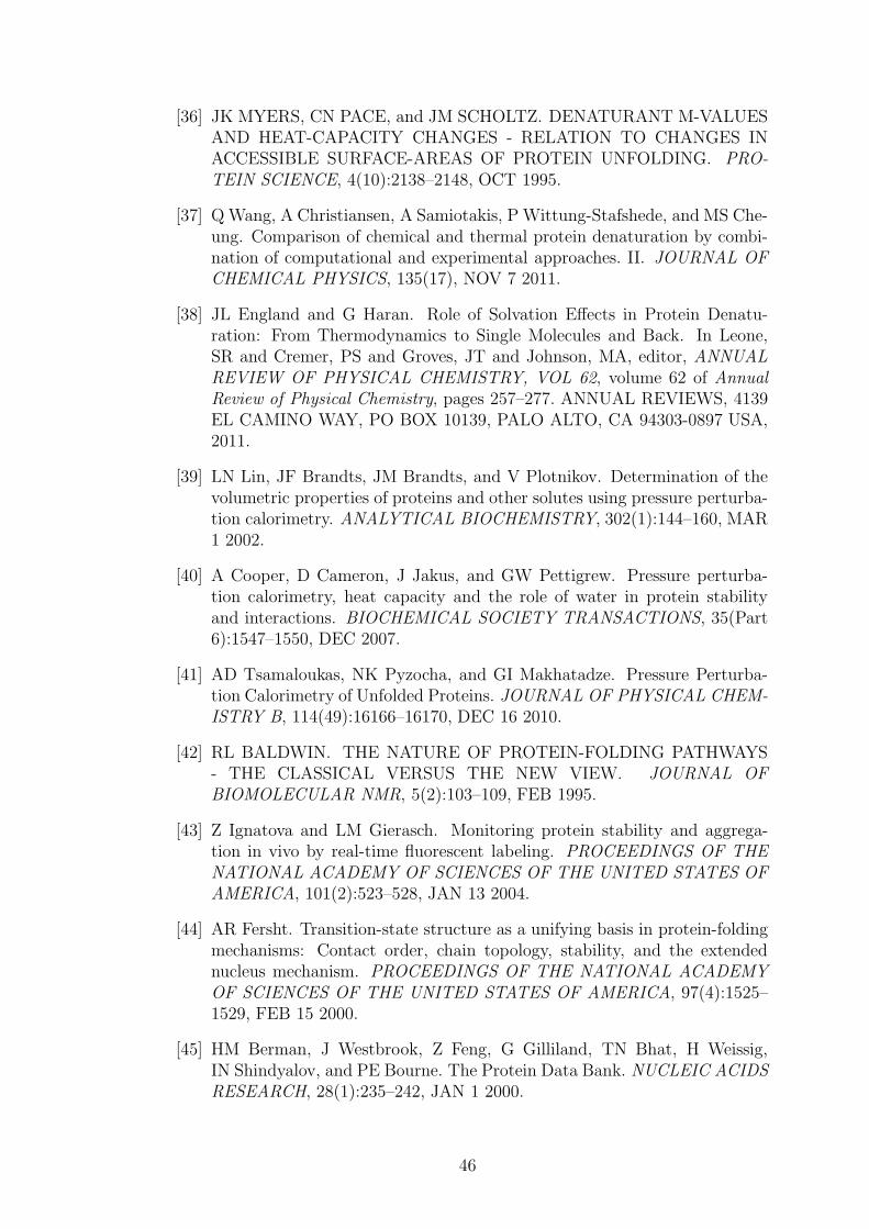
[36] JK MYERS, CN PACE, and JM SCHOLTZ. DENATURANT M-VALUESAND HEAT-CAPACITY CHANGES - RELATION TO CHANGES INACCESSIBLE SURFACE-AREAS OF PROTEIN UNFOLDING. PRO-TEIN SCIENCE, 4(10):2138–2148, OCT 1995.
[37] QWang, A Christiansen, A Samiotakis, P Wittung-Stafshede, and MS Che-ung. Comparison of chemical and thermal protein denaturation by combi-nation of computational and experimental approaches. II. JOURNAL OFCHEMICAL PHYSICS, 135(17), NOV 7 2011.
[38] JL England and G Haran. Role of Solvation Effects in Protein Denatu-ration: From Thermodynamics to Single Molecules and Back. In Leone,SR and Cremer, PS and Groves, JT and Johnson, MA, editor, ANNUALREVIEW OF PHYSICAL CHEMISTRY, VOL 62, volume 62 of AnnualReview of Physical Chemistry, pages 257–277. ANNUAL REVIEWS, 4139EL CAMINO WAY, PO BOX 10139, PALO ALTO, CA 94303-0897 USA,2011.
[39] LN Lin, JF Brandts, JM Brandts, and V Plotnikov. Determination of thevolumetric properties of proteins and other solutes using pressure perturba-tion calorimetry. ANALYTICAL BIOCHEMISTRY, 302(1):144–160, MAR1 2002.
[40] A Cooper, D Cameron, J Jakus, and GW Pettigrew. Pressure perturba-tion calorimetry, heat capacity and the role of water in protein stabilityand interactions. BIOCHEMICAL SOCIETY TRANSACTIONS, 35(Part6):1547–1550, DEC 2007.
[41] AD Tsamaloukas, NK Pyzocha, and GI Makhatadze. Pressure Perturba-tion Calorimetry of Unfolded Proteins. JOURNAL OF PHYSICAL CHEM-ISTRY B, 114(49):16166–16170, DEC 16 2010.
[42] RL BALDWIN. THE NATURE OF PROTEIN-FOLDING PATHWAYS- THE CLASSICAL VERSUS THE NEW VIEW. JOURNAL OFBIOMOLECULAR NMR, 5(2):103–109, FEB 1995.
[43] Z Ignatova and LM Gierasch. Monitoring protein stability and aggrega-tion in vivo by real-time fluorescent labeling. PROCEEDINGS OF THENATIONAL ACADEMY OF SCIENCES OF THE UNITED STATES OFAMERICA, 101(2):523–528, JAN 13 2004.
[44] AR Fersht. Transition-state structure as a unifying basis in protein-foldingmechanisms: Contact order, chain topology, stability, and the extendednucleus mechanism. PROCEEDINGS OF THE NATIONAL ACADEMYOF SCIENCES OF THE UNITED STATES OF AMERICA, 97(4):1525–1529, FEB 15 2000.
[45] HM Berman, J Westbrook, Z Feng, G Gilliland, TN Bhat, H Weissig,IN Shindyalov, and PE Bourne. The Protein Data Bank. NUCLEIC ACIDSRESEARCH, 28(1):235–242, JAN 1 2000.
46

[46] MM Gromiha, J An, H Kono, M Oobatake, H Uedaira, and A Sarai.ProTherm: Thermodynamic database for proteins and mutants. NUCLEICACIDS RESEARCH, 27(1):286–288, JAN 1 1999.
[47] V Potapov, M Cohen, and G Schreiber. Assessing computational meth-ods for predicting protein stability upon mutation: good on average butnot in the details. PROTEIN ENGINEERING DESIGN & SELECTION,22(9):553–560, SEP 2009.
[48] RA Friesner and JR Gunn. Computational studies of protein folding.ANNUAL REVIEW OF BIOPHYSICS AND BIOMOLECULAR STRUC-TURE, 25:315–342, 1996.
[49] AR Dinner, A Sali, LJ Smith, CM Dobson, and M Karplus. Understand-ing protein folding via free-energy surfaces from theory and experiment.TRENDS IN BIOCHEMICAL SCIENCES, 25(7):331–339, JUL 2000.
[50] M Vendruscolo and E Paci. Protein folding: bringing theory and experi-ment closer together. CURRENT OPINION IN STRUCTURAL BIOLO-GY, 13(1):82–87, FEB 2003.
[51] C LEVINTHAL. ARE THERE PATHWAYS FOR PROTEIN FOLD-ING. JOURNAL DE CHIMIE PHYSIQUE ET DE PHYSICO-CHIMIEBIOLOGIQUE, 65(1):44–&, 1968.
[52] M Paterson and T Przytycka. On the complexity of string folding. DIS-CRETE APPLIED MATHEMATICS, 71(1-3):217–230, DEC 5 1996.
[53] OK Vorov, DR Livesay, and DJ Jacobs. Conformational Entropy of an IdealCross-Linking Polymer Chain. ENTROPY, 10(3):285–308, SEP 2008.
[54] KA DILL, S BROMBERG, KZ YUE, KM FIEBIG, DP YEE,PD THOMAS, and HS CHAN. PRINCIPLES OF PROTEIN-FOLDING -A PERSPECTIVE FROM SIMPLE EXACT MODELS. PROTEIN SCI-ENCE, 4(4):561–602, APR 1995.
[55] KA DILL and D STIGTER. MODELING PROTEIN STABILITY ASHETEROPOLYMER COLLAPSE. In ADVANCES IN PROTEIN CHEM-ISTRY, VOL 46, volume 46 of ADVANCES IN PROTEIN CHEMISTRY,pages 59–104. ACADEMIC PRESS INC, 525 B STREET, SUITE 1900,SAN DIEGO, CA 92101-4495, 1995.
[56] A Hansen, MH Jensen, K Sneppen, and G Zocchi. Statistical mechanics ofwarm and cold unfolding in proteins. EUROPEAN PHYSICAL JOURNALB, 6(1):157–161, NOV 1998.
[57] E Shakhnovich. Protein folding thermodynamics and dynamics:Where physics, chemistry, and biology meet. CHEMICAL REVIEWS,106(5):1559–1588, MAY 2006.
[58] D Thirumalai, EP O’Brien, G Morrison, and C Hyeon. Theoretical Per-spectives on Protein Folding. In Rees, DC and Dill, KA and Williamson,
47

JR, editor, ANNUAL REVIEW OF BIOPHYSICS, VOL 39, volume 39 ofAnnual Review of Biophysics, pages 159–183. ANNUAL REVIEWS, 4139EL CAMINO WAY, PO BOX 10139, PALO ALTO, CA 94303-0897 USA,2010.
[59] GR Bowman, VA Voelz, and VS Pande. Taming the complexity of proteinfolding. CURRENT OPINION IN STRUCTURAL BIOLOGY, 21(1):4–11,FEB 2011.
[60] KA DILL. THEORY FOR THE FOLDING AND STABILITY OFGLOBULAR-PROTEINS. BIOCHEMISTRY, 24(6):1501–1509, 1985.
[61] DE Shaw, P Maragakis, K Lindorff-Larsen, S Piana, RO Dror, MP East-wood, JA Bank, JM Jumper, JK Salmon, Y Shan, andWWriggers. Atomic-Level Characterization of the Structural Dynamics of Proteins. SCIENCE,330(6002):341–346, OCT 15 2010.
[62] KE Riley, M Pitonak, P Jurecka, and P Hobza. Stabilization and StructureCalculations for Noncovalent Interactions in Extended Molecular SystemsBased on Wave Function and Density Functional Theories. CHEMICALREVIEWS, 110(9):5023–5063, SEP 2010.
[63] JC Faver, ML Benson, X He, BP Roberts, B Wang, MS Marshall, CD Sher-rill, and KMMerz. The Energy Computation Paradox and ab initio ProteinFolding. PLOS ONE, 6(4), APR 25 2011.
[64] JW Ponder and DA Case. Force fields for protein simulations. In PROTEINSIMULATIONS, volume 66 of ADVANCES IN PROTEIN CHEMISTRY,pages 27+. ACADEMIC PRESS INC, 525 B STREET, SUITE 1900, SANDIEGO, CA 92101-4495 USA, 2003.
[65] M Kolar, K Berka, P Jurecka, and P Hobza. On the Reliability of theAMBER Force Field and its Empirical Dispersion Contribution for theDescription of Noncovalent Complexes. CHEMPHYSCHEM, 11(11):2399–2408, AUG 2 2010.
[66] RB Best, NV Buchete, and G Hummer. Are current molecular dynamicsforce fields too helical? BIOPHYSICAL JOURNAL, 95(1):L7–L9, JUL 12008.
[67] J Hermans. Hydrogen bonds in molecular mechanics force fields. InPEPTIDE SOLVATION AND H-BONDS, volume 72 of ADVANCES INPROTEIN CHEMISTRY, pages 105–119. ELSEVIER ACADEMIC PRESSINC, 525 B STREET, SUITE 1900, SAN DIEGO, CA 92101-4495 USA,2006.
[68] ACM Ferreon and AA Deniz. Protein folding at single-molecule resolu-tion. BIOCHIMICA ET BIOPHYSICA ACTA-PROTEINS AND PRO-TEOMICS, 1814(8, SI):1021–1029, AUG 2011.
48
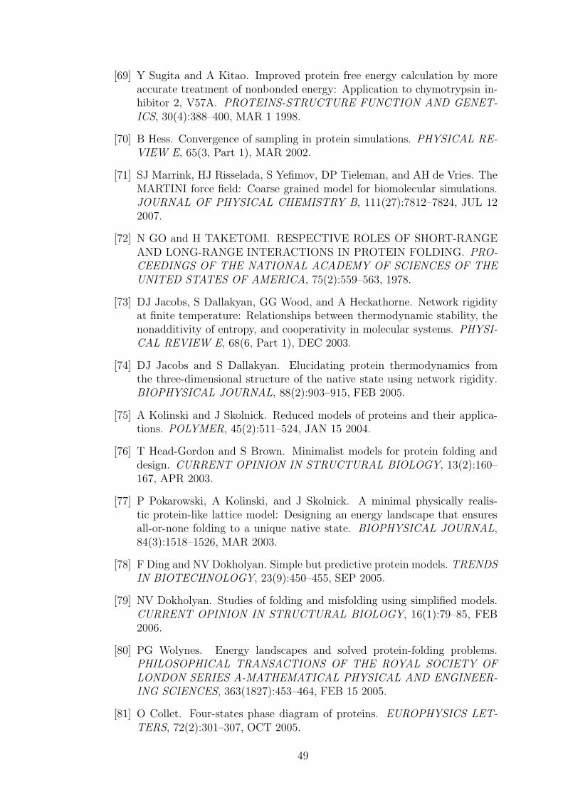
[69] Y Sugita and A Kitao. Improved protein free energy calculation by moreaccurate treatment of nonbonded energy: Application to chymotrypsin in-hibitor 2, V57A. PROTEINS-STRUCTURE FUNCTION AND GENET-ICS, 30(4):388–400, MAR 1 1998.
[70] B Hess. Convergence of sampling in protein simulations. PHYSICAL RE-VIEW E, 65(3, Part 1), MAR 2002.
[71] SJ Marrink, HJ Risselada, S Yefimov, DP Tieleman, and AH de Vries. TheMARTINI force field: Coarse grained model for biomolecular simulations.JOURNAL OF PHYSICAL CHEMISTRY B, 111(27):7812–7824, JUL 122007.
[72] N GO and H TAKETOMI. RESPECTIVE ROLES OF SHORT-RANGEAND LONG-RANGE INTERACTIONS IN PROTEIN FOLDING. PRO-CEEDINGS OF THE NATIONAL ACADEMY OF SCIENCES OF THEUNITED STATES OF AMERICA, 75(2):559–563, 1978.
[73] DJ Jacobs, S Dallakyan, GG Wood, and A Heckathorne. Network rigidityat finite temperature: Relationships between thermodynamic stability, thenonadditivity of entropy, and cooperativity in molecular systems. PHYSI-CAL REVIEW E, 68(6, Part 1), DEC 2003.
[74] DJ Jacobs and S Dallakyan. Elucidating protein thermodynamics fromthe three-dimensional structure of the native state using network rigidity.BIOPHYSICAL JOURNAL, 88(2):903–915, FEB 2005.
[75] A Kolinski and J Skolnick. Reduced models of proteins and their applica-tions. POLYMER, 45(2):511–524, JAN 15 2004.
[76] T Head-Gordon and S Brown. Minimalist models for protein folding anddesign. CURRENT OPINION IN STRUCTURAL BIOLOGY, 13(2):160–167, APR 2003.
[77] P Pokarowski, A Kolinski, and J Skolnick. A minimal physically realis-tic protein-like lattice model: Designing an energy landscape that ensuresall-or-none folding to a unique native state. BIOPHYSICAL JOURNAL,84(3):1518–1526, MAR 2003.
[78] F Ding and NV Dokholyan. Simple but predictive protein models. TRENDSIN BIOTECHNOLOGY, 23(9):450–455, SEP 2005.
[79] NV Dokholyan. Studies of folding and misfolding using simplified models.CURRENT OPINION IN STRUCTURAL BIOLOGY, 16(1):79–85, FEB2006.
[80] PG Wolynes. Energy landscapes and solved protein-folding problems.PHILOSOPHICAL TRANSACTIONS OF THE ROYAL SOCIETY OFLONDON SERIES A-MATHEMATICAL PHYSICAL AND ENGINEER-ING SCIENCES, 363(1827):453–464, FEB 15 2005.
[81] O Collet. Four-states phase diagram of proteins. EUROPHYSICS LET-TERS, 72(2):301–307, OCT 2005.
49

[82] P Ball. Water as an active constituent in cell biology. CHEMICAL RE-VIEWS, 108(1):74–108, JAN 2008.
[83] PL PRIVALOV and SJ GILL. STABILITY OF PROTEIN-STRUCTUREAND HYDROPHOBIC INTERACTION. ADVANCES IN PROTEINCHEMISTRY, 39:191–234, 1988.
[84] W Kauzmann. Some Factors in the Interpretation of Protein Denaturation.volume 14 of Advances in Protein Chemistry, pages 1 – 63. 1959.
[85] JH Hildebrand. Is there a ”Hydrophobic Effect”? Proceedings of the Na-tional Academy of Sciences of the United States of America, 76(1):194–194,January 1979.
[86] TV Chalikian. Structural Thermodynamics of Hydration. The Journal ofPhysical Chemistry B, 105(50):12566–12578, December 2001.
[87] Bhimalapuram P Widom, B and K Koga. The hydrophobic effect. PhysicalChemistry Chemical Physics, 5(15):3085, 2003.
[88] CJ Fennell and KA Dill. Physical Modeling of Aqueous Solvation. JOUR-NAL OF STATISTICAL PHYSICS, 145(2, SI):209–226, OCT 2011.
[89] F Avbelj and RL Baldwin. Limited validity of group additivity for thefolding energetics of the peptide group. PROTEINS-STRUCTURE FUNC-TION AND BIOINFORMATICS, 63(2):283–289, MAY 1 2006.
[90] A Warshel, PK Sharma, M Kato, and WW Parson. Modeling electrostaticeffects in proteins. BIOCHIMICA ET BIOPHYSICA ACTA-PROTEINSAND PROTEOMICS, 1764(11):1647–1676, NOV 2006.
[91] AWARSHEL and M LEVITT. THEORETICAL STUDIES OF ENZYMICREACTIONS - DIELECTRIC, ELECTROSTATIC AND STERIC STA-BILIZATION OF CARBONIUM-ION IN REACTION OF LYSOZYME.JOURNAL OF MOLECULAR BIOLOGY, 103(2):227–249, 1976.
[92] M Schaefer and M Karplus. A comprehensive analytical treatmentof continuum electrostatics. JOURNAL OF PHYSICAL CHEMISTRY,100(5):1578–1599, FEB 1 1996.
[93] A Papazyan and A Warshel. Continuum and dipole-lattice models of solva-tion. JOURNAL OF PHYSICAL CHEMISTRY B, 101(51):11254–11264,DEC 18 1997.
[94] RL JERNIGAN and PJ FLORY. DISTRIBUTION FUNCTIONSFOR CHAIN MOLECULES. JOURNAL OF CHEMICAL PHYSICS,50(10):4185–&, 1969.
[95] D Shortle. The denatured state (the other half of the folding equation) andits role in protein stability. FASEB JOURNAL, 10(1):27–34, JAN 1996.
50

[96] TR SOSNICK and J TREWHELLA. DENATURED STATES OFRIBONUCLEASE-A HAVE COMPACT DIMENSIONS AND RESIDUALSECONDARY STRUCTURE. BIOCHEMISTRY, 31(35):8329–8335, SEP8 1992.
[97] M KATAOKA, Y HAGIHARA, K MIHARA, and Y GOTO. MOLTENGLOBULE OF CYTOCHROME-C STUDIED BY SMALL-ANGLEX-RAY-SCATTERING. JOURNAL OF MOLECULAR BIOLOGY,229(3):591–596, FEB 5 1993.
[98] Y Wang and D Shortle. The equilibrium folding pathway of staphylococcalnuclease: Identification of the most stable chain-chain interactions by NMRand CD spectroscopy. BIOCHEMISTRY, 34(49):15895–15905, DEC 121995.
[99] EE LATTMAN, KM FIEBIG, and KA DILL. MODELING COMPACTDENATURED STATES OF PROTEINS. BIOCHEMISTRY, 33(20):6158–6166, MAY 24 1994.
[100] P Weinkam, EV Pletneva, HB Gray, JR Winkler, and PGWolynes. Electro-static effects on funneled landscapes and structural diversity in denaturedprotein ensembles. PROCEEDINGS OF THE NATIONAL ACADEMYOF SCIENCES OF THE UNITED STATES OF AMERICA, 106(6):1796–1801, FEB 10 2009.
[101] CN Pace. Energetics of protein hydrogen bonds. NATURE STRUCTURAL& MOLECULAR BIOLOGY, 16(7):681–682, JUL 2009.
[102] C TANFORD. CONTRIBUTION OF HYDROPHOBIC INTERACTIONSTO STABILITY OF GLOBULAR CONFORMATION OF PROTEINS.JOURNAL OF THE AMERICAN CHEMICAL SOCIETY, 84(22):4240–&, 1962.
[103] CN Pace, H Fu, KL Fryar, J Landua, SR Trevino, BA Shirley, MM Hen-dricks, S Iimura, K Gajiwala, JM Scholtz, and GR Grimsley. Contributionof Hydrophobic Interactions to Protein Stability. JOURNAL OF MOLEC-ULAR BIOLOGY, 408(3):514–528, MAY 6 2011.
[104] GS Ratnaparkhi and R Varadarajan. Thermodynamic and structural stud-ies of cavity formation in proteins suggest that loss of packing interactionsrather than the hydrophobic effect dominates the observed energetics. BIO-CHEMISTRY, 39(40):12365–12374, OCT 10 2000.
[105] JM Chen and WE Stites. Packing is a key selection factor in the evolutionof protein hydrophobic cores. BIOCHEMISTRY, 40(50):15280–15289, DEC18 2001.
[106] VV Loladze, DN Ermolenko, and GI Makhatadze. Thermodynamic conse-quences of burial of polar and non-polar amino acid residues in the proteininterior. JOURNAL OF MOLECULAR BIOLOGY, 320(2):343–357, JUL5 2002.
51

[107] J Vondrasek, L Bendova, V Klusak, and P Hobza. Unexpectedly strongenergy stabilization inside the hydrophobic core of small protein rubredox-in mediated by aromatic residues: Correlated ab initio quantum chemicalcalculations. JOURNAL OF THE AMERICAN CHEMICAL SOCIETY,127(8):2615–2619, MAR 2 2005.
[108] AE MARK and WF VANGUNSTEREN. DECOMPOSITION OF THEFREE-ENERGY OF A SYSTEM IN TERMS OF SPECIFIC INTERAC-TIONS - IMPLICATIONS FOR THEORETICAL AND EXPERIMEN-TAL STUDIES. JOURNAL OF MOLECULAR BIOLOGY, 240(2):167–176, JUL 8 1994.
[109] GP BRADY and KA SHARP. DECOMPOSITION OF INTERACTIONFREE-ENERGIES IN PROTEINS AND OTHER COMPLEX-SYSTEMS.JOURNAL OF MOLECULAR BIOLOGY, 254(1):77–85, NOV 17 1995.
[110] KA Dill. Additivity principles in biochemistry. JOURNAL OF BIOLOGI-CAL CHEMISTRY, 272(2):701–704, JAN 10 1997.
[111] HY Zhou and YQ Zhou. Quantifying the effect of burial of amino acidresidues on protein stability. PROTEINS-STRUCTURE FUNCTION ANDGENETICS, 54(2):315–322, FEB 1 2004.
[112] K Ghosh and KA Dill. Computing protein stabilities from their chainlengths. PROCEEDINGS OF THE NATIONAL ACADEMY OF SCI-ENCES OF THE UNITED STATES OF AMERICA, 106(26):10649–10654,JUN 30 2009.
[113] AY Istomin, MM Gromiha, OK Vorov, DJ Jacobs, and DR Livesay. Newinsight into long-range nonadditivity within protein double-mutant cy-cles. PROTEINS-STRUCTURE FUNCTION AND BIOINFORMATICS,70(3):915–924, FEB 15 2008.
[114] PK PONNUSWAMY and MM GROMIHA. ON THE CONFORMATION-AL STABILITY OF FOLDED PROTEINS. JOURNAL OF THEORETI-CAL BIOLOGY, 166(1):63–74, JAN 7 1994.
[115] D EISENBERG, MWESSON, and M YAMASHITA. INTERPRETATIONOF PROTEIN FOLDING AND BINDING WITH ATOMIC SOLVATIONPARAMETERS. CHEMICA SCRIPTA, 29A:217–221, SEP 1989.
[116] DJ BARLOW and JM THORNTON. ION-PAIRS IN PROTEINS. JOUR-NAL OF MOLECULAR BIOLOGY, 168(4):867–885, 1983.
[117] CN Pace, BA Shirley, M McNutt, and K Gajiwala. Forces contributing tothe conformational stability of proteins. FASEB JOURNAL, 10(1):75–83,JAN 1996.
[118] T Lazaridis, G Archontis, and M Karplus. Enthalpic contribution to pro-tein stability: Insights from atom-based calculations and statistical me-chanics. In ADVANCES IN PROTEIN CHEMISTRY, VOL 47, volume 47of ADVANCES IN PROTEIN CHEMISTRY, pages 231–306. ACADEMIC
52

PRESS INC, 525 B STREET, SUITE 1900, SAN DIEGO, CA 92101-4495,1995.
[119] CA Rohl, CEM Strauss, KMS Misura, and D Baker. Protein structureprediction using rosetta. In NUMERICAL COMPUTER METHODS, PTD, volume 383 ofMETHODS IN ENZYMOLOGY, pages 66+. ACADEMICPRESS INC, 525 B STREET, SUITE 1900, SAN DIEGO, CA 92101-4495USA, 2004.
[120] DJ Wales and HA Scheraga. Review: Chemistry - Global optimization ofclusters, crystals, and biomolecules. SCIENCE, 285(5432):1368–1372, AUG27 1999.
[121] MC Prentiss, C Hardin, MP Eastwood, C Zong, and PG Wolynes. Proteinstructure prediction: The next generation. JOURNAL OF CHEMICALTHEORY AND COMPUTATION, 2(3):705–716, MAY 2006.
[122] Z Zhang, L Wang, Y Gao, J Zhang, M Zhenirovskyy, and E Alexov. Pre-dicting folding free energy changes upon single point mutations. BIOIN-FORMATICS, 28(5):664–671, MAR 1 2012.
[123] S Yin, F Ding, and NV Dokholyan. Eris: an automated estimator of proteinstability. NATURE METHODS, 4(6):466–467, JUN 2007.
[124] J Schymkowitz, J Borg, F Stricher, R Nys, F Rousseau, and L Serrano. TheFoldX web server: an online force field. NUCLEIC ACIDS RESEARCH,33(2):W382–W388, JUL 1 2005.
[125] JM THORNTON. DISULFIDE BRIDGES IN GLOBULAR-PROTEINS.JOURNAL OF MOLECULAR BIOLOGY, 151(2):261–287, 1981.
[126] M Oncak, K Berka, and P Slavicek. Novel Covalent Bond in Pro-teins: Calculations on Model Systems Question the Bond Stability.CHEMPHYSCHEM, 12(17):3449–3457, DEC 9 2011.
[127] PP Ewald. The calculation of optical and electrostatic grid potential. AN-NALEN DER PHYSIK, 64(3):253–287, FEB 1921.
[128] U ESSMANN, L PERERA, ML BERKOWITZ, T DARDEN, H LEE, andLG PEDERSEN. A SMOOTH PARTICLE MESH EWALD METHOD.JOURNAL OF CHEMICAL PHYSICS, 103(19):8577–8593, NOV 15 1995.
[129] G Hummer, LR Pratt, and AE Garcia. Free energy of ionic hydra-tion. JOURNAL OF PHYSICAL CHEMISTRY, 100(4):1206–1215, JAN25 1996.
[130] YY Sham and A Warshel. The surface constraint all atom model providessize independent results in calculations of hydration free energies. JOUR-NAL OF CHEMICAL PHYSICS, 109(18):7940–7944, NOV 8 1998.
[131] MA Kastenholz and PH Hunenberger. Computation of methodology-independent ionic solvation free energies from molecular simulations. I. Theelectrostatic potential in molecular liquids. JOURNAL OF CHEMICALPHYSICS, 124(12), MAR 28 2006.
53

[132] S RYBAK, B JEZIORSKI, and K SZALEWICZ. MANY-BODYSYMMETRY-ADAPTED PERTURBATION-THEORY OF INTER-MOLECULAR INTERACTIONS - H2O AND HF DIMERS. JOURNALOF CHEMICAL PHYSICS, 95(9):6576–6601, NOV 1 1991.
[133] AJ Misquitta and K Szalewicz. Symmetry-adapted perturbation-theorycalculations of intermolecular forces employing density-functional descrip-tion of monomers. JOURNAL OF CHEMICAL PHYSICS, 122(21), JUN1 2005.
[134] E Arunan, GR Desiraju, RA Klein, J Sadlej, S Scheiner, I Alkorta,DC Clary, RH Crabtree, JJ Dannenberg, P Hobza, HG Kjaergaard,AC Legon, B Mennucci, and DJ Nesbitt. Definition of the hydrogen bond(IUPAC Recommendations 2011). PURE AND APPLIED CHEMISTRY,83(8):1637–1641, 2011.
[135] K Berka, R Laskowski, KE Riley, P Hobza, and J Vondrasek. Represen-tative Amino Acid Side Chain Interactions in Proteins. A Comparison ofHighly Accurate Correlated ab Initio Quantum Chemical and EmpiricalPotential Procedures. JOURNAL OF CHEMICAL THEORY AND COM-PUTATION, 5(4):982–992, APR 2009.
[136] J Kolafa. Time-reversible always stable predictor-corrector method formolecular dynamics of polarizable molecules. JOURNAL OF COMPU-TATIONAL CHEMISTRY, 25(3):335–342, FEB 2004.
[137] J Kolafa. Gear formalism of the always stable predictor-corrector methodfor molecular dynamics of polarizable molecules. JOURNAL OF CHEMI-CAL PHYSICS, 122(16), APR 22 2005.
[138] KG Tina, R Bhadra, and N Srinivasan. PIC: Protein Interactions Calcula-tor. NUCLEIC ACIDS RESEARCH, 35(S):W473–W476, JUL 2007.
[139] CP Lin, SW Huang, YL Lai, SC Yen, CH Shih, CH Lu, CC Huang, andJK Hwang. Deriving protein dynamical properties from weighted proteincontact number. PROTEINS-STRUCTURE FUNCTION AND BIOIN-FORMATICS, 72(3):929–935, AUG 15 2008.
[140] M Vendruscolo, NV Dokholyan, E Paci, and M Karplus. Small-world viewof the amino acids that play a key role in protein folding. PHYSICALREVIEW E, 65(6, Part 1), JUN 2002.
[141] KV Brinda and S Vishveshwara. A network representation of proteinstructures: Implications for protein stability. BIOPHYSICAL JOURNAL,89(6):4159–4170, DEC 2005.
[142] KW Plaxco, KT Simons, and D Baker. Contact order, transition stateplacement and the refolding rates of single domain proteins. JOURNALOF MOLECULAR BIOLOGY, 277(4):985–994, APR 10 1998.
54

[143] MM Gromiha and S Selvaraj. Comparison between long-range interactionsand contact order in determining the folding rate of two-state proteins:Application of long-range order to folding rate prediction. JOURNAL OFMOLECULAR BIOLOGY, 310(1):27–32, JUN 29 2001.
[144] L HOLM and C SANDER. PROTEIN-STRUCTURE COMPARISON BYALIGNMENT OF DISTANCE MATRICES. JOURNAL OF MOLECU-LAR BIOLOGY, 233(1):123–138, SEP 5 1993.
[145] Inken Wohlers, Francisco S. Domingues, and Gunnar W. Klau. Towardsoptimal alignment of protein structure distance matrices. BIOINFORMAT-ICS, 26(18):2273–2280, SEP 2010.
[146] AD Michie, CA Orengo, and JM Thornton. Analysis of domain struc-tural class using an automated class assignment protocol. JOURNAL OFMOLECULAR BIOLOGY, 262(2):168–185, SEP 20 1996.
[147] D Xu, CJ Tsai, and R Nussinov. Hydrogen bonds and salt bridges acrossprotein-protein interfaces. PROTEIN ENGINEERING, 10(9):999–1012,SEP 1997.
[148] JM Duarte, R Sathyapriya, H Stehr, I Filippis, and M Lappe. Optimalcontact definition for reconstruction of Contact Maps. BMC BIOINFOR-MATICS, 11, MAY 27 2010.
[149] S Kundu, JS Melton, DC Sorensen, and GN Phillips. Dynamics of proteinsin crystals: Comparison of experiment with simple models. BIOPHYSICALJOURNAL, 83(2):723–732, AUG 2002.
[150] M Vendruscolo, E Kussell, and E Domany. Recovery of protein structurefrom contact maps. Folding and Design, 2(5):295–306, 1997.
[151] M Vendruscolo, B Subramanian, I Kanter, E Domany, and J Lebowitz.Statistical properties of contact maps. Physical Review E, 59(1):977–984,January 1999.
[152] M Vendruscolo, R Najmanovich, and E Domany. Protein Folding in ContactMap Space. Physical Review Letters, 82(3):656–659, January 1999.
[153] A Kabakcioglu, I Kanter, M Vendruscolo, and E Domany. Statistical prop-erties of contact vectors. Physical Review E, 65(4):1–7, March 2002.
[154] J Khatun, SD Khare, and NV Dokholyan. Can contact potentials reliablypredict stability of proteins? JOURNAL OF MOLECULAR BIOLOGY,336(5):1223–1238, MAR 5 2004.
[155] M LEVITT, C SANDER, and PS STERN. PROTEIN NORMAL-MODEDYNAMICS - TRYPSIN-INHIBITOR, CRAMBIN, RIBONUCLEASEAND LYSOZYME. JOURNAL OF MOLECULAR BIOLOGY, 181(3):423–447, 1985.
55

[156] MM Tirion. Large amplitude elastic motions in proteins from a single-parameter, atomic analysis. PHYSICAL REVIEW LETTERS, 77(9):1905–1908, AUG 26 1996.
[157] HX Zhou and MK Gilson. Theory of Free Energy and Entropy in Nonco-valent Binding. CHEMICAL REVIEWS, 109(9):4092–4107, SEP 2009.
[158] JG Su, XJ Xu, CH Li, WZ Chen, and CX Wang. Identification of keyresidues for protein conformational transition using elastic network model.JOURNAL OF CHEMICAL PHYSICS, 135(17), NOV 7 2011.
[159] S MIYAZAWA and RL JERNIGAN. ESTIMATION OF EFFECTIVEINTERRESIDUE CONTACT ENERGIES FROM PROTEIN CRYSTAL-STRUCTURES - QUASI-CHEMICAL APPROXIMATION. MACRO-MOLECULES, 18(3):534–552, 1985.
[160] S Miyazawa and RL Jernigan. Residue-residue potentials with a favorablecontact pair term and an unfavorable high packing density term, for simu-lation and threading. Journal of Molecular Biology, 256(3):623–44, March1996.
[161] L Bendova-Biedermannova, P Hobza, and J Vondrasek. Identifying sta-bilizing key residues in proteins using interresidue interaction energy ma-trix. PROTEINS-STRUCTURE FUNCTION AND BIOINFORMATICS,72(1):402–413, JUL 2008.
[162] K Berka, RA Laskowski, P Hobza, and J Vondrasek. Energy Matrixof Structurally Important Side-Chain/Side-Chain Interactions in Proteins.JOURNAL OF CHEMICAL THEORY AND COMPUTATION, 6(7):2191–2203, JUL 2010.
[163] DG Fedorov and K Kitaura. Extending the power of quantum chemistryto large systems with the fragment molecular orbital method. JOURNALOF PHYSICAL CHEMISTRY A, 111(30):6904–6914, AUG 2 2007.
[164] Fackovec, B and Vondrasek, J. Decomposition of Intramolecular Inter-actions Between Amino-Acids in Globular Proteins - A Consequence forStructural Classes of Proteins and Methods of Their Classification . Inte-chOpen, 2011.
[165] D Van der Spoel, E Lindahl, B Hess, G Groenhof, AE Mark, and HJCBerendsen. GROMACS: Fast, flexible, and free. JOURNAL OF COMPU-TATIONAL CHEMISTRY, 26(16):1701–1718, DEC 2005.
[166] B Fackovec. Potential energy stabilizing a hydrophobic core of protein andits contribution to overall stability. Bachelor Thesis, 2010.
[167] WL Jorgensen, DS Maxwell, and J TiradoRives. Development and testing ofthe OPLS all-atom force field on conformational energetics and properties oforganic liquids. JOURNAL OF THE AMERICAN CHEMICAL SOCIETY,118(45):11225–11236, NOV 13 1996.
56

[168] SE Feller and AD MacKerell. An improved empirical potential energy func-tion for molecular simulations of phospholipids. JOURNAL OF PHYSICALCHEMISTRY B, 104(31):7510–7515, AUG 10 2000.
[169] G Bussi, D Donadio, and M Parrinello. Canonical sampling through velocityrescaling. JOURNAL OF CHEMICAL PHYSICS, 126(1), JAN 7 2007.
[170] KA SHARP and SW ENGLANDER. HOW MUCH IS A STABILIZINGBOND WORTH. TRENDS IN BIOCHEMICAL SCIENCES, 19(12):526–529, DEC 1994.
57

List of abbreviations
AA - Amino AcidBB - BackboneCATH - Class - Architecture - Topology - Homologous Superfamily ProteinStructure ClassificationCBS - Complete Basis SetCCSD(T) - Coupled Cluster with Single, Double and Perturbative Triple Exci-tationsCM - Contact MatrixCO - Contact OrderDSC - Differential Scanning CalorimetryFF - Force FieldHB - Hydrogen BondIE - Interaction EnergyIEM - Interaction Energy MatrixMD - Molecular DynamicsMM - Molecular MechanicsNMR - Nuclear Magnetic ResonancePDB - Protein Data BankPDB ID - Protein Data Bank Identification CodePES - Potential Energy SurfacePF - Protein FoldingPFP - Protein Folding ProblemQM - Quantum MechanicsRIE - Residue’s Interaction EnergySA - Surface AreaSAPT - Symmetry-Adapted Perturbation TheorySAS - Solvent Accessible SurfaceSB - Salt BridgeSC - Side-ChainSDoF - Stiff Degree of FreedomvdW - van der Waals
58

ALA - AlanineARG - Arginine (protonated)ASN - AsparagineASP - Aspartate (deprotonated)CYS - CysteineGLN - GlutamineGLU - Glutamate (deprotonated)GLY - GlycineHIS - Histidine (protonated)ILE - IsoleucineLEU - Leucine (protonated)LYS - LysineMET - MetioninePRO - ProlinePHE - PhenylalanineSER - SerineTHR - ThreonineTRP - TryptophanTYR - TyrosineVAL - ValineBBBB - backbone-backboneBBCH - backbone - chargedBBPO - backbone - polarBBNP - backbone - non-polarCHCH - charged - chargedCHPO - charged - polarCHNP - charged - non-polarPOPO - polar - polarPONP - polar - non-polarNPNP - non-polar - non-polar
59
Copyright © 2022 FDOKUMEN