Structural Basis for the Substrate Specificity of Tobacco Etch Virus Protease
8
Structure, Vol. 13, 1081–1088, July, 2005, ©2005 Elsevier Ltd All rights reserved. DOI 10.1016/j.str.2005.04.023 Structural Basis for Substrate Specificity of the Human Mitochondrial Deoxyribonucleotidase Karin Walldén, 1,2,4 Benedetta Ruzzenente, 3,4 the dephosphorylation of nucleoside monophosphates to nucleosides and inorganic phosphate. The general Agnes Rinaldo-Matthis, 1 Vera Bianchi, 3 properties of 5#-nucleotidases have recently been re- and Pär Nordlund 1,2, * viewed (Bianchi and Spychala, 2003). Although their 1 Department of Biochemistry and Biophysics substrate specificities are rather broad and partially Stockholm University overlapping, individual 5#-nucleotidases show differ- SE-106 91 Stockholm ences in substrate specificity and subcellular location. Sweden However, with the exception of the ecto-5#-nucleotid- 2 Department of Medical Biochemistry and Biophysics ase (eN), they all have a common sequence motif, Karolinska Institutet DXDXV/T, in their active site and depend on Mg 2+ for SE-171 77 Stockholm activity, which indicates a similar catalytic mechanism Sweden (Bianchi and Spychala, 2003). 3 Department of Biology The enzyme mdN is the only 5#-nucleotidase found University of Padova so far in mitochondria and prefers thymidine 5#-mono- I-35131 Padova phosphate (dTMP5#), deoxyuridine 5#-monophosphate Italy (dUMP5#), thymidine 3#-monophosphate (dTMP3#), and uridine 3#-monophosphate (UMP3#) as substrates (Ram- pazzo et al., 2000a; Gallinaro et al., 2002; Mazzon et al., Summary 2003). The sequence of mdN is 52% identical to that of the cytosolic deoxyribonucleotidase (cdN, previously The human mitochondrial deoxyribonucleotidase cat- named dNT-1) (Rampazzo et al., 2000b; Bianchi and alyzes the dephosphorylation of thymidine and deoxy- Spychala, 2003), which is active with the same sub- uridine monophosphates and participates in the regu- strates as those used by mdN but is also active with lation of the dTTP pool in mitochondria. We present deoxyguanosine 5#-monophosphate (dGMP5#) and de- seven structures of the inactive D41N variant of this en- oxyinosine 5#monophosphate (dIMP5#)(Höglund and zyme in complex with thymidine 3-monophosphate, Reichard, 1990; Rampazzo et al., 2000b). These two en- thymidine 5-monophosphate, deoxyuridine 5-mono- zymes are the only known 5#-nucleotidases that prefer phosphate, uridine 5-monophosphate, deoxyguano- the 2#-deoxyribo form over the ribo form of nucleoside sine 5-monophosphate, uridine 2-monophosphate, 5#-monophosphates. The activity of mdN on non- and the 5-monophosphate of the nucleoside analog nucleoside phosphates has never been tested, but cdN 3-deoxy 23-didehydrothymidine, and we draw con- has been shown to be active with p-nitrophenyl phos- clusions about the substrate specificity based on phate and inactive with β-glycerophosphate, glucose- comparisons with enzyme activities. We show that 6-phosphate, carbamoylphosphate, creatine phosphate, the enzyme’s specificity for the deoxyribo form of nu- and tetrasodium pyrophosphate (Höglund and Reichard, cleoside 5-monophosphates is due to Ile-133, Phe- 1990). 49, and Phe-102, which surround the 2 position of 5#-nucleotidases influence the input of nucleotides the sugar and cause an energetically unfavorable into the cellular pools by reversing the reaction cata- environment for the 2-hydroxyl group of ribonucleo- lyzed by nucleoside kinases. The two deoxyribonucleo- side 5-monophosphates. The close binding of the tidases, cdN and mdN, have been shown to modulate 3-hydroxyl group of nucleoside 5-monophosphates the phosphorylation of thymidine and deoxyuridine by to the enzyme indicates that nucleoside analog drugs creating regulatory substrate cycles with the cytosolic that are substituted with a bulky group at this position (Bradshaw and Deininger, 1984) and mitochondrial (Jo- will not be good substrates for this enzyme. hansson and Karlsson, 1997) thymidine kinases, re- spectively (Gazziola et al., 2001; Rampazzo et al., 2004). Introduction The physiological function of 5#-nucleotidases is how- ever still incompletely clarified. A role in the catabolism The DNA replication machinery requires a balanced of macromolecular nucleic acids is recognized for cN- supply of the four deoxyribonucleoside triphosphates III, a pyrimidine-specific 5#-nucleotidase specifically (dNTPs) to carry out a correct DNA synthesis. The cell expressed in red blood cells (Marinaki et al., 2001; Bi- uses the complex allosteric regulation of ribonucleotide anchi and Spychala, 2003). A similar function may be reductase and the action of various enzymes such as performed by other 5#-nucleotidases in other cell types. nucleoside and nucleotide kinases and 5#-nucleotid- 5#-nucleotidases participate in the metabolism of nu- ases to control dNTP pool sizes (Jordan and Reichard, cleoside analog drugs. Many antiviral and anticancer 1998). Here, we focus on the mitochondrial deoxyribo- nucleoside analogs are activated by phosphorylation to nucleotidase (mdN, previously named dNT-2) (Ram- their 5#-triphosphates and are then incorporated into pazzo et al., 2000a; Bianchi and Spychala, 2003), one the DNA of the tumor cell or virus, where they primarily of seven known human 5#-nucleotidases that catalyze act as DNA chain terminators. Nucleoside and nucleo- tide kinases and 5#-nucleotidases control the level of phosphorylation of the nucleoside analogs (Wang et al., *Correspondence: [email protected] 4 These authors contributed equally to this work. 1999; Mazzon et al., 2003; Van Rompay et al., 2003) and
-
Upload
independent -
Category
Documents
-
view
1 -
download
0
Transcript of Structural Basis for the Substrate Specificity of Tobacco Etch Virus Protease

Structure, Vol. 13, 1081–1088, July, 2005, ©2005 Elsevier Ltd All rights reserved. DOI 10.1016/j.str.2005.04.023
Structural Basis for Substrate Specificityof the Human Mitochondrial Deoxyribonucleotidase
Karin Walldén,1,2,4 Benedetta Ruzzenente,3,4
Agnes Rinaldo-Matthis,1 Vera Bianchi,3
and Pär Nordlund1,2,*1Department of Biochemistry and BiophysicsStockholm UniversitySE-106 91 StockholmSweden2Department of Medical Biochemistry and BiophysicsKarolinska InstitutetSE-171 77 StockholmSweden3Department of BiologyUniversity of PadovaI-35131 PadovaItaly
Summary
The human mitochondrial deoxyribonucleotidase cat-alyzes the dephosphorylation of thymidine and deoxy-uridine monophosphates and participates in the regu-lation of the dTTP pool in mitochondria. We presentseven structures of the inactive D41N variant of this en-zyme in complex with thymidine 3�-monophosphate,thymidine 5�-monophosphate, deoxyuridine 5�-mono-phosphate, uridine 5�-monophosphate, deoxyguano-sine 5�-monophosphate, uridine 2�-monophosphate,and the 5�-monophosphate of the nucleoside analog3�-deoxy 2�3�-didehydrothymidine, and we draw con-clusions about the substrate specificity based oncomparisons with enzyme activities. We show thatthe enzyme’s specificity for the deoxyribo form of nu-cleoside 5�-monophosphates is due to Ile-133, Phe-49, and Phe-102, which surround the 2� position ofthe sugar and cause an energetically unfavorableenvironment for the 2�-hydroxyl group of ribonucleo-side 5�-monophosphates. The close binding of the3�-hydroxyl group of nucleoside 5�-monophosphatesto the enzyme indicates that nucleoside analog drugsthat are substituted with a bulky group at this positionwill not be good substrates for this enzyme.
Introduction
The DNA replication machinery requires a balancedsupply of the four deoxyribonucleoside triphosphates(dNTPs) to carry out a correct DNA synthesis. The celluses the complex allosteric regulation of ribonucleotidereductase and the action of various enzymes such asnucleoside and nucleotide kinases and 5#-nucleotid-ases to control dNTP pool sizes (Jordan and Reichard,1998). Here, we focus on the mitochondrial deoxyribo-nucleotidase (mdN, previously named dNT-2) (Ram-pazzo et al., 2000a; Bianchi and Spychala, 2003), oneof seven known human 5#-nucleotidases that catalyze
*Correspondence: [email protected]
4 These authors contributed equally to this work.the dephosphorylation of nucleoside monophosphatesto nucleosides and inorganic phosphate. The generalproperties of 5#-nucleotidases have recently been re-viewed (Bianchi and Spychala, 2003). Although theirsubstrate specificities are rather broad and partiallyoverlapping, individual 5#-nucleotidases show differ-ences in substrate specificity and subcellular location.However, with the exception of the ecto-5#-nucleotid-ase (eN), they all have a common sequence motif,DXDXV/T, in their active site and depend on Mg2+ foractivity, which indicates a similar catalytic mechanism(Bianchi and Spychala, 2003).
The enzyme mdN is the only 5#-nucleotidase foundso far in mitochondria and prefers thymidine 5#-mono-phosphate (dTMP5#), deoxyuridine 5#-monophosphate(dUMP5#), thymidine 3#-monophosphate (dTMP3#), anduridine 3#-monophosphate (UMP3#) as substrates (Ram-pazzo et al., 2000a; Gallinaro et al., 2002; Mazzon et al.,2003). The sequence of mdN is 52% identical to thatof the cytosolic deoxyribonucleotidase (cdN, previouslynamed dNT-1) (Rampazzo et al., 2000b; Bianchi andSpychala, 2003), which is active with the same sub-strates as those used by mdN but is also active withdeoxyguanosine 5#-monophosphate (dGMP5#) and de-oxyinosine 5#monophosphate (dIMP5#) (Höglund andReichard, 1990; Rampazzo et al., 2000b). These two en-zymes are the only known 5#-nucleotidases that preferthe 2#-deoxyribo form over the ribo form of nucleoside5#-monophosphates. The activity of mdN on non-nucleoside phosphates has never been tested, but cdNhas been shown to be active with p-nitrophenyl phos-phate and inactive with β-glycerophosphate, glucose-6-phosphate, carbamoylphosphate, creatine phosphate,and tetrasodium pyrophosphate (Höglund and Reichard,1990).
5#-nucleotidases influence the input of nucleotidesinto the cellular pools by reversing the reaction cata-lyzed by nucleoside kinases. The two deoxyribonucleo-tidases, cdN and mdN, have been shown to modulatethe phosphorylation of thymidine and deoxyuridine bycreating regulatory substrate cycles with the cytosolic(Bradshaw and Deininger, 1984) and mitochondrial (Jo-hansson and Karlsson, 1997) thymidine kinases, re-spectively (Gazziola et al., 2001; Rampazzo et al., 2004).The physiological function of 5#-nucleotidases is how-ever still incompletely clarified. A role in the catabolismof macromolecular nucleic acids is recognized for cN-III, a pyrimidine-specific 5#-nucleotidase specificallyexpressed in red blood cells (Marinaki et al., 2001; Bi-anchi and Spychala, 2003). A similar function may beperformed by other 5#-nucleotidases in other cell types.
5#-nucleotidases participate in the metabolism of nu-cleoside analog drugs. Many antiviral and anticancernucleoside analogs are activated by phosphorylation totheir 5#-triphosphates and are then incorporated intothe DNA of the tumor cell or virus, where they primarilyact as DNA chain terminators. Nucleoside and nucleo-tide kinases and 5#-nucleotidases control the level ofphosphorylation of the nucleoside analogs (Wang et al.,1999; Mazzon et al., 2003; Van Rompay et al., 2003) and

Structure1082
therefore their pharmacological effect. Deoxyribonu-cleoside kinases and deoxyribonucleotidases show awide range of activities with different nucleoside ana-logs (Wang et al., 1999; Mazzon et al., 2003; Van Rom-pay et al., 2003). To be effective, a nucleoside analogshould be a good substrate for the kinases and be de-phosphorylated poorly by the 5#-nucleotidases.
Several anti-HIV nucleoside analogs, e.g., 3#-azido3#-thymidine (AZT) and 3#-deoxy 2#3#-didehydrothy-midine (d4T), show mitochondrial toxicity (Lewis andDalakas, 1995) generally attributed to incorporation ofthe analogs into mtDNA, which leads to DNA damageand mtDNA depletion (Tozser, 2001). In fact, the tri-phosphate form of anti-HIV nucleoside analogs such asAZT and d4T can be incorporated into mtDNA by DNApolymerase γ (Feng et al., 2001; Johnson et al., 2001;Lim and Copeland, 2001). Overexpression of the innermembrane nucleoside transporter ENT1 leads to an in-creased incorporation of antiviral nucleoside analogsinto mtDNA (Lai et al., 2004). To reduce the mitochon-drial toxicity of nucleoside analogs, the concentrationof their phosphates in mitochondria should be kept low.This may possibly be accomplished by constructingnucleoside analogs that are easily dephosphorylatedby mdN. The monophosphate form of AZT (AZTMP)and d4T (d4TMP) are in fact dephosphorylated poorlyby mdN (Mazzon et al., 2003).
The structure of mdN (Rinaldo-Matthis et al., 2002) F
dwas the first structure determined of a mammalian 5#-tnucleotidase, and it was determined with inorganic(
phosphate in the active site and also with the productthymidine trapped together with beryllium trifluoride(BeF3
−) (Rinaldo-Matthis et al., 2002). Thymidine bindso
with the 3#-hydroxyl group toward the BeF3−, indicating
Mthat the corresponding substrate would be dTMP3#.
tThe catalytic mechanism of mdN was outlined, and
pseveral relevant amino acids, water molecules, and co-sfactors for substrate recognition were identified (Ri-inaldo-Matthis et al., 2002).cThe aim of the present study was to further investi-
gate the structural basis for substrate specificity of(mdN by (1) looking at the difference in binding of sub-cstrates phosphorylated at the 2#-, 3#-, and 5#-positionAof the ribose and (2) comparing the binding of varioustnucleoside 5#-monophosphates. To achieve our aims, wesused site-directed mutagenesis to produce an inactivepmdN variant, D41N, in which substrates are bound butmnot dephosphorylated. Here, we present seven structuresiof the D41N variant in complex with the nucleoside mo-snophosphates dTMP5#, dTMP3#, dUMP5#, dGMP5#, uri-gdine 2#-monophosphate (UMP2#); uridine 5#-monophos-tphate (UMP5#); and d4TMP. By combining the newastructural information of substrate binding with activitysmeasurements, we have been able to provide a struc-atural explanation of why mdN prefers 2#-deoxyribo-4nucleoside over ribonucleoside 5#-monophosphatesTand why it shows low activity for dGMP5#. The new
information about substrate binding could potentiallypbe used in the design of novel anti-HIV nucleoside ana-slogs with reduced mitochondrial toxicity.UpResults and DiscussiononThe D41N variant of mdN was prepared with the pur-
pose of trapping various substrates in the active site
f mdN. In the reaction mechanism of mdN (Rinaldo-atthis et al., 2002), Asp-41 makes a nucleophilic at-
ack on the phosphate moiety of the nucleoside mono-hosphate. In the D41N variant, residue Asp-41 isubstituted by the poor nucleophile asparagine, whichnstead forms hydrogen bonds to the substrate, effi-iently trapping it in the active site.Similar to the previously published mdN structures
Rinaldo-Matthis et al., 2002), the present structuresontain 194 out of 197 residues (amino acids 34–227).mino acids 1–31 constitute the mitochondrial localiza-
ion signal and were not part of the expressed con-truct. It is a homodimer, and each monomer is com-osed of a large and a small domain. The active site ofdN is found in a cleft between the two domains and
s solvent accessible. All present and previously solvedtructures contain a Mg2+ ion, to which either the inor-anic phosphate, the phosphate moiety of the nucleo-ide, BeF3
−, or AlF4− is coordinated (Rinaldo-Matthis et
l., 2002; Rinaldo-Matthis, 2004). Many residues con-erved between the known 5#-nucleotidases are situ-ted around the phosphate binding site. These are Asp-1 (Asn-41), Asp-43, Lys-165, Asp-175, Asp-176, andhr-130.The electron density of the substrate is readily inter-
reted already in the initial Fo-Fc map, for all seventructures (Figure 1). In the structures with dUMP5# andMP5#, the density of the ribose and especially of thehosphate moiety is divided approximately equallyver two locations, indicating the presence of two alter-ative conformations (Figure 1). Here, we describe
igure 1. Electron Density and Bound Substrates
UMP5# and UMP5# are modeled with the two alternate conforma-ions superimposed. The OMIT maps are generated in Refmac5Murshudov et al., 1997).
these as two different phosphate binding modes, Mode
Structures of Mitochondrial Deoxyribonucleotidase1083
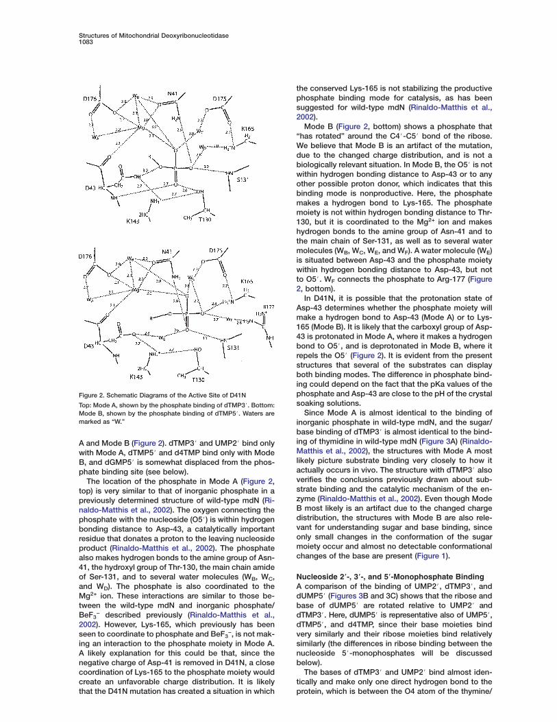
Figure 2. Schematic Diagrams of the Active Site of D41N
Top: Mode A, shown by the phosphate binding of dTMP3#. Bottom:Mode B, shown by the phosphate binding of dTMP5#. Waters aremarked as “W.”
A and Mode B (Figure 2). dTMP3# and UMP2# bind onlywith Mode A, dTMP5# and d4TMP bind only with ModeB, and dGMP5# is somewhat displaced from the phos-phate binding site (see below).
The location of the phosphate in Mode A (Figure 2,top) is very similar to that of inorganic phosphate in apreviously determined structure of wild-type mdN (Ri-naldo-Matthis et al., 2002). The oxygen connecting thephosphate with the nucleoside (O5#) is within hydrogenbonding distance to Asp-43, a catalytically importantresidue that donates a proton to the leaving nucleosideproduct (Rinaldo-Matthis et al., 2002). The phosphatealso makes hydrogen bonds to the amine group of Asn-41, the hydroxyl group of Thr-130, the main chain amideof Ser-131, and to several water molecules (WB, WC,and WD). The phosphate is also coordinated to theMg2+ ion. These interactions are similar to those be-tween the wild-type mdN and inorganic phosphate/BeF3
− described previously (Rinaldo-Matthis et al.,2002). However, Lys-165, which previously has beenseen to coordinate to phosphate and BeF3
−, is not mak-ing an interaction to the phosphate moiety in Mode A.A likely explanation for this could be that, since thenegative charge of Asp-41 is removed in D41N, a closecoordination of Lys-165 to the phosphate moiety wouldcreate an unfavorable charge distribution. It is likely
that the D41N mutation has created a situation in whichthe conserved Lys-165 is not stabilizing the productivephosphate binding mode for catalysis, as has beensuggested for wild-type mdN (Rinaldo-Matthis et al.,2002).
Mode B (Figure 2, bottom) shows a phosphate that“has rotated” around the C4#-C5# bond of the ribose.We believe that Mode B is an artifact of the mutation,due to the changed charge distribution, and is not abiologically relevant situation. In Mode B, the O5# is notwithin hydrogen bonding distance to Asp-43 or to anyother possible proton donor, which indicates that thisbinding mode is nonproductive. Here, the phosphatemakes a hydrogen bond to Lys-165. The phosphatemoiety is not within hydrogen bonding distance to Thr-130, but it is coordinated to the Mg2+ ion and makeshydrogen bonds to the amine group of Asn-41 and tothe main chain of Ser-131, as well as to several watermolecules (WB, WC, WE, and WF). A water molecule (WE)is situated between Asp-43 and the phosphate moietywithin hydrogen bonding distance to Asp-43, but notto O5#. WF connects the phosphate to Arg-177 (Figure2, bottom).
In D41N, it is possible that the protonation state ofAsp-43 determines whether the phosphate moiety willmake a hydrogen bond to Asp-43 (Mode A) or to Lys-165 (Mode B). It is likely that the carboxyl group of Asp-43 is protonated in Mode A, where it makes a hydrogenbond to O5#, and is deprotonated in Mode B, where itrepels the O5# (Figure 2). It is evident from the presentstructures that several of the substrates can displayboth binding modes. The difference in phosphate bind-ing could depend on the fact that the pKa values of thephosphate and Asp-43 are close to the pH of the crystalsoaking solutions.
Since Mode A is almost identical to the binding ofinorganic phosphate in wild-type mdN, and the sugar/base binding of dTMP3# is almost identical to the bind-ing of thymidine in wild-type mdN (Figure 3A) (Rinaldo-Matthis et al., 2002), the structures with Mode A mostlikely picture substrate binding very closely to how itactually occurs in vivo. The structure with dTMP3# alsoverifies the conclusions previously drawn about sub-strate binding and the catalytic mechanism of the en-zyme (Rinaldo-Matthis et al., 2002). Even though ModeB most likely is an artifact due to the changed chargedistribution, the structures with Mode B are also rele-vant for understanding sugar and base binding, sinceonly small changes in the conformation of the sugarmoiety occur and almost no detectable conformationalchanges of the base are present (Figure 1).
Nucleoside 2�-, 3�-, and 5�-Monophosphate BindingA comparison of the binding of UMP2#, dTMP3#, anddUMP5# (Figures 3B and 3C) shows that the ribose andbase of dUMP5# are rotated relative to UMP2# anddTMP3#. Here, dUMP5# is representative also of UMP5#,dTMP5#, and d4TMP, since their base moieties bindvery similarly and their ribose moieties bind relativelysimilarly (the differences in ribose binding between thenucleoside 5#-monophosphates will be discussedbelow).
The bases of dTMP3# and UMP2# bind almost iden-tically and make only one direct hydrogen bond to the
protein, which is between the O4 atom of the thymine/
Structure1084
Figure 3. The Binding of UMP2#, dTMP3#,and dUMP5#
(A) Comparison of the binding of substrate,product, and inorganic phosphate. The sub-strate dTMP3# is superimposed on the pre-viously determined structures with the prod-uct thymidine together with BeF3
−, and thenative structure is superimposed with inor-ganic phosphate (Rinaldo-Matthis et al.,2002). dTMP3# is shown in green, thymidineis shown in light gray, BeF3
− is shown in lightgray and indigo, and inorganic phosphate isshown in blue.(B) Comparison of the base binding ofdTMP3# and dUMP5# (Mode A), in stereo.The structures with dTMP3# (green) anddUMP5# (violet) are superimposed.(C) A stereo view comparing the ribose bind-ing of dTMP3# and UMP2#. The structurewith dTMP3# (green) is superimposed on thestructures with UMP2# (yellow) and dUMP5#(Mode A, transparent). No amino acid resi-dues are shown from the structure withdUMP5#.(D) Stereo view of the structure with dUMP5#,showing the two alternative conformations ofdUMP5#. dUMP5# (Mode A) is shown in violet,and dUMP5# (Mode B) is shown in dark violet.
uracil ring and the amide group of Val-77. The thymine/ Pmuracil ring makes several interactions with water mole-
cules that mediate contact with the Mg2+ ion and the 7protein (Figure 3B). The thymine ring of dTMP3# (andthe uracil ring of UMP2#) is stacked parallel to Phe-49 d
iwith a distance of 3.4 Å. It is also stacked parallel by
he-75 (3.9 Å), and the 5-methyl group of thymineakes van der Waals interactions of 3.5 Å to both Trp-
6 and Trp-96 (Figure 3B).The O4 atom of the uracil ring of dUMP5# makes hy-
rogen bonds to the amide group of Val-77, to the am-de group of Ser-78, and to a water molecule that medi-

Structures of Mitochondrial Deoxyribonucleotidase1085
Table 1. Relative Activity of mdN and Kinetic Parameters
Substrate Relative Activitya Km (mM) Vmax (Units/mg) Vmax/Km
dUMP5# 100 0.16 145 909UMP5# 30 0.14 46 323UMP2# 18 0.05 30 600dTMP5# 50 0.3 77 270dTMP3# 77 0.17 144 833dGMP5# 6 0.09 15 167d4TMP5#b 9 0.3 14 47
a Values are taken from a previous publication (Rampazzo et al., 2000a) and were measured at pH 5.5 with a substrate concentration of 2 mM.b Values are taken from a previous publication (Mazzon et al., 2003) and were measured at pH 7.5 with a substrate concentration of 2 mM.
the glycerol molecule is not affecting the binding mode 1); (2) the stacking interactions of both dTMP3# and
Figure 4. Comparisons of the Binding of Dif-ferent Nucleoside 5#-Monophosphates
(A) Superposition of the structure withdGMP5# (blue) and the structure with the twoalternative conformations of dUMP5# (trans-parent), in stereo.(B) Superposition of the structures withdUMP5# (purple) and UMP5# (yellow).(C) Superposition of the structures withdTMP5# (light gray) and d4TMP5# (orange).
ates interaction with the side chain of Ser-78 (Figure3B). The uracil ring of dUMP5# is stacked by Phe-49,Phe-75, Trp-76, and Trp-96, but to a lesser extent thanthe pyrimidine bases in dTMP3# and UMP2#. The uracilring is closer to Trp-76 and Trp-96 than to Phe-49,which is more than 4 Å away and is not stacked parallelto it. The N3 of the uracil ring of dUMP5# is relativelyclose to non-polar parts of the two tryptophans. Phe-75 and Trp-76 have moved slightly relative to their posi-tions in the structure with dTMP3# (Figure 3B). The resi-dues Phe-49, Phe-75, Trp-76, and Trp-96 form a pocket,which fits dTMP3# very well, but fits the dUMP5# (andall nucleoside 5#-monophosphates included in thisstudy) less well.
One glycerol molecule that probably originates fromthe cryosolution used for freezing the protein crystalsis found in the active site of the structure with dUMP5#,where it coordinates the Mg2+ ion (Glc in Figure 3B).None of the other structures included in this paper haveglycerol bound at this position, and since the bindingmode of dUMP5# is so similar to the binding modes ofUMP5#, dTMP5#, and d4TMP, it is most probable that
of the substrates but rather fills positions normally oc-cupied by water molecules.
The sugar moieties of dTMP3# and UMP2# bind towater molecules that mediate interaction with the pro-tein (Figure 3C). The nucleoside 5#-monophosphatesbind with two different sugar ring conformations, inwhich the 3#-hydroxyl group interacts with the enzymein two different ways (Figure 3D): (1) In dUMP5# (ModeA), UMP5# (Mode A), and dTMP5#, the 3#-hydroxylgroup makes hydrogen bonds to the main chain car-bonyl group of Ser-131 and the main chain amide groupof Ile-133. (2) In dGMP5#, dUMP5# (Mode B), andUMP5# (Mode B), it makes hydrogen bonds to the sidechain carboxyl group of Asp-43 and to a water mole-cule. In both binding modes, the hydroxyl group makesdirect interactions with the protein, leaving limitedspace for a putative bulky substituent, e.g., the azidogroup of the nucleoside analog AZT.
Four factors support the idea that not only nucleo-side 5#-monophosphates, but also nucleoside 3#-monophosphates, are biologically relevant substrates.These are: (1) mdN has high activity for dTMP3# (Table

Structure1086
Table 2. Crystallographic Data Collection and Refinement Statistics
Data CollectionParameters Substrate
dTMP3# dTMP5# dUMP5# UMP5# UMP2# dGMP5# d4TMP5#
Space group P43212 P43212 P43212 P43212 P43212 P43212 P43212Unit Cell Dimensions (Å)
a = b 73.18 73.65 73.7 73.57 73.45 73.38 73.77c 105.92 106.38 106.42 105.75 106.09 105.437 106.06
Data Collection Statistics
Resolution (Å) 20–1.75 40–1.8 20–2.0 40–1.7 37–1.8 40–2.00 20–2.05I/σ(I)a 15.9 (5.2) 17.7 (3.9) 23.6 (7.8) 24.5 (3.7) 27.3 (4.7) 25.5 (4.5) 21.5 (4.3)Number of unique 29,986 27,554 20,833 32,144 27,294 20,174 18,971
reflectionsRedundancy of 7.5 4.9 15.1 5.0 8.2 6.6 6.1
reflectionsRsym
a,b 7.2 (32) 5.6 (35) 8.0 (36) 4.7 (33.4) 6.1 (37) 7.2 (45) 7.3 (37)Completeness (%)a 92.7 (95.0) 99.4 (99.4) 99.8 (100) 98.5 (98.3) 98.4 (98.8) 99.6 (99.9) 99.7 (100)
Final Refinement Parameters
Rwork valuec 17 17 16 19 17 19 17Number of reflections, 26,334 26,283 19,762 30,463 25,876 19,103 17,970
working setRfree valuec 18 21 20.6 21 20 21.5 21Number of reflections, 1,420 1,383 1,069 1,624 1,369 1,027 976
test setMean B factors (Å2) 22.3 19.3 26.5 24.3 20.8 22.9 25.2Residues in most 92.9 92.9 92.4 91.8 92.9 91.8 92.4
favorable regions (%)d
Residues in additionally 7.1 7.1 7.6 8.2 7.1 8.2 7.6allowed regions (%)d
Rmsd bond length (Å) 0.018 0.019 0.019 0.015 0.018 0.019 0.02Rmsd bond angles (°) 1.682 1.790 1.690 1.631 1.755 1.684 1.672PDB code 1Z4K 1Z4L 1Z4I 1Z4M 1Z4J 1Z4P 1Z4Q
a Numbers in parentheses are for the highest resolution shell.b Rsym = (ShklSirIi(hkl) − <I(hkl)>r)/ShklSiIi(hkl) for n independent reflections and observations of a given reflection; <I(hkl)> is the averageintensity of the ith observation.c Rcryst = ShklrF0(hkl) − Fc(hkl)r/ShrF0(hkl)r.d Ramachandran plots are made by using PROCHECK Validation (Laskowski et al., 1993).
UMP2# are more favorable than those of the nucleoside A5#-monophosphates; (3) the two stacking phenylala- nnines (Phe-49 and Phe-75) are conserved between mmdN and cdN, which indicates the biological impor- otance of this stacking; and (4) in the previously solved Cstructure of mdN in complex with the nucleoside prod- puct, the 3#-hydroxyl group of the nucleoside product cis pointing toward the phosphate binding site, which pindicates that the enzyme has higher affinity for nucleo-side 3#-monophosphates than for nucleoside 5#-mono- dphosphates. a
r7Nucleoside 5�-Monophosphate SpecificitygPurine/Pyrimidine SpecificitywThe activity of mdN is much lower with purine nucleo-ttides, e.g., dGMP5#, than with the pyrimidine sub-mstrates dTMP5# and dUMP5# (Table 1). The differencedis especially marked at saturating substrate concentra-Dtions. The structure of mdN with bound dGMP5# revealsTthat dGMP5# is displaced 1 Å relative to the other nucleo-rside 5#-monophosphates, which causes a longer distancel(4.4 Å) between O5# and the catalytically important Asp-f43 (Figure 4A) than in the other substrate-D41N com-dplexes that display Mode A. This longer distance to
Asp-43 probably makes the transfer of a proton from (
sp-43 to the leaving nucleoside in the reaction mecha-ism less efficient, and thus causes the low activity ofdN with dGMP5#. The phosphate moiety of dGMP5#verlaps with that of Mode B, but a rotation around the5#-O5# bond of the nucleotide would not place thehosphate in a location similar to Mode A, since the lo-ation of the sugar ring and the rest of the nucleotiderohibits the phosphate moiety from reaching Mode A.The O6 of the guanine ring of dGMP5# makes hy-
rogen bonds to the amide of Val-77 and to the amidend hydroxyl groups of Ser-78. The N7 of the guanineing makes a hydrogen bond to the amide group of Val-7, and the O4# of the ribose moiety and the amideroup of the guanine ring make hydrogen bonds toater molecules (Figure 4A). It is probable that the in-
eractions with Val-77 and Ser-78 cause the displace-ent of dGMP5#, and thus the preference for pyrimi-ines over purines.eoxyribo/Ribo Specificityhe deoxyribonucleotidase mdN prefers the 2#-deoxy-ibo form of nucleoside 5#-monophosphates and showsower catalytic efficiency with the corresponding riboorm, e.g., UMP5# (Table 1). The substrates UMP5# andUMP5# bind remarkably similarly in the D41N variant
Figure 4B). However, the 2#-hydroxyl group of UMP5#

Structures of Mitochondrial Deoxyribonucleotidase1087
is positioned in a hydrophobic pocket, where it is 3.5 Åfrom Phe-49, 3.5 Å from Phe-102, and 2.6 Å from Ile-133, all energetically unfavorable interactions. FordUMP5#, on the other hand, these hydrophobic resi-dues constitute a favorable surface for interactions withthe 2#-carbon, which suggests that dUMP5# is boundwith higher affinity than UMP5#. However, judging fromthe kinetic parameters for these two substrates in Table1, this difference in the interactions with the enzymeaffects the Vmax, but not the Kms, which are the samefor dUMP5# and UMP5#.Importance of the 3#-Hydroxyl Group of dNMP5’sThe 5#-monophosphate of the anti-HIV thymidine ana-log d4T (d4TMP) is a poor substrate for mdN (Table 1).d4TMP lacks the 3#-hydroxyl group that is present indTMP5# and has a double bond between the C2# andC3# atoms. Even though the double bond makes theconformation limited to O4-endo envelope puckering,the sugar ring still occupies similar space as the sugarring of dTMP5#. The 3#-carbons of d4TMP and dTMP5#are in a similar position and the bases bind almost iden-tically. The only detectable difference in the interactionswith the enzyme is that dTMP5# has two additional hy-drogen bonds: the hydrogen bonds between the 3#-hy-droxyl group and the carbonyl group of Ser-131 and theamide group of Ile-133. These hydrogen bonds aremost likely more favorable than the exposure of the 3#-carbon of d4TMP to this polar environment, which is inagreement with the difference in relative activity be-tween the two substrates (Table 1). The lack of a polargroup at the 3# position of the sugar would probablyin general destabilize the binding in mdN, causing alower Vmax.Nucleoside Analog SpecificityThe binding of the nucleoside 5#-monophosphates re-veals that the parts of the base that are not involved indirect interactions with the protein are surrounded bywater molecules, which could potentially be substi-tuted by other solvent molecules (e.g., glycerol; Figure3B) or by a functional group of a nucleoside analog.The structures with UMP5# and d4TMP indicate that anucleoside analog should have a small polar or chargedgroup at the 3# position of the ribose and have no polaror charged functional group at the 2# position of theribose, in order to maintain activity.
Possibly, a nucleoside analog that is substituted onthe base (with the exception of the O4 position) is morelikely to be a good substrate for mdN than one substi-tuted on the ribose.
Experimental Procedures
Site-Directed MutagenesisSite-directed mutagenesis of mdN was performed as described bySambrook and Russel (2001). We used two complementary oligo-nucleotides to introduce the mutation: 5#-gccgtccatgttcaccagcaccc-3# and 5#-gggtgctggtgaacatggacggc-3# (small letters indi-cate wild-type bases; bold letters indicate mutated bases). As ex-ternal primers we used 5#-catacatatgggaggccgcgccctacg-3# and5#-ctgaggatccagccccacagagga-3#, which introduce a 5# NdeI anda 3# BamHI site, respectively (underlined). After PCR amplification,the mutated cDNA lacking the codons for the 31 N-terminal aminoacids with the mitochondrial targeting signal was cloned intopET20 vector (Novagen, Madison, WI, USA). The recombinant plas-mid, called pET20-D41NmdN, was sequenced to confirm the pres-ence of the mutation.
Protein Expression and PurificationPlasmid pET20-D41NmdN was transformed into Escherichia coliBL21(DE3) plysS . The bacterial culture (9 L) was grown at 37°C inLB medium to an OD600 of 0.5–0.6 and was induced with 0.4 mMisopropyl-β-D-thiogalactoside for 3 hr. Extracts were prepared asdescribed previously (Rampazzo et al., 2000b), and the D41N vari-ant of mdN was purified by following the same procedure as thatused previously for wild-type mdN (Rampazzo et al., 2000a). Briefly,the enzyme was purified by ammonium sulfate fractionation, chro-matographed on a DE52 column (Whatman) with a 0–150 mM NaClgradient in buffer A (20 mM Tris-HCl [pH 7.5], 1 mM EDTA [pH 7.6],2 mM DTT, and 20% [v/v] glycerol). The peak was rechromato-graphed on a Phenyl Sepharose CL-4B column (Amersham Phar-macia) with a 1.5–0 M gradient of ammonium sulfate in buffer A.The peak was loaded again in a desalting biogel column (BioRad)in order to remove ammonium sulfate.
Enzyme AssaysDephosphorylation of nucleotides was measured with a sensitivecolorimetric assay from the formation of inorganic phosphate (Gel-adopoulos et al., 1991). Incubations were at 37°C for 30 min withsubstrate, 10 mM Mg2+, 30 mM KCl, and 250 mM acetate buffer(pH 5.5) in a final volume of 50 �l. The reaction was stopped bythe addition of 1/8 volume H2SO4 (1.1 M), and, after centrifugation,inorganic phosphate was determined. One unit of enzyme activitycorresponds to the dephosphorylation of 1 �mol substrate per min-ute. Specific activity is units per mg protein. Km and Vmax valueswere obtained from the extrapolated maximal specific activity ofthe Lineweaver-Burk plots. All experiments were done in triplicateat different concentrations of the substrate and were repeated atleast three times. In all kinetic experiments, care was taken not toconsume more than 25% of the substrate during the reaction.
Crystallization, Soaking Experiments, and CryoprotectionThe D41N variant of mdN was crystallized under the same condi-tions as those described for mdN (Rinaldo-Matthis et al., 2002). Theprotein solution used for crystallization contained 20 mM Tris buffer(pH 7.5), 20% (v/v) glycerol, 2 mM DTT, 1 mM EDTA, 75 mM NaCl,and 10.1 mg ml−1 protein. Crystals were obtained by using thehanging drop vapor diffusion method with 1 �l protein solution and1 �l reservoir solution at 20°C. The reservoir solution contained15%–20% (w/v) PEG 8000 and 50 mM KH2PO4 (pH 4.5). Crystalsappeared after 1 day and reached a maximum size of 0.2 × 0.2 ×0.3 mm. The D41N crystals were soaked in 10 �l drops containing10 mM nucleoside monophosphate, 90 mM MgCl2, 50 mM sodiumacetate (pH 4.5), and 20% (w/v) PEG 8000 for 1–2 hr. The pH of thenucleoside monophosphate solutions was adjusted to pH 5 priorto soaking. The crystals were then transferred to a 10 �l drop ofcryosolution containing 20% (w/v) PEG 8000, 50 mM sodium ace-tate (pH 4.5), and 20% (v/v) glycerol for 20–60 s and subsequentlyflash frozen in liquid N2. For the structure with dGMP5#, 20% PEG400 (v/v) was used instead of glycerol in the cryosolution.
Data Collection, Structure Determination, and RefinementThe data sets of D41N in complex with dTMP3# and dUMP5# werecollected at the European Synchrotron Radiation Facility (ESRF) atthe beamline ID14.3, the data set of D41N in complex with dGMP5#was collected at the Protein Structure Factory-Berliner Elektronen-speicherring-Gesellschaft für Synchrotron Strahlung m. b. H. (PSF-BESSY) at beamline BL1, and the data sets of D41N in complexwith UMP5#, UMP2#, dTMP5#, and d4TMP were collected at TheEuropean Molecular Biology Laboratory (EMBL) Hamburg Outsta-tion at the beamline X11. The data sets with dTMP3# and dUMP5#were processed with XDS and XSCALE (Kabsch, 1988); the otherdata sets were processed by using the program package DENZO/SCALEPACK (Otwinowski and Minor, 1997). The crystals belong tothe space group P43212, with one polypeptide per asymmetric unit.
Refmac5 (Murshudov et al., 1997) was used for structure deter-mination and refinement, and the previously determined mdNstructure (Rinaldo-Matthis et al., 2002) was used as a template forthe initial Rigid Body refinement of the seven data sets. Quanta(Accelrys, San Diego, CA) was used for model building, and X-SOL-

Structure1088
VATE (Accelrys) was used for the addition of waters. Structural sta- Mstistics are shown in Table 2.sPAcknowledgmentsM
We would like to thank the staff at beamline ID14.3 at ESRF, at obeamline X11 at the EMBL Hamburg Outstation and at beamline ABL1 at PSF-BESSY for technical assistance. This work was sup- Oported by grants from Telethon Italia (V.B.), Associazione Italiana tper la Ricerca sul Cancro (AIRC) (V.B.), the Swedish Research 3Council (P.N.), the Swedish Cancer Society (P.N.), and from The
REuropean Commission Grant nr.QLGI-CT-2001-01004 (P.N. and V.B.).Pc
Received: October 29, 2004 tRevised: March 16, 2005 RAccepted: April 23, 2005 UPublished: July 12, 2005 5
oReferences C
RBianchi, V., and Spychala, J. (2003). Mammalian 5#-nucleotidases. BJ. Biol. Chem. 278, 46195–46198. sBradshaw, H.D., Jr., and Deininger, P.L. (1984). Human thymidine Rkinase gene: molecular cloning and nucleotide sequence of a cDNA nexpressible in mammalian cells. Mol. Cell. Biol. 4, 2316–2320. SFeng, J.Y., Johnson, A.A., Johnson, K.A., and Anderson, K.S. R(2001). Insights into the molecular mechanism of mitochondrial tox- Nicity by AIDS drugs. J. Biol. Chem. 276, 23832–23837. dGallinaro, L., Crovatto, K., Rampazzo, C., Pontarin, G., Ferraro, P., SMilanesi, E., Reichard, P., and Bianchi, V. (2002). Human mitochon- rdrial 5#-deoxyribonucleotidase. Overproduction in cultured cells
Tand functional aspects. J. Biol. Chem. 277, 35080–35087.A
Gazziola, C., Ferraro, P., Moras, M., Reichard, P., and Bianchi, V.V
(2001). Cytosolic high Km 5#-nucleotidase and 5#(3#)-deoxyribo-s
nucleotidase in substrate cycles involved in nucleotide metabo-n
lism. J. Biol. Chem. 276, 6185–6190.r
Geladopoulos, T.P., Sotiroudis, T.G., and Evangelopoulos, A.E.W
(1991). A malachite green colorimetric assay for protein phospha-T
tase activity. Anal. Biochem. 192, 112–116.m
Höglund, L.R., and Reichard, P. (1990). Cytoplasmic 5#(3#)-nucleoti- adase from human placenta. J. Biol. Chem. 265, 6589–6595. 1Johansson, M., and Karlsson, A. (1997). Cloning of the cDNA andchromosome localization of the gene for human thymidine kinase2. J. Biol. Chem. 272, 8454–8458.
Johnson, A.A., Ray, A.S., Hanes, J., Suo, Z.C., Colacino, J.M., An-derson, K.S., and Johnson, K.A. (2001). Toxicity of antiviral nucleo-side analogs and the human mitochondrial DNA polymerase. J.Biol. Chem. 276, 40847–40857.
Jordan, A., and Reichard, P. (1998). Ribonucleotide reductases.Annu. Rev. Biochem. 67, 71–98.
Kabsch, W. (1988). Automatic-indexing of rotation diffraction pat-terns. J. Appl. Crystallogr. 21, 67–71.
Lai, Y.R., Tse, C.M., and Unadkat, J.D. (2004). Mitochondrial expres-sion of the human equilibrative nucleoside transporter 1 (hENT1)results in enhanced mitochondrial toxicity of antiviral drugs. J. Biol.Chem. 279, 4490–4497.
Laskowski, R.A., Macarthur, M.W., Moss, D.S., and Thornton, J.M.(1993). Procheck—a program to check the stereochemical qualityof protein structures. J. Appl. Crystallogr. 26, 283–291.
Lewis, W., and Dalakas, M.C. (1995). Mitochondrial toxicity of anti-viral drugs. Nat. Med. 1, 417–422.
Lim, S.E., and Copeland, W.C. (2001). Differential incorporation andremoval of antiviral deoxynucleotides by human DNA polymerasegamma. J. Biol. Chem. 276, 23616–23623.
Marinaki, A.M., Escuredo, E., Dudley, J.A., Simmonds, H.A., Amici,A., Naponelli, V., Magni, G., Seip, M., Ben-Bassat, I., Harley, E.H.,et al. (2001). Genetic basis of hemolytic anemia caused by pyrimi-dine 5# nucleotidase deficiency. Blood 97, 3327–3332.
Mazzon, C., Rampazzo, C., Scaini, M.C., Gallinaro, L., Karlsson, A.,
eier, C., Balzarini, J., Reichard, P., and Bianchi, V. (2003). Cyto-olic and mitochondrial deoxyribonucleotidases: activity with sub-trate analogs, inhibitors and implications for therapy. Biochem.harmacol. 66, 471–479.
urshudov, G.N., Vagin, A.A., and Dodson, E.J. (1997). Refinementf macromolecular structures by the maximum-likelihood method.cta Crystallogr. D Biol. Crystallogr. 53, 240–255.
twinowski, Z., and Minor, W. (1997). Processing of X-ray diffrac-ion data collected in oscillation mode. Methods Enzymol. 276,07–326.
ampazzo, C., Gallinaro, L., Milanesi, E., Frigimelica, E., Reichard,., and Bianchi, V. (2000a). A deoxyribonucleotidase in mito-hondria: involvement in regulation of dNTP pools and possible linko genetic disease. Proc. Natl. Acad. Sci. USA 97, 8239–8244.
ampazzo, C., Johansson, M., Gallinaro, L., Ferraro, P., Hellman,., Karlsson, A., Reichard, P., and Bianchi, V. (2000b). Mammalian#(3#)-deoxyribonucleotidase, cDNA cloning, and overexpressionf the enzyme in Escherichia coli and mammalian cells. J. Biol.hem. 275, 5409–5415.
ampazzo, C., Ferraro, P., Pontarin, G., Fabris, S., Reichard, P., andianchi, V. (2004). Mitochondrial deoxyribonucleotides, pool sizes,ynthesis and regulation. J. Biol. Chem. 279, 17019–17026.
inaldo-Matthis, A. (2004). Crystal structures of human deoxyribo-ucleotidases. PhD thesis, Stockholm University, Stockholm,weden.
inaldo-Matthis, A., Rampazzo, C., Reichard, P., Bianchi, V., andordlund, P. (2002). Crystal structure of a human mitochondrialeoxyribonucleotidase. Nat. Struct. Biol. 9, 779–787.
ambrook, J., and Russel, D.W. (2001). Molecular Cloning: A Labo-atory Manual, Third Edition (New York: Cold Spring Harbor Press).
ozser, J. (2001). HIV inhibitors: problems and reality. Ann. N. Y.cad. Sci. 946, 145–159.
an Rompay, A.R., Johansson, M., and Karlsson, A. (2003). Sub-trate specificity and phosphorylation of antiviral and anticancerucleoside analogues by human deoxyribonucleoside kinases andibonucleoside kinases. Pharmacol. Ther. 100, 119–139.
ang, L., Munch-Petersen, B., Sjoberg, A., Hellman, U., Bergman,., Jornvall, H., and Eriksson, S. (1999). Human thymidine kinase 2:olecular cloning and characterisation of the enzyme activity with
ntiviral and cytostatic nucleoside substrates. FEBS Lett. 443,70–174.