Structural and energetic analysis of activation by a cyclic nucleotide binding domain
27
Structural and energetic analysis of activation by a cyclic nucleotide binding domain Stephen L. Altieri * , Gina M. Clayton * , William R. Silverman • , Adrian O. Olivares, Enrique M. De La Cruz, Lise R. Thomas †,‡ , and João H. Morais-Cabral †,§ Department of Molecular Biophysics and Biochemistry, Yale University, 266 Whitney Ave, New Haven, CT 06520-8114 Summary MlotiK1 is a prokaryotic homolog of cyclic nucleotide-dependent ion channels which contains an intracellular C-terminal cyclic nucleotide binding domain (CNB domain). X-ray structures have been solved of the CNB domain in the absence of ligand and bound to cAMP. Both the full-length channel and CNB domain fragment are easily expressed and purified, making MlotiK1 a useful model system for dissecting activation by ligand binding. We have used X-ray crystallography to determine three new MlotiK1 CNB domain structures: a second apo configuration, a cGMP-bound structure, and a second cAMP-bound structure. In combination, the five MlotiK1 CNB domain structures provide a unique opportunity for analyzing, within a single protein, the structural differences between the apo and bound states and the structural variability within each state. With this analysis as a guide, we have probed the nucleotide selectivity and importance of specific residue side chains in ligand binding and channel activation. These data help to identify ligand-protein interactions that are important for ligand-dependence in MlotiK1 and more globally in the class of nucleotide-dependent proteins. Keywords Mesorhizobium loti; potassium channel; ligand-dependent gating; cyclic nucleotide; conformational variability Introduction Cyclic nucleotides play vital roles in signal transduction, altering the activity of proteins as diverse as protein kinase A and G, the transcription factor CAP, the guanine nucleotide- exchange factor Epac and cyclic nucleotide-regulated ion channels 1 . Nucleotide-dependence † Corresponding authors. * These authors contributed equally to this work; • Current address: Dept. of Physiology and Biophysics, P.O. Box 016430, Miller School of Medicine, University of Miami, Miami, FL 33101 ‡ Current address: Department of Biology, Quinnipiac University, 275 Mount Carmel Avenue, Hamden, CT 06517 § Current address: Inst. Biologia Molecular e Celular, Rua do Campo Alegre 823, 1450-180 Porto, Portugal Publisher's Disclaimer: This is a PDF file of an unedited manuscript that has been accepted for publication. As a service to our customers we are providing this early version of the manuscript. The manuscript will undergo copyediting, typesetting, and review of the resulting proof before it is published in its final citable form. Please note that during the production process errors may be discovered which could affect the content, and all legal disclaimers that apply to the journal pertain. Author contributions Author contributions: All authors designed research; G.M.C. and S.L.A. performed crystallography experiments; W.R.S. and S.L.A. performed flux assays, S.L.A. and A.O.O. performed fluorescent binding assays, and S.L.A. performed matrix competition assays; all authors analyzed data; and S.L.A., L.R.T. and J.H.M.-C. wrote the paper. NIH Public Access Author Manuscript J Mol Biol. Author manuscript; available in PMC 2009 September 5. Published in final edited form as: J Mol Biol. 2008 September 5; 381(3): 655–669. doi:10.1016/j.jmb.2008.06.011. NIH-PA Author Manuscript NIH-PA Author Manuscript NIH-PA Author Manuscript
-
Upload
independent -
Category
Documents
-
view
0 -
download
0
Transcript of Structural and energetic analysis of activation by a cyclic nucleotide binding domain

Structural and energetic analysis of activation by a cyclicnucleotide binding domain
Stephen L. Altieri*, Gina M. Clayton*, William R. Silverman•, Adrian O. Olivares, Enrique M.De La Cruz, Lise R. Thomas†,‡, and João H. Morais-Cabral†,§Department of Molecular Biophysics and Biochemistry, Yale University, 266 Whitney Ave, NewHaven, CT 06520-8114
SummaryMlotiK1 is a prokaryotic homolog of cyclic nucleotide-dependent ion channels which contains anintracellular C-terminal cyclic nucleotide binding domain (CNB domain). X-ray structures have beensolved of the CNB domain in the absence of ligand and bound to cAMP. Both the full-length channeland CNB domain fragment are easily expressed and purified, making MlotiK1 a useful model systemfor dissecting activation by ligand binding. We have used X-ray crystallography to determine threenew MlotiK1 CNB domain structures: a second apo configuration, a cGMP-bound structure, and asecond cAMP-bound structure. In combination, the five MlotiK1 CNB domain structures provide aunique opportunity for analyzing, within a single protein, the structural differences between theapo and bound states and the structural variability within each state. With this analysis as a guide,we have probed the nucleotide selectivity and importance of specific residue side chains in ligandbinding and channel activation. These data help to identify ligand-protein interactions that areimportant for ligand-dependence in MlotiK1 and more globally in the class of nucleotide-dependentproteins.
KeywordsMesorhizobium loti; potassium channel; ligand-dependent gating; cyclic nucleotide; conformationalvariability
IntroductionCyclic nucleotides play vital roles in signal transduction, altering the activity of proteins asdiverse as protein kinase A and G, the transcription factor CAP, the guanine nucleotide-exchange factor Epac and cyclic nucleotide-regulated ion channels 1. Nucleotide-dependence
† Corresponding authors.*These authors contributed equally to this work;•Current address: Dept. of Physiology and Biophysics, P.O. Box 016430, Miller School of Medicine, University of Miami, Miami, FL33101‡Current address: Department of Biology, Quinnipiac University, 275 Mount Carmel Avenue, Hamden, CT 06517§Current address: Inst. Biologia Molecular e Celular, Rua do Campo Alegre 823, 1450-180 Porto, PortugalPublisher's Disclaimer: This is a PDF file of an unedited manuscript that has been accepted for publication. As a service to our customerswe are providing this early version of the manuscript. The manuscript will undergo copyediting, typesetting, and review of the resultingproof before it is published in its final citable form. Please note that during the production process errors may be discovered which couldaffect the content, and all legal disclaimers that apply to the journal pertain.Author contributions Author contributions: All authors designed research; G.M.C. and S.L.A. performed crystallography experiments;W.R.S. and S.L.A. performed flux assays, S.L.A. and A.O.O. performed fluorescent binding assays, and S.L.A. performed matrixcompetition assays; all authors analyzed data; and S.L.A., L.R.T. and J.H.M.-C. wrote the paper.
NIH Public AccessAuthor ManuscriptJ Mol Biol. Author manuscript; available in PMC 2009 September 5.
Published in final edited form as:J Mol Biol. 2008 September 5; 381(3): 655–669. doi:10.1016/j.jmb.2008.06.011.
NIH
-PA Author Manuscript
NIH
-PA Author Manuscript
NIH
-PA Author Manuscript

is conferred by a similar mechanism in each case: the ligand binds to a conserved cyclicnucleotide binding (CNB) domain, which itself undergoes a conformational change that isultimately propagated to and affects the activity of a catalytic domain 2. Biochemicalexperiments, particularly with the CAP and protein kinase CNB domains 3; 4; 5, together withthe crystal structures of CNB domains bound to nucleotides have led to the identification ofspecific residues that are functionally critical for ligand binding and activation 6; 7; 8; 9. Yet,determining which interactions and conformational changes are unique to a specific protein,and which are more universally important for the whole set of CNB dependent proteins, remainsan important challenge.
The prokaryotic MlotiK1 ion channel is the most recently characterized cyclic nucleotide-dependent protein 10; 11. The cyclic nucleotide-dependent ion channels all share a commonarchitecture: the core transmembrane channel domain with the topology and four-foldsymmetry of voltage-gated channels, attached to an intracellular C-terminal CNB domain 12.Eukaryotic cyclic nucleotide-channels are generally studied using electrophysiologicalapproaches, and are either directly activated by cyclic nucleotide (cyclic nucleotide-gated orCNG channels) 13, or have basal activity that is further modulated by cyclic nucleotide binding(hyperpolarization activated cyclic nucleotide-gated or HCN channels) 14. The functionalproperties of the prokaryotic MlotiK1 are typically studied using a radioactive uptake assay15; MlotiK1 displays basal activity which is further stimulated by sub-micromolarconcentrations of cAMP and cGMP 10; 11.
MlotiK1 has proven particularly useful as a tool for understanding cyclic-nucleotide-dependence, largely because of its potential for structural work 16; 17. The CNB domain iseasily purified as an independent soluble construct which binds nucleotide selectively with arelatively high affinity; cAMP actually co-purifies with the soluble CNB domain, but bindingis weakened by the introduction of a point mutation, R348A 10. Recently Cukkemane, et al.measured a binding affinity of 100 nM for cAMP for refolded wild type CNB domain 18. Wehave been unable to confirm this value using equilibrium experiments of native CNB domain,since in our hands the off-rate of nucleotide is immeasurably slow. Importantly, the MlotiK1CNB domain was the first CNB domain to be crystallized in both a cAMP-bound and an apoform (the apo form was stabilized by the introduction of the mutation R348A) 10. The twostructures display the characteristic architecture of other CNB domains, including those of theprokaryotic transcription factor CAP, two regulatory subunits of protein kinase A, Epac andthe HCN2 ion channel 6; 7; 8; 9. Both the cAMP-bound and apo MlotiK1 CNB domainstructures are organized as dimers in the asymmetric unit and show a ligand binding pocketthat is formed by a wide β-roll; the two structures diverge mainly in the position of the helicesof the domain. Binding of ligand induces a rearrangement of the four helices on the surface ofthe β-roll which is thought to be propagated to the channel regions and affect the state of thegate. The comparison between the cAMP-bound and apo structures provides a valuableframework for understanding the physical basis of ligand-dependence in cyclic nucleotide-dependent proteins, however, as there is only a single structure in each conformation, thequestion remains as to the extent to which the two structures reflect “universal” bound and apoconformations.
In this study, we have used X-ray crystallography to determine three new MlotiK1 CNB domainstructures. These include both a cGMP-bound structure, as well as a second apo configurationwhich is slightly different than that seen with R348A. Comparisons of these crystal structureswith each other and with the two original structures were used to guide mutagenesis studies ofligand binding and channel activation. Taken together, these data help to identify ligand-proteininteractions that are important for ligand-dependence in MlotiK1 and more globally in the classof nucleotide-dependent proteins.
Altieri et al. Page 2
J Mol Biol. Author manuscript; available in PMC 2009 September 5.
NIH
-PA Author Manuscript
NIH
-PA Author Manuscript
NIH
-PA Author Manuscript

ResultsNucleotide selectivity of binding and activation of Wild-Type MlotiK1
To fully understand ligand-protein interactions in MlotiK1, we first needed to establish theselectivity of ligand binding and channel activation. Although the high nucleotide affinity ofthe soluble CNB domain rendered direct equilibrium binding experiments impractical, we wereable to examine ligand binding using a crude competition assay. With this approach, purifiedCNB domain was bound, through a long incubation, to cAMP-immobilized matrix. The proteinwas then selectively eluted by incubation with different test ligands, and the quantity of elutedprotein was assessed by Coomassie staining of SDS-PAGE gels (Figure 1A). We tested a panelof ligands at 3 mM, since our initial trials showed that this concentration of cAMP was sufficientto release a substantial fraction of the bound domain. Among the purine nucleotides, cAMPyielded the most protein, followed by cGMP and cIMP. Both pyrimidine nucleotides, cCMPand cUMP, reproducibly yielded small quantities of CNB domain, while the non-cyclicnucleotides, 5’ AMP and 3’ AMP, failed to elute protein. Although the competition bindingassay is not quantitative, it does provide a qualitative measure of the relative binding affinitiesof the different ligands, since the higher the affinity, the more protein should be eluted. Basedon the relative yields observed in Figure 1A, the MlotiK1 CNB domain displays a bindingselectivity of cAMP >cGMP >cIMP ≫cCMP ≈cUMP ⋙5’AMP, 3’AMP.
Since the binding experiment might identify antagonists as well as agonists, we used afunctional assay to determine whether similar ligand specificity was observed in channelactivation. MlotiK1 activity is studied using a concentrative 86Rb+ flux assay, in whichchannel-containing proteoliposomes are loaded with high internal [K+], and the resultingdiffusion potential is used to drive uptake of 86Rb+ into the vesicles 15. We focused in on thethree purine nucleotides and cCMP (as a representative pyrimidine) and examined MlotiK1activity over a broad range of ligand concentrations (Figure 1B). Both cAMP and cGMP areknown to activate MlotiK1; our data show K1/2 values for cAMP and cGMP of 0.11 (±0.02)and 0.92 (±0.11) μM, respectively, closely matching the published work (0.06 and 0.59 μM,respectively, for cAMP and cGMP). Here the third purine ligand cIMP activates MlotiK1 withslightly lower potency (K1/2 of 2.1 μM) than cAMP or cGMP. In addition, the pyrimidinecCMP also activates MlotiK1 with a K1/2 of 15 μM. The activation curves were nicely fittedby a simple 1:1 isotherm and thus showed no obvious signs of cooperativity.
The selectivity of channel activation mirrors that observed in the binding assay: cAMP > cGMP> cIMP > cCMP, and thus indicates that cGMP, cIMP and cCMP are all potential tools forstructural studies of MlotiK1 activation.
Wild type MlotiK1 CNB domain in complex with cGMPObtaining crystallographic quantities of MlotiK1 CNB domain in complex with a second ligandpresented a technical hurdle, as the cAMP that co-purifies with the domain is not easily dialyzedaway. We applied the matrix-based approach described above to exchange cAMP, generatingCNB domain bound to either cGMP, cIMP or cCMP. Although the yield was low, the resultingprotein was free of residual cAMP as judged with a modified cell-based cAMP-specificdetection system (see Methods). We attempted crystallization of the cGMP, cIMP and cCMP-bound CNB domains, and while all three yielded crystals, we obtained well-diffracting crystalsof only the cGMP-exchanged protein.
We solved a 2.35 Å resolution structure of the wild type CNB domain bound to cGMP usingmolecular replacement with the wild type cAMP-bound structure as the search molecule (Table1). Overall, the new structure is nearly identical to the cAMP-bound structure and, as in theoriginal structures, there are two molecules in the asymmetric unit. No major changes were
Altieri et al. Page 3
J Mol Biol. Author manuscript; available in PMC 2009 September 5.
NIH
-PA Author Manuscript
NIH
-PA Author Manuscript
NIH
-PA Author Manuscript

observed in either the positions of the protein backbone or side chains (RMS deviation betweenall α-carbons is 0.2 Å). Like our previous cAMP-bound structure of the MlotiK1 CNB domain,the ligand binding pocket of the new structures has the β-roll typical of CNB domains, and theαC-helix “lid” is closed over the binding pocket (Figure 2A). A ligand is bound within thepocket and the density of the purine moiety shows two clear lobes, as expected for the purinering N2 and O6 groups of cGMP (Figure 2B). The relative rotation of the glycosidic bond(bond between ribose and base) places cGMP in the syn conformation, whereas cAMP is boundin the anti conformation in the original wild type structure 10 (Figure 2 C and 3A).
The conformational difference between nucleotides in MlotiK1 mirrors that previouslyobserved in the HCN2 crystal structures, which also had cAMP and cGMP in the anti andsyn conformations, respectively 9. In the HCN2 domain structures the two ligandconformations are related by a simple rotation around the glycosidic bond; as a result of thisflipping, the bases of the two ligands occupy distinct areas of the binding pocket. In MlotiK1,cAMP and cGMP are also related by an equivalent rotation around the glycosidic bond, yetthe bases of the two ligands occupy nearly overlapping volumes. Figure 2C shows the relativestructures and positions of cAMP and cGMP molecules when their two protein structures aresuperimposed. In addition to the rotation of the glycosidic bond, the conformation of the ribosegroup appears to change and, as a consequence, the base occupies nearly the same physicalspace in the two structures.
Many of the contacts between the protein and the nucleotide are identical in the cAMP andGMP-bound structures. At the cyclic phosphate and ribose moieties for example, the side chainof Glu298 forms a hydrogen bond with the 2’ hydroxyl group of the ribose, while those ofArg307 and Ser308 interact directly with an exocylic oxygen from the cyclic phosphate (Figure3A). Because the cAMP and cGMP bases occupy the same physical volume, the side chain ofArg348 still appears to stabilize the closure of the αC-helix “lid” through Van der Waal’scontacts with the nucleotide base of cGMP. The interaction surface between the bases andArg348 is almost identical: the R348 side chain buries ~92 Å2 of the cAMP surface areacompared to ~95 Å2 of cGMP.
Although the purine base of cAMP makes only Van der Waals contacts with the protein, cGMPis capable of Van der Waals interactions as well as specific hydrogen bonds with the bindingpocket. In particular, the N1 nitrogen of the cGMP purine ring hydrogen bonds with thebackbone carbonyl group of Arg348 while the N2 amine forms a hydrogen bond with thehydroxyl group of Ser308 (Figure 3A). A similar interaction was reported in the HCN2 CNBdomain-cGMP crystal structure, where Thr592 makes a specific hydrogen bond with the N2of cGMP. We investigated the functional impact of this interaction in MlotiK1 usingthe 86Rb+ uptake assay to study two mutant channels, S308A and S308V (Figure 3B). Asexpected, cAMP-activation was not dramatically altered by either mutation (K1/2 for wild-type,S308A and S308V of 0.11± 0.02, 0.3 ± 0.1 and 0.1 ± 0.03 μM, respectively). Although cGMPsensitivity was somewhat more affected (K1/2 of 0.92 ± 0.11, 1.6 ± 0.2, and 4.1 ± 0.6 μM forwild-type, S308V and S308A, respectively), the mutations had a relatively small effect overallon binding energy (< 1 kcal/ mol).
MlotiK1 CNB domain mutantsThe portion of the binding pocket formed by the “phosphate binding cassette” contains highlyconserved residues that interact directly with the cyclic phosphate moiety. In particular, onearginine (R307 in MlotiK1) is conserved in each of the CNB domain crystal structures to dateand its side chain forms an ionic bond with the cyclic phosphate in each case; mutations of thisside chain are known to decrease ligand binding in other CNB domain proteins. We evaluatedthe biochemical impact of three mutations that should interrupt the ionic interaction R307 haswith nucleotide: R307A and R307E, which neutralize or substitute an acidic side chain,
Altieri et al. Page 4
J Mol Biol. Author manuscript; available in PMC 2009 September 5.
NIH
-PA Author Manuscript
NIH
-PA Author Manuscript
NIH
-PA Author Manuscript

respectively, and R307W, which, in addition to being neutral, has increased side chain bulkand might have a steric effect on ligand binding.
All three R307 mutations substantially decreased ligand binding affinity. R307A was easilystudied using standard binding studies; a direct binding experiment measured a Kd of 45.1 ±5.8 μM for the fluorescent cAMP analogue 8-NBD-cAMP (Figure 4A), while a competitionbinding experiment showed a lower affinity for cAMP (205.1 ± 10.4 μM) (Figure 4B). Bycomparison, these affinities are over 1000-fold lower than recent analogous measurements onrefolded wild-type domain (0.022 and 0.068 μM, respectively, for 8-NBD-cAMP and cAMP).Ligand binding by the other two mutants, R307E and R307W, was even more severely reduced.Both domains displayed negligible binding to 8-NBD cAMP over the dynamic range of theassay (Kd estimated to be > 1.5 mM); the weak 8-NBD-cAMP binding precluded competitionassays with unlabelled ligands.
We turned to examine the R307A mutation in the context of the full-length MlotiK1 protein.The R307A mutant channel yielded protein, and its activity remains modulated by cAMP. TheR307A mutation has a large effect on the K1/2 of activation, shifting it by 300-fold relative towild type (K1/2 of 30 and 0.11 μM, for R307A and wild type, respectively) (Figure 4C).Although the qualitative effect of R307A is similar in the flux assay of full-length channel andcAMP binding to the isolated R307A mutant domain (right-shifting both dose-response curveswith respect to the wild-type protein, as though destabilizing a bound state(s) relative tounliganded conformation(s)), there is a curious quantitative difference: the mutation had alarger effect on ligand binding than on channel activation (R307A changes the ligand bindingKd by at least 3000-fold, but the K1/2 of activation by only 300-fold). This latter observationhas important consequences for the energetics of ligand-dependence in MlotiK1, as detailedin the discussion below. We were unable to obtain sufficient quantities of R307E to test itsactivity in the flux assay, and the R307W mutant channel displayed only nominal activity undercontrol conditions that was not increased by additional nucleotide.
To better understand the ligand binding properties of the Arg307 mutants, we determined thecrystal structures of R307E and R307W domains. R307E crystallized only in the presence ofa high cAMP concentration (20 mM). We have solved a 2.2Å resolution structure by molecularreplacement using the wild type structure (Table 1). Like in the original wild type structure,the R307E mutant crystals contain 2 molecules in the asymmetric unit and have bound cAMPin the anti conformation. The similarities between the wild type and R307E mutant structuresare striking (RMS deviation between all α-carbons is 0.3 Å), with just very small distortionsof the binding pocket (less than 1Å shift in the relative position of Cα of PBC residues). Thisobservation demonstrates that mutations of the highly conserved Arg307 in the binding pocketare well tolerated at the structural level, and that changes in ligand binding properties are notthe result of severe structural distortions of the bound state. With the exception of theinteractions formed with the side chain of residue 307, all molecular interactions between theligand and the CNB domain are conserved between the two structures. The density of theR307E glutamate side chain is well defined. The side chain is placed close to the phosphate ofthe cyclic nucleotide (Figure 5A), but the carboxylic group is not within hydrogen bondingdistance (>3.5 Å).
The R307W mutant was crystallized in the absence of cAMP and solved at 2.9 Å resolutionby performing a selenomethione multiwavelength anomalous diffraction experiment and thendocking and adjusting the R348A mutant structure into the density (Table 1). In these crystalsthere are 4 molecules per asymmetric unit and, as expected, there is no nucleotide density inthe binding pockets. The tryptophan side chain at position 307 adopts two differentconformations and appears to reduce ligand affinity by steric hinderance (Figure 5B).
Altieri et al. Page 5
J Mol Biol. Author manuscript; available in PMC 2009 September 5.
NIH
-PA Author Manuscript
NIH
-PA Author Manuscript
NIH
-PA Author Manuscript

Collection of apo and liganded structuresWith the determination of the three new crystal structures we have an extensive collection ofstructures of the MlotiK1 CNB domain in both the bound and apo states. All of these crystalstructures have more than one molecule in the asymmetric unit, each with a unique crystalenvironment, therefore the molecules are not necessarily identical and can be consideredindependent determinations. In the bound state we have 6 structures of MlotiK1 CNB domains:2 structures of the mutant R307E with cAMP, 2 wild type structures with cAMP and 2 morewild type structures with cGMP. We also have 6 different structures for the apo state: themutants R348A (2 molecules in asymmetric unit) and R307W (4 molecules in the asymmetricunit). This collection of structures provides us with the unique opportunity of analyzing, in thesame protein, the differences between states and within states, while controlling for theconformational effects due to crystal contacts and introduction of mutations.
The 12 structures were superimposed over the residue range 250 to 280 and 309 to 323, whichspans a large part of the β-roll and does not include the Phosphate Binding Cassette (PBC)region and any of the helices (Figure 6A, 6B and subsequent figures). It is immediately clearthat the structures in the bound state (in red) are almost identical, with RMS deviation betweendifferent structures varying between 0.15 to 0.3 Å (calculated for the all residues relative to acAMP-bound wild type structure (subunit A)). In contrast, the structures without nucleotide(in green and gray) are much more diverse, mainly on the αC-helix but also in the dispositionof the other helices on the surface of the β-roll and in the position of some of the residues thatform the binding pocket. This difference between the two states demonstrates that there is asingle, well defined bound state conformation which probably results from the addedinteractions formed between the ligand and the protein. In the absence of nucleotide, thestructure of the domain appears to be much “looser” and capable of adopting differentconformations.
The largest differences between the two states are observed in the structural unit formed byαA’-helix/loop/αA-helix, in the αC- and αB-helices, the binding pocket and in the β4-β5hairpin. In MlotiK1, the αA’-helix/loop/αA-helix unit (αA’-L- αA) is directly connected to thelast TM of the channel and is thought to be the major conveyer of the conformational changefrom the domain to the gate. Direct evidence for the movement of αA’-L- αA in the MlotiK1CNB domain first came from comparison of the structures of wild type (bound state) andR348A mutant (apo state) domains 10. However, the use of a mutant domain to generatecrystals in the apo form raised the possibility that the observed αA’-L- αA movement wasactually an artifact of the R348A mutation. The determination of the new apo structures,resulting from a mutation (R307W) in a completely different region of the domain, confirmsthe shift of the αA’-L- αA unit between apo and bound states (Figure 6A and 7A), and clearlyestablishes that the rearrangement is induced by ligand binding.
Ligand-dependent movement of the αA’-L-αA is associated with remodeling of a hydrophobiccluster formed by residues from the β-roll, the PBC and helices. In the bound state (Figure 7Aand 7B), the cluster includes residues on the β-roll (Met272, Phe 274, Phe295, Leu320,Leu322), residues from the αC helix (Ile337 and Phe341) and from the αB helix (Phe327 andLeu330). The cluster reorganizes upon ligand release (Figure 7C): the αC helix shifts away,the residues in the PBC and αB helix relocate and the αA’-L-αA unit moves in, covering theresidues on the surface of the β-roll (Figure 6A). αA’-L-αA residues involved in thehydrophobic cluster include Leu229 (αA’-helix), Val233, Leu235, Phe236 and Leu239 (allfrom the loop), as well as Ile247 (αA-helix) (Figure 7A and 7C). While the αA’-L-αA unitundergoes a large movement (Cα at Phe236 moves 8Å between the wild type and one of theR307W structures), the residues on the β-roll are basically unchanged between the two states(Phe274 Cα shifts just 0.3 Å between one copy of the wild type-cAMP and R307W). In theapo state the hydrophobic cluster also involves two residues that are functionally important
Altieri et al. Page 6
J Mol Biol. Author manuscript; available in PMC 2009 September 5.
NIH
-PA Author Manuscript
NIH
-PA Author Manuscript
NIH
-PA Author Manuscript

and will be discussed in detail below: the PBC residues Leu301, and its αB helix partnerPhe327.
The functional importance of the residues in the αA’-L-αA unit, in particular the loop residues,is demonstrated by a 9-fold change in the cAMP-response of the channel (K½ of 0.93 ± 0.02μM) when Leu235 is mutated to alanine. Moreover, the apolar character of residues in thisloop, more specifically Leu235 and Phe236, is conserved in CNB domains.
The importance of the C-terminal helix, αC, in ligand binding and domain function has beenwell demonstrated in many different CNB domains; in our bound state structures thisimportance is reflected by a well-defined position. In contrast, in the apo states αC can be indifferent positions, for example: relative to the wild type liganded position, F341 moves 6 Åin a R348A mutant structure, and 14 Å in a R307W mutant structure (Figure 6A). Interestingly,the αC-helix in some of the apo conformations has partially unwound or became disordered(Figure 8A). The αB-helix, which is directly connected to αC, also moves between the twostates (Figure 6B and 8B); for instance, the position of Leu330, a residue in the middle of αB,shifts by 6-8 Å relative to the bound state. However, unlike the C-terminal helix, αB adopts amuch better defined position in the apo states (Figure. 8B). This difference between the twotethered helices is most likely due to the extensive and specific contacts established betweenαB and residues in the β-roll and ligand binding pocket in both states. In contrast, the αC-helixseems to form specific contacts with the rest of the protein only when the ligand is present.
Previous structures show that binding of cyclic nucleotide results in a collapse of the bindingpocket around the ligand with the establishment of a network of interactions between ligandand protein. There are very few differences in the positions of the residues of the binding pocketbetween our bound state structures. As mentioned above, even the mutation of the importantarginine (R307E) causes only small distortions in some of the residues in this region. In theabsence of ligand, many residues in the binding pocket adopt divergent main chain positionsand side-chain conformations (Figure 9A). For example, in the different apo structures theCα atom of the highly conserved glutamate E298, which hydrogen bonds the sugar ring, isdisposed between 1.2 and 2.3 Å away from its position in the bound state. The side chaincarboxylic group occupies many different positions, with its carbon atom shifted by as little as1.2 Å (in one of the R348A mutant domains) or as much as 5.2 Å (in a R307W mutant) relativeto its position in the bound state. This extreme position is not the result of the mutation sincein another R307W mutant structure the carbon atom of the glutamate carboxylic group ispositioned just 1.2 Å relative to the bound state. Clearly, observation of residue movementbetween bound and apo structures does not necessarily mean that the residue plays a majorrole in the stability of the complex. For example, we previously described how the side chainof Met299 in the R348A structures has flipped relative to wild type and is accompanied by 1.9Å shift in the position of the main chain carbon 10. This shift is even more pronounced in theR307W structures, where the main chain distance can be as much as 4.4 Å relative to wildtype. However, when we mutated Met299 to Ala in the whole channel there was no change inthe activity dependence on cAMP (K½ = 0.109 ± 0.015 μM).
In the binding pocket region, Leu301 and the conserved arginine Arg307 are exceptions to thestructural variability of the apo state. In our structures the side chain of Leu301 occupies fairlywell-defined positions in both the apo and bound states (Figure 9B). This binary disposition isalso present in Phe327, a residue in the αB helix that is in direct contact with Leu301. Thestructural and functional importance of this pair of residues was demonstrated in the CNBdomain of Epac. The relatively restricted volume occupied by these two residues in both theapo and bound structures is most likely a reflection of their structural and energetic importanceas well of their structural connection.
Altieri et al. Page 7
J Mol Biol. Author manuscript; available in PMC 2009 September 5.
NIH
-PA Author Manuscript
NIH
-PA Author Manuscript
NIH
-PA Author Manuscript

The arginine in the ligand binding pocket (Arg307 in MlotiK1) is conserved across CNBdomains. It interacts directly with the ligand by forming an ionic interaction with the cyclicphosphate. Its contribution to the stability of the complex in MlotiK1 CNB domain ismanifested by a 3000-fold decrease in affinity when the arginine is mutated to alanine. Wewere surprised to notice that the position of the side chains at 307 is roughly retained in allstructures, independent of state or mutation (Figure 9A and 9C). The two mutations (tryptophanand glutamate) that replace the arginine at position 307 are very different in bulk and charge;it would not have been surprising to see them occupy a different region of the binding pocket.Instead the mutations overlap with the side chain of the arginine in the bound structures. Thesame happens with Arg307 in the apo state (mutant R348A), the side chain does not moveaway from the position it occupied when interacting with the ligand.
Analysis of the region surrounding residues at 307 reveals extensive contacts between the sidechains at this position and surrounding residues; these contacts are not necessarily the sameacross the different structures but they probably play a role in restricting the position of theside chains. It is also clear that the conformation and position of the main chain atoms of residue307 is highly restricted across the different structures (Figure 9A). This imposes strongconstraints on the side chain conformation and the region of the binding pocket that it occupies.In all the structures, the main chain carbonyl and amide groups of residue 307 are hydrogenbonded with the amide of Gly266 and carbonyl of Glu267, respectively (Figure 9D). Gly266and Glu367 are part of the loop between the β2 and β3 strands, which follows a parallel pathto the PBC region of the binding pocket; moreover, the glycine at position 266 is conserved inother CNB domains.
Our comparison between all the bound and unbound structures also reveals an interestingconformational change that it was not detected in the original comparison between the wild-type and R348A apo structure. The β4-β5 hairpin is situated at the mouth of the ligand bindingpocket and it undergoes a conformational readjustment, closing around the ligand upon binding(Figure 6B and 10A). Residues Val282, Thr284 and Val288 on the hairpin interact with thecAMP molecule, establishing Van der Waal’s contacts with the face of the nucleotide baseopposite the Arg348 side-chain (Figure 10B). In the cGMP structures, the flipping of the baseto a syn conformation removes the interaction with Val288 (Figure 10C). In the absence ofligand the hairpin is no longer tightly anchored and adopts different conformations (Figure10A).
DiscussionIn this work, we explore nucleotide binding and ligand-dependent activation in the MlotiK1bacterial ion channel using biochemical, functional and crystallographic approaches.
We have shown that MlotiK1 displays the following selectivity among ligands cAMP > cGMP> cIMP > cCMP, for both binding and activation. The selectivity of activation is relativelymodest, with a 9-fold difference between cAMP and cGMP and a 21-fold difference betweencAMP and cIMP. Eukaryotic cyclic nucleotide-regulated channels show wide variability intheir ligand specificity. The HCN channel spIH, for example, is exceptionally selective,preferring cAMP over cGMP by more than 500-fold 19. Other channels such as the CNGA2/CNGA4 native olfactory heterotetramer are largely unselective between cAMP and cGMP20. MlotiK1 is most reminiscent of the HCN2 channel, in which both cAMP and cGMPmodulate channel activity but cAMP is a slightly more effective ligand (10-60 fold difference)21; 22. Our observation that the pyrimidine nucleotide cCMP activates MlotiK1 channel hasprecedent in eukaryotic cyclic nucleotide-modulated channels. A recent study indicates thatthe HCN2 CNB domain is also capable of binding pyrimidines 23, and electrophysiological
Altieri et al. Page 8
J Mol Biol. Author manuscript; available in PMC 2009 September 5.
NIH
-PA Author Manuscript
NIH
-PA Author Manuscript
NIH
-PA Author Manuscript

studies of the native olfactory cyclic nucleotide-dependent channel show that cCMP can actas a agonist 24.
Our structures of wild type MlotiK1 CNB domain in complex with both cGMP and cAMPprovide the second case in which both ligands have been crystallized in the same protein 9. Asobserved for HCN2 CNB domain, the MlotiK1 CNB domain binding pocket has essentiallyidentical configurations when bound to either ligand. Thus, at least for these two ligands, thedomain appears to have a single “bound” state and to the extent that ligands have unique effects,it must be through differential stabilization of the bound conformation rather than by inducingunique conformations. Although the protein assumes similar structures, cAMP and cGMP havedifferent conformations: the ligands assume the anti and syn forms, respectively. The differencein geometry means that the nucleotide bases of the two ligands have different interactions withthe protein. The purine ring N2 group of cGMP makes a hydrogen bond with the hydroxyl sidechain of Ser308. The purine ring of cAMP, in contrast, first does not have an amine group atthis position, and second, is located too far to make any direct contact with Ser308. Ananalogous interaction to that between Ser308 and cGMP is suspected to underlie ligandspecificity in other CNB domains 25; 26; the relative importance of this bond in generatingcGMP selectivity appears to depend on the precise binding domain 25. In MlotiK1, despite thepresence of the hydroxyl-containing side chain Ser308, cAMP activates at lower concentrationsthan cGMP demonstrating that other factors also affect selectivity. Zagotta, et al. postulate thatfunctional specificity also arises from a hydrogen bond between Arg632 on the αC-helix andthe N6 amine of cAMP, but a similar argument cannot be made for MlotK1 where there areno specific hydrogen bonds between the cAMP purine ring and the CNB domain 9.
At least some of the difference in ligand selectivity must reflect the individual energetics ofthe ligand. Although each nucleotide can physically assume syn and anti configurations, therelative stabilities of the two configurations differ. Computational studies of cyclic nucleotidesin solution indicate that cAMP slightly favors the anti conformation (69:31), while cGMP isheavily biased toward the syn orientation (95:5) 27. Structural studies parallel thecomputational predictions: structures determined through solution NMR show cAMP andcGMP in the anti and syn configurations, respectively 28; 29. The configuration of thenucleotides in our structures exactly mirror their most stable forms in solution and thereforepreference to one ligand over the other may depend on the ability of the domain to accommodatethe syn or anti conformations. Also, the ligand selectivity observed in MlotiK1 may arisesimply because the electronic distribution of cAMP in the anti conformation is better suited toactivate the channel than the electronic distribution of cGMP in the syn conformation. If so,future work teasing apart the contributions of various characteristics of the ligands (sugarpucker, anti vs. syn configuration) might ultimately require adding a panel of engineeredligands to the arsenal.
Our parallel studies of binding and activation reveal a particular characteristic of MlotiK1: alarge disparity between the Kd of ligand binding (at least to the isolated domain) and theK1/2 of channel activation.
For the R307A mutant, with a weaker binding affinity (205 μM) than K1/2 of activation (30μM), ligand-dependence is easily described by a simple three state scheme of the sort typicallyused to model the gating of ligand-dependent ion channels (Scheme I) 30. If we assume theKd provided by binding experiments reflects the first step of the reaction (ligand binding todomain in the absence of any work required to open the channel), then it together with theK1/2 of activation (reflecting the linked equilibrium of the initial binding and subsequentchannel opening steps) can be used to calculate the equilibrium of the second, allosteric,rearrangement. If the final opening transition is heavily biased toward the open state, even sub-
Altieri et al. Page 9
J Mol Biol. Author manuscript; available in PMC 2009 September 5.
NIH
-PA Author Manuscript
NIH
-PA Author Manuscript
NIH
-PA Author Manuscript

Kd concentrations of ligand will drive channel opening; in R307A, the binding and activationdata described above are consistent with a closed:open distribution of 1:7.
Although Scheme I easily accommodates the data from the R307A mutant protein, it cannotaccount for the data obtained from the wild type MlotiK1, where the Kd for ligand binding issmaller that the K1/2 of channel activation. The paradox presented by the wild type MlotiK1,where high affinity binding is coupled with a comparatively low Kd for activation, requires adifferent gating scheme. In ligand-dependent proteins, very high ligand affinities are notuncommon when a binding domain is physically uncoupled from its effector domain 31; 32;33; in theory the energy difference could be harnessed within the effector domain to driveenzymatic catalysis, or in the case of ion channels, within the transmembrane core to promotechannel opening. Thus there is hope that, despite the very disparate behaviors of the mutantand wild-type channels, MlotiK1 may turn out to be a tractable system for dissecting thedetailed energetics of this type of coupling in future studies, as MlotiK1 clearly displays tightligand binding and weak ligand activation but is also uniquely amenable to biochemistry,structural and functional approaches.
All of the described crystal forms of the MlotiK1 CNB domain contain more than one moleculein the asymmetric unit: 4 molecules in the R307W mutant and 2 in all the others. Each one ofthese molecules can be considered as an independent determination and with the three newstructures we have created a collection that includes a total of 12 domain structures, 6 in thebound and 6 in the apo state. The dimer interface, relating molecules in the asymmetric unitand originally described in the wild type domain, is retained in all these crystals (in R307Wthe asymmetric unit contains a dimer of dimers). Therefore, the new structures do not provideany new evidence on the issue of domain organization in the channel. Importantly, ourstructural analysis at the level of the domain benefits greatly from having a broad panel ofstructures from the same protein under different conditions: the novel apo state domainstructure was attained using a mutation at a site distant from the original R348A mutation,while the two novel liganded structures included both a point mutant as well as a second ligand.These differences provide validation of previous observations as well as new insights intoligand gating. For example, this comparison clearly establishes that the changes within thebinding pocket as well as the large movements of the helices on the surface of the MlotiK1CNB domain result from ligand binding/unbinding.
Another clear conclusion from this analysis is that the bound state has a unique conformationindependent of the nature of bound ligand (cAMP versus cGMP) or introduction of a mutationin the binding pocket (R307E). In contrast, in the apo state the domain is looser and exploresmultiple conformations. For example, the αC helix establishes a single set of interactions withthe ligand and the rest of the protein in the bound state while, in the absence of ligand, the helixcan move large distances, form interactions with different areas of the domain and even unwind.The comparison also reinforces the importance of the hydrophobic cluster formed by residuesfrom the surface of the β-roll, αC and αB helices, binding pocket and αA’-helix/loop/αA-helixstructural unit. Many of the residues in this cluster move upon ligand binding, and consequentlythe hydrophobic cluster is central in the remodeling of the domain conformation.
The multiplicity of conformations in the apo state is also quite clearly seen in the binding pocketregion. However, the functional importance of some of its residues is reflected in a very specificfashion at the structural level. Leu301 (and its partner Phe327 in the αB helix) and Arg307 areexceptional among their immediate neighbors in the binding pocket because they occupy welldefined positions in both states. Arg307 directly interacts with the ligand but maintains thesame conformation in its absence due to restrictions imposed on its side chain and main chainatoms by neighboring protein regions. The positional restrictions imposed on Arg307 couldbe important in the interaction between ligand and protein by reducing entropic penalties or
Altieri et al. Page 10
J Mol Biol. Author manuscript; available in PMC 2009 September 5.
NIH
-PA Author Manuscript
NIH
-PA Author Manuscript
NIH
-PA Author Manuscript

favoring the steering of the ligand into the pocket through electrostatic effects. AlthoughLeu301 and Phe327 move upon ligand binding/unbinding their positions are almost fixedwithin a state and the positional relation between them is retained in both states. This behavioris probably required so that the conformational changes occurring in the binding pocket, andcaused by the ligand, can be propagated to the helices on the β-roll surface 8.
Finally, our analysis has revealed the importance of the hairpin formed by the β4 and β5 strands.Previous studies did not notice that the structure of this hairpin is affected by the presence ofthe ligand. This region, together with the αC helix, forms direct interactions with the nucleotidebase, closing over the ligand and shielding it from the solvent.
Materials and MethodsMaterials
All materials were reagent grade. The following were obtained from Sigma Aldrich (St. Louis,MO): adenosine 3’:5’ cyclic monophosphate (cAMP), guanosine 3’:5’ cyclic monophosphate(cGMP), inosine 3’:5’ cyclic monophospate (cIMP), cytidine 3’:5’ cyclic monophosphate(cCMP), uridine 3’:5’ cyclic monophosphate (cUMP), 5’ adenosine monophosphate (5’AMP),3’ adenosine monophosphate (3’AMP) and cAMP-tethered resin (via an 11 atom spacer atposition N-6; A7396). Decyl maltoside (DM) was obtained from Anatrace (Maumee, OH).The HitHunter cAMP detection kit (#90-002) was obtained from DiscoverRx (Freemont, CA).
Molecular BiologyThe pASK90 vector 34 containing the MlotiK1 gene with a C-terminal His-tag has beendescribed previously 10. The cyclic nucleotide binding domain (residues 216–355) of MlotiK1was cloned into either pET-15 vector which contains a C-terminal His-tag (Novagen, Madison,WI) for protein used in binding experiments or the GST fusion construct pGEX-2T (AmershamPharmacia, Piscataway, NJ) for protein used for crystallography. Site-directed mutagenesiswas performed using Quick-Change mutagenesis (Stratagene, LaJolla, CA) and confirmed byDNA sequencing.
Purification and ReconstitutionBacterial expression and Ni2+-affinity purification of full-length MlotiK1 and mutants wasperformed essentially as described previously 10. Briefly, protein was expressed in BL21 E.coli after a 90 min induction with anhydrotetracycline (ACROS organics, Geel, Belgium) at37°C with shaking. Membranes were solubilized in 25 mM DM and the protein was bound toNi2+-matrix in the presence of 25 mM imidizole at pH 7.8. The resin was washed with a buffercontaining 5 mM DM, 100 μM cAMP, 5 mM β-mercaptoethanol and 25 mM imidizole, pH7.8. The protein was eluted from the column by increasing the imidizole concentration to 500mM and reducing the pH from 7.8 to 7.0. In these studies the protein was subjected to a singleround of gel filtration chromatography (Superdex 200 column, Amersham, Piscataway, NJ) inthe presence of 100 μM cAMP. Channel protein was then immediately reconstituted into mixedE. coli lipids (Avanti Polar Lipids, Alabaster, AL) as described elsewhere 15 at a density of 5μg MlotiK1/ mg lipid in 100 μl per sample. Residual cAMP was removed during thereconstitution step, by gel filtration over a small Sephadex G50 (Sigma Aldrich, St. Louis,MO) column and subsequent overnight dialysis.
Cyclic nucleotide binding domain fragment cloned into pGEX-2T was expressed and purifiedas previously described 10. Briefly, protein was expressed in BL21 E. coli and induced with0.6 mM isopropyl β-D-1-thiogalactopyranoside (IPTG) overnight at 20°C. Cell lysate waspassed over a glutathione-Sepharose column (Amersham, Piscataway, NJ). The protein wascleaved off the column by the addition of thrombin (150 NIH Units/ 4L cell lysate, incubated
Altieri et al. Page 11
J Mol Biol. Author manuscript; available in PMC 2009 September 5.
NIH
-PA Author Manuscript
NIH
-PA Author Manuscript
NIH
-PA Author Manuscript

for 4h at room temperature) and further purified by gel filtration. Constructs in pET-15 wereexpressed in BL21-DE3 strain overnight at 20°C, induced with 0.6 mM IPTG and purified byNi2+ matrix and subsequent size exclusion chromatography. All CNB domain protein wasdialyzed against ~1000 volumes of appropriate buffer (see below) for at least 12 hour beforeexperiments.
CNB domain affinity matrix competition binding assays—Wild type MlotiK1 CNBdomain was incubated with a cAMP-agarose overnight at 4°C in PBS with 2 mM DTT. Afterextensive washing, protein bound matrix was incubated with sodium salts of the desired ligand(3 mM) at room temperature for 30 min. Eluted protein was separated from the matrix bycentrifugation. Purified protein was subjected to 15% SDS PAGE and stained with Commassieblue.
cAMP detection system—All experiments were in PBS with 5 mM β-mercaptoethanol(βME). After purification, CNB domain was dialyzed or run over gel filtration to remove freecAMP and the resultant protein was heat denatured (95°C for 1h) to release bound cAMP. Thefreed cAMP was separated from the unfolded protein by a series of centrifugations (3 spins at13, 000 RPM); the supernatant contained cAMP. The HitHunter cAMP detection kit procedurehas been described elsewhere 35.We followed the kit instructions exactly, with the followingexceptions: Steps 1 and 2 were omitted and the Step 5 incubation was for 1h at roomtemperature. cAMP from the CNB domains was diluted and aliquoted into Fluorolux blackpolystyrene 96-well plates (Dynex, Chantilly, VA, product number 3010). A set of cAMPstandards was made by serially diluting the stock provided in the kit ([cAMP] ranged from1.3×10-10 to 2.3×10⁻⁵ M) and measured along side MlotiK1 samples. All samples wereincubated with an anti-cAMP antibody, a fragment of the enzyme β-galactosidase tethered tocAMP and a mixture of proprietary fluorescent substrate and a second fragment of β-galactosidase for 1h each at room temperature. Measurements were taken with a FluoDia T70fluorescence plate reader (PTI, London, Ontario, Canada) with an excitation wavelength of530 nM and an emission wavelength of 610 nM.
Direct binding assays—All binding experiments were performed at room temperature inthe following buffer: 100 mM NaCl with 10 mM Hepes, pH 7.5. The reaction volume was 100μL. Protein was titrated against 2 μM 8-NBD cAMP (Biolog, Bremen, Germany). CNB domainand fluorescent ligand were incubated 1h at room temperature. Fluorescence measurementswere performed with a Quantamaster fluorescence spectrometer (Photon Technologies Intl.,South Brunswick, NJ) using a 0.3 × 0.3 cm Suprasil quartz cuvette, the Hellma Ultra-MicroCell (GmbH & Co KG, Müllheim, Germany). The excitation wavelength was 471 nm andemission spectra were collected between 490 and 590 nm. Raw fluorescent measurements werenormalized, first by subtracting out the background (no protein samples) and then by dividingby the value obtained for the highest protein tested. The resultant normalized fluorescence wasplotted as the dependent variable against the amount of protein concentration titrated in theexperiment (independent variable). These data were fit to a hyperbolic function using MicrocalOrigin (Origin Labs, Northampton, MA). Data shown reflects at least two independentpurifications and three independent experiments. Data is presented as mean ± SE, where notvisible, error bars are smaller than symbols.
Competition binding assays—For competition experiments, the protein concentrationand concentration of 8-NBD cAMP were held constant (for R307A, 50 μM protein with 150μM 8-NBD cAMP) while the concentration of unlabelled ligand was titrated (0 to 10 mM).Only sodium salts of ligands were used (Sigma Aldrich, St. Louis, MO). Measurements were
Altieri et al. Page 12
J Mol Biol. Author manuscript; available in PMC 2009 September 5.
NIH
-PA Author Manuscript
NIH
-PA Author Manuscript
NIH
-PA Author Manuscript

taken as in the direct binding experiments. Competition fitting was performed as describedexplicitly elsewhere 36; 37.
86Rb+ Uptake AssayFlux assays were conducted as previously described 10; 15. For cAMP and cGMP, free acidforms of the ligand were used; for cIMP, cCMP and 5’AMP only the sodium salt forms wereavailable. For these dose-response experiments, NaCl was added from a stock solution intoindividual samples to ensure that all samples contained the same concentration ofsodium. 86Rb+ uptake was quantified after uptake had proceeded for 1.5 hrs. Normalized uptakeis the fraction of uptake in comparison to the valinomycin-mediated uptake in an aliquot fromthe same sample. Data is presented as mean ± SE, performed in triplicate from at least twoindependent purifications. Where not visible, error bars are smaller than symbols.
CrystallographyAll crystals were grown using the hanging drop vapor diffusion method with the protein at 10mg/mL in 10 mM Tris (pH 7), 0.1 M NaCl, 4 mM dithiotheitol. The cGMP crystal also required4.4% 1-propanol, as an additive, in the protein drop (Hampton Research, Aliso Viejo, CA).The well solution for this crystal was: 1M ammonium sulfate, 150 mM sodium citrate (pH 5.6)with 1% 1-propanol. Crystallization of the R307E mutant required the addition of 20mM cAMP(sodium salt) to the drop. The pH of the drop was 5.7. The well solution for R307E was: 1Mammonium sulfate, 150 mM sodium citrate (pH 5.6). Both proteins crystallized in the P21 spacegroup. Both crystals were prepared for data collection by fast transfer into cryoprotectantconsisting of 0.2 M sodium citrate (pH 5.6), 0.3 M ammonium sulfate, 0.02 M NaCl, 0.08 MLi sulfate, 30% glycerol, and 2 mM Tris (pH 7). Crystals were flash frozen in liquid propane.All data (Table 1) were collected at X25 station, NSLS, Brookhaven National Laboratory. Thenative data were integrated and scaled using the CCP4 suite set of programs Mosfilm andSCALA 38. The structure was phased using molecular replacement using the CCP4 suite withthe cAMP bound structure serving as the search molecule. Refinement was done in CCP4 usingRefmac and CNS. Model building was performed in O 39. Figures were made using theprogram Pymol 40.
Crystals of the R307W mutant were grown and frozen as above, the well solution contained1.2 M ammonium sulfate, 100 mM sodium citrate (pH 5.6) and 100 mM NaCl. Protein wascrystallized in the space group P21212. Data was collected at the X29 beamline at NSLS,Brookhaven National Laboratory. We solved the phases by performing a selenomethioninemultiwavelength anomalous diffraction experiment. Selenomethione derivative crystals grewin the same conditions as the native ones. Data was processed in HKL2000 41. The R348Amutant structure was manually docked into an initial map from Resolve 42, altered and refinedwith CNS and Refmac with TLS refinement. Model building was performed in O 39. Figureswere made using the program Pymol 40.
Accession CodesStructures have been deposited in Protein Data Bank with the following codes: 3CL1, 3CLPand 3CO2.
AcknowledgementsThe authors declare they have no competing financial interests. We thank Fred Sigworth for helpful discussions ongating, Ron Albright for help with the figures and the staff of X12C, X25 and X29 beamlines at the NationalSynchrotron Light source. This work was supported in part by a grant from the American Heart Association(#0555781T) to L.R.T, and grants from the American Heart Association (#0555822T) and National Institutes of Health(GM068585) to J.H.M.-C.
Altieri et al. Page 13
J Mol Biol. Author manuscript; available in PMC 2009 September 5.
NIH
-PA Author Manuscript
NIH
-PA Author Manuscript
NIH
-PA Author Manuscript

References1. Rehmann H, Wittinghofer A, Bos JL. Capturing cyclic nucleotides in action: snapshots from
crystallographic studies. Nat Rev Mol Cell Biol 2007;8:63–73. [PubMed: 17183361]2. Berman HM, Ten Eyck LF, Goodsell DS, Haste NM, Kornev A, Taylor SS. The cAMP binding domain:
an ancient signaling module. Proc Natl Acad Sci U S A 2005;102:45–50. [PubMed: 15618393]3. Moore J, Kantorow M, Vanderzwaag D, McKenney K. Escherichia coli cyclic AMP receptor protein
mutants provide evidence for ligand contacts important in activation. J Bacteriol 1992;174:8030–5.[PubMed: 1334069]
4. Gronenborn AM, Sandulache R, Gartner S, Clore GM. Mutations in the cyclic AMP binding site ofthe cyclic AMP receptor protein of Escherichia coli. Biochem J 1988;253:801–7. [PubMed: 2845936]
5. Huang LJ, Taylor SS. Dissecting cAMP binding domain A in the RIalpha subunit of cAMP-dependentprotein kinase. Distinct subsites for recognition of cAMP and the catalytic subunit. J Biol Chem1998;273:26739–46. [PubMed: 9756917]
6. McKay DB, Steitz TA. Structure of catabolite gene activator protein at 2.9 A resolution suggestsbinding to left-handed B-DNA. Nature 1981;290:744–9. [PubMed: 6261152]
7. Su Y, Dostmann WR, Herberg FW, Durick K, Xuong NH, Ten Eyck L, Taylor SS, Varughese KI.Regulatory subunit of protein kinase A: structure of deletion mutant with cAMP binding domains.Science 1995;269:807–13. [PubMed: 7638597]
8. Rehmann H, Prakash B, Wolf E, Rueppel A, de Rooij J, Bos JL, Wittinghofer A. Structure andregulation of the cAMP-binding domains of Epac2. Nat Struct Biol 2003;10:26–32. [PubMed:12469113]
9. Zagotta WN, Olivier NB, Black KD, Young EC, Olson R, Gouaux E. Structural basis for modulationand agonist specificity of HCN pacemaker channels. Nature 2003;425:200–5. [PubMed: 12968185]
10. Clayton GM, Silverman WR, Heginbotham L, Morais-Cabral JH. Structural basis of ligand activationin a cyclic nucleotide regulated potassium channel. Cell 2004;119:615–27. [PubMed: 15550244]
11. Nimigean CM, Shane T, Miller C. A cyclic nucleotide modulated prokaryotic K+ channel. J GenPhysiol 2004;124:203–10. [PubMed: 15337819]
12. Craven KB, Zagotta WN. CNG and HCN channels: two peas, one pod. Annu Rev Physiol2006;68:375–401. [PubMed: 16460277]
13. Matulef K, Zagotta WN. Cyclic nucleotide-gated ion channels. Annu Rev Cell Dev Biol 2003;19:23–44. [PubMed: 14570562]
14. Biel M, Schneider A, Wahl C. Cardiac HCN channels: structure, function, and modulation. TrendsCardiovasc Med 2002;12:206–12. [PubMed: 12161074]
15. Heginbotham L, Kolmakova-Partensky L, Miller C. Functional reconstitution of a prokaryotic K+channel. J Gen Physiol 1998;111:741–9. [PubMed: 9607934]
16. Clayton GM, Altieri S, Heginbotham L, Unger VM, Morais-Cabral JH. Structure of thetransmembrane regions of a bacterial cyclic nucleotide-regulated channel. Proc Natl Acad Sci U SA 2008;105:1511–5. [PubMed: 18216238]
17. Chiu PL, Pagel MD, Evans J, Chou HT, Zeng X, Gipson B, Stahlberg H, Nimigean CM. The structureof the prokaryotic cyclic nucleotide-modulated potassium channel MloK1 at 16 A resolution.Structure 2007;15:1053–64. [PubMed: 17850745]
18. Cukkemane A, Gruter B, Novak K, Gensch T, Bonigk W, Gerharz T, Kaupp UB, Seifert R. Subunitsact independently in a cyclic nucleotide-activated K(+) channel. EMBO Rep 2007;8:749–55.[PubMed: 17668006]
19. Gauss R, Seifert R, Kaupp UB. Molecular identification of a hyperpolarization-activated channel insea urchin sperm. Nature 1998;393:583–7. [PubMed: 9634235]
20. Nakamura T, Gold GH. A cyclic nucleotide-gated conductance in olfactory receptor cilia. Nature1987;325:442–4. [PubMed: 3027574]
21. Ludwig A, Zong X, Jeglitsch M, Hofmann F, Biel M. A family of hyperpolarization-activatedmammalian cation channels. Nature 1998;393:587–91. [PubMed: 9634236]
22. Zhou L, Siegelbaum SA. Gating of HCN channels by cyclic nucleotides: residue contacts that underlieligand binding, selectivity, and efficacy. Structure 2007;15:655–70. [PubMed: 17562313]
Altieri et al. Page 14
J Mol Biol. Author manuscript; available in PMC 2009 September 5.
NIH
-PA Author Manuscript
NIH
-PA Author Manuscript
NIH
-PA Author Manuscript

23. Scott SP, Shea PW, Dryer SE. Mapping ligand interactions with the hyperpolarization activated cyclicnucleotide modulated (HCN) ion channel binding domain using a soluble construct. Biochemistry2007;46:9417–31. [PubMed: 17655202]
24. Dhallan RS, Yau KW, Schrader KA, Reed RR. Primary structure and functional expression of a cyclicnucleotide-activated channel from olfactory neurons. Nature 1990;347:184–7. [PubMed: 1697649]
25. Flynn GE, Black KD, Islas LD, Sankaran B, Zagotta WN. Structure and rearrangements in thecarboxy-terminal region of SpIH channels. Structure 2007;15:671–82. [PubMed: 17562314]
26. Weber IT, Shabb JB, Corbin JD. Predicted structures of the cGMP binding domains of the cGMP-dependent protein kinase: a key alanine/threonine difference in evolutionary divergence of cAMPand cGMP binding sites. Biochemistry 1989;28:6122–7. [PubMed: 2550070]
27. Yathindra N, Sundaralingam M. Conformations of cyclic 3’,5’-nucleotides. Effect of the base on thesynanti conformer distribution. Biochem Biophys Res Commun 1974;56:119–26. [PubMed:4362936]
28. Chwang AK, Sundaralingam M. Molecular conformation of guanosine 3,’5-cyclic monophosphate.Nat New Biol 1973;244:136–7. [PubMed: 4353292]
29. Watenpaugh K, Dow J, Jensen LH, Furberg S. Crystal and molecular structure of adenosine 3’,5’-cyclic phosphate. Science 1968;159:206–7. [PubMed: 5634913]
30. Varnum MD, Black KD, Zagotta WN. Molecular mechanism for ligand discrimination of cyclicnucleotide-gated channels. Neuron 1995;15:619–25. [PubMed: 7546741]
31. Chen J, Sharma S, Quiocho FA, Davidson AL. Trapping the transition state of an ATP-binding cassettetransporter: evidence for a concerted mechanism of maltose transport. Proc Natl Acad Sci U S A2001;98:1525–30. [PubMed: 11171984]
32. Dao KK, Teigen K, Kopperud R, Hodneland E, Schwede F, Christensen AE, Martinez A, DoskelandSO. Epac1 and cAMP-dependent protein kinase holoenzyme have similar cAMP affinity, but theircAMP domains have distinct structural features and cyclic nucleotide recognition. J Biol Chem2006;281:21500–11. [PubMed: 16728394]
33. Rehmann H, Schwede F, Doskeland SO, Wittinghofer A, Bos JL. Ligand-mediated activation of thecAMP-responsive guanine nucleotide exchange factor Epac. J Biol Chem 2003;278:38548–56.[PubMed: 12888551]
34. Schmidt TG, Skerra A. The random peptide library-assisted engineering of a C-terminal affinitypeptide, useful for the detection and purification of a functional Ig Fv fragment. Protein Eng1993;6:109–22. [PubMed: 8433964]
35. Golla R, Seethala R. A homogeneous enzyme fragment complementation cyclic AMP screen forGPCR agonists. J Biomol Screen 2002;7:515–25. [PubMed: 14599349]
36. Talavera MA, De La Cruz EM. Equilibrium and kinetic analysis of nucleotide binding to the DEAD-box RNA helicase DbpA. Biochemistry 2005;44:959–70. [PubMed: 15654752]
37. Wang ZX. An exact mathematical expression for describing competitive binding of two differentligands to a protein molecule. FEBS Lett 1995;360:111–4. [PubMed: 7875313]
38. Collaborative Computational Project, Number 4. The CCP4 suite: programs for proteincrystallography. Acta Crystallogr D Biol Crystallogr 1994;50:760–3. [PubMed: 15299374]
39. Jones TA, Zou JY, Cowan SW, Kjeldgaard M. Improved methods for building protein models inelectron density maps and the location of errors in these models. Acta Crystallogr 1991;A 47(Pt 2):110–9.
40. DeLano, WL. The PyMOL User’s Manual. DeLano Scientific; Palo Alto, CA, USA: 2002.41. Otwinowski Z, Minor W. Processing of X-ray diffraction data collected in oscillation mode. Methods
in Enzymology 1997;276:307–326.42. Terwilliger TC, Berendzen J. Automated MAD and MIR structure solution. Acta Crystallogr D Biol
Crystallogr 1999;55:849–61. [PubMed: 10089316]
Altieri et al. Page 15
J Mol Biol. Author manuscript; available in PMC 2009 September 5.
NIH
-PA Author Manuscript
NIH
-PA Author Manuscript
NIH
-PA Author Manuscript

Figure 1. MlotiK1 nucleotide selectivity of binding and activationa). Immobilized agarose elution assay. Purified MlotiK1 CNB domain was bound to cAMP-immobilized agarose and 3 mM of the indicated nucleotide was added to the slurry to elute theprotein from the agarose. Equal volumes of the elutions were separated by SDS-PAGE andvisualized by Coomassie staining. Nucleotides tested were adenosine 3’:5’ cyclicmonophosphate (cA), guanosine 3’:5’ cyclic monophosphate (cG), inosine 3’:5’ cyclicmonophospate (cI), cytidine 3’:5’ cyclic monophosphate (cC), uridine 3’:5’ cyclicmonophosphate (cU), 5’ adenosine monophosphate (5’) and 3’ adenosine monophosphate (3’).S refers to the starting material while W is the wash and B is a buffer-only control. b)Concentration dependence of nucleotide-mediated uptake in wild-type MlotiK1. The variousconcentrations of cNMPs (1nM-300μM) were added to vesicles reconstituted with MlotiK1.Uptake at 90 min was normalized to basal uptake in the absence of nucleotide (0.0) and themaximal uptake measured for each nucleotide (1.0). Symbols and errors show mean ± SEMfor 3-11 independent determinations. Solid lines show Hill fits to the data. Parameters fromthe fits – cAMP (■): K½ = 110 ± 16 nM and a Hill coefficient (n) of 1.3 ± 0.23; cGMP (●):K½ = 920 ± 109 nM, n = 1.1 ± 0.13; cIMP (▲): K½ = 2.1 ± 0.20 μM, n = 0.95 ± 0.10; cCMP(▼): K½ = 15 ± 3 μM, n = 1.05 ± 0.11.
Altieri et al. Page 16
J Mol Biol. Author manuscript; available in PMC 2009 September 5.
NIH
-PA Author Manuscript
NIH
-PA Author Manuscript
NIH
-PA Author Manuscript

Figure 2. Structure of cGMP bound MlotiK1 CNB domaina) The fold of the cGMP-bound wild type CNB domain mirrors that which has been observedfor other CNB domains. The ligand sits in a β-roll which is topped by the C-helix. b) Structureand electron density map (simulated annealing omit map contoured at 1.0 sigma) of cGMPbound to the CNB domain. c) Overlay of the ligands from both cAMP and cGMP-boundstructures. cAMP is in the anti conformation while cGMP is in the syn.
Altieri et al. Page 17
J Mol Biol. Author manuscript; available in PMC 2009 September 5.
NIH
-PA Author Manuscript
NIH
-PA Author Manuscript
NIH
-PA Author Manuscript

Figure 3. Ligand interactionsa) Comparison of binding pocket interactions from the cGMP-bound (left) and cAMP-bound(right) structures. b) Dose response relations for S308V (left) and S308A (right) activation bycAMP and cGMP. Solid lines are fits to the Hill equation. For 308V: cAMP (■): K½ = 0.1 ±0.03 μM, Hill coefficient (n) of 1.1 ± 0.4; cGMP (●): K½ = 1.6 ± 0.2 μM, n = 1.3 ± 0.2. ForS308A: cAMP (■): K½ = 0.3 ± 0.1 μM, Hill coefficient (n) of 0.9 ± 0.2; cGMP (●): K½ = 4.1± 0.6 μM, n = 1.1 ± 0.1.
Altieri et al. Page 18
J Mol Biol. Author manuscript; available in PMC 2009 September 5.
NIH
-PA Author Manuscript
NIH
-PA Author Manuscript
NIH
-PA Author Manuscript

Figure 4. Binding and activation of R307Aa) Fluorescence enhancement upon R307A CNB domain binding to 8-NBD cAMP. Purifiedprotein was titrated (0 to 1mM) against 2 μM fluorescent ligand and a binding affinity of 45.1± 5.8 μM was determined. b) Competition by unlabelled cAMP. 50 μM of pure R307A wasincubated with 150 μM 8-NBD cAMP and subsequently competed with unlabelled cAMP (0to 10 mM). A binding affinity of 205.1 ± 10.4 μM was determined. c) Concentrationdependence of nucleotide-mediated uptake as measured with the 86Rb+ flux assay. Uptake at90 min was normalized to basal uptake in the absence of nucleotide (0.0) and the maximaluptake (1.0). K1/2 Activation of 30 ± 4.9 μM and a Hill coefficient (n) of 1.7 ± 0.4 wasdetermined.
Altieri et al. Page 19
J Mol Biol. Author manuscript; available in PMC 2009 September 5.
NIH
-PA Author Manuscript
NIH
-PA Author Manuscript
NIH
-PA Author Manuscript

Figure 5. Binding pockets of Arg 307 mutant structuresa) Binding pocket of R307E showing the electron density of the glutamate side chain (simulatedannealing omit map contoured at 1.0 sigma). b) Binding pocket of R307W with the twotryptophan conformations, as observed in the different molecules of the asymmetric unit,superposed. Electron density (simulated annealing omit map contoured at 1.0 sigma) is shownfor just one of the tryptophan side chains. The density map of the other conformer is just asgood.
Altieri et al. Page 20
J Mol Biol. Author manuscript; available in PMC 2009 September 5.
NIH
-PA Author Manuscript
NIH
-PA Author Manuscript
NIH
-PA Author Manuscript

Figure 6. Superposition of the entire collection of MlotiK1 CNB domain structuresTwo stereo-views of α-Carbon trace for all structures. All bound structures (R307E, wild type-cAMP and wild type-cGMP) are shown in red while apo structures are in gray (R348A) orgreen (R307W). Structures were superimposed over the residue range 250 to 280 and 309 to323. a) View into the mouth of binding pocket. B) Small rotation around vertical axis relativeto view in A). Different regions of the molecule are labeled.
Altieri et al. Page 21
J Mol Biol. Author manuscript; available in PMC 2009 September 5.
NIH
-PA Author Manuscript
NIH
-PA Author Manuscript
NIH
-PA Author Manuscript

Figure 7. A hydrophobic patch is shielded by the A’- helix/loop/A-helix in the apo structuresa) Stereo-view of the relative position of the A’-helix/loop/A-helix unit in the bound state (red)and the apo state (green). b) The hydrophobic cluster in the bound state. Residues of the β-rollhydrophobic patch are in yellow. c) The hydrophobic cluster in the apo state. Residues of theβ-roll hydrophobic patch are in yellow. The molecules in figures b) and c) were oriented sothat residues in yellow superimpose.
Altieri et al. Page 22
J Mol Biol. Author manuscript; available in PMC 2009 September 5.
NIH
-PA Author Manuscript
NIH
-PA Author Manuscript
NIH
-PA Author Manuscript

Figure 8. C- and B-helix features in the bound and apo statesa) Three different αC helix arrangements: cAMP-bound wild type (red), partially unwound(R348A in gray) and disordered (R307W in green). The circle indicates the position of Phe341, a residue referred in the text (the R307W structure lacks this residue). b) Three examplesof the αB helix arrangement emphasizing how it occupies two orientations: bound (wild typein red) and apo (R348A in gray and R307W in green). The circle indicates the position of Leu330 (see text).
Altieri et al. Page 23
J Mol Biol. Author manuscript; available in PMC 2009 September 5.
NIH
-PA Author Manuscript
NIH
-PA Author Manuscript
NIH
-PA Author Manuscript

Figure 9. Features of the Binding Pocketa) Overlay of the binding pockets (residues 295-312) of two bound: wild type-cAMP (red),R307E (red) and 5 apo: R348A (gray) R307W (green) structures. Glu 298 and Met 299, bothhighly mobile, are highlighted. The side chains of Leu 301 and position 307 (various mutants)as well as cAMP are shown for reference. b) The positions of Leu 301 and Phe 327 in the bound(tan) and apo structures (green) showing clear differences in the position of the side chainsbetween the two states but not amongst the structures in the individual states. c) The positionof the Arg 307 side chain is invariant regardless of state or mutation. Side chain position shown:R307 from cAMP-bound wild type (red), R307 from the R348A apo structure (gray), Glu 307from the R307E bound structure (cyan) and two different Trp 307s from the R307W apostructure (green). d) Stabilization of position 307 by interactions between its main chain andresidues of a parallel loop. Hydrogen bonds between the backbones at Arg 307, Gly 266 andGlu 267 from the cAMP-bound wild type structure are shown.
Altieri et al. Page 24
J Mol Biol. Author manuscript; available in PMC 2009 September 5.
NIH
-PA Author Manuscript
NIH
-PA Author Manuscript
NIH
-PA Author Manuscript

Figure 10. Conformational changes in the β4-β5 hairpina) The β4-β5 hairpin is shown in the bound (in red, cAMP-bound wild type and R307E) andapo (R348 in grey and R307W in green) states. b) and c) Van der Waals interactions of thepurine base of cAMP (b) or cGMP (c) with Arg 348 and residues in the β4-β5 hairpin.
Altieri et al. Page 25
J Mol Biol. Author manuscript; available in PMC 2009 September 5.
NIH
-PA Author Manuscript
NIH
-PA Author Manuscript
NIH
-PA Author Manuscript
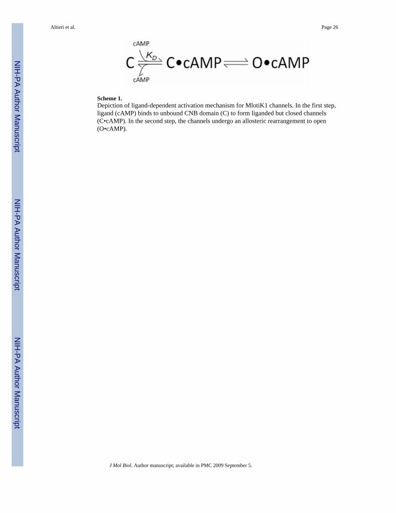
Scheme 1.Depiction of ligand-dependent activation mechanism for MlotiK1 channels. In the first step,ligand (cAMP) binds to unbound CNB domain (C) to form liganded but closed channels(C•cAMP). In the second step, the channels undergo an allosteric rearrangement to open(O•cAMP).
Altieri et al. Page 26
J Mol Biol. Author manuscript; available in PMC 2009 September 5.
NIH
-PA Author Manuscript
NIH
-PA Author Manuscript
NIH
-PA Author Manuscript

NIH
-PA Author Manuscript
NIH
-PA Author Manuscript
NIH
-PA Author Manuscript
Altieri et al. Page 27
Table 1Summary of crystallographic data and refinement statistics.
cGMP-bound wild type R307E R307WSpace Group P21 P21 P21212
Resolution (Å) 50.0-2.35 50.0-2.20* 41.09-2.90Rwork (%) 20.9 20.2* 26.0Rfree (%) 25.6 22.0* 30.6
Rmsd bond length (Å) 0.009 0.01 0.006Rmsd bond angle (°) 1.3 1.2 0.95
Completeness (%) 90.0 (94.0) 89.9(96.0)* 99.3 (97.7)Rsym (%) 8.0 (7.9) 7.3 (28.4) 6.6 (29.6)
I/Sig I 10.0 (7.0) 8.0 (2.3) 24.0 (5.3)Redundancy 3.3 (3.2) 4.1(2.9) 4.8 (4.6)
*During refinement data included reflections to 2.0 Å. Due to outer shell incompleteness, refinement statistics and completeness are reported to 2.2 Å
only.
J Mol Biol. Author manuscript; available in PMC 2009 September 5.