Crop Plant Anatomy This page intentionally left blank Crop Plant Anatomy
Upload
khangminh22Category
view
1download
0
University of Wollongong University of Wollongong
Research Online Research Online
University of Wollongong Thesis Collection 2017+ University of Wollongong Thesis Collections
2017
Structural anatomy of the breast Structural anatomy of the breast
Kathryn Maree Gaskin
Follow this and additional works at: https://ro.uow.edu.au/theses1
University of Wollongong University of Wollongong
Copyright Warning Copyright Warning
You may print or download ONE copy of this document for the purpose of your own research or study. The University
does not authorise you to copy, communicate or otherwise make available electronically to any other person any
copyright material contained on this site.
You are reminded of the following: This work is copyright. Apart from any use permitted under the Copyright Act
1968, no part of this work may be reproduced by any process, nor may any other exclusive right be exercised,
without the permission of the author. Copyright owners are entitled to take legal action against persons who infringe
their copyright. A reproduction of material that is protected by copyright may be a copyright infringement. A court
may impose penalties and award damages in relation to offences and infringements relating to copyright material.
Higher penalties may apply, and higher damages may be awarded, for offences and infringements involving the
conversion of material into digital or electronic form.
Unless otherwise indicated, the views expressed in this thesis are those of the author and do not necessarily Unless otherwise indicated, the views expressed in this thesis are those of the author and do not necessarily
represent the views of the University of Wollongong. represent the views of the University of Wollongong.
Recommended Citation Recommended Citation Gaskin, Kathryn Maree, Structural anatomy of the breast, Masters of Philosophy thesis, School of Medicine, University of Wollongong, 2017. https://ro.uow.edu.au/theses1/277
Research Online is the open access institutional repository for the University of Wollongong. For further information contact the UOW Library: [email protected]
Structural anatomy of the
breast
Kathryn Maree Gaskin
This thesis is presented as part of the requirements for the conferral of the degree:
Masters of Philosophy
Supervisor:
Dr Deirdre E. McGhee
Dr Gregory E. Peoples
The University of Wollongong School of Medicine
November, 2017

This work Qc copyright by Kathryn Maree Gaskin, 2017. All Rights Reserved.
No part of this work may be reproduced, stored in a retrieval system, transmitted, in any form or by any
means, electronic, mechanical, photocopying, recording, or otherwise, without the prior permission of the
author or the University of Wollongong.
This research has been conducted with the support of an Australian Government Research Training
Program Scholarship.
i
Declaration
I, Kathryn Maree Gaskin, declare that this thesis is submitted in partial fulfilment of
the requirements for the conferral of the degree Masters of Philosophy, from the
University of Wollongong, is wholly my own work unless otherwise referenced or
acknowledged. This document has not been submitted for qualifications at any other
academic institution.
Kathryn Maree Gaskin
7th
November, 2017
ii
Abstract
Introduction: The female breast, as originally described by Cooper in 1840[1]
, is a
fundamental part of the reproductive system, and therefore is included in essentially
every anatomy textbook [2-32]
. Surprisingly, there has been very little anatomical
research conducted on the breast since Cooper’s work[1]
. Cooper’s [1]
descriptions of the
anatomy of the breast were extensive. His methodology however lacked detail in terms
of the number of cadavers that anatomical descriptions were based on or any
quantitative data to support his descriptions.
Despite this, most of the current anatomical descriptions of the breast within
both textbooks and the literature are based on Cooper’s [1]
. Only 17 dissection studies
have been published since Cooper [1]
on the gross anatomy of the breast [33-48]
, with a
paucity of published research found on the internal fascial structure of the breast.
Although these studies have provided some additional anatomical detail of the breast,
they have also lacked quantitative data. In vivo studies using magnetic resonance
imaging (MRI) and ultrasound, and surgical studies investigating mastectomy tissue
have provided some data and further anatomical detail of breast composition and
structure [49-56]
. To date however, the published descriptions of the gross anatomy of the
breast have not been to be collated to form a consistent anatomical description of the
internal gross anatomy for anatomical education. Therefore the aim of this study was to
provide quantitative data on the gross anatomy of the breast to provide evidence-based
detail for anatomical illustrations and descriptions on the gross anatomy of the breast.
Methods: A cadaveric-based investigation was carried out on embalmed female
cadavers. The investigation focused on three (3) aspects of the female breast; (i) Breast
composition, (ii) Gross anatomical structure, and the (iii) Attachments of the breast to
iii
the chest wall. Eighteen breasts from nine cadavers were investigated quantitatively and
the gross anatomy was described qualitatively using this data. A range of dissection
techniques were used to investigate the breast in two planes, coronal and sagittal to
provide a three dimensional understanding of the structure of the breast.
The coronal dissections were conducted from a superficial to deep dissection
(n=15) with quantitative measurements taken at each depth of dissection. The sagittal
dissections were conducted on slices of different breasts (n=18 slices, n=3 breasts).
Different quantitative measurements were recorded in the sagittal plane compared to the
coronal plane.
Results: Composition
The mean total breast surface area and volume amongst the 15 breasts measured in the
coronal plane was 302±91 cm2 (range: 187-501 cm
2) and 381±272 mL (range: 56-959
mL). The mean total fibro-adipose and fibro-glandular mass per breast of the 14 breasts
dissected in the coronal plane was 172±103 g (range: 50–385 g) and 184±77 g (range:
56-300 g) respectively. Breast composition by mass in terms of the percentage of fibro-
glandular and fibro-adipose tissue was approximately 48% fibro-adipose tissue (range:
45-54 %). A moderate correlation was found between total breast mass and breast
volume (r2=0.577).
Gross Anatomical Structure
The mean number of fibro-adipose tissue pockets per coronal breast was 199±53 (range:
108 – 306). The number of pockets per breast was found to increase both with breast
volume (r2=0.737) and surface area (r
2=0.806; P<0.05). The mean size per pocket was
(mass) 0.4±0.15 g (range: 0.26-0.61 g) and (surface area) 0.88±0.37 cm2 (range: 0.31-
1.97 cm2). The fibro-adipose pockets found anterior to the gland were larger in size
(mean: 0.89±0.32 cm2, range: 0.34-1.20 cm
2) but less in number (mean: 35±16, range:
iv
20-49) compared to those posterior to the gland (mean size: 0.33±0.22 cm2, range: 0.22-
0.87 cm2; mean number: 53±31, range: 36-78). The mean length of the Anterior
Extensions of the Anterior Lamellae was 19±5 mm (range: 9-34 mm).
Attachment of the breast to the chest wall
The perimeter attachment of the breasts was consistently found to be stronger than the
Posterior Extensions of the Posterior Lamellae. The inferior perimeter was considered
to be the strongest attachment due periosteal attachments and required sharp dissection.
The sternal head of Pectoralis Major muscle was found within the perimeter of
all of the 12 breasts and made up the largest mean surface area (81%), followed by the
Serratus 10%), External Oblique (5%) and Rectus Abdominus muscles (4%).
Conclusion: This study adds to the understanding of the structural anatomy of the
breast by providing evidence to base new and more accurate illustrations of the gross
anatomical structure of the breast and written descriptions of the composition, gross
anatomical structure and the attachments of the breast to the chest wall. It can be used
to update the structural anatomy of the breast in anatomical texts used to teach medical,
allied health and science undergraduate students.
v
Acknowledgments
I would like to thank a number of people, who without their encouragement this thesis
would not have been possible. My sincerest thanks to:
- Dr. Deirdre McGhee, my primary supervisor who ensured I always kept to my
deadlines no matter how many track changes, sleepless nights or early morning
meetings it took. Your passion and dedication to your research students is
remarkable and I thank you for your knowledge and guidance.
- Dr. Gregory Peoples, my co-supervisor who was always available to lend a little
of his anatomical knowledge whenever necessary. Thank you for sharing your
wealth of knowledge and experiences and helping me to see problems from a
different angle.
- Dr. Simone Matousek, my expert advisor. Thank you for giving up your time to
provide an expert opinion of the structural anatomy of the breast from a
surgeon’s perspective. Thank you for sharing your extensive PhD research
which guided this research.
- Melbourne University, in particular the anatomy department. Thank you for
sharing you breast dissection methodology.
- Dr Venkata Krishna B Reddy, Thank you for sharing you insights and
knowledge of the structural anatomy of the breast. Your pre and post-operative
photographs were an invaluable resource to aid my understanding of the female
breast.
- My anatomical partner in crime, Natalia Munoz, and all the anatomy technical
staff, particularly Christine Wilson, Elle Padas and Christine McComb. Thank
vi
you for always providing a positive workspace filled with many long winded
stories and laughter. It has been a delight to work alongside you all.
- The donors of the UOW Anatomy Laboratory, who without their generosity and
selflessness I would be unable to perform this research.
- All the students and staff of the BRL. You were always there to lend a hand or
even give up a desk whenever needed.
- My family, all 12 of the Gaskin-Seghers clan. Thank you for always being there
at the end of the phone, sharing my highs and lows. I would like to acknowledge
in particular my mother, Kathleen, for your unconditional love and support and
my father, Dr. Gerard, for all of the editing and proof reading you willingly
endured without a moment’s hesitation.
- Finally, I would like to thank Peter Anstice who has been my rock throughout
this whole experience. Thank you for sharing my triumphs and putting up with
my tantrums and meltdowns. Without your constant support and willingness to
listen I would not have pulled this all together.
vii
Table of Contents
Declaration ........................................................................................................................ i
Abstract ............................................................................................................................ ii
Acknowledgments ........................................................................................................... v
Chapter 1: Introduction ................................................................................................. 1
1.1 Composition ............................................................................................................ 9
1.1.1 Literature Review .......................................................................................... 9
1.1.2 Textbook review .......................................................................................... 13
1.2 Gross Anatomical Structure .................................................................................. 16
1.2.1 Fibro-Adipose Structure (Literature Review) .............................................. 16
1.2.2 Fibro-Adipose Structure (Textbook review) ............................................... 24
1.2.3 Fibro-Glandular Structure (Literature Review) ........................................... 28
1.2.4 Fibro-Glandular Structure (Textbook review) ............................................. 29
1.3 Attachment of the breast to the chest wall ............................................................ 30
1.3.1 Regional Anatomy (Literature Review) ...................................................... 30
1.3.2 Regional Anatomy (Textbook review) ........................................................ 31
1.3.3 Attachment of the breast to the chest wall (Literature Review) .................. 32
1.3.4 Attachment of the breast to the chest wall (Textbook review) .................... 39
1.4 Literature Summary .............................................................................................. 42
1.5 Research Aims and Hypothesis ............................................................................. 43
1.5.1 Composition ................................................................................................. 43
1.5.2 Gross Anatomical Structure ......................................................................... 43
1.5.3 Attachment of the breast to the chest wall ................................................... 44
Chapter 2: Methods ...................................................................................................... 45
viii
2.1 Experimental Overview .................................................................................... 46
2.2 Ethics Consideration ......................................................................................... 46
2.3 Cadaveric Selection .......................................................................................... 46
2.4 Dissection Methods ............................................................................................... 50
2.4.1 Coronal Dissection Method ......................................................................... 50
2.4.2 Sagittal Dissection Method .......................................................................... 58
2.5 Quantitative Outcome Measures ........................................................................... 63
2.5.1 Composition ................................................................................................. 63
2.5.1.1 Surface Area ........................................................................................... 63
2.5.1.2 Volume .................................................................................................. 63
2.5.1.3 Fibro-adipose mass ............................................................................... 66
2.5.1.4 Fibro-glandular mass ............................................................................. 66
2.5.1.5 Breast Mass ........................................................................................... 66
2.5.1.6 Breast Composition ................................................................................. 66
2.5.2 Gross Anatomical Structure ......................................................................... 66
2.5.2.1 Number of fat lobule pockets ................................................................ 66
2.5.2.2 Adipose mass of each pocket ................................................................ 67
2.5.2.3 Surface area of each adipose pocket...................................................... 67
2.5.2.4 Length of the anterior extensions of the anterior lamellae .................... 67
2.5.2.5 The number of pockets anterior and posterior to the gland .................. 68
2.5.2.6 The cross sectional surface area ............................................................ 68
2.5.3 Attachment of the breast to the chest wall ................................................... 68
2.5.3.1 Muscles within the perimeter ................................................................ 68
2.5.3.2 Perimeter ............................................................................................... 69
2.6 Qualitative Measures ............................................................................................. 69
ix
2.7 Experimental Standardisation and Reliability ....................................................... 69
2.7.1 Chief Investigator ........................................................................................ 69
2.7.2 Reliability Study .......................................................................................... 70
2.7.3 Cadaver Preparation and Storage ................................................................. 71
2.7.4 Room Conditions During Measurement ...................................................... 71
2.8 Normalising Data (absolute and relative) .............................................................. 72
2.9 Statistical Design ................................................................................................... 72
Chapter 3: Results ......................................................................................................... 73
3.1 Composition .......................................................................................................... 74
3.1.1 Surface Area ................................................................................................ 74
3.1.2 Volume ........................................................................................................ 74
3.1.3 Fibro-adipose mass ...................................................................................... 78
3.1.4 Fibro-glandular mass ................................................................................... 78
3.1.5 Breast Mass .................................................................................................. 78
3.1.6 Breast Composition ..................................................................................... 82
3.1.7 Breast mass in relation to breast volume ..................................................... 82
3.2 Gross Anatomical Structure .................................................................................. 85
3.2.1 Coronal Qualitative Measures ..................................................................... 85
3.2.2 Number of fibro-adipose tissue pockets ...................................................... 92
3.2.3 Number of fat lobule pockets in relation to breast volume and surface area92
3.2.4 Adipose mass of each pocket ....................................................................... 97
3.2.5 Adipose mass within each pocket in relation to breast volume and surface
area 97
3.2.6 Surface area of adipose tissue pockets ....................................................... 101
3.2.7 Pocket mean surface area in relation to breast surface area and volume ... 101
x
3.2.8 Sagittal Qualitative Measures .................................................................... 105
3.2.9 Number (n) of pockets anterior and posterior to the gland ........................ 108
3.2.10 Length of the anterior extensions of the anterior lamellae ...................... 108
3.2.11 Pocket surface area (mm2) ....................................................................... 108
3.3 Attachment of the breast to the chest wall .......................................................... 112
3.3.1 Perimeter: Regional Anatomy ................................................................... 119
3.3.2 Perimeter: Quantitative Data ..................................................................... 120
3.3.3 Muscles within the perimeter ..................................................................... 124
Chapter 4: Discussion ................................................................................................. 126
4.1 Composition ........................................................................................................ 127
4.1.1 Breast Volume and Breast Surface Area ................................................... 127
4.1.2 Breast Mass ................................................................................................ 130
4.1.3 Composition of Fibro-adipose and Fibro-glandular tissues....................... 131
4.2 Gross Anatomical Structure ................................................................................ 135
4.2.1 Fibro-Adipose Structure ............................................................................ 135
4.2.2 Location of the Fibro-Glandular Structure ................................................ 140
4.3 Attachment of the breast to the chest wall .......................................................... 141
4.3.1 Posterior and Perimeter Attachments ........................................................ 143
4.4 Conclusion .......................................................................................................... 149
References .................................................................................................................... 151
Appendices ................................................................................................................... 156
Appendix A: Textbook review .................................................................................. 156
A.1 Review of the descriptions and illustrations of breast anatomy presented in
anatomical textbooks for Medicine and Allied Health subjects in Universities
across Australia ................................................................................................... 156
xi
Appendix B: Written descriptions for anatomical textbooks .................................... 159
B.1 Breast Composition ................................................................................... 159
B.2 Fibro-Adipose Structure ............................................................................ 160
B.3 Attachment of the Breast to the Chest Wall .............................................. 165
Appendix C: Conference Presentations .................................................................... 170
C.1 9th
Australasian Biomechanics Conference, 2014, University of
Wollongong, NSW AUS .................................................................................... 170
C.2 Tech-Net, 2015, University of Technology Sydney, NSW AUS ............. 173
C.3 Australasian Institute of Anatomical Science, 2016, University of Otago,
Otago NZ ............................................................................................................ 174
C.4 14th
Annual Conference of the Australia and New Zealand Association of
Clinical Anatomists, 2016, Australian National University, ACT AUS ............ 176
C.5 15th
Annual Conference of the Australia and New Zealand Association of
Clinical Anatomists, 2017, University of Auckland, Auckland, NZ .................. 178
xii
List of Figures
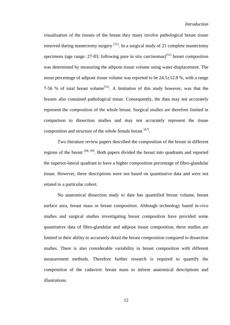
Figure 1 Example of textbook representation of the composition of the breast
(Moore, Agur et. al. (2015)[21])
15
Figure 2 Ligamenta Suspensoria (fat and skin removed) A) Nipple areola
complex, B) Ligamenta Suspensoria, C) Glandular tissue (Cooper
1840).
25
Figure 3 Example of textbook representation of the fibro-adipose structure of
the breast (Moore, Agur et. al. (2015)[21])
26
Figure 4 Cadaveric breast dissection showing A) Glandular tissue, B)
Superficial pectoral fascia, C) Posterior Extensions of the Posterior
Lamellae, D) Pectoralis major muscle (Jinde et. al 2006).
35
Figure 5 Horizontal septum spanning medio-laterally from the 5th rib to the
nipple (Wűringer et. al. 1998).
25
Figure 6 Entire perimeter attachments of the breast to the chest wall
(Matousek et. al. 2014).
40
Figure 7 Example of textbook representation of the regional anatomy of the
breast on the chest wall (Moore, Agur et. al. (2015)[21]
41
Figure 8 Experimental overview. 48
Figure 9 Coronal breast with a) Boarder outlined with permanent marker and
the viscous dye injection sites, b) Skin removed and nipple and
areola left intact, c) Skin removed with viscous dye injection sites
and separation of the breast into quadrants with flag pins and string.
53
Figure 10 Coronal breast showing a) Superficial layer with adipose tissue 54
xiii
located within fibrous pockets, b) Adipose tissue removed and
pockets pinned for further quantitative analysis.
Figure 11 Fibro-glandular tissue viewed from a) Anterior aspect, b) Lateral
aspect, borders of the breast highlighted by viscous dye injections.
56
Figure 12 Coronal breast showing a) Fibro-adipose tissue deep to the fibro-
glandular tissue (reflected), b) Breast removed to reveal chest wall
muscles with perimeter left intact.
57
Figure 13 Sagittal dissection with a) Chest plate in-tact with chest plate
removal lines marked using permanent marker, b) Chest plate
removed from the cadaver with bandsaw incision lines marked.
61
Figure 14 Sagittal sectioning of a) Chest plate embedded in foam which
ensured a smooth and even transition through the band saw, b)
Example of a mid-sagittal breast slice.
62
Figure 15 Geomagic analysis software, a) Full scan, b) Chest with left breast
removed, c) Left breast ready for surface area calculation, d) Left
breast ready for volume calculation.
64
Figure 16 Mean surface area per quadrant (n=15 breasts). 77
Figure 17 Mean volume per quadrant (n=15 breasts). 77
Figure 18 Mean fibro-adipose tissue mass per quadrant (n=14). 80
Figure 19 Mean distribution of fibro-glandular mass per quadrant (n=14). 81
Figure 20 Mean distribution of the total breast mass (fibro-glandular and fibro-
adipose mass combined) per quadrant (n=14).
81
Figure 21 Breast composition as percentage of fibro-adipose and fibro-
glandular mass within each breast (n=14).
83
xiv
Figure 22 Mean breast composition per quadrant as percentage of fibro-adipose
and fibro-glandular mass (n=14).
83
Figure 23 Correlation of a) Combined total breast mass and breast volume, b)
Fibro-adipose breast mass and breast volume, and c) Fibro-glandular
breast mass and breast volume (n=14; p<0.01).
84
Figure 24 Coronal breast showing a) Round nipples (skin removed), b) Oval
nipple (skin intact).
86
Figure 25 Fibrous cobweb of the breast from the a) Anterior aspect and b)
Sagittal aspect.
88
Figure 26 Example of pocket variation shape amongst the quadrants of the
breast.
89
Figure 27 Coronal breast showing a) Relative size and shape of the fibro-
glandular tissue, b) Fibro-glandular tissue reflected showing the
deeper layer of fibro-adipose tissue situated posterior to the fibro-
glandular tissue.
91
Figure 28 Mean number of pockets per quadrant (n=15 breasts). 94
Figure 29 Mean surface area, volume and number of pockets per quadrant
(Mean and SEM) in each quadrant.
95
Figure 30 Correlation graphs of a) Total breast surface area to total breast
number of pockets, b) Total breast volume to total breast number of
pockets, and c) Total breast volume to total breast surface area per
breast (n=15) (p<0.01).
96
Figure 31 Adipose tissue mass of each of the 80 pockets breast dissected in the
coronal plane (n=15 breasts).
99
xv
Figure 32 Mean (SD) adipose tissue mass of each pocket (n=80 pockets) per
breast dissected in the coronal plane (n=15 breasts).
99
Figure 33 Mean (SD) adipose tissue mass per pocket (n=80 pockets) per
quadrant of breast dissected in the coronal plane (n=15 breasts).
100
Figure 34 Mean adipose tissue mass (g) per pocket per quadrant (n=80 per
breast).
100
Figure 35 Surface area per pocket of the 80 pockets measured in each breast
dissected in the coronal plane (n=15 breasts).
102
Figure 36 Mean adipose tissue pocket surface area (n=80 pockets) measured in
each breast dissected in the coronal plane (n=15 breasts).
102
Figure 37 Mean surface area of each pocket (n=80 pockets) per quadrant
measured in each breast dissected in the coronal plane (n=15
breasts).
103
Figure 38 Mean surface area of the adipose tissue pockets per quadrant. 103
Figure 39 Correlation graphs of (a) Pocket mass to total breast volume, (b)
Pocket surface area to total breast volume and (c) Pocket surface
area to pocket mass per breast (n=15) (P<0.01).
104
Figure 40 Mid-sagittal slice of the breast through the nipple showing the
dermis (green) and the Superficial Pectoral fascia (light blue). The
regional depths are circled anterior to the gland (red), within the
gland (purple) and posterior to the gland (dark blue).
107
Figure 41 Number of fibro-adipose tissue pockets anterior to the gland per
breast measured in the sagittal breast slice (n=18 slices, n=3 breasts).
109
Figure 42 Number of fibro-adipose tissue pockets posterior to the gland per 110
xvi
breast measured in the sagittal breast slice (n=18 slices, n=3 breasts).
Figure 43 Anterior Extension of the Anterior Lamellae length per breast
measured in the sagittal breast slice (n=18 slices, n=3 breasts).
110
Figure 44 Anterior Extension of the Anterior Lamellae length on the a) Medial
and b) Lateral para-sagittal breast slices.
111
Figure 45 i: Mid-sagittal slice of the female breast through the nipple with the
5th rib marked (5), the inferior border of pectorals major muscle
marked (arrow) and the region of the Triangle Fascial Condensation
[41] circled.
ii: Sagittal slice of the female breast through the lateral edge of the
areola (a) and lateral breast boarder (b) with the 5th rib marked (5 –
where visible), the inferior border of pectorals major muscle marked
(arrow- where visible) and the region of the Triangle Fascial
Condensation [41] circled.
iii: Sagittal slice of the female breast through the medial edge of the
areola (a) and medial breast boarder (b) with the 5th rib marked (5),
the inferior border of pectorals major muscle marked (arrow) and the
region of the Triangle Fascial Condensation[41] circled.
116
117
118
Figure 46 Breast perimeter per breast. 121
Figure 47 Perimeter of the breast, with the lateral fascia confluence circled. A)
Pectoralis Major muscle (clavicular head), B) Pectoralis Major
muscle (sternal head), C) Serratus Anterior muscles, D) External
Oblique muscle, E) Rectus Abdominus muscle.
122
Figure 48 Posterior Extensions of the Posterior Lamellae viewed from the 123
xvii
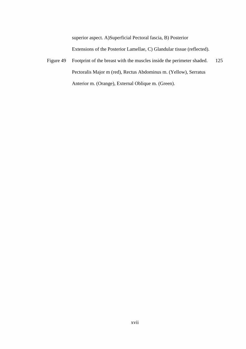
superior aspect. A)Superficial Pectoral fascia, B) Posterior
Extensions of the Posterior Lamellae, C) Glandular tissue (reflected).
Figure 49 Footprint of the breast with the muscles inside the perimeter shaded.
Pectoralis Major m (red), Rectus Abdominus m. (Yellow), Serratus
Anterior m. (Orange), External Oblique m. (Green).
125
xviii
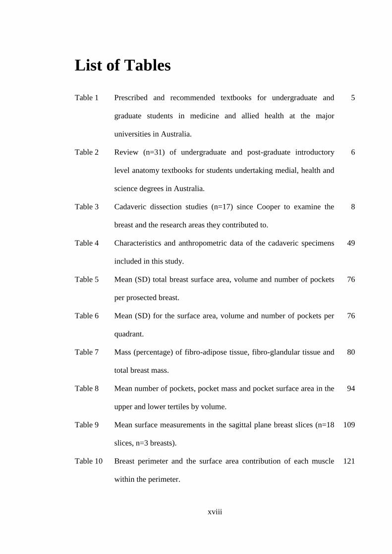
List of Tables
Table 1 Prescribed and recommended textbooks for undergraduate and
graduate students in medicine and allied health at the major
universities in Australia.
5
Table 2 Review (n=31) of undergraduate and post-graduate introductory
level anatomy textbooks for students undertaking medial, health and
science degrees in Australia.
6
Table 3 Cadaveric dissection studies (n=17) since Cooper to examine the
breast and the research areas they contributed to.
8
Table 4 Characteristics and anthropometric data of the cadaveric specimens
included in this study.
49
Table 5 Mean (SD) total breast surface area, volume and number of pockets
per prosected breast.
76
Table 6 Mean (SD) for the surface area, volume and number of pockets per
quadrant.
76
Table 7 Mass (percentage) of fibro-adipose tissue, fibro-glandular tissue and
total breast mass.
80
Table 8 Mean number of pockets, pocket mass and pocket surface area in the
upper and lower tertiles by volume.
94
Table 9 Mean surface measurements in the sagittal plane breast slices (n=18
slices, n=3 breasts).
109
Table 10 Breast perimeter and the surface area contribution of each muscle
within the perimeter.
121

1
Chapter 1:
Introduction
Introduction
2
The female breast is a fundamental part of the reproductive system, and therefore is
included in essentially every anatomy textbook recommended for undergraduate and
graduate students in medicine and allied health courses within Australian Universities [2-
32] (Table 1 and Table 2). Surprisingly, there has been very little anatomical research
conducted on this region of the body, with the most extensive research conducted by
Cooper in 1840[1]
. Cooper’s [1]
publication of his breast dissections were extensive and
included descriptions of the structure, composition and attachments of the breast to the
chest wall. However, research at that time typically did not provide detailed
methodology in terms of the number of cadavers that anatomical descriptions were
based on or any quantitative data of the anatomical structures dissected. Despite this,
most of the current anatomical descriptions of the breast within both textbooks and the
literature are based on Cooper’s [1]
descriptions, and hence also lack any quantitative
data to support or verify them. Only 17 dissection studies have been published since
Cooper [1]
on the gross anatomy of the breast [33-48]
(Table 3), with a paucity of
published research found on the internal fascial structure of the breast.
Although these studies have provided some additional anatomical detail of the
breast, they have also lacked quantitative data to support their descriptions and many
textbooks present conflicting descriptions of the internal fascial structure of the breast.
In-vivo studies using magnetic resonance imaging (MRI) and ultrasound, and
surgical studies investigating breast tissue removed during breast mastectomy have
provided further anatomical detail of breast composition and structure [49-56]
. They have
also provided some quantitative data on breast anatomy, composition and structure. To
date however, the published descriptions of the gross anatomy of the breast have not
been collated to form a consistent anatomical description of the internal gross anatomy
for anatomical education.
Introduction
3
The aim of this literature review was therefore to collate and summarise all of
the relevant descriptions and research on the gross anatomy of the breast. This was
achieved by two methods (i) a literature review of the published research, including the
work of Cooper, (ii) a textbooks review. The literature review was conducted using the
Scopus database for English articles from 1800-2016, using keywords female breast
AND anatomy OR fibrous tissue OR glandular tissue OR adipose tissue OR Cooper’s
Ligaments OR suspensory ligaments OR adipose tissue AND NOT cancer AND NOT
chicken. The terms cancer and chicken were included in an attempt to eliminate results
relating to breast cancer or chicken breasts. Studies were included if they met the
following selection criteria: i) examined breast anatomy using anatomical dissection
techniques; ii) included female breasts, though no criteria was imposed in regards to the
minimum number of breasts; iii) data presented in English. A total of 17 dissection
studies were included in the literature review [33-48, 57]
. A limitation of this study
however is the exclusion of non-English works which may have re-examined and
expanded on the breast anatomy originally investigated by Cooper [1].
Anatomical textbooks used in major universities around Australia for
undergraduate or post-graduate medicine and allied health degrees were included in the
textbook review of breast anatomy (Table 1). All of the major universities were
contacted by phone by the Chief Researcher and their websites reviewed. Thirty-one
textbooks were identified and included in the analysis (Table 2) [2-32]
. The majority of
the anatomical textbooks directed at introductory level anatomy for medical and allied
health students (Table 1) only contained a small amount of written text regarding the
gross anatomy of the breast, with one illustration of the breast. Considerable variation
was found in the gross anatomy presented in these illustrations, and the majority of
illustrations were of a relatively small, pert shaped breast, with very limited associated
Introduction
4
written text providing further detail of the illustration. Most of the written text within
the anatomical textbooks focused on the regional anatomy or the fibro-glandular tissues
of the breast. Not one textbook presented evidence or quantitative data to support either
their illustrations or written text (Table 2).
As Cooper’s [1]
descriptions were so extensive and form the basis of current
anatomical descriptions of the breast, the anatomical descriptions of the gross anatomy
of the breast presented in the literature and textbooks were summarised and compared to
the Cooper’s [1]
descriptions. The anatomy of the breast was investigated in the
following sections: (i) composition; (iii) gross anatomical structure; (iv) attachments of
breast to chest wall.
5
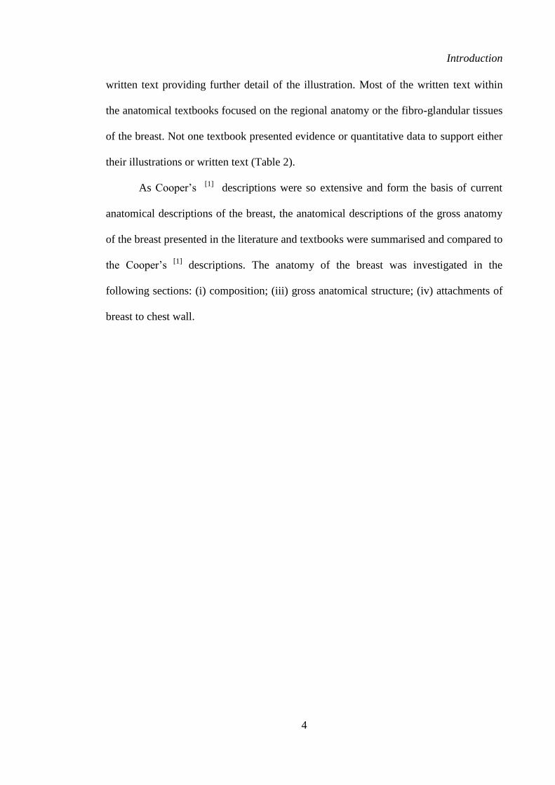
Intro
ductio
n
Table 1: Prescribed and recommended textbooks for undergraduate and graduate students in medicine and allied health at the major universities in Australia. (compiled as of June 2017 from personal conversations and online subject descriptions)
University Textbook: Author (Year)
Australian National University - ACT Drake et. al. (2005), Marieb and Hoehn (2012), McKinley and O'Loughlin (2006), Moore, Agur et. al. (2015)
[7, 17, 18, 21]
Canberra University- ACT Tortora et. al. (2015)[31]
Griffith University- Queensland Marieb and Hoehn (2012)[17]
James Cook University - Queensland Drake et. al. (2005), Marieb and Hoehn (2012), McKinley and O'Loughlin (2006), Moore, Agur et. al. (2015)
[7, 17, 18, 21]
La Trobe University- Victoria Drake et. al. (2005), Moore, Dalley et. al. (2014)[7, 22]
Macquarie University - New South Wales Tortora and Derrickson (2012)[30]
Melbourne University - Victoria Drake et. al. (2005), Eizenberg et. al. (2008), McMinn (1991), Moore, Dalley et. al. (2014)[7, 8, 19,
22]
Queensland University of Technology - Queensland
Clemente (2007), Olson (1996)[6, 24]
RMIT- Victoria Abrahams et. al. (2008), Drake et. al. (2005), Moore, Agur et. al. (2015), Moore, Dalley et. al. (2014), Tortora and Derrickson (2012), Rohen, Yokochi et. al. (2006)
[3, 7, 21, 22, 30, 58]
Southern Cross University- Queensland Marieb and Hoehn (2012)Marieb and Hoehn (2012)
Sydney University – New South Wales Agur and Dalley (1991), Drake et. al. (2005), Kapit and Elson (1993), Moore, Dalley et. al. (2014), Rohen et. al. (2006)
[4, 7, 15, 22, 58]
University of Adelaide - South Australia Agur and Dalley (1991), Drake et. al. (2005), Kapit and Elson (1993), Moore, Dalley et. al. (2014), Rohen et. al. (2006)
[4, 7, 15, 22, 58]
University of New England - New South Wales Tortora et. al. (2015)[31]
University of Newcastle - New South Wales Gilroy et. al. (2012)[11]
University of Queensland - Queensland Drake et. al. (2005), Hansen (2014), Rohen et. al. (2006)[7, 13, 58]
University of Tasmania - Tasmania Drake et. al. (2005), Gilroy (2013), Hansen (2014), Moore, Dalley et. al. (2014), Rohen et. al. (2006), Martini et. al. (2008)
[7, 10, 13, 22, 58, 59]
University of Technology Sydney - New South Wales
Marieb and Hoehn (2012)[17]
University of Western Australia - Western Australia
Saladin (2007)[26]
University of Wollongong – New South Wales Marieb and Hoehn (2012) [17]
UNSW- New South Wales Marieb and Hoehn (2012), Tortora and Derrickson (2012), Tortora et. al. (2015) [17, 30, 31]
Western Sydney University - New South Wales Tortora et. al. (2015)[31]
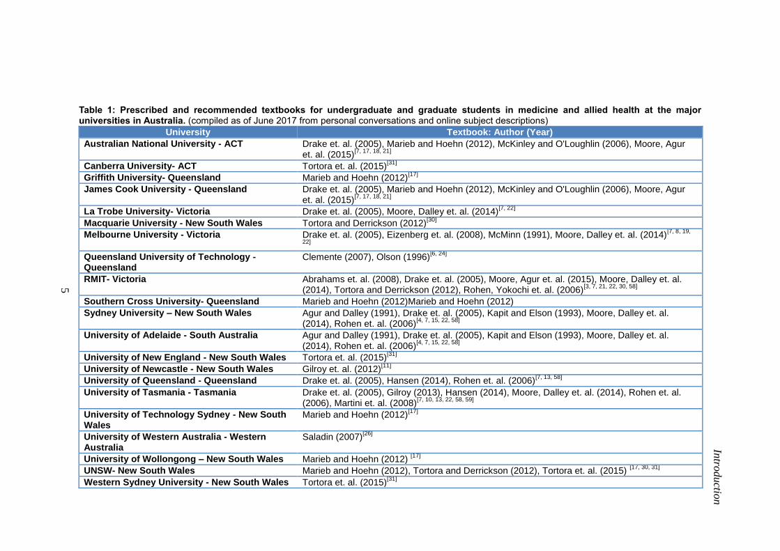
6
Intro
ductio
n
Table 2: Summary (n=31) of undergraduate and post-graduate introductory level anatomy textbooks for students undertaking medial, health and science degrees in Australia.
Author (Year) Sagittal Image (n)
Coronal Image (n)
Fibro-adipose structure
Fibro-glandular structure
Composition Attachment to chest wall
Based on Data
Abrahams, Cravem et. al. (2005)
[2]
Abrahams, Boom et. al (2008)
[3]
Agur & Dalley (1991)[4]
Applegate (2000)[5]
Clemente (2007)[6]
Drake et. al. (2005)[7]
Eizenberg et. al. (2008)[8]
Ellis (2002)[9]
Gilroy (2013)[10]
Gilroy et. al (2012)[11]
Gosling et. al. (2003)[12]
Hansen (2014)[13]
Hansen & Lambert (2008)[14]
Kapit & Elson (1993)[15]
Lindsay (1995)[16]
Marieb & Hoehn (2012)[17]
=included in the textbook
7
Intro
ductio
n
Table 2: ctnd.
Author (Year) Sagittal Image (n)
Coronal Image (n)
Fibro-adipose structure
Fibro-glandular structure
Composition Attachment to chest wall
Based on Data
McKinley & O’Loughlin (2006)
[18]
McMinn (1991)[19]
Moffat (1993)[20]
Moore, Agur et. al. (2015)[21]
Moore, Dalley et. al (2014)[22]
Moses et. al. (2005)[23]
Olson (1996)[24]
Rohen (2016)[25]
Saladin (2007)[26]
Sinnatamby (2006)[27]
Snell (1995)[28]
Tank & Gest (2008)[29]
Tortora & Derrickson (2012)
[30]
Tortora et. Al (2015)[31]
Van De Graff et. al. (2002)[32]
Total number of textbooks 23 (31
images)
26 (58
images)
19 24 6 22 0
=included in the textbook
8
Intro
ductio
n
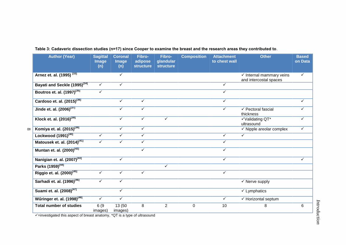
Table 3: Cadaveric dissection studies (n=17) since Cooper to examine the breast and the research areas they contributed to.
Author (Year) Sagittal Image
(n)
Coronal Image
(n)
Fibro-adipose
structure
Fibro-glandular structure
Composition Attachment to chest wall
Other Based on Data
Arnez et. al. (1995) [33]
Internal mammary veins and intercostal spaces
Bayati and Seckle (1995)[34]
Boutros et. al. (1997)[35]
Cardoso et. al. (2015)[36]
Jinde et. al. (2006)[37]
Pectoral fascial thickness
Klock et. al. (2016)[38]
Validating QT* ultrasound
Komiya et. al. (2015)[39]
Nipple areolar complex
Lockwood (1991)[40]
Matousek et. al. (2014)[41]
Muntan et. al. (2000)[42]
Nanigian et. al. (2007)[43]
Parks (1959)[44]
Riggio et. al. (2000)[45]
Sarhadi et. al. (1996)[46]
Nerve supply
Suami et. al. (2008)[47]
Lymphatics
Wűringer et. al. (1998)[48]
Horizontal septum
Total number of studies 6 (9 images)
13 (50 images)
8 2 0 10 8 6
=investigated this aspect of breast anatomy, *QT is a type of ultrasound
Introduction
9
Intro
ductio
n
1.1 Composition
1.1.1 Literature Review
Cooper [1]
described the breast to consist of four types of tissues; the overlying skin,
glandular tissue, fibrous tissue and adipose tissue. He did not however quantify the
relative percentage composition of each of these tissues or the volume, surface area or
mass of the breast. There has been a paucity of anatomical research on the structural
composition of the breast, consequently, anatomical descriptions and quantitative data
on breast composition is also lacking in anatomical textbooks. Although Cooper’s [1]
anatomical illustrations provided some information on the composition of the breast, his
illustrations were not labelled or associated with a figure caption. Therefore limited
detail of relative tissue composition can be taken from them. Since Cooper’s work[1]
, no
published dissection study was found that specifically aimed to dissect and quantify the
anatomical composition of the breast.
Breast volume, mass and surface area [60, 61]
data has been collected in
technology-based in vivo studies using three-dimensional scanning, MRI and
mammography, casting and with anthropometric, measurements, and via surgical
studies of mastectomy specimens using water displacement and tissue weighing [49-54, 61-
63]. The breast volumes measured have ranged from 75 to 3100 mL
[49-54, 61-63] with the
mean breast volume varying from 273 [62]
to 715 mL [49]
. The variation in the mean
volume measured can be attributed to the large range of breast volumes that exist in the
female population and the differences in the volume range of the cohorts recruited
within each study. Breast volume has also been found to vary with BMI [61]
and age [49]
.
The variation in the mean volume measured amongst different studies can also
be attributed to the different measurement techniques utilised to measured breast
Introduction
10
Intro
ductio
n
volume [50, 64]
and inter-rater variation [62]
. Studies that have utilised different breast
volume measurement techniques such as scanning, water displacement, and MRI on the
same cohort of breasts have found variation in the magnitude of volume measured [50,
62]. Surgical studies that have measured breast volume from mastectomy specimens
[51]
may also contain corrupted tissue, i.e. cancerous tissue. Only one study was found that
attempted to quantify the regional distribution of breast volume [65]
. The breasts of 176
women were scanned using three-dimensional scanning and the resultant scan was
divided into quadrants through the nipple. The study reported the upper-inner quadrant
to have the greatest distribution of breast volume and that the volume of the quadrants
was positively and strongly correlated with the total breast volume (r2=0.81–0.91,
n=176)
Only one published study was found that measured breast mass [51]
. A study of
21 complete mastectomy specimens following pure multifocal in situ carcinomas found
breast mass to range from 110 to 1100 g, with a mean of 420±261 g. However, similar
to the breast volume data from mastectomy studies, the breast tissue may also contain
cancerous tissue, affecting the accuracy of this measurement.
Although in vivo breast volume and mass data exists, it has not been translated
into anatomical texts. This may be related to the wide range of breast volumes measured
and the variation in mean breast volume measured in different cohorts of women
measured with different measurement technique. Further research is therefore required
to quantify breast volume and breast mass based on quantitative data obtained from
dissection specimens to update anatomical illustrations and descriptions presented in
anatomical textbooks.
Due to the limited detail of the anatomical structures within the breast visualised
by MRI and mammography compared to anatomical dissection research, breast
Introduction
11
Intro
ductio
n
composition research based on such technologies has been broadly quantified as a
percentage of fibro-glandular and adipose tissue. Although a mean of 66±18 % adipose
tissue was found in younger women (400 women, mean age 20.8 years[49]
) and 70±22 %
adipose tissue in older women (100 women, mean age 49.6 years[49]
), the relative
percentage of fibro-glandular and adipose tissue has also been found to vary with age,
race, mass, hormonal changes and pregnancy [49, 51, 55, 66]
. It has also been found to vary
with measurement method. A study comparing breast composition determined from
MRI and mammography on the same cohort of women (40 women, range: 20-83 years
[55]) reported the mean percentage of adipose tissue within the breast to be 66.5±18 %
when measured with MRI, and 42.5±30.3 % when measured on the same breast with
mammography[55]
. The authors attributed the differences to changes to the shape of the
breast during the two different measurement methods. The breast deforms in the
different body positions sustained during measurement, prone for MRI and standing for
mammography.
Breast composition measured with mammography has also varied in different
cohorts of women. Lee et. al. (1997)[55]
reported the mean percentage of adipose tissue
to be 42.5±30.3 % in 40 women between the ages of 20-83 years, while Engelken et. al.
(2014)[52]
reported the mean percentage of adipose tissue to account for 79% of tissue in
the breast in 174 women between the ages of 20-79 years (mean age: 55 years) [52]
.
Unfortunately both studies included a very large age range in their cohort of relatively
small numbers. Therefore the breast composition data attained from technology based in
vivo studies is limited due to the lower level of visualisation of anatomical tissue in
comparison to anatomical dissection studies and due to the discrepancies in the
composition measured using different measurement technologies.
Although surgical studies measuring breast composition allow for greater
Introduction
12
Intro
ductio
n
visualisation of the tissues of the breast they many involve pathological breast tissue
removed during mastectomy surgery [51]
. In a surgical study of 21 complete mastectomy
specimens (age range: 27-83; following pure in situ carcinomas)[51]
breast composition
was determined by measuring the adipose tissue volume using water-displacement. The
mean percentage of adipose tissue volume was reported to be 24.5±12.8 %, with a range
7-56 % of total breast volume[51]
. A limitation of this study however, was that the
breasts also contained pathological tissue. Consequently, the data may not accurately
represent the composition of the whole breast. Surgical studies are therefore limited in
comparison to dissection studies and may not accurately represent the tissue
composition and structure of the whole female breast [67]
.
Two literature review papers described the composition of the breast in different
regions of the breast [68, 69]
. Both papers divided the breast into quadrants and reported
the superior-lateral quadrant to have a higher composition percentage of fibro-glandular
tissue. However, these descriptions were not based on quantitative data and were not
related to a particular cohort.
No anatomical dissection study to date has quantified breast volume, breast
surface area, breast mass or breast composition. Although technology based in-vivo
studies and surgical studies investigating breast composition have provided some
quantitative data of fibro-glandular and adipose tissue composition, these studies are
limited in their ability to accurately detail the breast composition compared to dissection
studies. There is also considerable variability in breast composition with different
measurement methods. Therefore further research is required to quantify the
composition of the cadaveric breast mass to inform anatomical descriptions and
illustrations.
Introduction
13
Intro
ductio
n
1.1.2 Textbook review
Despite the lack of quantitative data on breast composition in the literature, anatomical
textbooks have provided some detail of breast composition through anatomical
illustrations more than through written text. Across the 31 textbooks included in the
summary, there were 58 coronal illustrations of the breast (from 26 textbooks, Table 2)
and 31 sagittal illustrations (from 23 textbooks, Table 2, Figure 1). Only three textbooks
[8, 19, 27] had no illustrations within their breast anatomy section. The coronal images
generally depicted the composition of the partially dissected breast to include the skin,
fascial tissue, glandular tissue and adipose tissue with the Tail of Spence projecting into
the axilla, consistent with Cooper [1]
. Of the 26 textbooks that contained coronal images,
three textbooks had cadaveric images [3, 23, 25]
and four textbooks had surface anatomy
photographs [2, 3, 6, 18]
. Of the 26 textbooks that contained coronal images, 15 depicted
the tissues that make up the breast (Table 2), however the relative quantitative
composition of these tissues and their relative location was either inconsistent or not
clearly quantified from the illustrations. Furthermore, no quantitative data or evidence
was provided to verify the breast compositions presented.
Of the 23 textbooks with sagittal images (Table 2, Figure 1), five textbooks had
cadaveric photographs [3, 12, 23, 25, 26]
and one textbook had surface anatomy photographs
[2]. Of the 31 sagittal images within the 23 textbooks, 24 were pure sagittal plane
sections through the nipple [4, 6, 10-16, 23, 24, 29-32]
and seven were from an anterior-medial
aspect [2, 5, 17, 21, 22, 26, 28]
. Twenty of the sagittal illustrations provided some detail of the
composition of the breast [3-6, 10-17, 21, 22, 25, 26, 29-32]
however it was not specifically
quantified. Although the sagittal images were consistent in their location of the
glandular tissue within the breast, they were inconsistent in terms of both the
quantitative composition of the glandular, fibrous and adipose tissues within the breast
Introduction
14
Intro
ductio
n
and the relative locations of the fibrous and adipose tissues within the breast. For
example, eight textbooks displayed adipose tissue both anterior and posterior to the
glandular tissue [5, 13, 14, 26, 29-32]
, while five displayed adipose tissue only anterior to the
glandular tissue [10, 11, 16, 17]
and four displayed no discernible difference between the
tissues of the breast [4, 6, 21, 22]
. Only three textbooks contained cadaveric images to
display breast composition and all of these were poorly labelled or captioned [3, 12, 25]
.
None of the textbooks included any quantitative data or referenced any studies to verify
the breast composition basis of their illustrations.
The written text of the majority of the anatomical textbooks did not include any
detail of the relative composition of the tissues within the breast. Indeed, breast
composition was only mentioned in six textbooks [2, 7, 8, 10, 15, 26]
, which only described
the types of tissue within the breast (adipose, fibrous and glandular tissues), not their
relative composition. One textbook stated that breast mass was related to the increase in
the adipose tissue mass (Table 2), however no data or evidence was provided to support
this notion.
Introduction
15
Intro
ductio
n
Figure 1 Example of textbook representation of the composition of the breast (Moore, Agur et. al. (2015)[21])
Introduction
16
Intro
ductio
n
1.2 Gross Anatomical Structure
1.2.1 Fibro-Adipose Structure (Literature Review)
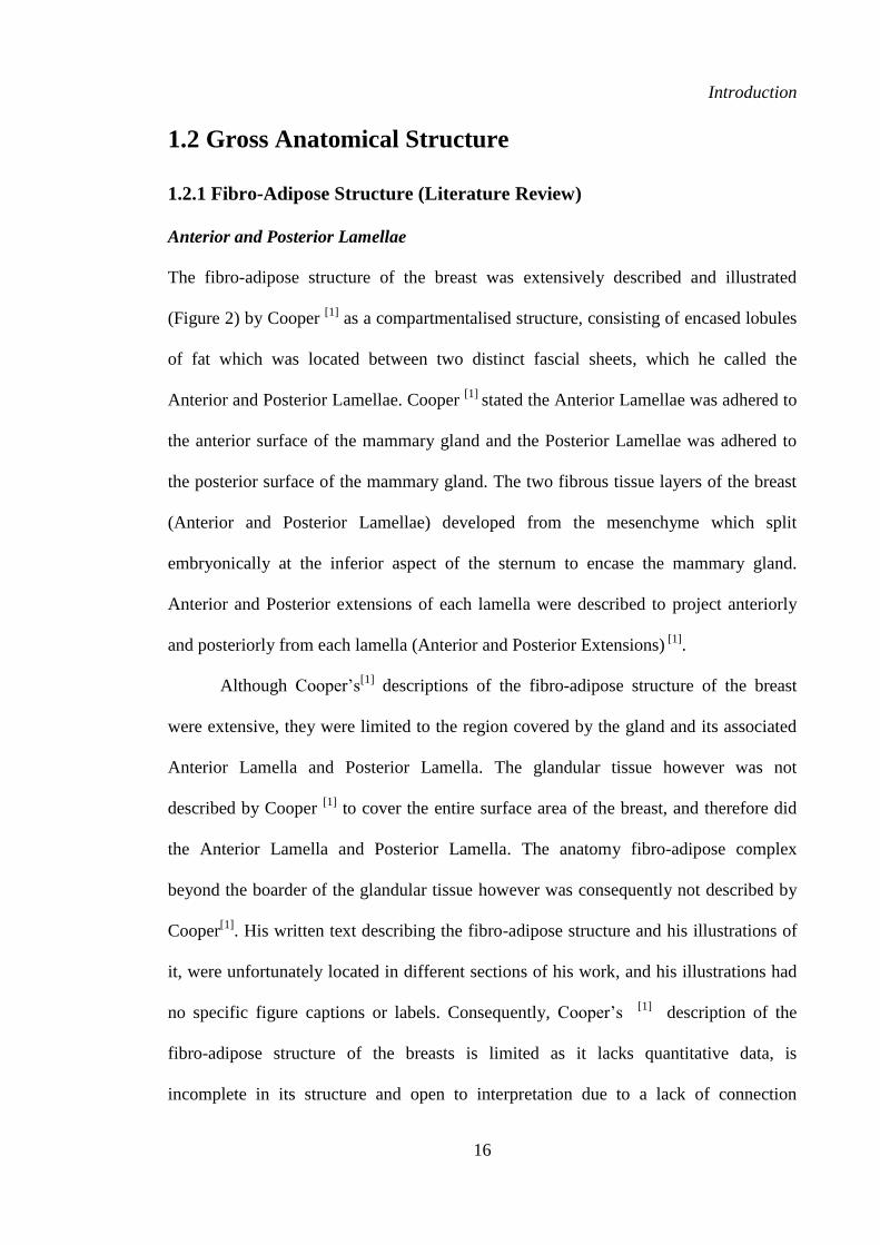
Anterior and Posterior Lamellae
The fibro-adipose structure of the breast was extensively described and illustrated
(Figure 2) by Cooper [1]
as a compartmentalised structure, consisting of encased lobules
of fat which was located between two distinct fascial sheets, which he called the
Anterior and Posterior Lamellae. Cooper [1]
stated the Anterior Lamellae was adhered to
the anterior surface of the mammary gland and the Posterior Lamellae was adhered to
the posterior surface of the mammary gland. The two fibrous tissue layers of the breast
(Anterior and Posterior Lamellae) developed from the mesenchyme which split
embryonically at the inferior aspect of the sternum to encase the mammary gland.
Anterior and Posterior extensions of each lamella were described to project anteriorly
and posteriorly from each lamella (Anterior and Posterior Extensions) [1]
.
Although Cooper’s[1]
descriptions of the fibro-adipose structure of the breast
were extensive, they were limited to the region covered by the gland and its associated
Anterior Lamella and Posterior Lamella. The glandular tissue however was not
described by Cooper [1]
to cover the entire surface area of the breast, and therefore did
the Anterior Lamella and Posterior Lamella. The anatomy fibro-adipose complex
beyond the boarder of the glandular tissue however was consequently not described by
Cooper[1]
. His written text describing the fibro-adipose structure and his illustrations of
it, were unfortunately located in different sections of his work, and his illustrations had
no specific figure captions or labels. Consequently, Cooper’s [1]
description of the
fibro-adipose structure of the breasts is limited as it lacks quantitative data, is
incomplete in its structure and open to interpretation due to a lack of connection
Introduction
17
Intro
ductio
n
between his illustrations and his written text.
Since Cooper, only eight cadaveric dissection studies have investigated the
fibro-adipose tissue of the breast [36-42, 45]
. Many of these studies visualised, in
consensus with Cooper, a dual layer of fibrous tissue that encased the gland both
anterior and posteriorly. Surprisingly, the studies since Cooper [1]
did not refer to theses
layers as the Anterior and Posterior Lamellae. The first study to investigate the
superficial fascial system of the body occurred 150 years after Cooper’s work [40]
. The
study included both cadaveric and surgical dissections of the fascial structure of 12
fresh and embalmed cadavers and 20 body contour patients (patients undergoing
cosmetic surgery). The dissections were cross-sectional dissections of both the
anatomical and surgical specimens in various parts of the body, including the breast.
The superficial fascia system of the various parts of the body was found to consistently
have a dual layer of connective tissue, which extended from the skin to the underlying
muscle fascia [40]
. This fascial network was found to be orientated horizontal to the skin,
separated by layers of fat of varying thicknesses. The dual layers were joined
perpendicularly by interconnecting fibrous septa[40]
much like the dual lamellae
structure of the breast described by Cooper [1]
. Lockwood et. al (1991)[40]
suggested the
primary function of the fascial network was to encase, support and shape the
subcutaneous fat of the body and anchor it to the underlying tissues. Although the
fascial system of the breast was also described to have this dual layer fascial
organisation, [40]
the study did not compare or refer this organisation to Cooper’s [1]
descriptions of the breast and therefore these dual fascial layers were not referred to as
the Anterior Lamellae and Posterior Lamellae. Nor were the perpendicular
interconnecting fibred referred to as Cooper’s [1]
Anterior and Posterior Extensions
despite the similarities between Lockwood’s and Cooper’s [1]
findings. Consequently
Introduction
18
Intro
ductio
n
this nomenclature did not allow the descriptions to be translated to anatomical
descriptions of the breast in anatomy textbooks despite the verification of Cooper’s
findings.
Although Cooper [1]
stated that the Anterior and Posterior Lamellae encased the
mammary gland, he did not consider them to act as a capsule to the breast. In contrast
with Cooper [1]
, three dissection studies[41, 42, 45]
have described a distinct breast capsule.
Unfortunately, none of these studies related the anterior and posterior breast capsules
that they found to the Anterior and Posterior Lamellae described by Cooper. Riggio et.
al. (2000)[45]
investigated the fibro-adipose structure of six cadavers (three male and
three female) and 21 plastic surgery patients. The study[45]
reported that superficial
fascial layers around the breast did not split to encapsulate the breast but rather sat
posterior to a breast capsule. A second study [42]
conducted on eight fresh female
cadavers, two embalmed female and two fresh male cadavers (age 77±15 years), also
described two membranes that encapsulated the mammary gland anteriorly and
posteriorly, with interconnections between the capsule walls consistent with Cooper’s
[1] descriptions of the Posterior Extensions of the Anterior Lamellae and the Anterior
extensions of the Posterior Lamellae [42]
. Unfortunately they also did not use any of
Cooper’s [1]
terminology for these fascial structures and re-named all of Coopers[1]
extensions “Suspensory Ligaments”. They did extend Coopers work however by
describing the fascial structure beyond the boarder of the gland. They described the
fascia of the anterior capsule to fuse with the superficial fascial system in the inferior
aspect of the breast beyond the boarder of the gland [42]
. Neither study [42, 45]
however
provided any quantitative data to support their descriptions of the fibro-adipose
structures of the breast.
More detail of the fibro-adipose structure beyond the perimeter of the gland
Introduction
19
Intro
ductio
n
inferiorly was provided in an anatomical study conducted in 2014 on 40 cadavers using
blunt and sharp dissections, with sagittal cross sections [41]
. This study also described a
well-defined breast capsule superior to the level of the fourth rib. Inferior to the 4th
rib
the capsule became obscured by glandular tissue and ducts forming the nipple-areola
complex, a system which was described to have direct dermal insertions of ligamentous
tissue, anchoring the breast to the 5th
rib [41]
. Unfortunately, the study did not comment
on the organisation of the breast capsule or fibro-adipose structure beyond the glandular
tissue in the medial, lateral and superior breast regions. The study did however relate
the anterior and posterior breast capsule they found to Cooper’s [1]
Anterior Lamella,
and Posterior Lamellae. The posterior capsule was described to function as a gliding
plane between the pectoral fascia and the breast [41]
. Furthermore, they did not specify
the location where the fascial layers split to encase the gland, refer to Cooper’s [1]
Extensions of the breast or provide quantitative data to verify their descriptions.
A limitation of most previous studies describing the fascial structure of the
breast has been the direct link of the breast capsule/Lamellae to the gland when the
gland does not cover the entire surface area of the breast. Therefore the fibro-adipose
structure beyond the gland has not been described and there has been little anatomical
quantitative data collected on the surface area of the breast that the gland actually
covers. Further research is therefore required to investigate and quantify the fascial
structure beyond the gland.
In vivo studies support the notion of an anterior and posterior breast capsule
(Cooper’s [1]
Anterior and Posterior Lamellae), with ultrasound studies finding the
breast parenchyma to be enveloped by a superficial and deep fascial plane [70, 71]
.
Pearson et al [71]
described the connective tissue and fascia as bright reflectors what run
between and interconnect the fascial planes, encasing the adipose tissue [70, 71]
.
Introduction
20
Intro
ductio
n
Unfortunately the visualisation of the detail of the fibro-adipose structure with
ultrasound is limited due to the highly reflective nature of the fascial tissue. Therefore,
although these studies confirm the existence of the fibro-adipose structure within a
fibrous capsule, they provide little detail of its structure.
Due to the inconsistencies in the nomenclature of the fascial tissue laying
anterior and posterior to the gland (Anterior/Posterior breast capsule versus
Anterior/Posterior Lamellae), the lack of information about the fibro-adipose structure
beyond the borders of the gland and the limited detail of the fibro-adipose structure that
can be visualised in vivo by technology-based research, further anatomical research of
the fibro-adipose tissue is required.
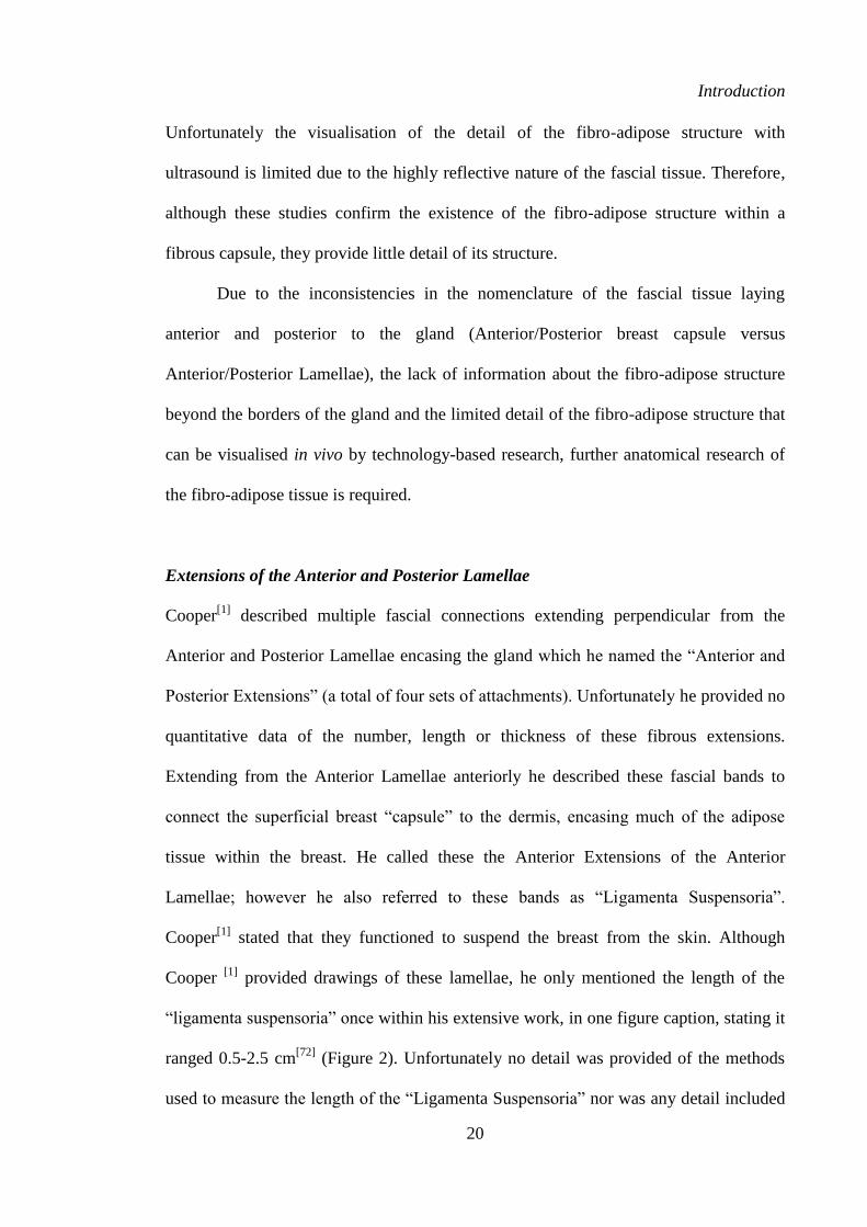
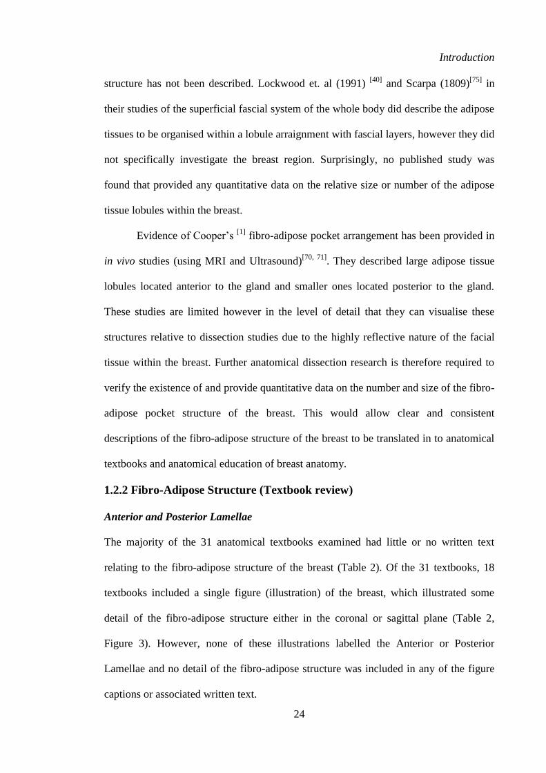
Extensions of the Anterior and Posterior Lamellae
Cooper[1]
described multiple fascial connections extending perpendicular from the
Anterior and Posterior Lamellae encasing the gland which he named the “Anterior and
Posterior Extensions” (a total of four sets of attachments). Unfortunately he provided no
quantitative data of the number, length or thickness of these fibrous extensions.
Extending from the Anterior Lamellae anteriorly he described these fascial bands to
connect the superficial breast “capsule” to the dermis, encasing much of the adipose
tissue within the breast. He called these the Anterior Extensions of the Anterior
Lamellae; however he also referred to these bands as “Ligamenta Suspensoria”.
Cooper[1]
stated that they functioned to suspend the breast from the skin. Although
Cooper [1]
provided drawings of these lamellae, he only mentioned the length of the
“ligamenta suspensoria” once within his extensive work, in one figure caption, stating it
ranged 0.5-2.5 cm[72]
(Figure 2). Unfortunately no detail was provided of the methods
used to measure the length of the “Ligamenta Suspensoria” nor was any detail included
Introduction
21
Intro
ductio
n
on the number of measurements recorded at the time.
No studies since Cooper [1]
have specifically investigated to the Anterior
Extensions of the Anterior Lamellae however many studies have described short fibrous
extensions connecting the anterior gland to the dermis in their illustrations[36-39]
.
Unfortunately these have not been labelled specifically but rather all of the fascial
within the breast has been referred to as a group as Cooper’s [1]
Ligaments (or
Suspensory ligaments). This has led to confusion in the location and existence of the
fibro-adipose structure within the breast. Furthermore, there is no quantitative data
amongst the dissection studies to clarify the location, structure and relative distance that
each set of extensions spans to verify the work of Cooper [1]
.
Five dissection studies [36-39]
have mentioned the “Suspensory ligaments”
although they were not specifically investigating them or the fibro-adipose structure of
the breast. One study[45]
described Cooper’s [1]
Anterior Extensions of the Anterior
Lamellae, however named them “Coopers Ligaments”. Similar to the Anterior
Extensions of the Anterior Lamellae, Riggio et. al. (2000)[45]
described “Coopers
Ligaments” connected the superficial fascial system to the skin directly. A dense bundle
of fibres spanning anteriorly from the superficial breast to the dermis was also identified
by Komiya et. al. (2015) [39]
in their dissections of the nipple-areolar complex (n=5
cadaveric breasts). The authors suggested that the purpose of this tissue is to act as a site
for peri-areolar attachment in surgery [39]
.
Extending posteriorly from the Posterior Lamellae Cooper described fibrous
bands which connected the breast to the chest wall via the Superficial Pectoral Fascia.
He named these the Posterior Extensions of the Posterior Lamella. Cooper [1]
suggested
that this complex fascial arrangement encasing the adipose tissue was essential to allow
the breast to swing in space and yield to pressure and violence[1]
.
Introduction
22
Intro
ductio
n
Cardoso et al. (2015) [36]
in a study using liposuction of adipose tissue described
the same fibrous structure in 14 breasts (age 66.7; range: 39-85 years) attaching the
breast to the chest wall but he did not refer to them as the Posterior Extensions of the
Posterior Lamellae. The Posterior Extensions of the Posterior Lamellae were also
briefly described, in a study of the superficial pectoral fascia, as weak and easy to
bluntly dissect [37]
. Though again they were not named or references to Cooper’s
Posterior Extensions of the Posterior Lamellae. Cooper also described perpendicular
fascial bands running from the Lamellae that penetrated the gland and helped to provide
structure to it. Extending posteriorly from the Anterior Lamellae he named them the
Posterior Extensions of the Anterior Lamellae and extending anteriorly from the
Posterior Lamellae he named them the Anterior Extensions of the Anterior Lamellae.
These fascial extensions were described to interconnect within the gland and functioned
to hold the glandular tissue together. These have een identified in dissection studies
since Cooper as part of the gross anatomy of the gland. They have been referred to as
fibro-glandular tissue and considered to be part of the support structure for the gland
rather than part of the fibro-adipose tissue of the breast.
Cooper’s [1]
Extensions were also identified in a study that compared the
ultrasound image of a coronal breast slice to the anatomy of the same breast slice.
Although these studies confirm that the four sets of fibrous tissue extensions exists,
further anatomical dissection research specifically investigating these extensions is
required that collates the various names of these tissues to allow the fibrous tissues
within the breast to be clearly understood and taught.
Not only have the names of the fascial interconnections of the breast varied in
the published literature, so has their physical dimensions. They have been described to
be “large, strong and numerous” structures, “tentacle like qualities, extending in all
Introduction
23
Intro
ductio
n
directions from the anterior fascia layer (lamellae) to the skin with no discernible
organised structure” [73, 74]
, and “fibrous bands of connective tissues that travel
throughout the breast and intersect with the dermis perpendicularly” [68]
. This confusion
in the physical structure of these fascial tissues within the breast has translated into
anatomical textbooks creating confusion regarding the structure of the fibro-adipose
anatomy of the breast. Further research is therefore required to provide quantitative data
to support the descriptions of the fibro-adipose structure of the breast so that the breast
anatomy can be consistently and accurately taught and illustrated in anatomical
textbooks
Fibro-Adipose Pockets Anterior and Posterior to the Gland
Cooper [1]
stated that the Anterior Extensions of the Anterior Lamella and the Posterior
Extensions of the Posterior Lamella formed multiple fascial pockets, which encased the
adipose lobules and organised the adipose tissue into compartments located anterior and
posterior to the gland [1]
. Unfortunately Cooper [1]
provided no quantitative data
regarding the number or size of the adipose tissue pockets in each region, only that the
adipose tissue pockets located anterior to the gland were larger and less numerous than
those located posterior to the gland. Cooper [1]
also described the presence of additional
adipose tissue pockets which were even fewer in number and size which were
embedded between the lobules of the mammary gland. These may have been formed by
the Posterior Extensions of the Anterior Lamella and the Anterior Extensions of the
Posterior Lamella; however this was not specifically dissected.
No previously published anatomical or surgical studies were found that have
investigated the fibro-adipose pocket structure of the breast to verify or dispute
Cooper’s[1]
descriptions. Some sagittal illustrations of the breast have illustrated adipose
tissue confined within the suspensory ligaments however a pocket-like fibro-adipose
Introduction
24
Intro
ductio
n
structure has not been described. Lockwood et. al (1991) [40]
and Scarpa (1809)[75]
in
their studies of the superficial fascial system of the whole body did describe the adipose
tissues to be organised within a lobule arraignment with fascial layers, however they did
not specifically investigate the breast region. Surprisingly, no published study was
found that provided any quantitative data on the relative size or number of the adipose
tissue lobules within the breast.
Evidence of Cooper’s [1]
fibro-adipose pocket arrangement has been provided in
in vivo studies (using MRI and Ultrasound)[70, 71]
. They described large adipose tissue
lobules located anterior to the gland and smaller ones located posterior to the gland.
These studies are limited however in the level of detail that they can visualise these
structures relative to dissection studies due to the highly reflective nature of the facial
tissue within the breast. Further anatomical dissection research is therefore required to
verify the existence of and provide quantitative data on the number and size of the fibro-
adipose pocket structure of the breast. This would allow clear and consistent
descriptions of the fibro-adipose structure of the breast to be translated in to anatomical
textbooks and anatomical education of breast anatomy.
1.2.2 Fibro-Adipose Structure (Textbook review)
Anterior and Posterior Lamellae
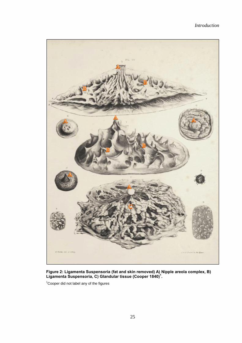
The majority of the 31 anatomical textbooks examined had little or no written text
relating to the fibro-adipose structure of the breast (Table 2). Of the 31 textbooks, 18
textbooks included a single figure (illustration) of the breast, which illustrated some
detail of the fibro-adipose structure either in the coronal or sagittal plane (Table 2,
Figure 3). However, none of these illustrations labelled the Anterior or Posterior
Lamellae and no detail of the fibro-adipose structure was included in any of the figure
captions or associated written text.
Introduction
25
Intro
ductio
n
Figure 2: Ligamenta Suspensoria (fat and skin removed) A) Nipple areola complex, B) Ligamenta Suspensoria, C) Glandular tissue (Cooper 1840)
1.
1Cooper did not label any of the figures
Introduction
26
Intro
ductio
n
Figure 3 Example of textbook representation of the fibro-adipose sturucture of the breast (Moore, Agur et. al. (2015)[21])
Introduction
27
Intro
ductio
n
Extensions of the Anterior and Posterior Lamellae
Although Cooper’s [1]
extensions from the Anterior or Posterior Lamellae appeared in
both the anatomical illustrations and the associated written text, there was considerable
variation in what they were called, their physical arrangement within the breast and in
their location (their origin and insertion). Rather than Anterior or Posterior Extensions,
the fascial tissue of the whole breast was generally referred to as “Coopers Ligaments”
or “Suspensory Ligaments”. These inconsistent terminologies also create further
confusion as Cooper’s [1]
used the name “suspensory ligaments” to specifically refer to
the Anterior Extensions of the Anterior Lamellae.
The arrangement and location of the Anterior or Posterior Extensions in the 19
textbooks that either mentioned or illustrated “Coopers Ligaments” or “suspensory
ligaments” (Table 2), was also inconsistent. Seven textbooks displayed the ligaments to
span from the dermis to the glandular tissue [13-17, 29, 32]
while eight other textbooks
displayed them spanning from the dermis to the superficial pectoral fascia [6, 10, 11, 21, 22,
26, 30, 31]. The four remaining textbooks did not clearly show where the ligaments
spanned from or connected to. Consequently, as the naming, location and relative length
difference of these ligaments within the breast in anatomical textbooks is inconsistent
and unclear, the current status of university-based anatomical education and the
understanding of the fibro-adipose structure of the breast of medical and allied health
graduates is generally poor.
Fibro-Adipose Pockets Anterior and Posterior to the Gland
The fibro-adipose pocket arrangement of the breast was illustrated in a coronal
illustration in only two of the 31 textbooks included in the summary [4, 16]
. Neither of
these textbooks had detailed accompanying text describing this structure. Only four
textbooks made any reference to an organised fibro-adipose structure (Table 2) but no
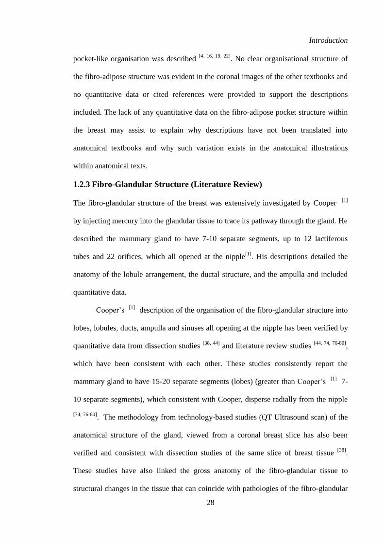
Introduction
28
Intro
ductio
n
pocket-like organisation was described [4, 16, 19, 22]
. No clear organisational structure of
the fibro-adipose structure was evident in the coronal images of the other textbooks and
no quantitative data or cited references were provided to support the descriptions
included. The lack of any quantitative data on the fibro-adipose pocket structure within
the breast may assist to explain why descriptions have not been translated into
anatomical textbooks and why such variation exists in the anatomical illustrations
within anatomical texts.
1.2.3 Fibro-Glandular Structure (Literature Review)
The fibro-glandular structure of the breast was extensively investigated by Cooper [1]
by injecting mercury into the glandular tissue to trace its pathway through the gland. He
described the mammary gland to have 7-10 separate segments, up to 12 lactiferous
tubes and 22 orifices, which all opened at the nipple[1]
. His descriptions detailed the
anatomy of the lobule arrangement, the ductal structure, and the ampulla and included
quantitative data.
Cooper’s [1]
description of the organisation of the fibro-glandular structure into
lobes, lobules, ducts, ampulla and sinuses all opening at the nipple has been verified by
quantitative data from dissection studies [38, 44]
and literature review studies [44, 74, 76-80]
,
which have been consistent with each other. These studies consistently report the
mammary gland to have 15-20 separate segments (lobes) (greater than Cooper’s [1]
7-
10 separate segments), which consistent with Cooper, disperse radially from the nipple
[74, 76-80]. The methodology from technology-based studies (QT Ultrasound scan) of the
anatomical structure of the gland, viewed from a coronal breast slice has also been
verified and consistent with dissection studies of the same slice of breast tissue [38]
.
These studies have also linked the gross anatomy of the fibro-glandular tissue to
structural changes in the tissue that can coincide with pathologies of the fibro-glandular
Introduction
29
Intro
ductio
n
tissue. The consistency of the anatomical descriptions of the gross anatomy of the fibro-
glandular tissue, supported by quantitative data has enabled the detail of the gross
anatomy of the fibro-glandular tissue to be translated into anatomical illustrations and
written text within both the published literature and anatomical textbooks. In contrast to
illustrations of the fibro-adipose structure of the breast, these anatomical illustrations
have labels, associated figure captions and associated written text.
1.2.4 Fibro-Glandular Structure (Textbook review)
The gross anatomy of the fibro-glandular tissue and its organisation regarding the
connection of lobes, lobules and ducts opening at the nipple structure was described in
24 of the 31 textbooks included within the textbook review (Table 2). These
descriptions were consistent with each other and the descriptions of Cooper[1]
and more
recent anatomical studies. Minor variations were found regarding the classification of
the mammary gland, the number of lobes and the location of the glandular tissue.
Twelve anatomical texts described the mammary gland as a modified sweat gland [7, 13,
14, 17, 19, 21-23, 26, 27, 30, 31]; 21 describe the gland to consist of 15-20 segments (lobes)
[2, 4-7,
12, 13, 15-17, 19-22, 26-28, 30-32]; and nine (9) texts described the mammary gland tissue to be
separated by a fibrous septa [4-6, 9, 17, 20, 26, 28]
.
The location of the fibro-glandular tissue within the breast had the greatest
variation amongst the textbook illustrations, mainly evident from the sagittal view of the
breast. Eight textbooks illustrated the fibro-glandular tissue to be encased between two
layers of fibro-adipose tissue [5, 13, 14, 26, 29-32]
, five displayed it directly anterior to the
superficial pectoral fascia (with only fibro-adipose tissue anterior) [10, 11, 16, 17]
, and four
had no discernible difference between the location of the fibro-adipose and fibro-
glandular tissues of the breast [4, 6, 21, 22]
. There was considerable variation noted within
the anatomical illustration amongst the textbooks regarding the relative percentage of
Introduction
30
Intro
ductio
n
the total composition of the breast with no two images comparable.
The gross anatomy of the fibro-glandular tissue has been consistently described
and included within the majority of anatomical textbooks, supported by quantitative
data. However, variation was found in the location of the glandular tissue relative to the
fibro-adipose structure within the breast, the surface area of the breast that the gland
covers and the percentage composition of the breast that is fibro-glandular tissue.
Further anatomical research is therefore required to provide data to verify these
anatomical descriptions in terms of the location, surface area and contribution of the
fibro-glandular tissue to the composition of the breast.
1.3 Attachment of the breast to the chest wall
1.3.1 Regional Anatomy (Literature Review)
Cooper [1]
described the breast to sit on the anterior chest wall between ribs 2-7 in the
superior-inferior directions, and between the sternum and mid-axillary line in the
medial-lateral direction. He stated that the posterior aspect of the breast sat on the
Pectoralis major muscle and the lateral edge of the breast was in contact with the
Serratus Anterior and External Oblique muscles [1]
. Unfortunately, he did not provide
data on the number or age of the female cadavers this described location was based on.
Of the 17 cadaveric dissection studies of the breast (Table 3), 10 studies
commented on the regional anatomy of the breast [34-37, 40-43, 45, 48]
which were all
consistent with Cooper’s [1]
descriptions. The only difference was in the addition of
another muscle having contact with the posterior wall of the breast, Rectus Abdominal
muscle [37]
. Although these studies verify the location and regional anatomy of the
breast on a greater number of cadavers (over 100 cadavers between the 10 studies) none
of the studies quantified the percentage of the ‘footprint’ of the breast that each muscle
Introduction
31
Intro
ductio
n
has contact with. Therefore, no anatomical data exists as evidence on the percentage
contribution of the superficial chest wall muscles (Pectoralis Major, Serratus Anterior,
External Oblique and Rectus Abdominus muscles) to the footprint of the breast.
The perimeter of the breast has also not been quantified in terms of its length by
Cooper [1]
or any dissection study since. Some data has been collected through in vivo
studies on the length of the inframammary fold, which was measured to be 10-12cm
long [77, 80]
and on the horizontal diameter of the breast, which has been found to be
14.3±1.4cm (range: 8.5–23.5cm [53]
). Although these measurements provide some
information of the perimeter of the breast, further quantitative data is required that
specifically measures the perimeter of the breast in relation to the surface area and
volume of the breast.
1.3.2 Regional Anatomy (Textbook review)
Sixteen textbooks included descriptions of the regional anatomy of the breast [2, 4, 6, 7, 9-14,
19, 21-23, 27], which again were consistent Cooper
[1] and the published literature
[1, 68, 74, 77-
80]. Of the 26 textbooks that contained coronal images, the regional anatomy was
consistently illustrated in 11 textbooks (Figure 7)[2, 4, 6, 7, 9, 14, 15, 22, 24, 25, 32]
. Only one
textbook illustrated the regional anatomy of the breast with a sagittal illustration [6]
. The
regional anatomy of the breast was consistent within the coronal and sagittal
illustrations regarding the muscles that the posterior wall of the breast was in contact
with (Pectoralis Major, Serratus Anterior, External Oblique and Rectus Abdominus),
however no quantitative data was provided however to verify the location of the breast,
and the percentage of area that each muscle occupies within the perimeter.
Within the written text however, there was some inconsistency regarding the
muscles that the posterior wall of the breast was in contact with. Nineteen of the 31
textbooks (Table 2) described the muscles which were located directly posterior to the
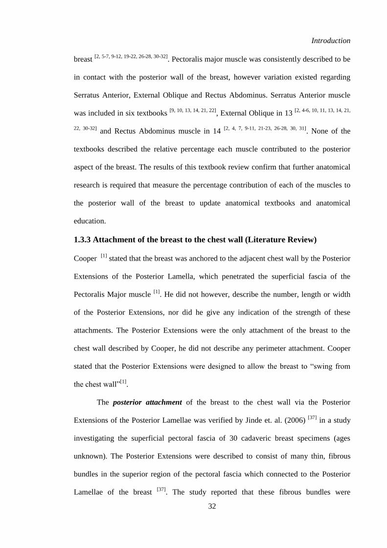
Introduction
32
Intro
ductio
n
breast [2, 5-7, 9-12, 19-22, 26-28, 30-32]
. Pectoralis major muscle was consistently described to be
in contact with the posterior wall of the breast, however variation existed regarding
Serratus Anterior, External Oblique and Rectus Abdominus. Serratus Anterior muscle
was included in six textbooks [9, 10, 13, 14, 21, 22]
, External Oblique in 13 [2, 4-6, 10, 11, 13, 14, 21,
22, 30-32] and Rectus Abdominus muscle in 14
[2, 4, 7, 9-11, 21-23, 26-28, 30, 31]. None of the
textbooks described the relative percentage each muscle contributed to the posterior
aspect of the breast. The results of this textbook review confirm that further anatomical
research is required that measure the percentage contribution of each of the muscles to
the posterior wall of the breast to update anatomical textbooks and anatomical
education.
1.3.3 Attachment of the breast to the chest wall (Literature Review)
Cooper [1]
stated that the breast was anchored to the adjacent chest wall by the Posterior
Extensions of the Posterior Lamella, which penetrated the superficial fascia of the
Pectoralis Major muscle [1]
. He did not however, describe the number, length or width
of the Posterior Extensions, nor did he give any indication of the strength of these
attachments. The Posterior Extensions were the only attachment of the breast to the
chest wall described by Cooper, he did not describe any perimeter attachment. Cooper
stated that the Posterior Extensions were designed to allow the breast to “swing from
the chest wall”[1]
.
The posterior attachment of the breast to the chest wall via the Posterior
Extensions of the Posterior Lamellae was verified by Jinde et. al. (2006) [37]
in a study
investigating the superficial pectoral fascia of 30 cadaveric breast specimens (ages
unknown). The Posterior Extensions were described to consist of many thin, fibrous
bundles in the superior region of the pectoral fascia which connected to the Posterior
Lamellae of the breast [37]
. The study reported that these fibrous bundles were
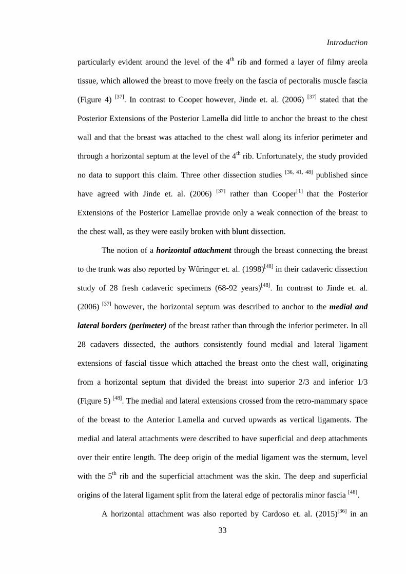
Introduction
33
Intro
ductio
n
particularly evident around the level of the 4th
rib and formed a layer of filmy areola
tissue, which allowed the breast to move freely on the fascia of pectoralis muscle fascia
(Figure 4) [37]
. In contrast to Cooper however, Jinde et. al. (2006) [37]
stated that the
Posterior Extensions of the Posterior Lamella did little to anchor the breast to the chest
wall and that the breast was attached to the chest wall along its inferior perimeter and
through a horizontal septum at the level of the 4th
rib. Unfortunately, the study provided
no data to support this claim. Three other dissection studies [36, 41, 48]
published since
have agreed with Jinde et. al. (2006) [37]
rather than Cooper[1]
that the Posterior
Extensions of the Posterior Lamellae provide only a weak connection of the breast to
the chest wall, as they were easily broken with blunt dissection.
The notion of a horizontal attachment through the breast connecting the breast
to the trunk was also reported by Wűringer et. al. (1998)[48]
in their cadaveric dissection
study of 28 fresh cadaveric specimens (68-92 years)[48]
. In contrast to Jinde et. al.
(2006) [37]
however, the horizontal septum was described to anchor to the medial and
lateral borders (perimeter) of the breast rather than through the inferior perimeter. In all
28 cadavers dissected, the authors consistently found medial and lateral ligament
extensions of fascial tissue which attached the breast onto the chest wall, originating
from a horizontal septum that divided the breast into superior 2/3 and inferior 1/3
(Figure 5) [48]
. The medial and lateral extensions crossed from the retro-mammary space
of the breast to the Anterior Lamella and curved upwards as vertical ligaments. The
medial and lateral attachments were described to have superficial and deep attachments
over their entire length. The deep origin of the medial ligament was the sternum, level
with the 5th
rib and the superficial attachment was the skin. The deep and superficial
origins of the lateral ligament split from the lateral edge of pectoralis minor fascia [48]
.
A horizontal attachment was also reported by Cardoso et. al. (2015)[36]
in an
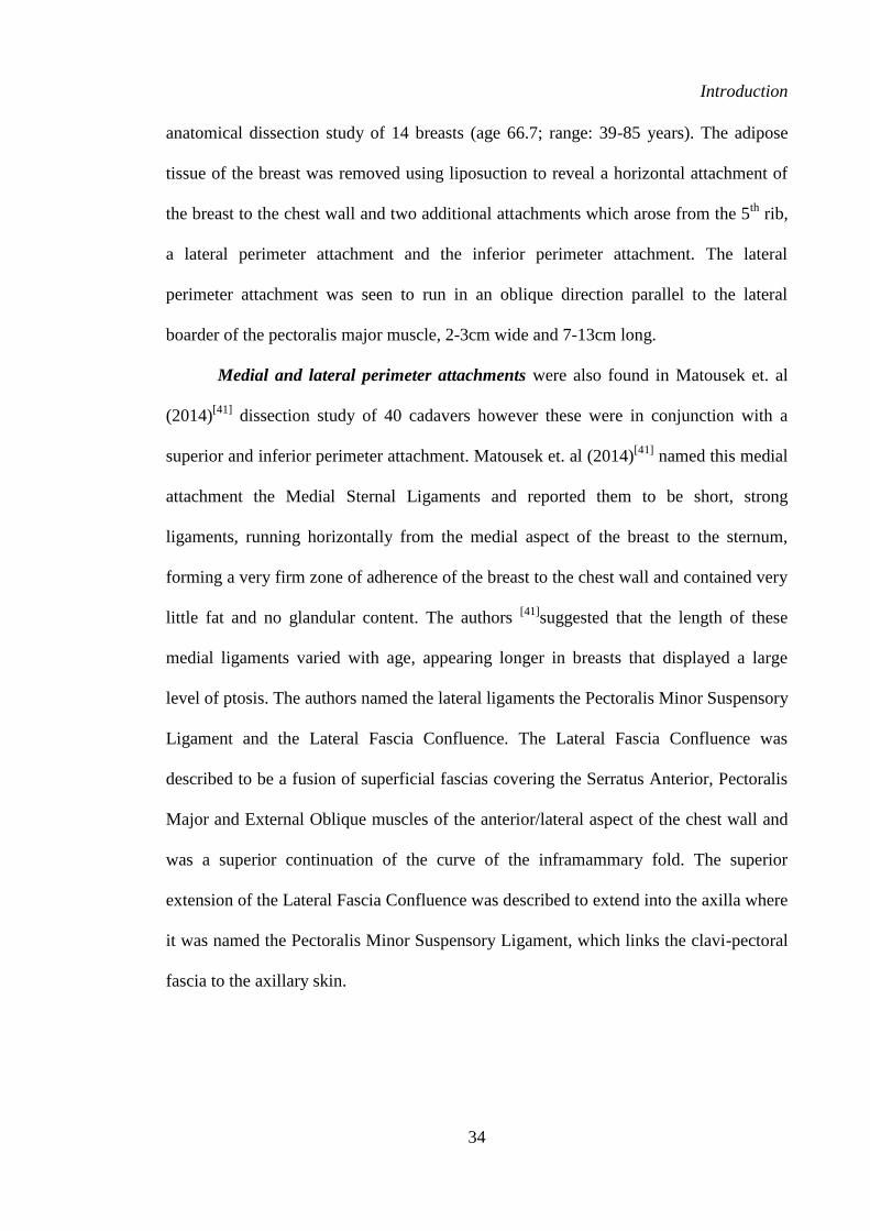
Introduction
34
Intro
ductio
n
anatomical dissection study of 14 breasts (age 66.7; range: 39-85 years). The adipose
tissue of the breast was removed using liposuction to reveal a horizontal attachment of
the breast to the chest wall and two additional attachments which arose from the 5th
rib,
a lateral perimeter attachment and the inferior perimeter attachment. The lateral
perimeter attachment was seen to run in an oblique direction parallel to the lateral
boarder of the pectoralis major muscle, 2-3cm wide and 7-13cm long.
Medial and lateral perimeter attachments were also found in Matousek et. al
(2014)[41]
dissection study of 40 cadavers however these were in conjunction with a
superior and inferior perimeter attachment. Matousek et. al (2014)[41]
named this medial
attachment the Medial Sternal Ligaments and reported them to be short, strong
ligaments, running horizontally from the medial aspect of the breast to the sternum,
forming a very firm zone of adherence of the breast to the chest wall and contained very
little fat and no glandular content. The authors [41]
suggested that the length of these
medial ligaments varied with age, appearing longer in breasts that displayed a large
level of ptosis. The authors named the lateral ligaments the Pectoralis Minor Suspensory
Ligament and the Lateral Fascia Confluence. The Lateral Fascia Confluence was
described to be a fusion of superficial fascias covering the Serratus Anterior, Pectoralis
Major and External Oblique muscles of the anterior/lateral aspect of the chest wall and
was a superior continuation of the curve of the inframammary fold. The superior
extension of the Lateral Fascia Confluence was described to extend into the axilla where
it was named the Pectoralis Minor Suspensory Ligament, which links the clavi-pectoral
fascia to the axillary skin.

Introduction
35
Intro
ductio
n
Figure 4: Cadaveric breast dissection showing A) Glandular tissue, B)
Superficial pectoral fascia, C) Posterior Extensions of the Posterior
Lamellae, D) Pectoralis major muscle (Jinde et. al 2006).
Figure 5: Horizontal septum spanning medio-laterally from the 5th
rib
to the nipple (Wűringer et. al. 1998).
Introduction
36
Intro
ductio
n
There has been some debate in the literature regarding the inferior perimeter
attachment of the breast to the chest wall, or the inframammary fold. Cooper [1]
did not
specifically mention the inframammary fold, however 10 studies since Cooper [1, 34-36,
41-43, 45, 48, 81, 82] (2 surgical and eight cadaveric) have investigated it. The 10 studies
agreed that the inframammary fold formed the inferior perimeter attachment of the
breast where fibrous tissue connected the chest wall with the dermis at the lower
curvature of the breast [34, 43, 83]
(Figure 6).
Although there is consensus on the presence of the inframammary fold, conflict
exists over the orientation and attachment of these fibres within the inframammary fold.
Three studies (two cadaver and one surgical) describe fibres orientated in the sagittal
plane, running between the superficial pectoral fascia level with the 5/6th
rib and
inserting into the deep dermis [34, 45, 81]
. Another three studies (one surgical and two
cadaver) [35, 48, 82]
describe transverse orientated fibres spanning from the mediolateral
width of the inframammary fold region, connecting into the fascia of the sternum [45, 82]
.
There is some consensus that fibres within the inframammary fold attach to the
dermis. Lockwood et. al. (1991)[40]
, in his study of the superficial fascial system of the
body, described the skin to have zones of adherence, where the topography of the skin
changed due to adherence of the skin to the underlying fascia to the dermis, the study
described the inframammary fold at the level of the 5th
rib to be one of these zones. One
of the most recent cadaveric studies of 40 cadavers, Matousek et.al (2014)[41]
verified
Lockwood’s dual ligament system at the inframammary fold, which provided support
and anchorage to the dermis above and below the region of the inframammary fold. A
study of 74 breast reconstruction patients [67]
also reported the ligamentous part of the
inframammary fold connected the deep dermis to the superficial pectoral fascia at the
level of the 5th
/6th
rib. Studies report that surgical re-connection of these attachments of
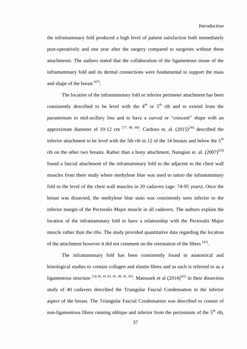
Introduction
37
Intro
ductio
n
the inframammary fold produced a high level of patient satisfaction both immediately
post-operatively and one year after the surgery compared to surgeries without these
attachments. The authors stated that the collaboration of the ligamentous tissue of the
inframammary fold and its dermal connections were fundamental to support the mass
and shape of the breast [67]
.
The location of the inframammary fold or inferior perimeter attachment has been
consistently described to be level with the 4th
or 5th
rib and to extend from the
parasternum to mid-axillary line and to have a curved or “crescent” shape with an
approximate diameter of 10-12 cm [77, 80, 84]
. Cardoso et. al. (2015)[36]
described the
inferior attachment to be level with the 5th rib in 12 of the 14 breasts and below the 5th
rib on the other two breasts. Rather than a bony attachment, Nanigian et. al. (2007)[43]
found a fascial attachment of the inframammary fold to the adjacent to the chest wall
muscles from there study where methylene blue was used to tattoo the inframammary
fold to the level of the chest wall muscles in 20 cadavers (age: 74-95 years). Once the
breast was dissected, the methylene blue stain was consistently seen inferior to the
inferior margin of the Pectoralis Major muscle in all cadavers. The authors explain the
location of the inframammary fold to have a relationship with the Pectoralis Major
muscle rather than the ribs. The study provided quantitative data regarding the location
of the attachment however it did not comment on the orientation of the fibres [43]
.
The inframammary fold has been consistently found in anatomical and
histological studies to contain collagen and elastin fibres and as such is referred to as a
ligamentous structure [34-36, 41-43, 45, 48, 81, 82]
. Matousek et al (2014)[41]
in their dissection
study of 40 cadavers described the Triangular Fascial Condensation in the inferior
aspect of the breast. The Triangular Fascial Condensation was described to consist of
non-ligamentous fibres running oblique and inferior from the periosteum of the 5th
rib,
Introduction
38
Intro
ductio
n
which fanned out in a triangular fashion and inserted into the dermis. Horizontal fibres
were also identified running between the deep Rectus Abdominal fascia and the dermis.
Regardless of the precise location, orientation or histological aspects of the
inframammary fold, breast surgeons consider it to be very influential to breast ptosis,
the shape of the inferior pole of the breast and the position of the breast mound on the
chest wall. As such, the inframammary fold has also been researched extensively in in
vivo studies [67, 85]
, as preservation of the inframammary fold in surgery is essential to
maintain the natural appearance and the aesthetics of a reconstructed breast [35, 67]
.
Only one published study was found that described the superior perimeter
attachment of the breast to the chest wall [41]
. The dissection study of 40 female
cadavers using blunt and sharp dissections described the attachment of the breast to the
chest wall along its entire perimeter. The authors also reported the perimeter to be the
primary anchor of the breast to the chest wall. The authors identified numerous
ligamentous attachments along the entire perimeter of the breast in 1.5-3cm sagittal
cross sections of the breast [41]
. The study named two superior attachments of the
perimeter, the Superficial Clavicular Ligament which connected the superior aspect of
the breast to the superficial pectoral fascia and the Deep Clavicular ligaments which
was located between the sternal and clavicular heads of the pectoralis major muscle and
attach to the inferior portion of the clavicle. The study did not speficiy however the
number of cadavers these perimeter attachments were found in or provide any
quantitative data of the perimeter attachments..
There is some consensus in the literature that the breast attaches to the chest wall
via both the Posterior Extensions of the Posterior Lamellae and along its perimeter, with
the perimeter attachment being the stronger anchor of the breast to the chest wall. There
is conflict however regarding the structure of the perimeter attachment. That is, whether
Introduction
39
Intro
ductio
n
the perimeter attachment is connected along its entire length or only in some sections.
Although there is consensus on the presence and importance of the infra-mammary fold
to the shape and support of the breast and its connection to the dermis, there is conflict
over its location relative to the ribs and the Pectorals Major muscle. Lastly, although it
is agreed that the Inframammary fold consists of collagen and elastin fibres, the
structure and naming of the triangular condensation of fascia connecting to the 5th
rib is
also a new concept. Therefore, further anatomical research is warranted to investigate
the inconsistencies in the literature regarding the perimeter attachments of the breasts to
the chest wall and the relative strength off the perimeter and Posterior Extensions of the
Posterior Lamellae attachments.
1.3.4 Attachment of the breast to the chest wall (Textbook review)
Despite some of the consistencies in the literature regarding the existence of the
posterior wall attachments and the perimeter attachments of the breast to the chest wall,
very little information of the attachments of the breast to the chest wall has been
translated into anatomical textbooks. Eight textbooks show in their illustrations a
connection of the breast to the Superficial Pectoral Fascia, however this connection
often runs the entire way through the breast to the dermis and cannot therefore be
differentiated from the fibro-adipose tissues, particularly the Posterior Extensions of the
Posterior Lamellae [6, 10, 11, 21, 22, 26, 30, 31]
. Although 22 of the 31 textbooks examined
mentioned the fact that the breast does attach to the chest wall, they failed to describe or
illustrate just how this occurs (posterior or perimeter attachment). Therefore, anatomical
research on the attachments of the breast to the chest wall via both the posterior wall
and the perimeter is required to provide a clear and concise description of the
attachments that can be translated into anatomy textbooks and anatomical education.
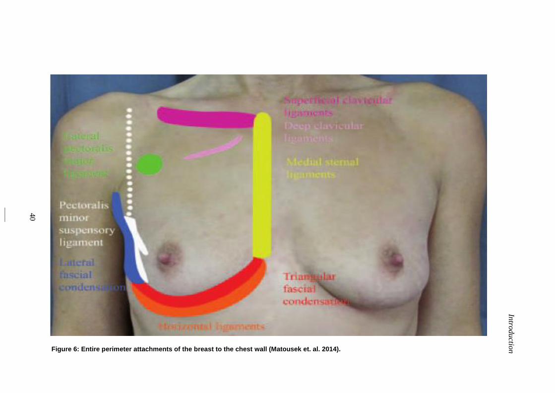
Intro
ductio
n
40
Figure 6: Entire perimeter attachments of the breast to the chest wall (Matousek et. al. 2014).
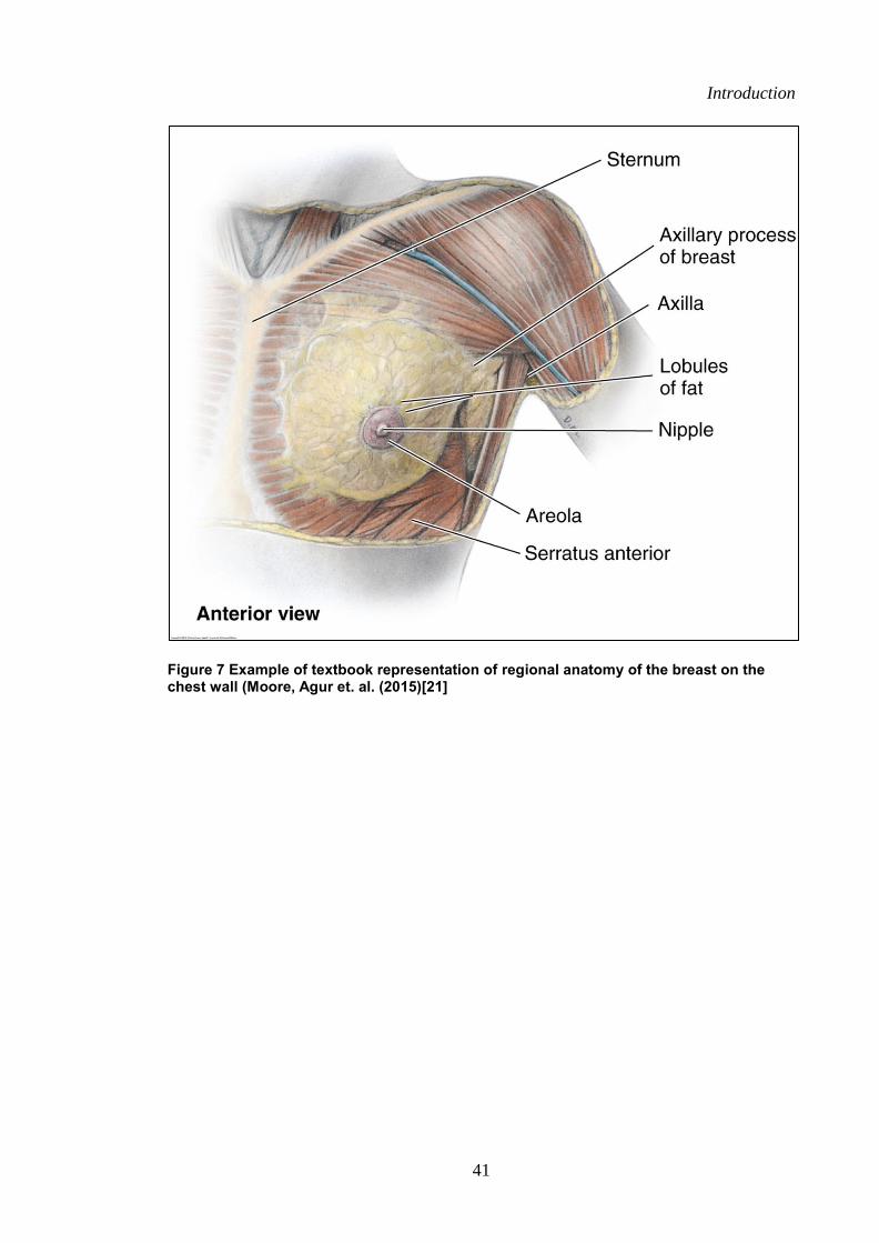
Introduction
41
Intro
ductio
n
Figure 7 Example of textbook representation of regional anatomy of the breast on the chest wall (Moore, Agur et. al. (2015)[21]
Introduction
42
Intro
ductio
n
1.4 Literature Summary
This literature review highlights the need for further anatomical dissection research on
the gross anatomy the breast, supported by quantitative data due to the dated research,
inconsistencies in the literature and the limited detail available in anatomical textbooks
currently used for medical, allied health and science students on the structural anatomy
of the breast. Specific gaps in the literature were identified on the structural anatomy of
the breast regarding:
(i) The composition of cadaveric breasts in terms of the relative percentage
of fibro-glandular and fibro-adipose tissue across a spectrum of breast
sizes.
(ii) The fibro-adipose structure of the breast in terms of descriptions of its
complex structure supported by quantitative data and a collation of the
various names used to describe the fascial tissue within this structure.
(iii) Attachments of the breast to the chest wall via the posterior wall and
perimeter attachments, supported by quantitative data.
(iv) Regional anatomy of the breast in terms of the percentage contribution of
the muscles adjacent to the posterior wall of the breast
Therefore further anatomical dissection research is required to verify the
descriptions of Cooper, complementing them with quantitative data. This research could
be used as a basis from which concise, evidence-based written text descriptions along
with accurate and consistent anatomical illustrations of the breast could be developed
supported by quantitative data. The improved understanding and education of the
structural anatomy of the breast could contribute positively to both breast surgery
outcomes and the diagnosis of pathology.
Introduction
43
Intro
ductio
n
1.5 Research Aims and Hypothesis
The aim of this study was to provide quantitative data on the gross anatomy of the
breast, to provide evidence-based detail for anatomical illustrations and descriptions on
the gross anatomy of the breast.
1.5.1 Composition
Aim one: To provide quantitative data of embalmed cadaveric breast size (volume,
surface area and mass) and embalmed cadaveric breast composition in terms of
percentage mass of fibro-adipose and fibro-glandular tissue. From the interpretations of
the literature the following experimental hypotheses were set:
Hypothesis one: Breast size (surface area and volume) will be within the ranges of
breast volume and surface area reported in in-vivo studies.
Hypothesis two: The relative percentage mass of the fibro-adipose and fibro-glandular
tissue will be consistent amongst the embalmed cadaveric breasts, irrespective of their
size (total mass).
1.5.2 Gross Anatomical Structure
Aim two: To investigate and to provide quantitative data on the gross anatomy of the
fibro-adipose structure of the breast. In agreement with the descriptions of Cooper the
following experimental hypotheses were set:
Hypothesis three: Singular sheets of fascia will be found both anterior (Anterior
Lamellae) and posterior to the mammary gland (Posterior Lamellae).
Hypothesis four: Fibrous bands of tissue (Anterior Extensions of the Anterior
Lamellae) will be found connecting the Anterior Lamellae to the dermis.
Hypothesis five: Fibrous bands of tissue (Posterior Extensions of the Posterior
Lamellae) will be found connecting the Posterior Lamellae to the superficial fascia of
the Pectoralis Major muscle.
Introduction
44
Intro
ductio
n
Hypothesis six: Adipose tissue will be encapsulated within fibrous pockets (fibro-
adipose pockets) located both anterior and posterior to the mammary gland.
Hypothesis seven: The number of the fibro-adipose pockets within the breast and the
size of the fibro-adipose pockets will be consistent amongst the breasts, irrespective of
their size.
Hypothesis eight: The size of the fibro-adipose pockets located anterior to the gland
will be larger and less numerous to those located posterior to the gland.
1.5.3 Attachment of the breast to the chest wall
Aim Three: To investigate the regional anatomy and attachment of the breast to the
chest wall. From the interpretation basis of the literature and the anatomical descriptions
of Cooper the following experimental hypotheses were set:
Hypothesis Nine: The posterior wall of the breast and the perimeter of the breast will
have fibrous connections to the superficial muscle fascia of the anterio-lateral aspect of
the chest wall.
Hypothesis Ten: The muscles found within the perimeter will include the Pectoralis
Major, Rectus Abdominus, Serratus Anterior and External Oblique muscles.

45
Chapter 2:
Methods
Methods
46
2.1 Experimental Overview
A cadaveric-based investigation was carried out on embalmed female cadaver’s post-
mortem. The investigation focused on three aspects of the female breast; (i)
Composition, (ii) Gross anatomical structure, (iii) Attachments of the breast to the
chest wall. Eighteen breasts from nine cadavers were investigated quantitatively and
the gross anatomy was described qualitatively using this data. A range of dissection
techniques were used including blunt and sharp dissection, coronal and sagittal
sectioning, macro and microscopic investigation, photography, tissue weighing and
three-dimensional scanning. The breast was investigated in two planes, coronal and
sagittal to provide a three dimensional understanding of the structure of the breast.
2.2 Ethics Consideration
All procedures portrayed in this proposal were submitted and approved by the
University of Wollongong Human Research Ethics Committee (HE14/001). The
University of Wollongong houses a PC2 Anatomy Laboratory where all cadaveric
specimens were stored, prepared and dissected according to NSW State Legislation
and University policy and guidelines.
2.3 Cadaveric Selection
The breasts of cadavers embalmed, using classical techniques through the left common
carotid with a formalin based embalming fluid (Genelyn Anatomical Series NF, GMS
Innovation, Australia), were selected, by the chief researcher (KG), from a list of
cadavers held in the University of Wollongong Anatomy Laboratory that were approved
and consented for anatomical research purposes (n= 24). From this list, specimens were
excluded based on the following criteria:
Methods
47
(i) The breast or chest plate had already been dissected, as the integrity of
the structure within the breast may have been compromised (n=8)
(ii) Recorded or observable signs of surgical procedures within the breast or
chest region, such as mastectomies, lumpectomies and augmentation
mammoplasty (n=3)
(iii) Recorded or observable signs of breast shape abnormalities, or
abnormalities due to the embalming process, which may skew the
accuracy of measurement (n=3)
One cadaver was selected for a reliability study. The nine most recently
embalmed cadavers, which were a range of breast sizes, satisfying the inclusion criteria
are shown in Figure 8 and Table 4.
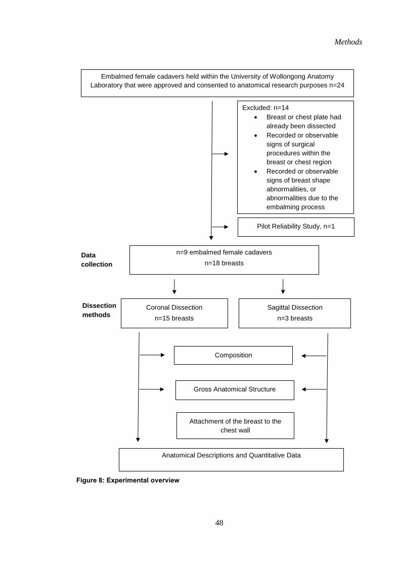
Methods
48
Figure 8: Experimental overview
Dissection
methods
Embalmed female cadavers held within the University of Wollongong Anatomy
Laboratory that were approved and consented to anatomical research purposes n=24
n=9 embalmed female cadavers
n=18 breasts
Data
collection
Excluded: n=14
Breast or chest plate had
already been dissected
Recorded or observable
signs of surgical
procedures within the
breast or chest region
Recorded or observable
signs of breast shape
abnormalities, or
abnormalities due to the
embalming process
Coronal Dissection
n=15 breasts
Sagittal Dissection
n=3 breasts
Composition
Gross Anatomical Structure
Attachment of the breast to the
chest wall
Anatomical Descriptions and Quantitative Data
Pilot Reliability Study, n=1
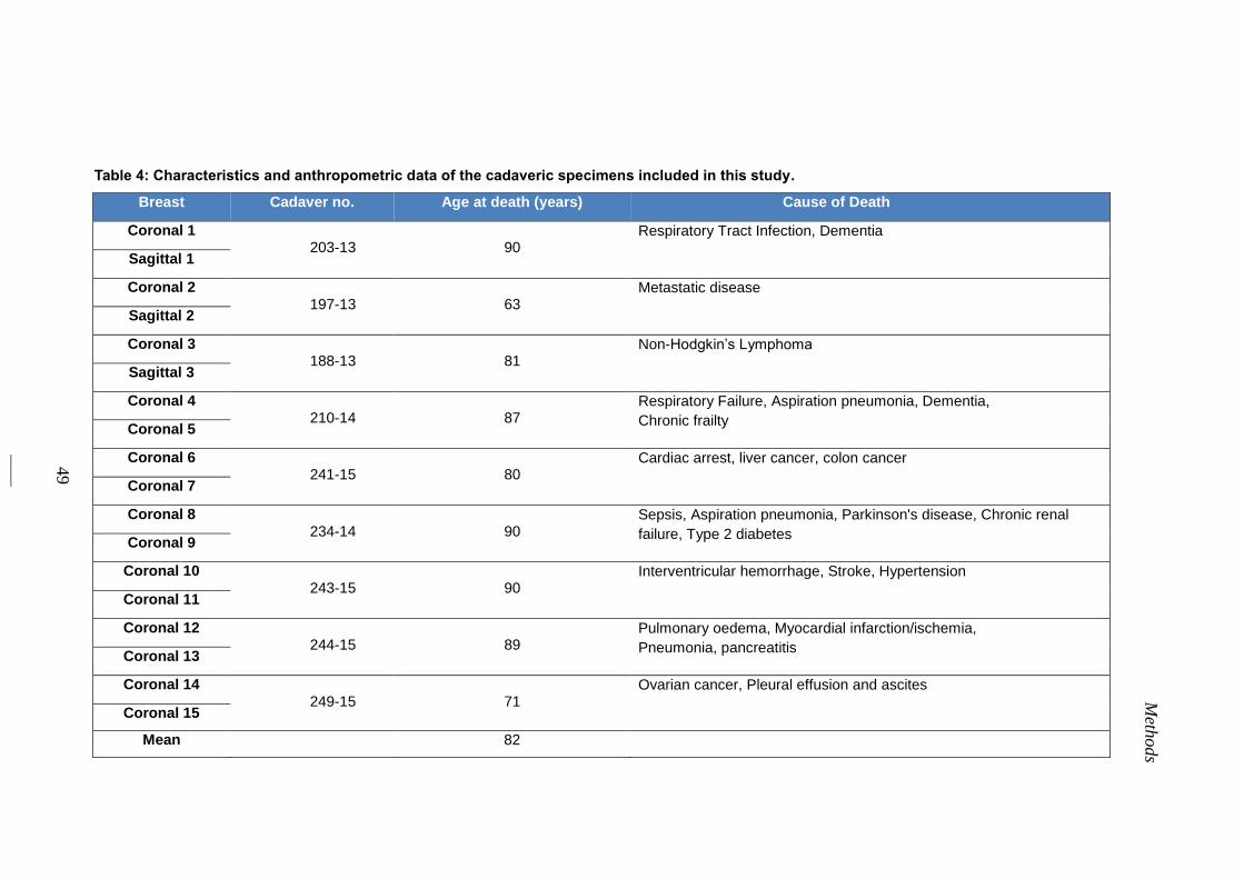
49
Meth
ods
Table 4: Characteristics and anthropometric data of the cadaveric specimens included in this study.
Breast Cadaver no. Age at death (years) Cause of Death
Coronal 1 203-13
90
Respiratory Tract Infection, Dementia
Sagittal 1
Coronal 2 197-13
63
Metastatic disease
Sagittal 2
Coronal 3 188-13
81
Non-Hodgkin’s Lymphoma
Sagittal 3
Coronal 4 210-14
87
Respiratory Failure, Aspiration pneumonia, Dementia,
Chronic frailty Coronal 5
Coronal 6 241-15
80
Cardiac arrest, liver cancer, colon cancer
Coronal 7
Coronal 8 234-14
90
Sepsis, Aspiration pneumonia, Parkinson's disease, Chronic renal
failure, Type 2 diabetes Coronal 9
Coronal 10 243-15
90
Interventricular hemorrhage, Stroke, Hypertension
Coronal 11
Coronal 12 244-15
89
Pulmonary oedema, Myocardial infarction/ischemia,
Pneumonia, pancreatitis Coronal 13
Coronal 14 249-15
71
Ovarian cancer, Pleural effusion and ascites
Coronal 15
Mean 82
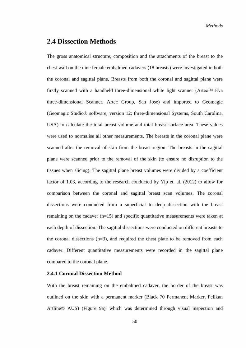
Methods
50
2.4 Dissection Methods
The gross anatomical structure, composition and the attachments of the breast to the
chest wall on the nine female embalmed cadavers (18 breasts) were investigated in both
the coronal and sagittal plane. Breasts from both the coronal and sagittal plane were
firstly scanned with a handheld three-dimensional white light scanner (Artec™ Eva
three-dimensional Scanner, Artec Group, San Jose) and imported to Geomagic
(Geomagic Studio® software; version 12; three-dimensional Systems, South Carolina,
USA) to calculate the total breast volume and total breast surface area. These values
were used to normalise all other measurements. The breasts in the coronal plane were
scanned after the removal of skin from the breast region. The breasts in the sagittal
plane were scanned prior to the removal of the skin (to ensure no disruption to the
tissues when slicing). The sagittal plane breast volumes were divided by a coefficient
factor of 1.03, according to the research conducted by Yip et. al. (2012) to allow for
comparison between the coronal and sagittal breast scan volumes. The coronal
dissections were conducted from a superficial to deep dissection with the breast
remaining on the cadaver (n=15) and specific quantitative measurements were taken at
each depth of dissection. The sagittal dissections were conducted on different breasts to
the coronal dissections (n=3), and required the chest plate to be removed from each
cadaver. Different quantitative measurements were recorded in the sagittal plane
compared to the coronal plane.
2.4.1 Coronal Dissection Method
With the breast remaining on the embalmed cadaver, the border of the breast was
outlined on the skin with a permanent marker (Black 70 Permanent Marker, Pelikan
Artline© AUS) (Figure 9a), which was determined through visual inspection and
Methods
51
palpation of the borders by the breast by the chief investigator (KG). The perimeter was
then marked again by injecting a streak of viscous dye mix of 1:10 India Ink (Black Ink,
Windsor and Newton© UK) and ultrasound gel (Aquasomic© 100, Parker Laboratories
USA). The injections, made at 2-3 cm intervals around the border, penetrated the
perimeter of the breast perpendicular to the skin and injected approximately 1 ml of
viscous dye to the entire depth of the breast perimeter to the chest wall. This ensured
that the border of the breast was clearly marked for the entire depth of the dissection
(Figure 9a).
Skin removal
The skin was then removed with sharp dissection using curved forceps (Swann- Morton,
England) and a scalpel handle with a size 10 blade (Swann-Morton, England). Skin
incisions were made following the midline of the sternum, at least 2 cm below the
inferior boarder of the breast laterally, past the mid-axillary line and superiorly 1 cm
above the level of the clavicle and the skin within these incisions was removed. The
skin of the nipple and areola region was not removed so that it could be used as a point
of reference during the coronal dissection (Figure 9b). The middle of the nipple and the
furthermost superior, inferior, medial and lateral points of the breast was then pinned
with flag pins (Figure 9c). The pins were also used as landmarks for the three-
dimensional scanning analysis. The dissections of the breast were also photographed
(Nikon 600D, 200mm Lens) at each stage of the dissection from superficial to deep.
Breast quadrants
The flag pins were then used to join pieces of string that were set up to divide the breast
in to four quadrants, superior-medial, superior-lateral, inferior-medial and inferior-
lateral. These divisions were used to investigate regional variation in the quantitative
measures taken during the coronal dissection. Photographs (Nikon 600D, 200mm Lens)
Methods
52
were taken to complement the qualitative descriptions of the breast at this stage (Figure
9c).
Fibro-adipose tissue structure (superficial to the glandular tissue)
The most superficial layer covering the entire breast directly below the skin consisted of
a thin layer (1-2 mm) of subcutaneous fat. This was removed from the external surface
of the breast with the back edge of curved forceps (Swann-Morton, England). Removal
of this layer revealed the fibro-adipose structure superficial to the glandular tissue. This
fibro-adipose tissue three-dimensional cobweb consisted of numerous small fibrous
pockets, each containing adipose tissue (Figure 10a). Using twin curved forceps
(Swann-Morton, England), the membrane of the adipose tissue, within each fibrous
pocket, was broken and the adipose tissue was intricately detached from the fibrous wall
of each pocket and carefully removed. This procedure was repeated on all of the fibro-
adipose pockets of the breast to reveal the entire fibrous structure minus the adipose
tissue (Figure 10b). Each pocket was then pinned with a dressmakers pin so they could
be accurately counted by the chief investigator (KG). The tissue from each pocket was
immediately placed into individual weigh boats for quantitative investigation.
Within each breast, the adipose tissue contents of a minimum of 20 fibrous
pockets per quadrant (80 pockets per breast) were placed in weigh boats of known
weights to the accuracy of three decimal places. The adipose tissue from each pocket
was weighed within the first two hours after it was dissected as pilot testing found this
to be the optimum duration to weigh the tissue post-dissection to prevent tissue drying.
Once all of the adipose tissue was completely dissected from the fibrous pockets,
the pockets were measured using callipers (Mitutoyo, Japan) to quantify the surface
area. The three-dimensional fibrous cobweb was described qualitatively and
photographed within the first two hours after it was dissected (Nikon 600D, 200mm).
Meth
od
s
53
Figure 9: Coronal breast with a) Boarder outlined with permanent marker and the viscous dye injection sites ( ), b) Skin removed and nipple and
areola left intact, c) Skin removed, viscous dye injection sites ( ) and separation of the breast into quadrants with flag pins and string.
a. b. c.
S
M L
I
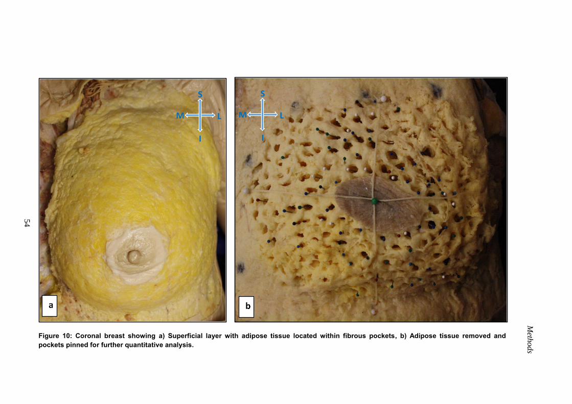
Meth
od
s
54
Figure 10: Coronal breast showing a) Superficial layer with adipose tissue located within fibrous pockets, b) Adipose tissue removed and
pockets pinned for further quantitative analysis.
S
M L
I
S
M L
I
Methods
55
Fibro-glandular tissue
Once all of the adipose tissue was dissected from the fibrous cobweb, the remaining fibrous
cobweb was then dissected from the anterior surface of the fibro-glandular tissue using sharp
dissection (scalpel handle with size 10 blade and curved forceps, Swan-Morten, England) and
weighed (Ohaus, Adventurer® Pro, USA). This dissection process ensured that the fibro-
glandular tissue structure was not disturbed. The exposed anterior surface of the fibro-glandular
was then described qualitatively and photographed (Nikon 600D, 200mm Lens; Figure 11). The
fibro-glandular tissue was then removed with sharp dissection within the perimeter of the breast
(scalpel handle with size 10 blade and curved forceps, Swan-Morten, England, Figure 12a). The
removed fibro-glandular was then immediately weighed immediately (Ohaus, Adventurer® Pro,
USA) and the structures deep to it photographed (Nikon 600D, 200mm Lens).
Fibro-adipose tissue (deep to the glandular tissue)
Dissection removal of the fibro-glandular tissue with sharp dissection (scalpel handle with size
10 blade and curved forceps, Swan-Morten, England) revealed a secondary layer of fibro-
adipose tissue posterior to the fibro-glandular tissue. Removal of this deeper layer of fibro-
adipose tissue within the perimeter of the breast revealed the superficial fascia of the muscles
covering the anterior/lateral chest wall (Figure 12b). The dissected layer of fibro-adipose tissue
lying posterior to the fibro-glandular tissue was then weighed (Ohaus, Adventurer® Pro, USA)
and photographed (Nikon 600D, 200mm Lens). The aforementioned separation of the breast
into four quadrants ensured that the tissue of each quadrant was kept separate from each other to
allow for comparisons of the quantitative data within each region. At all levels of the coronal
dissection the tissues were photographed (Nikon 600D, 200mm Lens), described and measured
quantitatively.
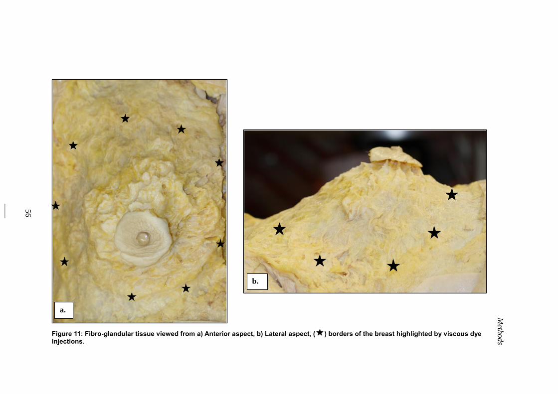
Meth
ods
56
Figure 11: Fibro-glandular tissue viewed from a) Anterior aspect, b) Lateral aspect, ( ) borders of the breast highlighted by viscous dye injections.
b.
a.
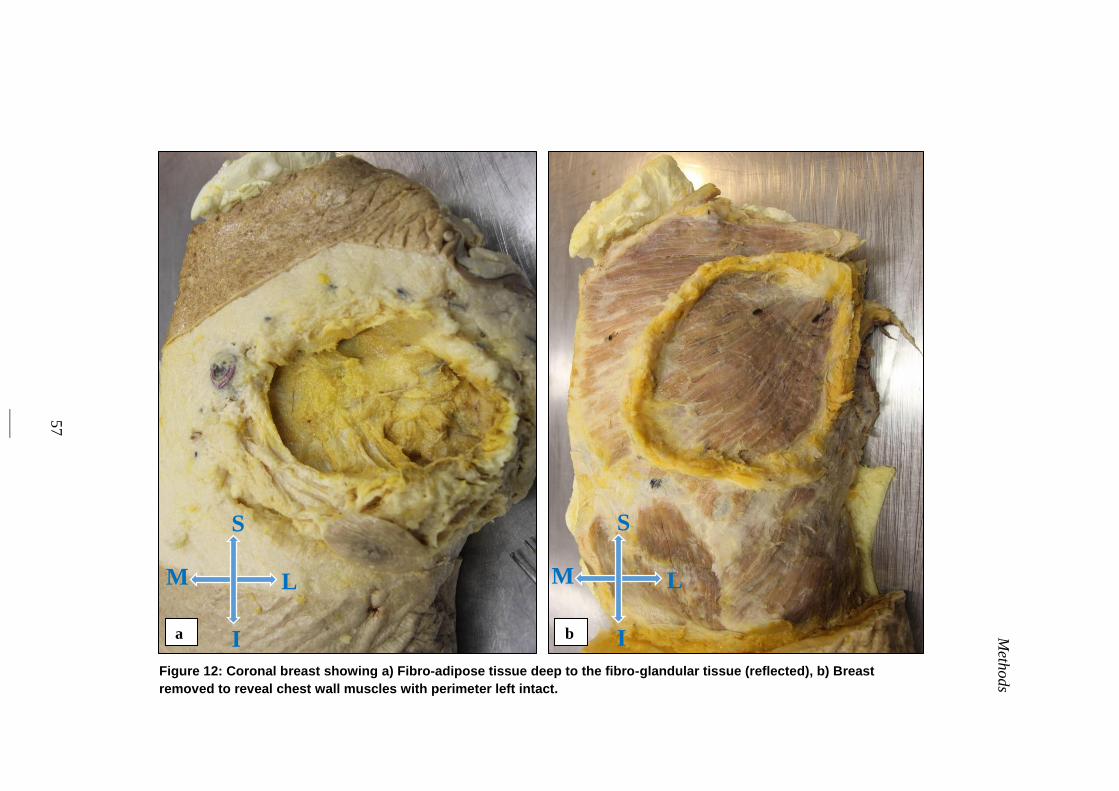
Meth
ods
57
Figure 12: Coronal breast showing a) Fibro-adipose tissue deep to the fibro-glandular tissue (reflected), b) Breast
removed to reveal chest wall muscles with perimeter left intact.
a b
S
M L
I
S
M L
I
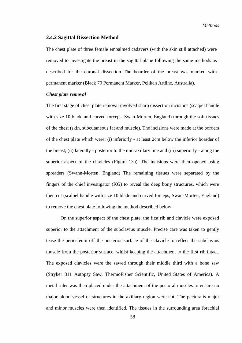
Methods
58
2.4.2 Sagittal Dissection Method
The chest plate of three female embalmed cadavers (with the skin still attached) were
removed to investigate the breast in the sagittal plane following the same methods as
described for the coronal dissection The boarder of the breast was marked with
permanent marker (Black 70 Permanent Marker, Pelikan Artline, Australia).
Chest plate removal
The first stage of chest plate removal involved sharp dissection incisions (scalpel handle
with size 10 blade and curved forceps, Swan-Morten, England) through the soft tissues
of the chest (skin, subcutaneous fat and muscle). The incisions were made at the borders
of the chest plate which were; (i) inferiorly - at least 2cm below the inferior boarder of
the breast, (ii) laterally - posterior to the mid-axillary line and (iii) superiorly - along the
superior aspect of the clavicles (Figure 13a). The incisions were then opened using
spreaders (Swann-Morten, England) The remaining tissues were separated by the
fingers of the chief investigator (KG) to reveal the deep bony structures, which were
then cut (scalpel handle with size 10 blade and curved forceps, Swan-Morten, England)
to remove the chest plate following the method described below.
On the superior aspect of the chest plate, the first rib and clavicle were exposed
superior to the attachment of the subclavius muscle. Precise care was taken to gently
tease the periosteum off the posterior surface of the clavicle to reflect the subclavius
muscle from the posterior surface, whilst keeping the attachment to the first rib intact.
The exposed clavicles were the sawed through their middle third with a bone saw
(Stryker 811 Autopsy Saw, ThermoFisher Scientific, United States of America). A
metal ruler was then placed under the attachment of the pectoral muscles to ensure no
major blood vessel or structures in the axillary region were cut. The pectoralis major
and minor muscles were then identified. The tissues in the surrounding area (brachial
Methods
59
plexus, subclavian artery and subclavian vein) were identified and carefully separated
from the posterior aspect of the pectoralis major and minor muscles. A metal ruler was
placed within the newly created gap below the pectoralis major and minor muscles to
protect the underlying structures (brachial plexus, subclavian artery and subclavian
vein). The pectoralis major and minor muscles were then incised 3cm distal to their
insertion (pectoralis major: bicipital groove of the humerus; pectoralis minor: coracoid
process of the scapula) using sharp dissection (scalpel handle with size 10 blade and
curved forceps, Swan-Morten, England) to reveal the ribs and intercostal muscles.
On the lateral aspect of the chest plate, the intercostal muscles were pierced
through to expose ribs 1-10, which were then cut using rib-snips (Swann-Morten,
England) posterior to the mid-axillary line. On the lateral aspect of the chest plate, the
abdominal cavity was opened along the entire inferior boarder of the skin incision (2cm
inferior to the breast) using sharp dissection (scalpel handle with size 10 blade and
curved forceps, Swan-Morten, England).
Once the chest plate was detached along its superficial borders, any soft tissue
attached to the deep surface of the internal surface of the chest plate (e.g. diaphragm,
blood vessels, cardiac sack) were then detached from the chest plate using sharp
dissection (scalpel handle with size 10 blade and curved forceps, Swan-Morten,
England). The dissected chest plate was then moved away from the rest of the cadaver
(Figure 13b).
Sagittal sectioning of the breast
The removed chest plate was then placed in a plastic container with the concave internal
surface facing downwards. Expanding foam (Sika, Australia) was then sprayed onto the
internal concave surface of the chest plate, until the concavity was filled, to form a flat
surface. The expanding foam was then left to set. The set expanding foam formed a flat
Methods
60
posterior surface which allowed a smooth transmission of the chest plate through the
band saw (Mark 1, H.T. Barnes, Australia) which was necessary to ensure that the
sectioning of the chest plate with the band-saw could be cut evenly. Vertical, parallel
lines were than marked on the skin of the breast through the nipple and either side of the
areola in the sagittal plane with a black marker (Black 70 Permanent Marker, Pelikan
Artline, Australia). These landmarks were used to ensure that consistent regions of the
breast were exposed amongst the three breasts dissected in the sagittal plane, which
were of varying shapes and sizes.
The chest plate was then sectioned along these marked lines with a using a band saw
(Mark 1, H.T. Barnes, Australia) (Figure 14a). The sawing produced four sagittal breast
slices per breast, which provided six surfaces of the breast for quantitative analysis
(Figure 14b). Each slice surface was photographed (Nikon 600D, 200mm Lens),
described qualitatively and measured quantitatively using a microscope and its
associated software (Nikon SMZ800, Australia).
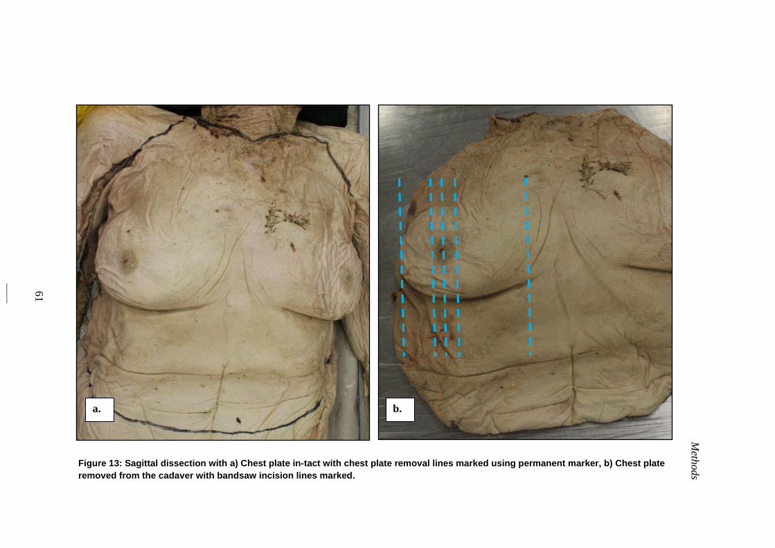
Meth
ods
61
Figure 13: Sagittal dissection with a) Chest plate in-tact with chest plate removal lines marked using permanent marker, b) Chest plate
removed from the cadaver with bandsaw incision lines marked.
b. a.
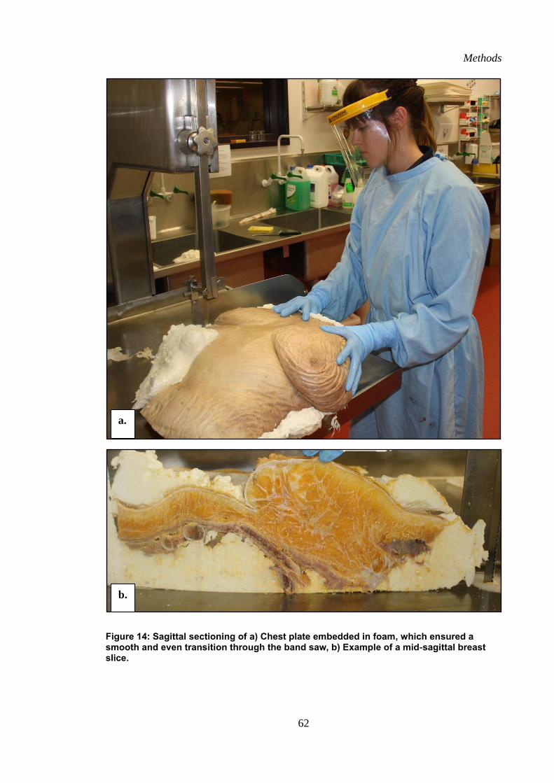
Methods
62
Figure 14: Sagittal sectioning of a) Chest plate embedded in foam, which ensured a smooth and even transition through the band saw, b) Example of a mid-sagittal breast slice.
a.
b.
Methods
63
2.5 Quantitative Outcome Measures
2.5.1 Composition
2.5.1.1 Surface Area
The total surface area (cm2) of each embalmed female breast was measured using a
hand held three-dimensional white light scanner (Artec™ Eva three-dimensional
Scanner, Artec Group, San Jose). With the skin removed, the breast and upper torso was
scanned ensuring complete visualization of the breast tissue. The scan was then
imported into Geomagic software (Geomagic Studio® software; version 12; three-
dimensional Systems, South Carolina, USA) to determine total breast surface area (cm2)
and quadrant surface area (cm2) (Figure 15a). The borders of the breast were digitally
outlined following the borders previously marked with ink. The scanned breast object
was removed from the scanned trunk to create a two dimensional object from which the
surface area (cm2) of the breast was calculated (Figure 15b, c). The 2D breast object
was then digitally divided into four quadrants by creating two planes that intersected
through the nipple (x-plane and y-plane), following the dressmaker pins previously
positioned on the cadaveric breast. The surface area (cm2) of the breast in each quadrant
was then calculated.
2.5.1.2 Volume
Total breast volume (mL) and quadrant volume (mL) of each embalmed female breast
was measured from the same scan of the breast used to determine surface area (cm2).
Volume required a three-dimensional object of each breast to be digitally created from
the previously developed two-dimensional object of the breast. The posterior surface of
the breast was created using a tangential cut plane tool to fill the anterior chest wall,
once the breast was removed (Figure 15b). Based on the recommendation of previous
Methods
64
studies [61, 86]
, the tangent tool was used to make a series of tangential cut planes to
mimic the curvature of the anterior chest wall muscles, which the posterior surface of
the breast sits on. The posterior wall of the breast object that was created was then
digitally attached to the rest of the breast to create a three-dimensional model of the
breast (Figure 15d). Geomagic software was then used to determine the volume (mL) of
each three-dimensional breast model (n=15). The three-dimensional breast model was
then digitally sectioning into four breast quadrants through the nipple as per the surface
area (cm2) measurement and the volume (mL) of each quadrant measured.
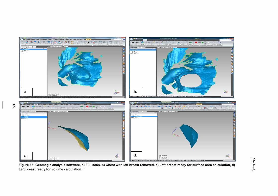
Meth
ods
65
Figure 15: Geomagic analysis software, a) Full scan, b) Chest with left breast removed, c) Left breast ready for surface area calculation, d)
Left breast ready for volume calculation.
a b.
c. d.
Methods
66
2.5.1.3 Fibro-adipose mass
When investigated in the coronal plane, the total fibro-adipose tissue mass (g) was
weighed (Ohaus, Adventurer® Pro, USA) in stages as it was dissected. The total fibro-
adipose mass (g) was the sum of the dissected parts of the fibro-adipose tissue mass (g)
both superficial and deep to the fibro-glandular tissue. The dissected fibro-adipose
tissue was weighed in triplicate and the average mass (g) of the tissue within in the
breast and within each quadrant was recorded for data analysis. The mass (g) of each
tissue was weighed within two hours of being dissected.
2.5.1.4 Fibro-glandular mass
The total fibro-glandular tissue encased between the two layers of fibro-adipose tissue
was weighed (Ohaus, Adventurer® Pro, USA) within two hours of being dissected from
the deep fibro-adipose layer. The tissue was weighed in triplicate and the average mass
(g) of the tissue in the breast and within each quadrant was recorded for data analysis.
2.5.1.5 Breast Mass
The total mass (g) of the fibro-adipose tissue mass (g) and the fibro-glandular tissue was
summated to calculate the total breast mass (g) and the mass within each quadrant (g).
2.5.1.6 Breast Composition
The fibro-adipose and fibro-glandular composition of each breast (%) and within each
breast quadrant (%) was expressed in terms of percentage of total breast mass.
2.5.2 Gross Anatomical Structure
2.5.2.1 Number of fat lobule pockets
The total number of fibro-adipose pockets within each breast and within each quadrant
were manually counted and photographed (Nikon 600D, 200mL Lens) by the chief
investigator (KG) from the dissection performed in the coronal plane. This was
performed at the stage of dissection when the adipose tissue within each pocket was
Methods
67
dissected. At this point, each pocket was pinned and counted (Figure 10b). Of the
pinned pockets counted, 80 pockets per breast (20 per quadrant) were included in
further quantitative measurements of pocket surface area and adipose tissue mass within
each pocket.
2.5.2.2 Adipose mass of each pocket
The adipose tissue mass (g) within each of the 80 marked fibro-adipose pockets (20
pockets per quadrant) per breast (n=15 breasts) dissected in the coronal plane was
meticulously dissected and weighed by the chief investigator (KG). Immediately after
the adipose tissue was removed from the pocket, it was placed into an individual weigh
boat of a known weight and weighed to the nearest 0.0001g using an anatomical scale
(Ohaus, Adventurer® Pro, USA). To ensure accuracy, the adipose tissue was weighted
within the first two hours after it was dissected in triplicate and the scale zeroed
between each measurement. The average of the triplicate measurements was then
recorded for data analysis.
2.5.2.3 Surface area of each adipose pocket
The surface area (mm2) of each the 80 marked fibro-adipose pockets (20 pockets per
quadrant) per breast (n=15 breasts) in the coronal plane dissection was measured by the
chief investigator (KG) immediately after the adipose tissue within the pocket was
dissected. The length and breadth of each fibrous pocket was measured medio-laterally
and superio-inferiorly respectively, in triplicate using Vernier callipers (Mitutoyo,
Japan) to the nearest 0.2mm. The average of each measure was recorded for data
analysis.
2.5.2.4 Length of the anterior extensions of the anterior lamellae
The length of a minimum of 20 anterior extensions of the anterior lamellae per slice
surface (6 breast slice surfaces per breast; n=3 breasts; therefore 120 extensions per
Methods
68
breast) were measured by the chief investigator (KG) using an anatomical microscope
(Nikon SMZ800, Australia) and its associated software. The software allowed for two
set points to be created by the chief investigator (KG), one at the deep end of the
Anterior Extension (Anterior Lamellae) and one at the superficial end (dermis). The
straight line distance between the two points was calculated by the software. Each
measurement was taken in triplicate and the average was recorded for data analysis.
2.5.2.5 The number of pockets anterior and posterior to the gland
The total number of fibro-adipose pockets located anterior and posterior to the gland
were pinned with dressmaker pins and manually counted by the chief investigator (KG)
from the dissection performed in the sagittal plane (Figure 14b). The measurement was
performed on six sagittal breast slice surfaces (3 slices from each of the 3 breasts). Each
slice was also photographed for verification (Nikon 600D, 200mL Lens).
2.5.2.6 The cross sectional surface area
The surface area (mm2) of a minimum of 20 fibro-adipose pockets located anterior and
posterior to the gland (Figure 14b) was measured using an anatomical microscope
(Nikon SMZ800, Australia) and its associated software. The chief investigator (KG)
used the software to digitally outline the perimeter of each pocket, from which the
surface area (mm2) was measured. All measurements were taken in triplicate and the
average was recorded for data analysis.
2.5.3 Attachment of the breast to the chest wall
2.5.3.1 Muscles within the perimeter
At the last stage of the coronal investigation, the entire structure of the breast was
removed within its perimeter, exposing the anterior-lateral chest wall muscles located
within the perimeter of the breast (n=12 breasts). Each cadaver was photographed at this
stage (Nikon 600D, 200mm Lens) and the chief investigator (KG) used software
Methods
69
(ImageJ, Wayne Rasband, Maryland, USA) to quantify the percentage surface area that
each muscle contributed to the total surface area within the perimeter of each breast.
The percentage contribution of each muscle to the total surface area per breast was
recorded.
2.5.3.2 Perimeter
The perimeter of each breast dissected in the coronal plane (n=12) was measured by the
chief investigator (KG) using surgical silk (Dynek, SA, Australia). The surgical silk was
laid precisely around the internal boarder of the breast border and the point at which the
silk reconnected with the start was noted. The length of the silk was measured in
triplicate using a measuring tape in (mm) and the average was recorded for data
analysis.
2.6 Qualitative Measures
Anatomical descriptions were used to describe the composition, gross anatomical
structure and the attachment of the breast to the chest wall in both the coronal and
sagittal planes.
2.7 Experimental Standardisation and Reliability
A number of different techniques were employed to ensure standardisation and
reliability during the entire data collection phase.
2.7.1 Chief Investigator
All dissection work was carried out by the chief investigator Kathryn Gaskin (KG). This
reduced inter-observational discrepancies and ensured consistency throughout the
dissection process. KG has been employed by the University of Wollongong as an
Anatomy Technical Officer, with four years anatomical dissection experience. As such,
Methods
70
she has achieved a high level of competence in anatomical dissection.
2.7.2 Reliability Study
Prior to the cadaveric study, a reliability pilot study was conducted on one female
cadaver aged 68, whom died of a pelvic chordoma. Each of the breasts was examined
using the two different dissection methods outline above (left breast - coronal plane
dissection; right breast - sagittal plane and the reliability of the various manual
measurement was tested. These were the weighing of tissue samples (g), the
measurement of straight-line distances with callipers (mm) and the counting of fat
lobule pockets (n). Interclass correlation (ICC) were interpreted as per Cicchetti (1992)
[87] as poor (< 0.40), fair (0.40-0.59), good (0.60-0.74) and excellent (0.75-1.00).
The reliability of tissue weighed using the anatomical scales (OHAUS
Adventurer, Anatomical Scales, USA; see sections: 2.5.1.3, 2.5.1.4, 2.5.1.5 and 2.5.2.2
for outcome measures) was determined by weighing three small weights of known
values on three separate occasions. The interclass correlation (ICC) measured across the
three separate occasions was found to be 0.99, a high reliability. The reliability of tissue
weighing at varying intervals subsequent to the removal from the body was conducted
on ten small samples of adipose tissue and fibrous tissue from various regions of the
breast. This was conducted to determine the likelihood of weight change due to drying
out due to exposure with air. Weighing of the samples was conducted one hour, two
hours and three hours subsequent to the dissection of the tissues. The interclass
correlation (ICC) from the first hour to the third hour was found to be 0.58, a moderate
reliability, and as such, tissue samples were weighed within the first two hours after
dissection. The reliability of the straight line distances measured using the Vernier
callipers (Mitutoyo, Japan; see sections: 2.5.2.3, 2.5.2.4 and 2.5.2.6 for outcome
measures), were measured on ten tissue pockets on three separate occasions and were
Methods
71
found to have ICC of 0.97, a high reliability. The number of pockets within the breast
were manually counted (see sections: 2.5.2.1 and 2.5.2.5 for outcome measures) by KG
on three separate occasions and KG was found to be highly reliable with an ICC of
1.00.
2.7.3 Cadaver Preparation and Storage
All cadavers used in the study were acquired through the University of Wollongong
Body Donation Programme. All cadavers were screened for Hep B, Hep C and HIV
upon arrival to the facility with positive cases rejected. All cadavers were embalmed
through the left common carotid artery with at least 20L of Arterial Embalming Fluid
(Genelyn, Australia) and left in the Anatomy Laboratory cool room for six months so all
tissues could be perfused.
When not in use all cadavers were stored inside a blue body bag, on stainless
steel body trays, in the University of Wollongong Anatomy Laboratory cool room. The
temperature of the cool room is a consistent 3.5°C. Every two hours, on the even hour,
between 8am and 6pm the cool room undergoes a 10-minute purge cycle to expel any of
the excess formaldehyde from the room.
2.7.4 Room Conditions During Measurement
The Anatomy Laboratory at the University of Wollongong has an independent air flow
system that operates solely within the laboratory space. The air is recycled 11 times
every hour. There is a negative pressure gradient, which pulls all of the air from the
teaching space in to the preparation space, where all investigation was conducted. The
air is then moved through vents and realised into the atmosphere above the three (3)
story building. The air temperature is set at 19°C. All tissue samples (fibrous tissue,
adipose tissue and fibro-glandular tissue) were weighed directly after removal from the
cadaver to ensure accuracy and reduce the likelihood of drying out due to the
Methods
72
independent air flow system.
2.8 Normalising Data (absolute and relative)
During to the variation in the breast size and shape of the 15 breasts used in the study,
the quantitative data of number and surface area of the fibrous breast pockets, weight of
adipose tissue within each pocket, breast perimeter, and breast composition were
normalized to the breast volume and surface area of each breast.
2.9 Statistical Design
The statistical analysis software used in this study was Statistix ver. 10 (Tallahassee,
USA). Descriptive statistics was performed on all quantitative data. A one-way analysis
of variance (ANOVA) was used to compare the quantitative data in each quadrant of the
breast, with Tukeys post-hock analysis. An independent T-test was used to compare the
number of pockets, mean pocket mass and mean pocket surface area in the upper and
lower tertiles by volume. Pearson’s correlations were used to compare breast volume,
breast surface area, breast mass, number of pockets, pocket mass and pocket surface
area.

73
Chapter 3:
Results
Results
74
3.1 Composition
3.1.1 Surface Area
The mean total breast surface area of the 15 breasts (with the skin removed) measured
from the three-dimensional scans was 302±91 cm2
(range: 187-501 cm2; Table 5). The
surface area per quadrant measured in cm2
and as a percentage of the total surface area
is displayed in Table 6 and Figure 16.The surface area was not evenly distributed
amongst the breast quadrants with the cadaver in the supine position. The superior-
medial quadrant had a significantly larger mean surface area than the other quadrants
(p<0.01; mean surface area 33 %, 99.3±3 5cm2). The superior-lateral quadrant
accounted for a mean of 26 % of the total breast surface area (mean surface area
79.6±25 cm2), which was significantly greater than the mean surface area of the
inferior-medial quadrant (21 %; mean surface area 63.3±24 cm2) and the inferior-lateral
quadrant (20 %; mean surface area 60.2±26 cm2). The mean surface areas of the
inferior-medial and inferior-lateral quadrants were not significantly different to each
other.
3.1.2 Volume
The mean total breast volume of the 15 breasts (with the skin removed) measured from
the three-dimensional scans was 381±272 mL (range: 56-959 mL; Table 5). The volume
per quadrant measured in mL and as a percentage of the total volume is displayed in
Table 6 and Figure 17. As with the breast surface area, the breast volume was also not
evenly distributed amongst the quadrants. The superior-medial quadrant had the greatest
quadrant volume (P<0.01) accounting for a mean 32 % of the total breast volume (mean
volume 115±90 mL). Contrary to the quadrant surface area pattern however, the
quadrant volume distribution was not different amongst the other three quadrants. The

Results
75
superior-lateral quadrant accounted for 24 % of the volume (mean volume 101±78 mL),
the inferior-lateral quadrant 23 % of the volume (mean volume 84±63 mL) and the
inferior-medial quadrant 21 % of the total volume (mean volume 81±58 mL).
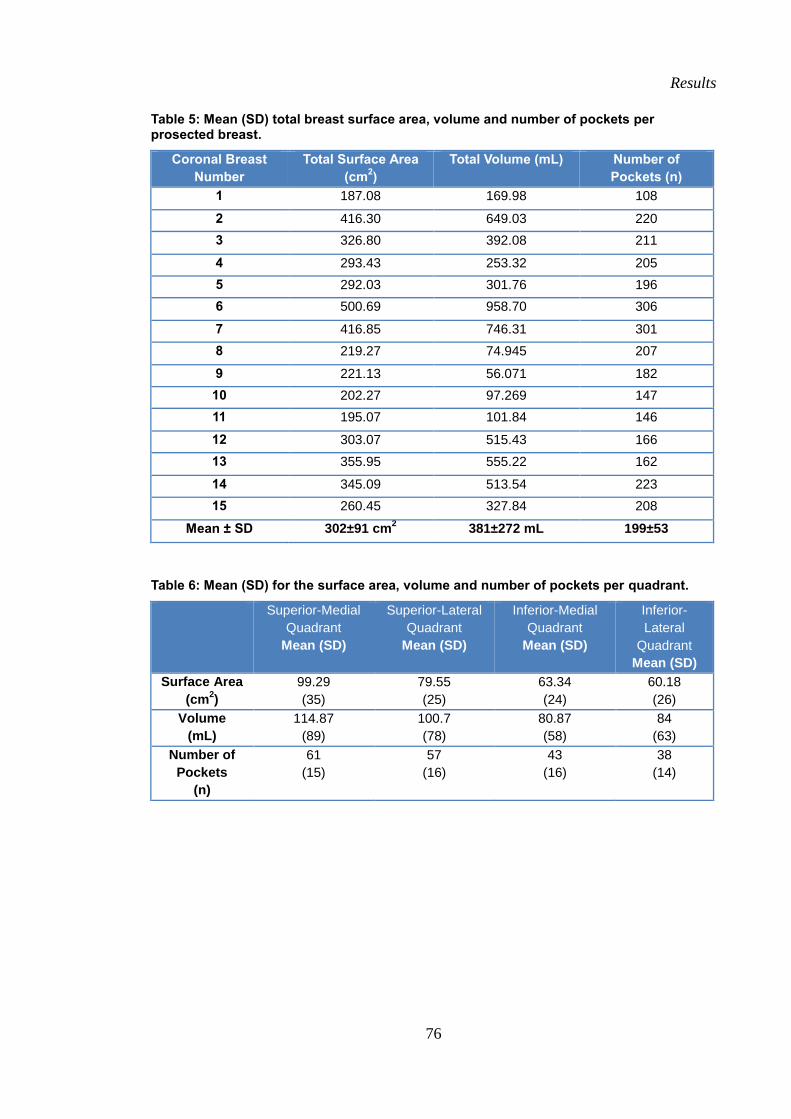
Results
76
Table 5: Mean (SD) total breast surface area, volume and number of pockets per prosected breast.
Coronal Breast
Number
Total Surface Area
(cm2)
Total Volume (mL) Number of
Pockets (n)
1 187.08 169.98 108
2 416.30 649.03 220
3 326.80 392.08 211
4 293.43 253.32 205
5 292.03 301.76 196
6 500.69 958.70 306
7 416.85 746.31 301
8 219.27 74.945 207
9 221.13 56.071 182
10 202.27 97.269 147
11 195.07 101.84 146
12 303.07 515.43 166
13 355.95 555.22 162
14 345.09 513.54 223
15 260.45 327.84 208
Mean ± SD 302±91 cm2 381±272 mL 199±53
Table 6: Mean (SD) for the surface area, volume and number of pockets per quadrant.
Superior-Medial
Quadrant
Mean (SD)
Superior-Lateral
Quadrant
Mean (SD)
Inferior-Medial
Quadrant
Mean (SD)
Inferior-
Lateral
Quadrant
Mean (SD)
Surface Area
(cm2)
99.29
(35)
79.55
(25)
63.34
(24)
60.18
(26)
Volume
(mL)
114.87
(89)
100.7
(78)
80.87
(58)
84
(63)
Number of
Pockets
(n)
61
(15)
57
(16)
43
(16)
38
(14)
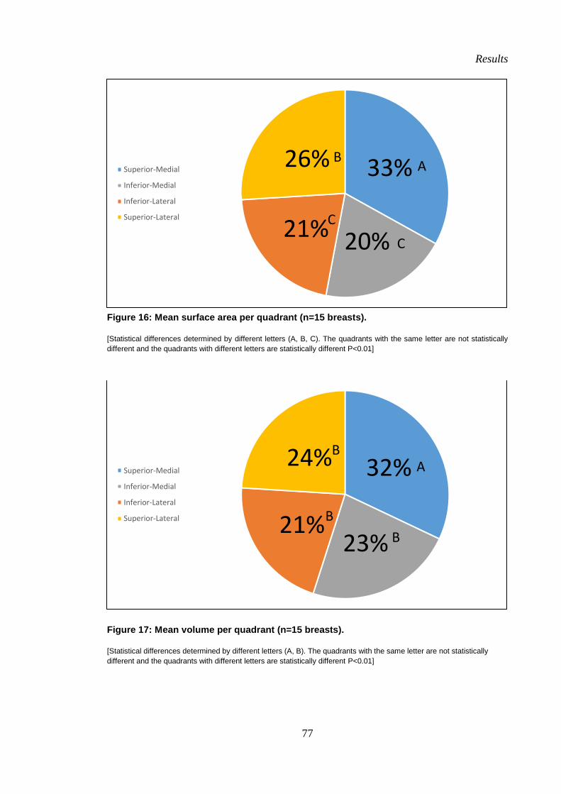
Results
77
Figure 16: Mean surface area per quadrant (n=15 breasts).
[Statistical differences determined by different letters (A, B, C). The quadrants with the same letter are not statistically
different and the quadrants with different letters are statistically different P<0.01]
Figure 17: Mean volume per quadrant (n=15 breasts).
[Statistical differences determined by different letters (A, B). The quadrants with the same letter are not statistically
different and the quadrants with different letters are statistically different P<0.01]
33%
20% 21%
26% Superior-Medial
Inferior-Medial
Inferior-Lateral
Superior-Lateral
A B
C
C
32%
23% 21%
24% Superior-Medial
Inferior-Medial
Inferior-Lateral
Superior-Lateral
A B
B
B
Results
78
3.1.3 Fibro-adipose mass
The mean total fibro-adipose mass per breast of the 14 breasts dissected in the coronal
plane was 172±103 g (range: 50–385 g). The fibro-adipose mass per quadrant (g) and as
a percentage of the total fibro-adipose mass of the breast within each quadrant is
displayed in Table 7 and Figure 18. The fibro-adipose mass within each quadrant was
not evenly distributed amongst the quadrant. The superior-medial and superior-lateral
quadrants were found to have a significantly greater (P<0.01) mean fibro-adipose mass
(32 %; mean 50.8±29 g) and (30 %; mean 50.8±29 g) respectively compared to the
inferior-medial and inferior-lateral quadrants (20 %; 35.3±25 g and 18 %; 29.3±19 g
respectively). The fibro-adipose mass within the superior-medial and superior-lateral
quadrants were not significantly different to each other and the fibro-adipose mass
within inferior-medial and the inferior-lateral quadrants were not significantly different
to each other (Figure 18; P>0.05)
3.1.4 Fibro-glandular mass
The mean total fibro-glandular mass of 14 breasts dissected and measured in the coronal
plane was 184±77 g (range: 56-300 g). The fibro-glandular mass per quadrant (g) and as
a percentage of the total fibro- glandular mass of the breast are displayed in Table 7 and
Figure 19. The distribution of the fibro-glandular mass was evenly distributed amongst
the quadrants, with no significant difference in the percentage of the fibro-glandular
mass in each quadrant found (P>0.05). The superior-medial had 22 % (40.4±24 g) of
the total fibro-glandular mass, the superior-lateral 24 % (46.3±25 g), inferior-medial 26
% (46.4±22 g) and the inferior-lateral quadrant 28 % (50.9±24 g).
3.1.5 Breast Mass
The mean combined breast mass per breast of 14 breasts dissected and measured in the
coronal plane was 355±157 g (range: 105–598 g). The combined breast mass of each of
Results
79
the breasts in grams (g) and as a percentage of total fibro-glandular and total fibro-
adipose tissue within the total breast and per quadrant is displayed in Table 7 and Figure
20. With the cadavers in a supine position, there was no significant difference found in
the combined breast mass found in each quadrant (P>0.05). The superior-lateral
quadrant had the greatest mean breast mass (102.7±56 g) with 28 % of the combined
breast mass, followed by the superior-medial quadrant (91.2±52 g) with 25 %, the
inferior-medial quadrant (81.7±34 g) with 24 % and the inferior-lateral quadrant
(80.2±31 g) with 24 %.
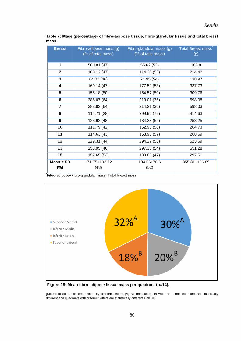
Results
80
Superior-Medial
Inferior-Medial
Inferior-Lateral
Superior-Lateral
Table 7: Mass (percentage) of fibro-adipose tissue, fibro-glandular tissue and total breast mass.
Breast
Fibro-adipose mass (g)
(% of total mass)
Fibro-glandular mass (g)
(% of total mass)
Total Breast mass*
(g)
1 50.181 (47) 55.62 (53) 105.8
2 100.12 (47) 114.30 (53) 214.42
3 64.02 (46) 74.95 (54) 138.97
4 160.14 (47) 177.59 (53) 337.73
5 155.18 (50) 154.57 (50) 309.76
6 385.07 (64) 213.01 (36) 598.08
7 383.83 (64) 214.21 (36) 598.03
8 114.71 (28) 299.92 (72) 414.63
9 123.92 (48) 134.33 (52) 258.25
10 111.79 (42) 152.95 (58) 264.73
11 114.63 (43) 153.96 (57) 268.59
12 229.31 (44) 294.27 (56) 523.59
13 253.95 (46) 297.33 (54) 551.28
15 157.65 (53) 139.86 (47) 297.51
Mean ± SD
(%)
171.75±102.72
(48)
184.06±76.6
(52)
355.81±156.89
*Fibro-adipose+Fibro-glandular mass=Total breast mass
Figure 18: Mean fibro-adipose tissue mass per quadrant (n=14).
[Statistical difference determined by different letters (A, B), the quadrants with the same letter are not statistically
different and quadrants with different letters are statistically different P<0.01]
32%A 30%A
18%B 20%B
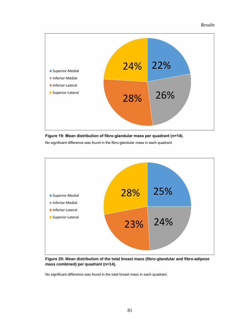
Results
81
Figure 19: Mean distribution of fibro-glandular mass per quadrant (n=14).
No significant difference was found in the fibro-glandular mass in each quadrant.
Figure 20: Mean distribution of the total breast mass (fibro-glandular and fibro-adipose
mass combined) per quadrant (n=14).
No significant difference was found in the total breast mass in each quadrant.
22%
26% 28%
24% Superior-Medial
Inferior-Medial
Inferior-Lateral
Superior-Lateral
25%
24% 23%
28% Superior-Medial
Inferior-Medial
Inferior-Lateral
Superior-Lateral
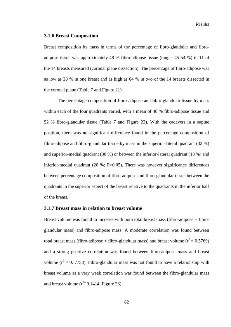
Results
82
3.1.6 Breast Composition
Breast composition by mass in terms of the percentage of fibro-glandular and fibro-
adipose tissue was approximately 48 % fibro-adipose tissue (range: 45-54 %) in 11 of
the 14 breasts measured (coronal plane dissection). The percentage of fibro-adipose was
as low as 28 % in one breast and as high as 64 % in two of the 14 breasts dissected in
the coronal plane (Table 7 and Figure 21).
The percentage composition of fibro-adipose and fibro-glandular tissue by mass
within each of the four quadrants varied, with a mean of 48 % fibro-adipose tissue and
52 % fibro-glandular tissue (Table 7 and Figure 22). With the cadavers in a supine
position, there was no significant difference found in the percentage composition of
fibro-adipose and fibro-glandular tissue by mass in the superior-lateral quadrant (32 %)
and superior-medial quadrant (30 %) or between the inferior-lateral quadrant (18 %) and
inferior-medial quadrant (20 %; P>0.05). There was however significance differences
between percentage composition of fibro-adipose and fibro-glandular tissue between the
quadrants in the superior aspect of the breast relative to the quadrants in the inferior half
of the breast.
3.1.7 Breast mass in relation to breast volume
Breast volume was found to increase with both total breast mass (fibro-adipose + fibro-
glandular mass) and fibro-adipose mass. A moderate correlation was found between
total breast mass (fibro-adipose + fibro-glandular mass) and breast volume (r2
= 0.5769)
and a strong positive correlation was found between fibro-adipose mass and breast
volume (r2
= 0. 7758). Fibro-glandular mass was not found to have a relationship with
breast volume as a very weak correlation was found between the fibro-glandular mass
and breast volume (r2=
0.1414; Figure 23).
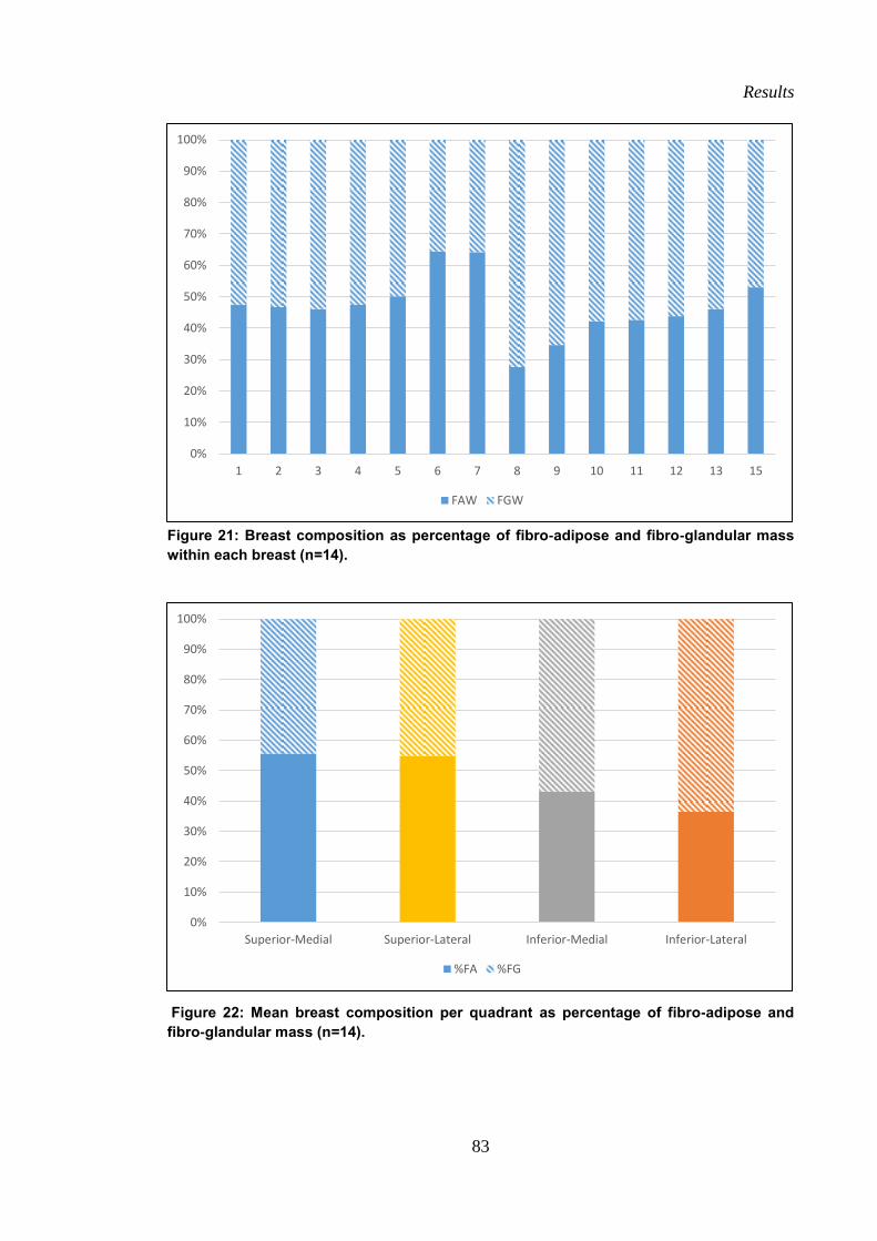
Results
83
Figure 21: Breast composition as percentage of fibro-adipose and fibro-glandular mass
within each breast (n=14).
Figure 22: Mean breast composition per quadrant as percentage of fibro-adipose and
fibro-glandular mass (n=14).
0%
10%
20%
30%
40%
50%
60%
70%
80%
90%
100%
1 2 3 4 5 6 7 8 9 10 11 12 13 15
FAW FGW
0%
10%
20%
30%
40%
50%
60%
70%
80%
90%
100%
Superior-Medial Superior-Lateral Inferior-Medial Inferior-Lateral
%FA %FG
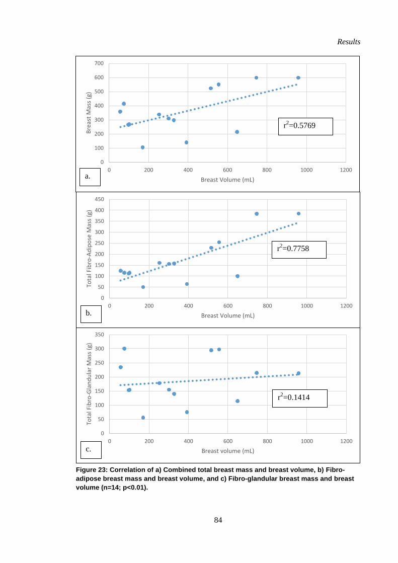
Results
84
Figure 23: Correlation of a) Combined total breast mass and breast volume, b) Fibro-
adipose breast mass and breast volume, and c) Fibro-glandular breast mass and breast
volume (n=14; p<0.01).
0
100
200
300
400
500
600
700
0 200 400 600 800 1000 1200
Bre
ast
Mas
s (g
)
Breast Volume (mL)
0
50
100
150
200
250
300
350
0 200 400 600 800 1000 1200
Tota
l Fib
ro-G
lan
du
lar
Mas
s (g
)
Breast volume (mL)
0
50
100
150
200
250
300
350
400
450
0 200 400 600 800 1000 1200
Tota
l Fib
ro-A
dip
ose
Mas
s (g
)
Breast Volume (mL)
a.
b.
c.
r2=0.1414
r2=0.7758
r2=0.5769
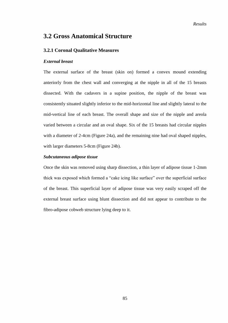
Results
85
3.2 Gross Anatomical Structure
3.2.1 Coronal Qualitative Measures
External breast
The external surface of the breast (skin on) formed a convex mound extending
anteriorly from the chest wall and converging at the nipple in all of the 15 breasts
dissected. With the cadavers in a supine position, the nipple of the breast was
consistently situated slightly inferior to the mid-horizontal line and slightly lateral to the
mid-vertical line of each breast. The overall shape and size of the nipple and areola
varied between a circular and an oval shape. Six of the 15 breasts had circular nipples
with a diameter of 2-4cm (Figure 24a), and the remaining nine had oval shaped nipples,
with larger diameters 5-8cm (Figure 24b).
Subcutaneous adipose tissue
Once the skin was removed using sharp dissection, a thin layer of adipose tissue 1-2mm
thick was exposed which formed a “cake icing like surface” over the superficial surface
of the breast. This superficial layer of adipose tissue was very easily scraped off the
external breast surface using blunt dissection and did not appear to contribute to the
fibro-adipose cobweb structure lying deep to it.
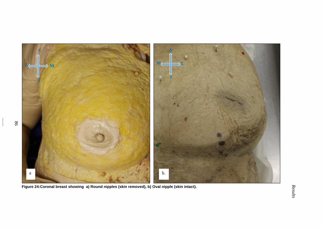
86
Resu
lts
Figure 24:Coronal breast showing a) Round nipples (skin removed), b) Oval nipple (skin intact).
a b.
S
L M
I
S
M L
I
Results
87
Fibro-adipose structure
The bulk of the external mound of the breast was formed by a three-dimensional fibro-
adipose cobweb structure (Figure 25), which was located deep to the superficial surface
layer of subcutaneous adipose tissue. This three-dimensional breast mound was formed
by a scaffolding of fibrous tissue, which formed a vast collection of fibrous tissue
pockets, with adjacent pockets sharing their walls. Each pocket was completely filled
with adipose tissue, such that a fairly flat external surface was formed. In 12 of the 15
breasts, the adipose tissue was easily dissected from the fibrous walls of the pocket
using blunt dissection. However, in three breasts it was very difficult to separate the
adipose tissue from the fibrous pocket with blunt dissection as the fibrous tissue
appeared to be thinner, which made the separation of tissues difficult without destroying
tissue.
Regional variation was found in the surface area and depth of the individual
pockets within the three-dimensional cobweb structure. In the superior-medial quadrant,
the surface area of the pockets was the largest and in the superior-lateral quadrant, the
depth of the pockets was the deepest (Figure 26). The shape of the pockets also
exhibited regional variations in the majority of the breasts (13 of the 15 breasts
dissected in the coronal plane). The pockets were square shaped with rounded edges in
the superior two quadrants of the breast and more of an oval shape in the inferior two
quadrants of the breast. Two of the 15 breasts however exhibited no regional variation
in the shape of the pockets, having oval shaped pockets in all of the quadrants.
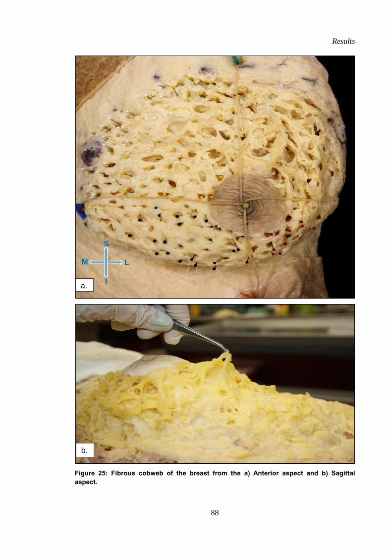
Results
88
Figure 25: Fibrous cobweb of the breast from the a) Anterior aspect and b) Sagittal
aspect.
a.
b.
S
M L
I
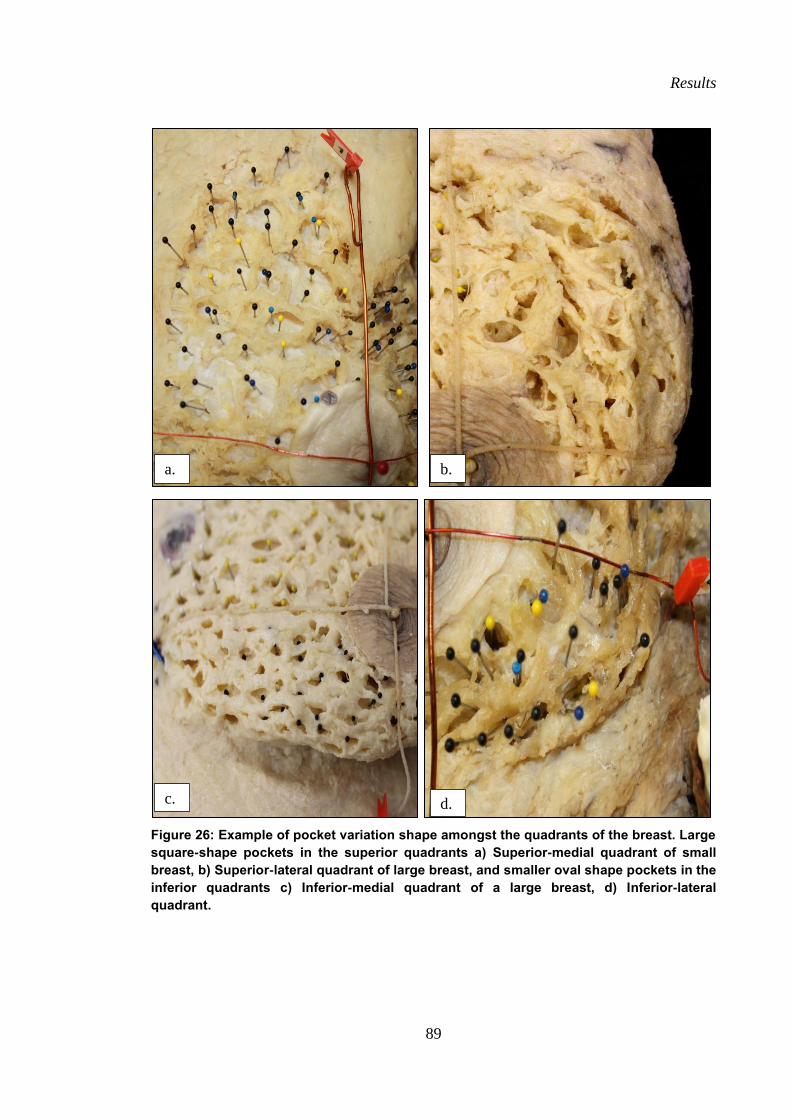
Results
89
Figure 26: Example of pocket variation shape amongst the quadrants of the breast. Large
square-shape pockets in the superior quadrants a) Superior-medial quadrant of small
breast, b) Superior-lateral quadrant of large breast, and smaller oval shape pockets in the
inferior quadrants c) Inferior-medial quadrant of a large breast, d) Inferior-lateral
quadrant.
a.
c.
b.
d.
Results
90
Fibro-glandular tissue
The fibro-glandular tissue was located deep to the bulk of the fibro-adipose cobweb
structure and adhered to a deeper, thinner and flatter layer of fibrous-adipose tissue
requiring sharp dissection to remove it. Within the non-uniform fibro-glandular
structure, the glandular tissue (gland and ducts) could not be differentiated
macroscopically from the fibrous tissue. In all 15 breasts, the fibro-glandular tissue was
located within the central region of the breast, with the bulk of the gland located inferior
to the nipple (Figure 27a). In the majority of the breasts, (12 of the 15 breasts), the
fibro-glandular structure extended unevenly into the all four of the quadrants. Three of
the breasts had fibro-glandular located in only three of the four quadrants, with no fibro-
glandular tissue found in the superior-medial quadrant.
Fibro-adipose tissue (deep to the fibro-glandular tissue)
Deep (posterior) to the fibro-glandular tissue, the thinner and flatter layer of fibrous-
adipose tissue did not have the same three-dimensional cobweb structure as the bulk of
the fibrous-adipose tissue structure superficial to the fibro-glandular tissue (Figure 27b).
Any pocket-like structures identified in this layer were very fragile and broke with blunt
dissection. This deeper fibrous-adipose tissue layer required only blunt dissection only
to separate it from superficial fascia of the following muscles, pectoralis major (sternal
head), serratus anterior (anterior fibres), rectus abdominals (superior fibres) and external
oblique muscles (superior fibres)
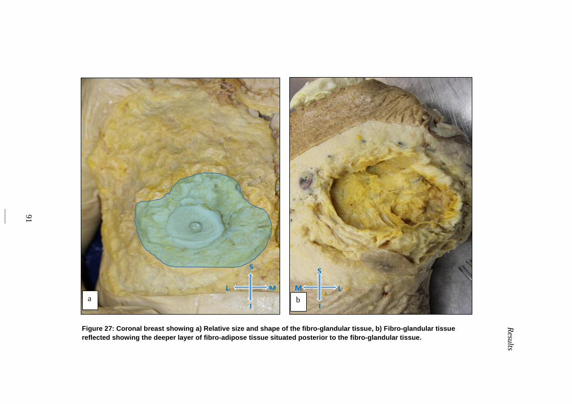
91
Resu
lts
Figure 27: Coronal breast showing a) Relative size and shape of the fibro-glandular tissue, b) Fibro-glandular tissue
reflected showing the deeper layer of fibro-adipose tissue situated posterior to the fibro-glandular tissue.
S
M L
I
S
L M
I a b
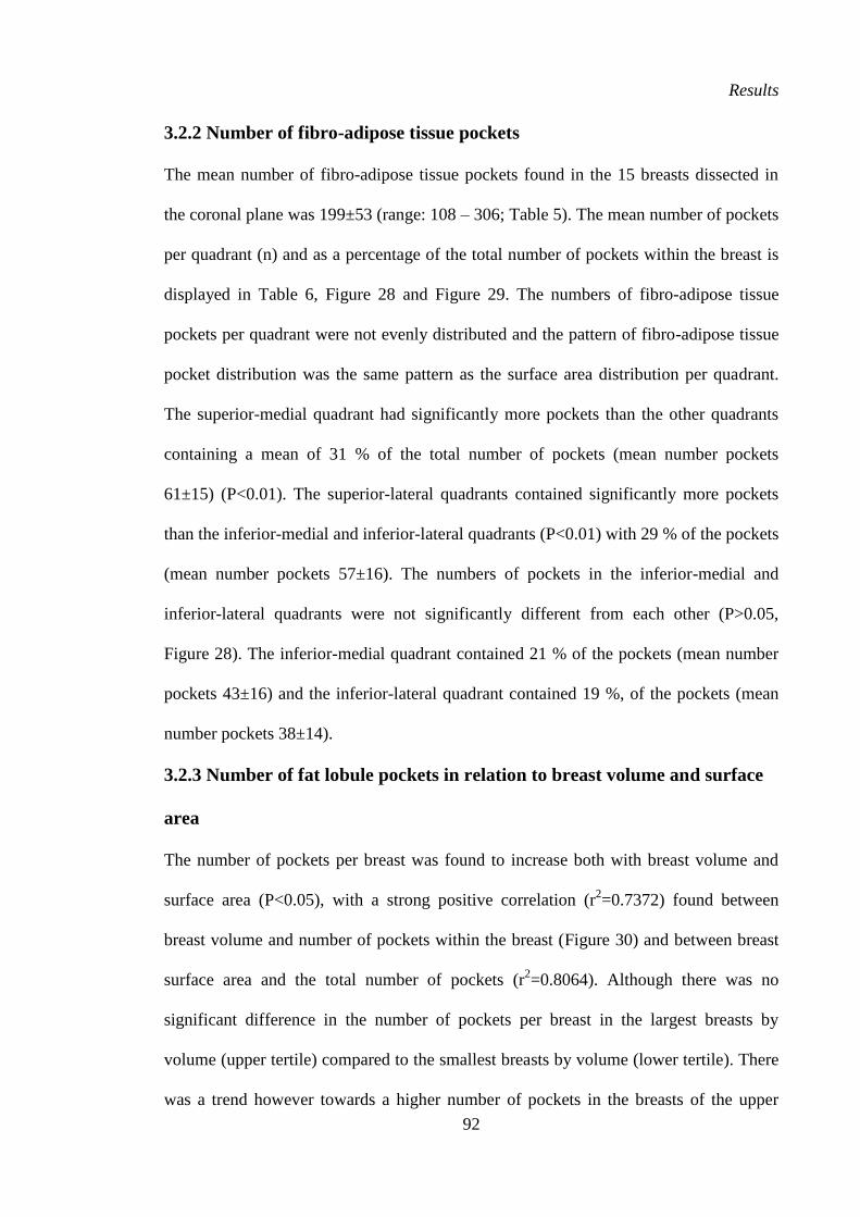
Results
92
3.2.2 Number of fibro-adipose tissue pockets
The mean number of fibro-adipose tissue pockets found in the 15 breasts dissected in
the coronal plane was 199±53 (range: 108 – 306; Table 5). The mean number of pockets
per quadrant (n) and as a percentage of the total number of pockets within the breast is
displayed in Table 6, Figure 28 and Figure 29. The numbers of fibro-adipose tissue
pockets per quadrant were not evenly distributed and the pattern of fibro-adipose tissue
pocket distribution was the same pattern as the surface area distribution per quadrant.
The superior-medial quadrant had significantly more pockets than the other quadrants
containing a mean of 31 % of the total number of pockets (mean number pockets
61±15) (P<0.01). The superior-lateral quadrants contained significantly more pockets
than the inferior-medial and inferior-lateral quadrants (P<0.01) with 29 % of the pockets
(mean number pockets 57±16). The numbers of pockets in the inferior-medial and
inferior-lateral quadrants were not significantly different from each other (P>0.05,
Figure 28). The inferior-medial quadrant contained 21 % of the pockets (mean number
pockets 43±16) and the inferior-lateral quadrant contained 19 %, of the pockets (mean
number pockets 38±14).
3.2.3 Number of fat lobule pockets in relation to breast volume and surface
area
The number of pockets per breast was found to increase both with breast volume and
surface area (P<0.05), with a strong positive correlation (r2=0.7372) found between
breast volume and number of pockets within the breast (Figure 30) and between breast
surface area and the total number of pockets (r2=0.8064). Although there was no
significant difference in the number of pockets per breast in the largest breasts by
volume (upper tertile) compared to the smallest breasts by volume (lower tertile). There
was a trend however towards a higher number of pockets in the breasts of the upper
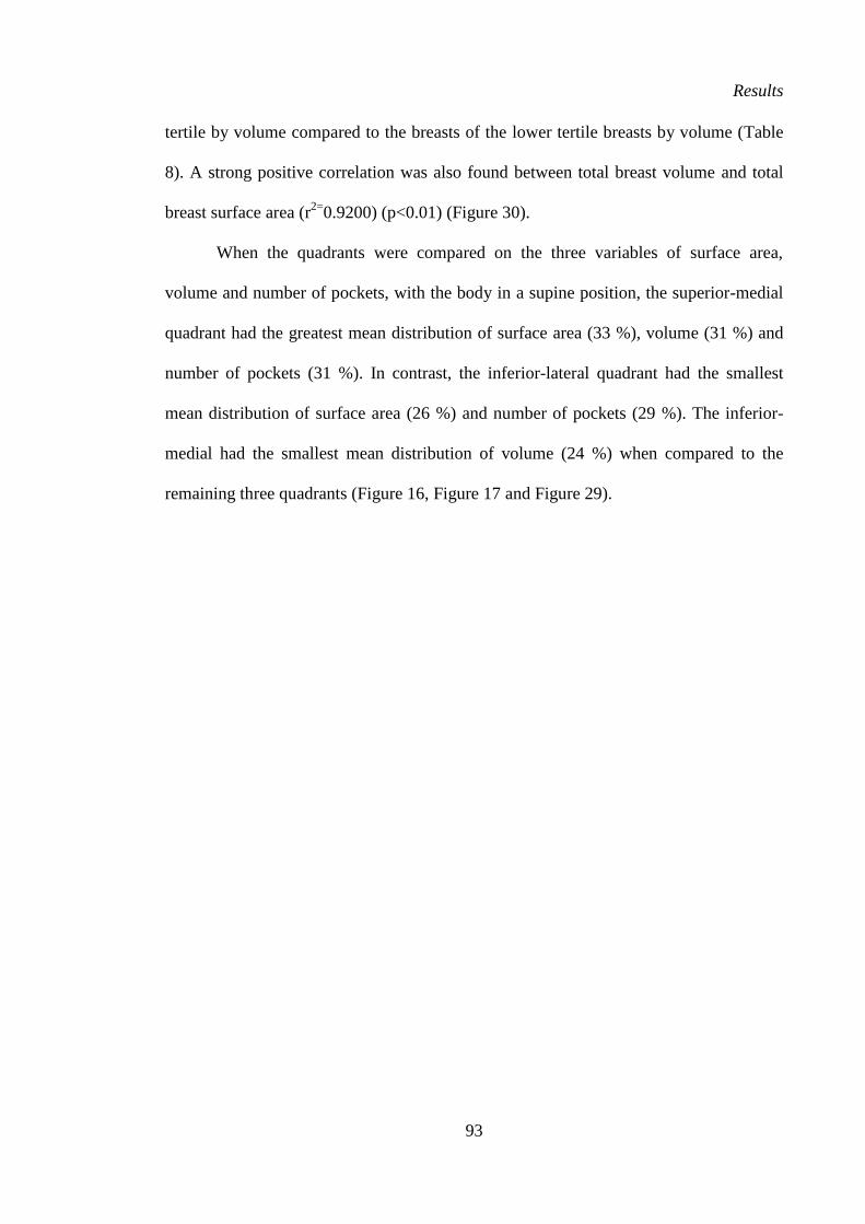
Results
93
tertile by volume compared to the breasts of the lower tertile breasts by volume (Table
8). A strong positive correlation was also found between total breast volume and total
breast surface area (r2=
0.9200) (p<0.01) (Figure 30).
When the quadrants were compared on the three variables of surface area,
volume and number of pockets, with the body in a supine position, the superior-medial
quadrant had the greatest mean distribution of surface area (33 %), volume (31 %) and
number of pockets (31 %). In contrast, the inferior-lateral quadrant had the smallest
mean distribution of surface area (26 %) and number of pockets (29 %). The inferior-
medial had the smallest mean distribution of volume (24 %) when compared to the
remaining three quadrants (Figure 16, Figure 17 and Figure 29).
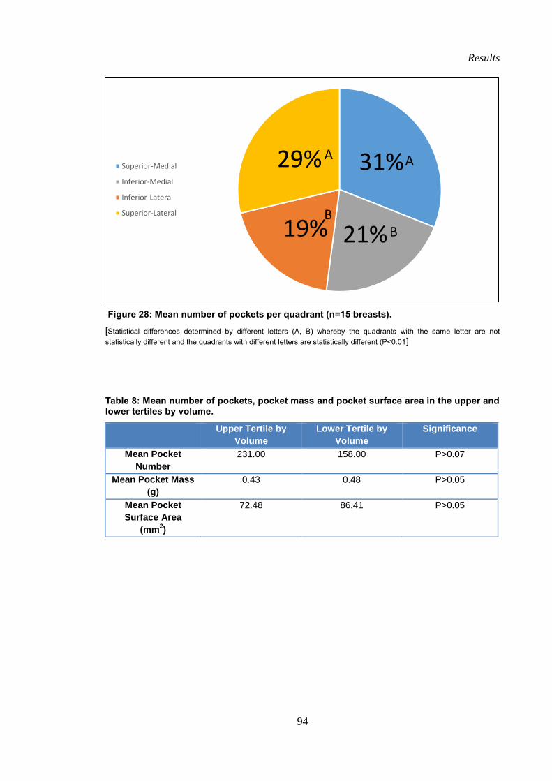
Results
94
Figure 28: Mean number of pockets per quadrant (n=15 breasts).
[Statistical differences determined by different letters (A, B) whereby the quadrants with the same letter are not
statistically different and the quadrants with different letters are statistically different (P<0.01]
Table 8: Mean number of pockets, pocket mass and pocket surface area in the upper and lower tertiles by volume.
Upper Tertile by
Volume
Lower Tertile by
Volume
Significance
Mean Pocket
Number
231.00 158.00 P>0.07
Mean Pocket Mass
(g)
0.43 0.48 P>0.05
Mean Pocket
Surface Area
(mm2)
72.48 86.41 P>0.05
31%
21% 19%
29% Superior-Medial
Inferior-Medial
Inferior-Lateral
Superior-Lateral
A A
B B
95
Resu
lts
Figure 29: Mean surface area, volume and number of pockets per quadrant (Mean and SEM) in each quadrant.
0
20
40
60
80
100
120
140
Superior-Medial Superior-Lateral Inferior-Medial Inferior-Lateral
Surf
ace
Are
a (c
m2 )
, V
olu
me
(mL)
, Nu
mb
er o
f P
ock
ets
(n)
Quadrant
Surface Area Volume No. Pockets
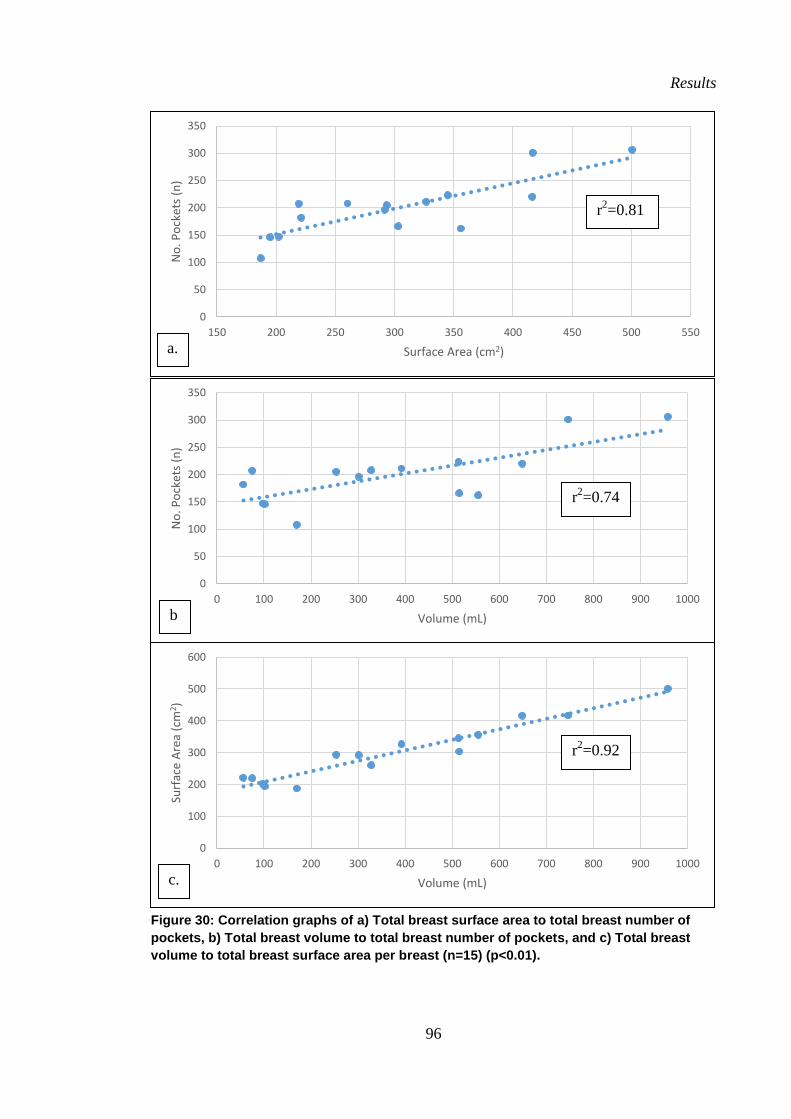
Results
96
Figure 30: Correlation graphs of a) Total breast surface area to total breast number of
pockets, b) Total breast volume to total breast number of pockets, and c) Total breast
volume to total breast surface area per breast (n=15) (p<0.01).
0
100
200
300
400
500
600
0 100 200 300 400 500 600 700 800 900 1000
Surf
ace
Are
a (c
m2)
Volume (mL)
r2=0.92
c.
0
50
100
150
200
250
300
350
0 100 200 300 400 500 600 700 800 900 1000
No
. Po
cket
s (n
)
Volume (mL)
r2=0.74
b
0
50
100
150
200
250
300
350
150 200 250 300 350 400 450 500 550
No
. Po
cket
s (n
)
Surface Area (cm2)
r2=0.81
a.
Results
97
3.2.4 Adipose mass of each pocket
The size of each of the adipose tissue pockets was determined from two measures, the
mass of the adipose tissue encased within the pockets (g) and the surface area of the
adipose tissue pockets measured immediately after the adipose tissue was dissected
(cm2). The adipose tissue mass per pocket of each of the 80 pockets per breast measured
(20 pockets per quadrant per breast) of the 15 breasts dissected in the coronal plane is
displayed in Figure 31. The mean adipose tissue mass per pocket of these 80 pockets
measured in each of the 15 breasts displayed in Figure 32. The overall mean adipose
tissue mass per pocket of the 80 pockets measured in the 15 breasts as a group was
0.4±0.15 g (range: 0.26- 0.61 g).
When isolated pocket size (mass) was compared within each quadrant, the mean
pocket mass within the superior-lateral quadrant was the largest (0.49±0.19 g; Figure
33) and significantly larger (P<0.05) than the mean pocket mass of the other three
quadrants. The mean pocket mass within the inferior-lateral quadrant was significantly
smaller than the other three quadrants (0.34±0.12 g) (P<0.05) and the mean pocket mass
in the superior-medial quadrant (0.43±0.11 g) and the inferior-medial (0.36±0.11 g)
were not significantly different (P>0.5) from each other (Figure 34).
3.2.5 Adipose mass within each pocket in relation to breast volume and
surface area
In contrast to the number of pockets per breast, which was found to have a high positive
correlation with both increases in breast volume and surface area, the pocket size
(measure by the adipose tissue mass within each pocket) was not found to vary with
breast volume or breast surface area. A weak positive correlation was found between the
mean adipose tissue mass of each pocket and total breast volume (r2=0.0588; Figure 39)
and a very weak negative correlation was found between the mean adipose tissue mass
Results
98
of each pocket and the total breast surface area (r2=-0.0055). There was also no
significant difference found in the adipose mass within each pocket of the largest
breasts by volume (upper tertile) compared to the smallest breasts by volume (lower
tertile) P>0.05; (Table 8).
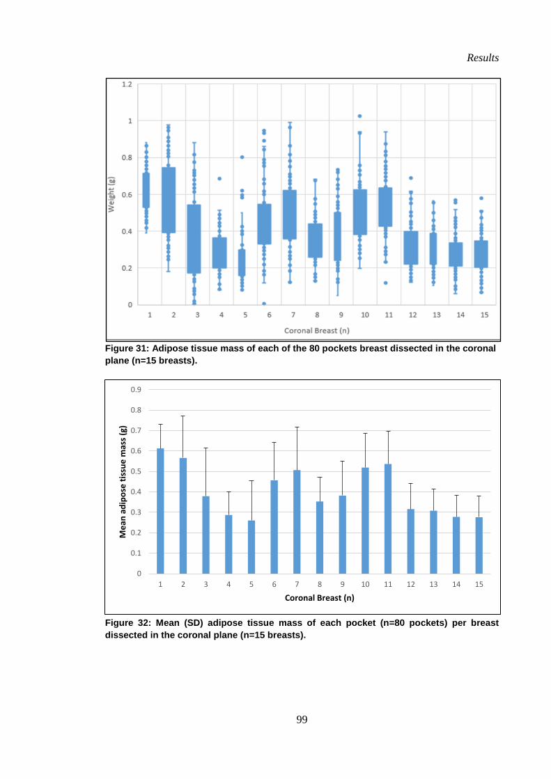
Results
99
Figure 31: Adipose tissue mass of each of the 80 pockets breast dissected in the coronal
plane (n=15 breasts).
Figure 32: Mean (SD) adipose tissue mass of each pocket (n=80 pockets) per breast
dissected in the coronal plane (n=15 breasts).
0
0.1
0.2
0.3
0.4
0.5
0.6
0.7
0.8
0.9
1 2 3 4 5 6 7 8 9 10 11 12 13 14 15
Me
an a
dip
ose
tis
sue
mas
s (g
)
Coronal Breast (n)
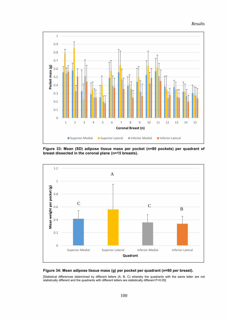
Results
100
Figure 33: Mean (SD) adipose tissue mass per pocket (n=80 pockets) per quadrant of breast dissected in the coronal plane (n=15 breasts).
Figure 34: Mean adipose tissue mass (g) per pocket per quadrant (n=80 per breast).
[Statistical differences determined by different letters (A, B, C) whereby the quadrants with the same letter are not statistically different and the quadrants with different letters are statistically different P<0.05]
0
0.1
0.2
0.3
0.4
0.5
0.6
0.7
0.8
0.9
1
1 2 3 4 5 6 7 8 9 10 11 12 13 14 15
Po
cke
t m
ass
(g)
Coronal Breast (n)
Superior-Medial Superior-Lateral Inferior-Medial Inferior-Lateral
0
0.2
0.4
0.6
0.8
1
1.2
Superior-Medial Superior-Lateral Inferior-Medial Inferior-Lateral
Me
an w
eig
ht
pe
r p
ock
et
(g)
Quadrant
A
B
C
C
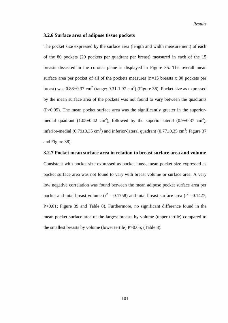
Results
101
3.2.6 Surface area of adipose tissue pockets
The pocket size expressed by the surface area (length and width measurement) of each
of the 80 pockets (20 pockets per quadrant per breast) measured in each of the 15
breasts dissected in the coronal plane is displayed in Figure 35. The overall mean
surface area per pocket of all of the pockets measures (n=15 breasts x 80 pockets per
breast) was 0.88±0.37 cm2 (range: 0.31-1.97 cm
2) (Figure 36). Pocket size as expressed
by the mean surface area of the pockets was not found to vary between the quadrants
(P>0.05). The mean pocket surface area was the significantly greater in the superior-
medial quadrant (1.05±0.42 cm2), followed by the superior-lateral (0.9±0.37 cm
2),
inferior-medial (0.79±0.35 cm2) and inferior-lateral quadrant (0.77±0.35 cm
2; Figure 37
and Figure 38).
3.2.7 Pocket mean surface area in relation to breast surface area and volume
Consistent with pocket size expressed as pocket mass, mean pocket size expressed as
pocket surface area was not found to vary with breast volume or surface area. A very
low negative correlation was found between the mean adipose pocket surface area per
pocket and total breast volume (r2=- 0.1758) and total breast surface area (r
2=-0.1427;
P<0.01; Figure 39 and Table 8). Furthermore, no significant difference found in the
mean pocket surface area of the largest breasts by volume (upper tertile) compared to
the smallest breasts by volume (lower tertile) P>0.05; (Table 8).
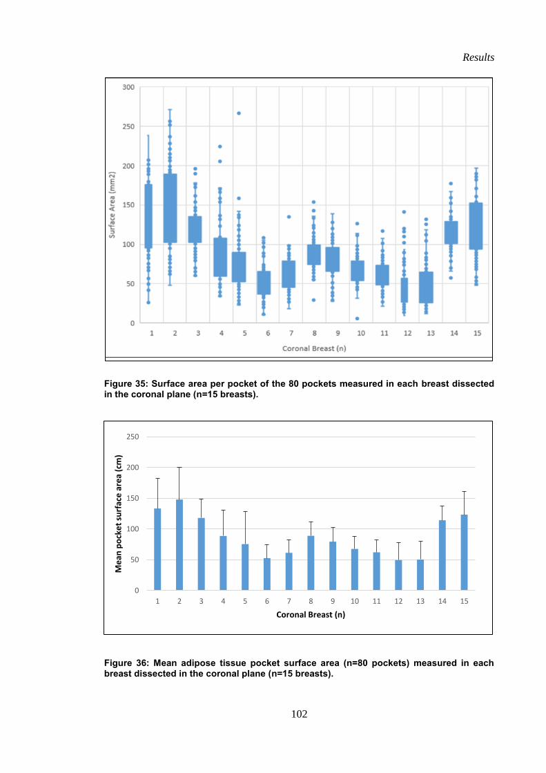
Results
102
Figure 35: Surface area per pocket of the 80 pockets measured in each breast dissected in the coronal plane (n=15 breasts).
Figure 36: Mean adipose tissue pocket surface area (n=80 pockets) measured in each breast dissected in the coronal plane (n=15 breasts).
0
50
100
150
200
250
1 2 3 4 5 6 7 8 9 10 11 12 13 14 15
Me
an p
ock
et
surf
ace
are
a (c
m)
Coronal Breast (n)
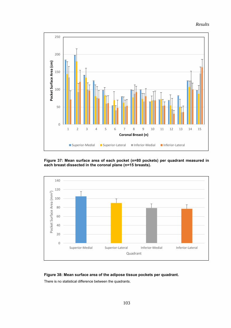
Results
103
Figure 37: Mean surface area of each pocket (n=80 pockets) per quadrant measured in each breast dissected in the coronal plane (n=15 breasts).
Figure 38: Mean surface area of the adipose tissue pockets per quadrant.
There is no statistical difference between the quadrants.
0
50
100
150
200
250
1 2 3 4 5 6 7 8 9 10 11 12 13 14 15
Po
cke
t Su
rfac
e A
rea
(cm
)
Coronal Breast (n)
Superior-Medial Superior-Lateral Inferior-Medial Inferior-Lateral
0
20
40
60
80
100
120
140
Superior-Medial Superior-Lateral Inferior-Medial Inferior-Lateral
Po
cket
Su
rfac
e A
rea
(mm
2)
Quadrant
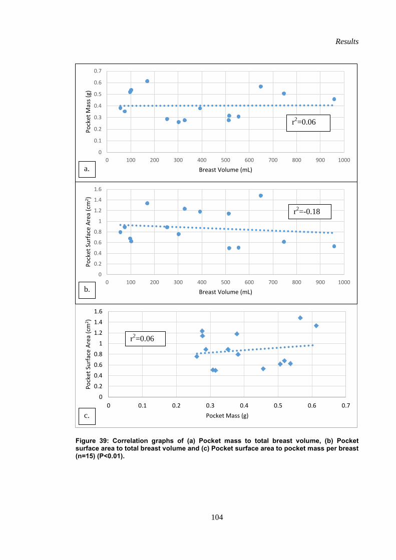
Results
104
Figure 39: Correlation graphs of (a) Pocket mass to total breast volume, (b) Pocket surface area to total breast volume and (c) Pocket surface area to pocket mass per breast (n=15) (P<0.01).
0
0.2
0.4
0.6
0.8
1
1.2
1.4
1.6
0 0.1 0.2 0.3 0.4 0.5 0.6 0.7
Po
cket
Su
rfac
e A
rea
(cm
2 )
Pocket Mass (g)
0
0.1
0.2
0.3
0.4
0.5
0.6
0.7
0 100 200 300 400 500 600 700 800 900 1000
Po
cket
Mas
s (g
)
Breast Volume (mL)
0
0.2
0.4
0.6
0.8
1
1.2
1.4
1.6
0 100 200 300 400 500 600 700 800 900 1000
Po
cket
Su
rfac
e A
rea
(cm
2 )
Breast Volume (mL)
r2=0.06
r2=-0.18
a.
c.
r2=0.06
b.
Results
105
3.2.8 Sagittal Qualitative Measures
Macroscopically the sagittal slices had six (6) observable tissue layers running
superficial to deep; skin, superficial fibro-adipose tissue, fibro-glandular tissue, deep
fibro-adipose tissue, muscle and bone. Amongst the slices, there was some variation in
the thickness of each layer but consistently in all 18 slices, the superficial fibro-adipose
tissue layer was observed visually to be the thickest (n=18).
The fibro-glandular layer was consistently found in all six slices found to be
thicker (>1 cm thick) in the midline slice compared to the more medial and lateral para-
sagittal slices. No glandular tissue was seen in the most medial slices (n=6), which
consisted of very small fibro-adipose pockets bound within tight fibrous strands. Some
glandular tissue was seen in the most lateral slices (<1cm thick), which were embedded
in a large cluster of fibro-adipose tissue pockets.
In contrast to the coronal dissection a clear breast capsule was not evident
macroscopically in any of the sagittal slices. A thin layer of fascia was observed
however running parallel to the skin through the superficial fibro-adipose tissue, which
had multiple fibrous tissue bands connecting this layer of fascial tissue to the dermis.
This was consistent with Cooper’s Anterior Extensions of the Anterior Lamellae. A
thin layer of fascia was not observed however running parallel to the skin posterior to
the gland. Multiple fibrous tissue bands connecting posterior surface of the gland to the
pectoralis major muscle fascia were observed. This was consistent with Cooper’s
Posterior Extensions of the Posterior Lamellae. The Anterior Extensions of the Anterior
Lamellae were consistently thicker than the Posterior Extensions of the Posterior
Lamellae. The Posterior Extensions of the Posterior Lamellae were very numerous but
were easy to separate using blunt dissection.
The three-dimensional fibro-adipose structure when viewed from the sagittal
Results
106
aspect had two distinct sections, one anterior to the gland and one posterior to the gland
(Figure 40). Fibro-adipose pockets were evident anterior and posterior to the gland on
all of the surfaces of the sagittal slices (Figure 40). The pockets anterior to the gland
were larger and less numerous compared to the pockets posterior to the gland. The
collagen of the fibro-adipose tissue anterior to the gland could be traced superficially to
the dermis (Anterior Extensions of the Anterior Lamellae). The collagen of the fibro-
adipose tissue posterior to the gland could be traced posteriorly to the pectoral fascia
(posterior extensions of the posterior lamellae) in an organised pattern. Posterior to the
gland, the adipose tissue pockets appeared to be smaller but more numerous compared
to the fibro-adipose pockets anterior to the gland. The posterior layer of fibro-adipose
tissue varied in depth across the different slices, however in all of the sagittal breast
slice surfaces, the posterior layer was thicker in the mid-nipple slices compared to the
medial and lateral slices. The posterior layer in the mid-nipple slices were consistently
>1 cm, whereas the medial and lateral slices consistently <1 cm thick.
Evidence of perimeter attachments of the breast to the chest wall were evident
on the sagittal breast slices. The superior perimeter attachment of the breast was
observed in four of the 18 slice surfaces, connecting the superior perimeter of the breast
to the inferior portion of the clavicle. An inferior perimeter of the breast connecting to
either the 5th
or 6th
rib was observed in six of the 18 sagittal slices. No lateral or medial
perimeter attachments were observed in the sagittal breast slices. The lateral confluence
was only observed on one slice and it was not connected to bone.

Results
107
Figure 40: Mid-sagittal slice of the breast through the nipple showing the dermis (green)
and the Superficial Pectoral fascia (light blue). The regional depths are circled anterior to
the gland (red), within the gland (purple) and posterior to the gland (dark blue).
Results
108
3.2.9 Number (n) of pockets anterior and posterior to the gland
The mean number of fibro-adipose pockets located anterior and posterior to the gland of
18 sagittal breast slice surfaces (six breast slice surfaces in each of the three breasts;
Figure 41 and Figure 42) is displayed in Table 9. In all 18 breast slices, the number of
fibro-adipose pockets found anterior to the gland was less in number (35±16, range: 20-
49) compared to the number found posterior to the gland (53±31, range: 36-78).
3.2.10 Length of the anterior extensions of the anterior lamellae
The mean length of the 180 number collagen fibres measured that connected the
superficial fibro-adipose cobweb to the dermis (Anterior Extensions of the Anterior
Lamellae) for the 18 breast slice surfaces (six breast slice surfaces in each of the three
breasts) examined in the sagittal plane is shown in Table 9 and Figure 43. The mean
length across the 18 breast slice surfaces was 19±5 mm (range: 9-34 mm). The Anterior
Extensions were longer at the mid-nipple sagittal breast slices (23.5 mm) compared to
the Anterior Extensions measured at the medial and lateral sagittal breast slices (13.5
mm and 19 mm respectively; Figure 44).
3.2.11 Pocket surface area (mm2)
The surface area of the fibro-adipose pockets (minimum 20 pockets measured per
sagittal breast slice surface both anterior and posterior to the gland) measured anterior
and posterior to the gland microscopically in the sagittal plane is displayed in Table 9.
The surface area of the fibro-adipose pockets located anterior to the gland in all of the
18 breast slices were consistently larger (mean: 0.89±0.32 cm2, range: 0.34-1.20 cm
2)
than those located posterior to the gland (mean: 0.33±0.22 cm2, range: 0.22-0.87 cm
2).
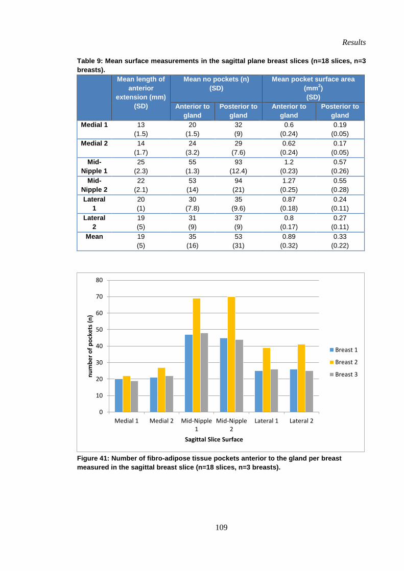
Results
109
Table 9: Mean surface measurements in the sagittal plane breast slices (n=18 slices, n=3
breasts).
Mean length of
anterior
extension (mm)
(SD)
Mean no pockets (n)
(SD)
Mean pocket surface area
(mm2)
(SD)
Anterior to
gland
Posterior to
gland
Anterior to
gland
Posterior to
gland
Medial 1 13
(1.5)
20
(1.5)
32
(9)
0.6
(0.24)
0.19
(0.05)
Medial 2 14
(1.7)
24
(3.2)
29
(7.6)
0.62
(0.24)
0.17
(0.05)
Mid-
Nipple 1
25
(2.3)
55
(1.3)
93
(12.4)
1.2
(0.23)
0.57
(0.26)
Mid-
Nipple 2
22
(2.1)
53
(14)
94
(21)
1.27
(0.25)
0.55
(0.28)
Lateral
1
20
(1)
30
(7.8)
35
(9.6)
0.87
(0.18)
0.24
(0.11)
Lateral
2
19
(5)
31
(9)
37
(9)
0.8
(0.17)
0.27
(0.11)
Mean 19
(5)
35
(16)
53
(31)
0.89
(0.32)
0.33
(0.22)
Figure 41: Number of fibro-adipose tissue pockets anterior to the gland per breast
measured in the sagittal breast slice (n=18 slices, n=3 breasts).
0
10
20
30
40
50
60
70
80
Medial 1 Medial 2 Mid-Nipple1
Mid-Nipple2
Lateral 1 Lateral 2
nu
mb
er
of
po
cke
ts (
n)
Sagittal Slice Surface
Breast 1
Breast 2
Breast 3
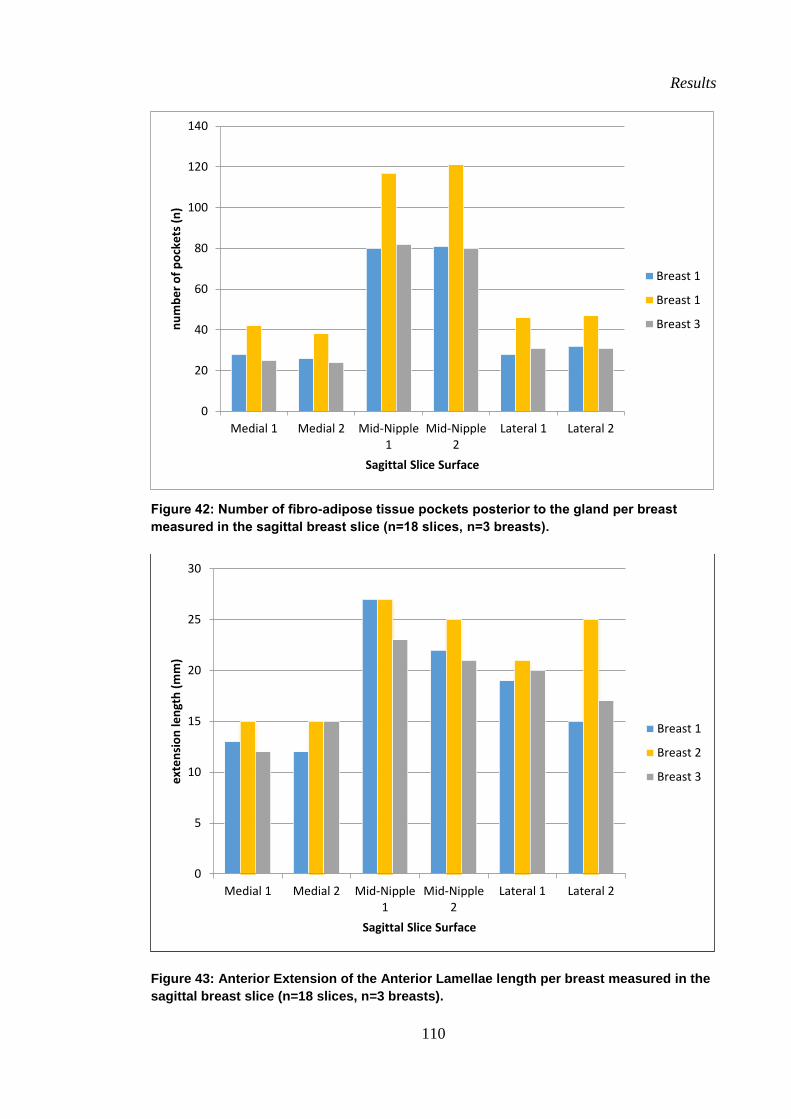
Results
110
Figure 42: Number of fibro-adipose tissue pockets posterior to the gland per breast
measured in the sagittal breast slice (n=18 slices, n=3 breasts).
Figure 43: Anterior Extension of the Anterior Lamellae length per breast measured in the
sagittal breast slice (n=18 slices, n=3 breasts).
0
20
40
60
80
100
120
140
Medial 1 Medial 2 Mid-Nipple1
Mid-Nipple2
Lateral 1 Lateral 2
nu
mb
er
of
po
cke
ts (
n)
Sagittal Slice Surface
Breast 1
Breast 1
Breast 3
0
5
10
15
20
25
30
Medial 1 Medial 2 Mid-Nipple1
Mid-Nipple2
Lateral 1 Lateral 2
ext
en
sio
n le
ngt
h (
mm
)
Sagittal Slice Surface
Breast 1
Breast 2
Breast 3
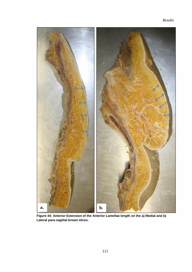
Results
111
Figure 44: Anterior Extension of the Anterior Lamellae length on the a) Medial and b)
Lateral para-sagittal breast slices.
a. b.
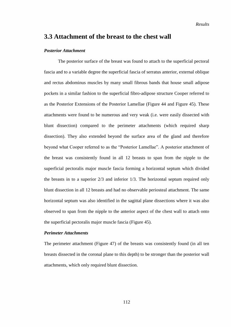
Results
112
3.3 Attachment of the breast to the chest wall
Posterior Attachment
The posterior surface of the breast was found to attach to the superficial pectoral
fascia and to a variable degree the superficial fascia of serratus anterior, external oblique
and rectus abdominus muscles by many small fibrous bands that house small adipose
pockets in a similar fashion to the superficial fibro-adipose structure Cooper referred to
as the Posterior Extensions of the Posterior Lamellae (Figure 44 and Figure 45). These
attachments were found to be numerous and very weak (i.e. were easily dissected with
blunt dissection) compared to the perimeter attachments (which required sharp
dissection). They also extended beyond the surface area of the gland and therefore
beyond what Cooper referred to as the “Posterior Lamellae”. A posterior attachment of
the breast was consistently found in all 12 breasts to span from the nipple to the
superficial pectoralis major muscle fascia forming a horizontal septum which divided
the breasts in to a superior 2/3 and inferior 1/3. The horizontal septum required only
blunt dissection in all 12 breasts and had no observable periosteal attachment. The same
horizontal septum was also identified in the sagittal plane dissections where it was also
observed to span from the nipple to the anterior aspect of the chest wall to attach onto
the superficial pectoralis major muscle fascia (Figure 45).
Perimeter Attachments
The perimeter attachment (Figure 47) of the breasts was consistently found (in all ten
breasts dissected in the coronal plane to this depth) to be stronger than the posterior wall
attachments, which only required blunt dissection.
Results
113
In all 12 coronal dissections, the superior arc attachment of the breast was
consistently found to attach to the anterior aspect of the chest wall onto both the
superficial pectoralis muscle fascia and the deep pectoralis muscle fascia. The medial
and lateral third of the superior arc perimeter attachment were consistently found to
attach to the superficial pectoralis muscle fascia. The most lateral third of the superior
arc attachment consistently curved in an inferior-lateral direction across the superior-
lateral aspect of the pectoralis major muscle, along the superficial pectoralis muscle
fascia where it merged with the lateral arc of the perimeter attachment. In a similar
fashion, the most medial third curved across the medial aspect of the superficial
pectoralis muscle fascia in an inferior-medial direction, where it merged with the fibres
of the medial arc of the perimeter attachment. The middle third of the superior arc
attachment (the apex of the curve) was consistently found to dive between the heads of
the pectoralis major muscle (sternal and clavicular) and intersect with the deep
pectoralis muscle fascia. This facial connection was consistently found to be deformable
but strong as it required sharp dissection. Although no direct periosteal attachment of
the middle third of the superior arc attachment was found, an indirect periosteal
attachment to the clavicle was found via the superficial and deep pectoral fascia which
were observed to traverse superiorly and attach to the clavicle.
The medial arc attachment of the breast was found to tightly adhere the breast
to the chest wall along the lateral edge of the sternum with short fibres running from the
dermis directly to the periosteum of the sternum. Little to no adipose tissue was found
in this region of the perimeter, only dense, short fibres of connective tissue. The medial
attachments were considered to be the strongest of the perimeter attachments due to the
time required to sharply dissect this tissue in comparison to the other perimeter
attachments.
Results
114
The lateral arc attachment consisted of a sling of fascia formed by multiple
irregular shaped bands of fascia from the superficial fascia of three muscles (pectoralis
major, serratus anterior and external oblique muscles). Fascial bands from the superior
edge of external oblique muscle formed the most inferior aspect of the lateral arc, with
fibres running in a transverse direction. These fibres merged with the fascia of the
anterior aspect of serratus anterior, with fibres running in a superior-lateral oblique
direction. The two bands of fibres then traversed the surface of the serratus anterior
muscle as two parallel fascial bands in a superior-medial direction where they
intersected with the fascial from the lateral edge of the pectoralis major muscle (Figure
47). The fascial contribution from each muscle originated separately at the inferior-
lateral border of the breast but had merged to form one sling, at the level of the nipple
(horizontally). Superior to the nipple, the lateral arc perimeter attachment was observed
to be a single sling (Lateral Fascia Confluence[36]
) which traversed the lateral boarder of
the pectoralis major muscle and merged with the most lateral aspect of the superior arc
perimeter attachment. There was no evidence of a “Tail of Spence” as described by
Cooper [1]. In contrast to the medial attachment, the lateral attachment was found to
consist of longer fibrous strands, which were interspersed with larger lobules of adipose
tissue within the axilla.
The inferior arc attachment of the breast, also known as the Inframammary
Fold (IMF) was found to be a tightly adhered band of tissue anchoring the inferior
portion of the breast strongly to the superficial fascia of the anterior chest wall
(superficial fascial pectoralis major, external oblique and rectus abdominus muscles).
From the sagittal breast slices there appears to be some space in which direct
connections may exist between the fibro-adipose tissue of the breast (posterior
extensions of the posterior lamellae) and the 5th
rib in the most breast slices. This occurs
Results
115
in the space between the inferior border of the pectoralis major muscle and the superior
border of the rectus abdominus muscle and external oblique muscle fibres (Figure 45i-
iii). The medial third of the IMF was found to arc in a superior-medial oblique direction
to the sternum where it merged with the fibres of the medial arc attachment. The lateral
third of the IMF was seen to merge with the fibres of the lateral perimeter (what
Matousek et al, 2014 referred to as the Lateral Fascial Confluence [36]
).
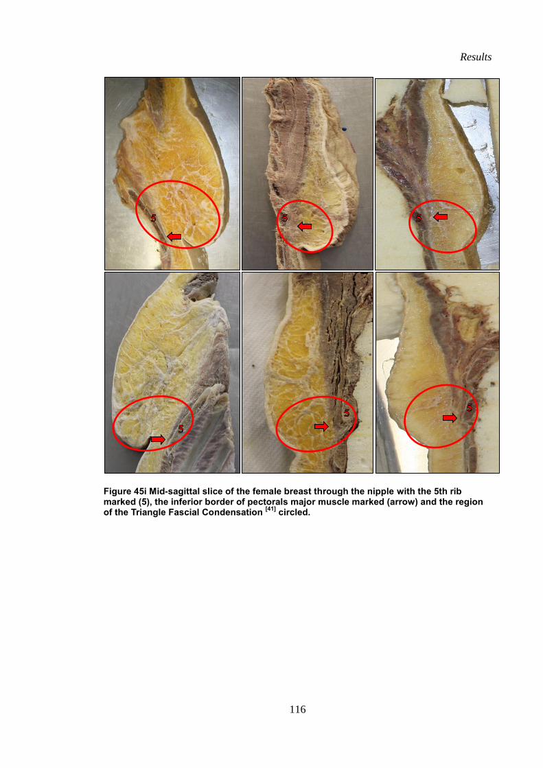
Results
116
Figure 45i Mid-sagittal slice of the female breast through the nipple with the 5th rib marked (5), the inferior border of pectorals major muscle marked (arrow) and the region of the Triangle Fascial Condensation
[41] circled.
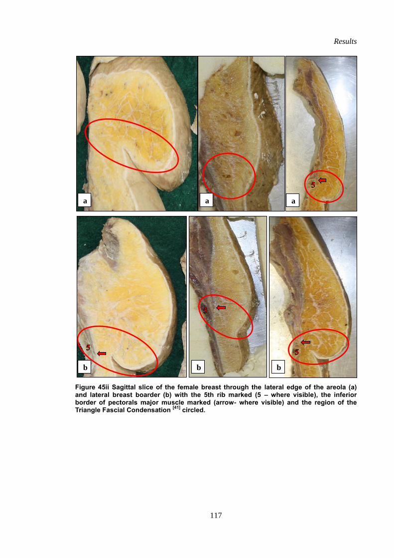
Results
117
Figure 45ii Sagittal slice of the female breast through the lateral edge of the areola (a) and lateral breast boarder (b) with the 5th rib marked (5 – where visible), the inferior border of pectorals major muscle marked (arrow- where visible) and the region of the Triangle Fascial Condensation
[41] circled.
a
.
a
.
a
.
b
.
b
.
b
.

Results
118
Figure 45iii Sagittal slice of the female breast through the medial edge of the areola (a) and medial breast boarder (b) with the 5th rib marked (5), the inferior border of pectorals major muscle marked (arrow) and the region of the Triangle Fascial Condensation
[41]
circled.
a
.
a
.
a
.
b
.
b
.
b
.
Results
119
3.3.1 Perimeter: Regional Anatomy
From the coronal plane dissection of the perimeter (at the depth where the fibro-adipose
and fibro-glandular tissues had been removed), the perimeter of the breasts were
consistently observed in the 12 breasts dissected to this depth to have an irregular and
uneven circular shape (Figure 47 and Figure 49). In 10 of the 12 breasts dissected to this
level, the apex of the superior perimeter (middle one third) was located between the
sternal and clavicular heads of the pectoralis major muscle, with the rest of the superior
perimeter attaching to the superficial fascia of pectoralis major muscle. In the remaining
two of the 12 breasts, the superior perimeter was located 2-3cm inferior to the division
of the sternal and clavicular heads of the pectoralis major muscle.
Once the superficial chest wall muscles were removed, the location of the
perimeter relative to the ribs revealed that the most superior aspect of the “circle”
shaped perimeter was located within the 3rd
intercostal space, in vertical alignment with
the mid clavicular line. The most inferior aspect of the “circle” of the perimeter was also
consistently in vertical alignment with the mid-clavicular line in all of the twelve
breasts, however the location of the inferior aspect of the “circle” of the perimeter
relative to the ribs was found to vary. In eight of the 12 breasts, it was level with the
sixth rib, in three it was level with the fifth rib and in one of the breasts there was no
visible bony attachment of the inferior perimeter of the breasts.
The medial aspect of the curvature of the perimeter was consistently found to be
in contact with the lateral boarder of the sternum at the level of the 4th
intercostal space.
The lateral curvature of the perimeter of the breasts were found to be have two distinct
parts, one slightly more lateral and superior (lateral/superior sling) than the other
(medial/inferior sling). The lateral/superior slings were consistently found to follow an
arc along the mid-axillary line and the medial/inferior slings were consistently found to
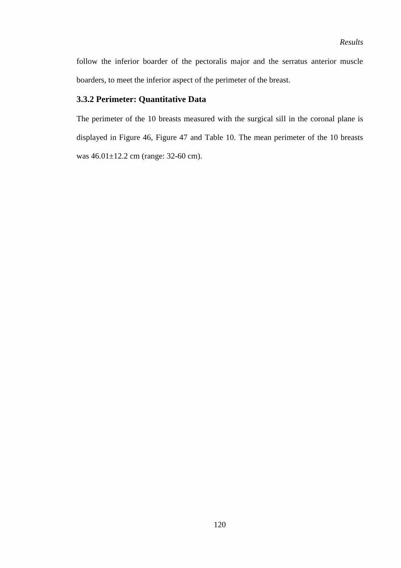
Results
120
follow the inferior boarder of the pectoralis major and the serratus anterior muscle
boarders, to meet the inferior aspect of the perimeter of the breast.
3.3.2 Perimeter: Quantitative Data
The perimeter of the 10 breasts measured with the surgical sill in the coronal plane is
displayed in Figure 46, Figure 47 and Table 10. The mean perimeter of the 10 breasts
was 46.01±12.2 cm (range: 32-60 cm).
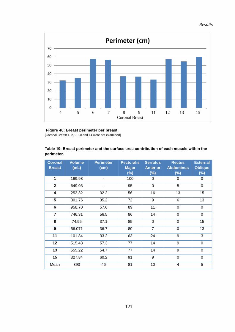
Results
121
Figure 46: Breast perimeter per breast. [Coronal Breast 1, 2, 3, 10 and 14 were not examined]
Table 10: Breast perimeter and the surface area contribution of each muscle within the
perimeter.
0
10
20
30
40
50
60
70
1 2 3 4 5 6 7 8 9 10
Perimeter (cm)
Coronal
Breast
Volume
(mL)
Perimeter
(cm)
Pectoralis
Major
(%)
Serratus
Anterior
(%)
Rectus
Abdominus
(%)
External
Oblique
(%)
1 169.98 - 100 0 0 0
2 649.03 - 95 0 5 0
4 253.32 32.2 56 16 13 15
5 301.76 35.2 72 9 6 13
6 958.70 57.6 89 11 0 0
7 746.31 56.5 86 14 0 0
8 74.95 37.1 85 0 0 15
9 56.071 36.7 80 7 0 13
11 101.84 33.2 63 24 9 3
12 515.43 57.3 77 14 9 0
13 555.22 54.7 77 14 9 0
15 327.84 60.2 91 9 0 0
Mean 393 46 81 10 4 5
4 5 6 7 8 9 11 12 13 15
Coronal Breast
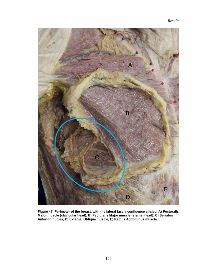
Results
122
Figure 47: Perimeter of the breast, with the lateral fascia confluence circled. A) Pectoralis Major muscle (clavicular head), B) Pectoralis Major muscle (sternal head), C) Serratus Anterior mucles, D) External Oblique muscle, E) Rectus Abdominus muscle.
A
B
C
E
D
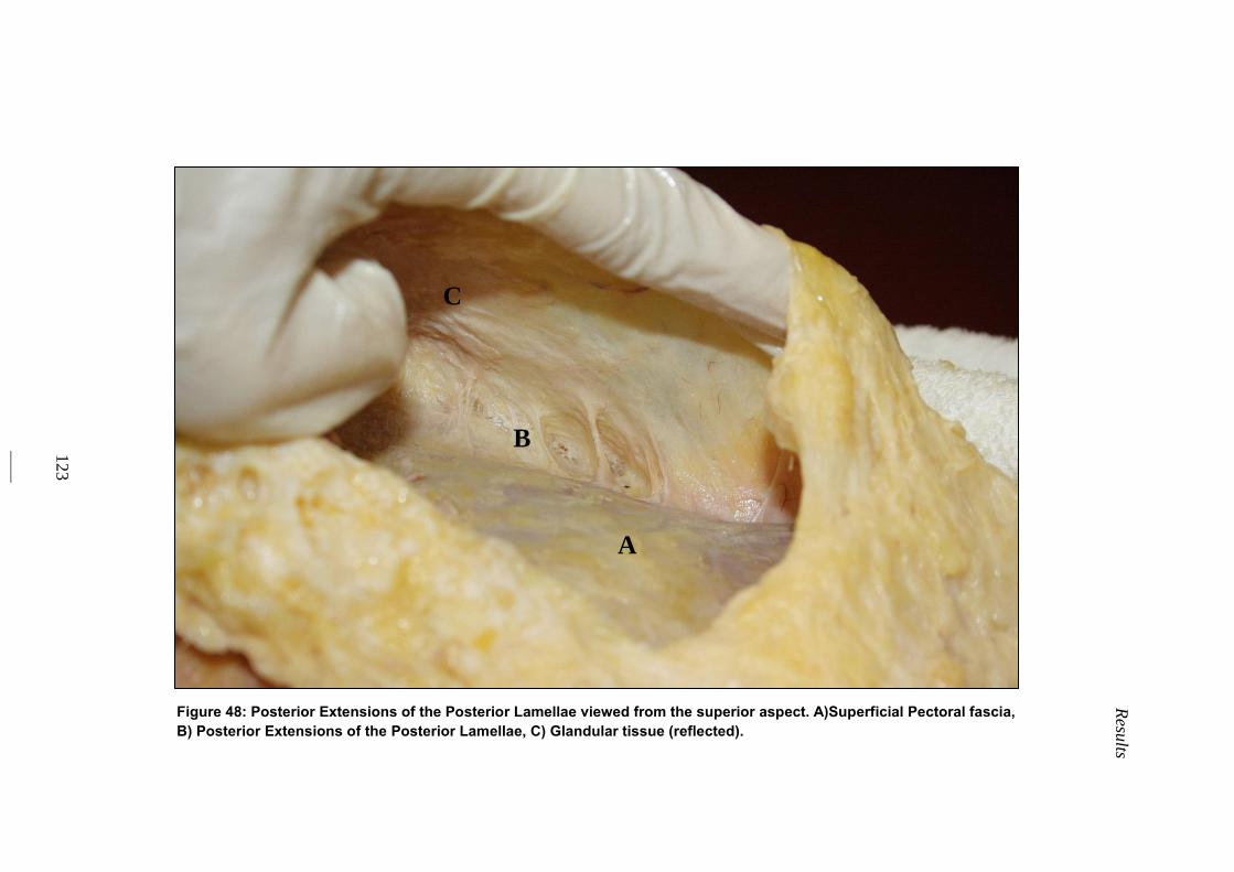
Resu
lts
123
Figure 48: Posterior Extensions of the Posterior Lamellae viewed from the superior aspect. A)Superficial Pectoral fascia,
B) Posterior Extensions of the Posterior Lamellae, C) Glandular tissue (reflected).
A
C
B
Results
124
3.3.3 Muscles within the perimeter
The chest wall muscles that were included within the perimeter of the breasts were the
pectoralis major muscle (sternal head), the serratus anterior muscle (anterior fibres), the
rectus abdominus muscle (superior fibres) and the external oblique muscle (superior
fibres) (Figure 49 and table 10). The sternal head of pectoralis major muscle was found
within the perimeter of all of the 12 breasts and made up the largest mean surface area
(81 %). The clavicular head of pectoralis major muscle was not found within the
perimeter in any of the 12 breasts. It was consistently found to be superior to the
superior perimeter of the breast in all 12 breasts. The serratus anterior muscle, rectus
abdominus muscle and external oblique muscle were not found within the perimeter of
every breast. Serratus anterior muscle was found in nine of the 12 breast perimeters and
made up the second largest mean surface area (10 %). External oblique muscle which
was only found within five of the 12 breast perimeters, with a mean surface area of 5 %
and rectus abdominus muscle which was found in six of the 12 breast perimeters, with a
mean surface area of 4 % (Figure 49 and Table 10).
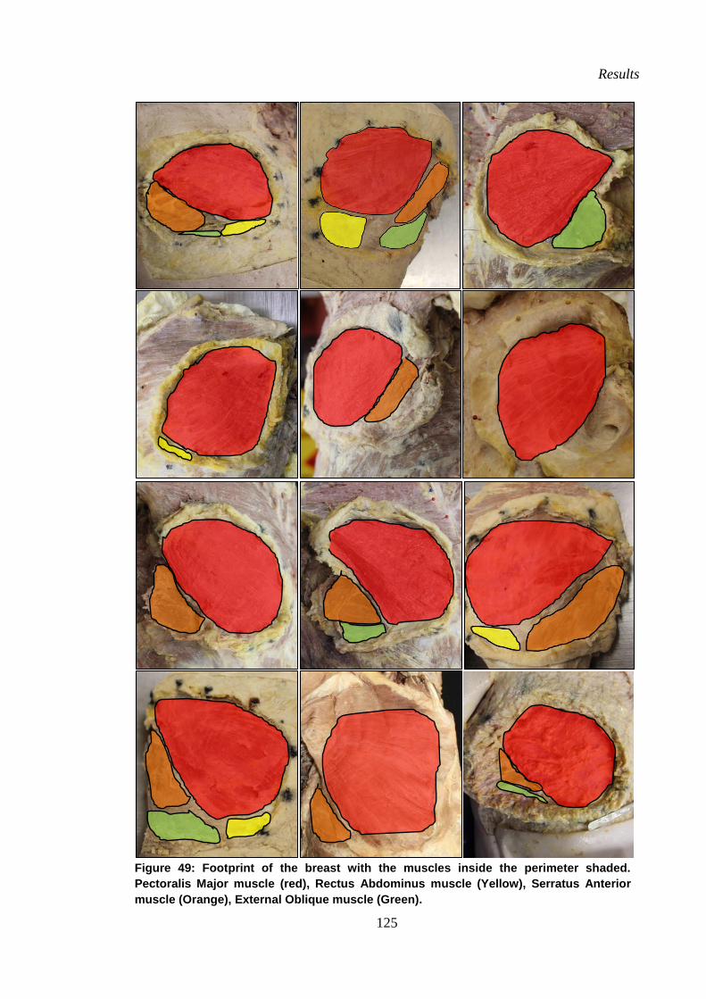
Results
125
Figure 49: Footprint of the breast with the muscles inside the perimeter shaded.
Pectoralis Major muscle (red), Rectus Abdominus muscle (Yellow), Serratus Anterior
muscle (Orange), External Oblique muscle (Green).

126
Chapter 4:
Discussion
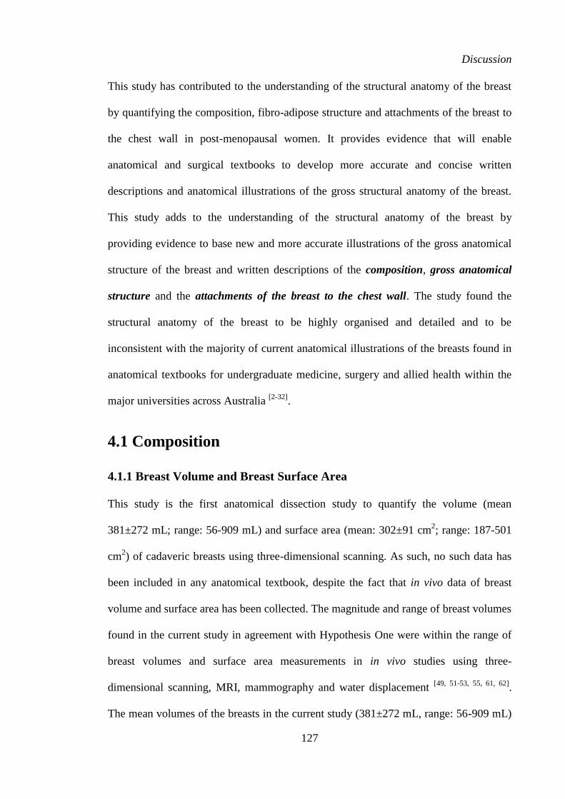
Discussion
127
This study has contributed to the understanding of the structural anatomy of the breast
by quantifying the composition, fibro-adipose structure and attachments of the breast to
the chest wall in post-menopausal women. It provides evidence that will enable
anatomical and surgical textbooks to develop more accurate and concise written
descriptions and anatomical illustrations of the gross structural anatomy of the breast.
This study adds to the understanding of the structural anatomy of the breast by
providing evidence to base new and more accurate illustrations of the gross anatomical
structure of the breast and written descriptions of the composition, gross anatomical
structure and the attachments of the breast to the chest wall. The study found the
structural anatomy of the breast to be highly organised and detailed and to be
inconsistent with the majority of current anatomical illustrations of the breasts found in
anatomical textbooks for undergraduate medicine, surgery and allied health within the
major universities across Australia [2-32]
.
4.1 Composition
4.1.1 Breast Volume and Breast Surface Area
This study is the first anatomical dissection study to quantify the volume (mean
381±272 mL; range: 56-909 mL) and surface area (mean: 302±91 cm2; range: 187-501
cm2) of cadaveric breasts using three-dimensional scanning. As such, no such data has
been included in any anatomical textbook, despite the fact that in vivo data of breast
volume and surface area has been collected. The magnitude and range of breast volumes
found in the current study in agreement with Hypothesis One were within the range of
breast volumes and surface area measurements in in vivo studies using three-
dimensional scanning, MRI, mammography and water displacement [49, 51-53, 55, 61, 62]
.
The mean volumes of the breasts in the current study (381±272 mL, range: 56-909 mL)
Discussion
128
are within the ranges presented in previous invivo studies using three-dimensional
scanning (Koch et. al. 2001[62]
: 274.4 mL; Coltman et. al. 2017[61]
: 680mL; range 75–
3100 mL). The lower minimum volume in the current study compared to previous
invivo studies may be attributed to the absence of skin in the current study, as well as
variations in BMI which is known to affect breast volume (Coltman et al, 2017) (BMI
Koch et al. 2011: 22.6 ± 3.3 kg/m², 18-32 kg/m²; Coltman et. al. 2017: 18.4–54.5 kg/m²;
Current Study: all cadavers weighed less than 100kg due to the requirement of the
UOW Body Donation).
The lower minimum volume measured in the current study compared to previous
in vivo studies may also be attributed to the absence of skin in the three-dimensional
scan with the breast volume of previous in vivo studies. Multiplying the breast volumes
of the current study by the coefficient factor (1.03), as described by Yip et. al. 2012, did
little to bridge the gap between the volumes reported in the current study and previous
in vivo studies. Quantifying breast volume and surface are in the current study allowed
other quantitative data of the structural anatomy of the breast, such as fibro-adipose
pocket number to be normalised to breast size.
Although the breast volumes of the current study were within the range of
previous in vivo studies that utilised measurements methods other than three-
dimensional scanning to measure breast volume including water displacement
(Vanderweyer and Hertens (2002)[51]
mean 446±286 mL, range: 110-1200 mL, n=21
breasts), anthropometric measurements (Huang et. al (2017)[53]
mean: 340.0±109.1 mL;
range: 91.8–919.2 mL, n=1210 breasts), MRI (Koch et. al. (2011)[62]
: mean 457 mL;
n=22 and Boyd et. al. (2009) [49]
: mean 715.1±496.6 mL; n=100) and mammography
(Boyd et. al. (2009)[52]
mean 470 mL; range: 63–1564 mL, n=211, mean age: 55 years),
the differences in the magnitude of the mean breast volume can be attributed to
Discussion
129
differences in the measurement methods (MRI taken in prone position, supine position
in dissection, standing in anthropometry and the possible inclusion of breast
pathologies), as well as cohort number, age and BMI (Vanderweyer and Hertens
(2002)[50]
: 55 years,: 33-84 years, Huang et. al. (2017) [53]
: 49 years, Koch et. al. (2011)
[62]: 43 years, Boyd et. al. (2009)
[49]: 51 years).
A limitation of this study however was the absence of an anterior chest wall scan
data to digitally create the posterior surface of the breast. A recommendation of this
study for future cadaveric breast research would be to include anterior chest wall scan
data, captured after the final stage of dissection of all of the breasts dissected in the
coronal plane to verify the surface of the anterior chest wall. This could then be digitally
meshed with the scan of the anterior breast surface in order to create a more accurate 3D
digital representation of each coronal breast. This was however beyond the scope of this
project as the image processing software did not allow for the combination of multiple
scans into a singular 3D object.
The range of surface area measurements (mean: 302±91 cm2; range: 187-501
cm2) of the current study were consistent with the range of the previous study found that
measured breast surface area in in vivo breasts using three-dimensional scanning
(Thomson et. al. (2009)[60]
mean: 213 cm2; range: 100-350 cm
2, n=7 females). The
volumes and surface area measurements in the current study are the first measurements
of cadaveric female breasts with the skin removed. They verify in vivo measurements
taken with the skin on where the exact boarders of the breast are only estimated beneath
the skin.
Quadrant variations
As no previous cadaveric study has investigated breast composition in quadrants of the
breasts, the quadrant volume results of the current study can only be compared to the
Discussion
130
quadrant volume results of one previously published in vivo study of 176 women [65]
.
Although this study also scanned the breasts of these women to determine the volume
and divided the scanned breasts into quadrants through the nipple, the position of the
body and therefore the breasts during scanning was different (supine in the current study
versus standing in the Pandarum et. al. (2011) [65]
study). As the breast is a deformable
structure, the distribution of the volume throughout the breast may be different in the
different positions. This may explain the differences found between the two studies,
along with differences in the BMI (current study: all <100kg; Pandarum et. al. (2011)[65]
n=83, 23% overweight and (77% obese) and the relative age of the subjects (current
study 83 years, range: 63-90 years; Pandarum et. al. (2011)[65]
age range: 23-65 years).
Both studies found the greatest distribution of the breast volume to be in the superior-
medial quadrant, however the quadrant with the least volume was not consistent being
the inferior-medial quadrant in the current study while Pandarum et. al. (2011)[65]
report
it to be the inferior-lateral quadrant. It should be noted that the distribution of breast
volume into quadrants described in the current study is specific to post-menopausal
cadaveric breasts of elderly women in the supine position. The results however provide
quantitative cadaveric data that could be used within anatomical texts. Future
anatomical research could investigate a larger cohort, including the breasts of women
across a range of ages and BMIs.
4.1.2 Breast Mass
As this is the first anatomical dissection study to quantify the breast mass of cadaveric
breasts, the breast mass data cannot be verified to previous dissection mass data. The
mean and range of total breast mass measured in the current study (mean: 355±157g;
range: 105–598 g) was similar to the breast mass reported in two previous in vivo
studies which measured the mass of 21 complete mastectomy specimens following pure
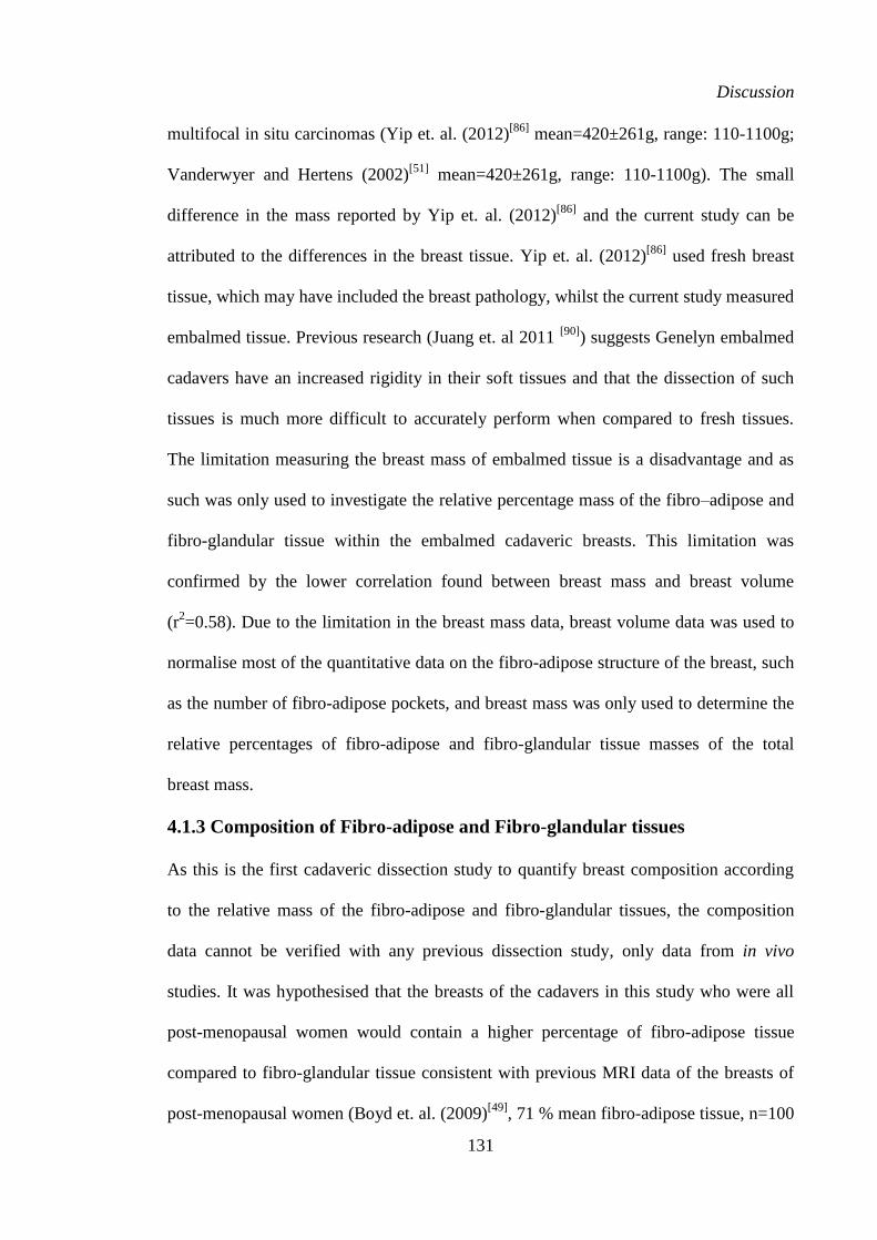
Discussion
131
multifocal in situ carcinomas (Yip et. al. (2012)[86]
mean=420±261g, range: 110-1100g;
Vanderwyer and Hertens (2002)[51]
mean=420±261g, range: 110-1100g). The small
difference in the mass reported by Yip et. al. (2012)[86]
and the current study can be
attributed to the differences in the breast tissue. Yip et. al. (2012)[86]
used fresh breast
tissue, which may have included the breast pathology, whilst the current study measured
embalmed tissue. Previous research (Juang et. al 2011 [90]
) suggests Genelyn embalmed
cadavers have an increased rigidity in their soft tissues and that the dissection of such
tissues is much more difficult to accurately perform when compared to fresh tissues.
The limitation measuring the breast mass of embalmed tissue is a disadvantage and as
such was only used to investigate the relative percentage mass of the fibro–adipose and
fibro-glandular tissue within the embalmed cadaveric breasts. This limitation was
confirmed by the lower correlation found between breast mass and breast volume
(r2=0.58). Due to the limitation in the breast mass data, breast volume data was used to
normalise most of the quantitative data on the fibro-adipose structure of the breast, such
as the number of fibro-adipose pockets, and breast mass was only used to determine the
relative percentages of fibro-adipose and fibro-glandular tissue masses of the total
breast mass.
4.1.3 Composition of Fibro-adipose and Fibro-glandular tissues
As this is the first cadaveric dissection study to quantify breast composition according
to the relative mass of the fibro-adipose and fibro-glandular tissues, the composition
data cannot be verified with any previous dissection study, only data from in vivo
studies. It was hypothesised that the breasts of the cadavers in this study who were all
post-menopausal women would contain a higher percentage of fibro-adipose tissue
compared to fibro-glandular tissue consistent with previous MRI data of the breasts of
post-menopausal women (Boyd et. al. (2009)[49]
, 71 % mean fibro-adipose tissue, n=100
Discussion
132
women, mean age: 49.6 years; Engelken et. al. (2014)[52]
, 79% mean fibro-adipose
tissue (mammography), n=174 women, mean age 55 years, range: 20-79 years). Only
two of the 14 breasts examined were in agreement with this hypothesis (both 64 %
fibro-adipose tissue), which were two of the largest breasts by volume (959 mL and 746
mL) in the study. The majority of the 14 breasts (n=12) contained a very similar
percentage of fibro-adipose (48 %) and fibro-glandular (52 %) tissue. One of the breasts
contained a very small percentage of fibro-adipose tissue (28 %) compared to fibro-
glandular, interestingly this was one of the smallest breasts by volume (75 mL). The
percentage of fibro-adipose tissue composition found in the majority of breasts within
the current study were closer to MRI results of younger women (Boyd et. al. (2009)[49]
n=400, mean age: 20.8 years), who had a mean of 53% fibro-adipose tissue and the
MRI results of women across a wider age range (Lee et. al. (1997)[55]
mean 66±18%
fibro-adipose tissue, n=40 women, age: 20-83 years). The difference may be attributed
to the variation in measurement method (MRI and mammography) versus
measurements of cadaveric tissue mass as in the current study. It is also possible that the
effect of embalming on the fibro-adipose and fibro-glandular tissue affected their mass
differently. Further research could verify this possibility.
The differences may also be due to the individual variation as a wide range of
relative percentages of fibro-adipose tissue have previously been found in mastectomy
specimens measured using tissue weighing (mean: 14.4 %; range: 7-56 %) and water
displacement (mean: 24.9 %; range: 3.6-37.6 %)[51]
, which have also been found to vary
with age and hormonal status. Despite the variation in the mean percentage of fibro-
adipose tissue found in the current study (mean: 48 %), the range of percentage mass
was consistent with previous research (range: 28-64 %).
The results of the current study provide data on the relative composition fibro-
Discussion
133
adipose and fibro-glandular tissues by mass of post-menopausal embalmed breasts. This
may provide quantitative data on breast composition for anatomical texts. A limitation
of the current study was that the percentage composition of fibro-adipose and fibro-
glandular tissue within the breasts were determined by mass only and not volume. This
was due to difficulties encountered in scanning the dissected fibro-adipose tissues. It is
also limited by the use of embalmed tissue, which may impact the breast mass.
Consequently, all tissue measurements were taken as relative compositions. Future
anatomical studies could overcome this by measuring total volume of the breast and
then dissect and scan the fibro-glandular volume to determine the percentage
composition by volume. This could then be compared to the data from MRI,
mammography and ultrasound studies.
Quadrant Variation
The gland was not found to cover the entire surface area of the breast in any of the
dissected breasts, rather some glandular tissue was found in each quadrant. The
distribution of the fibro-glandular tissue amongst the quadrants of the breast was in
contrast to a systemic literature review on the normal embryology and anatomy of the
adolescent female breast[68]
. Lemaine and Simmons (2013)[68]
reported the superior-
lateral quadrant of the breast to have a higher composition percentage of fibro-glandular
tissue. Unfortunately their descriptions were not supported by quantitative data as
evidence and the study did not report that it specifically measured the distribution of
tissues within the breast.
The variation found in the relative percentage of the fibro-adipose and fibro-
glandular tissue within the current dissection study and previous in vivo studies may
assist to explain the discrepancies found in breast composition in anatomical
illustrations. The photographic results of the current study provide data on the relative
Discussion
134
composition of the post-menopausal cadaveric breasts that may be used in anatomical
texts and as evidence to base anatomical illustrations upon. It is important that these
texts note the agr group that these photographs apply to as in vivo studies have found
breast composition to change with age. The relative location of fibro-adipose and fibro-
glandular tissues within the breast quadrants found in the current study, may assist with
the interpretation of MRI, mammography, and ultrasound images of the breast in post-
menopausal women and in turn the diagnosis of breast pathologies.
Discussion
135
4.2 Gross Anatomical Structure
4.2.1 Fibro-Adipose Structure
Anterior and Posterior Lamellae
In agreement with Hypothesis Three, two fascial sheets were found which were adhered
to the anterior and posterior surface of the gland. These results suggest that Cooper’s
Anterior and Posterior Lamellae and what other studies have referred to as the “anterior
and posterior breast capsule” [40, 41, 75]
both exist and are the same tissues. The findings
are also consistent with the superficial fascial system described by Lockwood (1991)
and Scarpa (1809)[40, 75]
, and more recently by Matousek et. al. (2014)[41]
. Previous
descriptions of the Anterior and Posterior Lamellae and anterior and posterior breast
capsule were limited to the region of the gland, which was known not to cover the entire
surface area of the breast. Beyond the borders of the glandular tissue (in all directions),
the current study found the Anterior and Posterior Lamellae converge together and
continue parallel to each other along the remainder of the breast in a similar manner to
the descriptions of the superficial fascial system in the remainder of the body [40, 75]
.
This is the first time the Anterior and Posterior Lamellae have been explored beyond the
glandular tissue.
The anatomical descriptions of the Anterior and Posterior Lamellae of the
current study were not consistent with two previous studies however, [42, 45]
that
described the superficial fascial system as defined by Lockwood (1991) and Scarpa
(1809)[40, 75]
to lie posterior to a breast capsule. This may be attributed to the dissection
method used in these two studies (current study: superficial to deep, Riggio et al
(2000)[45]
: inferior to superior and Muntan et. al. (2000)[42]
: examination of breast slices,
stained, embedded in paraffin and sliced again). The majority of the anatomical
Discussion
136
textbooks used by Australian Universities to teach undergraduate medicine, allied health
and science to teach breast anatomy did not include detail of the Lamellae or breast
capsule. The most common interpretation of the fascial structure of the breast within the
written text and illustrations of most anatomy textbooks was simply that the female
breast consisted of five layers; skin (dermis), sub-cutaneous tissue (fibro-adipose),
fibro-glandular tissue, retro-mammary space and pectoralis major muscle fascia with
Coopers ligaments spanning varying distances between the layers. The results of the
current study provide evidence to improve the level of detail on the fibrous tissue within
the breast to update anatomical textbooks.
Extensions of the Anterior and Posterior Lamellae
Fibrous tissue was found to span from the entire surface of the Anterior Lamellae to the
dermis in agreement with Hypothesis four. This was consistent with Cooper’s Anterior
Extensions of the Lamellae Anterior and the “Ligamenta Suspensoria”. This fascial
tissue and its location were also consistent with the findings of numerous other studies
[36, 39, 42, 45, 74] that unfortunately did not use Cooper’s terminology. These studies either
referred to this fascial tissue together with the entire fascia within the breast as
“Cooper’s Ligaments” or used inconsistent terminology. The mean length of the
Anterior Extensions of the Lamellae Anterior in the current study (measured in 20
ligaments in six breast slices from three breasts, mean 19±5 mm (range: 9-34 mm) was
also consistent with the length reported by Cooper (5-25 mm). Cooper however only
mentioned this length once throughout his work, in a figure caption[72]
. He also provided
no detail of the measurement method or the number of cadavers it was measured on.
The results of the current study provide evidence for the inclusion of anatomical
descriptions of the Anterior Extensions of the Lamellae Anterior or “Ligamenta
Suspensoria”, together with quantitative data of their length for accurate and labelled
Discussion
137
anatomical illustrations.
This study also confirmed the existence of Cooper, Posterior Extensions of the
Posterior Lamellae. Consistent with Cooper and Hypothesis five these we found to
consist of multiple fibrous bands of tissue running between a layer of loose areola
(adipose) tissue from the Posterior Lamellae on the posterior surface of the gland to the
superficial fascia of the Pectoralis Major muscle. The current study also extends the
descriptions of Cooper by describing the fascial structure beyond the perimeter of the
gland, where the same organisation of the tissues was observed. That is, the two fascial
layers (Anterior Lamellae and Posterior Lamellae) merged together in the absence of the
glandular tissue. At these extremities the Anterior Extensions of the Anterior Lamella
and the Posterior Extensions of the Posterior Lamellae were found to run to the dermis
and Superficial Pectoral Fascia on either side of this fascial plane. It was not however in
the scope of the study to investigate the other fascial tissue of the Lamellae, the
Posterior Extensions of the Anterior Lamellae and Anterior Extensions of the Posterior
Lamellae as they were not visible macroscopically.
The multiple names and lack of detail in the literature of the fibro-adipose
structure explains why the representation of the Coopers Ligaments in anatomical
textbooks varies greatly. Most anatomical textbooks provide an ambiguous sagittal
diagram of the breast with little to no accompanying text and no quantitative data. This
perpetuates the present confusion over the anatomical structure of the fibro-adipose
tissue of the breast and therefore there is a particular need to update both the
descriptions and illustrations based on the quantitative data collected in this study.
Fibro-Adipose Pockets Anterior and Posterior to the Gland
The fibro-adipose tissue was found to have a compartmentalised design where the
adipose tissue was embedded into multiple individual fibrous pockets, which were
Discussion
138
located both anterior and posterior to the gland. This organisational structure was in
agreement with Hypothesis Six and consistent with the descriptions of Cooper and Agur
et. al. (1991)[4]
. Also in agreement with Hypothesis Eight, a smaller number of fibro-
adipose tissue pockets were found anterior to the gland (mean: 35 pockets) compared to
posterior to the gland (mean: 53 pockets). The pockets anterior to the gland were
observed to be larger (0.89±0.32 mm2) in size compared to those posterior to the gland
(0.33±0.22 mm2). Although this organisation of the fibro-adipose tissue pocket structure
was consistent with Cooper’s descriptions, Cooper did not support his descriptions with
any quantitative data, nor has any study since, despite published descriptions of this
structure.
This study provides for the first time, quantitative data to support these
anatomical descriptions. It also provided additional detail on the size, shape and
distribution of the fibro-adipose tissue pockets, which has not previously been
investigated or published within the literature. Pocket size was found to have a mean
adipose tissue mass of 0.4±0.15 g (range: 0.26-0.61 g) and surface area was found to
have a mean of 0.88±0.37 cm2 (range: 0.31-1.97 cm
2). Pocket shape was found to vary
over the surface of the breast and to be unevenly distributed across the breast’s surface.
The shape of the fibro-adipose tissue pockets was more of a square shape with round
edges in the superior aspect of the breast and more oval shape in the inferior aspect of
the breast and 60% of these pockets were located within the superior half of the breast
surface. The detail of pocket number, size, distribution and shape provides important
detail for anatomical illustrations of this structure. It should be noted however that the
difference found in the pocket shape of the breasts in the different regions of the breasts
in the current study may be related to breast ptosis and consequence pocket
deformation, which is more likely considering the advanced age of the cadavers
Discussion
139
involved[61]
. Further investigation is required to verify this theory.
The number of the fibro-adipose pockets within the breast and the size of the
fibro-adipose pockets were not found to be consistent amongst the breasts of different
breast sizes, which was in disagreement with Hypothesis Seven. The mean number of
fibro-adipose pockets amongst the 15 breasts dissected was 200 pockets (range: 108-
306), with a positive correlation was found between the number of fibro-adipose tissue
pockets and breast volume, as well as breast surface area. This finding suggests that as
breast size increased (by volume and surface area), so did the number of fibro-adipose
tissue pockets. However, when the number of fibro-adipose pockets within the breasts
of the upper and lower tertiles by volume were compared, they were not statistically
significantly different, with only a trend for fibro-adipose pocket number to be greater
in breasts of a higher breast volume. Further research is required to verify whether
breasts of a larger size have a greater number of fibro-adipose pockets.
The very weak correlations between fibro-adipose pocket size (mass and surface
area) and breast volume and the lack of significant difference found between pocket
mass/surface areas of the breasts of the upper tertile by volume compared to the lower
tertile by volume suggest that larger breasts do not have fibro-adipose pockets that are
larger in size. The results of the current study suggest that it is more likely that breast
size increases by increasing the number of fibro-adipose pocket number rather than by
increasing the size of fibro-adipose pockets. again further research is required to verify
this notion
This data on fibro-adipose pocket number, size, shape, distribution and how
these vary with breast size provides evidence from which accurate anatomical
illustrations and written anatomical descriptions can be based. Many illustrations within
current anatomical texts display no discernible organisation of fibro-adipose tissue and
Discussion
140
limit descriptions of this complete structure to fibrous septa separating the glandular
tissue extending from the dermis to the superficial pectoral fascia [4-6, 9, 17, 20, 26, 28]
. Many
sagittal illustrations of the breast also neglect to include the fibro-adipose tissue located
posterior to the gland. There is an urgent need for an update on the detail of the breasts
structural anatomy currently being taught from these textbooks
4.2.2 Location of the Fibro-Glandular Structure
The current study was limited in its investigation of the fibro-glandular structure to its
regional location within the breast and its relative contribution to breast composition
(see 4.1). Consistent with Cooper[1]
, the fibro-glandular tissue with the body in a supine
position was found to be predominantly located in the inferior half of the breast. The
location was inconsistent however with two other studies (one dissection and one in
vivo study) [38, 44]
and many anatomical textbooks that found or reported the glandular
tissue to be predominantly in the superior-lateral quadrant projecting into the axilla as
the Tail of Spence. These studies had extensive descriptions of the micro-anatomy of
the breast but very little detail of the methodology of how the location and distribution
of the fibro-glandular tissue within the breast was measured or where they sourced such
information. The differences may be attributed to differences in the position of the
breasts in the current study during dissection. The breasts measured in the coronal plane
were dissected in the supine position. This was done as the chest plate was still intact
and as such the position of the torso, in supine, was necessary for dissection as the
weight was too great to suspend in any other position. The tissues of the embalmed
cadaveric breast are however deformable and this may have attributed to the differences
in the measurements of the current study to previous studies, as some of the tissue may
have changed relative locations. However, the positions of the breasts in current study
were the same as those reported by Cooper (1840) and as such a comparison could be
Discussion
141
made between the outcome measures of the two studies. Also the relative age of the
cadavers or women investigated in in vivo studies may affect the relative size and
location of the gland in the breast. Indeed many textbooks depict an illustration and
description of nulliparous or primiparous breasts, whereas the current study represented
the post-menopausal breast, with the mean age of the cadavers investigated in the
current study being 82 years.
The data collected in the current study on the gross anatomical structure of the
fibro-adipose and fibro-glandular tissues and their distributions within the breast
provides the quantitative data to base illustrations of the gross anatomical structure of
the breast for future anatomical texts based on the post-menopausal cadaveric breast in
the supine position. Increased understanding of the complex fascial three-dimensional
cobweb that encases the adipose tissue within the breast may also assist to explain the
scarring that can occur within the breast following breast surgery, such as a
lumpectomy. It also provides useful information about the basic design of breast tissue
for three-dimensional printing of the breast and for surgical breast reconstruction.
4.3 Attachment of the breast to the chest wall
Footprint of the breast
The regional anatomy of the breast in terms of its location on the chest wall was
consistent with Cooper[1]
and previous anatomical studies that described the breast to sit
between the 2-6/7th
ribs and extend from the lateral edge of the sternum to the mid-
axillary line. The current study added to the body of knowledge by providing
quantitative data from the number and age of the cadavers this regional anatomy
description is based on. The consensus in the literature regarding the location of the
breast on the chest wall may explain why this detail is commonly included in
Discussion
142
anatomical texts.
The length of the perimeter of the cadaveric breasts found in the current study
(mean: 46.01±12.2 cm; range: 33-58 cm) cannot be compared to previous research as it
has not previously been measured. The length was within the range however of previous
studies that have measured sections of the perimeter, namely the inframammary fold
(10-12 cm in length[77, 80]
), the diameter of the breast (14.3±1.4 cm in length, range: 8.5–
23.5 cm[53]
) and the lateral perimeter (7-13 cm in length[36]
).
Consistent with previous dissection studies[34-37, 40-43, 45, 48]
, descriptions in
anatomical textbooks [2, 4, 6, 7, 9-14, 19, 21-23, 27]
and in agreement with Hypothesis 10, the
muscles found within the perimeter the breast were Pectoralis Major, Serratus Anterior,
Rectus Abdominus and the External Oblique muscles. This is also consistent with
Cooper, except his descriptions did not include the Rectus Abdominus muscle. This
study is also consistent with two studies[43, 78]
that described the inframammary fold
(inferior perimeter attachment) to consistently sit below the inferior margin of the
Pectoralis Major muscle and the Pectoralis Major contributed the largest surface area
(two thirds) to the footprint of the breast. Unfortunately, no quantitative data was
provided to support these descriptions. The current study is consequently the first study
to provide quantitative data on the relative contribution of each muscle to the footprint
of the breast. Consistent with previous studies and anatomical texts, the Pectoralis
Major muscle was present within all breast perimeters and was found to contribute the
largest surface area (81%) of the footprint of the breast. The footprint of the breast
varied however in the mean contributions of the Serratus Anterior muscle (10%), which
was found within nine of the 12 breast perimeters, External Oblique muscle (5%),
which was found within five of the 12 breast perimeters and Rectus Abdominus muscle
(4%), which was found within six of the 12 breast perimeters.
Discussion
143
The data provides evidence for anatomical textbooks about the muscles adjacent
to the posterior wall of the breast. It also provides evidence of the shape of the posterior
wall of the breast, based on the adjacent muscle architecture, which has significant
application in three-dimensional digital scanning and digital breast reconstruction.
Current three-dimensional breast reconstruction creates a three-dimensional breast
created with a posterior wall, which has a curved shape [60, 61]
. This study provides new
evidence of the true shape of the posterior surface of the breast for digital breast
reconstruction. It may also assist breast surgery rehabilitation of the muscles that could
be affected by surgery such as mastectomy.
4.3.1 Posterior and Perimeter Attachments
Posterior Attachments
In agreement with Hypothesis Five and Nine, the posterior surface of the breast was
found to attach to the superficial pectoral fascia via numerous but weak fibrous bands
that house small adipose pockets in a similar fashion to the superficial fibro-adipose
structure namely Cooper’s Posterior Extensions of the Posterior Lamellae. This is
consistent with previous studies that describe small fibres within the “retro-mammary
space” or “bursa” (containing small areolar tissue) that allow the breast to “glide freely”
on the chest wall [37, 45, 66, 68, 70, 78, 80, 88]
.
Consistent with Wűringer et. al. (1998) [48]
(cadaveric study of 28 cadaveric
breasts, age range 68-92 years), a horizontal septum running from the nipple to the
posterior wall of the breast which divided the breast into a superior two thirds and
inferior one third was consistently found in both the coronal and sagittal dissections of
the current study. Contrary to the findings of Wűringer et. al. (1998) [48]
however, the
attachment of the horizontal septum to the chest wall was not considered to be strong as
it was required only blunt dissection, similar to the other posterior breast wall
Discussion
144
attachments. Also in contrast to the findings of Wűringer et. al. (1998) [48]
, no periosteal
attachment of the horizontal septum was found. The difference may be attributed to
differences in the direction and planes of dissection (cranial to caudal direction -
transverse plane (Wűringer et. al. (1998) [48]
study) and coronal and sagittal plane
(current study)). Although the sagittal plane dissection in the current study clearly
visualised both attachment ends of the septum perhaps with greater clarity compared to
the transverse plane dissections Wűringer et. al. (1998) [48]
, it is also possible that due to
the plane of the sagittal breast slices, that a single rib periosteal attachment was missed.
Further research is required that dissects the breast in multiple planes to verify whether
there is any periosteal attachment. Irrespective of any periosteal attachments, the results
of the current study do question however the strength of the attachment of the horizontal
septum as it required only blunt dissection to separate it from the anterior chest wall in
all of the dissections of the current study.
The horizontal attachment observed in the current study was different to the
description of the Triangle Fascial Condensation observed by Matousek et. al.
(2014)[41]
. Matousek et. al. (2014)[41]
described a fascial system that spanned from the
inferior portion of the breast to the chest wall, inserting on to the 5th
rib, however no
periosteal attachment was observed in the current study. No photographic evidence was
provided of this by Matousek et. al. (2014)[41]
, only an illustration of a mid-sagittal
section of a pert shaped breast with a fascial system inserting on to the 5th
rib. It is
possible however that a fascial attachment to the 5th
rib was not identified in the current
study due to the plane of the sagittal breast dissection. Due to the age of the cadavers
and the supine position that they were embalmed, the breasts were commonly splayed to
the side of the anterior chest wall and not pert in their shape. Consequently, a mid-
sagittal section of the breast did not cut through the ribs in a sagittal plane, but rather an
Discussion
145
oblique plane. This oblique angle may have obscured the view of a fascial attachment to
the 5th
rib. The current study also consistently found adipose and glandular tissue in the
inferior aspect of the breast in the region Matousek et. al. (2014)[41]
described as the
Triangle Fascial Condensation. Although Matousek et. al. (2014)[41]
stated that this
region consisted of dense fascial tissue with no breast tissue, in their images of the
sagittal slices of the cadaveric breast, breast tissue can clearly be seen within this area.
This is inconsistent with the current study which not only observed breast tissues in this
region but was able to both weight the tissue and measured the tissue pockets
dimensions. The observed difference may be attributed to the different dissection
methods used. The classical techniques used in the current study did not compromise
the facial tissue compared to the chemical dissection techniques used by Matousek et.
al. (2014)[41]
. In agreement with Matousek et. al. (2014)[41]
however, the current study
considered the perimeter attachments to be stronger than the posterior wall attachments
as they required sharp dissection along the entire perimeter..
Perimeter Attachments
The breast was found to be attached to the anterior aspect of the chest wall along
its entire circumferential perimeter. The attachments of the medial and lateral aspects of
the superior arc attachment were found to anchor to the superficial pectoralis major
muscle fascia consistent with the findings of Jinde et. al. (2006)[37]
. In contrast to Jinde
et. al. (2006)[37]
however, the middle (most superior) section of the superior arc
perimeter attachment was consistently found in all of the coronal dissections to dive
between the sternal and clavicular heads of the pectoralis major muscle and intersect
with the deep pectoralis muscle fascia. As both the superficial and deep pectoralis major
muscle fascia attach to the clavicle, the results of the current study were also consistent
with the findings of Matousek et. al. (2014)[41]
, who reported that superior attachment of
Discussion
146
the breast attaches to the clavicle. This clavicular attachment however was described by
Matousek et. al. (2014)[41]
to be via superficial and deep clavicular ligament whereas the
current study found the connection to be via the superficial and deep pectoralis muscle
fascia. Irrespective of this difference in terminology, both studies found an indirect and
distant connection between the superior arc perimeter attachment of the breast and the
clavicle (via muscle fascia or ligaments to the clavicle), with a considerable distance of
fascial tissue between the attachment. This extensive fascial connection may assist to
explain the ptosis of the breast with age.
The current study found the medial arc attachment of the breast required the
most time to dissect sharply in comparison to the other regions of the perimeter
attachments. The periosteal attachment to the lateral aspect of the sternum was
consistently found to be composed of dense collagen fibres with very little adipose
tissue. These findings were consistent with the descriptions of the medial perimeter
attachments in previous cadaveric dissection studies[36, 37, 41, 78, 81]
(including Cooper)
which describe the medial attachment to consist of short, strong fibres running in a
horizontal direction from the dermis to the periosteum, which required sharp dissection.
The medial arc attachment of the perimeter attachment observed in the current study
was contradictory to the descriptions of Wűringer et. al. (1998)[48]
who described the
medial attachment as a weak attachment, which allowed the breast to deform laterally.
Wűringer et. al. (1998)[48]
description however was not supported by any quantitative
data. The relative “strength” of the medial aspect of the perimeter attachment may
explain why the medial boarder of the breast can be consistently visualised in the post-
menopausal breast and why disruption of the medial tissues during breast surgery can
lead to poor aesthetic results, particularly in breast augmentations[67]
. The results also
emphasise the importance of the medial attachment for breast aesthetics during breast
Discussion
147
surgery.
The lateral arc attachment was found to be consistent with previous research
[36, 37, 41, 48] which described it to traverse the lateral aspect of the pectoralis major
muscle and “loosely” attach the breast to the axillary chest wall [36, 37, 48, 81]
. The inferior
aspect of the lateral arc attachment was found to be a sling of fascia formed by the
merging of muscle fascial sheets arising from the edges of the serratus anterior muscle
and the external oblique muscles (Figure 47). The inferior portion of the attachment was
similar to that described by Matousek et. al. (2014)[41]
, however the current study has
provided much more detail on this structure. The current study investigated this region
in both the sagittal and coronal planes using classical dissection techniques whereas the
Matousek et. al. (2014)[41]
used chemical dissection to reveal the structures. Pilot testing
in the current study revealed the connective tissues of the breast were affected by
chemical dissection and as such were not consistently reliable dissection techniques. As
such, the current study has been able to illustrate clearly, through cadaveric images, the
organisation of the attachment of the breast to the chest wall in both the sagittal and
coronal aspects whereas the Matousek et. al. (2014)[41]
study only represented this
attachment as a line drawn on an image of a female breast with the skin on. No other
studies have described or illustrated the fascial arrangement of the inferior-lateral
portion of the perimeter attachment.
The inferior arc attachment of the breast, also known as the Inframammary
Fold (IMF) was found to be a tightly adhered band of tissue anchoring the inferior
portion of the breast strongly to the muscle fascia’s of the anterior chest wall. The
relative location of the inferior perimeter attachments as described in previous studies is
consistent with the current study however, the attachment site (periosteal or fascial) was
unable to be confirmed in the current study as in most instances the pectoralis major
Discussion
148
muscle, or the angle of the bandsaw slice obscured the attachment. The inferior
attachment in the coronal plane dissection of the current study was seen to attach to the
muscle fascia of the anterior chest wall muscles, deep to which it could not be observed
to attach to the periosteum of the ribs. This is consistent with other research that has
also described the IMF (inferior perimeter) attach solely to the muscle fascia[81, 82]
. As
the IMF in the current study required sharp dissection to be removed from the anterior
chest wall during the coronal dissection, it suggests there could be a periosteal
attachment. This connection however was not clearly visible on the coronal dissections
which may explain why previous studies may not have found this connection. It was
however beyond the scope of this study to investigate.
The results of the sagittal dissection in the current study could not verify the
results of previous studies that have described a periosteal attachment of the
inframammary fold[34, 36, 37, 41, 48]
or a pure muscle fascia attachment [81, 82]
. The
periosteal attachments of the inframammary fold have been reported to be located at the
5th
rib [40, 41, 48]
and 6th
ribs [34]
. Consistent with Matousek et al. (2014)[41]
the 5th
rib was
exposed to breast tissue below the inferior border of the pectoralis major muscle and
above the superior border of the rectus abdominus muscle and external oblique muscle,
which could indicate a location of a fibrous attachment from the breast to the chest wall.
However, this was not clearly visible as the slices were taken through fixed points on
the breast, and due to the deformation and displacement of the embalmed breasts, these
were not true sagittal slices through the ribs. Consequently some slices were unable to
demonstrate successfully the entire breast depth from the skin to the ribs, and as such
the deep attachment of the IMF could not be observed.
The current study was unable therefore to verify the existence of the triangle
fascial condensation attaching to the 5th
rib as described by Matousek et. al. (2014)[41]
,
Discussion
149
this is likewise perhaps due to the angle of the cut through the breast in the sagittal
plane rather than through the midline of the breast. The current study did however
demonstrate the presence of breast tissue in the inferior portion of the breast which was
inconsistent with the findings of Matousek et. al. (2014)[41]
which described only fascial
attachments of the dermis to the 5th
rib in this region. The difference may be attributed
to the chemical dissection techniques used by Matousek et. al. (2014)[41]
, which would
have removed any adipose tissues from this region, whereas the sagittal slices in the
current study did not remove any adipose tissue. Further research could include a
dissection technique that clearly shows, by use of bandsaw slicing, the perpendicular
aspect of the breast rather than a classical sagittal aspect in order to verify the presence
of any periosteal attachment of the inframammary fold (inferior perimeter) to the chest
wall.
4.4 Conclusion
This study adds to the understanding of the structural anatomy of the breast in terms of
the composition of the breast by providing data on the relative percentage of the fibro-
adipose and fibro-glandular tissues by weight; gross anatomical structure of the breast
by providing quantitative data on the number, size, shape and distribution of fibro-
adipose pockets in both the coronal and sagittal plane, and quantitative data on the
length of the “Ligamenta Suspensoria”. It also provides some consistency in the naming
of the fascial tissues that form the fibro-adipose structure. Lastly it provides knowledge
on the relative strength and location of the posterior and perimeter attachments of the
breast to the chest wall and quantitative data of the muscles that form the footprint of
the breast on the anterior chest wall.
Discussion
150
This data as a whole provides evidence to base new and more accurate
illustrations of the gross anatomical structure of the breast and written descriptions of
the composition, fibro-adipose structure and the posterior and perimeter attachments
of the breast to the chest wall, which are currently lacking in anatomical texts used to
teach this breast anatomy to medical, allied health and science undergraduate students
within Australian Universities. Greater understanding of the structural anatomy of the
breast may assist clinically in the diagnosis of breast pathologies and the aesthetic
outcomes of breast surgery. It may also assist in the design of the breast tissue from
three-dimensional printing or in the reconstruction of breast tissue.
151
References
1. Cooper, A., 1840. On the anatomy of the breast I. Longman, Orme, Green,
Brown, and Longmans.
2. Abrahams, P., Craven, J., and Lumley, J., 2005. The Thorax, in Illustrated
Clinical Anatomy. Hodder Arnold.
3. Abrahams, P.H., Boon, J.M., and Spratt, J.D., 2008. Thoracic Wall, Surface
Markings and Breast, in McMinns Clinical Atlas of Human Anatomy. Elsevier
Limited.
4. Agur, A.M.R. and Dalley, A.F., 1991. The Thorax, in Grants Atlas of Anatomy.
Lippincott Williams & Wilkins.
5. Applegate, E.J., 2000. Reproductive System, in The Anatomy and Physiology
Learning System. W.B. Saunders.
6. Clemente, C.D., 2007. Pectoral Region, Axilla, Shoulder and Upper Limb, in
Anatomy: A Regional Atlas of the Human Body. Lippincott Williams and
Wilkins.
7. Drake, R.L., Vogl, W., and Mitchell, A.W., 2005. Thorax, in Grays Anatomy for
Students. Elsevier Inc.
8. Eizenberg, N., Briggs, C., Adams, C., and Ahern, G., 2008. Human
Development and Variation, in General Anatomy. Anatomedia Publishing.
9. Ellis, H., 2002. The Female Breast, in Clinical Anatomy: a revision and applied
anatomy for clinical students. Blackwell Publishers
10. Gilroy, A.M., 2013. Thoracic Wall, in Anatomy An Essential Textbook. Thieme
Medical Publishers Inc.
11. Gilroy, A.M., MacPherson, B.R., and Ross, L.M., 2012. Female Breast, in Atlas
of Anatomy. Thieme.
12. Gosling, J.A., Harris, P.F., Whitmore, I., and Willan, P.L.T., 2003. Thorax, in
Human Anatomy: Colour Atlas and Text. Mosby.
13. Hansen, J.T., 2014. Female Breast, in Netters Anatomy Colouring Book.
Saunders.
14. Hansen, J.T. and Lambert, D.R., 2008. Thorax, in Netters Clinical Anatomy.
Saunders Elsevier. 301-307.
15. Kapit, W. and Elson, L.M., 1993. Reproductive System, in Anatomy Colouring
Book. Kapit and Elson.
16. Lindsay, D., 1995. Reproductive System, in Functional Human Anatomy.
William C Brown Publishers. 818-819.
17. Marieb, E.N. and Hoehn, K.N., 2012. The Reproductive System, in Human
Anatomy & Physiology. Pearson Education.
18. McKinley, M. and O'Loughlin, V.D., 2006. Surface Anatomy, in Human
Anatomy. McGraw-Hill Companies Inc.
19. McMinn, R.M.H., 1991. UpperLimb, in Last's Anatomy: Regional and Applied.
Churchill Livingstone.
20. Moffat, D.F., Lecture Notes on Anatomy. 1993, Wiley/Blackwell Scientific
Publications.
21. Moore, K.L., Agur, A.M.R., and Dalley, A.F., 2015. Thorax, in Essential
Clinical Anatomy. Wolters Kluwer.
22. Moore, K.L., Dalley, A.F., and Agur, A.M.R., 2014. Thorax, in Clinically
Oriented Anatomy. Woltz Kluwer/Lippincott Williams & Wilkins.
152
23. Moses, Banks, J.C., Nava, P.B., and Petersen, D., 2005. Breast and Pectoral
Region, in Atlas of Clinical Gross Anatomy.
24. Olson, T.R., 1996. Anterior Body Wall, in A.D.A.M Student Atlas of Anatomy.
Lippincott Williams and Wilkins. 42.
25. Rohen, J.W., Yokochi, C., and Lutjen-Drecoll, 2016. Mamary Gland, in
Anatomy: A Photographic Atlas. Schattauer GmbH and Wolters Kluwer.
26. Saladin, K.S., 2007. The Female Reproductive System, in Anatomy and
Physiology: the Unity of Form and Function. McGraw-Hill Companies Inc.
27. Sinnatamby, C.S., 2006. Breast, in Lasts Anatomy: Regional and Applied.
Elsevier Ltd.
28. Snell, R.S., 1995. The Pectoral Region and the Axilla, in Clinical Anatomy for
Medical Students. Little Brown & Co: Boston, MA.
29. Tank, P.W. and Gest, T.R., 2008. Breast, in Lippincott Williams & Wilkins Atlas
of Anatomy. Wolters Kluwer.
30. Tortora, G.J. and Derrickson, B., 2012. The Reproductive System, in Principles
of Anatomy and Physiology. Biological Science Textbooks Inc.
31. Tortora, G.J., Derrickson, B., Burkett, B., Dye, D., Diversi, T., McKean, M.,
Cooke, J., Mellifont, R., Samalia, L., and Peoples, G., 2015. The Reproductive
System, in Principles of Anatomy & Physiology. John Wiley & Sons Australia
Ltd.
32. Van De Graff, K.M., Strete, D., and Creek, C.H., 2002. Reproductive Anatomy,
in Human Anatomy. McGraw Hill Higher Education.
33. Arnez, Z.M., Valdatta, L., Tyler, M.P., and Planinsek, F., 1995. Anatomy of the
internal mammary veins and their use in free TRAM flap breast reconstruction.
British Journal of Plastic Surgery, 48 (8), 540-5.
34. Bayati, S. and Seckle, B., 1995. Inframammary Crease Ligament. Plastic and
Reconstructive Surgery, 95 (3), 501-508.
35. Boutros, S., Kattash, M., Weinfeld, A., Yuksel, E., Baer, S., and Shenaq, S., The
Intradermal anatomy of the Inframammary Fold, in Texas Society of Plastic
Surgery Scientific Meeting. 1997: San Antonio.
36. Cardoso, A., Santos, D., Martins, J., Coelho, G., Barroso, L., and Costa, H.,
2015. Breast ligaments: an anatomical study. European Journal of Plastic
Surgery, 38 (2), 91-96.
37. Jinde, L., Jianliang, S., Xiaoping, C., Xiaoyan, T., Jiaqing, L., Qun, M., and Bo,
L., 2006. Anatomy and clinical significance of pectoral fascia. Plastic and
Reconstructive Surgery, 118 (7), 1557-60.
38. Klock, J.C., Iuanow, E., Malik, B., Obuchowski, N.A., Wiskin, J., and Lenox,
M., 2016. Anatomy-Correlated Breast Imaging and Visual Grading Analysis
Using Quantitative Transmission Ultrasound ™. International Journal of
Biomedical Imaging, 2016.
39. Komiya, T., Ito, N., Imai, R., Itoh, M., Naito, M., Matsubayashi, J., and
Matsumura, H., 2015. Anatomy of the superficial layer of superficial fascia
around the nipple-areola complex. Aesthetic Plastic Surgery, 39 (2), 209-213.
40. Lockwood, T., 1991. Superficial Fascial System (SFS) of the Trunk and
Extremities: A New Concept. Plastic and Reconstructive Surgery, 87 (6), 1009-
1018.
41. Matousek, S.A., Corlett, R.J., and Ashton, M.W., 2014. Understanding the
fascial supporting network of the breast: key ligamentous structures in breast
153
augmentation and a proposed system of nomenclature. Plastic Reconstructive
Surgery, 133 (2), 273-81.
42. Muntan, C.D., Sundine, M.J., Rink, R.D., and Acland, R.D., 2000.
Inframammary fold: A histologic reappraisal. Plastic and Reconstructive
Surgery, 105 (2), 549-557.
43. Nanigian, B.R., Wong, G.B., and Khatri, V.P., 2007. Inframammary crease:
positional relationship to the pectoralis major muscle origin. Aesthetic Surgery,
27 (5), 509-12.
44. Parks, A.G., 1959. The Micro-Anatomy of the Breast: Hunterian Lecture
delivered at the Royal College of Surgeons of England on 12th March 1959.
Annals of The Royal College of Surgeons of England, 25 (4), 235-251.
45. Riggio, E., Quattrone, P., and Nava, M., 2000. Anatomical Study of the
Superficial Fascial Ststem: The Inframammary Fold Unit. European Journal of
Pastic Surgery, 23, 310-315.
46. Sarhadi, N.S., Dunn, J.S., Lee, F.D., and Soutar, D.S., 1996. An anatomical
study of the nerve supply of the breast, including the nipple and areola. British
Journal of Plastic Surgery, 49 (3), 156-164.
47. Suami, H., Pan, W.R., Mann, G.B., and Taylor, G.I., 2008. The lymphatic
anatomy of the breast and its implications for sentinel lymph node biopsy: a
human cadaver study. Annals of Surgical Oncology, 15 (3), 863-71.
48. Wuringer, E., Mader, N., Posch, E., and Holle, J., 1998. Nerve and Vessel
Supplying Ligamentous Suspention of the Mammary Gland. Plastic and
Reconstructive Surgery, 101, 1486-1493.
49. Boyd, N., Martin, L., Chavez, S., Gunasekara, A., Salleh, A., Melnichouk, O.,
Yaffe, M., Friedenreich, C., Minkin, S., and Bronskill, M., 2009. Breast-tissue
composition and other risk factors for breast cancer in young women: a cross-
sectional study. The Lancet Oncology, 10 (6), 569-580.
50. Kayar, R., Civelek, S., Cobanoglu, M., Gungor, O., Catal, H., and Emiroglu, M.,
2011. Five Methods of Breast Volume Measurement: A Comparative Study of
Measurements of Specimen Volume in 30 Mastectomy Cases. Breast Cancer:
Basic & Clinical Research, (5), 43-52.
51. Vanderweyer, E. and Hertens, D., 2002. Qualification of glands and fat in breast
tissue: an experimental determination. Annals of Anatomy, 184 (2), 181-4.
52. Engelken, F., Singh, J., Fallenberg, E., Bick, U., Böttcher, J., and Renz, D.,
2014. Volumetric breast composition analysis: reproducibility of breast percent
density and fibroglandular tissue volume measurements in serial mammograms.
Acta Radiologica, 55 (1), 32-38.
53. Huang, N.-s., Quan, C.-l., Mo, M., Chen, J.-j., Yang, B.-l., Huang, X.-y., and
Wu, J., 2017. A prospective study of breast anthropomorphic measurements,
volume and ptosis in 605 Asian patients with breast cancer or benign breast
disease. PLOS ONE, 12 (2), 1-10.
54. Lu, L.J., Nishino, T.K., Johnson, R.F., Nayeem, F., Brunder, D.G., Ju, H.,
Leonard, M.H., Grady, J.J., and Khamapirad, T., 2012. Comparison of breast
tissue measurements using magnetic resonance imaging, digital mammography
and a mathematical algorithm. Physics in Medicine and Biology, 57 (21), 6903-
27.
55. Lee, N.A., Rusinek, H., Weinreb, J., Chandra, R., Toth, H., Singer, C., and
Newstead, G., 1997. Fatty and fibroglandular tissue volumes in the breasts of
154
women 20-83 years old: comparison of X-ray mammography and computer-
assisted MR imaging. American Journal of Roentgenology, 168 (2), 501-6.
56. Lee, H.-Y., Hong, K., and Kim, E.A., 2004. Measurement protocol of women’s
nude breasts using a 3D scanning technique. Applied Ergonomics, 35, 353-359.
57. Ricbourg, B., 1992. [Applied anatomy of the breast: blood supply and
innervation]. Annales de Chirurgie Plastique Esthétique, 37 (6), 603-20.
58. Rohen, J.W., Yokochi, C., and Lutjen-Drecoll, E., 2006. Mammary Gland, in
Color atlas of anatomy: a photographic study of the human body. Lippincott
Williams & Wilkins: Stuttgart.
59. Martini, F., Timmons, M.J., and Tallitsch, R.B., 2008. Thorax, in Human
anatomy. Pearson Benjamin Cummings: San Francisco.
60. Thomson, J.G., Liu, Y.-J., Restifo, R.J., Rinker, B.D., and Reis, A., 2009.
Surface area measurement of the female breast: phase I. Validation of a novel
optical technique. Plastic And Reconstructive Surgery, 123 (5), 1588-1596.
61. Coltman, C.E., Steele, J.R., and McGhee, D.E., 2017. Breast volume is affected
by body mass index but not age. Ergonomics, 1-10.
62. Koch, M.C., Adamietz, B., Jud, S.M., Fasching, P.A., Haeberle, L., Karbacher,
S., Veit, K., Schulz-Wendtland, R., Uder, M., Beckmann, M.W., Bani, M.R.,
Heusinger, K., Loehberg, C.R., and Cavallaro, A., 2011. Breast volumetry using
a three-dimensional surface assessment technique. Aesthetic Plastic Surgery, 35
(5), 847-855.
63. McGhee, D., Steele, J., and Zealey, W., 2010. Effects of high and low breast
support on breast kinematics and kinetics during treadmill running. Journal of
Science and Medicine in Sport, 12, e143.
64. Bulstrode, N., Bellamy, E., and Shrotria, S., 2001. Breast volume assessment:
comparing five different techniques. Breast, 10 (2), 117-23.
65. Pandarum, R., Yu, W., and Hunter, L., 2011. 3-D breast anthropometry of plus-
sized women in South Africa. Ergonomics, 54 (9), 866-875.
66. Hammond, D., 2009. Applied Anatomy, in Atlas of Aesthetic Breast Surgery.
Elsevier Inc. 1-9.
67. Bogetti, P., Cravero, L., Spagnoli, G., Devalle, L., Boriani, F., Bocchiotti, M.A.,
Renditore, S., and Baglioni, E., 2007. Aesthetic role of the surgically rebuilt
inframammary fold for implant-based breast reconstruction after mastectomy.
Plastic Reconstructive Aesthetic Surgery, 60 (11), 1225-32.
68. Lemaine, V. and Simmons, P.S., 2013. The adolescent female: Breast and
reproductive embryology and anatomy. Clinical Anatomy, 26 (1), 22-28.
69. Liu, S.M.D. and Bassett, L.W.M.D., Breasts. 2017, Salem Press.
70. Hilton, S., Leopold, G., Olson, L., and Wilson, S., 1986. Real Time Breast
Sonography: Application in 300 Consecutive Patients. American Journal of
Roentgenology, 147, 479-485.
71. Pearson, J.R., 1988. A Practical Review of Breast Anatomy and Pathology.
Journal of Diagnostic Medical Sonography, 4 (6), 305-308.
72. Cooper, A., 1840. On the anatomy of the breast II. Longman, Orme, Green,
Brown, and Longmans.
73. Azar, F., A Deformable finite element model of the breast for predicting
mechanical deformations under the external pertubations, in Dept. of
Bioengineering. 2001, University of Pennsylvania: Philadelphia.
74. Gefen, A. and Dilmoney, B., 2007. Mechanics of the normal woman's breast.
Technology and Health Care, 15, 259-271.
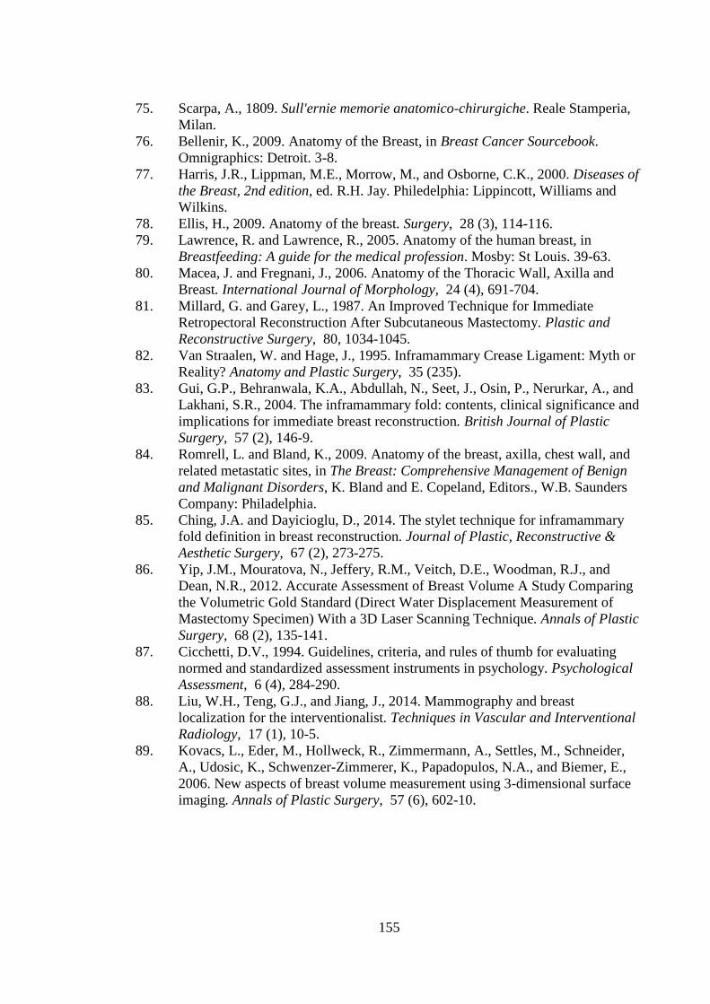
155
75. Scarpa, A., 1809. Sull'ernie memorie anatomico-chirurgiche. Reale Stamperia,
Milan.
76. Bellenir, K., 2009. Anatomy of the Breast, in Breast Cancer Sourcebook.
Omnigraphics: Detroit. 3-8.
77. Harris, J.R., Lippman, M.E., Morrow, M., and Osborne, C.K., 2000. Diseases of
the Breast, 2nd edition, ed. R.H. Jay. Philedelphia: Lippincott, Williams and
Wilkins.
78. Ellis, H., 2009. Anatomy of the breast. Surgery, 28 (3), 114-116.
79. Lawrence, R. and Lawrence, R., 2005. Anatomy of the human breast, in
Breastfeeding: A guide for the medical profession. Mosby: St Louis. 39-63.
80. Macea, J. and Fregnani, J., 2006. Anatomy of the Thoracic Wall, Axilla and
Breast. International Journal of Morphology, 24 (4), 691-704.
81. Millard, G. and Garey, L., 1987. An Improved Technique for Immediate
Retropectoral Reconstruction After Subcutaneous Mastectomy. Plastic and
Reconstructive Surgery, 80, 1034-1045.
82. Van Straalen, W. and Hage, J., 1995. Inframammary Crease Ligament: Myth or
Reality? Anatomy and Plastic Surgery, 35 (235).
83. Gui, G.P., Behranwala, K.A., Abdullah, N., Seet, J., Osin, P., Nerurkar, A., and
Lakhani, S.R., 2004. The inframammary fold: contents, clinical significance and
implications for immediate breast reconstruction. British Journal of Plastic
Surgery, 57 (2), 146-9.
84. Romrell, L. and Bland, K., 2009. Anatomy of the breast, axilla, chest wall, and
related metastatic sites, in The Breast: Comprehensive Management of Benign
and Malignant Disorders, K. Bland and E. Copeland, Editors., W.B. Saunders
Company: Philadelphia.
85. Ching, J.A. and Dayicioglu, D., 2014. The stylet technique for inframammary
fold definition in breast reconstruction. Journal of Plastic, Reconstructive &
Aesthetic Surgery, 67 (2), 273-275.
86. Yip, J.M., Mouratova, N., Jeffery, R.M., Veitch, D.E., Woodman, R.J., and
Dean, N.R., 2012. Accurate Assessment of Breast Volume A Study Comparing
the Volumetric Gold Standard (Direct Water Displacement Measurement of
Mastectomy Specimen) With a 3D Laser Scanning Technique. Annals of Plastic
Surgery, 68 (2), 135-141.
87. Cicchetti, D.V., 1994. Guidelines, criteria, and rules of thumb for evaluating
normed and standardized assessment instruments in psychology. Psychological
Assessment, 6 (4), 284-290.
88. Liu, W.H., Teng, G.J., and Jiang, J., 2014. Mammography and breast
localization for the interventionalist. Techniques in Vascular and Interventional
Radiology, 17 (1), 10-5.
89. Kovacs, L., Eder, M., Hollweck, R., Zimmermann, A., Settles, M., Schneider,
A., Udosic, K., Schwenzer-Zimmerer, K., Papadopulos, N.A., and Biemer, E.,
2006. New aspects of breast volume measurement using 3-dimensional surface
imaging. Annals of Plastic Surgery, 57 (6), 602-10.
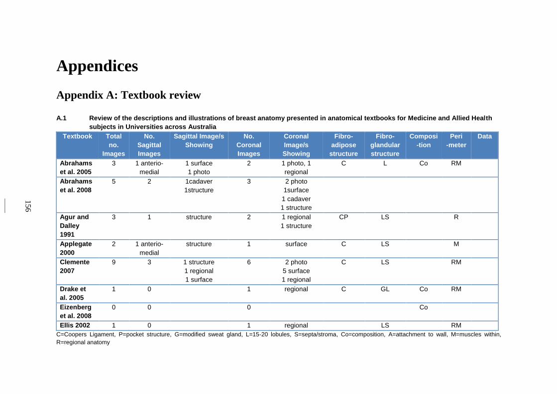
156
Appendices
Appendix A: Textbook review
A.1 Review of the descriptions and illustrations of breast anatomy presented in anatomical textbooks for Medicine and Allied Health
subjects in Universities across Australia
Textbook Total
no.
Images
No.
Sagittal
Images
Sagittal Image/s
Showing
No.
Coronal
Images
Coronal
Image/s
Showing
Fibro-
adipose
structure
Fibro-
glandular
structure
Composi
-tion
Peri
-meter
Data
Abrahams
et al. 2005
3 1 anterio-
medial
1 surface
1 photo
2 1 photo, 1
regional
C L Co RM
Abrahams
et al. 2008
5 2 1cadaver
1structure
3 2 photo
1surface
1 cadaver
1 structure
Agur and
Dalley
1991
3 1 structure 2 1 regional
1 structure
CP LS R
Applegate
2000
2 1 anterio-
medial
structure 1 surface C LS M
Clemente
2007
9 3 1 structure
1 regional
1 surface
6 2 photo
5 surface
1 regional
C LS RM
Drake et
al. 2005
1 0 1 regional C GL Co RM
Eizenberg
et al. 2008
0 0 0 Co
Ellis 2002 1 0 1 regional LS RM
C=Coopers Ligament, P=pocket structure, G=modified sweat gland, L=15-20 lobules, S=septa/stroma, Co=composition, A=attachment to wall, M=muscles within,
R=regional anatomy
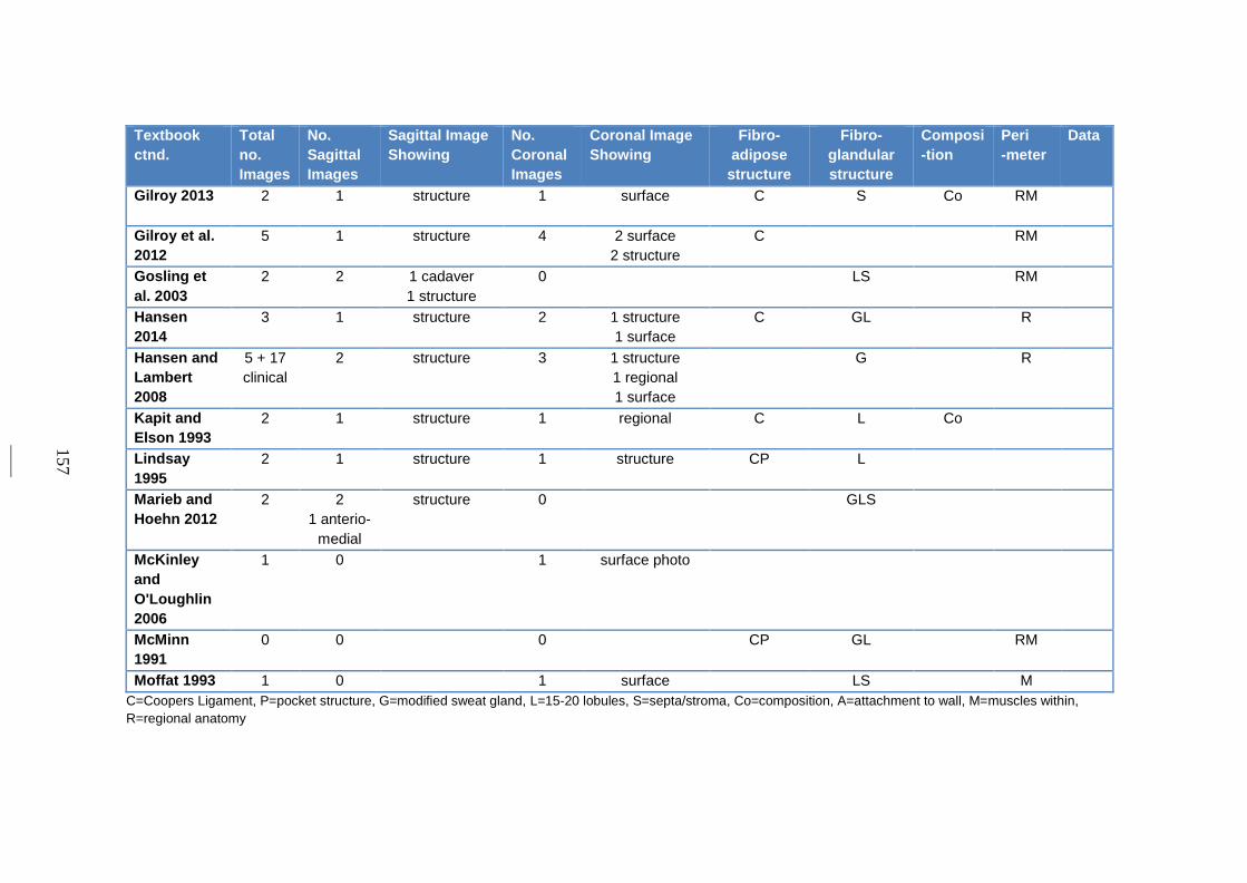
157
Textbook
ctnd.
Total
no.
Images
No.
Sagittal
Images
Sagittal Image
Showing
No.
Coronal
Images
Coronal Image
Showing
Fibro-
adipose
structure
Fibro-
glandular
structure
Composi
-tion
Peri
-meter
Data
Gilroy 2013 2 1 structure 1 surface C S Co RM
Gilroy et al.
2012
5 1 structure 4 2 surface
2 structure
C RM
Gosling et
al. 2003
2 2 1 cadaver
1 structure
0 LS RM
Hansen
2014
3 1 structure 2 1 structure
1 surface
C GL R
Hansen and
Lambert
2008
5 + 17
clinical
2 structure 3 1 structure
1 regional
1 surface
G R
Kapit and
Elson 1993
2 1 structure 1 regional C L Co
Lindsay
1995
2 1 structure 1 structure CP L
Marieb and
Hoehn 2012
2 2
1 anterio-
medial
structure 0 GLS
McKinley
and
O'Loughlin
2006
1 0 1 surface photo
McMinn
1991
0 0 0 CP GL RM
Moffat 1993 1 0 1 surface LS M
C=Coopers Ligament, P=pocket structure, G=modified sweat gland, L=15-20 lobules, S=septa/stroma, Co=composition, A=attachment to wall, M=muscles within,
R=regional anatomy
158
Textbook
ctnd.
Total
no.
Images
No.
Sagittal
Images
Sagittal Image
Showing
No.
Coronal
Images
Coronal Image
Showing
Fibro-
adipose
structure
Fibro-
glandular
structure
Composi
-tion
Peri
-meter
Data
Moore et al.
2015
5 +
6
clinical
1 anterio-
medial
structure 4 structure C GL RM
Moore et al.
2014
6 1 anterio-
medial
structure 5 2 regional
3 structure
CP GL RM
Moses et al.
2005
6 1 cadaver 5 1 cadaver
4 structure
G R
Olson 1996 1 0 1 regional
Rohen et al.
2016
4 1 1 cadaver
1 structure
3 2 cadaver
1 structure
1 regional
Saladin
2007
3 +
2
clinical
2 1 cadaver
1 structure
1 structure C GLS Co M
Sinnatamby
2006
0 0 0 C GL RM
Snell 1995 4 2 2 C LS RM
Tank and
Gest 2008
4 1 structure 3 structure
Tortora and
Derrickson
2012
2 1 structure 1 structure C GL M
Tortora et
al. 2015
2 1 structure 1 structure C GL M
Van De
Graff et al.
2002
3 1 structure 2 1 regional
1 structure
C L RM
C=Coopers Ligament, P=pocket structure, G=modified sweat gland, L=15-20 lobules, S=septa/stroma, Co=composition, A=attachment to wall, M=muscles within,
R=regional anatomy
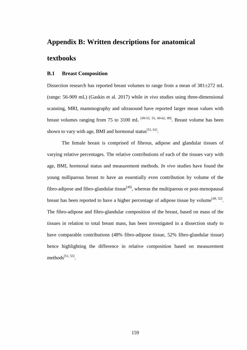
159
Appendix B: Written descriptions for anatomical
textbooks
B.1 Breast Composition
Dissection research has reported breast volumes to range from a mean of 381±272 mL
(range: 56-909 mL) (Gaskin et al. 2017) while in vivo studies using three-dimensional
scanning, MRI, mammography and ultrasound have reported larger mean values with
breast volumes ranging from 75 to 3100 mL [49-53, 55, 60-62, 89]
. Breast volume has been
shown to vary with age, BMI and hormonal status[51, 61]
.
The female breast is comprised of fibrous, adipose and glandular tissues of
varying relative percentages. The relative contributions of each of the tissues vary with
age, BMI, hormonal status and measurement methods. In vivo studies have found the
young nulliparous breast to have an essentially even contribution by volume of the
fibro-adipose and fibro-glandular tissue[49]
, whereas the multiparous or post-menopausal
breast has been reported to have a higher percentage of adipose tissue by volume[49, 52]
.
The fibro-adipose and fibro-glandular composition of the breast, based on mass of the
tissues in relation to total breast mass, has been investigated in a dissection study to
have comparable contributions (48% fibro-adipose tissue, 52% fibro-glandular tissue)
hence highlighting the difference in relative composition based on measurement
methods[51, 55]
.
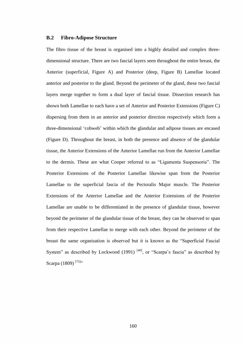
160
B.2 Fibro-Adipose Structure
The fibro tissue of the breast is organised into a highly detailed and complex three-
dimensional structure. There are two fascial layers seen throughout the entire breast, the
Anterior (superficial, Figure A) and Posterior (deep, Figure B) Lamellae located
anterior and posterior to the gland. Beyond the perimeter of the gland, these two fascial
layers merge together to form a dual layer of fascial tissue. Dissection research has
shown both Lamellae to each have a set of Anterior and Posterior Extensions (Figure C)
dispersing from them in an anterior and posterior direction respectively which form a
three-dimensional ‘cobweb’ within which the glandular and adipose tissues are encased
(Figure D). Throughout the breast, in both the presence and absence of the glandular
tissue, the Anterior Extensions of the Anterior Lamellae run from the Anterior Lamellae
to the dermis. These are what Cooper referred to as “Ligamenta Suspensoria”. The
Posterior Extensions of the Posterior Lamellae likewise span from the Posterior
Lamellae to the superficial fascia of the Pectoralis Major muscle. The Posterior
Extensions of the Anterior Lamellae and the Anterior Extensions of the Posterior
Lamellae are unable to be differentiated in the presence of glandular tissue, however
beyond the perimeter of the glandular tissue of the breast, they can be observed to span
from their respective Lamellae to merge with each other. Beyond the perimeter of the
breast the same organisation is observed but it is known as the “Superficial Fascial
System” as described by Lockwood (1991) [40]
, or “Scarpa’s fascia” as described by
Scarpa (1809) [75]
”
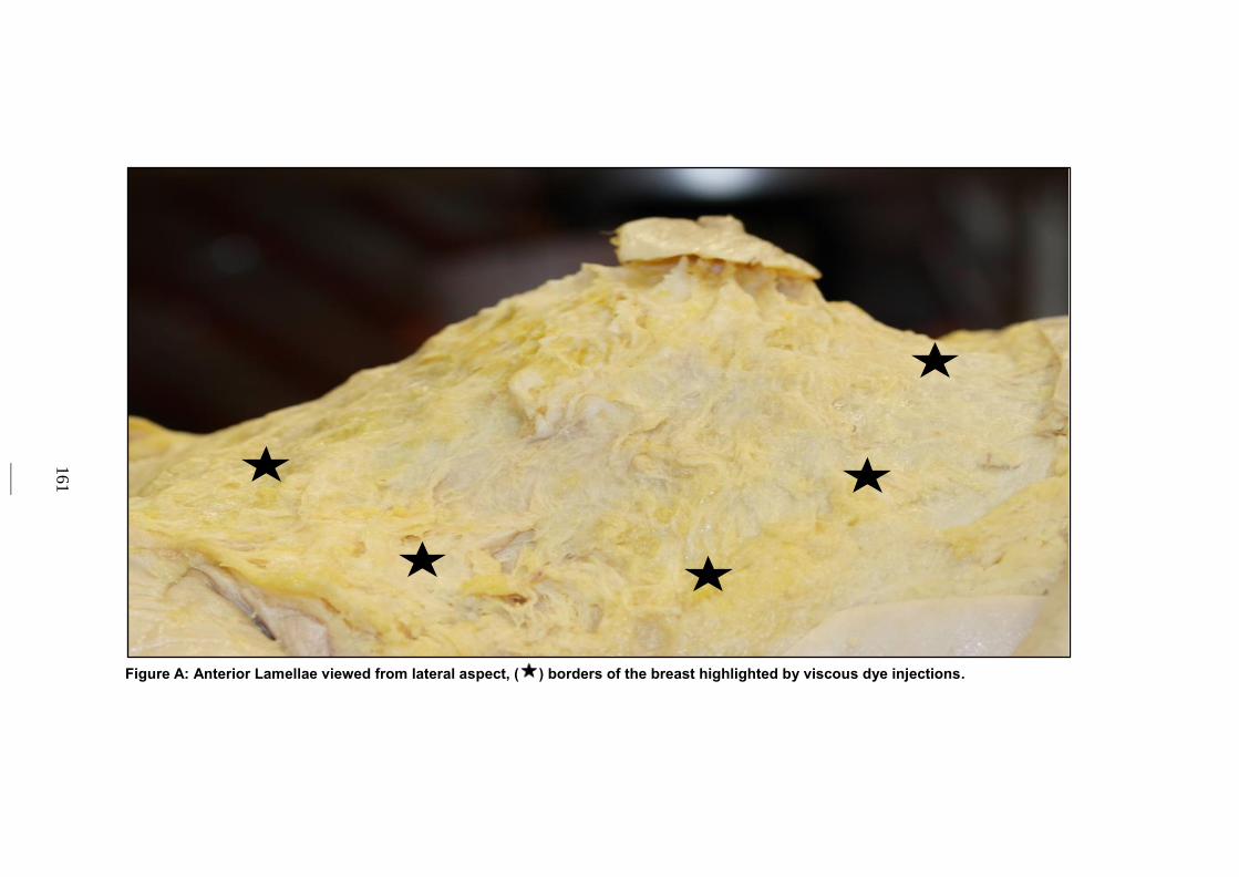
161
Figure A: Anterior Lamellae viewed from lateral aspect, ( ) borders of the breast highlighted by viscous dye injections.
162
Figure B: Coronal dissection of the breast showing the fibro-adipose tissue deep to
the fibro-glandular tissue and the Posterior Lamellae (reflected and coloured dark
blue).
S
M L
I
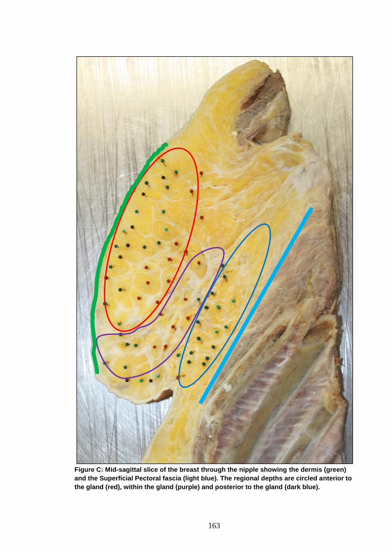
163
Figure C: Mid-sagittal slice of the breast through the nipple showing the dermis (green)
and the Superficial Pectoral fascia (light blue). The regional depths are circled anterior to
the gland (red), within the gland (purple) and posterior to the gland (dark blue).
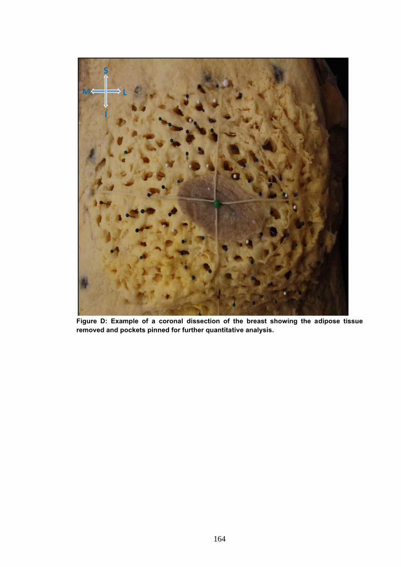
164
Figure D: Example of a coronal dissection of the breast showing the adipose tissue
removed and pockets pinned for further quantitative analysis.
S
M L
I
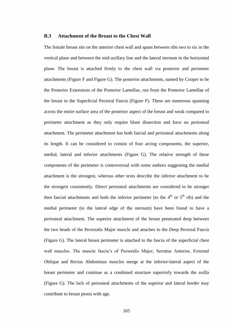
165
B.3 Attachment of the Breast to the Chest Wall
The female breast sits on the anterior chest wall and spans between ribs two to six in the
vertical plane and between the mid-axillary line and the lateral sternum in the horizontal
plane. The breast is attached firmly to the chest wall via posterior and perimeter
attachments (Figure F and Figure G). The posterior attachments, named by Cooper to be
the Posterior Extensions of the Posterior Lamellae, run from the Posterior Lamellae of
the breast to the Superficial Pectoral Fascia (Figure F). These are numerous spanning
across the entire surface area of the posterior aspect of the breast and weak compared to
perimeter attachment as they only require blunt dissection and have no periosteal
attachment. The perimeter attachment has both fascial and periosteal attachments along
its length. It can be considered to consist of four arcing components, the superior,
medial, lateral and inferior attachments (Figure G). The relative strength of these
components of the perimeter is controversial with some authors suggesting the medial
attachment is the strongest, whereas other texts describe the inferior attachment to be
the strongest consistently. Direct periosteal attachments are considered to be stronger
then fascial attachments and both the inferior perimeter (to the 4th
or 5th
rib) and the
medial perimeter (to the lateral edge of the sternum) have been found to have a
periosteal attachment. The superior attachment of the breast penetrated deep between
the two heads of the Pectoralis Major muscle and attaches to the Deep Pectoral Fascia
(Figure G). The lateral breast perimeter is attached to the fascia of the superficial chest
wall muscles. The muscle fascia’s of Pectoralis Major, Serratus Anterior, External
Oblique and Rectus Abdominus muscles merge at the inferior-lateral aspect of the
breast perimeter and continue as a combined structure superiorly towards the axilla
(Figure G). The lack of periosteal attachments of the superior and lateral border may
contribute to breast ptosis with age.
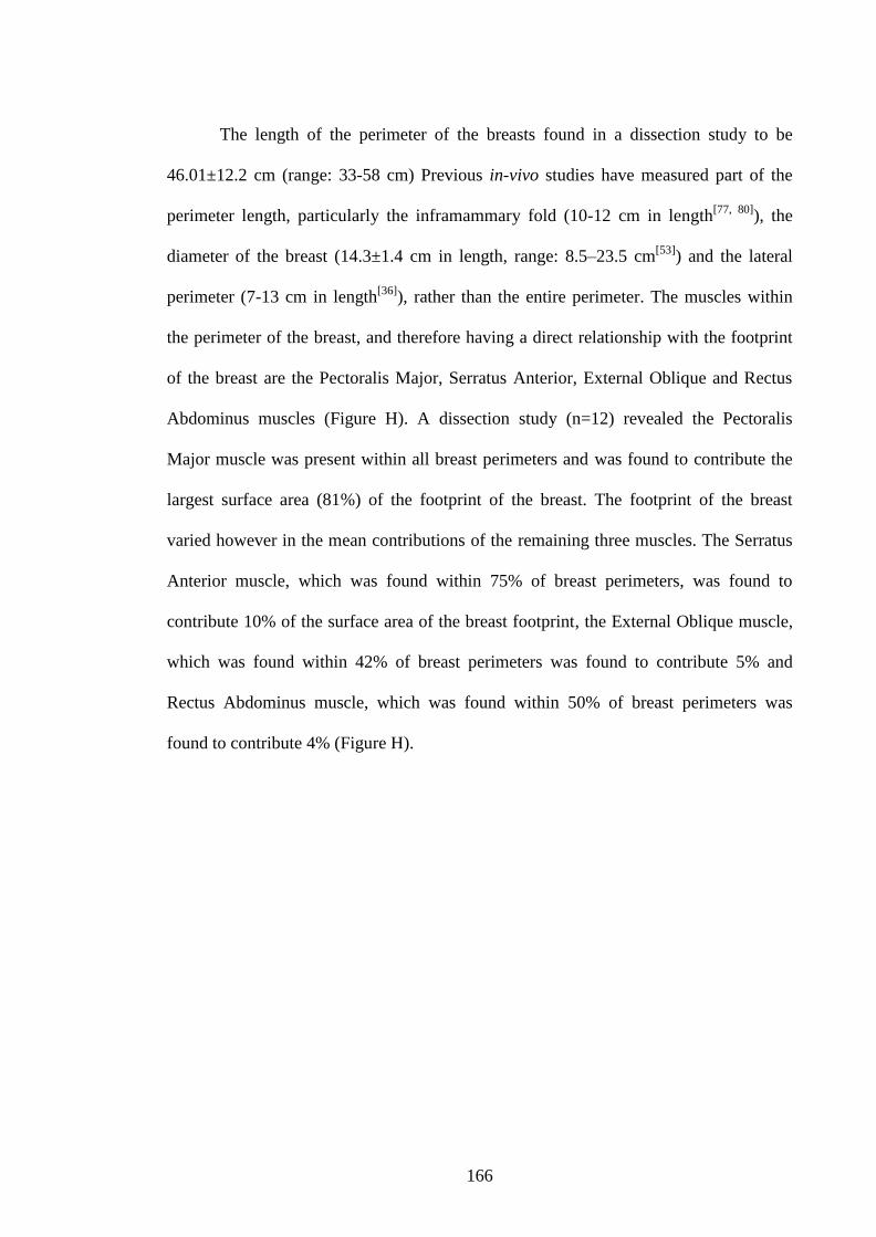
166
The length of the perimeter of the breasts found in a dissection study to be
46.01±12.2 cm (range: 33-58 cm) Previous in-vivo studies have measured part of the
perimeter length, particularly the inframammary fold (10-12 cm in length[77, 80]
), the
diameter of the breast (14.3±1.4 cm in length, range: 8.5–23.5 cm[53]
) and the lateral
perimeter (7-13 cm in length[36]
), rather than the entire perimeter. The muscles within
the perimeter of the breast, and therefore having a direct relationship with the footprint
of the breast are the Pectoralis Major, Serratus Anterior, External Oblique and Rectus
Abdominus muscles (Figure H). A dissection study (n=12) revealed the Pectoralis
Major muscle was present within all breast perimeters and was found to contribute the
largest surface area (81%) of the footprint of the breast. The footprint of the breast
varied however in the mean contributions of the remaining three muscles. The Serratus
Anterior muscle, which was found within 75% of breast perimeters, was found to
contribute 10% of the surface area of the breast footprint, the External Oblique muscle,
which was found within 42% of breast perimeters was found to contribute 5% and
Rectus Abdominus muscle, which was found within 50% of breast perimeters was
found to contribute 4% (Figure H).
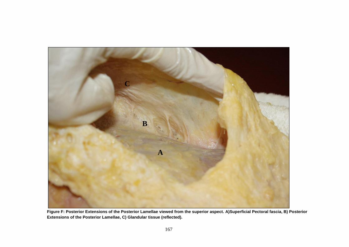
167
Figure F: Posterior Extensions of the Posterior Lamellae viewed from the superior aspect. A)Superficial Pectoral fascia, B) Posterior
Extensions of the Posterior Lamellae, C) Glandular tissue (reflected).
A
B
C
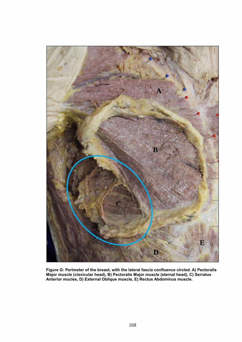
168
Figure G: Perimeter of the breast, with the lateral fascia confluence circled. A) Pectoralis Major muscle (clavicular head), B) Pectoralis Major muscle (sternal head), C) Serratus Anterior mucles, D) External Oblique muscle, E) Rectus Abdominus muscle.
A
B
C
E
D
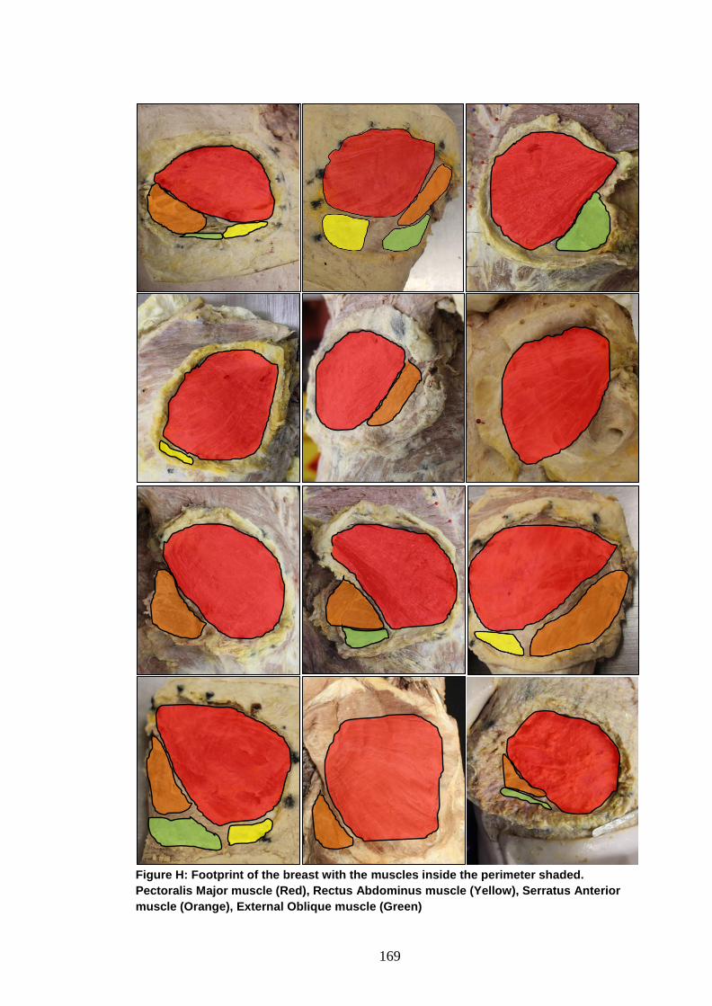
169
Figure H: Footprint of the breast with the muscles inside the perimeter shaded.
Pectoralis Major muscle (Red), Rectus Abdominus muscle (Yellow), Serratus Anterior
muscle (Orange), External Oblique muscle (Green)
170
Appendix C: Conference Presentations
C.1 9th
Australasian Biomechanics Conference, 2014, University of
Wollongong, NSW AUS
Abstract: Anatomy of the Breast: Work in Progress
By Kathryn Gaskin1, Dr Gregory Peoples
1, Dr Deirdre McGhee
1,
1Anatomy Laboratory, School of Medicine, Faculty of Science Medicine and Health,
University of Wollongong, NSW, Australia
Introduction: The anatomy of the structure of the breast appears in nearly every
anatomy textbook yet it is based on data and descriptions written by Cooper in 1840[1]
.
Although Cooper’s descriptions of the location, structure and composition of the breast
were extensive, they lacked quantitative data and no detail was provided of the number
of cadavers the descriptions were based on.
Since 1840, there have been only 10 anatomical studies that have verified or expanded
on Cooper’s work, particularly regarding the fascial and ligamentous structures of the
breast. Most of these studies also lacked quantitative data. Confusion and conflict exists
within both anatomy textbooks and studies regarding the terminology and location of
the fascial tissue within the breasts, in particular Cooper’s ligaments, the Inframammary
Fold (IMF) and the attachment of the breast to the chest wall. This is despite the
significance of these structures to breast surgery outcomes and the diagnosis of
pathology. Due to the paucity of research on the fascial structure of the breast and the
dated research that anatomical descriptions of the breast are based on, further research is
required which is supported by quantitative data.
Aim: To provide extensive quantitative data on the structural components of the breast
and to describe the gross structure of the female breast in terms of its structural supports
and composition.
171
Methods: The chest plate of 10 female cadavers will be removed and both breasts
dissected using two different dissection techniques. Study one will involve the left
breast, which will be examined in the frontal plane, in a superficial to deep dissection.
The skin around the borders of the breast will be injected to outline the perimeter of the
breast to its full depth, level of the superficial pectoral fascia. Once the skin is removed,
the breast will be scanned using a three-dimensional scanner (ArtecTM Eva three-
dimensional Scanner, San Jose) through which breast volume and surface area will be
measured. The superficial adipose and fascial layer will then be investigated
macroscopically to quantify the number and volume of the adipose pockets per quadrant
and the dimensions of fascial tissue bordering these pockets using anatomical grade
electronic Vernier Calipers (Mitutoyo, Japan; 0.02mL accuracy). These tissues will then
be removed and weighed as part of the total fibro-adipose weight (µg; Ohaus,
Adventurer® Pro, USA). The anterior surface of the gland will then be exposed,
outlined and scanned to quantify the surface area of the gland relative to the total breast
surface area. The fibro-glandular layer will then be removed and weighed to quantify
the total fibro-glandular weight. The fibro-adipose tissue posterior to the gland will then
be measured in a similar manner and weighed to determine the total fibro-adipose tissue
weight. The fibrous tissue attaching the breast to the chest wall and the IMF will then be
dissected and described. Finally the exposed anterior surface of the chest wall will be
scanned and its surface area described.
Study two will examine sagittal slices of the right breast made with a bandsaw through
standardised sections of the breast. Macroscopic investigation will include the number
adipose pockets, their location relative to the gland and the depth of each layer of the
breast. The fascial tissue inferior and superior to the gland, the IMF and the attachment
of the breast to the chest wall will also be measured and described. Microscopic
172
investigation will be performed using a microscope (Nikon SMZ800, Australia) and a
high-resolution photography (Photomicrographic system FX – III, Nikon, Australia) to
quantify the surface area of each adipose pocket and the dimensions of the fascial tissue
bordering these pockets.
Results and Discussion: This paper will provide state-of-the-art anatomical
descriptions and quantitative data on the structural components of the breast to benefit
both breast surgery and the diagnosis of breast pathology
References: [1] Cooper, S.A.P., 1840, On the Anatomy of the Breast, Volume 1.
London: Longman, Orme, Green, Brown and Longmans.
173
C.2 Tech-Net, 2015, University of Technology Sydney, NSW AUS
Abstract: Dissection Techniques to Explore the Fibro-adipose Structure of the
Female Breast
By Kathryn Gaskin1, Dr Gregory Peoples
1, Dr Deirdre McGhee
1
1Anatomy Laboratory, School of Medicine, Faculty of Science Medicine and Health,
University of Wollongong, NSW, Australia
Purpose: The anatomical terminology of the fibro-adipose structure of the female breast
is conflicting and based on the outdated work of Cooper in 1840. This study aimed to
investigate and quantify the structure of the breast to update anatomical descriptions for
the purpose of high quality dissection.
Methods: The structure of three embalmed cadaveric breasts was investigated using
dissection. The surface area (mm2) and volume (mL
3) of each breast was measured
using a three-dimensional scanner. The number of adipose pockets within the entire
breast and within quadrants of the breast was manually counted. The weight of adipose
tissue embedded in each pocket was weighed (g) and averaged per quadrant and per
breast.
Results: The smallest breast was associated the least number of adipose pockets and the
largest breast with the greatest number. Pocket size did not follow this same pattern and
was not consistent amongst the different quadrants.
Conclusion: The number of adipose pockets appeared to increase with breast volume
and surface area while the size of the pockets did not. The number and size of the
pockets in each quadrant varied. Therefore special attention needs to be paid when
dissecting the breast as each quadrant varies slightly in its fascial structure.
174
C.3 Australasian Institute of Anatomical Science, 2016, University of
Otago, Otago NZ
Abstract: Innovative techniques to explore the structural anatomy of the breast
By Kathryn Gaskin1, Dr Gregory Peoples
1, Dr Deirdre McGhee
1
1Anatomy Laboratory, School of Medicine, Faculty of Science Medicine and Health,
University of Wollongong, NSW, Australia
Introduction: The current anatomical knowledge of the structural anatomy of the breast
is based on dissections dating back to 1840. With only ten dissection studies published
since, much of the current knowledge is dated and lacking any quantitative data.
Anatomical texts contain limited detail of the structural anatomy of the breast and
anatomical illustrations are inconsistent. The aim of this study was to explore innovative
dissection techniques to investigate the fibro-adipose structure of the breast in three-
dimensions.
Methods: Four innovative techniques were critiqued; (i) Viscous dye injections to
outline the perimeter of the breast throughout its depth; (ii) Embedding the chest plate in
expanding for and slicing it with the bandsaw to investigate the breast in the sagittal
plane; (iii) Chemical dissection (NaOH) of differing temperatures and concentrations to
remove adipose tissue from the fibrous structure; (iv) Three-dimensional scanning to
quantify the volume, surface area of the breast and to digitally recreate the fibrous
structure of the breast.
Results: (i) Viscous-dye injected along the perimeter of the breast clearly outlined the
perimeter from the skin through to the ribs. (ii) Bandsaw slicing of the breast in the
sagittal plane displayed the structural anatomy of the breast without disrupting any
tissue. (iii) Chemical dissection only removed a percentage of adipose tissue and
compromised the fibrous structure. (iv) Three-dimensional scanning was useful to
175
quantify breast volume and surface area but was ineffective to digitally represent the
three-dimensional fibrous structure.
Conclusion: Viscous-dying, bandsaw slicing and three dimensional scanning are useful
techniques to investigate and quantify the fibro-adipose structure of the female breast.
Chemical dissection is not recommended to dissect adipose tissue to explore the
structural anatomy of the breast as it compromised the fibrous structure.
176
C.4 14th
Annual Conference of the Australia and New Zealand Association
of Clinical Anatomists, 2016, Australian National University, ACT AUS
Abstract: An update on the anatomy of the fibro-adipose structure of the female
breast
By Kathryn Gaskin1, Dr Gregory Peoples
1, Dr Deirdre McGhee
1
1Anatomy Laboratory, School of Medicine, Faculty of Science Medicine and Health,
University of Wollongong, NSW, Australia
Introduction: The anatomical terminology and descriptions of the fibro-adipose
structure of the female breast are inconsistent, dated and lack quantitative data. This
study aimed to investigate and quantify the fibro-adipose structure of the female to
update its anatomy.
Materials and Methods: The fibro-adipose structure of fifteen embalmed female breasts
was dissected in the coronal plane. Outcomes measures were (i) breast and breast
quadrant surface area (cm2) and volume (m
3); (ii) fibro-adipose pockets number (N),
surface area (cm2) and mass (g). Pearson’s correlations were used to investigate the
association between breast volume and fibro-adipose pocket number, and breast surface
area with fibro-adipose pocket number. T –tests compared the normalized fibro-adipose
pocket number and mass of the upper and lower volume tertile (P<0.01).
Results: The number of pockets per breast was found to increase both with breast
volume and surface area (P<0.05), with a strong positive correlation (r2=0.7372) found
between breast volume and number of pockets within the breast and between breast
surface area and the total number of pockets (r2=0.8064). A strong positive correlation
was also found between total breast volume and total breast surface area (r2=
0.9200)
(p<0.01)
177
Conclusion: This is the first anatomical study to describe the complex structure of the
breast with supporting quantitative data. Although there was no significant difference in
the number of pockets per breast in the largest breasts by volume (upper tertile)
compared to the smallest breasts by volume (lower tertile). There was a trend however
towards a higher number of pockets in the breasts of the upper tertile by volume
compared to the breasts of the lower tertile breasts by volume This provides the basis
for potential clinical application across a range of breast pathologies and procedures.
178
C.5 15th
Annual Conference of the Australia and New Zealand Association
of Clinical Anatomists, 2017, University of Auckland, Auckland, NZ
Abstract: Gross anatomy of the attachments and muscles involved in the
‘footprint’ of the female breast.
By Kathryn Gaskin1, Dr Deirdre McGhee
1, Dr Gregory Peoples
1
1Anatomy Laboratory, School of Medicine, Faculty of Science Medicine and Health,
University of Wollongong, NSW, Australia
Introduction: A summary of 31 anatomy textbooks detailing the anatomy of the breast
revealed limited descriptions of its attachments to the chest wall or the regional anatomy
of the footprint of the breast, with no associated quantitative data. This study aimed to
investigate the posterior and perimeter attachments of the breast to the chest wall and
quantify the muscles involved in the ‘footprint’ of the female breast.
Materials and Methods: Thirteen embalmed female breasts were dissected from
superficial to deep in the coronal and sagittal plane and the attachments of the breast to
the chest wall were compared and described. The percentage contribution of the
superficial muscles of the chest wall within the perimeter of the breast was quantified
using ImageJ software (Wayne Rasband, Maryland, USA).
Results: Multiple posterior attachments of the breast to the chest wall were found that
required blunt dissection only. The perimeter attachment was firmly adhered to the
underlying muscle fascia and required sharp dissection along its entire length. No direct
periosteal attachment was consistently found along the inferior aspect. The superficial
anterior chest wall muscles within the perimeter of the breast varied with breast volume.
Pectoralis Major muscle was consistently found within every perimeter and made up the
largest average surface area (81%), followed by the Serratus Anterior (10%), External
Oblique (5%) and Rectus Abdominus (4%) muscles.

179
Conclusion: This study provides gross anatomical descriptions and quantitative data on
the attachments and ‘footprint’ of the female breast to update descriptions and
illustrations in anatomical textbooks.
Copyright © 2022 FDOKUMEN