Identification of F-actin as the Dynamic Hub in a Microbial-Induced GTPase Polarity Circuit
Upload
independentCategory
view
5download
0
[CANCER RESEARCH 64, 2534–2543, April 1, 2004]
Stromal Cell-Derived Factor-1� Promotes Melanoma Cell Invasion across BasementMembranes Involving Stimulation of Membrane-Type 1 Matrix Metalloproteinaseand Rho GTPase Activities
Ruben A. Bartolome,1 Beatriz G. Galvez,2 Natividad Longo,3 Francoise Baleux,4 Goos N. P. van Muijen,5
Paloma Sanchez-Mateos,3 Alicia G. Arroyo,2 and Joaquin Teixido1
1Centro de Investigaciones Biologicas, Department of Immunology, Madrid, Spain;2Hospital Universitario de la Princesa, Department of Immunology, Madrid, Spain;3Serviciode Inmuno-Oncologıa, Hospital Universitario Gregorio Maran˜on, Madrid, Spain; 4Institut Pasteur, Unite de Chimie Organique, Paris, France;5Department of Pathology,University Medical Centre, Nijmegen, the Netherlands
ABSTRACT
Tissue invasion by tumor cells involves their migration across basementmembranes through activation of extracellular matrix degradation andcell motility mechanisms. Chemokines binding to their receptors providechemotactic cues guiding cells to specific tissues and organs; they there-fore could potentially participate in tumor cell dissemination. Melanomacells express CXCR4, the receptor for the chemokine stromal cell-derivedfactor-1� (SDF-1�). Using Matrigel as a model, we show that SDF-1�promotes invasion of melanoma cells across basement membranes. Stim-ulation of membrane-type 1 matrix metalloproteinase (MT1-MMP) activ-ity by SDF-1� was necessary for invasion, involving at least up-regulationin the expression of this metalloproteinase, as detected in the highlymetastatic BLM melanoma cell line. Moreover, SDF-1� triggered theactivation of the GTPases RhoA, Rac1, and Cdc42 on BLM cells, andexpression of dominant-negative forms of RhoA and Rac1, but not Cdc42,substantially impaired the invasion of transfectants in response to SDF-1�, as well as the increase in MT1-MMP expression. Furthermore,CXCR4 expression on melanoma cells was notably augmented by trans-forming growth factor-�1, a Matrigel component, whereas anti-trans-forming growth factor-� antibodies inhibited increases in CXCR4 expres-sion and melanoma cell invasion toward SDF-1�. The identification ofSDF-1� as a potential stimulatory molecule for MT1-MMP as well as forRhoA and Rac1 activities during melanoma cell invasion, associated withan up-regulation in CXCR4 expression by interaction with basementmembrane factors, could contribute to better knowledge of mechanismsstimulating melanoma cell dissemination.
INTRODUCTION
During the different steps of metastasis, tumor cells are likely tosense and respond to chemotactic cues that can contribute to theirinvasion and migration. Chemokines constitute a family of proteinsthat promote cell migration and activation, exerting these functions onbinding to seven transmembrane G-protein-linked chemokine recep-tors (1–4).
Expression of chemokine receptors on tumor cells and their in-volvement in metastasis has only recently been addressed, revealingthat these receptors can indeed provide migratory directions to tumorcells (5, 6). CXCR4 is a receptor for stromal cell-derived factor-1�(SDF-1�; also called CXCL12; Refs. 7, 8), a chemokine expressed inseveral tissues and organs, including skin, lymph nodes, lung, liver,and bone marrow (5, 9–11), which stimulates cell adhesion, migra-tion, and activation (10, 12–16). CXCR4 has been reported to beexpressed on different tumor cell types (5, 6, 17–23), and a key role
for CXCR4 has been documented in breast cancer metastasis (5).Expression of CXCR4 and its involvement in cell adhesion, migra-tion, and activation in melanoma was shown recently (5, 20, 24).Moreover, expression of CXCR4 in the B16 murine model of mela-noma conferred pulmonary metastatic potential to these cells com-pared with their CXCR4� counterparts (25). Therefore, these dataprovide a reasonable basis to propose that the SDF-1/CXCR4 axiscould play an important role in melanoma and breast cancer cellmetastasis.
During metastasis, tumor cells activate matrix digestion and loco-motion mechanisms to allow their invasion across basement mem-branes into surrounding connective tissues (26). Degradation of base-ment membranes by digestion of extracellular matrix (ECM) proteinsis accomplished by members of the matrix metalloproteinase (MMP)family, which are multidomain zinc-dependent endopeptidases in-volved in tissue remodeling and tumor invasion (27–31). Most MMPsare secreted in a latent form and subsequently processed to activespecies, but they can also be found associated with the cell membraneas membrane-type (MT) MMPs (27, 29, 30). Membrane-type 1 MMP(MT1-MMP; also known as MMP-14) is capable of degrading severalECM proteins, including fibronectin, laminin, collagen, gelatin, andvitronectin, as well as non-ECM substrates such as SDF-1 and severaladhesion receptors (29, 32–34). Furthermore, MT1-MMP is an acti-vator of pro-MMP-2 in coordination with tissue inhibitor of metallo-proteinase-2 (35–39), and it has been reported to be involved in tumorcell invasion and growth (35, 40–44). MT1-MMP has been found inmalignant melanoma specimens (45, 46), raising the possibility thatits proteolytic activity might be involved in melanoma cell dissemi-nation.
Cell migration is associated with a reorganization of actin cytoskel-eton that triggers changes in cell morphology. Activation of membersof the Rho family of GTPases, such as RhoA, Rac1, and Cdc42,controls the dynamics of the actin cytoskeleton; they thus representkey regulatory molecules during cell migration (47, 48). Mutationsleading to the expression of constitutively active Rho proteins havenot been found in human tumors, but rather a common finding is theiroverexpression (49, 50). Nevertheless, ectopic expression of domi-nantly activated and negative mutant forms or overexpression of theseGTPases has been crucial in characterizing their important roles intumor cell invasion (49, 51–55). These mutant forms act as compet-itors with wild-type endogenous GTPases for binding to RhoGEF(guanine nucleotide exchange factor) for dominant-negative activityor as inhibitors of endogenous GTPase activity for constitutivelyactive mutants (49).
One of the steps in melanoma cell metastasis potentially stimulatedon interaction of SDF-1�/CXCR4 could be the promotion of cellinvasion across basement membranes. As outlined above, matrixdegradation and migration mechanisms must be activated duringtumor cell invasion; therefore, characterization of these mechanismsrepresents an important issue of study in cancer science. In the presentwork we investigated whether MT1-MMP and Rho GTPase functions
Received 10/30/03; revised 1/8/04; accepted 1/20/04.Grant support: Grants SAF2002-00207 (J. Teixido), SAF2002-00068 (A. Arroyo),
and SAF2002-04615-C02-02 (P. Sanchez-Mateos) from the Ministerio de Ciencia yTecnologı´a.
The costs of publication of this article were defrayed in part by the payment of pagecharges. This article must therefore be hereby markedadvertisementin accordance with18 U.S.C. Section 1734 solely to indicate this fact.
Requests for reprints: Joaquin Teixido, Centro de Investigaciones Biologicas, De-partment of Immunology, Ramiro de Maeztu 9, 28040 Madrid, Spain. Phone: 34-91-8373112; Fax: 34-91-5360432; E-mail: [email protected].
2534
Research. on January 24, 2015. © 2004 American Association for Cancercancerres.aacrjournals.org Downloaded from
on melanoma cells constitute targets for activation by SDF-1� as wellas the consequences of these activations on melanoma cell invasionacross basement membranes. The results from this study could con-tribute to a better knowledge and characterization of the chemotacticcues guiding tumor cell invasion.
MATERIALS AND METHODS
Cells and Antibodies. The human melanoma cells lines BLM, Mel 57, andMeWo were cultured in DMEM (Gibco Invitrogen, Paisley, Scotland) supple-mented with 10% fetal bovine serum (BioWhittaker, Verviers, Belgium) andantibiotics. Melanoma lymph node metastases were obtained after informedconsent from patients undergoing surgery at the Department of Immuno-Oncology and Surgery, Hospital General Universitario Gregorio Maranon(Madrid, Spain). Histopathological diagnosis was confirmed for each speci-men. A homogeneous cell suspension was obtained by mechanic disruption oftumor tissue fragments. Cells were seeded in flasks coated with 0.5% gelatin(Sigma Chemical, St. Louis, MO) and cultured in MCDB 153/L-15 medium(78%:20%; Sigma Chemical) supplemented with 2% fetal bovine serum, 5�g/ml bovine insulin (Sigma Chemical), 15 �g/ml bovine pituitary extract(Clonetics BioWhittaker, Walkersville, MD), 5 ng/ml epidermal growth factor(Sigma), and 1.68 mM calcium chloride (complete medium). After 24 h,nonadherent cells were removed, and adherent cells were detached with 2 mM
EDTA in PBS and frozen in complete medium containing 10% DMSO (MerckKgaA, Darmstadt, Germany).
For flow cytometry and invasion assays, frozen cells were thawed andgrown to subconfluency in complete medium and used without any passage.Monoclonal antibody (mAb) to CXCR4 (44717.111) was purchased fromR&D Systems (Minneapolis, MN), and anti-�5-integrin and antipaxillin mAbswere from BD Biosciences PharMingen (San Diego, CA). The anti-MT1-MMP mAb LEM-2/15 has been described previously (56). The anti-CXCR4–01 mAb was kindly provided by Drs. Mario Mellado and JoseMiguel Rodrıguez-Frade (Centro Nacional de Biotecnologıa, Madrid, Spain),and integrin anti-�1 mAbs Lia 1/2.1 and anti-�2 TEA1/4.1, as well as controlP3X63 mAb, were gifts of Dr. Francisco Sanchez-Madrid (Hospital de laPrincesa, Madrid, Spain). Anti-transforming growth factor-� (TGF-�) anti-body 1D11.16.8 was obtained from the American Type Culture Collection(Rockville, MD), and antimacrophage colony-stimulating factor antibody wasfrom R&D Systems. Antifibroblast AS02-clon was purchased from Dianova(Hamburg, Germany), anti-HMB-45 mAb was from Signet Laboratories Inc.(Dedham, MA), anti-HMW-MAA was from BD Biosciences PharMingen, andanti-S100 and anti-Melan A were from Novocastra Laboratories Ltd. (New-castle upon Tyne, United Kingdom).
Flow Cytometry. Melanoma cell lines or melanoma cells from lymph nodemetastases were detached with 2 mM EDTA in PBS, and after washing,samples were resuspended in ice-cold PBS. Cells were incubated on ice for 30min with primary antibodies, followed by incubation with FITC-conjugatedantimouse or antirabbit secondary antibodies (DAKO A/S, Copenhagen, Den-mark) and analysis in a Coulter Epics XL or a FACScan cytofluorometer. ForS-100, Melan A, and HMB-45 intracellular staining, cells were fixed andpermeabilized with the Cytofix/Cytoperm TM kit (PharMingen) according tothe instructions of the manufacturer.
Invasion Assays. For Matrigel invasion assays, 15–30 � 103 BLM, Mel57, MeWo, transfected BLM, or melanoma cells isolated from lymph nodemetastases were resuspended in invasion medium (serum-free DMEM con-taining 0.4% BSA) and loaded onto 8 �m pore-size filters coated with 35–50�l of 1:3 dilution of Matrigel (BD Biosciences) in Transwells (Costar, Cam-bridge, MA). For collagen invasion assays, 20 � 103 BLM cells were loadedon filters coated with 60 �l of a 300 �g/ml solution of type I collagen (ICNBiomedicals Inc., Costa Mesa, CA). The lower compartments of invasionchambers were filled with medium alone or with medium containing differentconcentrations of SDF-1� (R&D Systems). After 22 h of incubation at 37°C,noninvading cells were removed from the upper surface of the filter, and cellsthat had migrated through the filter were fixed with 4% paraformaldehyde(Sigma), stained with crystal violet for Matrigel or toluidine blue for collageninvasions, and counted under a microscope. When invasion of transfected cellswas assessed, green fluorescent protein (GFP)-positive cells on the lowersurface of the filters were counted by fluorescence microscopy. When meta-
static melanoma samples were used, invasive cells beneath the filters werefixed, permeabilized with 0.5% Triton X-100 (Sigma) in Tris-buffered saline,blocked with Tris-buffered saline containing 0.5% blocking reagent (Boeh-ringer Mannheim GmbH, Mannheim, Germany), and incubated in the samesolution for 30 min with anti-S-100, anti-Melan-A, or control antibodies. Afterwashing, cells were incubated for 10 min with Alexa 546-conjugated antirabbit(Molecular Probes Inc., Eugene, OR) or FITC-conjugated antimouse (DAKOA/S) secondary antibodies. Finally, filters were washed, and melanoma cellswere counted by fluorescence microscopy.
Transfections. BLM cells were transfected with expression vectors(pEGFP-C1) coding for GFP alone; GFP-fused forms of wild-type RhoA,Rac1, and CDC42; dominant-negative N19-RhoA, N17-Rac1, and N17-Cdc42; activated V14-RhoA and V12-Rac1 (gifts of Dr. Francisco Sanchez-Madrid); wild-type full-length MT1-MMP or MT1-MMP mutant lacking itscytoplasmic domain (MT1�cyt-GFP; Ref. 57); or wild-type full-lengthCXCR4 (a gift of Dr. Antonio Serrano, Centro Nacional de Biotecnologıa,Madrid, Spain) by use of Lipofectamine reagent (Invitrogen) according to themanufacturer’s instructions. In brief, cells were incubated for 3.5 h with 1.5 �gof vectors in the presence of Lipofectamine in serum-free Optimem medium(Invitrogen), followed by incubation for 18 h in the same medium supple-mented with 10% FCS (BioWhittaker). After 36 h, cells were analyzed by flowcytometry to assess transfection efficiency, which was consistently �85%.
Zymography Assays. BLM cells incubated with or without SDF-1� orBLM cells transfected with MT1-MMP-GFP or MT1�cyt-GFP were washedtwice with PBS and lysed directly in Laemmli buffer. Samples were resolvedunder nonreducing conditions on SDS-PAGE gels embedded with 1 mg/mlfibrinogen (Calbiochem-Novabiochem Co., Darmstadt, Germany). Gels wererinsed three times with 2.5% Triton X-100, followed by incubation for 12 h at37°C in 50 mM Tris-HCl (pH 7.5), 10 mM CaCl2, and 200 mM NaCl. Gels werestained with Coomassie Blue, and areas of fibrinolytic activity were visualizedas transparent bands.
GTPase Activity Assays. The glutathione S-transferase (GST)-PAK-CDand GST-C21 fusion proteins were generated as described previously (58).Melanoma cells were starved for 2 h in DMEM without serum, and afterdetachment they were washed and resuspended at 2.5 � 106 cells/ml inDMEM containing 0.4% BSA. SDF-1� (150 ng/ml) was added to the cellsuspensions and incubated at 37°C for different times followed by rapidwashing with ice-cold PBS. Cells were lysed at 4°C in 300 �l of lysis buffer,as reported previously (20). Lysates were centrifuged; 15 �l of the supernatantwere kept for total lysate controls, and the remaining volume was mixed withfusion proteins in the presence of glutathione-agarose beads. The mixtureswere incubated for 16 h at 4°C, beads were pelleted and washed, and boundproteins were eluted in Laemmli electrophoresis buffer. Proteins were resolvedby SDS-PAGE on 12% polyacrylamide gels and transferred to polyvinylidenefluoride membranes (Hybond-P; Amersham Pharmacia Biotech), which wereincubated with antibodies to Rac1 or Cdc42 (BD Biosciences PharMingen) orto RhoA (Santa Cruz Biotechnology, Inc., Santa Cruz, CA). Recombinant C3transferase was expressed and purified as described previously (59).
Western Blotting and Reverse Transcription-PCR Analyses. BLM cells(2.5 � 105) incubated in the presence or absence of SDF-1�, or 5 � 105
transfected BLM cells expressing different GFP-fused proteins were solubilized at4°C in lysis buffer (20). Proteins were resolved by SDS-PAGE and electrotrans-ferred to polyvinylidene difluoride membranes (Amersham Pharmacia BiotechInc., Piscataway, NJ). Membranes were blocked with 5% skim milk in PBS;incubated with LEM 2/15 anti-MT1-MMP, anti-RhoA, anti-Rac1, or anti-Cdc42antibodies; washed with 0.1% Tween 20 in PBS; and then incubated with horse-radish peroxidase-conjugated secondary antibodies. Proteins were visualized byuse of SuperSignal chemiluminescent substrate (Pierce, Rockford, IL). Afterstripping and blocking, the same blots were reprobed with antipaxillin antibodies(BD Biosciences) to test for total protein content or with anti-GFP antibodies(Molecular Probes, Leiden, the Netherlands). For reverse transcription-PCR anal-ysis, BLM cells (2 � 106) treated under different conditions were lysed inTriReagent (Sigma-Aldrich, St. Louis, MO), and RNA was extracted and reverse-transcribed with use of Superscript reverse transcriptase (Gibco Invitrogen) andrandom hexadeoxynucleotide primers. Amplification of MT1-MMP cDNA wasperformed by PCR using AmpliTaq DNA polymerase (Roche Applied Science,Indianapolis, IN) with primers and profile conditions as described previously (60).Aliquots of each sample were amplified under the same conditions with thefollowing glyceraldehyde-3-phosphate dehydrogenase primers as cDNA loading
2535
SDF-1 PROMOTES MELANOMA CELL INVASION
Research. on January 24, 2015. © 2004 American Association for Cancercancerres.aacrjournals.org Downloaded from
control: 5�-GGCTGAGAACGGAAGCTTGTCA-3� and 5�-CGGCCATCACGC-CACAGTTTC-3�.
RESULTS
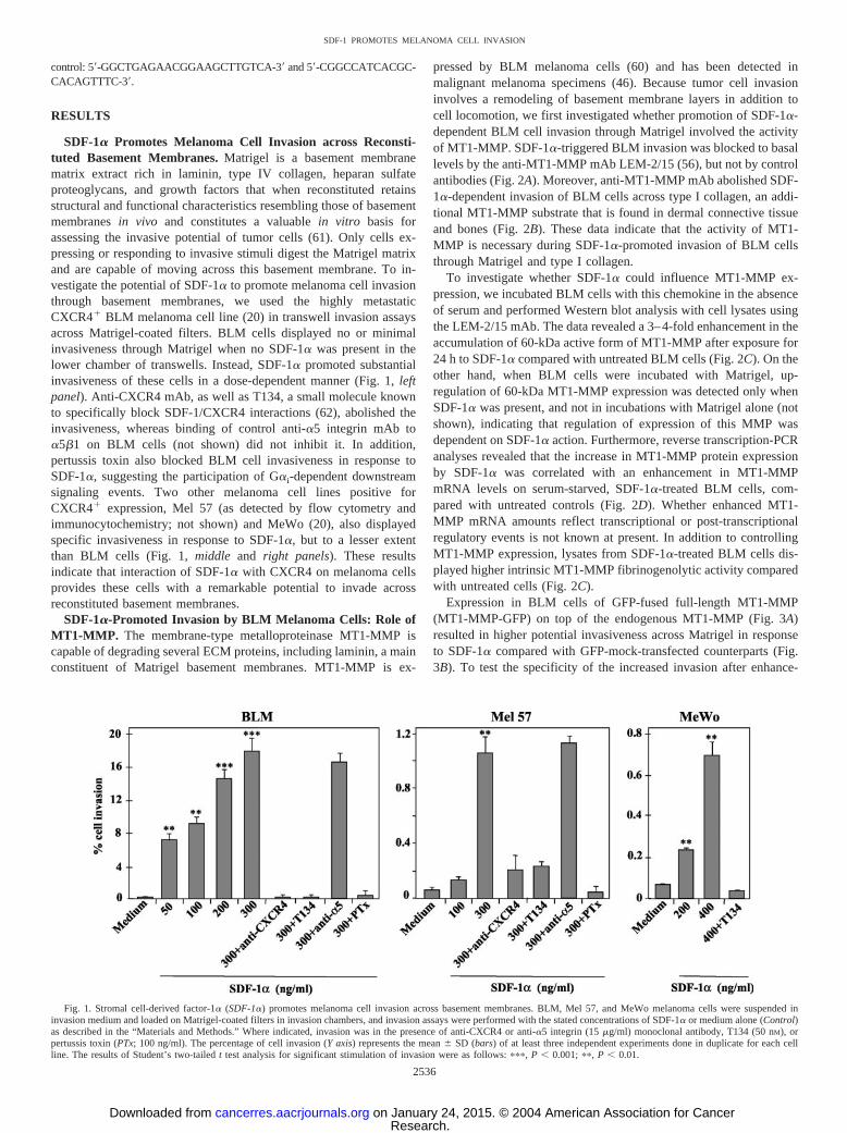
SDF-1� Promotes Melanoma Cell Invasion across Reconsti-tuted Basement Membranes. Matrigel is a basement membranematrix extract rich in laminin, type IV collagen, heparan sulfateproteoglycans, and growth factors that when reconstituted retainsstructural and functional characteristics resembling those of basementmembranes in vivo and constitutes a valuable in vitro basis forassessing the invasive potential of tumor cells (61). Only cells ex-pressing or responding to invasive stimuli digest the Matrigel matrixand are capable of moving across this basement membrane. To in-vestigate the potential of SDF-1� to promote melanoma cell invasionthrough basement membranes, we used the highly metastaticCXCR4� BLM melanoma cell line (20) in transwell invasion assaysacross Matrigel-coated filters. BLM cells displayed no or minimalinvasiveness through Matrigel when no SDF-1� was present in thelower chamber of transwells. Instead, SDF-1� promoted substantialinvasiveness of these cells in a dose-dependent manner (Fig. 1, leftpanel). Anti-CXCR4 mAb, as well as T134, a small molecule knownto specifically block SDF-1/CXCR4 interactions (62), abolished theinvasiveness, whereas binding of control anti-�5 integrin mAb to�5�1 on BLM cells (not shown) did not inhibit it. In addition,pertussis toxin also blocked BLM cell invasiveness in response toSDF-1�, suggesting the participation of G�i-dependent downstreamsignaling events. Two other melanoma cell lines positive forCXCR4� expression, Mel 57 (as detected by flow cytometry andimmunocytochemistry; not shown) and MeWo (20), also displayedspecific invasiveness in response to SDF-1�, but to a lesser extentthan BLM cells (Fig. 1, middle and right panels). These resultsindicate that interaction of SDF-1� with CXCR4 on melanoma cellsprovides these cells with a remarkable potential to invade acrossreconstituted basement membranes.
SDF-1�-Promoted Invasion by BLM Melanoma Cells: Role ofMT1-MMP. The membrane-type metalloproteinase MT1-MMP iscapable of degrading several ECM proteins, including laminin, a mainconstituent of Matrigel basement membranes. MT1-MMP is ex-
pressed by BLM melanoma cells (60) and has been detected inmalignant melanoma specimens (46). Because tumor cell invasioninvolves a remodeling of basement membrane layers in addition tocell locomotion, we first investigated whether promotion of SDF-1�-dependent BLM cell invasion through Matrigel involved the activityof MT1-MMP. SDF-1�-triggered BLM invasion was blocked to basallevels by the anti-MT1-MMP mAb LEM-2/15 (56), but not by controlantibodies (Fig. 2A). Moreover, anti-MT1-MMP mAb abolished SDF-1�-dependent invasion of BLM cells across type I collagen, an addi-tional MT1-MMP substrate that is found in dermal connective tissueand bones (Fig. 2B). These data indicate that the activity of MT1-MMP is necessary during SDF-1�-promoted invasion of BLM cellsthrough Matrigel and type I collagen.
To investigate whether SDF-1� could influence MT1-MMP ex-pression, we incubated BLM cells with this chemokine in the absenceof serum and performed Western blot analysis with cell lysates usingthe LEM-2/15 mAb. The data revealed a 3–4-fold enhancement in theaccumulation of 60-kDa active form of MT1-MMP after exposure for24 h to SDF-1� compared with untreated BLM cells (Fig. 2C). On theother hand, when BLM cells were incubated with Matrigel, up-regulation of 60-kDa MT1-MMP expression was detected only whenSDF-1� was present, and not in incubations with Matrigel alone (notshown), indicating that regulation of expression of this MMP wasdependent on SDF-1� action. Furthermore, reverse transcription-PCRanalyses revealed that the increase in MT1-MMP protein expressionby SDF-1� was correlated with an enhancement in MT1-MMPmRNA levels on serum-starved, SDF-1�-treated BLM cells, com-pared with untreated controls (Fig. 2D). Whether enhanced MT1-MMP mRNA amounts reflect transcriptional or post-transcriptionalregulatory events is not known at present. In addition to controllingMT1-MMP expression, lysates from SDF-1�-treated BLM cells dis-played higher intrinsic MT1-MMP fibrinogenolytic activity comparedwith untreated cells (Fig. 2C).
Expression in BLM cells of GFP-fused full-length MT1-MMP(MT1-MMP-GFP) on top of the endogenous MT1-MMP (Fig. 3A)resulted in higher potential invasiveness across Matrigel in responseto SDF-1� compared with GFP-mock-transfected counterparts (Fig.3B). To test the specificity of the increased invasion after enhance-
Fig. 1. Stromal cell-derived factor-1� (SDF-1�) promotes melanoma cell invasion across basement membranes. BLM, Mel 57, and MeWo melanoma cells were suspended ininvasion medium and loaded on Matrigel-coated filters in invasion chambers, and invasion assays were performed with the stated concentrations of SDF-1� or medium alone (Control)as described in the “Materials and Methods.” Where indicated, invasion was in the presence of anti-CXCR4 or anti-�5 integrin (15 �g/ml) monoclonal antibody, T134 (50 nM), orpertussis toxin (PTx; 100 ng/ml). The percentage of cell invasion (Y axis) represents the mean � SD (bars) of at least three independent experiments done in duplicate for each cellline. The results of Student’s two-tailed t test analysis for significant stimulation of invasion were as follows: ���, P � 0.001; ��, P � 0.01.
2536
SDF-1 PROMOTES MELANOMA CELL INVASION
Research. on January 24, 2015. © 2004 American Association for Cancercancerres.aacrjournals.org Downloaded from
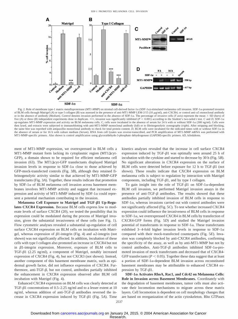
ment of MT1-MMP expression, we overexpressed in BLM cells aMT1-MMP mutant form lacking its cytoplasmic region (MT1�cyt-GFP), a domain shown to be required for efficient melanoma cellinvasion (63). The MT1�cyt-GFP transfectants displayed Matrigelinvasion levels in response to SDF-1� close to those achieved byGFP-mock-transfected controls (Fig. 3B), although they retained fi-brinogenolytic activity similar to that achieved by MT1-MMP-GFPtransfectants (Fig. 3A). Together, these results indicate that promotionby SDF-1� of BLM melanoma cell invasion across basement mem-branes involves MT1-MMP activity and suggest that increased ex-pression and activity of MT1-MMP induced by SDF-1� could repre-sent a potential mechanism contributing to the invasion.
Melanoma Cell Exposure to Matrigel and TGF-�1 Up-Regu-lates CXCR4 Expression. Because BLM cells express low to mod-erate levels of surface CXCR4 (20), we tested the possibility that itsexpression could be modulated during the process of Matrigel inva-sion, given the substantial invasiveness of these cells (see Fig. 1).Flow cytometric analyses revealed a substantial up-regulation of cellsurface CXCR4 expression on BLM cells on incubation with Matri-gel, whereas expression of �1-integrin (Fig. 4) and �2-integrin (notshown) was not significantly affected. In addition, incubation of thesecells with type I collagen also promoted an increase in CXCR4 but notin �1-integrin expression. Moreover, exposure of BLM cells toTGF-�1 (2.25 ng/ml), a component of Matrigel, notably increasedexpression of CXCR4 (Fig. 4), but not CXCR3 (not shown). Instead,another component of this basement membrane matrix, such as epi-dermal growth factor, did not influence expression of CXCR4. Fur-thermore, anti-TGF-�, but not control, antibodies partially inhibitedthe enhancement in CXCR4 expression observed after BLM cellincubation with Matrigel (Fig. 4).
Enhanced CXCR4 expression on BLM cells was clearly detected atTGF-�1 concentrations of 0.5–2.25 ng/ml and to a lesser extent at 10ng/ml, whereas addition of anti-TGF-� antibodies reversed the in-crease in CXCR4 expression induced by TGF-�1 (Fig. 5A). Time
kinetics analyses revealed that the increase in cell surface CXCR4expression induced by TGF-�1 was optimally seen around 25 h ofincubation with the cytokine and started to decrease by 30 h (Fig. 5B).No significant alterations in CXCR4 expression on the surface ofBLM cells were detected before exposure for 12 h to TGF-�1 (notshown). These results indicate that CXCR4 expression on BLMmelanoma cells is subject to regulation by interaction with Matrigelcomponents, including TGF-�1, and by type I collagen.
To gain insight into the role of TGF-�1 on SDF-1�-dependentBLM cell invasion, we performed Matrigel invasion assays in thepresence of anti-TGF-� antibodies. The results showed that theseantibodies partially inhibited invasion of BLM cells in response toSDF-1�, whereas invasions carried out with control antibodies werenot significantly affected (Fig. 5C). To test whether increased CXCR4expression could influence the invasiveness of BLM cells in responseto SDF-1�, we overexpressed CXCR4 in BLM cells by transfection ofCXCR4-GFP forms (Fig. 5D) and studied the Matrigel invasionpotential of transfectants in response to SDF-1�. These transfectantsexhibited 3–4-fold higher invasion levels in response to SDF-1�compared with their mock-transfected counterparts (Fig. 5E). Inva-sion was completely blocked by anti-CXCR4 antibodies, confirmingthe specificity of the assay, as well as by anti-MT1-MMP but not bycontrol antibodies. Anti-TGF-� antibodies inhibited SDF-1�-pro-moted invasion of mock transfectants and decreased that of CXCR4-GFP transfectants (P � 0.05). Together these data suggest that at leasta portion of SDF-1�-dependent BLM invasion across reconstitutedbasement membranes may be attributable to enhanced CXCR4 ex-pression by TGF-�1.
SDF-1� Activates RhoA, Rac1, and Cdc42 on Melanoma Cells:Role in Invasion across Basement Membranes. Coordinately withthe degradation of basement membranes, tumor cells must also acti-vate their locomotion mechanisms to migrate across these matrixlayers. Cell locomotion is dependent on cell morphology changes thatare based on reorganization of the actin cytoskeleton. Rho GTPases
Fig. 2. Role of membrane type-1 matrix metalloproteinase (MT1-MMP) on stromal cell-derived factor-1� (SDF-1�)-stimulated melanoma cell invasion. SDF-1�-promoted invasionof BLM cells through Matrigel (A) or type I collagen (B) was assessed in the presence of anti-MT1-MMP LEM-2/15 (10 �g/ml), anti-CXCR4, or control anti-�5 monoclonal antibodyor in the absence of antibody (Medium). Control denotes invasion performed in the absence of SDF-1�. The percentage of invasive cells (Y axis) represent the mean � SD (bars) offive (A) or three (B) independent experiments done in duplicate. ���, invasion was significantly inhibited (P � 0.001) according to the Student’s two-tailed t test. C and D, SDF-1�up-regulates MT1-MMP expression and activity on BLM melanoma cells. C, cells were incubated in the absence of serum for 24 h with or without SDF-1� (300 ng/ml). Cells werethen lysed, and extracts were subjected to immunoblotting with anti-MT1-MMP monoclonal antibody (left) or to fibrinogenolytic zymography (right). After stripping and blocking,the same blot was reprobed with antipaxillin monoclonal antibody to check for total protein content. D, BLM cells were incubated for the indicated times with or without SDF-1� inthe absence of serum or for 16 h with culture medium (Serum). RNA from cell lysates was reverse-transcribed, and PCR amplification of MT1-MMP mRNA was performed withMT1-MMP-specific primers. Also shown is control amplification using glyceraldehyde-3-phosphate dehydrogenase (GAPDH)-specific primers. kD, kilodaltons.
2537
SDF-1 PROMOTES MELANOMA CELL INVASION
Research. on January 24, 2015. © 2004 American Association for Cancercancerres.aacrjournals.org Downloaded from

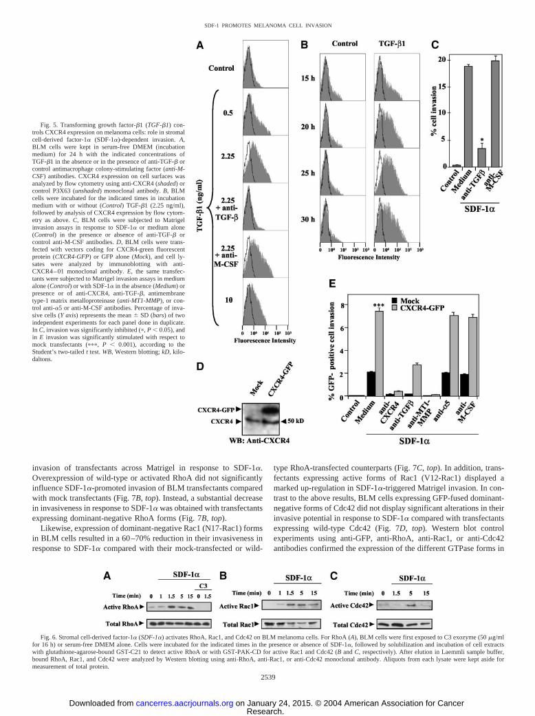
such as RhoA, Rac1, and Cdc42 are key regulators of the dynamics ofthe actin cytoskeleton, and their activation controls cell migration (47,48). Upstream events leading to the exchange of GDP for GTP boundto Rho GTPases activate them so they can interact with downstreamtargets to produce different biological responses. We investigatedwhether SDF-1� was capable of activating Rho GTPases on BLMcells and whether this activation could be involved in SDF-1�-promoted BLM invasion through reconstituted basement membranes.To test for Rho GTPase activation by SDF-1�, we performed GTPaseassays with lysates from SDF-1�-stimulated cells, using GST fusionproteins containing domains derived from Rho GTPase targets. Lowor minimal levels of RhoA, Rac1, and Cdc42 activation were detectedin unstimulated, serum-starved BLM cells. Incubation with SDF-1�rapidly promoted the activation of these cells, as detected with theGST-C21 fusion protein for RhoA or GST-PAK-CD for Rac1 andCdc42 activation (58; Fig. 6), whereas treatment with C3 transferase,a specific inhibitor of RhoA activation, blocked the activation of thisGTPase (Fig. 6A).
To investigate whether SDF-1�-promoted BLM cell invasionacross Matrigel was dependent on the activation of RhoA by thischemokine, we initially performed the invasion assays with C3 trans-ferase-treated BLM cells. The results showed that C3 abolished the
invasiveness of these cells in response to SDF-1� (Fig. 7A), suggest-ing that inhibition of RhoA activation could interfere with the inva-sion process. To further analyze the involvement of RhoA activationin SDF-1�-dependent BLM invasion, we expressed by transfectionGFP-fused wild-type, dominant-negative (N19-RhoA) or active (V14-RhoA) RhoA forms, as well as GFP alone (mock), and measured the
Fig. 4. Melanoma cell exposure to Matrigel up-regulates CXCR4 expression. BLMcells were resuspended in serum-free DMEM and cultured for 20 h in this medium alone(Control); in plates coated with Matrigel (0.4 �g/mm2) in the absence (Matrigel) orpresence of anti-transforming growth factor-�1 antibody (Matrigel � anti-TGF-�1) orcontrol antimacrophage colony-stimulating factor antibody (Matrigel � anti-M-CSF; 30�g/ml), or with type I collagen (Collagen type I; 0.12 �g/mm2), or were incubated in thepresence of transforming growth factor-�1 (TGF-�1; 2.25 ng/ml) or epidermal growthfactor (EGF; 0.6 ng/ml). After incubation, cells were detached, washed, and incubatedwith anti-CXCR4 or Lia 1/2.1 anti-�1-integrin (10 �g/ml; shaded curves) monoclonalantibody or with control P3X63 (10 �g/ml; unshaded curves) monoclonal antibody,followed by incubation with FITC-conjugated secondary antibody and analysis by flowcytometry.
Fig. 3. Enhancement in membrane type-1 matrix metalloproteinase (MT1-MMP)expression results in up-regulation of stromal cell-derived factor-1�-promoted melanomacell invasion. A, BLM cells were transfected with expression vectors coding for greenfluorescent protein (GFP)-fused full-length wild-type MT1-MMP (MT1-GFP), truncatedMT1-MMP lacking the cytoplasmic domain (MT1�cyt-GFP), or GFP alone (Mock).Expression and enzymatic activity of fusion proteins were checked in transfected celllysates by immunoblotting with anti-MT1-MMP monoclonal antibody (left) and byfibrinogenolytic zymography (right), respectively. kD, kilodaltons. B, the same transfec-tants were subjected to Matrigel invasion assays in the presence of stromal cell-derivedfactor-1� (SDF-1�; u) or medium alone (Control; f). Invasive GFP-positive cells werecounted under a fluorescence microscope, and the percentage of invasive cells (Y axis)represents the mean � SD (bars) of two independent experiments done in duplicate. �,invasion was significantly stimulated with respect to Mock transfectants (P � 0.05),according to the Student’s two-tailed t test.
2538
SDF-1 PROMOTES MELANOMA CELL INVASION
Research. on January 24, 2015. © 2004 American Association for Cancercancerres.aacrjournals.org Downloaded from
invasion of transfectants across Matrigel in response to SDF-1�.Overexpression of wild-type or activated RhoA did not significantlyinfluence SDF-1�-promoted invasion of BLM transfectants comparedwith mock transfectants (Fig. 7B, top). Instead, a substantial decreasein invasiveness in response to SDF-1� was obtained with transfectantsexpressing dominant-negative RhoA forms (Fig. 7B, top).
Likewise, expression of dominant-negative Rac1 (N17-Rac1) formsin BLM cells resulted in a 60–70% reduction in their invasiveness inresponse to SDF-1� compared with their mock-transfected or wild-
type RhoA-transfected counterparts (Fig. 7C, top). In addition, trans-fectants expressing active forms of Rac1 (V12-Rac1) displayed amarked up-regulation in SDF-1�-triggered Matrigel invasion. In con-trast to the above results, BLM cells expressing GFP-fused dominant-negative forms of Cdc42 did not display significant alterations in theirinvasive potential in response to SDF-1� compared with transfectantsexpressing wild-type Cdc42 (Fig. 7D, top). Western blot controlexperiments using anti-GFP, anti-RhoA, anti-Rac1, or anti-Cdc42antibodies confirmed the expression of the different GTPase forms in
Fig. 5. Transforming growth factor-�1 (TGF-�1) con-trols CXCR4 expression on melanoma cells: role in stromalcell-derived factor-1� (SDF-1�)-dependent invasion. A,BLM cells were kept in serum-free DMEM (incubationmedium) for 24 h with the indicated concentrations ofTGF-�1 in the absence or in the presence of anti-TGF-� orcontrol antimacrophage colony-stimulating factor (anti-M-CSF) antibodies. CXCR4 expression on cell surfaces wasanalyzed by flow cytometry using anti-CXCR4 (shaded) orcontrol P3X63 (unshaded) monoclonal antibody. B, BLMcells were incubated for the indicated times in incubationmedium with or without (Control) TGF-�1 (2.25 ng/ml),followed by analysis of CXCR4 expression by flow cytom-etry as above. C, BLM cells were subjected to Matrigelinvasion assays in response to SDF-1� or medium alone(Control) in the presence or absence of anti-TGF-� orcontrol anti-M-CSF antibodies. D, BLM cells were trans-fected with vectors coding for CXCR4-green fluorescentprotein (CXCR4-GFP) or GFP alone (Mock), and cell ly-sates were analyzed by immunoblotting with anti-CXCR4–01 monoclonal antibody. E, the same transfec-tants were subjected to Matrigel invasion assays in mediumalone (Control) or with SDF-1� in the absence (Medium) orpresence or of anti-CXCR4, anti-TGF-�, antimembranetype-1 matrix metalloproteinase (anti-MT1-MMP), or con-trol anti-�5 or anti-M-CSF antibodies. Percentage of inva-sive cells (Y axis) represents the mean � SD (bars) of twoindependent experiments for each panel done in duplicate.In C, invasion was significantly inhibited (�, P � 0.05), andin E invasion was significantly stimulated with respect tomock transfectants (���, P � 0.001), according to theStudent’s two-tailed t test. WB, Western blotting; kD, kilo-daltons.
Fig. 6. Stromal cell-derived factor-1� (SDF-1�) activates RhoA, Rac1, and Cdc42 on BLM melanoma cells. For RhoA (A), BLM cells were first exposed to C3 exozyme (50 �g/mlfor 16 h) or serum-free DMEM alone. Cells were incubated for the indicated times in the presence or absence of SDF-1�, followed by solubilization and incubation of cell extractswith glutathione-agarose-bound GST-C21 to detect active RhoA or with GST-PAK-CD for active Rac1 and Cdc42 (B and C, respectively). After elution in Laemmli sample buffer,bound RhoA, Rac1, and Cdc42 were analyzed by Western blotting using anti-RhoA, anti-Rac1, or anti-Cdc42 monoclonal antibody. Aliquots from each lysate were kept aside formeasurement of total protein.
2539
SDF-1 PROMOTES MELANOMA CELL INVASION
Research. on January 24, 2015. © 2004 American Association for Cancercancerres.aacrjournals.org Downloaded from

BLM transfectants (Fig. 7, B–D, bottom). The viability of dominant-negative, activated, and wild-type RhoA, Rac1, and Cdc42 BLMtransfectants was assessed 48 h post-transfection by trypan blueexclusion, which showed only 5–7% cell death and no significantvariations among the different transfectants. Altogether, these dataindicate that activation of endogenous RhoA and Rac1 by SDF-1� onBLM cells plays an important role in their invasion across basementmembranes promoted by this chemokine.
Role of SDF-1�-Promoted RhoA and Rac1 Activation on MT1-MMP Expression on Melanoma Cells. To investigate whether therewas a functional relationship between Rho-GTPase and MT1-MMPactivation promoted by SDF-1� on BLM melanoma cells, we firsttested the effect of C3 treatment on the expression of MT1-MMP.Up-regulation of MT1-MMP expression by SDF-1� was significantlyimpaired on exposure of BLM cells to C3 (Fig. 8A). Moreover,Western blot and reverse transcription-PCR analyses revealed thatexpression of dominant-negative RhoA and Rac1 forms on BLM cellsresulted in a blockade of increased MT1-MMP expression in responseto SDF-1� compared with the enhancement detected in transfectantsexpressing wild-type RhoA and Rac1 (Fig. 8, B–E), whereas expres-sion of dominant-negative forms of Cdc42 did not influence thisincrease (not shown). These results suggest that RhoA- and Rac1-promoted signaling originating in response to stimulation withSDF-1� controls the subsequent expression of MT1-MMP and that ittherefore could represent a mechanism regulating SDF-1�-triggeredinvasion of BLM melanoma cell across basement membranes.
SDF-1� Promotes Invasion of Metastatic Lymph Node Mela-noma Cell across Basement Membranes. Melanoma cells isolatedfrom lymph node metastases were thawed and cultured for 24–48 hbefore use in Matrigel basement membrane assays in the presence ofSDF-1�. Four of five samples exhibited SDF-1�-dependent invasionlevels ranging from 0.65 to 1% of the cell input that was blocked byanti-CXCR4 mAb and by T134 (P � 0.01), implicating the involve-ment of SDF-1�/CXCR4 interactions in this invasion. In addition,anti-MT1-MMP mAb (P � 0.05), but not control antibodies, inter-fered with SDF-1�-promoted invasion (Fig. 9A). Moreover, treatmentof melanoma cells with C3 also abolished their invasiveness in re-sponse to SDF-1�. To characterize the invaded cells, we performedimmunocytochemistry with a combination of the melanoma markersS100 and Melan-A. The results showed that �99% of �1-integrin-
positive cells expressed both markers (not shown), indicating that theinvading cell population corresponded to melanoma cells.
Melanoma cells isolated from lymph node metastases and incu-bated in the presence of Matrigel or TGF-�1 exhibited an enhance-ment in CXCR4 cell surface expression (Fig. 9B) similar to theincrease detected in BLM melanoma cells. These data indicate thatCXCR4 on metastatic melanoma cells is capable of mediating SDF-1�-dependent invasion across basement membranes and that MT1-
Fig. 7. Role of stromal cell-derived factor-1�(SDF-1�)-triggered RhoA and Rac1 activation onBLM melanoma cell invasion. A, BLM cells weresubjected to invasion assays across Matrigel in thepresence of SDF-1� (300 ng/ml) or medium alone,in the absence (�) or in the presence (�) of C3transferase (50 �g/ml). The percentage of cell in-vasion (Y axis) was determined as in Fig. 1. B–D,BLM cells were transfected with expression vec-tors coding for the wild-type (wt), dominant-nega-tive (DN), or active (Act) forms of RhoA-, Rac1-,or Cdc42-green fluorescent protein (GFP) fusionproteins or with GFP alone (Mock). Two days aftertransfection, 3 � 104 cells/condition were sub-jected to Matrigel invasion assays in the presenceof SDF-1� or medium alone (Control); only inva-sive GFP-positive cells were counted by fluores-cence microscopy. The rest of the cells (106/con-dition) were lysed to assess expression of fusionproteins by immunoblotting with anti-RhoA, anti-Rac1, anti-Cdc42, or anti-GFP monoclonal anti-body. The percentage of invasive cells (Y axis)represents the mean � SD (bars) of at least threeindependent experiments done in duplicate for eachpanel. Results of analysis with the Student’s two-tailed t test to determine whether invasion wassignificantly inhibited were as follows: ��,P � 0.01; �, P � 0.05. WB, Western blotting.
Fig. 8. Role of stromal cell-derived factor-1� (SDF-1�)-promoted RhoA and Rac1activation on membrane type-1 matrix metalloproteinase (MT1-MMP) expression onmelanoma cells. A, BLM cells were treated for 24 h with C3 transferase or in serum-freeDMEM alone in the absence (�) or presence (�) of SDF-1� (300 ng/ml). B–E, BLM cellswere transfected with expression vectors coding for wild-type (wt) or dominant-negative(DN) RhoA- or Rac1-green fluorescent protein (GFP) fusion proteins, and 2 days aftertransfection cells were incubated for 24 h (B and C) or 16 h (D and E) with (�) or without(�) SDF-1�. After incubation with this chemokine, cells were lysed, and extracts weresubjected to immunoblotting with anti-MT1-MMP monoclonal antibody. After strippingand blocking, the same blots were reprobed with antipaxillin (A, B, and C) or anti-RhoA(B) or anti-Rac1 mAb to check for total protein content. RNA from cell lysates wasreverse-transcribed, and amplification of MT1-MMP mRNA was performed by PCRusing MT1-MMP-specific primers (D and E). Also shown is control amplification usingglyceraldehyde-3-phosphate dehydrogenase (GAPDH)-specific primers.
2540
SDF-1 PROMOTES MELANOMA CELL INVASION
Research. on January 24, 2015. © 2004 American Association for Cancercancerres.aacrjournals.org Downloaded from

MMP activity, activation of RhoA, and possibly an increase inCXCR4 expression are involved in this invasion.
DISCUSSION
Basement membranes constitute biological barriers that must betraversed by tumor cells to invade surrounding tissues during metas-tasis. Chemokines are potential candidates to guide and activate tumorcells through basement membranes and tissues on binding to theirmembrane G-protein-coupled receptors. Melanoma cells express thechemokine receptor CXCR4, which interacts with its ligand SDF-1�,triggering their adhesion as well as mitogen-activated protein kinaseactivation (5, 20). Reconstituted basement membranes such as Matri-gel retain structural and functional characteristics resembling those ofbasement membranes in vivo, providing a useful system to test theinvasive potential of tumor cells. In the present study we showed thatSDF-1� promotes invasion of Matrigel basement membrane by theBLM, Mel 57, and MeWo melanoma cell lines, as well as invasion bymelanoma cells isolated from lymph node metastases. In addition toMatrigel invasion, SDF-1� was also capable of triggering invasion ofBLM cells across type I collagen, a constituent of dermal connectivetissues and bones.
Activating signals originated on SDF-1�/CXCR4 interaction there-fore lead to the invasion of melanoma cells across Matrigel, indicatingthat matrix degradation and migration mechanisms had been stimu-lated in these cells. Expression of the membrane-type metalloprotein-ase MT1-MMP has been found in malignant melanoma (45, 46), aswell as on melanoma BLM cells (60), raising the possibility that theproteolytic activity of this metalloproteinase could contribute to mel-anoma cell dissemination. We demonstrate here that MT1-MMPactivity is necessary for SDF-1�-promoted invasion of BLM andmetastatic melanoma cells across Matrigel, as well as for BLMinvasion through type I collagen matrices. Moreover, SDF-1� in-creased MT1-MMP expression on BLM cells, as well as its intrinsicfibrinogenolytic activity, and enhancement of MT1-MMP expressionby transfection with MT1-MMP-GFP forms resulted in up-regulated
BLM invasion across Matrigel in response to SDF-1�. These datasuggest that increased expression of MT1-MMP in response toSDF-1� could represent one of the mechanisms behind MT1-MMPproteolytic activity on Matrigel and type I collagen during SDF-1�-promoted melanoma cell invasion. Whether increased pro-MMP-2activation by MT1-MMP takes place during SDF-1�-triggered BLMinvasion requires further investigation.
Promotion by SDF-1� of melanoma cell invasion associated withthe stimulation of matrix digestion should take place coordinatelywith activation of locomotion mechanisms to allow migration of thesecells across basement membranes. Earlier reports revealed that ectopicexpression of constitutively active mutant forms of RhoA and Rac1 indifferent tumor cell types stimulated tumor cell locomotion and inva-sion (53–55). Here we showed that SDF-1� activates RhoA, Rac1,and Cdc42 on BLM melanoma cells and that expression of dominant-negative forms of RhoA and Rac1, but not Cdc42, or blocking ofRhoA activation by C3 transferase, resulted in a substantial reductionin SDF-1�-dependent melanoma cell Matrigel potential invasiveness.On the other hand, BLM transfectants expressing active Rac1 dis-played enhanced invasiveness in response to SDF-1� above the levelsachieved by transfectants expressing wild-type forms of this GTPase,suggesting that activated Rac1 functionally synergizes with SDF-1�-promoted Matrigel invasion. Together, these data strongly suggestthat activation of RhoA and Rac1 functions on melanoma cells inresponse to SDF-1� constitutes a potential mechanism that couldcontribute to their invasion across basement membranes in response tothis chemokine. It does not exclude, however, that although SDF-1�
is the trigger of BLM cell motility, it might not be the only stimuluscontributing to RhoA and Rac1 activation during invasion, becauseintegrin-mediated cell interactions with ECM components of Matrigelor mitogens present in Matrigel might also activate these GTPases(64).
In conjunction with activation of matrix digestion and cell migra-tion by SDF-1�, we show here that incubation of BLM and metastaticmelanoma cells with Matrigel or type I collagen, as well as with
Fig. 9. Stromal cell-derived factor-1� (SDF-1�)promotes invasion of metastatic lymph node mela-noma cells across Matrigel reconstituted basementmembranes. A, two different melanoma samples wereresuspended in invasion medium and subjected toMatrigel invasion assays with SDF-1� in the pres-ence of anti-CXCR4, anti-MT1-MMP, anti-�5 mono-clonal antibody or no antibodies (Medium) or in thepresence of T134 or C3 transferase. Control denotesinvasion performed in the absence of SDF-1�. After24 h, invaded cells were fixed, stained, and countedas in Fig. 1. Results of the Student’s two-tailed t testfor significant stimulation of invasion were as fol-lows: ��, P � 0.01; �, P �0.05. B, melanoma cellswere resuspended in serum-free DMEM and culturedfor 20 h in this medium alone (Control), in platescoated with Matrigel (Matrigel), or in the presence oftransforming growth factor-�1 (TGF-�1; 2.25 ng/ml). After incubation, cells were detached, washed,and incubated with anti-CXCR4 (shaded curves) orcontrol P3X63 (unshaded curves) monoclonal anti-body (mAb), followed by incubation with FITC-con-jugated secondary antibody and analysis by flow cy-tometry. Shown are two results representative of fourseparate experiments.
2541
SDF-1 PROMOTES MELANOMA CELL INVASION
Research. on January 24, 2015. © 2004 American Association for Cancercancerres.aacrjournals.org Downloaded from

TGF-�1, a major cytokine present in Matrigel, led to substantialenhancement in cell surface CXCR4 expression. Moreover, ectopicexpression of CXCR4-GFP forms in BLM cells led to enhancedinvasion in response to SDF-1�, suggesting that an increase inCXCR4 receptors could be a mechanism involved in SDF-1�-pro-moted invasion. Furthermore, anti-TGF-� antibodies inhibited bothincreased CXCR4 expression and SDF-1�-promoted BLM Matrigelinvasion. Therefore, these data suggest that enhancement in CXCR4expression on the surface of melanoma cells on contact with basementmembrane components, including TGF-�1, and/or with type I colla-gen might lead to a more efficient cell response to SDF-1� and thuscontribute to increased invasiveness. It is likely that melanoma cellsbecome exposed to TGF-� signaling because TGF-� is present in thetumor microenvironment (65), and this could result in up-regulatedCXCR4 expression, as reported here. The mechanisms underlying themodulation of CXCR4 expression by TGF-�1, as well as by type Icollagen, remain to be determined. Previous work showed that ex-pression of CXCR4 on T lymphocytes is also subject to modulation byTGF-�1 (66, 67).
Type I collagen is a main constituent of interstitial stroma, whichmust be traversed by tumor cells in addition to basement membranesduring invasion (26). MT1-MMP digestion of type I collagen wasshown previously to confer cell invasiveness (68). The present resultsshow that MT1-MMP activity is needed during SDF-1�-promotedBLM cell invasion of type I collagen matrices, which might involveincreased MT1-MMP expression in response to SDF-1�, as well as bytype I collagen (43). Furthermore, up-regulated CXCR4 expression inresponse to type I collagen could represent an additional factor con-tributing to the increased invasiveness in response to SDF-1�. Thesedata raise the possibility that SDF-1�/CXCR4 interactions mightprovide melanoma cells with additional means to invade acrossstroma rich in type I collagen.
Collectively, the results from the present work indicate that SDF-1�confers on melanoma cells a remarkable potential to invade acrossreconstituted basement membranes and type I collagen matrices,involving SDF-1�-promoted stimulation of MT1-MMP activity andRhoA- and Rac1-dependent cell motility, which could be contributedby increased CXCR4 expression during invasion. Because MT1-MMP function and activation of Rho GTPases have been shown toplay important roles during tumor cell invasion (28, 29, 53–55), theidentification of SDF-1� as a potential stimulatory molecule for bothtypes of molecules during melanoma cell invasion provides importantadditional knowledge of the mechanisms stimulating melanoma celldissemination. SDF-1� present in the tumor environment might worktogether with other cytokines and cell adhesion events, playing im-portant roles in the activation of MT1-MMP and Rho GTPases inmelanoma cells and contributing to the acquisition of an invasivephenotype. It is likely that cell locomotion and ECM degradation arefunctionally interdependent for melanoma cells to invade in responseto SDF-1� stimuli. In this regard, it has been reported that MMPlevels can be modulated by RhoA and Rac1 (69–71). We show herethat blocking of SDF-1�-promoted RhoA and Rac1 activation onBLM cells inhibits up-regulation of MT1-MMP mRNA and proteinexpression by this chemokine, which is compatible with regulation ofgene expression or post-transcriptional changes. These data suggestthat SDF-1�-triggered, Rho GTPase-dependent signaling leading tothe control of MT1-MMP expression could represent a coordinationmechanism between cell motility and cell surface proteolysis thatpossibly influences invasiveness in response to SDF-1�.
Recent in vivo data have shown that chemokines can contribute tothe homing of tumor cells to distinct organs in a manner similar to thatfor differential leukocyte tissue distribution, which depends on thechemokine expression pattern. For example, CXCR4 expression con-
veyed metastasis of human breast cancer cells into regional lymphnodes and lung (5). In line with these findings, down-regulation ofCXCR4 expression by RNA interference resulted in inhibition ofbreast cancer cell invasiveness in vitro (72). In melanoma, the expres-sion of CXCR4 and CCR7 on B16 murine cells enhanced theirpulmonary and lymph node metastatic potential, respectively (22, 24,25). Together with the in vitro melanoma cell invasion results pre-sented here, these data indicate that SDF-1� could play importantroles during melanoma cell metastasis, providing both invasion andmigratory directions for basement membrane and interstitial stromainfiltration in the skin (9), as well as representing an importantstimulus for homing of these cells to additional sites where thischemokine is expressed, such as lymph nodes, lung, and liver (5). Thefinding that CXCR4 expression could be modulated during melanomacell invasion provides an attractive potential mechanism for control-ling their dissemination. In support of this hypothesis, the involve-ment of the von Hippel-Lindau tumor suppressor pVHL in the mod-ulation of CXCR4 expression has been reported recently, suggestingthat it could control tumor cell dissemination (73).
In conclusion, interaction of SDF-1� with CXCR4 on melanomacells conveys invasive, migratory, and activating signals to these cells,similar to the signals for motile cells such as leukocytes. Character-ization of the stimuli modulating CXCR4 expression and intracellularsignaling triggered by SDF-1� could contribute to a better knowledgeof the scenario of tumor cell metastasis and could lead to potentialfindings to improve therapeutic approaches.
REFERENCES
1. Baggiolini M, Dewald B, Moser B. Human chemokines: an update. Annu RevImmunol 1997;15:675–705.
2. Rollins BJ. Chemokines Blood 1997;90:909–28.3. Rossi D, Zlotnik A. The biology of chemokines and their receptors. Annu Rev
Immunol 2000;18:217–42.4. Mellado M, Rodriguez-Frade JM, Manes S, Martinez AC. Chemokine signaling and
functional responses: the role of receptor dimerization and TK pathway activation.Annu Rev Immunol 2001;19:397–421.
5. Muller A, Homey B, Soto H, et al. Involvement of chemokine receptors in breastcancer metastasis. Nature (Lond) 2001;410:50–6.
6. Homey B, Muller A, Zlotnik A. Chemokines: agents for the immunotherapy ofcancer? Nat Rev Immunol 2002;2:175–84.
7. Bleul CC, Farzan M, Choe H, et al. The lymphocyte chemoattractant SDF-1 is aligand for LESTR/fusin and blocks HIV-1 entry. Nature (Lond) 1996;382:829–33.
8. Oberlin E, Amara A, Bachelerie F, et al. The CXC chemokine SDF-1 is the ligand forLESTR/fusin and prevents infection by T-cell-line-adapted HIV-1. Nature (Lond)1996;382:833–5.
9. Pablos JL, Amara A, Bouloc A, et al. Stromal-cell derived factor is expressed bydendritic cells and endothelium in human skin. Am J Pathol 1999;155:1577–86.
10. Aiuti A, Webb IJ, Bleul C, Springer T, Gutierrez-Ramos JC. The chemokine SDF-1is a chemoattractant for human CD34� hematopoietic progenitor cells and providesa new mechanism to explain the mobilization of CD34� progenitors to peripheralblood. J Exp Med 1997;185:111–20.
11. Tashiro K, Tada H, Heilker R, Shirozu M, Nakano T, Honjo T. Signal sequence trap:a cloning strategy for secreted proteins and type I membrane proteins. Science (WashDC) 1993;261:600–3.
12. Bleul CC, Fuhlbrigge RC, Casasnovas JM, Aiuti A, Springer TA. A highly effica-cious lymphocyte chemoattractant, stromal cell-derived factor 1 (SDF-1). J Exp Med1996;184:1101–9.
13. Campbell JJ, Hedrick J, Zlotnik A, Siani MA, Thompson DA, Butcher EC. Chemo-kines and the arrest of lymphocytes rolling under flow conditions. Science (Wash DC)1998;279:381–4.
14. Grabovsky V, Feigelson S, Chen C, et al. Subsecond induction of �4 integrinclustering by immobilized chemokines stimulates leukocyte tethering and rolling onendothelial vascular cell adhesion molecule 1 under flow conditions. J Exp Med2000;192:495–506.
15. Wright N, Hidalgo A, Rodriguez-Frade JM, et al. The chemokine stromal cell-derivedfactor-1� modulates �4�7 integrin-mediated lymphocyte adhesion to mucosal ad-dressin cell adhesion molecule-1 and fibronectin. J Immunol 2002;168:5268–77.
16. Ganju RK, Brubaker SA, Meyer J, et al. The �-chemokine, stromal cell-derivedfactor-1�, binds to the transmembrane G-protein-coupled CXCR-4 receptor andactivates multiple signal transduction pathways. J Biol Chem 1998;273:23169–75.
17. Scotton CJ, Wilson JL, Scott K, et al. Multiple actions of the chemokine CXCL12 onepithelial tumor cells in human ovarian cancer. Cancer Res 2002;62:5930–8.
18. Payne AS, Cornelius LA. The role of chemokines in melanoma tumor growth andmetastasis. J Investig Dermatol 2002;118:915–22.
2542
SDF-1 PROMOTES MELANOMA CELL INVASION
Research. on January 24, 2015. © 2004 American Association for Cancercancerres.aacrjournals.org Downloaded from

19. Youngs SJ, Ali SA, Taub DD, Rees RC. Chemokines induce migrational responses inhuman breast carcinoma cell lines. Int J Cancer 1997;71:257–66.
20. Robledo MM, Bartolome RA, Longo N, et al. Expression of functional chemokinereceptors CXCR3 and CXCR4 on human melanoma cells. J Biol Chem 2001;276:45098–105.
21. Geminder H, Sagi-Assif O, Goldberg L, et al. A possible role for CXCR4 and itsligand, the CXC chemokine stromal cell-derived factor-1, in the development of bonemarrow metastases in neuroblastoma. J Immunol 2001;167:4747–57.
22. Wiley HE, Gonzalez EB, Maki W, Wu MT, Hwang ST. Expression of CC chemokinereceptor-7 and regional lymph node metastasis of B16 murine melanoma. J NatlCancer Inst (Bethesda) 2001;93:1638–43.
23. Kijima T, Maulik G, Ma PC, et al. Regulation of cellular proliferation, cytoskeletalfunction, and signal transduction through CXCR4 and c-Kit in small cell lung cancercells. Cancer Res 2002;62:6304–11.
24. Cardones AR, Murakami T, Hwang ST. CXCR4 enhances adhesion of B16 tumorcells to endothelial cells in vitro and in vivo via �(1) integrin. Cancer Res 2003;63:6751–7.
25. Murakami T, Maki W, Cardones AR, et al. Expression of CXC chemokine receptor-4enhances the pulmonary metastatic potential of murine B16 melanoma cells. CancerRes 2002;62:7328–34.
26. Liotta LA, Kohn EC. The microenvironment of the tumour-host interface. Nature(Lond) 2001;411:375–9.
27. Nagase H, Woessner JF Jr. Matrix metalloproteinases. J Biol Chem 1999;274:21491–4.
28. Egeblad M, Werb Z. New functions for the matrix metalloproteinases in cancerprogression. Nat Rev Cancer 2002;2:161–74.
29. McCawley LJ, Matrisian LM. Matrix metalloproteinases: they’re not just for matrixanymore! Curr Opin Cell Biol 2001;13:534–40.
30. Stamenkovic I. Extracellular matrix remodelling: the role of matrix metalloprotein-ases. J Pathol 2003;200:448–64.
31. Overall CM, Lopez-Otin C. Strategies for MMP inhibition in cancer: innovations forthe post-trial era. Nat Rev Cancer 2002;2:657–72.
32. Kajita M, Itoh Y, Chiba T, et al. Membrane-type 1 matrix metalloproteinase cleavesCD44 and promotes cell migration. J Cell Biol 2001;153:893–904.
33. McQuibban GA, Butler GS, Gong JH, et al. Matrix metalloproteinase activityinactivates the CXC chemokine stromal cell-derived factor-1. J Biol Chem 2001;276:43503–8.
34. Deryugina EI, Ratnikov BI, Postnova TI, Rozanov DV, Strongin AY. Processing ofintegrin �(v) subunit by membrane type 1 matrix metalloproteinase stimulates mi-gration of breast carcinoma cells on vitronectin and enhances tyrosine phosphoryla-tion of focal adhesion kinase. J Biol Chem 2002;277:9749–56.
35. Sato H, Takino T, Okada Y, et al. A matrix metalloproteinase expressed on thesurface of invasive tumour cells. Nature (Lond) 1994;370:61–5.
36. Pei D, Weiss SJ. Transmembrane-deletion mutants of the membrane-type matrixmetalloproteinase-1 process progelatinase A and express intrinsic matrix-degradingactivity. J Biol Chem 1996;271:9135–40.
37. Ohuchi E, Imai K, Fujii Y, Sato H, Seiki M, Okada Y. Membrane type 1 matrixmetalloproteinase digests interstitial collagens and other extracellular matrix macro-molecules. J Biol Chem 1997;272:2446–51.
38. Strongin AY, Collier I, Bannikov G, Marmer BL, Grant GA, Goldberg GI. Mecha-nism of cell surface activation of 72-kDa type IV collagenase. Isolation of theactivated form of the membrane metalloprotease. J Biol Chem 1995;270:5331–8.
39. Fernandez-Catalan C, Bode W, Huber R, et al. Crystal structure of the complexformed by the membrane type 1-matrix metalloproteinase with the tissue inhibitorof metalloproteinases-2, the soluble progelatinase A receptor. EMBO J 1998;17:5238–48.
40. Ha HY, Moon HB, Nam MS, et al. Overexpression of membrane-type matrixmetalloproteinase-1 gene induces mammary gland abnormalities and adenocarcinomain transgenic mice. Cancer Res 2001;61:984–90.
41. Seftor RE, Seftor EA, Koshikawa N, et al. Cooperative interactions of laminin 5gamma2 chain, matrix metalloproteinase-2, and membrane type-1-matrix/metallopro-teinase are required for mimicry of embryonic vasculogenesis by aggressive mela-noma. Cancer Res 2001;61:6322–7.
42. Kurschat P, Zigrino P, Nischt R, et al. Tissue inhibitor of matrix metalloproteinase-2regulates matrix metalloproteinase-2 activation by modulation of membrane-type 1matrix metalloproteinase activity in high and low invasive melanoma cell lines. J BiolChem 1999;274:21056–62.
43. Gilles C, Polette M, Seiki M, Birembaut P, Thompson EW. Implication of collagentype I-induced membrane-type 1-matrix metalloproteinase expression and matrixmetalloproteinase-2 activation in the metastatic progression of breast carcinoma. LabInvestig 1997;76:651–60.
44. Koshikawa N, Giannelli G, Cirulli V, Miyazaki K, Quaranta V. Role of cell surfacemetalloprotease MT1-MMP in epithelial cell migration over laminin-5. J Cell Biol2000;148:615–24.
45. Hofmann UB, Westphal JR, Zendman AJ, Becker JC, Ruiter DJ, van Muijen GN.Expression and activation of matrix metalloproteinase-2 (MMP-2) and its co-local-ization with membrane-type 1 matrix metalloproteinase (MT1-MMP) correlate withmelanoma progression. J Pathol 2000;191:245–56.
46. Kurschat P, Wickenhauser C, Groth W, Krieg T, Mauch C. Identification of activatedmatrix metalloproteinase-2 (MMP-2) as the main gelatinolytic enzyme in malignantmelanoma by in situ zymography. J Pathol 2002;197:179–87.
47. Kaibuchi K, Kuroda S, Amano M. Regulation of the cytoskeleton and cell adhesionby the Rho family GTPases in mammalian cells. Annu Rev Biochem 1999;68:459–86.
48. Etienne-Manneville S, Hall A. Rho GTPases in cell biology. Nature (Lond) 2002;420:629–35.
49. Sahai E, Marshall CJ. RHO-GTPases and cancer. Nat Rev Cancer 2002;2:133–42.50. Preudhomme C, Roumier C, Hildebrand MP, et al. Nonrandom 4p13 rearrangements
of the RhoH/TTF gene, encoding a GTP-binding protein, in non-Hodgkin’s lym-phoma and multiple myeloma. Oncogene 2000;19:2023–32.
51. Clark EA, Golub TR, Lander ES, Hynes RO. Genomic analysis of metastasis revealsan essential role for RhoC. Nature (Lond) 2000;406:532–5.
52. del Peso L, Hernandez-Alcoceba R, Embade N, et al. Rho proteins induce metastaticproperties in vivo. Oncogene 1997;15:3047–57.
53. Michiels F, Habets GG, Stam JC, van der Kammen RA, Collard JG. A role for Racin Tiam1-induced membrane ruffling and invasion. Nature (Lond) 1995;375:338–40.
54. Keely PJ, Westwick JK, Whitehead IP, Der CJ, Parise LV. Cdc42 and Rac1 induceintegrin-mediated cell motility and invasiveness through PI(3)K. Nature (Lond)1997;390:632–6.
55. Banyard J, Anand-Apte B, Symons M, Zetter BR. Motility and invasion are differ-entially modulated by Rho family GTPases. Oncogene 2000;19:580–91.
56. Galvez BG, Matias-Roman S, Albar JP, Sanchez-Madrid F, Arroyo AG. Membranetype 1-matrix metalloproteinase is activated during migration of human endothelialcells and modulates endothelial motility and matrix remodeling. J Biol Chem 2001;276:37491–500.
57. Galvez BG, Matias-Roman S, Yanez-Mo M, Sanchez-Madrid F, Arroyo AG. ECMregulates MT1-MMP localization with�1 or �v�3 integrins at distinct cell compart-ments modulating its internalization and activity on human endothelial cells. J CellBiol 2002;159:509–21.
58. Sander EE, van Delft S, ten Klooster JP, et al. Matrix-dependent Tiam1/Rac signalingin epithelial cells promotes either cell-cell adhesion or cell migration and is regulatedby phosphatidylinositol 3-kinase. J Cell Biol 1998;143:1385–98.
59. Nobes CD, Hall A. Rho, rac, and cdc42 GTPases regulate the assembly of multimo-lecular focal complexes associated with actin stress fibers, lamellipodia, and filopo-dia. Cell 1995;81:53–62.
60. Hofmann UB, Westphal JR, Van Kraats AA, Ruiter DJ, Van Muijen GN. Expressionof integrin �(v)�(3) correlates with activation of membrane-type matrix metallopro-teinase-1 (MT1-MMP) and matrix metalloproteinase-2 (MMP-2) in human melanomacells in vitro and in vivo. Int J Cancer 2000;87:12–9.
61. Albini A, Iwamoto Y, Kleinman HK, et al. A rapid in vitro assay for quantitating theinvasive potential of tumor cells. Cancer Res 1987;47:3239–45.
62. Arakaki R, Tamamura H, Premanathan M, et al. T134, a small-molecule CXCR4inhibitor, has no cross-drug resistance with AMD3100, a CXCR4 antagonist with adifferent structure. J Virol 1999;73:1719–23.
63. Lehti K, Valtanen H, Wickstrom S, Lohi J, Keski-Oja J. Regulation of membrane-type-1 matrix metalloproteinase activity by its cytoplasmic domain. J Biol Chem2000;275:15006–13.
64. Schwartz MA, Shattil SJ. Signaling networks linking integrins and rho familyGTPases. Trends Biochem Sci 2000;25:388–91.
65. Taipale J, Saharinen J, Keski-Oja J. Extracellular matrix-associated transforminggrowth factor-�: role in cancer cell growth and invasion. Adv Cancer Res 1998;75:87–134.
66. Buckley CD, Amft N, Bradfield PF, et al. Persistent induction of the chemokinereceptor CXCR4 by TGF-�1 on synovial T cells contributes to their accumulationwithin the rheumatoid synovium. J Immunol 2000;165:3423–9.
67. Franitza S, Kollet O, Brill A, et al. TGF-�1 enhances SDF-1�-induced chemotaxisand homing of naive T cells by up-regulating CXCR4 expression and downstreamcytoskeletal effector molecules. Eur J Immunol 2002;32:193–202.
68. Hotary K, Allen E, Punturieri A, Yana I, Weiss SJ. Regulation of cell invasion andmorphogenesis in a three-dimensional type I collagen matrix by membrane-typematrix metalloproteinases 1, 2, and 3. J Cell Biol 2000;149:1309–23.
69. Zhuge Y, Xu J. Rac1 mediates type I collagen-dependent MMP-2 activation. role incell invasion across collagen barrier. J Biol Chem 2001;276:16248–56.
70. Kheradmand F, Werner E, Tremble P, Symons M, Werb Z. Role of Rac1 and oxygenradicals in collagenase-1 expression induced by cell shape change. Science (WashDC) 1998;280:898–902.
71. Matsumoto Y, Tanaka K, Harimaya K, Nakatani F, Matsuda S, Iwamoto Y. SmallGTP-binding protein, Rho, both increased and decreased cellular motility, activationof matrix metalloproteinase 2 and invasion of human osteosarcoma cells. Jpn J CancerRes 2001;92:429–38.
72. Chen Y, Stamatoyannopoulos G, Song CZ. Down-regulation of CXCR4 by induciblesmall interfering RNA inhibits breast cancer cell invasion in vitro. Cancer Res2003;63:4801–4.
73. Staller P, Sulitkova J, Lisztwan J, Moch H, Oakeley EJ, Krek W. Chemokine receptorCXCR4 downregulated by von Hippel-Lindau tumour suppressor pVHL. Nature(Lond) 2003;425:307–11.
2543
SDF-1 PROMOTES MELANOMA CELL INVASION
Research. on January 24, 2015. © 2004 American Association for Cancercancerres.aacrjournals.org Downloaded from

2004;64:2534-2543. Cancer Res Rubén A. Bartolomé, Beatriz G. Gálvez, Natividad Longo, et al. GTPase Activitiesof Membrane-Type 1 Matrix Metalloproteinase and RhoInvasion across Basement Membranes Involving Stimulation
Promotes Melanoma CellαStromal Cell-Derived Factor-1
Updated version
http://cancerres.aacrjournals.org/content/64/7/2534
Access the most recent version of this article at:
Cited Articles
http://cancerres.aacrjournals.org/content/64/7/2534.full.html#ref-list-1
This article cites by 73 articles, 37 of which you can access for free at:
Citing articles
http://cancerres.aacrjournals.org/content/64/7/2534.full.html#related-urls
This article has been cited by 28 HighWire-hosted articles. Access the articles at:
E-mail alerts related to this article or journal.Sign up to receive free email-alerts
SubscriptionsReprints and
To order reprints of this article or to subscribe to the journal, contact the AACR Publications
Permissions
To request permission to re-use all or part of this article, contact the AACR Publications
Research. on January 24, 2015. © 2004 American Association for Cancercancerres.aacrjournals.org Downloaded from
Copyright © 2022 FDOKUMEN