Stress Fractures and Knee Injuries in Runners - CiteSeerX
29
Stress Fractures and Knee Injuries in Runners Anne Z. Hoch, DO, PT a, * , Michelle Pepper, MD b , Venu Akuthota, MD c a Department of Orthopaedic Surgery, Medical College of Wisconsin, Froedtert East Clinics, Fifth Floor, 9200 West Wisconsin Avenue, Milwaukee, WI 53226, USA b Medical College of Wisconsin, 9200 West Wisconsin Avenue, Milwaukee, WI 53226, USA c Department of Physical Medicine and Rehabilitation University of Colorado School of Medicine, P.O. Box 6510, Mail Stop F721, Aurora, CO 80045, USA The increase of athletics in the lives of women has been muted by their rate of lower limb musculoskeletal injury. Stress fractures and knee overuse injuries, in particular, have become epidemic in female running athletes. The putative ‘‘female athlete triad’’ and subsequent bone demineralization has led to higher rates of stress fractures among women. Knee injuries, including patellofemoral pain and iliotibial band syndrome, are extremely common in sports medicine and occur with a much higher incidence in female runners. These entities are discussed in detail in this following article. Stress fractures Bone basic science Injury to bone encompasses an array of defects of bone architecture from bone strain to stress reaction to nondisplaced stress fracture to displaced fracture. Essentially, these injuries occur when bone fails to remodel adequately with the application of repetitive subthreshold stress. Because running and jogging involve ground reaction forces that are three to eight times greater than walking, distance runners and track athletes are particularly prone to developing stress fractures. An understanding of bone basic science is needed to illuminate the etiologies and treatment principles for stress fractures. Bone is a highly organized and dynamic living tissue with metabolic and structural * Corresponding author. E-mail address: [email protected] (A.Z. Hoch). 1047-9651/05/$ - see front matter Ó 2005 Elsevier Inc. All rights reserved. doi:10.1016/j.pmr.2005.02.008 pmr.theclinics.com Phys Med Rehabil Clin N Am 16 (2005) 749–777
-
Upload
khangminh22 -
Category
Documents
-
view
2 -
download
0
Transcript of Stress Fractures and Knee Injuries in Runners - CiteSeerX

Phys Med Rehabil Clin N Am
16 (2005) 749–777
Stress Fractures and Knee Injuriesin Runners
Anne Z. Hoch, DO, PTa,*, Michelle Pepper, MDb,Venu Akuthota, MDc
aDepartment of Orthopaedic Surgery, Medical College of Wisconsin, Froedtert East Clinics,
Fifth Floor, 9200 West Wisconsin Avenue, Milwaukee, WI 53226, USAbMedical College of Wisconsin, 9200 West Wisconsin Avenue, Milwaukee, WI 53226, USA
cDepartment of Physical Medicine and Rehabilitation University of Colorado School of
Medicine, P.O. Box 6510, Mail Stop F721, Aurora, CO 80045, USA
The increase of athletics in the lives of women has been muted by theirrate of lower limb musculoskeletal injury. Stress fractures and knee overuseinjuries, in particular, have become epidemic in female running athletes. Theputative ‘‘female athlete triad’’ and subsequent bone demineralization hasled to higher rates of stress fractures among women. Knee injuries, includingpatellofemoral pain and iliotibial band syndrome, are extremely common insports medicine and occur with a much higher incidence in female runners.These entities are discussed in detail in this following article.
Stress fractures
Bone basic science
Injury to bone encompasses an array of defects of bone architecture frombone strain to stress reaction to nondisplaced stress fracture to displacedfracture. Essentially, these injuries occur when bone fails to remodeladequately with the application of repetitive subthreshold stress. Becauserunning and jogging involve ground reaction forces that are three to eighttimes greater than walking, distance runners and track athletes areparticularly prone to developing stress fractures.
An understanding of bone basic science is needed to illuminate theetiologies and treatment principles for stress fractures. Bone is a highlyorganized and dynamic living tissue with metabolic and structural
* Corresponding author.
E-mail address: [email protected] (A.Z. Hoch).
1047-9651/05/$ - see front matter � 2005 Elsevier Inc. All rights reserved.
doi:10.1016/j.pmr.2005.02.008 pmr.theclinics.com
750 HOCH et al
components. These components are interdependent and responsive to eachother. The metabolic component involves mineral homeostasis and boneremodeling, whereas the structural component involves maintaining skeletalintegrity and bone remodeling.
At the microscopic level, bone has two forms, woven and lamellar.Woven bone is an immature type that is found in the embryo and newborn;lamellar bone is more mature bone and, through remodeling, replaceswoven bone by 4 years of age [1]. It is more highly organized, with stress-oriented collagen that makes it anisotropic (mechanical properties differdepending on the direction of applied force) [2].
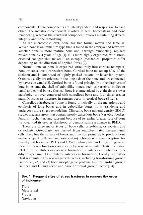
Normal lamellar bone is organized structurally into cortical (compact)bone or cancellous (trabecular) bone. Cortical bone makes up 80% of theskeleton and is composed of tightly packed osteons or haversian system.Osteons usually are oriented in the long axis of the bone and are connectedby haversian canals [3]. Cortical bone is found principally at the diaphysis oflong bones and the shell of cuboidlike bones, such as vertebral bodies ortarsal and carpal bones. Cortical bone is characterized by eight times slowermetabolic turnover compared with cancellous bone and four times greatermass. Most stress fractures in runners occur in cortical bone (Box 1).
Cancellous (trabecular) bone is found principally at the metaphysis andepiphysis of long bones and in cuboidlike bones. It is less dense andundergoes more stress remodeling. Clinically, bone mineral density (BMD)studies measure areas that contain mostly cancellous bone (vertebral bodies,femoral trochanter, and sacrum) because of its earlier/greater rate of boneturnover and its greater likelihood of demonstrating a change in BMD.
There are three major types of bone cells: osteoblasts, osteocytes, andosteoclasts. Osteoblasts are derived from undifferentiated mesenchymalcells. They line the surface of bones and function primarily to produce bonematrix (type I collagen and osteocalcin). Osteoblasts have receptors forparathyroid hormone (PTH) and 1,25-dihydroxyvitamin D [1,4]. In general,these hormones function systemically by way of an osteoblastic mediator.PTH directly inhibits osteoblastic formation of osteocalcin, whereas 1,25-dihydroxyvitamin D stimulates osteocalcin formation. Locally, an osteo-blast is stimulated by several growth factors, including transforming growthfactor–b-1, -2, and -3; bone morphogenic proteins 1–7; insulin-like growthfactors I and II; and acidic and basic fibroblast growth factor [1,4].
Box 1. Frequent sites of stress fractures in runners (by orderof incidence)
TibiaMetatarsalFibulaNavicular

751STRESS FRACTURES & KNEE INJURIES IN RUNNERS
Osteocytes are former osteoblasts that have become surrounded withbone mineral matrix (calcified bone). Osteocytes function to maintain boneand control extracellular concentrations of calcium and phosphorus. Thefinal cell type, osteoclasts, is derived from hematopoietic precursors andfunction to resorb bone. They bind to the bone surface and resorb anisolated area of bone by dissolving the hydroxyapatite crystals and digestingthe collagen. Osteoclasts have specific receptors for calcitonin and, whenbound, calcitonin directly inhibits bone resorption. Osteoclasts do not havereceptors for PTH or 1,25-dihydroxyvitamin D, and therefore, arestimulated indirectly by these hormones through an osteoblast-mediatedmechanism to increase bone resorption [1].
The macroscopic composition of bone differs depending on site, age,diet, and disease. In general, the mineral or inorganic phase accounts for60% of the tissue, the organic phase accounts for 35%, and water accountsfor the remaining 5%. The mineral or inorganic phase consists of crystallinecalcium hydroxyapatite (Ca10(PO4)6(OH)2). It is responsible for thecompressive strength of bone. The organic phase consists of 90% type Icollagen and is responsible for the tensile strength of bone. The remainder ofthe organic phase consists of proteoglycans, matrix proteins, growth factors,and cytokines.
In general, the expected age of peak bone mass accrual is between 25 and30 years [4]. After this age, men and women gradually lose bone mass. Girlsseem to acquire most of their bone mass at an earlier age than boys (age 11–14 years compared with 13–17 years) [4,5]. Women who are postmenopausalor hypoestrogenic for other reasons have accelerated bone loss that is due toincreased bone resorption compared with formation. Therefore, femaleathletes who are hypoestrogenic during adolescence can accrue a lower peakbone mass, which might be an irreversible problem after a certain age [4,6,7].Using bone turnover markers, it was observed that amenorrheic runnershave a reduced bone turnover, especially in bone formation, compared witheumenorrheic runners. This is believed to be linked to the various endocrineabnormalities (including hypoestrogenemia), low body mass index, and lowenergy intake relative to expenditure [8]. Recent studies suggest that thealtered energy balance ultimately may cause this imbalance in bonehomeostasis. Chronic undernutrition and acute dietary energy restrictionare accompanied by reduced bone formation. The latter also has beenassociated with depressed levels of insulin-like growth factor 1, a hormonethat was shown to stimulate the production of type I collagen [9–11].Uncoupling of bone formation and resorption can be seen with restrictedenergy availability as little as 126 kJ/kg of lean body mass/d [12].
Bone remodelingAccording to Wolff’s Law, bone has a cellular and molecular remodeling
response to applied mechanical stress. The bony adaptation is a function ofthe number of loading cycles, cycle frequency, amount of strain, strain rate,

752 HOCH et al
and strain duration per cycle [13]. Cortical and cancellous bone remodelcontinuously by osteoclastic and osteoblastic activity. This remodelingoccurs throughout life and is affected bymultiple factors, including metabolicstate, nutritional status, menstrual patterns, age, sex, level of fitness, andethnicity. Bone also responds to piezoelectric changes. Tensile forces createelectropositivity, and thereby, stimulate osteoclastic activity [13]. Compres-sive forces create electronegativity, and thereby, stimulate osteoblasticactivity. Most cortical stresses in nature are tension. Torsion or twistingprovides tension circumferentially, whereas bending produces tension on theconvex side and compression on the concave side. Tension forces were shownto result in microfracture and debonding at cement lines [14].
Clinically, this concept becomes important in the treatment of femoralneck stress fractures. Compression-side injuries (on the medial inferiorcortex of the femoral neck) heal much better than traction-side stressinjuries (on the lateral superior cortex of the femoral neck). Because of theirrobust ability to heal, nondisplaced compression-type stress fracturestypically are treated with nonweight bearing until the patient becomespain-free, which may take several days to weeks. Tension-type femoral neckstress fractures may fail to heal adequately with serious sequela, such asavascular necrosis, malunion or nonunion, or varus deformity. Thus,tension- side fractures often require internal fixation.
Stress injury
Stress injury occurs on a continuum from normal bone remodeling andrepair to frank cortical fracture. Overall bone health depends on mechanical,hormonal, nutritional, and genetic factors. The susceptibility of bone tofracture under fluctuating stresses is related to the crystal structure andcollagen orientation of the osteon. Fatigue load under certain strain ratescan cause progressive accumulation of microdamage [15]. When sucha process is prolonged, bone eventually may fail through crack propagation.Bone simultaneously repairs these cracks by new bone formation at theirtips, and thereby, decreases the chance for propagation.
Stress injury of bone is the result of excessive bone strain withaccumulation of microdamage and inability to keep up with appropriateskeletal repair (fatigue reaction/fracture), or depressed bony remodeling inresponse to normal strain (insufficiency reaction/fracture). The formersituation most likely occurs in athletes and military recruits. The latter mostlikely occurs with the female athlete triad, metabolic bone disease, andosteoporosis. Sacral insufficiency stress fractures have been found to occurin female runners and often mimic the presentation of sacroiliitis. There alsomay be a component of reperfusion injury following prolonged strenuousexercise that results in bone tissue ischemia. This may help to explain howsome stress fractures occur in cortical bone areas of lower strain and whenintracortical osteopenia precedes evidence of microscopic cracks [16].
753STRESS FRACTURES & KNEE INJURIES IN RUNNERS
An additional consideration in the athletic population is trainingregimens and stress injury. Muscles exert a protective effect on corticalbone by acting as the major shock absorber. With muscle contraction,cortical bone surface bending strains are reduced [17,18]. In most weight-bearing bones it is believed that with muscle fatigue, the shock-absorbingeffect is lessened and more force is transmitted directly to bone whichincreases the likelihood of microdamage accumulation. In nonweight-bearing and some weight-bearing bones, repetitive contraction of the muscleat its insertion may generate enough force to cause stress-induced injury[19].
Etiology of stress fractures
Numerous factors contribute to the risk of stress fractures in runners,particularly female athletes; however, many of these proposed etiologiesremain unproven. Much of the research that investigates stress fracture riskshas been done in male subjects and military recruits; therefore, findings maynot necessarily be generalized to female athletic populations. Furthermore,many risk factors for stress fractures are interrelated and methodologicallyare difficult to analyze independently.
The etiology of stress fractures is multifactorial; individual athletes varyin their susceptibility to stress injury. Risk factors (Box 2) are divided intoextrinsic (characteristics of the environment in which the athlete trains orcompetes) and intrinsic (characteristics of the athlete herself) types.
Extrinsic risk factorsTraining regimen. High training volume is a major risk factor in stressfracture development. Multiple studies in runners demonstrated that higherweekly running mileage correlates with increased incidence of stressfractures [20] and overall running injuries [21–23]. Ballet dancers who trainfor more than 5 hours per day have a significantly higher stress fracture riskthan those who train for less than 5 hours daily [24].
Abrupt or rapid changes in duration, frequency, or intensity of trainingprograms also increase an athlete’s risk of stress fracture. Reducing theintensity or frequency of the training program led to fewer stress fractures infemale and male military recruits [25–27]; however, this intervention has notbeen studied in athletes.
Footwear. Athletic footwear is designed to reduce impact on ground contactand provide stability by controlling foot and ankle motion [28]. Shoe age isa better indicator of shock-absorbing quality than shoe cost. Gardner et al[29] showed that training in shoes that are older than 6 months increases therisk for stress fracture; however, there has been no association between shoecost and stress fracture risk [29]. A woman’s foot has a greater forefoot tohindfoot ratio, which can result in poor shoe fit and leave the hindfoot less

754 HOCH et al
supported. Custom made biomechanical shoe orthoses that place the foot inneutral subtalar position and absorb shock decreased the overall incidenceof stress fractures in infantry recruits [30,31]; however, this may not beapplicable to running athletes [32].
Training surface. The surface on which an athlete trains also may contributeto her risk of stress fracture. Theoretically, training on uneven surfacescould increase the risk of stress fracture by causing increased muscle fatigueand redistributing load to bone. Hard or less compliant surfaces, such ascement, also could increase stress fracture risk through higher mechanicalforces being transmitted to bone during impact. It is difficult to control forand quantify training surface in observational or prospective studies;however, a correlation was demonstrated in some studies [21,33], whereas
Box 2. Risk factors for stress fractures
ExtrinsicTraining regimenFootwearTraining surfaceType of sport
IntrinsicDemographic factors
GenderAgeRaceAerobic fitnessMuscle strengthFlexibility
Biomechanical factorsBone mineral densityBone geometry
Anatomic factorsFoot morphologyLeg length discrepancyKnee alignment
Hormonal factorsDelayed menarcheMenstrual disturbanceContraception
Nutritional factorsLow calcium and vitamin D intakeDisordered eating

755STRESS FRACTURES & KNEE INJURIES IN RUNNERS
others showed no effect [20,23]. One small study found treadmill runners tobe at lower risk for developing tibial stress fracture, but also less likely toachieve tibial bone strengthening, than overground runners [33].
Type of sport. An Australian study in 1994 quantified the rate of stressfracture in men and women in different sports. In this study, the percentageof athletes per season who had stress fractures were as follows: softball6.3%, track 3.7%, basketball 2.9%, tennis 2.8%, gymnastics 2.8%, lacrosse2.7%, baseball 2.6%, volleyball 2.4%, crew 2.2%, and field hockey 2.2%[34]. Sprinters, hurdlers, and jumpers tended to have more foot fractures,whereas middle and long-distance runners had more long bone and pelvicfractures [35]. Rowers and golfers have increased rates of rib stress fractures[36,37].
Intrinsic risk factorsDemographic factors. Most studies have found that women have a higherincidence of stress fractures. It is probably multifactorial, and secondary, atleast in part, to gender-associated risk factors, such as dietary deficiencies,menstrual irregularities, lower BMD, and narrower bone width. Genderdifferences in muscle physiology, especially neuromuscular control, also maybe to blame. Several studies have shown that women have a slower rate offorce development in the muscle [38–40].
In the U.S. military, the risk of stress fractures in female recruits whoundergo the same training program as men is up to 10 times higher [4]. Thisincreased risk also has been observed in athletic populations [34,41,42].Bennell et al [35] reported no difference in the overall stress fractureincidence between male and female athletes; however, the data seem to showa trend for a higher risk of stress fractures in women when the amount oftraining hours were taken into account. Women, however, seem to havemore femoral neck, metatarsal, and pelvic stress fractures than men [43].Further research is needed to determine whether the apparent higherincidence of stress fractures in women is independent of other known riskfactors.
The role of age as a risk for stress fractures in female athletes is notestablished. Studies in military recruits have been inconsistent, with somefinding an increased risk of stress fractures with increasing age [44,45] andothers finding a decreased risk [29,46–48] or no effect [49,50]. This lack ofagreement is most likely due to confounding factors, such as previousphysical activity level, hormonal status, BMD, and training level. Moststudies in athletic populations have not found a correlation between age andstress fracture risk, although rigorous studies that controlled for otherpossible confounding variables are lacking.
The incidence of stress fractures is significantly higher in white and Asianwomen than in African American women [29,44–46]. This is believed to berelated to differences in bone turnover and peak bone density and not to
756 HOCH et al
race independently. It also seems that ethnic differences in bonemineralization and bone integrity in athletes are mediated by heritabledifferences in titratable acid, sodium, and calcium excretion [51].
Previously inactive or less active military recruits have a higher incidenceof stress fractures compared with those who are active before beginningbasic training [29,48,49]. Several possible factors include decreased aerobicfitness, decreased muscle strength, lower endurance, and poor flexibility. Astudy of military recruits found no association between aerobic fitness(predicted VO2max) and stress fracture risk [52]. It is unlikely that aerobicfitness alone accounts for the difference [53,54]. The role of flexibility onstress fractures has not been well-defined [35,47,55].
Biomechanical factors. Lower BMD, especially of the femoral neck, wasassociated with an increased risk of stress fractures in the female athlete [56].Although there are published case-control studies that support [57] andrefute [49] this finding, Bennell and colleagues [53] were the first to examinethis prospectively. They found that lower BMD in the lumbar spine and footwere significant predictors of later stress fracture development in femaletrack and field athletes. Of note, an athlete with an apparently normal BMD(due to the increased bone loading of sport) may be at increased risk ofstress fracture if she falls below the mean among female athletes. It also wasobserved in athletic females that cancellous stress fractures correlate withearly onset osteopenia by Dual Energy Xray Absortiometry scan muchmore so than cortical bone stress fractures [58]. This indicates the necessityof bone density evaluation in any young woman who has a cancellous stressfracture. Menstrual disturbance and lower BMD most likely are not riskfactors that are independent of each other, but are interrelated; amenorrheicathletes have lower BMD and higher stress fracture incidence [59].
The amount of force that a bone can withstand is proportional to itscross-sectional area and cross-sectional moment of inertia (a measure ofbone resistance to bending). Studies of military personnel found theseparameters to be significantly lower among those who develop stressfractures [60–62]. They also found that of persons who sustained femoral,tibial, or foot stress fractures, 31% had narrowed tibial width comparedwith those without fracture [63]. This narrowed tibial width may be anindicator of biomechanically weaker skeletal structures. It is hypothesizedthat women are likely to have overall narrower bones than men [64]; this isa possible contributing factor to the higher incidence of stress fractures inwomen.
Anatomic factors. The structure of the foot helps to determine how muchground contact force is absorbed in the foot and how much is transferred tothe bones of the leg and thigh. A rigid, high-arched foot (pes cavus) absorbsless stress and transmits greater force to the tibia and femur. A flexible,low-arched foot (pes planus) absorbs more force in the foot itself and

757STRESS FRACTURES & KNEE INJURIES IN RUNNERS
transmits less to the tibia, fibula, and femur. One military study thatevaluated foot morphology found that persons who had the highest archessustained 3.9 times as many stress fractures as those who had the lowestarches [65]. Other studies [65,66] suggest that individuals who have pescavus seem to be at increased risk of tibial and femoral stress fractures,whereas those who have pes planus may sustain more metatarsal stressfractures. Other investigators have not found a significant correlationbetween foot structure and stress fracture risk [20,53]. It is possible that pesplanus and pes cavus foot structure may increase the risk of stress fractureat various sites, but this has not been evaluated adequately or proven.
Leg length discrepancy also has been associated with an increased risk ofstress fractures in female athletes [20,53]. The degree of leg length differencemay correlate with increasing stress fracture risk [67]; however, one study ofmale military recruits did not confirm this relationship [68]. It is reasonableto evaluate and correct significant leg length discrepancy in runners,especially those with other stress fracture risks.
Valgus knee alignment and quadriceps angle greater than 15� also mayincrease the risk for tibial stress fracture [68,69].
Hormonal factors. Female athletes, in general, reach menarche at a later agethan nonathletes, particularly those in certain sports, such as ballet,running, and gymnastics [70,71]. Delayed menarche may cause lower peakbone mass attainment or may be a marker for other possible influences onstress fracture risk, such as low body fat, low body weight, future menstrualdisturbance, or excessive training. The effect of this delay on bone healthand risk of stress fractures is not well-studied; however, some studies suggestthat osteopenia, stress fracture, and scoliosis may be potential complicationsof delayed menarche [53,71]. Scoliosis, in particular, has been observed infemale ballet dancers with delayed menarche [71]. This also may lead topelvic obliquity and relative leg length discrepancies and the potential forincreasing the risk for stress fracture.
Multiple studies have demonstrated that stress fractures occur morecommonly in women who have amenorrhea or oligomenorrhea than ineumenorrheic women [17,46,48,53,57,71]. Athletes who have menstrualdisturbances have low basal estrogen concentrations [72] and a lower BMDthan eumenorrheic athletes [73]. It was hypothesized that estrogen depriva-tion increases the physiologic set point for bonemodeling and remodeling andmakes it more difficult to activate the cellular response that is necessary toinduce bone adaptation to stress [4,74] and increases the risk of stressfractures. Health care providers, athletes, coaches, and parents need to beaware that menstrual disturbance is not a normal product of training and thatsuch disturbances can have devastating consequences. Menstrual disturban-ces also are seen in association with disordered eating and endothelial celldysfunction (see ‘‘The female athlete Triad,’’ below). Therefore, athletes whohave menstrual irregularity should be evaluated further.

758 HOCH et al
Some studies have shown that oral contraceptive pills (OCPs) havea protective effect in preventing stress fractures in female athletes [17,75]. Itseems that exogenous estrogen may help to curb further bone loss in thehypoestrogenic amenorrheic athlete; however, it may not be sufficient tostimulate bone growth [71,76–79]. Several small studies among amenorrheicwomen or those who had anorexia found that BMD at the lumbar spine orhip was higher for those who were taking OCPs compared with those whowere not [76,77,80], whereas others showed no significant change [71,78]. Italso was theorized that OCPs may act through another mechanism, such asimproving bone microarchitecture and quality without significantly affectingBMD [4]. To add to the controversy, a recent German study by Hartardet al [81] showed that OCP use is associated with decreased BMD of thespine (7.9%) and the femoral neck (8.8%) in female endurance athletescompared with non-OCP users. They also found that early age at initiationof OCPs was an important risk factor for low peak bone mass in youngwomen. Based on the conflicting results from research and the lack of well-controlled studies, it is difficult to assess the effects of OCPs on skeletalhealth in normally menstruating women. In those who have menstrualdisturbances, OCPs or other hormonal replacement therapy may be effectivein preventing further bone loss; however, resumption of menses may maskan underlying nutritional disorder and provide a false sense of security.Recent evidence also suggests that depomedroxyprogesterone may contrib-ute to impaired bone accretion and low BMD, and it should be avoided inyoung women [82].
Nutritional factors. Low calcium intake is associated with low BMD [83],and therefore, may contribute to the development of stress fractures.Myburgh and colleagues [57] observed an association between decreasedcalcium intake and increased stress fracture risk. Other studies found noassociation between calcium intake and stress fracture risk; groups that didand did not have stress fractures had normal calcium intake [13,84,85].Athletes whose calcium intake is less than the daily-recommended value arelikely to be at risk for stress fractures, but for those who have a normaldietary calcium intake, other factors play a larger role.
Vitamin D also is essential to bone health and functions, includingstimulating calcium transport, osteoblastic stimulation, and decreasingparathyroid hormone. Recent studies focus on the role of the vitamin Dreceptor allele in predicting bone density. More research is necessary todetermine the clinical applications of its use in screening [86–88].
Inadequate caloric intake relative to energy expenditure that is requiredfor seems to be the primary mechanism by which female athletes arepredisposed to menstrual dysfunction and detrimental effects on bone.Anorexia nervosa has been associated with a significantly decreased BMD[7,89]. Nearly 75% of adolescent girls who had anorexia had a BMD thatwas more than two standard deviations below the normal value [90]. Not
759STRESS FRACTURES & KNEE INJURIES IN RUNNERS
surprisingly, women who have anorexia nervosa are at increased risk forstress fracture development [91,92]. Disordered eating is associated with lowBMD in the absence of menstrual irregularities [59].
The female athlete triad
The female athlete triad refers to an interrelated problem that consists ofdisordered eating, amenorrhea, and osteoporosis. Hoch et al [93] also foundthat amenorrheic athletes had reduced brachial artery endothelium-de-pendent flow-mediated vasodilation when compared with oligomenorrheicand eumenorrheic athletes. Furthermore, in a 2-year follow-up study, theoriginal amenorrheic athletes had a significant improvement in BMD withdifferent combinations of estrogen and progesterone or return of mensesnaturally. This female athlete triad is a potentially lethal combination ofmedical disorders that is reported in some female athletes [4,42]. Athleteswho are at greatest risk seem to be those who feel significant pressure toexcel in sports for which leanness and a low body weight are consideredadvantageous, such as gymnastics, figure skating, ballet, and distancerunning [94]. Also, athletes who participate in individual sports are at higherrisk than those who participate in team sports.
The problem usually begins with disordered eating; this includesa spectrum of abnormal and harmful eating patterns, such as bingeingand purging, restrictive eating, fasting, and the use of diet pills or laxatives.Preoccupation with food, a distorted body image, and intense fear ofbecoming fat are often present as well. Some athletes will meet theDiagnostic and Statistical Manual of Mental Disorders, Fourth Editioncriteria for anorexia nervosa or bulimia, whereas others may display similarbehaviors without meeting full diagnostic criteria. A new classification—eat-ing disorder, not otherwise specified—allows for identification of womenwho do not meet other classification criteria. This has been helpful in thispopulation because the athlete’s weight may seem to be adequate because ofincreased lean tissue mass; however, they are not consuming enough caloriesto meet their energy needs.
Abnormal eating patterns may lead to athletic-associated amenorrhea.Athletic amenorrhea is a complex multifactorial condition with seriousassociated comorbidities. Extreme caloric restriction, excessive exercise,physical and emotional stress that are associated with exercise/competition,percentage of body fat, and genetics contribute to the condition. There isincreasing evidence, however, that nutritional restrictions and the resultingendocrine and metabolic changes are a critical initiator of hypothalamic-induced athletic amenorrhea and osteoporosis [95].
Disordered eating, estrogen deficiency, and menstrual dysfunctionpredispose women to the third component of the triad, osteoporosis [89].Reduced BMD in premenopausal women seems to be irreversible, despiteweight gain, resumption of menses, or estrogen replacement [6,7]. One study

760 HOCH et al
found that with the resumption of menses there was a significant increase invertebral BMD; however, after 2 years of normal menses, BMD remainedbelow age-normative. Slemenda et al [96] showed that the low estrogenicstate that is associated with amenorrhea has a more profound effect oncancellous bone than on cortical bone. Cancellous bone is found in a higherpercentage in the pelvis, sacrum, and femoral neck—areas where femalestend to have a higher occurrence of stress fractures. These factors put thewoman who suffers from the female athlete triad at significant risk for stressfractures. Although some investigators found that weight-bearing exercisehas a skeletal protective effect and may attenuate the bone loss that usuallyis seen in anorexics [89], the use of excessive training to control weight alsocould contribute to the increased risk of stress fractures that is associatedwith the female athlete triad.
Several other factors are known to increase the risk for osteoporosis buthave not been investigated thoroughly as possible risks for stress fractures infemale athletes. These include smoking, caffeine consumption, and certainmedications, such as thyroid hormone and corticosteroids. In a study offemale army recruits, current or past smoking, consumption of more than 10alcoholic drinks/week, corticosteroid use, use of depomedroxyprogesteroneacetate, lower adult weight, and no history of regular exercise increased thelikelihood of stress fracture [45,82].
Screening and prevention of stress injury in female runners
Ideally, stress fractures and the female athlete triad are treated bestthrough prevention. With many of the risk factors for stress fracture nowknown, screening and prevention are much easier. Evaluation of allcomponents of the triad should be performed on every female runner. Athorough history is essential and should include assessments of nutrition;amount of, and changes in, physical activity; and menstrual history. Ahistory of stress fractures or overuse injuries as well as signs or symptoms ofan eating disorder justify further evaluation. Characteristics of anorexianervosa include cachexia, bradycardia, hypotension, lanugo, hypothermia,cold intolerance, yellow skin (hypercarotenemia), dry hair and skin,alopecia, and pruritus. Signs and symptoms of bulimia include fatigue,abdominal pain, chest pain, swollen parotid glands, sore throat, erodedtooth enamel, and knuckle calluses. Screening with bone density testingshould be considered in athletes who have known risk factors. The diagnosisof osteoporosis in premenopausal women should not be made on the basisof densitometric criteria alone. It also is not appropriate to use T-scoreswhen peak bone mass has not been reached; therefore, athletes who are 20years old and younger should be evaluated with Z-scores [97]. Treatment ofthe triad often requires a team approach. A sports physician, dietitian, andpsychologist may be needed to work with the athlete in addition to hercoach, parents, and close friends. Treatment for underlying disordered

761STRESS FRACTURES & KNEE INJURIES IN RUNNERS
eating, correction of an energy deficit, and restoration of menses areessential to stimulate bone accretion. Nutritional supplementation withcalcium and vitamin D and reduced training may be recommended. Earlyintervention speeds recovery.
Reductions in bone density and an increased risk of stress fracture arenoted when there is an uncoupling of bone formation and resorption ratesas seen when energy availability is too low [59]. Several metabolic hormonesthat influence bone formation (IGF-1, T3, leptin), as well as bone formationmarkers (serum type I procollagen carboxyl-terminal propeptide, osteocal-cin) and bone resorption markers (urine N-telopeptide, serum C-telopep-tide), can be followed to form an impression of the overall bone turnoverstatus and assess energy status indirectly. Uncoupling of bone resorptionand formation is seen with restricted energy availability as little as 126 kJ/kgof lean body mass/d [12]. Therefore, increasing caloric intake to offset thehigh energy demand may help to restore menses and stimulate boneaccretion. Calcium intakes of 1200 mg/d to 1500 mg/d and 400 IU to 800 IUof vitamin D also help to minimize bone loss. Antiresorptive therapies, suchas bisphosphonates and calcitonin, have not been tested and are notapproved by the U.S. Food and Drug Administration in younger patientswho have reduced BMD/osteoporosis. Their use in this population iscontroversial; they are teratogenic and have a half-life in bone of greaterthan 10 years.
Treatment with exogenous estrogens has been evaluated in numerousstudies with varying results. It seems that this may help to reduce furtherbone loss in hypoestrogenic amenorrheic athletes; however, it may beinsufficient to stimulate bone growth [79]. Another concern is that with therestoration of menses, estrogen replacement may mask the underlyingdisorder. Recent evidence suggests that depomedroxyprogesterone acetatecontributes to impaired bone accretion and loss in mean BMD and shouldbe avoided in amenorrheic athletes [82]. Other preventative measuresinclude not smoking and the minimal use of alcohol [45].
Because training errors frequently are the cause of stress fractures, anyabrupt increase in training should be avoided. Cyclical training—a limitedperiod of training followed by relative rest during the third week (theinterval of greatest skeletal vulnerability)—is recommended over progressivetraining [25]. For runners, mileage increases should be gradual, with someinvestigators recommending that mileage should not be increased by morethan 3.2 total km/wk [98]. Using shock-absorbing insoles and changingshoes every 6 months or before 800 km also is recommended.
Diagnosis of stress fractures
The treatment of stress fractures starts with a specific and accuratediagnosis. Diagnosis of stress fractures often can be difficult and requiresa high degree of suspicion. A thorough history and physical examinationoften diagnoses most stress fractures successfully. The onset of pain with an

762 HOCH et al
abrupt change in training, such as increasing mileage or intensity, shouldraise suspicion of a stress fracture. Pain typically occurs at the end of, orafter, a run. Point tenderness on physical examination is the hallmark. Forexample, the so-called ‘‘N-spot’’ on the dorsum of the navicular can be a siteof exquisite tenderness in runners who have navicular stress fractures.Swelling and redness may be present, but deformity usually is not.Percussion tapping of bone, vibrating tuning fork, single leg hop, femoralfulcrum, and lumbar extension tests (for pars stress fracture) are nonspecificphysical examination maneuvers that help in the identification of stressfractures. Application of stress to the area or passive stretching also canreproduce pain [99]. If multiple or recurrent stress fractures are present,evaluation for sources of metabolic bone disease and the female athlete triadis warranted. Some stress fractures are considered to be at high risk (Box 3)for nonunion and should be treated aggressively, often with a period ofnonweight bearing, and perhaps, surgery [106]. These high-risk fracturesinclude ones at the femoral neck, the mid shaft of the tibia, and thenavicular.
The most common location of stress fractures in runners is the tibia;however, tibial stress fractures may be difficult to discern from so-called‘‘shin splints,’’ more properly termed medial tibial stress syndrome (MTSS).Typically, pain with stress fractures worsens as the run goes on.Occasionally with MTSS, pain may diminish as the runner warms up.Physical examination reveals focal tenderness with tibial stress fractures,whereas MTSS presents with diffuse tenderness (with maximal tendernessat the junction between the middle and distal third of the medial tibia).Percussion tenderness, a tuning fork test, or hop tests are more likely to be
Box 3. High-risk stress fractures that require aggressivetreatment
Femoral neckAnterior cortex or midshaft of tibiaNavicularMedial malleolusTalus fracture extending to subtalar jointProximal second metatarsalProximal fifth metatarsal diaphysis (Jones fracture)SesamoidPars interarticularis
Data from Fredericson M, Bergman AG, Matheson GO. [Stress fractures inathletes] [German]. Orthopade 1997;26(11):961.

763STRESS FRACTURES & KNEE INJURIES IN RUNNERS
positive with tibial stress fractures than with MTSS. Posterior cortex tibialstress fractures can present with calf pain rather than shin pain.
A definitive diagnosis of a stress fracture often requires imaging. Plainradiographs have poor sensitivity but are highly specific and arerecommended as an initial step in diagnosis. The ‘‘gray cortex’’ sign maybe evident on the radiograph if the cortex has decreased density withassociated hyperemia, edema, and early resorption [106]. The ‘‘dreadedblack line’’ also can be seen on radiograph in individuals who havea midshaft tibia fracture that has failed to heal and developed a nonunion[106]. Many of these will be falsely negative as callus formation can take upto 3 months to appear. If suspicion is high with negative radiographs,conservative treatment should be initiated followed by repeat evaluationand radiographs in 2 weeks. If the injury is in the season of competition, it isreasonable to perform further imaging before implementing the 2-week rest.
MRI and bone scintigraphy (triple phase bone scan) have comparablesensitivity and either is acceptable. MRI is preferred because it is lessinvasive; avoids radiation exposure (an important consideration in thisyoung female population); has better specificity (ie, can differentiate fracturefrom tumor); and shows more diagnostic information, such as fracture lineand periosteal edema. MRI can show stress changes, or edema, within thebone, even before a fracture develops. Fat-suppressed T2-weighted imagesalso are recommended to help identify any bone marrow edema. Areas ofincreased activity by scintigraphy seem to be consistent with grades of MRI[99,100]. The main drawback of MRI, aside from cost, is that it does notimage cortical bone as well as CT. CT is useful in differentiating conditionsthat may mimic stress fracture on bone scan, such as osteoid osteoma,osteomyelitis with Brodie’s abscess, and various malignancies. It is alsohelpful in detecting fracture lines as evidence of stress fractures and oftencan differentiate between stress fracture and stress reaction [101]. This isparticularly important in the elite athlete as it may affect their rehabilitationprogram and upcoming competitions considerably. Ultrasound is notreliable in diagnosing stress fractures and is not recommended [102].
Conservative treatment for stress fractures
Conservative treatment for stress fractures is a three-part protocol usingpain as a guide to rehabilitation. Phase 1 includes cessation of painfulactivity, ice, analgesics, maintaining fitness by cross-training, modificationof risk factors, and possible bracing or electrical stimulation [101]. It isimportant that the runner who has a stress fracture be able to maintainaerobic fitness and strength during the healing period. The most commonways to unweight the fractured extremity and maintain cardiovascularfitness are cycling, water running, and swimming. Cross-training should beas sport specific as possible in duration and intensity. Extrinsic factors,especially training errors, need to be addressed because these are the most

764 HOCH et al
common cause of stress fractures. Particularly important to female runnersare intrinsic conditions, such as disordered eating, amenorrhea, andpremature osteoporosis. Moreover, certain stress fractures occur in areasof hypovascularity and are at risk for nonunion or avascular necrosis; inthese cases, surgery should be considered.
Adjunctive therapies, such as pneumatic braces or capacitative coupling,also may be instituted. The use of pneumatic braces in the rehabilitation oftibial stress fractures seems to reduce the time to recommencing training inathletes and military personnel; it allowed resumption of light activity in 7days (median) and full, unrestricted activity in 21 � 2 days versus 21 days(median) and 77 � 7 days with rest alone [31,103]. Capacitative couplingalso has been used to shorten healing time. Stress fractures typically take8 to 12 weeks to heal; however, one study found that healing time withcapacitative coupling was shortened to 7 weeks on average [104]. Weight-bearing trials should be performed every other day. After 3 to 5 days freefrom pain, phase 2 begins.
Phase 2 consists of light-weighted exercises and nonimpact-loadingactivities, such as walking, using a stair stepper, elliptical, or cross-countryskiing machine. Activity should resume slowly and increase by 5 to 10minutes per day up to 45 minutes. If any bony pain occurs, activity shouldstop for 1 to 2 days and restart at a level below which the pain occurred[101,105]. Recovery of strength that is lost during phase 1 also should beaddressed. Sport-specific muscle rehabilitation also may be started.
Phase 3 is gradual re-entry into the athlete’s sport-specific activity,starting every other day and gradually progressing to normal activity. Thisprogram may take from 3 to 18 weeks, depending on the extent of the injury[105]. All athletes also should have a biomechanical and gait evaluation toassess for leg length abnormalities, excessive pronation or supination, pesplanus, pes cavus, and other structural abnormalities following a stressfracture.
Knee injuries
The knee joint is an innocent bystander in the lower limb kinetic chain(ie, injuries at the knee usually are the result of biomechanical problemsabove or below the knee). The differential diagnosis of knee pain in therunner is described in Table 1.
Patellofemoral pain syndrome
Patellofemoral pain syndrome (PFPS) is the most common diagnosis forknee pain in athletic and nonathletic populations; the incidence may betwice as high in female athletes compared with their male counterparts[107,108]. Adolescent females seem to be at particular risk because theiraccelerated bony changes have not been accommodated by muscle changes

765STRESS FRACTURES & KNEE INJURIES IN RUNNERS
[107]. The PFPS pain syndrome is an amalgam of diagnoses and generallycan be classified into patellar instability, PFPS with malalignment, andPFPS without malalignment.
Patellofemoral pain syndrome pain generatorsTraditional thinking has assumed that the patellar cartilage is the
primary pain generator in the PFPS. Accordingly, the term ‘‘chondroma-lacia patellae’’ often is used for nebulous anterior knee pain; however, thereare no nerve endings in the articular cartilage [109]. Arthroscopy studies alsohave failed to correlate the degree of patellar chondral and subchondralinjury with the severity of anterior knee pain [109]. The current theory ofpathogenesis is injury to the subchondral bone from patellar maltrackingand increased patellofemoral joint reaction forces. Dye et al [110] proposedthat the actual pain generator is the loading of nerve endings (ie,a degenerative neuroma) in the patellar retinaculum which causes a resultantsynovitis. Other pain generators may exist in PFPS, such as irritatedinfrapatellar fat pad, plica, bursa, tendons, and apophyses.
Predisposing factors for patellofemoral pain syndromeBiomechanical structural problems may contribute to patellofemoral
pain [109]. For instance, miserable malalignment syndrome in womenpredisposes to PFPS. Muscle imbalances may exacerbate knee pain. Weakquadriceps musculature, particularly the vastus medialis obliquis (VMO)portion, has been implicated in patellofemoral pain. When the VMO is weakor inhibited, lateral vector forces that are created by the vastus lateralis,iliotibial band (ITB), and lateral retinaculum become dominant. Thus, thepatella becomes displaced laterally. Tight lateral structures (ITB and lateralretinaculum) exacerbate this phenomena. Specifically in runners, a tight ITBand tensor fascia lata muscle, coupled with weak gluteus medius, createsexcessive internal rotation of the femur and a lateral pelvic tilt. Tighthamstrings and gastrocnemius musculature also may exacerbate PFPS by
Table 1
Differential diagnosis of knee pain
Medial Lateral Anterior Posterior
PFPS PFPS PFPS Baker’s cyst
Plica ITBS Quadriceps tend. Hamstring tend.
MMT Popliteus Patellar tend. Popliteus
MCL sprain LCL sprain Patellar instability
Intra-articular LMT Hoffa’s syndrome
Osteoarthritis Intra-articular Patellar OCD
MMT Osteoarthritis Osteoarthritis
Bursitis
Abbreviations: ITBS, iliotibial band syndrome; LCL, lateral collateral ligament; LMT,
lateral meniscus tear; MCL, medical collateral ligament; MMT, medical meniscus tear; OCD,
osteochondral defect; PFPS, patellofemoral pain syndrome; tend., tendinopathy.
766 HOCH et al
increasing knee flexion and creating greater patellofemoral joint reactionforces. In addition, hyperpronation seems to predispose runners to PFPS.
Clinical presentationThe classic presentation of PFPS is an insidious onset of progressively
severe diffuse anterior knee pain, especially with loading activities thatinvolve repetitive flexion and extension, such as running. It may manifest asan ill-defined, usually bilateral, ache which is aggravated by hill training orstair climbing. Knee buckling can occur occasionally as a result of a painfulreflex inhibition of the knee extensor mechanism. Other complaints mayinclude crepitus or a ‘‘catching’’ sensation that is experienced with knee jointflexion and extension; however, crepitus may be common, even inasymptomatic individuals. Prolonged sitting with knee flexion, such as onan airplane flight or in a theater, may aggravate patellofemoral pain andlead individuals to extend their knee and legs into the aisle, also known asthe ‘‘movie theater sign.’’
In runners, patellar or quadriceps tendinopathy can occur, which ismanifested as pinpoint pain at the inferior and superior pole of the patella,respectively. Occasionally, patellar instability symptoms can occur withdescriptions of the ‘‘knee cap slipping out.’’ Infrapatellar fat padimpingement, occurs with forceful knee extension in individuals who havea prominent fat pad and posterior tilting of the patella. In adolescentrunners, apophysitis of the inferior pole of the patella (Sinding-Larsen-Johansen syndrome) or at the tibial tuberosity (Osgood-Schlatter syndrome)should be suspected [111].
DiagnosisThe history and physical evaluation of the runner who has knee pain
should include analysis of the kinetic chain. Plain radiographs, particularlylateral and ‘‘sunrise’’ (or ‘‘skyline’’) views, may be helpful in evaluating thepatellofemoral joint [112]. Sunrise radiographs are axial views that are takenwith the knee flexed 30� or 45�. If the patellofemoral joints shows joint spacenarrowing, osteophytes, subchondral sclerosis, and cysts, patellofemoralosteoarthritis should be suspected. Osteochondral lesions sometimes can beseen with these views. The lateral radiograph may demonstrate patella alta,rotational malalignment, or trochlear dysplasia. More commonly, particu-larly in the younger female runner who has PFPS, these radiographs arenormal. Occasionally, MRI is useful in identifying the quality of the patellarcartilage pathology and ruling out other knee pathologies.
TreatmentIn general, the management for PFPS should be individualized and aimed
at symptom relief acutely, and then focus on correcting the etiologic factorsthat contribute to the pain. The patient should be reminded that successfulalleviation and prevention of further discomfort with PFPS might take

767STRESS FRACTURES & KNEE INJURIES IN RUNNERS
several weeks to months. In the authors’ experience, patellar tendinopathycases may take even longer with a longer period of relative unloading.
Acute phase. Relative rest is often helpful in alleviating patellofemoral pain.Runners often benefit from switching to low-impact aerobic activities fora defined period of time, such as swimming, aqua aerobics, ‘‘elliptical’’training, or biking. Biking may exacerbate PFPS if the seat is too high or toolow or if increased resistance is maintained. Runners who have PFPS shouldavoid step aerobics or using the stair-climbing apparatus because these cancause tremendous repetitive compressive loads on the patellofemoral joint.Patellar tendinopathy cases often need 2 to 3 weeks of relative unloading ofthe tendon.
In the acutely swollen knee, ice may provide some relief from the acutediscomfort. Although anti-inflammatory medications have not been shownconclusively to benefit chronic PFPS, nonsteroidal anti-inflammatory drugsor acetaminophen may have a role in acute pain. Glucosamine/chondroitinalso has been suggested to provide relief in PFPS, particularly if osteo-arthritis is demonstrated on imaging [113].
Rehabilitation phase. Physical therapy was shown to be efficacious inrelieving pain in PFS in uncontrolled, and, more recently, controlled trials[114]. Crossley et al’s [114] program outlines elements of physical therapyintervention that can be used on all individuals (Box 4). It stands to reasonthat a more individualized program that attacks specific biomechanicaldeficits may be even more effective. Closed kinetic chain exercises serve asthe cornerstone of the muscle strengthening program in PFPS. Studiesshowed that although open and closed kinetic chain exercises seem toimprove PFPS symptoms, closed chain exercises also seem to improvefunctional performance. In particular, the literature has emphasized VMOstrengthening in the open and closed kinetic chain positions [109]. Isokineticopen chain exercises also have been used for lower limb strengthening;however, they require specialized equipment and are not functional. Forpatients who are in acute pain, isometric exercises may be painless andjust as beneficial as isokinetic exercise. A stretching program, as outlinedby Crossley et al [114], also has a sound theoretic basis in eliminatingbiomechanical deficits.
Debate exists about whether VMO strengthening is important andwhether VMO can be strengthened preferentially [115,116]. Some studiessuggest that VMO firing is delayed in patients who have patellofemoral pain[115]; however, a robust relationship has yet to be established. A recentstudy revealed that nine common exercises that are given for preferentialVMO strengthening showed equal firing of all of the different portions of thequadriceps [117]. Based on that study, some investigators have advocatedfor a generalized quadriceps strengthening program, rather than a selectiveVMO strengthening program. Other researchers showed that patients who

768 HOCH et al
have PFPS can learn the motor skill of selectively firing the VMO withpatellar taping or EMG biofeedback [118,119]. These patients may havea better short-term outcome [119].
Patellar taping, braces, and foot orthoses. Some investigators believe thattaping the patella may improve patellar alignment, tracking, tilt, or glide.Although numerous studies have been performed on this intervention forthe treatment of PFPS, there are variable results [109,113,120]; however, inthe authors’ clinical practice, runners who perform a single leg squat andhave improvement of pain with manual force on the patella may benefitfrom patellar taping. Patellar taping is continued only if a 50% reduction ofpain is achieved [120]. Patellar taping likely works by reducing pain andimproving proprioception rather than by way of a true improvement ofpatellar orientation. Taping may be especially beneficial in infrapatellar padimpingement by unloading the inflamed fat pad.
Elastic knee sleeves that have an anterior cut-out over the patella mayprovide some comfort, and because of their low cost, may be a usefulstrategy in some cases; however, the ability of the sleeve to alter patellarbiomechanics is not known, and if used, should be an adjunct withtherapeutic exercises. More expensive, more elaborate knee braces have notbeen shown to be particularly effective in patients who have PFPS [121].
Foot orthoses have been theorized to improve the biomechanics in thelower extremity, including the improvement of patellar tracking [122]. In
Box 4. Elements of physical therapy intervention
StretchesMedial/lateral glides and mobilization of the patellaDeep friction massage to lateral soft tissuesHamstring stretchAnterior hip stretch
Patellar tapingStrengthening
Isometric VMO strengthening with knees at 90�
Partial squats with isometric gluteal contractionIsometric hip abduction against wall while standingStep-downs with pelvis parallel to floorIsometric hip abduction while standing away from wallLunges
Home exercise program: two times per daySurface EMG
Adapted from Crossley K, Bennell K, Green S, Cowan S, McConnell J. Physicaltherapy for patellofemoral pain. Am J Sports Med 2002;30(6):860.
769STRESS FRACTURES & KNEE INJURIES IN RUNNERS
a study of 20 adolescent girls who had PFPS and exhibited excessiveforefoot varus or hindfoot valgus, individuals who were treated with footorthoses and an exercise regimen had a significant decrease in the level ofpain over an 8-week period compared with the control group whounderwent exercises alone [123]. It is more economical to try off-the-shelforthoses first to determine whether the patient derives any benefit initially,and progress to custom orthoses if increased support is required.
Surgery. Nonsurgical options in the treatment plan for PFPS should beattempted first; however, more than 100 surgical options have beendescribed for persistent patellofemoral pain that is nonresponsive toconservative treatment. Lateral retinacular release may be the most commonprocedure that is performed. It works best in individuals who have isolatedlateral patellar tilt as the primary cause of patellar malalignment. Lateralretinacular release does not correct a more global malpositioning of thepatella nor a large dynamic Q-angle. Inappropriate lateral release can leadto medial patellar subluxation, which can be a debilitating problem. Othermore extensive realignment procedures can be performed experimentally tocorrect Q-angles and patellar orientation. These procedures typically do notallow athletes to return to running activity [120].
Articular cartilage procedures may include open or arthroscopically-performed debridement or shaving of patellar cartilage to achieve a smootherpatellofemoral articulation, local excision of defects with drilling of thesubchondral bone, or facetectomy and transplantation of autologouschondrocytes [124]. Local excision of diseased cartilage and subchondraldrilling has been used commonly with satisfactory results, especially inpatients who are younger than 25 years [124]. The long-term results of thesearticular cartilage procedures are not known, and the relative effectiveness ofone procedure comparedwith the others also is unknown.Their role, althoughcommonplace, is still experimental.
Iliotibial band friction syndrome
In runners, the next most common knee injury is ITB friction syndrome(ITBS). The ITB is dense fascia that connects the gluteal muscles to theanterolateral tibia. An anatomic pouch can be found underlying theposterior ITB at the level of the lateral femoral epicondyle. The ITB passesover the lateral femoral epicondyle with knee flexion and extension.Maximum friction occurs when the posterior fibers of the ITB pass over thelateral femoral epicondyle at 20� to 30� of knee flexion—the putative‘‘impingement zone.’’ Repeated knee flexion and extension, particularlywith increased mileage per week, was shown to predispose to lateral kneepain. Although not studied extensively, poor neuromuscular control seemsto be an important modifiable risk factor for ITBS. Specifically, theneuromuscular system is needed to control the valgus/internal rotation

770 HOCH et al
vectors at the knee after heel strike. If appropriate control is not available,the ITB may have an abrupt increase in tension at its insertion site.Strengthening the gluteus medius and tensor fascia lata, which aredecelerators of the valgus/internal rotation vectors at the knee, was shownto ameliorate ITBS.
Clinical presentation
Symptoms of ITBS can emanate at three typical sites—proximal lateralhip, over the lateral femoral epicondyle, or at Gerdy’s tubercle. Runnersoften note more pain with downhill running because of the increased timethat is spent in the impingement zone. Paradoxically, runners state that fasterrunning and sprinting often does not produce pain. Fast running allows theathlete to spend more time in knee angles that are greater than 30�.
Physical examination
The modified Thomas and Ober tests are used to assess flexibility of theITB and its attached muscles. Knee tenderness is noted at the lateral femoralepicondyle (above the lateral joint line) or at Gerdy’s tubercle. Pain also canbe elicited frequently by the Noble compression test which is performed innonweight-bearing and standing positions; the knee is flexed and extendedthrough the impingement zone (20�–30� of knee flexion) while the examinerapplies pressure over the lateral femoral epicondyle.
Rehabilitation
Successful treatment of ITBS can be achieved by incorporatinga comprehensive, kinetic chain–oriented approach. Rehabilitation includesproper stretching of the ITB and associated muscles. A standing ITB stretchwith the affected leg crossed over, lateral trunk side-bending to theunaffected side, and overhead arm extension to the unaffected side may bethe most effective stretch [125] (Fig. 1). Some muscle groups do not respondto stretch unless myofascial and joint restrictions are addressed concomi-tantly. A qualified physical therapist or massage therapist can release triggerpoints and fascial adhesions that are identified on physical examination.Strengthening of weak or inhibited muscles can be started in conjunctionwith a flexibility program [126]. Weak hip abductors are seen often inpatients who have ITBS. Hip abductor strengthening, with single leg squatsand step-downs, is an efficacious treatment for runners who have ITBS[127]. The core strengthening concepts that were described previously playan empiric role in prevention and rehabilitation. The final stages ofrehabilitation focus on sports-specific activity. Correction of form flaws canbe invaluable to a runner. Frequently, runners have form deviations thatlead to a sine qua non of uncontrolled valgus/internal rotation of the knee.

771STRESS FRACTURES & KNEE INJURIES IN RUNNERS
Common abnormalities include excessive pronation, inability to shockattenuate at the knee, and Trendelenburg frontal plane gait at the pelvis.Changing to shock-absorbing or motion-control shoes can accommodatesupination and overpronation, respectively. Occasionally, foot orthoses maybe helpful in runners who have foot types that exacerbate ITBS.
Injections and surgery
Injections that are directed to the anatomic pouch underneath the ITB atthe lateral femoral epicondyle is a simple procedure that is used for patientswho have persistent pain and swelling. A mixture of anesthetic (eg, 1 mLof 1% lidocaine) and long-acting steroid (eg, 1 mL of betamethasone)is instilled to the affected site [126]. Surgical treatment for ITBS rarely isneeded. Surgery involves excising the posterior half of the ITB where itpasses over the lateral femoral epicondyle or removing the underlyingputative bursa. These surgical procedures give mixed results and should becontemplated only for patients in whom all other options have beenexhausted, including a comprehensive rehabilitation program as outlinedabove.
Fig. 1. An effective iliotibial band stretch.
772 HOCH et al
Summary
Running often can cause injuries to the knee as a result of kinetic chaindysfunctions. Addressing these dysfunctions in rehabilitation can preventfuture injury. Stress fractures often occur in runners who engage in trainingerrors. Female runners are particularly susceptible to stress fractures,especially in the setting of the female athlete triad. Proper identification andprevention of these injuries allows for athletes to return to runningexpeditiously.
References
[1] Kaplan FS, Hayes WC, Keaveny TM, et al. Form and function of bone. In: Simon SR,
editor. Orthopaedic basic science. Rosemont (IL): American Academy of Orthopaedic
Surgeons; 1994. p. 127–94.
[2] Monteleone GP Jr. Stress fractures in the athlete. Orthop Clin N Am 1995;26(3):423–32.
[3] Markey KL. Stress fractures. Clin Sports Med 1987;6(2):405–25.
[4] Nattiv A, Armsey TD Jr. Stress injury to bone in the female athlete. Clin Sports Med 1997;
16(2):197–224.
[5] Theintz G, Buchs B, Rizzoli R, et al. Longitudinal monitoring of bone mass accumulation
in healthy adolescents: evidence for a marked reduction after 16 years of age at the levels of
lumbar spine and femoral neck in female subjects. J Clin Endocrinol Metab 1992;75(4):
1060–5.
[6] Drinkwater BL, Nilson K, Ott S, et al. Bone mineral density after resumption of menses in
amenorrheic athletes. JAMA 1986;256(3):380–2.
[7] Rigotti NA, Neer RM, Skates SJ, et al. The clinical course of osteoporosis in anorexia
nervosa. A longitudinal study of cortical bone mass. JAMA 1991;265(9):1133–8.
[8] Zanker CL, Swaine IL. Relation between bone turnover, estradiol, and energy balance in
women distance runners. Br J Sports Med 1998;32(2):167–71.
[9] ZankerCL, Swaine IL.Responses of bone turnovermarkers to repeated endurance running
in humans under conditions of energy balance or energy restriction. Eur J Appl Physiol
2000;83(4–5):434–40.
[10] Ammann P, Rizzoli R, Muller K, et al. IGF-I and pamidronate increase bone mineral
density in ovariectomized adult rats. Am J Physiol 1993;265(5 Pt 1):E770–6.
[11] Chevalley T, Rizzoli R, Manen D, et al. Arginine increases insulin-like growth factor-I
production and collagen synthesis in osteoblast-like cells. Bone 1998;23(2):103–9.
[12] Ihle R, Loucks AB. Dose-response relationships between energy availability and bone
turnover in young exercising women. J Bone Miner Res 2004;19(8):1231–40.
[13] Carter DR, Caler WE. A cumulative damage model for bone fracture. J Orthop Res 1985;
3(1):84–90.
[14] Carter DR, Hayes WC. Compact bone fatigue damage: a microscopic examination. Clin
Orthop 1977;127:265–74.
[15] Fyhrie DP, Milgrom C, Hoshaw SJ, et al. Effect of fatiguing exercise on longitudinal bone
strain as related to stress fracture in humans. Ann Biomed Eng 1998;26(4):660–5.
[16] Otter MW, Qin YX, Rubin CT, et al. Does bone perfusion/reperfusion initiate bone
remodeling and the stress fracture syndrome? Med Hypotheses 1999;53(5):363–8.
[17] Barrow GW, Saha S. Menstrual irregularity and stress fractures in collegiate female
distance runners. Am J Sports Med 1988;16(3):209–16.
[18] Egol KA, Koval KJ, Kummer F, et al. Stress fractures of the femoral neck. Clin Orthop
1998;348:72–8.

773STRESS FRACTURES & KNEE INJURIES IN RUNNERS
[19] Stanitski CL, McMaster JH, Scranton PE. On the nature of stress fractures. Am J Sports
Med 1978;6(6):391–6.
[20] Brunet ME, Cook SD, Brinker MR, et al. A survey of running injuries in 1505 competitive
and recreational runners. J Sports Med Phys Fitness 1990;30(3):307–15.
[21] Macera CA, Pate RR, Powell KE, et al. Predicting lower-extremity injuries among habitual
runners. Arch Intern Med 1989;149(11):2565–8.
[22] Marti B, Vader JP, Minder CE, et al. On the epidemiology of running injuries. The 1984
Bern Grand-Prix Study. Am J Sports Med 1988;16(3):285–94.
[23] Walter SD, Hart LE, McIntosh JM, et al. The Ontario Cohort Study of Running-Related
Injuries. Arch Intern Med 1989;149(11):2561–4.
[24] Kadel NJ, Teitz CC, Kronmal RA. Stress fractures in ballet dancers. Am J Sports Med
1992;20(4):445–9.
[25] Scully TJ, Besterman G. Stress fracture—a preventable training injury. Mil Med 1982;
147(4):285–7.
[26] Rudzki SJ. Injuries in Australian Army recruits. Part I: decreased incidence and severity of
injury seen with reduced running distance. Mil Med 1997;162(7):472–6.
[27] Popovich RM, Gardner JW, Potter R, et al. Effect of rest from running on overuse injuries
in army basic training. Am J Prev Med 2000;18(3 Suppl):147–55.
[28] Frey C. Footwear and stress fractures. Clin Sports Med 1997;16(2):249–57.
[29] Gardner LI Jr, Dziados JE, Jones BH, et al. Prevention of lower extremity stress
fractures: a controlled trial of a shock absorbent insole. Am J Public Health 1988;78(12):
1563–7.
[30] Finestone A, Giladi M, Elad H, et al. Prevention of stress fractures using custom
biomechanical shoe orthoses. Clin Orthop 1999;360:182–90.
[31] Gillespie WJ, Grant I. Interventions for preventing and treating stress fractures and stress
reactions of bone of the lower limbs in young adults (Cochrane Review). The Cochrane
Library, edition 4. Chichester (UK): John Wiley & Sons, LTD; 2004.
[32] Ekenman I,MilgromC, FinestoneA, et al. The role of biomechanical shoe orthoses in tibial
stress fracture prevention. Am J Sports Med 2002;30(6):866–70.
[33] MilgromC, Finestone A, Segev S, et al. Are overground or treadmill runners more likely to
sustain tibial stress fracture? Br J Sports Med 2003;37(2):160–3.
[34] Goldberg B, Pecora C. Stress fractures: a risk of increased training in freshman. Phys
Sportsmed 1994;22(3):68–78.
[35] Bennell KL, Malcolm SA, Thomas SA, et al. The incidence and distribution of stress
fractures in competitive track and field athletes. A twelve-month prospective study. Am J
Sports Med 1996;24(2):211–7.
[36] Lord MJ, Ha KI, Song KS. Stress fractures of the ribs in golfers. Am J Sports Med 1996;
24(1):118–22.
[37] Hickey GJ, Fricker PA, McDonald WA. Injuries to elite rowers over a 10-yr period. Med
Sci Sports Exerc 1997;29(12):1567–72.
[38] Bell DG, Jacobs I. Electro-mechanical response times and rate of force development in
males and females. Med Sci Sports Exerc 1986;18(1):31–6.
[39] Hakkinen K. Force production characteristics of leg extensor, trunk flexor and extensor
muscles in male and female basketball players. J Sports Med Phys Fitness 1991;31(3):
325–31.
[40] Winter EM, Brookes FB. Electromechanical response times and muscle elasticity in men
and women. Eur J Appl Physiol Occup Physiol 1991;63(2):124–8.
[41] Johnson AW, Weiss CB Jr, Wheeler DL. Stress fractures of the femoral shaft in athletes–
more common than expected. A new clinical test. Am J Sports Med 1994;22(2):248–56.
[42] Zernicke RF, McNitt-Gray J, Otis C, et al. Stress fracture risk assessment among elite
collegiate women runners. J Biomech 1994;27:854.
[43] Lombardo SJ, BensonDW. Stress fractures of the femur in runners.Am J SportsMed 1982;
10(4):219–27.
774 HOCH et al
[44] Brudvig TJ, Gudger TD, Obermeyer L. Stress fractures in 295 trainees: a one-year study of
incidence as related to age, sex, and race. Mil Med 1983;148(8):666–7.
[45] Lappe JM, Stegman MR, Recker RR. The impact of lifestyle factors on stress fractures in
female Army recruits. Osteoporos Int 2001;12(1):35–42.
[46] Friedl KE, Nuovo JA, Patience TH, et al. Factors associated with stress fracture in young
army women: indications for further research. Mil Med 1992;157(7):334–8.
[47] Milgrom C, Finestone A, Shlamkovitch N, et al. Youth is a risk factor for stress fracture.
A study of 783 infantry recruits. J Bone Joint Surg Br 1994;76(1):20–2.
[48] Winfield AC, Moore J, Bracker M, et al. Risk factors associated with stress reactions in
female Marines. Mil Med 1997;162(10):698–702.
[49] Cline AD, Jansen GR, Melby CL. Stress fractures in female army recruits: implications of
bone density, calcium intake, and exercise. J Am Coll Nutr 1998;17(2):128–35.
[50] Reinker KA, Ozburne S. A comparison of male and female orthopaedic pathology in basic
training. Mil Med 1979;144(8):532–6.
[51] Vaitkevicius H, Witt R, Maasdam M, et al. Ethnic differences in titratable acid excretion
and bone mineralization. Med Sci Sports Exerc 2002;34(2):295–302.
[52] Swissa A, Milgrom C, Giladi M, et al. The effect of pretraining sports activity on the
incidence of stress fractures among military recruits. A prospective study. Clin Orthop
1989;(245):256–60.
[53] Bennell KL, Malcolm SA, Thomas SA, et al. Risk factors for stress fractures in track and
field athletes. A twelve-month prospective study. Am J Sports Med 1996;24(6):810–8.
[54] Beck TJ, RuffCB, ShafferRA, et al. Stress fracture inmilitary recruits: gender differences in
muscle and bone susceptibility factors. Bone 2000;27(3):437–44.
[55] Giladi M, Milgrom C, Stein M, et al. External rotation of the hip. A predictor of risk for
stress fractures. Clin Orthop 1987;(216):131–4.
[56] Lauder TD,Dixit S, PezzinLE, et al. The relation between stress fractures and bonemineral
density: evidence from active-duty Armywomen. Arch PhysMedRehabil 2000;81(1):73–9.
[57] Myburgh KH, Hutchins J, Fataar AB, et al. Low bone density is an etiologic factor for
stress fractures in athletes. Ann Intern Med 1990;113(10):754–9.
[58] Marx RG, Saint-Phard D, Callahan LR, et al. Stress fracture sites related to underlying
bone health in athletic females. Clin J Sport Med 2001;11(2):73–6.
[59] CobbKL, Bachrach LK,Greendale G, et al. Disordered eating, menstrual irregularity, and
bone mineral density in female runners. Med Sci Sports Exerc 2003;35(5):711–9.
[60] Beck TJ, Ruff CB, Mourtada FA, et al. Dual-energy X-ray absorptiometry derived
structural geometry for stress fracture prediction in male USMarine Corps recruits. J Bone
Miner Res 1996;11(5):645–53.
[61] Milgrom C, Giladi M, Simkin A, et al. An analysis of the biomechanical mechanism of
tibial stress fractures among Israeli infantry recruits. A prospective study. Clin Orthop
1988;(231):216–21.
[62] MilgromC, GiladiM, Simkin A, et al. The area moment of inertia of the tibia: a risk factor
for stress fractures. J Biomech 1989;22(11–12):1243–8.
[63] Giladi M,Milgrom C, Simkin A, et al. Stress fractures and tibial bone width. A risk factor.
J Bone Joint Surg Br 1987;69(2):326–9.
[64] Miller GJ, Purkey WW Jr. The geometric properties of paired human tibiae. J Biomech
1980;13(1):1–8.
[65] Giladi M, Milgrom C, Stein M. The low arch, a protective factor in stress fractures.
A prospective study of 295 military recruits. Orthop Rev 1985;(14):709–12.
[66] SimkinA, Leichter I, GiladiM, et al. Combined effect of foot arch structure and an orthotic
device on stress fractures. Foot Ankle 1989;10(1):25–9.
[67] Friberg O. Leg length asymmetry in stress fractures. A clinical and radiological study.
J Sports Med Phys Fitness 1982;22(4):485–8.
[68] Cowan DN, Jones BH, Frykman PN, et al. Lower limb morphology and risk of overuse
injury among male infantry trainees. Med Sci Sports Exerc 1996;28(8):945–52.

775STRESS FRACTURES & KNEE INJURIES IN RUNNERS
[69] FinestoneA, ShlamkovitchN, EldadA, et al. Risk factors for stress fractures among Israeli
infantry recruits. Mil Med 1991;156(10):528–30.
[70] Stager JM, Hatler LK. Menarche in athletes: the influence of genetics and prepubertal
training. Med Sci Sports Exerc 1988;20(4):369–73.
[71] Warren MP, Brooks-Gunn J, Hamilton LH, et al. Scoliosis and fractures in young ballet
dancers. Relation to delayed menarche and secondary amenorrhea. N Engl J Med 1986;
314(21):1348–53.
[72] Loucks AB, Horvath SM.Athletic amenorrhea: a review.Med Sci Sports Exerc 1985;17(1):
56–72.
[73] Drinkwater BL, Nilson K, Chesnut CH, et al. Bone mineral content of amenorrheic and
eumenorrheic athletes. N Engl J Med 1984;311(5):277–81.
[74] Frost HM. A new direction for osteoporosis research: a review and proposal. Bone 1991;
12(6):429–37.
[75] Bennell KL, Malcolm SA, Thomas SA, et al. Risk factors for stress fractures in female
track-and-field athletes: a retrospective analysis. Clin J Sport Med 1995;5(4):229–35.
[76] Hergenroeder AC. Bone mineralization, hypothalamic amenorrhea, and sex steroid
therapy in female adolescents and young adults. J Pediatr 1995;126(5)(Pt 1):683–9.
[77] Cumming DC, Wall SR, Galbraith MA, et al. Reproductive hormone responses to
resistance exercise. Med Sci Sports Exerc 1987;19(3):234–8.
[78] Klibanski A, Biller BM, Schoenfeld DA, et al. The effects of estrogen administration on
trabecular bone loss in young women with anorexia nervosa. J Clin Endocrinol Metab
1995;80(3):898–904.
[79] WarrenMP, PerlrothNE. The effects of intense exercise on the female reproductive system.
J Endocrinol 2001;170(1):3–11.
[80] Seeman E, Szmukler GI, Formica C, et al. Osteoporosis in anorexia nervosa: the influence
of peak bone density, bone loss, oral contraceptive use, and exercise. J Bone Miner Res
1992;7(12):1467–74.
[81] Hartard M, Kleinmond C, Kirchbichler A, et al. Age at first oral contraceptive use as
a major determinant of vertebral bone mass in female endurance athletes. Bone 2004;35(4):
836–41.
[82] BerensonAB, Radecki CM,Grady JJ, et al. A prospective, controlled study of the effects of
hormonal contraception on bone mineral density. Obstet Gynecol 2001;98(4):576–82.
[83] Specker BL. Evidence for an interaction between calcium intake and physical activity on
changes in bone mineral density. J Bone Miner Res 1996;11(10):1539–44.
[84] Bennell K, Matheson G, Meeuwisse W, et al. Risk factors for stress fractures. Sports Med
1999;28(2):91–122.
[85] Cooper KL, Beabout JW, Swee RG. Insufficiency fractures of the sacrum. Radiology 1985;
156(1):15–20.
[86] Eisman JA. Vitamin D receptor gene alleles and osteoporosis: an affirmative view. J Bone
Miner Res 1995;10(9):1289–93.
[87] Fleet JC, Harris SS, Wood RJ, et al. The BsmI vitamin D receptor restriction fragment
length polymorphism (BB) predicts low bone density in premenopausal black and white
women. J Bone Miner Res 1995;10(6):985–90.
[88] Morrison NA, Qi JC, Tokita A, et al. Prediction of bone density from vitamin D receptor
alleles. Nature 1994;367(6460):284–7.
[89] Rigotti NA, Nussbaum SR, Herzog DB, et al. Osteoporosis in women with anorexia
nervosa. N Engl J Med 1984;311(25):1601–6.
[90] Bachrach LK, Guido D, Katzman D, et al. Decreased bone density in adolescent girls with
anorexia nervosa. Pediatrics 1990;86(3):440–7.
[91] Frusztajer NT, Dhuper S, WarrenMP, et al. Nutrition and the incidence of stress fractures
in ballet dancers. Am J Clin Nutr 1990;51(5):779–83.
[92] Nattiv A, Puffer JC, Green GA. Lifestyles and health risks of collegiate athletes: a multi-
center study. Clin J Sport Med 1997;7(4):262–72.

776 HOCH et al
[93] Hoch AZ, Dempsey RL, Carrera GF, et al. Is there an association between athletic
amenorrhea and endothelial cell dysfunction? Med Sci Sports Exerc 2003;35(3):377–83.
[94] Nattiv A, Agostini R, Drinkwater B, et al. The female athlete triad. The inter-relatedness of
disordered eating, amenorrhea, and osteoporosis. Clin Sports Med 1994;13(2):405–18.
[95] Loucks AB. Energy availability, not body fatness, regulates reproductive function in
women. Exerc Sport Sci Rev 2003;31(3):144–8.
[96] Slemenda CW, Reister TK, Hui SL, et al. Influences on skeletal mineralization in children
and adolescents: evidence for varying effects of sexual maturation and physical activity.
J Pediatr 1994;125(2):201–7.
[97] WritingGroup for the ISCDPositionDevelopment Conference. Diagnosis of osteoporosis
in men, premenopausal women, and children. J Clin Densitom 2004;7(1):17–26.
[98] Sullivan D, Warren RF, Pavlov H, et al. Stress fractures in 51 runners. Clin Orthop
1984;(187):188–92.
[99] Miller C, Major N, Toth A. Pelvic stress injuries in the athlete: management and
prevention. Sports Med 2003;33(13):1003–12.
[100] Ishibashi Y, Okamura Y, Otsuka H, et al. Comparison of scintigraphy and magnetic
resonance imaging for stress injuries of bone. Clin J Sport Med 2002;12(2):79–84.
[101] Brukner P. Exercise-related lower leg pain: bone. Med Sci Sports Exerc 2000;32(3 Suppl):
S15–26.
[102] Boam WD, Miser WF, Yuill SC, et al. Comparison of ultrasound examination with bone
scintiscan in the diagnosis of stress fractures. J Am Board Fam Pract 1996;9(6):414–7.
[103] Swenson EJ Jr, DeHavenKE, Sebastianelli WJ, et al. The effect of a pneumatic leg brace on
return to play in athletes with tibial stress fractures. Am J Sports Med 1997;25(3):322–8.
[104] Benazzo F, Mosconi M, Beccarisi G, et al. Use of capacitive coupled electric fields in stress
fractures in athletes. Clin Orthop 1995;(310):145–9.
[105] VermaRB, ShermanO. Athletic stress fractures: part I. History, epidemiology, physiology,
risk factors, radiography, diagnosis, and treatment. Am J Orthop 2001;30(11):798–806.
[106] Fredericson M, Bergman AG, Matheson GO. [Stress fractures in athletes]. Orthopade
1997;26:961–71 [in German].
[107] Arendt E, Griffin LY. Musculoskeletal injuries. In: Drinkwater BL, editor. Women in
sport. Oxford (UK): Blackwell Science; 2000. p. 208–40.
[108] Almeida SA, Trone DW, Leone DM, et al. Gender differences in musculoskeletal injury
rates: a function of symptom reporting? Med Sci Sports Exerc 1999;31(12):1807–12.
[109] Heintjes E, BergerMY,Bierma-Zeinstra SM, et al. Exercise therapy for patellofemoral pain
syndrome. Cochrane Database Syst Rev 2003;4:CD003472.
[110] Dye SF, Vaupel GL, Dye CC. Conscious neurosensory mapping of the internal structures
of the human knee without intraarticular anesthesia. Am J Sports Med 1998;26(6):773–7.
[111] Duri ZA, Patel DV, Aichroth PM. The immature athlete. Clin Sports Med 2002;21(3):
461–82.
[112] Fredericson M. Common injuries in runners. Diagnosis, rehabilitation and prevention.
Sports Med 1996;21(1):49–72.
[113] Heintjes E, Berger MY, Bierma-Zeinstra SM, et al. Pharmacotherapy for patellofemoral
pain syndrome. Cochrane Database Syst Rev 2004;CD003470.
[114] CrossleyK,BennellK,Green S, et al. Physical therapy for patellofemoral pain.Am JSports
Med 2002;30(6):857–65.
[115] Cowan SM, Bennell KL, Hodges PW, et al. Delayed onset of electromyographic activity of
vastus medialis obliquus relative to vastus lateralis in subjects with patellofemoral pain
syndrome. Arch Phys Med Rehabil 2001;82(2):183–9.
[116] Cerny K. Vastus medialis oblique/vastus lateralis muscle activity ratios for selected
exercises in persons with and without patellofemoral pain syndrome. Phys Ther 1995;75(8):
672–83.
[117] Powers CM. Rehabilitation of patellofemoral joint disorders: a critical review. J Orthop
Sports Phys Ther 1998;28(5):345–54.

777STRESS FRACTURES & KNEE INJURIES IN RUNNERS
[118] Cowan SM, Bennell K, Hodges PW. Therapeutic patellar taping changes the timing of vasti
muscle activation in people with patellofemoral pain syndrome. Clin J Sport Med 2002;12:
339–47.
[119] Cowan SM, Bennell KL, Crossley KM, et al. Physical therapy for patellofemoral pain:
a randomized, double-blinded, placebo-controlled trial. Med Sci Sports Exerc 2002;34(12):
1879–85.
[120] Fredericson M, Powers CM. Practical management of patellofemoral pain. Clin J Sport
Med 2002;12(1):36–8.
[121] D’Hondt NE, Struijs PA, Kerkhoffs GM, et al. Orthotic devices for treating patellofemoral
pain syndrome. Cochrane Database Syst Rev 2002(2):CD002267.
[122] Hertel J, Sloss BR, Earl JE. Effect of foot orthotics on quadriceps and gluteus medius
electromyographic activity during selected exercises. Arch Phys Med Rehabil 2005;86(1):
26–30.
[123] Gross MT, Foxworth JL. The role of foot orthoses as an intervention for patellofemoral
pain. J Orthop Sports Phys Ther 2003;33(11):661–70.
[124] Fulkerson JP. Diagnosis and treatment of patients with patellofemoral pain. Am J Sports
Med 2002;30(3):447–56.
[125] Fredericson M, White JJ, Macmahon JM, et al. Quantitative analysis of the relative
effectiveness of 3 iliotibial band stretches. Arch Phys Med Rehabil 2002;83(5):589–92.
[126] Akuthota V, Stilp S, Lento P. Iliotibial band syndrome. In: Frontera W, Silver J, editors.
Essentials of physical medicine and rehabilitation. Philadelphia: Hanley and Belfus; 2002.
p. 328–33.
[127] FredericsonM, CookinghamCL, Chaudhari AM, et al. Hip abductor weakness in distance
runners with iliotibial band syndrome. Clin J Sport Med 2000;10(3):169–75.