Streptococcus pneumoniae in Biofilms Are Unable to Cause Invasive Disease Due to Altered Virulence...
13
Streptococcus pneumoniae in Biofilms Are Unable to Cause Invasive Disease Due to Altered Virulence Determinant Production Carlos J. Sanchez 1 , Nikhil Kumar 2 , Anel Lizcano 1 , Pooja Shivshankar 1 , Julie C. Dunning Hotopp 2 , James H. Jorgensen 1,3 , Herve ´ Tettelin 2 , Carlos J. Orihuela 1 * 1 Department of Microbiology and Immunology, The University of Texas Health Science Center at San Antonio, San Antonio, Texas, United States of America, 2 Department of Microbiology and Immunology, Institute for Genome Sciences, University of Maryland School of Medicine, Baltimore, Maryland, United States of America, 3 Department of Pathology, The University of Texas Health Science Center at San Antonio, San Antonio, Texas, United States of America Abstract It is unclear whether Streptococcus pneumoniae in biofilms are virulent and contribute to development of invasive pneumococcal disease (IPD). Using electron microscopy we confirmed the development of mature pneumococcal biofilms in a continuous-flow-through line model and determined that biofilm formation occurred in discrete stages with mature biofilms composed primarily of dead pneumococci. Challenge of mice with equal colony forming units of biofilm and planktonic pneumococci determined that biofilm bacteria were highly attenuated for invasive disease but not nasopharyngeal colonization. Biofilm pneumococci of numerous serotypes were hyper-adhesive and bound to A549 type II pneumocytes and Detroit 562 pharyngeal epithelial cells at levels 2 to 11-fold greater than planktonic counterparts. Using genomic microarrays we examined the pneumococcal transcriptome and determined that during biofilm formation S. pneumoniae down-regulated genes involved in protein synthesis, energy production, metabolism, capsular polysaccharide (CPS) production, and virulence. We confirmed these changes by measuring CPS by ELISA and immunoblotting for the toxin pneumolysin and the bacterial adhesins phosphorylcholine (ChoP), choline-binding protein A (CbpA), and Pneumococcal serine-rich repeat protein (PsrP). We conclude that biofilm pneumococci were avirulent due to reduced CPS and pneumolysin production along with increased ChoP, which is known to bind C-reactive protein and is opsonizing. Likewise, biofilm pneumococci were hyper-adhesive due to selection for the transparent phase variant, reduced CPS, and enhanced production of PsrP, CbpA, and ChoP. These studies suggest that biofilms do not directly contribute to development of IPD and may instead confer a quiescent mode of growth during colonization. Citation: Sanchez CJ, Kumar N, Lizcano A, Shivshankar P, Dunning Hotopp JC, et al. (2011) Streptococcus pneumoniae in Biofilms Are Unable to Cause Invasive Disease Due to Altered Virulence Determinant Production. PLoS ONE 6(12): e28738. doi:10.1371/journal.pone.0028738 Editor: Indranil Biswas, University of Kansas Medical Center, United States of America Received June 8, 2011; Accepted November 14, 2011; Published December 8, 2011 Copyright: ß 2011 Sanchez et al. This is an open-access article distributed under the terms of the Creative Commons Attribution License, which permits unrestricted use, distribution, and reproduction in any medium, provided the original author and source are credited. Funding: This work was supported by National Institutes of Health grants DE14318 to CJS and AI078972 to CJO. Work completed by NK, JCDH and HT was supported by University of Maryland internal funds. The funders had no role in study design, data collection and analysis, decision to publish, or preparation of the manuscript. Competing Interests: The authors have declared that no competing interests exist. * E-mail: [email protected] Introduction Streptococcus pneumoniae (the pneumococcus) is a leading cause of otitis media, community-acquired pneumonia, sepsis and menin- gitis. S. pneumoniae typically colonizes the human nasopharynx asymptomatically with invasive pneumococcal disease (IPD) occurring as a result of dissemination to, and bacterial replication at, normally sterile sites including the middle ear, lungs, and bloodstream. IPD is opportunistic in nature and primarily occurs in infants, the elderly, and those with underlying medical conditions [1,2,3,4]. Worldwide the pneumococcus is responsible for more than 14.5 million episodes of IPD annually and up to 11% of all deaths in children [5,6]. Notably, in individuals .65 years of age the case-fatality rate for IPD can be as high as 30% [7]. Thus pneumococcal infections are a major medical problem for both children and the elderly. S. pneumoniae biofilm formation has been shown to occur in humans during nasopharyngeal colonization and recurrent otitis media. Pneumococcal biofilms have been detected in human sinus mucosa biopsies, resected adenoids from individuals with tonsil- litis, and biofilms have been observed within tympanostomy tubes collected from children with chronic otitis media [8,9]. Fulfilling Koch’s postulates, biofilms and biofilm-like pneumococcal aggre- gates have been observed in the middle ears of experimentally infected chinchillas as well as bronchial and nasal lavage fluids taken from the nasopharynx of infected mice, respectively [10,11]. Thus biofilm formation is a naturally occurring, if not yet fully understood, biological mechanism for S. pneumoniae. During the past 10 years considerable effort has gone towards dissecting the molecular mechanisms underlying biofilm develop- ment in vitro and its recalcitrance to antimicrobial therapy in vivo [12,13,14,15,16]. Importantly, and despite these considerable findings, whether biofilm formation contributes towards the development of IPD remains unclear. For example, studies by Munoz-Elisa et al., Parker et al., and Trappetti et al., indicate that genes required for robust biofilm formation in vitro are important for nasopharyngeal colonization and in some instances progression towards lung disease [15,17,18,19]. In contrast, studies by PLoS ONE | www.plosone.org 1 December 2011 | Volume 6 | Issue 12 | e28738
-
Upload
independent -
Category
Documents
-
view
4 -
download
0
Transcript of Streptococcus pneumoniae in Biofilms Are Unable to Cause Invasive Disease Due to Altered Virulence...

Streptococcus pneumoniae in Biofilms Are Unable toCause Invasive Disease Due to Altered VirulenceDeterminant ProductionCarlos J. Sanchez1, Nikhil Kumar2, Anel Lizcano1, Pooja Shivshankar1, Julie C. Dunning Hotopp2,
James H. Jorgensen1,3, Herve Tettelin2, Carlos J. Orihuela1*
1 Department of Microbiology and Immunology, The University of Texas Health Science Center at San Antonio, San Antonio, Texas, United States of America,
2 Department of Microbiology and Immunology, Institute for Genome Sciences, University of Maryland School of Medicine, Baltimore, Maryland, United States of America,
3 Department of Pathology, The University of Texas Health Science Center at San Antonio, San Antonio, Texas, United States of America
Abstract
It is unclear whether Streptococcus pneumoniae in biofilms are virulent and contribute to development of invasivepneumococcal disease (IPD). Using electron microscopy we confirmed the development of mature pneumococcal biofilmsin a continuous-flow-through line model and determined that biofilm formation occurred in discrete stages with maturebiofilms composed primarily of dead pneumococci. Challenge of mice with equal colony forming units of biofilm andplanktonic pneumococci determined that biofilm bacteria were highly attenuated for invasive disease but notnasopharyngeal colonization. Biofilm pneumococci of numerous serotypes were hyper-adhesive and bound to A549 typeII pneumocytes and Detroit 562 pharyngeal epithelial cells at levels 2 to 11-fold greater than planktonic counterparts. Usinggenomic microarrays we examined the pneumococcal transcriptome and determined that during biofilm formation S.pneumoniae down-regulated genes involved in protein synthesis, energy production, metabolism, capsular polysaccharide(CPS) production, and virulence. We confirmed these changes by measuring CPS by ELISA and immunoblotting for the toxinpneumolysin and the bacterial adhesins phosphorylcholine (ChoP), choline-binding protein A (CbpA), and Pneumococcalserine-rich repeat protein (PsrP). We conclude that biofilm pneumococci were avirulent due to reduced CPS andpneumolysin production along with increased ChoP, which is known to bind C-reactive protein and is opsonizing. Likewise,biofilm pneumococci were hyper-adhesive due to selection for the transparent phase variant, reduced CPS, and enhancedproduction of PsrP, CbpA, and ChoP. These studies suggest that biofilms do not directly contribute to development of IPDand may instead confer a quiescent mode of growth during colonization.
Citation: Sanchez CJ, Kumar N, Lizcano A, Shivshankar P, Dunning Hotopp JC, et al. (2011) Streptococcus pneumoniae in Biofilms Are Unable to Cause InvasiveDisease Due to Altered Virulence Determinant Production. PLoS ONE 6(12): e28738. doi:10.1371/journal.pone.0028738
Editor: Indranil Biswas, University of Kansas Medical Center, United States of America
Received June 8, 2011; Accepted November 14, 2011; Published December 8, 2011
Copyright: � 2011 Sanchez et al. This is an open-access article distributed under the terms of the Creative Commons Attribution License, which permitsunrestricted use, distribution, and reproduction in any medium, provided the original author and source are credited.
Funding: This work was supported by National Institutes of Health grants DE14318 to CJS and AI078972 to CJO. Work completed by NK, JCDH and HT wassupported by University of Maryland internal funds. The funders had no role in study design, data collection and analysis, decision to publish, or preparation ofthe manuscript.
Competing Interests: The authors have declared that no competing interests exist.
* E-mail: [email protected]
Introduction
Streptococcus pneumoniae (the pneumococcus) is a leading cause of
otitis media, community-acquired pneumonia, sepsis and menin-
gitis. S. pneumoniae typically colonizes the human nasopharynx
asymptomatically with invasive pneumococcal disease (IPD)
occurring as a result of dissemination to, and bacterial replication
at, normally sterile sites including the middle ear, lungs, and
bloodstream. IPD is opportunistic in nature and primarily occurs
in infants, the elderly, and those with underlying medical
conditions [1,2,3,4]. Worldwide the pneumococcus is responsible
for more than 14.5 million episodes of IPD annually and up to
11% of all deaths in children [5,6]. Notably, in individuals .65
years of age the case-fatality rate for IPD can be as high as 30%
[7]. Thus pneumococcal infections are a major medical problem
for both children and the elderly.
S. pneumoniae biofilm formation has been shown to occur in
humans during nasopharyngeal colonization and recurrent otitis
media. Pneumococcal biofilms have been detected in human sinus
mucosa biopsies, resected adenoids from individuals with tonsil-
litis, and biofilms have been observed within tympanostomy tubes
collected from children with chronic otitis media [8,9]. Fulfilling
Koch’s postulates, biofilms and biofilm-like pneumococcal aggre-
gates have been observed in the middle ears of experimentally
infected chinchillas as well as bronchial and nasal lavage fluids
taken from the nasopharynx of infected mice, respectively [10,11].
Thus biofilm formation is a naturally occurring, if not yet fully
understood, biological mechanism for S. pneumoniae.
During the past 10 years considerable effort has gone towards
dissecting the molecular mechanisms underlying biofilm develop-
ment in vitro and its recalcitrance to antimicrobial therapy in vivo
[12,13,14,15,16]. Importantly, and despite these considerable
findings, whether biofilm formation contributes towards the
development of IPD remains unclear. For example, studies by
Munoz-Elisa et al., Parker et al., and Trappetti et al., indicate that
genes required for robust biofilm formation in vitro are important
for nasopharyngeal colonization and in some instances progression
towards lung disease [15,17,18,19]. In contrast, studies by
PLoS ONE | www.plosone.org 1 December 2011 | Volume 6 | Issue 12 | e28738

Tapianen et al., Camilli et al., and ourselves, have found no
correlation between the ability of isolates to form robust biofilms in
vitro and virulence potential in humans and mice [12,20,21]. Thus,
experiments directly testing the virulence potential of pneumo-
coccal biofilms are needed to confirm or disprove their role during
IPD.
In this study, we show that biofilm pneumococci are capable of
colonizing the nasopharynx yet unable to cause invasive disease.
We show this to be in part the result of altered production of
capsular polysaccharide (CPS) [22], pneumolysin [23], cell wall
phosphorylcholine (ChoP) [24], Choline binding protein A (CbpA)
[25], and Pneumococcal serine-rich repeat protein (PsrP) [26].
Our findings suggest a limited role for biofilms during IPD and
provide information on how biofilm pneumococci might modulate
their interactions with the host during nasopharyngeal coloniza-
tion to support long-term quiescent colonization. Importantly, due
to altered virulence determinant production by biofilm pneumo-
cocci, our findings have important implications towards the
selection of protein antigens for any next-generation vaccine
against S. pneumoniae.
Methods
Bacterial strains and growth conditionsStreptococcus pneumoniae serotype 4, strain TIGR4, T4R its
unencapsulated derivative, T4 DpsrP a psrP deficient mutant, R6
an un-encapsulated serotype 2 laboratory strain, A66.1 a serotype
3 isolate, and all the clinical isolates used in this study have been
previously described [22,27,28]. Bacterial strains were grown on
tryptic soy blood agar plates (Remel, USA) at 37uC in 5% CO2.
For planktonic growth, Todd Hewitt Broth (THB) was inoculated
with overnight plate cultures and grown to mid-logarithmic phase
(OD620 = 0.5; ,1.06108 CFU/ml) using normal culture condi-
tions. Mature S. pneumoniae biofilms were grown under once-
through flow conditions using a once-through biofilm line
reactor, as previously described [11]. Briefly, planktonic seed
cultures were used to inoculate 1 meter long silicone tubing
(0.89 mm internal diameter, Cole Parmer Inc.). Bacteria in the
line were allowed to attach for 2 h after which the flow rate of
media was adjusted to 0.035 ml/min. Bacterial biofilms were
grown for up to 2 days at 37uC in 5% CO2. Biofilm-derived
bacteria were harvested from the line by pinching the tube along
its entire length, thereby removing the bacterial cells. Frozen
stocks of both biofilm and planktonic derived bacteria were made
in THB containing 12% glycerol (vol/vol) and stored at 280uC.
For animal experiments with biofilm-derived planktonic pneu-
mococci, glass test tubes containing THB were inoculated with
biofilm pneumococci at 105 colony forming units (CFU)/ml.
Similar to the planktonic cultures, at mid-logarithmic growth
phase frozen stocks were created and stored. In all instances
viable bacterial CFU counts were determined by thawing
aliquots, and plating serial dilutions.
Scanning and transmission electron microscopy imagesof mature biofilms
Following growth of biofilms within the biofilm reactor lines, the
line containing the biofilms were cut in half to expose the lumen,
fixed for 2 h with 2.5% glutaraldehyde in PBS, and then rinsed
twice for 3 min in 0.1 M phosphate buffer (pH 7.4). Samples were
submerged in 1% osmium tetroxide diluted in Zetterquist’s Buffer
for 30 min then washed with the same buffer for 2 min. This was
followed by stepwise dehydration with ethanol (i.e. 70%, 95%, and
100%); the first two steps for 15 min, the last for 30 min. Samples
were treated with hexamethyldisilizane for 5 min prior to drying
in a desiccator overnight. The next day samples were sputter
coated with gold palladium and viewed with a JEOL-6610
scanning electron microscope [11]. For transmission electron
microscopy, biofilm derived bacteria grown under once through
conditions as above were harvested at the indicated time points
and fixed with a fixation solution containing 4% formaldehyde
and 1% glutaraldehyde for 1 h. Following several washes the
samples were then dehydrated with a graded series of acetone (10,
30, 50, 70, 90, and 100%) on ice for 15 min for each step and
embedded within acrylic resin. Ultrathin sections of samples were
cut with a diamond knife, and placed onto Formvar-coated copper
grids (300 mesh). Counterstaining of the sections was performed
with 4% aqueous uranyl acetate for 5 min. After air-drying,
samples were examined with a JEOL 100CX transmission
electron microscope.
Antimicrobial susceptibility of biofilm and planktonicbacteria
The effect of various antimicrobials was tested on biofilm and
planktonic bacteria in 96-well flat bottom plates using a modified
version of the standard microdilution assay including: erythromy-
cin (0.015–32 mg/ml), clindamycin (0.015–32 mg/ml), penicillin
(0.03–8 mg/ml), cefazolin (0.03–8 mg/ml), and vancomycin (0.06–
4 mg/ml) [29]. Briefly, planktonic or biofilm derived bacteria were
suspended in 10 ml of 0.85% saline and directly inoculated into 96
well plates containing a pre-diluted antibiotic in 100 ml of Cation-
adjusted Mueller-Hinton Broth enriched with 3% lysed horse
blood such that the final titer was 105 CFU/ml. The plates were
then incubated at 37uC for 5 h. Following incubation the content
of each well was diluted and plated onto blood agar plates for
colony counting. Bacterial susceptibility was determined by
measuring the concentration for which ,103 CFU/ml were
thereafter viable. All experiments were performed in triplicate
and the results are expressed as the average values.
Virulence studies in miceAll animal experimentation were conducted following the
National Institutes for Health guidelines for housing and care of
laboratory animals. Animal experiments were reviewed and
approved by the Institutional Animal Care and Use Committee
at The University of Texas Health Science Center at San
Antonio; protocol number 09022-34. Female BALB/cJ mice
(The Jackson Laboratories) of 5 to 6 week of age were
anesthetized with 2.5% isoflurane and infected with either
planktonic, biofilm, or biofilm-derived planktonic pneumococci
suspended in PBS. For intranasal challenge (n = 10/cohort), each
mouse was infected drop wise into the left nare with 106 CFU in
25 ml of PBS. On days 1, 3, and 5 bacterial titers in the
nasopharynx and blood were determined by nasopharyngeal
lavage with 10 ml saline or collection of blood from the tail vein
and plating of serial dilutions, respectively. For intratracheal
challenge (n = 6–9/cohort) 105 CFU in 100 ml PBS was instilled
into the lungs by forced aspiration; aspiration was induced by
gently pulling the tongue of anesthetized mice outward, placing
the bacterial suspension in the throat, and covering the nostrils.
Mice were sacrificed 24 h post-challenge and bacterial titers in
the lungs and blood determined. Bacterial titer in the lungs was
determined by plating serial dilutions of lung homogenates and
normalized per gram of total tissue. Finally, for intraperitoneal
challenge (n = 6–7/cohort), mice were injected with 104 CFU in
100 ml PBS using a 27-gauge needle. Bacterial titers in the blood
were determined at 24 h post challenge by plating of serial
dilutions of blood collected from the tail vein.
Altered Virulence of Biofilm Pneumococci
PLoS ONE | www.plosone.org 2 December 2011 | Volume 6 | Issue 12 | e28738
Bacterial adhesion assaysA549 and Detroit 562 cells (ATCC, Manassas, VA) were
maintained in F-12 media supplemented with 10% fetal bovine
serum and in Minimal Essential Medium supplemented with 10%
fetal bovine serum and 0.2% lactoalbumin hydrolase respectively.
All cell lines were maintained at 37uC in 5% CO2. Adhesion
assays were performed as previously described [30]. A549 cells and
Detroit 562 cells were grown to 95% confluence in COSTAR 24-
well polystyrene plates (,106 cells/well). Cells were washed with
sterile phosphate buffered saline (pH 7.4) and exposed to F12
media without serum containing 107 CFU/ml of either biofilm or
planktonic derived bacteria diluted from the frozen stocks. Cells
were incubated for 1 h at 37uC in 5% CO2. Following incubation,
non-adherent bacteria were removed by gently washing the cells
three times with PBS and the number of adherent bacteria was
determined by lysis of the cell monolayer with 0.1% Triton X-100
in PBS and plating the lysates on blood agar plates. Each
experiment contained 3 biological replicates per condition and was
repeated $3 times. Adhesion is expressed as a percentage
compared to the planktonic counterpart.
Isolation of pneumococcal RNAS. pneumoniae grown under biofilm conditions for 4, 12, 24, and
48 h were collected, immediately suspended in RNAprotect
(Qiagen) and stored at 220uC. Isolation of bacterial RNA was
performed using an RNeasy Mini kit (Qiagen) following the
manufacturer instructions with exception to bacteria lysis. Briefly,
bacterial cells were suspended in 350 ml buffer RLT and
transferred to 2 ml cryogenic safe-lock tubes containing approx-
imately 25 mg of 0.1 mm zirconia/silicon acid washed beads
(BioSpec). Cells were lysed by mechanical disruption using a Bead-
Beater (BioSpec) for 5 min at maximum speed. Following lysis,
beads were removed by passage through a QiaShredder column
(Qiagen) and the supernatant was transferred into a 2 ml tube
containing an equal volume of ethanol (70%). The lysate was
transferred directly onto an RNeasy Mini spin column and
bacterial RNA was purified as per the kit’s directions. Control
RNA samples were generated from the seed cultures used to
inoculate the biofilm lines (OD620 = 0.5) at 37uC in 5% CO2. RNA
quality and quantity was determined by (i) measurement of
absorbance at 260/280 nm, (ii) visualization of RNA samples
using a 1% formaldehyde gel, and (iii) analyzing RNA profiles
generated on the Agilent 2100 Bioanalyzer (Agilent Technologies,
Germany) using Prokaryote Total RNA Nano chips.
Microarray analysis of pneumococcal gene transcriptionand analysis of hybridization data
The S. pneumoniae microarrays used in this study consisted of 3482
70-mer oligonucleotide probes from the genome of 3 pneumococcal
strains (TIGR4, R6 and G54) as well as 10 amplicons and 500
oligonucleotides (70-mers) from Arabidopsis thaliana which served as
negative controls. Probes were printed 36 on aminosilane-coated
slides (SCHOTT Nexterion). The microarrays (version 8) were
kindly provided by the Pathogen Functional Genomics Resou-
rce Center (http://pfgrc.jcvi.org/index.php/microarray/array_
description/streptococcus_pneumoniae/version8.html) and experi-
ments were performed as previously described [31].
Aliquots of 2 mg of the total RNAs were reverse transcribed into
single-stranded cDNA using 200 U Superscript II reverse
transcriptase (Invitrogen), 6 mg random hexamers (Invitrogen),
16 first strand buffer (Invitrogen), 10 mM dithiothreitol (DTT),
0.5 mM dATP, 0.5 mM dCTP, 0.5 mM dGTP, 0.3 mM dTTP
and 0.2 mM of aminoallyl-modified nucleotide (Invitrogen). The
mixture was incubated overnight at 42uC and the reaction stopped
by addition of 10 ml 0.5 M EDTA and 10 ml 1 M NaOH. Amine-
modified cDNA was purified using QIAquick PCR purification kit
(QIAGEN) followed by chemical labeling with Cy3- or Cy5-NHS-
ester fluorescent dyes (GE Healthcare, Piscataway, NJ) in a final
step.
Slides were prehybridized in a 50 ml solution of 56SSC, 0.1%
SDS and 1% BSA for at least 1 h at 42uC, washed 106 in water
and once in isopropanol, then dried by brief centrifugation.
Labeled probes were re-suspended in hybridization buffer (50%
formamide, 56 SSC, 0.1% SDS, 1 mL 0.1 M DTT, 0.6 mg/mL
salmon sperm DNA) and hybridized to the microarray slides in a
42uC water bath for 16–20 h. Slides were washed twice in a low
stringency buffer (26SSC, 0.1% SDS) at 55uC for 5 min, twice in
a medium stringency buffer (0.16 SSC, 0.1% SDS) at room
temperature for 5 min, twice in a high stringency buffer (0.16SSC) at room temperature for 5 min, and finally in water for
2 min, and then dried by brief centrifugation.
Synthesized cDNA from each RNA sample from 3 (4 and 12 h
time points) or 2 (24 and 48 h time points) independent
preparations was hybridized on separate microarray slides
(biological replicates), and independently synthesized cDNA from
each of these RNA samples was hybridized in a repeat dye-swap
experiment (technical replicates, except for the 48 h time point
where RNA quantities were limiting) to test technical reproduc-
ibility.
Hybridized slides were scanned using a GenePix 4000B dual-
color laser scanner (Axon Instruments, CA, USA). Signal
intensities, generated using TIGR Spotfinder program version
2.2.3, http://www.tm4.org/spotfinder.html) [32], were imported
into TIGR MIDAS software (v2.19) for filtering and normaliza-
tion. Spots with Cy3 or Cy5 fluorescence intensities ,10,000 were
discarded. Intensities were normalized using iterative log-mean
centering. Data from replicate experiments (only where n$7,
except for the 48 h time point where n$4) were averaged using in-
house developed Perl scripts. Data points that did not meet these
requirements are labelled ‘‘NA’’ in Tables S1, S2 and S3.
Oligonucleotides with NA for all conditions tested were omitted
from the tables. The Gene Expression Omnibus (GEO, http://
www.ncbi.nlm.nih.gov/geo/) series accession number for the
microarray data of this study is GSE26976. The significance of
ratios of query (e.g. 4 h time point) over the reference (culture
prior to inoculation of the biofilm reactor) was assessed using a
one-class Student t-test with means of log2 of ratios tested against
0, p-values based on t-distribution with an overall threshold of 0.01
and Bonferroni correction.
Confirmation of microarray expression levels withqRT-PCR
Real-time quantitative reverse transcription-PCR (qRT-PCR)
was performed in a two-step reaction consisting of reverse
transcription and real-time PCR. Reverse transcription was
carried out using the QuantiTect Reverse Transcription Kit
(Qiagen) in accordance with the manufacturer’s instructions.
Briefly, 1 mg of total RNA was incubated in gDNA Wipeout
Buffer (76) and RNase-free water and incubated at 42uC for
2 min to remove contaminating genomic DNA. The cDNA was
synthesized from the RNA using Quantiscript reverse transcrip-
tase, Quantiscript RT buffer and a primer mix consisting of long
random primers and oligo-dT. The reaction was incubated at
42uC for 15 min and then at 95uC for 3 min to inactivate
Quantiscript reverse transcriptase. Quantitative real-time PCR
was performed as previously described [31]. Dilutions of the
cDNA (0.2 ml of stock cDNA per 20 ml reaction) were used as
Altered Virulence of Biofilm Pneumococci
PLoS ONE | www.plosone.org 3 December 2011 | Volume 6 | Issue 12 | e28738
template in a reaction containing 26 QuantiTect SYBR Green
mix (Qiagen), RNase-free water and 20 gene-specific primers.
The gene-specific primers were designed using Primer3 and
synthesized by Eurofins MWG Operon (Alabama, USA). The
qRT-PCR was conducted using an ABI 7900HT machine
(Applied Biosystems). The reactions were denatured at 95uC for
15 min followed by amplification with 45 cycles of 94uC for 15 s,
55uC for 30 s and 72uC for 30 s. Reactions were followed by a
melt curve analysis that starts at 55uC, with a dissociation step at
95uC for 1 min plus 0.5uC/cycle for 80 cycles.
The qRT-PCR data was analyzed using a comparative cycle
threshold (DCt) method [33]. The DCt was normalized to genes
that did not exhibit any significant change in expression as
identified by the microarray experiments. Each sample from each
biological replicate was analysed twice (technical replicates).
Quantification of CPS and teichoic acidLevels of CPS and teichoic acid containing ChoP were
determined using ELISA as previously described with minor
modifications [34]. Stocks of pneumococci grown under biofilm
and planktonic conditions were suspended in 1 ml of PBS and
sonicated for three 10 s intervals on ice prior to storage at
220uC. Total cellular protein was determined by bicinchoninic
acid (BCA) assay (Sigma) as per the manufactures instructions.
Bacterial sonicates at the designated protein concentrations
were diluted in a sodium bicarbonate/carbonate buffer
(100 mM, pH 9.6) and fixed overnight to 96-well polystyrene
plates (Nunc Maxisorp, Apogent, USA) at 4uC. The plates were
blocked with 1% BSA in PBS (blocking buffer) for 2 h at room
temperature then washed 3 times with PBS. For detection of
serotype 4 CPS, type specific rabbit antiserum against serotype
4 capsule (Statens Serum Institut, Denmark) was added at
1:20,000 in blocking buffer, and incubated at room tempera-
ture for 1 h. Binding of type-specific antiserum, was detected by
washing, incubation with an HRP- conjugated goat anti rabbit
antibody at 1:10,000 in blocking buffer, washing, and
development by standard methods using tetramethylbenzidine
and hydrogen peroxide as the substrate reagent. Plates were
read using a plate reader at 450 nm. Relative amounts of ChoP
were detected as above with some modifications. For quanti-
fication of teichoic acid mouse monoclonal IgA antibody (Clone
ID TEPC-15, Cat# M1421, Sigma) specific ChoP was used at
1:5,000 and binding of primary antibody was detected using an
alkaline phosphatase conjugated goat anti-mouse IgA
(1:10,000) (Cat# 1040-04, Southern Biotech, USA). Levels of
teichoic acid were also confirmed by Western blot analysis.
Equal amounts of whole bacterial cell sonicates (15 mg) as
measured by BCA assay were separated on a 15% SDS-PAGE
gel and electrophoretically transferred to nitrocellulose mem-
branes using standard methods. Membranes were blocked with
4% bovine serum albumin (BSA) in PBS for 2 h at room
temperature then incubated overnight at 4uC with mouse
monoclonal antiserum (TEPC-15) specific for ChoP at a
dilution of 1:10,000. Alkaline phosphatase conjugated goat
anti-mouse IgA at 1:5,000 was used to detect the primary
antibody and NBT/BCIP Ready-to Use Tablets (Roche) were
used as the alkaline phosphatase substrate for development.
Equal loading of sonicates was confirmed by Coomassie
brilliant blue staining of parallel loaded gels, and the staining
of membranes with Ponceau stain (Sigma) following immuno-
blot. For ELISA experimental values are expressed as the
average of a minimum of three independent experiments
performed in triplicate. For consistency immunoblots were
performed three times.
Immunoblot assaysWhole cell bacterial lysates were prepared by sonication of the
samples on ice for three 10 s intervals. Total cellular protein was
determined by bicinchoninic acid (BCA) assay (Sigma) as per the
manufacturer’s instructions. Whole cells lysates (25 mg) were
separated by 12% SDS-PAGE and electrophoretically transferred
to nitrocellulose membranes. For PsrP, samples were directly
blotted onto nitrocellulose membranes. Membranes were blocked
with PBS containing 4% bovine serum albumin (BSA) and 0.1%
Tween-20 for 1 h and incubated overnight at 4uC with rabbit sera
to PsrP or CbpA (a gracious gift from Elaine Tuomanen, Memphis
TN), or mouse monoclonal sera to or pneumolysin (NCL-SPN;
Novocastra Laboratories) or ChoP. A HRP-conjugated secondary
antibody was used for detection of the proteins by chemilumine-
sence. Equal loading of sonicates was confirmed by Coomassie
brilliant blue staining of parallel loaded gels, and the staining of
membranes with Ponceau stain (Sigma) following immunoblot.
Determination of phase variant phenotypePhase variation in pneumococci was assessed as previously
described [24]. Pneumococci were streaked onto tryptic soy broth
(TSB) plates supplemented with 1% agar onto which 100 ml of
catalase (6300 U) (Sigma) was added. Plates were incubated at
37uC in 10% CO2 for 16 h. Following incubation colony
morphologies (i.e. phase variation) were assessed under magnifi-
cation and oblique transmitted illumination by differential
interference contrast (DIC) microscopy using a Zeiss Axiovision
Imager Z1. Frequency of phase variants was determined by
counting and determining the phenotype of $100 random
colonies from at lest 6 plates with either seed culture or biofilm
derived TIGR4. Colony images were captured using a Leica S6D
light microscope and digital camera.
Statistical analysisFor pair-wise comparisons of groups statistical analyses were
performed using a two-tailed Student’s t-test. For multivariate
analyses a 1-Way ANOVA followed by a post-priori test using
Sigma Stat software was used. For survival studies a Kaplan Meier
Log-Rank test was used. Values were determined to be statistically
significant if the P,0.05.
Results
Verification of mature pneumococcal biofilm formationWe first utilized electron microscopy to verify that pneumococci
growing within the bioreactor lines were indeed in a mature
biofilm state. SEM of TIGR4 revealed that biofilm formation
occurred in a series of steps including initial attachment, formation
of a ‘‘bacteria lawn’’, then the striking incremental formation of a
large biofilm matrix (Figure 1A–D). By 24 h, pneumococci had
begun to form an extracellular polymeric matrix (EPM). At 48 h,
EPM was the main component of the biofilm, encompassing most
but not all pneumococci (Figure 1E). TEM analysis determined
that the mature biofilm was predominantly acellular and primarily
composed of dead pneumococci (Figure S1A). At 24 h only 67%
of biofilm pneumococci were presumably viable (having an intact
membrane and discernible electron dense body) and by 48 h only
27% were intact (Figure S1B). Thus, we determined that a major
component of the mature biofilm and by extension the EPM was
remnants of dead bacteria.
A key characteristic of biofilms is their recalcitrance to
antimicrobials [35,36]. We tested the resistance of pneumococci
isolated from the biofilm reactor using a modified version of the
broth antimicrobial microdilution assay (Figure 1F) [29]. Typically
Altered Virulence of Biofilm Pneumococci
PLoS ONE | www.plosone.org 4 December 2011 | Volume 6 | Issue 12 | e28738

antibiotic resistance assays are performed over a 16–24 hour
period, this was not done as the biofilm phenotype would be lost
over this extended time-period and a shortened testing period has
been accepted as a viable method for testing the biofilm phenotype
[37]. In contrast to TIGR4 grown in a planktonic state, biofilm
derived bacteria were highly tolerant to a 5 h exposure to
erythromycin, clindamycin, and to a modest level of penicillin.
Biofilm and planktonic bacteria were equally susceptible to
cefazolin and vancomycin. Notably, the observed tolerence to
protein translation inhibitors was consistent with other published
biofilm studies [16,29]. Thus these experiments served as
verification that our biofilm model was valid and that we would
be examining mature biofilm bacteria in our subsequent
experiments.
Biofilm-derived bacteria are avirulentHaving confirmed our ability to grow mature biofilms, we tested
whether biofilm pneumococci were virulent when compared to
their planktonic counterparts. Following intranasal challenge of
mice with equivalent CFU of TIGR4, no difference between the
biofilm and planktonic cultures could be discerned in nasal lavage
counts (Figure 2A), or in their ability to form the previously
described biofilm-like aggregates within the nasopharynx (n = 6/
cohort; data not shown) [11]. Importantly, only those mice that
received planktonic pneumococci proceeded to develop bacter-
emia and died. At 24, 72 and 120 h 4, 7, and 7 of the 9 mice
infected intranasal with planktonic bacteria (or 44%, 77%, and
77% respectively) had either positive blood cultures or had
succumbed to infection. In contrast, none of the biofilm-infected
Figure 1. Visual characterization of S. pneumoniae mature biofilm development in vitro. Scanning electron microscipy imagesrepresentative (n = 6) of mature S. pneumoniae (TIGR4) biofilms developed in vitro under once through conditions. Images of biofilm growth at 4 h(A), 12 h (B), 24 h (C) and 48 h (D) are depicted. Transmission electron microscopy image of a cross section of a mature pneumococcal biofilm grownfor 48 h is shown (E). Note the demarcation of the outer matrix edge (black arrows), the presence of viable and dead pneumococci enveloped withinthe matrix, and the presence of surface exposed diplococci available for dispersal (within black box). (F) Tolerance to the killing effect of thedesignated antimicrobials was determined by measuring the concentration for which .103 CFU were viable after 5 h’s of incubation with theantibiotic. Wells were inoculated with 105 CFU/ml. Results from three independent experiments are shown. Statistical analysis was performed using atwo-tailed Student’s t-test.doi:10.1371/journal.pone.0028738.g001
Altered Virulence of Biofilm Pneumococci
PLoS ONE | www.plosone.org 5 December 2011 | Volume 6 | Issue 12 | e28738

animals had detectable bacteria in the blood at any time tested.
This difference in virulence potential was verified by intratracheal
challenge of mice (Figure 2B). All mice receiving planktonic
bacteria developed pneumonia and bacteremia, whereas the
majority of the mice infected with biofilm derived bacteria had
low levels of bacteria in their lungs and no detectable levels of
bacteria in the blood. The biofilm-infected mice all successfully
cleared the infection. Finally, 24 h after intraperitoneal challenge,
43% of the mice challenged with planktonic bacteria had died,
whereas all of the biofilm bacteria remained alive (Figure 2C).
Thus, despite challenge with equal bacterial titers as determined
by CFU, mice infected with biofilm-derived pneumococci cleared
infection, indicating that the biofilm phenotype was attenuated for
invasive disease but not for colonization.
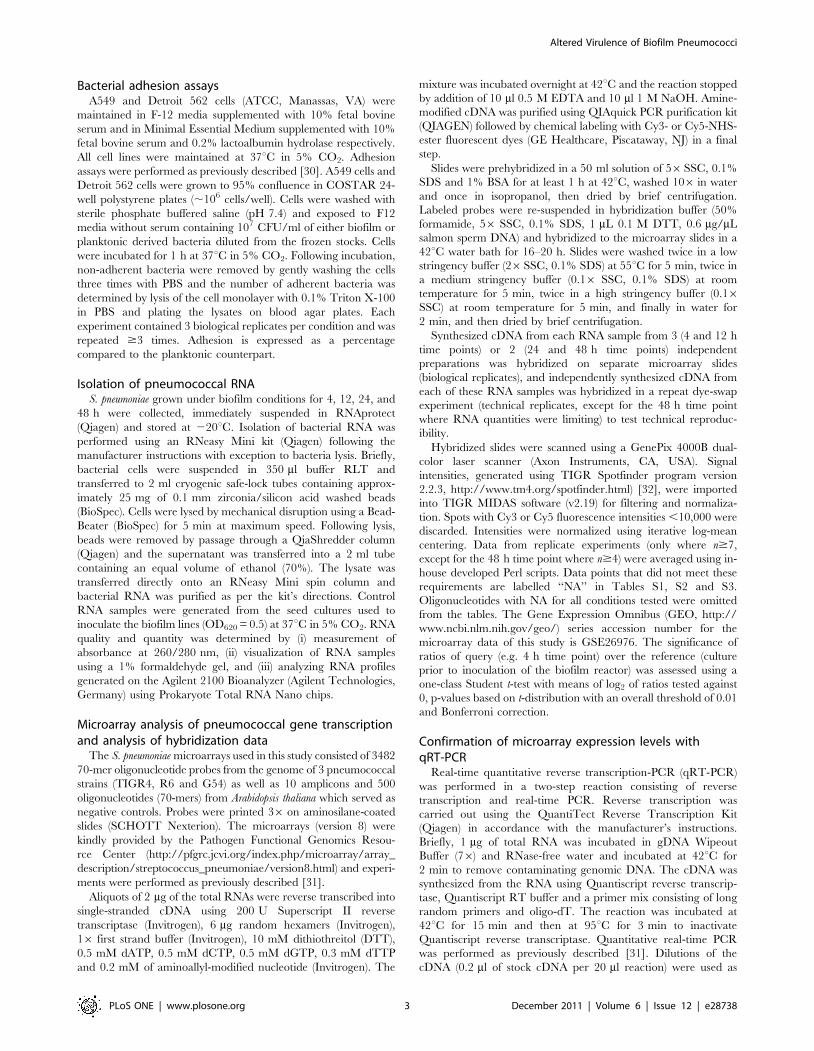
Biofilm pneumococci are hyper-adhesiveWe tested whether the decreased virulence of biofilm bacteria
was due to an inability to bind epithelial cells. Biofilm-derived
bacteria had a 9-fold and 12-fold greater ability to adhere to A549
and Detroit 562 cells versus their planktonic counterparts,
respectively (Figure 3A). Using clinical isolates of 3 additional
serotypes (i.e. 6A, 14 and 15), we confirmed that the hyper-
adhesive biofilm state occurred in both a serotype and strain-
independent manner to both cell lines (Figure 3B). Thus, an
inability to attach to host cells was not responsible for the
attenuated virulence of biofilm pneumococci.
Temporal biofilm-related changes in gene expressionTo determine why biofilm pneumococci were avirulent we
compared pneumococcal gene expression after 4, 12, 24 and 48 h
of biofilm growth versus that of planktonic exponential phase
cultures using microarrays. RNA isolated from our exponential
phase TIGR4 seed cultures and after 4 or 12 h of biofilm growth
could be obtained at high yields and in an intact state. In
contrast, samples collected from the 24 and 48 h biofilms
demonstrated substantial and escalating levels of total RNA
degradation (Figure S2). The levels of degraded RNA were
consistent with the increasing numbers of dead bacteria observed
by transmission electron microscopy at these time points (Figure
S1). Thus this suggested that the degraded RNA was most likely
isolated from the dead or dying pneumococci found within these
biofilms.
Figure 2. Biofilm-derived pneumococci are unable to cause invasive disease. Bacterial titers were measured for individual 5-week oldfemale BALB/cJ mice infected with either planktonic (dark circles), biofilm-derived (white circles), or biofilm-derived planktonic (grey circles) S.pneumoniae TIGR4 via intranasal (106 CFU; n = 10) (A), intratracheal (105 CFU; n = 6–8) (B), or intraperitoneal (104 CFU n = 6–8) (C) routes. For intranasalchallenge, nasal lavages and blood were collected at 24, 72, and 120 h post-infection. For intratracheal and intraperitoneal challenge, samples werecollected at 24 h post-infection. Horizontal bars represent the median value. Statistical analysis was performed using a two-tailed Student’s t-test.doi:10.1371/journal.pone.0028738.g002
Altered Virulence of Biofilm Pneumococci
PLoS ONE | www.plosone.org 6 December 2011 | Volume 6 | Issue 12 | e28738
Despite degradation, sufficient amounts of RNA could be
isolated from the 24 and 48 h time points for efficient Cy3 and
Cy5 labeling and subsequent microarray gene expression analyses
using S. pneumoniae version 8 microarrays obtained from the NIAID
Pathogen Functional Genomics Research Center (http://pfgrc.
org). Nonetheless, to ensure that our microarray results were
robust we processed 2–3 (3 replicates for 4 and 12 h time points, 2
replicates for 24 and 48 h time points) independent RNA samples
for each time point (biological replicates) and performed dye-
flipped hybridizations (technical replicates) for each sample, except
for the 48 h time point where no technical replicates could be
performed due to limiting amounts of RNA material. Stringent
rules for hybridized signal strength and required minimum
number of usable data points per time point were applied prior
to the calculation of average expression levels and P-values (see
methods). Thus in many instances, and in particular at later time
points, data on transcript levels of some pneumococcal genes are
not presented (labeled as ‘‘NA’’ in Tables S1, S2, and S3).
During transition from planktonic to biofilm growth we
observed changes in TIGR4 gene transcription that encompassed
almost all aspects of pneumococcal cell biology. The majority of
genes with differential expression were down-regulated with
significantly lower RNA levels for 40, 62, 32, and 62 genes at 4,
12, 24, and 48 h, respectively (Table S1). In contrast, 16, 40, 14,
and 8 genes were determined to have enhanced transcription, at
the same time points, respectively (Table S2). In total, a
surprisingly small number of genes were observed to have altered
transcription during biofilm growth. When compared to plank-
tonic culture, 6.2% of the 1674 TIGR4 genes tested for by the
microarray were determined to be significantly altered after 12 h
of biofilm culture, the time point with the greatest number of
differentially expressed genes. A complete list of the genes spotted
on the microarray and their expression during bioflm growth is
provided through the Gene Expression Omnibus (GEO, http://
www.ncbi.nlm.nih.gov/geo/) accession number GSE26976. Val-
idation of the microarray results was also performed by qRT-PCR
(Figure S3).
Consistent with the concept that biofilms are in a quiescent state
[38], 32 genes encoding either ribosomal proteins or translation
initiation and elongation factors were down-regulated during
biofilm growth. This was accompanied by a reduction in 8 genes
encoding the ATP synthase machinery (SP_ 1506- SP_1514) and 9
other genes, including the Fab operon, involved in fatty acid
metabolism and phospholipid biosynthesis (SP_0415-SP_0427).
Other indicators that biofilm bacteria were in an inert state
included decreased expression of the cell division gene ftsZ
(SP_1666), decreased expression of assorted Sec pre-protein
translocase components (SP_0230, SP_1702,SP_2029), and re-
duced expression of over 32 conserved hypothetical or hypothet-
ical proteins. Despite an established role for capsule as a major
Figure 3. Biofilm-derived pneumococci have an enhanced ability to adhere to host cells. (A) Comparison of the adhesive properties ofplanktonic bacteria and biofilm-derived S. pneumoniae TIGR4 and T4DpsrP to A549 cells and Detroit 562 cells in vitro. (B) Bacterial adhesion assaysusing a panel of invasive clinical isolates of S. pneumoniae unrelated to TIGR4. Values are expressed as fold-increase adhesion relative to theplanktonic counterparts. All experiments were performed in triplicate and repeated independently at least three times. Statistical analysis wasperformed using a two-tailed Student’s t-test. Single asterisks denotes P,0.001, double asterisks denotes P,0.00001.doi:10.1371/journal.pone.0028738.g003
Altered Virulence of Biofilm Pneumococci
PLoS ONE | www.plosone.org 7 December 2011 | Volume 6 | Issue 12 | e28738
component of the EPM [39], 8 of the genes encoded within the
CPS cassette (SP_0346-SP_0360) were down-regulated during
biofilm growth. Other virulence determinants with decreased
expression included the type I pilus ancillary protein RrgC
(SP_0464), pneumolysin (SP_1923), and choline binding protein
PcpA (SP_2136).
In contrast, Pneumococcal serine-rich repeat protein (PsrP;
SP_1772) was the only established virulence determinant with
enhanced expression during mature biofilm growth. PsrP is a host
cell and intra-species bacterial adhesin previously shown to
contribute to biofilm formation. PsrP is encoded within the
pathogenicity island psrP-secY2A2 (SP_1755-SP_1772) along with
10 glycosyltransferases and 7 components of an alternate Sec
translocase [11,26,40]. These accessory genes are putatively
responsible for PsrP glycosylation and transport and, along with
psrP, were significantly up-regulated during biofilm growth
[41,42,43]. Not surprisingly, genes encoding stress-related chaper-
onins and proteases were also enhanced. These included genes
encoding GroEL (SP_1906), Class I heat shock proteins (SP_0516-
SP_0517), a member of the universal stress protein family
(SP_1996), thioredoxin (SP_1000) and 2 Clp proteases (SP_0338,
SP_0820). Three genes involved in the high affinity phosphate
transport system were also elevated (SP_2084-SP_2088), as were 3
genes involved in glycogen synthesis (SP_1121-SP_1123). Interest-
ingly, genes encoding transposases (SP_0392, SP_0814, SP_0850,
SP_1485, SP_1593, SP_1613) were also up-regulated suggesting
they are sensitive and responsive to physiological stress. Finally, 18
hypothetical proteins were also enhanced, among which SP_1793
was found to be up-regulated as high as 45-fold.
To verify our microarray results we performed qRT-PCR on 19
genes identified by the microarray as either down, no change, or
up-regulated using RNA from each biological replicate (See
methods; Table S3). We then determined the correlation
coefficient between our microarray data with the results from
the qRT-PCR. A strong positive correlation was observed with the
correlation coefficients ranging from 0.66 to 0.94, except for a
single 4 h replicate 1 at 0.59. Thus transcript levels as determined
by microarray were reliable. Included among the genes tested by
qRT-PCR were pspA (SP_0117) and spxB (SP_0730) that encode
proteins responsible for complement resistance and production of
hydrogen peroxide, respectively, and were confirmed as un-
changed [44,45]. ply that encodes pneumolysin that was confirmed
as reduced. Finally, psrP, the Clp protease SP_0338, and SP_1793,
which were all enhanced. Unexpectedly cbpA (SP_2190), which
encodes the pneumococcal adhesin Choline binding protein A
(CbpA) [46,47,48], and nanA (SP_1693), the gene encoding
neuraminidase A [49], were up-regulated during biofilm growth
as measured by qRT-PCR. Likewise, rlrA (SP_0461) and rrgA
(SP_0462) the type I pilus transcriptional regulator and the pilus
adhesin protein, respectively, were down-regulated [50,51].
Presumably, the increased sensitivity of PCR versus detection of
hybridized labeled cDNA during microarray analyses explains the
uncovering of these additional differentially regulated genes by
qRT-PCR.
In summation, the microarray data was in agreement with
studies that suggest biofilm bacteria are quiescent. Furthermore,
these studies suggest that bacteria within a mature biofilm: 1)
produce less capsular polysaccharide, 2) are undergoing consid-
erable physiological stress, and 3) with exception to PsrP and
possibly CbpA and Neuraminidase A, have reduced virulence
determinant expression. Notably, as determined by the unchanged
expression of genes involved with competence, we did not observe
that pneumococci within biofilms were in a competent state. This
was consistent with findings reported by Trappetti et al., showing
that deletion of ComD, which senses competence stimulating
peptide, had no effect on pneumococcal biofilm formation in a
continuous flow-through reactor [52].
Reduced capsule production by biofilm pneumococciSince deletion of capsule enhances bacterial adhesion but results
in a complete loss of virulence [53], and because we observed a
similar phenotype with biofilm pneumococci, we used a direct
ELISA approach to measure total levels of CPS in TIGR4 cultures
and determined that planktonic bacteria had a 3 to 5-fold greater
amount of capsular polysaccharide than their biofilm derivatives
(Figure 4A). We subsequently tested the ability of T4R, an
unencapsulated derivative of TIGR4, and R6, an unrelated and
un-encapsulated serotype 2 derivative that naturally lacks PsrP, to
adhere to cells following either planktonic or biofilm culture
(Figure 4B) [28,54]. For both strains tested, a less dramatic but
persistent biofilm hyper-adhesive phenotype was observed,
indicating that capsule levels played an important, but not
complete, role in the observed hyper-adhesive phenotype. Thus,
we confirmed that the capsule is down-regulated during biofilm
growth and that its reduction most likely contributed to, but was
not solely responsible for, the observed hyper-adhesive state.
Biofilm formation selects for the transparent phenotypeS. pneumoniae undergoes phase variation, alternating between a 1)
transparent, low-capsule and high teichoic acid phase, and 2) an
opaque, high-capsule and low teichoic acid state [34]. During
nasopharyngeal colonization the majority of pneumococci are of
the transparent phenotype, which has an enhanced ability to bind
to host cells. This is due to reduced capsule, but also an increased
amount of surface exposed ChoP, which binds to the host protein
platelet-activating factor receptor (PAFr) on epithelial cells [27].
Importantly, C-reactive protein binds to bacterial ChoP, opson-
izing the bacteria and promoting its phagocytosis [55].
We observed that colonies on plates grown from biofilm isolated
pneumococci were consistently smaller than those used to seed the
biofilm reactor. Upon close examination by trans-oblique
illumination they were determined to more frequently belong to
the transparent phenotype (Figure 5A,B). We determined that
69% of pneumococci isolated from a mature biofilm were
transparent. In contrast, pneumococci in the cultures used to
inoculate the biofilm reactor were predominantly of the opaque
phenotype (63% opaque). We confirmed the transparent pheno-
type by measuring teichoic acid levels. Using direct ELISA to
calculate relative levels of teichoic acid in cell lysates, we observed
a 3–4 fold increase in optical density of the developed ELISA for
biofilm cultures (Figure 5C). This was confirmed by immunoblot
for teichoic acids using TEPC-15, a mouse IgA monoclonal
against ChoP (Figure 5D). Not only was a difference observed in
the total amount of teichoic acid for biofilm pneumococci, but two
additional, higher molecular weight teichoic acid polymers were
also observed for biofilm cultures. Presence of these additional
bands was consistent with published studies contrasting the two
phenotypes [34].
Biofilm pneumococci modulate pneumolysin, PsrP, andCbpA production
Following SDS-PAGE separation of equal protein amounts, we
confirmed decreased production of pneumolysin by biofilm
pneumococci as well as increased production of the bacterial
adhesin CbpA which binds to laminin receptor and has been
shown to be enhanced during the transparent phenotype (Figure 6)
[27,34,56]. Immunodot blot was used to confirm enhanced PsrP
Altered Virulence of Biofilm Pneumococci
PLoS ONE | www.plosone.org 8 December 2011 | Volume 6 | Issue 12 | e28738

production (Figure 6). Immunodot blot was used because PsrP is
glycosylated and separates at a molecular weight of .4000 kDa,
thus it does not readily enter the SDS-PAGE gels normally used to
separate proteins [26]. Thus we confirmed changes increased
biofilm production of three adhesins and reduced production of
the toxin pneumolysin.
Figure 4. Modifications of polysaccharide capsule during biofilm growth contribute to hyper-adhesive phenotype. (A) Direct ELISAtechnique, using sera to type 4 polysaccharide capsule, comparing the amount of capsule present in three dilutions of whole cell lysates fromplanktonic or biofilm-derived bacteria. (B). Bacterial adhesion assay comparing planktonic and biofilm cultures of T4R and R6, an unencapsulatedderivative of TIGR4 and serotype 2 respectively, to A549 cells. Values are expressed as fold increase adhesion relative to the planktonic counterparts.Statistical analysis was performed using a two-tailed Student’s t-test.doi:10.1371/journal.pone.0028738.g004
Figure 5. Selection for the transparent phenotype occurs during biofilm growth. (A) Representative images (n = 6) of individual bacterialcolonies grown from either seed cultures (planktonic) or after 48 h of biofilm growth on blood agar plates. (B) Phase variation during biofilm growthas visualized by oblique, transmitted illumination of the planktonic culture and biofilm-derived bacteria. Percentage of phase variants in planktonicand biofilm cultures as determined by random counts of .100 individual bacterial colonies. (C) Relative levels of phosphorylcholine (ChoP) measuredin whole cell lysates of planktonic or biofilm-derived bacteria were measured by Direct ELISA and by western blot analysis using monoclonalantibodies to ChoP (TEPC-15) (D) Immunoblot analysis of teichoic acids were performed using whole cell lysates of planktonic and biofilm-derivedbacteria separated on 15% SDS-PAGE (E).doi:10.1371/journal.pone.0028738.g005
Altered Virulence of Biofilm Pneumococci
PLoS ONE | www.plosone.org 9 December 2011 | Volume 6 | Issue 12 | e28738

As PsrP binds to surface-exposed Cytokeratin 10, which is
present on A549 cells but absent in Detroit 562 cells [26], we
sought to determine the sole contribution of PsrP to the hyper-
adhesive biofilm phenotype. To do this, we tested the ability of
biofilm and planktonic cultures of T4 DpsrP, a PsrP deficient
mutant, to adhere to these cells (Figure 3A–B). As expected,
planktonic T4 DpsrP failed to adhere A549 cells but bound
normally to Detroit 562 cells. Importantly, biofilm-derived T4
DpsrP still adhered to A549 and Detroit cells at levels 4 to7-fold
greater than its planktonic counterparts, indicating that other
factors including, but possibly not limited to, ChoP and CbpA
were involved in adhesion. In support of this notion, the serotype
14 clinical isolate used in Figure panel 3B, which naturally lacks
psrP (i.e. as determined by comparative genomic hybridization
[40]), also showed enhanced biofilm-mediated adhesion.
Planktonic growth of biofilm pneumococci partiallyrestores virulence
Finally, we sought to determine the durability of the observed
biofilm phenotype. We did this by testing the virulence of
planktonic pneumococci derived from a mature biofilm sample.
Following intranasal and intraperitoneal challenge, biofilm-
derived planktonic pneumococci remained attenuated colonizing
the nasopharynx normally but being unable to enter the
bloodstream or rapidly kill mice, respectively (Figure 2A, 2C). In
contrast, following intratracheal challenge, biofilm-derived plank-
tonic pneumococci were able to establish pneumonia and present
in the lungs at bacterial titers equivalent to mice challenged with
stock planktonic bacteria. Interestingly, biofilm-derived planktonic
pneumococci remained unable to enter the bloodstream
(Figure 2B), suggesting that they had an intermediate phenotype
that specifically affected translocation into the bloodstream or
survival therein.
Discussion
Intranasal and intratracheal challenge of mice with disrupted
mature biofilms allowed us to directly test whether pneumococci
within biofilms were in a virulent state and modeled the aspiration
of bacterial aggregates that might be present in mucosal secretions
from the nasopharynx or that which might be introduced during
intubation [57,58]. Our results indicate that pneumococci within
biofilms are highly suited for attachment to mucosal epithelial
cells, but as a result are avirulent. This was unexpected as the
formation of biofilms has been suggested to be a pivotal event to
numerous infectious diseases [59]. One important limitation of this
study is that this approach does not examine the pathogenic
potential of biofilms that form in vivo and de novo, such as within the
nasopharynx during normal colonization. Thus there is the
possibility that in vivo biofilms might act differently.
Based on our experimental results, the hyper-adhesive pheno-
type of biofilm pneumococci could be attributed to: i) reduced
capsule which exposes bacterial surface proteins [53], ii) selection
for the transparent phenotype which carries greater amounts of
ChoP that binds to the host-protein PAFr [60], iii) enhanced
production of CbpA which binds to Laminin receptor [20], as well
as PsrP, which binds to Keratin 10 [26]. Unconfirmed by protein
analysis, but possibly also contributing to the hyper-adhesive
phenotype, we observed increased expression of the gene encoding
Neuraminidase A by qRT-PCR, which has been shown to
enhance bacterial adhesion by cleaving sialic acid moieties and
thereby exposing cryptic ligands on the host-cell surface [61]. The
attenuated phenotype of biofilm pneumococci could be attributed
to: i) a reduced metabolic rate that would delay its ability to
respond to stressors in a novel host-environment, ii) enhanced
ChoP, which would enhance opsonization by C-reactive protein
[55], iii) reduced capsule, which would also facilitate phagocytosis
[34], iv) a reduction in pneumolysin production [25], v) reduced
PcpA and possibly type I pilus [62]. While a reduction in capsule
and pneumolysin expression along with enhanced neuraminidase
has been shown for S. pneumoniae biofilms [14,15,63], ours is the
first study to suggest they act collectively to dramatically impact
the ability of biofilm pneumococci to progress from the
nasopharynx and cause invasive disease; in particular bloodstream
infections. Thus, implying that pneumococci within biofilms do
not directly contribute to the development of invasive disease.
Using TEM to examine pneumococci within a mature biofilm
structure we were surprised to determine that only a small
percentage of the mature biofilm was composed of electron dense
and presumably viable pneumococci. A finding that suggests
robust pneumococcal biofilm formation occurred through the
accumulation of dead pneumococci. Most recently, Trappetti et al.
have shown that it is the opaque phase variant of S. pneumoniae that
is responsible for formation of the EPM and not the transparent.
As our biofilms contained both transparent and opaque S.
pneumoniae, the opaque variant most likely accounts for the EPM
we detected by electron microscopy. Furthermore, and in contrast
to our in vivo findings, Trappetti et al. observed that opaque but not
transparent biofilm-derived pneumococci, were able to translocate
from the nasopharynx to the lungs and brain of mice [19]. One
possible explanation for this discrepancy in results is that the
Figure 6. Pneumococcal modulate pneumolysin, CbpA, andPsrP during biofilm growth. Immunoblots comparing proteinexpression in whole cell lysates of planktonic and biofilm-derivedbacteria. Whole cell lysates (5 mg) were confirmed to be equally loadedby Coomassie brilliant blue staining. Membranes were probed withantisera to pneumolysin (Ply), choline binding protein A (CbpA), and thepneumococcal serine-rich repeat protein (PsrP). For PsrP immunodotblot was used because PsrP is glycosylated and separates at a molecularweight of .4000 kDa, thus it does not enter SDS-PAGE gels normallyused to separate proteins [26].doi:10.1371/journal.pone.0028738.g006
Altered Virulence of Biofilm Pneumococci
PLoS ONE | www.plosone.org 10 December 2011 | Volume 6 | Issue 12 | e28738

transparent pneumococci present in our biofilms facilitated the
opsonophagocytosis of the attached opaque bacteria. This would
suggest that naturally occurring mixed biofilms are avirulent.
Alternatively, is our use of a continuous flow reactor for mature
biofilm development; Trappetti et al. used a static biofilm model.
In separate studies both Trappetti et al. and ourselves found that
that the use of different biofilm models resulted in variable
phenotypes [11,52]. Finally, is our use of the TIGR4 strain of S.
pneumoniae whereas Trappetti et al. used a 19F clinical isolate.
Despite our observation of considerable EPM surrounding the
electron dense and presumably viable bacteria, biofilm pneumo-
cocci were determined to be hyper-adhesive, suggesting that in
addition to a loss in capsule and increased ChoP, CbpA, and PsrP
protein levels by pneumococci, the EPM may also contain
adhesive elements. This possibility is also supported by findings
by Trappetti et al., which determined that opaque sessile (i.e
biofilm) pneumococci adhere to A549 and Detroit 562 cells better
than transparent sessile pneumococci. The latter was unexpected
as the transparent phenotype is associated with increased
expression of CbpA and ChoP and transparent planktonic
pneumococci have been shown to adhere to cells in an enhanced
manner [24,34,64]. It is for this latter reason that we believe mice
challenged with biofilm-derived planktonic pneumococci, which
would be mostly transparent, developed pneumonia but were
unable to cause bloodstream infection. Importantly, our observa-
tion of enhanced biofilm adhesion by numerous strains and
enhanced PsrP, ChoP, and CbpA production indicates that the
hyper-adhesive phenotype is a pan-pneumococcal biofilm property
that is multi-factorial, involving numerous components along with
the production of EPM.
In an effort to develop a working model that coalesces the
published data with our own, we propose that the selective death
of opaque pneumococci might be occurring during biofilm
formation. Opaque cell death would provide a mechanism for
the release of DNA and other components that are known make
up the EPM. It would also provide an explanation for the high
numbers of dead bacteria observed in our biofilms as well as our
recovery of predominantly transparent pneumococci from mature
biofilms. In context of in vivo transmission our model implies that
the opaque variant would be responsible for formation of the EPM
in the nasopharynx and thereby confer in vivo persistence, whereas
the transparent variant, which is better able to attach to cells and
colonize naıve animals [34], would remain available in greater
numbers for spread to the next host and gain from the enhanced
adhesive capacity of the surrounding EPM. In the next host,
partial reversion to the opaque variant would t also be necessary to
reform a biofilm. Thus, studies are warranted to ascertain if
differential cell death dependent on phase-variation occurs within
biofilms and to test its impact on transmission.
The observed tolerance of biofilm pneumococci to antimicro-
bials was in agreement with previously published studies [29],
moreover, was indicative that we were in fact examining mature
biofilms. Importantly, biofilm pneumococci remained susceptible
to cell wall acting antimicrobials suggesting that maintenance of
the cell wall remained a critical function during the quiescent state.
Ours is the most comprehensive analysis of pneumococcal gene
expression during biofilm growth to date. However, our gene
expression data reflects the biases of our biofilm model which
includes biofilm-related changes in the ratio of opaque and
transparent pneumococci as well as increasing amounts of
remnant mRNA from dead bacteria. This most likely explains
why our microarray results do not exactly match previous studies
that explore differences between opaque and transparent pneu-
mococci [19,65].
We determined that biofilm bacteria down-regulated .50 genes
involved in protein translation, the ATP synthase machinery, fatty
acid metabolism, phospholipid synthesis, and replication. A strong
reduction in capsule operon cassette expression was observed
consistent with a previous study by Moscoso et al., which showed
that cps3A, the first gene in the capsule operon cassette was down-
regulated during biofilm production [63]. Previously, for serotype
3 strains, a non-phase variable deletion within the capsule operon
cassette resulting in a rough mutant has been shown to occur and
contribute towards biofilm formation [66]. A reduction in capsule
would serve to expose surface components such as adhesins and
facilitate attachment. This notion is supported by our previous
findings with PsrP, where a version of the protein unable to extend
past the capsule layer failed to mediate adhesion, as well studies as
completed by Munoz-Elias et al., that found use of an
unencapsulated strain facilitated the identification of genes
involved in biofilm formation in vitro [17,26]. Concomitantly, a
reduction in capsule would reduce the virulence potential of
individual pneumococci.
As indicated, the observed reduction in the physiological state of
bacteria may also contribute to their attenuated phenotype.
Metabolically inert bacteria would take longer to adapt to hostile
host environments such as the lower respiratory tract and produce
the necessary determinants required for survival such as
pneumolysin. Along this line, our observation that biofilm
pneumococci down-regulate pneumolysin allows for speculation
that biofilm pneumococci stop producing factors that elicit a
strong inflammatory response during biofilm formation within the
nasopharynx. Presumably, this would promote long-term coloni-
zation by modulating the immune response. This concept is
supported by the finding that invasive serotypes of S. pneumoniae
colonize the nasopharynx for a shorter duration than non-invasive
serotypes [67]. The reduction in pneumolysin and PcpA levels
during biofilm growth also suggests that immunization with
pneumolysin or PcpA would also have a modest effect against
colonization but might still protect against disseminated (i.e.
planktonic) disease. In contrast, antibodies against CbpA and PsrP,
which are up-regulated during biofilm growth, might deter
nasopharyngeal colonization and thereby promote species re-
placement, such as with Staphylococcus aureus, in immunized
individuals. As such the differential production of protein vaccine
candidates during biofilm versus planktonic growth should be an
important consideration in the design of any future protein vaccine
against S. pneumoniae or other bacterial pathogens [68]. Of note this
concept is consistent with findings by Oggioni et al., showing
altered pneumococcal virulence gene expression occurred during
sessile bacterial growth on fixed surfaces versus planktonic [14].
Finally, the enhanced expression of psrP and its accessory
proteins was in agreement with our previous studies that showed
PsrP contributes to robust biofilm formation [26]. While
unconfirmed microarray and qRT-PCR data implies that the
type I pilus might be down regulated during biofilm production;
this would be surprising as the pilus of Group A Streptococci and
Group B Streptococci have been shown to play an important role
in biofilm formation [69,70]. Of note, microarrays did not reveal
enhanced cbpA expression, however, it was determined by qRT-
PCR and immunoblot that CbpA levels were increased. This
emphasizes the necessity for validation of RNA data with protein
studies and raises the possibility that other determinants are also
altered. Thus a proteomics approach is warranted to address this
gap. Of note, our microarray findings are in stark contrast to those
by Allegrucci et al., who found a dramatic increase in the number
of detectable biofilm proteins when examining 2-dimensional gels
of a serotype 3 isolate [13]. A possible explanation for this
Altered Virulence of Biofilm Pneumococci
PLoS ONE | www.plosone.org 11 December 2011 | Volume 6 | Issue 12 | e28738

discrepancy is the accumulation of dead bacteria and their
proteins in a mature biofilm. This would be an inseparable and
confounding factor in any proteomic analysis of mature biofilms.
In summary, we observed a dramatic enhancement in the ability
of biofilm pneumococci to attach to host cells as well as a dramatic
reduction in their ability to cause invasive disease. Notably, biofilm
pneumococci colonized the nasopharynx normally. As the vast
majority of S. pneumoniae do not cause invasive disease, it is most likely
that these biofilm related changes occur so as to facilitate long-term
colonization of the nasopharynx rather than promote development
of invasive disease. Therefore, and based on the available
information, we suggest that the ability to form robust biofilms is
not required for virulence, but instead contributes towards long-term
colonization and transmission of the pneumococcus.
Supporting Information
Figure S1 Quantification of viable and dead cells duringmature pneumococcal biofilm development. (A) Repre-
sentative Transmission Electron Microscopy (TEM) images of
biofilm bacteria at designated time points; white bars represent 2
microns. Note that viable cells are electron dense, while dead cells
appear as lighter images or ‘‘ghosts’’. (B) Enumeration of viable
and dead cells during mature biofilm development. Percentages of
viable and dead pneumococci as determined by cell counts from
six images per indicated time point.
(EPS)
Figure S2 Assessment of the quality of isolated bacterialRNA from mature pneumococcal biofilms. (A) Formalde-
hyde gels demonstrating the quality of RNA samples taken from S.
pneumoniae biofilms grown under once-through conditions at the
designated time points. (B) RNA profiles of samples collected from
biofilms above using the Agilent 2100 Bioanalyzer. Note the
considerable amount of degradation of RNAs at the later time points.
(EPS)
Figure S3 Validation of microarray studies. RNA levels
obtained with microarrays (X-axis) and qRT-PCR (y-axis) are in
good agreement. The qRT-PCR DCt values (Livak and Schmitt-
gen, 2001) (y-axis) are compared to the log2 transformation of
microarray query/reference ratios (x-axis) for each biological
replicate (2–3) of each time point (4–48 h). Log2 values are used to
obtain a linear correlation. Strong correlation coefficients are
observed, ranging from 0.66 to 0.94, except for at the 4 h replicate
1 at 0.59.
(EPS)
Table S1 Genes with significantly decreased expressionin at least one time point during biofilm growth.
(DOCX)
Table S2 Genes with significantly increased expressionin at least one time point during biofilm growth.
(DOCX)
Table S3 Relative expression of S. pneumoniae genesfollowing biofilm growth versus planktonic cultures asdetermined by microarray and qRT-PCR DCT values.
(DOCX)
Acknowledgments
We thank Dr. Elaine Tuomanen at St. Jude Children’s Research Hospital
for the gift of antibodies against CbpA. We also thank Ms. Barbara Hunter
at the University of Texas Health Science Center Electron Microscopy
Core for her assistance in processing samples for analyses.
Author Contributions
Conceived and designed the experiments: CJS JHJ HT CJO. Performed
the experiments: CJS NK AL PS JCDH. Analyzed the data: CJS NK HT
CJO. Contributed reagents/materials/analysis tools: JHJ HT CJO. Wrote
the paper: CJS HT CJO.
References
1. WHO (1999) Pneumococcal vaccines. WHO position paper. Wkly Epidemiol
Rec 74: 177–183.
2. Lexau CA, Lynfield R, Danila R, Pilishvili T, Facklam R, et al. (2005) Changing
epidemiology of invasive pneumococcal disease among older adults in the era of
pediatric pneumococcal conjugate vaccine. JAMA 294: 2043–2051.
3. Overturf G, Powars D (1980) Infections in sickle cell anemia: pathogenesis and
control. Tex Rep Biol Med 40: 283–292.
4. Wong WY, Overturf GD, Powars DR (1992) Infection caused by Streptococcus
pneumoniae in children with sickle cell disease: epidemiology, immunologic
mechanisms, prophylaxis, and vaccination. Clin Infect Dis 14: 1124–1136.
5. O’Brien KL, Wolfson LJ, Watt JP, Henkle E, Deloria-Knoll M, et al. (2009)
Burden of disease caused by Streptococcus pneumoniae in children younger than 5
years: global estimates. Lancet 374: 893–902.
6. Roush SW, Murphy TV (2007) Historical comparisons of morbidity and
mortality for vaccine-preventable diseases in the United States. JAMA 298:
2155–2163.
7. Maruyama T, Gabazza EC, Morser J, Takagi T, D’Alessandro-Gabazza C,
et al. (2010) Community-acquired pneumonia and nursing home-acquired
pneumonia in the very elderly patients. Respir Med 104: 584–592.
8. Hall-Stoodley L, Hu FZ, Gieseke A, Nistico L, Nguyen D, et al. (2006) Direct
detection of bacterial biofilms on the middle-ear mucosa of children with chronic
otitis media. JAMA 296: 202–211.
9. Hoa M, Tomovic S, Nistico L, Hall-Stoodley L, Stoodley P, et al. (2009)
Identification of adenoid biofilms with middle ear pathogens in otitis-prone
children utilizing SEM and FISH. Int J Pediatr Otorhinolaryngol 73:
1242–1248.
10. Reid SD, Hong W, Dew KE, Winn DR, Pang B, et al. (2009) Streptococcus
pneumoniae forms surface-attached communities in the middle ear of experimen-
tally infected chinchillas. J Infect Dis 199: 786–794.
11. Sanchez CJ, Shivshankar P, Stol K, Trakhtenbroit S, Sullam PM, et al. (2010)
The pneumococcal serine-rich repeat protein is an intra-species bacterial
adhesin that promotes bacterial aggregation in vivo and in biofilms. PLoS
Pathog 6.
12. Camilli R, Pantosti A, Baldassarri L (2010) Contribution of serotype and genetic
background to biofilm formation by Streptococcus pneumoniae. Eur J Clin Microbiol
Infect Dis 30: 97–102.
13. Allegrucci M, Sauer K (2007) Characterization of colony morphology variants
isolated from Streptococcus pneumoniae biofilms. J Bacteriol 189: 2030–2038.
14. Oggioni MR, Trappetti C, Kadioglu A, Cassone M, Iannelli F, et al. (2006)
Switch from planktonic to sessile life: a major event in pneumococcal
pathogenesis. Mol Microbiol 61: 1196–1210.
15. Parker D, Soong G, Planet P, Brower J, Ratner AJ, et al. (2009) The NanA
neuraminidase of Streptococcus pneumoniae is involved in biofilm formation. Infect
Immun 77: 3722–3730.
16. del Prado G, Ruiz V, Naves P, Rodriguez-Cerrato V, Soriano F, et al. (2010)
Biofilm formation by Streptococcus pneumoniae strains and effects of human serum
albumin, ibuprofen, N-acetyl-l-cysteine, amoxicillin, erythromycin, and levo-
floxacin. Diagn Microbiol Infect Dis 67: 311–318.
17. Munoz-Elias EJ, Marcano J, Camilli A (2008) Isolation of Streptococcus pneumoniae
biofilm mutants and their characterization during nasopharyngeal colonization.
Infect Immun 76: 5049–5061.
18. Trappetti C, Kadioglu A, Carter M, Hayre J, Iannelli F, et al. (2009) Sialic acid:
a preventable signal for pneumococcal biofilm formation, colonization, and
invasion of the host. J Infect Dis 199: 1497–1505.
19. Trappetti C, Ogunniyi AD, Oggioni MR, Paton JC (2011) Extracellular Matrix
Formation Enhances the Ability of Streptococcus pneumoniae to Cause Invasive
Disease. PloS one 6: e19844.
20. Lizcano A, Chin T, Sauer K, Tuomanen EI, Orihuela CJ (2010) Early biofilm
formation on microtiter plates is not correlated with the invasive disease
potential of Streptococcus pneumoniae. Microb Pathog 48: 124–130.
21. Tapiainen T, Kujala T, Kaijalainen T, Ikaheimo I, Saukkoriipi A, et al. (2010)
Biofilm formation by Streptococcus pneumoniae isolates from paediatric patients.
APMIS 118: 255–260.
22. Briles DE, Crain MJ, Gray BM, Forman C, Yother J (1992) Strong association
between capsular type and virulence for mice among human isolates of
Streptococcus pneumoniae. Infect Immun 60: 111–116.
Altered Virulence of Biofilm Pneumococci
PLoS ONE | www.plosone.org 12 December 2011 | Volume 6 | Issue 12 | e28738

23. Rubins JB, Charboneau D, Paton JC, Mitchell TJ, Andrew PW, et al. (1995)
Dual function of pneumolysin in the early pathogenesis of murine pneumococcalpneumonia. J Clin Invest 95: 142–150.
24. Weiser JN, Markiewicz Z, Tuomanen EI, Wani JH (1996) Relationship between
phase variation in colony morphology, intrastrain variation in cell wallphysiology, and nasopharyngeal colonization by Streptococcus pneumoniae. Infect
Immun 64: 2240–2245.25. Orihuela CJ, Gao G, Francis KP, Yu J, Tuomanen EI (2004) Tissue-specific
contributions of pneumococcal virulence factors to pathogenesis. J Infect Dis
190: 1661–1669.26. Shivshankar P, Sanchez C, Rose LF, Orihuela CJ (2009) The Streptococcus
pneumoniae adhesin PsrP binds to Keratin 10 on lung cells. Mol Microbiol 73:663–679.
27. Cundell DR, Gerard C, Idanpaan-Heikkila I, Tuomanen EI, Gerard NP (1996)PAf receptor anchors Streptococcus pneumoniae to activated human endothelial cells.
Adv Exp Med Biol 416: 89–94.
28. Hoskins J, Alborn WE, Jr., Arnold J, Blaszczak LC, Burgett S, et al. (2001)Genome of the bacterium Streptococcus pneumoniae strain R6. J Bacteriol 183:
5709–5717.29. Garcia-Castillo M, Morosini MI, Valverde A, Almaraz F, Baquero F, et al.
(2007) Differences in biofilm development and antibiotic susceptibility among
Streptococcus pneumoniae isolates from cystic fibrosis samples and blood cultures.J Antimicrob Chemother 59: 301–304.
30. Rose L, Shivshankar P, Hinojosa E, Rodriguez A, Sanchez CJ, et al. (2008)Antibodies against PsrP, a novel Streptococcus pneumoniae adhesin, block adhesion
and protect mice against pneumococcal challenge. J Infect Dis 198: 375–383.31. Jiang SM, Ishmael N, Hotopp JD, Puliti M, Tissi L, et al. (2008) Variation in the
Group B Streptococcus CsrRS regulon and effects on pathogenicity. J Bacteriol
190: 1956–1965.32. Saeed AI, Sharov V, White J, Li J, Liang W, et al. (2003) TM4: a free, open-
source system for microarray data management and analysis. Biotechniques 34:374–378.
33. Livak KJ, Schmittgen TD (2001) Analysis of relative gene expression data using
real-time quantitative PCR and the 2(-Delta Delta C(T)) Method. Methods 25:402–408.
34. Kim JO, Weiser JN (1998) Association of intrastrain phase variation in quantityof capsular polysaccharide and teichoic acid with the virulence of Streptococcus
pneumoniae. J Infect Dis 177: 368–377.35. Stewart PS, Costerton JW (2001) Antibiotic resistance of bacteria in biofilms.
Lancet 358: 135–138.
36. Stewart PS (2002) Mechanisms of antibiotic resistance in bacterial biofilms.Int J Med Microbiol 292: 107–113.
37. Allison KR, Brynildsen MP, Collins JJ (2011) Metabolite-enabled eradication ofbacterial persisters by aminoglycosides. Nature 473: 216–220.
38. Gilbert P, Maira-Litran T, McBain AJ, Rickard AH, Whyte FW (2002) The
physiology and collective recalcitrance of microbial biofilm communities. AdvMicrob Physiol 46: 202–256.
39. Hall-Stoodley L, Nistico L, Sambanthamoorthy K, Dice B, Nguyen D, et al.(2008) Characterization of biofilm matrix, degradation by DNase treatment and
evidence of capsule downregulation in Streptococcus pneumoniae clinical isolates.BMC Microbiol 8: 173.
40. Obert C, Sublett J, Kaushal D, Hinojosa E, Barton T, et al. (2006) Identification
of a candidate Streptococcus pneumoniae core genome and regions of diversitycorrelated with invasive pneumococcal disease. Infect Immun 74: 4766–4777.
41. Bensing BA, Gibson BW, Sullam PM (2004) The Streptococcus gordonii plateletbinding protein GspB undergoes glycosylation independently of export.
J Bacteriol 186: 638–645.
42. Bensing BA, Sullam PM (2002) An accessory sec locus of Streptococcus gordonii isrequired for export of the surface protein GspB and for normal levels of binding
to human platelets. Mol Microbiol 44: 1081–1094.43. Bensing BA, Takamatsu D, Sullam PM (2005) Determinants of the streptococcal
surface glycoprotein GspB that facilitate export by the accessory Sec system. Mol
Microbiol 58: 1468–1481.44. Spellerberg B, Cundell DR, Sandros J, Pearce BJ, Idanpaan-Heikkila I, et al.
(1996) Pyruvate oxidase, as a determinant of virulence in Streptococcus pneumoniae.Mol Microbiol 19: 803–813.
45. Ren B, Szalai AJ, Thomas O, Hollingshead SK, Briles DE (2003) Both family 1and family 2 PspA proteins can inhibit complement deposition and confer
virulence to a capsular serotype 3 strain of Streptococcus pneumoniae. Infect Immun
71: 75–85.46. Orihuela CJ, Mahdavi J, Thornton J, Mann B, Wooldridge KG, et al. (2009)
Laminin receptor initiates bacterial contact with the blood brain barrier inexperimental meningitis models. J Clin Invest 119: 1638–1646.
47. Zhang JR, Mostov KE, Lamm ME, Nanno M, Shimida S, et al. (2000) The
polymeric immunoglobulin receptor translocates pneumococci across humannasopharyngeal epithelial cells. Cell 102: 827–837.
48. Rosenow C, Ryan P, Weiser JN, Johnson S, Fontan P, et al. (1997) Contribution
of novel choline-binding proteins to adherence, colonization and immunoge-nicity of Streptococcus pneumoniae. Mol Microbiol 25: 819–829.
49. Camara M, Boulnois GJ, Andrew PW, Mitchell TJ (1994) A neuraminidasefrom Streptococcus pneumoniae has the features of a surface protein. Infect Immun
62: 3688–3695.
50. Moschioni M, Emolo C, Biagini M, Maccari S, Pansegrau W, et al. (2010) Thetwo variants of the Streptococcus pneumoniae pilus 1 RrgA adhesin retain the same
function and elicit cross-protection in vivo. Infect Immun 78: 5033–5042.51. Hava DL, Hemsley CJ, Camilli A (2003) Transcriptional regulation in the
Streptococcus pneumoniae rlrA pathogenicity islet by RlrA. J Bacteriol 185: 413–421.52. Trappetti C, Gualdi L, Di Meola L, Jain P, Korir CC, et al. (2011) The impact
of the competence quorum sensing system on Streptococcus pneumoniae biofilms
varies depending on the experimental model. BMC Microbiol 11: 75.53. Hammerschmidt S, Wolff S, Hocke A, Rosseau S, Muller E, et al. (2005)
Illustration of pneumococcal polysaccharide capsule during adherence andinvasion of epithelial cells. Infect Immun 73: 4653–4667.
54. Gosink KK, Mann ER, Guglielmo C, Tuomanen EI, Masure HR (2000) Role of
novel choline binding proteins in virulence of Streptococcus pneumoniae. InfectImmun 68: 5690–5695.
55. Casey R, Newcombe J, McFadden J, Bodman-Smith KB (2008) The acute-phase reactant C-reactive protein binds to phosphorylcholine-expressing Neisseria
meningitidis and increases uptake by human phagocytes. Infect Immun 76:1298–1304.
56. Li-Korotky HS, Lo CY, Banks JM (2010) Interaction of pneumococcal phase
variation, host and pressure/gas composition: virulence expression of NanA,HylA, PspA and CbpA in simulated otitis media. Microb Pathog 49: 204–210.
57. Ramirez P, Ferrer M, Torres A (2007) Prevention measures for ventilator-associated pneumonia: a new focus on the endotracheal tube. Curr Opin Infect
Dis 20: 190–197.
58. Heo SM, Haase EM, Lesse AJ, Gill SR, Scannapieco FA (2008) Geneticrelationships between respiratory pathogens isolated from dental plaque and
bronchoalveolar lavage fluid from patients in the intensive care unit undergoingmechanical ventilation. Clin Infect Dis 47: 1562–1570.
59. Costerton JW, Stewart PS, Greenberg EP (1999) Bacterial biofilms: a commoncause of persistent infections. Science 284: 1318–1322.
60. Cundell DR, Gerard NP, Gerard C, Idanpaan-Heikkila I, Tuomanen EI (1995)
Streptococcus pneumoniae anchor to activated human cells by the receptor forplatelet-activating factor. Nature 377: 435–438.
61. Tong HH, James M, Grants I, Liu X, Shi G, et al. (2001) Comparison ofstructural changes of cell surface carbohydrates in the eustachian tube
epithelium of chinchillas infected with a Streptococcus pneumoniae neuraminidase-
deficient mutant or its isogenic parent strain. Microb Pathog 31: 309–317.62. Glover DT, Hollingshead SK, Briles DE (2008) Streptococcus pneumoniae surface
protein PcpA elicits protection against lung infection and fatal sepsis. InfectImmun 76: 2767–2776.
63. Moscoso M, Garcia E, Lopez R (2006) Biofilm formation by Streptococcus
pneumoniae: role of choline, extracellular DNA, and capsular polysaccharide in
microbial accretion. J Bacteriol 188: 7785–7795.
64. Weiser JN, Austrian R, Sreenivasan PK, Masure HR (1994) Phase variation inpneumococcal opacity: relationship between colonial morphology and nasopha-
ryngeal colonization. Infect Immun 62: 2582–2589.65. King SJ, Hippe KR, Gould JM, Bae D, Peterson S, et al. (2004) Phase variable
desialylation of host proteins that bind to Streptococcus pneumoniae in vivo and
protect the airway. Mol Microbiol 54: 159–171.66. Allegrucci M, Sauer K (2008) Formation of Streptococcus pneumoniae non-phase-
variable colony variants is due to increased mutation frequency present underbiofilm growth conditions. J Bacteriol 190: 6330–6339.
67. Crook DW, Brueggemann AB, Sleeman KL, Peto TEA (2004) Pneumococcal
Carriage. In: Tuomanen EI, Mitchell TJ, Morrison DA, Spratt BG, eds. ThePneumococcus. Washington D.C.: ASM Presss. pp 136–147.
68. Harro JM, Peters BM, O’May GA, Archer N, Kerns P, et al. (2010) Vaccinedevelopment in Staphylococcus aureus: taking the biofilm phenotype into
consideration. FEMS Immunol Med Microbiol 59: 306–323.69. Konto-Ghiorghi Y, Mairey E, Mallet A, Dumenil G, Caliot E, et al. (2009) Dual
role for pilus in adherence to epithelial cells and biofilm formation in Streptococcus
agalactiae. PLoS Pathog 5: e1000422.70. Manetti AG, Zingaretti C, Falugi F, Capo S, Bombaci M, et al. (2007)
Streptococcus pyogenes pili promote pharyngeal cell adhesion and biofilm formation.Mol Microbiol 64: 968–983.
Altered Virulence of Biofilm Pneumococci
PLoS ONE | www.plosone.org 13 December 2011 | Volume 6 | Issue 12 | e28738