Sticky water surfaces: Helix–coil transitions suppressed in a cell-penetrating peptide at the...
10
Sticky water surfaces: Helix–coil transitions suppressed in a cell-penetrating peptide at the air-water interface Denise Schach, Christoph Globisch, Steven J. Roeters, Sander Woutersen, Adrian Fuchs, Clemens K. Weiss, Ellen H. G. Backus, Katharina Landfester, Mischa Bonn, Christine Peter, and Tobias Weidner Citation: The Journal of Chemical Physics 141, 22D517 (2014); doi: 10.1063/1.4898711 View online: http://dx.doi.org/10.1063/1.4898711 View Table of Contents: http://scitation.aip.org/content/aip/journal/jcp/141/22?ver=pdfcov Published by the AIP Publishing Articles you may be interested in Effect of hydrophobic mismatch on domain formation and peptide sorting in the multicomponent lipid bilayers in the presence of immobilized peptides J. Chem. Phys. 141, 074702 (2014); 10.1063/1.4891931 More than the sum of its parts: Coarse-grained peptide-lipid interactions from a simple cross-parametrization J. Chem. Phys. 140, 115101 (2014); 10.1063/1.4867465 Inverted micelle formation of cell-penetrating peptide studied by coarse-grained simulation: Importance of attractive force between cell-penetrating peptides and lipid head group J. Chem. Phys. 134, 095103 (2011); 10.1063/1.3555531 Lipid membranes with transmembrane proteins in shear flow J. Chem. Phys. 132, 025101 (2010); 10.1063/1.3285269 Statistical thermodynamics of helix–coil transitions in biopolymers Am. J. Phys. 67, 1212 (1999); 10.1119/1.19107 This article is copyrighted as indicated in the article. Reuse of AIP content is subject to the terms at: http://scitation.aip.org/termsconditions. Downloaded to IP: 128.135.29.185 On: Tue, 04 Nov 2014 20:04:58
Transcript of Sticky water surfaces: Helix–coil transitions suppressed in a cell-penetrating peptide at the...

Sticky water surfaces: Helix–coil transitions suppressed in a cell-penetrating peptide atthe air-water interfaceDenise Schach, Christoph Globisch, Steven J. Roeters, Sander Woutersen, Adrian Fuchs, Clemens K. Weiss,Ellen H. G. Backus, Katharina Landfester, Mischa Bonn, Christine Peter, and Tobias Weidner Citation: The Journal of Chemical Physics 141, 22D517 (2014); doi: 10.1063/1.4898711 View online: http://dx.doi.org/10.1063/1.4898711 View Table of Contents: http://scitation.aip.org/content/aip/journal/jcp/141/22?ver=pdfcov Published by the AIP Publishing Articles you may be interested in Effect of hydrophobic mismatch on domain formation and peptide sorting in the multicomponent lipid bilayers inthe presence of immobilized peptides J. Chem. Phys. 141, 074702 (2014); 10.1063/1.4891931 More than the sum of its parts: Coarse-grained peptide-lipid interactions from a simple cross-parametrization J. Chem. Phys. 140, 115101 (2014); 10.1063/1.4867465 Inverted micelle formation of cell-penetrating peptide studied by coarse-grained simulation: Importance ofattractive force between cell-penetrating peptides and lipid head group J. Chem. Phys. 134, 095103 (2011); 10.1063/1.3555531 Lipid membranes with transmembrane proteins in shear flow J. Chem. Phys. 132, 025101 (2010); 10.1063/1.3285269 Statistical thermodynamics of helix–coil transitions in biopolymers Am. J. Phys. 67, 1212 (1999); 10.1119/1.19107
This article is copyrighted as indicated in the article. Reuse of AIP content is subject to the terms at: http://scitation.aip.org/termsconditions. Downloaded to IP:
128.135.29.185 On: Tue, 04 Nov 2014 20:04:58

THE JOURNAL OF CHEMICAL PHYSICS 141, 22D517 (2014)
Sticky water surfaces: Helix–coil transitions suppressed in acell-penetrating peptide at the air-water interface
Denise Schach,1,a),b) Christoph Globisch,2,b) Steven J. Roeters,3 Sander Woutersen,3
Adrian Fuchs,1 Clemens K. Weiss,1,4 Ellen H. G. Backus,1 Katharina Landfester,1
Mischa Bonn,1 Christine Peter,2 and Tobias Weidner1,5,c)
1Max Planck Institute for Polymer Research, 55128 Mainz, Germany2Department of Chemistry, University of Konstanz, 78457 Konstanz, Germany3Van’t Hoff Institute for Molecular Sciences, University of Amsterdam, 1098 XH Amsterdam, The Netherlands4Life Sciences and Engineering, Universtiy of Applied Sciences Bingen, 55411 Bingen, Germany5Department of Chemical Engineering, University of Washington, Seattle, Washington 98195, USA
(Received 14 August 2014; accepted 8 October 2014; published online 29 October 2014)
GALA is a 30 amino acid synthetic peptide consisting of a Glu-Ala-Leu-Ala repeat and is knownto undergo a reversible structural transition from a disordered to an α-helical structure when chang-ing the pH from basic to acidic values. In its helical state GALA can insert into and disintegratelipid membranes. This effect has generated much interest in GALA as a candidate for pH triggered,targeted drug delivery. GALA also serves as a well-defined model system to understand cell penetra-tion mechanisms and protein folding triggered by external stimuli. Structural transitions of GALA insolution have been studied extensively. However, cell penetration is an interfacial effect and poten-tial biomedical applications of GALA would involve a variety of surfaces, e.g., nanoparticles, lipidmembranes, tubing, and liquid-gas interfaces. Despite the apparent importance of interfaces in thefunctioning of GALA, the effect of surfaces on the reversible folding of GALA has not yet been stud-ied. Here, we use sum frequency generation vibrational spectroscopy (SFG) to probe the structuralresponse of GALA at the air-water interface and IR spectroscopy to follow GALA folding in bulksolution. We combine the SFG data with molecular dynamics simulations to obtain a molecular-level picture of the interaction of GALA with the air-water interface. Surprisingly, while the fullyreversible structural transition was observed in solution, at the water-air interface, a large fraction ofthe GALA population remained helical at high pH. This “stickiness” of the air-water interface canbe explained by the stabilizing interactions of hydrophobic leucine and alanine side chains with thewater surface. © 2014 AIP Publishing LLC. [http://dx.doi.org/10.1063/1.4898711]
I. INTRODUCTION
Peptides can be used in drug delivery systems and en-able drugs or drug-loaded particles to be delivered to specificlocations within tissues or cells.1–6 The pH sensitive peptideGALA (WEAALAEALAEALAEHLAEALAEALEALAA)is a mimic of viral fusion proteins and provides a promis-ing route to achieve site-specific delivery of therapeuticcompounds.1 The membrane penetrating activity of GALA istriggered by changes in the environmental pH. At basic pH,GALA assumes a disordered structure, but when lowering thepH to acidic conditions, the peptide transitions into an alpha-helical structure. In this state, GALA has the ability to pene-trate cells by pore formation and membrane degradation. Thismechanism is driven by the protonation and deprotonation ofthe Glu residues. The pH driven peptide activity is particularlyinteresting, because it can facilitate the escape of particle-encapsulated drugs from endosomes after endocytosis.7, 8 Inanalogy to viral escape strategies, this approach makes use
a)Present address: Department of Chemistry, University of Chicago, 929 East57th Street, GCIS E139D, Chicago, Illinois 60637, USA
b)D. Schach and C. Globisch contributed equally to this work.c)Author to whom correspondence should be addressed. Electronic mail:
of pH differences between endosomes and the cytosol. Thelow pH environment in endosomes (pH ≈ 5.5) can activateGALA immobilized at drug-loaded particle surfaces and al-lows the particles to escape into the cytosol. The higher pH inthe cytosol (pH ≈ 7) deactivates the peptides, rendering themharmless for other cell organelles.
GALA’s transition from random coil to an amphiphilicα-helix roughly occurs when the pH is decreased from 7 to5, the pH of inflection between the two conformational stateswas reported to be near pH 6.9–12 At low pHs the Glu sitesare protonated and uncharged. The hydrophobic periodicityof the EALA repeats allows the peptide to fold into a sta-ble amphiphilic helix with hydrophobic leucine and alaninesites on one, and hydrophilic glutamic acids on the other sideof the helix (Scheme 1). At neutral pH, however, charge-charge interactions between the deprotonated Glu side chainsdestabilize the helix fold. In the following, the protonatedand deprotonated states of GALA will be denoted as GALA0
and GALA7−, respectively. Note that this nomenclature onlyrefers to the charges on the Glu sidechains, not to the totalcharge of the peptide, where the protonation state of the Hissidechains and the termini would have to be accounted for.
Fourier-transform infrared (FTIR) and circular dichroism(CD) studies in solution show that in its helical state GALA
0021-9606/2014/141(22)/22D517/9/$30.00 © 2014 AIP Publishing LLC141, 22D517-1
This article is copyrighted as indicated in the article. Reuse of AIP content is subject to the terms at: http://scitation.aip.org/termsconditions. Downloaded to IP:
128.135.29.185 On: Tue, 04 Nov 2014 20:04:58

22D517-2 Schach et al. J. Chem. Phys. 141, 22D517 (2014)
SCHEME 1. (Left) GALA in the folded state with protonated (neutral) glu-tamic acid side chains and (right) in the unfolded state with deprotonated(charged) glutamic acid side chains. The charged side chains are spatiallyseparated while the neutral ones can safely interact with each other. Glutamicacid side chains are shown in red. Lipophilic leucine, alanine, and trypto-phane residues are shown in white and histidine in blue. The backbone col-oring corresponds to α-helices shown in blue, and coil and turn in white andyellow, respectively.
self-associates into oligomers with the ability to penetrate thehydrophobic core of lipid membranes, form pores and in-duce leakage.13–15 Along those lines, multiple investigationsof GALA as a vector for drug or gene delivery have beenconducted.9 What remains to be clarified is how hydropho-bic surfaces—for example, drug loaded polymeric nanopar-ticles, biochips, and biotechnical equipment—may influenceGALA’s pH-driven helix-coil transitions. Folding (and ag-gregation) at the (hydrophobic) air-water interface has beeninvestigated in the context of understanding the conforma-tional equilibrium of several protein systems that are report-edly intrinsically disordered in solution, undergo transitionsto helical structures when in contact with membranes or hy-drophobic interfaces, but are also able to from β-sheet rich(amyloid) aggregates at higher protein concentrations. Ex-amples include amyloid-beta-peptide, α-synuclein and LKpeptides.16–18
The aim of the present study is an improved understand-ing of how GALA interacts with aqueous-hydrophobic inter-faces and how binding to such surfaces affects the folding ofGALA. As the easiest accessible water-hydrophobic interfacewe have chosen the air-water interface for this investigation.We probed the pH-dependent secondary structure by a com-bination of surface sensitive vibrational sum frequency gener-ation (SFG) spectroscopy, infrared spectroscopy, and molec-ular dynamics (MD) simulations of GALA in solution and atthe air-water interface.
II. MATERIALS AND METHODS
For practical reasons, vibrational spectroscopy was per-formed in D2O instead of water and we therefore followdeuteration instead of protonation. For the sake of simplicitywe refer to the interface as air-water interface and we replacethe term (de)deuteration by (de)protonation. The pH values ofall D2O solutions were adjusted using a H2O-calibrated pH-meter. The direct reading of the pH-meter in D2O solutions,referred to as pH, was used throughout this article. The rela-tion between pD and pH is pD = pH + 0.44.19
A. Synthesis
All Fmoc-protected L-amino acids and preloadedresin (Fmoc-Gly-Wang resin, 100–200 mesh, low loaded0.30 mmol g−1) for solid phase peptide synthesis werepurchased by Novabiochem (Merck). The purity of thecommercial amino acids was ≥98%. N-[(1H-benzotriazol-1-yl)(dimethylamino)methylene]-N-methyl-methanaminiumhexafluoro-phosphate N-oxide (HBTU, Novabiochem), ethylcyano-glyoxylate-2-oxime (Oxyma Pure, Merck, ≥98%),N,N-diisopropyethylamine (DIEA, Fluka, ≥98%), trifluo-roacetic acid (TFA, Acros, 99%), triisopropylsilane (TIS,Alfa Aesar, 99%), N-methyl-2-pyrrolidone (NMP, BDH,99%), piperazine (Merck, ≥99%), and all solvents were usedas received.
The peptide sequences were prepared using standardsolid-phase Fmoc chemistry with a microwave assisted au-tomated peptide-synthesizer (Liberty, CEM). Tryptophanwas used as Fmoc-L-Trp(Boc)-OH, Histidine as Fmoc-L-His(Trt)-OH, and Glutamic acid as Fmoc-L-Glu(OtBu)-OH.The parameters used for the coupling and deprotection stepsare given below and relate to 0.1 mmol of peptide. Cou-pling was achieved under 300 s of microwave heating, witha temperature reaching and stabilized at 75 ◦C after around90 s, with Oxyma Pure as activator (5 equivalents), DIEAas base (10 equivalents), and amino acid (5 equivalents).Then a first deprotection stage of 30 s (temperature reach-ing around 50 ◦C at the end) was followed by a second cy-cle of 180 s (temperature 75 ◦C) with a 20 wt. % solution ofpiperazine in dimethyl formamide. The resin was washed 3to 5 times between each coupling or deprotection step. Be-fore cleavage the resin was dispersed in dichloromethane andfiltered. Cleavage of peptide from the resin was performedusing a mixture of TFA/TIS/H2O (95%/2.5%/2.5%) for 6 h atambient temperature. After filtration, the peptides were pre-cipitated and centrifuged three times in cold diethyl ether,and dried over vacuum at 30 ◦C. To exchange the TFA withDCl counterions, 10 mg of the GALA were solubilized in20 μl deuterated 2,2,2-trifluoroethanol (TFE) with 40 μl ofD2O (pH = 2.3, DCl) while being placed in an ultrasonic bathfor 15 min. To support the deuteration of the N–H groups ofthe peptide bonds the vial was heated up to 38 ◦C in a tempera-ture controlled shaker for 2 h. After deep-freezing the solutionin liquid nitrogen the peptide was placed in a freeze-dryingsystem. After one night of freeze drying the procedure wasrepeated three more times. The Glu side chains in the purifiedstate can be considered fully deuterated yielding the state ofGALA0.
B. FTIR
The FTIR spectra were recorded on a Bruker Vertex 70equipped with a DLaTGS detector in transmission mode. Atransmission cell with CaF2 windows and 10 μm spacerswas used for all measurements. All spectra were recorded atroom temperature. Each spectrum was averaged for 62 scansat a resolution of 2 cm−1. As a background (BG) referencethe CaF2 cell containing buffer solution without GALA wasused. 15 μl aliquots of a 2.2 GALA solution (0.5 M K2HPO4,
This article is copyrighted as indicated in the article. Reuse of AIP content is subject to the terms at: http://scitation.aip.org/termsconditions. Downloaded to IP:
128.135.29.185 On: Tue, 04 Nov 2014 20:04:58

22D517-3 Schach et al. J. Chem. Phys. 141, 22D517 (2014)
pH = 3 and pH = 12) were used for each measurement. Thesample chamber was purged with dry air for 10 min beforeevery measurement. The software OPUS was used to display,record, and analyze the spectra. From all spectra we sub-tracted the spectrum of the BG recorded in the same mea-surement session since residual superposition of the amidebands with the broad D2O bending mode band at 1555 cm−1
did not allow comparing the spectra without this treatment.The exact peak positions were identified by the minimum po-sitions in the second derivative spectra, which were calcu-lated by means of the Savitzky-Golay algorithm (degree 2)allowing a concomitant smoothing (9 smoothing points). Theamide I′ region was fitted with a sum of profiles consisting of50% Lorentzian shape and 50% Gaussian shape20 using theLevenberg-Marquardt algorithm.21 The resonance positionsobtained from the second derivative spectra were fixed andthe full width at half maximum (FWHM) as well as the bandarea were iterated. To check and further improve the quality ofthe fit we also compared the second derivative spectra of thefit with the original second derivative spectra. The resonancepositions are reported with an error margin of ±2 cm−1.
C. Tensiometry
Surface pressure curves were taken simultaneously withSFG measurements using a Kibron DeltaPi tensiometer,which was calibrated to air and the pure buffer solution first.The constant baseline measured at the beginning in absenceof the peptide was set to zero.
D. SFG
For the SFG experiments we used broadband infraredpulses generated by an OPG/OPA (TOPAS, Light Conver-sion), which was pumped by ∼1.7 W average power of800 nm pulses from a Spitfire Ace (Spectra Physics) am-plified laser system (1 kHz, ∼40 fs FWHM). In addition,∼1 W of the laser output was branched off and directedthrough an etalon to generate the narrow band visible pulse(FWHM bandwidth of ∼15 cm−1, 20 μJ). The broadband in-frared pulse (∼3.5 μJ) provided a 200 cm−1 spectral windowcentered at 1700 cm−1 in the amide I region. The visible andinfrared beams were spatially and temporally overlapped atthe solution surface. The incident angles of the visible (VIS)and infrared (IR) beam were ∼35◦ and ∼40◦ with respect tothe surface normal. The VIS beam was focused down to a di-ameter of approximate 400 μm. The SFG light was spectrallydispersed by a monochromator and detected by an Electron-Multiplied Charge Coupled Device (EMCCD, Andor Tech-nologies).
A trough (70 × 40 × 5 mm) was filled with 4 ml D2Ocontaining 10 mM K2HPO4. The initial pH was set to 11–12. 20 μl of the same solution containing 0.14 mg of GALA0
was sonicated for twenty minutes and added to the subphaseof the solution in the trough to achieve a final concentrationof 9 μM, which is below the concentration at which self-association occurs in bulk solution.9 The samples were al-lowed to equilibrate until the surface pressure remained con-
stant before the SFG measurements were performed. To fullyprotonate or deprotonate the Glu at the air-water interface weadded a certain amount of DCl or NaOD to the subphase toyield pHs of 3 or 12, respectively. The amount of DCl andNaOD needed was determined independent of the SFG ex-periment using a pH-electrode.
The spectra were corrected for the background and nor-malized through a reference spectrum generated by the non-resonant SFG signal of a silver surface. Spectra were recordedin ssp (s-polarized SFG, VIS, and p-polarized IR beams) andsps polarization with 600 s of acquisition time. The back-ground signal was also recorded with 600 s of acquisition timewhile blocking the IR pulse.
E. Molecular dynamics simulations
Two sizes of GALA peptides were studied by MDsimulations: the full-length peptide consisting of 30residues and a shorter version with only 16 residues(WEAALAEALAEALAEH) corresponding to the first halfof the repetitive sequence, from now on denoted as half-GALA. The latter was chosen to more extensively study thefolding/unfolding equilibrium and the partitioning process be-tween the bulk water phase and the air-water interface at dif-ferent pH conditions, i.e., protonation states of the peptide.Simulations were performed with the GROMACS simula-tion package22 using the GROMOS 57a7 force field23 and theSPC water model.24 The peptide was solvated in a rectangu-lar box for the simulations with an air-water interface or in adodecahedral box for the bulk-solution simulations. The sys-tem was neutralized by sodium ions depending on the peptidecharge (GALA0, GALA7−, half-GALA0, and half-GALA4−).The initial box size was chosen such that the minimum dis-tance between the box edge and the protein was larger than1.5 nm. Non-bonded interactions were calculated with a twin-range cutoff scheme, with an update of the short range vander Waals and the real-space Coulomb interactions (<1.0 nm)every time step, and an evaluation of the van der Waals inter-actions between 1.0 and 1.4 nm together with the neighborlist every 10 time steps. The long-range Coulomb interactionswere calculated by the particle mesh ewald method25, 26 witha grid spacing of 0.12 nm. All bonds were constrained by theLINCS algorithm27 and a time step of 2 fs was used. The sol-vated and neutralized systems were energy-minimized withposition restraints on the protein atoms (1000 kJ mol−1 nm−2)and subsequently without restraints by the steepest descentand then by the conjugate gradient algorithm. In the nextstep, several 200 ps long equilibration simulations were per-formed with position restraints on a decreasing number ofpeptide atoms: initially all peptide atoms were restrained,then only the backbone atoms and finally only the alpha car-bon atoms. During the equilibration phase the Bussi velocityrescale method28 was used to keep the temperature at 298 K(with a coupling time of τT = 0.1 ps). The interface simula-tions were performed under constant volume conditions whilefor the bulk systems the pressure was maintained at 1 bar us-ing the Berendsen weak coupling method29 with a couplingtime of τ P = 0.5 ps. For the production simulations (total
This article is copyrighted as indicated in the article. Reuse of AIP content is subject to the terms at: http://scitation.aip.org/termsconditions. Downloaded to IP:
128.135.29.185 On: Tue, 04 Nov 2014 20:04:58
22D517-4 Schach et al. J. Chem. Phys. 141, 22D517 (2014)
length: 2000 ns for the half-GALA systems, and 100 ns forthe full-length GALA systems) the temperature was main-tained by the Langevin thermostat with a friction coefficientof 1 ps−1.
F. SFG spectra calculation
SFG spectra were calculated using methods describedin Ref. 30. We obtained the relative spatial arrangement ofthe amide groups in the protein from the molecular dynam-ics simulation. From the coordinates we determined the cou-plings between the amide groups: nearest-neighbor couplingsare calculated using an ab initio map that gives the couplingas a function of the dihedral angle between the subsequentamide groups, and non-nearest neighbor couplings are cal-culated with a transition dipole coupling model. The localamide-I mode frequencies are shifted according to the hydro-gen bonds that they participate in, and are also red shifted foramide bonds upstream of proline residues. After diagonaliz-ing the Hamiltonian obtained in this way, we calculate the IRtransition-dipole moments and Raman tensors of the amideI normal modes from the eigenvalues and eigenvectors, andwe take their tensor product to calculate the vibrational SFGresponse.
III. RESULTS AND DISCUSSION
A. pH-driven refolding of solution state GALA
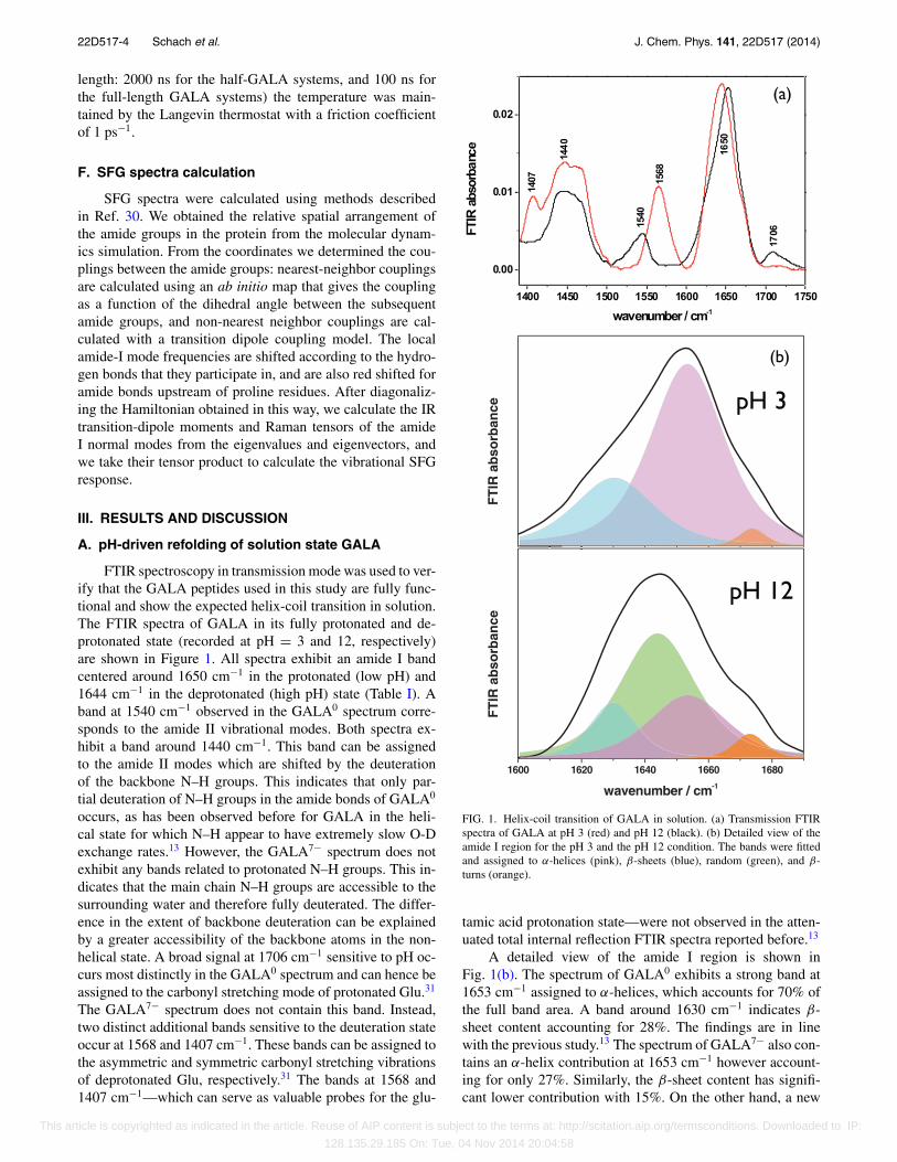
FTIR spectroscopy in transmission mode was used to ver-ify that the GALA peptides used in this study are fully func-tional and show the expected helix-coil transition in solution.The FTIR spectra of GALA in its fully protonated and de-protonated state (recorded at pH = 3 and 12, respectively)are shown in Figure 1. All spectra exhibit an amide I bandcentered around 1650 cm−1 in the protonated (low pH) and1644 cm−1 in the deprotonated (high pH) state (Table I). Aband at 1540 cm−1 observed in the GALA0 spectrum corre-sponds to the amide II vibrational modes. Both spectra ex-hibit a band around 1440 cm−1. This band can be assignedto the amide II modes which are shifted by the deuterationof the backbone N–H groups. This indicates that only par-tial deuteration of N–H groups in the amide bonds of GALA0
occurs, as has been observed before for GALA in the heli-cal state for which N–H appear to have extremely slow O-Dexchange rates.13 However, the GALA7− spectrum does notexhibit any bands related to protonated N–H groups. This in-dicates that the main chain N–H groups are accessible to thesurrounding water and therefore fully deuterated. The differ-ence in the extent of backbone deuteration can be explainedby a greater accessibility of the backbone atoms in the non-helical state. A broad signal at 1706 cm−1 sensitive to pH oc-curs most distinctly in the GALA0 spectrum and can hence beassigned to the carbonyl stretching mode of protonated Glu.31
The GALA7− spectrum does not contain this band. Instead,two distinct additional bands sensitive to the deuteration stateoccur at 1568 and 1407 cm−1. These bands can be assigned tothe asymmetric and symmetric carbonyl stretching vibrationsof deprotonated Glu, respectively.31 The bands at 1568 and1407 cm−1—which can serve as valuable probes for the glu-
1600 1620 1640 1660 1680
FT
IR a
bso
rban
ce
wavenumber / cm-1
A
1600 1620 1640 1660 1680
B
FT
IR a
bso
rban
ce
wavenumber / cm-1
(a)
pH 3
pH 12
(b)
FIG. 1. Helix-coil transition of GALA in solution. (a) Transmission FTIRspectra of GALA at pH 3 (red) and pH 12 (black). (b) Detailed view of theamide I region for the pH 3 and the pH 12 condition. The bands were fittedand assigned to α-helices (pink), β-sheets (blue), random (green), and β-turns (orange).
tamic acid protonation state—were not observed in the atten-uated total internal reflection FTIR spectra reported before.13
A detailed view of the amide I region is shown inFig. 1(b). The spectrum of GALA0 exhibits a strong band at1653 cm−1 assigned to α-helices, which accounts for 70% ofthe full band area. A band around 1630 cm−1 indicates β-sheet content accounting for 28%. The findings are in linewith the previous study.13 The spectrum of GALA7− also con-tains an α-helix contribution at 1653 cm−1 however account-ing for only 27%. Similarly, the β-sheet content has signifi-cant lower contribution with 15%. On the other hand, a new
This article is copyrighted as indicated in the article. Reuse of AIP content is subject to the terms at: http://scitation.aip.org/termsconditions. Downloaded to IP:
128.135.29.185 On: Tue, 04 Nov 2014 20:04:58
22D517-5 Schach et al. J. Chem. Phys. 141, 22D517 (2014)
TABLE I. Frequencies, widths, and assignments of the bands obtained fromfits to the amide I FTIR spectra.a
Band (cm−1) HWHM (cm−1) Percent full area Assignment
GALA7−
1630 17.8 15 (0.5) β-sheet1644 28.4 53 (1.6) Random1653 28.9 27 (0.8) α-helix1673 11.9 4 (0.1) β-turn/-sheet
GALA0
1630 29.8 28 (0.8) β-sheet1653 27.8 70 (2.1) α-helix1674 10.1 2 (0.1) β-turn/-sheet
aThe error margin for the band position is ±2 cm−1. The experimental error of the peakareas is given in parentheses.
band at 1644 cm−1 indicates random structures and accountsfor 53%. From these results, we can confirm the majorityof GALA molecules adopt random structures in the depro-tonated state, whereas in the protonated state GALA adoptsmostly α-helical structures.
To obtain a detailed picture of pH induced helix-coil tran-sitions of GALA in solution we have performed MD sim-ulations of GALA in its protonated and deprotonated state.Due to the relatively large size of the full GALA peptideit is unfeasible to explore the full folding equilibrium at alldifferent states of interest by MD simulations. We there-fore decided to study half-GALA: a peptide consisting ofthe first 16 amino acids of the full sequence starting fromthe C-terminus (WEAALAEALAEALAEH). This allowed usto run longer simulations and reach timescales where fold-ing/unfolding takes place and an assessment of the confor-
mational equilibrium is accessible to molecular simulations.Due to the repetitive sequence of GALA, these simulations ofhalf-GALA should permit conclusions on the conformationalpreferences of the full-length peptide, which will then be usedto guide the interpretation of the IR and SFG data.
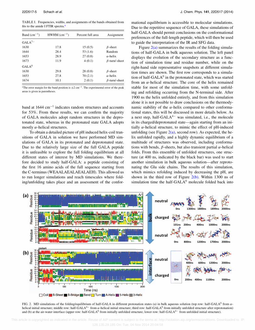
Figure 2(a) summarizes the results of the folding simula-tions of half-GALA in bulk aqueous solution. The left paneldisplays the evolution of the secondary structure as a func-tion of simulation time and residue number, while on theright-hand side representative snapshots at different simula-tion times are shown. The first row corresponds to a simula-tion of half-GALA0 in the protonated state, which was startedfrom an α-helical structure. The core of the helix remainedstable for most of the simulation time, with some unfold-ing and refolding occurring from the N-terminal side. After1900 ns the helix unfolded entirely, and from this simulationalone it is not possible to draw conclusions on the thermody-namic stability of the α-helix compared to other conforma-tional states, this will be discussed in more details below. Asa next step, half-GALA4− was simulated, i.e., the moleculein its charged/deprotonated state—again starting from an ini-tially α-helical structure, to mimic the effect of pH-inducedunfolding (see Figure 2(a), second row). As expected, the he-lix unfolded rapidly, and a highly dynamic equilibrium of amultitude of structures was observed, including conforma-tions with bends, β-sheets, but also transient partial α-helicalfolds. From this ensemble of unfolded structures, one struc-ture (at 400 ns, indicated by the black bar) was used to startanother simulation in bulk aqueous solution—after reproto-nating the Glu side chains. The results of this simulation,which mimics refolding induced by decreasing the pH, areshown in the third row of Figure 2(b). Within 1300 ns ofsimulation time the half-GALA0 molecule folded back into
(a)
(b)
FIG. 2. MD simulations of the folding/equilibrium of half-GALA in different protonation states (a) in bulk aqueous solution (top row: half-GALA0 from α-helical initial structure; middle row: half-GALA4− from α-helical initial structure; third row: half-GALA0 from initially unfolded structure after reprotonation)and (b) at the air-water interface (upper row: half-GALA0 from initially unfolded structure; lower row: half-GALA4− from unfolded initial structure).
This article is copyrighted as indicated in the article. Reuse of AIP content is subject to the terms at: http://scitation.aip.org/termsconditions. Downloaded to IP:
128.135.29.185 On: Tue, 04 Nov 2014 20:04:58

22D517-6 Schach et al. J. Chem. Phys. 141, 22D517 (2014)
a predominantly α-helical structure, transiently even the fullα-helix was observed. This simulation nicely complementsthat of half-GALA0, which had been started from the helicalstructure (first row of Figure 2(a)). While the long “lifetime”of the α-helix had already been indicative of a certain stabil-ity, the observation of refolding into the α-helix after repro-tonation shows clearly that this is a stable, probably the moststable, secondary structural motif for the protonated GALAsequence. The helical fold is probably in equilibrium withother metastable states, as indicated by the unfolding eventobserved at the end of the simulation in the first row as wellas by the formation of different partially formed helices in thesimulation in the third row. These simulation data are in verygood agreement with the results from the IR experiments offull-length GALA described above.
B. At the air-water interface
To study how the pH driven refolding of GALA is af-fected by the presence of a hydrophobic surface, we combinedSFG experiments and MD simulations of GALA at the air-water interface. Figure 2(b) summarizes the MD simulationsof half-GALA, again showing the secondary structure evolu-tion as a function of time together with a few representativesnapshots. The first row of Figure 2(b) shows the simulationof half-GALA0, i.e., the molecule in its protonated state withuncharged Glu side chains, while the second row shows thesimulation of half-GALA4−, i.e., the molecule in its depro-tonated state with charged Glu side chains. Both simulationshad been started from an unfolded conformation (the sameconformation which had already been used in the simulationdepicted in the third row of Figure 2(a)). In both cases, thehalf-GALA molecule rapidly moved to the air-water inter-face and remained there for the rest of the simulation time. Inboth cases, the molecule folded into α-helices within roughly1000 ns, and remained in the α-helical state, irrespective ofthe protonation state of the side chains. Inspection of theobtained structures revealed that the hydrophobic Leu sidechains are oriented towards the vapor phase while the hy-drophilic Glu sites are pointing towards the water phase. Dueto this partitioning of hydrophobic and hydrophilic residues,the helical conformation is stabilized, to an extent that evenovercomes the charge repulsion between deprotonated Gluside chains, which drives the unfolding of the GALA helixthat had been observed in the bulk solution case.
This—in its extent somewhat surprising—stabilizationof the α-helical state of GALA at a hydrophobic surfacewas examined experimentally using SFG spectroscopy at theair-water interface. In analogy to other vibrational spectro-scopies, amide modes observed in SFG spectra can be usedto identify secondary structure motives. However, the selec-tion rules of SFG dictate that an SFG response will onlybe visible from an interface where inversion symmetry isbroken.32, 33 As a result of these selection rules we can ex-pect that any amide vibrational mode observed within thespectra will only originate from ordered secondary struc-tures at the air-water interface. Unbound peptides in the watersubphase—even if close to the surface—will not contribute tothe signal.34 SFG has been successfully used to probe pro-
IR
VIS
SFG
Tensiometer
0 100 200 300 400 500
0
5
10
15
20
25
30
d
addition NaOD
addition DCl
c
addition NaOD
addition DCl
/ m
N m
-1
time / minutes
addition of GALA pH* 8
a b
GALA
a
DCl
bc
NaOD
DCl
d
NaOD
(a)
(b)
FIG. 3. (a) Schematic drawing of the experimental setup for combined SFGand tensiometry measurements at the air-water interface. (b) Surface pres-sure recordings during GALA adsorption to the air-water interface and pHchanges. SFG spectra were recorded at times a, b, c, and d.
tein and peptide secondary structures in the past. In thoseSFG studies α-helix,35–37 β-sheet,35, 37 310-helix,38 and pro-tein aggregates39–41 have been identified.
Figure 3(a) shows a scheme of the experimental setup.In parallel with the SFG measurements we recorded the sur-face pressure throughout the experiment to follow the inter-face activity of GALA. The surface tension data are shownin Figure 3(b) along with the times of GALA injection, acidand base titration as well as the time points of SFG spectrameasurements. The surface tension data show that, after theaddition of solubilized GALA7− (at pH 12) to the subphaseof D2O (at pH 12), the surface pressure increases and levelsoff after 160 min. The increase and saturation of surface ten-sion indicates GALA7− is surface active and forms a layer atthe air-water interface. This result is in good agreement withthe observation in the simulations that GALA peptides showa strong affinity to the surface. The amide I SFG spectrumrecorded using the ssp polarization combination (s-polarizedSFG, s-polarized visible, p-polarized IR) after GALA7−
injection (“a” in the surface tension curve) is shown inFigure 4. The spectrum consists of a resonance near 1645–1650 cm−1, which can be attributed to α-helices.34, 42, 43
In complete agreement with the MD simulations, chargedGALA—disordered in solution state—binds to the air-waterinterface and folds into a helical structure.
After 200 min the pH was decreased to 3. The surfacepressure increased and stabilized after 300 min (Figure 3,
This article is copyrighted as indicated in the article. Reuse of AIP content is subject to the terms at: http://scitation.aip.org/termsconditions. Downloaded to IP:
128.135.29.185 On: Tue, 04 Nov 2014 20:04:58
22D517-7 Schach et al. J. Chem. Phys. 141, 22D517 (2014)
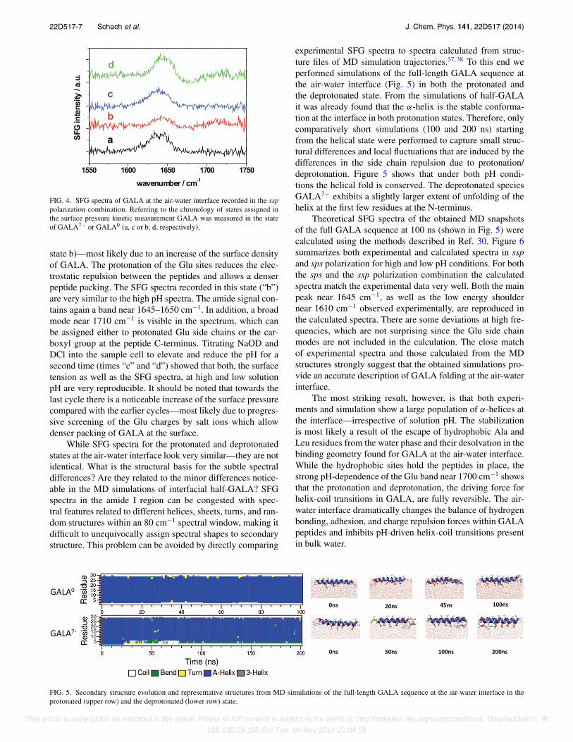
FIG. 4. SFG spectra of GALA at the air-water interface recorded in the ssppolarization combination. Referring to the chronology of states assigned inthe surface pressure kinetic measurement GALA was measured in the stateof GALA7− or GALA0 (a, c or b, d, respectively).
state b)—most likely due to an increase of the surface densityof GALA. The protonation of the Glu sites reduces the elec-trostatic repulsion between the peptides and allows a denserpeptide packing. The SFG spectra recorded in this state (“b”)are very similar to the high pH spectra. The amide signal con-tains again a band near 1645–1650 cm−1. In addition, a broadmode near 1710 cm−1 is visible in the spectrum, which canbe assigned either to protonated Glu side chains or the car-boxyl group at the peptide C-terminus. Titrating NaOD andDCl into the sample cell to elevate and reduce the pH for asecond time (times “c” and “d”) showed that both, the surfacetension as well as the SFG spectra, at high and low solutionpH are very reproducible. It should be noted that towards thelast cycle there is a noticeable increase of the surface pressurecompared with the earlier cycles—most likely due to progres-sive screening of the Glu charges by salt ions which allowdenser packing of GALA at the surface.
While SFG spectra for the protonated and deprotonatedstates at the air-water interface look very similar—they are notidentical. What is the structural basis for the subtle spectraldifferences? Are they related to the minor differences notice-able in the MD simulations of interfacial half-GALA? SFGspectra in the amide I region can be congested with spec-tral features related to different helices, sheets, turns, and ran-dom structures within an 80 cm−1 spectral window, making itdifficult to unequivocally assign spectral shapes to secondarystructure. This problem can be avoided by directly comparing
experimental SFG spectra to spectra calculated from struc-ture files of MD simulation trajectories.37, 38 To this end weperformed simulations of the full-length GALA sequence atthe air-water interface (Fig. 5) in both the protonated andthe deprotonated state. From the simulations of half-GALAit was already found that the α-helix is the stable conforma-tion at the interface in both protonation states. Therefore, onlycomparatively short simulations (100 and 200 ns) startingfrom the helical state were performed to capture small struc-tural differences and local fluctuations that are induced by thedifferences in the side chain repulsion due to protonation/deprotonation. Figure 5 shows that under both pH condi-tions the helical fold is conserved. The deprotonated speciesGALA7− exhibits a slightly larger extent of unfolding of thehelix at the first few residues at the N-terminus.
Theoretical SFG spectra of the obtained MD snapshotsof the full GALA sequence at 100 ns (shown in Fig. 5) werecalculated using the methods described in Ref. 30. Figure 6summarizes both experimental and calculated spectra in sspand sps polarization for high and low pH conditions. For boththe sps and the ssp polarization combination the calculatedspectra match the experimental data very well. Both the mainpeak near 1645 cm−1, as well as the low energy shouldernear 1610 cm−1 observed experimentally, are reproduced inthe calculated spectra. There are some deviations at high fre-quencies, which are not surprising since the Glu side chainmodes are not included in the calculation. The close matchof experimental spectra and those calculated from the MDstructures strongly suggest that the obtained simulations pro-vide an accurate description of GALA folding at the air-waterinterface.
The most striking result, however, is that both experi-ments and simulation show a large population of α-helices atthe interface—irrespective of solution pH. The stabilizationis most likely a result of the escape of hydrophobic Ala andLeu residues from the water phase and their desolvation in thebinding geometry found for GALA at the air-water interface.While the hydrophobic sites hold the peptides in place, thestrong pH-dependence of the Glu band near 1700 cm−1 showsthat the protonation and deprotonation, the driving force forhelix-coil transitions in GALA, are fully reversible. The air-water interface dramatically changes the balance of hydrogenbonding, adhesion, and charge repulsion forces within GALApeptides and inhibits pH-driven helix-coil transitions presentin bulk water.
FIG. 5. Secondary structure evolution and representative structures from MD simulations of the full-length GALA sequence at the air-water interface in theprotonated (upper row) and the deprotonated (lower row) state.
This article is copyrighted as indicated in the article. Reuse of AIP content is subject to the terms at: http://scitation.aip.org/termsconditions. Downloaded to IP:
128.135.29.185 On: Tue, 04 Nov 2014 20:04:58

22D517-8 Schach et al. J. Chem. Phys. 141, 22D517 (2014)
GALA0 GALA0
GALA7GALA7
sps
sps ssp
ssp
SF
G in
ten
sity
(a.
u.)
Wavenumber (cm-1) Wavenumber (cm-1)1600 1650 1700 1600 1650 1700 1750
FIG. 6. Comparison of experimental ssp and sps polarization SFG spectra (orange) and spectra calculated from the 100 ns GALA snapshots (blue) for high andlow pHs shown in Figure 5.
IV. SUMMARY
While we can confirm the pH-driven helix-coil transi-tions of GALA peptides in bulk solution observed in previousstudies, we find that GALA does not show a pH-dependentstructural transition at the hydrophobic air-water interface.Together, SFG experiments and MD simulations demonstratethat the unfolding of the GALA sequence is inhibited at the in-terface, most likely due to adherence of the hydrophobic sidechains of alanine and leucine to the air-water interface—watersurfaces are “sticky” for GALA peptides. This result stronglysuggests that the reliability of the folding mechanism ofGALA—for example, in pharmaceutical applications—willbe strongly affected by side chain-surface interactions. Ourstudy demonstrates that interfaces can generally change thebalance of forces involved in structural transitions in “smartpeptides,” and that surface–protein interactions will have tobe taken into account for the rational design of responsivebiomedical surfaces.
ACKNOWLEDGMENTS
T.W. thanks the Deutsche Forschungsgemeinschaft (WE4478/2-1) and European Union Marie Curie Program forsupport of this work (CIG Grant No. 322124). This workis part of the research program of the Max Planck Soci-ety. S.J.R. and S.W. would like to acknowledge the Euro-pean Research Council for funding through Starting GrantNo. 210999. C.P. gratefully acknowledges support by theDeutsche Forschungsgemeinschaft via the Emmy NoetherProgram (Grant No. CP1625/1).
1J. Andrieu, N. Kotman, M. Maier, V. Mailänder, W. S. Strauss, C. K. Weiss,and K. Landfester, Macromol. Rapid Commun. 33, 248 (2012).
2I. A. Khalil, K. Kogure, S. Futaki, S. Hama, H. Akita, M. Ueno, H. Kishida,M. Kudoh, Y. Mishina, K. Kataoka, M. Yamada, and H. Harashima, GeneTher. 14, 682 (2007).
3J. H. Lee, J. A. Engler, J. F. Collawn, and B. A. Moore, FEBS J. 268, 2004(2001).
4S. Santra, H. Yang, D. Dutta, J. T. Stanley, P. H. Holloway, W. Tan, B. M.Moudgil, and R. A. Mericle, Chem. Commun. 2004, 2810.
5J. Nicolas, S. Mura, D. Brambilla, N. Mackiewicz, and P. Couvreur, Chem.Soc. Rev. 42, 1147 (2013).
6K. Landfester and V. Mailänder, Expert Opin. Drug Delivery 10, 593(2013).
7V. Mailänder and K. Landfester, Biomacromolecules 10, 2379 (2009).8K. Narayanan, S. K. Yen, Q. Dou, P. Padmanabhan, T. Sudhaharan, S.Ahmed, J. Y. Ying, and S. T. Selvan, Sci. Rep. 3, 2184 (2013).
9W. Li, F. Nicol, and F. C. Szoka, Jr., Adv. Drug Delivery Rev. 56, 967(2004).
10R. Qiao, Q. Jia, S. Hüwel, R. Xia, T. Liu, F. Gao, H.-J. Galla, and M. Ga,ACS Nano 6, 3304 (2012).
11B. L. Droumaguet, J. Nicolas, D. Brambilla, S. Mura, A. Maksimenko,L. D. Kimpe, E. Salvati, C. Zona, C. Airoldi, M. Canovi, M. Gobbi, M.Noiray, B. L. Ferla, F. Nicotra, W. Scheper, O. Flores, M. Masserini, K.Andrieux, and P. Couvreur, ACS Nano 6, 5866 (2012).
12D. Bartczak, O. L. Muskens, S. Nitti, T. Sanchez-Elsner, T. M. Millar, andA. G. Kanaras, Small 8, 122 (2012).
13E. Goormaghtigh, J. De Meutter, F. Szoka, V. Cabiaux, R. A. Parente, andJ.-M. Ruysschaert, Eur. J. Biochem. 195, 421 (1991).
14R. A. Parente, L. Nadasdi, N. K. Subbarao, and F. C. Szoka, Biochemistry29, 8713 (1990).
15N. K. Subbarao, R. A. Parente, F. C. Szoka, L. Nadasdi, and K. Pongracz,Biochemistry 26, 2964 (1987).
16C. Wang, N. Shah, G. Thakur, F. Zhou, and R. M. Leblanc, Chem. Com-mun. 46, 6702 (2010).
17M. Hoernke, J. A. Falenski, C. Schwieger, B. Koksch, and G. Brezesinski,Langmuir 27, 14218 (2011).
18S. Campioni, G. Carret, S. Jordens, L. Nicoud, R. Mezzenga, and R. Riek,J. Am. Chem. Soc. 136, 2866 (2014).
19K. Mikkelsen and S. O. Nielsen, J. Phys. Chem. 64, 632 (1960).20J. L. R. Arrondo and F. M. Goñi, Prog. Biophys. Mol. Biol. 72, 367
(1999).21D. W. Marquardt, J. SIAM 11, 431 (1963).22B. Hess, C. Kutzner, D. van der Spoel, and E. Lindahl, J. Chem. Theory
Comput. 4, 435 (2008).
This article is copyrighted as indicated in the article. Reuse of AIP content is subject to the terms at: http://scitation.aip.org/termsconditions. Downloaded to IP:
128.135.29.185 On: Tue, 04 Nov 2014 20:04:58

22D517-9 Schach et al. J. Chem. Phys. 141, 22D517 (2014)
23N. Schmid, A. P. Eichenberger, A. Choutko, S. Riniker, M. Winger, A. E.Mark, and W. F. van Gunsteren, Eur. Biophys. J. 40, 843 (2011).
24H. J. C. Berendsen, J. P. M. Postma, W. F. van Gunsteren, and J. Hermans,in Intermolecular Forces, edited by B. Pullman (Reidel Publishing Com-pany, Dordrecht 1981), p. 331.
25T. Darden, D. York, and L. Pedersen, J. Chem. Phys. 98, 10089(1993).
26U. Essmann, L. Perera, M. L. Berkowitz, T. Darden, H. Lee, and L. G.Pedersen, J. Chem. Phys. 103, 8577 (1995).
27B. Hess, J. Chem. Theory Comput. 4, 116 (2008).28G. Bussi, D. Donadio, and M. Parrinello, J. Chem. Phys. 126, 014101
(2007).29H. J. C. Berendsen, J. P. M. Postma, W. F. Vangunsteren, A. Dinola, and J.
R. Haak, J. Chem. Phys. 81, 3684 (1984).30S. J. Roeters, C. N. van Dijk, A. Torres-Knoop, E. H. G. Backus, R.
K. Campen, M. Bonn, and S. Woutersen, J. Phys. Chem. A 117, 6311(2013).
31A. Barth, Prog. Biophys. Mol. Biol. 74, 141 (2000).32Y. R. Shen, The Principles of Nonlinear Optics, 1st ed. (John Wiley & Sons,
New York, 1984).
33R. W. Boyd, Nonlinear Optics, 1st ed. (Academic Press, London, 1992).34T. Weidner, N. Samuel, K. McCrea, L. Gamble, R. Ward, and D. Castner,
Biointerphases 5, 9 (2010).35J. Wang, S.-H. Lee, and Z. Chen, J. Phys. Chem. B 112, 2281 (2008).36T. Weidner, J. S. Apte, L. J. Gamble, and D. G. Castner, Langmuir 26, 3433
(2010).37K. T. Nguyen, S. V. Le Clair, S. J. Ye, and Z. Chen, J. Phys. Chem. B 113,
12169 (2009).38J. E. Baio, T. Weidner, N. T. Samuel, K. R. McCrea, L. Baugh, P. S. Stayton,
and D. G. Castner, J. Vac. Sci. Technol., B 28, C5D1 (2010).39I. I. Rzeznicka, R. Pandey, M. Schleeger, M. Bonn, and T. Weidner, Lang-
muir 30, 7736 (2014).40D. Xiao, L. Fu, J. Liu, V. S. Batista, and E. C. Y. Yan, J. Mol. Biol. 421,
537 (2012).41C. C. Vandenakker, M. F. Engel, K. P. Velikov, M. Bonn, and G. H. Koen-
derink, J. Am. Chem. Soc. 133, 18030 (2011).42B. R. Singh, in ACS Symposium Series (American Chemical Society, Wash-
ington, DC, 2000), Vol. 750.43X. Y. Chen, M. L. Clarke, J. Wang, and Z. Chen, Int. J. Mod. Phys. A 19,
691 (2005).
This article is copyrighted as indicated in the article. Reuse of AIP content is subject to the terms at: http://scitation.aip.org/termsconditions. Downloaded to IP:
128.135.29.185 On: Tue, 04 Nov 2014 20:04:58