Stem cells associated with macroporous bioceramics for long bone repair: 6-to 7-year outcome of a...
10
Stem Cells Associated with Macroporous Bioceramics for Long Bone Repair: 6- to 7-Year Outcome of a Pilot Clinical Study MAURILIO MARCACCI, M.D., 1 ELIZAVETA KON, M.D., 1 VLADIMIR MOUKHACHEV, M.D., 2 ANDREI LAVROUKOV, M.D., 2,{ SERGEJ KUTEPOV, M.D., 3 RODOLFO QUARTO, M.D., 4,* MADDALENA MASTROGIACOMO, Ph.D., 4 and RANIERI CANCEDDA, M.D. 4 ABSTRACT Extensive bone loss is still a major problem in orthopedics. A number of different therapeutic approaches have been developed and proposed, but so far none have proven to be fully satisfactory. We used a new tissue engineering approach to treat four patients with large bone diaphysis defects and poor therapeutic alternatives. To obtain implantable three-dimensional (3D) living constructs, cells isolated from the pa- tients’ bone marrow stroma were expanded in culture and seeded onto porous hydroxyapatite (HA) ceramic scaffolds designed to match the bone deficit in terms of size and shape. During the surgical session, an Ilizarov apparatus or a monoaxial external fixator was positioned on the patient’s affected limb and the ceramic cylinder seeded with cells was placed in the bone defect. Patients were evaluated at different postsurgery time intervals by conventional radiographs and computed tomography (CT) scans. In one patient, an angiographic evaluation was performed at 6.5 years follow-up. In this study we analyze the long-term outcome of these patients following therapy. No major complications occurred in the early or late postoperative periods, nor were major complaints reported by the patients. No signs of pain, swelling, or infection were observed at the implantation site. Complete fusion between the implant and the host bone occurred 5 to 7 months after surgery. In all patients at the last follow-up (6 to 7 years postsurgery in patients 1 to 3), a good integration of the implants was maintained. No late fractures in the implant zone were observed. The present study shows the long-term durability of bone regeneration achieved by a bone engineering approach. We consider the obtained results very promising and propose the use of culture- expanded osteoprogenitor cells in conjunction with porous bioceramics as a real and significant im- provement in the repair of critical-sized long bone defects. INTRODUCTION E XTENSIVE BONE LOSS OR DESTRUCTION is still a major problem in orthopedics, due in large part to the lack of predictability in obtaining functional bone reconstruction. Present therapeutic approaches to repair large bone defects can be divided into two groups: excluding graft transplant (Ilizarov bone transport) or including graft transplant (auto- logous, homologous, or heterologous bone grafts using dif- ferent biomaterial implants). The Ilizarov technique, osteotomy followed by bone distrac- tion, relies on the bone regeneration potential, thus avoiding 1 Istituti Ortopedici Rizzoli, Bologna, Italy. 2 Ural Orthopedic Research Institute, Ekaterinburg, Russia. 3 Ural State Medical Academy, Ekaterinburg, Russia. 4 Dipartimento di Oncologia Biologia e Genetica, Universita’ di Genova & Istituto Nazionale per la Ricerca sul Cancro, Genova, Italy. *Present address: Dipartimento di Chimica e Tecnologie Farmaceutiche ed Alimentari, Universita ` di Genova, Italy. { Unfortunately, Dr. Lavroukov died in a car accident in summer 2005. The other coauthors wish to dedicate this manuscript to him. TISSUE ENGINEERING Volume 13, Number 5, 2007 # Mary Ann Liebert, Inc. DOI: 10.1089/ten.2006.0271 947
Transcript of Stem cells associated with macroporous bioceramics for long bone repair: 6-to 7-year outcome of a...
Stem Cells Associated with Macroporous Bioceramics for Long
Bone Repair: 6- to 7-Year Outcome of a Pilot Clinical Study
MAURILIO MARCACCI, M.D.,1 ELIZAVETA KON, M.D.,1 VLADIMIR MOUKHACHEV, M.D.,2
ANDREI LAVROUKOV, M.D.,2,{ SERGEJ KUTEPOV, M.D.,3 RODOLFO QUARTO, M.D.,4,*
MADDALENA MASTROGIACOMO, Ph.D.,4 and RANIERI CANCEDDA, M.D.4
ABSTRACT
Extensive bone loss is still a major problem in orthopedics. A number of different therapeutic approacheshave been developed and proposed, but so far none have proven to be fully satisfactory. We used a newtissue engineering approach to treat four patients with large bone diaphysis defects and poor therapeuticalternatives. To obtain implantable three-dimensional (3D) living constructs, cells isolated from the pa-tients’ bone marrow stroma were expanded in culture and seeded onto porous hydroxyapatite (HA)ceramic scaffolds designed to match the bone deficit in terms of size and shape. During the surgical session,an Ilizarov apparatus or a monoaxial external fixator was positioned on the patient’s affected limb and theceramic cylinder seeded with cells was placed in the bone defect. Patients were evaluated at differentpostsurgery time intervals by conventional radiographs and computed tomography (CT) scans. In onepatient, an angiographic evaluation was performed at 6.5 years follow-up. In this study we analyze thelong-term outcome of these patients following therapy. No major complications occurred in the early orlate postoperative periods, nor were major complaints reported by the patients. No signs of pain, swelling,or infection were observed at the implantation site. Complete fusion between the implant and the host boneoccurred 5 to 7 months after surgery. In all patients at the last follow-up (6 to 7 years postsurgery inpatients 1 to 3), a good integration of the implants was maintained. No late fractures in the implant zonewere observed. The present study shows the long-term durability of bone regeneration achieved by a boneengineering approach. We consider the obtained results very promising and propose the use of culture-expanded osteoprogenitor cells in conjunction with porous bioceramics as a real and significant im-provement in the repair of critical-sized long bone defects.
INTRODUCTION
EXTENSIVE BONE LOSS OR DESTRUCTION is still a major
problem in orthopedics, due in large part to the lack of
predictability in obtaining functional bone reconstruction.
Present therapeutic approaches to repair large bone defects
can be divided into two groups: excluding graft transplant
(Ilizarov bone transport) or including graft transplant (auto-
logous, homologous, or heterologous bone grafts using dif-
ferent biomaterial implants).
The Ilizarov technique,osteotomy followedbybonedistrac-
tion, relies on the bone regeneration potential, thus avoiding
1Istituti Ortopedici Rizzoli, Bologna, Italy.2Ural Orthopedic Research Institute, Ekaterinburg, Russia.3Ural State Medical Academy, Ekaterinburg, Russia.4Dipartimento di Oncologia Biologia e Genetica, Universita’ di Genova & Istituto Nazionale per la Ricerca sul Cancro, Genova, Italy.
*Present address: Dipartimento di Chimica e Tecnologie Farmaceutiche ed Alimentari, Universita di Genova, Italy.{Unfortunately, Dr. Lavroukov died in a car accident in summer 2005. The other coauthors wish to dedicate this manuscript to him.
TISSUE ENGINEERINGVolume 13, Number 5, 2007# Mary Ann Liebert, Inc.DOI: 10.1089/ten.2006.0271
947
concerns related to graft integration, but it is highly incon-
venient for the patient.1,2
Autologous bone implants are used as either nonvascu-
larized or vascularized grafts. Implants of nonvascularized
grafts are usually performed to treat small bone defects.
Vascularized grafts are most commonly used for extensive
bone reconstructions such as in tumor surgery, but require
long and difficult surgical operations. The success rate is
high, but complications such as infections and nonunions are
common, especially in large shaft reconstructions.3–6 Fur-
ther, large reconstructions by autologous bone require a
large harvest of healthy tissue resulting in significant donor
site morbidity.7 Homologous grafts from bone banks are
widely used due to their availability without concerns of do-
nor site morbidity. However, this material presents potential
risk of viral or bacterial infection and possible immune re-
sponse of the host tissue8 that can impair the graft integration
and result in long recovery times and high rate of com-
plications.9–11 Moreover, the complete substitution of the
cortical graft with the host bone is not achieved, and it
eventually often results in the late graft fracture.12
Different biomaterials have been proposed as bone sub-
stitutes with conflicting results. Among these, hydroxyapa-
tite (HA) and other calcium phosphate ceramics have shown
the most promising results due to their osteoconductive
properties, unlimited availability, and absence of immune re-
sponse and risk of virus transmission.13–16 The main diffi-
culty to their wider use remains the absence of osteoinductive
properties, thus limiting their application to repairing large
segmental bone loss.
Tissue engineering has been proposed as an alternative to
the traditional techniques in repairing bone defects.8,17–24
The general principle of tissue engineering involves the
association of cells with a natural or synthetic support, or
scaffold, to produce a three-dimensional (3D) living, im-
plantable construct. Among the different biomaterials con-
sidered as scaffolds for bone tissue engineering, calcium
phosphate–based ceramics have proven to be of great interest
given their osteoconductivity and ability to ‘‘integrate’’ with
the bone tissue.13,15,16,25–28 These characteristics can be fur-
ther improved by varying the structural characteristics of the
scaffolds. In a recent study of ours, we considered two
HA bioceramics with identical microstructure but different
macroporosity, pore size distribution, and pore interconnec-
tion pathway. The histological analysis of specimens at dif-
ferent times after in vivo implantation revealed in both
materials a significant extent of bone matrix deposition, but
we observed that porosity and pore interconnection of these
scaffolds influenced the total amount of deposited bone, the
pattern of blood vessel invasion, and finally the kinetics of
the bone neoformation process.27
There are a number of sources of pluripotent mesenchy-
mal cells potentially suitable for bone repair in association
with porous ceramic scaffolds.28–32 The best characterized
are those derived from the bone marrow stroma, which yields
a mesenchymal stem/progenitor cell population from which
differentiated cells of various connective tissues can be de-
rived. Bone marrow–derived pluripotent mesenchymal stem
cells (BMSCs) grown in vitro are capable of self-renewal
for many generations without significant loss of their char-
acteristics.33 They are also able to generate several distinct
phenotypes including osteoblasts, chondrocytes, and adipo-
cytes by relatively simple adjustments of culture conditions
and biochemical supplements to which they are exposed.
We were the first to report the repair of large bone defects
in humans using autologous in vitro expanded pluripotent
mesenchymal cells associated to a porous ceramic.34 The
study was based on promising results from earlier studies on
large animal models and those already reported in the lit-
erature at the time we initiated our clinical study (late
1990s).18,21,35 Additional reports dealing with bone repair by
a tissue engineering approach in large animal models have
been published in the following years.17,28,29,33,36–39 In the
present paper, we analyze the long-term outcome of a case
series of four patients with large bone diaphysis defects and
limited therapeutic alternatives. Initial clinical results at 1–2
years postsurgery were previously reported for three of these
patients.34 The aim of the present work was to determine the
durability of this type of implant in this first group of patients
treated with this innovative tissue engineering method.
MATERIALS AND METHODS
Patients
The study protocol was approved by the ethical commit-
tees of the involved orthopedic centers. The patients, with
age ranging from 16 to 41 years, possessed no neoplastic
pathologies and were selected for this treatment after alter-
native, more ‘‘conventional’’ surgical therapies failed. The
patients were informed of the nature of the treatment and
gave their written consent. Essential information on the four
patients is listed in Table 1.
Case 1. A 41-year-old woman presented a 4 cm shorten-
ing of the right leg and a severe osteoarthritis of the ankle
and subtalar joints as the result of an ankle fracture com-
plicated by an osteomyelitic process that occurred in 1992.
At admission, the osteomyelitic process had completely re-
solved. The patient underwent arthrodesis of the right ankle
and subtalar joints. To obtain leg lengthening, in the same
surgical session, a tibia osteotomy was performed at the prox-
imal diaphysis level and the bone was stabilized by an Ili-
zarov apparatus. The distraction of the tibia resulted in poor
bone formation. After 10 months, a 4 cm gap between the
two stumps with only a thin bone bridge located at the back
was detectable by radiography. In May 1998, the patient was
selected for the study.
Case 2. A 16-year-old girl presented a fracture of the left
femoral neck and an exposed biosseous fracture of the left
forearm as the result of a trauma. At admission, the patient
948 MARCACCI ET AL.
underwent reduction of both ulna and radius fractures with
Kirshner wires, together with surgical debridement and su-
ture of the skin. Immobilization was obtained by a plaster
cast. Two weeks later, the bone was stabilized by an Ilizarov
forearm apparatus. After 1.5 months, the Ilizarov apparatus
was removed due to osteomyelitis. In the same surgical
session a sequestrectomy was performed at the distal di-
aphysis of the ulna. In December 1998, the osteomyelitis of
the left forearm was completely healed with 4 cm bone loss
at distal diaphysis of the ulna. In April 1999, the patient was
selected for the study.
Case 3. A 22-year-old man presented a plurifragmented
exposed fracture of the right humerus and of the elbow with
an 8 cm bone loss in the distal third of the humerus diaph-
ysis and a complete disarrangement of the elbow as the
result of a car accident. The fracture was stabilized by an
external fixator, leaving a gap of 7 cm at the distal humerus
diaphysis. At the same time, the elbow arthrodesis was
performed. No local infection was present. In June 1999,
the patient was selected for the study.
Case 4. A 29-year-old woman presented multiple frac-
tures (facial bones, right tibia, left femur, right wrist, left
humerus) and an exposed biosseous fracture of the left
forearm as a consequence of a car accident in 1999. In
September 2004, the patient returned to the clinic with an
ulnar preudoarthrosis and 6 cm bone loss, and was selected
for the study.
Isolation and culture of marrow-derived
osteogenic progenitors
Human BMSCs were obtained and cultured as described
by Martin et al., with minor modifications within 36 hours
from harvest.30 Briefly said, 20 mL samples of iliac crest
marrow aspirates from each patient were washed with phos-
phate buffered saline (PBS), pH 7.2. Nucleated cells were
counted, suspended in Coon’s modified Ham’s F12 medium
supplemented with 10% fetal calf serum (FCS, Hyclone,
Milano, Italy) and 1 ng/mL recombinant human basic fibro-
blast growth factor (FGF-2) (Austral Biologicals, San Ramon,
CA), and plated in 100 mm dishes at 4–5�106 nucleated cells
per dish. Cultures were incubated at 378C in a humidified
atmosphere containing 95% air and 5% carbon dioxide (CO2).
Medium was changed after 2 days and then three times a
week. When culture dishes became confluent (usually 3
weeks after the primary culture), the cells were detached
with 0.05% trypsin and 0.01% EDTA, counted, centrifuged,
and suspended in a small volume of culture medium.
For each bone marrow aspirate, the number of colony-
forming units of fibroblasts (CFUf) present was tested by
plating 100 mL of aspirate in 10 mm Petri dish. After 15 days,
cell cultures were washed with PBS, pH 7.2, and stained
with 1% methylene blue in borate buffer (10 mM, pH 8.8)
for 30 min, followed by a washing with distilled water, for
cell-colony counting.
BMSC/bioceramics composite preparation
The material selected as the scaffold for this study was a
porous bioceramic based on 100% HA (Finblock) produced
and kindly provided by FinCeramica Srl, Faenza, Italy.
Finblock has an average density of 1.26 (� 0.16) g/cm3
and a total porosity of 60� 5 vol.%. Parameters derived
by an image analyzer system: mean diaphyseal wall thick-
ness, 255.94� 35.04 mm; mean pore diameter, 613.63�92.69 mm. In the case of patient no. 4, the scaffold was a
ceramic (Engipore), also manufactured by FinCeramica
with the same chemistry as Finblock but with a higher po-
rosity and a different pore structure. The average density of
the Engipore scaffold is 0.72 (� 0.09) g/cm3 and the total
porosity is 80� 3 vol.%. Parameters derived by an image
analyzer system: mean diaphyseal wall thickness, 106.66�9.04 mm; mean pore diameter, 430.53� 52.22 mm.40
Bioceramic cylinders were prepared according to the size
and shape of the bone gaps. Case 1 required a 4-cm-high
cylinder with a diameter of 3 cm and a central canal of 0.5 cm.
Case 2 required a 4-cm-high cylinder with a diameter of 1 cm
and a central canal of 0.2 cm. Case 3 required a 7-cm-high
cylinder with a diameter of 2.5 cm and a central canal of
0.5 cm. Case 4 required a 6-cm-high cylinder with a diameter
of 1 cm and a central canal of 0.3 cm. Cylinders were dry
sterilized for 4 hours at 2008C.
In vitro expanded autologous BMSCs were suspended
in Tissucol (Baxter AG, Wien, Austria) at a density of 2.0�107 cells/mL. The cell suspension was seeded onto the scaf-
folds by capillarity and incubated at 378C for 60 min. After
addition of Thrombin (Baxter AG) to achieve fibrinogen po-
lymerization, the ceramic-cell composite was incubated for
30 min at 378C and then placed in a sterile container, filled
with nutrient medium supplemented with 5% autologous
TABLE 1. CASES
Case
number Male/female Age
Affected
bone
Size of
defect (cm)
Type of scaffold
(100% HA)
Fixator removal
(month)
Last follow-up
(year)
1 Female 41 tibia 4 Finblock 5.5 7
2 Female 16 ulna 4 Finblock 6 6
3 Male 22 Humerus 7 Finblock 8 6.5
4 Female 29 ulna 6 Engipore 7 1.25
BONE REPAIR BY TISSUE ENGINEERING 949

serum, sealed and shipped to the orthopedic center via over-
night delivery in a thermal box.
Surgical procedure
All patients were given general anesthesia and antibiotic
prophylaxis. In case 1 the Ilizarov apparatus had been already
positioned on the right tibia during the previous surgery. In
case 2 an Ilizarov apparatus and in case 4 a monoaxial exter-
nal fixator were positioned on the forearm during the surgical
session. In case 3 the patient already possessed a monoaxial
external fixator positioned when the elbow arthrodesis was
performed; during the surgical session its mechanical stability
was tested and improved. In the operating room, bone shafts
were exposed and bone stumps regularized. The ceramic cyl-
inders seeded with cells were positioned in the bone defects,
soft tissues were apposed, and fascia and skin were closed
following standard procedures.
Patient evaluation
Patients were hospitalized for 1 week after surgery.
Clinical examination was carried out for signs of pain, swell-
ing, and infection. Patients were evaluated every 30 days by
soft radiography; a first computed tomography (CT) scan
was performed 6–10 months after surgery. At longer follow-
up periods, patients underwent clinical, radiographic, and
CT evaluation every 12 months. In patient no. 3, an angio-
graphic evaluation was performed at 6.5 years follow-up.
RESULTS
Bone marrow stromal cells were isolated from the bone
marrow of the patients based on their adherence to the plastic
dish, expanded in vitro up to 12–14 cell doublings, and seeded
onto the porous ceramic scaffolds. After in vitro expansion
and before seeding onto the scaffold, the clonogenic potential
of the cell population was tested. The determined CFUf value
ranged between 50 and 2500 CFUf per mL.
No major complications occurred in the early or late
postoperative periods in any of the patients, nor were major
complaints reported by them. No significant signs of pain,
swelling, or infection were observed at the implantation site.
The X-ray, CT scan, and angiographic evaluation of the
patients are illustrated in figures 1–5. Callus formation was
observed by radiography at the interface between the host
bone and the HA cylinder after 1–2 months. Peri-implant
bone formation was still undetectable by that time, but it
became detectable during the following months. Progres-
sively, the radiolucent line of the bone-implant interface
disappeared. Consolidation between the implant and the host
bone was completed 5 to 7 months after surgery. At this time,
in case 1 and case 2, the external fixation apparatus were re-
moved and the patients were allowed to gradually regain
limb function. Six months after surgery, patient no. 1 was
able to walk without support and with full weight bearing on
the involved leg. Case 3 was the most complex from the
biomechanical point of view. The Ilizarov apparatus was
removed at 8 months after surgery and a custom-made cast
was positioned. At this time, at the bone-implant interface,
the ceramic was well integrated with the host bone as dis-
played by CT scan analysis that failed to demonstrate a
radiolucent osteotomy line (Fig. 3, ‘‘8 months’’ panel). At
16–24 months, the graft incorporation was complete and the
patient recovered a full function of the upper extremity (with
the exclusion of the limitations created by the elbow ar-
throdesis) (Fig. 3, ‘‘16 months’’ panel). At the time of sub-
mitting this manuscript (i.e., at about 7 years follow-up), the
patient is completely pain-free and satisfied with the treat-
ment outcome. In patient no. 4, 7 months after surgery,
loosening of the Hoffman fiches on proximal ulna was noted;
therefore, the external fixator was removed and a plate with a
contraposed cortical allograft was positioned on the arm to
improve mechanical stability of the implant. During this
second surgery, the implant was visualized and peri-implant
bone formation was noted. Eight months after the first sur-
gery, the patient was able to resume her activities.
FIG. 1. Case 1. On the preoperative radiograph a 4-cm-long gap
of the proximal tibia is shown. At 2 months, bone callus forma-
tion around the implant was evident, but the radiolucent line of
the bone-implant interface was still detectable in the lateral view.
At 6 months, formation of extensive callus and peri-implant bone
with a good integration between the implant and the tibia was
evident. At 2.5 years follow-up, complete bone-implant integra-
tion with no evidence of implant fractures was detected. CT scan
analysis at 7 years demonstrated complete healing of the gap, pres-
ence of a medullary channel within the implant, and persistence of
new bone formation within the bioceramic scaffold pores.
The HA ceramic was still present. Color images available online
at www.liebertpub.com/ten.
950 MARCACCI ET AL.
With time, the implants revealed a progressive appear-
ance of cracks and fissures indicative of some bioceramic
disintegration, while bone formation progressed and the im-
plants were completely integrated to the host bone. In all
patients at the last follow-up (6 to 7 years postsurgery in
patients 1 to 3), a good integration of the implants was
maintained. At the last radiographic and CT evaluation, the
amount and the 3D structure of the implant ceramic scaf-
folds were essentially unaltered with regard to images of the
same scaffolds taken immediately postsurgery.
Angiographic evaluation, performed 6.5 years postsur-
gery, indicated a vascularization of the grafted zone (Fig. 4,
‘‘6.5 years’’ panel).
DISCUSSION
The reconstruction of large bone segments still presents
major biological and clinical problems. Several different
therapeutic approaches, such as the Ilizarov bone transport,
the transplant of autologous, allogeneic, and xenogeneic
bone grafts, or the use of different biomaterial implants,
have been proposed, but so far none have shown to be totally
appropriate. We used a tissue engineering approach to treat
patients with important bone loss and limited therapeutical
alternatives. Initial clinical results at 1–2 years postsurgery
were previously reported for three of these patients.34 Since
our first description, only short-term follow-ups of few other
patients treated with MSCs to repair long bone segmental
defects have been reported in literature.41,42
In the present study, we describe the 6- to 7 years follow-
up of our first three patients and of the fourth case treated
more recently, in which a ceramic scaffold with the same
chemistry but a different geometry was used. The findings
reported here are the first to clinically support the long-term
success of the bone tissue engineering approach.
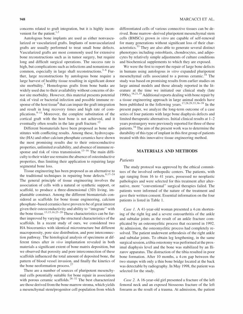
FIG. 2. Case 2. A 4 cm bone loss in the proximal ulna is evident
on the preoperative radiograph. At 2 months, callus formation is
observed by radiography at the interface between the host bone and
the HA cylinder. Seven months after surgery a complete integration
between the implant and the host bone together with extensive bone
formation throughout the implant occurred. The K-wire was still
positioned inside the medullary channel. At 2.5 years follow-up, the
complete bone-implant integration was maintained and there was
no evidence of implant fractures. CT scan analysis (2-D and 3-D
reconstructions) at 6 years follow-up demonstrated a complete
reconstruction of ulna with the presence of a medullary chan-
nel within the implant. No radiographic signs of bioceramic re-
absorbtion were detectable. Color images available online at www
.liebertpub.com/ten.
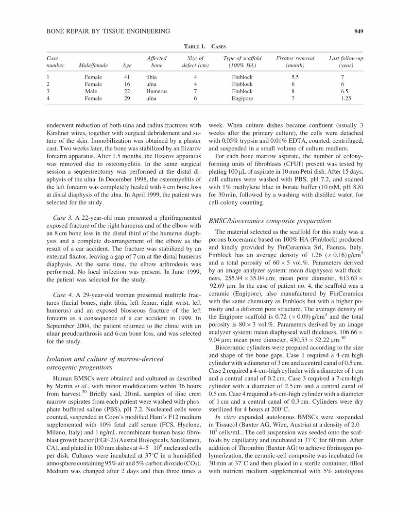
FIG. 3. Case 3. The preoperative radiograph shows a plurifrag-
mental complex fracture of elbow and humerus with 7 cm bone loss
in the distal humerus. At 2 months, an initial callus formation at the
bone-implant interface was observed. At 8 months, CT scan 2-D
analysis evidenced a callus formation along the HA cylinder. Neo-
formed bone was visible within the porous ceramics and a complete
healing of the implant to the host bone with no radiolucent osteo-
tomy line was evident. At 16 months, radiographs showed complete
graft incorporation within the humerus. Color images available
online at www.liebertpub.com/ten.
BONE REPAIR BY TISSUE ENGINEERING 951
For our study we selected clearly ‘‘challenging’’ patients,
thus validating the effectiveness of the treatment. In case 1,
the Ilizarov technique failed, while in cases 2, 3, and 4 the
Ilizarov technique was to be avoided due to the need of radius
resection and the high risk of nerve palsies and other com-
plications. In cases 1, 2, and 4, the bone loss was too extensive
to permit implantation of biomaterials not loaded with cells,
while an autograft, either vascularized or not vascularized,
would have caused a serious donor site morbidity. In case 3,
the particularly relevant bone substance loss made it virtually
impossible to use any type of autograft. In cases 1 and 2, full
functional recovery of the limbs was achieved 6.5 and 7
months after the implant surgery. The use of alternative
methods would have implied longer recovery times. In cases 3
and 4, the functional recovery took longer time due to the
unfavorable mechanical situation of the grafted area. In fact,
due to the insufficient mechanical properties of the initial
fixation, a second surgery to obtain a more stable fixation was
mandatory for patient recovery. Nevertheless, we consider the
obtained results very encouraging because, by a more tradi-
tional approach, the expected recovery time would have been
at least 12–18 months, in the most favorable hypothesis and in
the absence of complications.6
It is to note that no control group (only scaffold and no
cells) was used in our study. Selected patients were all
extremely difficult clinical cases to treat and we wanted to
guarantee all patients the best possible treatment and the
highest bone regeneration capacity of the implant. The crit-
ical role of BMSCs in promoting bone repair by a tissue
engineering approach was previously demonstrated by us in
animal models.32 Anyway, the lack of a control group re-
mains a drawback of our study.
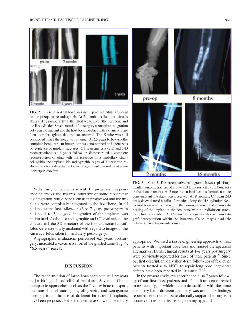
FIG. 4. Case 3. Analysis at 6 to 6.5 years follow-up. The ra-
diographic evaluation at 6 years demonstrated a good integration
of the implant within the host bone with complete healing of bone
loss (A). This finding was confirmed by 3D and 2D CT scans at 6
years (B, C). No evidence of reabsorption of the bioceramic was
present. An angiographic evaluation performed at 6.5 years fol-
low-up showed new vessel formation with complete vasculariza-
tion of the implant area (D, E).
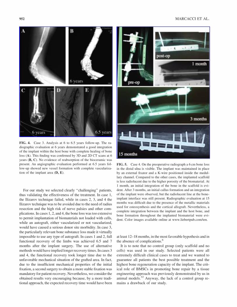
FIG. 5. Case 4. On the preoperative radiograph a 6 cm bone loss
in the distal ulna is visible. The implant was maintained in place
by an external fixator and a K-wire positioned inside the medul-
lary channel. Compared to the other cases, the implanted scaffold
is less radiolucent due to the higher porosity of the biomaterial. At
1 month, an initial integration of the bone in the scaffold is evi-
dent. After 3 months, an initial callus formation and an integration
of the implant were observed, but the radiolucent line at the bone-
implant interface was still present. Radiographic evaluation at 15
months was difficult due to the presence of the metallic materials
used for osteosynthesis and the cortical allograft. Nevertheless, a
complete integration between the implant and the host bone, and
bone formation throughout the implanted biomaterial were evi-
dent. Color images available online at www.liebertpub.com/ten.
952 MARCACCI ET AL.
Tissue engineering strategies involving the use of BMSCs
are based on the recognized degree of pluripotency of these
cells. BMSCs can be easily isolated from iliac crest bone
marrow aspirates. Nevertheless, a step of extensive in vivo
expansion is required to obtain the number of cells necessary
for reconstruction and repair of bone, given the low fre-
quency of BMSCs in marrow aspirates. Number of cells per
implant was chosen based on our previous experience with
the ovine model.32
BMSCs in culture undergo progressive replicative aging
and osteogenic differentiation, which are relevant to their
successful clinical use,43 and this should be considered when
designing cell-based therapies. Culture conditions that we
defined and utilized to expand the BMSCs of the patients
allowed us to obtain more than 20 cell doublings main-
taining the cell osteogenic potential. The age of the patients
selected for this study ranged between 16 and 41 years.
Performing cell culture in strictly controlled culture condi-
tions will be of particular relevance when expanding bone
marrow stromal cells from older patients where the number
of CFUf per mL of bone marrow aspirate is expected to be
lower.44,45 Hernigou et al. have reported the use of fresh
bone marrow directly injected into a nonunion lesion to ob-
tain bone healing, and they have evaluated the number and
concentration of progenitor cells in the marrow sample.46
Difference between the number of cells in the bone marrow
directly injected into nonunion defects and the number of
cells we have seeded onto the scaffold to be implanted de-
pends on dissimilarity in the number of osteogenic cells
(CFUf) present in these two cell populations.
BMSCs were seeded on 100% HA porous ceramics.
These scaffolds presented good osteoconductivity, resulting
in good functional recovery, but they were not resorbed after
more than 7 years postimplantation. It is important to un-
derline that, although no late fractures occurred in our pa-
tients, the permanence of biomaterial on long follow-up
could compromise the biomechanics of the new bone. In
addition, the high density of these scaffolds and, especially
in the case of the scaffolds used to treat the first three
patients, their relatively low porosity made radiographic
follow-up rather difficult as the ceramic was masking the
newly formed bone. The development of some second-
generation resorbable ceramic scaffolds is therefore essen-
tial before a tissue engineering approach to bone repair could
be widely applied in the clinical practice.
In a recent study we evaluated in an ovine model the
performance of implants of resorbable ceramic based on
silicon-stabilized tricalcium phosphate (Si-TCP) in promot-
ing the repair of critical-sized bone defects. A progressive
increase in new bone deposition into the pore of the scaffold
together with a reduction of the scaffold ceramic occurred
between 3 months and 1 year postsurgery. After 2 years the
scaffold was essentially completely resorbed. In a second
series of experiments, we compared the outcome of osteo-
genic cell–seeded implants versus unseeded implants in the
same ovine model system.47 Only BMSC-loaded ceramics
displayed a progressive scaffold resorption, coincident with
new bone deposition. To investigate the coupled mechanisms
of bone formation and scaffold resorption, X-ray computed
microtomography (mCT) and mX-ray diffraction analysis
were performed on BMSC-seeded small ceramic cubes im-
planted in immunodeficient mice for 2 or 6 months.48
In summary, based on the four patient outcomes some
general conclusions can be made:
1. The pattern of the bone healing process in the pa-
tients was similar to the one described in the large
animal model.18,19,28,29,32 The healing process can be
considered to occur in four main steps: (a) bone for-
mation on the outer surface of the implant, (b) bone
formation in the inner cylinder canal, (c) formation of
fissures and cracks in the implant body, and (d) bone
formation in the bioceramic pores. Radiography and
tomography showed that bone formation was far more
prominent over the external surface and within the
inner canal of the implants. This could be due to a
higher density of loaded cells and/or a better survival
of cells within the outermost portions of the HA
bioceramics. Alternatively, the implanted cells could
stimulate, via a paracrine or delayed paracrine mech-
anism, resident osteoprogenitor cells, located within
the skeletal tissues at the resected ends. At the last
follow-up, all patients, and in particular the three pa-
tients with a longer assessment, maintained a good in-
tegration of the implants and no late fractures were
observed. No major complaints were reported by the
patients, and no major adverse conditions were ob-
served.
2. A high porosity and a high degree of interconnection
between the pores are absolute requirements for vas-
cularization of the implant and new bone forma-
tion. Vascularization of the implant is certainly critical
for its survival and therefore its future stability. In
some animal studies, new vascularization of the graf-
ted area was obtained by a surgically created vascular
pedunculus.49,50 In our clinical study, the presence of
mesenchymal stem/progenitor cells was sufficient to
induce vascularization of the grafted area. An angio-
graphic examination was performed at the last follow-
up in the most challenging case. The detection of new
vessel growth into the implant confirmed the presence
of a vital bone in the grafted area.
3. In agreement with previous studies that implanted HA,
which is inert and remains within the body for ex-
tended periods, no visible signs of the biomaterial
reabsorption were detected as long as 6–7 years
postimplant. An ideal scaffold should provide an initial
support for bone-forming cells and then it should be
slowly reabsorbed at the same rate that the new bone is
deposited inside the scaffold pores. Future similar stud-
ies should consider the use of such types of scaffolds.
In recently performed animal trials, we established
BONE REPAIR BY TISSUE ENGINEERING 953
the feasibility of using implants of porous calcium
phosphate–based resorbable scaffolds to obtain site-
specific new bone formation in a large-sized bone
defect in a tibia sheep model.32,47
In conclusion, the aim of the present study was to analyze
the long-term durability of bone regeneration achieved by
a bone engineering approach. We observed progressive in-
tegration of the implants with the host bone, progressive new
bone formation inside the bioceramic pores, and progressive
vascular ingrowth. A good integration of the implants with
the preexisting bone was maintained during all the follow-up
periods and no major adverse conditions were observed.
In this pilot study, we used the tissue-engineered ap-
proach in very challenging cases with few therapeutical al-
ternatives. It is our opinion that the tissue engineering
approach for long bone reconstruction should be utilized
more widely in the future, thus avoiding autologous bone
harvesting or use of allogenic bone grafts. Presently, the
most important bias of this method is represented by the low
resorbability of porous HA bioceramics and in some cases
by a low mechanical stability of the implant. Use of better
bioresorbable constructs and application of a more stable
fixation should help to avoid these problems. Eventually,
controlled randomized clinical trials will have to clarify de-
finitively the effectiveness and the cost/benefit superiority
of the tissue engineering approach compared to other meth-
ods of bone reconstruction.
ACKNOWLEDGMENTS
The authors wish to thank FinCeramica Srl, Faenza, Italy,
for providing samples of ceramic scaffolds before they were
available on the market.
This work was supported by funds from the Italian Min-
istry of Instruction, University and Research (MIUR) and
from the European and the Italian Space Agencies (ESA &
ASI).
We wish to thank Dr. A. Montaperto for technical
assistance.
REFERENCES
1. Ilizarov, G.A. The tension-stress effect on the genesis and
growth of tissues: Part II. The influence of the rate and fre-
quency of distraction. Clin Orthop Relat Res 239, 263, 1989.
2. Goldstrohm, G.L., Mears, D.C., and Swartz, W.M. The results
of 39 fractures complicated by major segmental bone loss
and/or leg length discrepancy. J Trauma 24, 50, 1984.
3. Enneking, W.F., Eady, J.L., and Burchardt, H. Autogenous
cortical bone grafts in the reconstruction of segmental skeletal
defects. J Bone Joint Surg Am 62, 1039, 1980.
4. Taylor, G.I. The current status of free vascularized bone
grafts. Clin Plast Surg 10, 185, 1983.
5. Sowa, D.T., and Weiland, A.J. Clinical applications of vascu-
larized bone autografts. Orthop Clin North Am 18, 257, 1987.
6. Weiland, A.J., Moore, J.R., and Daniel, R.K. Vascularized
bone autografts. Experience with 41 cases. Clin Orthop Relat
Res 174, 87, 1983.
7. Laurie, S.W., Kaban, L.B., Mulliken, J.B., and Murray, J.E.
Donor-site morbidity after harvesting rib and iliac bone. Plast
Reconstr Surg 73, 933, 1984.
8. Stevenson, S. The immune response to osteochondral allo-
grafts in dogs. J Bone Joint Surg Am 69, 573, 1987.
9. Lord, C.F., Gebhardt, M.C., Tomford, W.W., and Mankin, H.J.
Infection in bone allografts. Incidence, nature, and treatment.
J Bone Joint Surg Am 70, 369, 1988.
10. Mankin, H.J., Gebhardt, M.C., and Tomford, W.W. The use
of frozen cadaveric allografts in the management of patients
with bone tumors of the extremities. Orthop Clin North Am
18, 275, 1987.
11. Alman, B.A., De Bari, A., and Krajbich, J.I. Massive allo-
grafts in the treatment of osteosarcoma and Ewing sarcoma in
children and adolescents. J Bone Joint Surg Am 77, 54, 1995.
12. Heiple, K.G., Goldberg, V.M., Powell, A.E., Bos, G.D., and
Zika, J.M. Biology of cancellous bone grafts. Orthop Clin
North Am 18, 179, 1987.
13. Heise, U., Osborn, J.F., and Duwe, F. Hydroxyapatite ceramic
as a bone substitute. Int Orthop 14, 329, 1990.
14. Oonishi, H. Orthopaedic applications of hydroxyapatite. Bio-
materials 12, 171, 1991.
15. Sartoris, D.J., Holmes, R.E., and Resnick, D. Coralline hy-
droxyapatite bone graft substitutes: radiographic evaluation.
J Foot Surg 31, 301, 1992.
16. Marcacci, M., Kon, E., Zaffagnini, S., Giardino, R., Rocca, M.,
Corsi, A., Benvenuti, A., Bianco, P., Quarto, R., Martin, I.,
Muraglia, A., and Cancedda, R. Reconstruction of extensive
long-bone defects in sheep using porous hydroxyapatite sponges.
Calcif Tissue Int 64, 83, 1999.
17. Ohgushi, H., Miyake, J., and Tateishi, T. Mesenchymal stem
cells and bioceramics: strategies to regenerate the skeleton.
Novartis Found Symp 249, 118, 2003.
18. Boyde, A., Corsi, A., Quarto, R., Cancedda, R., and Bianco,
P. Osteoconduction in large macroporous hydroxyapatite ce-
ramic implants: evidence for a complementary integration and
disintegration mechanism. Bone 24, 579, 1999.
19. Mnaymneh, W., Malinin, T.I., Makley, J.T., and Dick, H.M.
Massive osteoarticular allografts in the reconstruction of ex-
tremities following resection of tumors not requiring che-
motherapy and radiation. Clin Orthop Relat Res 76, 1985.
20. Brittberg, M., Lindahl, A., Nilsson, A., Ohlsson, C., Isaksson,
O., and Peterson, L. Treatment of deep cartilage defects in the
knee with autologous chondrocyte transplantation. N Engl J
Med 331, 889, 1994.
21. Krebsbach, P.H., Mankani, M.H., Satomura, K., Kuznetsov,
S.A., and Robey, P.G. Repair of craniotomy defects using
bone marrow stromal cells. Transplantation 66, 1272, 1998.
22. Matsumura, G., Hibino, N., Ikada, Y., Kurosawa, H., and
Shin’oka, T. Successful application of tissue engineered vas-
cular autografts: clinical experience. Biomaterials 24, 2303,
2003.
23. Dezawa, M., Hoshino, M., Nabeshima, Y., and Ide, C. Mar-
row stromal cells: implications in health and disease in the
nervous system. Curr Mol Med 5, 723, 2005.
24. Brodie, J.C., and Humes, H.D. Stem cell approaches for the
treatment of renal failure. Pharmacol Rev 57, 299, 2005.
954 MARCACCI ET AL.

25. Elsinger, E.C., and Leal, L. Coralline hydroxyapatite bone
graft substitutes. J Foot Ankle Surg 35, 396, 1996.
26. Ge, Z., Baguenard, S., Lim, L.Y., Wee, A., and Khor, E.
Hydroxyapatite-chitin materials as potential tissue engineered
bone substitutes. Biomaterials 25, 1049, 2004.
27. Mastrogiacomo, M., Scaglione, S., Martinetti, R., Dolcini, L.,
Beltrame, F., Cancedda, R., and Quarto, R. Role of scaffold
internal structure on in vivo bone formation in macroporous
calcium phosphate bioceramics. Biomaterials 27, 3230, 2006.
28. Kruyt, M.C., Dhert, W.J., Oner, C., van Blitterswijk, C.A.,
Verbout, A.J., and de Bruijn, J.D. Optimization of bone-tissue
engineering in goats. J Biomed Mater Res B Appl Biomater
69, 113, 2004.
29. Zhu, L., Liu, W., Cui, L., and Cao, Y. Tissue-engineered bone
repair of goat-femur defects with osteogenically induced bone
marrow stromal cells. Tissue Eng 12, 423, 2006.
30. Martin, I., Muraglia, A., Campanile, G., Cancedda, R., and
Quarto, R. Fibroblast growth factor-2 supports ex vivo ex-
pansion and maintenance of osteogenic precursors from hu-
man bone marrow. Endocrinology 138, 4456, 1997.
31. Noshi, T., Yoshikawa, T., Ikeuchi, M., Dohi, Y., Ohgushi, H.,
Horiuchi, K., Sugimura, M., Ichijima, K., and Yonemasu, K.
Enhancement of the in vivo osteogenic potential of marrow/
hydroxyapatite composites by bovine bone morphogenetic
protein. J Biomed Mater Res 52, 621, 2000.
32. Kon, E., Muraglia, A., Corsi, A., Bianco, P., Marcacci, M.,
Martin, I., Boyde, A., Ruspantini, I., Chistolini, P., Rocca, M.,
Giardino, R., Cancedda, R., and Quarto, R. Autologous bone
marrow stromal cells loaded onto porous hydroxyapatite ce-
ramic accelerate bone repair in critical-size defects of sheep
long bones. J Biomed Mater Res 49, 328, 2000.
33. Cancedda, R., Mastrogiacomo, M., Bianchi, G., Derubeis, A.,
Muraglia, A., and Quarto, R. Bone marrow stromal cells and
their use in regenerating bone. Novartis Found Symp 249, 133,
2003.
34. Quarto, R., Mastrogiacomo, M., Cancedda, R., Kutepov,
S.M., Mukhachev, V., Lavroukov, A., Kon, E., and Marcacci,
M. Repair of large bone defects with the use of autologous
bone marrow stromal cells. N Engl J Med 344, 385, 2001.
35. Bruder, S.P., Jaiswal, N., Ricalton, N.S., Mosca, J.D., Kraus,
K.H., and Kadiyala, S. Mesenchymal stem cells in osteobi-
ology and applied bone regeneration. Clin Orthop Relat Res
355 Suppl, S247, 1998.
36. Xiao, Y., Qian, H., Young, W.G., and Bartold, P.M. Tissue
engineering for bone regeneration using differentiated alveolar
bone cells in collagen scaffolds. Tissue Eng 9, 1167, 2003.
37. Livingston, T., Ducheyne, P., and Garino, J. In vivo evaluation
of a bioactive scaffold for bone tissue engineering. J Biomed
Mater Res 62, 1, 2002.
38. Warren, S.M., Nacamuli, R.K., Song, H.M., and Longaker,
M.T. Tissue-engineered bone using mesenchymal stem cells
and a biodegradable scaffold. J Craniofac Surg 15, 34, 2004.
39. Logeart-Avramoglou, D., Anagnostou, F., Bizios, R., and
Petite, H. Engineering bone: challenges and obstacles. J Cell
Mol Med 9, 72, 2005.
40. Martinetti, R., Mastrogiacomo, M., Cancedda, R., and Peyrin,
F. SEM and synchrotron radiation microtomography in the
study of bioceramic scaffolds for tissue engineering applica-
tions. Biotechnol Bioeng, in press.
41. Kitoh, H., Kitakoji, T., Tsuchiya, H., Mitsuyama, H., Naka-
mura, H., Katoh, M., and Ishiguro, N. Transplantation of
marrow-derived mesenchymal stem cells and platelet-rich
plasma during distraction osteogenesis—a preliminary result
of three cases. Bone 35, 892, 2004.
42. Vacanti, C.A., Bonassar, L.J., Vacanti, M.P., and Shuf-
flebarger, J. Replacement of an avulsed phalanx with tissue-
engineered bone. N Engl J Med 344, 1511, 2001.
43. Banfi, A., Bianchi, G., Galotto, M., Cancedda, R., and Quarto,
R. Bone marrow stromal damage after chemo/radiotherapy:
occurrence, consequences and possibilities of treatment. Leuk
Lymphoma 42, 863, 2001.
44. Galotto, M., Berisso, G., Delfino, L., Podesta, M., Ottaggio, L.,
Dallorso, S., Dufour, C., Ferrara, G.B., Abbondandolo, A., Dini,
G., Bacigalupo, A., Cancedda, R., and Quarto, R. Stromal
damage as consequence of high-dose chemo/radiotherapy in
bone marrow transplant recipients. Exp Hematol 27, 1460, 1999.
45. Stenderup, K., Justesen, J., Clausen, C., and Kassem, M.
Aging is associated with decreased maximal life span and
accelerated senescence of bone marrow stromal cells. Bone
33, 919, 2003.
46. Hernigou, P., Mathieu, G., Poignard, A., Manicom, O.,
Beaujean, F., and Rouard, H. Percutaneous autologous bone-
marrow grafting for nonunions. Surgical technique. J Bone
Joint Surg Am 88 Suppl 1 Pt 2, 322, 2006.
47. Mastrogiacomo, M., Corsi, A., Francioso, E., Di Comite, M.,
Monetti, F., Scaglione, S., Favia, A., Crovace, A., Bianco, P.,
and Cancedda, R. Reconstruction of extensive long bone
defects in sheep using resorbable bioceramics based on silicon
stabilized tricalcium phosphate. Tissue Eng 12, 1261, 2006.
48. Mastrogiacomo, M., Papadimitropoulos, A., Cedola, A.,
Peyrin, F., Giannoni, P., Pearce, S.G., Alini, M., Giannini, C.,
Guagliardi, A., and Cancedda, R. Engineering of bone using
bone marrow stromal cells and a silicon-stabilized tricalcium
phosphate bioceramic: evidence for a coupling between bone
formation and scaffold resorption. Biomaterials 28, 1376,
2007. Epub 2006 Nov 28.
49. Nakasa, T., Ishida, O., Sunagawa, T., Nakamae, A., Yasu-
naga, Y., Agung, M., and Ochi, M. Prefabrication of vascu-
larized bone graft using a combination of fibroblast growth
factor-2 and vascular bundle implantation into a novel inter-
connected porous calcium hydroxyapatite ceramic. J Biomed
Mater Res A 75, 350, 2005.
50. Akita, S., Tamai, N., Myoui, A., Nishikawa, M., Kaito, T.,
Takaoka, K., and Yoshikawa, H. Capillary vessel network
integration by inserting a vascular pedicle enhances bone
formation in tissue-engineered bone using interconnected
porous hydroxyapatite ceramics. Tissue Eng 10, 789, 2004.
Address reprint requests to:
Dott.ssa Elizaveta Kon
Laboratorio di Biomeccanica
Istituti Ortopedici Rizzoli
Via di Barbiano, 1/10
40136 Bologna
Italy
E-mail: [email protected]
BONE REPAIR BY TISSUE ENGINEERING 955