Stem Cell Reports Shor t Ar ticle Sustained ERK Activation Underlies Reprogramming in Regeneration-...
17
Stem Cell Reports Short Article Sustained ERK Activation Underlies Reprogramming in Regeneration- Competent Salamander Cells and Distinguishes Them from Their Mammalian Counterparts Maximina H. Yun, 1, * Phillip B. Gates, 1 and Jeremy P. Brockes 1 1 Division of Biosciences, Institute of Structural and Molecular Biology, University College London, Gower Street, London WC1E 6BT, UK *Correspondence: [email protected] http://dx.doi.org/10.1016/j.stemcr.2014.05.009 This is an open access article under the CC BY-NC-ND license (http://creativecommons.org/licenses/by-nc-nd/3.0/). SUMMARY In regeneration-competent vertebrates, such as salamanders, regeneration depends on the ability of various differentiated adult cell types to undergo natural reprogramming. This ability is rarely observed in regeneration-incompetent species such as mammals, providing an explanation for their poor regenerative potential. To date, little is known about the molecular mechanisms mediating natural reprogram- ming during regeneration. Here, we have identified the extent of extracellular signal-regulated kinase (ERK) activation as a key compo- nent of such mechanisms. We show that sustained ERK activation following serum induction is required for re-entry into the cell cycle of postmitotic salamander muscle cells, partially by promoting the downregulation of p53 activity. Moreover, ERK activation induces epige- netic modifications and downregulation of muscle-specific genes such as Sox6. Remarkably, while long-term ERK activation is found in salamander myotubes, only transient activation is seen in their mammalian counterparts, suggesting that the extent of ERK activation could underlie differences in regenerative competence between species. INTRODUCTION In most vertebrates, the process of myogenic differentia- tion entails the withdrawal of precursors from the cell cycle, followed by their fusion into myotubes. The multi- nucleate state is characterized by a permanent postmitotic arrest, which renders the myotubes unable to respond to proliferative cues (Pajalunga et al., 2008; Walsh and Perl- man, 1997). In contrast, salamander myotubes remain responsive to such cues, being able to re-enter the cell cycle upon serum stimulation in culture (Tanaka et al., 1997) or after implantation within regenerating structures (Kumar et al., 2000). In salamander (Notophthalmus viridescens) A1 myotubes (Ferretti and Brockes, 1988), serum stimulation induces a reprogramming process that includes partial dedifferentia- tion, as suggested by the downregulation of the myogenic gene Myf5 (Imokawa et al., 2004), and re-entry into the cell cycle, which is also considered an aspect of dedifferentia- tion. The latter depends on the phosphorylation of Rb (Tanaka et al., 1997) and the downregulation of p53 activ- ity (Yun et al., 2013). The serum component that triggers these responses is not a conventional growth factor but an as-yet-unidentified thrombin-activated serum compo- nent that acts as a mitogen for myotubes, but not for mononucleate precursors (Lo ¨o ¨f et al., 2007; Straube et al., 2004; Tanaka et al., 1999). Even though mammalian myo- tube nuclei cannot be reprogrammed upon exposure to this factor (Lo ¨o ¨f et al., 2007), they are able to re-enter the cell cycle after forming heterokaryons with salamander myo- tubes (Velloso et al., 2001). This suggests that even when the initial response may be different, part of the pathway leading to serum-mediated reprogramming is conserved. Both the identity of the serum factor and the signaling pathways driving the reversal of the differentiated state in regeneration-competent salamander cells remain un- known, although extensive efforts to identify the serum factor are ongoing (Straube et al., 2004). In proliferating cells, the extracellular signal-regulated kinase (ERK) family of mitogen-activated protein kinases (MAPKs) plays a critical role in driving cell-cycle progres- sion in a variety of cell types (Albeck et al., 2013; Cook and McCormick, 1996; Murphy et al., 2002; Weber et al., 1997; Yamamoto et al., 2006). In fibroblasts, sustained ERK activation is required for successful S phase progres- sion by promoting the downregulation of antiproliferative genes during G1 phase and controlling the state of Rb phosphorylation (Yamamoto et al., 2006). Hence, it is possible that ERK activation plays a role during the reprog- ramming of differentiated salamander cells. Herein, we have tested this hypothesis using the salamander A1 cell line as a model for serum-induced reprogramming. RESULTS AND DISCUSSION Sustained ERK Activation in Cell-Cycle Re-entry of Salamander Myotubes Serum stimulation of A1 myotubes triggers an early activa- tion of the ERK pathway, which is sustained for up to 48 hr post stimulation (Figures 1A and 1B). This is accompanied by a long-term increase in the protein levels of c-FOS Stem Cell Reports j Vol. 3 j 1–9 j July 8, 2014 j ª2014 The Authors 1 Please cite this article in press as: Yun et al., Sustained ERK Activation Underlies Reprogramming in Regeneration-Competent Salamander Cells and Distinguishes Them from..., Stem Cell Reports (2014), http://dx.doi.org/10.1016/j.stemcr.2014.05.009
-
Upload
independent -
Category
Documents
-
view
1 -
download
0
Transcript of Stem Cell Reports Shor t Ar ticle Sustained ERK Activation Underlies Reprogramming in Regeneration-...

Please cite this article in press as: Yun et al., Sustained ERK Activation Underlies Reprogramming in Regeneration-Competent SalamanderCells and Distinguishes Them from..., Stem Cell Reports (2014), http://dx.doi.org/10.1016/j.stemcr.2014.05.009
Stem Cell Reports
Short ArticleSustained ERK Activation Underlies Reprogramming in Regeneration-Competent Salamander Cells and Distinguishes Them from Their MammalianCounterparts
Maximina H. Yun,1,* Phillip B. Gates,1 and Jeremy P. Brockes11Division of Biosciences, Institute of Structural and Molecular Biology, University College London, Gower Street, London WC1E 6BT, UK
*Correspondence: [email protected]
http://dx.doi.org/10.1016/j.stemcr.2014.05.009
This is an open access article under the CC BY-NC-ND license (http://creativecommons.org/licenses/by-nc-nd/3.0/).
SUMMARY
In regeneration-competent vertebrates, such as salamanders, regeneration depends on the ability of various differentiated adult cell types
to undergo natural reprogramming. This ability is rarely observed in regeneration-incompetent species such as mammals, providing an
explanation for their poor regenerative potential. To date, little is known about themolecularmechanismsmediating natural reprogram-
ming during regeneration. Here, we have identified the extent of extracellular signal-regulated kinase (ERK) activation as a key compo-
nent of suchmechanisms.We show that sustained ERK activation following serum induction is required for re-entry into the cell cycle of
postmitotic salamandermuscle cells, partially by promoting the downregulation of p53 activity. Moreover, ERK activation induces epige-
netic modifications and downregulation of muscle-specific genes such as Sox6. Remarkably, while long-term ERK activation is found in
salamander myotubes, only transient activation is seen in their mammalian counterparts, suggesting that the extent of ERK activation
could underlie differences in regenerative competence between species.
INTRODUCTION
In most vertebrates, the process of myogenic differentia-
tion entails the withdrawal of precursors from the cell
cycle, followed by their fusion into myotubes. The multi-
nucleate state is characterized by a permanent postmitotic
arrest, which renders the myotubes unable to respond to
proliferative cues (Pajalunga et al., 2008; Walsh and Perl-
man, 1997). In contrast, salamander myotubes remain
responsive to such cues, being able to re-enter the cell cycle
upon serum stimulation in culture (Tanaka et al., 1997) or
after implantation within regenerating structures (Kumar
et al., 2000).
In salamander (Notophthalmus viridescens) A1 myotubes
(Ferretti and Brockes, 1988), serum stimulation induces a
reprogramming process that includes partial dedifferentia-
tion, as suggested by the downregulation of the myogenic
geneMyf5 (Imokawa et al., 2004), and re-entry into the cell
cycle, which is also considered an aspect of dedifferentia-
tion. The latter depends on the phosphorylation of Rb
(Tanaka et al., 1997) and the downregulation of p53 activ-
ity (Yun et al., 2013). The serum component that triggers
these responses is not a conventional growth factor but
an as-yet-unidentified thrombin-activated serum compo-
nent that acts as a mitogen for myotubes, but not for
mononucleate precursors (Loof et al., 2007; Straube et al.,
2004; Tanaka et al., 1999). Even though mammalian myo-
tube nuclei cannot be reprogrammedupon exposure to this
factor (Loof et al., 2007), they are able to re-enter the cell
cycle after forming heterokaryons with salamander myo-
tubes (Velloso et al., 2001). This suggests that even when
the initial response may be different, part of the pathway
leading to serum-mediated reprogramming is conserved.
Both the identity of the serum factor and the signaling
pathways driving the reversal of the differentiated state
in regeneration-competent salamander cells remain un-
known, although extensive efforts to identify the serum
factor are ongoing (Straube et al., 2004).
In proliferating cells, the extracellular signal-regulated
kinase (ERK) family of mitogen-activated protein kinases
(MAPKs) plays a critical role in driving cell-cycle progres-
sion in a variety of cell types (Albeck et al., 2013; Cook
and McCormick, 1996; Murphy et al., 2002; Weber et al.,
1997; Yamamoto et al., 2006). In fibroblasts, sustained
ERK activation is required for successful S phase progres-
sion by promoting the downregulation of antiproliferative
genes during G1 phase and controlling the state of Rb
phosphorylation (Yamamoto et al., 2006). Hence, it is
possible that ERK activation plays a role during the reprog-
ramming of differentiated salamander cells. Herein, we
have tested this hypothesis using the salamander A1 cell
line as a model for serum-induced reprogramming.
RESULTS AND DISCUSSION
Sustained ERK Activation in Cell-Cycle Re-entry of
Salamander Myotubes
Serum stimulation of A1 myotubes triggers an early activa-
tion of the ERK pathway, which is sustained for up to 48 hr
post stimulation (Figures 1A and 1B). This is accompanied
by a long-term increase in the protein levels of c-FOS
Stem Cell Reports j Vol. 3 j 1–9 j July 8, 2014 j ª2014 The Authors 1

(legend on next page)
2 Stem Cell Reports j Vol. 3 j 1–9 j July 8, 2014 j ª2014 The Authors
Stem Cell ReportsERK Activation in Reprogramming
Please cite this article in press as: Yun et al., Sustained ERK Activation Underlies Reprogramming in Regeneration-Competent SalamanderCells and Distinguishes Them from..., Stem Cell Reports (2014), http://dx.doi.org/10.1016/j.stemcr.2014.05.009
Stem Cell ReportsERK Activation in Reprogramming
Please cite this article in press as: Yun et al., Sustained ERK Activation Underlies Reprogramming in Regeneration-Competent SalamanderCells and Distinguishes Them from..., Stem Cell Reports (2014), http://dx.doi.org/10.1016/j.stemcr.2014.05.009
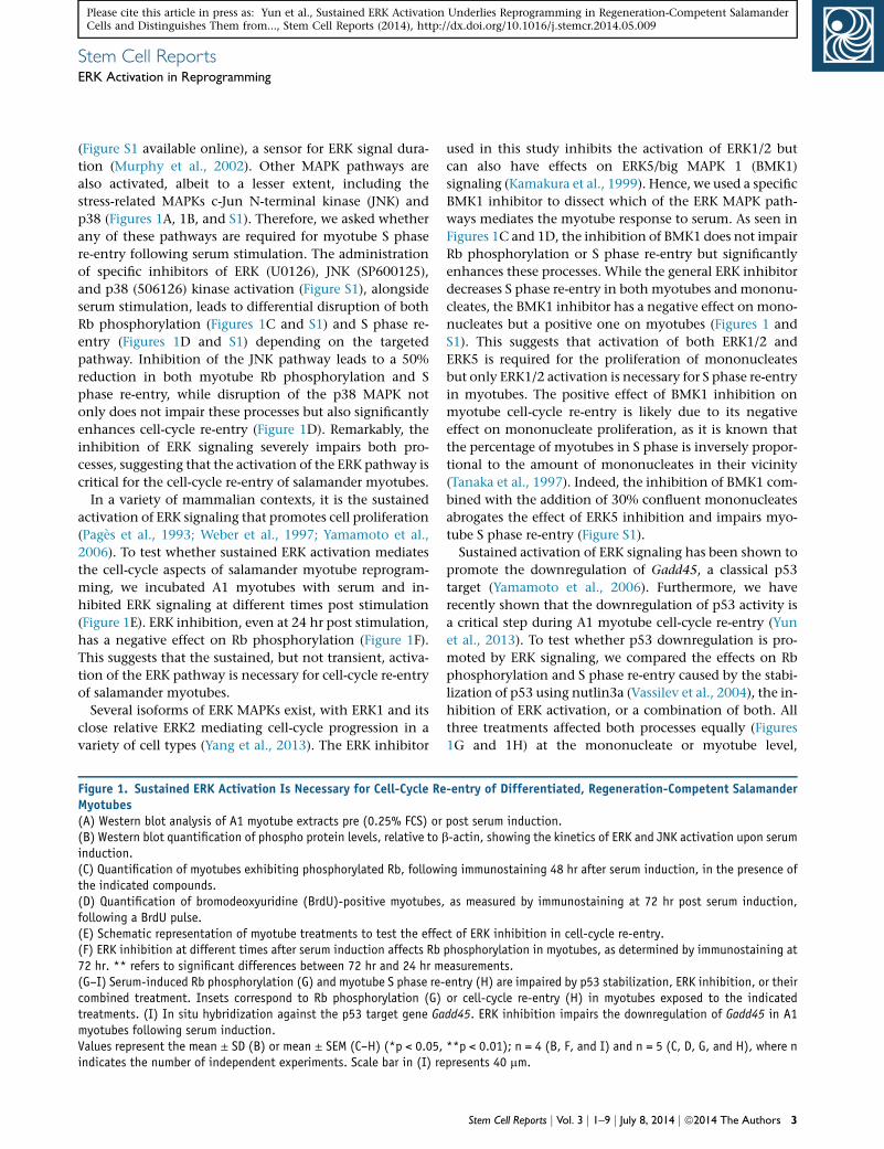
(Figure S1 available online), a sensor for ERK signal dura-
tion (Murphy et al., 2002). Other MAPK pathways are
also activated, albeit to a lesser extent, including the
stress-related MAPKs c-Jun N-terminal kinase (JNK) and
p38 (Figures 1A, 1B, and S1). Therefore, we asked whether
any of these pathways are required for myotube S phase
re-entry following serum stimulation. The administration
of specific inhibitors of ERK (U0126), JNK (SP600125),
and p38 (506126) kinase activation (Figure S1), alongside
serum stimulation, leads to differential disruption of both
Rb phosphorylation (Figures 1C and S1) and S phase re-
entry (Figures 1D and S1) depending on the targeted
pathway. Inhibition of the JNK pathway leads to a 50%
reduction in both myotube Rb phosphorylation and S
phase re-entry, while disruption of the p38 MAPK not
only does not impair these processes but also significantly
enhances cell-cycle re-entry (Figure 1D). Remarkably, the
inhibition of ERK signaling severely impairs both pro-
cesses, suggesting that the activation of the ERK pathway is
critical for the cell-cycle re-entry of salamander myotubes.
In a variety of mammalian contexts, it is the sustained
activation of ERK signaling that promotes cell proliferation
(Pages et al., 1993; Weber et al., 1997; Yamamoto et al.,
2006). To test whether sustained ERK activation mediates
the cell-cycle aspects of salamander myotube reprogram-
ming, we incubated A1 myotubes with serum and in-
hibited ERK signaling at different times post stimulation
(Figure 1E). ERK inhibition, even at 24 hr post stimulation,
has a negative effect on Rb phosphorylation (Figure 1F).
This suggests that the sustained, but not transient, activa-
tion of the ERK pathway is necessary for cell-cycle re-entry
of salamander myotubes.
Several isoforms of ERK MAPKs exist, with ERK1 and its
close relative ERK2 mediating cell-cycle progression in a
variety of cell types (Yang et al., 2013). The ERK inhibitor
Figure 1. Sustained ERK Activation Is Necessary for Cell-Cycle ReMyotubes(A) Western blot analysis of A1 myotube extracts pre (0.25% FCS) or(B) Western blot quantification of phospho protein levels, relative to binduction.(C) Quantification of myotubes exhibiting phosphorylated Rb, followithe indicated compounds.(D) Quantification of bromodeoxyuridine (BrdU)-positive myotubes,following a BrdU pulse.(E) Schematic representation of myotube treatments to test the effe(F) ERK inhibition at different times after serum induction affects Rb72 hr. ** refers to significant differences between 72 hr and 24 hr m(G–I) Serum-induced Rb phosphorylation (G) and myotube S phase re-combined treatment. Insets correspond to Rb phosphorylation (G)treatments. (I) In situ hybridization against the p53 target gene Gamyotubes following serum induction.Values represent the mean ± SD (B) or mean ± SEM (C–H) (*p < 0.05,indicates the number of independent experiments. Scale bar in (I) re
used in this study inhibits the activation of ERK1/2 but
can also have effects on ERK5/big MAPK 1 (BMK1)
signaling (Kamakura et al., 1999). Hence, we used a specific
BMK1 inhibitor to dissect which of the ERK MAPK path-
ways mediates the myotube response to serum. As seen in
Figures 1C and 1D, the inhibition of BMK1 does not impair
Rb phosphorylation or S phase re-entry but significantly
enhances these processes. While the general ERK inhibitor
decreases S phase re-entry in both myotubes and mononu-
cleates, the BMK1 inhibitor has a negative effect on mono-
nucleates but a positive one on myotubes (Figures 1 and
S1). This suggests that activation of both ERK1/2 and
ERK5 is required for the proliferation of mononucleates
but only ERK1/2 activation is necessary for S phase re-entry
in myotubes. The positive effect of BMK1 inhibition on
myotube cell-cycle re-entry is likely due to its negative
effect on mononucleate proliferation, as it is known that
the percentage of myotubes in S phase is inversely propor-
tional to the amount of mononucleates in their vicinity
(Tanaka et al., 1997). Indeed, the inhibition of BMK1 com-
bined with the addition of 30% confluent mononucleates
abrogates the effect of ERK5 inhibition and impairs myo-
tube S phase re-entry (Figure S1).
Sustained activation of ERK signaling has been shown to
promote the downregulation of Gadd45, a classical p53
target (Yamamoto et al., 2006). Furthermore, we have
recently shown that the downregulation of p53 activity is
a critical step during A1 myotube cell-cycle re-entry (Yun
et al., 2013). To test whether p53 downregulation is pro-
moted by ERK signaling, we compared the effects on Rb
phosphorylation and S phase re-entry caused by the stabi-
lization of p53 using nutlin3a (Vassilev et al., 2004), the in-
hibition of ERK activation, or a combination of both. All
three treatments affected both processes equally (Figures
1G and 1H) at the mononucleate or myotube level,
-entry of Differentiated, Regeneration-Competent Salamander
post serum induction.-actin, showing the kinetics of ERK and JNK activation upon serum
ng immunostaining 48 hr after serum induction, in the presence of
as measured by immunostaining at 72 hr post serum induction,
ct of ERK inhibition in cell-cycle re-entry.phosphorylation in myotubes, as determined by immunostaining ateasurements.entry (H) are impaired by p53 stabilization, ERK inhibition, or theiror cell-cycle re-entry (H) in myotubes exposed to the indicateddd45. ERK inhibition impairs the downregulation of Gadd45 in A1
**p < 0.01); n = 4 (B, F, and I) and n = 5 (C, D, G, and H), where npresents 40 mm.
Stem Cell Reports j Vol. 3 j 1–9 j July 8, 2014 j ª2014 The Authors 3

Stem Cell ReportsERK Activation in Reprogramming
Please cite this article in press as: Yun et al., Sustained ERK Activation Underlies Reprogramming in Regeneration-Competent SalamanderCells and Distinguishes Them from..., Stem Cell Reports (2014), http://dx.doi.org/10.1016/j.stemcr.2014.05.009
suggesting that the downregulation of p53 activity and the
activation of ERK signaling form part of the same pathway.
We additionally observed that while treatment of myo-
tubes with the p53 inhibitor a-pifithrin did not affect Rb
phosphorylation or cell-cycle re-entry, the combined inhi-
bition of ERK signaling and p53 had a negative effect on
this processes. However, the joint inhibition of ERK and
p53 significantly increased the percentage of myotubes
positive for Rb phosphorylation and re-entering the cell
cycle, when compared with the inhibition of ERK signaling
on its own (Figures 1G and 1H). This suggests that ERK
signaling is required for cell-cycle re-entry by, at least in
part, promoting the downregulation of p53 activity. To
test this further, we investigated the effect of inhibiting
different MAPKs on the regulation of Gadd45 gene expres-
sion by in situ hybridization. ERK inhibition abrogated the
downregulation of Gadd45 induced by serum stimulation
(Figure 1I), suggesting that the activation of the ERK
pathway promotes cell-cycle re-entry in salamander myo-
tubes by downregulating p53 activity.
ERK Activation Promotes Phenotypic Changes in
Salamander Myotubes
A second aspect of the response to serum stimulation in sal-
amander myotubes is the promotion of transcriptional
changes that lead to partial dedifferentiation. As ERK acti-
vation is required at such an early stage of the response, it
is plausible that it contributes to the promotion of dediffer-
entiation as well as cell-cycle progression. To address this
possibility we cloned the newt homolog of the muscle-spe-
cific gene Sox6 (Hagiwara et al., 2007) (Figure S2) and exam-
ined its expression in purified myotube cultures. While
serum stimulation leads to a 50% reduction of the relative
levels of Sox6 (Figure 2A), ERK inhibition abrogates this
effect, suggesting that the downregulation of Sox6 upon
serum stimulation is dependent on ERK activation. This
observation is consistent with a previous study suggesting
that ERK activation drives the dedifferentiation of rodent
Schwann cells (Harrisingh et al., 2004). While the expres-
sion of Sox6 changed upon serum stimulation, we did not
detect any changes in the protein levels of muscle-specific
myosin heavy chain under similar conditions (Figure 2C).
This is consistent with the notion that serum promotes a
limited dedifferentiation process in regeneration-compe-
tent salamander myotubes.
Furthermore, we noticed that there are large-scale epige-
netic changes in the A1 myotubes following serum stimu-
lation. Immunostaining against the repressive histone
mark dimethyl H3K9 (Jenuwein and Allis, 2001) revealed
a decrease in signal intensity in myotubes upon serum
stimulation, consistent with previous observations (Loof
et al., 2007). Interestingly, this decrease is abrogated only
by ERK, but not by JNK or BMK1 inhibition (Figure 2B).
4 Stem Cell Reports j Vol. 3 j 1–9 j July 8, 2014 j ª2014 The Authors
Western blot analysis of purified myotube extracts indi-
cates that there is a 50% decrease in dimethyl H3K9 upon
serum stimulation but only a 20% decrease when ERK
signaling is inhibited (Figure 2C). This is particularly inter-
esting for two reasons. First, the demethylation of H3K9 at
E2F-regulated promoters is required for the progression
into S phase (Ogawa et al., 2002). Second, H3K9 demethy-
lation is required for the expression of pluripotency-associ-
ated genes and the maintenance of embryonic stem cells
(Loh et al., 2007).
In the light of these observations, it is possible that the
changes in H3K9 demethylation, which we show to be
dependent on ERK activation, generate a favorable envi-
ronment for altering gene expression in the direction of
partial dedifferentiation and cell-cycle progression in
regeneration-competent cells.
Lack of Sustained ERK Activation in Regeneration-
Incompetent Mammalian Myotubes
While salamander myotubes are able to respond to serum
stimulation by promoting a signaling cascade leading to
partial dedifferentiation and cell-cycle re-entry, mamma-
lian myotubes are unable to do so (Tanaka et al., 1997).
Nevertheless, the latter appear to have retained some
responsiveness, as serum stimulation (Tiainen et al.,
1996) or incubation with partially purified serum factor
preparations (Loof et al., 2007) leads to the upregulation
of immediate-early proliferative response genes such as
c-fos. However, the subsequent steps in the pathway lead-
ing to reprogramming do not take place. In the light of
our observations, this suggests that altered or absent ERK
activation could underlie the lack of responsiveness found
in mammalian myotubes.
We explored this possibility using the murine myogenic
cell line Pmi28 (Figure 3A), which can be induced to form
myotubes that cannot dedifferentiate or re-enter the cell
cycle upon serum stimulation (Duckmanton et al.,
2005). Remarkably, we found that serum stimulation of
Pmi28 myotubes leads to transient ERK1/2 activation,
which peaks at 1 hr post induction and returns to baseline
in approximately 3 hr (Figures 3B and 3C). In this system,
the intensity of ERK activation is considerably lower
than in the induced salamander myotubes (Figure 3C).
Furthermore, unlike A1 myotubes, stimulated Pmi28
myotubes do not activate JNK and do not exhibit any
changes in the total amounts of the repression marker
dimethyl H3K9 (Figure 3D). However, serum stimulation
promotes the upregulation of c-FOS and an increase in
cyclin D1 and D3, which depend on the transient activa-
tion of ERK (Figures 3D and 3E). This suggests that the
transient response to serum previously observed in
mammalian myotubes is mediated by ERK signaling.
Nonetheless, consistent with previous observations (Loof

Figure 2. ERK Activation Leads to Downregulation of Muscle-Specific Gene Expression and Promotes Epigenetic Changes duringReprogramming of Salamander Myotubes(A) Left: schematic representation of the variation in Sox6mRNA levels upon myogenic differentiation. Right: quantitative RT-PCR analysisof Sox6 (with two different primers, Sox6 1 and Sox6 2) in RNA extracts from purified A1 myotubes, normalized to Ef1-a or L27.(B) Representative images of myotubes following immunostaining against dimethylated H3K9 and myosin heavy chain (MyHC) 1.5 daysafter the indicated treatments. Scale bar represents 50 mm.(C) Western blot analysis of purified myotube extracts. Note the decrease in dimetH3K9 upon serum induction and its impairment when ERKsignaling is inhibited. The relative amount of dimethylated H3K9 was quantified for the indicated treatments.Values represent the mean ± SEM (*p < 0.05); n = 4 (A–C), where n indicates the number of independent experiments.
Stem Cell ReportsERK Activation in Reprogramming
Please cite this article in press as: Yun et al., Sustained ERK Activation Underlies Reprogramming in Regeneration-Competent SalamanderCells and Distinguishes Them from..., Stem Cell Reports (2014), http://dx.doi.org/10.1016/j.stemcr.2014.05.009
et al., 2007; Tiainen et al., 1996), we did not observe any
changes in late proliferative genes such as Cyclin E2 and
PCNA (Figure 3E). Together, these observations suggest
that the extent of ERK signaling could underlie differences
in regenerative capabilities between salamander and
mammalian cells.
Stem Cell Reports j Vol. 3 j 1–9 j July 8, 2014 j ª2014 The Authors 5

Figure 3. Regeneration-Incompetent Mammalian Myotubes Do Not Trigger Sustained ERK Activation upon Serum Stimulation(A) Representative image of Pmi28 myotubes 3 days after myogenesis. Nuclei are stained with Hoechst 33258 (blue). Scale bar represents100 mm.(B, C, and E) Western blot analysis of Pmi28 myotube extracts pre (6% HS) or at different times after induction with 20% FCS.(D) Differential kinetics of ERK activation in mammalian versus salamander myotubes at different times after serum induction followingwestern blot quantification of relative levels of phosphorylated ERK.(F) Western blot analysis of whole-cell extracts from myotubes overexpressing pnGFPN2 (GFPN2), MEK1-R4F (R4F), andMEK1S218E/S222D(SE/SD). The relative amount of c-fos and PCNA was quantified for the indicated treatments.(G) A model for the role of ERK1/2 activation during serum-induced reprogramming of regeneration-competent salamander myotubescompared to their mammalian counterparts.Values represent the mean ± SD (*p < 0.05, **p < 0.01); n = 4 (B–F), where n indicates the number of independent experiments.
6 Stem Cell Reports j Vol. 3 j 1–9 j July 8, 2014 j ª2014 The Authors
Stem Cell ReportsERK Activation in Reprogramming
Please cite this article in press as: Yun et al., Sustained ERK Activation Underlies Reprogramming in Regeneration-Competent SalamanderCells and Distinguishes Them from..., Stem Cell Reports (2014), http://dx.doi.org/10.1016/j.stemcr.2014.05.009

Stem Cell ReportsERK Activation in Reprogramming
Please cite this article in press as: Yun et al., Sustained ERK Activation Underlies Reprogramming in Regeneration-Competent SalamanderCells and Distinguishes Them from..., Stem Cell Reports (2014), http://dx.doi.org/10.1016/j.stemcr.2014.05.009
Finally, we tested whether the artificial promotion of
ERK activation could promote cell-cycle re-entry in this sys-
tem. Overexpression of two different constitutively active
ERK activator kinase 1 (MEK1) mutants, MEK1-R4F and
MEK1S218E/S222D (Mansour et al., 1994), does not lead to S
phase re-entry in Pmi28 myotubes. Such failure in promot-
ing cell-cycle progression could be attributed to the lack of
coactivation of other MAPKs (e.g., JNK), the presence of
epigenetic factors in mammalian myotubes that stabilize
the differentiated state, or the intrinsic limitations of the
overexpression system, which does not permit an exact
mimicry of the required kinetics of ERK activation. How-
ever, constitutive ERK activation by overexpression of the
MEK1 mutants does promote an increase in the protein
levels of c-FOS and, importantly, the late proliferative
gene PCNA (Figure 3F). These changes could result from
either the sustained activation of ERK signaling or the tran-
sient elevation of phosphorylated ERK (pERK) levels upon
constitutive expression of the mutants. The induction of
untransfected Pmi myotubes with 20% serum leads to a
pERK/ERK ratio of 0.87 ± 0.13. In contrast, the overexpres-
sion of MEK1-R4F and MEK1S218E/S222D leads to lower
pERK/ERK ratios (0.34 ± 0.05 and 0.53 ± 0.07, respectively,
corrected for total protein load); however, these are sus-
tained over time. Hence, our results suggest that the
promotion of sustained ERK activation can further the pro-
gression of the cell-cycle re-entry program in mammalian
myotubes.
A Model for the Role of ERK Signaling during the
Reprogramming of Postmitotic Differentiated Cells
We propose that in regeneration competent salamander
myotubes—and possibly other cell types—an as-yet-
unidentified thrombin-activated serum factor triggers sus-
tained activation of ERK signaling (Figure 3G). Such
activation leads to the downregulation of p53 activity,
which facilitates cell-cycle re-entry via Rb phosphoryla-
tion, and promotes alterations in the gene expression land-
scape that could favor both partial dedifferentiation and
cell-cycle re-entry. ERK activation could also crosstalk
with the JNK and p38 stress-related signaling cascades,
for example by modulating the apoptotic response medi-
ated by these kinases. In contrast, regeneration-incompe-
tent mammalian cells are incapable of inducing sustained
ERK activation, most likely due to a lack of an upstream
receptor or signaling component, and are therefore unable
to undergo reprogramming.
In this study, we show that sustained ERK activation is
required for the promotion of phenotypic changes sugges-
tive of partial dedifferentiation and, critically, for cell-cycle
re-entry in salamander myotubes. It remains an issue
whether other muscle-specific genes are downregulated
upon serum stimulation and whether this partial dediffer-
entiation is dependent on cell-cycle re-entry. In this regard,
an earlier study showed that cellularization (Velloso et al.,
2000), another index of dedifferentiation, proceeds in the
absence of cell-cycle re-entry. Further studies should deter-
mine the connection between these processes.
It is particularly interesting that a similar mechanism to
that used by proliferating cells following standard growth
factor stimulation (sustained ERK signaling) is activated
during salamander myotube reprogramming, as the serum
factor responsible for triggering this process is not one of
the conventional growth factors known to promote prolif-
eration in undifferentiated cells (Straube et al., 2004;
Tanaka et al., 1999). Indeed, the inhibition of fibroblast
growth factor (FGF) and VEGF, growth factors that elicit
sustained ERK1/2 signaling in a range of proliferating cell
types, does not perturb either ERK1/2 or Rb phosphoryla-
tion in purified myotubes (Figure S3). Nevertheless, the
use of a general receptor tyrosine kinase inhibitor abrogates
ERK, JNK, and Rb phosphorylation following serum stimu-
lation, suggesting that a receptor tyrosine kinase is required
for the initiation of the reprogramming response. This un-
identified RTK may be a target, direct or indirect, of the
thrombin-activated serum factor. Further efforts should
focus on defining the nature of both the serum factor as
well as the RTK, so as to gain a better understanding of
the mechanisms regulating the plasticity of the differenti-
ated state in regeneration-competent cells and eventually
apply this knowledge in a clinical setting.
CONCLUSIONS
Together, our results suggest that the ability to trigger
sustained ERK activation may underlie species-specific dif-
ferences in the generation of cells of regenerative potential.
Furthermore, they suggest that current approaches to
induce regenerative potential in mammalian systems
should consider the manipulation of MAPK signaling,
with a focus on the promotion of ERK activation.
EXPERIMENTAL PROCEDURES
Cell CultureA1 cells were previously derived from newt (N. viridescens) limb
mesenchyme (Ferretti and Brockes, 1988). The cells were grown
on 0.75% gelatine-coated plastic dishes in minimal essential
medium (Gibco) supplemented with 10% heat-inactivated fetal
calf serum (FCS; Gibco), 25% H2O, 2nM L-glutamine (Gibco),
10 mg/ml insulin (Sigma), and 100 U/ml penicillin/ streptomycin
(Gibco) in a humidified atmosphere of 2.5% CO2 at 25�C. Routine
cell subculture was performed as previously described (Lo et al.,
1993).
Pmi28 cells were a kind gift from A. Starzinski-Powitz (Goethe
Universitat, Germany). Mouse Pmi28 cells were grown on
Stem Cell Reports j Vol. 3 j 1–9 j July 8, 2014 j ª2014 The Authors 7

Stem Cell ReportsERK Activation in Reprogramming
Please cite this article in press as: Yun et al., Sustained ERK Activation Underlies Reprogramming in Regeneration-Competent SalamanderCells and Distinguishes Them from..., Stem Cell Reports (2014), http://dx.doi.org/10.1016/j.stemcr.2014.05.009
BIOCOAT I T-75 flasks (Becton-Dickinson) in F-10 (HAM) media
supplemented with 20% FCS (Gibco) and 100 U/ml penicillin/
streptomycin (Gibco) in a humidified atmosphere of 6% CO2 at
37�C. Cells were passaged at a 1:10 ratio every 3 days.
Myotube Formation AssayMyogenesis was induced in confluent A1 cells by lowering the
fetal calf serum concentration from 10% to 0.25%. Cells were
incubated in a humidified atmosphere of 2.5% CO2 at 25�C.After 5 days, >90% of cells fused into multinucleate myotubes
as previously described (Lo et al., 1993). Reprogramming was
induced by incubating the myotubes in standard growth me-
dium. All inhibitors used in the present manuscript are specified
in Table S1.
In Pmi28 cell cultures, myogenesis was induced as previously
described (Duckmanton et al., 2005). Cells were plated at high
density onto collagen-coated plastic dishes and maintained
for 3 days in differentiation media, consisting of Dulbecco’s
modified Eagle’s medium (Gibco) supplemented with 6% horse
serum (HS; Sigma) and 100 U/ml penicillin/ streptomycin
(Gibco).
Myotube PurificationMyotubes were trypsinized for 1 min, neutralized with media, and
sieved through a 100 mm mesh followed by a 35 mm mesh (Cell
MicroSieve; BioDesign Inc. of New York). Myotubes were retained
on the 35 mm mesh, washed with media to eliminate mononucle-
ates, and collected from the sieve into dishes precoatedwith 0.75%
gelatin.
Pmi28 myotubes were purified by addition of 50 mM Ara-C
(Sigma), which specifically eliminates proliferating cells, to the
differentiation media. The purity of the resulting cultures was
assessed by fluorescence microscopy following incubation with
Hoechst 33258 (2 mg/ml).
Statistical AnalysisStatistical analyses were performed with Prism 4.0, and unpaired
two-tailed t tests were applied unless otherwise stated. Paired
two-tailed t tests were carried out to analyze RT-PCR experiments.
ACCESSION NUMBERS
The GenBank accession number for the CDS of Notophthalmus
viridescens Sox6 reported in this paper is KJ801973.
SUPPLEMENTAL INFORMATION
Supplemental Information includes Supplemental Experimen-
tal Procedures, three figures, and two tables and can be found
with this article online at http://dx.doi.org/10.1016/j.stemcr.
2014.05.009.
ACKNOWLEDGMENTS
We are grateful to the members of the Brockes lab and Dr. Yutaka
Matsubayashi for fruitful discussions and to Dr. Emmanuel Bou-
crot for providing reagents. This research was supported by an
MRC Programme grant to J.P.B.
8 Stem Cell Reports j Vol. 3 j 1–9 j July 8, 2014 j ª2014 The Authors
Received: November 25, 2013
Revised: May 12, 2014
Accepted: May 14, 2014
Published: June 19, 2014
REFERENCES
Albeck, J.G., Mills, G.B., and Brugge, J.S. (2013). Frequency-modu-
lated pulses of ERK activity transmit quantitative proliferation sig-
nals. Mol. Cell 49, 249–261.
Cook, S.J., and McCormick, F. (1996). Kinetic and biochemical
correlation between sustained p44ERK1 (44 kDa extracellular
signal-regulated kinase 1) activation and lysophosphatidic acid-
stimulated DNA synthesis in Rat-1 cells. Biochem. J. 320, 237–245.
Duckmanton, A., Kumar, A., Chang, Y.T., and Brockes, J.P. (2005). A
single-cell analysis ofmyogenic dedifferentiation induced by small
molecules. Chem. Biol. 12, 1117–1126.
Ferretti, P., and Brockes, J.P. (1988). Culture of newt cells from
different tissues and their expression of a regeneration-associated
antigen. J. Exp. Zool. 247, 77–91.
Hagiwara, N., Yeh, M., and Liu, A. (2007). Sox6 is required for
normal fiber type differentiation of fetal skeletal muscle in mice.
Dev. Dyn. 236, 2062–2076.
Harrisingh, M.C., Perez-Nadales, E., Parkinson, D.B., Malcolm,
D.S.,Mudge, A.W., and Lloyd, A.C. (2004). The Ras/Raf/ERK signal-
ling pathway drives Schwann cell dedifferentiation. EMBO J. 23,
3061–3071.
Imokawa, Y., Gates, P.B., Chang, Y.T., Simon,H.G., and Brockes, J.P.
(2004). Distinctive expression of Myf5 in relation to differentia-
tion and plasticity of newt muscle cells. Int. J. Dev. Biol. 48,
285–291.
Jenuwein, T., and Allis, C.D. (2001). Translating the histone code.
Science 293, 1074–1080.
Kamakura, S., Moriguchi, T., and Nishida, E. (1999). Activation of
the protein kinase ERK5/BMK1 by receptor tyrosine kinases.
Identification and characterization of a signaling pathway to the
nucleus. J. Biol. Chem. 274, 26563–26571.
Kumar, A., Velloso, C.P., Imokawa, Y., and Brockes, J.P. (2000). Plas-
ticity of retrovirus-labelled myotubes in the newt limb regenera-
tion blastema. Dev. Biol. 218, 125–136.
Lo, D.C., Allen, F., and Brockes, J.P. (1993). Reversal of muscle dif-
ferentiation during urodele limb regeneration. Proc. Natl. Acad.
Sci. USA 90, 7230–7234.
Loh, Y.H., Zhang, W., Chen, X., George, J., and Ng, H.H.
(2007). Jmjd1a and Jmjd2c histone H3 Lys 9 demethylases regu-
late self-renewal in embryonic stem cells. Genes Dev. 21, 2545–
2557.
Loof, S., Straube, W.L., Drechsel, D., Tanaka, E.M., and Simon, A.
(2007). Plasticity of mammalian myotubes upon stimulation
with a thrombin-activated serum factor. Cell Cycle 6, 1096–
1101.
Mansour, S.J., Matten,W.T., Hermann, A.S., Candia, J.M., Rong, S.,
Fukasawa, K., VandeWoude, G.F., and Ahn, N.G. (1994). Transfor-
mation of mammalian cells by constitutively active MAP kinase
kinase. Science 265, 966–970.

Stem Cell ReportsERK Activation in Reprogramming
Please cite this article in press as: Yun et al., Sustained ERK Activation Underlies Reprogramming in Regeneration-Competent SalamanderCells and Distinguishes Them from..., Stem Cell Reports (2014), http://dx.doi.org/10.1016/j.stemcr.2014.05.009
Murphy, L.O., Smith, S., Chen, R.H., Fingar, D.C., and Blenis, J.
(2002). Molecular interpretation of ERK signal duration by imme-
diate early gene products. Nat. Cell Biol. 4, 556–564.
Ogawa, H., Ishiguro, K., Gaubatz, S., Livingston, D.M., and Naka-
tani, Y. (2002). A complex with chromatin modifiers that occupies
E2F- and Myc-responsive genes in G0 cells. Science 296, 1132–
1136.
Pages, G., Lenormand, P., L’Allemain, G., Chambard, J.C., Me-
loche, S., and Pouyssegur, J. (1993). Mitogen-activated protein
kinases p42mapk and p44mapk are required for fibroblast prolifer-
ation. Proc. Natl. Acad. Sci. USA 90, 8319–8323.
Pajalunga, D., Mazzola, A., Franchitto, A., Puggioni, E., and Cre-
scenzi, M. (2008). The logic and regulation of cell cycle exit and
reentry. Cell. Mol. Life Sci. 65, 8–15.
Straube, W.L., Brockes, J.P., Drechsel, D.N., and Tanaka, E.M.
(2004). Plasticity and reprogramming of differentiated cells in
amphibian regeneration: partial purification of a serum factor
that triggers cell cycle re-entry in differentiated muscle cells.
Cloning Stem Cells 6, 333–344.
Tanaka, E.M., Gann, A.A., Gates, P.B., and Brockes, J.P. (1997).
Newt myotubes reenter the cell cycle by phosphorylation of the
retinoblastoma protein. J. Cell Biol. 136, 155–165.
Tanaka, E.M., Drechsel, D.N., and Brockes, J.P. (1999). Thrombin
regulates S-phase re-entry by cultured newt myotubes. Curr. Biol.
9, 792–799.
Tiainen, M., Pajalunga, D., Ferrantelli, F., Soddu, S., Salvatori, G.,
Sacchi, A., andCrescenzi,M. (1996). Terminally differentiated skel-
etal myotubes are not confined to G0 but can enter G1 upon
growth factor stimulation. Cell Growth Differ. 7, 1039–1050.
Vassilev, L.T., Vu, B.T., Graves, B., Carvajal, D., Podlaski, F., Fili-
povic, Z., Kong, N., Kammlott, U., Lukacs, C., Klein, C., et al.
(2004). In vivo activation of the p53 pathway by small-molecule
antagonists of MDM2. Science 303, 844–848.
Velloso, C.P., Kumar, A., Tanaka, E.M., and Brockes, J.P. (2000).
Generation of mononucleate cells from post-mitotic myotubes
proceeds in the absence of cell cycle progression. Differentiation
66, 239–246.
Velloso, C.P., Simon, A., and Brockes, J.P. (2001). Mammalian post-
mitotic nuclei reenter the cell cycle after serum stimulation in
newt/mouse hybrid myotubes. Curr. Biol. 11, 855–858.
Walsh, K., and Perlman, H. (1997). Cell cycle exit upon myogenic
differentiation. Curr. Opin. Genet. Dev. 7, 597–602.
Weber, J.D., Cheng, J., Raben, D.M., Gardner, A., and Baldassare,
J.J. (1997). Ablation of Goalpha overrides G1 restriction point con-
trol through Ras/ERK/cyclin D1-CDK activities. J. Biol. Chem. 272,
17320–17326.
Yamamoto, T., Ebisuya, M., Ashida, F., Okamoto, K., Yonehara, S.,
and Nishida, E. (2006). Continuous ERK activation downregulates
antiproliferative genes throughout G1 phase to allow cell-cycle
progression. Curr. Biol. 16, 1171–1182.
Yang, S.H., Sharrocks, A.D., and Whitmarsh, A.J. (2013). MAP
kinase signalling cascades and transcriptional regulation. Gene
513, 1–13.
Yun,M.H., Gates, P.B., and Brockes, J.P. (2013). Regulation of p53 is
critical for vertebrate limb regeneration. Proc. Natl. Acad. Sci. USA
110, 17392–17397.
Stem Cell Reports j Vol. 3 j 1–9 j July 8, 2014 j ª2014 The Authors 9

Stem Cell Reports, Volume 3
Supplemental Information
Sustained ERK Activation Underlies Reprogramming in
Regeneration-Competent Salamander Cells and
Distinguishes Them from Their Mammalian Counterparts
Maximina H. Yun, Phillip B. Gates, and Jeremy P. Brockes

* * * * * * * Figure S1. Activation of JNK, p38 and c-Fos during myotube S-phase re-entry (A, C) Western blot analysis of A1 myotube extracts pre (0.25%FCS) or at different times post induction with 10%FCS. ERK indicates treatment with an ERK inhibitor, JNK/p38 denote treatment with either a JNK or p38 inhibitor respectively. Treatments were initiated at 0h post induction. (B) Western blot analysis of A1 myotube extracts 1 hour post serum induction, treated with the indicated inhibitors. Note that the ERK inhibitor specifically abrogates ERK phosphorylation. Inhibition of BMK1/ERK5 promotes S phase re-entry by decreasing A1 mononucleate proliferation (D) Western blot analysis of A1 myotube extracts pre (0.25%FCS) or 1 hour post serum induction. (E)

Representative image of A1 myotubes after 2d in high serum stained with antibodies against p-RBS807/811, MyHC and Hoechst 33258. (F) Representative image of A1 myotubes at 3d post-induction in high serum following a BrdU pulse. Myotubes were stained with antibodies against BrdU and Hoechst 33258. (G,H) Quantification of BrdU positive mononucleate cells (G) or combined cultures (H), as measured by immunostaining at 72h post serum induction following a BrdU pulse. Cells were treated with the indicated compounds. In (H), myotubes were induced and 30% confluent A1 proliferating cells were added where indicated (A1). All values represent the mean ± s.e.m (*p<0.05). n=3 (A-D), n=4 (G,H), were n indicates the number of independent experiments.

Figure S2. Sequence alignment of human (Homo sapiens) and salamander (Notophthalmus viridescens) SOX6. MacVector alignment of full-length protein sequences of the muscle-specific gene Sox6. The analysis reveals a high degree of evolutionary conservation.

Figure S3. A tyrosine kinase receptor, not responsive to FGF or VEGF, is required for serum-induced sustained ERK activation. (A) Western blot analysis of A1 myotube extracts pre (0.25%FCS), and at 3h and 24h post induction with 10%FCS. Myotubes were treated as indicated. FV indicates treatment with an FGF/VEGF inhibitor, whereas G denotes treatment with a general Receptor Tyrosine Kinase inhibitor. Treatments were initiated at 0h post induction. n=3, were n indicates the number of independent experiments Supplementary Experimental Procedures
Lipofection
The vector pnGFP-N2 was modified from pEGFP-N2 (Clontech) by addition of a
nuclear localization signal to the original EGFP sequence. The MEK1-R4F
(plasmid 40810) and MEK1S218E/S222D (plasmid 40809) vectors were obtained
from Addgene. These constructs were delivered to Pmi28 cells by lipofection
using Lipofectamine 2000 (Invitrogen) as per manufacturer’s instructions.
Briefly, cells were transfected with 1.5µg of pnGFP-N2, 1.5µg of the indicated
vector, and 8µl Lipofectamine 2000 per 3.5cm dish for 4 hours. Cells were then
incubated in myotube differentiation media for 3 days in the presence of Ara-C.
This method gave a transfection efficiency of 50%, as assessed by
immunofluorescence of nuclear GFP. Whole cell extracts were collected at 4 days
post lipofection and analysed as described.

Western blot Analysis
Protein extracts were prepared by resuspending cells in 0.02M Hepes (pH 7.9),
0.2mM EDTA, 1.5mM MgCl2, 0.42M NaCl, 25% glycerol, incubating for 30
minutes at 4°C and clearing the debris by centrifugation. The resulting extracts
were analysed by SDS polyacrylamide gel electrophoresis and transferred to a
nitrocellulose membrane (Whatman), which was incubated in Odyssey blocking
buffer (Licor) and incubated with the indicated antibodies overnight
(Supplementary Table 3). The membrane was then washed twice in TBS,
incubated with IR labelled secondary antibodies, AlexaFluor680 and
AlexaFluor800, against the corresponding species (Licor) and analysed with an
Odyssey scanner (Licor).
Quantitative RT-PCR
RNA was isolated from purified myotube cell cultures using Tri Reagent (Sigma)
and random primed cDNA synthesised using Superscript II (Invitrogen).
Notophthalmus viridescens Sox6 (GenBank KJ801973) gene expression was
determined by quantitative real time PCR with two sets of primers (Sox6 1 fwd:
GGCAGTACAGAAACCTGT; Sox6 1 rev: CCCCTATTGTAGCATATCTGGC; Sox6 2 fwd:
GTGCAGTATTGACGTGAGG; Sox6 2 rev: GGTTGAAAGGACAGTCTTGAGG ; Ef1
fwd: AACATCGTGGTCATCGGCCAT; Ef1 rev: GGAGGTGCCAGTGATCATGTT) and
iQ SYBR Green supermix (Bio-rad), on a Chromo 4 instrument running Opticon 3
software (Bio-rad). All reactions were run in triplicate and at least 3 independent
RNA preparations were analysed for each sample.
In situ hybridization
A 688 kb fragment of axolotl Gadd45 (tctcgagGCAAGGATTGGCATATCAC,
tctagaGAGACCGAAGGCACCCACGTG) was cloned into pciNEO vector (Promega)
and the resulting construct was linearised with either XbaI or XhoI. The
respective linearised templates were transcribed with T3 or T7 RNA
polymerases to generate digoxigenin-UTP labelled antisense or sense riboprobes
following the manufacturer’s protocol (Roche). In situ hybridization of cultured

newt A1 cells with the digoxigenin-UTP labelled riboprobes was performed as
previously described (Imokawa et al., 2004).
Immunofluorescence staining
Cells were fixed in 2% PFA for 1 minute, followed by a 5-minute incubation in
cold 100% methanol and processed as described elsewhere (Duckmanton et al.,
2005). Cells were incubated with primary antibodies overnight. In all cases, anti–
mouse or anti-rabbit AlexaFluor488 and AlexFluor594 antibodies (Invitrogen)
were used for secondary staining. Hoechst 33258 (2µg/ml) was used for nuclei
counterstaining. Samples were observed under a Zeiss Axiskop2 microscope and
images were acquired with a Hamamatsu Orca camera using Openlab
(Improvision) software. Whenever comparative analyses between different cell
treatments were performed, all images were acquired with identical camera
settings and illumination control. Image processing (contrast enhancement) was
equally applied to all matched experimental and control samples using Openlab
software.
BrdU Analysis
Cells and myotubes were labeled for 2h and 24h respectively by adding 1 μl/ml
5-bromo-2 deoxyuridine/5-fluoro-2-deoxyuridine (BrdU) to the growth media.
Following the corresponding incubation period, cells were fixed in 4%
paraformaldehyde for 1 minute followed by 100% methanol for 5 minutes, and
stained for bromodeoxyuridine as previously described (Barres et al., 1994;
Tanaka et al., 1997).
Supplementary Table 1 – Inhibitors
Inhibitor Inhibits Company Final concentration
U0126 MEK1 Calbiochem 10µM
SP600125 JNK Calbiochem 15µM
p38 MAP kinase inh p38 MERK 50µM
BMK1 (XDM8) ERK5/BMK1 Santa Cruz biotech 5µM
(-)Nutlin3a MDM2-p53 Cayman 1µM

interaction
FGF/VEGF (PD173074) FGF/VEGF Insight biotech 5µM
RTK Receptor tyrosine
kinases
MERK 10µM
Supplementary Table 2 – Antibodies
Antibody Origin Clonality Species Dilution
WB
dilution
IHC
p-ERK1/2 Sigma monoclonal rabbit 1:1000 1:500
Total ERK1/2 Cell signalling polyclonal rabbit 1:200 1:100
actin Sigma monoclonal mouse 1:2000 1:1000
p-JNK NEB monoclonal mouse 1:5000 1:1000
p-p38 ABD Serotec polyclonal rabbit 1:500 N/A
Dimethyl H3K9 Millipore polyclonal rabbit 1:500 1:300
MyHC custom made monoclonal mouse 1:2000 1:1000
BrdU Sigma monoclonal mouse N/A 1:3000
pRB S807/811 Cell signalling polyclonal rabbit 1:1000 1:500
H3 (D1H2) Cell signalling monoclonal rabbit 1:500 N/A
c-FOS (Ab-1) Millipore monoclonal mouse 1:500 N/A
CCND1-D3 from E. Boucrot monoclonal mouse 1:500 N/A
Cyclin E2 Cell signalling monoclonal rabbit 1:500 N/A
PCNA (PC10) Cell signalling monoclonal mouse 1:500 N/A
Supplementary References
Barres, B.A., Lazar, M.A., and Raff, M.C. (1994). A novel role for thyroid hormone, glucocorticoids and retinoic acid in timing oligodendrocyte development. Development 120, 1097-1108. Duckmanton, A., Kumar, A., Chang, Y.T., and Brockes, J.P. (2005). A single-cell analysis of myogenic dedifferentiation induced by small molecules. Chem Biol 12, 1117-1126. Imokawa, Y., Gates, P.B., Chang, Y.T., Simon, H.G., and Brockes, J.P. (2004). Distinctive expression of Myf5 in relation to differentiation and plasticity of newt muscle cells. Int J Dev Biol 48, 285-291. Tanaka, E.M., Gann, A.A., Gates, P.B., and Brockes, J.P. (1997). Newt myotubes reenter the cell cycle by phosphorylation of the retinoblastoma protein. J Cell Biol 136, 155-165.