Cholesterol and statins in Alzheimer's disease: Current controversies
Upload
independentCategory
view
2download
0
Cleveland State UniversityEngagedScholarship@CSUBiological, Geological, and Environmental FacultyPublications
Biological, Geological, and Environmental SciencesDepartment
9-3-2008
Statins Induce Apoptosis in Ovarian Cancer CellsThrough Activation of JNK and Enhancement ofBim ExpressionHongli LiuMedical College of Xi’an Jiaotong University,
Shu-Ling LiangHarvard Medical School, [email protected]
Sheetal KumarCleveland State University
Crystal M. WeymanCleveland State University, [email protected]
Wendy LiuCase Western Reserve University, [email protected]
See next page for additional authorsHow does access to this work benefit you? Let us know!Publisher's Statement© Springer-Verlag 2008Follow this and additional works at: http://engagedscholarship.csuohio.edu/scibges_facpub
Part of the Cancer Biology Commons
This Article is brought to you for free and open access by the Biological, Geological, and Environmental Sciences Department atEngagedScholarship@CSU. It has been accepted for inclusion in Biological, Geological, and Environmental Faculty Publications by an authorizedadministrator of EngagedScholarship@CSU. For more information, please contact [email protected].
Recommended CitationLiu, H., Liang, S., Kumar, S., Weyman, C. M., Liu, W., , & Zhou, A. (2009). Statins induce apoptosis in ovarian cancer cells throughactivation of JNK and enhancement of Bim expression. Cancer Chemotherapy and Pharmacology, 63(6), 997 - 1005. doi:10.1007/s00280-008-0830-7

AuthorsHongli Liu, Shu-Ling Liang, Sheetal Kumar, Crystal M. Weyman, Wendy Liu, and Aimin Zhou
This article is available at EngagedScholarship@CSU: http://engagedscholarship.csuohio.edu/scibges_facpub/4

Statins induce apoptosis in ovarian cancer cells through activation of JNK and enhancement of Bim expression
Hongli Liu · Shu-Ling Liang · Sheetal Kumar · Crystal M. Weyman · Wendy Liu · Aimin Zhou
Abstract Purpose Ovarian cancer is the leading cause of death among all gynecological malignancies in Western countries. Although therapy for ovarian cancer has been greatly improved in the past 20 years, the overall survival for patients with advanced ovarian cancer has not changed signiWcantly. The poor survival rates in patients with advanced ovarian cancer are due both to late diagnosis and to lack of eVective drugs for the majority of patients who have a relapse and develop resistance to current chemotherapy agents used for ovarian cancer. Thus, developing and discovering eVective novel drugs with diVerent molecular structures from conventional chemotherapy agents have become an urgent clinical need.
H. Liu The First AYliated Hospital, Medical College of Xi’an Jiaotong University, 710061 Xi’an, China
S.-L. Liang Department of Pathology, Beth Israel Deaconess Medical Center, Harvard Medical School, Boston, MA 02215, USA
C. M. Weyman Department of BGES, Cleveland State University, Cleveland, OH 44115, USA
W. Liu Department of Pathology, Case Western Reserve University, Cleveland, OH 44106, USA
H. Liu · S. Kumar · A. Zhou (&) Clinical Chemistry Program, Department of Chemistry, SI 424, Cleveland State University, Cleveland, OH 44115, USA e-mail: [email protected]
Methods Ovarian cancer cells were treated with lovastatin and atorvastatin. Apoptosis in these cells and tumor formation in soft agar were determined. The molecular mechanism by which statins suppress ovarian cancer cell growth was evaluated. Results Both lovastatin and atorvastatin eVectively induced apoptosis in ovarian cancer cells and suppressed anchorage-independent growth of these cells in soft agar. Further investigation of the molecular mechanism has revealed that the expression of Cdc42 and Rac1, small GTPase family members, was highly induced in the cells by these statins along with the activation of Jun N-terminal kinases (JNK). In addition, Bim, a proapoptotic protein, was signiWcantly induced by these statins. Conclusions Our Wndings provide new insight into the molecular mechanism by which statins induce apoptosis in ovarian cancer cells and may lead to novel therapies for advanced ovarian cancer.
Keywords Statin · Apoptosis · Small GTPase · Ovarian cancer
Introduction
Ovarian cancer is the leading cause of death among gynecological malignancies in the Western countries and aVects nearly 1 in 70 women aged 50–70 years [1]. According to the American Cancer Society, approximately 21,650 women will be diagnosed with ovarian cancer, and over 15,520 will die from this disease in 2008, in the United States [2]. Ovarian cancer has the highest mortality of all cancers associated with the female reproductive system, which reXects, in part, a lack of early symptoms and the associated diYculty of early detection. Thus, ovarian

cancer is often diagnosed at an advanced stage. Most patients with ovarian cancer at an advanced stage are treated with cytoreductive surgery, followed by combination chemotherapy. Although an initial complete clinical response to chemotherapy is seen in 70% of patients, the majority of these patients will experience recurrence of the disease within 2 years and the 5-year survival rates are approximately 20–25% [3]. The Wrst-line chemotherapy drugs approved by the Food and Drug Administration (FDA) for advanced ovarian cancer include platinum-containing drugs, such as cisplatin and carboplatin. These drugs as well as paclitaxel, a taxane compound, are classi-Wed as anti-microtubular agents. Other agents, including hexamethylmelamine, topotecan, and pegylated liposomal doxorubicin, are used as second-line chemotherapy [4, 5]. However, patients with recurrent ovarian cancer usually develop resistance to these drugs. Therefore, development and discovery of new drugs capable of prolonging survival either by increasing long-term remission rates and/or duration of eVective Wrst-line treatment or to augment the eYcacy of second-line treatment have become an urgent clinical need.
The statin drug family has been used successfully in the treatment of hypercholesterolemia for more than a decade. To date, they are still the most powerful drugs for lowering cholesterol levels in blood [6–8]. In recent years, statins have been considered as potent candidates for treating malignant diseases including acute myelogenous leukemia, lymphoma, squamous cell carcinomas, colorectal cancer, breast cancer and melanoma. The anticancer function of statins is based on preclinical evidence of their antiproliferative, pro-apoptotic and anti-invasive properties. Statins can induce apoptosis and inhibit proliferation in a variety of cancer cell types [9–13]. In vivo studies have revealed that statins can inhibit tumor cell growth, invasion and metastasis [14–16]. In addition, statins sensitize cancer cells to chemotherapy drugs [17–19]. Lovastatin treatment enhances the antitumor activity of doxorubicin, a common chemotherapeutic agent for a wide range of cancer [20]. A combination of atorvastatin and celecoxib synergistically induces cell cycle arrest and apoptosis in colon cancer cells [21].
Statins are inhibitors of HMG-CoA reductase and thus aVect the prenylation of Rho family of small GTPases including Rac and Cdc42. Although the expectation is that statin would inhibit signaling by these GTPases, recent reports indicate that lovastatin can increase expression of Rho and cause an increase in the active form of Rho:GTP [22]. These GTPases exert similar biological activities in cells. However, each of them can also mediate distinct cellular functions through interaction with down-stream eVector proteins in diVerent cell types [23, 24]. Well-documented evidence has also shown that there are extensive cross talk and cooperation between GTPases and other
signal transduction pathways. Rac1 and Cdc42 can synergize with Raf to activate ERK [25–27]. Furthermore, Rac1 cooperates with PI-3 kinase in controlling cell migration and polarity [28]. Interestingly, these small GTPases are found to mediate apoptosis in a wide-range of cell types. Overexpression of active Cdc42 in Jurkat T lymphocytes induces increased ceramide levels, resulting in cell death [29, 30]. Both Rac1 and Cdc42 mediate apoptosis induced by diverse stimuli [31]. It seems that the contribution of Rac1 and Cdc42 to apoptosis is to regulate mainly activation of the JNK pathway [32–34].
The eVect of statins on ovarian cancer cell lines is limited to one study showing lovastatin-induced growth inhibition [35]. Here we report that statins induce apoptosis in ovarian cancer cells and inhibit tumor formation in soft agar. Further, we report the novel Wndings that statins can also induce the expression of Rac 1 and Cdc42, and subsequently JNK activity. Our results suggest that statins alone or in combination with other conventional drugs may provide novel therapies for advanced ovarian cancer.
Materials and methods
Reagents and antibodies
Lovastatin (Mevinolin), atorvastatin,farnesylpryophosphate (FPP), geranylgeranylpyrophosphate (GGPP), and SP600125, were from Sigma. Antibodies to RhoA, Rac1, Cdc42, phospho-c-Jun and /-actin were from Santa Cruz Biotechnology, Inc. Antibodies were from diVerent companies: Bcl-2 from R&D system (Minneapolis, MN), Bim and Bax from BD Bioscience (San Jose, CA). The antibody to /-Actin was purchased from Chemicon (Temecula, CA).
Cell culture and assay for cell viability
Hey 1B cells (Dr. Yan Xu, University of Indiana) and Ovcar-3 (ATCC) cells were grown in RPMI-1640 (Cleveland Clinic Foundation Core Facility) supplemented with 10% cosmic calf serum (Hyclone) and antibiotics in a humidiWed atmosphere of 5% CO2 at 37°C. The viability of Hey 1B cells was determined using the colorimetric CellTiter 96 aqueous Cell Proliferation Assay (MTT) according to the instructions provided by the manufacturer (Promega, Madison, WI). BrieXy, cells (1 £ 104 cells per well) were seeded in 96-well plates. One day after seeding, the cells were treated with or without 10 JM lovastatin in the presence or absence of 5 JM GGPP or 5 JM FPP for 48 h. At the end of incubation, 50 Jl CellTiter 96 Aqueous reagent (40% v/v dilution in 1£ PBS) was added to each well. Plates were incubated at 37°C for 2 h, and absorbance was

measured at 490 nm with a 96-well plate reader (model Spectra Max 340; Molecular Devices).
Annexin V assay
Annexin V assay was performed using an Annexin V-FITC/propidium iodine apoptosis detection kit (BD Biosciences, San Jose, CA). BrieXy, cells treated with or without 10 JM of lovastatin for 48 h were scraped and centrifuged at 1,000£g for 10 min at 4°C, and washed with ice cold PBS, and then resuspended in 1£ binding buVer provided by the manufacturer at a concentration of 1 £ 106/ml. FITC-Annexin V (5 Jl) and propidium iodide (5 Jl) were added to 100 Jl of the cell suspension and cells were incubated at room temperature for 15 min in the dark. After incubation, 400 Jl of 1£ binding buVer was added to the cell suspension and cells were analyzed by two-color cytometry using a FACScan™ (Becton Dickinson, Franklin Lake, NJ).
Caspase assays
The activity of caspase 3 in the cells treated with lovastatin was examined by using the Caspase-GloTM 3/7 (Promega, Madison, WI). The assay is based on caspase cleavage of a proluminescent substrate linked with the tetrapeptide DEVD, resulting in the generation of a “glow-type” luminescent signal, produced by luciferase. In brief, cells were treated as described above and cytosolic extracts were prepared by suspension of cell pellets in NP-40 lysis buVer (10 mM Tris– HCl, pH 8.0, 5 mM Mg(OAc)2, 90 mM KCl, 0.2 mM PMSF, 100 units/ml aprotinin, 10 Jg/ml leupeptin and 2% NP-40). After centrifugation at 10,000g for 5 min, cell extracts containing 40 Jg proteins were transferred into a 96-well plate to mix with 50 Jl of the Caspase-Glo3/7 reagent added. After incubation for 1 h at 37°C, caspase activity was determined by a Xuorescent plate reader (Microtiter Plate Luminometer, Dynex Technologies, Chantilly, VA).
Western blot analysis
After treatments, cells were washed twice with ice-cold phosphate-buVered saline (PBS) and collected with a scraper. Cytoplasmic extracts were prepared by suspension of cell pellets in NP-40 lysis buVer as described above. After centrifugation at 10,000£g in a microcentrifuge at 4°C for 10 min, cell extracts (100 Jg per sample) were fractionated on SDS-10% polyacrylamide gels and transferred to PVDF membranes (Millipore, Billerica, MA). The membranes were blocked with 5% nonfat milk in PBS containing 0.02% sodium azide and 0.2% (v/v) Tween 20, and incubated with diVerent primary antibodies for 1 h at room temperature. The membranes were then washed with PBS containing 0.2% (v/v) Tween 20 and incubated with
speciWc secondary antibodies conjugated with horseradish peroxidase (Cell Signaling, Billerica, MA) for 1 h at room temperature. After washing, these proteins were detected by a chemiluminescence method according to the manufacturer’s speciWcation (Pierce, Rockford, IL).
Anchorage-independent cell growth
LMP agar (0.6%) in 3 ml of 1£ MEM plus 10% FBS was poured in each well of a six-well plate. After the gel was solidiWed, ovarian cancer cells (6 £ 103) mixing with 3 ml of 0.35% LMP agar in 1£ MEM plus 10% FBS were laid on the top of previous gel in each well. The plate was incubated in a humidiWed atmosphere of 5% CO2 at 37°C for 1–2 weeks. The cell colonies were counted and the pictures were taken under Olympus model CKX31 at 100£ magniWcation at day 7 and 14.
Results
Statins induce apoptosis in ovarian cancer cells
Statins are well known for their cholesterol-lowering eVect. Recent studies have revealed that statins inhibit certain cancer cell proliferation, invasion and migration in vitro and in vivo [9–16]. To determine the direct eVect of statins on ovarian cancer cells, we treated Hey 1B cells, a human ovarian carcinoma cell line, with lovastatin, a member of the statin family, at various doses for 48 h and the lovastatin-induced Hey 1B cell death could be observed by a phase contrast microscope (Fig.1a). To rule out the possibility that this result was cell line speciWc, Ovcar-3 cells, another ovarian cancer cell line, were treated with lovastatin (Fig.1b) and similar results were obtained. Lovastatin is a product of fungal fermentation in the statin family. To determine, if other statin members have the same eVect on ovarian cancer cells, we treated Hey 1B and Ovcar-3 cells with atorvastatin, a synthetic compound in this family. As shown in Fig. 1a, b, atorvastatin exerted a similar eVect on these ovarian cancer cells. The data indicated that statins at 10 JM were suYcient to induce ovarian cancer cell death in vitro.
To quantitatively analyze statin-induced ovarian cancer cell death, the viability of Hey 1B cells in the presence or absence of 10 JM lovastatin was analyzed using MTT assay according to the manufacturer’s instructions. MTT [3-(4,5-dimethylthiazol-2-yl)2,5-diphenyl tetrazolium bromide] is a compound that can be taken up by viable cells and reduced by a mitochondrial dehydrogenase forming a formazan product in living cells. The absorbance of the formazan product at 490 nm is in linear proportion to cell numbers. As shown in Fig. 2, the cell viability dramatically
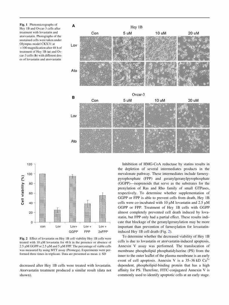
Fig. 1 Photomicrographs of Hey 1B and Ovcar-3 cells after treatment with lovastatin and atorvastatin. Photographs of the unstained cells were taken under Olympus model CKX31 at £100 magniWcation after 48 h of treatment of Hey 1B (a) and Ovcar-3 cells (b) with diVerent doses of lovastatin and atorvastatin
120
Ce
ll v
iab
ilit
y (%
)
80
100
60
20
40
0 Lov + Lov +
FPP 2xFPP
con Lov Lov+
GGPP
Fig. 2 EVect of lovastatin on Hey 1B cell viability Hey 1B cells were treated with 10 JM lovastatin for 48 h in the presence or absence of 2.5 JM GGPP or 2.5 JM and 5 JM FPP. The percentage of viable cells was measured by using MTT assay (Promega). Experiments were performed three times in triplicate. Data are presented as mean § SD
decreased after Hey 1B cells were treated with lovastatin. Atorvastatin treatment produced a similar result (data not shown).
Inhibition of HMG-CoA reductase by statins results in the depletion of several intermediates products in the mevalonate pathway. These intermediates include farnesypyrophosphate (FPP) and geranylgeranylpyrophosphate (GGPP)—isoprenoids that serve as the substrates for the prenylation of Ras and Rho family of small GTPases, respectively. To determine whether supplementation of GGPP or FPP is able to prevent cells from death, Hey 1B cells were co-incubated with 10 JM lovastatin and 2.5 JM GGPP or FPP. Treatment of Hey 1B cells with GGPP almost completely prevented cell death induced by lovastatin, but FPP only had a partial eVect. These results indicate that blockage of the geranylgeranylation may be more important than prevention of farnesylation for lovastatininduced Hey 1B cell death (Fig. 2).
To determine whether the decreased viability of Hey 1B cells is due to lovastatin or atorvastatin-induced apoptosis, Annexin V assay was performed. The translocation of membrane phospholipid phosphatidylserine (PS) from the inner to the outer leaXet of the plasma membrane is an early event of cell apoptosis. Annexin V is a 35–36 kD Ca2+
dependent, phospholipid-binding protein that has a high aYnity for PS. Therefore, FITC-conjugated Annexin V is commonly used to identify apoptotic cells at an early stage.

Hey 1B cells were treated with 10 JM of lovastatin for 48 h and subjected to Annexin V assay (Fig. 3). Lovastatin treatment of Hey 1B cells increased the percentage of apoptotic cells in the late stage (Annexin V and PI positive) from 13 to 48%. While GGPP reduced the percentage to 18.1%, FPP only slightly decreased the percentage of apoptotic cells at that stage to 37.8%. Interestingly, the Annexcin V labeled cell population induced by lovastatin was not signiWcantly changed in response to GGPP. This suggests that while the Wnal stages of apoptosis induced by lovastatin are inhibited by GGPP (Figs. 2, 3), this inhibition occurs at a step subsequent to the externalization of phosphatidylserine. To determine whether lovastatin induces apoptosis immortalized non-cancerous cells, we treated MC3T3-E1, a mouse preosteoblast cell line and WI-38, a human lung Wbroblast cell line, with lovastatin. Although lovastatin was able to induce apoptosis in these cells, a higher concentration (30–50 JM) and a prolonged incubation time (72– 96 h) were required to achieve the similar level of apoptosis (data not shown). These results are consistent with the previous reports that lovastatin at a higher concentration is needed to induce apoptosis in myeloma plasma cells, smooth muscle cells and endothelial cells [36–38], suggest-
Fig. 3 Determination of apoptotic cells by Annexin V assay Hey 1B cells were treated with or without 10 JM lovastatin for 48 h in the presence or absence of 2.5 JM GGPP and FPP. Apoptotic cells were analyzed by Annexin V assay (BD Pharmingen). Atorvastatin treatment produced a similar result (data not shown)
ing that ovarian cancer cells may be particularly vulnerable to statin-induced apoptosis.
Statin treatment activates caspase 3
To characterize further the statin-induced apoptosis in ovarian cancer cells, we determined the activity of caspase 3 in Hey 1B cells after treatment with lovastatin. The activity of caspase 3 in the cells treated with lovastatin was examined by using a Caspase-GloTM 3/7 assay kit (Promega). Lovastatin induced the activity of caspase 3 in Hey 1B cells by 3.5-fold. Consistent with our previous results, supplementation of GGPP was able to block caspase 3 activation induced by lovastatin, more eVectively than FPP (Fig. 4).
Statin treatment enhances the expression of GTPases
Small GTPases act as molecular switches to regulate diverse biochemical functions in all eukaryotic cells through mediating gene expression. Therefore, we investigated the eVect of statins on the expression of certain proteins including the members of the small GTPase family. Since we had already determined that inhibition of geranyl
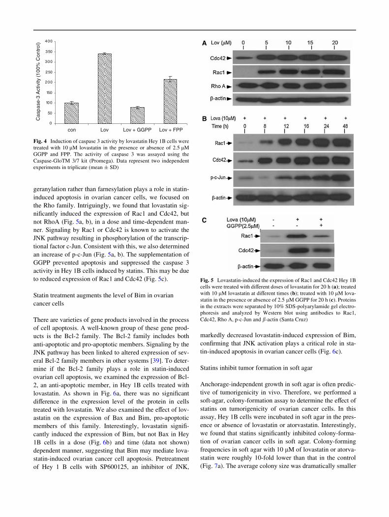
400 C
asp
ase
-3 A
ctiv
ity (1
00%
Co
ntro
l)
350
300
250
200
150
10 0
50
0
con Lov Lov + GGPP Lov + FPP
Fig. 4 Induction of caspase 3 activity by lovastatin Hey 1B cells were treated with 10 JM lovastatin in the presence or absence of 2.5 JM GGPP and FPP. The activity of caspase 3 was assayed using the Caspase-GloTM 3/7 kit (Promega). Data represent two independent experiments in triplicate (mean § SD)
geranylation rather than farnesylation plays a role in statininduced apoptosis in ovarian cancer cells, we focused on the Rho family. Intriguingly, we found that lovastatin signiWcantly induced the expression of Rac1 and Cdc42, but not RhoA (Fig. 5a, b), in a dose and time-dependent manner. Signaling by Rac1 or Cdc42 is known to activate the JNK pathway resulting in phosphorylation of the transcriptional factor c-Jun. Consistent with this, we also determined an increase of p-c-Jun (Fig. 5a, b). The supplementation of GGPP prevented apoptosis and suppressed the caspase 3 activity in Hey 1B cells induced by statins. This may be due to reduced expression of Rac1 and Cdc42 (Fig. 5c).
Statin treatment augments the level of Bim in ovarian cancer cells
There are varieties of gene products involved in the process of cell apoptosis. A well-known group of these gene products is the Bcl-2 family. The Bcl-2 family includes both anti-apoptotic and pro-apoptotic members. Signaling by the JNK pathway has been linked to altered expression of several Bcl-2 family members in other systems [39]. To determine if the Bcl-2 family plays a role in statin-induced ovarian cell apoptosis, we examined the expression of Bcl2, an anti-apoptotic member, in Hey 1B cells treated with lovastatin. As shown in Fig. 6a, there was no signiWcant diVerence in the expression level of the protein in cells treated with lovastatin. We also examined the eVect of lovastatin on the expression of Bax and Bim, pro-apoptotic members of this family. Interestingly, lovastatin signiWcantly induced the expression of Bim, but not Bax in Hey 1B cells in a dose (Fig. 6b) and time (data not shown) dependent manner, suggesting that Bim may mediate lovastatin-induced ovarian cancer cell apoptosis. Pretreatment of Hey 1 B cells with SP600125, an inhibitor of JNK,
Fig. 5 Lovastatin-induced the expression of Rac1 and Cdc42 Hey 1B cells were treated with diVerent doses of lovastatin for 20 h (a); treated with 10 JM lovastatin at diVerent times (b); treated with 10 JM lovastatin in the presence or absence of 2.5 JM GGPP for 20 h (c). Proteins in the extracts were separated by 10% SDS-polyarylamide gel electrophoresis and analyzed by Western blot using antibodies to Rac1, Cdc42, Rho A, p-c-Jun and /-actin (Santa Cruz)
markedly decreased lovastatin-induced expression of Bim, conWrming that JNK activation plays a critical role in statin-induced apoptosis in ovarian cancer cells (Fig. 6c).
Statins inhibit tumor formation in soft agar
Anchorage-independent growth in soft agar is often predictive of tumorigenicity in vivo. Therefore, we performed a soft-agar, colony-formation assay to determine the eVect of statins on tumorigenicity of ovarian cancer cells. In this assay, Hey 1B cells were incubated in soft agar in the presence or absence of lovastatin or atorvastatin. Interestingly, we found that statins signiWcantly inhibited colony-formation of ovarian cancer cells in soft agar. Colony-forming frequencies in soft agar with 10 JM of lovastatin or atorvastatin were roughly 10-fold lower than that in the control (Fig. 7a). The average colony size was dramatically smaller

A 160
140
Nu
mb
er
of
Co
loni
es
120
100
80
60
40
20
0
con Lova Ato
Fig. 6 EVect of lovastatin on the expression of Bcl-2 family members Hey 1B cells were treated with 10 JM lovastatin for various doses and the expression of the Bcl-2 family members was determined by Western blot analysis using antibodies to Bcl-2 (BD Biosciences), and Bax (Santa Cruz) (a); treated with diVerent doses of lovastatin for 20 h (b); pretreated with or without 10 JM SP600125 for 2 h and then treated with 10 JM lovastatin for 20 h (c), and the level of Bim was determined by Western blot analysis using a monoclonal antibody to human Bim (BD Biosciences)
in soft agar containing lovastatin or atorvastatin (data not shown) after incubation for 14 days, suggesting the therapeutic potential of statins in suppression of ovarian tumor growth (Fig. 7b).
Discussion
Ovarian cancer is often diagnosed at an advanced stage. Thus, although ovarian cancer is the fourth most common cancer in women, it is the leading cause of death among all gynecological malignancies in industrialized countries. The current therapeutic strategy for advance ovarian cancer is a combination of surgery and antimicrotubular chemotherapy. However, a majority of these patients relapses and develops resistance to these chemotherapeutic agents.
The link between the etiology of ovarian cancer and lipid metabolism has been known for decades [40, 41], yet this knowledge has not been exploited in the search for better treatment strategies. In addition to their role in lowering cholesterol, the statin family of drugs has shown a broad range of functions including suppression of cancer cell growth in vitro and in vivo [9–16] in many cell types. Surprisingly, the eVect of statins on ovarian cancer is limited to one study documenting lovastatin-induced growth inhibition [35]. In this study, we report that two diVerent statins induce apoptosis in ovarian cancer cells and inhibit growth
B
Fig. 7 EVect of statins on anchorage-independent cell growth Hey 1B cells were grown in soft agar in the presence or absence of 10 JM lovastatin; visible colonies were counted after crystal violet staining. The mean value § SD of three independent experiments is shown (a). The photos were taken under Olympus model CKX31 at £100 magniWcation at day 7 and 14 (b)
of ovarian tumors in soft agar. Our results suggest that stat-ins alone, or possibly in combination with other conventional drugs, may represent a novel therapy for advanced ovarian cancer.
Statins are known to inhibit HMG-CoA reductase and thus can inhibit the prenylation of several G-proteins by reducing geranylgeranylpyrophosphate (GGPP) and farnesylpyrophosphate (FPP). Based on this, the expectation would be that statins would inhibit the signaling of prenylated G-proteins. Recently, however, lovastatin was shown to induce the expression of Rho A, B and C in human erythroleukemia cells and to increase the active GTP-bound form of Rho by 3.7 fold [22]. Further, lovastatin did not aVect the prenylation of Ras in erythroleukemia cells, suggesting that inhibition of geranyl-geranylation was more pronounced than inhibition of farnesylation. Neither Rho downstream signaling events nor the status of Rac and cdc 42 were assessed in these cells. Consistent with the Wnding that statins can increase the expression of geranyl-geranylated G-proteins, we have shown that lovastatin treatment increased the expression of both Rac and Cdc 42 in ovarian

cancer cells, which could be abolished by inclusion of GGPP but not FPP. However, we did not observe an increase of Rho expression in both Hey 1B and Ovar-3 cells after statin treatment. This distinction is most likely a consequence of cell types. Possible mechanisms whereby the inhibition of prenylation could result in increased expression of prenylated G-proteins would include the possibility that either the unprenylated G-protein aVects the signaling pathway controlling its own expression, or the lack of prenylation impacts this pathway through some other molecules.
Consistent with the fact that JNK is a downstream target of Rac and cdc 42 signaling [31, 32], and that phosphorylation of the transcription factor c-Jun is downstream of JNK activation, we observed an increase in the level of active, phosphorylated c-jun in the ovarian cancer cells under statin treatment. Further investigation revealed that the expression of Bim, a proapoptotic protein, is signiWcantly induced in the cells by statins. These Wndings are consistent with the observation that Bim induction in neurons deprived of NGF is regulated by c-Jun [42]. SP600125 is a competitive inhibitor of ATP binding to JNK and some other kinases [43]. Pretreatment of Hey 1B cells with SP600125 signiWcantly reduced lovastatin-induced expression of Bim, con-Wrming the role of JNK in induction of Bim in these cells. Our results demonstrating statin-induced apoptosis and inhibition of anchorage independent growth of ovarian cancer cells suggest the validity of investigating the use of stat-ins as a treatment for ovarian cancer. Further, our investigation of the molecular mechanism involved in statin signaling in ovarian cancer cells provides novel potential targets for therapeutic manipulation.
ConXict of interest statement None.
References
1. Sharma S, Odunsi K (2005) Targeted therapy for epithelial ovarian cancer. Expert Opin Ther Targets 9(3):501–513
2. American Cancer Society, http://www.cancer.org 3. Ozols RF (2005) Treatment goals in ovarian cancer. Int J Gynecol
Cancer 15(Suppl 1):3–11 4. Du Bois A, PWsterer J (2005) Future options for Wrst-line therapy of
advanced ovarian cancer. Int J Gynecol Cancer 15(Suppl 1):42–50 5. Bookman MA (2005) Gemcitabine monotherapy in recurrent
ovarian cancer: from the bench to the clinic. Int J Gynecol Cancer 15(Suppl 1):12–17
6. Caslake MJ, Packard CJ (2004) Phenotypes, genotypes and response to statin therapy. Curr Opin Lipidol 15(4):387–392
7. Stein EA (2003) The power of statins: aggressive lipid lowering. Clin Cardiol 26(4 Suppl 3):III25–31
8. Schmitz G, Drobnik W (2003) Pharmacogenomics and pharmacogenetics of cholesterol-lowering therapy. Clin Chem Lab Med 41(4):581–589
9. Chan KK, Oza AM, Siu LL (2003) The statins as anticancer agents. Clin Cancer Res 9(1):10–19
10. CaVorio P, Dammacco F, Gernone A, Silvestris F (2005) Statins activate the mitochondrial pathway of apoptosis in human lymphoblasts and myeloma cells. Carcinogenesis 26(5):883–891
11. Muck AO, Seeger H, Wallwiener D (2004) Inhibitory eVect of statins on the proliferation of human breast cancer cells. Int J Clin Pharmacol Ther 42(12):695–700
12. Koyuturk M, Ersoz M, Altiok N (2004) Simvastatin induces proliferation inhibition and apoptosis in C6 glioma cells via c-jun N-terminal kinase. Neurosci Lett 370(2–3):212–217
13. Otsuki T, Sakaguchi H, Hatayama T, Fujii T, Tsujioka T, Sugihara T, Takata A, Hyodoh F, Eto M (2004) EVects of an HMG-CoA reductase inhibitor, simvastatin, on human myeloma cells. Oncol Rep 11(5):1053–1058
14. Collisson EA, Kleer C, Wu M, De A, Gambhir SS, Merajver SD, Kolodney MS (2003) Atorvastatin prevents RhoC isoprenylation, invasion, and metastasis in human melanoma cells. Mol Cancer Ther 2(10):941–948
15. Gliemroth J, Feyerabend T, Gerlach C, Arnold H, Terzis AJ (2003) Proliferation, migration, and invasion of human glioma cells exposed to fractionated radiotherapy in vitro. Neurosurg Rev 26(3):198–205
16. Holstein SA, Knapp HR, Clamon GH, Murry DJ, Hohl RJ (2005) Pharmacodynamic eVects of high dose lovastatin in subjects with advanced malignancies. Cancer Chemother Pharmacol 30:1–10
17. Issat T, Nowis D, Jakobisiak M, Golab J (2004) Lovastatin potentiates antitumor eVects of saquinavir against human lymphoma cells. Oncol Rep 12(6):1371–1375
18. Kozar K, Kaminski R, Legat M, Kopec M, Nowis D, Skierski JS, Koronkiewicz M, Jakobisiak M, Golab J (2004) Cerivastatin demonstrates enhanced antitumor activity against human breast cancer cell lines when used in combination with doxorubicin or cisplatin. Int J Oncol 24(5):1149–1157
19. Agarwal B, Bhendwal S, Halmos B, Moss SF, Ramey WG, Holt PR (1999) Lovastatin augments apoptosis induced by chemotherapeutic agents in colon cancer cells. Clin Cancer Res 5(8):2223– 2229
20. Feleszko W, Jakóbisiak M (2000) Lovastatin potentiates antitumor activity and attenuates cardiotoxicity of doxorubicin in three tumor models in mice. Clin Cancer Res 6(5):2044–2052
21. Xiao H, Zhang Q, Lin Y, Reddy BS, Yang CS (2008) Combination of atorvastatin and celecoxib synergistically induces cell cycle arrest and apoptosis in colon cancer cells. Int J Cancer 122(9):2115– 2124
22. Turner SJ, Zhuang S, Zhang T, Boss GR, Pilz RB (2008) EVects of lovastatin on Rho isoform expression, activity, and association with guanine nucleotide dissociation inhibitors. Biochem Pharmacol 75(2):405–413
23. Bokoch GM, Diebold BA (2002) Current molecular models for NADPH oxidase regulation by Rac GTPase. Blood 100(8):2692– 2696
24. Rul W, Zugasti O, Roux P, Peyssonnaux C, Eychene A, Franke TF, Lenormand P, Fort P, Hibner U (2002) Activation of ERK, controlled by Rac1 and Cdc42 via Akt, is required for anoikis. Ann NY Acad Sci 973:145–148
25. Frost JA, Steen H, Shapiro P, Lewis T, Ahn N, Shaw PE, Cobb MH (1991) Cross-cascade activation of ERKs and ternary complex factors by Rho family proteins. EMBO J 16(21):6426–6438
26. Clerk A, Pham FH, Fuller SJ, Sahai E, Aktories K, Marais R, Marshall C, Sugden PH (2001) Regulation of mitogen-activated protein kinases in cardiac myocytes through the small G protein Rac1. Mol Cell Biol 21(4):1173–1184
27. Keely PJ, Westwick JK, Whitehead IP, Der CJ, Parise LV (1997) Cdc42 and Rac1 induce integrin-mediated cell motility and invasiveness through PI(3)K. Nature 390(6660):632–636
28. Aznar S, Lacal JC (2001) Rho signals to cell growth and apoptosis. Cancer Lett 165(1):1–10
29. Chuang TH, Hahn KM, Lee JD, Danley DE, Bokoch GM (1997) The small GTPase Cdc42 initiates an apoptotic signaling pathway in Jurkat T lymphocytes. Mol Biol Cell 8(9):1687–1698
30. Subauste MC, Von Herrath M, Benard V, Chamberlain CE, Chuang TH, Chu K, Bokoch GM, Hahn KM (2000) Rho family proteins modulate rapid apoptosis induced by cytotoxic T lymphocytes and Fas. J Biol Chem 275(13):9725–9733
31. Minden A, Lin A, Claret FX, Abo A, Karin M (1995) Selective activation of the JNK signaling cascade and c-Jun transcriptional activity by the small GTPases Rac and Cdc42Hs. Cell 81(7):1147–1157
32. Coso OA, Chiariello M, Yu JC, Teramoto H, Crespo P, Xu N, Miki T, Gutkind JS (1995) The small GTP-binding proteins Rac1 and Cdc42 regulate the activity of the JNK/SAPK signaling pathway. Cell 81(7):1137–1146
33. Teramoto H, Coso OA, Miyata H, Igishi T, Miki T, Gutkind JS (1996) Signaling from the small GTP-binding proteins Rac1 and Cdc42 to the c-Jun N-terminal kinase/stress-activated protein kinase pathway. A role for mixed lineage kinase 3/protein-tyrosine kinase 1, a novel member of the mixed lineage kinase family. J Biol Chem 271(44):27225–27228
34. Chuang TH, Hahn KM, Lee JD, Danley DE, Bokoch GM (1997) The small GTPase Cdc42 initiates an apoptotic signaling pathway in Jurkat T lymphocytes. Mol Biol Cell 8(9):1687–1698
35. Melichar B, Ferrandina G, Verschraegen CF, Loercher A, Abbruzzese JL, Freedman RS (1998) Growth inhibitory eVects of aromatic fatty acids on ovarian tumor cell lines. Clin Cancer Res 4(12):3069–3076
36. van de Donk NW, Kamphuis MM, van Kessel B, Lokhorst HM, Bloem AC (2003) Inhibition of protein geranylgeranylation
induces apoptosis in myeloma plasma cells by reducing Mcl-1 protein levels. Blood 102(9):3354–3362
37. Erl W, Hristov M, Neureuter M, Yan ZQ, Hansson GK, Weber PC (2003) HMG-CoA reductase inhibitors induce apoptosis in neointima-derived vascular smooth muscle cells. Atherosclerosis 169:251–258
38. Kaneta S, Satoh K, Kano S, Kanda M, Ichihara K (2003) All hydrophobic HMG-CoA reductase inhibitors induce apoptotic death in rat pulmonary vein endothelial cells. Atherosclerosis 170:237–243
39. Zhu BK, Wang P, Zhang XD, Jiang CC, Chen LH, Avery-Kiejda KA, Watts R, Hersey P (2008) Activation of Jun N-terminal kinase is a mediator of vincristine-induced apoptosis of melanoma cells. Anticancer Drugs 19(2):189–200
40. Avall-Lundqvist EH, Peterson CO (1996) Serum cholesterol and apolipoprotein B levels may reXect disease activity in ovarian cancer patients. Acta Oncol 35(8):1007–1010
41. Risch HA, Jain M, Marrett LD, Howe GR (1994) Dietary fat intake and risk of epithelial ovarian cancer. J Natl Cancer Inst 86(18):1409–1415
42. Biswas SC, Shi Y, Sproul A, Greene LA (2007) Pro-apoptotic Bim induction in response to nerve growth factor deprivation requires simultaneous activation of three diVerent death signaling pathways. J Biol Chem 282(40):29368–29374
43. Bennett BL, Sasaki DT, Murray BW, O’Leary EC, Sakata ST, Xu W, Leisten JC, Motiwala A, Pierce S, Satoh Y, Bhagwat SS, Manning AM, Anderson DW (2001) SP600125, an anthrapyrazolone inhibitor of Jun N-terminal kinase. Proc Natl Acad Sci USA 98(24):13681–13686
Copyright © 2022 FDOKUMEN