Startling Mosaicism of the Y-Chromosome and Tandem Duplication of the SRY and DAZ Genes in Patients...
11
Startling Mosaicism of the Y-Chromosome and Tandem Duplication of the SRY and DAZ Genes in Patients with Turner Syndrome Sanjay Premi, Jyoti Srivastava, Ganesan Panneer ¤ , Sher Ali* Molecular Genetics Laboratory, National Institute of Immunology, Aruna Asaf Ali Marg, New Delhi, India Abstract Presence of the human Y-chromosome in females with Turner Syndrome (TS) enhances the risk of development of gonadoblastoma besides causing several other phenotypic abnormalities. In the present study, we have analyzed the Y chromosome in 15 clinically diagnosed Turner Syndrome (TS) patients and detected high level of mosaicisms ranging from 45,XO:46,XY = 100:0% in 4; 45,XO:46,XY:46XX = 4:94:2 in 8; and 45,XO:46,XY:46XX = 50:30:20 cells in 3 TS patients, unlike previous reports showing 5–8% cells with Y- material. Also, no ring, marker or di-centric Y was observed in any of the cases. Of the two TS patients having intact Y chromosome in .85% cells, one was exceptionally tall. Both the patients were positive for SRY, DAZ, CDY1, DBY, UTY and AZFa, b and c specific STSs. Real Time PCR and FISH demonstrated tandem duplication/multiplication of the SRY and DAZ genes. At sequence level, the SRY was normal in 8 TS patients while the remaining 7 showed either absence of this gene or known and novel mutations within and outside of the HMG box. SNV/ SFV analysis showed normal four copies of the DAZ genes in these 8 patients. All the TS patients showed aplastic uterus with no ovaries and no symptom of gonadoblastoma. Present study demonstrates new types of polymorphisms indicating that no two TS patients have identical genotype-phenotype. Thus, a comprehensive analysis of more number of samples is warranted to uncover consensus on the loci affected, to be able to use them as potential diagnostic markers. Citation: Premi S, Srivastava J, Panneer G, Ali S (2008) Startling Mosaicism of the Y-Chromosome and Tandem Duplication of the SRY and DAZ Genes in Patients with Turner Syndrome. PLoS ONE 3(11): e3796. doi:10.1371/journal.pone.0003796 Editor: Samir K. Brahmachari, Institute of Genomics and Integrative Biology, India Received May 15, 2008; Accepted October 27, 2008; Published November 24, 2008 Copyright: ß 2008 Premi et al. This is an open-access article distributed under the terms of the Creative Commons Attribution License, which permits unrestricted use, distribution, and reproduction in any medium, provided the original author and source are credited. Funding: This work was supported by DBT Grants No. BT/PR2752/AAQ/01/113/2001 and BT/PR8476/AAQ/01/315/2006 to SA and a core grant from the Department of Biotechnology, Govt. of India to the National Institute of Immunology, New Delhi. SP is thankful to Council of Scientific and Industrial Research (CSIR), New Delhi for the award of Senior Research Fellowship. This work has been seen and approved by all the authors and they do not have any conflict of personal communication or financial interests. Competing Interests: The authors have declared that no competing interests exist. * E-mail: [email protected] ¤ Current address: Department of Zoology, University of Lucknow, Lucknow, India Introduction Turner Syndrome (TS), the common genetic abnormalities affecting ,1 in 1500–2000 live female births [1–3], is suggested to be due to absence of the second X chromosome in part or full [3– 4]. However, in ,3–6% cells, the second sex chromosome is Y [5– 6] that often triggers development of gonadoblastoma [7]. The Y chromosome in Turner patients is structurally abnormal showing deletions, inversions, dicentrics and ring forms [8–9] and becomes unstable resulting in 45/XO karyotype. Chromosomal constitu- tion influences phenotypic sex and 45, XO cell line is frequently detected in males with gonadal dysgenesis in addition to TS patients [10–11]. It is largely believed that no two TS patients are identical with respect to the number of Y bearing cells or Y-linked loci. Moreover, this mosaicism varies across the tissues and thus accurate interpretation depends upon the number of cells analyzed and tissues selected [12–13]. The phenotypic sex is under the influence of Y chromosome and expression of Y linked loci in gonads [14–15]. In several instances, gonadectomy is conducted due to increased risks of gonadoblastoma [16]. However, actual distribution of the Y chromosome in tissues of the TS patients and its role remain a murky proposition. Present study was conducted to investigate molecular alterations in the Y-linked loci in 15 clinically diagnosed TS patients. We detected large scale Y chromosome mosaicism ranging from pure 45/XO conceptus to ,90% cells positive for an intact Y chromosome and XXX, XYY, XXY constitutions. Further, several Turners patients showed tandemly arranged multiple copies of the SRY and DAZ genes in addition to known and novel SRY mutations within and 59/39 regions of the HMG box. Results Turner Karyotypes and the Y chromosome TS patients analyzed were in the age group of 14–25 yrs. Presence of Y chromosome detected with G-Banding was confirmed by FISH with Y specific probes SRY, 46A6 (DAZ) and 336F2 (gr/gr AZFc amplicons). Patients showed two extreme karyotypes, ones with .85% cells harboring Y chromosome (AT1 and AT15) and others with almost negligible presence of the Y chromosome (AT4, AT5, AT6 and AT7) (Table 1). Marker, ring or dicentrics Y chromosome was not observed in any of the patients. Clinically, all the TS patients had webbed neck, shield like chest and other characteristic features (Table 1) but no PLoS ONE | www.plosone.org 1 November 2008 | Volume 3 | Issue 11 | e3796
-
Upload
independent -
Category
Documents
-
view
1 -
download
0
Transcript of Startling Mosaicism of the Y-Chromosome and Tandem Duplication of the SRY and DAZ Genes in Patients...

Startling Mosaicism of the Y-Chromosome and TandemDuplication of the SRY and DAZ Genes in Patients withTurner SyndromeSanjay Premi, Jyoti Srivastava, Ganesan Panneer¤, Sher Ali*
Molecular Genetics Laboratory, National Institute of Immunology, Aruna Asaf Ali Marg, New Delhi, India
Abstract
Presence of the human Y-chromosome in females with Turner Syndrome (TS) enhances the risk of development ofgonadoblastoma besides causing several other phenotypic abnormalities. In the present study, we have analyzed the Ychromosome in 15 clinically diagnosed Turner Syndrome (TS) patients and detected high level of mosaicisms ranging from45,XO:46,XY = 100:0% in 4; 45,XO:46,XY:46XX = 4:94:2 in 8; and 45,XO:46,XY:46XX = 50:30:20 cells in 3 TS patients, unlikeprevious reports showing 5–8% cells with Y- material. Also, no ring, marker or di-centric Y was observed in any of the cases.Of the two TS patients having intact Y chromosome in .85% cells, one was exceptionally tall. Both the patients werepositive for SRY, DAZ, CDY1, DBY, UTY and AZFa, b and c specific STSs. Real Time PCR and FISH demonstrated tandemduplication/multiplication of the SRY and DAZ genes. At sequence level, the SRY was normal in 8 TS patients while theremaining 7 showed either absence of this gene or known and novel mutations within and outside of the HMG box. SNV/SFV analysis showed normal four copies of the DAZ genes in these 8 patients. All the TS patients showed aplastic uteruswith no ovaries and no symptom of gonadoblastoma. Present study demonstrates new types of polymorphisms indicatingthat no two TS patients have identical genotype-phenotype. Thus, a comprehensive analysis of more number of samples iswarranted to uncover consensus on the loci affected, to be able to use them as potential diagnostic markers.
Citation: Premi S, Srivastava J, Panneer G, Ali S (2008) Startling Mosaicism of the Y-Chromosome and Tandem Duplication of the SRY and DAZ Genes in Patientswith Turner Syndrome. PLoS ONE 3(11): e3796. doi:10.1371/journal.pone.0003796
Editor: Samir K. Brahmachari, Institute of Genomics and Integrative Biology, India
Received May 15, 2008; Accepted October 27, 2008; Published November 24, 2008
Copyright: � 2008 Premi et al. This is an open-access article distributed under the terms of the Creative Commons Attribution License, which permitsunrestricted use, distribution, and reproduction in any medium, provided the original author and source are credited.
Funding: This work was supported by DBT Grants No. BT/PR2752/AAQ/01/113/2001 and BT/PR8476/AAQ/01/315/2006 to SA and a core grant from theDepartment of Biotechnology, Govt. of India to the National Institute of Immunology, New Delhi. SP is thankful to Council of Scientific and Industrial Research(CSIR), New Delhi for the award of Senior Research Fellowship. This work has been seen and approved by all the authors and they do not have any conflict ofpersonal communication or financial interests.
Competing Interests: The authors have declared that no competing interests exist.
* E-mail: [email protected]
¤ Current address: Department of Zoology, University of Lucknow, Lucknow, India
Introduction
Turner Syndrome (TS), the common genetic abnormalities
affecting ,1 in 1500–2000 live female births [1–3], is suggested to
be due to absence of the second X chromosome in part or full [3–
4]. However, in ,3–6% cells, the second sex chromosome is Y [5–
6] that often triggers development of gonadoblastoma [7]. The Y
chromosome in Turner patients is structurally abnormal showing
deletions, inversions, dicentrics and ring forms [8–9] and becomes
unstable resulting in 45/XO karyotype. Chromosomal constitu-
tion influences phenotypic sex and 45, XO cell line is frequently
detected in males with gonadal dysgenesis in addition to TS
patients [10–11]. It is largely believed that no two TS patients are
identical with respect to the number of Y bearing cells or Y-linked
loci. Moreover, this mosaicism varies across the tissues and thus
accurate interpretation depends upon the number of cells analyzed
and tissues selected [12–13]. The phenotypic sex is under the
influence of Y chromosome and expression of Y linked loci in
gonads [14–15]. In several instances, gonadectomy is conducted
due to increased risks of gonadoblastoma [16]. However, actual
distribution of the Y chromosome in tissues of the TS patients and
its role remain a murky proposition.
Present study was conducted to investigate molecular alterations
in the Y-linked loci in 15 clinically diagnosed TS patients. We
detected large scale Y chromosome mosaicism ranging from pure
45/XO conceptus to ,90% cells positive for an intact Y
chromosome and XXX, XYY, XXY constitutions. Further,
several Turners patients showed tandemly arranged multiple
copies of the SRY and DAZ genes in addition to known and novel
SRY mutations within and 59/39 regions of the HMG box.
Results
Turner Karyotypes and the Y chromosomeTS patients analyzed were in the age group of 14–25 yrs.
Presence of Y chromosome detected with G-Banding was
confirmed by FISH with Y specific probes SRY, 46A6 (DAZ) and
336F2 (gr/gr AZFc amplicons). Patients showed two extreme
karyotypes, ones with .85% cells harboring Y chromosome (AT1
and AT15) and others with almost negligible presence of the Y
chromosome (AT4, AT5, AT6 and AT7) (Table 1). Marker, ring
or dicentrics Y chromosome was not observed in any of the
patients. Clinically, all the TS patients had webbed neck, shield
like chest and other characteristic features (Table 1) but no
PLoS ONE | www.plosone.org 1 November 2008 | Volume 3 | Issue 11 | e3796

Ta
ble
1.
De
tails
of
the
kary
oty
pe
san
dh
orm
on
alp
rofi
les
of
dif
fere
nt
Tu
rne
rp
atie
nts
anal
yze
d#
Pa
tie
nts
Ph
en
oty
pe
Ka
ryo
typ
e(%
cell
s)C
lin
ica
lF
ea
ture
sH
orm
on
al
Pro
file
s
AT
1F
46
,XX
(10
):46
,XY
(75
):45
,XO
(5):4
7,X
XY
(2):4
7,X
YY
(3)
Ag
e=
14
year
s,m
en
arch
en
ot
atta
ine
dye
t,se
con
dar
yse
xch
arac
ters
no
td
eve
lop
ed
,e
xte
rnal
ge
nit
alia
no
rmal
,p
ub
ert
yg
rad
e0
,ap
last
icu
teru
s,o
vari
es
no
tse
en
,sh
ield
like
che
st,
no
ske
leta
lan
om
aly,
we
bb
ed
ne
ck
LH=
16
.2U
/L,F
SH=
89
.8U
/L,P
RL
=1
0.4
mg/L
,TSH
=1
.7m
U/L
AT
2F
46
,XX
(77
):4
6,X
Y(6
):45
,XO
(14
):4
7,X
XX
(3)
Pri
mar
yam
en
orr
he
a,at
op
icva
gin
a,sm
all
ute
rus
see
nd
uri
ng
gyn
eco
log
ical
exa
min
atio
n,
no
en
do
me
tria
lti
ssu
ese
en
inb
iop
sy,
USG
sho
we
dan
teve
rte
dan
dan
tefl
exe
du
teru
s
LH=
22
U/L
,FS
H=
75
–8
0U
/L,
PR
L=
12
mg/L
,T
SH=
1.2
mU
/L
AT
3F
46
,XX
(2):4
6,X
Y(3
):45
,XO
(95
)P
rim
ary
ame
no
rrh
ea,
smal
ln
od
ule
like
ute
rus
bu
tn
oo
vari
es,
smal
lst
atu
rean
dw
eb
be
dn
eck
LH=
18
U/L
,FS
H=
75
U/L
,P
RL
=9
mg/L
,T
SH=
1.2
mU
/L
AT
4F
46
,XX
(0):4
6,X
Y(0
):45
,XO
(10
0)
Sho
rtst
atu
re,
we
bb
ed
ne
ck,
hyp
op
last
icu
teru
san
do
vari
es,
no
rmal
ext
ern
alg
en
ital
ia,
un
de
rde
velo
pe
db
reas
tsLH
=3
2.0
U/L
,FS
H=
3.0
,P
RL
=1
4.0
mg/L
AT
5F
46
,XX
(5):4
6,X
Y(0
):45
,XO
(95
)W
eb
be
dn
eck
,n
od
ule
like
ute
rus,
Pri
mar
yam
en
orr
he
a,u
nd
er
de
velo
pe
dse
xual
char
acte
rsLH
=2
0U
/L,
FSH
=4
5U
/L,
PR
L=
18
mg/L
,T
SH=
1–
1.5
mU
/L
AT
6F
46
,XX
(0):4
6,X
Y(0
):45
,XO
(10
0)
Pri
mar
yam
en
orr
he
a,sm
all
un
de
rd
eve
lop
ed
ute
rus,
no
ova
rie
sse
en
NA
AT
7F
46
,XX
(5):4
6,X
Y(0
):45
,XO
(95
)N
AN
A
AT
8F
46
,XX
(5):4
6,X
Y(3
):45
,XO
(92
)T
urn
er
vari
ant,
Pri
mar
yam
en
orr
he
a,e
xtre
me
lyh
ypo
pla
stic
ute
rus,
bre
ast
and
ext
ern
alg
en
ital
iaw
ere
un
de
rde
velo
pe
dLH
=3
7U
/L,
FSH
=8
9.8
U/L
,P
RL
=1
0.4
mg/L
,T
SH=
1.7
mU
/L
AT
9F
46
,XX
(5):4
6,X
Y(5
):45
,XO
(90
)Sh
ort
stat
ure
,w
eb
be
dn
eck
,p
rim
ary
ame
no
rrh
ea,
dys
ge
nic
go
nad
s,te
stis
we
resu
rgic
ally
rem
ove
dFS
H=
35
U/L
,P
RL
=1
0.4
mg/L
,T
SH=
1.7
mU
/L
AT
10
F4
6,X
X(5
):46
,XY
(3):4
5,X
O(9
2)
Pri
mar
yA
me
no
rrh
ea
FSH
=9
0U
/L,
PR
L=
18
mg/L
AT
11
F4
6,
XX
(2):4
6,
XY
(2):4
5,
XO
(96
)P
rim
ary
ame
no
rrh
ea,
we
bb
ed
ne
ck,
no
ske
leta
ld
efo
rmat
ion
,e
xte
rnal
ge
nit
alia
no
rmal
,u
nd
erd
eve
lop
ed
bre
asts
NA
AT
12
F4
6,
XX
(5):4
6,
XY
(3):4
5,
XO
(92
)W
eb
be
dn
eck
,sh
ort
stat
ure
,p
oo
rly
de
velo
pe
dse
con
dar
yse
xual
char
acte
rsLH
=5
5–
80
U/L
,FS
H=
85
U/L
,P
RL
=1
0–
15
mg/L
,T
SH=
2m
U/L
AT
13
F4
6,
XX
(40
):46
,X
Y(3
0):4
5,
XO
(20
)Sh
ort
stat
ure
,w
eb
be
dn
eck
,sh
ield
like
che
st,
seco
nd
ary
ame
no
rrh
ea,
un
de
rd
eve
lop
ed
bre
asts
,e
xte
rnal
ge
nit
alia
no
rmal
,U
SGsh
ow
ed
stre
akg
on
ads.
NA
AT
14
F4
6,X
X(5
):46
,XY
(3):4
5,X
O(9
2)
Sho
rtst
atu
re,
we
bb
ed
ne
ck,
shie
ldlik
ech
est
,n
ob
reas
tn
od
ule
sLH
=5
6U
/L,
FSH
=8
5U
/L,
PR
L=
20
mg/L
,T
SH=
1.5
–2
mU
/L
AT
15
F4
6,X
X(5
):46
,XY
(80
):45
,XO
(5):4
7,X
XY
(2):4
7,X
YY
(3)
Bo
ne
age
25
year
s,h
eig
ht
=1
75
cm,
we
igh
t=
50
kg,
no
ske
leta
ld
efo
rmat
ion
,Se
con
dar
yam
en
orr
he
a,e
xte
rnal
ge
nit
alia
no
rmal
,ap
last
icu
teru
s,n
oo
vari
es
ob
serv
ed
exc
ep
tst
reak
go
nad
s.
LH=
25
U/L
,FS
H=
92
U/L
,P
RL
=8
mg/L
,T
SH=
2.0
mU
/L
NM
M4
6,
XY
No
rmal
LH=
0.0
07
–0
.02
4U
/L,
FSH
=5
–2
0U
/L
NF
F4
6X
XN
orm
alLH
=5
–2
0U
/L,
FSH
=3
–2
0U
/L,
PR
L=
10
–2
5mg
/L
#N
M=
No
rmal
mal
e,
NF
=N
orm
alfe
mal
e,
NA
=N
ot
avai
lab
le,
USG
=U
ltra
son
og
rap
hy,
PR
L=
Pro
lact
inre
leas
ing
ho
rmo
ne
,T
SH=
Th
yro
idst
imu
lati
ng
ho
rmo
ne
,FS
H=
Folli
cle
stim
ula
tin
gh
orm
on
e,
LH=
Lute
iniz
ing
ho
rmo
ne
.N
um
be
rsin
par
en
the
sis
un
de
rka
ryo
typ
ed
en
ote
pe
rce
nta
ge
of
the
cells
wit
hth
ep
arti
cula
rch
rom
oso
mal
con
stit
uti
on
.A
llth
eka
ryo
typ
es
we
reco
nfi
rme
dan
alyz
ing
,4
00
me
tap
has
ech
rom
oso
me
sets
pe
rin
div
idu
al.
do
i:10
.13
71
/jo
urn
al.p
on
e.0
00
37
96
.t0
01
Y-Mosaicism & Turner Syndrome
PLoS ONE | www.plosone.org 2 November 2008 | Volume 3 | Issue 11 | e3796

Figure 1. Fluorescence in-situ hybridization (FISH) using LSI SRY probe from VYSIS (which binds simultaneously to the SRY gene andcentromere of the X chromosome) within the interphase nuclei and metaphase chromosomes of Turner AT1. (A i–iv) shows presence of both X(green dot) and Y (red dot) chromosomes in the interphase nuclei. Note structurally normal Y chromosome and absence of ring or dicentric one in(Bi) and (Ci) where the SRY gene is localized on the Yp. Some cells showed absence of the X chromosome, denoted by pink arrows (Bii) and (Cii).The classical Turner karyotypes (45, XO) are shown by yellow arrows. Some cells showed 47, XYY (Civ). (D), Cells without Y but variable numbers of Xchromosome ranging from 1 (45, XO) to 2 (46, XX) were also detected. Only representative cells with different karyotypes are shown here. Singlelocalized signal of the SRY gene (copy number 16) in AT1 suggests tandem duplication of this gene.doi:10.1371/journal.pone.0003796.g001
Figure 2. FISH with interphase nuclei and metaphase chromosomes of a Turner patient (AT4) with LSI-SRY probe. Note absence of theSRY signals in all the cells. No detectable Y chromosome at the level of PCR or G-banding was detected in this patient. The alterations detected in thenumber of X-Chromosomes are indicated by arrows. Pink arrows show cells with three X-Chromosomes (47, XXX) and the yellow ones highlights thecells with a single X-Chromosome (45, XO). Remaining interphases showed two X-Chromosomes (46, XX). Analysis of metaphase chromosomes (i–ii)further substantiated absence of the Y-chromosome. This is in contrast to Turner AT1 where .80% cells harbored Y-chromosome.doi:10.1371/journal.pone.0003796.g002
Y-Mosaicism & Turner Syndrome
PLoS ONE | www.plosone.org 3 November 2008 | Volume 3 | Issue 11 | e3796

symptoms/trace of gonadoblastoma. Only one Turner (AT9)
showed dysgenic testis which was removed surgically. One TS
patient (AT15) was exceptionally tall with a normal female
phenotype, but with clinical features similar to that of the
Turners’. In addition to normal mosaic karyotypes, two Turners’,
AT1 and AT15 also showed another cell lines with 47, XYY, or
47, XXY chromosomal constitutions (Figure 1). Pure XO
conceptus was also detected in some Turners, showing two, one
or no signal for X chromosome but none at all for the Y
chromosome (Figure 2).
Structural integrity of Y the chromosomeStructural integrity of the Y chromosome was assessed by
routine STS mapping. In TS patients with more than 40% cells
positive for Y chromosome, most of the STS’s used were positive
(Figure 3). STS mapping nullified any event of the gr/gr or b1/b3
major deletion phenotypes [17]. STSs lying in the crucial regions
like DAZ gene, HERV sequences, AZF boundaries and other
crucial genes were found to be intact except few randomly
scattered microdeletions (Figure 4).
Tandem duplication of the SRY and DAZ genesTurners were assessed for the possible duplication of two
candidate genes DAZ and SRY using TaqMan chemistry and Real
Time PCR. In most of the patients, DCt (Ct SRY/DAZ – Ct
RNAseP easy) values observed were unexpected. In case of single
copy SRY and 4 copy DAZ genes in a normal male, DCt values
observed are 1 and 21, respectively [18]. Unexpectedly, DCt for
SRY and DAZ gene were .1 in case of TS patients (Figures 5 and
6, Table 2). This is possible only if the copies are less than one,
which technically can not be true. The reason behind this was
presence of RNAseP gene in all the cells but that of Y chromosome
in a small cell population. Even with this mosaicism, few TS
patients showed ,2 to 3 rounds of duplication of the SRY and DAZ
genes (Table 2). FISH conducted for SRY and DAZ genes showed
single localized signal in all the TS patients. This correlated with
the events of tandem duplications (See Figures 1 and 7).
Fate of AZFc region in TurnersCosmid probes for DAZ genes and BAC probe used for FISH
corresponding to green amplicon demonstrated yet another
Figure 3. Representative gels showing STS mapping of the Y chromosomes in TS patients. STSs used are given on the right and sampleIDs on top. The IDs ‘AT’ are Turners and their details are given in the table 1. A10 and A10g represent blood and semen DNA samples, respectively,from a single azoospermic male. HF denotes human female DNA sample. b-actin primers were used to normalize the quality and quantity of DNAused as template in PCR. Note presence of most of the STSs in Turners AT1 and AT15. Some STSs were positive in case of Turner AT13 as well butowing to non-availability of the fresh blood, the FISH experiments could not be conducted (see Tables 1 and 2 for details of the Turner patients).doi:10.1371/journal.pone.0003796.g003
Y-Mosaicism & Turner Syndrome
PLoS ONE | www.plosone.org 4 November 2008 | Volume 3 | Issue 11 | e3796

mosaicism in TS patients. Probe C uncovered expected 2 to a
single, widely spaced 3, or no FISH signals. Some cells showed two
signals of which one was with higher intensity compared to that of
the other suggesting unilocus duplication of the DAZ genes [18].
In the dual color FISH, probe A showed expected 2 or a single
signal in most of the cells but probe C again showed multiple
signals in certain cell population. Ideally, the signals for probe A
and B should overlap owing to their vicinity which was not
observed in any of the Turner Patients. This important
observation has been explained in a separate report. Significantly,
probes C and D uncovered unexpected widely spaced 3 to
multiple signals (Figure 7).
SRY mutationsSRY was taken as a candidate gene since the same has been well
characterized. This gene was sequenced from all the TS patients
who showed positive PCR amplification. All three Turners’
positive for SRY gene showed normal sequence except a few
unclear point nucleotide changes. Mostly, the nucleotide changes
were silent except a few affecting the protein sequence. The in-silico
translation and sequence comparisons showed few well defined
amino acid changes upstream, within and down stream of the
HMG box. Details of the nucleotide changes of SRY gene observed
in Turner AT1 are given in figure S2 and corresponding amino
acid changes in figure 8. Father of this patient showed a single
amino acid change (Figure 9) suggesting that the changes in AT1
were de novo, and not inherited from the father.
Discussion
Turner Syndrome (TS) is globally acknowledged and well
defined genetic anomaly, postulated to be an effect of the absence
of genes located on the second sex chromosome. However, it is not
clear as to how many loci/genes are affected in TS nor do we
know whether the abnormal genes/loci are cause or effects.
Similarly, there is no information on the type of Y linked genes/
loci affected in somatic tissue and gonad of a Turner Patient.
Primary focus of the present study was to analyze the fate of the
human Y chromosome and its linked loci/genes in TS patients
with respect to their possible copy number variation and new type
of mutations. This study was undertaken with an ultimate view to
uncover consensus changes in the genes/loci to use the resultant
information for molecular diagnosis. Extreme mosaicism in terms
of the presence or absence of Y chromosome with almost similar
hormonal profile and characteristic Turners’ features was found to
be most astonishing. This suggests that genes involved in control
and regulation of hormonal profiles were not affected. Clinically,
most accepted karyotype of TS is 45/XO. However, we detected
.85% cells harboring intact Y chromosome (46, XY karyotype) in
some phenotypically females TS patients. Despite such a large
percentage of Y bearing cells, these patients did not represent
Swyer syndrome (Gonadal dysgenesis). We hypothesize that in
addition to sex chromosomes, there may be several other factors
including autosomal genes, responsible for TS phenotype. Other
significant part of this study was the absence of dicentrics, marker
or ring Y chromosomes in any of the 15 TS patients analyzed
though these features were reported to be common in Turner
mosaics [16]. Instance(s) of pure XO conceptus cannot be
demonstrated unequivocally since chromosome analysis does not
uncover the lowest level of Y chromosome mosaicism in
lymphocytes or any other tissues. In order to resolve all the
ambiguous cases, we used Real Time PCR to monitor copy
number status of the genes/loci linked to Y chromosome. This is
true also for the other patients suffering from sex chromosome
related anomalies such as Turners, Klinefelters, azoospermic and
oligospermic ones. It has already been demonstrated that level of
mosaicism varies with higher percentage of Y bearing cells in other
tissues/organs than that in blood [16]. Gonadal tissue though most
important for such analyses is not feasible to be analyzed. Thus,
analysis of the blood may fail to allow accessing the actual levels of
mosaicism and its correlation with phenotypic sex in TS cases.
Copy number analyses demonstrated multiple rounds of tandem
duplications wherein two Turners AT1 and AT15 were found to
harbor 16 and 8 copies of the SRY gene, respectively (Table 2).
Based on the karyotype, it is hypothesized that there are varying
number of SRY genes per cell leading to 16 and 8 copies. Further,
Figure 4. Analysis of the AZFa region of the Y chromosome in Turner Patients for possible HERV mediated recombination. (A) AZFaregion of the human Y chromosome indicated as horizontal bar with centromere towards left and Yq to right. Various STS markers used for theanalysis of the AZFa region and the candidate genes (DBY, UTY, USP9Y) are mentioned in the figure. The positions of provirus element A and B areshown by red dotted lines. (B) Detailed structure of the provirus A and B. Note the LINE insertion in provirus B. Various STS markers used to assessrecombination events involving provirus elements are also indicated. (C) Results of the provirus (HERV) mapping of the Turner’s syndrome. It may benoted that none of the males showed characteristic patterns of HERV mediated recombination leading to the AZFa deletion or duplication.doi:10.1371/journal.pone.0003796.g004
Y-Mosaicism & Turner Syndrome
PLoS ONE | www.plosone.org 5 November 2008 | Volume 3 | Issue 11 | e3796

two copies of the SRY gene detected in father of AT1 suggested
that this patient (AT1) did not inherit all the 16 copies instead the
same was the result of multiple rounds of tandem duplications.
Although no direct evidence is available, these observations
suggest that non-disjunction of the Y chromosome and duplica-
tions of the linked genes are two independent events. As
mentioned earlier, total number of genes/loci affected in case of
TS is not known. In addition, involvement of the autosomal genes,
their possible up-, down- regulation or genetic imprinting remains
allusive in such patients. It would therefore be of relevance if
expression level of Y linked genes and possibly autosomal ones are
assessed in TS with or without cytogenetically detectable Y
chromosome or its mosaicism. Of all the autosomal genes, those that
are candidates for testicular functions and hormonal profiles may
prove to be attractive targets. Similarly, in-depth mutational analysis
of all the candidate genes involved in testicular functions in addition
to Y linked loci would also prove to be equally informative.
Information on this line is envisaged to be of relevance not only for
molecular diagnosis of TS but also for prenatal prognosis and
management of clinical cases on routine basis.
Figure 5. Real Time PCR plots for SRY in Turner patients. (A) Due to very low number or absence of cells harboring Y chromosome in Turners’AT2 to AT12, the Ct for SRY remained undetermined and thus copy number of the same could not be calculated. (B) Real Time PCR plot of a normalmale with DCt = 1 corresponding to copies of the SRY = 1. (C) and (D) represent plots for additional mosaicisms in the context of percentage of the Ychromosome (and thus for the SRY gene) in Turner AT13. The DCt values (2 or 4) are unexpected, suggesting that percentage of cells harboring SRY isless compared to the ones harboring RNaseP gene. In Turners AT1 and AT15, DCt 23 and 22 respectively, were observed resulting in 16 and 8 copiesof the SRY gene (not shown).doi:10.1371/journal.pone.0003796.g005
Y-Mosaicism & Turner Syndrome
PLoS ONE | www.plosone.org 6 November 2008 | Volume 3 | Issue 11 | e3796

ConclusionsPresent study is an attempt to analyze status of the human Y
chromosome in patients with Turner Syndrome. Clinical Turner
symptoms were common both in females with negligible presence
of Y (45, XO) and the ones carrying Y in .85% cells (46, XY). We
also infer that in addition to +/2 mosaicism of the Y
chromosome, Turners may harbor copy number polymorphism
of several Y linked genes and possibly that of autosomes.
Notwithstanding a great deal of information available in the
literature, it is still not proven whether the Y mosaicism observed is
a cause or consequence of Turner Syndrome.
Materials and Methods
Sample collection and isolation of DNABlood samples from all the TS patients were collected from J.N.
Medical College, Aligarh Muslim University, Aligarh, India, with
the informed consent of patients following strictly the guidelines of
Figure 6. Real Time PCR plots for copy number calculation of the DAZ genes in Turner patients. (A) and (B) Similar to the SRY, lack of Ychromosome in some Turners resulted a increase in Ct values of DAZ genes. (C) Representative plot showing 8 copies instead of 4 of the DAZ gene inTurners Patients. (D) Representative plot showing normal 4 copies of the DAZ genes with DCt = 21.doi:10.1371/journal.pone.0003796.g006
Y-Mosaicism & Turner Syndrome
PLoS ONE | www.plosone.org 7 November 2008 | Volume 3 | Issue 11 | e3796
the Institute’s Ethical and Biosafety Committees. All the patients,
except two, were phenotypically females with short stature and
webbed neck. TS patients had shield like chest, reproductive
sterility and primary amenorrhea (Table 1). DNA isolation was
done from the blood samples using standard protocols [19].
Several DNA samples p2b/14, p216c, p4, (p20, p21 Swyer),
p65971, p65972, p17698, p6697, p65975, (p75973 and 74,
cryptorchidism), p65975 were available from our previous study
that were also included [20].
Majority of the Turner patients were illiterate, therefore, their oral
consent were obtained. This was facilitated by a recognized local
clinician who was also known to them. From the literate people,
written consents were obtained following which Institute’s Ethical
Committee accorded its due clearance. After Institute’s Ethical
Clearance, no additional approval regarding a particular gene
sequence/gene variant was required for publication of the data.
Estimation of hormonal levelsLevel of FSH, TSH, LH and PRL was estimated taking 100 ml
serum obtained from each sample by radioimmunoassay using
commercial kits from Bhaba Atomic Research Center, Bombay
according to supplier’s instructions.
Detection of various Y linked loci by PCRPCR was performed to amplify SRY, CDY, DAZ, XKRY, DBY,
UTY, CDY2 genes and several STS markers. The amplified
product of SRY gene from the TS patients was sequenced for its
mutational analysis. The DAZ genes were assessed for the presence
of sequence family variants [18] which was confirmed by
sequencing of the PCR products. In addition to direct sequencing,
SRY fragments were also sequenced in multiples by cloning into
pGEMT-easy vector (Promega).
Structural analysis of crucial Y regions was done by selected
STSs employing single and multiplex PCRs. The absence of a
particular STS was confirmed by repeating the PCR reactions
thrice followed by Southern hybridization using amplified PCR
product as probe from the normal male. The AZFa region was also
assessed for possible provirus mediated recombination leading to
duplication or deletion [21].
Copy number assessment of the SRY and DAZ genesSRY and DAZ were chosen as candidate genes for their copy
number status. Copy number of was calculated using Real Time
PCR and TaqMan/SYBR green assays following procedures
described earlier [18,22].
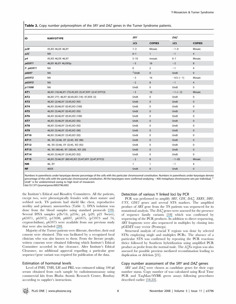
Table 2. Copy number polymorphism of the SRY and DAZ genes in the Turner Syndrome patients.
ID KARYOTYPE SRY DAZ
DCt COPIES DCt COPIES
p2B 45,XO 46,XX 46,XY 1–3 Mosaic 21–0 Mosaic
p2C NA 0–1 1 21 4
p4 45,XO 46,XX 46,XY 5–10 mosaic 0–1 Mosaic
p65971 46,XX 46,XY 46,XXXp 23 16 22 8
F- p65971 NA 0 2 21 4
p6697 NA #Undt 0 Undt 0
p65972 NA 23 16 20.5–(21) Mosaic
p65975 NA 22 8 21 4
p17698 NA Undt 0 Undt 0
AT1 46,XX (10):46,XY (75):45,XO (5):47,XXY (2):47,XYY(3) 23 16 21–(22) Mosaic
AT2 46,XX (77): 46,XY (6):45,XO (14): 47,XXX (3) Undt 0 Undt 0
AT3 46,XX (2):46,XY (3):45,XO (95) Undt 0 Undt 0
AT4 46,XX (0):46,XY (0):45,XO (100) Undt 0 Undt 0
AT5 46,XX (5):46,XY (0):45,XO (95) Undt 0 Undt 0
AT6 46,XX (0):46,XY (0):45,XO (100) Undt 0 Undt 0
AT7 46,XX (5):46,XY (0):45,XO (95) Undt 0 Undt 0
AT8 46,XX (5):46,XY (3):45,XO (92) Undt 0 Undt 0
AT9 46,XX (5):46,XY (5):45,XO (90) Undt 0 Undt 0
AT10 46,XX (5):46,XY (3):45,XO (92) Undt 0 Undt 0
AT11 46, XX (2):46, XY (2):45, XO (96) Undt 0 Undt 0
AT12 46, XX (5):46, XY (3):45, XO (92) Undt 0 Undt 0
AT13 46, XX (40):46, XY (30):45, XO (20) Undt 0 Undt 0
AT14 46,XX (5):46,XY (3):45,XO (92) Undt 0 Undt 0
AT15 46,XX (5):46,XY (80):45,XO (5):47,XXY (2):47,XYY(3) 22 8 21–(0) Mosaic
NM 46, XY 1 1 21 4
NF 46XX Undt 0 Undt 0
Numbers in parenthesis under karyotype denote percentage of the cells with the particular chromosomal constitution. Numbers in parenthesis under karyotype denotepercentage of the cells with the particular chromosomal constitution. All the karyotypes were confirmed analyzing ,400 metaphase chromosome sets per individual. #
Undt’’ is for undetermined owing to high level of mosaicism.doi:10.1371/journal.pone.0003796.t002
Y-Mosaicism & Turner Syndrome
PLoS ONE | www.plosone.org 8 November 2008 | Volume 3 | Issue 11 | e3796
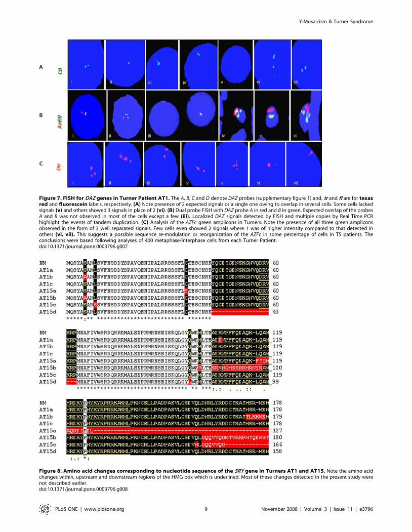
Figure 7. FISH for DAZ genes in Turner Patient AT1. The A, B, C and D denote DAZ probes (supplementary figure 1) and, tr and fl are for texasred and fluorescein labels, respectively. (A) Note presence of 2 expected signals or a single one owing to overlap in several cells. Some cells lackedsignals (v) and others showed 3 signals in place of 2 (vi). (B) Dual probe FISH with DAZ probe A in red and B in green. Expected overlap of the probesA and B was not observed in most of the cells except a few (iii). Localized DAZ signals detected by FISH and multiple copies by Real Time PCRhighlight the events of tandem duplication. (C) Analysis of the AZFc green amplicons in Turners. Note the presence of all three green ampliconsobserved in the form of 3 well separated signals. Few cells even showed 2 signals where 1 was of higher intensity compared to that detected inothers (vi, vii). This suggests a possible sequence re-modulation or reorganization of the AZFc in some percentage of cells in TS patients. Theconclusions were based following analyses of 400 metaphase/interphase cells from each Turner Patient.doi:10.1371/journal.pone.0003796.g007
Figure 8. Amino acid changes corresponding to nucleotide sequence of the SRY gene in Turners AT1 and AT15. Note the amino acidchanges within, upstream and downstream regions of the HMG box which is underlined. Most of these changes detected in the present study werenot described earlier.doi:10.1371/journal.pone.0003796.g008
Y-Mosaicism & Turner Syndrome
PLoS ONE | www.plosone.org 9 November 2008 | Volume 3 | Issue 11 | e3796

Chromosome preparation and fluorescence in-situhybridization (FISH)
Approximately, 400 ml of whole blood was cultured for
chromosome preparation following standard protocols [22]. LSI
SRY (Cat 32-191007) DNA FISH probe for SRY/CEP X was
purchased from VYSIS (Illinois, USA). FISH probes for DAZ
included Cosmids 18E8 for 59 DAZ, Probe A; 46A6 for 39DAZ,
probe B and 63C9 containing exons 2 through 11, Probe C [23].
For AZFc green amplicon, FISH probe used was BAC RP11-
336F2, probe D. The cosmid probes were purchased from Gene
service, UK (www.geneservice.co.uk/home) and BAC from
Children’s Hospital Oakland Research Institute (CHORI). Details
of the clones are given in Figure S1. FISH was conducted
following standard protocol [18,22]. Biotynilated anti-fluorescein
and anti-texas red antibodies coupled with fluorescein and texas
red avidin DCS (Vector Labs) were used in the dual probe FISH
experiments. Over 400 interphases/metaphases per individual
were analyzed for the presence/absence of the Y chromosome.
Supporting Information
Figure S1 Details of the FISH probes used for the DAZ genes
are listed in the table
Found at: doi:10.1371/journal.pone.0003796.s001 (11.64 MB
TIF)
Figure S2 Nucleotide sequence polymorphism of the SRY gene
in Turners AT1 and AT15.
Found at: doi:10.1371/journal.pone.0003796.s002 (14.71 MB
TIF)
Acknowledgments
The equipment donation from the Alexander Von Humboldt Foundation,
Bonn, Germany is gratefully acknowledged. We thank Dr. Sangeeta Thatai
and Shri Khem Singh Negi for their technical assistance.
Author Contributions
Conceived and designed the experiments: SA. Performed the experiments:
SP JS GP. Analyzed the data: SP JS SA. Wrote the paper: SP SA.
References
1. Saenger P (1997) Turner’s syndrome. Curr Ther Endocrinol Metab 6: 239–243.
2. Gravholt CH (1994) Epidemeological, endocrine and metabolic features in
Turner’s syndrome. Eur J Endocrinol 151: 657–687.
3. Ranke MB (2001) Turner’s syndrome. Lancet 358: 309–314.
4. Meng H, Hager K, Rivkees SA, Gruen JR (2005) Detection of Turner syndrome
using high-throughput quantitative genotyping. J Clin Endocrinol Metab 90(6):
3419–3422.
5. Hassold T, Benham F, Leppert M (1988) Cytogenetic and molecular analysis of
sex chromosome monosomy. Am J hum Genet 42: 534–541.
6. Jacobs P, Dalton P, James R, Mosse K, Powr M, et al. (1997) Turner syndrome:
a cytogenetic and molecular study. Ann Hum Genet 61: 471–483.
7. Gravholt CH, Fedder J, Naeraa RW, Muller J (2000) Occurrence of
gonadoblastoma in females with Turner Syndrome and Y chromosome
material: a population study. J Clin Endocrinol Metab 85: 3199–3202.
8. Hsu LYF (1994) Phenotype/Karyotype correlations of the Y chromosome
aneuploidy with emphasis on structural aberrations in postnatally diagnosed
cases. Am J Hum Genet 53: 108–140.
9. Tuck-Muller CM, Chen H, Martinez JE, Shen CLS, Kusyk C, et al. (1995)
Isodicentric Y chromosome: Cytogenetic, molecular and clinical studies and
review of literature. Hum Genet 96: 119–129.
10. Van Assche E, Boudelle M, Tournaye H, Joris H, Verheyen G, et al. (1996)
Cytogenetics of infertrile men. Genetic and assisted human conception. Hum
Reprod 11: 1–26.
11. Taraoka M, Narahara K, Yokayama Y, Tsuji K, Kikkawa K, et al. (1998) 45,X/
46X,idic(Yq) mosaicism: clinical, cytogenetic and molecular studies in four
individuals. Am J Hum Genet 78: 424–428.
12. Hook EB (1977) Exclusion of chromosomal mosaicism: Tables of 90%, 95%,
and 99% confidence limits and comments on use. Am J Hum Genet 29: 94–97.
13. Proctor SE, Walt JL, Lloyd DJ, Duffy P (1984) Problems of detecting mosaicism
in skin. A case of trisomy 8 mosaicism illustrating the advantage of in situ tissue
culture. Clin Genet 25: 273–277.
14. Reddy KS, Sulcava V (1998) Pathogenesis of 45,X/56,XY gonadal mosaicism.
Cytogenet Clin Genet 82: 52–57.
15. Kelly TE, Franko JB, Ragol A, Golden WL (1998) Discordant phenotypes and
45, X/46,x(idic)Y. J Med Genet 35: 862–864.
16. Quilter CR, Nathwani N, Conway GS, Stanhope R, Ralph D, et al. (2002) A
comparative study between infertile males and patients with Turner syndrome to
determine the influence of sex chromosome mosaicism and the breakpoints of
structurally abnormal Y chromosome on phenotypic sex. J Med Genet 39:
80–84.
Figure 9. Amino acid changes corresponding to nucleotide sequence of the SRY gene in father of a Turner patient AT1. In total , 40SRY recombinant plasmids were sequenced identifying two types of sequences AT1F2 and ATF3. Note that except the change N41I in AT1F2, rest ofthe amino acid sequence was normal. This highlights de novo status of the amino acid changes detected in the Turner AT1.doi:10.1371/journal.pone.0003796.g009
Y-Mosaicism & Turner Syndrome
PLoS ONE | www.plosone.org 10 November 2008 | Volume 3 | Issue 11 | e3796

17. Repping S, Skaletsky H, Brown L, van Daalen SK, Krover CM, et al. (2003)
Polymorphism for a 1.6-Mb deletion of the human Y chromosome persiststhrough balance between recurrent mutation and haploid selection. Nat Genet
35: 247–251.
18. Premi S, Srivastava J, Sebastian PC, Ali S (2007) AZFc Somatic microdeletionsand copy number polymorphism of the DAZ genes in human males exposed to
natural background radiations. Hum Genet 121(3): 337–346.19. Ali S, Muller CR, Epplen JT (1986) DNA fingerprinting by oligonucleotides
probes specific for simple repeats. Hum Genet 74: 239–243.
20. Bashamboo A, Rahman MM, Prasad A, Sebastian PC, Ahmad J, et al. (2005)Fate of SRY, PABY, DYS1, DYZ3 and DYZ1 loci in Indian patients harbouring
sex chromosome anomalies. Mol Hum Reprod 11: 117–127.
21. Sun C, Skaletsky H, Rozen S, Gromoll J, Nieschlag E, et al. (2000) Deletion of
the azoospermia factor a (AZFa) region of human Y chromosome caused by
recombination between HERV15 proviruses. Hum Mol Genet 9: 2291–2296.
22. Premi S, Srivastava J, Sebastian PC, Ali S (2006) Tandem Duplication and Copy
Number Polymorphism of the SRY Gene in Patients with Sex Chromosome
Anomalies and Males Exposed to Natural Background Radiation. Mol Hum
Reprod 12(2): 113–121.
23. Saxena R, De Vries JWA, Repping S, Algappan R, Skaletsky H (2000) Four
DAZ genes in two clusters found in AZFc region of the human Y chromosome.
Genomics 67: 256–267.
Y-Mosaicism & Turner Syndrome
PLoS ONE | www.plosone.org 11 November 2008 | Volume 3 | Issue 11 | e3796