Specific phosphorylation and activation of ERK1c by MEK1b: a unique route in the ERK cascade
12
Specific phosphorylation and activation of ERK1c by MEK1b: a unique route in the ERK cascade Yoav D. Shaul, 1 Gilad Gibor, 1 Alexander Plotnikov, and Rony Seger 2 Department of Biological Regulation, Weizmann Institute of Science, Rehovot 76100, Israel Extracellular signal-regulated kinases (ERKs) are key signaling molecules that regulate a large number of cellular processes, including mitosis. We showed previously that ERK1c, an alternatively spliced form of ERK1, facilitates mitotic Golgi fragmentation without the involvement of ERK1 and ERK2. Here we demonstrate that activation of ERK1c is mainly mediated by mitogen-activated protein kinase (MAPK)/ERK kinase 1b (MEK1b), which is an alternatively spliced form of MEK1 that was previously considered an inactive kinase. MEK1b phosphorylation and activity are preferentially stimulated by nocodazole, to induce its specific activity toward ERK1c. MEK1/2, on the other hand, preferentially target ERK1/2 in response to growth factors, such as EGF. As previously demonstrated for ERK1c, also MEK1b expression and activity are elevated during mitosis, and thereby enhance Golgi fragmentation and mitotic rate. MEK1 activity is also increased during mitosis, but this isoform facilitates mitotic progression without affecting the Golgi architecture. These results illustrate that the ERK cascade is divided into two routes: the classic MEK1/2–ERK1/2 and the splice-variant MEK1b–ERK1c, each of which regulates distinct cellular processes and thus extends the cascade specificity. [Keywords: MEK; ERK; signaling cascades; Golgi fragmentation; cell cycle] Supplemental material is available at http://www.genesdev.org. Received January 29, 2009; revised version accepted June 3, 2009. The extracellular signal-regulated kinase (ERK) cascade is a central mitogen-activated protein kinase (MAPK) path- way, which is activated by a large variety of extracellular agents, including growth factors and hormones (Seger and Krebs 1995; Shaul and Seger 2007). Upon activation, this cascade plays a central role in the induction of cellular processes such as proliferation, differentiation, learning and memory, and development. Under certain condi- tions, the cascade also regulates cell survival, migration, apoptosis, morphology determination, oncogenic trans- formation, and more. The ERK cascade is often activated by the small GTPase Ras, which recruits the Ser/Thr kinases Raf-1 and B-Raf to the plasma membrane leading to their activation (Wellbrock et al. 2004). Once activated, the Raf kinases transmit the signals to MAPK/ERK kinases 1 and 2 (MEK1/2), which are dual-specificity pro- tein kinases specific to the ERK cascade (Shaul and Seger 2007). A-Raf, MOS, Tpl2, and MEKK1 are also activators of MEK1/2, but operate under distinct and unique con- ditions. Upon activation, MEKs phosphorylate ERK1 and ERK2 (ERK1/2), as well as ERK1b, and this phosphoryla- tion results in the full activation of these kinases. The active ERK1/2 phosphorylate a large number of regula- tory proteins, including MAPKAPKs and transcription factors, which leads to regulation of the diverse cellular processes influenced by the ERK cascade. The 45-kDa MEK1, 46-kDa MEK2, and 43-kDa MEK1b constitute an evolutionarily conserved group of highly homologous mammalian proteins (Bendetz-Nezer and Seger 2005). Structural analysis of MEK1/2 revealed that they contain a catalytic domain, which is similar to that of other protein kinases (Ohren et al. 2004). This domain is surrounded by an N-terminal and shorter C-terminal regions, which are regulatory domains. MEK1/2 are acti- vated by their upstream regulators through phosphoryla- tion on two Ser residues in their activation loop (Ser218 and Ser222) (Alessi et al. 1994). They are localized mostly in the cytoplasm (Jaaro et al. 1997), where they interact with ERK1/2 through their N-terminal D domain, and thereby can induce cytoplasmic anchoring, as well as rapid activation of ERK1/2 upon stimulation (Tanoue et al. 2000). An alternative spliced isoform of MEK1, termed MEK1b, which lacks 26 amino acids within its kinase domain, was identified in humans (Seger et al. 1992; Zheng and Guan 1993). This isoform does not significantly activate ERK1/2 upon stimulation and, therefore, was suggested to be an inactive MEK isoform (Zheng and Guan 1993). 1 These authors contributed equally to this work. 2 Corresponding author. E-MAIL [email protected]; FAX 972-8-9344116. Article is online at http://www.genesdev.org/cgi/doi/10.1101/gad.523909. GENES & DEVELOPMENT 23:1779–1790 Ó 2009 by Cold Spring Harbor Laboratory Press ISSN 0890-9369/09; www.genesdev.org 1779
-
Upload
independent -
Category
Documents
-
view
0 -
download
0
Transcript of Specific phosphorylation and activation of ERK1c by MEK1b: a unique route in the ERK cascade

Specific phosphorylation and activationof ERK1c by MEK1b: a unique routein the ERK cascade
Yoav D. Shaul,1 Gilad Gibor,1 Alexander Plotnikov, and Rony Seger2
Department of Biological Regulation, Weizmann Institute of Science, Rehovot 76100, Israel
Extracellular signal-regulated kinases (ERKs) are key signaling molecules that regulate a large number of cellularprocesses, including mitosis. We showed previously that ERK1c, an alternatively spliced form of ERK1, facilitatesmitotic Golgi fragmentation without the involvement of ERK1 and ERK2. Here we demonstrate that activationof ERK1c is mainly mediated by mitogen-activated protein kinase (MAPK)/ERK kinase 1b (MEK1b), which is analternatively spliced form of MEK1 that was previously considered an inactive kinase. MEK1b phosphorylationand activity are preferentially stimulated by nocodazole, to induce its specific activity toward ERK1c. MEK1/2,on the other hand, preferentially target ERK1/2 in response to growth factors, such as EGF. As previouslydemonstrated for ERK1c, also MEK1b expression and activity are elevated during mitosis, and thereby enhanceGolgi fragmentation and mitotic rate. MEK1 activity is also increased during mitosis, but this isoform facilitatesmitotic progression without affecting the Golgi architecture. These results illustrate that the ERK cascade isdivided into two routes: the classic MEK1/2–ERK1/2 and the splice-variant MEK1b–ERK1c, each of whichregulates distinct cellular processes and thus extends the cascade specificity.
[Keywords: MEK; ERK; signaling cascades; Golgi fragmentation; cell cycle]
Supplemental material is available at http://www.genesdev.org.
Received January 29, 2009; revised version accepted June 3, 2009.
The extracellular signal-regulated kinase (ERK) cascade isa central mitogen-activated protein kinase (MAPK) path-way, which is activated by a large variety of extracellularagents, including growth factors and hormones (Seger andKrebs 1995; Shaul and Seger 2007). Upon activation, thiscascade plays a central role in the induction of cellularprocesses such as proliferation, differentiation, learningand memory, and development. Under certain condi-tions, the cascade also regulates cell survival, migration,apoptosis, morphology determination, oncogenic trans-formation, and more. The ERK cascade is often activatedby the small GTPase Ras, which recruits the Ser/Thrkinases Raf-1 and B-Raf to the plasma membrane leadingto their activation (Wellbrock et al. 2004). Once activated,the Raf kinases transmit the signals to MAPK/ERKkinases 1 and 2 (MEK1/2), which are dual-specificity pro-tein kinases specific to the ERK cascade (Shaul and Seger2007). A-Raf, MOS, Tpl2, and MEKK1 are also activatorsof MEK1/2, but operate under distinct and unique con-ditions. Upon activation, MEKs phosphorylate ERK1 andERK2 (ERK1/2), as well as ERK1b, and this phosphoryla-tion results in the full activation of these kinases. The
active ERK1/2 phosphorylate a large number of regula-tory proteins, including MAPKAPKs and transcriptionfactors, which leads to regulation of the diverse cellularprocesses influenced by the ERK cascade.
The 45-kDa MEK1, 46-kDa MEK2, and 43-kDa MEK1bconstitute an evolutionarily conserved group of highlyhomologous mammalian proteins (Bendetz-Nezer andSeger 2005). Structural analysis of MEK1/2 revealed thatthey contain a catalytic domain, which is similar to thatof other protein kinases (Ohren et al. 2004). This domainis surrounded by an N-terminal and shorter C-terminalregions, which are regulatory domains. MEK1/2 are acti-vated by their upstream regulators through phosphoryla-tion on two Ser residues in their activation loop (Ser218and Ser222) (Alessi et al. 1994). They are localized mostlyin the cytoplasm (Jaaro et al. 1997), where they interactwith ERK1/2 through their N-terminal D domain, andthereby can induce cytoplasmic anchoring, as well asrapid activation of ERK1/2 upon stimulation (Tanoueet al. 2000). An alternative spliced isoform of MEK1,termed MEK1b, which lacks 26 amino acids within itskinase domain, was identified in humans (Seger et al.1992; Zheng and Guan 1993). This isoform does notsignificantly activate ERK1/2 upon stimulation and,therefore, was suggested to be an inactive MEK isoform(Zheng and Guan 1993).
1These authors contributed equally to this work.2Corresponding author.E-MAIL [email protected]; FAX 972-8-9344116.Article is online at http://www.genesdev.org/cgi/doi/10.1101/gad.523909.
GENES & DEVELOPMENT 23:1779–1790 � 2009 by Cold Spring Harbor Laboratory Press ISSN 0890-9369/09; www.genesdev.org 1779

In the next level of the cascade, ERK1/2 are two evo-lutionarily conserved homologous genes that encode twomain proteins, 44-kDa ERK1 and 42-kDa ERK2 (Seger andKrebs 1995). Both are activated by phosphorylation of theregulatory Thr and Tyr residues in their activation loops.In resting cells, ERK1/2 are localized in the cytoplasmdue to interaction with various anchoring proteins, in-cluding MEKs. This interaction is mainly mediated bya region in ERK1/2, termed cytosolic retention sequence(CRS)/common docking (CD) motif that also regulatesERK1/2 interaction with MEK1/2 and phosphatases.Upon activation, they dissociate from their cytoplasmicanchors and translocate into the nucleus (Shaul and Seger2007). Aside from ERK1/2, several alternatively splicedforms of these proteins have been described, includingthe rodent ERK1b (Yung et al. 2000), the primate ERK1c(Aebersold et al. 2004), and ERK2b (Gonzalez et al. 1992).All these kinases are composed of a catalytic kinase do-main wrapped by regulatory stretches, but also containunique inserts that distinguish them from the mainERK1/2 proteins.
ERK1c is generated by the insertion of intron 7 into thecoding region of primate ERK1. This insertion containsa stop codon, resulting in the expression of a 41-kDa pro-tein kinase that contains modified 18 amino acids, justC-terminal to the CRS/CD domain. We showed thatthis protein is regulated differently from ERK1/2 due toits altered regulatory CRS/CD and C-terminal regions(Aebersold et al. 2004). Moreover, we established thatduring mitosis and elevations in cell density, ERK1crelocates to the Golgi complex, whereby it affects thearchitecture of the organelle (Aebersold et al. 2004; Shauland Seger 2006). Modulation of ERK1c expression influen-ces mitotic Golgi fragmentation, which in turn modulatesmitosis progression. These features are unique to ERK1c, asthese effects were not carried out by the main ERK1protein. These findings shed a new light on research byother laboratories that previously demonstrated an involve-ment of the ERK cascade in the regulation of Golgifragmentation without participation of doubly phosphory-lated ERK1/2 (Acharya et al. 1998; Colanzi et al. 2000; Chaand Shapiro 2001). Interestingly, the kinetics and rate ofERK1c phosphorylation upon distinct stimulations aredifferent from that of ERK1/2 (Shaul and Seger 2006), and,therefore, the protein kinases that phosphorylate ERK1cand their mechanism of action are still obscure.
We studied the possibility that ERK1c is phosphory-lated mainly by MEK1b and not MEK1/2. AlthoughMEK1b was suggested to be an inactive kinase towardERK1/2, we found that it is, in fact, active toward ERK1c.MEK1b is mainly activated upon nocodazole treatment,whereas MEK1/2, which are mainly activated by mito-genic agents, are not activated by this stimulant. Modu-lation of expression and activation levels of MEK1b, butnot of MEK1, affects ERK1c activation, as well as Golgifragmentation and mitotic rate. Thus, ERK1c lies specif-ically downstream from MEK1b, while MEK1/2 prefer-entially phosphorylate ERK1/2. Therefore, this study pro-vides evidence that during mitosis, the ERK cascade issplit into two routes—the known MEK1/2–ERK1/2 that
regulates several processes, such as CDC25 phosphoryla-tion (Wang et al. 2007); and an alternative cascade com-posed of MEK1b and ERK1c that specifically regulatesGolgi fragmentation. Thus, alternatively spliced isoformsof components of the ERK cascade can form an alterna-tive signaling route. This may join other mechanisms,such as duration and strength of the signal (Shaul andSeger 2007), as a general way of regulating specificitydeterminants that allow induction of so many distinct,and even opposing, cellular processes downstream fromRas.
Results
ERK1c phosphorylation in mitosis is preferentiallymediated by the alternative spliced MEK1b
It is well established that the ERK signaling cascade playsa role in the regulation of G2/M progression (for review,see Chambard et al. 2007). One of the G2/M-related pro-cesses regulated by the ERK cascade is Golgi fragmenta-tion (Acharya et al. 1998), and we showed previously thatthis process is regulated by ERK1c and not ERK1/2 (Shauland Seger 2006). Interestingly, the kinetics and mode ofERK1c activation were distinct from that of ERK1/2(Shaul and Seger 2006), and therefore it became importantto identify the unique mechanism of ERK1c activation.We thus examined whether MEK1/2, or the presumablyinactive MEK1b, participate in ERK1c activation. We firsttested MEKs phosphorylation upon nocodazole treat-ment, which differentially stimulates ERK1c but notERK1/2, as was previously shown by the specific de-tection with anti-pERK antibody (Ab) (Shaul and Seger2006). For this purpose, GFP-conjugated constructs ofMEK1b, MEK1, and MEK2, as well as GFP control, weretransfected into HeLa cells, and this was followed byeither nocodazole or mock treatments. Western blottingwith anti-pMEK Ab revealed that MEK1b-GFP is themain overexpressed phosphorylated MEK (Fig. 1A). In-terestingly, two endogenous proteins were recognized bythe anti-pMEK Ab as well, and this was reproduced witha similar Ab from another source. One of the identifiedproteins is the 35-kDa nucleoplasmin (NPM), which isexpressed mainly during the G2/M phase of the cell cycle.Importantly, although this protein does not have a signif-icant resemblance to MEKs, it is nonspecifically recog-nized by all anti-pMEK Abs (Cha et al. 2004). The other,more interesting band was a weak 42-kDa band that waselevated by the treatment and is likely to be the endog-enous MEK1b. Since the phosphorylation of MEK1/2 ismediated under most conditions by Raf kinases (Shauland Seger 2007), we examined whether MEK1b may bephosphorylated downstream from these kinases as well.Indeed, we found that phosphorylation of MEK1b uponnocodazole treatment is inhibited by the selective Rafinhibitor Sorafenib (Supplemental Fig. S1). Thus, in simi-larity to ERK1c, which is the main nocodazole-activatedERK, MEK1b is the main MEK isoform phosphorylatedunder the same conditions, and this is mediated, at leastin part, by the upstream Raf kinases.
Shaul et al.
1780 GENES & DEVELOPMENT

In an effort to further evaluate the role of MEK1b asa major ERK1c kinase, we compared the ability of thevarious MEKs to phosphorylate the regulatory Thr andTyr in ERK1c. Thus, we precipitated the GFP-MEKs fromnocodazole-stimulated and untreated cells and subjectedthem to an in vitro phosphorylation reaction with puri-fied GST-ERK1c, GST-ERK1, and GST-ERK2 as sub-strates. The nocodazole-treated MEK1b phosphorylatedERK1c much stronger than MEK1/2, while no phos-phorylation of ERK1 could be detected (Fig. 1B). On theother hand, MEK1 slightly phosphorylated ERK1 andERK2; MEK2 phosphorylated only ERK1; and as wouldbe expected from the minor ERK1/2 activation (Shaul andSeger 2006), these phosphorylations were only slightlystimulated upon nocodazole treatment. Thus, MEK1b,previously suggested to be an inactive kinase (Zheng andGuan 1993), is, in fact, active toward ERK1c and is likelyto be the main activator of ERK1c during mitosis.
To further link the activation of MEK1b with that ofERK1c, we also studied the EGF-induced activation ofMEK1b, as compared with MEK1. Thus, we overex-pressed GFP-MEK1, GFP-MEK1b, or GFP alone as controlin HeLa cells and either left the cells untreated, or treatedthem with nocodazole. Alternatively, the transfectedcells were serum-starved and then left either untreatedor stimulated with EGF. The exogenous MEKs werethen immunoprecipitated by anti-GFP Ab and used tophosphorylate either GST-ERK1c or GST-ERK1. Uponnocodazole treatment, MEK1b demonstrated high activ-ity toward ERK1c, but only a minor activity toward ERK1(Fig. 1C). However, upon EGF treatment, the activity ofMEK1b toward ERK1c was marginal, and the activitytoward ERK1 was not stimulated. On the other hand, EGFstimulated the MEK1-activated ERK1 but had muchless effect on ERK1c. These results further demonstratea differential regulation in which EGF primarily acti-vates MEK1/2, which are specific to ERK1/2, whilenocodazole primarily stimulates MEK1b, which is spe-cific to ERK1c.
In vitro phosphorylation reveals that MEK1b isa specific ERK1c kinase, while MEK1 prefers ERK1
To obtain an insight into the kinetic parameters of ERK1cand ERK1 phosphorylation by MEK1b and MEK1, weused recombinant active MEKs, purified from bacteria.Thus, we prepared the construct 6His-DN-EE-MEK1b,which by analogy to the previously published 6His-DN-EE-MEK1 (Mansour et al. 1996; Jaaro et al. 1997) shouldbe constitutively active. This construct, as well as 6His-DN-EE-MEK1, was then tested for their ability to doublyphosphorylate ERK1c or ERK1. A calibration curve, con-taining three distinct amounts of bacterially expressedpERK2 (Yung et al. 1997), was added to each of theWestern blots to allow for a similar exposure and com-parison between the phosphorylation levels in the dif-ferent experiments. Under these conditions, the activityof MEK1b toward ERK1c was ;15-fold higher than to-ward ERK1 (Fig. 2A,B), while MEK1 phosphorylated ERK1;10-fold better than ERK1c. In addition, the phosphorylation
Figure 1. ERK1c phosphorylation in mitosis is preferentiallymediated by the alternative spliced MEK1b. (A) MEK1b undergoesphosphorylation during mitosis. HeLa cells were transfected withGFP, MEK1-GFP, MEK2-GFP, or MEK1b-GFP. Each of these trans-fected cells was treated as follows: untreated (�), nocodazole [100ng/mL, 24 h (+)], or serum-starved; and then stimulated with EGF [50ng/mL, 3min (E)]. The cells were harvested and subjected to Westernblotting with anti-pMEK, anti-GFP, and anti-gMEK(C-18) Abs. (B)MEK1b preferentially phosphorylates ERK1c during mitosis. HeLacells were transfected as in A. The cells were treated with nocoda-zole [100 ng/mL, 24 h (+)] or left untreated (�). The cells were thenharvested, and the GFP proteins were immunoprecipitated withanti-GFPAb. The precipitated proteins were subjected to an in vitrophosphorylation system using GST-ERK1, 2 and 1c as substrates, asdescribed in the Materials and Methods. The amounts of ERK1, 2and 1c as well as their phosphorylation levels were determined byWestern blotting using apERK, and agERK(CRS) Abs. The relativeamounts of MEKs were determined by anti-gMEK(C-18) Ab, theMEKs used to phosphorylate ERK1/1c are in panel 5, and for ERK2are in panel 8. (C) Differences between MEK1b and MEK1 stimula-tion and substrate determination. HeLa cells were transfected withMEK1b-GFP, MEK1-GFP, or GFP. The cells were treated as follows:untreated (�), nocodazole [100 ng/mL, 24 h (N)], serum-starved [16h 0.1% FCS, (S)], or serum-starved and then stimulated with EGF[50 ng/mL, 3 min (E)]. The cells were harvested and the GFP-taggedproteins were immunoprecipitated by aGFP Ab, and each one ofthem was subjected to an in vitro phosphorylation using GST-ERK1/1c as substrates. The amounts of ERK1/1c as well as theirphosphorylation levels were determined by Western blotting usinganti-pERK, anti-gERK(CRS), and anti-GFP Abs.
MEK1b–ERK1c is an ERK cascade subroute
GENES & DEVELOPMENT 1781

of ERK1c by MEK1b was about fivefold higher thanby MEK1, while MEK1 phosphorylated ERK1 30-foldhigher than MEK1b. The relative phosphorylation rates,which were obtained by using different SDS gels withcalibration curves, were verified by comparing some ofthe samples side-by-side on one SDS gel (Fig. 2C).
To validate the results with the recombinant proteins,we also used immunoprecipitation of stimulated MEK1-GFP or MEK1b-GFP that were overexpressed in HeLacells (Supplemental Fig. S2). Maximal stimulation wasachieved by stimulating MEK1b-GFP-containing cellswith nocodazole and MEK1-GFP-expressing cells withEGF. The stimulated MEKs were then immunoprecipi-tated, stringently washed, and used to phosphorylateGST-ERK1c or GST-ERK1 in vitro. As above, we addedcalibration curves of bacterially expressed pERK2 to eachof the Western blots to allow similar exposures. As ex-pected, MEK1b phosphorylated ERK1c much better thanERK1, while MEK1 preferentially phosphorylated ERK1.In addition, the rate of ERK1c phosphorylation by MEK1bwas much better than that by MEK1, while MEK1 phos-phorylated ERK1 much better than MEK1b. When U0126was added, it significantly inhibited both MEKs, confirm-ing that the ERK1/2 phosphorylation was indeed exe-cuted by MEKs. Taken together, our results indicate thatMEK1b, which was thought previously to be an inactivekinase (Zheng and Guan 1993), is indeed only marginallyactive toward ERK1/2 but can serve as an active kinase ofERK1c.
Endogenous MEK1b is activated by nocodazoleand specifically phosphorylates ERK1c
Since the above experiments were performed with re-combinant MEK1b, it was important to show thatthe endogenous protein exhibits similar characteristics.For this purpose, we fractionated extracts from nocoda-zole-treated HeLa cells (Fig. 3A) from cells in G2/M, 9 hafter release from double-thymidine block and from un-treated cells (data not shown) on a Mono-Q FPLC column.In all columns, MEK1b was eluted by similar NaClconcentrations (0.09–0.13 M, fractions 40–47), whileMEK1/2 were eluted in the flow-through. With the useof anti-pMEK Ab, we found that the endogenous MEK1bas well as MEK1/2 from nocodazole-treated and thymi-dine-released cells were phosphorylated on their activi-tory Ser residues. However, as would be expected fromthe results with overexpressed constructs (Fig. 1), thespecific phosphorylation of MEK1b was much higherthan that of MEK1, and both of them were not phosphor-ylated in untreated cells (Fig. 3B,C). No significantamount of a 35-kDa band was detected in any of thefractions by the anti-pMEK Ab, indicating that NPM,expressed in G2/M cells (Fig. 1), was either degradedor eluted by higher NaCl concentrations. Importantly,when the activity of MEKs was assayed, the fractionatedMEK1b from both nocodazole-treated and from thymi-dine-released cells, but not from unstimulated cells, phos-phorylated ERK1c and to a much lower extent also ERK1(Fig. 3D,E). MEK1/2, which is activated to a lesser extentby nocodazole or after release from double-thymidineblock, did not significantly phosphorylate ERK1c underthese conditions. These results are in agreement with theresults with overexpressed MEKs, supporting again the spe-cific role of endogenous MEK1b in the activation of ERK1cduring G2/M.
Figure 2. Specificity studies using constitutively active MEK1band MEK1. (A) Time course of ERK1 and ERK1c phosphoryla-tion by constitutively active MEK1b and MEK1. In vitro phos-phorylation was preformed as described under Materials andMethods with recombinant His-DN-EE-MEK1b and His-DN-EE-MEK1 as kinases and GST-ERK1 and GST-ERK1c as substrates.The amounts of ERK1/1c and their phosphorylation levels weredetermined by Western blotting with anti-pERK, anti-gERK(CRS),and anti-gMEK(C-18) Abs. (B) Quantification of ERK1/1c phos-phorylation. Known amounts of phosphorylated ERK were addedto the blots to form a calibration curve, which enabled the de-termination of ERK1/1c phosphorylation levels (see text fordetails). The results in the graphs represent means 6 SE of threeexperiments. (Squares) ERK1 phosphorylation; (triangles) ERK1cphosphorylation. (C) Side-by-side kinetics of ERK1/1c dual phos-phorylation by MEK1/1b. In vitro phosphorylation was performedusing amounts of recombinant proteins that were found optimalfor each phosphorylation [His-DN-EE-MEK1 (CA-MEK1), 0.5 mgper reaction; His-DN-EE-MEK1b (CA-MEK1b), 0.75 mg; GST-ERK1, 0.5 mg or GST-ERK1c 0.5 mg] for 0, 2, and 10 min at30°C as described in the Materials and Methods. The levels ofERK1/1c phosphorylation were detected by resolving the samplesby one SDS-PAGE and Western blotting using anti-pERK, andanti-gERK(CRS) Abs.
Shaul et al.
1782 GENES & DEVELOPMENT

Modulation of MEKs expression verifies MEK1band MEK1 specificities in vivo
In order to verify that the specificities of MEK1b andMEK1 toward ERK1c and ERK1 occur also in vivo, wemodulated MEK1 and MEK1b levels by either overex-pression or specific Sh-RNAs’ knockdown. The trans-fected cells were stimulated with nocodazole, ERK1c andERK1 were each immunoprecipitated, and their activitieswere determined using myelin basic protein (MBP) asa substrate. As reported (Shaul and Seger 2006), ERK1cwas strongly activated upon nocodazole treatment ofHeLa cells, while U0126 prevented this effect (Fig.4A,B). Overexpression of MEK1b slightly elevated ERK1cactivity, while overexpression of MEK1 had no significanteffect. The fact that overexpression of MEK1b had onlya small effect could be explained by saturation in thekinase levels, as was previously reported for the effects ofMEK1 toward ERK1/2 (Seger et al. 1992). On the otherhand, reduced MEK1b expression (Fig. 4C) gave a strongereffect, as its Sh-RNA (Sh-MEK1b) transfection signifi-cantly reduced ERK1c activity (Fig. 4A,B). The influenceof Sh-MEK1b was not due to any off-target effects, as re-vealed by rescue experiments using expression of WT-MEK1b in cells containing Sh-MEK1b (data not shown).As expected from our results above, Sh-RNA of MEK1
had only a small effect on ERK1c activity, and ERK1activity was not significantly altered, mainly due to lackof sensitivity to nocodazole treatment.
The expression levels and activity of MEK1bare elevated during mitosis
Our findings indicate that MEK1b regulates ERK1c ac-tivity, and ERK1c expression and activity are elevated inmitosis (Shaul and Seger 2006). Therefore, we examinedwhether MEK1b activity indeed correlates with that ofERK1c. For this purpose, we synchronized HeLa cells,using a double-thymidine block (Merrill 1998). FACSanalysis (data not shown) and mitotic index determina-tion (Fig. 5A) revealed that most of the cells are found inG2 within 6 h, in mitosis after 10 h, and cycled back to thenext G1 within 11–12 h. We then examined the behaviorof MEK1b as compared with MEK1/2 in the synchro-nously cycling cells, 6 h (G2), 9 h (G2/M), 10 h (peakmitosis), and 11 h (late mitosis/early G1) after release.Western blotting with anti-MEK Ab showed that MEK1blevels are increased during G2/M and mitosis, while theexpression levels of MEK1/2 were constant during thetimes examined (Fig. 5B). In addition, blotting with anti-pMEK Ab revealed that the phosphorylation of endoge-nous MEK1b, and to some extent endogenous MEK1/2,
Figure 3. Fractionation of endogenousMEK1b verifies its specificity towardERK1c. (A) Fractionation of MEK1b ona Mono-Q FPLC column. HeLa cells (10plates of 10 cm) were treated with nocoda-zole (100 ng/mL, 24 h) and harvested inBuffer A. Cell extracts were loaded ontoa Mono-Q FPLC column, which waswashed with Buffer A and developed usingan increasing NaCl gradient (dashed line),to give rise to the protein profile shown bythe solid line. Aliquots (40 mL, boiled insample buffer) of every third fraction weresubjected to Western blotting with anti-gMEK(H-8) Abs. Identical fractionation ofMEK1/2 and MEK1b was observed withextracts from HeLa cells that were har-vested 9 h after release from double-thymi-dine block and from untreated cells (datanot shown). (B,C) Endogenous MEK1b fromG2/M cells is phosphorylated on its regula-tory residues. Fractions containing MEK1/2(#4, #5, #9) or MEK1b (#41, #42, #43) fromcells that underwent treatment with noco-dazole as described above (B), 9 h afterrelease from double-thymidine block asdescribed above (C), or untreated cells(B,C, indicated fractions) were subjected toWestern blotting with anti-pMEK (top
panel) and anti-gMEK(H-8) Ab (bottom panel). Similar results were obtained with Ab from other companies (data not shown). (D,E)The endogenous activated MEK1b from G2/M cells phosphorylates mainly ERK1c in vitro. The indicated aliquots of MEK1/2- orMEK1b-containing fractions from nocodazole-treated cells (D), from cells that were released from double-thymidine block (E), or fromuntreated cells (E,D, indicated fractions) were used to phosphorylate GST-ERK1c (0.5 mg) or GST-ERK1 (0.5 mg), as described in Materialand Methods. The phosphorylated proteins were analyzed by autoradiography (top panels) or by Western blotting with anti-pERK andanti-gERK(CRS) Abs (bottom panels) as indicated.
MEK1b–ERK1c is an ERK cascade subroute
GENES & DEVELOPMENT 1783
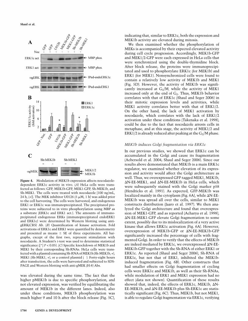
was elevated during the same time. The fact that thehigher pMEK1b is due to specific phosphorylation, andnot elevated expression, was verified by equilibrating theamount of MEK1b in the different lanes. Indeed, alsounder these conditions, MEK1b phosphorylation wasmuch higher 9 and 10 h after the block release (Fig. 5C),
indicating that, similar to ERK1c, both the expression andMEK1b activity are elevated during mitosis.
We then examined whether the phosphorylation ofMEKs is accompanied by their expected elevated activityduring cell cycle progression. Accordingly, MEK1b-GFPand MEK1/2-GFP were each expressed in HeLa cells thatwere synchronized using the double-thymidine block.After block release, the proteins were immunoprecipi-tated and used to phosphorylate ERK1c (for MEK1b) andERK1 (for MEK1). Nonsynchronized cells were found tocontain a relatively low activity of MEK1b and MEK1(Fig. 5D). However, the activity of MEK1b was signifi-cantly increased at G2/M, while the activity of MEK1increased only at the end of G2. Thus, MEK1b behaviorcorrelates with that of ERK1c (Shaul and Seger 2006) intheir mitotic expression levels and activities, whileMEK1 activity correlates better with that of ERK1/2.On the other hand, the lack of MEK1 activation bynocodazole, which correlates with the lack of ERK1/2activation under these conditions (Takenaka et al. 1998),could be due to the fact that nocodazole arrests cells inmetaphase, and at this stage, the activity of MEK1/2 andERK1/2 is already reduced after peaking at the G2/M phase.
MEK1b induces Golgi fragmentation via ERK1c
In our previous studies, we showed that ERK1c can beaccumulated in the Golgi and cause its fragmentation(Aebersold et al. 2004; Shaul and Seger 2006). Since ourresults above demonstrated that MEK1b is a main ERK1cregulator, we examined whether elevation of its expres-sion and activity would affect the Golgi architecture aswell. Thus, we overexpressed GFP-tagged MEK1, MEK1b,DN-EE-MEK1, and DN-EE-MEK1b in HeLa cells, whichwere subsequently stained with the Golgi marker p58(Hendricks et al. 1991). As expected, GFP-MEK1b waslocalized mainly in the cytoplasm (Fig. 6A), while DN-EE-MEK1b was spread all over the cells, similar to MEK1constructs distribution (Jaaro et al. 1997). We then ana-lyzed the Golgi architecture and found that overexpres-sion of MEK1-GFP, and as reported (Acharya et al. 1998),DN-EE-MEK1-GFP elevate Golgi fragmentation to someextent, possibly due to its mislocalization of a very activekinase that allows ERK1c activation (Fig. 6A). However,overexpression of MEK1b-GFP or DN-EE-MEK1b-GFPsignificantly increased the percentage of cells with frag-mented Golgi. In order to verify that the effects of MEK1bare indeed mediated by ERK1c, we overexpressed DN-EE-MEK1b-GFP together with the Sh-RNA of either ERK1 orERK1c. As reported (Shaul and Seger 2006), Sh-RNA ofERK1c, but not that of ERK1, inhibited the MEK1b-induced fragmentation (Fig. 6B). Other constructs thathad smaller effects on Golgi fragmentation of cyclingcells were ERK1c and MEK1b, as well as their Sh-RNAs,while modulation of ERK1 and MEK1 expression had noeffect (data not shown). Quantification of these resultsshowed that, indeed, the effects of ERK1c, MEK1b, DN-EE-MEK1b, and DN-EE-MEK1b plus Sh-ERK1c are statis-tically significant (Fig. 6C). Thus, MEK1b, but not MEK1,is able to regulate Golgi fragmentation via ERK1c, verifying
Figure 4. Modulation of MEK1b expression affects nocodazole-dependent ERK1c activity in vivo. (A) HeLa cells were trans-fected as follows: GFP, MEK1b-GFP, MEK1-GFP, Sh-MEK1b, andSh-MEK1. The cells were treated with nocodazole [100 ng/mL,24 h, (+)]. The MEK inhibitor U0126 (5 mM, 1 h) was added priorto the cell harvesting. The cells were harvested, and endogenousERK1 or ERK1c was immunoprecipitated. The precipitated pro-teins were subjected to in vitro phosphorylation using MBP asa substrate (ERK1c and ERK1 act.). The amounts of immuno-preipitated endogenous ERKs (immunoprecipitated endoERK1and ERK1c) were determined by Western blotting using anti-gERK(CRS) Ab. (B) Quantification of kinase activation. Foldactivations of ERK1c and ERK1 were quantified by densitometryand presented as means 6 SE of three experiments. All bar-graphs, except of the first two, represent stimulation withnocodazole. A Student’s t-test was used to determine statisticalsignificance ([*] P < 0.05). (C) Specific knockdown of MEK1b andMEK1 by their corresponding Sh-RNAs. HeLa cells were trans-fected with a plasmid containingSh-RNA of MEK1b (Sh-MEK1b, +),MEK1 (Sh-MEK1, +), or a control plasmid (�). Forty-eight hoursafter transfection, the cells were harvested and subjected to SDS-PAGE and Western blotting with anti-gMEK (H-8) Abs.
Shaul et al.
1784 GENES & DEVELOPMENT

that both MEK1b and ERK1c participate in the regulationof Golgi fragmentation.
Importantly, the effects of MEK1b were not restricted toHeLacells thatwereused in all theaboveexperiments.Thus,nocodazole treatment of HEK293T cell resulted in TEYphosphorylation of ERK1c in a MEK-dependent manner,whereas ERK1 or ERK2 was only slightly phosphorylated(Supplemental Fig. S3). Similar results were also seen inCOS7 cells (data not shown). In addition, the release ofa double-thymidine block showed that the expressionand phosphorylation of MEK1b, but much less of MEK1/2, are elevated in G2/M (9–11 h after release). Finally, incorrelation to Sh-ERK1c, Sh-MEK1b (but not Sh-MEK1)reduced the number of cells with fragmented Golgi, asshown above in HeLa cells. Together, these resultsindicate that the MEK1b/ERK1c role in Golgi fragmen-tation is general and not restricted to HeLa cells.
We showed previously that ERK1c regulates Golgi frag-mentation particularly during mitosis (Shaul and Seger2006), and, therefore, it was important to demonstratethat MEK1b can induce the same effects. For this purpose,we modulated MEK1b expression by overexpressing itsGFP-tagged form or knocking it down by Sh-RNA, andfollowed Golgi architecture with the Golgi marker p58.As expected from the ERK1c results (Shaul and Seger2006), elevated MEK1b expression levels increased thenumber of cells with fragmented Golgi, especially duringprophase (Fig. 7). Lowering MEK1b expression signifi-cantly reduced the percentage of cells with fragmentedGolgi during prophase, prometaphase, and modulatedGolgi distribution during metaphase. Moreover, in corre-lation with the effect of Sorafenib on MEK1b activity(Supplemental Fig. S1), this Raf inhibitor also signifi-cantly reduced Golgi fragmentation (Supplemental Fig.S4). Therefore, these results strongly support a role ofa specific MEK1b–ERK1c cascade downstream from Rafkinases, in the regulation of Golgi fragmentation duringmitosis. Interestingly, brefeldin-A (BFA), which inducesGolgi fragmentation by various mechanisms (Chardinand McCormick 1999), induced Golgi fragmentation inour system as well (Supplemental Fig. S4) but failed toinduce MEK1b activation (Supplemental Fig. S1). Thisindicates that the MEK1b–ERK1c cascade induces Golgifragmentation only under selected conditions.
MEK1b regulates mitotic progression
We then examined whether cellular MEK1b regulatesmitotic progression as much as ERK1c (Shaul and Seger2006). Therefore, the expression levels of MEK1b, MEK1,and ERK1c were modulated; the cells were synchronizedwith or without the MEK inhibitor U0126; and the cellcycle stage was determined by FACS. Thus, 6 h afterblock release, the various transfected cells were found inthe G2 phase of the cell cycle, indicating that neitherMEK1b and MEK1, nor ERK1 and ERK1c play a role inS-to-G2 progression of HeLa cells (Supplemental Fig. S5).On the other hand, 9 h after release, MEK1b had a markedinfluence on mitotic progression. Thus, overexpression ofMEK1b promoted the mitotic progression, and knockdown
Figure 5. The expression levels and activity of MEK1b areelevated during mitosis. HeLa cells were synchronized by a dou-ble-thymidine block, followed by the removal of the block toenable cell cycle progression (Syn), or left untreated (non-synchronized, N.S.) as a control. (A) Mitotic index determination.The cells were fixed and stained with DAPI. The percentage ofmitotic cells out of the total number of nuclei counted is pre-sented as the means 6 SE of three experiments; N = 300. (B)MEK1b expression and phosphorylation are elevated in the G2/Mphase of the cell cycle. The cells were harvested and subjected toWestern blotting with anti-gMEK(H-8) and anti-pMEK Abs. Fora better resolution of the MEK1b, SDS-PAGE was longer than inprevious experiments. Exposure times for gMEK1/2 and gMEK1bwere different. (C) Specific MEK1b phsphorylation is elevatedduring mitosis. Equal amounts of MEK1b (judged by a calibratingWestern blot) from each time after block release were subjected toWestern blotting with anti-pMEK Ab. (D) MEK1b activity is en-hanced in the G2/M phase of the cell cycle. HeLa cells weretransfected with MEK1b-GFP or MEK1-GFP. The cells were syn-chronized by double-thymidine block, and times after release areindicated. In addition, one plate was serum-starved and stim-ulated with EGF (50 ng/mL, 3 min; E). The cells were harvested,and MEK1b or MEK1 was immunoprecipitated using anti-GFPAb. The immunoprecipitated proteins were used as kinases for invitro phosphorylation, where GST-ERK1c served as a substrate forMEK1b and GST-ERK1 for MEK1. The amounts of ERK1/1c as wellas their phosphorylation levels were determined by Western blot-ting using anti-pERK, anti-gMEK(H-8), and anti-gERK(CRS) Abs.
MEK1b–ERK1c is an ERK cascade subroute
GENES & DEVELOPMENT 1785

of MEK1b delayed the cells at G2 with similarity to theU0126 treatment. In these experiments, MEK1 had noinfluence on these cell cycle parameters. These effects ofMEK1b were mostly mediated by ERK1c, since the in-terfering construct of the latter, not of ERK1, significantlyreduced the MEK1b-induced mitotic progression. Theseeffects were restricted to 10–12 h after the block release,
as no significant effect on cell cycle distribution wasobserved 13 h after the release. The effects on the cellcycle correlated well with the rate of Golgi fragmenta-tion. Interestingly, the constitutively active MEK1 thatdid induce Golgi fragmentation (Fig. 6) also had a strong
Figure 7. Modulation of MEK1b expression affects Golgi frag-mentation during mitosis. (A) MEK1b affects Golgi architectureduring stages of mitosis. HeLa cells were transfected with GFP,GFP-MEK1b (MEK1b), or Sh-MEK1b with GFP (Sh-MEK1b). Thecells were synchronized by double-thymidine block, and 9 hafter the release from the block were stained with p58 Abs andDAPI. Cells from interphase, prophase, prometaphase, and meta-phase were selected by their DNA structure. Bar, 5 mm. (B)Quantification of Golgi fragmentation in prophase prometa-phase and metaphase. The cells in prophase and prometaphasewere counted, and the percentage of cells with fragmented Golgiout of total transfected cells is presented as means 6 SE of threeexperiments (n = 40). The cells in metaphase were counted, andthe percentage of cells with partially intact Golgi per totaltransfected cells is presented as means 6 SE of three experi-ments. A Student’s t-test was used to determine statisticalsignificance. (*) P < 0.05; (**) P < 0.01.
Figure 6. MEK1b induces Golgi fragmentation. (A) The effect ofMEKs constructs on Golgi architecture. HeLa cells were trans-fected with GFP, MEK1b-GFP (MEK1b), MEK1-GFP (MEK1), DN-EE-MEK1b-GFP (DN-EE-MEK1b), or DN-EE-MEK1-GFP (DN-EE-MEK1-GFP). Fourteen hours after transfection, the cells werefixed, stained with anti-p58 Ab, and visualized by a fluorescentmicroscope. Arrows indicate fragmented Golgi. Bar, 8 mm. (B)ERK1c is required for MEK1b-induced Golgi fragmentation. HeLacells were transfected with GFP alone as control or cotransfectedwith DN-EE-MEK1b-GFP together with GFP, ERK1c Sh-RNA (Sh-ERK1c), or ERK1 Sh-RNA (Sh-ERK1). The cells were then fixed,stained with anti-p58 Ab, and visualized by a fluorescent micro-scope. (Arrows indicate fragmented Golgi. Bar, 8 mm.) (C) Quan-tification of Golgi fragmentation. The HeLa cells in A and B werecounted, and the percentage of fragmented Golgi per total trans-fected cells is presented. The results are means 6 SE of threeexperiments, n = 200. A Student’s t-test was used for statisticalsignificance. (*) P < 0.05; (**) P < 0.01.
Shaul et al.
1786 GENES & DEVELOPMENT

effect on mitotic progression, but this effect is likely toinvolve other processes besides Golgi fragmentation(Wang et al. 2007). The lack of effect of Sh-MEK1 andSh-ERK1 on mitotic progression could be explained bythe compensation of MEK2 or ERK2 that were notsimultaneously reduced. Overall, our results suggest thatMEK1b participates in the regulation of mitotic progres-sion by affecting Golgi fragmentation, and this is medi-ated via ERK1c, which demonstrates similar effects.Therefore, these results provide another indication thatthese two kinases function within their own unique sig-naling cascade.
Discussion
In this study, we demonstrate a novel ERK signaling route,composed of the alternatively spliced forms MEK1b andERK1c that specifically function during mitosis. We dem-onstrated previously that ERK1c plays a unique role inregulating Golgi fragmentation (Shaul and Seger 2006).Although the activation of ERK1c was inhibited by MEKinhibitors, its mode and kinetics of phosphorylation weredistinct from those of ERK1/2, suggesting a differentmechanism of activation of ERK1c. Therefore, we exam-ined whether the alternatively spliced isoform of MEK1,namely, MEK1b (Seger et al. 1992; Zheng and Guan 1993),could be the upstream kinase of ERK1c. Indeed, we foundthat this isoform may be a specific ERK1c kinase, as con-cluded from the following results: (1) MEK1b is phos-phorylated and activated better than MEK1/2 uponnocodazole treatment, correlating with the preferentialactivation of ERK1c over ERK1/2 (Shaul and Seger 2006).(2) Active MEK1b preferentially phosphorylated ERK1c invitro, whereas active MEK1 phosphorylated ERK1 better.(3) Changes in MEK1b expression and activation levelsmodulated ERK1c, but not ERK1 activities. (4) In corre-lation with ERK1c, the expression levels and activity ofMEK1b is enhanced during mitosis. (5) In correlation withERK1c, modulation of MEK1b expression and activityaffects Golgi fragmentation as well as the rate of mitosis.(6) MEK1b-induced Golgi fragmentation and mitotic pro-gression is inhibited by ERK1c, but not ERK1 knock-down. These findings strongly suggest the existence oftwo distinct signaling routes—one is composed specifi-cally from MEK1b–ERK1c, and the other contains MEK1–ERK1/2, with a very little cross-talk between them.
MEK1b was suggested to be an inactive form of MEK1(Zheng and Guan 1993), as it failed to phosphorylateERK1/2 upon EGF and TPA stimulation. Indeed, in ourstudy, we showed that it exhibits only a marginal activitytoward ERK1/2, but we demonstrated that MEK1b isactivated during mitosis, when it phosphorylates primaryERK1c. Thus, the elimination of 26 amino acids does notcause inactivation due to the expected major conforma-tional changes. However, this deletion does modify thesubstrate specificity of MEK1b and makes it suitable tophosphorylate ERK1c without modifying the uniquefeature of MEKs, which are very selective kinases to theirrespective ERKs. Since the C-terminal region, just afterthe CRS/CD domain, is modified in ERK1c, it is still
possible that the interaction between the kinases is medi-ated by the MEK1b’s D-domain (Tanoue et al. 2000). Thisis supported by the fact that the ERK1c phosphorylationrate by MEK1b is higher than the rate of CRS/CD-deficient ERK1 phosphorylation, and some interactionsbetween MEK1b and ERK1c do exist in coimmunopreci-pitation assays (data not shown). Therefore, the changesin interaction of ERK1c and MEK1b are likely to be medi-ated mainly by the deleted regions, which are consequen-tly identified here to contribute some binding affinity inthe ERK1–MEK1 association.
The distinct activation of the two ERK routes raises thequestion as to what might be the mechanism of theiractivation. The difference in activation upon EGF stim-ulation, as compared with nocodazole treatments, mayindicate a distinct upstream mechanism of activation ofMEK1b and MEK1. Since Raf-1 was reported to partici-pate in mitotic ERK1/2 activation (Colanzi et al. 2003b;Liu et al. 2004), it is possible that MEK1/2 are activatedat this stage by this isoform. However, it was shown thatthe phosphorylation of MEK1 during G2/M of the cellcycle is different from that in interphase cells (Colanziet al. 2000). This effect could be due to a different up-stream component that phosphorylates mainly MEK1b,resulting in a distinct phosphorylation pattern. Such adistinct upstream component is likely to be activatedonly during mitosis and not upon mitogenic stimulation.Since MEK1b effects are inhibited by the general Rafinhibitor Sorafenib (Supplemental Fig. S1), this compo-nent might be either A-Raf or an alternatively splicedform of the other Raf kinases that are activated specifi-cally in G2/M.
ERK1/2 are activated by a wide variety of extracellularagents that induce different, or even opposing, cellularprocesses. For example, ERK1/2 were shown to inducecell proliferation upon mitogenic stimulation (Seger andKrebs 1995), but at the same time, they can mediateapoptotic signals upon certain proapoptotic stimulations(Bacus et al. 2001). These effects raise the question as tohow the specificity of the different signals transmitted bythe ERK cascade is regulated. Several mechanisms thatparticipate in this specificity determination have beenproposed in the past years (Shaul and Seger 2007); one ofthem is the presence of several similar isoforms (Lloyd2006), including alternatively spliced forms (Shaul andSeger 2007) at each tier of the cascade. Such isoforms,which might be differentially regulated and have distinctsubstrates, may extend the specificity of the signal trans-mitted by the cascade.
Several alternatively spliced isoforms of components ofMAPK cascades have been demonstrated to function dif-ferently from their main isoforms. In the p38 cascade,Mxi2 and Exip, which differ from the p38a in their Cterminus (18 amino acids in Mxi2) (Zervos et al. 1995) and58 amino acids in Exip (Sudo et al. 2002), have uniquesignaling functions. Mxi2 binds ERK1/2 and regulatestheir subcellular localization (Casar et al. 2007), whileExip binds Toll-interacting protein and down-regulatesthe NF-kB pathway (Yagasaki et al. 2004). Since the full-length p38a is not involved in these processes, Exip and
MEK1b–ERK1c is an ERK cascade subroute
GENES & DEVELOPMENT 1787
Mxi2 extend the number of p38 functions. Differentalternatively spliced forms were also reported for ERK5(Yan et al. 2001), MEK5 (English et al. 1995), JNK (Guptaet al. 1996), MKK7 (Tournier et al. 1999), and others.In addition, besides ERK1c, two alternatively splicedforms—ERK1b (Yung et al. 2000) and ERK2b (Gonzalezet al. 1992)—have been reported for ERKs, and otherforms may exist as well (Ben-David et al. 2006). Thus,alternatively spliced isoforms of components of MAPKcascades add to the complexity of the signaling network,which allows the regulation of the wide range of pro-cesses governed by these cascades.
The involvement of the ERK cascade in regulatingG2/M and mitosis phases of the cell cycle is well estab-lished (Chambard et al. 2007). In the past few years, theERK cascade has been implicated in the regulation ofseveral mitotic components, such as CDC25 (Wang et al.2007), CENP-E (Zecevic et al. 1998), SWI/SNF (Sif et al.1998), and PLK3 (Xie et al. 2004). In addition, the ERKcascade may play a role in the mitotic Golgi fragmenta-tion (Acharya et al. 1998; Kano et al. 2000; Feinstein andLinstedt 2007). It was also suggested that MEK1 isactivated at early stages of mitosis when it is presumablyphosphorylated by Raf1 (Colanzi et al. 2000, 2003a).However, the mitotic MEK generated different pep-tides from the interphase MEK1 upon trypsin digestion(Colanzi et al. 2000). In addition, cleavage of the mitoticMEK with anthrax lethal factor (which cleaves the Nterminus of MEK1 and disrupts its activity) did not affectGolgi fragmentation (Colanzi et al. 2000). Taken together,our results indicate that the MEK that participates inGolgi fragmentation is distinct from MEK1/2, and wepropose here that it is, in fact, MEK1b. However, ourresults do not exclude the possibility that MEK1/2 aremodified in mitosis differently from their modificationsduring interphase, and this modification allows them toparticipate in the fragmentation as well.
In summary, we show here that ERK1c is activatedmainly by MEK1b and not by MEK1/2. We propose herethat during mitosis, the ERK cascade is divided intotwo routes, the classic MEK1/2–ERK1/2 and the alterna-tive MEK1b–ERK1c. Both of these pathways are likelyto participate in the regulation of G2/M and mitosis.However, Golgi fragmentation within this phase seemsto be regulated exclusively by the MEK1b–ERK1c route.Therefore, this study provides a new insight into the regu-lation of the ERK signaling cascade and suggests that theERK cascade specificity could be achieved by the existenceofacascadeofalternativelyspliced isoforms,withadistinctmode of regulation and downstream effects.
Materials and methods
Reagents and antibodies
Thymidine, nocodazole, MBP, BFA, propidium iodide, EGF, andATP were purchased from Sigma. Protein A/G PLUS-agarosebeads were from Santa Cruz Biotechnologies. Sepharose immo-bilized protein A, ECL kit, glutathione beads, and [g32-P]ATPwere from GE-Healthcare. 49,6-diamidino-2-phenylindole (DAPI)
was from Molecular Probes, and U0126 was from Calbiochem.Sorafenib was provided by Bayer Pharmaceuticals. Anti-p58,phospho-MEK (pMEK), CRS/CD domain of ERK [gERK(CRS)],and diphospho-ERK (pERK) Abs were obtained from Sigma Israel.The anti-phospho-MEK (pMEK) Ab used in most experimentswas from Cell Signaling Technology. Anti-gMEK(C-18), anti-gMEK (H-8), and gERK(C-term) (C-16) Abs were from Santa CruzBiotechnologies. The anti-ERK1c Ab was prepared by the Ab unitof the Weizmann Institute of Science as described previously(Aebersold et al. 2004). The anti-GFP Ab was from RocheDiagnostics. The developing substrates NBT and BCIP were fromPromega. FITC, rhodamine, alkaline phosphatase, and horse-radish peroxidase conjugated secondary Abs were from JacksonImmunoResearch.
Buffers
The buffers used were Buffer A (50 mM b-glycerophosphate (pH7.3), 1.5 mM EGTA, 1 mM EDTA, 1 mM DTT, and 0.1 mMsodium vanadate) and Buffer B (1 M NaCl in Buffer A). Buffer Hwas Buffer A supplemented with 1 mM benzamidine, 10 mg/mLaprotinin, 10 mg/mL leupeptin, and 2 mg/mL pepstatin-A. BufferRM (reaction mixture at threefold concentration) was 30 mMMgCl2, 4.5 mM DTT, 75 mM b-glycerophosphate (pH 7.3), 0.15mM sodium vanadate, 3.75 mM EGTA, 30 mM calmidazolium,and 2.5 mg/mL bovine serum albumin. The radioimmunoproteinassay (RIPA) buffer was 137 mM NaCl, 20 mM Tris (pH 7.4), 10%(v/v) glycerol, 1% Triton X-100, 0.5% deoxycholate, 0.1% (w/v)SDS, 2.0 mM EDTA, 1.0 mM PMSF, and 20 mM leupeptin.
DNA constructs and Sh-RNA
GFP in pEGFP was purchased from Clontech. MEK1 wassubcloned into pEGFP-N1 as described previously (Jaaro et al.1997; Yao et al. 2001), and MEK2 was subcloned into the samevector. MEK1b-GFP was prepared by PCR wherein MEK1-GFPwas used as a template, the sense primer was GTAATAAAAGGCCTGACA (517–535 of MEK1), and antisense ATCTGCATGGAGCACATG (421–438 of MEK1). DN-EE-MEK1-GFP wasprepared previously (Burgermeister et al. 2007), and DN-EE-MEK1b-GFP was prepared by inserting DN-EE-MEK1b into theBamHI and EcoRI of pEGFP-N1. The HIS-DN-EE-MEK1b (inpRSET) was prepared in a similar way to the preparation ofMEK1 (Jaaro et al. 1997). GFP-ERK1c, was prepared in a pEGFPvector, and GST-ERK1c, as well as ERK1 and ERK2, was preparedin pGEX-2T (Aebersold et al. 2004). To generate the Sh-RNAconstruct pSUPER-MEK1, we chose a MEK1b or MEK1 uniquesequence and cloned them into pSUPER (Brummelkamp et al.2002). For MEK1, the sequence that was used is TGGATCAAGTCCTGAAGAA (base pairs 452–470). For MEK1b, the se-quence that was used is ATGGAGCACATGGTAATAA (basepairs 427–445). As described (Shaul and Seger 2006) for ERK1c,the sequence was CGACGGATGAGGTGGGCCA (base pairs1007–1025). For ERK1, the sequence was AGCTGGATGACCTACCTAA (base pairs 1052–1070).
Cell culture and transfection
HeLa and HEK293T cells were grown in Dulbecco’s modifiedEagle’s medium (DMEM) supplemented with 10% fetal calfserum (FCS; GIBCO). Transfection into the cells was carriedout using the polyethylenimine (PEI) method as described(Boussif et al. 1995). Briefly, the cells were grown to 50%–70%confluence in 12-well plates. The plasmid (1.5 mg) was suspendedin 125 mL of PBS and mixed with PEI solution (5 mL of 3 mM PEIin 125 mM NaCl). The mix was left for 15 min at 23°C and then
Shaul et al.
1788 GENES & DEVELOPMENT
incubated with the cells for 90 min, after which the cells werewashed and placed in DMEM + 10% FCS.
Immunofluorescence studies
Cells were fixed (5 min methanol at �20°C), followed by a 20-min permeabilization/blocking solution (0.1% Triton X-100/2%BSA). The Ab of choice was added for 1 h, then washed out andincubated with fluorescence-tagged (Cy2, FITC, or rhodamine)secondary Abs for 1 h (all at 23°C). Nuclei were stained with 0.1mg/mL DAPI in PBS. The slides were visualized using fluores-cence microscope (Optiphot; Nikon) with 403/0.7 objectives.Digital images of cells were captured using a DVC camera andC-view V2.1 DVC Imaging software. Image processing was doneusing Photoshop 7.0 (Adobe Systems).
Preparation of cell extracts and Western blotting
After treatment, the cells were rinsed twice with ice-cold PBSand scraped into RIPA buffer (0.5 mL per plate). The extracts werecentrifuged (20,000g, 15 min, 4°C), and the supernatants werefurther kept at 4°C. The supernatants were then separated bya 12% SDS-PAGE, transferred onto a nitrocellulose membrane,and probed with the appropriate Ab as described (Shaul and Seger2006).
Immunoprecipitation
Cell extracts prepared as described above were incubated (2 h,4°C) with anti-ERK1c (affinity-purified) bound to protein A-Sepharose, or anti-ERK1 (C-term) or GFP Abs coupled to proteinA/G-agarose. For determination of the kinase activity, the beadswere washed twice with 0.5 M LiCl in 0.1 M Tris (pH 8.0) andonce with 1 mL of Buffer A. Precipitates were taken either toWestern blotting or to phosphorylation as below.
In vitro phosphorylation
Immunoprecipitated, active MEKs attached to beads (15 mL),recombinant 6His-DN-EE-MEK1, as well as DN-EE-MEK1b-HIS(0.5 mg per reaction), or Mono-Q fractionated MEK1/MEK1b(16 mL) were mixed with either recombinant GST-ERK1c, GST-ERK1, or GST-ERK2 (0.5 mg per reaction). Similarly, immuno-precipitated ERK1/2 attached to beads (15 mL) were used askinases to phosphorylate MBP (8 mg per reaction). Then, BufferRM containing 100 mM [g-32P] ATP (4000 cpm/pmol) was addedto the reaction in a final volume of 30 mL and incubated for 20min at 30°C. The reaction was terminated by adding 10 mL of4xSB, and the phosphorylated proteins were resolved on SDS-PAGE and subjected to autoradiography and Western blottingwith the proper Abs.
Anion exchange chromatography
Chromatographic separations were carried out at 4°C using thePharmacia FPLC system with a Mono-Q HR5/5 column. Thus,cytosplasmic extracts (;10 mg of protein, representing cells from10 culture plates; 10 cm) were loaded on a Mono-Q columnequilibrated with Buffer A (1 mL/min), and after 12-mL washingwith Buffer A, were eluted with 70 mL of gradient from 0 to 0.4 MNaCl (in Buffer A; 1 mL/min) and additional 6 mL of gradientfrom 0.4 to 1 M NaCl (Buffer B). Column fractions of 1 mL werecollected in polyethylene tubes and stored at 4°C, retainingactivity of at least 2 wk.
Cell synchronization and FACS analysis
Cells were synchronized at the G1/S boundary by the double-thymidine block (Merrill 1998). In short, HeLa cells were treatedwith 2 mM thymidine, washed twice with PBS, grown for 8 h inDMEM/10% FCS, and then treated again with 2 mM thymidinefor 16 h and washed again with PBS. This marks time 0, afterwhich the cells were grown under the regular conditions for theindicated times. For FACS analysis, HeLa cells were trypsin-ized, washed with PBS, and fixed in 70% ice-cold methanol for atleast 1 h. The samples were then spun down (500g, 2 min) andresuspended in 0.5 mL of staining solution (0.001% Triton X-100,0.1 mM EDTA, 100 mg/mL RNase, 50 mg/mL propidium iodide inPBS). The cells were analyzed by FACsort (Becton Dickinson),and the percentage of cells was calculated using the CellQuestsoftware. HeLa cells were also synchronized at the M phase using100 ng/mL nocodazole for 24 h (Merrill 1998).
Acknowledgments
We thank Ms. Tamar Hanoch for her technical help, Ms. MartieSpiegel for proofreading the manuscript, and Professor VivekMalhotra for helpful discussions. This work was supported bya grant from the Binational Science foundation (BSF) USA–Israel.R.S. is an Incumbent of the Yale S. Lewine and Ella Miller Lewineprofessorial chair for cancer research.
References
Acharya U, Mallabiabarrena A, Acharya JK, Malhotra V. 1998.Signaling via mitogen-activated protein kinase kinase(MEK1) is required for Golgi fragmentation during mitosis.Cell 92: 183–192.
Aebersold DM, Shaul YD, Yung Y, Yarom N, Yao Z, Hanoch T,Seger R. 2004. Extracellular signal-regulated kinase 1c(ERK1c), a novel 42-kilodalton ERK, demonstrates uniquemodes of regulation, localization, and function. Mol Cell Biol
24: 10000–10015.Alessi DR, Saito Y, Campbell DG, Cohen P, Sithanandam G,
Rapp U, Ashworth A, Marshall CJ, Cowley S. 1994. Identi-fication of the sites in MAP kinase kinase-1 phosphorylatedby p74raf-1. EMBO J 13: 1610–1619.
Bacus SS, Gudkov AV, Lowe M, Lyass L, Yung Y, Komarov AP,Keyomarsi K, Yarden Y, Seger R. 2001. Taxol-induced apo-ptosis depends on MAP kinase pathways (ERK and p38) andis independent of p53. Oncogene 20: 147–155.
Ben-David H, Aruna BV, Seger R, Sela M, Mozes E. 2006. A 50-kDa ERK-like protein is up-regulated by a dual alteredpeptide ligand that suppresses myasthenia gravis-associatedresponses. Proc Natl Acad Sci 103: 18232–18237.
Bendetz-Nezer S, Seger R 2005. Mek1. UCSD-Nature Molecule
Pages doi: 10.1038/mp.a001505.01.Boussif, O, Lezoualc’h, F, Zanta, MA, Mergny, MD, Scherman,
D, Demeneix, B, and Behr, JP 1995. A versatile vector forgene and oligonucleotide transfer into cells in culture and invivo: Polyethylenimine. Proc Natl Acad Sci 92: 7297–7301.
Brummelkamp TR, Bernards R, Agami R. 2002. A system forstable expression of short interfering RNAs in mammaliancells. Science 296: 550–553.
Burgermeister E, Chuderland D, Hanoch T, Meyer M, LiscovitchM, Seger R. 2007. Interaction with MEK causes nuclearexport and downregulation of peroxisome proliferator-acti-vated receptor g. Mol Cell Biol 27: 803–817.
Casar B, Sanz-Moreno V, Yazicioglu MN, Rodriguez J, BercianoMT, Lafarga M, Cobb MH, Crespo P. 2007. Mxi2 promotes
MEK1b–ERK1c is an ERK cascade subroute
GENES & DEVELOPMENT 1789

stimulus-independent ERK nuclear translocation. EMBO J
26: 635–646.Cha H, Shapiro P. 2001. Tyrosine-phosphorylated extracellular
signal–regulated kinase associates with the Golgi complexduring G2/M phase of the cell cycle: Evidence for regulationof Golgi structure. J Cell Biol 153: 1355–1367.
Cha H, Hancock C, Dangi S, Maiguel D, Carrier F, Shapiro P.2004. Phosphorylation regulates nucleophosmin targeting tothe centrosome during mitosis as detected by cross-reactivephosphorylation-specific MKK1/MKK2 antibodies. Biochem
J 378: 857–865.Chambard JC, Lefloch R, Pouyssegur J, Lenormand P. 2007. ERK
implication in cell cycle regulation. Biochim Biophys Acta1773: 1299–1310.
Chardin P, McCormick F. 1999. Brefeldin A: The advantage ofbeing uncompetitive. Cell 97: 153–155.
Colanzi A, Deerinck TJ, Ellisman MH, Malhotra V. 2000. Aspecific activation of the mitogen-activated protein kinasekinase 1 (MEK1) is required for Golgi fragmentation duringmitosis. J Cell Biol 149: 331–339.
Colanzi A, Suetterlin C, Malhotra V. 2003a. Cell-cycle-specificGolgi fragmentation: How and why? Curr Opin Cell Biol 15:462–467.
Colanzi A, Sutterlin C, Malhotra V. 2003b. RAF1-activatedMEK1 is found on the Golgi apparatus in late prophase andis required for Golgi complex fragmentation in mitosis. J Cell
Biol 161: 27–32.English JM, Vanderbilt CA, Xu S, Marcus S, Cobb MH. 1995.
Isolation of MEK5 and differential expression of alternativelyspliced forms. J Biol Chem 270: 28897–28902.
Feinstein TN, Linstedt AD. 2007. Mitogen-activated proteinkinase kinase 1-dependent Golgi unlinking occurs in G2
phase and promotes the G2/M cell cycle transition. Mol Biol
Cell 18: 594–604.Gonzalez FA, Raden DL, Rigby MR, Davis RJ. 1992. Heteroge-
neous expression of four MAP kinase isoforms in humantissues. FEBS Lett 304: 170–178.
Gupta S, Barrett T, Whitmarsh AJ, Cavanagh J, Sluss HK,Derijard B, Davis RJ. 1996. Selective interaction of JNKprotein kinase isoforms with transcription factors. EMBO J
15: 2760–2770.Hendricks LC, Gabel CA, Suh K, Farquhar MG. 1991. A 58-kDa
resident protein of the cis Golgi cisterna is not terminallyglycosylated. J Biol Chem 266: 17559–17565.
Jaaro H, Rubinfeld H, Hanoch T, Seger R. 1997. Nuclear trans-location of mitogen-activated protein kinase kinase (MEK1)in response to mitogenic stimulation. Proc Natl Acad Sci 94:3742–3747.
Kano F, Takenaka K, Yamamoto A, Nagayama K, Nishida E,Murata M. 2000. MEK and cdc2 kinase are sequentiallyrequired for golgi disassembly in MDCK cells by the mitoticXenopus extracts. J Cell Biol 149: 357–368.
Liu X, Yan S, Zhou T, Terada Y, Erikson RL. 2004. The MAPkinase pathway is required for entry into mitosis and cellsurvival. Oncogene 23: 763–776.
Lloyd A. 2006. Distinct functions for ERKs? J Biol 5: 13.1–13.4.doi: 10.1186/jbiol46.
Mansour SJ, Candia JM, Gloor KK, Ahn NG. 1996. Constitu-tively active mitogen-activated protein kinase kinase 1(MAPKK1) and MAPKK2 mediate similar transcriptionaland morphological responses. Cell Growth Differ 7: 243–250.
Merrill GF. 1998. Cell synchronization. Methods Cell Biol 57:229–249.
Ohren JF, Chen H, Pavlovsky A, Whitehead C, Zhang E, Kuffa P,Yan C, McConnell P, Spessard C, Banotai C, et al. 2004.Structures of human MAP kinase kinase 1 (MEK1) and MEK2
describe novel noncompetitive kinase inhibition. Nat Struct
Mol Biol 11: 1192–1197.Seger R, Krebs EG. 1995. The MAPK signaling cascade. FASEB J
9: 726–735.Seger R, Seger D, Lozeman FJ, Ahn NG, Graves LM, Campbell
JS, Ericsson L, Harrylock M, Jensen AM, Krebs EG. 1992.Human T-cell MAP kinase kinases are related to yeast signaltransduction kinases. J Biol Chem 267: 25628–25631.
Shaul YD, Seger R. 2006. ERK1c regulates Golgi fragmentationduring mitosis. J Cell Biol 172: 885–897.
Shaul YD, Seger R. 2007. The MEK/ERK cascade: From signalingspecificity to diverse functions. Biochim Biophys Acta 1773:1213–1226.
Sif S, Stukenberg PT, Kirschner MW, Kingston RE. 1998. Mitoticinactivation of a human SWI/SNF chromatin remodelingcomplex. Genes & Dev 12: 2842–2851.
Sudo T, Yagasaki Y, Hama H, Watanabe N, Osada H. 2002. Exip,a new alternative splicing variant of p38 a, can induce anearlier onset of apoptosis in HeLa cells. Biochem Biophys Res
Commun 291: 838–843.Takenaka K, Moriguchi T, Nishida E. 1998. Activation of the
protein kinase p38 in the spindle assembly checkpoint andmitotic arrest. Science 280: 599–602.
Tanoue T, Adachi M, Moriguchi T, Nishida E. 2000. A conserveddocking motif in MAP kinases common to substrates,activators and regulators. Nat Cell Biol 2: 110–116.
Tournier C, Whitmarsh AJ, Cavanagh J, Barrett T, Davis RJ.1999. The MKK7 gene encodes a group of c-Jun NH2-terminal kinase kinases. Mol Cell Biol 19: 1569–1581.
Wang R, He G, Nelman-Gonzalez M, Ashorn CL, Gallick GE,Stukenberg PT, Kirschner MW, Kuang J. 2007. Regulation ofCdc25C by ERK–MAP kinases during the G2/M transition.Cell 128: 1119–1132.
Wellbrock C, Karasarides M, Marais R. 2004. The RAF proteinstake centre stage. Nat Rev Mol Cell Biol 5: 875–885.
Xie S, Wang Q, Ruan Q, Liu T, Jhanwar-Uniyal M, Guan K, DaiW. 2004. MEK1-induced Golgi dynamics during cell cycleprogression is partly mediated by Polo-like kinase-3. Onco-
gene 23: 3822–3829.Yagasaki Y, Sudo T, Osada H. 2004. Exip, a splicing variant of
p38a, participates in interleukin-1 receptor proximal com-plex and downregulates NF-kB pathway. FEBS Lett 575: 136–140.
Yan C, Luo H, Lee JD, Abe J, Berk BC. 2001. Molecular cloning ofmouse ERK5/BMK1 splice variants and characterization ofERK5 functional domains. J Biol Chem 276: 10870–10878.
Yao Z, Flash I, Raviv Z, Yung Y, Asscher Y, Pleban S, Seger R.2001. Non-regulated and stimulated mechanisms cooperate inthe nuclear accumulation of MEK1. Oncogene 20: 7588–7596.
Yung Y, Dolginov Y, Yao Z, Rubinfeld H, Michael D, Hanoch T,Roubini E, Lando Z, Zharhary D, Seger R. 1997. Detection ofERK activation by a novel monoclonal antibody. FEBS Lett
408: 292–296.Yung Y, Yao Z, Hanoch T, Seger R. 2000. ERK1b, a 46-kDa ERK
isoform that is differentially regulated by MEK. J Biol Chem
275: 15799–15808.Zecevic M, Catling AD, Eblen ST, Renzi L, Hittle JC, Yen TJ,
Gorbsky GJ, Weber MJ. 1998. Active MAP kinase in mitosis:Localization at kinetochores and association with the motorprotein CENP-E. J Cell Biol 142: 1547–1558.
Zervos AS, Faccio L, Gatto JP, Kyriakis JM, Brent R. 1995. Mxi2,a mitogen-activated protein kinase that recognizes and phos-phorylates Max protein. Proc Natl Acad Sci 92: 10531–10534.
Zheng CF, Guan KL. 1993. Properties of MEKs, the kinases thatphosphorylate and activate the extracellular signal-regulatedkinases. J Biol Chem 268: 23933–23939.
Shaul et al.
1790 GENES & DEVELOPMENT