Spatio-temporal expression of Prospero is finely tuned to allow the correct development and function...
13
Spatio-temporal expression of Prospero is finely tuned to allow the correct development and function of the nervous system in Drosophila melanogaster Laure Guenin a,1 , Yaël Grosjean a,c,1 , Stéphane Fraichard a , Angel Acebes b , Fawzia Baba-Aissa a, ⁎ , Jean-François Ferveur a a Unité Mixte de Recherche 5548 Associée au Centre National de la Recherche Scientifique, Université de Bourgogne, 6, Bd Gabriel, 21 000 Dijon, France b Departamento de Neurobiología del Desarrollo, Instituto Cajal de Neurobiología, Avda. Dr. Arce 37, 28002 Madrid, Spain c Department of Biological Sciences, University of Illinois at Chicago, 950 S Halsted, Chicago, IL 60607, USA Received for publication 24 April 2006; revised 5 December 2006; accepted 8 December 2006 Available online 12 December 2006 Abstract Adaptive animal behaviors depend upon the precise development of the nervous system that underlies them. In Drosophila melanogaster, the pan-neural prospero gene (pros), is involved in various aspects of neurogenesis including cell cycle control, axonal outgrowth, neuronal and glial cell differentiation. As these results have been generally obtained with null pros mutants inducing embryonic lethality, the role of pros during later development remains poorly known. Using several pros-Voila (prosV) alleles, that induce multiple developmental and behavioral anomalies in the larva and in adult, we explored the relationship between these phenotypes and the variation of pros expression in 5 different neural regions during pre-imaginal development. We found that the quantity of pros mRNA spliced variants and of Pros protein varied between these alleles in a tissue- specific and developmental way. Moreover, in prosV1 and prosV13 alleles, the respective decrease or increase of pros expression, affected (i) neuronal and glial cell composition, (ii) cell proliferation and death and (iii) axonal-dendritic outgrowth in a stage and cellular context dependant way. The various phenotypic consequences induced during development, related to more or less subtle differences in gene expression, indicate that Pros level needs a precise and specific adjustment in each neural organ to allow its proper function. © 2007 Elsevier Inc. All rights reserved. Keywords: Prospero; Central nervous system; Antenno-maxillary complex; Mitotic activity; Glial cell; Neuronal cells; Drosophila Introduction Animal behavior, if it is adaptive, depends upon the proper development of the underlying neuronal network, implying a precise genetic control of neuronal development (Baker et al., 2001). As single genes (and their mutations) often have pleiotropic effects, a major challenge of biology is to analyze the various levels (molecular, genetic, developmental, cellular, physiological, behavioral, evolutionary) at which each gene intervenes (Hall, 1994a). Moreover, given that complex behavioral phenotypes are composed of multiple traits, the detailed genetic analysis of such behaviors using mutations can pinpoint subtle alterations of developmental genes (Greenspan, 1997, 2001). Having many available mutations, Drosophila melanogaster is the ideal organism to assess the effect of developmental genes on behavior (Baker et al., 2001; Hall, 1994b; Toma et al., 2002). However, most Drosophila mutations used are null alleles (i.e., no protein produced) and there are several difficulties using such mutants to study the development of behavior. These mutations which induce embryonic lethality (Lindsley and Zimm, 1992)(www.flybase), for example, cannot be used to study preimaginal development and larval or adult behavior. Some of these null mutations can be used in a clonal fashion in an otherwise normal organism (i.e., a mosaic) to study the biological interaction between mutants and control tissues Developmental Biology 304 (2007) 62 – 74 www.elsevier.com/locate/ydbio ⁎ Corresponding author. Fax: +33 380396289. E-mail address: [email protected] (F. Baba-Aissa). 1 Both authors equally contributed to this work. 0012-1606/$ - see front matter © 2007 Elsevier Inc. All rights reserved. doi:10.1016/j.ydbio.2006.12.016
Transcript of Spatio-temporal expression of Prospero is finely tuned to allow the correct development and function...

304 (2007) 62ndash74wwwelseviercomlocateydbio
Developmental Biology
Spatio-temporal expression of Prospero is finely tuned toallow the correct development and function of the
nervous system in Drosophila melanogaster
Laure Guenin a1 Yaeumll Grosjean ac1 Steacutephane Fraichard a Angel Acebes bFawzia Baba-Aissa a Jean-Franccedilois Ferveur a
a Uniteacute Mixte de Recherche 5548 Associeacutee au Centre National de la Recherche Scientifique Universiteacute de Bourgogne 6 Bd Gabriel 21 000 Dijon Franceb Departamento de Neurobiologiacutea del Desarrollo Instituto Cajal de Neurobiologiacutea Avda Dr Arce 37 28002 Madrid Spain
c Department of Biological Sciences University of Illinois at Chicago 950 S Halsted Chicago IL 60607 USA
Received for publication 24 April 2006 revised 5 December 2006 accepted 8 December 2006Available online 12 December 2006
Abstract
Adaptive animal behaviors depend upon the precise development of the nervous system that underlies them In Drosophila melanogaster thepan-neural prospero gene (pros) is involved in various aspects of neurogenesis including cell cycle control axonal outgrowth neuronal and glialcell differentiation As these results have been generally obtained with null pros mutants inducing embryonic lethality the role of pros during laterdevelopment remains poorly known Using several pros-Voila (prosV) alleles that induce multiple developmental and behavioral anomalies in thelarva and in adult we explored the relationship between these phenotypes and the variation of pros expression in 5 different neural regions duringpre-imaginal development We found that the quantity of pros mRNA spliced variants and of Pros protein varied between these alleles in a tissue-specific and developmental way Moreover in prosV1 and prosV13 alleles the respective decrease or increase of pros expression affected (i)neuronal and glial cell composition (ii) cell proliferation and death and (iii) axonal-dendritic outgrowth in a stage and cellular context dependantway The various phenotypic consequences induced during development related to more or less subtle differences in gene expression indicate thatPros level needs a precise and specific adjustment in each neural organ to allow its proper functioncopy 2007 Elsevier Inc All rights reserved
Keywords Prospero Central nervous system Antenno-maxillary complex Mitotic activity Glial cell Neuronal cells Drosophila
Introduction
Animal behavior if it is adaptive depends upon the properdevelopment of the underlying neuronal network implying aprecise genetic control of neuronal development (Baker et al2001) As single genes (and their mutations) often havepleiotropic effects a major challenge of biology is to analyzethe various levels (molecular genetic developmental cellularphysiological behavioral evolutionary) at which each geneintervenes (Hall 1994a) Moreover given that complexbehavioral phenotypes are composed of multiple traits the
Corresponding author Fax +33 380396289E-mail address FawziaBaba-Aissau-bourgognefr (F Baba-Aissa)
1 Both authors equally contributed to this work
0012-1606$ - see front matter copy 2007 Elsevier Inc All rights reserveddoi101016jydbio200612016
detailed genetic analysis of such behaviors using mutations canpinpoint subtle alterations of developmental genes (Greenspan1997 2001)
Having many available mutations Drosophila melanogasteris the ideal organism to assess the effect of developmental geneson behavior (Baker et al 2001 Hall 1994b Toma et al 2002)However most Drosophila mutations used are null alleles (ieno protein produced) and there are several difficulties usingsuch mutants to study the development of behavior Thesemutations which induce embryonic lethality (Lindsley andZimm 1992) (wwwflybase) for example cannot be used tostudy preimaginal development and larval or adult behaviorSome of these null mutations can be used in a clonal fashion inan otherwise normal organism (ie a mosaic) to study thebiological interaction between mutants and control tissues
63L Guenin et al Developmental Biology 304 (2007) 62ndash74
during later development but these mosaics do not allow us tostudy the effect of the mutation on the whole organism Finallygiven that null mutations induce the complete loss of one orseveral specific product(s) that normally interact with otherfactors these mutations can drastically disrupt biologicalpathways and networks in a way that can mislead us aboutnormal development (Greenspan 1997 Van Swinderen andGreenspan 2005)
prospero (pros) is a pan-neural gene involved in variedaspects of neuronal development in the central and in theperipheral nervous systems (CNSPNS) In brief pros encodesan atypical homeodomain protein (Pros) expressed in all neuro-nal lineages (Chu-Lagraff et al 1991 Doe et al 1991Matsuzaki et al 1992 Vaessin et al 1991) ldquoLoss-of-functionrdquoand ldquoectopic expressionrdquo studies in Drosophila embryosshowed that pros affects mitotic activity by regulating severalother cell cycle genes (Li and Vaessin 2000 Liu et al 2002) aswell as axonal and dendritic outgrowth (Gao et al 1999Vaessin et al 1991) Pros is also important for glial de-velopment (Akiyama-Oda et al 1999 Freeman and Doe 2001)and thus represents a key factor for neuronal vs glial dif-ferentiation In the CNS Pros plays at least two antagonisticroles it can promote or inhibit cell cycle exit depending on thecellular context (Griffiths and Hidalgo 2004 Li and Vaessin2000 Liu et al 2002)
We previously characterized a large set of pros-Voila alleles(prosV) all derived by remobilization from the same variantallele (prosV1) that contains a PGal4 transposable elementinserted at 216 bp of the pros coding region (Balakireva et al1998 2000 Grosjean et al 2001) These variant prosV allelesshowed several pleiotropic effects of pros on post-embryonicdevelopmental and behavioral phenotypes (Grosjean et al2001 2003) A clear relationship was found between the size ofthe transposon fragment the stage of developmental lethality(from larvae to late adult) the density of synaptic ramificationand connection at the larval neuro-muscular junction and thedegree of alteration (i) of two larval behaviors (locomotoractivity and taste response) and (ii) of adult male courtship andlocomotor activity (Grosjean et al 2003 2004) Moreover theintensity of Pros protein detected in the embryonic precursor ofthe antenno-maxillary complex (AMC prefigurating the futurelarval chemosensory organ Stocker 1994) was related to thedegree of alteration of larval taste and locomotor behaviors(Grosjean et al 2003) Briefly the prosV mutant allelescontaining a large fragment inserted showed the mostdramatically affected embryonic andor larval viability andphenotypes whereas milder alleles ndash with a smaller fragmentinserted ndash showed more subtle defects during later develop-ment and sometimes only affecting adult male-specificbehaviors (Grosjean et al 2004)
To further investigate the functional link between develop-mental and behavioral phenotypes we have compared fourprosV alleles for (1) their levels of Pros in 5 embryonic neuraltissues in late embryo and (2) their levels of two pros mRNAstranscripts during embryonic and larval development We foundthat the quantity of pros mRNA and Pros protein variedbetween different regions of the peripheral nervous system and
of the central nervous system (CNS) in a non-linear mannerduring embryonic and larval development We have also foundthat these variations affected in a subtle manner the mitoticactivity and the pattern of neuronal and glial cells during pre-imaginal development indicating that a fine control of proslevel is necessary to allow the correct development of thenervous system
Materials and methods
Drosophila strains and alleles
Strains used are the wild-type Canton-S (Cs) strain prosV1 an enhancer-trap line containing a PGal4 transposon inserted 216 pb upstream of prostranscription initiation site (Voila1 or V1 is a larval lethal allele Balakireva et al1998 Grosjean et al 2001 see also Fig 1) prosV17 a pros null allele (V17 isembryonic lethal) prosV13 (V13 is lethal during early imaginal life) prosV14(V14 where the transposon has been precisely excised is fully viable) (Grosjeanet al 2001 2003 2004)
Strains were kept at 25 degC under a 12 h darklight cycle on standardcornmeal and yeast medium V1 V13 and V17 alleles were maintained over aTM3 Sb Kruumlppel-Gal4 UAS-GFP balancer chromosome (Bloomington 5185)allowing to distinguish homo- and heterozygous genotypes during lateembryonic stages Genotypes were screened under UV light using thepresenceabsence of the fluorescence pattern driven by the GFP balancerchromosome A description of the balancer chromosomes (and their associatedmutations) used in this study can be found in Lindsley and Zimm (1992) and inSullivan et al (2000)
Pros quantification in embryos
To quantify Pros expression we used the method described in Grosjean etal (2003) Stage 14 15 and 16 embryos were collected from 2 agar platescontaining 2 ethanol and 1 acetic acid and dechorionated for 2ndash3 min in 6bleach solution rinsed extensively with water and hand picked under UVaccording to genotype Homozygous embryos where then fixed (37formaldehyde in PEM) and incubated with the MR1A mouse anti-Prospero at14 dilution then with Cy3 rabbit anti-mouse at 1100 (Sigma) MR1Amonoclonal antibody recognizes the Prospero C-terminal residues of both S andL isoforms (Spana and Doe 1995) Fixation and immunohistology wereperformed with standard methods (Sullivan et al 2000) Digital images ofembryos were made with a black and white camera (Coolsnap PhotometricsPrinceton Instruments) coupled to a fluorescent microscope (20times LeicaDMRB) Intensity of staining in the 5 selected neural regions was quantifiedin standardized images (exposure time 200 ms image scaling 0ndash4075binning 1 16 bits γ=15 TIFF format Princeton instruments) with Metavuesoftware Measures were taken in twenty spots corresponding to 20 differentcells within each neural region and were averaged for each embryo Basicallythe focal plane with the best definition was retained and the 20 spots showingthe clearest signal were selected Cells were characterized for both their shapeand size but we could not assume whether they were neuronal glial or sheathcells Pros was always expressed (even at a low level) in V1 V13 and V14embryos at a level that was higher than the background noise which wasconstant in all tissues of V17 embryos (This background value was subsequentlydeduced from all the values obtained for the other genotypes see Fig 2B)Protein quantification in larvae could not be performed in situ the presence ofthe thick cuticle reduces the penetration of the antibody inside the cellsTherefore possible variation between alleles can be generated by technicalartefact rather than by real modification of the Pros expression between thedifferent mutants
Analysis of neural defects in embryos and larvae
For staining experiments embryos were incubated with various primaryantibodies mouse monoclonal 22C10 diluted at 1500 (an antibody marker for
Fig 1 Relationship between genomic structure developmental lethality and larval taste response in various prosValleles On the left a schematic representation of thepros locus is shown for the different alleles In prosV1 (V1) the full length PGal4 transposon is inserted upstream of Pros coding region prosV13 (V13) contains afragment of the transposon In prosV14 (V14) the transposon has been precisely removed restoring the wild type phenotype while in prosV17 (V17) a null allele166 pb upstream of the +1 initiation and a part of the pros coding region has been deleted On the right two phenotypes associated with these alleles (whenhomozygous) are shown the peak of developmental lethality and the taste response of late 2nd instar larvae (redrawn from Grosjean et al 2001 2003)
64 L Guenin et al Developmental Biology 304 (2007) 62ndash74
the PNS morphology Fujita et al 1982) rat anti-Elav at 11000 (a neuronalmarker given by A Giangrande) mouse monoclonal anti-BP102 at 1100(a marker of the ventral region of the CNS Seeger et al 1993) rabbit anti-Repo at 12000 (a glial marker given by A Giangrande and Joachim Urban)and rabbit anti-phosphohistone H3 at 11000 (p-Histone H3 a marker formitotic activity Sigma) Visualization of these primary antibodies was possiblewith the following secondary antibodies anti-mouse Cy3 at 1100 (Sigma)anti-mouse Alexa 568 and Alexa 594 at 1200 (Molecular probes USA) anti-goat Cy2 at 1250 (Sigma) anti-rat Alexa 488 at 1400 (Molecular probesUSA) anti-rabbit Alexa 488 at 1400 (Molecular probes USA) Immunohis-tology was performed with standard methods (Sullivan et al 2000) Embryoswere mounted on Vectashield (Vector Laboratories CA) before theirobservation under a fluorescence microscope (Leica DMRB) or a confocalmicroscope (Leica 4SD)
The larval nervous system was studied on young 3rd-instar larvae whichwere transversally opened by having their cuticle cut near the mouth hookswith fine needles Internal organs were carefully removed to enhance theexposure of the larval CNS and PNS Larvae were fixed for 15 min in 4paraformaldehyde in PBS 1 times Specimens were preincubated for 1 h in PBT(01 Triton X-100 in PBS)ndash02 BSA Primary and secondary antibodyincubations washes mounting and observation were carried out similarly asfor embryos
To determine the number of cells of each type staged Drosophila embryosand larval brains labeled with the appropriate markers were viewed byconfocal microscopy Complete series of optical sections were taken at 07 nminterval and digitized images were imported into the IMARIS 40 softwareprogram to generate 3D reconstructions In some neural area a reliable scoreof the different cell types could not be provided This was the case for Elav+cells in the AMC of embryos and for both Elav+ and Pros+ cells in the opticlobes of larvae which were present at a very high density they were soclustered that they could not be clearly delimitated and counted Thus forthese particular cell clusters no quantification is given in this study
TUNEL labeling of embryos
Stage 16 embryos were dechorionated with the previously describedmethod fixed in 4 paraformaldehyde PBS with heptane and devitellinizedwith methanol After several washes with PBS plus 01 Triton (PBT) embryoswere incubated in a permeabilization solution (01 sodium citratendashTriton01) for 2 min and rinsed in PBT Finally embryos were incubated in theTUNEL reaction mixture (Roche) for 1 h at 37 degC and washed in PBT They
were mounted in Vectashield (Vector Labs) to be analyzed under fluorescencemicroscope
Q-PCR on embryos and larval AMC and brain
RNA extractions from stage 16 embryos and third instar larvae were carriedout according to the TRIzolTM Reagent protocol (Invitrogen) Third instarlarvae were collected 30 min after metamorphosis and neural regions weredissected in RNA laterTM (Ambion) To remove traces of genomic DNA theextracted RNA was digested with RQ1 RNase free DNase (Promega) Reversetranscription of 2 μg total RNAwas performed in 20 μl with 10 U of SuperscriptII RnaseH free reverse transcriptase (Invitrogen) 05 mM of each dNTP 10 U ofribonuclease inhibitor (RnaseOutTM Promega) 10 mM DTT and 125 nglrandom hexamers (Promega) in 1times First Strand Buffer (Invitrogen)
Real-time PCR reactions were performed with 110 diluted cDNA (madefrom 2 μg of total RNA) with 125 μl of 2times SybrGreen PCRmaster mix (AppliedBiosystems) 02 mM of each specific primers (Primers were designed to amplifypros-L and pros-S mRNA forms separately pros-L F 5primeCCTACTATATC-CTTTTA3prime and R 5primeATGTAGAAGAGTGCAAAGG3prime pros-S F 5prime TCGCC-GGACTACAAGACCT3prime and R 5primeGTAGAAGAAGTAGGAGCCATG3prime) orcontrol primers (actine 5C F 5primeGCCCATCTACGAGGGTTATGC3prime and actine5C R 5primeCAAATCGCGACCAGCCAG3prime) in a final volume of 25 μl Theseprimers were designed using the software Primer Expresstrade (AppliedBiosystems) parameters For all reactions each sample was duplicated on 96-well plates with optical sealing tape and signals were measured with the ABIPrism 7000trade Sequence Detection System software (Applied Biosystems) Allsignal thresholds to be compared were standardized with the actin 5C mRNA
Statistics
To compare the number of cells or the amount of mRNA between alleles weused a MannndashWhitney U test To compare Pros level between tissues allelesand stages we used ANOVA and a post-hoc PLSD Fisher test to comparesamples two by two
Results
Previous measurement of Pros level in the precursor cellsof the antenno-maxillary complex (AMC) revealed gradual
Fig 2 Quantification of pros expression during development (A) pros mRNA expression was measured in embryos and in 3rd instar larvae homozygous for variousprosValleles Histograms represent the mean (plusmnsem) amount of two prosmRNA leading to the short (S) and long (L) isoform proteins that were measured by Q-PCRin stage16-whole embryos (left) and in dissected larval antenno-maxillary complex (AMC pooled with pharyngeal gustatory organs) and central nervous system(CNS right) The level of mRNA in the two mutant alleles V1 and V13 was compared to that of the control V14 allele with the same genetic background (representedby the dotted line at 100) Significant difference were tested with a MannndashWhitney U test is shown above bars plt005 plt001 N=6 (B) Pros level wasmeasured at embryonic stages 14 and 16 in 20 cells of the hemispheric lobes (brain) of the ventral nerve cord (VNC) of the precursor antenno-maxillary complex(AMC) of the lateral sensory organs (LNS) and in 20 cells innervating the gut (gut) The histograms represents the mean (plusmnsem) for the relative intensity of thesignal detected by immuno-fluorescence (measured in arbitrary units AU see Materials and methods) in V14 (black) V1 (light grey) and V13 (dark grey) alleles Forsome neural regions a significant difference (measured with a Fisher PLSD test) was found between mutant alleles and the V14 control they are shown above barsplt005 plt0001 N=5ndash10 except for stage 16 in V13 (N=2)
65L Guenin et al Developmental Biology 304 (2007) 62ndash74
variations between homozygous embryos of different prosVgenotypes (Grosjean et al 2003) To see whether similarvariation also exists in other neural regions we measured thelevels of (i) prosmRNA and of (ii) Pros immunodetection signalduring embryonic and larval development Four prosV alleleswere used prosV1 (V1) which carries the complete transposonprosV14 (V14) from which the transposon has been preciselyremoved prosV13 (V13) which retains a small fragment of thetransposon and prosV17 (V17) a null allele resulting from adeletion of the 5prime end of the pros coding region (including theATG initiation site Fig 1 Grosjean et al 2001 2003)
Tissue- and allele dependent variation of pros mRNAtranscripts
We first measured the levels of the two major pros mRNAspliced forms (L and S respectively generating a long and a
short protein isoform) in stage 16 whole embryos and indissected larval AMC and central nervous system (CNS Fig2A) The expression of the two RNAs was differently affectedin carriers of the V1 and in V13 alleles when compared tocarriers of the control V14 allele In V13 the L formincreased in embryos and in the larval CNS but was notaffected in the larval AMC In V1 either or both mRNAforms decreased in embryos (S) and in the larval AMC (L)and CNS (S and L) These results show that the level of bothtranscripts can vary independently in different tissues andduring development
The level of Pros expression varies between neural regionsdevelopmental stages and alleles
We next used a Pros antibody to measure the level of Prosexpression in embryos at stages 14 and 16 embryos (Spana and
66 L Guenin et al Developmental Biology 304 (2007) 62ndash74
Doe 1995) We measured the Pros level in two regions of theCNS the ventral nerve cord (VNC) and the brain hemispheresand in three regions of the peripheral nervous system (PNS) theprecursor of the antenno-maxillary complex (AMC) the lateralnervous system (LNS) and sensory cells of the digestive tract(gut Fig 2B)
Pros levels (measured in the five tissues during embryonicstages 14 and 16 see also Fig 2B) were similar betweenCanton-S (a wild-type laboratory strain) and V14 (a straincarrying the precise excision of the PGal4 transposon) Thisindicates that the quantity of Pros is remarkably constant in agiven tissue and developmental stage Given that V14 shares asimilar genetic background with the other prosV strains it waschosen to be the control strain in this study
In control V14 embryos the level of Pros signal variedsignificantly among the five neural tissues during embryonicdevelopment (F975=9683 plt00001) the strongest signalwas detected in the AMC and the weakest one was found in theVNC (Fig 2B) Moreover between embryonic stages 14 and16 the level of Pros always increased in the PNS (plt005ndash001) whereas it remained constant in the VNC or stronglydecreased (plt0001) in the brain This indicates that duringembryonic development Pros is differentially required in theseneural regions
A comparison carried out between alleles and betweenstages 14 and 16 revealed that both V13 and V1 alleles hadsignificantly different levels of Pros if compared to the V14control More specifically at stage 16 a higher Pros signal wasdetected in the brain of V13 embryos (F19132 =1059plt00001) whereas it was significantly reduced in the threePNS regions of V1 embryos (F19132=7546 plt00001) Takentogether these data reveal that the regulation of pros in the PNSand CNS could be uncoupled and that the spatial variationdepends on the specific prosV alleles
Because of technical limitation Pros could not be quantifiedin-situ on larvae (see Materials and methods)
A reduced Pros expression causes axon pathfinding disruptionsin the PNS but not in the CNS
Given that V17 V1 and V14 alleles showed different Proslevels in embryos and induced markedly different phenotypiceffects during development (Grosjean et al 2003) weinvestigated to which extent these variations could affect themorphological development of the embryonic nervous systemStage 16 embryos were stained with BP102 (an axonal markerfor the CNS) and the 22C10 (specific to the PNS) antibodies
Significant alterations were found in the ventral nerve cordof the V17 null mutant As shown in Fig 3B most axons withinlongitudinal connectives failed to project across segmentalboundaries and both anterior and posterior commissures weremalformed This pattern is similar to that described with anotherpros null allele (pros17 Doe et al 1991) In the PNS V17showed also a strong disruption of axon routing in the lateralmedian region of the embryo and many neural tracts connectingthe AMC to the CNS in the anterior part of the embryo were alsoclearly affected (Fig 3A)
In V1 embryos anomalies were exclusively detected in thePNS (Fig 3A) Interestingly it is also the only region where thePros protein level decreased (Fig 2B) The neuropile and theaxonal pathway were not affected in the VNC of this mutant(Fig 3B) Finally and in accordance with our proteinquantification V13 showed a normal phenotype both in thePNS and in the VNC (Figs 3A and B)
Decrease of Pros expression affects glial cell fate in the larvalAMC
The complete loss of pros function was previously found tobe associated with developmental defects in the neuronal andglial lineages in embryos (Akiyama-Oda et al 2000 Doe et al1991 Griffiths and Hidalgo 2004) To assess the effect of a lessdrastic Pros variation on glial and neuronal differentiation weused Repo (a glial marker) and Elav (a neuronal marker) to studythe embryonic and larval development of the AMC and of theCNS in prosV alleles Although pros expression has frequentlybeen characterized during embryonic expression this has rarelybeen done during larval development (Ceron et al 2001)
As expected Pros labeling was not detected in V17 nullmutant embryos (Fig 4B) In comparison to V14 (517plusmn15)and V13 (523plusmn12) V1 embryos showed fewer Pros+ cells(42plusmn23 Table 1 see also Fig 4A) In the AMC of control V14embryos some of the Pros-stained cells co-expressed Elav butnone of them co-expressed the Repo marker (Fig 4B)indicating that Pros was absent from these glial cells In allcases the Repo cell pattern (Table 1) was not affected in anymutant suggesting that the differentiation of these glial cells donot require the presence of Pros Because of technicallimitation we could not score the Elav+ cells (see Materialsand methods) but we noted that the cluster of cells that co-expressed Pros and Elav was clearly reduced in V1 mutant Webelieve that this effect was mostly due to the decreased numberof Pros+ cells in this region (Table 1) However in the absenceof a reliable measure we cannot exclude the possibility that thenumber of Elav+ cells also varied
The AMC of 3rd instar larvae (Fig 4C Table 1) was alsoaltered In control V14 Pros expression was detected both in theterminal organ (mainly gustatory) and in the dorsal organ(mainly olfactory) of the AMC Similarly to embryos some ofthe Pros+ cells co-expressed the Elav protein but none of themco-expressed the Repo marker This suggests that Pros was notinherited by Repo+ glial cells
Surprisingly the AMC of V1 larvae showed a complete lossof Pros expression (Fig 4C) This could be related to the highapoptotic activity which was observed in this region usingTUNEL assay (not shown) Moreover a significant increase ofthe number of glial cells (25plusmn28) was seen if compared to V14(102plusmn02) However this could be due to an indirect effect ofPros
Finally the larval AMC of V13 showed slightly less Pros+cells (383plusmn15) than in V14 whereas the expression pattern ofRepo showed no alteration (Fig 4C Table 1) It should bementioned that neuronal differentiation was not affected in thedifferent mutants (Fig 4C Table 1)
Fig 3 Morphological consequences of Pros misexpression in the embryonic nervous system (A) Lateral view of stage 16 embryos stained with mAb 22C10 antibodyto visualize the sensory neurons of the PNS (left) whose neural pathways were redrawn (center) V1 and V17 embryos show normal number and position of sensoryneurons but many of them present abnormal outgrowth pattern specially in relation with the AMC (included inside the dotted circle drawn in control V14 embryo)The PNS of V13 was not altered (B) Ventral views of stage 16 embryonic showing axons of the ventral nerve cord (VNC) visualized with mAb BP102 antibody Incontrol V14 embryos as well as in V13 the VNC has a regular ladder-like aspect with distinct anterior (aC) and posterior commissures (pC) and two longitudinalconnectives (LC) Note that in V17 null mutant but not in V1 aCs and pCs are fused and the LCs are missing Orientation anterior is left in panels A and B and dorsalis up for panel B Bar=60 μm (penetrance pe 100 ngt20)
67L Guenin et al Developmental Biology 304 (2007) 62ndash74
As V17 died during embryonic development no larvaecarrying this allele could be studied
Pros deregulation affects neurons and glial cells differentiationin the CNS
The differentiation of glial cells was also altered in the CNSof V17 embryos We did not study specific glial cell lineagesbut we noted like others (Griffiths and Hidalgo 2004) that inthe V17 mutant some longitudinal glia (LG) cells were missing(see Supplemental data) On the other hand the pattern of theseglial cells was apparently not affected in V1 (see Supplementaldata) and V13 embryos
The larval CNS was clearly affected by these mutations inV1 and V13 The most dramatic difference resulted from theincorrect subcellular localization of Pros to the cytoplasm inmany CNS cells of V1 larvae (Figs 5C F) This suggests thatthe signal normally required to allow a proper nucleusconfinement of Pros is missing Further analysis of the larvalCNS in these alleles was made in two selected regions of the
CNS the optic lobes (OL) and the midline region of theVNC
In the OL of the control V14 the Pros+ cells that co-expressed Elav marker formed a compact cluster (Fig 5A)These were probably ganglion cells (GC) as Elav was onlypresent in postmitotic cells and absent from GMC (Ceron et al2001) Because of the high cell density in this cluster we couldnot score the different cell types However as it could be seenon Figs 5AndashC it was obvious that the cluster of double-labeledElavPros cells present in V14 larvae almost disappeared in V13and V1 Therefore the GC cells that normally inherited Prosdecreased in this particular brain region for both mutant allelesUsing Pros labeling (Figs 5BndashC within frames) we could seethat Pros+ cells were present at a lower density in this region forboth V1 and V13
In the midline part of the VNC we precisely scored thedifferent cell types In the control V14 we found that all Pros+cells co-expressed Repo Marker (Fig 5D) but none of themexpressed Elav (Fig 5A) If V13 presented a similardistribution pattern a higher number of glial cells (that did
Fig 4 Pros Elav andRepo expression in the embryonic and larval AMCAnterior region of stage 16 embryos labeledwithMR1A anti-Pros (A) or co-labeledwith Elavor Repo markers (B) Orientation anterior is up The antenno-maxillary complex (AMC) is shown within the dotted frame As compared to V14 (see also Table 1) Proslabeling decreases in V1 and disappears in V17 null allele Note that the Pros+ cells can co-localized with Elav but not with Repo (C) Labeling of the anterior region ofthird instar larvae including the AMC (inside the dotted frame) with Pros Elav or Repo antibodies In V1 no signal was detected with anti-Pros antibody (pe 100ngt20) whereas Repo+ cells were present at a higher density (see also Table 1) No difference was observed using Elav marker Since V17 individual died duringembryonic development they yielded no viable larva Scale bars=20 μm
68 L Guenin et al Developmental Biology 304 (2007) 62ndash74
not co-express Pros) and neurons was generated in this mutant(Figs 5B E Table 2) As mentioned above V1 showed aclear cytoplasmic localization of Pros protein in all cells of theVNC (see higher magnification view in Fig 5C) Interestinglythe total number of Repo+ and Elav+ cells did not change atleast in the midline part of the VNC (Table 2 see also Figs5C F) By comparison to V14 and also to V13 the cells (notscored) located on both side of the midline VNC andexpressing the cytoplasmic Pros showed an altered patternThey were present at a high density and some of themexpressed the nuclear Elav protein suggesting a modificationof their identity
In conclusion each mutant allele altered pros expression in adifferent manner This means that each allele induced adistinctive tissue-specific and developmental-stage-specificexpression pattern
Effect of pros deregulation on cell proliferation
Because of the change in the number of glial and neuronalcells induced by each of these mutant pros alleles and given therole played by Pros on cell proliferation we checked whetherthe change of cells number could be related to variation inmitotic activity andor cell death Mitotic cells were identifiedwith the anti-phospho histone-H3 antibody (pH3 a M phasespecific marker) Apoptotic activity was checked using theTUNEL assay
We detected no pH3 signal in the AMC of the various allelesneither in embryos nor in larvae (data not shown) Thisindicated that in this specific region cell proliferation occurredearlier during development stages
More contrasting results were found in the embryonic andlarval CNS Previous studies on embryos had revealed that loss
Table 2Quantification of different cell type identified with Pros Elav and Repo markersin larval VNC midline region of the different prosV alleles
Pros+Repo+ cells ProsminusRepo+ cells Total Repo+ cells Elav+ cells
V14 609plusmn54 985plusmn62 1594plusmn112 1107plusmn52V1 0 1483plusmn21 1483plusmn21 1155plusmn45V13 615plusmn51 1226plusmn112 1842plusmn164 130plusmn62
Elav+ Repo+ and Pros+ cells were counted in the midline region of the VNCOnly cells that expressed Pros in the nucleus were scored (in V1 Pros wasalways localized in the cytoplasm) All Pros+ cells co-expressed the Repomarker Data represent the mean (plusmnsem) obtained with 5 to 9 larvae for eachgenotype Statistical significance measured with a MannndashWhitney U testperformed between mutant and control genotypes is indicated as followsplt0001 plt005 N=5 to 9
Table 1Quantification of different cell types in embryonic and larval AMC for thedifferent prosV alleles
Genotypes Embryos Larva
Pros+cells Repo+ cells Pros+cells Elav+ cells Repo+ cells
V14 517plusmn15 202plusmn08 492plusmn13 658plusmn14 102plusmn02V17 0 176plusmn04 No viable larva stageV1 420plusmn23 157plusmn32 0 623plusmn09V13 523plusmn12 173plusmn08 383plusmn15 648plusmn16 116plusmn06
Cells expressing Pros Repo or Elav markers were quantified in the AMC of thethree prosV mutant alleles (V17 V1 and V13) and of the V14 control alleleQuantification of Elav+ cells in embryos is not shown (see Materials andmethods) Data represent the mean (plusmnsem) obtained with N=10 for eachgenotype Statistical significance was measured with a MannndashWhitney U testperformed between mutant and control genotypes and is indicated as followsplt0001 plt005
69L Guenin et al Developmental Biology 304 (2007) 62ndash74
of Pros expression tends to increase cell proliferation whereasectopic Pros expression causes a premature termination of celldifferentiation and a reduced mitotic activity (Li and Vaessin
Fig 5 Pros Repo and Elav expression in the CNS of third instar larva All pictures showV13 (B E) and V1 (C F) third instar larvae Double-staining was performed with ProB C) or Repo (green D E F) antibodies In control CNS (A D) Pros was highly ecells co-expressed Elav In the VNC all Pro+ cells co-expressed Repo marker (D) InVNC a clear hyperplasia is visible due to an excess of neurons (arrowheads) It shoTable 2) In V1 the VNC (and to a much lesser extend the OL) shows an abnormalC) Note that some of the cells located on both side of the midline VNC express tpe=95 n=20 V13 pe=85 n=20
2000) In agreement with these data we found an increase ofmitotic activity in V17 null mutant (Fig 6A) and a reducednumber of pH3 labeled cells in V13 embryos (which over-expressed Pros protein in the brain see Fig 2B) SurprisinglyV1 embryo whose level of Pros protein was normal in the
the dorsal view (anterior=up) of the central nervous system (CNS) inV14 (A D)s (red see also single Pros staining within the frames) and either Elav (green Axpressed in the optic lobes (OL see magnification view in panel A) where mostV13 (B) the ProsElav co-localization strongly decreases in the OL while in theuld be noted that a slight excess of Repo+ cells was also observed (E see alsocytoplasmic localization of Pros protein (see higher magnification view in panelhe cytoplasmic Pros and the nuclear Elav protein Scale bars=25 μm For V1
70 L Guenin et al Developmental Biology 304 (2007) 62ndash74
CNS showed a reduced mitotic activity very similarly to V13(Fig 6A) To see whether the similarity between both mutantscould result of the reciprocal variation of apoptotic and mitoticactivities combined we have used TUNEL assay Thecomparison with V14 shows that the number of apoptoticcells largely increased in V1 (Fig 7D) This indicates that thedecrease of pH3 labeled cells was caused by a cell loss ratherthan by a decrease of mitotic activity Therefore it is likely thatin V1 cell proliferation is not (or only slightly) affected butearly initiation of programmed cell death induces an importantcell loss Conversely V13 no or very few apoptotic cells wasdetected (Fig 7A) suggesting that the decrease of mitoticactivity was mainly due to a drastic cell cycle inhibition TheV17 null mutant showed also a high apoptotic activity combinedwith a high mitotic activity (Fig 7C) Therefore the completeloss of Pros expression in V17 induced a drastic upregulation ofcell proliferation which was only partly compensated by cellloss Taken together our results suggest that the preciseadjustment of Pros level is critical and is an efficient way tomodulate cell cycle control
In contrast to the situation described in embryos cellproliferation was upregulated in the CNS of V13 larva (Fig 6B)indicating that Pros could assume divergent role during CNSdevelopment It should be mentioned that at this stage the larvalCNS of V1 presented a normal mitotic activity similar to thewild type (Fig 6B) but it contained a high number of apoptoticcells (data not shown)
Fig 6 Mitotic activity in the embryonic and larval CNS of various prosValleles (A) Vdorsal=up) labeled with an anti-pHistone-H3 antibody (pH3) to reveal cell mitosispositive cells detected in CNS of third instar larvae Data shown in the tables indicalobes (HC) and in the VNC Statistical differences measured with a MannndashWhitneA=100 μm in B=50 μm pe=100 with ngt20 for all alleles
Discussion
The expression of prospero (pros) greatly varies betweenneural tissues and during pre-imaginal development suggestingthat expression of the wild type pros allele is finely tunedspatially and temporally during preimaginal development Usingseveral mutant pros-Voila (prosV) alleles that induce mildereffects than the null mutant we found that among differentalleles pros mRNA expression and Pros levels were differen-tially affected between tissues and developmental stages Fig 8summarizes the altered phenotypes induced in the three prosValleles studied here (V17 V1 and V13) when compared to thecontrol (V14) In these alleles pros expression increased ordecreased in a uncoordinated manner in the central andor theperipheral nervous systems (CNSPNS) Moreover the fact thatsame sign variations of pros expression andor of Pros leveldifferentially affected cell differentiation growth mitoticactivity andor death between tissues and during developmentsuggests that pros expression varies with the cellular context
Variations of pros expression level and of the number ofPros+ cells
Our analysis of specific neural regions suggests that thevariation of pros mRNA level is not correlated with the numberof Pros+ cells for example a lower number of Pros+ cells wasfound in the AMC and in the brain of V13 larvae where pros
entral view of the ventral nerve cord (VNC) in stage 16 embryos (anterior= leftin the mutant V17 V1 and V13 alleles and in the V14 control allele (B) pH3te the mean number (plusmnsem) of pH3 positive cells detected in the hemisphericy U test are indicated as follows plt0001 plt005 N=5 Scale bars in
Fig 7 Detection of embryonic cell death with the Tunel reaction All embryos were taken at stage 16 (anterior= left) As compared to V14 (A) a high density ofapoptotic cells was detected in both the CNS and PNS of the null V17 allele (C) and in the VNC of V1 (D) embryos (pe=100 ngt20) (B) DNase treatmentperformed on V14 embryos is a positive control inducing ubiquitous cell death
71L Guenin et al Developmental Biology 304 (2007) 62ndash74
mRNA increased Therefore the over-expression of prosmRNA can be the consequence of (i) a higher number of cellsexpressing pros mRNA orand (ii) of an increased amount ofpros mRNA per cell in a constant number of Pros+ cells Inall cases the variation of Pros level is not necessarily associatedwith a change of the Pros+ cells number and depends on its role
Fig 8 Various effects induced by pros deregulation during development The effect ilarval development (right) In the center the different aspects modified by these mutalead respectively to the short and long isoforms) level of Pros protein (detected by immitotic activity of apoptotic cells At the bottom the principal abnormal neurally-relaof mRNA and of protein and the number of cells are compared with those found in codecrease ldquondrdquo not done All these characters were separately measured in the antennopros mRNAs that were measured in whole embryos Because of technical problems Pof neurons were not counted in embryos 1In the brain only 2Mainly in the VNC 3Sothem were missing in V17
on cell cycle control in each lineage With this respect thetranslocation of Pros to GMC nuclei in the embryonic CNS wasfound to restrict the capacity of cells to undergo additionaldivision whereas the ectopic expression of pros induced theearly termination of cell differentiation without generatingmorphologic defects in postmitotic cells (Li and Vaessin 2000)
nduced by V17 V1 and V13 mutations was followed during embryonic (left) andtions are shown from top to bottom as follows quantity of pros mRNA (S and Lmunohistology) number of Pros+ cells of Repo+ cells of neurons of cells withted defective phenotypes observed during development are indicated The levelsntrol strains 0 absence =similarity ascending arrow increase descending arrow-maxillary complex (AMC) and in the central nervous system (CNS) except forros level was not measured in larval tissues and the number of Repo+ cells andle the LG cells were studied (see supplemental data) and we found that some of
72 L Guenin et al Developmental Biology 304 (2007) 62ndash74
Therefore the upregulation of Pros expression in embryosunlikely causes an increased number of Pros+ cells
The role of Pros is less known in the larval nervous systemCeron et al (2001) have shown that Pros is only expressed inone of the two post-mitotic GCs sibling cells that undergoesneuron specification Therefore the upregulation of pros inGCs cells could induce an early cell differentiation with theconsequence of a reduced number of Pros+ GC cells Thishypothesis is supported by our immunocytochemistry data
Varied effects of pros deregulation on cell differentiationmitotic activity and on neural growth
The use of prosV alleles with diverse effects on the spatio-temporal pattern of Pros expression allows us to separately alterseveral aspects of the nervous system related to the knownfunctions of pros on (i) cell differentiation (ii) mitotic activityand cell death and (iii) axonal andor dendritic growth
Pros controls glial cell differentiation in cellular contextdependant way
Our results show that in the in AMC Pros is not expressed inglial cells neither in embryos nor in larvae Previous studieshave shown that in the lineages generated through the sequentialdivision of the sensory organ precursors (SOPs) Pros isinherited by the glial cells (Gho et al 1999 Manning and Doe1999 Reddy and Rodrigues 1999ab) Therefore it is possiblethat in the AMC chemosensory organ glial cells are generatedthrough a mechanism which does not require the presence ofPros The increased number of cells in the AMC may be theresult of abnormal migration of peripheral glial cells
The situation is different in the CNS where Pros has acrucial role in the glial cell differentiation During CNSdevelopment glial cells must be present in the correct numberand at the right spatio-temporal location to specify correctaxonal pattern cell fate and cellular identity Glidegcm (glialcell deficientglial cell missing) and glide2 are two genesnecessary and sufficient to induce glial cell fate in the CNS andPNS (see for review Edenfeld et al 2005 Van De Bor andGiangrande 2002) Moreover pros can maintain gcm expres-sion in specific cell lineages (Freeman and Doe 2001 Ragoneet al 2001) For example the level of gcm is sufficient torepress neuron-specific genes but insufficient to induce glial-specific genes in the NGB6-4T lineage of the pros null mutantembryo (Akiyama-Oda et al 2000 Freeman and Doe 2001)This suggests that a complete loss of pros function does notallow neither neuronal nor glial cells to differentiate normallyAlthough specific glial lineages were not investigated in thisstudy we have noticed that some Repo + cells derived from LGlineage are missing in our V17 null mutant (see Supplementaldata) However no alteration of this lineage was found in bothV1 and V13 alleles This is consistent with the facts that (i) anormal Pros level was detected in the embryonic VNC of V1and V13 and that (ii) the use of BP102 antibody revealed nodefect of axonal pathway and on neuropile
In this context it is worth to note that in the larval CNS theoverexpression of pros enhances the number of neural and glial
cells at least in the midline region of the VNC During the thirdinstar larval stage there is a large increase of glial cells causedby the proliferation of neuroblasts and neuroglioblasts (Pereanuet al 2005) The fact that glial and neuronal cells are producedin equal excess suggests that they are generated from amisregulation of the cell cycle control in the NGB precursorcells Therefore a slight variation of pros expression in theseembryonic precursor cells could explain the important hyper-plasia detected in the VNC of V13 larvae
Pros affects cell proliferation in a spatio- temporal mannerThe role played by Pros in cell proliferation has yielded
contrasting interpretations In embryonic CNS Pros promotesthe exit from the mitotically active state of the GMC whichundergoes terminal differentiation (Li and Vaessin 2000) Liand Vaessins study showed that ectopic Pros expressioninhibits cell proliferation whereas Pros loss of function tendedto increase the mitotic activity of GMCs (but induced nosubstantial hyperplasia since increased mitosis was partlycompensated by the programmed death of supernumerarycells) However other authors found that in the CNS Proswas able to prevent terminal differentiation of longitudinal gliaprecursors by maintaining them in an immature state withmitotic potential (Griffiths and Hidalgo 2004)
Can Pros variously affect factors regulating cell proliferationduring development and in different cell lineages In responseto this question we found that Pros did not change the mitoticactivity of the embryonic and larval AMC This indicates thatthe induction of factor(s) controlling cell proliferation in theAMC occurred earlier during embryonic development (stages12ndash13 L Guenin and S Fraichard unpublished data)
In contrast cell proliferation is clearly affected ndash althoughnot linearly during development ndash in the CNS (see above)Ceron et al (2001) proposed that in the larval CNS althoughneuroblasts have an embryonic origin (Prokop and Technau1991) their proliferative properties can change during devel-opment In the CNS of V13 we found that the upregulation ofpros expression had reciprocal effects on cell proliferation itwas decreased in the embryo but increased in the larva This issupported by the finding that Pros can ndash depending on thedevelopmental stage ndash either promote or antagonize theexpression of target genes like dacapo a cyclin-dependantkinase inhibitor that promotes cell cycle exit (Liu et al 2002)Other genes like the homeobox Vnd protein (which specifies theventral NB identity) can either activate or repress transcriptionaccording to context dependent interaction (Yu et al 2005)
Role of Pros on axonal guidance and sensory-guided behaviorsAlthough Pros is expressed at a high level in control
embryonic and larval AMC its involvement in larval tasteresponses is still unknown Gustatory defects observed in theprosV mutants could result either from hypothetical indirecteffects of Pros misregulation or from the defective proliferationof glial cells in relation to axon pathfinding defects Bothmechanisms are interrelated since pros enables glial precursorsto proliferate in response to neurons (Griffiths and Hidalgo2004) If most axonal tracts of the CNS develop in close
73L Guenin et al Developmental Biology 304 (2007) 62ndash74
association with different glial cells peripheral glial cells ndashwhich generally arise from neuroglioblasts ndash constitute thesubstrate for sensory axons migrating towards the CNS (Sepp etal 2001) Moreover the glial cells of the sensory organprecursor (SOP) lineages of the michrochaetae present on thenotum express higher Pros levels than do the neurons arisinglater in the same SOPs (Gho et al 1999 Reddy and Rodrigues1999b) If the loss of Pros in sensory clones can induce multipledefects including the failure of axon pathfinding and neuronalloss (Manning and Doe 1999 Reddy and Rodrigues 1999b)then glial cells are also important for the neuronal guidance andmigration (i) of photoreceptor axons into the optic stalk ofDrosophila and (ii) of olfactory receptor neurons in the mothManduca sexta (Rangarajan et al 1999 Rossler et al 1999)
Pros follows a complex spatio-temporal regulationThe variation of pros expression and of Pros level between
tissues and during development suggests that pros follows acomplex spatio-temporal regulation These Pros variations areconsistent with the dynamic change ndash increase in V13 decreasein V1 ndash noted between embryonic stages 14 and 16 Althoughwe could not quantify the level of the different mRNA splicedforms in each neural embryonic tissue we can speculate that theS form preferentially resides in the PNS while the L form isfound primarily in the CNS It is possible that a minimumamount of the L ndash but not of the S ndash form is required for theproper development of the embryonic CNS as the mutantlacking ndash or having lower levels of ndash the S form can surviveuntil late larval stage (Otake et al 2002 Scamborova et al2004) Alternatively each mutant prosV allele could induce aform-dependent effect during embryonic development asobserved during larval development (Fig 2A) The fact thatV1 ndash but not V13 ndash embryos showed marked anatomicaldefects related to the development of their PNS could be relatedto the decreased amount of the S mRNA and or to decrease ofPros protein andor Pros+ cells in the embryonic AMC
For the sake of clarity we showed only the data obtainedusing the four alleles V1 V13 V14 and V17 which displayedvery contrasted developmental and behavioral phenotypesHowever we must briefly mention the V24 and V26 alleleswhich induced different effect on lethality and behavior(Grosjean et al 2003 2004) V24 individuals which showeda larvo-nymphal lethality and partly altered larval behaviors hadhigher Pros levels in the PNS ndash except in the AMC ndash and alower level in the brain than V14 embryos The V26 mutantshows milder phenotypes (its adult viability and larval tasteresponse are rescued but its larval and adult locomotor activityremain partly altered) and has Pros levels similar to those ofallele V14 except in the VNC V13 V14 V24 and V26 allelesdiffer only slightly in the size of the transposon fragmentinserted at 216 bp upstream of the pros coding sequence(Grosjean et al 2001) Given the clear tissue-specific variationof Pros level that these prosV alleles induce the promoter ofpros probably contains several regulatory sequences that mayeither enhance or silence pros expression in discrete neuralsubsets during a given developmental stage The regulation ofpros was studied only with regard to R7 photoreceptor cells
and no information is yet available for the other neural regions(Xu et al 2000) The present data together with those from ourcurrent experiment involving new fusion-reporter transgenesmade with various fragments of the pros promoter suggests thatthe regulation of pros expression can be uncoupled in the CNSand in the PNS (L Guenin and F Baba-Aiumlssa unpublisheddata)
In conclusion our study shows that the pros gene displays ahighly complex pattern of expression that probably determineswith very high fidelity several critical features of the neuronalnetwork in the larva and in the adult These features in turnunderly the correct realization of complex behaviors that arenecessary for the survival and reproduction of the organism
Acknowledgments
Reinhard F Stocker is thanked for sharing unpublished dataon Pros expression in the AMC Angela Giangrande for advicesand antibodies Joachim Urban for the Repo antibody Jerry ACoyne for throughout rewriting and comments on the ms onereviewer for extensive comments on a previous version of thems and members of the UMR5548 for their technical help Thiswork was partially funded by the Centre National de laRecherche Scientifique and by the Burgundy Regional Council
Appendix A Supplementary data
Supplementary data associated with this article can be foundin the online version at doi101016jydbio200612016
References
Akiyama-Oda Y Hosoya T Hotta Y 1999 Asymmetric cell division ofthoracic neuroblast 6-4 to bifurcate glial and neuronal lineage in DrosophilaDevelopment 126 1967ndash1974
Akiyama-Oda Y Hotta Y Tsukita S Oda H 2000 Distinct mechanismstriggering glial differentiation in Drosophila thoracic and abdominalneuroblasts 6-4 Dev Biol 222 429ndash439
Baker BS Taylor BJ Hall JC 2001 Are complex behaviors specifiedby dedicated regulatory genes Reasoning from Drosophila Cell 10513ndash24
Balakireva M Stocker RF Gendre N Ferveur JF 1998 Voila a newDrosophila courtship variant that affects the nervous system behavioralneural and genetic characterization J Neurosci 18 4335ndash4343
Balakireva M Gendre N Stocker RF Ferveur JF 2000 The geneticvariant Voila causes gustatory defects during Drosophila developmentJ Neurosci 20 3425ndash3433
Ceron J Gonzalez C Tejedor FJ 2001 Patterns of cell division andexpression of asymmetric cell fate determinants in postembryonic neuroblastlineages of Drosophila Dev Biol 230 125ndash138
Chu-Lagraff Q Wright DM McNeil LK Doe CQ 1991 The prosperogene encodes a divergent homeodomain protein that controls neuronalidentity in Drosophila Development Suppl 2 79ndash85
Doe CQ Chu-LaGraff Q Wright DM Scott MP 1991 The prosperogene specifies cell fates in the Drosophila central nervous system Cell 65451ndash464
Edenfeld G Stork T Klambt C 2005 Neuronndashglia interaction in the insectnervous system Curr Opin Neurobiol 15 34ndash39
Freeman MR Doe CQ 2001 Asymmetric Prospero localization is requiredto generate mixed neuronalglial lineages in the Drosophila CNSDevelopment 128 4103ndash4112
74 L Guenin et al Developmental Biology 304 (2007) 62ndash74
Fujita SC Zipursky SL Benzer S Ferrus A Shotwell SL 1982Monoclonal antibodies against the Drosophila nervous system Proc NatlAcad Sci U S A 79 7929ndash7933
Gao FB Brenman JE Jan LY Jan YN 1999 Genes regulating dendriticoutgrowth branching and routing in Drosophila Genes Dev 132549ndash2561
Gho M Bellaiche Y Schweisguth F 1999 Revisiting the Drosophilamicrochaete lineage a novel intrinsically asymmetric cell division generatesa glial cell Development 126 3573ndash3584
Greenspan RJ 1997 A kinder gentler genetic analysis of behavior dissectiongives way to modulation Curr Opin Neurobiol 7 805ndash811
Greenspan RJ 2001 The flexible genome Nat Rev Genet 2 383ndash387Griffiths RL Hidalgo A 2004 Prospero maintains the mitotic potential of
glial precursors enabling them to respond to neurons EMBO J 232440ndash2450
Grosjean Y Balakireva M Dartevelle L Ferveur JF 2001 PGal4 excisionreveals the pleiotropic effects of Voila a Drosophila locus that affectsdevelopment and courtship behaviour Genet Res 77 239ndash250
Grosjean Y Lacaille F Acebes A Clemencet J Ferveur JF 2003 Tastemovement and death varying effects of new prospero mutants duringDrosophila development J Neurobiol 55 1ndash13
Grosjean Y Savy M Soichot J Everaerts C Cezilly F Ferveur JF 2004Mild mutations in the pan neural gene prospero affect male-specificbehaviour in Drosophila melanogaster Behav Processes 65 7ndash13
Hall JC 1994a In Greenspan RJ Kyriacou CP (Eds) Pleiotropy ofBehavioral Genes in Flexibility and Constraint in Behavioral Systems JohnWiley Chichester UK pp 15ndash28
Hall JC 1994b The mating of a fly Science 264 1702ndash1714Li L Vaessin H 2000 Pan-neural Prospero terminates cell proliferation
during Drosophila neurogenesis Genes Dev 14 147ndash151Lindsley DL Zimm GG 1992 The Genome of Drosophila melanogaster
Academic Press LondonLiu TH Li L Vaessin H 2002 Transcription of the Drosophila CKI gene
dacapo is regulated by a modular array of cis-regulatory sequences MechDev 112 25ndash36
Manning L Doe CQ 1999 Prospero distinguishes sibling cell fate withoutasymmetric localization in the Drosophila adult external sense organ lineageDevelopment 126 2063ndash2071
Matsuzaki F Koizumi K Hama C Yoshioka T Nabeshima Y 1992Cloning of the Drosophila prospero gene and its expression in ganglionmother cells Biochem Biophys Res Commun 182 1326ndash1332
Otake LR Scamborova P Hashimoto C Steitz JA 2002 The divergentU12-type spliceosome is required for pre-mRNA splicing and is essential fordevelopment in Drosophila Mol Cell 9 439ndash446
Pereanu W Shy D Hartenstein V 2005 Morphogenesis and proliferation ofthe larval brain glia in Drosophila Dev Biol 283 191ndash203
Prokop A Technau GM 1991 The origin of postembryonic neuroblasts in
the ventral nerve cord of Drosophila melanogaster Development 11179ndash88
Ragone G Bernardoni R Giangrande A 2001 A novel mode of asymmetricdivision identifies the fly neuroglioblast 6-4T Dev Biol 235 74ndash85
Rangarajan R Gong Q Gaul U 1999 Migration and function of glia in thedeveloping Drosophila eye Development 126 3285ndash3292
Reddy GV Rodrigues V 1999a A glial cell arises from an additional divisionwithin the mechanosensory lineage during development of the microchaeteon the Drosophila notum Development 126 4617ndash4622
Reddy GV Rodrigues V 1999b Sibling cell fate in the Drosophila adultexternal sense organ lineage is specified by prospero function which isregulated by Numb and Notch Development 126 2083ndash2092
Rossler W Oland LA Higgins MR Hildebrand JG LP T 1999Development of a glia-rich axon-sorting zone in the olfactory pathway of themoth Manduca sexta J Neurosci 19 9865ndash9877
Scamborova P Wong A Steitz JA 2004 An intronic enhancer regulatessplicing of the twintron ofDrosophila melanogaster prospero pre-mRNA bytwo different spliceosomes Mol Cell Biol 24 1855ndash1869
Seeger M Tear G Ferres-Marco D Goodman CS 1993 Mutationsaffecting growth cone guidance in drosophila genes necessary for guidancetoward or away from the midline Neuron 10 409ndash426
Sepp KJ Schulte J Auld VJ 2001 Peripheral glia direct axon guidanceacross the CNSPNS transition zone Dev Biol 238 47ndash63
Spana EP Doe CQ 1995 The prospero transcription factor is asymme-trically localized to the cell cortex during neuroblast mitosis in DrosophilaDevelopment 121 3187ndash3195
Stocker RF 1994 The organization of the chemosensory system in Droso-phila melanogaster a review Cell Tissue Res 275 3ndash26
Sullivan W Ashburner M Hawley RS 2000 Drosophila Protocols ColdSpring harbor Laboratory Press Cold Spring harbor NY
Toma DP White KP Hirsch J Greenspan RJ 2002 Identification ofgenes involved in Drosophila melanogaster geotaxis a complex behavioraltrait Nat Genet 31 349ndash353
Vaessin H Grell E Wolff E Bier E Jan LY Jan YN 1991 prospero isexpressed in neuronal precursors and encodes a nuclear protein that isinvolved in the control of axonal outgrowth in Drosophila Cell 67 941ndash953
Van De Bor V Giangrande A 2002 glidegcm at the crossroads betweenneurons and glia Curr Opin Genet Dev 12 465ndash472
van Swinderen B Greenspan RJ 2005 Flexibility in a gene networkaffecting a simple behavior in Drosophila melanogaster Genetics 1692151ndash2163
Xu C Kauffmann RC Zhang J Kladny S Carthew RW 2000Overlapping activators and repressors delimit transcriptional response toreceptor tyrosine kinase signals in the Drosophila eye Cell 103 87ndash97
Yu Z Syu L Mellerick D 2005 Contextual interactions determine whetherthe Drosophila homeodomain protein Vnd acts as a repressor or activatorNucleic Acids Res 33 1ndash12
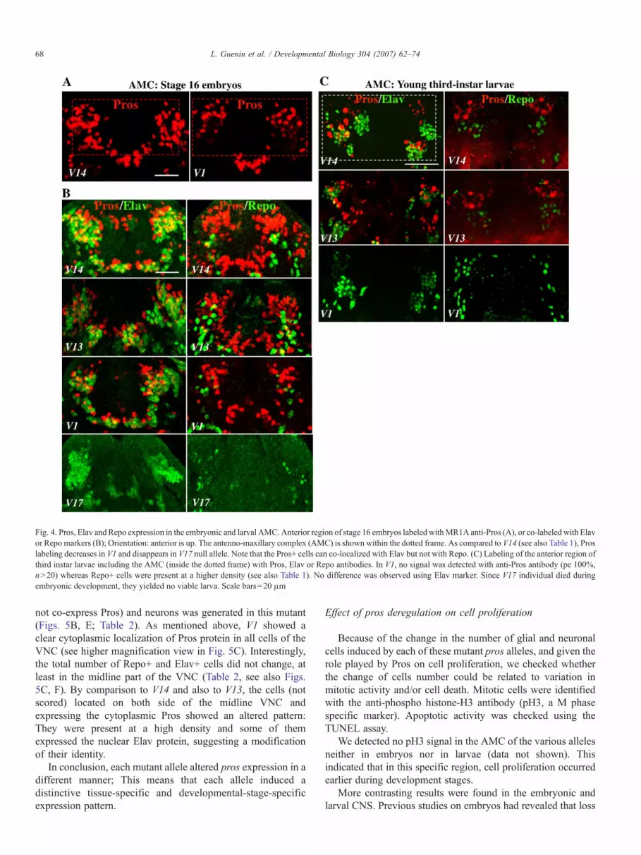