Hydrogenated and fluorinated surfactants derived from Tris(hydroxymethyl)-acrylamidomethane allow...
12
Hydrogenated and fluorinated surfactants derived from Tris (hydroxymethyl)-acrylamidomethane allow the purification of a highly active yeast F1-F0 ATP-synthase with an enhanced stability Jean-Claude Talbot & Alain Dautant & Ange Polidori & Bernard Pucci & Touria Cohen-Bouhacina & Abdelhamid Maali & Bénédicte Salin & Daniel Brèthes & Jean Velours & Marie-France Giraud Received: 25 May 2009 / Accepted: 13 July 2009 / Published online: 15 September 2009 # Springer Science + Business Media, LLC 2009 Abstract Loss of stability and integrity of large membrane protein complexes as well as their aggregation in a non-lipidic environment are the major bottlenecks to their structural studies. We have tested C 12 H 25 -S-poly-Tris-(hydroxymethyl) acrylamidomethane (H 12 -TAC) among many other deter- gents for extracting the yeast F 1 F 0 ATP-synthase. H 12 -TAC was found to be a very efficient detergent for removing the enzyme from mitochondrial membranes without altering its sensitivity towards specific ATP-synthase inhibitors. This extracted enzyme was then solubilized by either dodecyl maltoside (DDM), H 12 -TAC or fluorinated surfactants such as C 2 H 5 -C 6 F 12 -C 2 H 4 -S-poly-Tris-(hydroxymethyl)acrylami- domethane (H 2 F 6 -TAC) or C 6 F 13 -C 2 H 4 -S-poly-Tris-(hydrox- ymethyl)acrylamidomethane (F 6 -TAC), two surfactants exhibiting a comparable polar head to H 12 -TAC but bearing a fluorinated hydrophobic tail. Preparations from enzymes purified in the presence of H 12 -TAC were found to be more adapted for AFM imaging than ATP-synthase purified with DDM. Keeping H 12 -TAC during the Ni-NTA IMAC purification step or replacing it by DDM at low concen- trations did not however allow preserving enzyme activity, while fluorinated surfactants H 2 F 6 -TAC and F 6 -TAC were found to enhance enzyme stability and integrity as indicated by sensitivity towards inhibitors. ATPase specific activity was higher with F 6 -TAC than with H 2 F 6 -TAC. When enzymes were mixed with egg phosphatidylcholine, ATP- synthases purified in the presence of H 2 F 6 -TAC or F 6 -TAC were more stable upon time than the DDM purified enzyme. Furthermore, in the presence of lipids, an activation of ATP- synthases was observed that was transitory for enzymes purified with DDM, but lasted for weeks for ATP-synthases isolated in the presence of molecules with Tris polyalcoholic moieties. Relipidated enzymes prepared with fluorinated surfactants remained highly sensitive towards inhibitors, even after 6 weeks. Keywords Membrane protein complexes . F 1 F 0 ATP-synthase . Detergent . Fluorinated surfactants Abbreviations Tris Tris(hydroxymethyl)aminomethane J.-C. Talbot : A. Dautant : B. Salin : D. Brèthes : J. Velours : M.-F. Giraud (*) CNRS, Institut de Biochimie et Génétique Cellulaires, Université Bordeaux 2, 1 rue Camille Saint-Saëns, 33077 Bordeaux cedex, France e-mail: [email protected] A. Polidori : B. Pucci Faculté des Sciences, Laboratoire de Chimie Bioorganique et des systèmes moléculaires vectoriels (LCBOSMV), Université d’Avignon, 33 rue Louis Pasteur, 84000 Avignon, France T. Cohen-Bouhacina : A. Maali Centre de Physique Moléculaire Optique et Hertzienne (CPMOH), Université Bordeaux 1, 351 cours de la Libération, 33400 Talence cedex, France J Bioenerg Biomembr (2009) 41:349–360 DOI 10.1007/s10863-009-9235-5
Transcript of Hydrogenated and fluorinated surfactants derived from Tris(hydroxymethyl)-acrylamidomethane allow...

Hydrogenated and fluorinated surfactants derived from Tris(hydroxymethyl)-acrylamidomethane allow the purificationof a highly active yeast F1-F0 ATP-synthasewith an enhanced stability
Jean-Claude Talbot & Alain Dautant & Ange Polidori & Bernard Pucci &Touria Cohen-Bouhacina & Abdelhamid Maali & Bénédicte Salin & Daniel Brèthes &
Jean Velours & Marie-France Giraud
Received: 25 May 2009 /Accepted: 13 July 2009 /Published online: 15 September 2009# Springer Science + Business Media, LLC 2009
Abstract Loss of stability and integrity of large membraneprotein complexes as well as their aggregation in a non-lipidicenvironment are the major bottlenecks to their structuralstudies. We have tested C12H25-S-poly-Tris-(hydroxymethyl)acrylamidomethane (H12-TAC) among many other deter-gents for extracting the yeast F1F0 ATP-synthase. H12-TACwas found to be a very efficient detergent for removing theenzyme from mitochondrial membranes without altering itssensitivity towards specific ATP-synthase inhibitors. Thisextracted enzyme was then solubilized by either dodecylmaltoside (DDM), H12-TAC or fluorinated surfactants such
as C2H5-C6F12-C2H4-S-poly-Tris-(hydroxymethyl)acrylami-domethane (H2F6-TAC) or C6F13-C2H4-S-poly-Tris-(hydrox-ymethyl)acrylamidomethane (F6-TAC), two surfactantsexhibiting a comparable polar head to H12-TAC but bearinga fluorinated hydrophobic tail. Preparations from enzymespurified in the presence of H12-TAC were found to be moreadapted for AFM imaging than ATP-synthase purified withDDM. Keeping H12-TAC during the Ni-NTA IMACpurification step or replacing it by DDM at low concen-trations did not however allow preserving enzyme activity,while fluorinated surfactants H2F6-TAC and F6-TAC werefound to enhance enzyme stability and integrity as indicatedby sensitivity towards inhibitors. ATPase specific activitywas higher with F6-TAC than with H2F6-TAC. Whenenzymes were mixed with egg phosphatidylcholine, ATP-synthases purified in the presence of H2F6-TAC or F6-TACwere more stable upon time than the DDM purified enzyme.Furthermore, in the presence of lipids, an activation of ATP-synthases was observed that was transitory for enzymespurified with DDM, but lasted for weeks for ATP-synthasesisolated in the presence of molecules with Tris polyalcoholicmoieties. Relipidated enzymes prepared with fluorinatedsurfactants remained highly sensitive towards inhibitors,even after 6 weeks.
Keywords Membrane protein complexes .
F1F0 ATP-synthase . Detergent . Fluorinated surfactants
AbbreviationsTris Tris(hydroxymethyl)aminomethane
J.-C. Talbot :A. Dautant : B. Salin :D. Brèthes : J. Velours :M.-F. Giraud (*)CNRS, Institut de Biochimie et Génétique Cellulaires,Université Bordeaux 2,1 rue Camille Saint-Saëns,33077 Bordeaux cedex, Francee-mail: [email protected]
A. Polidori : B. PucciFaculté des Sciences, Laboratoire de Chimie Bioorganiqueet des systèmes moléculaires vectoriels (LCBOSMV),Université d’Avignon,33 rue Louis Pasteur,84000 Avignon, France
T. Cohen-Bouhacina :A. MaaliCentre de Physique Moléculaire Optique et Hertzienne (CPMOH),Université Bordeaux 1,351 cours de la Libération,33400 Talence cedex, France
J Bioenerg Biomembr (2009) 41:349–360DOI 10.1007/s10863-009-9235-5

H12-TAC C12H25-S-poly-Tris(hydroxymethyl)acrylamidomethane
Chol-TAC Cholesteryl-COC2H5-S-poly-Tris-(hydroxymethyl)acrylamidomethane
H2F6-TAC C2H5-C6F12-C2H4-S-poly-Tris-(hydroxymethyl)acrylamidomethane
F6-TAC C6F13-C2H4-S-poly-Tris-(hydroxymethyl) acrylamidomethane
DDM dodecyl maltosideC12E8 octaethyleneglycol mono-n-dodecyletherCHAPS 3-[(3-cholamidopropyl)
dimethylammonio]-1-propanesulfonate
bigCHAP N,N-bis[3-(D-gluconamido)propyl]cholamide
Dig digitoninPC egg phosphatidylcholineAFM atomic force microscopyBN-PAGE blue-native poly-acrylamide gel
electrophoresiscmc critical micellar concentrationSMP sub-mitochondrial particlesNTA nitrilo-triacetic acidIMAC immobilized metal affinity
chromatographyPK pyruvate kinaseLDH L-lactate dehydrogenasePEP phosphoenolpyruvatePMSF phenylmethanesulfonyl fluorideε-ACA ε-aminocaproic acidDCCD dicyclohexyl carbodiimideDPR detergent/protein (W/W) ratioLPR lipid/protein molar ratio
Introduction
Structural studies of large membrane complexes havebeen hampered by the difficulty to handle them uponextraction, solubilization, purification and crystallizationsteps. Many reviews and textbooks describe in detailsdetergents and methods to manipulate such complexes(Bowie 2001; Garavito and Ferguson-Miller 2001; Hunteet al. 2003; Privé 2007). Loss of stability and integrity ofoligomeric proteins as well as their aggregation in a non-lipidic environment are not easy to overcome and are themajor brakes to structural studies.
The yeast F1F0 ATP-synthase is a complex of 590 kDaconsisting of 17 distinct subunits. This enzyme is embed-ded in the inner mitochondrial membrane, where it uses theproton electrochemical gradient generated by the respirato-ry chain to synthesize ATP from ADP and inorganic
phosphate. This enzyme represents 10% of total yeastmitochondrial proteins. It comprises a catalytic head F1(α3,β3,γ,δ,ε), a channel region (910, 6), a peripheral stalk(4, 8, d, OSCP, i, f, h) (Velours and Arselin 2000) and non-essential subunits (e, g and k) that were found tospecifically be associated with dimeric and oligomericATP-synthases (Arnold et al. 1998; Arselin et al. 2003).F1 catalyzes ATP hydrolysis but ATP synthesis requires theassociation of F1 to the channel region and the peripheralstalk. High sensitivities to specific ATP-synthase inhibitorslike dicyclohexyl carbodiimide (DCCD) or oligomycinindicate a good physical and functional coupling betweenF1 and the rest of the enzyme. Crystallographic data havebeen obtained on the catalytic head F1 (Kabaleeswaran etal. 2006, 2009), on a sub-complex F1-910 (Stock et al.1999) and on a soluble part of the peripheral stalk (Dicksonet al. 2006) but the structure of the whole enzyme remainsto be elucidated to understand its energetic couplingmechanism.
Although the complex extracted and isolated in thepresence of dodecyl maltoside (DDM) contained mostsubunits, subunit 6 involved in the proton channel and allthe subunits of the peripheral stalk were further lost duringcrystallization (Stock et al. 1999). In similar experimentscarried out in our group with lower DDM concentrationsupon the purification and crystallization steps, the purifiedcomplex contained 14 distinct subunits and like in Stock etal. (Stock et al. 1999) only six of them (α3β3γδε910) werefound in electron density maps (manuscript in preparation).This indicates that DDM may not be the most appropriatedetergent for structural studies of ATP-synthase and pointedto the necessity to find alternative detergents/surfactants topreserve the enzyme integrity and activity upon all the stepsrequired for its isolation. New non-ionic synthetic deter-gents like C12H25-S-poly-Tris(hydroxymethyl)acrylamido-methane (H12-TAC) have been engineered to extract andsolubilize membrane proteins (Pucci et al. 1991, 1993).H12-TAC has a thio dodecanoyl chain combined with apolar group made of Tris polyalcoholic moieties. Othermolecules, like C2H5-C6F12-C2H4-S-poly-Tris(hydroxy-methyl)acrylamidomethane (H2F6-TAC) or C6F13-C2H4-S-poly-Tris(hydroxymethyl)acrylamidomethane (F6-TAC)have a polar head chemically similar to H12-TAC but theiraliphatic chains are totally or partially fluorinated (seeFig. 1 for chemical structures) (Barthélémy et al. 1999;Chabaud et al. 1998; Myrtil et al. 1995; Pavia et al. 1991).F6-TAC has an aliphatic chain with eight carbons in which thesix terminal carbons are perfluorinated, while the fluorinatedchain of H2F6-TAC is end-capped with an ethyl groupleading to a hemifluorinated hydrophobic chain with tencarbon atoms. The grafting of such an ethyl group wasdesigned to improve the H2F6-TAC affinity towards proteinhydrophobic surface. Fluorocarbon surfactants are more
350 J Bioenerg Biomembr (2009) 41:349–360

hydrophobic than hydrocarbon surfactants and their criticalmicellar concentration (cmc) is much lower than the cmc ofhydrocarbon analogues. Moreover they are lipophobic(Krafft 2001) and consequently unable to extract membraneproteins from their native lipidic environment (Pucci et al.1993) whereas they are very effective agents to maintainthem in solution after their extraction from the membrane byclassical detergents (Chabaud et al. 1998). These fluorinatedcompounds have been shown to preserve activity and toprevent aggregation of membrane proteins such as bacterio-rhodopsin, cytochrome b6f complex or bacterial outermembrane protein OmpA (Breyton et al. 2004). Their roleas molecular chaperones for insertion of diphtheria toxin inlipid bilayers has also been reported (Posokhov et al. 2008;Rodnin et al. 2008) and they were successfully used tomaintain in solution a mecanosensitive channel during itsexpression in a cell-free system (Park et al. 2007).
Here we compare ATP-synthases extracted with DDM orH12-TAC. We found that H12-TAC was very efficient forextracting the yeast ATP-synthase from the mitochondrialmembrane. Furthermore, H12-TAC purified ATP-synthasemolecules adsorbed on mica plates were still active andsuitable for atomic force microscopy (AFM) imaging. Wealso show results on ATP-synthases extracted with H12-TAC and kept in solution with DDM, H12-TAC, H2F6-TACor F6-TAC. The presented data demonstrate the efficiencyof F6-TAC to stabilize this large complex after its extractionfrom membranes, with or without additional lipids.
Material and methods
Chemicals
H12-TAC is a non-ionic detergent with a low cmc(∼0.15 mM) synthesized as previously described (Pucci etal. 1991). The batch used in the present work had anaverage number of 5.7 Tris motifs (<MW> ≈ 1,200 g/mol).H2F6-TAC (<MW> ≈ 1,615 g/mol, cmc ∼0.45 mM), F6-
TAC (<MW> ≈ 1,378 g/mol, cmc ∼0.5 mM), Chol-TAC(<MW> ≈ 5,200 g/mol, cmc ∼0.38 mM) were synthesizedas described in (Pavia et al. 1991) (Fig. 1). DDM (Anagrade,cmc ∼0.16 mM), octyl glucoside (cmc ∼21 mM), Anzergent3–12 (cmc ∼2.8 mM) were purchased from Anatrace.CHAPS (cmc ∼5 mM) and bigCHAP (cmc ∼3.4 mM) werefrom Calbiochem. C12E8 was a gift from Pr M. le Maire. PK,LDH from rabbit muscle and inhibitor cocktail werepurchased from Roche. Digitonin, ATP, NADH, DCCD,HEPES, EDTA and oligomycin were from Sigma-Aldrich;PEP was from Fluka.
Detergent extraction
Mitochondria were obtained with the zymolyase method(Guérin et al. 1979) from a genetically modified D273-10/B/A strain (met6, his3, ura3, i-his6) in which a (His)6-taghad been added at the C-terminus of subunit i, a componentof the peripheral stalk. Preparations were adjusted to amitochondrial protein concentration of 10 mg mL−1 andincubated in extraction buffer “A” (150 mM potassiumacetate, 10% (V/V) glycerol, 2 mM PMSF, 2 mM ε-ACA,30 mM HEPES pH 7.4 and EDTA-free protease inhibitorcocktail) supplemented with appropriate concentrations ofH12-TAC, DDM or other detergents. After a 30 minincubation at 4 °C, samples were centrifuged (30 min,4 °C, 25,000 g) and extracted proteins (0.4 mg) wereanalyzed under native conditions (BN-PAGE) on a 3–13%linear gradient polyacrylamide gel (Schägger et al. 1994).ATPase activity was revealed directly on gel (Grandier-Vazeille and Guérin 1996) before Coomassie brilliant bluestaining. ATPase activities of extracts were measured aspreviously described (Somlo 1968). Enzyme purity wasassessed by silver staining after Tricine SDS-PAGE.
Purification of ATP-synthase from yeast mitochondria
Submitochondrial particles (SMP) were prepared accordingto (MacLennan et al. 1968) and centrifuged at 48,000g for
Fig. 1 Chemical structures ofpoly-Tris derivatives surfactantsH12-TAC, H2F6-TAC, F6-TACand Chol-TAC
J Bioenerg Biomembr (2009) 41:349–360 351

45 min at 4 °C. Pellets were suspended at 10 mg of startingmitochondrial proteins per mL of buffer “A” containing0.75% H12-TAC (W/V) (6.65 mM). After 30 min ofincubation at 4 °C, the extract was clarified by centrifuga-tion (30 min, 4 °C, 25,000 g). The supernatant was dilutedwith two volumes of washing buffer “W” (50 mM NaCl,10% (V/V) glycerol, 10 mM imidazole, 20 mM sodiumphosphate pH 7.9) and mixed with Ni-NTA-agarose beads(Qiagen) (0.25 mL of slurry per 10 mg of startingmitochondrial proteins). After an overnight incubation at4 °C, beads were washed with 25 volumes of buffer “W”containing either DDM (0.32 mM), H12-TAC (0.85 mM),H2F6-TAC (1.15 mM) or F6-TAC (1.2 mM). ATP-synthasewas eluted with buffer “E” (50 mM NaCl, 10% (V/V)glycerol, 250 mM imidazole, 20 mM sodium phosphatepH 7.9) containing either DDM (0.64 mM) or TACderivatives at the above-mentioned concentrations.
The ATPase activity of extracted F1F0 ATP-synthase wasevaluated as described in (Somlo 1968) or for the Ni-NTApurified enzyme by monitoring NADH oxidation at 340 nmthrough a coupled reaction with pyruvate kinase (PK) andlactate dehydrogenase (LDH) (Tietz and Ochoa 1958). Anabsorbance decrease at 340 nm reflects ATPase activity.Activity assays were performed at 28 °C under magneticstirring in 1.5 mL of activity buffer (20 units mL−1 of PK,30 units mL−1 of LDH, 5 mM ATP, 2 mM PEP, 0.2 mMNADH, 5 mM MgCl2, 50 mM HEPES pH 7.5). The molarextinction coefficient of NADH at 340 nm was6,220 M−1 cm−1 and the pathlength was 1 cm.
Protein concentration was estimated by the Lowry method(Lowry et al. 1951) after a trichloroacetic acid precipitationstep and solubilization of pellets in 5% SDS (W/V).
Incubation with egg phosphatidylcholine
Egg phosphatidylcholine (PC) purified according to(Singleton et al. 1965) was suspended in 5 mM MgCl2,50 mM HEPES pH 7.5 to a concentration of 10 mg mL−1
and then sonicated three times, for 5 min at 4 °C (75TSAnnemasse sonicator, 120 V). Purified enzymes weremixed just after their elution from the IMAC resin with PCto a lipid/protein molar ratio of ∼250 and stored at 13 °C.ATPase activity was determined at 28 °C using the PK/LDH coupling reaction.
Preparation of specimens for AFM imaging
Freshly cleaved mica plates (15×8 mm2 for enzymaticactivity control and 5×5 mm2 for AFM) were incubated for1 min in 1 mM NiCl2. Mica plates were washed with waterand incubated with the enzyme (0.1 mg mL−1) in buffer “P”(100 mM NaCl, 25 mM D(+)trehalose, 2 mM MgCl2,0.5 mM EDTA, 0.5 mM PMSF, 20 mM Tris-HCl pH 8.0)
containing either 0.85 mM H12-TAC or 0.64 mM DDM.After 10 min of incubation, plates were rinsed with buffer“P” gradually diluted in observation buffer (50 mM KCl,2 mM MgCl2, 10 mM Tris-HCl pH 8.0) to decreasedetergent concentration step by step and to removeunadsorbed materials (Schabert and Engel 1994).
The ATPase activity of F1F0 ATP-synthase bound onmica was controlled by monitoring NADH oxidation at340 nm (Tietz and Ochoa 1958). In typical experimentsplates incubated or not with ATP-synthase were plungedout of the light beam in stirred reaction mixture, removed,and plunged again.
AFM experiments were performed at room temperaturein a liquid cell with a Nanoscope IIIa Multimode scanningprobe microscope (Digital Instruments, Santa Barbara, CA)equipped with an “E” scanner (maximum X and Y scan of14 µm). Mica plates were glued on a Teflon disc andmounted in the liquid cell. Silicon nitride cantilevers with0.06 N m−1 and 0.35 N m−1 nominal spring constants andtips with a semi-angle of 35° were used. Force curves (orapproach-retract curves) were obtained in the force calibra-tion mode and collected by measuring the cantileverdeflection (or the amplitude of cantilever oscillations) asthe sample was moved towards the tip. The experimentalforce (or approach-retract) curves were obtained with a Zscan velocity between 2 µm s−1 and 8 µm s−1 and driveamplitude and set point were optimized for each image.Images were recorded in tapping mode with a scan angle of90°. For each experiment, three images were recorded:height, phase and amplitude. The amplitude (signal error)was also recorded in order to avoid artefact duringmeasurement. For clarity, only height images are shown.
Preparation of specimens for electron microscopy
F1F0 ATP-synthases purified in the presence of H12-TACwere diluted 20–40 fold in water and immediately appliedfor 1 min to formvar/carbon-coated copper grids, previous-ly glow discharged, washed with distilled water and stainedwith 2% uranyl acetate for 1 min. Specimens wereexamined on a Hitachi 7650 transmission electron micro-scope operating at 80 kV.
Results
The enzyme extracted by H12-TAC is highly activeand sensitive to specific inhibitors
When mitochondria were incubated with increasing concen-trations of DDM, the maximum of ATPase specific activitythat was detected in the detergent extract was about4 µmol min−1 mg −1 for a detergent/protein ratio (DPR) of
352 J Bioenerg Biomembr (2009) 41:349–360

0.25 g/g (Fig. 2a). The lower activity observed at a DPR of0.75 (g/g) was reproducible in several experiments. Forhigher concentrations of DDM, a small increase in activitywas observed as well as a decrease in DCCD and oligomycin(not shown) sensitivity, indicating a slight functional and/or aphysical uncoupling of ATP-synthase. When DDM-extractedproteins were analyzed by BN-PAGE, only monomericforms were obtained with a DPR from 0.25 to 2.00(Fig. 2b and c).
As an alternative to DDM for ATP-synthase extraction,we looked for detergents that could extract ATP-synthasewithout affecting enzyme sensitivity toward its inhibitorsand that could preferably preserve oligomeric species.Many detergents usually used to extract membrane proteinswere tested. Some of them like C12E8, octyl glucoside or
Anzergent 3–12 disrupted the interactions between F1 andF0, even when low concentrations (around their cmc) wereused (not shown). Monomeric forms of ATP-synthase wereobtained with the zwitterionic detergent CHAPS (4 mM to64 mM) (not shown). Dimeric and oligomeric forms ofATP-synthase were observed with non ionic detergents likebigCHAP (up to 22 mM) and digitonin (below 6 mM), adetergent commonly used to study oligomeric forms ofATP-synthase (Schägger and Pfeiffer 2000) but that cannotbe used for structural studies because of its impurity. WhenH12-TAC was used to extract mitochondrial proteins, thehighest ATPase specific activity (6 µmol min−1 mg−1) wasdetected for a DPR of 0.75 (g/g) and sensitivity towardsDCCD was maintained (Fig. 2a). The same inhibition trendwas observed with oligomycin (not shown). For higher
Fig. 2 Analyses of DDM andH12-TAC extracts. a ATPasespecific activities(µmol min−1 mg−1) were mea-sured in DDM (white bars) orH12-TAC (grey bars) extractsobtained at various DPR withoutor with ATPase inhibitor(DCCD (5 µg mL−1), hatchedbars). Protein concentrationswere measured in triplicate foreach extract. Extracted proteinquantities (DDM extracts (opencircles) and H12-TAC extracts(plain circles)) are given inpercentage of starting mito-chondrial proteins (right axis).Values are the average of threeindependent experiments. Forclarity, detergent concentrations(g/g of mitochondrial proteins)used for extraction are alsogiven in mM. 40 µl of DDM (b)or H12-TAC (d) extracts wereanalyzed by BN-PAGE andATPase activity was revealed ingel. Gels were then stained withCoomassie brilliant blue (cDDM extracts, e H12-TACextracts). O: oligomers; D:dimers; M: monomer; F1: freecatalytic sector of ATP-synthase.Dig: Digitonin
J Bioenerg Biomembr (2009) 41:349–360 353

H12-TAC ratios, above 1 g/g, ATPase activity in the extractsdecreased, with a lower sensitivity towards ATPase inhib-itors. Although Chol-TAC has a cholesterol moiety likedigitonin and a polar head chemically similar to H12-TAC,it had a very poor extraction power even at a concentrationaround 20 × cmc (not shown).
A BN-PAGE analysis of mitochondrial extractsobtained with increasing concentrations of H12-TAC(DPR from 0.10 to 2.00) is shown in Fig. 2d and e. WithDPR between 0.1 and 0.25, mainly dimeric species wereextracted. A faint band, corresponding to a higheroligomeric (O) species was also detected when ATPaseactivity was revealed in gel (Fig. 2d). When DPR werecomprised between 0.50 and 1.00, dimeric (D) andmonomeric (M) species were present in extracts andabove a DPR of 1.00, monomeric species becamepredominant. Only a very faint active band of free F1was present with 2% H12-TAC and no corresponding bandwas discernible on the Coomassie blue stained gel(Fig. 2e).
Activity due to free F1 sectors was observed on BN-PAGE for digitonin- and DDM-extracted samples (Fig. 2b),but no free F1 activity was detected in solution in DDMextracts as indicated by a high sensitivity towards DCCD(Fig. 2a). This F1 band with a strong ATPase activity onBN-PAGE may indicate a higher weakness of the DDMextracted enzyme upon electrophoresis compared to H12-TAC extracted complexes.
As there were less total proteins extracted with H12-TACthan with DDM, H12-TAC seems more useful to specifi-cally extract the ATP-synthase from yeast mitochondrialmembranes than DDM. As ATP-synthase oligomericstructures were better preserved with H12-TAC than withDDM, H12-TAC seems to be a milder detergent comparedto DDM. For these reasons, H12-TAC was preferred for theextraction step of the purification protocol of the yeastATP-synthase.
To minimize contamination by matricial soluble proteins,H12-TAC extraction was performed on sub-mitochondrialparticles (SMP). Figure 3 shows specific activities that weremeasured in extracts obtained from SMP at various H12-TACconcentrations. Starting from SMP, the highest specificactivity (20 µmol min−1 mg−1) was obtained for a deter-gent/mitochondrial protein ratio of 0.75 (W/W). PerformingH12-TAC extraction on mitochondria or on SMP did not alteroligomer/monomer ratios within the same range of H12-TACconcentrations (data not shown).
Enzymes purified in the presence of H12-TAC or DDMare monomeric
We first attempted to purify the dimeric entities startingfrom SMP with H12-TAC using a DPR of 0.25 for the
extraction step, a concentration that gave small enzymequantities but for which a large proportion of dimericspecies was found in the extract. Enzymes were bound onNi-NTA agarose beads and eluted with 0.85 mM H12-TAC(0.1% W/V). BN-PAGE analyses of eluates indicated thatdimeric species were lost upon this unique purification step.Decreasing detergent concentrations upon washing stepsdid not prevent the loss of dimers.
Thus we performed ATP-synthase purification using6.25 mM H12-TAC (i.e. a DPR of 0.75) for the SMPextraction step, a concentration that gave a mix ofmonomeric and dimeric enzymes in the extract andcorresponded to the highest specific activity (Fig. 3).However, after the Ni-NTA IMAC step, ATPase specificactivity dropped to ∼4 µmol min−1mg−1 and sensitivitytowards oligomycin was decreased. When H12-TAC wasreplaced by 0.64 mM DDM upon washing and elutionsteps, the specific activity was ∼2 µmol min−1 mg−1 andsensitivity towards inhibitors was often lost or dramaticallydecreased. This drop could vary from 50% to almost 100%depending on preparations or ATPase activity measurementconditions (different pH and enzyme concentrations).
Analyses by BN-PAGE of enzymes purified after the Ni-NTA IMAC step in the presence of DDM or H12-TAC(Fig. 4a) indicated that the purified ATP-synthase was ahomogeneous monomer containing all ATP-synthase sub-units except e and g. These two components required forenzyme dimerization were not revealed by Western blot(not shown). Western blot analyses did not also reveal thepresence of the Pi or ADP/ATP carriers in enriched ATP-synthase fractions, as was the case with the ATP syntha-some from rat mitochondria (Chen et al. 2004; Ko et al.2003). Figure 4b shows the high degree of purity of ATP-synthase prepared in the presence of H12-TAC.
Fig. 3 Analyses of ATPase specific activity in H12-TAC extracts fromSMP. ATPase activities in H12-TAC extracts were measured intriplicate in the absence or in the presence of oligomycin(6 µg mL−1). DPR values were calculated using starting mitochondrialprotein quantities
354 J Bioenerg Biomembr (2009) 41:349–360

AFM imaging of the yeast ATP-synthase
Atomic Force Microscopy imaging on membrane proteinsamples is extremely dependent on enzyme preparations. Todetermine if H12-TAC purified enzymes could be imagedwith this technique, isolated enzymes were adsorbed on micaplates. Imaging in liquid by contact mode AFM of ATP-synthase deposits destroyed the observed objects. Thus allimages were obtained in tapping mode (Fig. 5). Controlexperiments performed without protein showed no depositon the surface and looked like regular mica surfaces (insert Aof Fig. 6).
AFM images of Fig. 5a and b as well as the sampleobserved after the activity assay (insert B of Fig. 6) showedthat the whole mica surfaces were covered by a relativelydense monolayer. A significant number of very largeaggregates (Fig. 5b, black arrows) remained on mica in spiteof extensive rinses. Such aggregates were, however, morenumerous with DDM (results not shown) indicating thatDDM purified enzymes were less suitable for informativeAFM observations. As shown in Fig. 5b smaller particleswere clearly visible between aggregates. At the experimentalresolution, particles appeared as roughly spherical objects.When the surface shown in Fig. 5c was scanned with highermagnifications some isolated spherical particles were ob-
served (see Fig. 5d–e). Consecutive scans in the same areadid not alter the deposits because the images were recordedwith a vertical force adjusted to a few hundreds ofpiconewtons (almost 300 pN) to avoid displacement ofisolated and weakly bound proteins. At forces above thisvalue, proteins were flattened and reproducible images couldnot be recorded. Images recorded with higher magnificationthan in Fig. 5e in order to enhance the resolution did notbring more detailed information than the one shown inFig. 5e and did not allow identifying F1 and F0 sectors.Statistical analyses on isolated particles like those indicatedby arrows on Fig. 5d, gave a mean width of 25 nm. Bothshape and size were in good agreement with those measuredon chloroplast F1F0 ATP-synthase by AFM in contact mode(Neff et al. 1997). However the measured mean height ofisolated particles was around 5 nm (±1 nm) instead of 9 nmobtained for the chloroplast enzyme in constant force mode.
Some objects appeared spherical (Fig. 5d, arrows) andsome looked like adjacent balls with slightly different radiiand height (Fig. 5d, asterisks). This lack of homogeneitycould reflect different orientations of the enzyme on micaplate and the two adjacent balls might correspond to the twosectors of the enzyme, seen on its long axis. Figure 5f showsan electron micrograph obtained after negative staining of adeposit of the H12-TAC purified sample, on which the twosectors of the enzyme can clearly be recognized (Fig. 5f,insert). Particles seem larger when imaged by AFM but theobserved size is a convolution of the actual size of themolecule with that of the tip. Although the experimentalresolution obtained with the AFM tip is low, these dataclearly indicate that H12-TAC is a promising surfactant forAFM observations of membrane protein complexes.
ATPase activity of mica-adsorbed enzymes
To ensure that bound enzymes were still active, we used asensitive activity assay in which ADP produced by adsorbedATP-synthase is used by coupled enzymes (PK/LDH) to leadto NADH oxidation. A typical experiment is shown in Fig. 6.When mica plates treated with buffer “P” containing only0.85 mM H12-TAC (without enzyme) were immersed in theactivity buffer, no decrease in the absorbance was detected(step 1). By AFM, no object was observed (Fig. 6, insert A).NADH oxidation was detected only when the plate, pre-incubated with buffer “P” containing 0.85 mM H12-TAC and0.1 mg mL−1 of ATP-synthase and then extensively rinsed,was immersed in the activity buffer (step 3). When the platewas removed, NADH oxidation was reduced but the rate ofoxidation was higher than the one detected prior to plateaddition (step 4). This is probably due to some ATP-synthases (or free F1 sectors) that were released after10 min of reaction in the stirred medium. Once again, afteraddition of the plate, an increase in NADH oxidation was
Fig. 4 Native and denaturing electrophoresis analyses of Ni-NTAIMAC-purified samples. a BN-PAGE showing monomers purified inthe presence of H12-TAC (lanes 1 and 2) or DDM (lanes 3 and 4)revealed by ATPase activity (lanes 2 and 4) or Coomassie brilliantblue (lanes 1 and 3). M: monomer. D: migration zone of dimers. bSilver stained Tricine SDS-PAGE (12.5% (W/V) polyacrylamide slabgel) of ATP-synthase purified with H12-TAC. ATP-synthase subunitsare indicated
J Bioenerg Biomembr (2009) 41:349–360 355

observed (step 5). After the plate was removed, a decrease inNADH oxidation was recorded (step 6). Finally this platewas imaged by AFM (Fig. 6, insert B). These results clearlyshow that enzymes were adsorbed on mica and were stillactive.
H2F6-TAC and F6-TAC preserve sensitivity towardsinhibitors
In order to avoid ATPase activity loss upon the Ni-NTAIMAC purification step, we tested whether fluorinated
surfactants such as H2F6-TAC and F6-TAC could advanta-geously replace H12-TAC or DDM if they were used at theelution step from the IMAC resin. It can be seen from Fig. 7athat F6-TAC was the most effective compound to maintain ahigh activity of the purified enzyme. Two days after the dayof extraction from mitochondrial membranes (day referred asday 0), activities in the presence of F6-TAC were three andsix times higher than activities detected at day 2 for enzymespurified with H12-TAC and DDM, respectively (Fig. 7a).Samples purified in the presence of H2F6-TAC displayed ahigher ATPase activity than with DDM and H12-TAC a few
Fig. 5 Imaging of H12-TACpurified ATP-synthases withAFM and electron microscopy.AFM images of two differentsamples deposited in thepresence of 0.85 mM H12-TACshow dense monolayers ofATP-synthase: a Sample 1(drive amplitude 150 mV,set-point voltage 1.7 V),b Sample 2 (drive amplitude340 mV, set-point voltage1.7 V); Black arrows indicateaggregates. c–e) AFM serialimages of another sampledeposited in the presence of0.15 mM H12-TAC showisolated particles (driveamplitude 350 mV, set-pointvoltage 0.56 V). Images d ande correspond to new scans of theboxed area shown on theprevious image. Arrows indicatespherical objects and asteriskstwo adjacent spherical objects ofdifferent diameters f Electronmicrograph of negativelystained H12-TAC purifiedATP-synthases. The insertshows a zoom of a F1F0ATP-synthase
356 J Bioenerg Biomembr (2009) 41:349–360
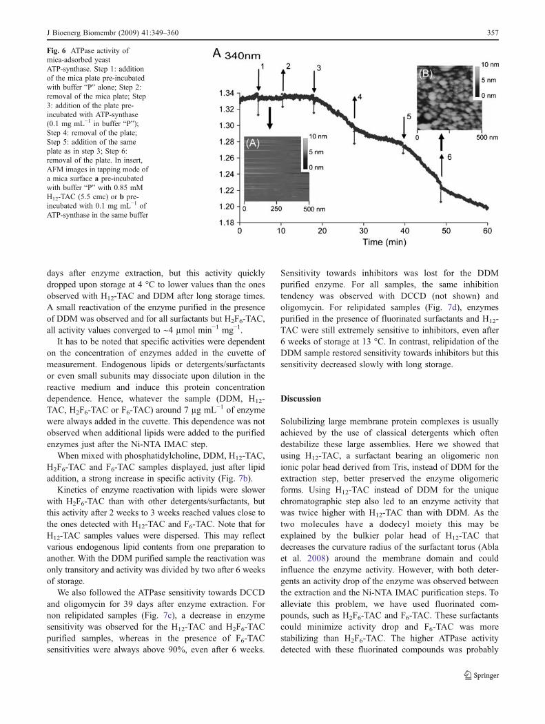
days after enzyme extraction, but this activity quicklydropped upon storage at 4 °C to lower values than the onesobserved with H12-TAC and DDM after long storage times.A small reactivation of the enzyme purified in the presenceof DDM was observed and for all surfactants but H2F6-TAC,all activity values converged to ∼4 µmol min−1 mg−1.
It has to be noted that specific activities were dependenton the concentration of enzymes added in the cuvette ofmeasurement. Endogenous lipids or detergents/surfactantsor even small subunits may dissociate upon dilution in thereactive medium and induce this protein concentrationdependence. Hence, whatever the sample (DDM, H12-TAC, H2F6-TAC or F6-TAC) around 7 µg mL−1 of enzymewere always added in the cuvette. This dependence was notobserved when additional lipids were added to the purifiedenzymes just after the Ni-NTA IMAC step.
When mixed with phosphatidylcholine, DDM, H12-TAC,H2F6-TAC and F6-TAC samples displayed, just after lipidaddition, a strong increase in specific activity (Fig. 7b).
Kinetics of enzyme reactivation with lipids were slowerwith H2F6-TAC than with other detergents/surfactants, butthis activity after 2 weeks to 3 weeks reached values close tothe ones detected with H12-TAC and F6-TAC. Note that forH12-TAC samples values were dispersed. This may reflectvarious endogenous lipid contents from one preparation toanother. With the DDM purified sample the reactivation wasonly transitory and activity was divided by two after 6 weeksof storage.
We also followed the ATPase sensitivity towards DCCDand oligomycin for 39 days after enzyme extraction. Fornon relipidated samples (Fig. 7c), a decrease in enzymesensitivity was observed for the H12-TAC and H2F6-TACpurified samples, whereas in the presence of F6-TACsensitivities were always above 90%, even after 6 weeks.
Sensitivity towards inhibitors was lost for the DDMpurified enzyme. For all samples, the same inhibitiontendency was observed with DCCD (not shown) andoligomycin. For relipidated samples (Fig. 7d), enzymespurified in the presence of fluorinated surfactants and H12-TAC were still extremely sensitive to inhibitors, even after6 weeks of storage at 13 °C. In contrast, relipidation of theDDM sample restored sensitivity towards inhibitors but thissensitivity decreased slowly with long storage.
Discussion
Solubilizing large membrane protein complexes is usuallyachieved by the use of classical detergents which oftendestabilize these large assemblies. Here we showed thatusing H12-TAC, a surfactant bearing an oligomeric nonionic polar head derived from Tris, instead of DDM for theextraction step, better preserved the enzyme oligomericforms. Using H12-TAC instead of DDM for the uniquechromatographic step also led to an enzyme activity thatwas twice higher with H12-TAC than with DDM. As thetwo molecules have a dodecyl moiety this may beexplained by the bulkier polar head of H12-TAC thatdecreases the curvature radius of the surfactant torus (Ablaet al. 2008) around the membrane domain and couldinfluence the enzyme activity. However, with both deter-gents an activity drop of the enzyme was observed betweenthe extraction and the Ni-NTA IMAC purification steps. Toalleviate this problem, we have used fluorinated com-pounds, such as H2F6-TAC and F6-TAC. These surfactantscould minimize activity drop and F6-TAC was morestabilizing than H2F6-TAC. The higher ATPase activitydetected with these fluorinated compounds was probably
Fig. 6 ATPase activity ofmica-adsorbed yeastATP-synthase. Step 1: additionof the mica plate pre-incubatedwith buffer “P” alone; Step 2:removal of the mica plate; Step3: addition of the plate pre-incubated with ATP-synthase(0.1 mg mL−1 in buffer “P”);Step 4: removal of the plate;Step 5: addition of the sameplate as in step 3; Step 6:removal of the plate. In insert,AFM images in tapping mode ofa mica surface a pre-incubatedwith buffer “P” with 0.85 mMH12-TAC (5.5 cmc) or b pre-incubated with 0.1 mg mL−1 ofATP-synthase in the same buffer
J Bioenerg Biomembr (2009) 41:349–360 357
due to their more rigid hydrophobic part. Their rigid chainscould have a lesser denaturing effect than hydrogenatedalkyl chains by less removing peripheral subunits or bykeeping some important endogenous lipids (Lebaupain etal. 2006).
Our experiments, performed in the presence of PC arguein favour of a loss of lipids during the single purificationstep with hydrogenated surfactants. Whereas the ATPasespecific activity of the F6-TAC purified sample almostdoubled after relipidation (from 12 µmol min−1 mg−1 with-out PC to above 20 µmol min−1 mg−1 with PC), the ATPaseactivity was increased ten-fold when the DDM purified
enzyme was mixed with PC, to reach the same values asthose obtained with enzymes purified in the presence offluorinated surfactants and relipidated. This indicates thatthe drop in activity of the DDM or H12-TAC purified ATP-synthases did not arise from the loss of important subunitsor the dissociation of the two ATP-synthase sectors sincethis could be reversed by a simple addition of lipids. Whileenzymes purified in the presence of H12-TAC or DDMwere reactivated in less than 1 h with lipids, thisreactivation was slower with samples isolated in thepresence of fluorinated surfactants. This may reflectdifferent protein/surfactants dissociation rates or a low
Fig. 7 Stability of ATPase activity and oligomycin sensitivity of ATP-synthase purified with different surfactants and incubated without orwith PC. Specific ATPase activity (left) and inhibition by 6 µg mL−1
of oligomycin (right) of non relipidated ATP-synthase (a and c) orrelipidated with PC at a LPR of 250 (b and d). Inhibitions (c and d)are expressed in percentage of the remaining activity determined at
indicated days after extraction from membranes. Enzymes isolated inthe presence of DDM (▪▪▪○▪▪▪), H12-TAC (– –□– –), H2F6-TAC(––▲––) and F6-TAC (– ∙◆– ∙). Measures in triplicate were performedfrom 2 days to 39 days after the extraction from membranes. Valuesare the averages of two or three independent purifications except forH2F6-TAC where only one experiment could be carried out
358 J Bioenerg Biomembr (2009) 41:349–360
association rate of lipids with protein-fluorinated surfactantcomplexes. This effect is more pronounced with H2F6-TACindicating that the ethyl group designed to facilitate theadsorption of the hydrophobic chain onto trans-membranedomains of proteins has a strong impact on surfactant/lipidexchange. With F6-TAC, it took 2 days to 3 days ofincubation with PC before reaching a high activity value. Ithas to be noted that after 10 months of storage with lipids at13 °C an ATPase activity of 14 µmol min−1 mg−1 was stillmeasured for the F6-TAC purified sample and the remain-ing activity was fully sensitive towards inhibitors. HenceF6-TAC combined or not with lipids, seems to be theappropriate surfactant to maintain a high activity level ofATP-synthase for periods of time compatible with 3Dcrystallization experiments. Although the polar head ispaucidisperse, we have started 3D crystallisation screensbut no hit has yet been obtained. Since F6-TAC has beenshown not to alter lipid layers (Rodnin et al. 2008), this isalso a promising molecule for 2D crystallization experi-ments either for reconstituting ATP-synthase in liposomesor for the monolayer crystallization method (Dauvergne etal. 2008; Lévy et al. 1999). We have tried to image withAFM F6-TAC purified enzymes but have not yet obtainedinteresting results. Images obtained with H12-TAC enzymepreparations have shown that this detergent is suitable forAFM experiments and allowed us to clearly distinguishspherical objects. A specific activity of ∼4 µmol min−1 mg−1
was measured for the H12-TAC purified ATP-synthase. Theactivity derived from the slope in Fig. 6 was around 0.9 nmolof ATP hydrolyzed per min. Thus if each enzyme on micaoccupies at most a surface of ∼220 nm2 (enzyme lain alongits longitudinal axis), and assuming all bound enzymes are asactive as the free purified enzyme, this would indicate thatnearly 40% of the plate would be covered by the enzyme.This estimation is in agreement with the surface coverageseen in Fig. 5d and e, but since there was some heterogeneityin deposits, as seen in the insert B of Fig. 6, it cannot beascertain that all bound enzymes were as active as theenzyme in solution.
Electron microscopy (Rubinstein et al. 2003) and X-ray structures (Stock et al. 1999) gave 11×11 nm2 and20×11 nm2 for the minimum and maximum projectionsizes of mitochondrial ATP-synthase, if the object is seenfrom a top view or a lateral view respectively. Whateverthe orientation adopted by the adsorbed enzymes, there isclearly an underestimation of the actual height. Theapparent flattening is due to the AFM signal that combinesvarious mechanical informations such as wear processes,elastic or visco-elastic response as well as topography, thesize and the shape of the cantilever. For typical commer-cial cantilevers with a length ranging from 100 µm to200 µm, a width of about 20 µm and an oscillationamplitude of about 10 nm, hydrodynamic damping
induces forces that can reach values as high as severalnanonewtons close to the surface (Dubourg et al. 2003).When imaging biological samples, one seeks forces in thepiconewton range; therefore hydrodynamic forces producea large background that may mask the more specificinteractions between the tip apex and the biologicalsample. This viscous damping effect reduces the AFMsensitivity when working in dynamic mode to measure thephase and the amplitude. In order to minimize theseeffects, the width and the length of the cantilever must bereduced or the tip height must be increased. It was shownthat the hydrodynamic forces due to the drag fluid couldbe decreased by almost one order of magnitude whenreducing the cantilever width (Maali et al. 2005). Hencethe current experimental device requires an adaptation forthe use of such cantilevers in order to enhance the ATP-synthase image resolution. However, even if tips and AFMelectronics still need to be improved to reach resolution asthe one obtained with electron microscopy, an importantpoint is the fact that binding on mica did not abolishATPase activity. This shows potential for future dynamicexperiments under the AFM tip.
Acknowledgements We wish to thank Pr Marc le Maire forproviding C12E8 detergent and Jean-Pierre Aimé for stimulatingdiscussions on AFM. This project was supported by the RégionAquitaine and by the Agence Nationale pour la Recherche (grantANR-06-PCVI-0016).
References
Abla M, Durand G, Pucci B (2008) J. Org. Chem. 73:8142–8153Arnold I, Pfeiffer K, Neupert W, Stuart RA, Schägger H (1998)
EMBO J. 17:7170–7178Arselin G, Giraud M-F, Dautant A, Vaillier J, Brèthes D, Coulary-
Salin B, Schaeffer J, Velours J (2003) Eur. J. Biochem.270:1875–1884
Barthélémy P, Ameduri B, Chabaud E, Popot J-L, Pucci B (1999) Org.Lett. 1:1689–1692
Bowie JU (2001) Curr. Opin. Struct. Biol. 11:397–402Breyton C, Chabaud E, Chaudier Y, Pucci B, Popot J (2004) FEBS
Lett. 564:312–318Chabaud E, Barthélémy P, Mora N, Popot JL, Pucci B (1998)
Biochimie. 80:515–530Chen C, Ko Y, Delannoy M, Ludtke SJ, Chiu W, Pedersen PL (2004)
J. Biol. Chem. 279:31761–31768Dauvergne J, Polidori A, Vénien-Bryan C, Pucci B (2008) Tet. Lett.
49:2247–2250Dickson VK, Silvester JA, Fearnley IM, Leslie AGW, Walker JE
(2006) EMBO J. 25:2911–2918Dubourg F, Aimé J-P, Marsaudon S, Couturier G, Boisgard R (2003)
J. Phys. Condens. Matter. 15:6167–6177Garavito RM, Ferguson-Miller S (2001) J. Biol. Chem. 276:32403–
32406Grandier-Vazeille X, Guérin M (1996) Anal. Biochem. 242:248–254Guérin B, Labbe P, Somlo M (1979) Methods Enzymol. 55:149–159Hunte C, Von Jagow G, Schagger H (2003) Membrane protein
purification and crystallization. Academic, London
J Bioenerg Biomembr (2009) 41:349–360 359

Kabaleeswaran V, Puri N, Walker JE, Leslie AGW, Mueller DM(2006) EMBO J. 25:5433–5442
Kabaleeswaran V, Shen H, Symersky J, Walker J, Leslie AW, MuellerD (2009) J. Biol. Chem. 284:10546–10551
Ko YH, Delannoy M, Hullihen J, Chiu W, Pedersen PL (2003) J. Biol.Chem. 278:12305–12309
Krafft MP (2001) Adv. Drug. Deliv. Rev. 47:209–228Lebaupain F, SalvayAG, Olivier B, DurandG, Fabiano A,Michel N, Popot
J-L, Ebel C, Breyton C, Pucci B (2006) Langmuir 22:8881–8890Lowry OH, Rosebrough NJ, Farr AL, Randall RJ (1951) J. Biol.
Chem. 193:265–275Lévy D, Mosser G, Lambert O, Moeck GS, Bald D, Rigaud J-L
(1999) J. Struct. Biol. 127:44–52Maali A, Hurth C, Boisgard R, Jai C, Cohen-Bouhacina T, Aimé J-P
(2005) J. Appl. Phys. 97:74907.1–74907.6MacLennan DH, Smoly JM, Tzagoloff A (1968) J. Biol. Chem.
243:1589–1597Myrtil E, Zarif L, Greiner J, Riess JG, Pucci B, Pavia AA (1995) J.
Fluorine Chem. 71:101–105Neff D, Tripathi S, Middendorf K, Stahlberg H, Butt H, Bamberg E,
Dencher N (1997) J. Struct. Biol. 119:139–148Park K, Berrier C, Lebaupain F, Pucci B, Popot J-L, Ghazi A, Zito F
(2007) Biochem J. 403:183–187
Pavia AA, Pucci B, Riess JG, Zarif L (1991) Bioorg. Med. Chem.Lett. 1:103–106
Posokhov YO, Rodnin MV, Das SK, Pucci B, Ladokhin AS (2008)Biophys J. 95:L54–L56
Privé GG (2007) Methods. 41:388–397Pucci B, Maurizis J, Pavia A (1991) Eur. J. Polym. 27:1101–1106Pucci B, Maurizis J, Pavia A (1993) BioOrg. Med. Chem. Lett. 3:161–
164Rodnin MV, Posokhov YO, Contino-Pépin C, Brettmann J,
Kyrychenko A, Palchevskyy SS, Pucci B, Ladokhin AS(2008) Biophys J. 94:4348–4357
Rubinstein JL, Walker JE, Henderson R (2003) EMBO J. 22:6182–6192
Schabert FA, Engel A (1994) Biophys. J. 67:2394–2403Schägger H, Pfeiffer K (2000) EMBO J. 19:1777–1783Schägger H, Cramer WA, von Jagow G (1994) Anal. Biochem.
217:220–230Singleton WS, Gray MS, Brown ML, White JL (1965) J. Am. Oil.
Chem. Soc. 42:53–56Somlo M (1968) Eur. J. Biochem. 5:276–284Stock D, Leslie AG, Walker JE (1999) Science. 286:1700–1705Tietz A, Ochoa S (1958) Arch. Biochem. Biophys. 78:477–493Velours J, Arselin G (2000) J. Bioenerg. Biomembr. 32:383–390
360 J Bioenerg Biomembr (2009) 41:349–360