Sources and seasonal dynamics of inoculum for brown spot disease of pear
13
Sources and seasonal dynamics of inoculum for brown spot disease of pear Vittorio Rossi & Elisabetta Pattori & Riccardo Bugiani Received: 7 August 2007 / Accepted: 19 November 2007 / Published online: 13 December 2007 # KNPV 2007 Abstract The dynamics of the production of Stem- phylium vesicarium conidia and Pleospora allii ascospores from different inoculum sources on the ground were compared in a model system of a wildflower meadow mainly composed of yellow foxtail, creeping cinquefoil and white clover. The meadow was either inoculated (each October) or not inoculated with a virulent strain of S. vesicarium, and either covered or not covered with a litter of inoculated pear leaves. Spore traps positioned a few centimetres above the ground were exposed for 170 7-day periods between October 2003 and December 2006. Ascospores and conidia were trapped in 46 and 25% of samples, respectively. Ascospore numbers trapped from the pear leaf litter were about five times higher than those from the meadow, while conidial numbers were similar from the different inoculum sources. The ascosporic season was very long, with two main trapping periods: December–April, and August–October; the former was most important for the leaf litter, the latter for the meadow. The conidial season lasted from April to November, with 92% of conidia caught between July and September. The fungus persistently colonized the meadow: the mead- ow inoculated in early October 2003 produced spores until autumn 2006. The present work demonstrates that orchard ground is an important source of inoculum for brown spot of pear. Thus, it is important to reduce inoculum by managing the orchard ground all year long. Keywords Ascospores . Conidia . Leaf litter . Meadow plants . Stemphylium vesicarium . Pleospora allii Introduction Brown spot of pear (Pyrus communis), caused by Stemphylium vesicarium (Ellis and Ellis 1985), is a disease of economic importance in fruit-growing areas of Europe (Llorente and Montesinos 2006). Disease symptoms consist of necrotic lesions on leaves, twigs and fruits. Fruits show small necrotic spots that progressively enlarge and deepen in round- shaped brown areas that can rot; infected fruits are unmarketable. Epidemics usually begin in late spring when fruits are highly susceptible (Montesinos et al. 1995), and increase progressively to reach the maximum just before harvest (Montesinos and Eur J Plant Pathol (2008) 121:147–159 DOI 10.1007/s10658-007-9258-x V. Rossi (*) : E. Pattori Istituto di Entomologia e Patologia vegetale, Università Cattolica S. Cuore, Via E. Parmense 84, 29100 Piacenza, Italy e-mail: [email protected] R. Bugiani Servizio Fitosanitario, Regione Emilia-Romagna, Via di Corticella 133, 40129 Bologna, Italy
-
Upload
emilia-romagna -
Category
Documents
-
view
4 -
download
0
Transcript of Sources and seasonal dynamics of inoculum for brown spot disease of pear
Sources and seasonal dynamics of inoculumfor brown spot disease of pear
Vittorio Rossi & Elisabetta Pattori &Riccardo Bugiani
Received: 7 August 2007 /Accepted: 19 November 2007 /Published online: 13 December 2007# KNPV 2007
Abstract The dynamics of the production of Stem-phylium vesicarium conidia and Pleospora alliiascospores from different inoculum sources on theground were compared in a model system of awildflower meadow mainly composed of yellowfoxtail, creeping cinquefoil and white clover. Themeadow was either inoculated (each October) or notinoculated with a virulent strain of S. vesicarium, andeither covered or not covered with a litter ofinoculated pear leaves. Spore traps positioned a fewcentimetres above the ground were exposed for 1707-day periods between October 2003 and December2006. Ascospores and conidia were trapped in 46 and25% of samples, respectively. Ascospore numberstrapped from the pear leaf litter were about five timeshigher than those from the meadow, while conidialnumbers were similar from the different inoculumsources. The ascosporic season was very long, withtwo main trapping periods: December–April, andAugust–October; the former was most important for
the leaf litter, the latter for the meadow. The conidialseason lasted from April to November, with 92% ofconidia caught between July and September. Thefungus persistently colonized the meadow: the mead-ow inoculated in early October 2003 produced sporesuntil autumn 2006. The present work demonstratesthat orchard ground is an important source ofinoculum for brown spot of pear. Thus, it is importantto reduce inoculum by managing the orchard groundall year long.
Keywords Ascospores . Conidia . Leaf litter .
Meadow plants . Stemphylium vesicarium .
Pleospora allii
Introduction
Brown spot of pear (Pyrus communis), caused byStemphylium vesicarium (Ellis and Ellis 1985), is adisease of economic importance in fruit-growingareas of Europe (Llorente and Montesinos 2006).Disease symptoms consist of necrotic lesions onleaves, twigs and fruits. Fruits show small necroticspots that progressively enlarge and deepen in round-shaped brown areas that can rot; infected fruits areunmarketable. Epidemics usually begin in late springwhen fruits are highly susceptible (Montesinos et al.1995), and increase progressively to reach themaximum just before harvest (Montesinos and
Eur J Plant Pathol (2008) 121:147–159DOI 10.1007/s10658-007-9258-x
V. Rossi (*) : E. PattoriIstituto di Entomologia e Patologia vegetale,Università Cattolica S. Cuore,Via E. Parmense 84,29100 Piacenza, Italye-mail: [email protected]
R. BugianiServizio Fitosanitario, Regione Emilia-Romagna,Via di Corticella 133,40129 Bologna, Italy
Vilardell 1992) when high spore densities andconducive environmental conditions for infection(Montesinos et al. 1995) can result in 80–90% ofinfected fruits (Ponti and Laffi 1993).
The disease cycle is characterized by a sexualphase during winter and spring, when the pathogenproduces pseudothecia and ascospores of Pleosporaallii, and an asexual phase in spring to autumn, whenthe fungus produces the typical conidia of S.vesicarium. The role of these two phases in theepidemiology of this disease is not completelyunderstood (Cavanni and Ponti 1994), particularlythe sources of inoculum for pear infection (Llorenteand Montesinos 2006). Pseudothecia were observedonly recently on infected pear leaves on orchardground (Llorente et al. 2003; Maccaferri et al. 2003).Ascospores are released from February to May orearly June (Llorente et al. 2003), but diseasesymptoms usually first appear from mid-June (Pontiet al. 1982), when few or no ascospores are present inthe orchard air (Picco et al. 1996). In this period,conidia are usually abundant and continue to beairborne during the pear-growing season (Rossi et al.2005b), though sporulation rarely occurs on infectedpear tissues (Maccaferri et al. 2003). Recent studies(Rossi et al. 2005a) demonstrated that S. vesicariumstrains are able to colonize dead tissues of herb plantsas saprophytes and to produce abundant ascosporesand conidia that are capable of infecting pear. Basedon these findings, the life cycle of the fungus wasredrawn (Rossi et al. 2005a). Pseudothecia areproduced in autumn on both the fallen pear leavescolonised by the pathogen and the saprophyticallycolonized plant materials of the meadow. Ascosporesmature and disperse from winter to spring and mayinfect pear leaves or, more likely, initiate thesaprophytic colonization of the dead material of themeadow. These produce conidia and may act as asource of inoculum for the infections on pear leavesand fruits during the whole season. The pear leaflitter, on the contrary, diminishes progressively and isnearly destroyed in August.
Even though this new hypothesis was readilyaccepted (Llorente and Montesinos 2006), there isno definitive proof that the orchard ground is thesource of airborne inoculum for infections duringsummer on pear. This study was therefore undertakenwith the aim of: (1) investigating the dynamics ofairborne spores from the meadow; (2) comparing the
production of spores from the pear leaf litter and fromthe meadow; (3) demonstrating the capability of S.vesicarium to saprophytically colonize the dead herbsof the meadow persistently. The work was necessarilycarried out in a model system, because the orchardfloor is already naturally colonised by S. vesicariumand both saprophytic and pathogenic strains coexist.
Materials and methods
Experimental design, plants and fungal materials
Experiments were carried out at the campus of theUniversity of Piacenza (north Italy) on a 60 m2 15 year-old grassy area naturally converted into a wildflowermeadow. At the beginning of the experiment, themeadow was composed of several herb species. Yellowfoxtail (Setaria glauca), creeping cinquefoil (Potentillarepens) and white clover (Trifolium repens) were theprevalent plants, all with about 15% frequency, groundivy (Gleochoma hederacea) and dovefoot geranium(Geranium molle) were both 8–10% in frequency;Bermuda grass (Cynodon dactylon), heal-all (Prunellavulgaris), prostrate knotweed (Polygonum aviculare),violets (Viola spp.), woodsorrel (Oxalis sp.), fieldbindweed (Convolvolus arvensis), hawkbeard (Crepissp.), and hawkbit (Leontodon spp.) were presentat lower frequencies. This study area was chosenbecause: (1) it was representative of the lawns thatusually cover the ground of the pear orchards in the PoValley, which progressively become wildflower mead-ows with age; (2) it was 30 km from any source of S.vesicarium inoculum from pear orchards. Conditionsof temperature (T, in °C), relative humidity (RH, in %)and rainfall (R, in mm) were continuously monitoredwith electronic equipment (Weather Monitor IITM,Davis Instruments, San Francisco, U.S.A.) sited 50 mfrom the study area.
Fifteen plots (80×40 cm) were prepared inSeptember 2003, arranged in a square, separated by1 m wide bands to avoid interplot interference, andmaintained until December 2006. No irrigation,manuring or pesticides were applied to the meadowfor the entire period of the experiment; the sward wascut when it was about 20 cm in height. A cover madewith a metallic netting (2 cm mesh) was placed overeach plot to prevent damage due to rodents or birds.
148 Eur J Plant Pathol (2008) 121:147–159
Five treatments were arranged in these plotsaccording to a complete randomized design, withthree replicates. Treatments are shown in Table 1.There were combinations of two sources of S.vesicarium inoculum: the leaf litter composed by thefallen pear leaves that had been infected during theseason and the dead leaves of the groundcover plantssaprophytically colonized by the fungus. The leaflitter was formed with artificially inoculated pearleaves. Leaves of cv. Abate Fétel were collected eachyear just before leaf fall from an orchard at Villanovad’Arda (40 km from the university campus) that hadnever shown brown spot symptoms. Inoculationswere performed on 2 October 2003, 29 September2004, and 5 October 2005, following the method ofRossi et al. (2005a). Leaves were washed underrunning tap water for 15 min, autoclaved at 120°Cfor 20 min, laid on blotting paper, and then inoculatedby uniformly distributing with a hand-nebulizer100 ml of inoculum suspension per 100 g of leaves.After 2 days of incubation at 25°C, 12 h day lengthand 100% RH, leaves were uniformly placed onto theground of the plots, 300 g of leaves per plot. Themeadow was also inoculated with a hand-nebulizer,by distributing 100 ml of inoculum suspension perplot. Inoculations were performed on 4 October 2003,1 October 2004, and 7 October 2005, i.e. the sameday when the inoculated pear leaves were arrangedonto the plot ground.
The inoculum suspension was prepared using thestrain ISA94 of S. vesicarium, which causes severe
disease symptoms on pear leaves (Rossi et al. 2005a).Conidia were collected from 14 day-old fungalcolonies grown on V8 (Campbell Ltd., Italy) agar inPetri dishes incubated under fluorescent light at 20°Cwith 12 h day length. The colonies were dispersed in10 ml of distilled water using a spatula and theresulting suspension was filtered through a doublelayer of cheesecloth and adjusted to 1×10⁵ conidiaml−1. The germination rate of these conidia washigher than 90% after 2 h of incubation at 25°C.
Observation of pseudothecia
To study the maturation course of pseudothecia, 20–30 fruiting bodies were excised from leaves of the leaflitter and of the meadow plants, at an interval of15 days from November to mid-July each year; theywere placed onto microscope slides, crushed, stainedwith 0.1% acid fuchsin in lactophenol, and examinedunder a microscope. The frequency of pseudotheciawith mature ascospores was determined following themethod of Prados-Ligero et al. (1998).
Trapping of S. vesicarium spores
To determine seasonal dynamics of S. vesicariumconidia and P. allii ascospores, spore traps wereinstalled in each plot between October 2003 andDecember 2006. Traps consisted of glass slides(76 mm×26 mm) with the underside coated with asilicon grease (Lanzoni Srl, Bologna, Italy) that werepositioned at 3–5 cm above the ground. Glass slideswere removed and replaced by new ones every 7 days,for a total of 170 replacements. Four slides were usedper plot. Slides were brought to the laboratory andfixed using a jelly solution (Lanzoni Srl, Bologna,Italy). Ascospores and conidia were counted under amicroscope in two longitudinal traverses at 100–200magnification, and numbers of spores cm−2 wereestimated. The two types of spores were distinguishedbased on the characters reported by Simmons (1969):size, shape, colour and number of transverse septa. Incase of doubt, spores were examined for presence/absence of the basal scar-like zone. All counts weremade by the same observer.
Between July and November 2006, additionalspore traps were used to collect viable propagulesfor use in the pathogenicity tests. Glass slides wereexposed as previously described for 24 h at weekly
Table 1 Characteristics of the different inoculum sources forPleospora allii ascospores and Stemphylium vesicarium conidia
Type ofinoculumsource
Inoculated pearleaf littera
Inoculatedmeadowb
Inoculationperformedc
I Yes No Each yearII Yes Yes Each yearIII No Yes In 2003 onlyIV No Yes Each yearV No No Never
a Pear leaves of cv. Abate Fétel were artificially inoculated andplaced onto the meadow 2 days laterb The meadow was a grassy area naturally converted into awildflower meadow.c Inoculations were performed in early October with conidialsuspensions of the virulent strain ISA94 of S. vesicarium.
Eur J Plant Pathol (2008) 121:147–159 149
intervals; the trapping surface was covered with alayer (about 1 mm in thickness) of a substrate madewith carboxy methyl cellulose (19%), glycerol (27%)and distilled water (46%). After exposure, the slideswere brought to the laboratory and the substrate wasdispersed in 10 ml of peptonate water. One ml of theresulting suspension was diluted in distilled water(10−2, 10−3, 10−4) and three replicate aliquots (0.1 ml)of each diluted suspension were plated on PotatoDextrose Agar (PDA) medium. After 48 h ofincubation at 20°C in the dark, the colonies resem-bling Stemphylium spp. were transferred onto newPetri dishes with PDA, incubated at 20°C, and thenidentified according to Ellis (1971). Single-sporestrains of S. vesicarium were obtained from thesecolonies and maintained.
Pathogenicity of the trapped spores
The bioassay described by Rossi et al. (2005a) wasused to verify that the S. vesicarium propagules fromthe different inoculum sources belonged to the strainthat had been used for inoculation. The single-sporestrains obtained as described previously were testedfor pathogenicity in comparison with the ISA94 strainand an uninoculated control. Young leaves of cv.Abate Fétel, a highly sensitive pear variety, weredetached from plants grown in a glasshouse andinoculated on the lower leaf surface with 10 µl dropsof a conidial suspension prepared as previouslydescribed (four replicates, four leaves per replicate,six inoculation drops per leaf). Control leaves wereinoculated with water. Leaves were incubated at25°C, 100% RH and 12 h day length for 14 days,and inspected daily to determine the number ofinoculated drops causing necrosis of the leaf tissue.Disease incidence was calculated as a percentage ofthe inoculation points showing necrosis.
Data analyses
Three years of spore trap data were defined, eachconsisting of 53 7-day samplings following theinoculations made in October 2003, 2004 and 2005;an additional 11 samples were collected in October toDecember 2006.
An analysis of variance (ANOVA) of repeatedmeasurements was performed to test differences in thenumbers of ascospores and conidia trapped during the
3 years (2003/04 to 2005/06), the four inoculum sources(I–IVof Table 1) and the 53 7-day samplings per year,with 12 replicates (12 spore traps per treatment: threereplicate plots, four traps per plot). Before performingthe ANOVA, the data were transformed using naturallogarithms. Mauchly’s Test was applied to test thesphericity of the common covariance matrix. Whenthis test was significant (no sphericity) the degrees offreedom (df) were corrected by the Huynh–FeldtEpsilon to have valid F-tests and associated probabilityvalues (Everitt and Howell 2005). Means werecompared using Fisher’s protected least significantdifference (LSD) at P=0.05.
To study seasonality in spore trappings, eachsample (of the 680 samples collected, 170 perinoculum source) was defined as positive (=1), whenat least one spore was trapped, or negative (=0), whenno spores were trapped. An analysis of similarity forthese binary data was applied to test the level ofmatching in spore trap data between different inocu-lum sources. Similarity was calculated for eachcomparison (for instance I versus II) as the ratiobetween matching pairs of data in a sample and totalnumber of samples, with positive (1 versus 1) andnegative (0 versus 0) matches being weighted equally.The Yates chi-square test (χ2) was used to test forindependence in the corresponding 2×2 contingencytables (Everitt and Howell 2005).
To test differences between disease incidencescaused by the fungal strains from the differentinoculum sources and the strain ISA94, an ANOVAwas applied to the apparent rates of disease progres-sion. Apparent rates were calculated as the slope ofthe regression lines that fits the gompit transformationof disease proportion with time, where gompit is thelinearizing transformation of the Gompertz function:gompit=−ln[−ln(y)] (Campbell and Madden 1990).The statistical analyses were performed using SPSS(ver. 11.5, SPSS Inc., Chicago, USA).
Results
Inoculum production by the different sources
Ascospores and conidia were never trapped from theuninoculated meadow (inoculum type V, Table 1);therefore, these data were not included in furtheranalyses.
150 Eur J Plant Pathol (2008) 121:147–159

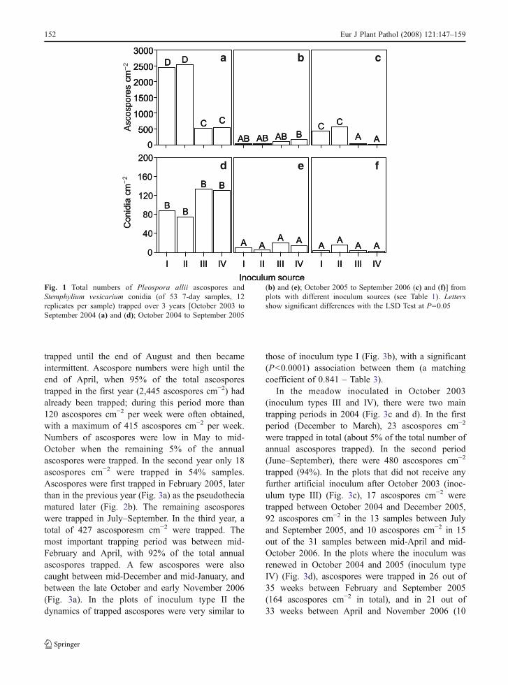
Numbers of the ascospores trapped were significant-ly (P<0.0001) influenced by the year and the inoculumsource, and by their interaction; these accounted for26.3% of the total variance (Table 2). In the first year(Fig. 1a), the treatments with pear leaves as theinoculum source (I and II) produced five times moreascospores than those with the meadow only (III andIV). The presence of an inoculated meadow in additionto the pear leaf litter (II) did not significantly increasethe total number of ascospores compared to the leaflitter alone (I). In the second year (Fig. 1b), the totalnumber of ascospores was less than in the previousyear and was not significantly influenced by theinoculum sources. In particular, the meadow inoculatedwith S. vesicarium in autumn 2003 (III) produced asimilar number of ascospores to that inoculated in both2003 and 2004 (IV). Results obtained in the third year(Fig. 1c) were consistent with those found in the firstyear, but the ascospore numbers were low. Also in thisyear, total number of ascospores trapped from themeadow inoculated only in autumn 2003 was similarto that from the meadow inoculated for 3 years.
Ascospore numbers were also significantly (P<0.0001) influenced by the time of sampling: samplingtime and all interactions were significant (P<0.0001),accounting for 73.7% of the total variance (Table 2).Between-sampling differences ranged from no asco-spores (in 54% of the total samples) to 417 ascosporescm−2 in the traps over inoculum source II in the weekbetween 20 and 27 February 2004 (Fig. 3a).
Numbers of conidia were significantly (P<0.0001)influenced by the year and the time of sampling, andby their interaction, and this accounted for 6.4, 32.7
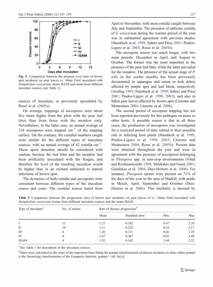
and 49.7% of the total variance, respectively. Theinoculum source and its interaction with the year werenot significant, while interactions between inoculumsources and sampling time were significant (Table 2).Conidia were more abundant in the first year than inthe other 2 years (Fig. 1e–f). Numbers of conidiaranged from zero (in 75% samples) to 49 conidiacm−2 in the traps over inoculum source III in the weekbetween 13 and 20 August 2004 (Fig. 4c).
Maturation of pseudothecia
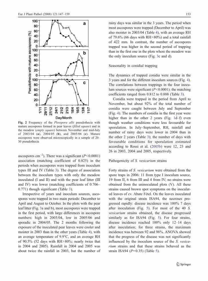
In the first year, the pseudothecia matured over a longperiod: the first and last mature pseudothecia wereobserved on 9 December 2003 and 25 May 2004,respectively; the peak of maturation occurred inJanuary. Maturation lasted for a longer time in themeadow than on the pear leaves (Fig. 2a). In thesecond year, maturation occurred later over a shorterperiod of time, especially in the meadow. Mostpseudothecia matured between the end of April andmid-June on the pear leaves, whereas in the meadowthis period began 1 month later (Fig. 2b). In the thirdyear, pseudothecia on pear leaves and the meadowmatured almost in the same period of time betweenearly December and mid-June (Fig. 2c).
Seasonality in ascospore trappings
In plots covered with the pear leaf litter in October2003 (inoculum type I), ascospores were first trappedin early December (Fig. 3a), according to the presenceof mature pseudothecia (Fig. 3a). Ascospores were
Table 2 Results of the ANOVA of repeated measurements applied to the Pleospora allii ascospores and Stemphylium vesicariumconidia trapped in 3 years, with four types of inoculum sources and 53 7-day samplings per year
Source of variation Ascospores Conidia
% variance df source Df error F P % variance df source df error F P
1. Year 15.9 2 22 110.2 <0.0001 6.4 2 22 25.3 <0.00012. Inoculum sourcea 3.8 3 33 45.9 <0.0001 0.2 3 33 1.6 0.2023. Samplingsb 15.2 9 100 26.0 <0.0001 32.7 4 42 17.9 <0.00014. 1×2b 6.6 3 34 40.0 <0.0001 0.3 3 35 1.8 0.1575. 1×3b 28.2 9 101 22.3 <0.0001 49.7 4 40 12.9 <0.00016. 2×3b 15.5 21 232 14.0 <0.0001 3.6 29 321 1.8 0.0087. 1×2×3b 14.8 28 307 6.9 <0.0001 7.2 28 313 1.5 0.042
a See Table 1 for description of the inoculum sources.b For these sources of variation the Mauchly_s Test of sphericity was significant, so the degrees of freedom (df) were corrected by theHuynh–Feldt Epsilon.
Eur J Plant Pathol (2008) 121:147–159 151
trapped until the end of August and then becameintermittent. Ascospore numbers were high until theend of April, when 95% of the total ascosporestrapped in the first year (2,445 ascospores cm−2) hadalready been trapped; during this period more than120 ascospores cm−2 per week were often obtained,with a maximum of 415 ascospores cm−2 per week.Numbers of ascospores were low in May to mid-October when the remaining 5% of the annualascospores were trapped. In the second year only 18ascospores cm−2 were trapped in 54% samples.Ascospores were first trapped in February 2005, laterthan in the previous year (Fig. 3a) as the pseudotheciamatured later (Fig. 2b). The remaining ascosporeswere trapped in July–September. In the third year, atotal of 427 ascosporesm cm−2 were trapped. Themost important trapping period was between mid-February and April, with 92% of the total annualascospores trapped. A few ascospores were alsocaught between mid-December and mid-January, andbetween the late October and early November 2006(Fig. 3a). In the plots of inoculum type II thedynamics of trapped ascospores were very similar to
those of inoculum type I (Fig. 3b), with a significant(P<0.0001) association between them (a matchingcoefficient of 0.841 – Table 3).
In the meadow inoculated in October 2003(inoculum types III and IV), there were two maintrapping periods in 2004 (Fig. 3c and d). In the firstperiod (December to March), 23 ascospores cm−2
were trapped in total (about 5% of the total number ofannual ascospores trapped). In the second period(June–September), there were 480 ascospores cm−2
trapped (94%). In the plots that did not receive anyfurther artificial inoculum after October 2003 (inoc-ulum type III) (Fig. 3c), 17 ascospores cm−2 weretrapped between October 2004 and December 2005,92 ascospores cm−2 in the 13 samples between Julyand September 2005, and 10 ascospores cm−2 in 15out of the 31 samples between mid-April and mid-October 2006. In the plots where the inoculum wasrenewed in October 2004 and 2005 (inoculum typeIV) (Fig. 3d), ascospores were trapped in 26 out of35 weeks between February and September 2005(164 ascospores cm−2 in total), and in 21 out of33 weeks between April and November 2006 (10
a b c
d e f
Fig. 1 Total numbers of Pleospora allii ascospores andStemphylium vesicarium conidia (of 53 7-day samples, 12replicates per sample) trapped over 3 years [October 2003 toSeptember 2004 (a) and (d); October 2004 to September 2005
(b) and (e); October 2005 to September 2006 (c) and (f)] fromplots with different inoculum sources (see Table 1). Lettersshow significant differences with the LSD Test at P=0.05
152 Eur J Plant Pathol (2008) 121:147–159
ascospores cm−2). There was a significant (P<0.0001)association (matching coefficient of 0.825) in theperiods when ascospores were trapped from inoculumtypes III and IV (Table 3). The degree of associationbetween the inoculum types with only the meadowinoculated (I and II) and with the pear leaf litter (IIIand IV) was lower (matching coefficients of 0.706–0.771) though significant (Table 3).
Irrespective of years and inoculum sources, asco-spores were trapped in two main periods: December toApril and August to October. In the plots with the pearleaf litter (Fig. 3a and b), most ascospores were trappedin the first period, with large differences in ascosporenumbers: high in 2003/04, low in 2005/06 andsporadic in 2004/05. The 2 months following theexposure of the inoculated pear leaves were cooler andmoister in 2003 than in the other years (Table 4), withan average temperature of 9.9°C, and an average RHof 90.5% (52 days with RH>80%: nearly twice thatin 2004 and 2005). Rainfall in 2004 and 2005 wasabout twice the rainfall in 2003, but the number of
rainy days was similar in the 3 years. The period whenmost ascospores were trapped (December to April) wasalso moister in 2003/04 (Table 4), with an average RHof 79.6% (66 days with RH>80%) and a total rainfallof 422 mm. In contrast, the number of ascosporestrapped was higher in the second period of trappingthan in the first one in the plots where the meadow wasthe only inoculum source (Fig. 3c and d).
Seasonality in conidial trapping
The dynamics of trapped conidia were similar in the3 years and for the different inoculum sources (Fig. 4).The correlations between trappings in the four inocu-lum sources were significant (P<0.0001): the matchingcoefficients ranged from 0.812 to 0.888 (Table 3).
Conidia were trapped in the period from April toNovember, but about 92% of the total number ofconidia were caught between July and September(Fig. 4). The numbers of conidia in the first year werehigher than in the other 2 years (Fig. 1d–f) eventhough weather conditions were less favourable forsporulation. In July–September, RH, rainfall andnumber of rainy days were lower in 2004 than inthe other 2 years (Table 3); the number of days withfavourable conditions for sporulation estimatedaccording to Rossi et al. (2005b) were 12, 23 and26 in 2003, 2004 and 2005, respectively.
Pathogenicity of S. vesicarium strains
Forty strains of S. vesicarium were obtained from thespore traps in 2006: 11 from type I inoculum source,19 from II, 6 from III and 4 from IV; no strains wereobtained from the uninoculated plots (V). All thesestrains caused brown spot symptoms on the inoculat-ed leaves of cv. Abate Fétel. On the leaves inoculatedwith the original strain ISA94, the necroses pro-gressed rapidly: disease incidence was 100% 7 daysafter inoculation (Fig. 5). For most of the 40 S.vesicarium strains obtained, the disease progressedsimilarly as for ISA94 (Fig. 5). For four strains,disease incidence reached 100% only 12–14 daysafter inoculation; for three strains, the maximumincidence was between 92 and 96%. ANOVA showedthat the progress of the disease was not significantlyinfluenced by the inoculum source of the S. vesica-rium strains and that these strains behaved as thestrain ISA94 (P=0.35) (Table 5).
a
b
c
Fig. 2 Frequency of the Pleospora allii pseudothecia withmature ascospores formed in pear leaves (filled square) and inthe meadow (empty square) between November and mid-Julyof 2003/04 (a), 2004/05 (b), and 2005/06 (c). Matureascospores were observed microscopically in a sample of 20–30 pseudothecia
Eur J Plant Pathol (2008) 121:147–159 153

Discussion
This work concerns sources of the airborne spores ofS. vesicarium and its teleomorph P. allii, which
constitute the inoculum for brown spot disease onpear. Previous studies focused on the densities anddynamics of these airborne spores during the season,with no reference to their sources (Picco et al. 1996;
a
b
c
d
Fig. 3 Numbers of Pleo-spora allii ascosporestrapped weekly (means of12 replicates), in October2003 to December 2006,from plots with differentinoculum sources: a corre-sponds to the inoculum typeI (see Table 1), b to II, c toIII, d to IV. Filled triangle isthe time when the inoculumsource was inoculated withStemphylium vesicarium;filled circle represents aweek with positive sporetrapping
154 Eur J Plant Pathol (2008) 121:147–159
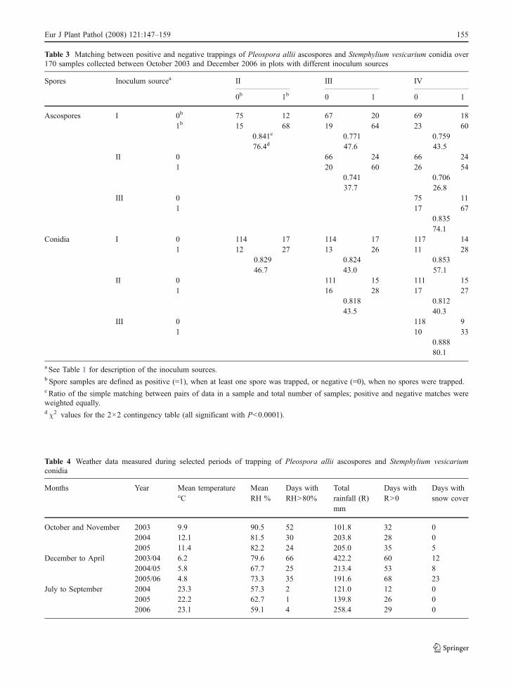
Table 3 Matching between positive and negative trappings of Pleospora allii ascospores and Stemphylium vesicarium conidia over170 samples collected between October 2003 and December 2006 in plots with different inoculum sources
Spores Inoculum sourcea II III IV
0b 1b 0 1 0 1
Ascospores I 0b 75 12 67 20 69 181b 15 68 19 64 23 60
0.841c 0.771 0.75976.4d 47.6 43.5
II 0 66 24 66 241 20 60 26 54
0.741 0.70637.7 26.8
III 0 75 111 17 67
0.83574.1
Conidia I 0 114 17 114 17 117 141 12 27 13 26 11 28
0.829 0.824 0.85346.7 43.0 57.1
II 0 111 15 111 151 16 28 17 27
0.818 0.81243.5 40.3
III 0 118 91 10 33
0.88880.1
a See Table 1 for description of the inoculum sources.b Spore samples are defined as positive (=1), when at least one spore was trapped, or negative (=0), when no spores were trapped.c Ratio of the simple matching between pairs of data in a sample and total number of samples; positive and negative matches wereweighted equally.dχ2 values for the 2×2 contingency table (all significant with P<0.0001).
Table 4 Weather data measured during selected periods of trapping of Pleospora allii ascospores and Stemphylium vesicariumconidia
Months Year Mean temperature°C
MeanRH %
Days withRH>80%
Totalrainfall (R)mm
Days withR>0
Days withsnow cover
October and November 2003 9.9 90.5 52 101.8 32 02004 12.1 81.5 30 203.8 28 02005 11.4 82.2 24 205.0 35 5
December to April 2003/04 6.2 79.6 66 422.2 60 122004/05 5.8 67.7 25 213.4 53 82005/06 4.8 73.3 35 191.6 68 23
July to September 2004 23.3 57.3 2 121.0 12 02005 22.2 62.7 1 139.8 26 02006 23.1 59.1 4 258.4 29 0
Eur J Plant Pathol (2008) 121:147–159 155
Maccaferri et al. 2003; Rossi et al. 2005b; Giosuèet al. 2006). Thus the inoculum sources for thisdisease were not well understood (Llorente and
Montesinos 2006). The present results demonstratedthat pear leaf litter and the meadow of herbaceousplants covering the ground are potentially important
a
b
c
d
Fig. 4 Numbers of Stem-phylium vesicarium conidiatrapped weekly (means of12 replicates), in October2003 to December 2006,from plots with differentinoculum sources: a corre-sponds to inoculum type I(see Table 1), b to II, c toIII, d to IV. Filled triangle isthe time when the inoculumsource was inoculated withStemphylium vesicarium;Filled circle represents aweek with positive sporetrapping
156 Eur J Plant Pathol (2008) 121:147–159
sources of inoculum, as previously speculated byRossi et al. (2005a).
On average, trappings of ascospores were aboutfive times higher from the plots with the pear leaflitter than from those with the meadow only.Nevertheless, in the latter case, an annual average of218 ascospores were trapped cm−2 of the trappingsurface. On the contrary, the conidial numbers caughtwere similar for the different types of inoculumsources, with an annual average of 42 conidia cm−2.These spore densities should be considered withcaution, because the leaf litter and the meadow hadbeen artificially inoculated with the fungus, andtherefore the level of the resulting inoculum wouldbe higher than in an orchard subjected to naturalinfections of brown spot.
The dynamics of both conidia and ascospores wereconsistent between different types of the inoculumsource and years. The conidial season lasted from
April to November, with most conidia caught betweenJuly and September. The presence of airborne conidiaof S. vesicarium during the warmer period of the yearwas in substantial agreement with previous studies(Hausbeck et al. 1999; Suheri and Price 2001; Prados-Ligero et al. 2003; Rossi et al. 2005b).
The ascospore season was much longer, with twomain periods: December to April, and August toOctober. The former was the most important in thepresence of the pear leaf litter, while the latter prevailedfor the meadow. The presence of the sexual stage of P.allii in the cooler months has been previouslydocumented in asparagus and onion or leek debrisaffected by purple spot and leaf block, respectively(Aveling 1993; Hausbeck et al. 1999; Suheri and Price2001; Prados-Ligero et al. 1998, 2003), and also infallen pear leaves affected by brown spot (Llorente andMontesinos 2004; Llorente et al. 2006).
The second period of ascospore trapping has notbeen reported previously for this pathogen on pears orother hosts. A possible reason is that in all thesecases, the production of ascospores was investigatedfor a restricted period of time related to their possiblerole in infecting host plants (Hausbeck et al. 1999;Prados-Ligero et al. 1998, 2003; Llorente andMontesinos 2004; Rossi et al. 2005b). Present datawere obtained throughout the year and were inagreement with the presence of ascospores belongingto Pleospora spp. in non-crop environments (Vittaland Krishnamoorthi 1988; Mitakakis and Guest 2001;Gioulekas et al. 2004; Díez-Herrero et al. 2006). Forinstance, Pleospora spores were present on 71% ofthe days of the year in the area of Madrid, with peaksin March, April, September and October (Díez-Herrero et al. 2006). This similarity is stressed by
Fig. 5 Comparison between the progress over time of brownspot incidence on pear leaves cv. Abate Fétel inoculated withStemphylium vesicarium, strain ISA94 and strain from differentinoculum sources (see Table 1)
Table 5 Comparison between the progression rates of brown spot incidence on pear leaves of cv. Abate Fétel inoculated withStemphylium vesicarium strains from different inoculum sources and the strain ISA94
Type of inoculuma No. of strains Rate of disease progressionb
Mean Standard error Min. Max.
I 11 1.27 0.242 0.47 2.29II 19 1.11 0.225 0.34 3.17III 6 1.20 0.111 0.66 1.39IV 4 1.67 0.367 0.91 2.40ISA94 4 1.92 0.162 1.44 2.22
a See Table 1 for description of the inoculum sources.b Rates were calculated as the slope of the regression lines fitting the gompit transformation of disease incidence to time, where gompitis the linearizing transformation of the Gompertz function: gompit=−ln[−ln(y)].
Eur J Plant Pathol (2008) 121:147–159 157

the fact that the ascospores we trapped in the secondperiod were from the meadow plants. Therefore, thesaprophytic colonisation by S. vesicarium of deadplant material from the meadow could act as animportant source of inoculum.
Results from the present work contribute to a betterunderstanding of the role of ascospores in the lifecycle of the pathogen. It has been assumed that thesespores initiate the saprophytic colonisation of thedead material in the meadow during the winter andspring (Rossi et al. 2005a; Llorente and Montesinos2006) with new sexually-derived genotypes, whichmaintains their infectivity on pear (Llorente andMontesinos 2006; Rossi et al. 2006). This study hasdemonstrated that this process begins in autumn andcontributes to a significant proportion of colonisationof the meadow and the consequent production ofabundant conidia. In fact, the weather conditions fromOctober 2003 to April 2004 favoured early maturationof pseudothecia that continued for long time, withearly, long and high production of ascospores be-tween December and April, so that in summer 2004,the numbers of conidia were high. In autumn 2004 tospring 2005, the weather conditions were lessfavourable, pseudothecia matured later and for ashorter period with low numbers of ascosporesproduced, so that the production of conidia in summer2005 was lower than in 2004, despite the fact thatweather conditions were more favourable for asexualsporulation in 2005. Therefore, the inoculum produc-tion in summer depends on the colonisation rate of thedead material on the orchard floor, which in turndepends on the weather conditions from autumn tospring, as well as the current summer conditions(Rossi et al. 2005b; Giosuè et al. 2006).
This study also demonstrated, for the first time,that the pathogen is capable of colonizing the orchardfloor persistently. The plots with a wildflowermeadow inoculated in early October 2003 with thevirulent stain ISA94 of S. vesicarium producedairborne spores until the end of the experiment: thelast conidia were trapped on 20–27 October 2006, andthe last ascospores on 3–10 November 2006. Consid-ering that: (1) there were no other sources of S.vesicarium inoculum from pears in the vicinity; (2)the spore traps were placed a few centimetres abovethe ground, thus the ingression of spores from theother plots, which were separated by 1 m wide bands,may be considered not important; (3) neither asco-
spores nor conidia were trapped from the uninoculat-ed meadow; (4) the fungal strains obtained from theairborne propagules in 2006 were all able to causebrown spot on pear leaves as did strain ISA94, it canbe reasonably concluded that the airborne sporestrapped were produced from the meadow that hadbeen inoculated in 2003.
In conclusion, the present work demonstrates thatthe orchard ground is a permanent source of bothsexual (ascosporic) and asexual (conidial) inoculumfor brown spot of pear. The implications of thisinformation for disease control concern the possibilityof reducing the amount of inoculum by managing theorchard ground. Recently, sanitation measures for thepear orchard ground were investigated with the aim ofreducing overwintering sexual inoculum (Llorenteet al. 2006). Further work should be aimed at verifyingthe effectiveness of these measures in reducing theinoculum all year long, particularly the asexualinoculum during epidemics.
Acknowledgements This work is part of research promotedand supported by the Emilia-Romagna Region (Italy) incollaboration with the Centro Ricerche Produzioni Vegetali(CRPV).
References
Aveling, T. A. S. (1993). Stemphylium leaf blight of garlic inSouth Africa. Phytophylactica, 25, 293–294.
Campbell, C. L., & Madden, L. V. (1990). Temporal analysis ofepidemics I: description and comparison of diseaseprogress curves. In Introduction to plant disease epidemi-ology, pp. 161–202. New York: Wiley.
Cavanni, P., & Ponti, I. (1994). Maculatura bruna del pero: Unamicopatia sempre d’attualità. Rivista di frutticoltura, 12,37–42.
Díez-Herrero, A., Bustillo, M. G., & Cervigòn Morales, P.(2006). Study of airborne fungal spores in Madrid, Spain.Aerobiologia, 22, 135–142.
Ellis, M. B. (1971). Dematiaceous hyphomycetes. Kew, Surrey,UK: Commonwealth Mycological Institute.
Ellis, M. B., & Ellis, J. P. (1985). Microfungi on land plants.New York: MacMillan.
Everitt, B., & Howell, D. (Eds.) (2005). Encyclopedia ofstatistics in behavioral science. Winnipeg, Canada: Wiley.
Giosuè, S., Rossi, V., Bugiani, R., & Mazzoni, C. (2006).Modelling dynamics of airborne conidia of Stemphyliumvesicarium, the causal agent of brown spot of pear. IOBC/WPRS Bulletin, 29(1), 223–230.
Gioulekas, D., Damialis, A., Papakosta, D., Spieksma, F.,Giouleka, P., & Patakas, D. (2004). Allergenic fungi sporerecords (15 years) and sensitization in patients with respira-tory allergy in Thessaloniki-Greece. Journal of Investigation-al Allergology and Clinical Immunology, 14(3), 225–231.
158 Eur J Plant Pathol (2008) 121:147–159

Hausbeck, M. K., Hartwell, J., Byrne, J. M., & Benson, B.(1999). Epidemiology of Stemphylium leaf spot andpurple spot in no-till asparagus. Acta Horticulturae, 479,205–210.
Llorente, I., & Montesinos, E. (2004). Development and fieldevaluation of a model to estimate the maturity ofpseudothecia of Pleospora allii on pear. Plant Disease,88, 215–219.
Llorente, I., & Montesinos, E. (2006). Brown spot of pear: Anemerging disease of economic importance in Europe.Plant Disease, 90(11), 1368–1375.
Llorente, I., Vilardell, A., & Montesinos, E. (2006). Infectionpotential of Pleospora allii and evaluation of methods forreduction of the overwintering inoculum of brown spot ofpear. Plant Disease, 90, 1511–1516.
Llorente, I., Vilardell, P., Moragrega, C., Bonaterra, A., &Montesinos, E. (2003). Biology, epidemiology and inte-grated control of Stemphylium vesicarium on pear, anemerging disease of economic impact in Europe. Paperpresented at the 8th International Congress of PlantPathology, Christ Church, New Zealand.
Maccaferri, E., Collina, M., & Brunelli, A. (2003). Studies onthe epidemiology of Stemphylium vesicarium on pear.Journal of Plant Pathology, 85, 310.
Mitakakis, T. Z., & Guest, D. I. (2001). A fungal spore calendarfor the atmosphere of Melbourne, Australia, for the year1993. Aerobiologia, 17, 171–176.
Montesinos, E., Moragrega, C., Llorente, I., & Vilardell, P.(1995). Susceptibility of selected European pear cultivarsto infection by Stemphylium vesicarium and influence ofleaf and fruit age. Plant Disease, 79, 471–473.
Montesinos, E., & Vilardell, P. (1992). Evaluation of FAST as aforecasting system for scheduling fungicide sprays forcontrol of Stemphylium vesicarium on pear. Plant Disease,76, 1221–1226.
Picco, A. M., Betto, A., & Porri, A. (1996). Stemphyllium,Pleospora and Alternaria airspores in a pear tree orchard:A three year quantitative monitoring in Italy. Paper
presented at the 1st European Symposium on Aerobiology,Santiago de Compostela, Spagna, 11–13 Septembre 1996,pp. 156–157.
Ponti, I., Cavanni, P., & Brunelli, A. (1982). Maculatura brunadelle pere: Eziologia e difesa. Informatore Fitopatologico,3, 35–40.
Ponti, I., & Laffi, F. (1993). Malattie crittogamiche delle pianteda frutto. Verona, Italy: Edizioni L’informatore Agrario.
Prados-Ligero, A.M., Gonzalez-Andujar, J. L., Melero-Vara, J.M.,& Basallote-Ubera, M. J. (1998). Development of Pleosporaallii on garlic debris infected by Stemphylium vesicarium.European Journal of Plant Pathology, 104, 861–870.
Prados-Ligero, A. M., Melero-Vara, J. M., Corpas-Hervìas, C.,& Basallote-Ureba, M. J. (2003). Relationships betweenweather variables, airborne spore concentrations andseverity of leaf blight of garlic caused by Stemphyliumvesicarium in Spain. European Journal of Plant Pathol-ogy, 109, 301–310.
Rossi, V., Bugiani, R., Giosuè, S., & Natali, P. (2005b). Patternsof airborne conidia of Stemphylium vesicarium, the causalagent of brown spot disease of pears, in relation to weatherconditions. Aerobiologia, 21, 203–216.
Rossi, V., Pattori, E., & Bugiani, R. (2006). Temperature andhumidity requirements for germination and infection byascospores of Pleospora allii, the teleomorph of Stemphy-lium vesicarium. IOBC/WPRS Bulletin, 29(1).
Rossi, V., Pattori, E., Giosuè, S., & Bugiani, R. (2005a). Growthand sporulation of Stemphylium vesicarium, the causal agentof brown spot of pear, on herb plants of orchard lawns.European Journal of Plant Pathology, 111, 361–370.
Simmons, E.G. (1969). Perfect states of Stemphylium. Mycolo-gia, 61(1), 1–26.
Suheri, H., & Price, T. V. (2001). The epidemiology of purpleleaf blotch on leeks in Victoria, Australia. EuropeanJournal of Plant Pathology, 107, 503–510.
Vittal, B. P. R., & Krishnamoorthi, K. (1988). A census ofairborne spores in the atmosphere of the city of Madras,India. Annals of Allergy, 60(2), 99–101.
Eur J Plant Pathol (2008) 121:147–159 159