Flexible blue-emitting encapsulated organic semiconductor DFB laser
Upload
independentCategory
view
2download
0
Journal ofMaterials Chemistry B
PAPER
Publ
ishe
d on
23
Apr
il 20
14. D
ownl
oade
d by
Uni
vers
ity o
f H
oust
on o
n 06
/06/
2014
15:
12:2
5.
View Article OnlineView Journal
aInstitute of High Pressure Physics RAS, 142bA.N.Belozersky Institute of Physico-Chem
119992 Moscow, RussiacLaboratoire d'Innovation et d'Analyse de
Montreal, Succursale Centre-ville Montreal,dLaboratoire Biologie Cellulaire et Microsc
Universite François Rabelais, 37032 Tours,eFaculty of Bioengineering and Bioinform
Moscow, RussiafGREMAN, UMR CNRS 7347, Universite F
E-mail: [email protected] of Chemical and Biomolecular
Calhoun Rd, Houston, Texas 77204, USA. E
† Electronic supplementary informa10.1039/c3ta21599g
‡ Present address: Center for TechnolHouston, TX, 77040, USA.
Cite this: DOI: 10.1039/c3tb21599g
Received 14th November 2013Accepted 20th April 2014
DOI: 10.1039/c3tb21599g
www.rsc.org/MaterialsB
This journal is © The Royal Society of
Solid state synthesis of carbon-encapsulated ironcarbide nanoparticles and their interaction withliving cells†
Valery Davydov,a Alexandra Rakhmanina,a Igor Kireev,b Irina Alieva,b
Oksana Zhironkina,b Olga Strelkova,b Varvara Dianova,b
Taraneh Djavanbakht Samani,c Karina Mireles,c L. 'Hocine Yahia,c Rustem Uzbekov,de
Viatcheslav Agafonov*f and Valery Khabashesku‡*g
Superparamagnetic carbon-encapsulated iron carbide nanoparticles (NPs), Fe7C3@C, with unique
properties, were produced from pure ferrocene by high pressure–high temperature synthesis. These NPs
combine the merits of nanodiamonds and SPIONs but lack their shortcomings which limit their use for
biomedical applications. Investigation of these NPs by X-ray diffraction, electron microscopy techniques,
X-ray spectroscopic and magnetic measurement methods has demonstrated that this method of
synthesis yields NPs with perfectly controllable physical properties. Using magnetic and subsequent
fractional separation of magnetic NPs from residual carbon, the aqueous suspensions of Fe7C3@C NPs
with an average particle size of �25 nm were prepared. The suspensions were used for in vitro studies of
the interaction of Fe7C3@C NPs with cultured mammalian cells. The dynamics of interaction of the living
cells with Fe7C3@C was studied by optical microscopy using time-lapse video recording and also by
transmission electron microscopy. Using novel highly sensitive cytotoxicity tests based on the cell
proliferation assay and long-term live cell observations it was shown that the internalization of Fe7C3@C
NPs has no cytotoxic effect on cultured cells and does not interfere with the process of their mitotic
division, a fundamental property that ensures the existence of living organisms. The influence of NPs on
the proliferative activity of cultured cells was not detected as well. These results indicate that the carbon
capsules of Fe7C3@C NPs are air-tight which could offer great opportunities for future use of these
superparamagnetic NPs in biology and medicine.
1 Introduction
Currently, superparamagnetic NPs represent one of the mostpromising classes of materials because of their possible
190 Troitsk, Moscow region, Russia
ical Biology, Moscow State University,
Bioperformance Ecole Polytechnique de
Quebec H3C 3A7, Canada
opie Electronique, Faculte de Medecine,
France
atics, 11992, Moscow State University,
rançois Rabelais, 37200 Tours, France.
Engineering, University of Houston, 4800
-mail: [email protected]
tion (ESI) available. See DOI:
ogy Innovation, Baker Hughes Inc.,
Chemistry 2014
applications in various elds of biomedicine.1–6 The ability tocontrol magnetic properties of superparamagnetic NPs opensup a number of specic uses of these NPs in the biomedicaleld, and makes them unique among a wide range of non-magnetic nanostructured systems. The possibility of using suchmaterials as intracellular markers, diagnostic probes and baseplatforms for the delivery of drugs and gene therapy materialsand other purposes has also been actively investigated in recentyears.7–14
To date, various physical and chemical methods have beendeveloped for the synthesis of magnetic NPs on the basis of puremetals (Fe, Co, Ni), metal alloys, metal oxides (g-Fe2O3, Fe3O4,MgFeO4, CoFe2O4, Mn3O4), carbides (Fe7C3, Fe3C) and othersubstances.1–6,15–20
However, direct use of these pristine NPs in biology andmedicine, in general, is not feasible, because these NPs do notmeet a number of specic requirements for their biomedicalapplications. These requirements include biocompatibility,chemical resistance and resistance to aggregation in biologicalmedia, dimensional homogeneity, and the ability to be func-tionalized at their surface by different types of ligands. A
J. Mater. Chem. B

Journal of Materials Chemistry B Paper
Publ
ishe
d on
23
Apr
il 20
14. D
ownl
oade
d by
Uni
vers
ity o
f H
oust
on o
n 06
/06/
2014
15:
12:2
5.
View Article Online
number of challenges to the application of superparamagneticNPs in biomedicine can be overcome by the design of their socalled “core/shell” complexes with biocompatible and durablecoatings. The coatings can be of various inorganic or organicnature, depending on the specic research and applicationobjectives. Currently, as a biocompatible coating of magneticNPs, various surfactants, synthetic polymers, natural macro-molecules, silicones, and layers of noble metals2–6 are widelyused. For example, to induce hydrophilic properties, poly-ethylene glycol (PEG) or polyvinyl alcohol is used for the coatingof the NPs.21–23 Other coatings, such as polyethylenimine,increase cationic surface properties.24 Coatings of proteins andpeptides help to reduce cytotoxicity and enhance the capture ofthe nanocomplex by the cells.25
Here we describe a method to produce Fe7C3@C NPs thatcombine the merits of nanodiamonds and SPIONs yet lackingtheir shortcomings. Like the surface of nanodiamonds, thecarbon shell coating of Fe7C3@C NPs can be easily modiedwith appropriate biologically active moieties. In addition,unlike nanodiamond particles, our NPs possess magneticproperties that exceed those of SPIONs, ensuring better mag-netocontrollability. We also expected that the presence of aprotective carbon shell would signicantly decrease toxicitytypically observed for unmodied SPIONs. The cytotoxicity ofNPs is a key factor limiting their use in biomedicine both atcellular and organism levels. The toxic effects are usuallyassayed by scoring live/dead cells, however, extremely sensitivecellular processes, such as DNA replication and cell division,can also be affected causing irreversible physiological changes(such as stem cell population homeostasis, tissue renewal, etc.),yet not directly leading to cell death. Therefore in cytotoxicitystudies of Fe7C3@C NPs we used novel highly sensitive cyto-toxicity tests based on proliferation assays and long-term livecell imaging in order to exclude subtle effects of the NPs on celldivision.
Obviously, the practical use of such nanocomplexes requiresextensive studies of their behavior in biological systems. Todate, a detailed study of various aspects of biomedical use ofsuperparamagnetic nanocomplexes was conducted essentiallyon only one type of NP, namely SPIONs – superparamagneticiron oxide NPs. In a series of reviews,2–6 the synthesis, applica-tion of protective shells, their functionalization and behavior ofNPs in the biological eld have been discussed in detail. Despitesignicant progress in the development of methods for thesynthesis of nanosystems based on SPIONs, numerous studieshave shown that their relatively low value of magnetizationcreates difficulties in the control of their magnetic behavior in anumber of applications. For this reason, carbon-encapsulatedsuperparamagnetic NPs of metals and their carbides attractincreasing attention.26,27 Signicant advantages of these mate-rials, as compared to SPIONs, for a number of biomedicalapplications have been clearly demonstrated in a recent paper27
discussing the possibility of the use of carbon-encapsulatedsuperparamagnetic FeCo@C NPs as multimodal imagingprobes and cooperative therapeutic agents for tumor cells. Thiswork suggests that because of the high value of the magneti-zation saturation FeCo@C NPs have higher potential than
J. Mater. Chem. B
SPIONs for biomedical applications, particularly in MRI andhyperthermia.
The present work is devoted to studies of the biocompati-bility and possibility of biomedical use of superparamagneticNPs of iron carbide Fe7C3 encapsulated into a carbon shell. It isknown, that iron carbides Fe7C3 and Fe3C (cementite) areferromagnetic with Curie temperatures of 250 and 210 �Chaving the magnetization saturation values of 120 emu g�1 and125 emu g�1, respectively, at room temperature.28
Given the relatively high value of the magnetization of ironcarbides, the NPs with superparamagnetic cores encapsulatedinto a stable carbon shell, designated as Fe7C3@C and Fe3C@C,appear to be very interesting for biomedical applications.Note that the presence of a carbon shell on the NPs offersgreat opportunities for a variety of options for furtherfunctionalization.
To date, there are several knownmethods for the synthesis ofiron carbide NPs surrounded by a carbon shell. One of the rstversions of the directed synthesis of NPs based on Fe3C(cementite) and Fe7C3 phases has been the pyrolysis of a gas–vapor mixture of Fe(CO)5 + C2H4 in argon by using high-powerlaser radiation.17 The disadvantage of this method is thecontamination of the product by a-Fe particles, which are toxic,as well as by iron oxide Fe3O4, due to the presence of oxygen inthe initial reaction mixture. Recently, different methods forsynthesis of encapsulated iron carbide NPs have beenproposed.19,20 However, the size of these particles was quitelarge, therefore they displayed ferromagnetic, but not super-paramagnetic behavior. The proof-of-concept capability forsynthesis of superparamagnetic encapsulated NPs Fe7C3@Cand Fe3C@C using ferrocene transformations induced by highpressures and high temperatures treatment (HPHTT) wasdemonstrated in preliminary reports.29,30 By analogy withSPIONs, these particles were termed SCICNs (Super-paramagnetic Carbon-encapsulated Iron Carbide NPs).
The objectives of the present work were (i) more detailedstudies of the possibilities of the HPHTT method for thesynthesis of SCICNs, namely Fe7C3@C, with controlled particlesizes, (ii) the development of methods for magnetic separationand size fractionation of synthesized superparamagnetic NPs,(iii) in vitro studies of interactions of water-dispersed Fe7C3@CNPs with living cultured cells, (iv) testing the cytotoxicity of theNPs and exploring their possible effect on the proliferativeactivity of the cells and cell division.
2 Experimental
Ferrocene (Aldrich, impurity contents less than 2%) was used asa starting material without further purication. Cold-pressedpellets of the pristine ferrocene (4.5 mm diameter and 3 mmheight) were inserted into a graphite heater and then placedinto a high pressure cell. High pressure high temperaturetreatment (HPHTT) of the samples was performed with the useof a high pressure apparatus of the “Toroid” type.31 Details ofthe experimental procedure have been described in thepreliminary report.29 XRD experiments were carried out on anINEL CPS 120 diffractometer using Co Ka1 radiation.
This journal is © The Royal Society of Chemistry 2014

Fig. 1 XRD pattern of the sample obtained by thermal treatment offerrocene at 8.0 GPa and 560 �C. The diffraction peak correspondingto graphitic-like carbon states is marked by G. The inset shows theexperimental and simulated XRD patterns of the hexagonal phase ofFe7C3.
Paper Journal of Materials Chemistry B
Publ
ishe
d on
23
Apr
il 20
14. D
ownl
oade
d by
Uni
vers
ity o
f H
oust
on o
n 06
/06/
2014
15:
12:2
5.
View Article Online
Microscopy characterization of the samples was carried outusing a scanning electronmicroscope Ultra-Plus Gemini (Zeiss),and transmission electron microscopes JEM 1230 and JEOL2100 for high resolution microscopy (HRTEM). Room temper-ature magnetization curves of the samples and the temperaturedependence of the magnetization were measured with aquantum design physical property measurement systemmagnetometer. Separation of superparamagnetic NPs fromproducts obtained as a result of HPHTT decomposition offerrocene and preparation of an aqueous suspension ofencapsulated Fe7C3@C NPs was carried out in several stages.
During the rst stage, the samples were mechanicallycrushed in a ball mill. Further dispersion of the particles wascarried out by using the ultrasonic bath for 48 hours in a 35%solution of hydrogen peroxide. At the nal stage of processing,the magnetic NPs were isolated from the total solution bymagnetic separation, washed 3 times with distilled water andthen dried for 24 hours. Subsequent fractional separation ofNPs and their aggregates was carried out by centrifugation ofthe obtained powder mixture in three-layer aqueous solutionsof sucrose of different densities (30, 50 and 70% of sucrose) for10 min at an acceleration of 1000 rpm. Aer centrifugation, thecontents of the middle layer were selected from the separatingcapsule and then washed and dried. Size distribution in theseparated fraction of NPs was measured by SEM and TEM. Thestructure and chemical composition of the surface layer of theobtained NPs were examined by XPS (VG ESCALAB 3 MK II, withan Mg Ka radiation source (hn ¼ 1253.6 eV, and an instrumentresolution of 0.7 eV)). At the same time, the magnetic propertiesof the selected fraction of superparamagnetic NPs were rede-termined. The selected NPs were then dispersed in water usingultrasonication for 10 minutes. Different concentrations ofwater suspensions of Fe7C3@C NPs were prepared for biologicalresearch.
Biological tests, conducted in the current study, wereprimarily aimed at the evaluation of cytotoxic effects of water-based dispersions of non-modied nanocomplexes of Fe7C3@Csuperparamagnetic NPs on cultured mammalian cells ofvarious origins (human transformed cell lines and a pigembryonic cell). Since similar investigations have not yet beenconducted with such NPs, several types of sensitive cytotoxicitytests were performed including the NP internalization dynamicsin vitro, mitotic activity measurements, DNA replication, andoverall proliferative activity of cellular population upon pro-longed cultivation in the presence of high concentrations of NPsin culture media.
Cytotoxicity tests were performed on pig kidney epithelialcells (PK). PK cells (kindly provided by Russian collection of celllines, St. Petersburg) were cultured in DMEM culture medium(Sigma, USA) supplemented with 10% fetal calf serum(HyClone, USA) and antibiotic–antimycotic (Sigma, USA). Formicroscopy experiments, cells were placed on coverslips andincubated with various concentrations of Fe7C3@C NPs (from 5to 100 mg ml�1). 20 mg ml�1 was found to be an optimalconcentration for easy particle detection by light and electronmicroscopy while MNP agglomerates did not signicantlyinterfere with sectioning and imaging.
This journal is © The Royal Society of Chemistry 2014
For live imaging, PK cells were placed on glass-bottomedPetri dishes (LabTek, USA) at a density of 105 cells per ml andincubated with Fe7C3@C NPs for 24 h. Cell observation wasperformed in an environmental chamber kept at 37 �C andunder 5% CO2. The chamber was mounted on an Olympus IX70inverted microscope equipped with a CCD-camera Orca-RT +(Hamamatsu, Japan) and controlled by Micromanager 1.4soware.32 Illumination conditions (ND lters, lamp voltage,and exposure time) were set to minimize phototoxicity. Imagesequences were analyzed and time-lapse movies of cells loadedwith SCICNS were assembled using ImageJ soware.33,34 Imageswere taken every 10 min for 72 h for long recording or every 1min during short recording.
For further TEM experiments, PK cells were washed severaltimes with fresh pre-warmed media to remove free particles,xed in 2.5% glutaraldehyde in 100 mM phosphate buffer (pH7.4) for 2 hours with subsequent post-xation in 1% OsO4 andembedding in Epon (Sigma, USA). Serial ultrathin sections(70 nm) were prepared using a Leica ultramicrotome andobserved using a JEM 1011 (JEOL, Japan) at 100 kV.
3 Results3.1 Structural characterization of Fe7C3@C NPs
The X-ray diffraction (XRD) pattern and TEM images of thesample obtained by heat treatment of ferrocene at 8.0 GPa and560 �C are shown in Fig. 1 and 2, respectively. In the XRDpattern, shown in Fig. 1, the diffraction line at 2Q ¼ 30.4�
corresponds to the (002) reection of graphite-like materials,which represent a packing of graphene layers. The group ofreections in the angular range of 44–65� mainly belongs tothose of the crystalline phase of iron carbide. The latter wasindexed with the hexagonal unit cell (with the parameters a ¼6.85 (1) and c¼ 4.53 (1) A) corresponding to a space group P63mc(isostructural to Ru7B3).35 The substantial background levelobserved in the low-angle region of the diffractogram is due to
J. Mater. Chem. B
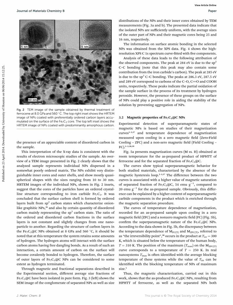
Fig. 2 TEM image of the sample obtained by thermal treatment offerrocene at 8.0 GPa and 560 �C. The top right inset shows the HRTEMimage of NPs coated with preferentially ordered carbon layers accu-mulated on the surface of the Fe7C3 core. The top left inset shows theHRTEM image of NPs coated with predominantly amorphous carbon.
Journal of Materials Chemistry B Paper
Publ
ishe
d on
23
Apr
il 20
14. D
ownl
oade
d by
Uni
vers
ity o
f H
oust
on o
n 06
/06/
2014
15:
12:2
5.
View Article Online
the presence of an appreciable content of disordered carbon inthe sample.
This interpretation of the X-ray data is consistent with theresults of electron microscopic studies of the sample. An over-view of a TEM image presented in Fig. 2 clearly shows that theanalyzed sample represents individual NPs dispersed in asomewhat poorly ordered matrix. The NPs exhibit very distin-guishable inner cores and outer shells, and show mostly quasi-spherical shapes with the sizes ranging from 10 to 30 nm.HRTEM images of the individual NPs, shown in Fig. 2 insets,suggest that the cores of the particles have an ordered crystal-line structure corresponding to iron carbide Fe7C3. It wasconcluded that the surface carbon shell is formed by orderedlayers built from sp2 carbon states which characterize onion-like graphitic NPs,36 and also by certain quantity of disorderedcarbon mainly representing the sp3 carbon state. The ratio ofthe ordered and disordered carbon fractions in the surfacelayers is not constant and slightly changes from one nano-particle to another. Regarding the structure of carbon layers inthe Fe7C3@C NPs obtained at 8 GPa and 560 �C, it should benoted that at this temperature the system retains some amountsof hydrogen. The hydrogen atoms will interact with the surfacecarbon atoms having free dangling bonds. As a result of such aninteraction, a certain amount of carbon on the surface willbecome covalently bonded to hydrogen. Therefore, the surfaceof outer layers of Fe7C3@C NPs can be considered to someextent as hydrogen terminated.
Through magnetic and fractional separations described inthe Experimental section, different average size fractions ofFe7C3@C have been isolated in pure form. Fig. 3 (le) shows theSEM image of the conglomerate of separated NPs as well as size
J. Mater. Chem. B
distributions of the NPs and their inner cores obtained by TEMmeasurements (Fig. 3a and b). The presented data indicate thatthe isolated NPs are sufficiently uniform, with the average sizesof the outer part of NPs and their magnetic cores being 25 and16 nm, respectively.
The information on surface atomic bonding in the selectedNPs was obtained from the XPS data. Fig. 4 shows the high-resolution XPS C 1s spectrum curve-tted with ve components.
Analysis of these data leads to the following attribution ofthe observed components. The peak at 284 eV is due to the sp2
C–C bonding (note that this peak may also contain somecontribution from the iron carbide's carbon). The peak at 285 eVis due to the sp3 C–C bonding. The peaks at 286.3 eV, 287.5 eVand 289 eV correspond to carbons of the C–O, C]O and COOHunits, respectively. These peaks indicate the partial oxidation ofthe sample surface in the process of its treatment by hydrogenperoxide. However, the presence of these groups on the surfaceof NPs could play a positive role in aiding the stability of thesolution by preventing aggregation of NPs.
3.2 Magnetic properties of Fe7C3@C NPs
Experimental detection of superparamagnetic states ofmagnetic NPs is based on studies of their magnetizationcurves1–3,37 and temperature dependence of magnetizationmeasured upon cooling in a zero magnetic eld (Zero-FieldCooling – ZFC) and a non-zero magnetic eld (Field Cooling –
FC).1–3,38,39
Fig. 5a presents magnetization curves (M vs. H) obtained atroom temperature for the as-prepared product of HPHTT offerrocene and for the separated fraction of Fe7C3@C.
The curves show typical superparamagnetic behavior forboth studied materials, characterized by the absence of themagnetic hysteresis loop.1–3,37 The difference between the twocurves is associated with a higher value of magnetic saturationof separated fraction of Fe7C3@C, 54 emu g�1, compared to20 emu g�1 for the as-prepared sample. Obviously, this differ-ence can be explained by a higher content of superparamagneticcarbide components in the product which is enriched throughthe magnetic separation procedure.
The curves of temperature dependence of magnetization,recorded for an as-prepared sample upon cooling in a zeromagnetic eld (ZFC) and a nonzero magnetic eld (FC) (Fig. 5b),conrm the superparamagnetic nature of the Fe7C3@C NPs.According to the data shown in Fig. 5b, the discrepancy betweenthe temperature dependence of MFC(T) and MZFC(T), referred toas “the irreversibility point”,38 occurs in the product at Tirr¼ 300K, which is situated below the temperature of the human body,T ¼ 310 K. The position of the maximum (Tmax) on the MZFC(T)
curve corresponds to a temperature of T ¼ 250 K. In realnanosystems Tmax is oen identied with the average blockingtemperature of these systems while the value of Tirr can beidentied with the blocking temperature of NPs of maximumsize.1
Thus, the magnetic characterization, carried out in thiswork, shows that the as-produced Fe7C3@C NPs, resulting fromHPHTT of ferrocene, as well as the separated NPs both
This journal is © The Royal Society of Chemistry 2014

Fig. 3 SEM image of separated Fe7C3@CNPs (left), and dispersion diagrams (right) of the outer sizes of the NPs (a) and the sizes of their magneticcores (b).
Fig. 4 High resolution XPS C 1s carbon peak observed for the Fe7C3@C sample.
Paper Journal of Materials Chemistry B
Publ
ishe
d on
23
Apr
il 20
14. D
ownl
oade
d by
Uni
vers
ity o
f H
oust
on o
n 06
/06/
2014
15:
12:2
5.
View Article Online
represent truly superparamagnetic materials. However, forbiological tests we have selected only the medium size fractionof Fe7C3@C NPs which are more homogeneous in composition.
3.3 Biological properties of Fe7C3@C NPs
3.3.1 In vitro studies of Fe7C3@C NP interactions withcells. In this study we have investigated the dynamics of inter-nalization of 25 nm diameter Fe7C3@C NPs into pig kidney (PK)epithelial cells. These cells were selected for analysis of theirinteraction with the NPs since they are not specialized forphagocytosis. Our choice is based on the fact that the futuretargets in humans will be located in the similar type of cells. Inour opinion, the phagocyte cells usually used in similar exper-iments are not suitable for our purposes since these cells easilyabsorb any external particles independent of their nature.
PK cells (kindly provided by Russian Collection of cell lines,St. Petersburg) were cultured in DMEM culture media (Sigma,USA) supplemented with 10% fetal calf serum (HyClone, USA)and antibiotic–antimycotic (100 units per ml penicillin G,
This journal is © The Royal Society of Chemistry 2014
100 mg ml�1 streptomycin sulfate and 0.25 mg ml�1 amphoter-icin B) (Sigma, USA). For microscopic experiments cells wereplated onto cover slips at a concentration of 10 000 cells per cm2
and grown for 48 h to reach 50% conuence before the additionof various concentrations of Fe7C3@C NPs.
Kinetics of cell interaction with Fe7C3@C was studied byTEM on serial ultrathin sections, and also by optical microscopyusing time lapse video recording of living cells. Quantitativecharacteristics of Fe7C3@C penetration into the cells are shownin Table 1.
TEM analysis showed that Fe7C3@C (Fig. 6a–c) existed in theculture media not as individual NPs but as agglomerates ofvarious sizes (from tens to hundreds of Fe7C3@C particles).Several stages of Fe7C3@C–cell interactions have been observed.(1) At rst, Fe7C3@C accumulate on the cell surface (Fig. 6a,Table 1). This type of interaction was detected already aer 15min of incubation of cells in the presence of Fe7C3@C NPs. (2)At the second stage (15–30min), the agglomerates were found tobe associated with the plasma membrane invaginations, andFe7C3@C NPs during this phase started to be covered by the
J. Mater. Chem. B

Fig. 5 The curves of magnetization versus the applied field obtained at room temperature for the as-prepared sample (1) and separated fractionof Fe7C3@C NPs (2) (a). The curves of zero field and field cooled magnetization (MZFC and MFC) versus temperature (from 5 to 325 K) (b).
Table 1 Statistical analysis of cell–Fe7C3@C NP interactionsa
Time durationof cell incubation
Cells without contactwith Fe7C3@C, %
Contact of the cellmembrane withFe7C3@C, %
Invagination of thecell membrane in the place ofcontact with Fe7C3@C, %
Fe7C3@Cinto the cell, %
15 min Fe7C3@C (61) 80.3 (49) 16.4 (10) 3.3 (2) 0 (0)30 min Fe7C3@C (64) 62.5 (40) 26.6 (17) 9.4 (6) 1.6 (1)60 min Fe7C3@C (65) 44.6 (29) 26.2 (17) 10.7 (7) 18.5 (12)120 min Fe7C3@C (51) 25.5 (13) 11.8 (6) 27.4 (14) 35.3 (18)
a Numbers given in parentheses: the rst column – number of cells studied, other columns – number of cells found under given conditions.
Journal of Materials Chemistry B Paper
Publ
ishe
d on
23
Apr
il 20
14. D
ownl
oade
d by
Uni
vers
ity o
f H
oust
on o
n 06
/06/
2014
15:
12:2
5.
View Article Online
membrane's lamellipodia (Fig. 6b and Table 1). (3) Startingfrom 30 min, and more oen aer 60 and 120 min of incuba-tion, the Fe7C3@C agglomerates appear inside the cells (Fig. 6cand Table 1).
Since Fe7C3@C agglomerates were dispersed through thewhole volume of cell culture media, the cell–Fe7C3@C inter-action process has progressively continued during a wholeperiod of the experiment, so we could observe all stages ofinteractions simultaneously in different cells, although, theratio of different morphological types has changed with time(see Table 1).
A prolonged incubation of the cells with Fe7C3@CNPs (6 h orlonger) led to formation of rather large Fe7C3@C agglomeratesinside the cell cytoplasm. However, the penetration of NPs intothe cell nucleus has never been observed (Fig. 6d–f). Themaximum cellular uptake of Fe7C3@C NPs was approximatelyestimated using an optical microscope method and found totake place during between 12 and 48 hours incubation timewhen the cytoplasm became lled with slightly aggregatedFe7C3@C NPs, while the cells still maintained normalmorphology. Simultaneously, the overall concentration ofFe7C3@C NPs per cell has increased with the incubation timereaching a maximum at 24 hours when all aggregates precipi-tated at the bottom from solution (Fig. 6d–f).
Three types of arrangements for NP agglomerates have beenobserved in the cell by TEM: agglomerates of particles that canbe either completely surrounded by membranes, partiallycovered by a membrane (Fig. 6e) or be positioned freely in thecell cytoplasm (Fig. 6c).
J. Mater. Chem. B
Upon internalization and fusion of endosomes, largeragglomerates were found to be formed inside the cells.Fe7C3@C agglomerates also precipitated onto a cell-free surfaceof a cover slip and were later internalized by nearby cells duringtheir spreading or crawling. This process was studied using livecell imaging. Results of live observations showed that cellsactively capture NP agglomerates disposed on the surface of thecultivation substrate. In this case the cells were able to separatelarge agglomerates into smaller parts (Fig. 7). Absorbed NPagglomerates moved inside the cells at a considerable distancein the direction from the cell surface to the center of the cell.The mechanism of this movement is not yet fully understoodand will be studied in detail later. Active modication of theleading edge of the cell, the formation of lamellipodia andlopodia as well as active endocytosis indicated the absence ofany signicant effect of the NP presence on the cell surface orinside the cell on its physiological state.
3.3.2 The effect of Fe7C3@C NPs on cell division. Time-lapse imaging also shows that the uptake of Fe7C3@C NPs doesnot interfere with the cell cycle progression. Live cell imagingclearly demonstrates that the cells loaded with NPs successfullyenter and complete mitosis at the rate comparable to that ofnon-treated cells (Fig. 8, see the ESI video recording 2†). Thetime duration of mitosis and morphological features of the cellsat different mitotic stages, including spindle size and its overallarchitecture, the chromosome segregation process and cytoki-nesis, were not affected by the presence of large numbers ofFe7C3@C NPs. These agglomerates moved chaotically in thecytoplasm outside the mitotic spindle and were randomly
This journal is © The Royal Society of Chemistry 2014

Fig. 6 TEM images of ultrathin sections cut in the direction perpendicular to the cell cultivation substrate to represent three stages of Fe7C3@CNP–cell interactions (a–c) and Fe7C3@CNP distribution in the cytoplasm of PK cells, t¼ 24 h (d–f). Contact of Fe7C3@CNPs with the cell surface;the inset shows a high magnification image of the selected region; t¼ 30 min (after addition of Fe7C3@C NPs to the cell culture medium) (a). Cellmembrane invagination – the beginning of endocytosis; the inset shows the high magnification image of the selected region; t ¼ 30 min,arrowheads show two membrane's lamellipodia, which cover a cluster of NPs (b). Fe7C3@C agglomerates inside the cell, t ¼ 60 min. The largeinset shows a high magnification image of the part of a big cluster surrounded by the membrane, the small inset (pointed at by an arrow) shows asmall cluster without the membrane around it. N designates the cell nucleus. The scale bar for all low magnification pictures is 500 nm, for theinsets – 100 nm (c). General view of the cell with the Fe7C3@C agglomerates incorporated into the cytoplasm, while Fe7C3@C NPs are missing inthe nucleus (d). Enlarged region of the cell cytoplasm with the vesicle containing Fe7C3@C NPs. The arrow shows that the cell membrane is notcontinuous (e). High magnification image of the Fe7C3@C agglomerate in the cell, where the average diameter of an individual particle is near 25nm. N – nucleus, Nu – nucleolus, m – mitochondria (f).
Paper Journal of Materials Chemistry B
Publ
ishe
d on
23
Apr
il 20
14. D
ownl
oade
d by
Uni
vers
ity o
f H
oust
on o
n 06
/06/
2014
15:
12:2
5.
View Article Online
distributed between two daughter cells upon cytokinesis (videorecording 2 in the ESI†). These observations indicate that thepresence of Fe7C3@C agglomerates in the cytoplasm of dividingcells does not interfere with the process of mitotic division.
3.3.3 The effect of Fe7C3@C NPs on DNA synthesis.Traditional cytotoxicity tests used for the analysis of NP toxicitysuch as MTT, live/dead cell discrimination, etc. display ratherlow sensitivity as they score dead versus live cells, while toxiceffects of NPs may affect cellular physiology in more subtle wayswithout causing cell death. For example, activation of cell cyclecheckpoints prevents cellular proliferation while cells remainalive for a long time. In this respect, various cell proliferationassays are believed to be most efficient. In this study we studiedthe effect of Fe7C3@C NPs on DNA synthesis using a Click-ITHCS proliferation kit (Invitrogen, USA).
Cells incubated with or without Fe7C3@C for 24–48 h werelabelled with a 10 mM EdU DNA synthesis precursor for 15 min,then xed and analyzed using a uorescence microscope Ti-E(Nikon, Japan). EdU-positive cells as well as mitotic andapoptotic cells were counted in order to evaluate Fe7C3@C NPcytotoxicity. Statistical analysis was performed using Excelspreadsheet soware. We found that a fraction of cells in thesynthetic phase of the cell cycle (S-stage) did not display
This journal is © The Royal Society of Chemistry 2014
statistically signicant changes between control (38%, n ¼ 296)and Fe7C3@C NP loaded (39%, n ¼ 182) cell populations.Moreover, even cells heavily loaded with Fe7C3@C NPs arecapable of DNA synthesis. Thus, a microscopy based cellproliferation assay showed that the DNA synthesis precursorEdU incorporated aer 24 h incubation with Fe7C3@C NPs wasnot affected by accumulation of NPs.
3.3.4 The effect of Fe7C3@C NPs on cell viability andmitotic activity. Upon prolonged cultivation of PK cells withFe7C3@C NPs (three consecutive passages within 10 days) weanalyzed the cellular proliferation rate and cell viability. It wasfound that these parameters of cell population did not differbetween control and Fe7C3@C NP-loaded samples since we didnot observe any noticeable cellular death above the control level.
The mitotic index at the beginning of the experiment beforeadding Fe7C3@C NPs to cell culture medium was 4.1 � 0.46%,while aer 72 h of incubation with Fe7C3@C NPs at a concen-tration of 20 mg ml�1 the mitotic index was 4.3 � 0.77%. Nearly500 cells were analyzed for each point, and 4 independentmeasurements were taken.
We also did not notice any increase in the amount of cellssubject to apoptotic degradation as assayed by microscopicanalysis of nuclear morphology. In the control samples this
J. Mater. Chem. B

Fig. 7 Schemes and optical microscopy photographs representing the dynamics of interaction of Fe7C3@CNPs with living PK cells in the culture.Scheme of the cell and the positions of several agglomerates around it. Agglomerates are numbered from 1 to 5 (a). Successive photographs ofthe cell during living observations where the time elapsed after the beginning of the experiment is indicated in the right top corner of each photo.In the course of the experiment, the agglomerates 1, 2, and 3 remained motionless on the surface of the glass, while cluster number 5 aftercontact with the cell became divided into three separate agglomerates 5a, 5b, and 5c, agglomerate numbers 4, 5a, 5b remained associated withthe cell membrane and moved with it, and cluster 5c became absorbed by the cell and moved quickly into it (b–h). The scheme shows themorphology of the cell and positions of the aggregates 4, 5a–c (white spots) after 63minutes from the start of observation. The initial positions ofthe various agglomerates are shown by black spots. During the experiment, the cell nucleus is rotated clockwise and at the end of the experimentit reaches the position intersecting with the trajectory of the agglomerate 5c (i). Note that the cluster moves along the right side of the nucleusand never under, over or inside it. (see the ESI video recording 1†).
Journal of Materials Chemistry B Paper
Publ
ishe
d on
23
Apr
il 20
14. D
ownl
oade
d by
Uni
vers
ity o
f H
oust
on o
n 06
/06/
2014
15:
12:2
5.
View Article Online
amount was found to be 0.3� 0.11%, 72 h later it reached 1.3 �0.56%, while in the cells with the added MNPs at a concentra-tion of 20 mg ml�1 it comprised only 0.4 � 0.19%.
4 Discussion
Several critical reviews devoted to studies of properties ofdifferent types of magnetic NPs6,16,27 have emphasized that thecarbon-coated magnetic NPs of Fe, Co, Ni and iron carbidesmust possess a number of advantages in potential biomedicalapplications, as compared to the FePt alloy, and g-Fe2O3, andFe3O4 oxide NPs. It is considered that the carbon coatingprevents aggregation of metal or carbide cores, preserving their
J. Mater. Chem. B
single-domain structure and therefore consistency of thesuperparamagnetic nature of the particles. However, the wide-spread use of carbon-coated superparamagnetic NPs inbiomedicine, until recently, was hampered by the fact that theexisting methods for the synthesis of these particles are oencharacterized by low yields of the desired product. Anotherinconvenience is an incomplete carbon coating of individualNPs during their synthesis leading to the presence in theproducts of a certain percentage of pure metals with possiblehigh toxicity.40 Taking into account these observations, thereview16 stated that the synthesis of individual dispersed NPshaving carbon coatings as well as control of the quality of thesecoatings are still unsolved problems.
This journal is © The Royal Society of Chemistry 2014

Fig. 8 Photographs of mitotic division in the PK cell with agglomerates of Fe7C3@C NPs. The scheme of the cell at the end of prophase ofmitosis. Fe7C3@C agglomerates are shown in black, cytoplasm in light-grey, nucleus in grey, nucleolus and chromosomes (inside the nucleus ofthe central mitotic cell) in dark grey color and area free from cells in white (a). Successive photographs of cells during living observations, timeelapsed after the beginning of the experiment is indicated in the left top corner of each photo. During the experiment agglomerates movedactively into the cell and finally turned to be unequally distributed between two daughter cells (b–e). The scheme of two daughter cells after theend of cell division (f), see the ESI video recording 2†.
Paper Journal of Materials Chemistry B
Publ
ishe
d on
23
Apr
il 20
14. D
ownl
oade
d by
Uni
vers
ity o
f H
oust
on o
n 06
/06/
2014
15:
12:2
5.
View Article Online
The present study shows that the development of methodsfor synthesis of carbon-coated magnetic NPs based on pressureand temperature induced transformations of organometalliccompounds, yielding superparamagnetic NPs comprisingnearly 100% of the content of carbon-coated NPs, can give apossible solution to these problems. The existence of a cleardependence of the main characteristics of the produced carbideNPs on pressure, temperature parameters and time ofisothermal treatment of the organometallic compound providesa reliable method for synthesis of nanomaterials with desiredproperties.
In the rst step of the present study, the main controllableparameters of the synthesized material were the average size ofthe inner part of NPs, and the size of the carbide cores, whichensured their superparamagnetic properties. The methods ofmagnetic and fractional separation by sedimentation ofFe7C3@C NPs from the products of ferrocene transformationproposed in the present work allow obtaining pure fractions ofencapsulated NPs with almost any desired size distribution.
Except the size and shape, the most important factor whichdenes the nature of the interaction of the nano-system with a
This journal is © The Royal Society of Chemistry 2014
biological object is the surface structure of the NPs, whichaffects the charge state, adsorption characteristics and otherproperties of the surface.41 It is known that the surfaces of manycarbon nanomaterials, such as fullerenes, carbon nanotubes,polyhedral and carbon onions are hydrophobic, which presentsa big obstacle to their direct use for biomedical applications.42
However, the graing of various hydrophilic groups (such asCOOH, OH, NH2, . etc.) to the surface of these carbon mate-rials can lead to modied NPs being able to produce sufficientlystable aqueous suspensions suitable for use in biomedicalapplications.42 As already noted, in the case of carbon-encap-sulated Fe7C3@C NPs, the outer layers of carbon shellsproduced directly in a high-pressure device, contain somehydrogen, and can be considered as hydrogen-terminatedsurfaces.
Moreover, when hydrogen peroxide, which is a mild oxidant,is used for partial deagglomeration of a related carbonmatrix ofthe primary product of HPHTT of ferrocene, the surface modi-cation of the encapsulated NPs occurs as a side process. Thisresults in appearance on the surface of NPs of numerousoxygen-containing functional groups such as OH, C]O, COOH,
J. Mater. Chem. B
Journal of Materials Chemistry B Paper
Publ
ishe
d on
23
Apr
il 20
14. D
ownl
oade
d by
Uni
vers
ity o
f H
oust
on o
n 06
/06/
2014
15:
12:2
5.
View Article Online
detected by the XPS method, which increase hydrophilicity ofthe NPs making them suitable for aqueous dispersibility. Forthis reason, the aqueous suspensions of our Fe7C3@C NPs aresufficiently stable.
In general, the study of the interaction of NPs with biologicalproles at their various levels presents an extremely complexandmultifaceted task. Even at the “simplest” cellular level, as inthis work, the analysis of the processes of interaction of NPswith the cells involves the identication of ways to boostpenetration into cells, studies of the distribution and the natureof the interaction of NPs with the various elements of theinternal cell structure, and denition of the kinetic character-istics of the processes of endocytosis and exocytosis. From abiological point of view, important characteristics of the inter-action are cytotoxicity, genotoxicity, oxidative stress andinammatory effects resulting from the presence of the NPs inthe biological system.43 Currently, the penetration of extracel-lular materials into the cell was studied in sufficient detail.44–46
Qualitatively, they can be divided into three main categories:diffusion (passive and active), pinocytosis and phagocytosis.The mechanism of passive diffusion involves the possibility ofovercoming the cell membrane by individual atoms and smallmolecules due to the concentration gradient of the diffusingsubstance between the environment and the cytoplasm. Activediffusion uses the energy of ATP for transport of molecules andions against the concentration gradient. Various options ofpinocytosis provide the possibility for passage of externalobjects with the size ranging from a few up to hundreds ofnanometers into the cell. Macropinocytosis and phagocytosisare the primary mechanisms for penetration into the cell oflarger objects that can reach the size of several microns.
Our TEM investigations show that the penetration ofFe7C3@CNPs into the cell, as a rule, is carried out in the form oflarge, but rather weakly bound NP agglomerates. The formationof these agglomerates occurs on the outer surface of the cellmembrane due to agglomeration of smaller clusters existing inextracellular solution (Fig. 6c). Part of large agglomerates maybe formed in the volume of the culture medium or on thesubstrate surface free from cells. Given that the Fe7C3@C NPspenetrate into the cell with the extracellular uid and that thecharacteristic size of the endocytotic vesicles is �500 nm, wecan assume that the macropinocytosis is the main mechanismof Fe7C3@C NP penetration into the studied cells. Membraneruffling, being characteristic of this internalization pathway andshown in Fig. 6b, serves as a further conrmation of thishypothesis. However, since we observed sometimes in the cellssignicantly smaller agglomerates (Fig. 6c), we cannot excludethe possibility of the existence of other mechanisms of pene-tration of Fe7C3@C NPs into the studied cells. Note that thepenetration of Fe7C3@C NPs into the PK cells is reminiscent ofthe penetration of nanodiamonds coated with polyethylenimine(ND–PEI) into the cells.47 According to this report,47 ND–PEI NPscan be also accumulated on the membrane in the form of“dynamic aggregates” having a critical size of �500 nm andbecome nally absorbed by the cells through the mechanism ofmacropinocytosis. These “dynamical aggregates”, dened asaggregates of nanoparticles with weak attractive interactions
J. Mater. Chem. B
allowing changes in the shape,47 present to the cell membrane alarge surface of interaction at the early stages of internalizationthat may favor macropinocytosis. Interestingly, in the studies ofinternalization of unfunctionalized (ND) and acid-functional-ized (ND–COOH) detonation nanodiamonds,50 it was also notedthat the characteristic dimensions of the aggregates of nano-particles observed in the cytoplasm is �500 nm. The authors48
have found that the nanodiamonds, both with and withoutsurface modication by an acid or a base, are biocompatiblewith a variety of cells of different origins including neuroblas-toma, macrophage, keratinocyte and PC-12 cells. The compar-ison of our experiments with work carried out withnanodiamonds (ND)44 shows that the rate of Fe7C3@C NPpenetration into the cells is generally comparable with that forthe ND.47 We found that the rst step of the uptake (15 to120 min) approximately follows a linear behavior with time(Table 1), however, next 48 h of this behavior is exponential,with an estimated characteristic uptake half-life of 2.5 h. Thismeasurement is compatible with the 2.6 h uptake half-lifeobserved for NDs.47 The characteristics of movement of absor-bed agglomerates of Fe7C3@C NPs suggest a possible partici-pation of cytoskeletal units, particularly the system ofmicrotubules, in their intracellular transport. In our case, weobserved a centripetal almost rectilinear motion of Fe7C3@C NPagglomerates with the rate of up to 1.6 microns per minute. Thissuggests the participation of the dynein-associated cellulartransport systems in moving the NP clusters, enclosed in amembrane envelope.
The presence of intracellular agglomerates of Fe7C3@C NPshas no signicant effect on the cell morphology. The ultra-structure of all organelles is not different from control cells.Cells pass mitosis without apparent delays and the Fe7C3@C NPagglomerates become randomly distributed between daughtercells. We did not nd even more remoted consequences ofFe7C3@C NP action on cell morphology and physiology. Evenaer 10 days of culturing cells with Fe7C3@CNPs, no increase inthe cell death was found. However, a comprehensive andextensive investigation, with practical dosages that are adequateand suitable for an in vivo study, is urgently needed for futureclinical applications.
5 Conclusions
We have successfully synthesized superparamagnetic carbon-encapsulated iron carbide NPs by HPHTT of ferrocene withnearly 100% efficiency. This method shows advantagescompared to the existing synthetic protocols because it allowscontrol of the main physical characteristics of Fe7C3@C NPs.The developed purication and separation protocol in this workallows preparation of sufficiently uniform quasi-spherical NPswith the given average size.
Summarizing the results of all three cytotoxicity assays wecan conclude that Fe7C3@C NPs under chosen experimentalconditions display high efficiency of cellular uptake and do notaffect cytophysiological parameters of in vitro cultured cells. Thehigh efficiency of Fe7C3@C NP internalization by the studied PKcells is a key parameter for evaluation of Fe7C3@C NPs as
This journal is © The Royal Society of Chemistry 2014
Paper Journal of Materials Chemistry B
Publ
ishe
d on
23
Apr
il 20
14. D
ownl
oade
d by
Uni
vers
ity o
f H
oust
on o
n 06
/06/
2014
15:
12:2
5.
View Article Online
potential drug delivery vectors. The relatively large size of ourFe7C3@C agglomerates (500 nm) can reduce the chance forthem to be excreted from an organism with the urine.50 Mac-ropinocytosis is the main mechanism of Fe7C3@C NP penetra-tion into the studied PK cells. We have also demonstrated thatthe relatively high concentrations of Fe7C3@C NPs in culturemedia did not affect the time course of mitosis, progressionthrough the synthetic phase of the cell cycle or overall prolif-erative activity of the cells. The previous conclusions concerninglow toxicity and high chemical stability of nanoparticles withcarbon-modied surfaces16,49 were conrmed.
The next step in the continuation of this work will be to studythe specic mechanism of NP movement in the cells and theaction of the magnetic eld on this behavior of NPs.
Acknowledgements
This work was supported by the Russian Foundation for BasicResearch (Grants 12-03-00787, 12-04-00488, 13-04-00885) andthe Moscow State University Development Program PNR 5.13.We thank Pierre-Yves Sizaret for help in the fraction analysisand Jean Yves Tartu for technical assistance in the mechanicaladaptation of the microscopy table for living observations. Ourdata were obtained with the assistance of the RIO ElectronMicroscopy Facility of François Rabelais University and CHRUde Tours.
References
1 S. P. Gubin, Y. A. Koksharov, G. B. Khomutov and G. YuYurkov, Russ. Chem. Rev., 2005, 74, 489–520.
2 T. Neuberger, B. Sehopf, H. Hofmann, M. Hofmann andB. von Rechenberg, J. Magn. Magn. Mater., 2005, 293, 483–496.
3 Q. A. Pankhurst, J. Connolly, S. K. Jones and J. Dobson, J.Phys. D: Appl. Phys., 2003, 36, R167–R181.
4 M. Hofmann-Amtenbrink, B. von Rechenberg andH. Hofmann. Nanostructured Materials for BiomedicalApplications, ed. M. C. Tan, Research Signpost, 2009, pp.119–143.
5 M. Mahmudi, S. Sant, B. Wang, S. Laurent and T. Sen, Adv.Drug Delivery Rev., 2011, 63, 24–46.
6 A. V. Bychkova, O. N. Sorokina, M. A. Rosenfeld andA. L. Kovarski, Russ. Chem. Rev., 2012, 81, 1026–1050.
7 J. L. Gilmore, X. Yi, L. Quan and A. V. Kabanov, Journal ofNeuroImmune Pharmacology, 2008, 3, 83–94.
8 T. J. Deerinck, Toxicol. Pathol., 2008, 36, 112–116.9 S. J. Son, X. Bai and S. B. Lee, Drug Discovery Today, 2007, 12,650–656.
10 R. Duncan and F. Spreaco, Clin. Pharmacokinet., 1994, 27,290–306.
11 B. J. Boyd, Expert Opin. Drug Delivery, 2008, 5, 69–85.12 R. Duncan, Biochem. Soc. Trans., 2007, 35, 56–60.13 K. Holt, Philos. Trans. R. Soc., A, 2007, 365, 2845–2861.14 A. M. Schrand, S. A. C. Hens and O. A. Shenderova, Crit. Rev.
Solid State Mater. Sci., 2009, 34, 18–74.15 D. L. Huber, Small, 2005, 1, 482–501.
This journal is © The Royal Society of Chemistry 2014
16 A. H. Lu, E. L. Salabas and F. Schuth, Angew. Chem., Int. Ed.,2007, 46, 1222–1244.
17 P. C. Eklund, X. X. Bi. Quartery progress report, Center forApplied Energy Research, University of Kentucky,Lexington, 1992.
18 J. Jiao, S. Seraphin, X. Wang and J. C. Withers, J. Appl. Phys.,1996, 80, 103–108.
19 L. Ning, L. Xiaojie, W. Xiaohong, Y. Honghao, Z. Chengjiaoand W. Haitao, Carbon, 2010, 48, 3858–3863.
20 Z. Schnepp, S. C. Wimbuch, M. Antonietti and C. Girdano,Chem. Mater., 2010, 22, 5340–5344.
21 A. K. Gupta and S. Wells, IEEE Transactions onNanoBioscience, 2004, 3, 66–73.
22 L. Xu, M.-J. Kim, K.-D. Kim, Y.-H. Choa and H.-T. Kim,Colloids Surf., A, 2009, 350, 8–12.
23 G. B. Shan, J. M. Xing, M. F. Luo, H. Z. Liu and J. Y. Chen,Biotechnol. Lett., 2003, 25, 1977–1981.
24 S. Prijie and G. Sersa, Radiol. Oncol., 2011, 45, 1–16.25 C. C. Berry, S. Wells, S. Charles and A. S. G. Curtis,
Biomaterials, 2003, 24, 4551–4557.26 M. Uo, K. Tamura, Y. Sato, A. Yokoyama, F. Watari,
Y. Totsuka and K. Tohji, Small, 2005, 1, 816–819.27 J. K. Park, J. Jung, P. Subramaniam, B. P. Shah, Ch. Kim,
J. K. Lee, J.-H. Cho, Ch. Lee and K.-B. Lee, Small, 2011, 7,1647–1652.
28 A. Tsuzuki, S. Sago, S.-I. Hirano and S. Naka, J. Mater. Sci.,1984, 19, 2513–2518.
29 V. Davydov, A. Rakhmanina, H. Allouchi, C. Autret,P. Limelette and V. Agafonov, Fullerenes, Nanotubes, CarbonNanostruct., 2012, 20, 451–454.
30 R. H. Bagramov, V. D. Blank, N. V. Serebryanaya,G. A. Dubitsky, E. V. Tatyanin and V. V. Aksenenkov,Fullerenes, Nanotubes, Carbon Nanostruct., 2012, 20, 41–48.
31 L. G. Khvostantsev, L. F. Vereshchagin and A. P. Novikov,High Temp. – High Pressures, 1977, 9, 637–639.
32 C. A. Schneider, W. S. Rasband and K. W. Eliceiri, Nat.Methods, 2012, 9, 671–675.
33 M. W. Freeman, A. Arrot and J. H. L. Watson, J. Appl. Phys.,1960, 31, 404–405.
34 A. Edelstein, N. Amodaj, K. Hoover, R. Vale andN. Stuurman, Current Protocols in Molecular Biology, 2010,14201–142017.
35 F. H. Hebstein and J. A. Snyman, Inorg. Chem., 1964, 3, 894–896.
36 D. Ugarte, Carbon, 1995, 33, 989–993.37 C. P. Bean and J. D. Livingston, J. Appl. Phys., 1959, 30, 120–
129.38 R. Sappey, E. Vincent, N. Hadacek, F. Chaput, J. P. Boilot and
D. Zins, J. Phys. Chem. B, 1997, 56, 14551–14559.39 M. F. , Hansen and S. J. Morup, J. Magn. Magn. Mater., 1999,
203, 214–216.40 J. Geng, D. A. Jefferson and B. F. G. Johnson, Chem.
Commun., 2004, 2442–2443.41 G. Oberdorster, V. Stone and K. Donaldson, Nanotoxicology,
2007, 1, 2–25.42 H.-W. Yang, M.-Y. Hua, H.-L. Liu, C.-Y. Huang and
K.-Ch. Wei, Nanotechnol., Sci. Appl., 2012, 5, 73–86.
J. Mater. Chem. B
Journal of Materials Chemistry B Paper
Publ
ishe
d on
23
Apr
il 20
14. D
ownl
oade
d by
Uni
vers
ity o
f H
oust
on o
n 06
/06/
2014
15:
12:2
5.
View Article Online
43 J. Boczkowski and P. Hoet, Nanototoxicology, 2010, 4, 1–14.44 S. D. Conner and S. L. Schmid, Nature, 2003, 422, 37–44.45 H. Hillaireau and P. Couvreur, Cell. Mol. Life Sci., 2009, 66,
2873–2896.46 A. Panariti, G. Miserocchi and I. Rivolta, Nanotechnol., Sci.
Appl., 2012, 5, 87–100.47 C. Alhaddad, A. Durieu, G. Dantelle, E. Le Cam, C. Malvy,
F. Treussart and J.-R. Bertrand, PLoS One, 2012, 7, 1–8.
J. Mater. Chem. B
48 A. M. Schrand, H. Huang, C. Carlson, J. J. Schlager,E. Osawa, S. M. Hussain and L. J. Dai, J. Phys. Chem. B,2007, 111, 2–7.
49 S. I. Nikitenko, Y. Koltypin, O. Palchik, I. Felner, X. N. Xuand A. Gedanken, Angew. Chem., Int. Ed., 2001, 40, 4447–4449.
50 C. Boyer, M. R. Whittaker, V. Bulmus, J. Liu and T. P. Davis,NPG Asia Mater., 2010, 2, 23–30.
This journal is © The Royal Society of Chemistry 2014
Copyright © 2022 FDOKUMEN