Electrospun collagen–chitosan–TPU nanofibrous scaffolds for tissue engineered tubular grafts
Upload
independentCategory
view
5download
0
Smurf1 Knocked-Down, Mesenchymal Stem Cells and BMP‑2 in anElectrospun System for Bone RegenerationMaría Rodríguez-Evora,† Emiliano García-Pizarro,† Carlos del Rosario,† Javier Perez-Lopez,§
Ricardo Reyes,†,‡ Araceli Delgado,†,‡ Jose C Rodríguez-Rey,§ and Carmen Evora*,†,‡
†Department of Chemical Engineering and Pharmaceutical Technology, and ‡Institute of Biomedical Technologies (ITB), Center forBiomedical Research of the Canary Islands, University of La Laguna, 38200 La Laguna, Spain§Department of Molecular Biology, University of Cantabria, IFIMAV, Santander, Spain
ABSTRACT: A sandwich-like system, fabricated with electrospun, poly(lactic-co-glycolic-acid) (PLGA) membranesincorporating either human recombinant bone morphogenetic protein 2 (BMP-2) enriched microspheres, rat bone marrowmesenchymal stem cells (rMSC), or rMSC with their Smurf1 (SMAD ubiquitin regulatory factor-1) expression knocked down bymeans of siRNA (rMSC573) at varying densities was evaluated in a rat calvarial, critical-size defect. The behavior of fourmembrane varieties, fabricated with different PLGA copolymers, was initially studied in rMSC cultures to decide on optimalmembrane degradation and cell proliferation and differentiation characteristics. PLGA75:25 provided the most stable structure,and favored the cell environment. Radiological and histological analyses indicated bone repair in animals treated with thePLGA75:25 bioactivated systems. We found no synergist interaction between BMP-2 and rMSC 8 to 12 weeks postimplantation.By contrast, synergistic defect repair of around 85% was detected after 8 weeks of combined BMP-2 and rMSC573 treatment.
■ INTRODUCTION
Nonunion or critically sized bone fractures habitually requireexternal support for healing. Numerous techniques andmaterials like autogenous bone, metallic screws, plates,prosthesis, and so on are applied to reconstruct bone. But allentail well-known drawbacks.1 Bone tissue engineering intendsto overcome these difficulties by developing constructs thatcombine porous, three-dimensional (3D) scaffolds with growthfactors (GFs) and cells.2 Electrospinning is a relatively simpleand cost-effective technique,3−5 which allows to fabricatepolymeric micro- and nanoscale fiber scaffolds that mimic thenative, extracellular matrix (ECM) in bone tissue engineering.6
Electrospun structures typically present a high specific surfacearea for cell attachment and porosity for improved cellinfiltration and nutrient diffusion.7−9 Fiber diameter andspacing between fibers and the thereby generated porosityand interconnectivity directly condition the suitability of ascaffold for cell propagation. Electrospun membranes with fiberdiameters between 100 nm and 5 μm and interfibrillar spacesnot exceeding 200 μm will mimic the ECM, thus, facilitatinginteraction with cells and promoting new bone formation.10−13
Bone formation is achieved through a sequential cascade ofevents relying on chemotaxis and mitosis of mesenchymal cellsand their differentiation into osteoblasts.14 This process isdirected by the coordinated expression of GFs. Therefore, tomimic the physiological process, GFs should act in concert withmesenchymal stem cells. The GF bone morphogenetic protein2 (BMP-2) has been specifically identified as an osteoinductiveand repair-related signaling molecule.15,16 Despite existingcontroversy about the optimal release of this protein, ourgroup had observed more bone formation upon controlled,long-term protein release (2 or 6 weeks, conditioned by theapplied material) than after immediate delivery of BMP-2.17,18
In recent years, gene therapy approaches to enhance boneregeneration have received increasing attention, and someauthors have pointed at Smurf1, a modulator of BMP signaling,as a potential target to promote bone formation.19,20 Smurf1codes for an E3-ubiquitin ligase that regulates the turnover of
Received: December 17, 2013Revised: February 6, 2014Published: February 22, 2014
Article
pubs.acs.org/Biomac
© 2014 American Chemical Society 1311 dx.doi.org/10.1021/bm401854d | Biomacromolecules 2014, 15, 1311−1322
several members of the BMP signaling cascades. Smurf1ubiquitinates Smad1/5, Smad 4, and Smad7, thereby targetingthem for degradation in the proteasome. Types I and II BMPreceptors as well as the osteogenic transcription factors RunX2/3 are Smurf1 targets, too. As a consequence, Smurf1 is able toblock the osteogenic signal elicited by BMP-2 at different levels.Accordingly, Smurf1−/− mice show an age-dependent increasein bone mass.21 Also, Smurf1 transgenic mice showeddecreased bone volume and a reduced bone formation rate.22
At present, different, Smurf1 involving strategies are tested toboost bone formation. These range from various ways ofmanipulating Smurf1regulatory proteins to the use of smallmolecules with inhibitory activity on Smurf1-mediated SMADdegradation.23
We hypothesized that a three-dimensional, porous matrix forcell growth, which at the same time provides BMP-2 for aprolonged period, could constitute a well suited approach toimprove bone formation. Thus, silencing Smurf1 couldintensify the signal elicited by adding BMP-2 and wouldallow for lower doses of the GF. To test this hypothesis, weneeded (1) to generate a scaffold that facilitates cell growth andat the same time controlled release of BMP-2 to act locally atthe defect site and (2) to test the effect of short-term, Smurf1silencing on bone formation. Both objectives are addressed inthis work.Initially, an electrospun system suitable to enhance in vivo
bone formation was designed. To select the most appropriateelectrospun membrane, two PLGAs with different lactide/glycolide mole fraction ratios (50:50 and 75:25) and twopegylated PLGAs (50:50), a diblock poly(ethylene lactide-co-glycolide) (PEG-PLGA) and a triblock (PLGA-PEG-PLGA),were characterized in vitro, in terms of degradation kinetics,biocompatibility, and promotion of rat bone marrowmesenchymal stem cell (rMSC) proliferation and differ-entiation. The resultant, optimized membrane was used in asandwich-like system to incorporate BMP-2, rMSC, or both in arat calvaria, critical-size defect. In addition, efficacy of acombination of the system with rMSC stably silenced forSmurf1 expression (rMSC573) was assessed under identicalconditions.
■ EXPERIMENTAL SECTIONMaterials and Methods. Material was processed under aseptic
conditions. Polymers were sterilized prior to use by γ-irradiation at 25kGy from a 60Co source (Gamma Sterilization Unit of Aragogamma,Barcelona, Spain). Except for BMP-2, all liquid components and labinstruments were autoclaved (121 °C, 30 min, Autester Selecta,Spain).Electrospinning. PLGA50:50 (ResomerRG504), PLGA75:25 (Re-
somerRG755S), PLGA-PEG (ResomerRGPd50105), and PLGA-PEG-PLGA (Resomer RGP t50106) were from Boehringer-Ingelheim(Germany). Polymers dissolved in hexafluoroisopropanol (HFIP,Fluka, Switzerland) were loaded into a Luer-lock syringe (Norm-Ject)with an 18-gauge needle attached to a syringe pump (HarvardApparatus, MA) providing a flow rate of 1 mL/h. The solutions wereelectrospun horizontally on a rotating collector with a high voltagepower supply. The collector rotating speed was set at 200 rpm toobtain random fibers from 700 μL of polymer solution. Electro-spinning condition details are given in Table 1.Membrane Characterization. Scanning electron microscopy
(SEM) images (SEM, Jeol JSM-6300) were analyzed for membranequality and fiber diameters.Porosity was calculated as
ρ
ρ= − ×
⎛⎝⎜⎜
⎞⎠⎟⎟P 1 100app
true
where ρtrue and ρapp are the real and apparent densities of themembranes, respectively. Real densities were assessed by means of ahelium pycnometer (MicromeriticsAccuPyc 1330). Apparent sampledensities were calculated by dividing the obtained mass by thecalculated volume (length × width × height). Membrane thicknesswas measured from photographs obtained by stereo microscopy (LeicaM205 C, Leica LAS, v3 software).
Surface wettability (10 μL water droplets) was assessed by watercontact angle measurement using a goniometer (Phoenix 300 SEO,Korea). Images were captured immediately and 5, 10, and 30 min afterdroplet dripping.
Native polymers and membranes were analyzed by gel permeationchromatography (GPC), nuclear magnetic resonance (NMR), andFourier transforms infrared spectroscopy (FTIR).
Weight-average molecular weights (Mw) and polydispersity of thesamples in tetrahydrofuran (THF, Merck) were determined by GPCusing polystyrene, monodisperse standards (Tokyo Soda, Japan). AWaters chromatograph equipped with a pump (Model 510),Rheodyne injector, differential refraction index detector (Model410), and three columns (Styragel) at 31 °C was used. Data wereacquired using Breeze software.
FTIR spectra were obtained on a Bruker IFS 66/S spectrometer inthe spectral range of 4500 to 500 cm−1, averaging 32 scans, with aresolution of 8 cm−1.
Samples dissolved in CDCl3 were analyzed by NMR using a BrukerAVANCE III 600 spectrometer with a 5 mm TCI cryoprobe. NMRassignments were carried out with the aid of standard 2D spectroscopypulse sequences (COSY, TOCSY, HSQC, and HMBC).
Cells. Centrifugal isolation was applied, as described elsewhere,24 toobtain rMSC from 6 weeks old male Sprague−Dawley rats. Briefly,whole bone marrow was pooled and resuspended in Dulbecco’sMinimal Essential Medium (DMEM) with 4.5 g/L glucose,supplemented with 20% fetal bovine serum, 50 UI/mL penicillin, 50μg/mL streptomycin, and 200 mM stabilized L-glutamine, seeded oncell culture plastic, and incubated at 37 °C and 5% CO2. After 3 days,nonadhered cells were removed and fresh medium was added.Confluent cells were detached and frozen.
Silencing of Smurf1 in rMSC. Three commercial plasmids(TRCN0000003473, TRCN0000040573, and TRCN0000040574)coding for shRNAs already validated for the specific inhibition ofeither human or mouse Smurf1 gene and identical to thecorresponding rat gene sequence were purchased from Sigma-Aldrich.To prepare the viral particles, 293T cells, grown to 60% confluence,were transfected using the Cl2Ca method,
25 with a 20:6:15 mixture ofthe plasmids coding for the Smurf1-specific shRNAs, the envelopevector pCIVSG, and the packaging vector psPAX2, respectively. Themedium was replaced 12 h after transfection, and the new medium,now containing virus particles, was collected at 24, 48, and 72 h,cleared of cells and cellular debris by low-speed centrifugation andfiltered through a 0.45 μm filter. The purification of lentiviral particleswas done by centrifuging the filtrate at 120000 g for 2 h at 4 °C and
Table 1. Optimized Fabrication Parameters Used in theElectrospinning Process of the Polymeric MembranesPoly(D,L-lactic-co-glycolic Acid 50:50) (PLGA50:50),Poly(D,L-lactic-co-glycolic Acid 75:25) (PLGA75:25), andPoly(ethylene lactide-co-glycolide) di-(PEG-PLGA), andTriblock (PLGA-PEG-PLGA)
polymer viscosity (Pa·s)
solutionconcn(w/v)
voltage(kV)
collectordistance(cm)
PLGA50:50 1.96 16 12 12PLGA75:25 1.53 16 12 12PEG-PLGA 1.97 12 10 15PLGA-PEG-PLGA 3.64 14 15 15
Biomacromolecules Article
dx.doi.org/10.1021/bm401854d | Biomacromolecules 2014, 15, 1311−13221312
resuspending of the pellet in fresh medium. The suspensions weretitrated by infecting 293T cells with serial dilutions of the preparationand counting the percentage of cells expressing the green fluorescentprotein. Cells were selected through puromycin resistance andcounted in a hemocytometer using trypan blue dye exclusion.Mesenchymal cells were seeded at a concentration of 1 × 10⁶ cells/
flask and cultured for 24 h to an approximate 80% confluence prior toinfection with the lentiviral particles in complete DMEM medium. Atotal of 100 μL of lentiviral suspension was added to the rMSC inculture. To improve infection efficacy, hexadimethrinebromide(Polybrene, Sigma-Aldrich) was added at 4 μg/mL as an adjuvant.Infected cells were selected for 72 h in culture medium containing 3μg/mL puromycin (Biosigma, Spain).To confirm Smurf1 gene silencing, qRT-PCR was carried out from
total RNA preparations using the Smurf-specific primers 5′-CTACC-AGCGTTTGGATCTAT-3′ and 5′-TTCATGATGTGGTGA-AGCCG-3′.26 RNeasy mini Kit (Qiagen) was used for RNApreparation, and 5 ng of total RNA was reversely transcribed withRT-PCR Kit (Qiagen) and qPCR performed in the qPCR thermalcycler (Bio-Rad IQ5 analysis system). Cells infected with empty vectorpLKO.1-puro (Sigma) virus were used as controls for Smurf1expression.Culture of rMSC on Electrospun Membranes. Electrospun
membranes of 1 cm2 were exposed to UV light for 30 min on eitherside and subsequently immersed for 5 min in 70% ethanol. Then, theywere washed three times with PBS and finally with culture medium.Membranes were placed in 24-well culture plates, seeded with 20 μL(1 × 10⁶ cells/mL) of rMSC (passage 2) in culture medium, andincubated for 2 h to permit cell adhesion. Subsequently, 1.5 mL ofcomplete DMEM was added. Medium was changed every 3−4 daysand the pH measured (pH meter GLP21, Crison, Barcelona, Spain).After 5, 10, 15, and 20 days, membranes were washed with PBS andadhered cells quantified. To this end, the metabolic reduction of XTT(2,3-bis(2-methoxy-4-nitro-5-sulfophenyl)-5-[(phenylamino) carbon-yl)]-2H-tetrazolium hydroxide) (XTT colorimetric assay, Roche,Madrid, Spain) was assessed at 492 nm/reference 690 nm. Variabilityof the viability cell assay (OD of XTT) was expressed as coefficient ofvariation (CV = σ/μ × 100), ratio of the standard deviation (σ) to themean (μ).To visualize viable cells, membranes were incubated for 30 min in
500 μL of 1 μg/mL calcein AM (Fluka, Sigma-Aldrich, Madrid, Spain)in PBS. After a wash step, cells were observed with a Zeiss (Jena,Germany) Axiovert 40 CFL inverted microscope and photographstaken with a Canon (Tokyo, Japan) PowerShot A620 digital camera.To induce osteogenic differentiation, rMSC were first cultured for 5
days in normal medium and then in an osteogenic medium consistingof complete DMEM supplemented with 10 mM β-glycerol phosphate,50 μM ascorbate-2-phosphate, and 10−7 M dexamethasone.27 Themedium was changed every 3−4 days. After 20 days, the membraneswere washed three times with Milli-Q water for chemical analysis andSEM observation.Chemical elements of membrane-deposits were analyzed by SEM-
EDX (SEM, Jeol JSM-6300), a microanalytical method based onenergy dispersive X-ray spectroscopy (EDX) combined with electronmicroscopy.Sample Ca2+ content was quantified, dissolved in HCl 0.5 N, by
flame atomic absorption spectrometry (Varian Spectra AA 220 FS,Lake Forest, CA) using a nitrous oxide−acetylene flame at 422.7 nm.To study membrane degradation, electrospun sheets were incubated
either without cells in PBS or cell-loaded in DMEM. After 5, 10, 15,and 20 days, sample membranes were washed with Milli-Q water,dried, and stored in a desiccator until analysis by GPC, FTIR, NMR,and SEM observation. Cell-seeded membranes were washed with PBSand used for XTT, calcein assays, and SEM inspection.Microsphere Preparation and Characterization. Microspheres
containing rhBMP-2 were fabricated by the previously describeddouble emulsion method under aseptic conditions.28 Briefly, the firstemulsion was made by vortexing 200 μL of rhBMP-2 (180 μg;Biomedal Life Sciences, Sevilla, Spain) in 0.07% polyvinyl alcohol(PVA) with 2 mL of the PLGA75:25 methylene chloride solution (50
mg/mL) at power level 7 for 3 min (Vortex-Genie 2, ScientificIndustries Inc., U.S.A.). Then, the organic solvent was evaporated in100 mL of a 0.1% PVA (w/v) solution under constant, magneticstirring (2 h). Microspheres were filtered and lyophilized. Somebatches were prepared with 125I-BMP-2 (Perkin-Elmer) as a tracer todetermine rhBMP-2 encapsulation efficiency by measuring theradioactivity of three aliquots per batch of microspheres with agamma counter (Cobra II, Packard). Microsphere size was determinedby laser diffractometry using a Mastersizer 2000 (Malvern Instru-ments).
System Preparation. The sandwich-like system (membrane-micro-spheres-membrane) was assembled in situ and consisted of twoPLGA75:25 electrospun sheets of 1 cm2 and 25 mg of PLGA75:25-microspheres in 20 μL of hydrogel (15% w/v of an aqueous solution ofPluronic F-127; Sigma) to facilitate handling. The bottom and topmembranes were seeded with 2 × 10⁴rMSC or puromycin selectedrMSC573 on one or both sides, according to the experimental groups(Table 2), and cultured overnight prior to implantation. All
membranes were washed twice with PBS (37 °C) and subsequentlytransferred in fresh PBS to the operating room. In addition, fourcontrol sheets per round of surgery were cultured to check cell viabilityby XTT. Approximately 11.5% of the in vivo experiments werecanceled because variability, expressed as the coefficient of variationfrom XTT cell viability OD measures (absorbance at 492 nm),exceeded 35%. This value was set based on the CVs estimatedbeforehand with PLGA 75:25 membranes (Figure 3a). Blankmicrospheres or microspheres containing 6 μg of BMP-2, completedto 25 mg with blank microspheres, were placed in between the twosheets. As shown in Table 2, microspheres were supplemented with1.5 × 105 rMSC or rMSC573 in 30 μL of PBS.
In Vivo Study. Male Sprague−Dawley rats, weighing 250−300 g,were used in this study. Experiments were performed according to theEC directive 2010/63/EU on Care and Use of Animals inExperimental Section. Furthermore, animal experiments wereapproved by the committee for animal studies of the University ofLa Laguna.
Surgery was carried out as previously described.17 The rats wereanesthetized with intraperitoneal ketamine (100 mg/kg) and xylazine(10 mg/kg). A standardized, critical, circular, transosseous defect, 8mm in diameter, was created on the cranium with a bone trephine burunder saline buffer irrigation. The systems were assembled into thesedefects, the skin was closed and stapled, and atipamezole (1 mg/kgweight) was injected subcutaneously to reverse anesthesia. Then,animals were allowed free movement and food and water uptake.Buprenorphine (0.05 mg/kg) was injected subcutaneously 6 and 24 hpost-operation.
Table 2. Experimental Groups of Nine Rats Each: Blank Ms,empty microspheres; BMP-2 Ms, microspheres containing 6μg of BMP-2
group comments
C control group: empty defectB blank group: blank system (blank Ms)BMP-2 system with BMP-2 MsrMSC membranes with 4 × 10⁴ rMSCs + blank MsBMP-2-rMSC membranes with 4 × 10⁴ rMSCs + BMP-2 MsrMSC* membranes with 8 × 10⁴ rMSCs + 1.5 × 10⁵ rMSC on
blank MsBMP-2-rMSC* membranes with 8 × 10⁴ rMSCs + 1.5 × 10⁵ rMSC on
BMP-2 MsrMSC573 membranes with 4 × 10⁴ rMSCs573 + blank MsBMP-2-rMSC573
membranes with 4 × 10⁴ rMSCs573 + BMP-2 Ms
rMSC573* membranes with 8 × 10⁴ rMSCs573 + 1.5 × 10⁵ rMSC573on blank Ms
BMP-2-rMSC573*
membranes with 8 × 10⁴ rMSCs573 + 1.5 × 10⁵ rMSC573on BMP-2 Ms
Biomacromolecules Article
dx.doi.org/10.1021/bm401854d | Biomacromolecules 2014, 15, 1311−13221313
Eleven groups of nine animals each were used to evaluate bonerepair induced by BMP-2, MSC, and MSC573 to contrast with emptydefects (control group) and defects filled with the blank system, asdetailed in Table 2. To this end, three animals per group weresacrificed at 4, 8, and 12 weeks post-surgery (Table 2), and defectenclosing segments were resected from the calvariae. Radiographyimages of the extracted crania were performed with a Philips OptimusX-ray equipment (44 kV, 3.6 mA/s, and 15.4 mSv). Thereafter,samples were fixated (10% formalin solution) and decalcified inHistofix Decalcifier (Panreac) and prepared for histological analysis, aspreviously described.28 New bone formation was identified byhematoxylin-erythrosin staining. The degree of new bone mineraliza-tion was assessed with VOF trichrome stain, in which red stainingindicates advanced mineralization, whereas less mineralized, newlyformed bone stains blue.29 Sections were analyzed by light microscopy(LEICA DM 4000B). Computer based image analysis software (Leica
Q-win V3 Pro-image analysis system, Barcelona, Spain) was used toevaluate histomorphometrically all sections per specimen. A region ofinterest (ROI) for quantitative evaluation of new bone formation wasdefined as the area of the tissue within the defect. The ROI consistedof a circular region of 50 mm2, the center of which coincided with thatof the defect site. New bone formation was expressed as a percentageof repair, applying the equation
= ×% repairnew bone area
original defect area within the ROI100
Immunohistochemical analysis was carried out as previouslydescribed.28 Collagen type I and osteocalcin antisera (Millipore,Barcelona, Spain) were diluted 1:100. Collagen type I and osteocalcinstainings were evaluated using computer based image analysis software(Leica Q-win V3 Pro-image analysis system, Barcelona, Spain). A fixed
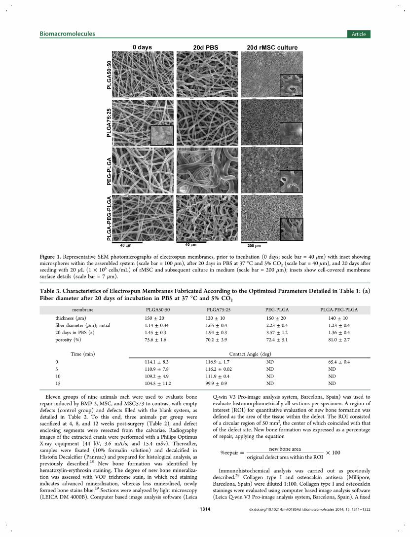
Figure 1. Representative SEM photomicrographs of electrospun membranes, prior to incubation (0 days; scale bar = 40 μm) with inset showingmicrospheres within the assembled system (scale bar = 100 μm), after 20 days in PBS at 37 °C and 5% CO2 (scale bar = 40 μm), and 20 days afterseeding with 20 μL (1 × 10⁶ cells/mL) of rMSC and subsequent culture in medium (scale bar = 200 μm); insets show cell-covered membranesurface details (scale bar = 7 μm).
Table 3. Characteristics of Electrospun Membranes Fabricated According to the Optimized Parameters Detailed in Table 1: (a)Fiber diameter after 20 days of incubation in PBS at 37 °C and 5% CO2
membrane PLGA50:50 PLGA75:25 PEG-PLGA PLGA-PEG-PLGA
thickness (μm) 150 ± 20 120 ± 10 150 ± 20 140 ± 10fiber diameter (μm); initial 1.14 ± 0.34 1.65 ± 0.4 2.23 ± 0.4 1.23 ± 0.420 days in PBS (a) 1.45 ± 0.3 1.94 ± 0.3 3.57 ± 1.2 1.36 ± 0.4porosity (%) 75.6 ± 1.6 70.2 ± 3.9 72.4 ± 5.1 81.0 ± 2.7
Time (min) Contact Angle (deg)
0 114.1 ± 8.3 116.9 ± 1.7 ND 65.4 ± 0.45 110.9 ± 7.8 116.2 ± 0.02 ND ND10 109.2 ± 4.9 111.9 ± 0.4 ND ND15 104.5 ± 11.2 99.9 ± 0.9 ND ND
Biomacromolecules Article
dx.doi.org/10.1021/bm401854d | Biomacromolecules 2014, 15, 1311−13221314
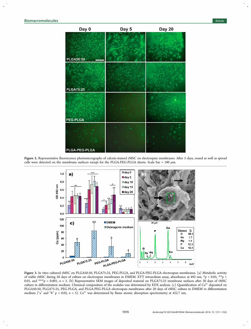
Figure 2. Representative fluorescence photomicrographs of calcein-stained rMSC on electrospun membranes. After 5 days, round as well as spreadcells were detected on the membrane surfaces except for the PLGA-PEG-PLGA sheets. Scale bar = 180 μm.
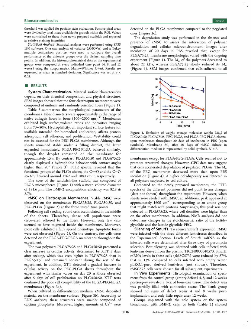
Figure 3. In vitro cultured rMSC on PLGA50:50, PLGA75:25, PEG-PLGA, and PLGA-PEG-PLGA electrospun membranes. (a) Metabolic activityof viable rMSC during 20 days of culture on electrospun membranes in DMEM. XTT tetrazolium assay, absorbance at 492 nm, *p < 0.05, **p <0.01, and ***p < 0.001, n = 5. (b) Representative SEM images of deposited material on PLGA75:25 membrane surfaces after 20 days of rMSCculture in differentiation medium. Chemical composition of the nodules was determined by EDX analysis. (c) Quantification of Ca2+ deposited onPLGA50:50, PLGA75:25, PEG-PLGA, and PLGA-PEG-PLGA electrospun membranes after 20 days of rMSC culture in DMEM vs differentiationmedium (“a” and “b” p < 0.05, n = 5). Ca2+ was determined by flame atomic absorption spectrometry at 422.7 nm.
Biomacromolecules Article
dx.doi.org/10.1021/bm401854d | Biomacromolecules 2014, 15, 1311−13221315
threshold was applied for positive stain evaluation. Positive pixel areaswere divided by total tissue available for growth within the ROI. Valueswere normalized to those from newly prepared scaffolds and reportedas relative staining intensities.Statistical Analysis. Statistical analyses were performed using SPSS
18.0 software. One-way analysis of variance (ANOVA) and a Tukeymultiple comparison post-test were used to compare the overallperformance of the different groups over the distinct sampling timepoints. In addition, the histomorphometrical data of the experimentalgroups were compared at every individual time point (4, 8, and 12weeks) using the nonparametric Mann−Whitney U-test. Results areexpressed as mean ± standard deviation. Significance was set at p <0.05.
■ RESULTS
System Characterization. Material surface characteristicsdepend on their chemical composition and physical structure.SEM images showed that the four electrospun membranes werecomposed of uniform and randomly oriented fibers (Figure 1).Table 3 summarizes the morphological parameters of the
membranes. Fiber diameters were approximately in the range ofnative collagen fibers in bone (100−2000 nm).30 Membranesexhibited high surface/volume ratios and porosities rangingfrom 70−80%. Hydrophilicity, an important surface feature forscaffolds intended for biomedical application, affects proteinadsorption, cell adhesion, and proliferation. Wettability couldnot be assessed for the PEG-PLGA membranes. Although thesheets remained stable under a falling droplet, the latterexpanded immediately. PLGA-PEG-PLGA behaved similarly,though the droplet remained on the membrane forapproximately 15 s. By contrast, PLGA50:50 and PLGA75:25clearly displayed a hydrophobic behavior with contact angleshigher than 90° (Table 3). FTIR spectra revealed that thefunctional groups of the PLGA chains, the CO and the C−Ostretch, hovered around 1762 and 1080 cm−1, respectively.The core of the sandwich-like scaffold was composed of
PLGA microspheres (Figure 1) with a mean volume diameterof 181.6 μm. The BMP-2 encapsulation efficiency was 82.8 ±3.7%.rMSC on Electrospun Membranes. Viable rMSC were
observed on the membranes PLGA75:25, PLGA50:50, andPEG-PLGA (Figure 2) at the three tested time points.Following cell seeding, round cells accumulated in the middle
of the sheets. Thereafter, dense cell populations werediscovered adhered to the fibers. However, only few cellsseemed to have migrated inside the membranes. Moreover,most cells exhibited a fully spread phenotype. Apoptotic formswere not observed (Figure 2). On the contrary, few cells weredetected on the PLGA-PEG-PLGA membranes throughout theexperiment.The two polymers PLGA75:25 and PLGA50:50 presented a
clear increase in cellular activity, determined by XTT 5 daysafter seeding, which was even higher in PLGA75:25 than inPLGA50:50 and remained constant during the rest of theexperiment. In addition, XTT detected a gradual increase incellular activity on the PEG-PLGA sheets throughout theexperiment with similar values on day 20 as those observedafter 5 days of cell proliferation on PLGA50:50. XTT testsconfirmed the poor cell compatibility of the PLGA-PEG-PLGAmembranes (Figure 3a).When cultured in differentiation medium, rMSC deposited
material on the membrane surfaces (Figure 3b). According toEDX analyses, these structures were mainly composed ofcalcium phosphates. Moreover, higher amounts of Ca2+ were
detected on the PLGA membranes compared to the pegylatedones (Figure 3c).The degradation study was performed in the absence and
presence of rMSC to assess the interaction of polymerdegradation and cellular microenvironment. Images afterincubation of 20 days in PBS revealed that, except forPLGA75:25, membrane morphologies varied with the ongoingexperiment (Figure 1). The Mw of the polymers decreased toabout 22 kDa, whereas PLGA75:25 slowly reduced its Mw(Figure 4). SEM images confirmed that cells adhered to all
membranes except for PLGA-PEG-PLGA. Cells seemed not topromote structural changes. However, GPC data may suggestthat cells accelerated degradation of pegylated PLGAs. The Mwof the PEG membranes decreased more than upon PBSincubation (Figure 4). A higher polydispersity was detected inall polymers subjected to cell culture.Compared to the newly prepared membranes, the FTIR
spectra of the different polymers did not point to any changes(data not shown) throughout the experiment. However, whensheets were seeded with rMSC, an additional peak appeared atapproximately 1600 cm−1, corresponding to an amine groupthat might match with proteins. Surprisingly, this peak was notdetected in PLGA75:25, where cell densities were higher thanon the other membranes. In addition, NMR analyses did notdetect any changes in the stoichiometric ratio of the lactide-glycolide and the lactide-glycolide-PEG.
Silencing of Smurf1. To silence Smurf1 expression, rMSCwere infected with the three different lentiviruses described inthe Experimental Section. Levels of Smurf1 mRNA in theinfected cells were determined after three days of puromycinselection. Best silencing was obtained with cells infected withlentivirus derived from the plasmid TRCN0000040573. Smurf1mRNA levels in these cells (rMSC573) were reduced by 87%,that is, 13% compared to cells infected with empty vectorpLKO.1-puro derived lentivirus (not shown). Therefore,rMSC573 cells were chosen for all subsequent experiments.
In Vivo Experiments. Histological examination of speci-mens from the control group (empty defect) 4, 8, and 12 weekspostsurgery revealed a lack of bone-like tissue. The defect areawas partially filled with connective tissue. The blank groupshowed no signs of defect repair 4 and 8 weeks post-implantation and only little repair after 12 weeks.Groups implanted with the sole system or the system
bioactivated with BMP-2, cells, or both (Table 2) showed
Figure 4. Evolution of weight average molecular weight (Mw) ofPLGA50:50, PLGA75:25, PEG-PLGA, and PLGA-PEG-PLGA electro-spun membranes throughout 20 days of incubation in PBS (opensymbols). Membrane Mw after 20 days of rMSC culture indifferentiation medium is represented by solid symbols. N = 3.
Biomacromolecules Article
dx.doi.org/10.1021/bm401854d | Biomacromolecules 2014, 15, 1311−13221316
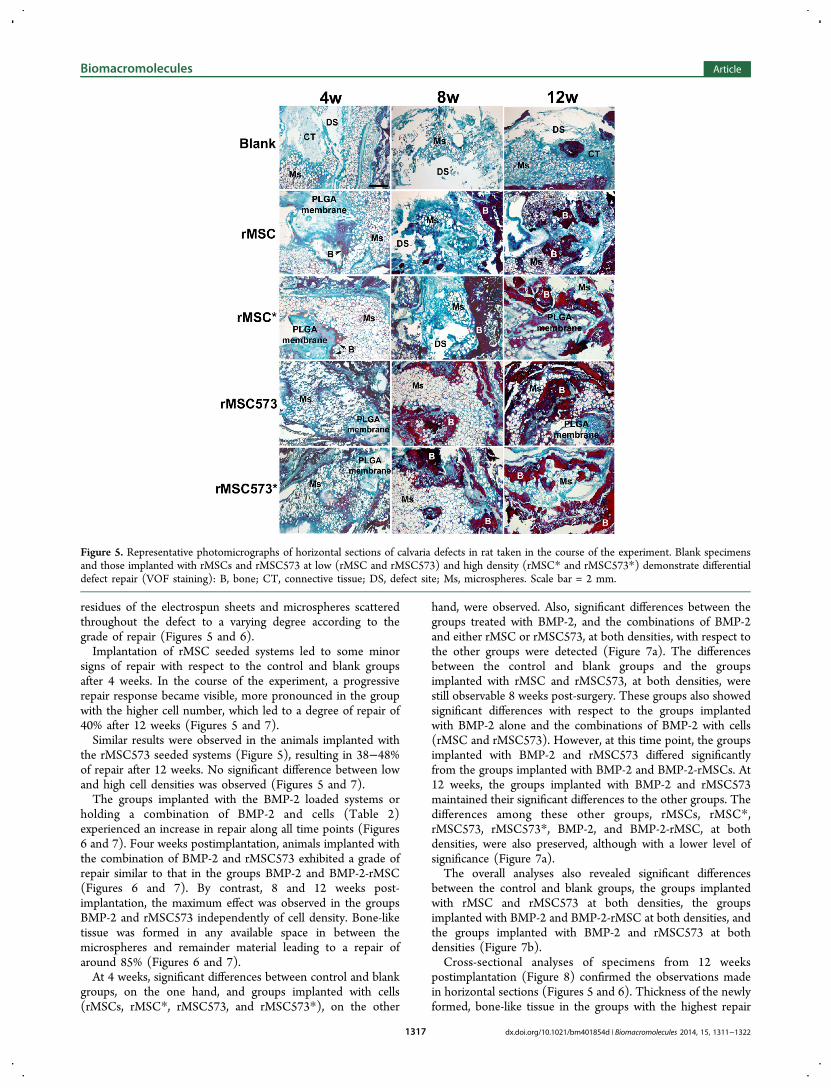
residues of the electrospun sheets and microspheres scatteredthroughout the defect to a varying degree according to thegrade of repair (Figures 5 and 6).Implantation of rMSC seeded systems led to some minor
signs of repair with respect to the control and blank groupsafter 4 weeks. In the course of the experiment, a progressiverepair response became visible, more pronounced in the groupwith the higher cell number, which led to a degree of repair of40% after 12 weeks (Figures 5 and 7).Similar results were observed in the animals implanted with
the rMSC573 seeded systems (Figure 5), resulting in 38−48%of repair after 12 weeks. No significant difference between lowand high cell densities was observed (Figures 5 and 7).The groups implanted with the BMP-2 loaded systems or
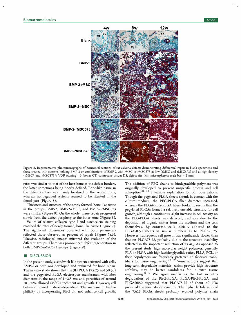
holding a combination of BMP-2 and cells (Table 2)experienced an increase in repair along all time points (Figures6 and 7). Four weeks postimplantation, animals implanted withthe combination of BMP-2 and rMSC573 exhibited a grade ofrepair similar to that in the groups BMP-2 and BMP-2-rMSC(Figures 6 and 7). By contrast, 8 and 12 weeks post-implantation, the maximum effect was observed in the groupsBMP-2 and rMSC573 independently of cell density. Bone-liketissue was formed in any available space in between themicrospheres and remainder material leading to a repair ofaround 85% (Figures 6 and 7).At 4 weeks, significant differences between control and blank
groups, on the one hand, and groups implanted with cells(rMSCs, rMSC*, rMSC573, and rMSC573*), on the other
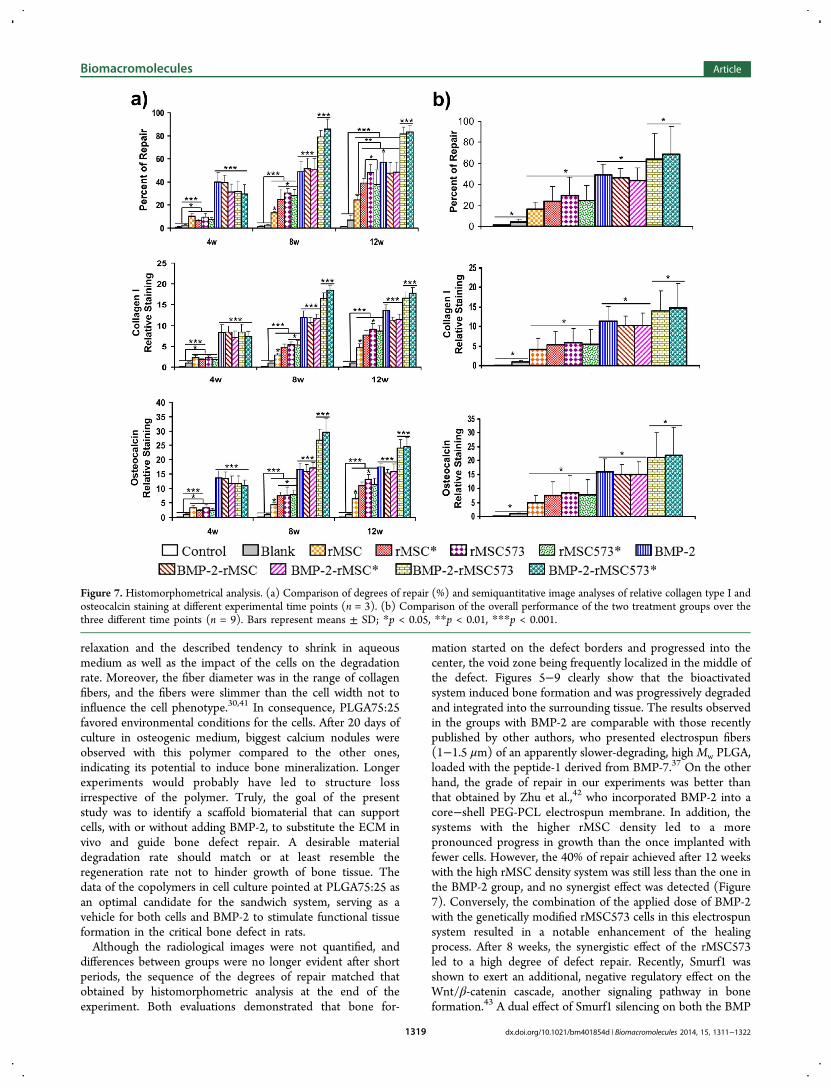
hand, were observed. Also, significant differences between thegroups treated with BMP-2, and the combinations of BMP-2and either rMSC or rMSC573, at both densities, with respect tothe other groups were detected (Figure 7a). The differencesbetween the control and blank groups and the groupsimplanted with rMSC and rMSC573, at both densities, werestill observable 8 weeks post-surgery. These groups also showedsignificant differences with respect to the groups implantedwith BMP-2 alone and the combinations of BMP-2 with cells(rMSC and rMSC573). However, at this time point, the groupsimplanted with BMP-2 and rMSC573 differed significantlyfrom the groups implanted with BMP-2 and BMP-2-rMSCs. At12 weeks, the groups implanted with BMP-2 and rMSC573maintained their significant differences to the other groups. Thedifferences among these other groups, rMSCs, rMSC*,rMSC573, rMSC573*, BMP-2, and BMP-2-rMSC, at bothdensities, were also preserved, although with a lower level ofsignificance (Figure 7a).The overall analyses also revealed significant differences
between the control and blank groups, the groups implantedwith rMSC and rMSC573 at both densities, the groupsimplanted with BMP-2 and BMP-2-rMSC at both densities, andthe groups implanted with BMP-2 and rMSC573 at bothdensities (Figure 7b).Cross-sectional analyses of specimens from 12 weeks
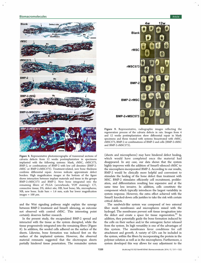
postimplantation (Figure 8) confirmed the observations madein horizontal sections (Figures 5 and 6). Thickness of the newlyformed, bone-like tissue in the groups with the highest repair
Figure 5. Representative photomicrographs of horizontal sections of calvaria defects in rat taken in the course of the experiment. Blank specimensand those implanted with rMSCs and rMSC573 at low (rMSC and rMSC573) and high density (rMSC* and rMSC573*) demonstrate differentialdefect repair (VOF staining): B, bone; CT, connective tissue; DS, defect site; Ms, microspheres. Scale bar = 2 mm.
Biomacromolecules Article
dx.doi.org/10.1021/bm401854d | Biomacromolecules 2014, 15, 1311−13221317
rates was similar to that of the host bone at the defect borders,the latter sometimes being poorly defined. Bone-like tissue inthe defect centers was mainly localized in the ventral zone,whereas nondegraded systems seemed to be situated in thedorsal part (Figure 8).Thickness and structure of the newly formed, bone-like tissue
in the groups BMP-2, BMP-2-rMSC, and BMP-2-rMSC573were similar (Figure 8). On the whole, tissue repair progressedslowly from the defect periphery to the inner zone (Figure 8).Values of relative collagen type I and osteocalcin staining
matched the rates of newly formed, bone-like tissue (Figure 7).The significant differences observed with both parametersreflected those observed as percent of repair (Figure 7a,b).Likewise, radiological images mirrored the evolution of thedifferent groups. There was pronounced defect regeneration inboth BMP-2-rMSC573 groups (Figure 9).
■ DISCUSSIONIn the present study, a sandwich-like system activated with cells,BMP-2 or both was developed and evaluated for bone repair.The in vitro study shows that the 3D PLGA (75:25 and 50:50)and the pegylated PLGA electrospun membranes, with fiberdiameters in the range of 1−2.5 μm and porosities of around70−80%, allowed rMSC attachment and growth. However, cellbehavior proved material-dependent. The increase in hydro-philicity by incorporating PEG did not enhance cell growth.
The addition of PEG chains to biodegradable polymers wasoriginally developed to prevent unspecific protein and celladsorption,31−33 a feasible explanation for our observations.Though the pegylated PLGA sheets shrank in contact with theculture medium, the PEG-PLGA fiber diameter increased,whereas the PLGA-PEG-PLGA fibers broke. It seems that thepegylated PLGAs formed a relatively unstable structure for cellgrowth, although a continuous, slight increase in cell activity onthe PEG-PLGA sheets was detected, probably due to thedeposition of organic matter from the medium and the cellsthemselves. By contrast, cells initially adhered to thePLGA50:50 sheets in similar numbers as to PLGA75:25.However, subsequent cell growth was significantly slower thanthat on PLGA75:25, probably due to the structure instabilityreflected in the important reduction of its Mw. As opposed tothe present study, high molecular weight polymers, generallyPLA or PLGA with high lactide/glycolide ratios, PLLA, PCL, ortheir copolymers are frequently preferred to fabricate nano-fibers for tissue engineering.33−39 Some authors suggest thatlong-term degradable materials, which provide high structurestability, may be better candidates for in vitro tissueengineering.35,40 We agree insofar as the fast in vitrodegradation of the PEG-PLGA, PLGA-PEG-PLGA, andPLGA50:50 suggested that PLGA75:25 of about 60 kDaprovided the most stable structure. The higher lactide ratio ofthe 75:25 PLGA sheets probably avoided polymer chain
Figure 6. Representative photomicrographs of horizontal sections of rat calvaria defects demonstrating differential repair in blank specimens andthose treated with systems holding BMP-2 or combinations of BMP-2 with rMSC or rMSC573 at low (rMSC and rMSC573) and at high density(rMSC* and rMSC573*; VOF staining): B, bone; CT, connective tissue; DS, defect site; Ms, microspheres; scale bar = 2 mm.
Biomacromolecules Article
dx.doi.org/10.1021/bm401854d | Biomacromolecules 2014, 15, 1311−13221318
relaxation and the described tendency to shrink in aqueousmedium as well as the impact of the cells on the degradationrate. Moreover, the fiber diameter was in the range of collagenfibers, and the fibers were slimmer than the cell width not toinfluence the cell phenotype.30,41 In consequence, PLGA75:25favored environmental conditions for the cells. After 20 days ofculture in osteogenic medium, biggest calcium nodules wereobserved with this polymer compared to the other ones,indicating its potential to induce bone mineralization. Longerexperiments would probably have led to structure lossirrespective of the polymer. Truly, the goal of the presentstudy was to identify a scaffold biomaterial that can supportcells, with or without adding BMP-2, to substitute the ECM invivo and guide bone defect repair. A desirable materialdegradation rate should match or at least resemble theregeneration rate not to hinder growth of bone tissue. Thedata of the copolymers in cell culture pointed at PLGA75:25 asan optimal candidate for the sandwich system, serving as avehicle for both cells and BMP-2 to stimulate functional tissueformation in the critical bone defect in rats.Although the radiological images were not quantified, and
differences between groups were no longer evident after shortperiods, the sequence of the degrees of repair matched thatobtained by histomorphometric analysis at the end of theexperiment. Both evaluations demonstrated that bone for-
mation started on the defect borders and progressed into thecenter, the void zone being frequently localized in the middle ofthe defect. Figures 5−9 clearly show that the bioactivatedsystem induced bone formation and was progressively degradedand integrated into the surrounding tissue. The results observedin the groups with BMP-2 are comparable with those recentlypublished by other authors, who presented electrospun fibers(1−1.5 μm) of an apparently slower-degrading, high Mw PLGA,loaded with the peptide-1 derived from BMP-7.37 On the otherhand, the grade of repair in our experiments was better thanthat obtained by Zhu et al.,42 who incorporated BMP-2 into acore−shell PEG-PCL electrospun membrane. In addition, thesystems with the higher rMSC density led to a morepronounced progress in growth than the once implanted withfewer cells. However, the 40% of repair achieved after 12 weekswith the high rMSC density system was still less than the one inthe BMP-2 group, and no synergist effect was detected (Figure7). Conversely, the combination of the applied dose of BMP-2with the genetically modified rMSC573 cells in this electrospunsystem resulted in a notable enhancement of the healingprocess. After 8 weeks, the synergistic effect of the rMSC573led to a high degree of defect repair. Recently, Smurf1 wasshown to exert an additional, negative regulatory effect on theWnt/β-catenin cascade, another signaling pathway in boneformation.43 A dual effect of Smurf1 silencing on both the BMP
Figure 7. Histomorphometrical analysis. (a) Comparison of degrees of repair (%) and semiquantitative image analyses of relative collagen type I andosteocalcin staining at different experimental time points (n = 3). (b) Comparison of the overall performance of the two treatment groups over thethree different time points (n = 9). Bars represent means ± SD; *p < 0.05, **p < 0.01, ***p < 0.001.
Biomacromolecules Article
dx.doi.org/10.1021/bm401854d | Biomacromolecules 2014, 15, 1311−13221319
and the Wnt signaling pathway might explain the synergybetween BMP-2 treatment and Smurf1 silencing, an outcomenot observed with control rMSC. This interesting pointcertainly deserves further research.In the present study, the encapsulated BMP-2 spread and
interacted with the tissue as the system disrupted, while thetissue progressively integrated into the remaining fibers (Figure8). In addition, the seeded cells adhered on the surface of thesheets. Likewise, bone formation was induced first on thesurface of the implanted systems. However, the observedmaterial remnants suggested that the electrospun sheetspartially hindered tissue penetration. The remainder system
(sheets and microspheres) may have hindered defect healing,which would have completed once the material haddisappeared. In any case, our data shows that the systemhighly improves with the addition of Smurf1-silenced rMSC tothe microsphere-incorporated BMP-2. According to our results,BMP-2 would be clinically more helpful and convenient tostimulate the healing of the bone defect than treatment withMSC. BMP-2 stimulates efficiently cell recruitment, prolifer-ation, and differentiation resulting less expensive and at thesame time less invasive. In addition, cells constitute thecomponent which typically introduces the largest variability insystem response. However, the extra effect achieved with theSmurf1 knocked-down cells justifies to take the risk with certaincritical defects.The sandwich-like system was composed of two external
fiber mesh membranes and microspheres mixed with thehydrogel. The membranes prevent soft tissue invagination intothe defect and create a space for tissue regeneration.44 Inaddition, they potentially guide the bone formation induced bythe cells on their surface and/or the osteogenic factor releasedfrom the system. Its high versatility is one of the advantages ofthis system. The membranes favor conditions for cellattachment and growth. A variety of GFs can be included inthe system, within the fibers by incorporating the protein in thepolymer solution as well as in the microspheres. Furthermore, asystem developed this way allows for easy adjustment to the
Figure 8. Representative photomicrographs of transversal sections ofcalvaria defects from 12 weeks postimplantation in specimensimplanted with the following systems: blank, rMSC, rMSC573,BMP-2, or combinations of BMP-2 with low cell densities (BMP-2-rMSC or BMP-2-rMSC573). Treatment-related, new bone thicknessconfirms differential repair. Arrows indicate approximate defectborders. High magnification images at the bottom of the figureshows interaction between implant materials and tissue in the groupsBMP-2-rMSC573 and BMP-2. New bone integrated into theremaining fibers of PLGA (arrowheads; VOF staining): CT,connective tissue; DS, defect site; HB, host bone; Ms, microspheres;NB, new bone. Scale bars = 1.6 mm, scale bar lower magnificationimages = 100 μm.
Figure 9. Representative, radiographic images reflecting theregeneration process of the calvaria defects in rats. Images from 4and 12 weeks postimplantation show differential repair in blankspecimens and those treated with systems bioactivated with rMSC,rMSC573, BMP-2 or combinations of BMP-2 and cells (BMP-2-rMSCand BMP-2-rMSC573).
Biomacromolecules Article
dx.doi.org/10.1021/bm401854d | Biomacromolecules 2014, 15, 1311−13221320
defect size, a potential advantage for clinical application, bydosing the quantity of protein-containing microspheres andsize-adapting the sheets while assembling the system duringimplantation. Further studies are required to better match thesystem degradation kinetics with the bone formation rate. Also,the effect of the implanted rMSC573 requires in-depthcharacterization to corroborate the obtained, beneficial resultsand the potential for clinical application.
■ CONCLUSIONTaken together, the electrospun system presents good bonedefect repair outcomes within 8−12 weeks. As a new strategyfor bone regeneration, a combination of microsphere-encapsulated BMP-2 with mesenchymal stem cells, knockeddown for Smurf1 expression, led to a strongly improvedresponse. The membranes fabricated with unconventionallylow Mw PLGAs were not sturdy enough for in vitro tissueengineering with the exception of PLGA75:25. However, theobserved bone repair suggests that a material degradation fasterthan that of PLGA75:25 should be supportive.
■ AUTHOR INFORMATIONCorresponding Author*Tel.: (34) 922318957. Fax: (34) 922318506. E-mail: [email protected] authors declare no competing financial interest.
■ ACKNOWLEDGMENTSThis work was supported by the Ministry of Science andTechnology (MAT2011-23819) and Proyecto Motiva ofACIISI. M.R-E. was supported by Beca CajaCanarias paraposgraduados. The authors are indebted to Javier Gonzalez(Instituto Tecnologico de Canarias) for technical assistancewith electrospinning. The authors would like to thank membersof the Department of Cell Biology of the University of LaLaguna for receiving R.R. in their laboratory. We thank Dr.Carmen Damas for assistance with statistical analysis.
■ REFERENCES(1) Giannoudis, P. V.; Dinopoulus, H.; Tsiridis, E. Bone substitutes:an update. Injury 2005, 36 (Suppl 3), S20−S27.(2) Holzwartha, J. M.; Ma, P. X. Biomimetic nanofibrous scaffolds forbone tissue engineering. Biomaterials 2011, 32, 9622−9629.(3) Andrady, A. L. Science and Technology of Polymer Nanofibers; J.Wiley & Sons Inc.: New York, 2008.(4) Ding, B.; Wang, M. R.; Wang, X. F.; Yu, J. Y.; Sun, G. Electrospunnanomaterials for ultrasensitive sensor. Mater. Today 2010, 13, 16−27.(5) Mao, X.; Ding, B.; Wang, M. R.; Yin, Y. B. Self-assembly ofphthalocyanine and polyacrylic acid composite multilayers on cellulosenanofibers. Carbohydr. Polym. 2010, 80, 839−844.(6) Teo, W. E.; He, W.; Ramakrishna, S. Electrospun scaffold tailoredfor tissue specific extracellular matrix. Biotechnol. J. 2006, 1, 918−929.(7) Liang, D.; Jsiao, B.; Chu, B. Functional electrospun nanofibrousscaffolds for biomedical application. Adv. Drug Delivery Rev. 2007, 59,1392−1412.(8) Newton, D.; Mahajan, R.; Ayres, C.; Bowman, J. R.; Bowlin, G.L.; Simpson, D. G. Regulation of material properties in electrospunscaffolds: role of cross-linking and fiber tertiary structure. ActaBiomater. 2009, 5, 518−529.(9) Hsiu-Mei, L.; Yi-Hsuan, L.; Fu-Yin, H. Preparation andcharacterization of mesoporous bioactive glass/polycaprolactonenanofibrous matrix for bone tissues engineering. J. Mater. Sci.: Mater.Med. 2012, 23, 2619−2630.
(10) Li, D.; Xia, Y. N. Direct fabrication of composite and ceramichollow nanofibers by electrospinning. Nano Lett. 2004, 4, 933−938.(11) Burguera, E. F.; Xu, H. H.; Takagi, S.; Chow, L. C. High earlystrength calcium phosphate bone cement: effects of dicalciumphosphate dihydrate and absorbable fibers. J. Biomed. Mater. Res.,Part A 2005, 75, 966−975.(12) Zhang, Y.; Xu, H. H. Effects of synergistic reinforcement andabsorbable fiber strength on hydroxyapatite bone cement. J. Biomed.Mater. Res., Part A 2005, 75, 832−840.(13) Sun, T.; Norton, D.; McKean, R. J.; Haycock, K. W.; Ryan, A. J.;MacNeil, S. Development of a 3D cell culture system for investigatingcell interactions with electrospun fibers. Biotechnol. Bioeng. 2007, 97,1318−1328.(14) Jones, E.; Yang, X. Mesenchymal stem cells and boneregeneration: current status. Injury 2011, 42, 562−568.(15) Erices, A.; Conget, P.; Minguell, J. J. Mesenchymal progenitorcells in human umbilical cord blood. Br. J. Hamaetol. 2000, 109, 235−242.(16) Dragoo, J. L.; Choi, J. Y.; Lieberman, J. R.; Huang, J.; Zuk, P. A.;Zhang, J.; Hedrick, M. H.; Benhaim, P. Bone induction by BMP-2transduced stem cells derived from human fat. J. Orthop. Res. 2003, 21,622−629.(17) Rodríguez-Evora, M.; Delgado, A.; Reyes, R.; Hernandez-Daranas, A.; Soriano, I.; San Roman, J.; Evora, C. Osteogenic effect oflocal, long versus short term BMP-2 delivery from a novel SPU-PLGA-βTCP concentric system in a critical size defect in rats. Eur. J. Pharm.Sci. 2013, 49, 873−884.(18) Rodríguez-Evora, M.; Reyes, R.; Alvarez-Lorenzo, C.;Concheiro, A.; Delgado, A.; Evora, C. Bone regeneration induced byan in situ gel-forming poloxamine, bone morphogenetic protein-2system. J. Biomed. Nanotechnol. 2014, 10, 959−969.(19) Okada, M.; Sangadala, S.; Liu, Y.; Yoshida, M.; Reddy, B. V.;Titus, L.; Boden, S. D. Development and optimization of a cell-basedassay for the selection of synthetic compounds that potentiate bonemorphogenetic protein-2 activity. Cell Biochem. Funct. 2009, 27 (8),526−534.(20) Kato, S.; Sangadala, S.; Tomita, K.; Titus, L.; Boden, S. D. Asynthetic compound that potentiates bone morphogenetic protein-2-induced transdifferentiation of myoblasts into the osteoblasticphenotype. Mol. Cell. Biochem. 2011, 349, 97−106.(21) Yamashita, M.; Ying, S.; Zhang, G.; Li, C.; Cheng, S. Y.; Deng,C.; Zhang, Y. Ubiquitin ligase Smurf1controls osteoblast activity andbone homeostasis by targeting MEKK2 for degradation. Cell 2005,121, 101−113.(22) Zhao, M.; Qiao, M.; Harris, S. E.; Oyajobi, B. O.; Mundy, G. R.;Chen, D. Smurf1 inhibits osteoblast differentiation and boneformation in vitro and in vivo. J. Biol. Chem. 2004, 279, 12854−12859.(23) Cao, Y.; Zhang, L. Pharmaceutical perspectives of HECT-typeubiquitin ligase Smurf1. Curr. Pharm. Des. 2013, 19, 3226−3233.(24) Dobson, K. R.; Reading, L.; Haberey, M.; Marine, X.; Scutt, A.Centrifugal isolation of bone marrow from bone: an improved methodfor the recovery and quantitation of bone marrow osteoprogenitorcells from rat tibiae and femurae. Calcif. Tissue Int. 1999, 65, 411−413.(25) Graham, F. L.; van der Eb, A. J. A new technique for the assay ofinfectivity of human adenovirus 5 DNA. Virology 1973, 52, 456.(26) Nie, J.; Wu, M.; Wang, J.; Xing, G.; He, F.; Zhang, L.REGgamma proteasome mediates degradation of the ubiquitin ligaseSmurf1. FEBS Lett. 2010, 584, 3021−3027.(27) Cassiede, P.; Dennis, J. E.; Ma, F.; Caplan, A. I.Osteochondrogenic potential of marrow mesenchymal progenitorcells exposed to TGF-b1 or PDGF-BB as assayed in vivo and in vitro. J.Bone Miner. Res. 1996, 11, 1264−1273.(28) Hernandez, A.; Sanchez, E.; Soriano, I.; Reyes, R.; Delgado, A.;Evora, C. Material-related effects of BMP-2 delivery systems on boneregeneration. Acta Biomater. 2012, 8, 781−791.(29) Martínez-Sanz, E.; Ossipov, D. A.; Hilborn, J.; Larsson, S.;Jonsson, K. B.; Varghese, O. P. Bone reservoir: injectable hyaluronicacid hydrogel for minimal invasive bone augmentation. J. ControlledRelease 2011, 152, 232−240.
Biomacromolecules Article
dx.doi.org/10.1021/bm401854d | Biomacromolecules 2014, 15, 1311−13221321

(30) Stevens, M. M.; George, J. H. Exploring and engineering the cellsurface interface. Science 2005, 3101135−1138.(31) Groll, J.; Fiedler, J.; Engelhard, E.; Ameringer, T.; Tugulu, S.;Klok, H. A.; Brenner, R. E.; Moeller, M. A novel star PEG-derivedsurface coating for specific cell adhesion. J. Biomed. Mater. Res., Part A2005, 74, 607−617.(32) Yang, D. J.; Zhang, L. F.; Xu, L.; Xiong, C. D.; Ding, J.; Wang, Y.Z. Fabrication and characterization of hydrophilic electrospunmembranes made from the block copolymer of poly(ethylene glycol-co-lactide). J. Biomed. Mater. Res., Part A 2007, 82, 680−688.(33) Grafahrend, D.; Calvet, J. L.; Klinkhammer, K.; Salber, J.;Dalton, P. D.; Moller, M.; Klee, D. Control of protein adsorption onfunctionalized electrospun fibers. Biotechnol. Bioeng. 2008, 101, 609−621.(34) Dong, Y.; Liao, S.; Ngiam, M.; Chan, C. K.; Ramakrishna, S.Degradation behaviors of electrospun resorbable polyester nanofibers.Tissue Eng., Part B 2009, 15, 333−351.(35) Dong, Y.; Yong, T.; Liao, S.; Chan, C. K.; Stevens, M. M.;Ramakrishna, S. Distinctive degradation behaviors of electrospunpolyglycolide, poly(DL-lactide-co-glycolide), and poly(L-lactide-co-ε-caprolactone) nanofibers cultured with/without porcine smoothmuscle cells. Tissue Eng., Part A 2010, 16, 283−298.(36) Schofer, M. D.; Roessler, P. P.; Schaefer, J.; Theisen, C.;Schlimme, S.; Heverhagen, J. T.; Voelker, M.; Dersch, R.; Agarwal, S.;Fuchs-Winkelmann, S.; Paletta, J. R. Electrospun PLLA nanofiberscaffolds and their use in combination with BMP-2 for reconstructionof bone defects. PLoS One 2011, 6, e25462.(37) Wang, B. Y.; Fu, S. Z.; Ni, P. Y.; Peng, J. R.; Zheng, L.; Luo, F.;Liu, H.; Qian, Z. Y. Electrospun polylactide/poly/ethyleneglycol)hybrid fibrous scaffolds for tissue engineering. J. Biomed. Mater. Res.,Part A 2012, 100A, 441−449.(38) Fu, S.; Yang, L.; Fan, J.; Wen, Q.; Lin, S.; Wang, B.; Chen, L.;Meng, X.; Chen, Y.; Wu, J. In vitro mineralization of hydroxyapatite onelectrospun poly(ε-caprolactone)-poly(ethylene glycol)-poly(ε-capro-lactone) fibrous scaffolds for tissue engineering application. ColloidsSurf., B 2013, 107, 167−173.(39) Lee, Y. J.; Lee, J. H.; Choo, H. J.; Kim, H. K.; Yoon, T. R.; Shin,H.. Electrospun fibers immobilized with bone forming peptide-1derived from BMP7 for guided bone regeneration. Biomaterials 2013,34, 5059−5069.(40) Li, W. J.; Cooper, J. A., Jr.; Mauck, R. L.; Tuan, R. S. Fabricationand characterization of six electrospun poly(α-hydroxy ester)-basedfibrous scaffolds for tissue engineering applications. Acta Biomater.2006, 2, 377−385.(41) Boland, E. D.; Wnek, G. E.; Simpson, D. G.; Palowski, K. J.;Bowlin, G. L. Tailoring tissue engineering scaffolds using electrostaticprocessing techniques: a study of poly(glycolic acid) electrospinning. J.Macromol. Sci., Part A 2001, 38, 1231−1243.(42) Zhu, H.; Yu, D.; Zhou, Y.; Wang, C.; Gao, M.; Jiang, H.; Wang,H. Biological activity of a nanofibrous barrier membrane containingbone morphogenetic protein formed by core-shell electrospinning as asustained delivery vehicle. J. Biomed. Mater. Res., Part B 2013, 1B, 541−552.(43) Fei, C.; Li, Z.; Li, C.; Chen, Y.; Chen, Z.; He, X.; Mao, L.; Wang,X.; Zeng, R.; Li, L. Smurf1-mediated Lys29-linked nonproteolyticpolyubiquitination of axin negatively regulates Wnt/β-catenin signal-ing. Mol. Cell. Biol. 2013, 33, 4095−4105.(44) Kolambkar, Y. M.; Dupont, K. M.; Boerckel, J. D.; Huebsch, N.;Mooney, D. J.; Hutmacher, D. W.; Guldberg, R. E. An alginate-basedhybrid system for growth factor delivery in the functional repair oflarge bone defects. Biomaterials 2011, 32, 65−74.
Biomacromolecules Article
dx.doi.org/10.1021/bm401854d | Biomacromolecules 2014, 15, 1311−13221322
Copyright © 2022 FDOKUMEN