Effect of scaffold architecture and BMP-2/BMP-7 delivery on in vitro bone regeneration
10
Effect of scaffold architecture and BMP-2/BMP-7 delivery on in vitro bone regeneration Pinar Yilgor • Rui A. Sousa • Rui L. Reis • Nesrin Hasirci • Vasif Hasirci Received: 12 March 2010 / Accepted: 7 August 2010 / Published online: 26 August 2010 Ó Springer Science+Business Media, LLC 2010 Abstract The aim of this study was to develop 3-D tissue engineered constructs that mimic the in vivo conditions through a self-contained growth factor delivery system. A set of nanoparticles providing the release of BMP-2 initially followed by the release of BMP-7 were incorpo- rated in poly(e-caprolactone) scaffolds with different 3-D architectures produced by 3-D plotting and wet spinning. The release patterns were: each growth factor alone, simultaneous, and sequential. The orientation of the fibers did not have a significant effect on the kinetics of release of the model protein BSA; but affected proliferation of bone marrow mesenchymal stem cells. Cell proliferation on random scaffolds was significantly higher compared to the oriented ones. Delivery of BMP-2 alone suppressed MSC proliferation and increased the ALP activity to a higher level than that with BMP-7 delivery. Proliferation rate was suppressed the most by the sequential delivery of the two growth factors from the random scaffold on which the ALP activity was the highest. Results indicated the distinct effect of scaffold architecture and the mode of growth factor delivery on the proliferation and osteogenic differ- entiation of MSCs, enabling us to design multifunctional scaffolds capable of controlling bone healing. 1 Introduction Bone can repair itself but it has been recognized that this has not always been satisfactory. Especially in the case of large defects a filler is required to temporarily fill the site over which the new tissue forms. The ideal bone filler or substitute is expected to satisfy some essential require- ments such as being osteoconductive, mimicking the porosity and microstructure of the natural tissue, having the ability to harbor osteoinductive factors and osteogenic cells. Among all the bone substitutes currently in use in the clinic including biological (autogenic, allogenic, or xeno- genic grafts, demineralized bone matrix, cadaver bone) and synthetic (metallic, ceramic, or polymeric) grafts, the only one which has all the required properties is the autograft. However, due to donor site morbidity, risk of infection, etc. the search for alternative therapies is continuing. A serious alternative to the autograft is growth factor releasing tissue engineered constructs. Tissue engineered bone substitutes consist of bone forming cells, porous scaffolds that serve as the microenvironment, and factors that guide proliferation and differentiation of cells. A multifunctional construct that combines a local bioactive agent delivery system with an osteoconductive scaffold and stem cells would be ideal. Such a graft would serve as an artificial ECM that com- bines osteoconduction and osteoinduction and protects the activity and maintains the local dose of the biological cues. P. Yilgor Á N. Hasirci Á V. Hasirci METU, BIOMAT, Department of Biotechnology, Biotechnology Research Unit, 06531 Ankara, Turkey R. A. Sousa Á R. L. Reis 3B’s Research Group—Biomaterials, Biodegradables and Biomimetics, IBB—Institute for Biotechnology and Bioengineering, PT Associated Laboratory, Headquarters of the European Institute of Excellence on Tissue Engineering and Regenerative Medicine, University of Minho, AvePark, Taipas, 4806-909 Guimaraes, Portugal N. Hasirci METU, BIOMAT, Faculty of Arts and Sciences, Department of Chemistry, 06531 Ankara, Turkey V. Hasirci (&) METU, BIOMAT, Department of Biological Sciences, Biotechnology Research Unit, 06531 Ankara, Turkey e-mail: [email protected] 123 J Mater Sci: Mater Med (2010) 21:2999–3008 DOI 10.1007/s10856-010-4150-1
Transcript of Effect of scaffold architecture and BMP-2/BMP-7 delivery on in vitro bone regeneration

Effect of scaffold architecture and BMP-2/BMP-7 deliveryon in vitro bone regeneration
Pinar Yilgor • Rui A. Sousa • Rui L. Reis •
Nesrin Hasirci • Vasif Hasirci
Received: 12 March 2010 / Accepted: 7 August 2010 / Published online: 26 August 2010
� Springer Science+Business Media, LLC 2010
Abstract The aim of this study was to develop 3-D tissue
engineered constructs that mimic the in vivo conditions
through a self-contained growth factor delivery system.
A set of nanoparticles providing the release of BMP-2
initially followed by the release of BMP-7 were incorpo-
rated in poly(e-caprolactone) scaffolds with different 3-D
architectures produced by 3-D plotting and wet spinning.
The release patterns were: each growth factor alone,
simultaneous, and sequential. The orientation of the fibers
did not have a significant effect on the kinetics of release of
the model protein BSA; but affected proliferation of bone
marrow mesenchymal stem cells. Cell proliferation on
random scaffolds was significantly higher compared to the
oriented ones. Delivery of BMP-2 alone suppressed MSC
proliferation and increased the ALP activity to a higher
level than that with BMP-7 delivery. Proliferation rate was
suppressed the most by the sequential delivery of the two
growth factors from the random scaffold on which the ALP
activity was the highest. Results indicated the distinct
effect of scaffold architecture and the mode of growth
factor delivery on the proliferation and osteogenic differ-
entiation of MSCs, enabling us to design multifunctional
scaffolds capable of controlling bone healing.
1 Introduction
Bone can repair itself but it has been recognized that this
has not always been satisfactory. Especially in the case of
large defects a filler is required to temporarily fill the site
over which the new tissue forms. The ideal bone filler or
substitute is expected to satisfy some essential require-
ments such as being osteoconductive, mimicking the
porosity and microstructure of the natural tissue, having the
ability to harbor osteoinductive factors and osteogenic
cells. Among all the bone substitutes currently in use in the
clinic including biological (autogenic, allogenic, or xeno-
genic grafts, demineralized bone matrix, cadaver bone) and
synthetic (metallic, ceramic, or polymeric) grafts, the only
one which has all the required properties is the autograft.
However, due to donor site morbidity, risk of infection, etc.
the search for alternative therapies is continuing. A serious
alternative to the autograft is growth factor releasing tissue
engineered constructs. Tissue engineered bone substitutes
consist of bone forming cells, porous scaffolds that serve as
the microenvironment, and factors that guide proliferation
and differentiation of cells. A multifunctional construct
that combines a local bioactive agent delivery system with
an osteoconductive scaffold and stem cells would be ideal.
Such a graft would serve as an artificial ECM that com-
bines osteoconduction and osteoinduction and protects the
activity and maintains the local dose of the biological cues.
P. Yilgor � N. Hasirci � V. Hasirci
METU, BIOMAT, Department of Biotechnology, Biotechnology
Research Unit, 06531 Ankara, Turkey
R. A. Sousa � R. L. Reis
3B’s Research Group—Biomaterials, Biodegradables and
Biomimetics, IBB—Institute for Biotechnology and
Bioengineering, PT Associated Laboratory, Headquarters of the
European Institute of Excellence on Tissue Engineering and
Regenerative Medicine, University of Minho, AvePark, Taipas,
4806-909 Guimaraes, Portugal
N. Hasirci
METU, BIOMAT, Faculty of Arts and Sciences, Department
of Chemistry, 06531 Ankara, Turkey
V. Hasirci (&)
METU, BIOMAT, Department of Biological Sciences,
Biotechnology Research Unit, 06531 Ankara, Turkey
e-mail: [email protected]
123
J Mater Sci: Mater Med (2010) 21:2999–3008
DOI 10.1007/s10856-010-4150-1

Scaffold design is one of the crucial steps of tissue engi-
neering approach. Rapid prototyping (RP) can produce a
scaffold directly from computed tomography (CT) or mag-
netic resonance imaging (MRI) scans of the defect site or from
a computer design of an irregular defect site [1–3]. Although
RP has its own drawbacks such as the requirement of a spe-
cific form of input material (e.g. filament, powder, pellet,
solution), the resolution (ca. 100 lm) that limits the detail of
the produced construct, the morphology which generally
presents edges and corners, the technique still holds great
promise for the production of custom-made scaffolds and
prostheses. 3-D plotting is the most convenient method for
bone tissue engineering due to its milder operation conditions,
absence of left over polymer powder within the scaffold and
the ability to produce scaffolds without any binders.
During bone regeneration, a variety of growth factors
including transforming growth factor-beta (TGF-b) super-
family proteins (especially bone morphogenetic proteins,
BMPs), insulin-like growth factor (IGF), fibroblast growth
factor (FGF), platelet-derived growth factor (PDGF) and
vascular endothelial growth factor (VEGF) function with a
complex time and concentration pattern [4]. Among these,
BMPs were shown to induce bone formation by inducing
mesenchymal stem cells (MSCs) toward osteoblastic dif-
ferentiation and are the most osteogenic growth factors
presently described [5–7]. Some studies combined BMPs
with other growth factors to achieve improved repair. For
example, BMP-2 and TGF-b3 were combined and their
delivery led to enhanced bone formation in vivo while the
effect of a single growth factor was negligible [8]. The
positive effects of VEGF combined with BMP-2 on in vivo
bone regeneration [9, 10] and IGF-1 jointly encapsulated in
microparticles with BMP-2 and then loaded in porous scaf-
folds for in vitro periodontal regeneration were also reported
[11]. BMP-7 was shown to couple with BMP-2 in inducing
bone morphogenesis and both were approved by FDA for use
in clinical applications [12–15]. Therefore, it was considered
that developing scaffolds that could deliver a combination of
BMP-2 with BMP-7 in a time dependent manner is a viable
biomimetic approach towards bone healing. Our previous
studies illustrated that delivering BMP-2 and BMP-7 in a
sequential manner in both free nanoparticulate form [16] and
after incorporation into scaffolds [17, 18] enhance osteo-
genic differentiation of MSCs in vitro.
In this study, poly(e-caprolactone) (PCL) scaffolds were
produced by 3-D plotting and wet spinning and tested for in
vitro bone regeneration. Nanoparticles carrying BMP-2 and
BMP-7 were added into these scaffolds to produce a
multifunctional construct. The effect of scaffold geometry
and growth factor delivery from the scaffolds were inves-
tigated using bone marrow MSCs.
The multifunctional scaffolds prepared in this study
could be used in all types of congenital and acquired
fractures and defects to achieve enhanced and accelerated
healing through the self-contained sequential growth factor
delivery system. These scaffolds are especially suitable for
use in large bone defects, non-unions, comminuted and
osteoporotic fractures that need osteoinduction as well as
structural and mechanical support.
2 Materials and methods
2.1 Materials
PCL (Mw 3.7 9 104) was purchased from Solvay Capro-
lactones (CAPA 6404; UK). Poly(lactic acid-co-glycolic acid)
(PLGA) (50:50) (Resomer� RG503H) (i.v. 0.32–0.44 dl/g,
0.1% in chloroform, 25�C) was purchased from Boehringer-
Ingelheim (Germany). Poly(3-hydroxybutyrate-co-3-hydro-
xyvalerate) (PHBV) (HV content 8% w/w), dexamethasone,
b-glycerophosphate disodium salt, L-ascorbic acid were
bought from Sigma-Aldrich (Germany). Bovine serum
albumin (BSA) and polyvinyl alcohol (PVA) (Mw
1.5 9 104) were obtained from Fluka (USA). Recombinant
human BMP-2 from InductOs� (Wyeth Pharmaceuticals,
USA) and recombinant human BMP-7 from Prospec Tany
Technogene (Israel) were used. Dulbecco’s Modified Eagle
Medium (DMEM, high glucose) and fetal bovine serum
(FBS) were obtained from Hyclone (USA). NucleoCounter
reagents were supplied by Chemometec (Denmark) and
Alamar Blue cell proliferation assay was from Biosource
(USA). For the assessment of cell differentiation, alkaline
phosphatase kit (Randox, USA) was used.
2.2 Preparation of BSA, BMP-2 and BMP-7 loaded
nanocapsules
PLGA and PHBV nanocapsules containing BMP-2 and
BMP-7, respectively, or BSA in both type of nanocapsules,
were prepared by the w/o/w double emulsion technique as
reported earlier [16]. Briefly, an aqueous solution of BSA or
BMP was emulsified in a dichloromethane solution of PLGA
or PHBV and this was then introduced to a larger volume of
aqueous PVA solution. Nanocapsules were collected by
centrifugation, washed with Tris–HCl (pH 7.4), resus-
pended in the buffer and dried with lyophilization.
2.3 Production of PCL scaffolds
2.3.1 3-D plotting
Oriented PCL scaffolds were fabricated using a Bioplotter�
(Envisiontec GmbH, Germany) [19]. Before the plotting
procedure, PCL cartridges were prepared by manually
3000 J Mater Sci: Mater Med (2010) 21:2999–3008
123

compressing ca. 5 g of polymer in plastic tubes while
heating to 100�C until the polymer slightly melts. After
cooling, the cartridges were removed from the holder,
placed in the stainless steel syringe of the Bioplotter and
brought to 140�C in the heated cartridge unit. Meanwhile,
rectangular block models (20 mm 9 20 mm) were uploa-
ded on the Bioplotter CAD/CAM software. When the
polymer melted, CO2 pressure (5 mm Hg) was applied to
the syringe through a pressurized cap, and 3-D scaffolds
were plotted up to 10 layers by extrusion of polymer fibers.
Each layer was 20 mm 9 20 mm with a thickness of
0.25 mm yielding a final 10 layered scaffold of 20 9 20 9
2.5 mm.
Scaffolds with different architectures were produced by
changing the respective orientation of the deposited fibers
using the Bioplotter’s CAD/CAM software. PCL scaffolds
with two different standard architectures were produced:
basic (B), basic-offset (BO). The B architecture (Fig. 1)
had each layer (N) orthogonal to the layers below (N - 1),
and above (N ? 1). Here N - 1 and N ? 1 were exactly
in the same x–y position. The BO architecture (Fig. 1) was
similar to B, but N - 1 and N ? 1 layers were offset by a
given distance in the x–y plane. Finally, the scaffolds were
cut using a circular die with 5 mm diameter.
2.3.2 Wet spinning
PCL scaffolds with randomly oriented fibers were prepared
by wet spinning. A polymer paste was made in chloroform
(90%, w/v) which was then loaded into a syringe pump
(World Precision Instruments, UK), and extruded through a
needle (i.d. 0.5 mm) into cold methanol at a rate of 6 ml/h.
Randomly oriented fibers formed upon manual movement
of the coagulation bath in the x–y plane. In order to prevent
fusion of the fibers in the various layers, separate layers of
ca. 0.8 mm thickness were produced from 0.2 ml polymer
paste, the layers were washed with distilled water and dried
overnight at room temperature. Then, 3 of these layers
were moistened with hexane:chloroform (4:1 v/v) and
manually pressed together to form 3-D random structure
(R) (Fig. 1).
2.4 Micro-computed tomography
3-D representations of the scaffolds were obtained by using
micro-computed tomography (l-CT 20, SCANCO Medi-
cals, Switzerland). Scanner settings were 40 keV and
248 lA. Entire scaffolds were scanned in slices of 7 lm
thickness. CT Analyser and CT Vol Realistic 3D
3-D representations
Uncoated Coated Coated Magnified
B
BO
R
Fig. 1 3-D representations of PCL scaffolds (left column) and SEM of PLGA nanocapsules incorporated onto the fiber surfaces of PCL scaffolds
(uncoated, coated and coated magnified)
J Mater Sci: Mater Med (2010) 21:2999–3008 3001
123

Visualization (SkyScan, Belgium) softwares were used for
image processing in CT reconstructions, and in the visu-
alization of the 3-D representations.
2.5 Loading nanocapsules onto PCL scaffolds
Scaffolds were treated with oxygen plasma at 50 W for
1 min (Advanced Plasma Systems Inc., USA) to change the
hydrophobicity of the fibers and also to remove any rem-
nants of extrusion formed in the Bioplotter.
Dry nanocapsules were suspended in 1% (w/v) alginic
acid solution at a concentration of 20 mg/ml for PLGA and
100 mg/ml for PHBV nanocapsules. After O2 plasma
treatment of the scaffolds, 100 ll of the nanocapsule sus-
pension was placed onto one side of the scaffold (diameter
5 mm, height 2.5 mm) and was allowed to dry at room
temperature. After drying, another 100 ll of suspension
was applied onto the other side of the scaffold. In total,
40 ng of BMP loaded nanoparticles were incorporated per
scaffold for all delivery conditions (single, simultaneous or
sequential delivery of BMP-2 and BMP-7). The scaffolds
were finally dipped into ethanol prior to 1 h incubation in
5% (w/v) CaCl2 to crosslink the alginic acid and dried at
room temperature. The constructs were ethylene oxide
(EtO) sterilized (Steri-Vac gas sterilizer 5XL, T = 37�C,
t = 4 h 45 min) prior to in vitro experiments.
2.6 In situ release studies
In the studies of release kinetics of growth factors from the
nanoparticles in the constructs, BSA was used as a model
molecule. Scaffolds incorporated with BSA loaded nano-
capsules were put into the 24 well plates, 1 ml of sterile
PBS was added into each well and incubated at 37�C. At
various time points (1, 3, 5, 10, 15 and 25 days), the
medium was removed and the released protein amount was
determined spectrophotometrically by using Coomassie
Plus Assay (Pierce, USA). Briefly, 150 ll of the sample
was put into the 96 well plate and 150 ll of Bradford
reagent was added onto these wells. After 10 min at room
temperature, the absorbance at 595 nm was determined
using a plate reader (Molecular Devices, USA). The
absorbance was correlated with the protein concentration
by a calibration curve. Then, 1 ml of fresh, sterile PBS was
added to the wells and incubation was continued. Experi-
ments were carried out in triplicate.
2.7 In vitro studies
Bone marrow MSCs were isolated from 6 week old, male
Sprague-Dawley rats. The rats were euthanized and their
femurs and tibia were excised, washed with DMEM con-
taining 1000 U/ml penicillin and 1000 lg/ml streptomycin
under aseptic conditions. The marrow in the midshaft was
flushed out with DMEM containing 20% FBS, 100 U/ml
penicillin and 100 lg/ml streptomycin. The cells were
centrifuged at 5009g for 5 min, and the resulting cell
pellet was resuspended and plated in T-75 flasks. These
primary cultures were incubated for 2 days. The hemato-
poietic and other unattached cells were removed from the
flasks by repeated washes with phosphate buffered saline
(PBS) (10 mM, pH 7.4) and the medium of the flasks was
renewed every other day until confluency. These primary
cultures were then stored in liquid nitrogen until use. EtO
sterilized constructs incorporating nanoparticles were then
seeded with these cells at a density of 50,000 cells/scaf-
fold. The viable cell number during cell seeding was
determined with the Nucleocounter (Chemometec, Den-
mark). Incubation was performed at 37�C and 5% CO2 in
DMEM supplemented with 10% FBS, 10 mM b-glycero-
phosphate, 50 lg/ml L-ascorbic acid, 10 nM dexametha-
sone and penicillin/streptomycin/amphotericin B. Viable
cell number was assessed with Alamar Blue assay (US-
Biological). ALP activity was determined by using Randox
kit (USA) where the absorbance of p-nitrophenol formed
from p-nitrophenyl phosphate was measured at 405 nm.
2.8 Actin filament staining
At the end of 21 days of incubation, cell seeded PCL
constructs were rinsed with PBS (pH 7.4) in order to
remove the media, treated with paraformaldehyde (4%) for
30 min at room temperature for fixation, and then with
Triton X-100 (0.1%) for 5 min to permeabilize the cell
membranes. After washing with PBS, samples were incu-
bated at 37�C for 30 min in BSA solution (1% in PBS)
solution before staining to prevent non-specific binding.
After washing with a diluted BSA solution (0.1% in PBS),
the scaffolds were stained with FITC-labeled phalloidin
(1:100 dilution of the stock) for actin filaments. After
several washings with PBS to remove the unbound stain,
the samples were studied with the confocal laser scanning
microscope (CLSM) (Leica TCS SPE, Germany) with a
488 nm laser for FITC-phalloidin.
2.9 Scanning electron microscopy
The structure of the scaffolds, constructs incorporated with
nanocapsules and the cell attachment on the fiber surfaces
after 21 days of incubation were studied by Scanning
Electron Microscopy (SEM) after sputter coating with gold
(Leica Cambridge S360, Germany). Cell seeded scaffolds
were fixed after 21 days of incubation with glutaraldehyde
(2.5% in cacodylate buffer, pH 7.4) for 2 h and then
washed with cacodylate buffer several times and lyophi-
lized prior to SEM examination.
3002 J Mater Sci: Mater Med (2010) 21:2999–3008
123
3 Results
3.1 Incorporation of the delivery system onto PCL
scaffolds
We had previously reported the in situ and in vitro effect of
3-D fiber organization on the structural and mechanical
properties of the PCL scaffolds and on rat bone marrow
MSC proliferation and differentiation, respectively, using
3-D plotted basic (B), basic-offset (BO), crossed (C), and
crossed-offset (CO) structures [19]. In the present study, two
of these structures (B and BO) and one randomly oriented
PCL scaffold (R) produced by wet spinning were loaded
with nanoparticles that can provide sequential delivery of
BMP-2 and BMP-7 to further investigate the effect of
growth factors and 3-D fiber organization on rat bone mar-
row MSC attachment, proliferation and differentiation.
The nanoparticulate delivery system based on PLGA
nanocapsules carrying BMP-2 and PHBV nanocapsules
carrying BMP-7 to provide a rapid release of BMP-2 and a
slower release of BMP-7 was previously reported [16].
Release profile of BMP had revealed that BMP-2 was
released during the initial days of the incubation while
BMP-7 release was achieved in a gradual manner during
the first 10 days, just like in nature. The effectiveness of
this system in delivering BMP-2 and BMP-7 in sequential
manner and maximizing the osteogenic activity of MSCs
were previously shown in both free nanoparticulate form
and upon adsorption onto chitosan scaffolds [16–18].
In the present study, nanocapsules were adsorbed onto
the PCL scaffolds and entrapped under a thin coat of
alginate. SEM micrographs revealed the nanocapsules
under the thin layer of alginic acid coat (ca. 5 lm), that did
not significantly affect the porosity of the scaffolds
(Fig. 1).
PLGA and PHBV nanocapsule carrying PCL scaffolds
were incubated under normal culture conditions (37�C, 5%
CO2) for 21 days and were then examined with SEM for
signs of detectable change in the structure. It was observed
that the nanocapsules were still present on the fiber sur-
faces of scaffold B at the end of 21 days even though the
alginic acid layer was substantially removed (data not
shown).
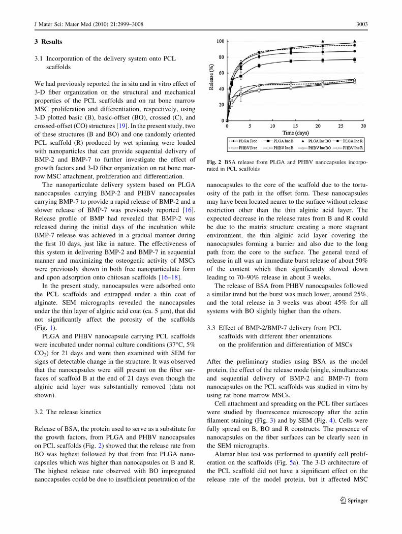
3.2 The release kinetics
Release of BSA, the protein used to serve as a substitute for
the growth factors, from PLGA and PHBV nanocapsules
on PCL scaffolds (Fig. 2) showed that the release rate from
BO was highest followed by that from free PLGA nano-
capsules which was higher than nanocapsules on B and R.
The highest release rate observed with BO impregnated
nanocapsules could be due to insufficient penetration of the
nanocapsules to the core of the scaffold due to the tortu-
osity of the path in the offset form. These nanocapsules
may have been located nearer to the surface without release
restriction other than the thin alginic acid layer. The
expected decrease in the release rates from B and R could
be due to the matrix structure creating a more stagnant
environment, the thin alginic acid layer covering the
nanocapsules forming a barrier and also due to the long
path from the core to the surface. The general trend of
release in all was an immediate burst release of about 50%
of the content which then significantly slowed down
leading to 70–90% release in about 3 weeks.
The release of BSA from PHBV nanocapsules followed
a similar trend but the burst was much lower, around 25%,
and the total release in 3 weeks was about 45% for all
systems with BO slightly higher than the others.
3.3 Effect of BMP-2/BMP-7 delivery from PCL
scaffolds with different fiber orientations
on the proliferation and differentiation of MSCs
After the preliminary studies using BSA as the model
protein, the effect of the release mode (single, simultaneous
and sequential delivery of BMP-2 and BMP-7) from
nanocapsules on the PCL scaffolds was studied in vitro by
using rat bone marrow MSCs.
Cell attachment and spreading on the PCL fiber surfaces
were studied by fluorescence microscopy after the actin
filament staining (Fig. 3) and by SEM (Fig. 4). Cells were
fully spread on B, BO and R constructs. The presence of
nanocapsules on the fiber surfaces can be clearly seen in
the SEM micrographs.
Alamar blue test was performed to quantify cell prolif-
eration on the scaffolds (Fig. 5a). The 3-D architecture of
the PCL scaffold did not have a significant effect on the
release rate of the model protein, but it affected MSC
Fig. 2 BSA release from PLGA and PHBV nanocapsules incorpo-
rated in PCL scaffolds
J Mater Sci: Mater Med (2010) 21:2999–3008 3003
123

proliferation. Especially after the first week, cell numbers
on R scaffolds was significantly higher compared to B and
BO, probably due to the higher level of initial attachment.
When ALP activities were measured, it was observed
that the ALP activity reached its highest level when BMP-2
and BMP-7 were introduced to the growth medium in a
sequential manner regardless of the time or the scaffold
architecture (Fig. 5b). ALP activity increased gradually in
time for all scaffolds (B, BO and R) and under every
delivery condition tested (single, simultaneous and
sequential) during the 21 days of incubation. When the
ALP activity of the various scaffold types was compared
for the sequential case, it was observed that R scaffolds had
a higher ALP activity compared to B and BO, as was the
case with the growth factor-free scaffolds. The ALP
activity on day 21 for B, BO and R were 0.767 ± 0.016,
0.891 ± 0.010 and 1.109 ± 0.002 nmol/min/cell, respec-
tively [19].
4 Discussion
Tissue engineering is a promising strategy to produce bone
substitutes; however, the control of the cell organization
and cell behavior to create complex, fully functional 3-D
constructs has not yet been achieved. To overcome these,
efforts have been concentrated on the development of
multifunctional tissue engineering scaffolds capable of
delivering the required bioactive agents to induce and
guide cellular activities. The aim of this study was to
prepare MSC seeded tissue engineered constructs consist-
ing of PCL scaffolds with highly organized architecture
impregnated with a biodegradable nanoparticulate growth
factor delivery system that sequentially delivers BMP-2
and BMP-7 to mimic the natural bone healing process.
Scaffolds play a central role in the success of tissue
engineering by defining the form of the construct. Since
most of our tissues are highly organized and anisotropic,
Fig. 3 Fluorescence microscopy of PCL scaffolds seeded with MSC
on day 21 of incubation incorporated with PLGA and PHBV
nanocapsules to provide sequential BMP-2/BMP-7 delivery.
a Scaffold B, b scaffold BO, c scaffold R, d x-section of the fiber
of BO scaffold showing complete coverage of the fiber surface with
MSCs. 910
3004 J Mater Sci: Mater Med (2010) 21:2999–3008
123

design of the scaffold that could mimic the natural ECM
structurally and mechanically is of utmost importance. A
number of techniques such as solvent casting, particulate
leaching [20–22], electrospinning [23–26], fiber bonding
[27], membrane lamination [28], melt molding [29], and
gas foaming [30] were employed in scaffold production.
However, none of these techniques can produce structures
with a predefined architecture. RP technique, however,
offers the capability to produce architectures that could
perfectly fit into the defect site.
In the present study, PCL scaffolds produced by 3-D
plotting with two different standard architectures (B, BO)
and scaffolds produced by wet spinning with randomly
oriented fibers (R) (Fig. 1) were used to study the effect of
3-D fiber organization on rat bone marrow MSC behavior.
It was observed that random scaffolds perform better than
the oriented ones (B, BO) in terms of MSC proliferation
and differentiation (Fig. 5). Most of the studies in the lit-
erature investigating the effect of fiber organization
involving RP techniques focused on the dependence of
structural and mechanical properties of the scaffolds in
relation with fiber organization and pore properties [31,
32]. In a recent study from Melchels et al. [33], the effect
of pore architecture on the behavior of immortalised bone
marrow derived MSCs was studied by using P(D,L/LA)-
based scaffolds produced by stereolithography and salt
leaching leading to scaffolds with highly oriented and
random pores, respectively. As in our study, the overall
porosity of the scaffolds with different pore geometries was
almost the same (ca. 68%). They reported that scaffolds
produced by stereolithography had open and more acces-
sible pores compared to the more tortuous pores of random
scaffolds. This larger pore interconnectivity caused the
oriented scaffold to have higher cellular attachment and
proliferation in the static culture. This is to be expected
because the levels of interconnectivity is different. The
pore interconnectivity in this study is not different in the
scaffolds used, as assessed by u-CT analysis previously
[19]. Therefore, pore interconnectivity in the current case is
not a property that affects cellular behavior. Thus, it must
be the 3-D fiber organization that defines the geometry of
sites for the cells to adhere and the distances between them
must be influential on the cell adhesion, proliferation and
differentiation. Since in the random scaffold the distances
vary from point to point, cells must have found more sites
to attach to and stretch across.
Fig. 4 SEM of MSCs on BMP-2 loaded PLGA nanocapsule incorporated PCL scaffolds a B, b BO, c, d R at the end of 21 days of culture
J Mater Sci: Mater Med (2010) 21:2999–3008 3005
123

The dynamic mechanical analysis of the RP PCL scaf-
folds used in this study were previously carried out and the
scaffolds were found to exhibit a stiffness in between those
of human trabecular and cortical bone [19]. In previous
studies by our group, cell proliferation and the following
ECM secretion were observed to enhance stiffness and
ultimate tensile strength of cell seeded collagen scaffolds
[34, 35]. Thus, cell seeding and appropriate in vitro con-
ditions during the tissue engineering process are expected
to improve the mechanical properties of PCL based scaf-
folds and make them more compatible with the cortical
bones.
The scaffolds with random and oriented fibers were
made to carry a delivery system to further improve its
function by mimicking the biological guidance provided by
the microenvironment during healing. For this, a model
protein, BSA, and two osteogenic growth factors were
encapsulated in polymeric nanocapsules and loaded onto
the fibers of the scaffolds under a thin alginate layer. BSA
was used as a model compound because it is a protein
comparable in size especially to BMP-7 (BSA: 65 kDa,
BMP-7: 49 kDa, BMP-2: 18 kDa), and has a similar pI
value (BSA: 5.5, BMPs: 5.0), indicating that it would have
the same charge as the growth factors under the test con-
ditions (pH 7.4). Thus, it is an appropriate molecule to
substitute the growth factors in the release studies. Also, in
our previous study [16], same polyester nanocapsules
(without the scaffold) loaded with BSA and BMPs pre-
sented similar release kinetics. It was observed that the
release rate of BSA from PLGA nanocapsules was higher
in comparison to its PHBV counterpart (Fig. 2), therefore,
the nanoparticles were capable of constituting the early and
late release components of a two-component sequential
delivery system. Similarly, Biondi et al. [36] used BSA as a
model protein encapsulated in PLGA microspheres which
were then embedded in a collagen-hyaluronic acid (HA)
semi-interpenetrating networks (semi-IPNs).
The studies in the literature concerning the incorporation
of various growth factors into tissue engineering scaffolds
generally involve mixing of the polymer with the growth
factors during the preparation phase, thus, forming scaf-
folds that carry the factor embedded within the polymer
matrix as in a monolithic device [8, 37]. Therefore, the
control on the release rate is not very precise in this case as
the release of the agent is dependent on the degradation of
the scaffold or solubilization of the drug molecules starting
from the surface. In other studies, growth factors were
loaded into large microparticles, which upon incorporation
in the scaffold affected its structure and properties [9–11,
17]. In the study of Patel et al. [38], VEGF and BMP-2
were encapsulated in gelatin microparticles of ca. 30 lm,
therefore, blocking most the pores of the scaffold that is
essential for cell seeding, proliferation and also for oxygen
and nutrient delivery. In another study [11], BMP-2 and
IGF-1 (alone or together) were encapsulated in glycidyl
methacrylated dextran/gelatin microparticles and embed-
ded in the porous scaffold made from the same polymeric
material. Sustained release from the constructs was shown,
however, it was clear in the SEM micrographs that the
porosity of the scaffold was considerably decreased due to
the presence of the microparticles. In the present study,
growth factors were encapsulated in nanocapsules which
were then introduced to PCL scaffolds without affecting
the structural properties of the scaffold (Fig. 1). Moreover,
by maintaining the nanoparticles on the surface of the
fibers rather than within the fibers, the release from the
particles was not dependent on scaffold degradation or
growth factor solubilization and could be fine tuned by
changing the nanoparticle properties and loading
conditions.
Fig. 5 a Cell proliferation on BMP loaded particle incorporated PCL
scaffolds. b Specific ALP activity of MSCs on BMP loaded particle
incorporated PCL scaffolds. B basic, BO basic-offset, R random,
BMP-2 single BMP-2 delivery from PLGA nanocapsules loaded onto
scaffolds, BMP-7 single BMP-7 delivery from PHBV nanocapsules
loaded onto scaffolds, SIM simultaneous BMP-2 and BMP-7 delivery
(both from PLGA nanocapsules), SEQ sequential BMP-2 and BMP-7
delivery (BMP-2 from PLGA and BMP-7 from PHBV nanocapsules)
3006 J Mater Sci: Mater Med (2010) 21:2999–3008
123
The proliferation of MSCs and their differentiation into
osteoblasts after induction with BMP-2 and BMP-7
sequentially released from the PCL scaffolds was studied.
Fluorescence microscopy and SEM analysis revealed
proper spreading of the cells on the fiber surfaces in the
presence of nanoparticles (Figs. 3, 4). Cell proliferation
was observed to have a similar trend to that of the previous
results reported with free nanocapsules [16] and chitosan
scaffolds [18] (Fig. 5a). Release of BMP-2 suppressed
MSC proliferation when compared to the unloaded PCL
scaffolds (the number of cells on day 21 for unloaded B,
BO and R scaffolds were 2.22 ± 0.12 9 105, 2.62 ±
0.57 9 105 and 2.75 ± 0.33 9 105, respectively [19]) and
the suppression was higher than with BMP-7. In all three
BMP-2 carrying systems proliferation was suppressed. The
most suppressed was the BMP-2/BMP-7 sequential deliv-
ery system. This trend did not change during the 3-week
test period. The ALP activity trend is also similar to our
earlier results [16, 18]; the highest ALP activity, the indi-
cation of MSC differentiation, was observed in the case of
sequential delivery of the growth factors (Fig. 5b). Similar
results were obtained by other researchers as well, where
the co-administration of BMP-2 and TGF-b3 [8], BMP-2
and VEGF [9, 10] and BMP-2 and IGF-1 led to enhanced
bone formation while the effect of a single growth factor
was negligible.
An interesting result of this study was that the use of R
scaffolds seemed to perform better in housing higher cell
numbers than the oriented scaffolds (B and BO) (Fig. 5).
This might be due to the higher initial cell attachment on
the R fibers due to the tortuosity of the path during cell
seeding which led to the higher cell numbers on this
scaffold for the rest of the in vitro culture. This result might
also indicate that the scaffold design should mimic the
natural ECM structure and that ordered structures are not
that favored by the biological system. Besides, the dis-
tances between the organized fibers might have been too
large. Therefore, scaffold architecture was influential on
cell response, in terms of cell attachment and proliferation.
Furthermore, cell differentiation, as measured by ALP
activity, appears to be also affected by scaffold structure as
well as by the delivery mode of the growth factors.
5 Conclusion
The positive effect of co-administration of BMP-2 and
BMP-7 in a sequential manner as in nature was reflected as
increased osteogenic activity. The 3-D architecture of the
scaffolds affected MSC proliferation and differentiation,
and scaffold with randomly organized fibers performed
better than ordered PCL scaffolds. As a conclusion, it is
possible to obtain multi-functional bioactive tissue
engineering scaffolds with different architectures capable
of delivering osteogenic factors in a number of ways and
using this information it will be possible to control the level
and rate of maturation of a tissue engineered construct.
Acknowledgments This project was conducted within the scope of
the EU FP6 NoE Project Expertissues (NMP3-CT-2004-500283). We
acknowledge the support to PY through the same project in the form
of an integrated PhD grant. We also would like to acknowledge the
support from Scientific and Technical Research Council of Turkey
(TUBITAK) through project METUNANOBIOMAT (TBAG
105T508).
References
1. Lam CXF, Mo XM, Teoh SH, Hutmacher DW. Scaffold devel-
opment using 3D printing with a starch-based polymer. Mater Sci
Eng C. 2002;20:49–56.
2. Yeong WY, Chua CK, Leong KF, Chandrasekaran M. Rapid
prototyping in tissue engineering: challenges and potential.
Trends Biotechnol. 2004;22:643–52.
3. Moroni L, de Wijn JR, van Blitterswijk CA. 3D fiber-deposited
scaffolds for tissue engineering: influence of pores geometry and
architecture on dynamic mechanical properties. Biomaterials.
2006;27:974–85.
4. Allori AC, Sailon AM, Warren SM. Biological basis of bone
formation, remodeling, and repair—part I: biochemical signaling
molecules. Tissue Eng Part B. 2008;14:259–73.
5. Urist MR. Bone: formation by autoinduction. Science. 1965;150:
893–9.
6. Wang EA, Rosen V, D’Alessandro JS, Bauduy M, Cordes P,
Harada T, et al. Recombinant human bone morphogenetic protein
induces bone formation. Proc Natl Acad Sci USA. 1990;87:
2220–4.
7. Cowan CM, Soo C, Ting K, Wu B. Evolving concepts in bone
tissue engineering. Curr Top Dev Biol. 2005;66:239–85.
8. Simmons CA, Alsberg E, Hsiong S, Kim WJ, Mooney DJ. Dual
growth factor delivery and controlled scaffold degradation
enhance in vivo bone formation by transplanted bone marrow
stromal cells. Bone. 2004;35:562–9.
9. Patel ZS, Young S, Tabata Y, Jansen JA, Wong MEK, Mikos
AG. Dual delivery of an angiogenic an osteogenic growth factor
for bone regeneration enhances in a critical size defect model.
Bone. 2008;43:931–40.
10. Young S, Patel ZS, Kretlow JD, Murphy MB, Mountziaris PM,
Baggett LS, et al. Dose effect of dual delivery of vascular
endothelial growth factor and bone morphogenetic protein-2 on
bone regeneration in a rat critical-size defect model. Tissue Eng
Part A. 2009;15:2347–62.
11. Chen FM, Chen R, Wang XJ, Sun HH, Wu ZF. In vitro cellular
responses containing two microencapsulated growth factors.
Biomaterials. 2009;30:5215–24.
12. White AP, Vaccaro AR, Hall JA, Whang PG, Friel BC, McKee
MD. Clinical applications of BMP-7/OP-1 in fractures, non-
unions and spinal fusion. Int Orthop. 2007;31:735–41.
13. McKay WF, Peckham SM, Badura JM. A comprehensive clinical
review of recombinant human bone morphogenetic protein-2
(INFUSE� Bone Graft). Int Orthop. 2007;31:729–34.
14. Bessa PC, Casal M, Reis RL. Bone morphogenetic proteins in
tissue engineering: the road from the laboratory to the clinic, part
I (basic concepts). J Tissue Eng Regen Med. 2008;2:1–13.
15. Cho TJ, Gerstenfeld LC, Einhorn TA. Differential temporal
expression of members of the transforming growth factor beta
J Mater Sci: Mater Med (2010) 21:2999–3008 3007
123

superfamily during murine fracture healing. J Bone Miner Res.
2002;17:513–20.
16. Yilgor P, Hasirci N, Hasirci V. Sequential BMP-2/BMP-7
delivery from polyester nanocapsules. J Biomed Mater Res. 2010;
93A:528–36.
17. Basmanav FB, Kose GT, Hasirci V. Sequential growth factor
delivery from complexed microspheres for bone tissue engi-
neering. Biomaterials. 2008;29:4195–204.
18. Yilgor P, Tuzlakoglu K, Reis RL, Hasirci N, Hasirci V. Incor-
poration of a sequential BMP-2/BMP-7 delivery system into
chitosan-based scaffolds for bone tissue engineering. Biomateri-
als. 2009;30:3551–9.
19. Yilgor P, Sousa RA, Reis RL, Hasirci N, Hasirci V. 3D plotted
PCL scaffolds for stem cell based bone tissue engineering.
Macromol Symp. 2008;269:92–9.
20. Kose GT, Kenar H, Hasirci N, Hasirci V. Macroporous poly(3-
hydroxybutyrate-co-3-hydroxyvalerate) matrices for bone tissue
engineering. Biomaterials. 2003;24:1949–58.
21. Cao H, Kuboyama N. A biodegradable porous composite scaffold
of PGA/b-TCP for bone tissue engineering. Bone. 2010;46:
386–95.
22. Zhang P, Hong Z, Yu T, Chen X, Jing X. In vivo mineralization
and osteogenesis of nanocomposite scaffold of poly(lactide-co-
glycolide) and hydroxyapatite surface-grafted with poly(l-lac-
tide). Biomaterials. 2009;30:58–70.
23. Santos MI, Tuzlakoglu K, Fuchs S, Gomes ME, Peters K, Unger
RE, et al. Endothelial cell colonization and angiogenic potential
of combined nano- and micro-fibrous scaffolds for bone tissue
engineering. Biomaterials. 2008;29:4306–13.
24. Yucel D, Kose GT, Hasirci V. Polyester based nerve guidance
conduit design. Biomaterials. 2009;31:1596–603.
25. Cao H, Chen X, Huang L, Shao Z. Electrospinning of reconsti-
tuted silk fiber from aqueous silk fibroin solution. Mater Sci Eng
C. 2009;29:2270–4.
26. Yu HS, Jang JH, Kim TI, Lee HH, Kim HW. Apatite-mineralized
polycaprolactone nanofibrous web as a bone tissue regeneration
substrate. J Biomed Mater Res. 2009;88:747–54.
27. Gomes ME, Holtorf HL, Reis RL, Mikos AG. Influence of the
porosity of starch-based fiber mesh scaffolds on the proliferation
and osteogenic differentiation of bone marrow stromal cells
cultured in a flow perfusion bioreactor. Tissue Eng. 2006;12:
801–9.
28. Mikos AG, Sarakinos G, Leite SM, Vacanti JP, Langer R.
Laminated three-dimensional biodegradable foams for use in
tissue engineering. Biomaterials. 1993;14:323–30.
29. Se HO, Soung GK, Jin HL. Degradation behavior of hydrophil-
ized PLGA scaffolds prepared by melt-molding particulate-
leaching method: comparison with control hydrophobic one.
J Mater Sci Mater Med. 2006;17:131–7.
30. Almirall A, Larrecq G, Delgado JA, Martı́nez S, Planell JA,
Ginebra MP. Fabrication of low temperature macroporous
hydroxyapatite scaffolds by foaming and hydrolysis of an a-TCP
paste. Biomaterials. 2004;25:3671–80.
31. Lipowiecki M, Brabazon D. Design of bone scaffolds structures
for rapid prototyping with increased strength and osteoconduc-
tivity. Adv Mater Res. 2010;83–86:914–22.
32. Li JP, de Wijn JR, van Blitterswijk CA, de Groot K. The effect of
scaffold architecture on properties of direct 3D fiber deposition of
porous Ti6Al4V for orthopedic implants. J Biomed Mater Res.
2010;92A:33–42.
33. Melchels FPW, Barradas AMC, van Blitterswijk CA, de Boer J,
Feijen J, Grijpma DW. Effects of the architecture of tissue
engineering scaffolds on cell seeding and culturing. Acta Bio-
mater. 2010. doi:10.1016/j.actbio.2010.06.012.
34. Vrana NE, Elsheikh A, Builles N, Damour O, Hasirci V. Effect of
human corneal keratocytes and retinal pigment epithelial cells on
the mechanical properties of micropatterned collagen films.
Biomaterials. 2007;28:4303–10.
35. Zorlutuna P, Elsheikh A, Hasirci V. Nanopatterning of collagen
scaffolds improve the mechanical properties of tissue engineered
vascular grafts. Biomacromolecules. 2009;10:814–21.
36. Biondi M, Indolfi L, Ungaro F, Quaglia F, La Rotonda MI, Netti
PA. Bioactivated collagen-based scaffolds embedding protein-
releasing biodegradable microspheres: tuning of protein release
kinetics. J Mater Sci Mater Med. 2009;20:2117–28.
37. Raiche AT, Puleo DA. In vitro effects of combined and
sequential delivery of two bone growth factors. Biomaterials.
2004;25:677–85.
38. Patel ZS, Yamamoto M, Ueda H, Tabata Y, Mikos AG. Biode-
gradable gelatin microparticles as delivery systems for the con-
trolled release of bone morphogenetic protein-2. Acta Biomater.
2008;4:1126–38.
3008 J Mater Sci: Mater Med (2010) 21:2999–3008
123