Dorsomorphin inhibits BMP signals required for embryogenesis and iron metabolism
21
Dorsomorphin inhibits BMP signals required for embryogenesis and iron metabolism Paul B Yu 1,2,6 , Charles C Hong 1,5,6 , Chetana Sachidanandan 1,6 , Jodie L Babitt 3 , Donna Y Deng 1 , Stefan A Hoyng 1 , Herbert Y Lin 3 , Kenneth D Bloch 1,4 , and Randall T Peterson 1,2 1 Cardiovascular Research Center and Division of Cardiology, Department of Medicine, Massachusetts General Hospital, Harvard Medical School, 149 13 th Street, Charlestown, Massachusetts 02129, USA. 2 Broad Institute of MIT and Harvard, 7 Cambridge Center, Cambridge, Massachusetts 02142, USA. 3 Program in Membrane Biology and Division of Nephrology, Department of Medicine, Massachusetts General Hospital, Harvard Medical School, 165 Cambridge Street, Boston, Massachusetts 02114, USA. 4 Department of Anesthesia and Critical Care, Massachusetts General Hospital, Harvard Medical School, 55 Fruit Street, Boston, Massachusetts 02114, USA. Abstract Bone morphogenetic protein (BMP) signals coordinate developmental patterning and have essential physiological roles in mature organisms. Here we describe the first known small-molecule inhibitor of BMP signaling—dorsomorphin, which we identified in a screen for compounds that perturb dorsoventral axis formation in zebrafish. We found that dorsomorphin selectively inhibits the BMP type I receptors ALK2, ALK3 and ALK6 and thus blocks BMP-mediated SMAD1/5/8 phosphorylation, target gene transcription and osteogenic differentiation. Using dorsomorphin, we examined the role of BMP signaling in iron homeostasis. In vitro, dorsomorphin inhibited BMP-, hemojuvelin- and interleukin 6–stimulated expression of the systemic iron regulator hepcidin, which suggests that BMP receptors regulate hepcidin induction by all of these stimuli. In vivo, systemic challenge with iron rapidly induced SMAD1/5/8 phosphorylation and hepcidin expression in the liver, whereas treatment with dorsomorphin blocked SMAD1/5/8 phosphorylation, normalized hepcidin expression and increased serum iron levels. These findings suggest an essential physiological role for hepatic BMP signaling in iron-hepcidin homeostasis. Signals mediated by BMP ligands serve diverse roles throughout the life of vertebrates. During embryogenesis, the dorsoventral axis is established by BMP signaling gradients formed by the coordinated expression of ligands, receptors, co-receptors and soluble antagonists 1–3 . Excess BMP signaling causes ventralization (an expansion of ventral structures at the expense of dorsal structures) whereas diminished BMP signaling causes dorsalization (an expansion of dorsal structures at the expense of ventral structures) 1,3,4 . BMPs are key regulators of gastrulation, © 2007 Nature Publishing Group Correspondence should be addressed to K.D.B. ([email protected]) or R.T.P. ([email protected]). 5 Present address: Division of Cardiovascular Medicine and Department of Pharmacology, Vanderbilt University School of Medicine, 2220 Pierce Avenue, Nashville, Tennessee 37232, USA. 6 These authors contributed equally to this work. AUTHOR CONTRIBUTIONS P.B.Y., C.C.H., C.S., J.L.B., H.Y.L., K.D.B and R.T.P. designed experiments, performed experiments, analyzed data and helped write the manuscript. D.Y.D. and S.A.H. performed experiments. K.D.B. and R.T.P. contributed equally as senior authors to this work. Reprints and permissions information is available online at http://npg.nature.com/reprintsandpermissions NIH Public Access Author Manuscript Nat Chem Biol. Author manuscript; available in PMC 2009 August 17. Published in final edited form as: Nat Chem Biol. 2008 January ; 4(1): 33–41. doi:10.1038/nchembio.2007.54. NIH-PA Author Manuscript NIH-PA Author Manuscript NIH-PA Author Manuscript
-
Upload
independent -
Category
Documents
-
view
2 -
download
0
Transcript of Dorsomorphin inhibits BMP signals required for embryogenesis and iron metabolism

Dorsomorphin inhibits BMP signals required for embryogenesisand iron metabolism
Paul B Yu1,2,6, Charles C Hong1,5,6, Chetana Sachidanandan1,6, Jodie L Babitt3, Donna YDeng1, Stefan A Hoyng1, Herbert Y Lin3, Kenneth D Bloch1,4, and Randall T Peterson1,21Cardiovascular Research Center and Division of Cardiology, Department of Medicine,Massachusetts General Hospital, Harvard Medical School, 149 13th Street, Charlestown,Massachusetts 02129, USA.2Broad Institute of MIT and Harvard, 7 Cambridge Center, Cambridge, Massachusetts 02142, USA.3Program in Membrane Biology and Division of Nephrology, Department of Medicine,Massachusetts General Hospital, Harvard Medical School, 165 Cambridge Street, Boston,Massachusetts 02114, USA.4Department of Anesthesia and Critical Care, Massachusetts General Hospital, Harvard MedicalSchool, 55 Fruit Street, Boston, Massachusetts 02114, USA.
AbstractBone morphogenetic protein (BMP) signals coordinate developmental patterning and have essentialphysiological roles in mature organisms. Here we describe the first known small-molecule inhibitorof BMP signaling—dorsomorphin, which we identified in a screen for compounds that perturbdorsoventral axis formation in zebrafish. We found that dorsomorphin selectively inhibits the BMPtype I receptors ALK2, ALK3 and ALK6 and thus blocks BMP-mediated SMAD1/5/8phosphorylation, target gene transcription and osteogenic differentiation. Using dorsomorphin, weexamined the role of BMP signaling in iron homeostasis. In vitro, dorsomorphin inhibited BMP-,hemojuvelin- and interleukin 6–stimulated expression of the systemic iron regulator hepcidin, whichsuggests that BMP receptors regulate hepcidin induction by all of these stimuli. In vivo, systemicchallenge with iron rapidly induced SMAD1/5/8 phosphorylation and hepcidin expression in theliver, whereas treatment with dorsomorphin blocked SMAD1/5/8 phosphorylation, normalizedhepcidin expression and increased serum iron levels. These findings suggest an essentialphysiological role for hepatic BMP signaling in iron-hepcidin homeostasis.
Signals mediated by BMP ligands serve diverse roles throughout the life of vertebrates. Duringembryogenesis, the dorsoventral axis is established by BMP signaling gradients formed by thecoordinated expression of ligands, receptors, co-receptors and soluble antagonists1–3. ExcessBMP signaling causes ventralization (an expansion of ventral structures at the expense of dorsalstructures) whereas diminished BMP signaling causes dorsalization (an expansion of dorsalstructures at the expense of ventral structures)1,3,4. BMPs are key regulators of gastrulation,
© 2007 Nature Publishing GroupCorrespondence should be addressed to K.D.B. ([email protected]) or R.T.P. ([email protected]).5Present address: Division of Cardiovascular Medicine and Department of Pharmacology, Vanderbilt University School of Medicine,2220 Pierce Avenue, Nashville, Tennessee 37232, USA.6These authors contributed equally to this work.AUTHOR CONTRIBUTIONSP.B.Y., C.C.H., C.S., J.L.B., H.Y.L., K.D.B and R.T.P. designed experiments, performed experiments, analyzed data and helped writethe manuscript. D.Y.D. and S.A.H. performed experiments. K.D.B. and R.T.P. contributed equally as senior authors to this work.Reprints and permissions information is available online at http://npg.nature.com/reprintsandpermissions
NIH Public AccessAuthor ManuscriptNat Chem Biol. Author manuscript; available in PMC 2009 August 17.
Published in final edited form as:Nat Chem Biol. 2008 January ; 4(1): 33–41. doi:10.1038/nchembio.2007.54.
NIH
-PA Author Manuscript
NIH
-PA Author Manuscript
NIH
-PA Author Manuscript

mesoderm induction, organogenesis and endochondral bone formation, and they regulate thefates of multipotent cell populations5. BMP signals also have critical roles in physiology anddisease, and they are implicated in primary pulmonary hypertension, hereditary hemorrhagictelangiectasia syndrome, fibrodysplasia ossificans progressiva and juvenile polyposissyndrome6–8.
The BMP signaling family is a diverse subset of the transforming growth factor-β (TGF-β)superfamily9. Over 20 known BMP ligands are recognized by three distinct type II receptors(BMPRII, ActRIIa and ActRIIb) and at least three type I receptors (ALK2, ALK3 and ALK6).Dimeric ligands facilitate assembly of receptor heteromers, thereby allowing the constitutivelyactive type II receptor serine/threonine kinases to phosphorylate type I receptor serine/threonine kinases. Activated type I receptors phosphorylate BMP-responsive SMAD effectors(SMAD1, SMAD5 and SMAD8) to facilitate nuclear translocation in complex with SMAD4,a co-SMAD that also facilitates TGF signaling. In addition, BMP signals can activateintracellular effectors such as mitogen-activated protein kinase (MAPK) p38 in a SMAD-independent manner10. Soluble BMP antagonists such as noggin, chordin, gremlin andfollistatin limit BMP signaling by ligand sequestration. Co-receptors such as members of therepulsive guidance molecule (RGM) family (including RGMa, DRAGON (RGMb) andhemojuvelin (HJV, RGMc)) enhance the response to low levels of BMP ligands in the targettissues in which they are expressed11,12.
Recent work suggests a role for BMP signals in regulating expression of hepcidin, a peptidehormone and central regulator of systemic iron balance13–16. Hepcidin binds and promotesdegradation of ferroportin, the sole iron exporter in vertebrates. Loss of ferroportin activityprevents mobilization of iron to the bloodstream from intracellular stores in enterocytes,macrophages and hepatocytes17. Hepatic hepcidin expression is responsive to states of irondeficiency or overload, inflammation, and hypoxia, but the mechanisms by which the liversenses these stimuli and regulates hepcidin expression remain unclear, especially because thepromoter for the HAMP gene (the gene that encodes hepcidin) does not seem to have elementsknown to be modulated by iron levels. Mice conditionally deficient in SMAD4 fail to inducehepcidin following iron or interleukin 6 (IL-6) challenge18, which suggests that BMP and/orTGF-β pathways transduce signals mediated by iron or inflammation. As further evidence ofa link between BMP signaling and iron metabolism, mutations in Hfe2 (which encodes theBMP co-receptor HJV) have been identified in juvenile hemochromatosis, a disease in whichiron overload fails to trigger hepcidin expression7.
Pharmacological tools that selectively inhibit BMP signaling, but not TGF-β or other pathways,could help elucidate the roles of BMP signaling in diverse physiological processes, includingiron homeostasis. Specific inhibitors of the BMP receptors have not yet been identified, perhapsbecause of the difficulty in targeting a set of BMP receptor kinases while avoiding hundredsof structurally similar kinases. Traditional high-throughput in vitro screens identify smallmolecules with activity against a kinase of interest, but both extensive counterscreening againstother kinases and animal testing are required to confirm specificity. To circumvent thesechallenges, we sought an in vivo screening approach that would identify compounds that inhibitBMP signaling while selecting against those with nonspecific or undesirable biological effects.By screening a diverse chemical library for small molecules that dorsalize zebrafish embryos,we identified a compound that blocks BMP signaling by inhibiting type I receptors ALK2,ALK3 and ALK6. Although previously described as an inhibitor of AMP-activated proteinkinase (compound C), we found that the compound elicits phenotypes reflecting a high degreeof selectivity for BMP signaling in vivo. Using this selective BMP inhibitor, we report thatBMP signaling is essential for iron homeostasis and that pharmacological BMP inhibition canbe used to increase serum iron levels in vivo.
Yu et al. Page 2
Nat Chem Biol. Author manuscript; available in PMC 2009 August 17.
NIH
-PA Author Manuscript
NIH
-PA Author Manuscript
NIH
-PA Author Manuscript

RESULTSDorsomorphin induces dorsalization in zebrafish embryos
Small-molecule inhibitors of BMP signaling were sought using an in vivo screening assaybased on the premise that BMP antagonists would dorsalize developing zebrafish embryos.Over 7,500 compounds were tested from our small-molecule collection, which consists ofknown bioactive molecules, US Food and Drug Administration–approved drugs and acommercial molecular diversity library. Zebrafish embryos were arrayed into 96-well plates,incubated with compounds starting 4 h post fertilization (h.p.f.) and assessed visually at 24 and48 h.p.f. One compound, which we call dorsomorphin (1, Fig. 1a), produces substantial andreproducible dorsalization of zebrafish embryos. Dorsomorphin-treated embryos showexpansion of structures derived from the dorsal pole of spherical embryos at the expense ofstructures derived from the ventral pole1,3,4 (Fig. 1b–e).
The extent of dorsalization induced by dorsomorphin varied as a function of dose and timing.When added at 6–8 h.p.f., dorsomorphin caused mild dorsalization manifested as the absenceof the ventral tail fin (Fig. 1b,c), similar to that of noggin-overexpressing and lost-a-fin (ALK8mutant) zebrafish2,3. Zebrafish treated with dorsomorphin at 4 h.p.f. occasionally had anectopic tail appendage in addition to the absent ventral fin (Fig. 1d), which resembled transgeniczebrafish expressing a heat-shock-inducible dominant negative BMP type I receptor after theshield stage19. When dorsomorphin was added to embryos at 1–2 h.p.f., caudal and posteriorstructures of the tail derived from the embryonic ventral pole were more profoundly disrupted(Fig. 1e), which resembled fish with more severe BMP signaling defects2. The addition ofcompound at 2 h.p.f. led to altered gastrulation and somitogenesis similar to that observed inlost-a-fin fish2, with ovoid dorsalized morphology at the bud stage (Fig. 1f,g) and abnormaltailbud morphology at the 10-somite stage (Fig. 1h,i). Treatment of embryos with ≥5 µMdorsomorphin at ≤8 h.p.f. phenocopied with high penetrance the spectrum of dorsalizationdefects observed in zebrafish with defective BMP signaling (Supplementary Fig. 1 andSupplementary Table 1 online) but did not result in cyclopia, a phenotype associated withdefective TGF-β signaling20.
Dorsomorphin is structurally identical to compound C, a molecule previously shown toantagonize AMP-activated kinase (AMPK) activity in vitro with an estimated functional Ki of10 to 20 µM (ref. 21). To test whether AMPK inhibition contributes to the effects ofdorsomorphin on dorsoventral axis formation, we tested structurally unrelated inhibitors ofAMPK activity such as C75 (3) and Ara-A (2)22,23. These inhibitors did not dorsalize zebrafishembryos but instead caused nonspecific toxicity such as developmental delay and death thatwas not observed with dorsalizing concentrations of dorsomorphin (Supplementary Table 2online). Moreover, knock-down of AMPK activity in zebrafish by morpholino injection didnot cause dorsalization (Supplementary Fig. 2 online). Dorsomorphin is structurally related toa series of compounds that has been shown to inhibit the activity of the receptor tyrosine kinaseKDR (ref. 24). At the doses of dorsomorphin used in our experiments, however, we did notobserve any of the known effects of KDR inhibition on zebrafish vascular development, suchas disrupted formation of intersomitic blood vessels25 (Fig. 1c–e). Therefore, dorsomorphin-induced dorsalization is not caused by AMPK or KDR inhibition.
Dorsomorphin expands dorsal marker expressionTo confirm the impact of dorsomorphin on dorsoventral axis formation, the expression of dorsaland ventral markers were examined. Expression of the ventral marker eve1 at the 18-somitestage was reduced in dorsomorphin-treated embryos relative to controls (Fig. 1j,k), which is
Note: Supplementary information and chemical compound information is available on the Nature Chemical Biology website.
Yu et al. Page 3
Nat Chem Biol. Author manuscript; available in PMC 2009 August 17.
NIH
-PA Author Manuscript
NIH
-PA Author Manuscript
NIH
-PA Author Manuscript

consistent with dorsalization2. At the 6-somite stage, pax2a (pax2.1) expression demarcatesthe optic stalk, mid-hindbrain boundary and otic vesicles26. Dorsomorphin treatment inducedexpansion of these dorsal tissues (Fig. 1l,m). Similarly, dorsomorphin induced lateralexpansion of egr2b (krox20), a marker of rhombomeres 3 and 5, and myod, a marker of posteriormyotomes (Fig. 1n,o). Thus, dorsomorphin induced changes in the expression of dorsal andventral markers consistent with the effects of dorsalization seen in zebrafish BMP pathwaymutants27.
Dorsomorphin reverses ventralization in chordin morphantsBecause treatment with dorsomorphin mimics BMP antagonism in zebrafish, it was postulatedthat dorsomorphin might rescue ventralization caused by loss of the endogenous BMPantagonist chordin28,29. Zebrafish were injected with chordin morpholino (1 ng) at the 1-cellstage, which caused moderate ventralization with expansion of the ventral fin and theintermediate cell mass (ICM) (Fig. 1p)29. Dorsomorphin treatment of chordin morphants at 2h.p.f. reduced the size of the ventral fin and ICM (Fig. 1q). Therefore, dorsomorphin treatmentcompensates for the loss of an endogenous BMP antagonist in zebrafish embryos, thusconfirming its function as a BMP antagonist.
Dorsomorphin inhibits SMAD-dependent BMP signalsThe effect of dorsomorphin on BMP signaling was tested in vitro. Cultured mouse pulmonaryartery smooth muscle cells (PASMCs) express a complement of BMP receptors. In responseto diverse BMP ligands, PASMCs show SMAD1/5/8 activation, MAPK p38 activation, andtranscription of inhibitor of differentiation (Id) genes, a group of dominant negative–actingbasic helix-loop-helix transcription factors that generally act to promote proliferation andinhibit differentiation30,31. Dorsomorphin inhibited BMP4-induced phosphorylation of BMP-responsive SMADs in a dose-dependent manner (half maximal inhibitory concentration(IC50) = 0.47 µM), without affecting MAPK p38 activation (Fig. 2a,b). The unique ability ofdorsomorphin to functionally separate SMAD1/5/8 and MAPK p38 activation suggests thatBMPs activate these effectors by distinct mechanisms. In contrast to dorsomorphin, noggininhibited BMP4-induced phosphorylation of both SMAD1/5/8 and MAPK p38 (Fig. 2c), whichis consistent with inhibition of SMAD-dependent and SMAD-independent signaling bysequestration of BMP ligands. Dorsomorphin (4 µM) inhibited the activity of structurallydiverse BMPs, blocking BMP-responsive SMAD activation by BMP2, BMP4, BMP6 andBMP7 (Fig. 2d) and reducing phosphorylated SMAD levels in untreated cells to below basallevels. Dorsomorphin did not inhibit SMAD2 activation by TGF-β1 at concentrations equal toor greater than those that were inhibitory to BMP signaling, and it modestly inhibited SMAD2activation by activin A only at concentrations ≥10 µM (Fig. 2e,f). These results are depictedquantitatively in Supplementary Figure 3a–e online.
Dorsomorphin inhibits BMP type I receptor functionTo test whether or not dorsomorphin inhibits the function of BMP type II receptors, we testeddorsomorphin in PASMCs lacking the BMP type II receptor (BMPR-II). BMPR-II–deficientcells use the activin type IIa receptor (ActR-IIa) instead of BMPR-II to activate the type I BMPreceptors30. Dorsomorphin inhibited signaling by BMP2, BMP4, BMP6 and BMP7 in BMPR-II–deficient cells (Supplementary Fig. 3f), which demonstrates that dorsomorphin inhibitsBMP signals in the presence or absence of BMPR-II. This finding suggested dorsomorphinmight inhibit signaling of BMP type I receptors downstream of type II receptors. To test thishypothesis, cells were transfected with constitutively active (ca) forms of the BMP type Ireceptors ALK2, ALK3 and ALK6. Transfection with these constitutively active receptorsincreased the phosphorylation of SMAD1/5/8 in cell extracts—an effect that was inhibited bypreincubation with dorsomorphin (Fig. 2g). Similarly, transfection with caALK2, caALK3 or
Yu et al. Page 4
Nat Chem Biol. Author manuscript; available in PMC 2009 August 17.
NIH
-PA Author Manuscript
NIH
-PA Author Manuscript
NIH
-PA Author Manuscript

caALK6 increased Id1 promoter (BRE-Luc) activity by 5- to 12-fold in the absence of ligand.Dorsomorphin inhibited the activity of caALK2, caALK3 and caALK6, with apparentselectivity for caALK2 and caALK3 over caALK6 (Fig. 2h). At similar concentrations,dorsomorphin did not inhibit the function of constitutively active ALK4, ALK5 or ALK7, theTGF-β and activin type I receptors (Supplementary Fig. 4 online). The ability of dorsomorphinto (i) block BMP-mediated SMAD1/5/8 phosphorylation and (ii) inhibit the ability of activatedBMP type I receptors to phosphorylate SMAD1/5/8 and induce downstream transcriptionargues strongly that dorsomorphin inhibits BMP type I receptor kinase function.
Dorsomorphin inhibits osteoblast differentiationThe ability of dorsomorphin to block BMP-mediated signaling and transcriptional activitysuggested that dorsomorphin might block BMP-induced osteoblast differentiation. Alkalinephosphatase activity (a marker of osteogenic differentiation) in myofibroblast C2C12 cells wasefficiently induced by BMP4 and was induced to a lesser extent by BMP6 after 5 d of culture(Fig. 3a). This activity was almost completely abolished by the addition of dorsomorphin at 4µM, a concentration at which no significant effects on cell count (Fig. 3b) were observed. Thesefindings suggest that, consistent with inhibition of BMP function, dorsomorphin inhibits BMP-induced osteogenic differentiation in multipotent mesenchyme-derived cells withoutcytotoxicity.
To test whether or not the effects of dorsomorphin on osteogenic differentiation in vitro couldbe extended to osteogenesis in vivo, we analyzed bone mineralization in zebrafish embryos.To circumvent dorsalization during early embryogenesis, zebrafish were exposed todorsomorphin beginning 24 h.p.f.—that is, after establishment of the dorsoventral axis. 10 dpost fertilization, calcein staining was performed to assess bone mineralization. Treatment withdorsomorphin resulted in inhibition of bone mineralization, with fewer calcein-stainedvertebral segments relative to control larvae (Fig. 3c–f). Despite altered bone mineralization,fish treated with dorsomorphin otherwise appeared morphologically and functionally normalby brightfield microscopy (Supplementary Fig. 5 online).
Dorsomorphin inhibits hepcidin transcriptionBMPs, HJV and IL-6 induce hepcidin expression in cultured hepatocytes11,32–34 and culturedhepatoma cells32,35. HJV functions as a BMP co-receptor, but it is unclear whether or not IL-6–induced hepcidin expression requires BMP signaling18. Treatment with BMP2 (Fig. 4a) ortransfection with complementary DNA encoding HJV (Fig. 4b) induced transcriptional activityof the hepcidin promoter in hepatoma-derived Hep3B cells. BMP2- and HJV-induced hepcidinpromoter activities were both effectively abrogated by the addition of dorsomorphin. HepG2cells show higher basal hepcidin expression than Hep3B cells11. Treatment with dorsomorphinreduced basal hepcidin expression by over 50-fold in HepG2 cells and abolished the inductionof hepcidin expression by BMP2 (Fig. 4c). In contrast to dorsomorphin, treatment of Hep3Bcells with Ara-A at concentrations sufficient to inhibit AMPK (ref. 36) did not impair BMP-induced hepcidin mRNA expression, making it unlikely that dorsomorphin inhibited hepcidinexpression via effects on AMPK (Supplementary Fig. 6 online). Treatment with IL-6 inducedhepcidin and Id1 expression in Hep3B cells, whereas the addition of dorsomorphin blockedIL-6–mediated induction of both hepcidin and Id1 (Fig. 4d,e). These findings suggest thatBMP-, HJV- and IL-6–mediated hepcidin expression are all regulated by signaling via BMPtype I receptors.
Dorsomorphin inhibits iron-mediated SMAD activationIn vivo, iron induces the hepatic expression of hepcidin to adaptively curtail excess ferroportin-mediated iron transport14,17, but it is unclear whether or not BMP signaling is involved in thisfeedback process. To ascertain the potential role of BMP signaling in iron feedback regulation
Yu et al. Page 5
Nat Chem Biol. Author manuscript; available in PMC 2009 August 17.
NIH
-PA Author Manuscript
NIH
-PA Author Manuscript
NIH
-PA Author Manuscript

of hepcidin, the effect of systemic iron challenge on hepatic SMAD1/5/8 activation wasexamined. Intraperitoneal injection of iron-dextran in adult zebrafish led to a nearly three-foldincrease in SMAD1/5/8 phosphorylation in liver extracts within 1 h, relative to dextran-injectedcontrols (Fig. 5a). When zebrafish were coinjected with iron-dextran and dorsomorphin,SMAD1/5/8 phosphorylation decreased by nearly three-fold relative to fish injected with iron-dextran and vehicle (Fig. 5b). Similarly in mice, intravenous injection of iron-dextran andvehicle led to a more than three-fold increase in SMAD1/5/8 phosphorylation in liver extractsafter 1 h, relative to mice injected with dextran and vehicle (Fig. 5c). Coinjection ofdorsomorphin in mice abolished the iron-mediated increase in hepatic SMAD1/5/8phosphorylation. These observations in two species demonstrate that hepatic SMAD1/5/8activation is rapidly induced by iron and is effectively inhibited by dorsomorphin.
Dorsomorphin inhibits iron-induced hepcidin transcriptionAlthough systemic iron challenge is known to induce hepcidin expression in vivo, theresponsible mechanisms are incompletely understood given that the hepcidin promoter lacksrecognizable transcriptional elements that are known to respond to changes in iron levels. Theobservation that iron challenge causes hepatic SMAD1/5/8 activation led to the hypothesis thatBMP signaling mediates hepcidin expression in response to iron. Adult zebrafish normallyexpress relatively low levels of hepcidin mRNA. After 3 h, fish treated with iron-dextran andvehicle showed a four-fold increase in hepatic hepcidin mRNA levels relative to fish injectedwith dextran and vehicle (Fig. 5d). Cotreatment of fish with dorsomorphin inhibited theincrease in hepcidin mRNA levels seen with iron-dextran injection, which suggests that ironinduces hepcidin expression via a BMP-dependent mechanism.
Dorsomorphin induces hyperferremia in miceWild-type (WT) C57BL/6 adult mice that are fed a standard iron-replete diet express highlevels of hepcidin15, which is presumably induced by abundant provision of iron. The abilityof dorsomorphin to block iron-induced hepcidin expression in zebrafish led to the hypothesisthat dorsomorphin could inhibit basal hepcidin expression and increase serum iron levels iniron-replete mice. 6 h after dorsomorphin was administered intravenously, hepatic hepcidinmRNA levels were reduced to one-third of that of vehicle-injected mice (P < 0.01) (Fig. 5e).Alterations in hepcidin levels affect serum iron concentrations within 24 h via the alteredmobilization of intracellular iron by ferroportin33. Administration of dorsomorphin over 24 hled to a 60% increase in total serum iron concentrations (Fig. 5f). Dorsomorphin treatment istherefore effective in reducing basal levels of hepcidin expression and increasing serum ironconcentrations in adult mice.
DISCUSSIONUsing the new approach of screening for stereotyped deviations in embryonic dorsoventralpatterning, we have identified a molecule that selectively inhibits BMP signaling. This small-molecule inhibitor recapitulates with high specificity and low toxicity the phenotypic spectrumof embryos with genetic defects in the BMP signaling pathway. The functional specificity ofdorsomorphin is demonstrated in vivo by its ability to compensate for the loss of the BMPantagonist chordin during dorsoventral axis formation and by its ability later in developmentto inhibit axial skeleton mineralization in the absence of other observable defects. Functionalselectivity for BMP signaling is further supported by in vitro data demonstrating thatdorsomorphin preferentially inhibits BMP signaling over TGF-β or activin signaling.
Despite the apparent selectivity of dorsomorphin for BMP signaling, it is important to considerthe possibility that dorsomorphin, like all known kinase inhibitors, may affect other kinasetargets, particularly at higher doses. Although dorsomorphin is structurally identical to a
Yu et al. Page 6
Nat Chem Biol. Author manuscript; available in PMC 2009 August 17.
NIH
-PA Author Manuscript
NIH
-PA Author Manuscript
NIH
-PA Author Manuscript

molecule previously shown to antagonize AMPK activity21, AMPK inhibition does not seemto be responsible for the effects of dorsomorphin on dorsoventral axis formation or hepcidinregulation, because neither pharmacological nor morpholino-mediated inactivation of AMPKreproduces these effects. Nevertheless, when using dorsomorphin, the possibility ofconfounding effects from secondary targets needs to be considered, and the lowest effectiveconcentrations should be used to maximize selectivity for BMP signaling.
Previously, in vivo inhibition of BMP signaling in experimental therapy has been accomplishedby administration of soluble receptor extracellular domains or ligand-specific neutralizingantibodies37, or by gene transfer of endogenous antagonists such as noggin or follistatin38,39.Because all of these strategies for inhibiting BMP signaling function by sequestering BMPligands, they do not offer the possibility of differentiating between SMAD-dependent andSMAD-independent BMP signaling mechanisms. Dorsomorphin inhibits SMAD activationwithout compromising the ability of BMPs to activate MAPK p38 in PASMCs, which suggeststhat this function may be independent of SMAD activation. It is possible that adaptor proteinsthat transduce MAPK signals from BMP receptors40 do not require type I receptor kinasefunction. Dorsomorphin might help to elucidate the mechanisms by which SMAD-independentBMP signals are transduced, as well as the functional significance of such signals.
A small-molecule inhibitor provides great advantages in that it permits dissecting the functionof BMP signals with exquisite temporal control while bypassing early embryonic lethality.Dorsomorphin inhibits osteogenic differentiation of myofibroblasts in vitro and alters bonemineralization in zebrafish, which is consistent with inhibition of BMP signals critical forosteoblast commitment. In a similar fashion, time- and dose-controlled use of dorsomorphinmay allow characterization of BMP functions throughout early and late development and information of multiple organ systems including the heart, kidneys and vasculature. BecauseBMPs also regulate regeneration of mature tissues and stem cell differentiation41,42,modulating BMP signaling might also prove useful for the manipulation of progenitor cells.
Beyond development and cell fate determination, a potential role for BMP signaling in ironmetabolism has recently emerged. The identification of Hfe2 gene mutations in juvenilehemochromatosis, which is characterized by low hepcidin levels and excessive ironaccumulation, and the recognition that Hfe2 encodes the BMP co-receptor HJV, both suggestthat BMPs have a role in iron regulation7,11. The observation that BMPs and HJV inducehepatocytes to express hepcidin, which in turn inactivates ferroportin, provides a mechanismby which BMP signals may modify systemic iron metabolism. Recently it was reported thatholotransferrin inhibits shedding of HJV from hepatoma cells, which reveals a potentialmechanism by which iron might modulate BMP signaling43. Here we provide the firstexperimental evidence that systemic iron administration rapidly activates BMP-responsiveSMADs in the liver, and that iron-mediated hepcidin expression requires BMP signaling. Thepresent data suggest that IL-6–induced hepcidin expression also requires intact BMP signalingin hepatocytes35. The functional importance of BMP signaling in iron regulation is supportedby the ability of dorsomorphin to inhibit hepcidin expression and raise serum iron levels invivo. Taken together, these findings demonstrate an essential role of BMP signaling in iron-and inflammation-mediated hepcidin expression (Fig. 5g).
Whereas impaired hepcidin expression leads to iron overload, excessive hepcidin expressionleads to hypoferremia and anemia. Anemia of chronic disease, a condition affecting almosthalf of all chronically ill people, is thought to be caused by maladaptive overexpression ofhepcidin induced by chronic inflammation33,34,44. Dorsomorphin’s ability to block hepaticBMP-responsive SMAD activation, inhibit hepcidin expression and increase serum ironconcentrations strongly suggests that BMP type I receptor inhibition may be an effectivetherapeutic approach for reversing states of elevated hepcidin levels of diverse etiologies.
Yu et al. Page 7
Nat Chem Biol. Author manuscript; available in PMC 2009 August 17.
NIH
-PA Author Manuscript
NIH
-PA Author Manuscript
NIH
-PA Author Manuscript
Therefore, in addition to being a useful tool for fundamental studies of BMP signaling,dorsomorphin may lead to new therapies for treating anemia of chronic disease.
Until now, traditional methods for identifying small-molecule inhibitors have not yielded aspecific BMP pathway inhibitor, perhaps because of the homology between BMP receptorkinases and other kinases. Kinase inhibitor discovery has been an area of intense interest, inpart because of the therapeutic potential of kinase inhibitors in cancer chemotherapy. Despitethis interest, identifying specific kinase inhibitors remains arduous, requiring coordinatedselection for activity toward the target and selection against activity toward related kinases thatmight interfere with the desired effect. In this report, a rapid in vivo screening approachfacilitated the identification of an inhibitor of the target pathway while excluding manynonspecific compounds with confounding toxicity. We screened for small molecules that couldphenocopy genetic mutations in the BMP pathway without phenocopying genetic mutationsin the TGF-β pathway. The selectivity of the resulting molecule for BMP over TGF-β signalingsupports the notion that organism-based screening using well-defined phenotypes can identifypreviously elusive inhibitors of important target pathways.
METHODSScreening for small molecules that perturb dorsoventral axis in zebrafish embryos
All zebrafish experiments were approved by the Massachusetts General HospitalSubcommittee on Research Animal Care. Pairs of WT zebrafish were mated, and newlyfertilized eggs were arrayed in triplicate in 96-well plates containing E3 buffer (5 mM NaCl,0.17 mM KCl, 0.33 mM CaCl2, 0.33 mM MgSO4). At 4 h.p.f., compounds were added to wellsat 5–10 µM. Embryos were incubated at 28.5 °C, and gross morphology of embryos wasexamined at 12, 24 and 48 h.p.f. for dorsalization or ventralization of embryonic axis, aspreviously described2. In total, 7,570 compounds were screened, including synthetic screeningcompounds (Chembridge Corporation, 5,580 small molecules) and known bioactivecompounds (Microsource Discovery Systems, 1,840 small molecules, and Sigma-Aldrich, 150small molecules). For follow-up studies, dorsomorphin (compound C, 6-[4-(2-piperidin-1-yl-ethoxy)phenyl]-3-pyridin-4-yl-pyrazolo[1,5-a]pyrimidine) and C75 were purchased fromEMD Biosciences, and Ara-A (adenine 9-β-D-arabinofurano-side) was purchased from Sigma.
Wholemount zebrafish in situ hybridizationIn situ hybridization was performed as previously described45. Zebrafish egr2b, pax2a andmyod probes were produced as previously described46.
Cell cultureWT and BMPR-II–deficient PASMCs were isolated as previously described30 and cultured inRPMI medium (Invitrogen) supplemented with 10% fetal bovine serum (FBS). C2C12 cells(American Type Culture Collection) were cultured in DMEM (Invitrogen) supplemented withglutamine and 10% FBS. HepG2 cells and Hep3B cells (ATCC) were cultured in minimalessential alpha medium with L-glutamine (α-MEM, Invitrogen) containing 10% FBS. Forstudies of SMAD activation in vitro, PASMCs were incubated in 0.5% FCS RPMI for 24 h,followed by preincubation with drug compounds, noggin or vehicle for 30 min, followed bythe addition of recombinant BMP2, BMP4, BMP6, BMP7, TGF-β1 or activin A (R&DSystems) for 30 min.
Immunoblot analysis of BMP and TGF-β–responsive SMAD phosphorylationCell extracts or tissues were mechanically homogenized in SDS-lysis buffer (62.5 mM Tris-HCl (pH 6.8), 2% SDS, 10% glycerol, 50 mM DTT, 0.01% bromophenol blue), separated by
Yu et al. Page 8
Nat Chem Biol. Author manuscript; available in PMC 2009 August 17.
NIH
-PA Author Manuscript
NIH
-PA Author Manuscript
NIH
-PA Author Manuscript

SDS-PAGE, immunoblotted with anti-phospho-SMAD1/5/8, anti-phospho-SMAD2 (CellSignaling) or anti-α-tubulin (Upstate/Millipore) antibodies, and visualized using ECL Plus (GEHealthcare). Levels of immunoreactive proteins were quantitated by ImageQuant on a Stormphosphorimager (GE Healthcare).
Alkaline phosphatase activityC2C12 cells were seeded into 96-well plates at 2,000 cells per well in DMEM supplementedwith 2% FBS. Wells were treated in quadruplicate with BMP ligands and dorsomorphin orvehicle. Cells were harvested after 5 d in culture with 50 µl Tris buffered saline, 1% TritonX-100. Lysates were added to p-nitro-phenylphosphate reagent in 96-well plates (Sigma) for1 h, and alkaline phosphatase activity expressed as absorbance at 405 nM. Cell viability andquantity were measured by Cell-titer Glo (Promega) and binding of nuclear dye CyQuant(Invitrogen), respectively, using replicate wells treated identically to those used for alkalinephosphatase measurements.
Zebrafish bone mineralizationWT zebrafish embryos were raised in E3 buffer containing phenylthiourea. At 1 day postfertilization (d.p.f.), embryos were treated with dorsomorphin (1–4 µM) or DMSO vehicle. At5 d.p.f. and onward, larvae were fed for 1 h every other day. Following each feeding, residualfood was washed out and medium was replaced with E3 containing dorsomorphin or vehicle.At 10 d.p.f., larvae were immersed in 0.2% calcein (Sigma) for 30 min. Embryos were washedrepeatedly in E3 buffer for 3 h to remove unbound calcein and anesthetized with tricaine.Calcified skeletal structures were visualized by green fluorescence, and the number of vertebralbodies were counted.
BMP-responsive element and hepcidin promoter luciferase reporter assaysMouse PASMCs grown to 50% confluence in 6-well plates were transiently transfected with0.3 µg Id1 promoter luciferase reporter construct (BRE-Luc)47 in combination with 0.6 µg ofplasmids expressing constitutively active forms of BMP type I receptors (caALK2, caALK3or caALK6)48 using Fugene6 (Roche). To assess activin and TGF-β type I receptor function,PASMCs were transiently transfected with 0.3 µg PAI-1 promoter luciferase reporter construct(CAGA-Luc49) in combination with 0.6 µg of plasmids expressing constitutively active formsof type I receptors (caALK4, caALK5 and caALK7)50. For both reporter plasmids, 0.2 µg ofpRL-TK Renilla luciferase (Promega) was used to control for transfection efficiency. PASMCswere incubated with dorsomorphin (4–10 µM) or vehicle starting 1 h after transfection. Cellextracts were harvested, and relative promoter activity was quantitated by the ratio of fireflyto Renilla luciferase activity using the dual luciferase assay kit (Promega). HepG2 or Hep3Bcells were transiently transfected with 2.5 µg hepcidin promoter luciferase reporter11 incombination with 0.25 µg pRL-TK to control for transfection efficiency, with or without 20ng cDNA encoding FLAG-tagged human HJV. 2 d after transfection, HepG2 and Hep3B cellswere incubated in 1% FBS α-MEM for 6 h, treated with dorsomorphin (10 µM) or vehicle for30 min, and then incubated for 16 h in the presence or absence of BMP2 (25 ng ml−1). Hepcidinpromoter activity was measured by luciferase assay as described above. See Acknowledgmentsfor sources of luciferase reporter constructs and caALKs.
Iron-dextran injectionsAdult fish were anesthetized with tricaine and injected with 10 µl of iron-dextran solution (100mg ml−1, average dextranMW= 5,000, Sigma) into the abdominal cavity with dorsomorphin(23 µg g−1) or vehicle (DMSO). Control fish were injected with 10 µl of dextran (average MW= 5,000, Sigma). Fish were revived in water. 1 h after injection, fish were anesthetized on ice,and livers were collected into 200 µl SDS-lysis buffer and homogenized mechanically. 15 µl
Yu et al. Page 9
Nat Chem Biol. Author manuscript; available in PMC 2009 August 17.
NIH
-PA Author Manuscript
NIH
-PA Author Manuscript
NIH
-PA Author Manuscript

of protein extract was fractionated by SDS-PAGE and immunoblotted, as described above. 3h after injection, total RNA was extracted from mechanically homogenized zebrafish liversusing Trizol reagent (Invitrogen).
All mouse experiments were approved by the Massachusetts General Hospital Subcommitteeon Research Animal Care. 12-week-old C57BL/6 mice raised on a standard diet (Isopro RMH3000, Prolab) were injected via the tail vein with 0.2 g kg−1 of dextran (average MW = 5,000,Sigma) or 0.2 g kg−1 of iron-dextran USP (DexFerrum, American Regent Laboratories).Dextran was injected with vehicle only, whereas iron-dextran was injected with either vehicleor dorsomorphin (10 mg kg−1). 1 h after injection, mice were killed and liver segments werecollected in 500 µl of SDS-lysis buffer and mechanically homogenized. 20 µl of liver extractswere resolved by SDS-PAGE and immunoblotted. Total RNA was harvested using Trizol frommechanically homogenized mouse livers (6 h after injection with a single intraperitoneal doseof dorsomorphin (10 mg kg−1) or DMSO).
Quantitative hepcidin RT-PCRHep3B or HepG2 cells were incubated in α-MEM with 1% fetal calf serum for 6 h in thepresence or absence of dorsomorphin (10 µM) with or without BMP2 (25 ng ml−1) or humanIL-6 (R&D Biosystems, 100 ng ml−1), and RNA was extracted with Trizol. Quantitative RT-PCR was performed for mRNA from Id1 or hepcidin, as previously described11,30, andnormalized to 18S rRNA or β-actin mRNA levels.
In mouse and zebrafish experiments, quantitative RT-PCR was performed on cDNA generatedfrom equal quantities of RNA extracted from livers of treated animals. Triplicate orquadruplicate experiments were performed as indicated. Expression levels of mRNA fromzebrafish and mouse hepcidin were measured as previously described14,18 and normalized toliver fatty acid binding protein mRNA (zebrafish) and 18S rRNA (mouse) levels, respectively.
Serum iron measurementsWhole blood was collected from mice by cardiac puncture into Microtainer serum separatortubes (BD Scientific), and serum was isolated according to the manufacturer’s instructions.Serum iron levels were measured by colorimetric assay using the Iron/UIBC kit (Thermo FisherScientific).
Statistical analysisThe statistical significance of compared measurements was measured using the Student’s two-tailed t-test or one-way ANOVA with Bonferroni correction. Hill plot analysis of dorsomorphininhibition of SMAD1/5/8 phosphorylation was performed using GraFit (Erithacus Software).
ACKNOWLEDGMENTSWe are grateful to H. Beppu, C. MacRae and I. Drummond for feedback and advice and to A. Graveline for technicalassistance. We thank P. ten Dijke (Leiden University Medical Center) for the BRE-Luc and the CAGA-Luc, and wethank K. Miyazono (University of Tokyo) for the caALK2, caALK3, caALK4, caALK5, caALK6 and caALK7. Thiswork was supported by US National Institutes of Health grants HL079943 (P.B.Y.), HL081535 (C.C.H.), DK075846(J.L.B.), DK071837 (H.Y.L.), HL074352 (K.D.B.), HL079267 (R.T.P.) and CA118498 (R.T.P.). This work was alsosupported by a Pulmonary Hypertension Association Mentored Clinical Scientist Award (P.B.Y.) and a grant fromthe GlaxoSmithKline Research & Education Foundation for Cardiovascular Disease (P.B.Y.).
References1. Nguyen VH, et al. Ventral and lateral regions of the zebrafish gastrula, including the neural crest
progenitors, are established by a bmp2b/swirl pathway of genes. Dev. Biol 1998;199:93–110.[PubMed: 9676195]
Yu et al. Page 10
Nat Chem Biol. Author manuscript; available in PMC 2009 August 17.
NIH
-PA Author Manuscript
NIH
-PA Author Manuscript
NIH
-PA Author Manuscript

2. Mullins MC, et al. Genes establishing dorsoventral pattern formation in the zebrafish embryo: theventral specifying genes. Development 1996;123:81–93. [PubMed: 9007231]
3. Furthauer M, Thisse B, Thisse C. Three different noggin genes antagonize the activity of bonemorphogenetic proteins in the zebrafish embryo. Dev. Biol 1999;214:181–196. [PubMed: 10491267]
4. Mintzer KA, et al. Lost-a-fin encodes a type I BMP receptor, Alk8, acting maternally and zygoticallyin dorsoventral pattern formation. Development 2001;128:859–869. [PubMed: 11222141]
5. Zhao GQ. Consequences of knocking out BMP signaling in the mouse. Genesis 2003;35:43–56.[PubMed: 12481298]
6. Waite KA, Eng C. From developmental disorder to heritable cancer: it’s all in the BMP/TGF-betafamily. Nat. Rev. Genet 2003;4:763–773. [PubMed: 14526373]
7. Papanikolaou G, et al. Mutations in HFE2 cause iron overload in chromosome 1q-linked juvenilehemochromatosis. Nat. Genet 2004;36:77–82. [PubMed: 14647275]
8. Shore EM, et al. A recurrent mutation in the BMP type I receptor ACVR1 causes inherited and sporadicfibrodysplasia ossificans progressiva. Nat. Genet 2006;38:525–527. [PubMed: 16642017]
9. Sebald W, Nickel J, Zhang JL, Mueller TD. Molecular recognition in bone morphogenetic protein(BMP)/receptor interaction. Biol. Chem 2004;385:697–710. [PubMed: 15449706]
10. Nohe A, Keating E, Knaus P, Petersen NO. Signal transduction of bone morphogenetic proteinreceptors. Cell. Signal 2004;16:291–299. [PubMed: 14687659]
11. Babitt JL, et al. Bone morphogenetic protein signaling by hemojuvelin regulates hepcidin expression.Nat. Genet 2006;38:531–539. [PubMed: 16604073]
12. Babitt JL, et al. Repulsive guidance molecule (RGMa), a DRAGON homologue, is a bonemorphogenetic protein co-receptor. J. Biol. Chem 2005;280:29820–29827. [PubMed: 15975920]
13. Pigeon C, et al. A new mouse liver-specific gene, encoding a protein homologous to humanantimicrobial peptide hepcidin, is overexpressed during iron overload. J. Biol. Chem 2001;276:7811–7819. [PubMed: 11113132]
14. Fraenkel PG, Traver D, Donovan A, Zahrieh D, Zon LI. Ferroportin1 is required for normal ironcycling in zebrafish. J. Clin. Invest 2005;115:1532–1541. [PubMed: 15902304]
15. Nicolas G, et al. Severe iron deficiency anemia in transgenic mice expressing liver hepcidin. Proc.Natl. Acad. Sci. USA 2002;99:4596–4601. [PubMed: 11930010]
16. Nicolas G, et al. Constitutive hepcidin expression prevents iron overload in a mouse model ofhemochromatosis. Nat. Genet 2003;34:97–101. [PubMed: 12704388]
17. Nemeth E, et al. Hepcidin regulates cellular iron efflux by binding to ferroportin and inducing itsinternalization. Science 2004;306:2090–2093. [PubMed: 15514116]
18. Wang RH, et al. A role of SMAD4 in iron metabolism through the positive regulation of hepcidinexpression. Cell Metab 2005;2:399–409. [PubMed: 16330325]
19. Pyati UJ, Webb AE, Kimelman D. Transgenic zebrafish reveal stage-specific roles for Bmp signalingin ventral and posterior mesoderm development. Development 2005;132:2333–2343. [PubMed:15829520]
20. Sampath K, et al. Induction of the zebrafish ventral brain and floorplate requires cyclops/nodalsignalling. Nature 1998;395:185–189. [PubMed: 9744278]
21. Zhou G, et al. Role of AMP-activated protein kinase in mechanism of metformin action. J. Clin. Invest2001;108:1167–1174. [PubMed: 11602624]
22. Kim EK, et al. C75, a fatty acid synthase inhibitor, reduces food intake via hypothalamic AMP-activated protein kinase. J. Biol. Chem 2004;279:19970–19976. [PubMed: 15028725]
23. Yoon MJ, et al. Adiponectin increases fatty acid oxidation in skeletal muscle cells by sequentialactivation of AMP-activated protein kinase, p38 mitogen-activated protein kinase, and peroxisomeproliferator-activated receptor alpha. Diabetes 2006;55:2562–2570. [PubMed: 16936205]
24. Fraley ME, et al. Synthesis and initial SAR studies of 3,6-disubstituted pyrazolo[1,5-a] pyrimidines:a new class of KDR kinase inhibitors. Bioorg. Med. Chem. Lett 2002;12:2767–2770. [PubMed:12217372]
25. Habeck H, Odenthal J, Walderich B, Maischein H, Schulte-Merker S. Analysis of a zebrafish VEGFreceptor mutant reveals specific disruption of angiogenesis. Curr. Biol 2002;12:1405–1412.[PubMed: 12194822]
Yu et al. Page 11
Nat Chem Biol. Author manuscript; available in PMC 2009 August 17.
NIH
-PA Author Manuscript
NIH
-PA Author Manuscript
NIH
-PA Author Manuscript

26. Little SC, Mullins MC. Extracellular modulation of BMP activity in patterning the dorsoventral axis.Birth Defects Res. C Embryo. Today 2006;78:224–242. [PubMed: 17061292]
27. Little SC, Mullins MC. Twisted gastrulation promotes BMP signaling in zebrafish dorsal-ventralaxial patterning. Development 2004;131:5825–5835. [PubMed: 15525664]
28. Hammerschmidt M, et al. Mutations affecting morphogenesis during gastrulation and tail formationin the zebrafish, Danio rerio. Development 1996;123:143–151. [PubMed: 9007236]
29. Leung AY, et al. Characterization of expanded intermediate cell mass in zebrafish chordin morphantembryos. Dev. Biol 2005;277:235–254. [PubMed: 15572152]
30. Yu PB, Beppu H, Kawai N, Li E, Bloch KD. Bone morphogenetic protein (BMP) type II receptordeletion reveals BMP ligand-specific gain of signaling in pulmonary artery smooth muscle cells. J.Biol. Chem 2005;280:24443–24450. [PubMed: 15883158]
31. Miyazono K, Miyazawa K. Id: a target of BMP signaling. Sci. STKE 2002;2002:PE40. [PubMed:12297674]
32. Truksa J, Peng H, Lee P, Beutler E. Bone morphogenetic proteins 2, 4, and 9 stimulate murine hepcidin1 expression independently of Hfe, transferrin receptor 2 (Tfr2), and IL-6. Proc. Natl. Acad. Sci.USA 2006;103:10289–10293. [PubMed: 16801541]
33. Nemeth E, et al. IL-6 mediates hypoferremia of inflammation by inducing the synthesis of the ironregulatory hormone hepcidin. J. Clin. Invest 2004;113:1271–1276. [PubMed: 15124018]
34. Lee P, Peng H, Gelbart T, Beutler E. The IL-6- and lipopolysaccharide-induced transcription ofhepcidin in HFE-, transferrin receptor 2-, and beta 2-microglobulindeficient hepatocytes. Proc. Natl.Acad. Sci. USA 2004 101;:9263–9265. [PubMed: 15192150]
35. Babitt JL, et al. Modulation of bone morphogenetic protein signaling in vivo regulates systemic ironbalance. J. Clin. Invest 2007;117:1933–1939. [PubMed: 17607365]
36. Chen J, et al. AMPK regulation of mouse oocyte meiotic resumption in vitro. Dev. Biol 2006;291:227–238. [PubMed: 16443210]
37. Sugimoto H, et al. BMP-7 functions as a novel hormone to facilitate liver regeneration. FASEB J2007;21:256–264. [PubMed: 17116741]
38. Feeley BT, et al. Overexpression of noggin inhibits BMP-mediated growth of osteolytic prostatecancer lesions. Bone 2006;38:154–166. [PubMed: 16126463]
39. Takabe K, et al. Adenovirus-mediated overexpression of follistatin enlarges intact liver of adult rats.Hepatology 2003;38:1107–1115. [PubMed: 14578849]
40. Kimura N, Matsuo R, Shibuya H, Nakashima K, Taga T. BMP2-induced apoptosis is mediated byactivation of the TAK1-p38 kinase pathway that is negatively regulated by Smad6. J. Biol. Chem2000;275:17647–17652. [PubMed: 10748100]
41. Qi X, et al. BMP4 supports self-renewal of embryonic stem cells by inhibiting mitogen-activatedprotein kinase pathways. Proc. Natl. Acad. Sci. USA 2004;101:6027–6032. [PubMed: 15075392]
42. Xu RH, et al. Basic FGF and suppression of BMP signaling sustain undifferentiated proliferation ofhuman ES cells. Nat. Methods 2005;2:185–190. [PubMed: 15782187]
43. Zhang AS, et al. Evidence that inhibition of hemojuvelin shedding in response to iron is mediatedthrough neogenin. J. Biol. Chem 2007;282:12547–12556. [PubMed: 17331953]
44. Weiss G, Goodnough LT. Anemia of chronic disease. N. Engl. J. Med 2005;352:1011–1023.[PubMed: 15758012]
45. Westerfield, M. The Zebrafish Book. Eugene, Oregon, USA: University of Oregon Press; 1993. p.1-385.
46. Weinberg ES, et al. Developmental regulation of zebrafish MyoD in wild-type, no tail and spadetailembryos. Development 1996;122:271–280. [PubMed: 8565839]
47. Korchynskyi O, ten Dijke P. Identification and functional characterization of distinct criticallyimportant bone morphogenetic protein-specific response elements in the Id1 promoter. J. Biol. Chem2002;277:4883–4891. [PubMed: 11729207]
48. Fujii M, et al. Roles of bone morphogenetic protein type I receptors and Smad proteins in osteoblastand chondroblast differentiation. Mol. Biol. Cell 1999;10:3801–3813. [PubMed: 10564272]
Yu et al. Page 12
Nat Chem Biol. Author manuscript; available in PMC 2009 August 17.
NIH
-PA Author Manuscript
NIH
-PA Author Manuscript
NIH
-PA Author Manuscript

49. Dennler S, et al. Direct binding of Smad3 and Smad4 to critical TGF beta-inducible elements in thepromoter of human plasminogen activator inhibitor-type 1 gene. EMBO J 1998;17:3091–3100.[PubMed: 9606191]
50. Shimizu A, et al. Identification of receptors and Smad proteins involved in activin signalling in ahuman epidermal keratinocyte cell line. Genes Cells 1998;3:125–134. [PubMed: 9605406]
Yu et al. Page 13
Nat Chem Biol. Author manuscript; available in PMC 2009 August 17.
NIH
-PA Author Manuscript
NIH
-PA Author Manuscript
NIH
-PA Author Manuscript

Figure 1.Dorsomorphin induces dorsalization in zebrafish embryos. (a) Structure of dorsomorphin.(b) Vehicle-treated WT zebrafish embryo 36 h.p.f. Ventral tail fin is highlighted in brackets.(c) Zebrafish embryo treated with 10 µM dorsomorphin (DM) at 6–8 h.p.f. and photographedat 36 h.p.f. (d) Zebrafish embryos treated with 10 µM dorsomorphin at 6 h.p.f. occasionallydevelop ectopic tails (*) at 48 h.p.f. (e) Embryos treated with 10 µM dorsomorphin at 1–2 h.p.f.show severe dorsalization at 48 h.p.f. Embryos b–e are shown on lateral view. (f,g) Bud stageembryos left untreated (f) or treated at 2 h.p.f. with dorsomorphin (g) are shown on ventralview. (h,i) 10-somite-stage embryos left untreated (h) or treated at 2 h.p.f. with dorsomorphin(i) are shown on lateral view. (j,k) In situ hybridization of 18-somite stage WT embryos (lateral
Yu et al. Page 14
Nat Chem Biol. Author manuscript; available in PMC 2009 August 17.
NIH
-PA Author Manuscript
NIH
-PA Author Manuscript
NIH
-PA Author Manuscript

view) reveals typical expression of the ventral marker eve1 (arrow) in the distal tail (j), whereasembryos treated with dorsomorphin (10 µM) at 1 h.p.f. do not (k). (l–o) In situ hybridizationof 6-somite-stage (12 h.p.f.) embryos (dorsal view, anterior left). The mid-hindbrain boundaryis marked by pax2a (l and m, arrowheads). Rhombomeres 3 and 5 are marked by egr2b(krox20) (n and o, arrowheads), and somitic mesoderm is marked by myod (n and o). (p,q)Embryos injected with 1 ng chordin morpholino were treated with vehicle (p) or 20 µMdorsomorphin at 2 h.p.f. (q) and photographed 36 h.p.f. Dorsomorphin treatment normalizesexpanded intermediate cell mass (*) and ventral tail fin structures (arrow).
Yu et al. Page 15
Nat Chem Biol. Author manuscript; available in PMC 2009 August 17.
NIH
-PA Author Manuscript
NIH
-PA Author Manuscript
NIH
-PA Author Manuscript

Figure 2.Dorsomorphin inhibits BMP-mediated activation of SMAD by inhibiting BMP type I receptorfunction. (a) Phosphorylation of SMAD1/5/8 and MAPK p38 in PASMCs detected byimmunoblot after pretreatment with dorsomorphin for 30 min followed by treatment withBMP4 for 30 min. Equivalent protein loading was confirmed by detection of total SMAD1 andα-tubulin. (b) Hill plot of the inhibition of BMP4-stimulated SMAD1/5/8 phosphorylation byincubating PASMCs with dorsomorphin. RLU, relative light units. (c) Phosphorylation ofSMAD1/5/8 and MAPK p38 in PASMCs after pretreatment with noggin for 30 min followedby treatment with BMP4 for 30 min. (d) Phosphorylation of SMAD1/5/8 in PASMCs afterstimulation by BMP2, BMP4, BMP6 and BMP7 (10 ng ml−1) detected by immunoblot withand without pretreatment with dorsomorphin (4 µM) for 30 min. (e) SMAD2 phosphorylationin PASMCs treated with TGF-β1. Pretreatment with dorsomorphin (0.1–20 µM) did not inhibitTGF-β1–mediated activation of SMAD2 at 30 min. (f) SMAD2 phosphorylation in PASMCstreated with activin A. Dorsomorphin inhibited activin A–mediated activation of SMAD2 onlymodestly at concentrations ≥10 µM. (g) Levels of phosphorylated SMAD1/5/8 in cell extractsfrom BMPR-II–deficient PASMCs transiently transfected with constitutively active type Ireceptor cDNA (caALK2, caALK3 or caALK6), with or without coincubation withdorsomorphin (10 µM), as detected by immunoblot. pcDNA indicates plasmid-only control.(h) Transient transfection of BMPR-II–deficient PASMCs with caALK2, caALK3 or caALK6resulted in 5- to 12-fold increases in Id1 promoter activity (BRE-Luc). Id1 promoter activitymediated by each of the constitutively active type I receptors was decreased by cotreatmentwith dorsomorphin in a dose-dependent manner (n = 3, results expressed as mean ± s.d.).
Yu et al. Page 16
Nat Chem Biol. Author manuscript; available in PMC 2009 August 17.
NIH
-PA Author Manuscript
NIH
-PA Author Manuscript
NIH
-PA Author Manuscript

Figure 3.Dorsomorphin inhibits osteogenic differentiation in vitro and bone mineralization in vivo. (a)Alkaline phosphatase activity as a marker of osteoblastic differentiation in C2C12 cells aftertreatment with BMP4 or BMP6 during 5 d of culture. Pretreatment with dorsomorphin (4 µM)inhibited BMP-mediated induction of alkaline phosphatase activity (n = 6 for each condition,results expressed as mean ± s.d.). (b) Treatment of C2C12 cells with dorsomorphin did notaffect cell count as assayed using DNA-binding dye (CyQuant). (c–e) Visualization of calcifiedskeletal structures in zebrafish by calcein fluorescence staining at 10 d.p.f., left lateral view.DMSO-treated fish showed normal vertebral staining of 11–14 segments (c). Treatment ofzebrafish at 24 h.p.f. with dorsomorphin (1–4 µM) resulted in viable fish at 10 d.p.f. without
Yu et al. Page 17
Nat Chem Biol. Author manuscript; available in PMC 2009 August 17.
NIH
-PA Author Manuscript
NIH
-PA Author Manuscript
NIH
-PA Author Manuscript

evidence of dorsalization. However, a decrease in vertebral segment and craniofacial bonecalcification was observed with dorsomorphin-treated fish relative to vehicle-treated fish (dand e). (f) With dorsomorphin treatment at 4 µM, a 45% decrease (P < 0.001) in the numberof mineralized vertebrae was observed (n = 19 for DMSO; n = 18 for dorsomorphin treated;results expressed as mean ± s.d.).
Yu et al. Page 18
Nat Chem Biol. Author manuscript; available in PMC 2009 August 17.
NIH
-PA Author Manuscript
NIH
-PA Author Manuscript
NIH
-PA Author Manuscript

Figure 4.Dorsomorphin inhibits BMP- and HJV-induced hepcidin expression in cultured hepatoma-derived cells. (a) Treatment of Hep3B cells with BMP2 (25 ng ml−1) increased hepcidinpromoter activity nearly 50-fold compared with untreated cells, as measured by the hepcidinluciferase reporter assay (Hep-Luc). Cotreatment with dorsomorphin (10 µM) abrogated theinduction of hepcidin promoter activity by BMP2 (triplicate measurements, results expressedas mean ± s.d.). (b) Transfection of Hep3B cells with HJV increased hepcidin promoter activity12-fold. Treatment with dorsomorphin blocked the HJV-mediated increase in hepcidinpromoter activity (triplicate measurements, results expressed as mean ± s.d.). (c) Levels ofhepcidin mRNA were measured in HepG2 cells by qRT-PCR. Treatment with dorsomorphin(10 µM) reduced basal hepcidin expression by 50-fold. Treatment with BMP2 increasedhepcidin by 16-fold over the basal level. Treatment with dorsomorphin (10 µM) in combinationwith BMP2 reduced the levels of hepcidin mRNA to below the basal level (triplicatemeasurements, results are expressed as mean ± s.d.) (d–e) In Hep3B cells, levels of hepcidin(d) and Id1 (e) mRNA measured by qRT-PCR were increased in response to IL-6 (100 ngml−1) treatment for 6 h. Treatment with dorsomorphin (4 µM) alone or in combination withIL-6 reduced the levels of hepcidin and Id1 mRNA to below basal levels, with no effect onexpression of the control gene (18S rRNA) (triplicate measurements, results are expressed asmean ± s.d.).
Yu et al. Page 19
Nat Chem Biol. Author manuscript; available in PMC 2009 August 17.
NIH
-PA Author Manuscript
NIH
-PA Author Manuscript
NIH
-PA Author Manuscript
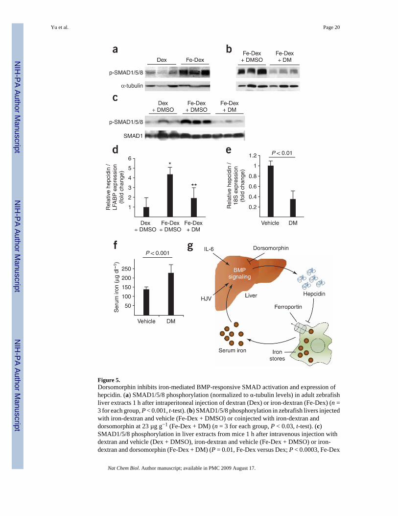
Figure 5.Dorsomorphin inhibits iron-mediated BMP-responsive SMAD activation and expression ofhepcidin. (a) SMAD1/5/8 phosphorylation (normalized to α-tubulin levels) in adult zebrafishliver extracts 1 h after intraperitoneal injection of dextran (Dex) or iron-dextran (Fe-Dex) (n =3 for each group, P < 0.001, t-test). (b) SMAD1/5/8 phosphorylation in zebrafish livers injectedwith iron-dextran and vehicle (Fe-Dex + DMSO) or coinjected with iron-dextran anddorsomorphin at 23 µg g−1 (Fe-Dex + DM) (n = 3 for each group, P < 0.03, t-test). (c)SMAD1/5/8 phosphorylation in liver extracts from mice 1 h after intravenous injection withdextran and vehicle (Dex + DMSO), iron-dextran and vehicle (Fe-Dex + DMSO) or iron-dextran and dorsomorphin (Fe-Dex + DM) (P = 0.01, Fe-Dex versus Dex; P < 0.0003, Fe-Dex
Yu et al. Page 20
Nat Chem Biol. Author manuscript; available in PMC 2009 August 17.
NIH
-PA Author Manuscript
NIH
-PA Author Manuscript
NIH
-PA Author Manuscript

+ DM versus Fe-Dex; t-test). (d) Hepatic hepcidin mRNA levels (normalized to liver fatty acidbinding protein mRNA levels) from zebrafish injected intraperitoneally with dextran andvehicle (Dex + DMSO), iron-dextran and vehicle (Fe-Dex + DMSO), or iron-dextran with 23µg g−1 dorsomorphin (Fe-Dex + DM, n = 4 for each group, *P < 0.01, **P < 0.02, ANOVA).(e) Hepatic hepcidin mRNA levels in C57BL/6 mice 6 h after a single tail vein injection ofvehicle or dorsomorphin (10 mg kg−1) (n = 6 for each group, P < 0.01, t-test). (f) Serum ironlevels in mice 24 h after the first of two intraperitoneal injections of dorsomorphin (10 mgkg−1) 12 h apart (n = 8 WT, n = 7 dorsomorphin, P < 0.001, t-test). Results in d–f are expressedas mean ± s.d. Panels are representative of two (c–e) or three (a,b,f) independent experimentseach. (g) Proposed model for the role of BMP signaling in iron homeostasis. See discussionfor details.
Yu et al. Page 21
Nat Chem Biol. Author manuscript; available in PMC 2009 August 17.
NIH
-PA Author Manuscript
NIH
-PA Author Manuscript
NIH
-PA Author Manuscript