Six First Reports of Pin Nematodes from Portugal, with ... - MDPI
29
Citation: Rosmaninho, T.; Mota, M.; Inácio, M.L.; Eisenback, J.D.; Gutiérrez-Gutiérrez, C. Six First Reports of Pin Nematodes from Portugal, with an Update of the Systematics, Genetic Diversity, and Phylogeny of the Genus Paratylenchus (Nematoda: Tylenchulidae). Horticulturae 2022, 8, 343. https://doi.org/10.3390/ horticulturae8040343 Academic Editor: Carmelo Peter Bonsignore Received: 30 March 2022 Accepted: 15 April 2022 Published: 17 April 2022 Publisher’s Note: MDPI stays neutral with regard to jurisdictional claims in published maps and institutional affil- iations. Copyright: © 2022 by the authors. Licensee MDPI, Basel, Switzerland. This article is an open access article distributed under the terms and conditions of the Creative Commons Attribution (CC BY) license (https:// creativecommons.org/licenses/by/ 4.0/). horticulturae Article Six First Reports of Pin Nematodes from Portugal, with an Update of the Systematics, Genetic Diversity, and Phylogeny of the Genus Paratylenchus (Nematoda: Tylenchulidae) Teresa Rosmaninho 1 , Manuel Mota 1 , Maria L. Inácio 2 , Jonathan D. Eisenback 3 and Carlos Gutiérrez-Gutiérrez 1, * 1 NemaLab/MED, Instituto Mediterrâneo para a Agricultura, Ambiente e Desenvolvimento, Universidade de Évora, Núcleo da Mitra, Ap. 94, 7002-554 Evora, Portugal; [email protected] (T.R.); [email protected] (M.M.) 2 Instituto Nacional de Investigação Agrária e Veterinária (INIAV), Quinta do Marquês, 2780-159 Oeiras, Portugal; [email protected] 3 School of Plant and Environmental Science, Virginia Tech, Blacksburg, VA 24060, USA; [email protected] * Correspondence: [email protected] Abstract: Pin nematodes (Paratylenchus spp.) currently comprise 132 species of polyphagous plant ectoparasites with at least seven species that are plant pathogenic emphasizing the need for correct identification to establish an appropriate management strategy. Sequences of highly conserved regions of ribosomal and mitochondrial RNA genes are a powerful species-level diagnostic tool within Tylenchulidae. A nematological survey was conducted from 2019 to 2021 in commercial vineyards distributed in four major wine-producing regions in the Central and South Portugal. Nine populations of Paratylenchus spp. were isolated from the rhizosphere of grapevines that were characterized from morphological data and molecular phylogenetic analysis using two rRNA genes (D2–D3 expansion segments of the 28S, and partial 18S) and a region partial of the COI mtRNA gene. Contrasting morphological hypotheses with molecular data provided rapid detection of six species, specifically P. goodeyi, P. hamatus, P. pedrami, P. tenicaudatus, P. variabilis, and P. veruculatus. Bayesian inference (BI) phylogenetic trees for these molecular markers established phylogenetic relationships underscore the importance of using genomic and molecular phylogenetic data for accurate pin nematode identification at the species level. To our knowledge, this is the first report of Paratylenchus spp. in Portugal, extending the geographical distribution of these species in the Mediterranean Basin, and the first record of P. goodeyi, P. pedrami, P. variabilis, P. veruculatus and P. tenicaudatus parasitizing grapevine. Keywords: Bayesian inference; D2–D3 expansion segments of large ribosomal subunit 28S; cytochrome c oxidase subunit 1; mitochondrial RNA; partial small ribosomal subunit; morphology; ribosomal RNA; Vitis spp. 1. Introduction Rhabditida Chitwood, 1933 is one of the most diverse and biologically versatile orders within the phylum Nematoda [1,2]. In fact, they are one of the largest in terms of numbers of species [1,2] including mycophagous species, parasites of invertebrate and a wide diversity of plant parasites. Members of the genus Paratylenchus Micoletzky, 1922 sensu lato, commonly known as pin nematodes, belong to the family Tylenchulidae Skarbilovich, 1947 (superfamily Criconematoidea Taylor, 1936 (Geraert, 1966); infraorder Tylenchomorpha De Ley & Blaxter, 2002; suborder Tylenchina Chitwood, 1950; order Rhabditida) [2–4]. According to morphological features and new molecular phylogenetic data [5–8], currently members belonging to Paratylenchus sensu lato are divided on two subgroups without taxonomic validity: (i) Paratylenchus species with a straight and short stylet, apparently Horticulturae 2022, 8, 343. https://doi.org/10.3390/horticulturae8040343 https://www.mdpi.com/journal/horticulturae
-
Upload
khangminh22 -
Category
Documents
-
view
4 -
download
0
Transcript of Six First Reports of Pin Nematodes from Portugal, with ... - MDPI

�����������������
Citation: Rosmaninho, T.; Mota, M.;
Inácio, M.L.; Eisenback, J.D.;
Gutiérrez-Gutiérrez, C. Six First
Reports of Pin Nematodes from
Portugal, with an Update of the
Systematics, Genetic Diversity, and
Phylogeny of the Genus Paratylenchus
(Nematoda: Tylenchulidae).
Horticulturae 2022, 8, 343.
https://doi.org/10.3390/
horticulturae8040343
Academic Editor: Carmelo
Peter Bonsignore
Received: 30 March 2022
Accepted: 15 April 2022
Published: 17 April 2022
Publisher’s Note: MDPI stays neutral
with regard to jurisdictional claims in
published maps and institutional affil-
iations.
Copyright: © 2022 by the authors.
Licensee MDPI, Basel, Switzerland.
This article is an open access article
distributed under the terms and
conditions of the Creative Commons
Attribution (CC BY) license (https://
creativecommons.org/licenses/by/
4.0/).
horticulturae
Article
Six First Reports of Pin Nematodes from Portugal, with anUpdate of the Systematics, Genetic Diversity, and Phylogeny ofthe Genus Paratylenchus (Nematoda: Tylenchulidae)Teresa Rosmaninho 1, Manuel Mota 1 , Maria L. Inácio 2 , Jonathan D. Eisenback 3
and Carlos Gutiérrez-Gutiérrez 1,*
1 NemaLab/MED, Instituto Mediterrâneo para a Agricultura, Ambiente e Desenvolvimento,Universidade de Évora, Núcleo da Mitra, Ap. 94, 7002-554 Evora, Portugal; [email protected] (T.R.);[email protected] (M.M.)
2 Instituto Nacional de Investigação Agrária e Veterinária (INIAV), Quinta do Marquês,2780-159 Oeiras, Portugal; [email protected]
3 School of Plant and Environmental Science, Virginia Tech, Blacksburg, VA 24060, USA; [email protected]* Correspondence: [email protected]
Abstract: Pin nematodes (Paratylenchus spp.) currently comprise 132 species of polyphagous plantectoparasites with at least seven species that are plant pathogenic emphasizing the need for correctidentification to establish an appropriate management strategy. Sequences of highly conservedregions of ribosomal and mitochondrial RNA genes are a powerful species-level diagnostic toolwithin Tylenchulidae. A nematological survey was conducted from 2019 to 2021 in commercialvineyards distributed in four major wine-producing regions in the Central and South Portugal.Nine populations of Paratylenchus spp. were isolated from the rhizosphere of grapevines that werecharacterized from morphological data and molecular phylogenetic analysis using two rRNA genes(D2–D3 expansion segments of the 28S, and partial 18S) and a region partial of the COI mtRNAgene. Contrasting morphological hypotheses with molecular data provided rapid detection of sixspecies, specifically P. goodeyi, P. hamatus, P. pedrami, P. tenicaudatus, P. variabilis, and P. veruculatus.Bayesian inference (BI) phylogenetic trees for these molecular markers established phylogeneticrelationships underscore the importance of using genomic and molecular phylogenetic data foraccurate pin nematode identification at the species level. To our knowledge, this is the first reportof Paratylenchus spp. in Portugal, extending the geographical distribution of these species in theMediterranean Basin, and the first record of P. goodeyi, P. pedrami, P. variabilis, P. veruculatus andP. tenicaudatus parasitizing grapevine.
Keywords: Bayesian inference; D2–D3 expansion segments of large ribosomal subunit 28S; cytochromec oxidase subunit 1; mitochondrial RNA; partial small ribosomal subunit; morphology; ribosomalRNA; Vitis spp.
1. Introduction
Rhabditida Chitwood, 1933 is one of the most diverse and biologically versatile orderswithin the phylum Nematoda [1,2]. In fact, they are one of the largest in terms of numbersof species [1,2] including mycophagous species, parasites of invertebrate and a widediversity of plant parasites. Members of the genus Paratylenchus Micoletzky, 1922 sensu lato,commonly known as pin nematodes, belong to the family Tylenchulidae Skarbilovich, 1947(superfamily Criconematoidea Taylor, 1936 (Geraert, 1966); infraorder TylenchomorphaDe Ley & Blaxter, 2002; suborder Tylenchina Chitwood, 1950; order Rhabditida) [2–4].According to morphological features and new molecular phylogenetic data [5–8], currentlymembers belonging to Paratylenchus sensu lato are divided on two subgroups withouttaxonomic validity: (i) Paratylenchus species with a straight and short stylet, apparently
Horticulturae 2022, 8, 343. https://doi.org/10.3390/horticulturae8040343 https://www.mdpi.com/journal/horticulturae

Horticulturae 2022, 8, 343 2 of 29
arrow-like form, usually less than 40 µm, composed of a conus about half of the totalstylet length (Paratylenchus sensu stricto), and (ii) Paratylenchus species with a flexible andlong stylet, often ventrally twisted, normally between 40 to 120 µm, composed of a conusoccupying more than 70% of the total stylet (Gracilacus spp.). The systematic position ofthese two groups has been discussed several times [7,9–16].
The pin nematodes of genus Paratylenchus sensu lato are a large group of small andvermiform, metazoan parasites of annual, perennial and biennial herbaceous plants, woodyshrubs and trees [4,17,18]. They are commonly found in natural and cultivated environ-ments, especially layers of soil around roots of woody plants and bushes [17]. Their life-cycle is relatively short, with an average of 30–40 days at 25–28 ◦C, but this varies consider-ably among species [17,19,20]. During this period all vermiform juvenile stages and adultspenetrate the root by moving their stylet through epidermal and root hair cell walls to feedecto-parasitically which causes diverse degrees of negative effects on the roots of their hosts,including root injury and poor plant development, consequently suppressing yield andeven plant longevity [4–6,17,19,20]. In addition, some species appear to be able to penetratethe lateral roots from the mechanical action of their stylet allowing them to enter into theintercellular spaces deep within the cortex and feeding endo-parasitically [17,21,22], whichcauses the symptoms to become more severe. Within Paratylenchus, at least seven specieswith short stylet (less than 40 µm) are plant pathogenic [17,20]. Some examples on fruitsand woody shrubs are P. hamatus Thorne & Allen, 1950 [23–33], P. neoamblycephalus Geraert,1965 [22,34] and P. nanus Cobb, 1923 [35–37], which emphasizes the need for the correctidentification of species.
Pin nematodes are an adaptable cosmopolitan group of plant-parasitic nematodescommonly found in most of soil types of temperate regions worldwide [4,17]. They showa remarkable adaptability to changing environmental conditions such as hot and coldtemperatures, and drought [5,6]. Like other nematodes, their success in unfavourableconditions is associated with their parthenogenetic reproduction where males are absentor rare, and the occurrence of a dauer juvenile stage (usually pre-adult or J4) [5,6,37].Several previous studies have reported the association of this nematode group withperennial woody plants in the Mediterranean Region [5,6,38–40]. Thus, the occurrenceand geographical distribution of pin nematodes in the Iberian Peninsula was reviewedby Peña-Santiago et al. [41] who reported a total of 22 pin nematode species that in-cluded Paratylenchus aonli Misra & Edward, 1971 (Hernández & Jordana, 1992), P. arculatusLuc & De Guiran, 1962, P. baldaccii Raski, 1975, P. ciccaronei Raski, 1975, P. curvitatusVan der Linde, 1938, P. enatus (Raski, 1976) Siddiqi, 1986, P. goodeyi Oostenbrink, 1953,P. macrodorus Brzeski, 1963, P. microdorus Andrássy, 1959, P. minusculus Tarjan, 1960, P. mirus,(Raski, 1962) Siddiqi & Goodey, 1964, P. nanus Cobb, 1923, P. neoamblycephalus Geraert,1965, P. peraticus (Raski, 1962) Siddiqi & Goodey, 1964, P. projectus Jenkins, 1956, P. sheri,(Raski, 1973) Siddiqi, 1986), P. similis Khan, Prasad & Mathur, 1967, P. steineri Golden,1961, P. straeleni (de Coninck, 1931) Oostenbrink, 1960, P. tenuicaudatus Wu, 1961, P. teres(Raski, 1976) Siddiqi, 1986, P. vandenbrandei De Grisse, 1962, and P. veruculatus Wu, 1962.Later on, Munawar et al. [42] reported the presence of several Spanish populations ofP. tateae Wu & Townsend, 1973 on Northern and Southeast of the Iberian Peninsula,Spain. Recently, Clavero-Camacho et al. [5,6] revealed a remarkable biodiversity of pinnematode species associated with cultivated and uncultivated environments in Spain. Infact, Clavero-Camacho et al. [5,6] described five new species (P. caravaquenus Clavero-Camacho, Cantalapiedra-Navarrete, Archidona-Yuste, Castillo and Palomares-Rius, 2021,P. indalus Clavero-Camacho, Cantalapiedra-Navarrete, Archidona-Yuste, Castillo andPalomares-Rius, 2021, P. pedrami Clavero-Camacho, Cantalapiedra-Navarrete, Archidona-Yuste, Castillo and Palomares-Rius, 2021, P. parastraeleni Clavero-Camacho, Cantalapiedra-Navarrete, Archidona-Yuste, Palomares-Rius and Castillo Clavero-Camacho, Cantalapiedra-Navarrete, Archidona-Yuste, Castillo and Palomares-Rius, 2021, and P. zurgenerus Clavero-Camacho, Cantalapiedra-Navarrete, Archidona-Yuste, Castillo and Palomares-Rius, 2021),and thirteen additional new reports for Spain; however, most of all these previous studies in
Horticulturae 2022, 8, 343 3 of 29
the Iberian Peninsula were with Spanish populations of pin nematodes [5,6,38,40,41], withexception of Lima [43] and Macara [44]. To our knowledge, no previous studies of detaileddescriptions included measurements and/or molecular characterizations of Portuguesepopulations of pin nematodes.
According to the morphological features and morphometric measurements of femalesand males, and that of juveniles [mainly fourth-stage juveniles (J4)], each Paratylenchusspecies was defined from a compendium of diagnostic characters used for identificationat the level of species with the key published by Ghaderi et al. [45]. However, the rela-tively small body size of these nematodes, together with their high inter- and intra-specificvariability and marked plasticity in morphology, makes species identification based onthese traits difficult and sometimes unreliable and challenging to study and identify. Se-quencing RNA-based markers is a powerful tool for identifying and discriminating pinnematode species within the family Tylenchulidae [5–7,42]. Over the last years severalstudies [5–7,20,42,46–60] have shown the usefulness of the combination of two or moremolecular markers based on ribosomal RNA (rRNA) (particularly D2–D3 domains of 28Sgene and the internal transcribed spacer 1 (ITS1) region) for an accurate and fast diagnosisof Paratylenchus species. D2–D3 domains of 28S rRNA and ITS1 have proved more effectivein species identification compared to partial 18S, as both these molecular markers displaymore species variability with respect to partial 18S. In fact, the 18S rRNA gene has beenused for species diagnosis, and phylogeny in a more restricted number of species insideParatylenchus [7,42]. Additionally, studies have revealed that the mitochondrial markergene, particularly the partial cytochrome c oxidase subunit I or COI, is useful for the delin-eation of closely related species within Tylenchulidae [5–7,49,61]. However, an integrativetaxonomic approach based on morphological and molecular phylogenetic analysis, allow-ing the linking of genomic and phenotypic data, is the best strategy to solve practical issuesinvolving species delimitation in pin nematodes [5–7]. To our knowledge, approximately60%, or more than half of the 132 valid species of this genus, have no molecular markers inthe GenBank database, and there is a need for new molecular information.
Members of the genus Paratylenchus have not been studied in detail in Portugal sincethe last to study them were Lima [43] and Macara [44]. They recorded Paratylenchus spp.in cultivated and natural environments, however none have been formally identified tospecies. Thus, updated information on the present biodiversity including molecular data,occurrence and distribution of pin nematodes in Portuguese vineyards is lacking. For thesereasons, an accurate diagnosis of pin nematode species in Portugal is essential. This studyreports on the biodiversity of Paratylenchus spp. in grapevine soil samples from four majorgrapevine-growing areas in the Central and South Portugal. A robust taxonomical approachbased on contrasting morphology with molecular analysis revealed that these Paratylenchuspopulations belong to morphospecies groups 3 and 10 defined by Ghaderi et al. [45]. Thus,the aims of the present work was (1) to characterize and illustrate several populationsof pin nematode species from four major grapevine-growing areas of Central and SouthPortugal using an integrative approach based on contrasting morphological data withmolecular analysis, (2) to update the diversity of pin nematodes recorded from Portugaland (3) to establish phylogenetic relationships of several isolates of pin nematode speciesfound in this survey with available sequences of other pin nematode species depositedin Genbank. The results of this study will add information distinguishing pin nematodespecies and reveal new knowledge on the genetic diversity and the geographic distributionof pin nematode species.
2. Materials and Methods2.1. Nematode Population Sampling
During three consecutive years (2019–2021) in Central and South Portugal, soil sampleswere collected from around the roots of grapevine (Vitis vinifera L.) in 44 commercialvineyards which are located in four of the main grapevine-growing regions (Alentejo, Tejo,Setubal and Lisbon). During this survey several populations of Paratylenchus spp. were
Horticulturae 2022, 8, 343 4 of 29
isolated from infested soil samples (Table 1). They were extracted from soil by a modifiedCobb sieving and flotation, followed with a final extraction in an Oostenbrink dish [62]and a rapid centrifugal flotation [63]. Additional soil was collected afterwards from thesame sample to guarantee enough specimens for morpho-anatomical analysis and/ormulti-locus sequencing.
Table 1. Taxa sampled for Paratylenchus species and sequences used in this study.
Species Sample Code Locality Host Genbank Accessions
18S 28S COI
P. goodeyi T1-3 Monte da Ribeira,São Manços grapevine OM345189-
OM345190
OM348556-OM348560;OM348553
OM348572-OM348573
09-02-20 Carvalhal, Bombarral grapevine - OM348543 -
P. hamatus 45-007-20 Roliça, Bombarral grapevine OM345185 OM348545-OM348547
OM348567-OM348568
P. pedramiAL-V4 Santa Catarina de Sítimos,
Alcácer do Sal grapevine OM345191-OM345192
OM348562-OM348566-
OM348574-OM348577
CF-1 Aldeia Galega daMerceana, Alenquer grapevine - OM348550-
OM348551 OM348570
P. tenuicaudatus 198-33-19 Carvalhal, Bombarral grapevine OM345184 OM348544 -
P. variabilis 197-32-19 São Domingos de Carmões,Torres Vedras grapevine OM345186 OM348548-
OM348549 OM348569
P. veruculatus T1-3 Monte da Ribeira,São Manços grapevine OM345187-
OM345188
OM348552;OM348554-OM348555;OM348561
OM348571
(-) Not obtained or not performed.
2.2. Morphological and Morphometrical Study
After extracting nematodes from soil, fresh nematodes were examined under a stereomicroscope (Olympus SZX112) and immediately picked into an embryo glass dish andstored in sterile, distilled water at 4 ◦C until further processing. For light microscopy(LM) studies, live nematodes were placed in a drop of distilled water, gently heat killed,and fixed for 48–72 h at room temperature (25 ◦C) in a fixative solution composed of 4%formaldehyde, 1% glycerol and 85% distilled water. After nematodes were fixed, theywere processed into pure glycerine using a modification of the Seinhorst method [64] andtransferred to a small drop of glycerol on a glass slide ready for mounting of permanentslides. Light micrographs of nematode specimens mounted permanently on slides wereacquired using a light microscope (Olympus BX50, Hamburg, Germany) with differentialinterference contrast (DIC) up to 1000× magnification. Phenotypic image analysis andmeasurements were done using an Olympus DP70 camera Cell® software (Olympus Corp.,Tokyo, Japan). All measurements were expressed in micrometers (µm). All abbreviationsused are defined by Siddiqi [4]. According to metric (e.g., de Man body ratios, lip regionand width, stylet length, lip maximum body width, vulva position, pharyngeal length,and tail length and diameter) and non-metric (e.g., lip region shape, number of laterallines, presence or absence of males, shape of spermatheca, presence of sperm, presenceor absence of advulval flaps, tail shape, vulva size and shape, and tail terminus shape)morphological data of adult specimens and juveniles (mainly J4) (when available), all pinnematode species found in this survey were well defined phenotypically by a compendiumof diagnostic characters given by Ghaderi et al. [45]. LM studies were carried out at theNematology Lab, University of Évora, Portugal. For scanning electron microscopy (SEM)studies, fixed specimens were dehydrated in a graded ethanol series, immersed in HMDS(hexamethyldisilazane 98%), mounted on SEM stubs, sputter-coated with a thin layer of
Horticulturae 2022, 8, 343 5 of 29
gold [65], and observed with a Hitachi S3700N (Tokyo, Japan) SEM coupled to a Bruker(Karlsruhe, Germany) XFlash 5010 SDD Detector system [66]. The SEM coupled withenergy-dispersive X-ray spectrometry (SEM-EDS) experiments were conducted on highvacuum mode with acceleration voltage of 5–10 kV. SEM studies were carried out at theHercules Lab, University of Évora, Portugal.
2.3. DNA Extraction
Genomic DNA was prepared according to Gutiérrez-Gutiérrez et al. [67]. DNA wasextracted from single individual live adult nematodes (females and males when available)and even juvenile stages (when available), which were previously examined by LM ontemporary glass slide mounts for taking photomicrographs and measurements to recordtheir phenotypic data that matched their associated genotype. For DNA extraction, individ-ual nematodes were placed in a 1.5 µL drop of sterile water on the microscope glass slide,and each specimen was chopped into three small pieces with a surface-sterilized, pointedneedle. Subsequently, all individual nematode bits were transferred to a PCR tube with20 µL of solution containing 12 µL ddH2O, 6 µL 10× PCR buffer, and 2 µL of proteinase K(20 mg/mL) (Nalgene®). Tubes were centrifuged at 11,000 rpm for 1 min and later frozenat −80 ◦C (45 min). Samples were mixed for 10 s, centrifuged at 11,000 rpm for 30 s, andincubated at 57 ◦C (2 h) and 95 ◦C (15 min).
2.4. PCR Amplification, DNA Purification and Sequencing
Genomic DNA from a single individual specimen was used to amplify two rDNAfragments: D2–D3 of 28S, and partial 18S rRNA gene and the partial fragment of COImtRNA gene. The PCR was performed in a final volume of 50 µL containing: 1 µL of DNAtemplate, 25 µL NZYTaq 2× Green Master Mix (2.5 mM MgCl2, 200 mM dNTPs, 0.2 U/µLDNA Polymerase) (NZYTech, Lisbon, Portugal), 1.25 µL of each primer (10 mM), and22.75 µL of ddH2O. Each rDNA and mtDNA fragment was amplified using several primerpairs (Table S1). PCR assays were conducted as described by Clavero-Camacho et al. [5,6]and Singh et al. [7]. PCR cycle conditions included one cycle of 95 ◦C for 3 min; followedby 30 cycles of 94 ◦C for 30 s; an annealing temperature of 51 ◦C (391F/D3B), and 50 ◦C(988F/1912R, 1813F/2646R, J3/J4.5, COIF/COIF) for 30 s, 72 ◦C for 15–45 s; and one cycle of72 ◦C for 7 min. The PCR products were purified [67] and were used as template for directsequencing on a DNA multicapillary sequencer (ABI 3730xl DNA Sequencer; AppliedBiosystems, Foster City, CA, USA), using a BigDye Terminator V3.1 Cycle Sequencing Kit atthe STABVIDA facilities (Caparica, Portugal). Additional D2A primer (Table S1) was usedfor guaranteeing a robust and complete sequence. All new sequences of the pin nematodespecies found were deposited in the GenBank under the accession numbers indicated onthe Table 1.
2.5. Phylogenetic Analyses
The newly obtained D2–D3 expansion segments of 28S rRNA, partial 18S rRNA andpartial COI mtRNA sequences from all known pin nematode species found in this survey(Table 1), together with the available sequences of other pin nematode species obtained fromthe National Center for Biotechnology Information (NCBI) were used for phylogenetic anal-yses. Outgroup taxa for each gene studied was chosen according to previously publisheddata by Clavero-Camacho et al. [5,6], Singh et al. [7] and Subbotin et al. [49]. The sequenceswere aligned using an online version of MAFFT v. 7 [68] with default parameters. Sequencealignments were visualized with ClustalX2 [69] and edited by Gblocks v. 0.91b [70] withless stringent selection Gblocks parameters (www.phylogeny.fr, accessed on 2 December2021). Homogeneities of base frequencies and optional substitution models for 28S rRNA,18S rRNA, and COI datasets were tested with Kakusan4 [71]. The homogeneity test indi-cated that the base composition of each dataset was significantly homogeneous. Bayesianinference (BI) trees of all molecular data were constructed with MrBayes v. 3.2.1 [72]. ForBI analysis, the substitution model was tested by the Bayesian Information Criterion and
Horticulturae 2022, 8, 343 6 of 29
two models were selected for our three molecular regions studied. GTR model with agamma-shaped distribution was selected for D2–D3 regions of 28S rRNA and COI, whilethe K80 model with a gamma-shaped distribution was selected for the partial 18S rRNA.Convergence of the MCMC chain and appropriate burn-in were assessed with Tracer1.7.1 [73]. BI analysis was run for 5,000,000 generations, sampling every 100th tree anddiscarding ‘burn-in’for the first 25% of the sampled tree. The resulting trees were visualizedand edited using FigTree v1.4.3 [74].
3. Results
From the 44 samples analysed, Paratylenchus nematodes were detected in 7 (15.9%),showing a moderate incidence in Central and South Portugal. A total of six species wereidentified from eight selected isolates of Paratylenchus spp. from seven soil samples insix localities in Portugal (Table 1). The population densities of Paratylenchus spp. rangedbetween 10 to 250 nematodes/500 cm3 of soil. All specimens of pin nematodes (includingmales, females and juveniles when available) were examined to get a fast first preliminarytaxonomic check. Subsequently, adult specimens found within each population studiedwere morphometrical and morphologically examined in detail checking their complexity.Also, these phenotypic data were contrasted with molecular data. Molecular data wereuseful to resolve the taxonomic identity of the species complex of pin nematode speciesfound in this study. Juveniles were used only for molecular analaysis since few specimenswere available. For each population studied, we provided a set of metrical and non-metrical morphological data of adult specimens (females and males when available) aswell as mitochondrial and ribosomal molecular markers for their identification (Table 1).Morphological analysis was followed by barcoding sequences and molecular phylogeneticdata allowed the discernment a total of six known pin nematode species, some of themsharing samples, namely P. goodeyi, P. hamatus, P. pedrami, P. tenicaudatus, P. variabilis Raski,1975, and P. veruculatus. All these pin nematode species must to be considered as firstreports for Portugal and measurements from adults (females and males when available), aswell as molecular phylogenetic analysis using rDNA and mtDNA molecular markers wereprovided for their phylogenetic studies and unequivocal diagnosis at the species level.
3.1. Systematics3.1.1. Morphological Features and Morphometric Measurements
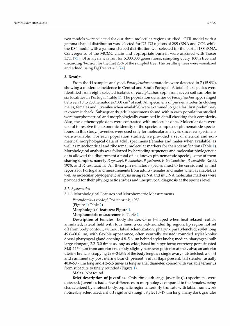
Paratylenchus goodeyi Oostenbrink, 1953(Figure 1; Table 2)Morphological features: Figure 1.Morphometric measurements: Table 2.Description of females. Body slender, C- or J-shaped when heat relaxed; cuticle
annulated; lateral field with four lines; a conoid-rounded lip region, lip region not setoff from body contour, without labial sclerotization; pharynx paratylenchid; stylet long49.6–60.6 µm, with flexible appearance, often ventrally twisted; rounded stylet knobs;dorsal pharyngeal gland opening 4.8–5.6 µm behind stylet knobs; median pharyngeal bulblarge elongate, 2.2–3.0 times as long as wide; basal bulb pyriform; excretory pore situated84.0–115.0 µm from anterior end; body slightly narrower posterior at the vulva; an anterioruterine branch occupying 29.6–34.8% of the body length; a single ovary outstretched; a shortand rudimentary post uterine branch present; vulval flaps present; tail slender, usually40.0–60.7 µm long and 4.2–5.5 times as long as anal diameter, conoid with variable terminusfrom subacute to finely rounded (Figure 1).
Males. Not found.Brief description of juveniles. Only three 4th stage juvenile (J4) specimens were
detected. Juveniles had a few differences in morphology compared to the females, beingcharacterized by a robust body, cephalic region anteriorly truncate with labial frameworknoticeably sclerotized, a short rigid and straight stylet 15–17 µm long; many dark granules
Horticulturae 2022, 8, 343 7 of 29
into the body; anus difficult to distinguish; and a posterior body similar to female with arounded tail terminus (Figure 1).
Horticulturae 2022, 8, x FOR PEER REVIEW 8 of 31
Figure 1. Light micrographs of Paratylenchus goodeyi Oostenbrink, 1953 (A–I). (A–D): Female phar-yngeal region; (E): Entire female with vulva (arrowhead); (F): Detail of vulva (arrowhead); (G): Fe-male posterior region showing vulva (arrowhead); (H): Female posterior region showing advulval flap (arrowhead); (I): Fourth-stage juvenile anterior region. Scale bars ((A) = 38 μm, (B–D) = 29 μm, (E) = 58 μm, (F–H) = 15 μm, (I) = 16 μm). (Abbreviations: avf = advulval flap; V = vulva).
Males. Not found. Brief description of juveniles. Only three 4th stage juvenile (J4) specimens were de-
tected. Juveniles had a few differences in morphology compared to the females, being characterized by a robust body, cephalic region anteriorly truncate with labial framework noticeably sclerotized, a short rigid and straight stylet 15–17 μm long; many dark granules
Figure 1. Light micrographs of Paratylenchus goodeyi Oostenbrink, 1953 (A–I). (A–D): Female pharyn-geal region; (E): Entire female with vulva (arrowhead); (F): Detail of vulva (arrowhead); (G): Femaleposterior region showing vulva (arrowhead); (H): Female posterior region showing advulval flap(arrowhead); (I): Fourth-stage juvenile anterior region. Scale bars ((A) = 38 µm, (B–D) = 29 µm,(E) = 58 µm, (F–H) = 15 µm, (I) = 16 µm). (Abbreviations: avf = advulval flap; V = vulva).
Horticulturae 2022, 8, 343 8 of 29
Table 2. Morphometrics of Paratylenchus goodeyi Oostenbrink, 1953 and P. tenuicaudatus Wu, 1961from the rhizosphere of grapevines (Vitis vinifera L.) in vineyards in Portugal. All measurements inµm and in the format: mean ± s.d. (range) *.
Measurements and Ratios P. goodeyi P. tenuicaudatus
Sample Code T1-3 09-02-20 198-33-19
Locality Monte da Ribeira,São Manços Carvalhal, Bombarral Carvalhal, Bombarral
n 5 females 1 female 5 females 1 male
L 449.2 ± 33.5(399.5–481.5) 414.5 296.8 ± 20.7
(276.7–331.3) 354.4
a 31.1 ± 1.8 (28.4–32.7) 28.1 24.4 ± 1.1 (22.8–25.5) 26.4b 3.6 ± 0.2 (3.5–3.9) 3.4 4.1 ± 0.3 (3.7–4.4) 4.6c 8.7 ± 1.3 (7.3–10.0) 9.1 11.1 ± 1.9 (8.4–13.3) 13.1c’ 5.0 ± 0.5 (4.5–5.5) 4.2 3.0 ± 0.3 (2.6–3.5) 2.7V or T 79.8 ± 1.2 (78.2–81.0) 80.8 81.2 ± 1.2 (79.2–81.9) 63.6G1(%) 31.9 ± 2.2 (29.6–34.8) 41.7 22.8 ± 2.8 (20.9–26.0) -Stylet length 57.9 ± 2.2 (54.8–60.6) 49.6 30.2 ± 1.1 (29.0–31.6) -Cone length 47.7 ± 1.7 (46.5–48.9) 39.7 25.5 ± 0.3 (25.3–25.7) -(Stylet length/body length) × 100 13.0 ± 0.6 (12.3–14.0) 12.0 10.2 ± 0.5 (9.5–10.8) -m 84.6 ± 0.3 (84.4–84.9) 80.1 81.6 ± 2.1 (80.1–83.0) -DGO 5.2 ± 0.5 (4.8–5.6) 5.5 5.1 ± 0.6 (4.7–5.6) -O 9.2 ± 0.6 (8.8–9.6) 11.0 16.5 ± 2.3 (14.9–18.1) -Lip width 2.9 ± 0.5 (2.4–3.5) 2.7 3.3 ± 0.7 (2.7–4.2) 2.9Median bulb length 22.8 ± 1.6 (20.7–25.0) 35.5 14.3 ± 1.6 (12.0–16.2) 16.5Median bulb width 8.5 ± 0.5 (7.8–9.3) 9.2 5.9 ± 0.9 (5.0–7.3) 4.7Anterior end to center median bulb 76.0 ± 4.2 (70.5–81.0) 69.7 41.1 ± 2.1 (37.6–43.1) 40.9MB 61.8 ± 1.7 (59.9–63.6) 57.3 56.2 ± 4.1 (49.5–59.5) 49.1Excretory pore to anterior end 93.9 ± 12.2 (84.0–115.0) 86.1 63.3 ± 3.7 (58.0–68.4) 75.5
Pharynx length 123.0 ± 4.7(115.0–127.3) 121.6 73.2 ± 2.7 (70.1–76.0) 83.3
Maximum body diam. 14.4 ± 0.5 (13.8–14.9) 14.7 12.2 ± 1.3 (11.5–14.5) 13.4Tail length 52.4 ± 8.5 (40.0–60.7) 45.5 27.2 ± 4.3 (22.4–33.0) 27.1Anal body diam. 10.5 ± 1.0 (9.0–11.4) 10.8 9.2 ± 1.1 (7.7–10.6) 10.2Spicules - - - 19Gubernaculum - - - 6.5
* Abbreviations are defined in Siddiqi (2000). (-) Not obtained or not performed.
Remarks. According to Ghaderi et al. [45], our two populations of this species belongto group 10 (stylet length more than 40 µm, four lateral lines, and advulval flaps present).This pin nematode species was found in two soil samples of grapevine (Vitis vinifera L.)from commercial vineyards with unknown rootstock Monte da Ribeira locality, São Manços(Évora district) and Carvalhal locality, Bombarral (Leiria district) in Southern and CentralPortugal, respectively (Table 1). These populations presented low nematode densities inboth soil samples (35 to 45 individuals/500 cm3 of soil). The morphological and morpho-metrical traits closely agree with the original description of this species [75]. It matcheswell with other populations on the Iberian populations described by Castillo et al. [76] andClavero-Camacho et al. [5,6], with the exception of some minor differences (Table 2). Inspite of this species has been reported by a large list of authors [7,10,75–84], only someof them included morphometric data [5–7,10,75,76,79,80]. Apparently, our populationsof P. goodeyi are almost indistinguishable from several Paratylenchus species belonging togroup 10 after Ghaderi et al. [45] including P. ivorensis Luc & de Guiran, 1962, P. pandatus(Raski, 1976) Siddiqi, 1989 and P. straeleni [5–7,45], except for some subtle differences inmorphological traits and measurements and DNA molecular markers (Tables 1 and 2). Thisspecies was originally described parasitizing grass roots in Netherlands [75,81] and waslater found mainly associated to natural environments and fruits in Europe [5–7,10,77–81]and Asia [82–84]. To our knowledge this is the first record of this species parasitizing
Horticulturae 2022, 8, 343 9 of 29
grapevine. Furthermore, these findings represent the first detection of this species inPortugal and also confirm that this species is widely distributed in Iberian Peninsula.
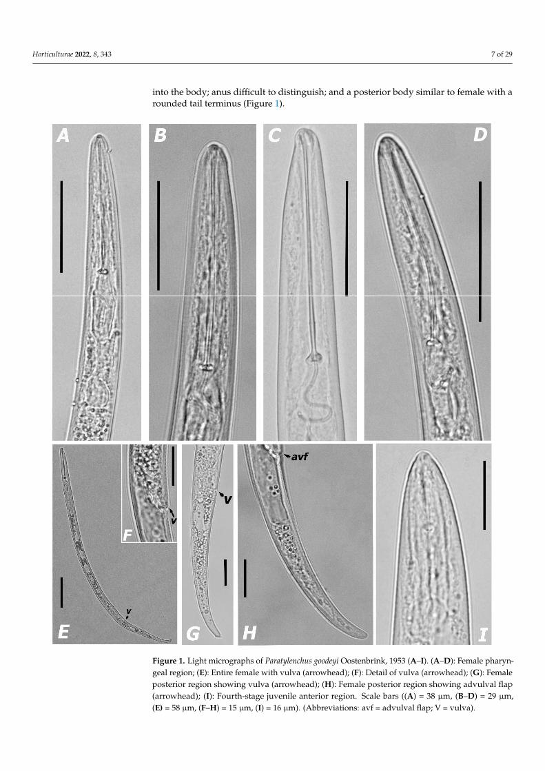
Paratylenchus hamatus Thorne & Allen, 1950.(Figure 2; Table 3)Morphological features: Figure 2.Morphometric measurements: Table 3.Description of females. Body slender, slightly ventrally curved, usually J-shaped
when heat relaxed; cuticle thin and finely annulated; lateral field with four incisures;lip region not set off from body contour, with the presence of small submedian lobes;framework weakly sclerotized; pharynx paratylenchid; stylet moderately short, usually28.8–31.9 µm long, showing rigidity and straightness; rounded stylet knobs; dorsal pha-ryngeal gland opening 5.9–7.3 µm behind stylet knobs; median pharyngeal bulb largeelongate, 2.5–3.2 times as long as wide; basal bulb pyriform; excretory pore situated slightlyanterior to or at level of anterior part of pharyngeal basal bulb, usually 52.0–70.4 µm fromanterior end; body slightly narrower posterior to the vulva; an anterior uterine branchoccupying 34.9–36.4% of the body length; a single ovary outstretched, well developed; asmall and rudimentary post uterine branch; advulval flaps present, but not prominent;tail slender, 27.7–35.9 µm long, curved ventrally, gradually tapering to form a subacute tofinely rounded terminus (Figure 2).
Horticulturae 2022, 8, x FOR PEER REVIEW 11 of 31
Figure 2. Light micrographs of Paratylenchus hamatus Thorne & Allen, 1950 (A–J). (A–D): Female pharyngeal region; (E): Female posterior region showing vulva (arrowhead); (F): Detail of advulval flap (arrowhead); (G): Female posterior region showing vulva and anus (arrowhead); (H): Female posterior region showing advulval flap (arrowhead); (I,J): Male posterior region showing spicules (arrowhead). Scale bars ((A–D) = 21 μm, (E) = 28 μm, (F) = 7 μm, (G,H) = 14 μm, (I,J) = 9 μm). (Abbreviations: a = anus; avf = advulval flap; sp = spicule; V = vulva).
Remarks. According to measurements and morphological data of female and male adults, our populations of Paratylenchus species belong to group 3 as defined by Ghaderi et al. [45] and characterized by stylet length less than 40 μm, four lateral lines and advulval flaps present. This pin nematode species was found in one soil samples of grapevine (Vitis vinifera L.) in a commercial vineyard with unknown rootstock in Roliça locality, Bombar-ral(Leiria district, Central Portugal) (Table 1). This Portuguese population presented low nematode densities in the soil (25 individuals/500 cm3 of soil). The morphological and morphometrical traits closely agree with the original description (paratype specimens) of the species P. hamatus [32] and other subsequent records worldwide [6,45,60], no crucial metrical differences at intraspecific level (Table 3). Apparently, our population of this spe-cies is almost indistinguishable from several species (P. baldaccii, P. tenuicaudatus and oth-ers) belonging to the P. hamatus “species complex” [6,60], except for some subtle morpho-anatomical differences and DNA molecular markers (Tables 1 and 3). This species was originally described parasitizing fig (Ficus carica L.) roots in Planada, CA, USA [32] and was later found in the rhizosphere from a large list of fruits, vegetables and ornamental plants worldwide [6,45,60]. These findings represent the first record of this pin nematode species for Portugal.
Paratylenchus pedrami Clavero-Camacho, Cantalapiedra-Navarrete, Archidona-Yuste, Castillo and Palomares-Rius, 2021.
(Figure 3; Table 4) Morphological features: Figure 3. Morphometric measurements: Table 4.
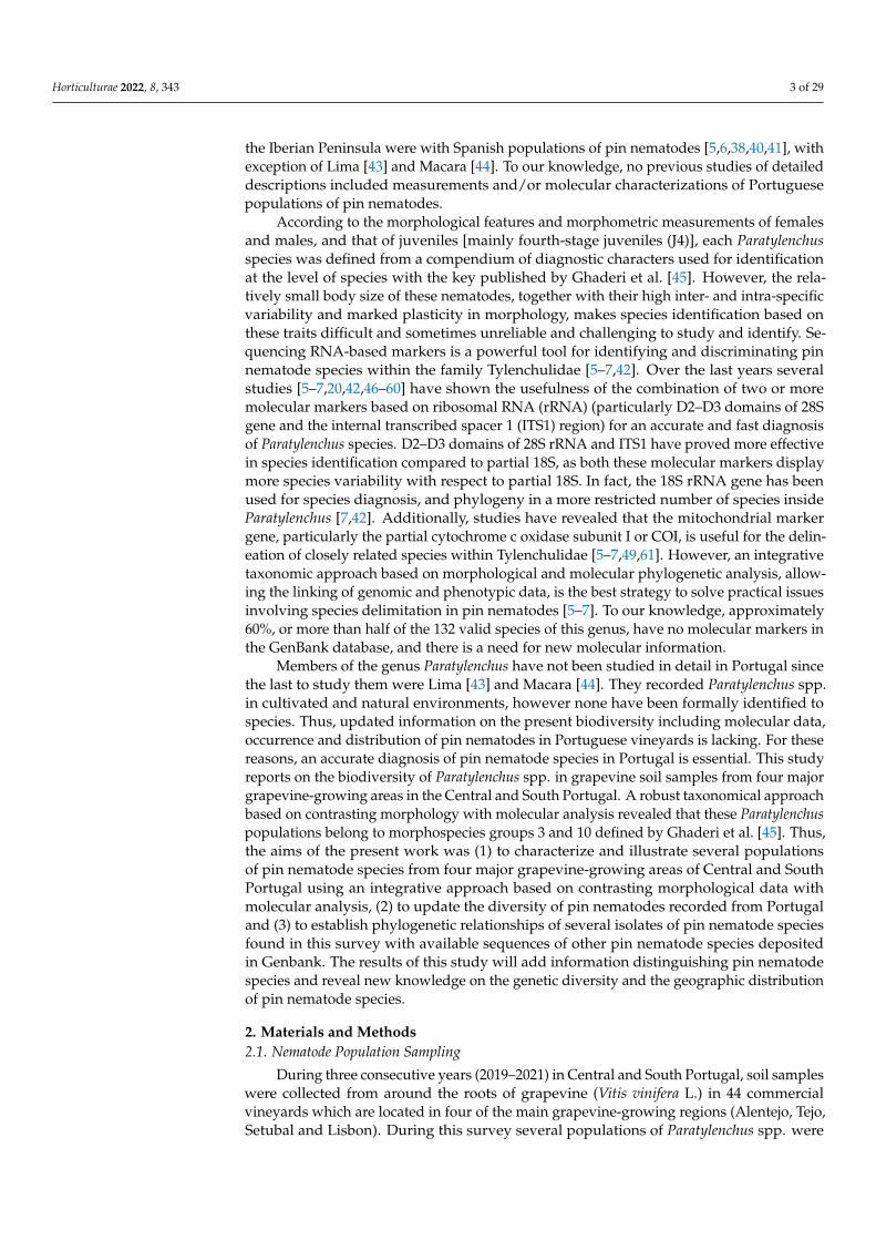
Figure 2. Light micrographs of Paratylenchus hamatus Thorne & Allen, 1950 (A–J). (A–D): Femalepharyngeal region; (E): Female posterior region showing vulva (arrowhead); (F): Detail of advulvalflap (arrowhead); (G): Female posterior region showing vulva and anus (arrowhead); (H): Femaleposterior region showing advulval flap (arrowhead); (I,J): Male posterior region showing spicules(arrowhead). Scale bars ((A–D) = 21 µm, (E) = 28 µm, (F) = 7 µm, (G,H) = 14 µm, (I,J) = 9 µm).(Abbreviations: a = anus; avf = advulval flap; sp = spicule; V = vulva).
Description of males. Males are as abundant as females. Appearance of body similarto female, except for reproductive organs, and the development of stylet and tail shape.Stylet 16.0–20.7 µm long, smaller than in the female, with small stylet knobs. Excretory porelocated 68.2 µm from anterior end. Testis outstretched, with small spermatozoa; spiculeslender, roughly 21.8 µm, slightly curved towards end; gubernaculum slightly curved;bursa absent; almost straight to ventrally slightly curved tapering slightly and gradually toa narrow, rounded tip (Figure 2).
Juveniles. Not found.

Horticulturae 2022, 8, 343 10 of 29
Table 3. Morphometrics of Paratylenchus hamatus Thorne & Allen, 1950, P. variabilis Raski, 1975 and P.veruculatus Wu, 1962 from the rhizosphere of grapevines (Vitis vinifera L.) in a vineyard in Portugal.All measurements in µm and in the format: mean ± s.d. (range) *.
Measurements andRatios P. hamatus P. variabilis P. veruculatus
Sample Code 045-007-20 197-32-19 T1-3
Locality Roliça, Bombarral São Domingos deCarmões, Torres Vedras
Monte da Ribeira,São Manços
n 4 females 5 males 13 females 2 females
L 320.1 ± 16.4(298.3–337.8)
442.9 ± 19.3(321.9–362.7)
296.1 ± 23.2(247.7–336.2)
306.8 ± 22.0(291.3–322.4)
a 22.8 ± 1.9 (20.4–25.0) 27.1 ± 2.1 (24.7–29.6) 22.8 ± 1.4 (20.5–25.0) 21.9 ± 3.0 (19.8–24.0)b 4.1 ± 0.1 (4.0–4.1) 4.6 ± 0.3 (4.1–4.8) 4.2 ± 0.4 (3.8–4.9) 3.8 ± 0.3 (3.6–4.0)c 10.6 ± 0.9 (9.4–11.6) 13.3 ± 1.4 (11.7–14.8) 12.9 ± 1.6 (9.1–15.0) 11.7 ± 0.6 (11.3–12.1)c’ 3.2 ± 0.4 (2.8–3.6) 3.1 ± 0.4 (2.5–3.5) 2.6 ± 0.4 (2.0–3.4) 2.7 ± 0.4 (2.4–2.9)V or T 81.8 ± 1.7 (80.6–84.1) 68.2 ± 1.5 (66.4–69.7) 83.9 ± 1.0 (82.4–85.4) 83.8 ± 1.1 (83.0–84.6)G1(%) 35.7 ± 0.7 (34.9–36.4) - 30.1 ± 6.4 (18.0–38.4) 19.8 ± 0.3 (19.5–20.0)Stylet length 30.6 ± 1.3 (28.8–31.9) 19.0 ± 1.9 (16.0–20.7) 17.6 ± 0.8 (16.3–19.0) 14.0 ± 1.3 (13.1–14.9)Cone length 19.5 ± 1.9 (18.1–20.9) 14.3 ± 0.5 (14.0–14.7) 13.0 ± 1.0 (12.0–14.0) 8.3 ± 1.1 (7.5–9.0)(Stylet length/bodylength) × 100 9.6 ± 0.4 (9.0–10.0) 5.6 ± 0.7 (4.4–6.1) 6.0 ± 0.7 (5.1–7.7) 5.2 ± 0.6 (4.8–5.6)
m 65.7 ± 4.0 (62.8–68.5) 73.6 ± 3.8 (70.9–76.3) 72.1 ± 8.1 (63.2–80.5) 68.8 ± 8.8 (62.5–75.0)DGO 6.6 ± 1.0 (5.9–7.3) - 5.4 ± 0.2 (5.2–5.6) -O 22.3 ± 4.2 (19.4–25.3) - 29.9 ± 2.2 (27.8–32.4) -Lip width 3.4 ± 0.4 (3.0–3.9) 3.1 ± 0.2 (2.9–3.4) 3.5 ± 0.5 (2.7–4.3) 3.8 ± 0.4 (3.5–4.1)Median bulb length 19.8 ± 2.7 (16.9–22.4) 17.4 ± 3.1 (15.2–22.0) 17.1 ± 1.5 (15.2–20.5) 20.3 ± 1.4 (19.3–21.3)Median bulb width 6.8 ± 0.6 (5.8–7.2) 4.6 ± 0.6 (4.0–5.2) 7.4 ± 1.0 (5.7–9.9) 7.4 ± 0.1 (7.4–7.3)Anterior end to centermedian bulb 41.5 ± 2.8 (37.5–44.0) 39.9 ± 0.2 (38.8–40.0) 36.4 ± 2.2 (32.1–40.5) 41.9 ± 0.3 (51.7–42.2)
MB 53.8 ± 3.3 (50.5–56.8) 51.5 ± 1.6 (50.3–52.6) 51.9 ± 1.8 (49.1–54.7) 51.6 ± 0.3 (51.4–51.9)Excretory pore toanterior end 69.1 ± 3.5 (66.7–74.1) 68.2 ± 2.0 (65.3–70.0) 64.4 ± 3.1 (57.4–68.4) 73.1 ± 0.3 (72.9–73.3)
Pharynx length 77.1 ± 2.7 (74.3–80.9) 75.1 ± 2.9 (71.9–79.0) 70.2 ± 4.2 (62.1–75.0) 81.2 ± 0.1 (81.1–81.3)Maximum body diam. 14.1 ± 0.5 (13.5–14.7) 12.8 ± 1.6 (10.9–14.6) 13.0 ± 1.0 (11.9–15.4) 14.1 ± 0.9 (13.4–14.7)Tail length 30.4 ± 3.7 (27.7–35.9) 26.0 ± 2.9 (21.8–28.2) 23.3 ± 3.3 (16.5–31.7) 26.3 ± 3.1 (24.2–28.5)Anal body diam. 9.6 ± 1.0 (8.1–10.4) 8.5 ± 0.7 (8.1–9.7) 9.1 ± 0.9 (6.9–1.3) 9.8 ± 0.1 (9.8–9.9)Spicules - 21.8 ± 1.1 (20.1–22.6) - -Gubernaculum - 11.7 ± 2.0 (9.0–13.9) - -
* Abbreviations are defined in Siddiqi (2000). (-) Not obtained or not performed.
Remarks. According to measurements and morphological data of female and male adults,our populations of Paratylenchus species belong to group 3 as defined by Ghaderi et al. [45]and characterized by stylet length less than 40 µm, four lateral lines and advulval flapspresent. This pin nematode species was found in one soil samples of grapevine (Vitisvinifera L.) in a commercial vineyard with unknown rootstock in Roliça locality, Bombar-ral(Leiria district, Central Portugal) (Table 1). This Portuguese population presented lownematode densities in the soil (25 individuals/500 cm3 of soil). The morphological andmorphometrical traits closely agree with the original description (paratype specimens)of the species P. hamatus [32] and other subsequent records worldwide [6,45,60], no cru-cial metrical differences at intraspecific level (Table 3). Apparently, our population ofthis species is almost indistinguishable from several species (P. baldaccii, P. tenuicaudatusand others) belonging to the P. hamatus “species complex” [6,60], except for some subtlemorpho-anatomical differences and DNA molecular markers (Tables 1 and 3). This specieswas originally described parasitizing fig (Ficus carica L.) roots in Planada, CA, USA [32] andwas later found in the rhizosphere from a large list of fruits, vegetables and ornamentalplants worldwide [6,45,60]. These findings represent the first record of this pin nematodespecies for Portugal.
Horticulturae 2022, 8, 343 11 of 29
Paratylenchus pedrami Clavero-Camacho, Cantalapiedra-Navarrete, Archidona-Yuste,Castillo and Palomares-Rius, 2021.
(Figure 3; Table 4)Horticulturae 2022, 8, x FOR PEER REVIEW 12 of 31
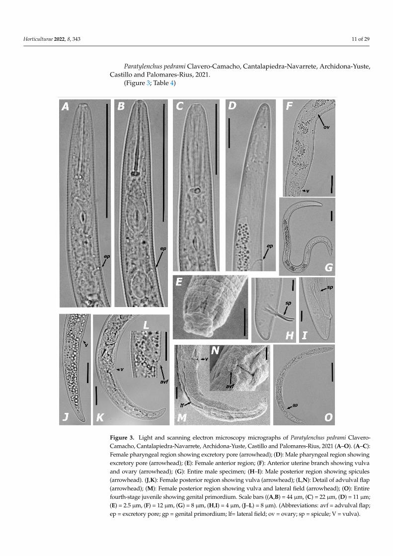
Figure 3. Light and scanning electron microscopy micrographs of Paratylenchus pedrami Clavero-Camacho, Cantalapiedra-Navarrete, Archidona-Yuste, Castillo and Palomares-Rius, 2021 (A–O). (A–C): Female pharyngeal region showing excretory pore (arrowhead); (D): Male pharyngeal region showing excretory pore (arrowhead); (E): Female anterior region; (F): Anterior uterine branch show-ing vulva and ovary (arrowhead); (G): Entire male specimen; (H–I): Male posterior region showing spicules (arrowhead). (J,K): Female posterior region showing vulva (arrowhead); (L,N): Detail of
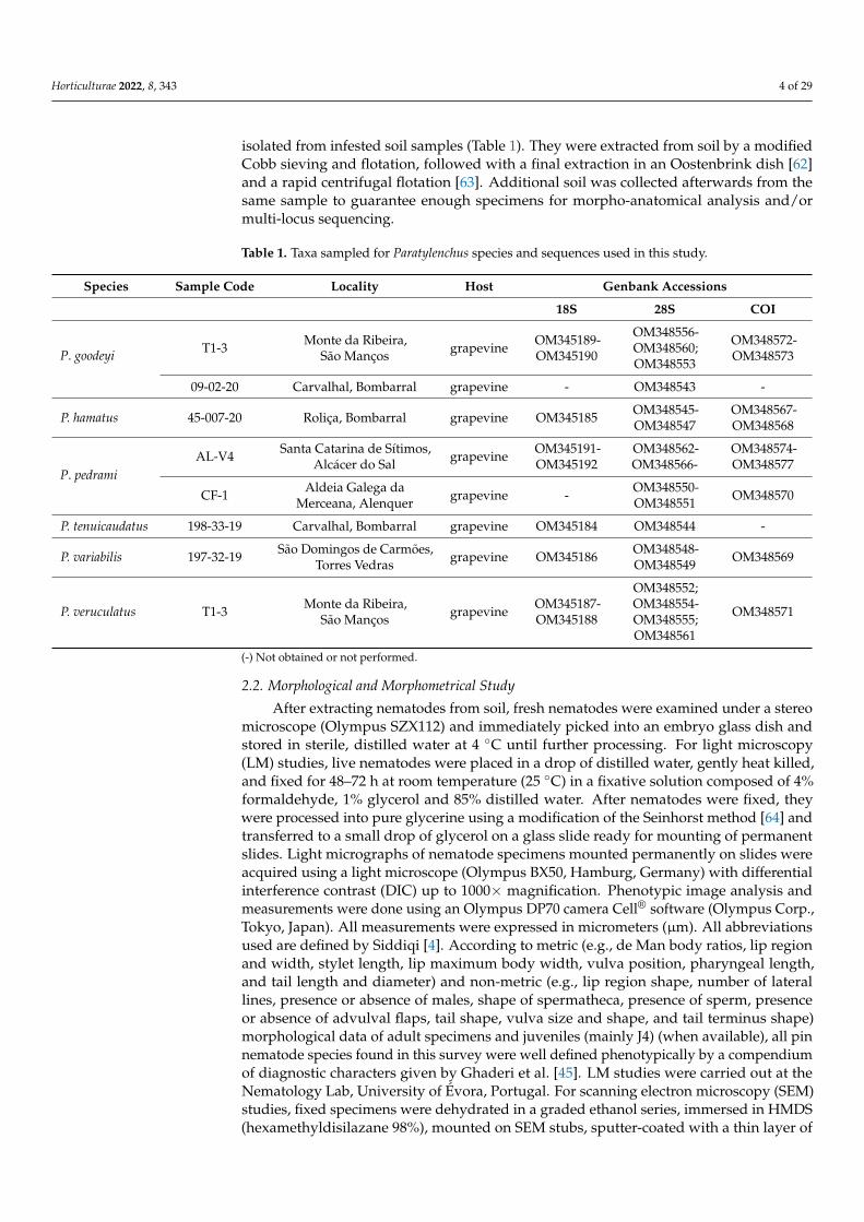
Figure 3. Light and scanning electron microscopy micrographs of Paratylenchus pedrami Clavero-Camacho, Cantalapiedra-Navarrete, Archidona-Yuste, Castillo and Palomares-Rius, 2021 (A–O). (A–C):Female pharyngeal region showing excretory pore (arrowhead); (D): Male pharyngeal region showingexcretory pore (arrowhead); (E): Female anterior region; (F): Anterior uterine branch showing vulvaand ovary (arrowhead); (G): Entire male specimen; (H–I): Male posterior region showing spicules(arrowhead). (J,K): Female posterior region showing vulva (arrowhead); (L,N): Detail of advulval flap(arrowhead); (M): Female posterior region showing vulva and lateral field (arrowhead); (O): Entirefourth-stage juvenile showing genital primordium. Scale bars ((A,B) = 44 µm, (C) = 22 µm, (D) = 11 µm;(E) = 2.5 µm, (F) = 12 µm, (G) = 8 µm, (H,I) = 4 µm, (J–L) = 8 µm). (Abbreviations: avf = advulval flap;ep = excretory pore; gp = genital primordium; lf= lateral field; ov = ovary; sp = spicule; V = vulva).

Horticulturae 2022, 8, 343 12 of 29
Table 4. Morphometrics of Paratylenchus pedrami Clavero-Camacho, Cantalapiedra-Navarrete,Archidona-Yuste, Castillo and Palomares-Rius, 2021 from the rhizosphere of grapevine (Vitis viniferaL.) in vineyards in Portugal. All measurements in µm and in the format: mean ± s.d. (range) *.
Character/Sample Code AL-V4 CF-1
Locality Santa Catarina de Sítimos, Alcácer do Sal Aldeia Galega da Merceanan 20 females 3 males 12 femalesL 298.5 ± 22.8 (239.3–337.4) 255.2 ± 32.2 (227.3–290.5) 294.3 ± 16.9 (270.0–330.6)a 24.4 2.7 (19.7–28.9) 23.2 ± 4.3 (20.7–28.1) 24.4 ± 1.9 (20.4–27.1)b 4.0 ± 0.3 (3.6–4.7) 3.8 ± 0.1 (3.8–3.9) 4.2 ± 0.5 (3.2–5.2)c 13.0 ± 2.4 (9.7–17.7) 24.1 ± 4.5 (19.4–28.3) 14.3 ± 1.5 (11.2–16.4)c’ 2.9 ± 0.5 (2.0–3.8) 1.4 ± 0.3 (1.1–1.6) 2.4 ± 0.3 (2.0–3.0)V or T 81.2 ± 1.4 (79.2–85.4) 28.7 ± 1.9 (27.4–30.1) 80.5 ± 1.3 (78.9–82.9)G1 29.3 ± 9.7 (17.8–53.3) - 25.3 ± 3.2 (19.9–28.0)Stylet length 29.8 ± 1.2 (27.5–32.0) - 29.2 ± 1.3 (26.2–30.5)Cone length 20.1 ± 1.2 (18.3–22.4) - 20.6 ± 0.5 (20.0–21.0)(Stylet length/body length) × 100 10.0 ± 0.7 (9.1–11.9) - 10.0 ± 0.3 (9.7–10.7)m 67.6 ± 2.6 (61.4–72.6) - 69.7 ± 1.6 (67.8–71.5)DGO 5.1 ± 0.6 (4.0–6.1) - 4.3 ± 0.4 (3.8–4.5)O 17.1 ± 1.9 (13.7–19.9) - 14.4 ± 1.3 (12.9–15.2)Lip width 3.0 ± 0.2 (2.5–3.4) 1.8 ± 0.1 (1.6–1.9) 3.3 ± 0.4 (2.3–3.8)Median bulb length 18.7 ± 1.6 (16.4–21.7) - 16.5 ± 1.1 (14.5–18.1)Median bulb width 7.1 ± 0.8 (6.2–8.3) - 5.9 ± 0.6 (4.9–7.2)Anterior end to center median bulb 44.2 ± 1.5 (41.5–46.6) - 38.7 ± 3.7 (32.0–42.6)MB 58.7 ± 2.9 (53.6–65.6) - 55.9 ± 4.7 (44.5–61.3)Excretory pore to anterior end 65.2 ± 3.7 (57.7–70.5) 55.1 ± 3.0 (528–58.5) 60.0 ± 3.2 (52.0–63.1)Pharynx length 74.3 ± 5.5 (64.0–83.2) 65.2 ± 2.5 (60.4–64.0) 69.5 ± 6.5 (59.6–83.2)Maximum body diam. 12.4 ± 1.4 (9.5–14.4) 11.1 ± 0.8 (10.3–12.0) 12.0 ± 1.0 (10.8–14.3)Tail length 23.7 ± 4.4 (17.0–30.3) 10.7 ± 1.7 (8.8–11.7) 20.5 ± 2.2 (17.0–26.0)Anal body diam 8.1 ± 1.0 (5.7–9.8) 7.6 ± 0.4 (7.2–8.0) 8.5 ± 1.2 (6.9–10.4)Spicules - 16.2 ± 0.5 (15.7–16.7) -Gubernaculum - 3.8 ± 0.1 (3.8–3.9) -
* Abbreviations are defined in Siddiqi (2000). (-) Not obtained or not performed.
Morphological features: Figure 3.Morphometric measurements: Table 4.Description of females. Body slender, ventrally curved, J- or C-shaped when heat
relaxed; cuticle thin and finely annulated; lateral field equidistant with four distinct lines;lip region rounded, with anterior end flattened, continuous with the remainder of thebody, presence of small submedian lobes; framework weakly sclerotized; pharynx typicalparatylenchid; stylet moderately short, usually 26.2–32.0 µm long, apparently rigid andstraight; rounded stylet knobs; dorsal pharyngeal gland opening 3.8–6.1 µm behind styletknobs; median pharyngeal bulb large elongate, 2.2–3.4 times as long as wide; basal bulbpyriform; excretory pore situated 52.0–70.4 µm from anterior end; body slightly narrower inthe rear portion of the vulva; advulval lips prominent; lateral vulval flaps slightly rounded;a single ovary outstretched, well developed; the aperture of the vulva occupying halfof the corresponding body width; anterior uterine branch occupying 19.0–23.0% of thebody length; a small and rudimentary post uterine branch; tail slender, 17.0–30.3 µm long,2.0–3.8 times as long as wide, curved ventrally, gradually tapering to form a rounded orsubacute terminus (Figure 3).
Description of males. Males are less abundant than females. Appearance of bodysimilar to female, except for reproductive organs, the level of development of the pharynxstructures and tail shape. They are characterized by lacking a stylet and a rudimentarypharynx with the procorpus, metacorpus and basal bulb inconspicuous and lacking offunctional ability. Excretory pore located 55.1 µm from anterior end. Testis outstretched,with small spermatozoa; spicule slender, slightly curved towards end; gubernaculumcurved; bursa absent; tail short and rounded (Figure 3).

Horticulturae 2022, 8, 343 13 of 29
Brief description of juveniles. Only 3 juvenile specimens in the fourth developmentalstages (J4) were detected. J4 showed many dark granules along the body cavity and amarked position of the developing vulva. They were similar in morphology to the adultfemales; however, they were characterized with a less developed stylet (19.5–25.5 µm long)and pharynx than in the females. The pharynx was usually visible in fresh specimens;undeveloped genital primordium; anus difficult to distinguish; and a posterior body similarto the female, but a slightly more rounded tail terminus (Figure 3).
Remarks. According to the measurements and morphological data of female and maleadults and that of J4, both populations of this Paratylenchus species were defined from thecompendium of key diagnostic characters to be within the group 3 (stylet length less than40 µm, four lateral lines and advulval flaps present) as defined by Ghaderi et al. [45]. This pinnematode species was found in two soil samples of grapevine in commercial vineyards withunknown rootstock in Santa Catarina de Sítimos locality, Alcácer do Sal, Setubal district, andAldeia Galega da Merceana locality, Alenquer, Lisbon district (Southern Portugal) (Table 1).The population presented moderate to high numbers of individuals in soil (200 and 250 indi-viduals/500 cm3 of soil). The morphological and morphometrical traits closely agree with theoriginal description (paratype specimens) of the species [6] and another subsequent recordof this species [5,6], except for minor intraspecific variations (Table 4). Apparently, our twopopulations of this species are almost indistinguishable from other species belonging to thegroup 3 as defined by Ghaderi et al. [45], such as P. arculatus, P. baldaccii and P. salubris Raski,1975, except by minor differences in morpho-anatomical data, morphometric measures andDNA molecular markers (Tables 1 and 4). This species was originally described parasitizingalmond roots in Southern Spain [6] and later was reported in soil samples around plantroots in natural environments [5]. These findings represent the first and third records forthis species for Portugal and Europe, respectively. To our knowledge these results representthe first records of this species parasitizing grapevine. We confirm a wider geographicaldistribution on the Iberian Peninsula and an apparently wide range of host species.
Paratylenchus tenuicaudatus Wu, 1961(Figure 4; Table 2)Morphological features: Figure 4.Morphometric measurements: Table 2.Description of females. Body slender, curved ventrally, J- or C-shaped when heat
relaxed; cuticle annulated; lateral field with four equally spaced incisures; lip regionslightly rounded, continuous with the rest of the body, with indistinct submedian lobeswhen observed from lateral view; pharynx typical paratylenchid; moderately short stylet29.0–31.6 µm long, apparently rigid and straight; rounded stylet knobs; dorsal pharyngealgland opening 4.7–5.6 µm behind stylet knobs; median pharyngeal bulb large elongate,2.0–2.8 times as long as wide; basal bulb oval to oblong; excretory pore was situated58.0–68.4 µm from anterior end; body narrower in the posterior portion of the vulva (moreaccentuated in older specimens); advulval flaps present; vulval flaps prominent; a singleovary outstretched; anterior uterine branch occupying 20.9–26.0% of the body length; avestigial and rudimentary post uterine branch; tail slender, 22.4–33.7 µm long, 2.6–3.5 timesas long as wide, narrows gradually to finely rounded terminus, showing hook-like curvedend in older specimens (Figure 4).
Description of males. Males are relatively less abundant than females. Appearance ofbody similar to female, except for reproductive organs, the developmental level of pharynxstructures and tail shape. Stylet lacking and pharynx inconspicuous. Excretory porelocated 75.5 µm from anterior end. Testis outstretched, with small spermatozoa; spiculeslender 19.0 µm, slightly curved towards end; gubernaculum curved; bursa absent; taillong 27.1 µm, showing a pointed terminus (Figure 4).
Juveniles. Not found.Remarks. According to Ghaderi et al. [45], this species belongs to group 3 (stylet
length less than 40 µm, four lateral lines and advulval flaps present). This Portuguesepopulation was found in one soil sample of grapevine (Vitis vinifera L.) in a commercial

Horticulturae 2022, 8, 343 14 of 29
vineyard with unknown rootstock in Carvalhal locality, Bombarral (Leiria district, CentralPortugal) (Table 1). This population presented low nematode densities in soil (10 individ-uals/500 cm3 of soil). The morphological and morphometrical traits closely agree withthe original description of this species from Canada [85] and others populations [6,59,60],except for some insignificant intraspecific differences (Table 2). Apparently, our populationof P. tenuicaudatus is almost indistinguishable from several Paratylenchus species belongingto P. hamatus-species complex [5–7,45], except by some subtle differences in measurementsand DNA molecular markers (Tables 1 and 2). Wu [85] described this species in the rhizo-sphere of soil natural environments. Later on, it has also been reported in several localitiesof USA [60,86–88], Iran [59] and Spain [6,88]. These findings represent the first detection ofthis species in Portugal and the seventh after the original description from Canada [85]. Toour knowledge these results represent the first records of this species parasitizing grapevine.We confirm a wider geographical distribution of this species in the Iberian Peninsula.
Horticulturae 2022, 8, x FOR PEER REVIEW 15 of 31
Description of males. Males are relatively less abundant than females. Appearance of body similar to female, except for reproductive organs, the developmental level of phar-ynx structures and tail shape. Stylet lacking and pharynx inconspicuous. Excretory pore located 75.5 μm from anterior end. Testis outstretched, with small spermatozoa; spicule slender 19.0 μm, slightly curved towards end; gubernaculum curved; bursa absent; tail long 27.1 μm, showing a pointed terminus (Figure 4).
Juveniles. Not found. Remarks. According to Ghaderi et al. [45], this species belongs to group 3 (stylet
length less than 40 μm, four lateral lines and advulval flaps present). This Portuguese population was found in one soil sample of grapevine (Vitis vinifera L.) in a commercial vineyard with unknown rootstock in Carvalhal locality, Bombarral (Leiria district, Central Portugal) (Table 1). This population presented low nematode densities in soil (10 individ-uals/500 cm3 of soil). The morphological and morphometrical traits closely agree with the original description of this species from Canada [85] and others populations [6,59–60], except for some insignificant intraspecific differences (Table 2). Apparently, our popula-tion of P. tenuicaudatus is almost indistinguishable from several Paratylenchus species be-longing to P. hamatus-species complex [5–7,45], except by some subtle differences in meas-urements and DNA molecular markers (Tables 1 and 2). Wu [85] described this species in the rhizosphere of soil natural environments. Later on, it has also been reported in several localities of USA [60,86–88], Iran [59] and Spain [6,88]. These findings represent the first detection of this species in Portugal and the seventh after the original description from Canada [85]. To our knowledge these results represent the first records of this species par-asitizing grapevine. We confirm a wider geographical distribution of this species in the Iberian Peninsula.
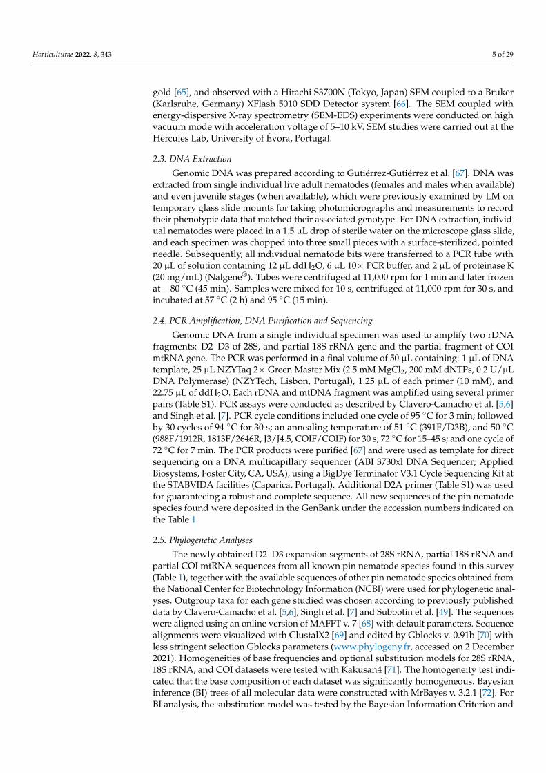
Figure 4. Light micrographs of Paratylenchus tenuicaudatus Wu, 1961 (A–F). (A–C): Female pharyn-geal region; (D): Entire female specimen; (E): Female posterior region showing vulva (arrowhead); Figure 4. Light micrographs of Paratylenchus tenuicaudatus Wu, 1961 (A–F). (A–C): Female pharyngealregion; (D): Entire female specimen; (E): Female posterior region showing vulva (arrowhead); (F): De-tail of advulval flap (arrowhead). Scale bars ((A) = 15 µm, (B,C) = 21 µm, (D) = 60 µm, (E–F) = 12 µm).(Abbreviations: avf = advulval flap; V = vulva).
Paratylenchus variabilis Raski, 1975(Figure 5; Table 3)Morphological features: Figure 5.Morphometric measurements: Table 3.
Horticulturae 2022, 8, 343 15 of 29
Horticulturae 2022, 8, x FOR PEER REVIEW 17 of 31
the body behind vulva reduced gradually; advulval flaps present; vulval flaps prominent; a single ovary outstretched; anterior uterine branch occupying 18.0–38.4% of the body length; a small and vestigial post uterine branch present; tail 24.2–28.5 μm, 2.4–2.9 times anal diameter, narrows gradually to conoid with often broadly rounded terminus (Figure 6).
Males. Not found. Juveniles. Not found.
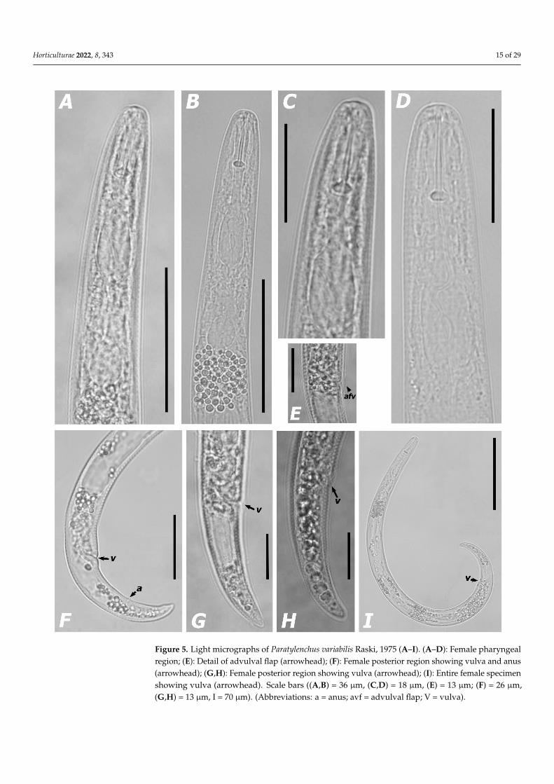
Figure 5. Light micrographs of Paratylenchus variabilis Raski, 1975 (A–I). (A–D): Female pharyngeal region; (E): Detail of advulval flap (arrowhead); (F): Female posterior region showing vulva and anus (arrowhead); (G,H): Female posterior region showing vulva (arrowhead); (I): Entire female specimen showing vulva (arrowhead). Scale bars ((A,B) = 36 μm, (C,D) = 18 μm, (E) = 13 μm; (F) = 26 μm, (G,H) = 13 μm, I = 70 μm). (Abbreviations: a = anus; avf = advulval flap; V = vulva).
Figure 5. Light micrographs of Paratylenchus variabilis Raski, 1975 (A–I). (A–D): Female pharyngealregion; (E): Detail of advulval flap (arrowhead); (F): Female posterior region showing vulva and anus(arrowhead); (G,H): Female posterior region showing vulva (arrowhead); (I): Entire female specimenshowing vulva (arrowhead). Scale bars ((A,B) = 36 µm, (C,D) = 18 µm, (E) = 13 µm; (F) = 26 µm,(G,H) = 13 µm, I = 70 µm). (Abbreviations: a = anus; avf = advulval flap; V = vulva).

Horticulturae 2022, 8, 343 16 of 29
Description of females. Body slender, ventrally curved, C-shaped when heat relaxed;cuticle thin and finely annulated; lateral field with four lines; rounded lip region withindistinct, submedian lobes, continuous with the rest of the body; framework weaklysclerotized; pharynx typical paratylenchid; short stylet 16.3–17.6 µm long, apparentlyrigid and straight; rounded stylet knobs; dorsal pharyngeal gland opening 5.2–5.6 µmbehind stylet knobs; median pharyngeal bulb large elongate, 1.8–3.2 times as long as wide;basal bulb pyriform; excretory pore situated 57.4–68.4 µm from anterior end; body slightlynarrower in the posterior portion of the vulva, abruptly in older specimens; advulvalflaps present; vulval flaps prominent, apparently rounded; a single ovary outstretched;anterior uterine branch occupying 18.0–38.4% of the body length; a short and rudimentarypost uterine branch present; tail slender, 16.5–31.7 µm long, 2.0–3.4 times as long as wide,narrows gradually to a bluntly rounded terminus (Figure 5).
Males. Not found.Juveniles. Not found.Remarks. According to Ghaderi et al. [45], our population of Paratylenchus species be-
longs to group 3 (stylet length less than 40 µm, four lateral lines and advulval flaps present).This Portuguese population was found in one soil sample of grapevine in commercial vine-yards with unknown rootstock in São Domingos de Carmões locality, Torres Vedras (Lisbondistrict, Southern Portugal) (Table 1). This population presented low nematode densities insoil (50 individuals/500 cm3 of soil). The morphological and morphometrical traits closelyagree with the original description of the species from US [89] and subsequent records [5,45],except for minor intraspecific differences (Table 3). This species is morphologically close toP. microdorus and another Paratylenchus species belonging to P. microdorus-species complex.Apparently, our pin nematode population is almost indistinguishable from several speciesbelong to P. microdorus species complex (e.i. P. microdorus, P. recisus Siddiqi, 1996, andP. zurgenerus) [5,60,89], except by some indiscernible morpho-anatomical differences andDNA molecular markers (Tables 1 and 3). This species was originally described parasitiz-ing sourberry (Rhus trilobata Nutt.) roots in US [89] and later was reported parasitizingroots of herbaceous plants and shrubs in natural environments (i.e., scrub oak (Quercusspp.), sagebrush (Artemisia spp.) and burrobrush (Hymenoclea monogyra T. & G. ex Gray)in the U.S.A. [89], turfgrass roots in Iran and almond (Prunus amygdalus Batsch) rootsin Spain [5,60]. These findings represent the first and second records of this species forPortugal and Europe, respectively. To our knowledge, these findings represent the firstrecord of this species parasitizing grapevine. We confirm a wide global distribution and anapparently ability to parasitize a diverse and wide range of host species.
Paratylenchus veruculatus Wu, 1962(Figure 6; Table 3)Morphological features: Figure 6.Morphometric measurements: Table 3.Description of females. Body robust, slightly ventrally curved, C-shaped when heat
relaxed; body cuticle distinctly annulated; lateral field equidistant showing four distinctlines; lip region broadly rounded, continuous with the rest of the body; submedian lobesnot protruding; framework weakly sclerotized; pharynx typical paratylenchid type; shortstylet, usually 13.1–14.9 µm long, apparently rigid and straight; well-developed roundedstylet knobs; median pharyngeal bulb large elongate, 2.6–2.9 times as long as wide; basalbulb pyriform; excretory pore was situated 72.9–73.3 µm from anterior end; diameter of thebody behind vulva reduced gradually; advulval flaps present; vulval flaps prominent; asingle ovary outstretched; anterior uterine branch occupying 18.0–38.4% of the body length;a small and vestigial post uterine branch present; tail 24.2–28.5 µm, 2.4–2.9 times analdiameter, narrows gradually to conoid with often broadly rounded terminus (Figure 6).
Males. Not found.Juveniles. Not found.

Horticulturae 2022, 8, 343 17 of 29Horticulturae 2022, 8, x FOR PEER REVIEW 18 of 31
Figure 6. Light micrographs of Paratylenchus veruculatus Wu, 1962 (A–G). (A–C): Female pharyngeal region; (D): Female posterior region showing vulva (arrowhead); (E): Female posterior region show-ing advulval flap (arrowhead); (F): Entire female specimen showing vulva (arrowhead); (G): Detail of advulval flap (arrowhead). Scale bars (A–C = 42 μm, D–F = 28 μm, G = 14 μm;). (Abbreviations: avf = advulval flap; ep = excretory pore; V = vulva).
Remarks. According to Ghaderi et al. [45], this population of Paratylenchus species is characterized by stylet length less than 40 μm, four lateral lines and advulval flaps pre-sent. This Portuguese population was found in one soil sample of grapevine (Vitis vinifera L.) in a commercial vineyard with unknown rootstock in Monte da Ribeira locality, São
Figure 6. Light micrographs of Paratylenchus veruculatus Wu, 1962 (A–G). (A–C): Female pharyngealregion; (D): Female posterior region showing vulva (arrowhead); (E): Female posterior region showingadvulval flap (arrowhead); (F): Entire female specimen showing vulva (arrowhead); (G): Detail ofadvulval flap (arrowhead). Scale bars (A–C = 42 µm, D–F = 28 µm, G = 14 µm;). (Abbreviations:avf = advulval flap; ep = excretory pore; V = vulva).

Horticulturae 2022, 8, 343 18 of 29
Remarks. According to Ghaderi et al. [45], this population of Paratylenchus speciesis characterized by stylet length less than 40 µm, four lateral lines and advulval flapspresent. This Portuguese population was found in one soil sample of grapevine (Vitisvinifera L.) in a commercial vineyard with unknown rootstock in Monte da Ribeira locality,São Manços (Évora district, Southern Portugal) (Table 1). Low nematode densities werepresent in the soil (20 individuals/500 cm3 of soil). The morphological and morphometricaltraits closely agree with the original description of this species [90] and other subsequentrecords [5–7,10,45,79,82,89], except for some negligible intraspecific variations (Table 3).On the basis of morphological and morphometric data our population of this species is toclose other Paratylenchus spp. (e.i. P. microdorus, P. recisus and P. variabilis) [5–7,45]. Appar-ently, our population is morphologically almost indistinguishable from these three species,except by some indiscernible morphological differences and DNA molecular markers(Tables 1 and 3). This species was originally described parasitizing heather (Calluna vulgaris(L.) Hull) roots in Scotland [90] and later was reported in soil around roots of uncultivatedplants in natural environments from Belgium, Russia, Poland and Spain [5–7,10,45,79,82,89],sugarcane (Saccharum officinarum L.) from Iran [45], and fruit orchards (e.g., almond, cherry(Prunus spp.) and peach (Prunus persica (L.) Hatsch) [5,6]. This species has not been reportedin Portugal and these findings represent the first record for this country. To our knowledge,these results represent the first record of this species parasitizing grapevine, and confirmalso that this species is widely distributed in the Iberian Peninsula.
3.1.2. Molecular Results and Phylogenetic Relationships of the Six Known Paratylenchusspp. and Other Members of Genus Paratylenchus
For the six known pin nematode species obtained, the three genes (the D2–D3 expan-sion segments of 28S rRNA, and the complete 18S rRNA) and partial COI mtRNA gene) hadan approximate size of 1000, 1600, and 400 bp, respectively. Ribosomal and mitochondrialsequences of six known Paratylenchus spp. (P. goodeyi, P. hamatus, P. pedrami, P. tenicaudatus,P. variabilis and P. veruculatus) (OM348543–OM348577; OM345184–OM345192) matchedwell with sequences from the same species previously deposited in GenBank, increasingknowledge of the genotypic diversity in Paratylenchus (Table 1). For these Paratylenchus spp.,there were multiple failed attempts to sequence a partial COI mtRNA and the complete18SrRNA genes before our study was concluded (Table 1). The D2–D3 expansion segments of28S rRNA gene sequences from P. pedrami (OM348562–OM348566; OM348550–OM348551)matched closely (98–99% similarity) to several sequences of other populations of this samespecies deposited in GenBank (MZ265118, Spain; MW798283–MW798285, Spain); and thevariations among these D2–D3 sequences ranged from 4 to 13 nucleotides. Intra-specificvariation of D2–D3 detected among the two Portuguese populations of P. pedrami (twofrom grapevines) (Table 1) was from 10 to 18 nucleotides (98–99% similarity and 0 to2 indels). For P. pedrami, four COI mtRNA gene sequences from Alcácer do Sal (AL-V4,grapevine) (OM348574–OM348577) and one sequence from Aldeia Galega da Merceana(CF-01, grapevine) (OM348570) were sequenced and showed a sequence similarity of 95%,with some minor intra-specific variations (2–19 nucleotides). Likewise our COI sequences(OM348574–OM348577, OM348570) had 94–96% similarity to other deposited in GenBankfor P. pedrami (MW797009, Spain); and the variations among these COI sequences rangedfrom 11 to 15 nucleotides. Two homogeneous sequences of the complete 18S rRNA gene(99% of similarity) for P. pedrami (OM345191–OM345192) were 96% of similar to nine se-quences deposited in GenBank belonging to P. shenzhenensis Wang, Xie, Li, Xu, Yu & Wang,2013 (KF668494–KF668497, KF668499, KF668502–KF668504, China) and Paratylenchus sp.JH-2014 (KJ636431). The variations among the 18S sequences of these species were from68 to74 nucleotides and 10 to 12 indels, being a new molecular marker for this species.The D2–D3 sequences from P. veruculatus (OM348552; OM348554–OM348555; OM348561)matched closely (99–100% similarity) to sequences of Spanish and Belgian populations ofthis species in GenBank (MW798310, Spain; MW798313–MW798314, Spain; MZ265134–MZ265135, Spain; MW798311–MW798312, Spain; MW413687, Belgium); and the variations
Horticulturae 2022, 8, 343 19 of 29
among them ranged from 0 to 12 nucleotides and 0 to 2 indels. For P. veruculatus, theCOI mtRNA gene sequence (OM348571) showed a high and variable sequence homologywith other sequences of this same species in Genbank; the homology ranged from greaterthan or equal to 98% (MW421717–MW421722, Belgium; MW797027–MW797029, Spain),to 94% (MW79702424–MW79702426, Spain). The variations among these COI sequencesranged from 1 to 18 nucleotides. Two identical sequences of the complete 18S rRNA gene(100% of similarity) of P. veruculatus (OM343587–OM343588) showed a similar homology(more than 99%) with several sequences deposited in GenBank belonging to P. verucula-tus (MW413747–MW413748, Spain), P. nanus (MW413707–MW413708, and MW413712,Belgium; MN783664–MN783667, Belgium) and P. goodeyi (MW413699, Belgium). The vari-ations among these 18S sequences of these species were from 0 to 5 nucleotides. D2–D3sequences of P. variabilis (OM348548–OM348549) had a sequence similarity greater than 99%to several sequences of Spanish populations (MZ265127–MZ265129); and a variation amongthese sequences of this species from 1 nucleotide and 1 indel. For P. variabilis, COI sequence(OM348569) showed a sequence similarity of 97% to other sequence of this same speciesdeposited in Genbank (MZ262265, Spain). The variation among these COI sequences was8 nucleotides. The complete 18S of rRNA gene sequence of P. variabilis (OM345186) was96%, similar to Paratylenchus sp. N2508 (MF094926, US). The variations among these 18Ssequences of these species were from 0 to 5 nucleotides. This complete 18S rRNA genesequence is a new molecular marker for this Paratylenchus species. D2–D3 sequences of P.tenuicaudatus (OM348544) matched well with a high sequence homology (more than 99%similarity) to sequences of several populations of this same species in GenBank (MW798306–MW798309, Spain; KF242223–KF242224, US; KU291239, Iran); and a variation among thesesequences of this species from 0 to 4 nucleotides. Our complete 18S rRNA gene sequence ofP. tenuicaudatus (OM345184) was 98% similar to several sequences deposited in GenBankbelonging to P. projectus (MF094890, US; MF094897, US; KJ636433;-KJ636434). The variationsamong these 18S sequences of these species were from 18 to 24 nucleotides and 4 indels.D2–D3 sequences of P. hamatus (OM348545–OM348547) matched well with a high sequencehomology (more than 99% similarity) to sequences of several populations of this samespecies in GenBank (KF242213–KF242218, US; KF242204, US; MW413565–MW413566, US;MW413558, US; MW798295–MW798299, Spain); and a variation among these sequences ofthis species from 0 to 3 nucleotides and 0 to 2 indels. For P. hamatus, the partial portion ofCOI gene sequenced agrees with results obtained from the D2–D3 fragments; in fact theCOI sequences (OM348567–OM348568) showed a sequence homology higher than to 99%to several isolates of the same species (MW411822–MW411824, US; MW797016–MW797017;Spain) and Paratylenchus sp. NM TSH-2020 (MN711355–MN711357). The variation amongthese COI sequences was 0 to 1 nucleotide. Two homogeneous sequences of the complete18S rRNA gene of P. hamatus (OM345185) were 98% similar to P. projectus (MF094890,US; MF094897, US; KJ636433–KJ636434). The variations among these 18S sequences ofthese species were from 18 to 24 nucleotides and 1 indel. This complete 18S rRNA genesequence is a new molecular marker for P. hamatus. The D2–D3 sequences from P. goodeyi(OM348556–OM348560; OM348553; OM348543) had a sequence similarity greater than 98%to several sequences of this same species in GenBank (MW413631–MW413633, Belgium;MW798293–MW798294, Spain; MZ265102, Spain; MZ265104–MZ265105, Spain; MZ265083–MZ265084, MZ265086, Spain; MZ265088–MZ265090, Spain; MZ265093, Spain; MZ265096,Spain; MZ265099–MZ265100, Spain); and the variations among them ranged from 2 to15 nucleotides and 0 to 3 indels. Intra-specific variation of D2–D3 detected among thePortuguese populations of P. goodeyi (Table 1) was from 8 to 13 nucleotides and 1 to 3 in-dels (98–99% similarity). For P. goodeyi, our COI gene sequences (OM348572–OM348573)had 94–95% similarity to other deposited in GenBank (MW421647–MW421649, Belgium;MW797014–MW797015, Spain; MZ262227–MZ262229, Spain; MZ262233–MZ262238, Spain;MZ262231, Spain). The variations among these COI sequences ranged from 15 to 17 nu-cleotides. The partial 18S rRNA gene sequences of P. goodeyi (OM343589–OM343590)showed a similar homology (97%) with a large number of sequences deposited in Gen-
Horticulturae 2022, 8, 343 20 of 29
Bank belonging to Paratylenchus spp., such as Paratylenchus sp. N2508 (MF094926, US),Paratylenchus sp. (KJ636435), and P. projectus (MF094890, US; MF094897, US; KJ636433). Thevariations among these 18S sequences of these species were from 38 to 47 nucleotides.
Using Bayesian inference (BI), we compared the phylogenetic position of six knownParatylenchus spp. (P. goodeyi, P. hamatus, P. pedrami, P. tenicaudatus, P. variabilis, and P. veru-culatus) by using the D2–D3 fragments of 28S rRNA and, the complete 18S rRNA genesequences, and the partial COI mtRNA gene sequences (Figures 7–9). The BI tree (50%majority rule consensus tree) of a multiple-edited alignment included 108 18S rRNA se-quences of Paratylenchus spp. (Figure 7) and two outgroup species [Hemicycliophora aquatica(Micoletzky, 1913) Loos, 1948 (MF094911), and Hemicriconemoides kanayaensis Nakasono &Ichinoe, 1961 (MG029558)] (Figure 7). A total of nine new sequences were obtained for thisribosomal molecular marker. The BI tree inferred from the analysis of the 18S sequencealignment contained highly or moderately supported major clades (PP = 1.0 and P = 0.99)(Figure 7). The general topology of this BI tree, including its clades, is slightly coincidentwith recent phylogenetic studies on paratylenchids [7,42]. Likewise, the BI tree (50% major-ity rule consensus tree) of the D2–D3 segments of 28S rRNA gene (Figure 8) was based ona multiple-edited alignment included 182 28S sequences of Paratylenchus spp. and threeoutgroup species (Basiria gracilis (Thorne, 1949) Siddiqi, 1963, MW716278; Filenchus sp.,JQ005014; Aglenchus geraerti Mizukubo, 1989, MK639370) (Figure 8). A total of twenty-fournew sequences were obtained for this ribosomal molecular marker and included in thisanalysis. BI tree inferred from the analysis of D2–D3 sequence alignment contained highlyor moderately supported major clades (PP = 1.0, PP = 0.94 and PP = 0.92). The generaltopology of this BI tree, including its clades, is mainly coincident with recent phylogeneticstudies on paratylenchids [5–7]. Similarly, the BI tree (50% majority rule consensus tree) ofa multiple-edited alignment included 169 COI mtRNA sequences of Paratylenchus spp. andtwo outgroup species (Mesocriconema sp., KY574819; Mesocriconema discus (Thorne & Malek,1968) Loof & De Grisse, 1989, KY574622) (Figure 9). A total of eleven new sequences thatinclude all the Portugueses populations of Paratylenchus spp. except for P. tenuicaudatuswere obtained for this mitochondrial molecular marker and included in this phylogeneticanalysis. BI tree inferred from the analysis of COI sequence alignment contained highly ormoderately supported major clades (PP = 1.0, and PP = 0.98). The general topology of thisBI tree, including its clades, was similar to that of the D2–D3 segments of the 28S gene andslightly coincident with recent phylogenetic studies on paratylenchids [5–7].
The generated phylogenetic tree inferred from 18S rRNA gene sequences showedincongruences between the pattern of branching in the phylogenetic tree and the currenttaxonomy within Paratylenchus genus (Figure 7). So, this ribosomal molecular markerfailed to delimit well established species; for example P. goodeyi (OM345189– OM345190,Portugal; MW413698–MW413699, Belgium), P. nanus (MW413707–MW413708, MW413711,Belgium) and P. veruculatus (OM345187– OM345188, Portugal; MW413747–MW413748,Belgium) were grouped together as belonging to the same species. Furthermore, thegenerated D2–D3 phylogenetic tree showed a congruent position for all known species(P. goodeyi, P. hamatus, P. pedrami, P. tenuicaudatus, P. variabilis and P. veruculatus) foundin this study (Figure 8). D2–D3 phylogenetic tree clustered, with strong support, our Por-tuguese sequences of P. pedrami (OM348562–OM348566, Portugal; OM348550–OM348551,Portugal) from this study with other Spanish isolates of this same species (MZ265118, Spain;MW798283–MW798285, Spain). Thus, all these P. pedrami populations form a cluster clearlyseparated from a morphologically related species as P. baldaccii (MZ265079, MW798290–MW798291, Spain). Paratylenchus hamatus sequences (OM348545–OM348547, Portugal)were grouped in a well-supported clade also containing this same species (MW798297–MW798298; Spain) which are clearly separated of P. tenuicaudatus (OM348544, Portugal;MW798308–MW798309, Spain; KU291239, Iran) and other members of the P. hamatus“species complex” (KF242220–KF242222, MW413670). D2–D3 tree clustered, with strongsupport, our Portuguese sequences of P. veruculatus (OM348552, OM348554–OM348555,OM348561, Portugal) from this study with other Spanish and Belgian isolates of this same

Horticulturae 2022, 8, 343 21 of 29
species (MW413687, Belgium; MW798310–MW798315, Spain). Thus, all these P. veruculatuspopulations constitute a cluster clearly separated from Iberian populations of P. variabilis(OM348548–OM348549, Portugal; MZ265127–MZ265129, Spain) and other Paratylenchusspp. as P. microdorus (MW413654, Belgium) and P. recisus (MZ265119–MZ265120, Spain).Paratylenchus goodeyi (OM348543, OM348553, OM348556–OM348560, Portugal) clusteredin a well-supported clade also contain Spanish and Belgian isolates of this same species(MW798293–MW798294, Spain; MW413631, Belgium) which are separated of close Paraty-lenchus species as P. pandatus (MZ265116–MZ265117, Spain) and P. straeleni (KF242234,USA). Likewise, phylogenetic analysis showed that the COI tree is congruent with 28SrRNA phylogenies (Figure 9). Both D2–D3 and COI trees showed topologies partially coin-cident with other recent studies with, in some cases, similar or different clade support [5–7].Horticulturae 2022, 8, x FOR PEER REVIEW 23 of 31
Figure 7. Phylogenetic relationships within the genus Paratylenchus. Bayesian 50% majority rule con-sensus trees as inferred from 18S rRNA sequences alignments under the K80 model with a gamma-shaped distribution. Posterior probabilities of more than 0.70 are given for appropriate clades. Newly obtained sequences in this study are coloured in red, green, yellow, pink, navy blue, and sky blue. Scale bar = expected changes per site.
Figure 7. Phylogenetic relationships within the genus Paratylenchus. Bayesian 50% majority ruleconsensus trees as inferred from 18S rRNA sequences alignments under the K80 model with a gamma-shaped distribution. Posterior probabilities of more than 0.70 are given for appropriate clades. Newlyobtained sequences in this study are coloured in red, green, yellow, pink, navy blue, and sky blue.Scale bar = expected changes per site.

Horticulturae 2022, 8, 343 22 of 29Horticulturae 2022, 8, x FOR PEER REVIEW 24 of 31
Figure 8. Phylogenetic relationships within the genus Paratylenchus. Bayesian 50% majority rule con-sensus trees as inferred from 28S rRNA sequences alignments under the GTR model with a gamma-shaped distribution. Posterior probabilities of more than 0.70 are given for appropriate clades.
Figure 8. Phylogenetic relationships within the genus Paratylenchus. Bayesian 50% majority ruleconsensus trees as inferred from 28S rRNA sequences alignments under the GTR model with agamma-shaped distribution. Posterior probabilities of more than 0.70 are given for appropriate clades.Newly obtained sequences in this study are coloured in red, green, yellow, pink, navy blue, and skyblue. Scale bar = expected changes per site.
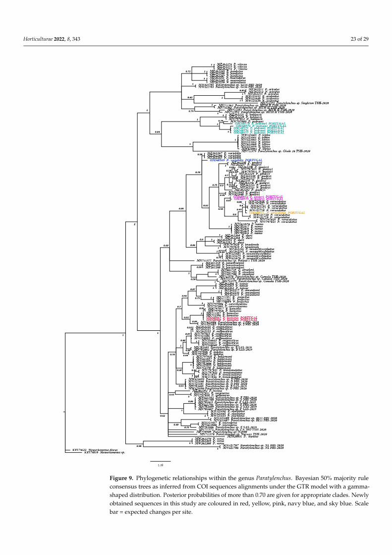
Horticulturae 2022, 8, 343 23 of 29
Horticulturae 2022, 8, x FOR PEER REVIEW 25 of 31
Newly obtained sequences in this study are coloured in red, green, yellow, pink, navy blue, and sky blue. Scale bar = expected changes per site.
Figure 9. Phylogenetic relationships within the genus Paratylenchus. Bayesian 50% majority ruleconsensus trees as inferred from COI sequences alignments under the GTR model with a gamma-shaped distribution. Posterior probabilities of more than 0.70 are given for appropriate clades. Newlyobtained sequences in this study are coloured in red, yellow, pink, navy blue, and sky blue. Scalebar = expected changes per site.
Horticulturae 2022, 8, 343 24 of 29
4. Discussion
This research is the first study of pin nematodes of the genus Paratylenchus carried outin Portugal improving the knowledge of their biodiversity, and expanding the molecularinformation useful for their unequivocal identification. The main goal of our study wasto identify and describe, morphologically and molecularly, Paratylenchus spp. parasitiz-ing the roots of grapevines in vineyards of the Southern and Central Portugal. This wasconducted in a nematological survey that included 44 sampling sites. Seven soil samples,each infested with only one pin nematode population, except one of them with two pop-ulations, were selected for this detailed study. Our results confirmed the usefulness ofdeveloping a robust taxonomic approach based on the contrasting of morphological datawith genotyping rRNA markers to correctly discriminate among Paratylenchus species. Weidentified several populations by integrating morphological observations, morphometrics,and molecular data based on rRNA sequences to elucidate their phylogenetic relationshipswithin Paratylenchus spp. Molecular markers were generated for the known pin nematodespecies, and the molecular genetic diversity of these six species (P. goodeyi, P. hamatus,P. pedrami, P. tenicaudatus, P. variabilis, and P. veruculatus) was evaluated. New molecularmarkers were generated for some of these pin nematode species.
This integrative taxonomic study of eight Portuguese populations of Paratylenchus spp.confirmed that the identification of pin nematode species from phenotypic features isnot easy because of their small body size together with high inter- and intra-specifictrait variations and plasticity in morphological traits. As for previous biogeographicstudies [5,6], our study has revealed six first report of Paratylenchus spp. for Portugal(P. goodeyi, P. hamatus, P. pedrami, P. tenicaudatus, P. variabilis, and P. veruculatus). Ourfindings confirm previous results [5,6], that some Paratylenchus species are spreading inmore than one continent, and thus more widely spread than supposed.
Forty-four sequences belonging to two nuclear rRNA and one mtRNA markers weregenerated in this study: 24 D2–D3, 9 18S, and 11 partial COI sequences. The results corrob-orate previous studies [5,6,8,42,46–48] in finding the utility of this integrative approach andmolecular data for species discrimination in Paratylenchus. Our findings are in accordancewith the results of recent studies [5,6], since they showed that the D2–D3 of the 28S rRNAand the COI region within mtRNA were the most decisive, precise, and reliable molecularmarkers for discriminating among species and for the diagnosis at species level withinParatylenchus genus. Therefore, our findings confirm that the D2–D3 region of the 28SrRNA gene should be considered the most effective molecular marker for the species-level identification of paratylenchids. In spite of the excellent reputation of the partialCOI mtRNA gene has for allowing discrimination among Paratylenchus species [61], ourfindings agree with other previous studies [5–7] confirming that this molecular markersometimes shows high intraspecific variation. The 18S rRNA gene is highly conservedintra-species (so that similarities are close to 99–100%); in fact our complete sequence(approximately 1600 bp) did not provide good resolution at the lower taxonomic levelsfailing to delimit well established Paratylenchus species. However, this 18S ribosomalmolecular marker was useful for discrimination and delimitation of some specific specieswithin the Paratylenchus genus. For example, in P. pedrami, the complete 18S rRNA se-quence (OM345191–OM345191) was compared to other sequences belonging to knownParatylenchus species (KF668497–KF668497, KF668499, KF668502, KF668504, KJ636331) andwas 96% similar with fewer than 74 nucleotides differences, whereas sequences of theCOI region (OM348574–OM348577, OM348570) also had a low homology with maximumsimilarity values of 96% to P. pedrami (MW797009) and the fragments D2–D3 of 28S rRNA(OM348562–OM348566; OM348550–OM348551) showed a high homology with maximumsimilarity values of 99% to P. pedrami (MZ265118, MW798283–MW798285).
Phylogenetic trees reconstructed by the BI approach using new Paratylenchus sequencesand others from Genbank inferred similar patterns. Our 28S phylogenetic trees indi-cated that P. pedrami (OM348562–OM348566, OM348550–OM348551, Portugal; MW738282–MW738285, Spain; MZ265118, Spain) was clearly grouped in a sub-clade with P. bal-
Horticulturae 2022, 8, 343 25 of 29
daccii (MW738290–MW738291, Spain; MZ265079, Spain), and P. minor (MK660188, China),which are three species sharing a similar morphology characterized mainly by a shortto medium-sized style length (around 30 µm). However, as is common in phylogeneticstudies, this three species were also inside in an upper sub-clade with other morpho-logically related species showing smaller stylet length (P. leptus Raski 1975, KR270602,MW413645; P. rostrocaudatus Huang & Raski, 1987, KR270601; P. aquaticus Merny, 1966,KF242240–KF242241; P. shenzhenensis Wang, Xie, Li, Xu, Yu, & Wang, 2013, KF668518).Likewise, P. variabilis (OM348548–OM348549, Portugal; MZ265127–MZ265129, Spain),was grouped in a well-supported sub-clade with a large number of species (P. nanus,P. veruculatus, P. goodeyi, P. similis, P. variabilis) which are six species sharing quite phe-notypic features among them. For example, P. similis (MK506806), P. nanus (KF242197)and P. veruculatus (OM348561, OM348552–OM348553, Portugal; MW798310–MW798311,MW798313–MW798314, Spain; MZ265134–MZ265135, Spain; MW413687, Belgium) sharinga relatively short-sized style length (less than 25 µm), however P. goodeyi show a longerstylet (OM358543, OM358556–OM358560, Portugal; MZ265084–MZ265085, MZ265091–MZ265095, MZ265097–MZ265105, Spain; MW413631, Belgium). Similarly, P. tenuicaudatus(OM348544, Portugal; MW798306–MW798309, Spain; KU291239) was clearly grouped in asub-clade to several species characterized by a short to medium-sized style length (around30 µm) (P. hamatus: OM348545–OM348547, MW798285–MW798289, KF242215; P. enigmati-cus Munawar, Yevtushenko, Palomares-Rius and Castillo, 2021: MW798292, MN535545;MZ265080- MZ265084; P. indalus: MW798273–MW798282). Thus, the phylogenetic treetopologies, particularly based on our 28S rRNA dataset, agrees with previous phylogeneticstudies [5–7,42]. Regarding stylet in Paratylenchus spp. it is worth to note that there is ahigh morphological diversity and its feeding role denotes a decisive factor in the fitnesswithin this genus. Likewise, the 28S tree topology denotes possibly a monophyletic originof Paratylenchus spp., in which a long stylet (more than 40 µm) is a plesiomorphic character.In addition, the pattern of branching in the 28S phylogenetic tree denotes two synapomor-phies, so that intermediate stylet length (around 30 µm) and short stylet length (less than25 µm), in the major evolutionary events of the Paratylenchus genus. Munawar et al. [42] andClavero-Camacho et al. [5,6] supported a significant association between the phylogenetictree topologies and the division defined by Ghaderi et al. [45] regarding stylet length (lessthan 40 vs. more than 40 µm), however they did not keep a intermedium-size stylet lengthin mind. Furthermore, this study confirms the results of previous studies [5–7,42,45] that itis common to find a same soil sample showing the co-existence of Paratylenchus species.Niche partitioning is the ecological process that reduces interspecific competition in whichnatural selection drives competing species into different patterns of resource use or nichein time and/or space [91]. Thus, the ecological theory could help to better understand ofthis phylogenetic flexibility characterized by multiple evolutionary synapomorphies. Forexample, P. goodeyi and P. veruculatus are two species close phylogenetically but they haveclear phenotypic differences regarding the stylet length that are commonly found togetherin the same sample soil. In fact, P. goodeyi has a long stylet that allows this species to feedin root cell layers deep in both taproots and fibrous roots, primary or secondary roots ofherbaceous and woody plants and does not need to move frequently to new feeding sites,while P. veruculatus has a short and rigid stylet that allows this nematode to feed in rootcells of outside layers in thin lateral roots of herbaceous and woody plants and need tomove drilling new feeding sites..
5. Conclusions
Our work contributes with relevant understanding of the occurrence, distributionand biodiversity of Paratylenchus species infesting grapevine soils in Portugal. This studydescribes six first reports of Paratylenchus spp. (P. goodeyi, P. hamatus, P. pedrami, P. tenicau-datus, P. variabilis, and P. veruculatus) for the country by utilizing morphological features,morphometric measures and molecular data, and establishes these species’ phylogeneticrelationships within the genus Paratylenchus. Our study also highlights the value of using
Horticulturae 2022, 8, 343 26 of 29
rRNA molecular markers for the identification of Paratylenchus spp., when other conven-tional methods alone based on morphology are inconclusive in this taxonomically confusinggenus. Likewise, we generated molecular markers for precise and unequivocal diagnosis ofdifferent known Paratylenchus species, evaluated their molecular genetic diversity and es-tablished molecular phylogenetic relationship that shows new insights into the systematicsand the evolution of Paratylenchus genus. In addition, our results confirm that the diversityof paratylenchid nematodes in commercial vineyards is underrepresented, since the speciesreported in this study are all new records for Portugal. However, this is the first systematicstudy for Paratylenchus detection in Portugal using a taxonomic integrative approach andincreasing species diversity is expected to be found in the future.
Supplementary Materials: The following are available online at https://www.mdpi.com/article/10.3390/horticulturae8040343/s1, Table S1: List of the primers used in this study.
Author Contributions: Conceptualization, T.R. and C.G.-G.; methodology, data collection, andanalysis, T.R. and C.G.-G.; writing—original draft preparation, T.R and C.G.-G.; writing—review andediting, C.G.-G., M.L.I., J.D.E. and M.M.; supervision, C.G.-G., M.L.I., J.D.E. and M.M. All authorshave read and agreed to the published version of the manuscript.
Funding: This work is funded by National Funds through FCT—Foundation for Science and Tech-nology under the Project UIDB/05183/2020 (Portugal).
Institutional Review Board Statement: Not applicable.
Informed Consent Statement: Not applicable.
Data Availability Statement: Not applicable.
Acknowledgments: Authors thank A. Candeias and L. Dias from HERCULES Laboratory, Uni-versidade de Évora and J.A. Paulo Mirão from Instituto de Ciências da Terra (ICT) for their helpwith SEM studies, and I. Ferreira from the Instituto Mediterrâneo para a Agricultura, Ambiente eDesenvolvimento (MED), Universidade of Évora for his excellent technical assistance.
Conflicts of Interest: All authors declare no conflict of interest.
References1. Jairajpuri, M.S.; Ahmad, W. Dorylaimida: Free-Living, Predaceous and Plant-Parasitic Nematodes; Brill Publishers: Leiden,
The Netherlands, 1992.2. Ahmed, M.; Holovachov, O. Twenty Years after De Ley and Blaxter—How Far Did We Progress in Understanding the Phylogeny
of the Phylum Nematoda? Animals 2021, 11, 3479. [CrossRef] [PubMed]3. Decraemer, W.; Hunt, D.J. Structure and classification. In plant Nematology; Perry, R.N., Moens, M., Eds.; CABI Publishing:
Wallingford, UK, 2006; pp. 3–32.4. Siddiqi, M.R. Tylenchida: Parasites of Plants and Insects, 2nd ed.; CABI Publishing: Wallingford, UK, 2000.5. Clavero-Camacho, I.; Palomares-Rius, J.E.; Cantalapiedra-Navarrete, C.; León-Ropero, G.; Martín-Barbarroja, J.; Archidona-Yuste,
A.; Castillo, P. Integrative taxonomy reveals hidden cryptic diversity within pin nematodes of the genus Paratylenchus (Nematoda:Tylenchulidae). Plants 2021, 10, 1454. [CrossRef] [PubMed]
6. Clavero-Camacho, I.; Cantalapiedra-Navarrete, C.; Archidona-Yuste, A.; Castillo, P.; Palomares-Rius, J.E. Remarkable crypticdiversity of Paratylenchus spp. (Nematoda: Tylenchulidae) in Spain. Animals 2021, 11, 1161. [CrossRef] [PubMed]
7. Singh, P.R.; Karssen, G.; Couvreur, M.; Subbotin, S.A.; Bert, W. Integrative taxonomy and molecular phylogeny of the plant-parasitic nematode genus Paratylenchus (Nematoda: Paratylenchinae): Linking species with molecular barcodes. Plants 2021,10, 408. [CrossRef]
8. Kantor, M.R.; Handoo, Z.A.; Subbotin, S.A.; Bauchan, G.R.; Mowery, J.D. Morphological and molecular characterization ofParatylenchus beltsvillensis n. sp. (Tylenchida: Paratylenchidae) from the rhizosphere of pine tree (Pinus virginiana Mill) inMaryland, USA. J. Nematol. 2021, 53, 1–10. [CrossRef]
9. Andrássy, I. Free-Living Nematodes of Hungary, II (Nematoda errantia); Hungarian Natural History Museum: Budapest, Hun-gary, 2007.
10. Brzeski, M.W. Nematodes of Tylenchina in Poland and Temperate Europe; Muzeum i Instytut Zoologii, Polska Akademia Nauk:Warszawa, Poland, 1998.
11. Esser, R.P. A diagnostic compendium to species included in Paratylenchinae Thorne, 1949 and Tylenchocriconematinae Raski &Siddiqi, 1975 (Nematoda: Criconematoidea). Nematologica 1992, 38, 146–163.

Horticulturae 2022, 8, 343 27 of 29
12. Raski, D.J. Tylenchulidae of agricultural soils. In Manual of Agricultural Nematology; Nickle, W.R., Ed.; Marcel Dekker Inc.: NewYork, NY, USA, 1991; pp. 761–794.
13. Ebsary, B.A. Catalog of the Order Tylenchida (Nematoda); Agriculture Canada, Research Branch: Ottawa, ON, Canada, 1991.14. Maggenti, A.R.; Luc, M.; Raski, D.J.; Fortuner, R.; Geraert, E. A reappraisal of Tylenchina (Nemata). 11. List of generic and
supra-generic taxa, with their junior synonyms. Rev. Nématol. 1988, 11, 177–188.15. Raski, D.J.; Luc, M. A reappraisal of Tylenchina (Nemata). 10: The superfamily Criconematoidea Taylor, 1936. Rev. Nématol. 1987,
10, 409–444.16. Goodey, T. Soil and Freshwater Nematodes, 2nd ed.; Goodey, J.B., Ed.; Methuen: London, UK, 1963.17. Ghaderi, R. The damage potential of pin nematodes, Paratylenchus Micoletzky, 1922 sensu lato spp. (Nematoda: Tylenchulidae). J.
Crop Prot. 2019, 8, 243–257.18. Subbotin, S.A.; Chitambar, J.J. Plant Parasitic Nematodes in Sustainable Agriculture of North America; Springer: Cham, Switzerland, 2018.19. Upadhaya, A.; Yan, G.P.; Pasche, J. Reproduction ability and growth effect of pin nematode, Paratylenchus nanus, with selected
field pea cultivars. Plant Dis. 2019, 103, 2520–2526. [CrossRef]20. Claerbout, J.; Vandevelde, I.; Venneman, S.; Kigozi, A.; de Sutter, N.; Neukermans, J.; Bleyaert, P.; Bert, W.; Hofte, M.; Viaene, N.
A thorough study of a Paratylenchus sp. in glasshouse-grown lettuce: Characterisation, population dynamics, host plants anddamage threshold as keys to its integrated management. Ann. Appl. Biol. 2020, 178, 62–79. [CrossRef]
21. Wang, K.; Li, Y.; Xie, H.; Wu, W.J.; Xu, C.L. Pin nematode slow decline of Anthurium andraeanum, a new disease caused by the pinnematode Paratylenchus shenzhenensis. Plant Dis. 2016, 100, 940–945. [CrossRef] [PubMed]
22. Braun, A.L.; Lownsbery, B.F. The pin nematode, Paratylenchus neoamblycephalus, on Myrobalan plum and other hosts. J. Nematol.1975, 7, 336–343.
23. Riga, E.; Porter, L.D.; Mojtahedi, H.; Erickson, D. Pratylenchus neglectus, P. thornei, and Paratylenchus hamatus nematodes causingyield reduction to dryland peas and lentils in Idaho. Plant Dis. 2008, 92, 979. [CrossRef] [PubMed]
24. Philis, J. Controlling parasitic nematodes in an established vineyard in Cyprus. Nematol. Medit. 2003, 31, 61–63.25. MacDonald, D.H. Effects of Paratylenchus hamatus on productivity of greenhouse roses. J. Nematol. 1976, 8, 294.26. Abivardi, C. Occurrence of Paratylenchus hamatus on citrus in Iran and its sensitivity to two nematicides under laboratory
conditions. Plant Dis. Rep. 1970, 54, 1085–1088.27. French, A.M.; Lownsbery, B.F.; Ayoub, S.M.; Weiner, A.C.; Gholl, N.E. Pythiaceous fungi and plant-parasitic nematodes in
California pear orchards. II. Incidence and distribution of parasitic nematodes in orchard soils. Hilgardia 1964, 35, 603–610.[CrossRef]
28. Faulkner, L.R. Pathogenicity and population dynamics of Paratylenchus hamatus on Mentha spp. Phytopathology 1964, 54, 344–348.29. Raski, D.J.; Lider, L.A. Nematodes in grape production. California Agriculture 1959, 13, 13–15.30. Weischcr, B. Neuere Gesichtspunktezur Frage der biologie und ökologie der w and erndenwurzel nematodes. Nematologica 1957,
2, 406–412.31. Rich, A.E. The occurrence and control of Paratylenchus hamatus on celery in New Hampshire. Plant Dis. Rep. 1955, 39, 307–308.32. Thorne, G.; Allen, M.W. Paratylenchus hamatus n.sp, and Xiphinema index n. sp., two nematodes associated with fig roots, with
a note on Paratylenchus anceps Cobb. Proc. Helm. Soc. Wash. 1950, 17, 27–35.33. Lowensbry, B.F.; Stoddars, E.M.; Lownsbery, J.W. Paratylenchus hamatus pathogenic on celery. Phytopathology 1952, 42, 651–653.34. Fisher, J.M. Effect of temperature and host on Paratylenchus neoamblycephalus and effect of the nematode on the host. Aust. J. Agric.
Res. 1967, 18, 921–929. [CrossRef]35. Viketoft, M.; Palmborg, C.; Sohlenius, B.; Huss-Danell, K.; Bengtsson, J. Plant species effects on soil nematode communities in
experimental grasslands. Appl. Soil Ecol. 2005, 30, 90–103. [CrossRef]36. Viketoft, M. Effects of six grassland plant species on soil nematodes: A glasshouse experiment. Soil Biol. Biochem. 2008, 40,
906–915. [CrossRef]37. Bell, N.L. The biology of the plant parasitic nematodes Paratylenchus nanus and Paratrichodorus minor in soil under pasture.
Ph.D. Thesis, Massey University, Palmerston North, New Zealand, 1999.38. Archidona-Yuste, A.; Wiegand, T.; Castillo, P.; Navas-Cortés, J.A. Dataset on the diversity of plant-parasitic nematodes in
cultivated olive trees in southern Spain. Data Brief. 2019, 27, 104658. [CrossRef]39. Ali, N.; Chapuis, E.; Tavoillot, J.; Mateille, T. Plant-parasitic nematodes associated with olive tree (Olea europaea L.) with a focus
on the Mediterranean Basin: A review. Comptes Rendus Biologies 2014, 337, 423–442. [CrossRef]40. Téliz, D.; Landa, B.B.; Rapoport, H.F.; Pérez Camacho, F.; Jiménez Díaz, R.M.; Castillo, P. Plant-parasitic nematodes infecting
grapevine in Southern Spain and susceptible reaction to root-knot nematodes of rootstocks reported as moderately resistant.Plant Dis. 2007, 91, 1147–1154. [CrossRef]
41. Peña-Santiago, R.; Castillo, P.; Escuer, M.; Guerrero, P.; Talavera, M.; Vieira, P. Tylenchid Species (Nematoda, Tylenchida) Recorded inthe Iberian Peninsula and the Balearic Islands: A Compendium; Universidad de Jaén: Jaén, Spain, 2004; pp. 1–127.
42. Munawar, M.; Yevtushenko, D.P.; Palomares-Rius, J.E.; Castillo, P. Species diversity of pin nematodes (Paratylenchus spp.) frompotato growing regions of southern Alberta, Canada. Plants 2021, 10, 188. [CrossRef] [PubMed]
43. Lima, M.B. First record of the genera Hemicycliophora, Paralongidorus and Paratylenchus in Portugal. Agronomía lush. 1966, 28,143–144.
44. Macara, A.M. Nematodos asociados a plantas forestales en Portugal. Bol San. Veg. Plagas 1988, 14, 185–225.

Horticulturae 2022, 8, 343 28 of 29
45. Ghaderi, R.; Kashi, L.; Karegar, A. Contribution to the study of the genus Paratylenchus Micoletzky, 1922 sensu lato (Nematoda:Tylenchulidae). Zootaxa 2014, 3841, 151–187. [CrossRef]
46. Munawar, M.; Miao, W.; Castillo, P.; Zheng, J.-W. A new pin nematode, Paratylenchus sinensis n. sp. (Nematoda: Paratylenchinae)in the rhizosphere of white mulberry from Zhejiang Province, China. Eur. J. Plant Pathol. 2020, 156, 1023–1029. [CrossRef]
47. Munawar, M.; Miao, W.; Ye, W.; Zheng, J. Updated description of Paratylenchus lepidus Raski 1975 and P. minor Sharma, Sharmaand Khan, 1986 by integrating molecular and ultra-structural observations. J. Nematol. 2019, 51, 1–13. [CrossRef]
48. Munawar, M.; Cai, R.; Ye, W.; Powers, T.O.; Zheng, J. Description of Gracilacus paralatescens n. sp. (Nematoda: Paratylenchinae)found from the rhizosphere of Bamboo in Zhejiang, China. J. Nematol. 2018, 50, 611–622. [CrossRef]
49. Subbotin, S.A.; Yan, G.; Kantor, M.; Handoo, Z. On the molecular identity of Paratylenchus nanus Cobb, 1923 (Nematoda:Tylenchida). J. Nematol. 2020, 52, 1–7. [CrossRef]
50. Le, T.M.L.; Nguyen, H.T.; Nguyen, T.D.; Trinh, Q.P. First report of Paratylenchus lepidus Raski, 1975 associated with green tea(Camellia sinensis (L.) Kuntze) in Vietnam. J. Nematol. 2020, 52, 1–4. [CrossRef]
51. Li, Y.; Wang, K.; Xie, H.; Xu, C.L. Morphology and molecular analysis of Paratylenchus chongqingensis n. sp. (Nematoda:Paratylenchinae) from soil associated with Ophiopogon japonicas in China. Eur. J. Plant Pathol. 2019, 154, 597–605. [CrossRef]
52. Hesar, A.M.; Karegar, A.; Ghaderi, R. Phylogenetic relationships of Cacopaurus pestis Thorne, 1943 within representatives of theTylenchulidae Skarbilovich, 1947 as inferred from ITS and D2–D3 expansion segments of 28S-rRNA sequences. Nematology 2019,21, 971–994. [CrossRef]
53. Etongwe, C.M.; Singh, P.R.; Bert, W.; Subbotin, S.A. Molecular characterisation of some plant-parasitic nematodes (Nematoda:Tylenchida) from Belgium. Russ. J. Nematol. 2020, 28, 1–28.
54. Phani, V.; Somvanshi, V.S.; Rao, U.; Khan, M.R. Paratylenchus jasmineae sp. n. (Nematoda: Paratylenchinae) from rhizosphere ofJasminum sambac in India. Nematology 2019, 21, 469–478. [CrossRef]
55. Zhuo, K.; Liu, X.; Tao, Y.; Wang, H.; Lin, B.; Liao, J. Morphological and molecular characterisation of three species of ParatylenchusMicoletzky, 1922 (Tylenchida: Paratylenchidae) from China, with a first description of the male P. rostrocaudatus. Nematology 2018,20, 837–850. [CrossRef]
56. Wang, K.; Xie, H.; Li, Y.; Wu, W.J.; Xu, C.L. Morphology and molecular analysis of Paratylenchus nanjingensis n. sp. (Nematoda:Paratylenchinae) from the rhizosphere soil of Pinus massoniana in China. J. Helminthol. 2016, 90, 166–173. [CrossRef]
57. Wang, K.; Li, Y.; Xie, H.; Xu, C.L.; Wu, W.J. Morphology and molecular analysis of Paratylenchus guangzhouensis n. sp. (Nematoda:Paratylenchinae) from the soil associated with Bambusa multiplex in China. Eur. J. Plant Pathol. 2016, 145, 255–264. [CrossRef]
58. Yu, Q.; Ye, W.; Powers, T. Morphological and molecular characterization of Gracilacus wuae n. sp. (Nematoda: Criconematoidea)associated with cow parsnip (Heracleum maximum) in Ontario, Canada. J. Nematol. 2016, 48, 203–213. [CrossRef]
59. Esmaeili, M.; Heydari, R.; Castillo, P.; Bidhendi, M.Z.; Palomares-Rius, J.E. Molecular characterisation of two known species ofParatylenchus Micoletzky, 1922 from Iran with notes on the validity of Paratylenchus audriellus Brown, 1959. Nematology 2016, 18,591–604. [CrossRef]
60. Van den Berg, E.; Tiedt, L.R.; Subbotin, S.A. Morphological and molecular characterisation of several Paratylenchus Micoletzky,1922 (Tylenchida: Paratylenchidae) species from South Africa and USA, together with some taxonomic notes. Nematology 2014,16, 323–358. [CrossRef]
61. Powers, T.; Harris, T.S.; Higgins, R.S.; Mullin, P.G.; Powers, K.S. Nematode biodiversity assessments need vouchered databases:A BOLD reference library for plant-parasitic nematodes in the superfamily Criconematoidea. Genome 2020, 5, 1–10. [CrossRef]
62. Whitehead, A.G.; Hemming, J.R. A comparison of some quantitative methods of extracting small vermiform nematodes from soil.Ann. Appl. Biol. 1965, 55, 25–38. [CrossRef]
63. Jenkins, W.R.B. A rapid centrifugal-flotation technique for separating nematodes from soil. Plant Dis. Rep. 1964, 48, 692.64. Seinhorst, J.W. Killing nematodes for taxonomic study with hot F.A. 4:1. Nematologica 1966, 12, 178. [CrossRef]65. Fawzia H Abdel Rahman. Chemical drying of nematodes for Scanning Electron Microscopy observations. World J. Agr. Soil Sci.
2021, 7, 1–5. [CrossRef]66. Gutiérrez-Gutiérrez, C.; Mota, M.; Castillo, P.; Teixeira-Santos, M.; Palomares-Rius, J.E. Description and molecular phylogeny of
one new and one known needle nematode of the genus Paralongidorus (Nematoda: Longidoridae) from grapevine in Portugalusing integrative approach. Eur. J. Plant Pathol. 2018, 151, 155–172. [CrossRef]
67. Gutiérrez-Gutiérrez, C.; Teixeira Santos, M.; Inácio, M.L.; Eisenback, J.D.; Mota, M. Description of Longidorus bordonensis sp.nov. from Portugal, with systematics and molecular phylogeny of the genus (Nematoda, Longidoridae). Zoosyst. Evol. 2020, 96,175–193. [CrossRef]
68. Katoh, K.; Rozewicki, J.; Yamada, K.D. MAFFT online service: Multiple sequence alignment, interactive sequence choice andvisualization. Brief. Bioinform. 2019, 20, 1160–1166. [CrossRef]
69. Larkin, M.A.; Blackshields, G.; Brown, N.P.; Chenna, R.; McGettigan, P.A.; McWilliam, H.; Valentin, F.; Wallace, I.M.; Wilm, A.;Lopez, R.; et al. Clustal W and Clustal X version 2.0. Bioinformatics 2007, 23, 2947–2948. [CrossRef]
70. Castresana, J. Selection of conserved blocks from multiple alignments for their use in phylogenetic analysis. Mol. Biol. Evol. 2000,17, 540–552. [CrossRef]
71. Tanabe, A.S. Kakusan4 and Aminosan: Two programs for comparing nonpartitioned, proportional, and separate models forcombined molecular phylogenetic analyses of multilocus sequence data. Mol. Ecol. Notes 2011, 11, 914–921. [CrossRef]
Horticulturae 2022, 8, 343 29 of 29
72. Ronquist, F.; Teslenko, M.; van der Mark, P.; Ayres, D.L.; Darling, A.; Höhna, S.; Larget, B.; Liu, L.; Suchard, M.A.; Huelsenbeck,J.P. MrBayes 3.2: Efficient Bayesian phylogenetic inference and model choice across a large model space. Syst. Biol. 2012, 61,539–542. [CrossRef] [PubMed]
73. Rambaut, A.; Suchard, M.A.; Xie, D.; Drummond, A.J. Tracer v1.6. Available online: http://beast.bio.ed.ac.uk/Tracer (accessedon 2 December 2021).
74. Available online: http://tree.bio.ed.ac.uk/software/figtree/ (accessed on 2 December 2021).75. Oostenbrink, M. A note on Paratylenchus in the Netherlands with the description of P. goodeyi n. sp. (Nematoda, Criconematidae).
Tijdschr. Plantenziekten 1953, 59, 207–216. [CrossRef]76. Castillo, P.; González-País, M.A.; Gómez-Barcina, A. El género Gracilacus Raski, 1962 en España (Paratylenchinae: Tylenchida).
Rev. Ibér. Parasitol. 1989, 49, 321–328.77. Sturhan, D. Plant-parasitic nematodes in Germany—an annotated checklist. Soil Org. 2014, 83, 22.78. Hánel, L.; Cerevková, A. Diversity of soil nematodes in meadows of the White Carpathians. Helminthologia 2006, 43, 109–116.
[CrossRef]79. Brzeski, M.W. Paratylenchinae: Morphology of some known species and descriptions of Gracilacus bilineata sp. n. and G. vera sp.
n. (Nematoda: Tylenchulidae). Nematologica 1995, 41, 535–565. [CrossRef]80. Szczygiel, A. Plant parasitic nematodes associated with strawberry plants in Poland. Zesz. Probl. Postepów Nauk. Rol. 1974, 154,
9–132.81. Geraert, E. The Genus Paratylenchus. Nematologica 1965, 11, 301–334. [CrossRef]82. Solov’eva, G.I. Parasitic Nematodes of Woody and Herbaceous Plants. A Review of the Genus Paratylenchus Micoletzky, 1922 (Nematoda:
Criconematidae); Amerind Publishing Co.: New Delhi, India, 1975.83. Lisetskaya, L.F. Nematode fauna of ethereal oil plants of Moldavia. Mater. Nats. Konf. Po Parazitol. Sofia 1968, 13, 47–48.84. Izatullaeva, R.I. Nematodes of the flowering plants of Kazakhstan; Abstract Kandidat’s Dissertation: Alma-Ata, Kazakhstan, 1967;
pp. 1–18.85. Wu, L.Y. Paratylenchus tenuicaudatus n. sp. (Nematoda: Criconematidae). Can. J. Zool. 1961, 39, 163–165. [CrossRef]86. Subbotin, S.A.; Vovlas, N.; Crozzoli, R.; Sturhan, D.; Lamberti, F.; Moens, M.; Baldwin, J.G. Phylogeny of Criconematina Siddiqi,
1980 (Nematoda: Tylenchida) based on morphology and D2–D3 expansion segments of the 28S-rRNA gene sequences withapplication of a secondary structure model. Nematology 2005, 7, 927–944. [CrossRef]
87. Myers, R.F. Plant parasitic nematodes in New Jersey. J. Nematol. 1986, 18, 272–274. [PubMed]88. Hernández, M.; Mateo, M.D.; Jordana, R. Estudio comparativo entre grupos tróficos de nematodos del suelo de cinco bosques de
Navarra (tres naturales y dos de repoblación). Actas II Congr. Mund. Vasco. Sec. Biol. Ambient. 1988, 2, 323–335.89. Raski, D.J. Revision of the genus Paratylenchus Micoletzky, 1922 and descriptions of new species. Part II of three parts. J. Nematol.
1975, 7, 274–295.90. Wu, L.Y. Paratylenchus veruculatus n. sp. (Criconematidae: Nematoda) from Scotland. Can. J. Zool. 1962, 40, 773–775. [CrossRef]91. Duyck, P.F.; Dortel, E.; Tixier, P.; Vinatier, F.; Loubana, P.M.; Chabrier, C.; Quénéhervé, P. Niche partitioning based on soil type
and climate at the landscape scale in a community of plant-feeding nematodes. Soil Biol. Biochem. 2012, 44, 49–55. [CrossRef]