Treponema species enrich the gut microbiota of traditional ...
Upload
independentCategory
view
1download
0
Sommer et al. Genome Biology (2015) 16:62 DOI 10.1186/s13059-015-0614-4
RESEARCH Open Access
Site-specific programming of the host epithelialtranscriptome by the gut microbiotaFelix Sommer1, Intawat Nookaew2,3, Nina Sommer1, Per Fogelstrand1 and Fredrik Bäckhed1,4*
Abstract
Background: The intestinal epithelium separates us from the microbiota but also interacts with it and thus affectshost immune status and physiology. Previous studies investigated microbiota-induced responses in the gut usingintact tissues or unfractionated epithelial cells, thereby limiting conclusions about regional differences in the epithelium.Here, we sought to investigate microbiota-induced transcriptional responses in specific fractions of intestinal epithelialcells. To this end, we used microarray analysis of laser capture microdissection (LCM)-harvested ileal and colonic tip andcrypt epithelial fractions from germ-free and conventionally raised mice and from mice during the time course ofcolonization.
Results: We found that about 10% of the host’s transcriptome was microbially regulated, mainly including genesannotated with functions in immunity, cell proliferation, and metabolism. The microbial impact on host geneexpression was highly site specific, as epithelial responses to the microbiota differed between cell fractions.Specific transcriptional regulators were enriched in each fraction. In general, the gut microbiota induced a morerapid response in the colon than in the ileum.
Conclusions: Our study indicates that the microbiota engage different regulatory networks to alter host geneexpression in a particular niche. Understanding host-microbiota interactions on a cellular level may facilitatesignaling pathways that contribute to health and disease and thus provide new therapeutic strategies.
BackgroundThe human gut harbors a diverse microbial community(gut microbiota) that mainly consists of bacteria. Theircombined genomes (the microbiome) encode more than10 million genes, outnumbering our own genetic potentialby two to three orders of magnitude [1-3], and providebiochemical and metabolic functions that complementour physiology. For example, the gut microbiota metabo-lizes otherwise indigestible polysaccharides and producesessential vitamins, instructs the development of the intes-tinal epithelium and immune system, and plays a key rolein maintaining tissue homeostasis [4]. Maintaining symbi-osis seems to be a key requirement for health as dysbiosisis associated with the development of common diseases,
* Correspondence: [email protected] Wallenberg Laboratory and Sahlgrenska Center for Cardiovascular andMetabolic Research, Department of Molecular and Clinical Medicine,University of Gothenburg, Gothenburg 41345, Sweden4Novo Nordisk Foundation Center for Basic Metabolic Research, Section forMetabolic Receptology and Enteroendocrinology, Faculty of Health Sciences,University of Copenhagen, Copenhagen DK-2200, DenmarkFull list of author information is available at the end of the article
© 2015 Sommer et al.; licensee BioMed CentraCommons Attribution License (http://creativecreproduction in any medium, provided the orDedication waiver (http://creativecommons.orunless otherwise stated.
including obesity, atherosclerosis and inflammatory boweldisease [5-7].Within the gastrointestinal tract, microbial distribution
varies considerably and diversity increases along thelength of both the gut and the tissue-lumen axis [8],impacting function [9] and architecture [10] of the gut.A few recent studies have investigated microbiota-induced host responses along the length of the gut[11-15]. However, these reports used intact tissue sam-ples and could not, therefore, be used to draw conclu-sions about the cellular responses in the gut epitheliumsince intestinal epithelial cell (IEC) types are differen-tially distributed in the intestinal tissue. The epitheliumtips mainly contain differentiated epithelial cells, such asenterocytes and goblet cells, whereas the crypts mainlyconsist of proliferative and Paneth cells. Thus, the residentmicrobiota might induce alternative local host responsesin different cell populations within the intestinal epithe-lium, and along the crypt-villus axis, which may contrib-ute to different functional properties of the tissue.
l. This is an Open Access article distributed under the terms of the Creativeommons.org/licenses/by/4.0), which permits unrestricted use, distribution, andiginal work is properly credited. The Creative Commons Public Domaing/publicdomain/zero/1.0/) applies to the data made available in this article,

Sommer et al. Genome Biology (2015) 16:62 Page 2 of 15
To investigate the microbiota-induced transcriptionalresponses in specific and well-defined cell populations ofthe host’s epithelium, we used extensive microarray ana-lysis of laser capture microdissection (LCM)-harvestedileal and colonic tip and crypt fractions from germ-free(GF) and conventionally raised (CR) mice as well as dur-ing the time course of colonization of GF mice.
ResultsThe gut microbiota induces specific differentialtranscriptional responses depending on location in theintestinal epitheliumTo investigate a potential effect of the gut microbiota onIEC differentiation, we determined the cellular compos-ition in ileum and proximal colon of GF and CR miceusing immunostaining with a recently developed multi-channel setup [16] and an automated slide scanning andpicture analysis pipeline. Overall, we found only minoralterations of IEC type abundance between GF and CRmice with slightly more Paneth and goblet cells in CRileum (Figure 1A).As the gut microbiota only had a minor impact on
IEC composition we investigated microbiota-induced re-sponses in defined IEC populations using LCM to isolatetip and crypt fractions from the ileum and proximalcolon of GF and CR mice (Figure 1B). We performed 40microarray hybridizations using the LCM-harvested tipand crypt epithelial fractions to evaluate their transcrip-tional profiles according to bacterial status and location.Hierarchical clustering revealed a clear separation of thesamples depending on epithelial population (tip versuscrypt), then according to tissue (ileum versus colon) andfinally according to bacterial status (GF versus CR)(Figure 1C).We used statistical Student t’s test scoring for bacterial
status to identify microbiota-regulated genes. In total,2,256 genes (P < 0.001) were regulated by the gut micro-biota, most of them in the ileum tip (Additional file 1).Eighty-six percent of all microbiota-regulated genes werespecific for the individual sample types (ileum crypt,340; ileum tip, 894; colon crypt, 273; colon tip, 444;Figure 1D), indicating highly site-specific transcriptionalresponses of the intestinal epithelium to the microbiota.The site-specific microbial modulation of host gene ex-pression was validated for several representative genes ofdifferent functional categories using quantitative PCR(Figure 2).
Functional alterations in the intestinal epithelium causedby the microbiotaWe next investigated functions of the microbially regu-lated genes in the four epithelial fractions using geneontology (GO) analysis. In total, 81 GO categories werealtered significantly (P < 10−5 with more than 5
members; Figure 3; see Additional file 2 for full list),with most GO categories being enriched in the presenceof the microbiota. As expected, about half of all GOterms were related to immune functions and were pre-dominantly increased in the tip fractions of both ileumand colon. Antigen presentation and interferon-γ re-sponses were enriched in both tissues. In contrast, pro-cesses related to T-cell regulation were restricted to theileum tip whereas virus response functions were mainlyenriched in the colon tip. Several GO terms related tothe cell cycle were highly enriched in the crypts of bothileum and colon, indicating altered cell proliferation inthe presence of the gut microbiota. Increased prolifera-tion in crypts from CR compared with GF mice wasconfirmed by staining sections from ileum and cryptwith the proliferative marker Ki-67 (Additional file 3),which is in agreement with previous publications[4,14,17]. In addition, several functions related to proteinbiosynthesis were enriched in the crypts. Metabolic pro-cesses were also altered predominantly in the tips ofboth ileum and colon by the gut microbiota. However,the specific functions differed in the two tissues. In theileum tip cholesterol/lipid metabolism was enrichedwhereas in the colon tip glutathione redox processesand production of antibacterial monoterpenoids wereenriched. By analyzing the glutathione-S-transferase ac-tivity in isolated tip epithelium from ileum and colon ofGF and CR mice, we validated that glutathione-S-transferase activity was increased by the microbiota incolonic, but not ileal, tip epithelium (Additional file 4).The only GO terms that were depleted in the host epi-thelium by the presence of the microbiota were aminoacid transport, sodium excretion and glycogen catabol-ism in the colon tip.
The gut microbiota engages distinct transcriptionalregulators in the different host epithelial fractionsNext, we used two different approaches to identify regu-latory pathways through which the gut microbiota elicitsthe observed transcriptional responses in the epithelium:(i) we screened the promoter sequences (1 kb) of allmicrobiota-altered genes for transcription factor bindingsites; and (ii) we compared the lists of microbially re-sponsive genes with target genes of transcriptional regu-lators using published chromatin immunoprecipitation(ChIP)-seq datasets (Additional file 5). We then consid-ered only the 50 most significantly enriched regulatorsfor each of the four epithelial fractions, yielding a totalof 101 unique factors. This analysis revealed that 19%(19 of 101) of the enriched regulatory factors wereshared between the four epithelial fractions (Figure 4).Most of the shared regulators represented global factorssuch as histone modifications, which could account forthe broad microbially induced alterations in the host

Figure 1 The microbiota elicits specific transcriptional responses in different locations of the gut epithelium. (A) Immunostaining using epithelialcell type-specific antibodies and quantification. n = 8 (GF) and 9 (CR); *P < 0.05, ***P < 0.001 (two-way ANOVA). All pictures are in the samemagnification. The scale bar indicates 100 μm. Data show mean ± standard error of the mean. (B) Schematic overview of the experimentalsetup. Tip and crypt epithelial fractions were harvested by LCM from ileum and colon of GF and CR mice and then used for microarray analysis.(C) Hierarchical clustering of the microarray data. (D) Venn diagram of microbiota-dependent genes (P < 0.001 for GF versus CR) in the four differentLCM fractions.
Sommer et al. Genome Biology (2015) 16:62 Page 3 of 15
transcriptome (10% of all genes). About 50% (51 of 101)of the predicted regulatory factors were specificallyenriched in one of the epithelial fractions and mainly as-sociated with cellular processes that were also enrichedin GO analysis. In the ileum crypt, 13 of the 24 enrichedregulatory factors were associated with cell cycle control
and proliferation. These included, for example, the tran-scription factors E2F1 (E2F transcription factor 1) andthe heterodimer MYC/MAX (v-myc avian myelocytoma-tosis viral oncogene homolog/MYC associated factor X),whose target genes Cdc25c (cell division cycle 25C),Cdc6, Aurka (aurora kinase A), Nbn (nibrin) and Cep55

Figure 2 Quantitative PCR validation of site-specific microbiota-induced transcriptional responses in the gut epithelium. Tip and crypt epithelialfractions were harvested via LCM from ileum and colon of GF and CR mice. RNA was isolated from each fraction, linear amplified and directly usedfor quantitative PCR analysis. n = 4 to 5 per group. Data show mean ± standard error of the mean. *P < 0.05, ***P < 0.001, ****P < 0.0001 (two-wayANOVA). ns, not significant; nd, not detectable.
Sommer et al. Genome Biology (2015) 16:62 Page 4 of 15
(centrosomal protein 55) [18-24] are all involved in dif-ferent aspects of cell cycle control and were among themost significantly altered genes in ileum crypt. Similarly,in the ileum tip, 5 of the 11 enriched factors were associ-ated with immune processes and included, for example,the transcription factors NFkB (nuclear factor-kappaB)and CEBPB (CCAAT/enhancer-binding protein beta).Their target genes H2-DMb1 (histocompatibility 2, class II,locus Mb1), Pigr (polymeric immunoglobulin receptor) orMif (macrophage migration inhibitory factor) [25-31] wereamong the most significantly altered genes in ileum tip andare involved in immune regulation: antigen presentation,IgA secretion and macrophage chemotaxis. CEBPB seemedto have a dual role in the ileum tip, regulating not onlyimmune but also several metabolic genes, including, forexample, the glycolysis genes Pfkfb3 (6-phosphofructo-2-kinase/fructose-2,6-biphosphatase 3) and G6pc (glucose-6-phosphatase) as well as the gut hormone Fgf15(fibroblast growth factor 15) [32-36], thereby integrat-ing immune and metabolic responses.
Kinetics of the transcriptional programming by the gutmicrobiotaTo elucidate the temporal sequence of the transcriptionalresponses elicited by the microbiota in the different epi-thelial fractions, we colonized GF mice with a normalmicrobiota and performed LCM followed by microarrayas above using samples harvested before and 1, 3, 5 and7 days after colonization (Figure 5A). Again hierarchicalclustering clearly separated the samples depending on epi-thelial fraction, then tissue and finally bacterial status(Figure 5B). On the level of bacterial status, the samplesclustered in different groups depending on the tissue. Ilealsamples GF, d1 (day 1 post-colonization) and d3 clusteredagainst d5 and d7 whereas colonic samples GF and d1clustered against d3, d5 and d7. Not all but a greater pro-portion of the genes responded faster and to a full CRlevel to the microbial colonization in colon than in ileum(Additional files 1 and Additional file 6). Together, thissuggests that the transcriptional response of the epithe-lium seems faster in the colon than in the ileum.

Figure 3 Microbiota-responsive gene functions in different locations of the gut epithelium. Gene ontology analysis of microbiota-dependentgenes in the LCM fractions. Only terms with P < 10−5 in any of the four epithelial fractions and more than five members were considered.Figure depicts log-transformed adjusted P-values for GF versus CR comparison.
Sommer et al. Genome Biology (2015) 16:62 Page 5 of 15

Figure 4 Regulatory factors enriched among promoters of microbiota-responsive genes. Transcription factor binding sites were predicted inpromoter sequences (1 kb) of microbiota-responsive genes. Lists of microbially responsive genes were also compared with target genes inferredfrom published ChIP-seq data. Results were merged and only the 50 most significantly enriched regulators considered for each of the four epithelialfractions. TF, transcription factor.
Sommer et al. Genome Biology (2015) 16:62 Page 6 of 15
We then clustered all genes according to their expres-sion variation over the colonization period (Figure 6).Clustering yielded four different clusters for each of theanalyzed epithelial fractions (Additional file 7). Cluster 1contained genes whose expression increased drasticallybetween days three and five in the ileum or days oneand three in the colon. This cluster mainly includedgenes related to immune functions. However, out of the239 genes assigned to cluster 1 for all epithelial frac-tions, very few were shared whereas about 55% (133 outof 239) were specific for one of the four epithelial frac-tions. Most cluster 1 genes specific for ileum crypt wereassociated with antigen sampling and pattern recognition
receptor signaling and included H2-Ab1 (histocompatibil-ity 2, class II antigen A, beta 1), Cd14 (CD14 antigen) andTifa (TRAF-interacting protein with forkhead-associateddomain). Most ileum tip-specific cluster 1 genes had func-tions in antigen sampling and mucosa fortification; for ex-ample, Pigr, Nos2 (inducible nitric oxide synthase 2), Lbp(lipopolysaccharide binding protein) and Sprr2a (smallproline-rich protein 2A). The cluster 1 colon crypt-specific genes were mainly chemokines such as Cxcl9(chemokine (C-X-C motif ) ligand 9), Cxcl10 or Cxcl11.Finally, most cluster 1 genes specific for the colon tipwere interferon-dependent, such as Ifi204 (interferonactivated gene 204), Ifit1 (interferon-induced protein

Figure 5 The colonic epithelium responds quicker to microbial exposure than the ileal. (A) Schematic overview of the experimental setup. GFmice were colonized with a normal microbiota. Tip and crypt epithelial fractions were harvested by LCM on days (d) 0, 1, 3, 5 and 7 during thetime course of colonization and then used for microarray analysis. (B) Hierarchical clustering of the microarray data.
Sommer et al. Genome Biology (2015) 16:62 Page 7 of 15
with tetratricopeptide repeats 1) and Irf8 (interferonregulatory factor 8).The genes of cluster 2 showed a decrease in expression
over time. Cluster 2 mainly contained genes with meta-bolic functions, namely lipid metabolism, but as for clus-ter 1, most cluster 2 genes were specific for a singleepithelial fraction. The lipid metabolism cluster 2 genesin the ileum crypt included Cryl1 (crystallin, lambda 1),Hmgcs2 (3-hydroxy-3-methylglutaryl-coenzyme A syn-thase 2) and Hsd3b3 (hydroxy-delta-5-steroid dehydro-genase, 3 beta- and steroid delta-isomerase 3) whereasthose in the colon crypt included Kdsr (3-ketodihydro-sphingosine reductase), Hsd17b4 (hydroxysteroid (17-beta)dehydrogenase 4), Hadh (hydroxyacyl-coenzyme A de-hydrogenase) and Ces1d (carboxylesterase 1D) and thosein the colon tip included Apob (apolipoprotein B) or
Apoa1. In contrast, most cluster 2 genes specific for theileum tip were involved with responses to reactive oxygensuch as Sod1 (superoxide dismutase 1, soluble), Gstp1(glutathione S-transferase, pi 1) and Cat (catalase).The remaining two clusters, 3 and 4, included genes
with expression patterns similar to those of clusters 1and 2, respectively, but with smaller changes in their ex-pression levels. Clusters 3 and 4 contained a highernumber of genes and GO analysis did not yield domin-ant cellular processes (Additional file 8). However, as forclusters 1 and 2, most genes of clusters 3 and 4 werealso specific for an epithelial fraction. Together, thisdemonstrates the site-specificity of the microbiota-induced transcriptional responses.Next, we screened for putative transcriptional regula-
tory factors that are enriched among promoter
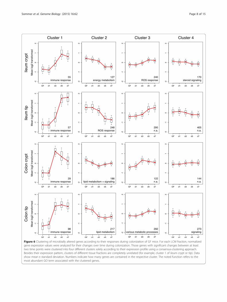
Figure 6 Clustering of microbially altered genes according to their responses during colonization of GF mice. For each LCM fraction, normalizedgene expression values were analyzed for their changes over time during colonization. Those genes with significant changes between at leasttwo time points were clustered into four different clusters solely according to their expression profile using a consensus-clustering approach.Besides their expression pattern, clusters of different tissue fractions are completely unrelated (for example, cluster 1 of ileum crypt or tip). Datashow mean ± standard deviation. Numbers indicate how many genes are contained in the respective cluster. The noted function refers to themost abundant GO term associated with the clustered genes.
Sommer et al. Genome Biology (2015) 16:62 Page 8 of 15

Sommer et al. Genome Biology (2015) 16:62 Page 9 of 15
sequences of the microbially regulated gene clusters(Additional file 9). Of the 50 most significant regulatoryfactors, 63 to 69% were specific for one of the four clus-ters in any epithelial fraction. Furthermore, only about20% of the regulatory factors in clusters 1 or 2 of thefour epithelial fractions were shared, although they allcontained genes with immune or metabolic functions,respectively. This indicates, therefore, that the gutmicrobiota elicits its site-specific transcriptional re-sponses in the different host epithelial fractions usingdistinct regulatory networks.
DiscussionWe systematically investigated the microbiota-inducedtranscriptional responses in specific subpopulations ofintestinal epithelial cells using extensive microarray ana-lysis of LCM-harvested ileal and colonic tip and cryptfractions from GF and CR mice as well as during thetime course of colonization. This analysis revealed thatapproximately 10% of the host’s transcriptome wasmicrobially regulated and that the transcriptional re-sponse of the epithelium towards the microbiota wasfaster in the colon than in the ileum. The main genefunctions that were affected by the microbiota were hostimmunity, cell proliferation and metabolism. However,the microbial impact on epithelial gene expression wasalso highly site specific as the microbiota-induced hosttranscriptional responses differed not only between ilealand colonic samples but also among tip and crypt frac-tions. For example, we corroborated that the gut micro-biota promoted cell proliferation in crypts of the ileumand colon as well as glutathione-S-transferase activity inthe colonic tip epithelium. The microbiota seemed touse different networks of transcriptional regulators toelicit the observed specific alterations in the epithelialtranscriptome profiles. To facilitate a further decipheringof the microbiota-epithelium interactions in the intestineby the research community we generated an online data-base [37] using the data presented here which allowseasy access to our dataset presented here.
Most microbially induced transcriptional changes arerestricted to epithelial cellsIn a previous study we compared the transcriptionalprofiles in duodenum, jejunum, ileum and colon be-tween GF and CR mice [15]. Few other recent studiesalso investigated the instructive capacity of the gutmicrobiota on the host transcriptome in a systematicway during a non-diseased state. El Aidy and colleaguesexplored the interaction between the microbiota and thehost intestinal tissue during colonization of germ-freemice in jejunum, ileum and colon [11,13,14]. Further-more, a few recent studies analyzed a microbial impacton isolated but unfractionated epithelium [38-40]. In our
current study we extended these earlier findings by usingLCM-harvested epithelial fractions instead of whole tis-sue samples or unfractionated epithelium, thus enablingthe analysis of the interactions between the gut micro-biota and specific epithelial fractions. The value of ourexperimental approach is demonstrated by the fact thatmany findings of microbially regulated genes or pro-cesses observed in intact tissues or even in unfractio-nated epithelium from previous studies are not universalbut limited to specific epithelial fractions. For example,Donohue and colleagues [38] showed that the micro-biota alters colonocyte energy metabolism by providingbutyrate as energy substrate and thus upregulating genesinvolved in ß-oxidation (Acads, Hadha) or the tricarb-oxylic acid cycle (Aco2, Mdh2). However, we found thatthe changes in expression of these genes were restrictedto IECs of the colon tip epithelium. Pott and colleagues[39] showed that the microbiota modulates expressionof Tlr4 in the small intestinal epithelium. Our data dem-onstrate that this effect is restricted to the ileum tip.Camp and colleagues [40] showed similar epithelial re-sponses in ileum and colon towards the microbiota.However, we found that many of the microbially inducedfunctional alterations are site-specific: immune re-sponses in the tip epithelium of ileum and colon, regula-tion of cell proliferation in the crypt fractions of ileumand colon, whereas lipid metabolism and glutathionemetabolism were specific for ileum and colon tip frac-tions, respectively. In a recent study, Kim and colleagues[41] reported transcriptional profiles for Lgr5-positivestem cells, IEC progenitors of the secretory and entero-cyte lineage, and mature enterocytes. To explorewhether the microbiota-induced transcriptional changesreported here were partly due to alterations in abun-dance or activity of specific IEC types, we aimed to com-pare these two epithelial transcriptome datasets.Unfortunately, these comparisons were technically in-appropriate due to confounding factors between the twodatasets, for example, the use of different microarraytechnologies and normalization of the data.In line with the data from previous reports, we found
that the gut microbiota has wide-ranging effects on hostgene expression, mainly inducing genes with immunefunctions in the ileum and genes with metabolic func-tion in the colon. In fact, most genes, for example Cxcl5,Cxcl9 or Sprr1a, which function in mucosa fortification,showed a similar expression pattern as reported in ourprevious study [15]. This might, therefore, indicate thatmany of the microbiota-induced transcriptional changesare based in epithelial cells of the intestinal mucosa.However, in contrast to the earlier reports we did notdetect alterations in several immune genes previouslyfound to be microbially regulated. For example, wefound no increase and in fact not even detectable levels

Sommer et al. Genome Biology (2015) 16:62 Page 10 of 15
of the regulatory T cell markers T-bet and Foxp3 or theTh17 gene Cxcl-17 during conventionalization as was re-ported previously [14,42]. These differences are mostlikely due to our experimental design involving LCM,which allowed analyzing the transcriptional responses ofisolated epithelial fractions and therefore did not containregulatory T cell or Th17 lymphocytes, demonstratingthat many of the microbially induced immune responsesdetected by studies using whole tissues might be due tothe presence of immune cells. Thus, restricting the studyof microbially induced expression to epithelial cells, aswe performed here, allowed delineation towards onlythis cell type. However, this experimental design alsorepresents a potential limitation as possible interactiveor indirect responses - for example, together with or viasensory immune cells - were omitted from this analysisdue to its epithelial focus. We can thus not exclude thatsome genes are indirectly regulated by the gut micro-biota through its interactions with immune cells. Fur-thermore, our analyses do not allow us to identify whichepithelial cells respond to the gut microbiota. This willrequire further experiments using single cell analyses.
Epithelial cells in different locations respond differentiallyto the microbiotaThe use of LCM facilitated a detailed analysis of host-microbial interactions in specific subpopulations of in-testinal epithelial cells and revealed that the majority ofmicrobially regulated genes (1,941 of 2,256) were specificfor one of the four epithelial fractions (ileum crypt,ileum, tip, colon crypt and colon tip). For example, wefound the gene Angptl4, which is involved in lipid metab-olism and was already described earlier as microbially reg-ulated [43], to be repressed by bacteria specifically in theileal tip epithelium (P < 0.001). Similarly, the microbiallyregulated genes Fgf15 and Sprr1a [15,44] were only modu-lated in the ileal and colonic tip epithelium, respectively.Since the gut microbiota only had a minor impact on
IEC composition as supported by published data [14,45],the observed site-specific microbial impact on epithelialgene expression seems to be mainly caused by directtranscriptional reprogramming in the epithelial fractions.There are several factors that could account for thestrong site specificity. First, the tip and crypt fractionscontain different types of epithelial cells (enterocytesand goblet cells in the tip versus stem and Paneth cellsin the crypt) with different cellular functions. Second,the microbial composition differs among both theproximal-distal and tissue lumen axes [4]. Thus,microbiota-derived signaling stimuli likely differ fromileum to colon and between tip and crypt in the sameintestinal segment and might thereby induce differentsignaling events leading to alternative transcriptional re-sponses in the host epithelium. In line with this
hypothesis, by screening for enriched transcriptional reg-ulators among the microbially responsive genes, weidentified regulatory factors that were specificallyenriched in a single epithelial fraction and associatedwith the most significant GO functions (for example,NFkB and CEBPB) with the immune genes Pigr or Mifin the ileum tip epithelium. This is further supported byour observation that, when clustering all microbially reg-ulated genes according to their changes in expressionduring colonization, we identified different genes andregulatory factors in each of the four gene clusters of asingle epithelial fraction (for example, clusters 1 to 4 ofileum tip) but also among the same cluster of differentepithelial fractions (for example, cluster 1 in ileum crypt,ileum tip, colon crypt and colon tip), although they oftenare involved in similar cellular functions such as lipidmetabolism. This highlights, therefore, the specific regu-latory networks engaged by the gut microbiota to alterhost epithelial gene expression and function accordingto the requirements in that particular niche. It is import-ant to note, however, that many of the published data-bases that we used to predict transcriptional regulatorswere not derived from mouse intestine and thus func-tional proof of the predicted transcriptional regulatoryinteractions in the intestine remain to be demonstrated.
Microbial impact on epithelial reprogrammingThe gut microbiota has been implicated to impact thehost’s epigenetic program [46-49]. Bacterially producedfolate is the essential methyl group donor during histonemethylation and thereby leads to inhibition of transcrip-tion [49]. In contrast, the microbially produced short-chain fatty acids butyrate and propionate are potent his-tone deacetylase (HDAC) inhibitors and thereby increasetranscriptional activity [47]. It was recently shown thatepithelial HDAC3 is critical for intestinal homeostasis byproper integration of microbially derived signals [46].Many of the GO functions that were enriched in ouranalysis, including lipid metabolism, glutathione metab-olism, antigen presentation or immune response, werealso enriched in IECs isolated from HDAC3 knockoutcompared with wild-type mice [46]. Furthermore, manyof the transcriptional regulators that we found enrichedamong the microbially regulated genes were methylationsand acetylations of histones, including the prototypicalHDAC3 target H3k9ac. Thus, many of the microbially re-sponsive genes might be regulated epigenetically, poten-tially via short-chain fatty acid-repressed HDACs or afolate-induced increase in DNA methylation.
The colonic epithelium appears to respond faster tomicrobial stimuli than the ilealBased on these associations between the gut microbiotaand host epigenetics, we investigated whether the gut

Sommer et al. Genome Biology (2015) 16:62 Page 11 of 15
microbiota can impact epithelial reprogramming. There-fore, we analyzed the kinetics in the epithelial responseto microbial colonization expecting a delayed instead ofimmediate responses in the tip compared with the cryptfractions since all epithelial cells originate from thecrypts. In contrast to our hypothesis we did not noteany delayed responses in the tips compared with cryptsbut we found that, on average, the colonic epitheliumappeared to respond faster than the ileal epithelium. Thefaster response in the colonic versus the ileal tip fractionmight be explained by a shorter distance between theproliferative stem cells in the crypt and the differentiatedcells in the tip leading to a faster epithelial turnovertime: 1 to 2 days versus 3 to 5 days in the colon versusileum, respectively [50,51]. This, however, would not ex-plain why the crypts in the colon also responded fasterto microbial colonization than ileal crypts. This is par-ticularly intriguing as the colon is more protectedagainst bacterial contact than the ileum owing to athicker and continuous mucus layer [52]. On the otherhand, the colon harbors the highest density of bacteriaand therefore potentially more signals to induce fastertranscriptional responses [8]. Another possible explan-ation for the faster response in the colon might be thatthe microbiota used for colonization of the mice washarvested from the colon of donor mice, and thus thecolonizing microbiota might be more adapted to the co-lonic environment. Furthermore, apart from direct tran-scriptional control, other processes such as mRNAstability or IEC turnover and differentiation might alsocontribute to the overall transcript levels.
ConclusionOur data show that the gut microbiota induces wide-ranging host responses. Epithelial cells are the primaryresponders to microbial signals but not all epithelial cellsrespond equally to microbial contact. In fact, themicrobiota-induced responses are extremely site specific.Therefore, the database presented here will facilitatedeciphering the microbiota-epithelium interactions inthe intestine.
Materials and methodsMiceC57Bl6/J female 12-week-old mice were maintainedunder standard specific pathogen free or GF conditionsas described previously [53]. Mice were kept under a 12-h light cycle and fed autoclaved chow diet ad libitum(Labdiet, St Louis, MO, USA). For colonization the cecalcontent from an adult CR mouse was resuspended in5 ml sterile reduced phosphate-buffered saline (PBS) andGF mice were orally gavaged with 200 μl. The colonizedmice were maintained in standard makrolon cages forup to seven days. Mice were killed by cervical
dislocation and the intestine removed. The distal part ofthe small intestine (ileum) and the proximal part of thecolon were excised, flushed with PBS and finally flushedand embedded with OCT freezing medium. Thecomplete procedure took less than 5 minutes to preserveRNA integrity. Animal protocols were approved by theGothenburg Animal Ethics Committee.
Intestinal epithelial cell compositionIntestinal specimens were harvested and fixed in 4% para-formaldehyde prior to cryoprotection, OCT embeddingand cryosectioning. Sections (10 μm) were stained usingvarious IEC subtype-specific antibodies or lectins: rat-anti-E-cadherin (all IECs; ECCD-2, Takara, #M108, MountainView, CA, USA), goat-anti-Anterior Gradient 2 (gobletcells; Santa Cruz, #sc54561, Dallas, Texas, USA), rabbit-anti-Chromogranin A (enteroendocrine cells; ImmunoStar,#20086, Hudson, WI, USA), Ulex europaeus lectin-FITC(Paneth cells; Sigma-Aldrich, #L9006, St. Louis, MO,USA). Secondary antibodies were donkey-anti-rat-AF594(Life Technologies, #A21209 Carlsbad, CA, USA), donkey-anti-goat-Cy3 (Jackson Immuno Research, #705-166-147,West Grove, PA, USA) and donkey-anti-rabbit-BV421(Nordic BioSite, #406410, Täby, Stockholm, Sweden).HOECHST (Life Technologies, #H1399) was used asDNA counterstaining. Slides were scanned using Meta-fer automated slide scanner (MetaSystems, Altlussheim,Baden-Württemberg, Germany) and composite picturesanalyzed using the Visiopharm Integrator System pro-gram [16]. Total area for each staining was determinedfor complete intestinal sections and expressed as per-centage of epithelial area (E-cadherin).
Sectioning and laser capture microdissectionAll used material and solutions were RNase free. Cryosec-tions (8 μm thick) were cut from the OCT blocks at −20°Cusing a microtome (Leica), placed onto PEN membraneslides (ZEISS) and immediately stained for LCM. Briefly,slides were dehydrated in absolute and 70% ethanol for30 s, dipped into RNase-free water to remove excessiveOCT and then incubated in 1% cresyl violet in a 50% etha-nol step for another 30 s. Slides were partially de-stainedby dipping in 70% and absolute ethanol before air-drying.Slides were stored in airtight containers at −80°C untilLCM. Airtight containers were equilibrated to ambienttemperature before opening. LCM was performed usinga PALM Microbeam Microdissection microscope(ZEISS) and fractions were collected dry. The harvestedfractions were immediately lyzed in RLT containing 1%β-mercaptoethanol and stored at −80°C.
RNA isolationRNA was isolated from the lysates of the LCM harvestedfractions using RNeasy Micro Kit (Qiagen, Hilden,

Sommer et al. Genome Biology (2015) 16:62 Page 12 of 15
Germany). RNA concentration and quality were evalu-ated using capillary electrophoresis on a 2100 Bioanaly-zer with RNA 6000 Pico kit (Agilent Technologies,Santa Clara, CA, USA).
MicroarrayTotal RNA (1,000 pg) from each sample were used togenerate amplified and biotinylated sense transcriptcDNA from the entire expressed transcriptome accord-ing to the Nugen Technologies (San Carlos, CA, USA)Ovation® Pico WTA System V2 (M01224v2) and EncoreBiotine Module (M01111v5). GeneChip ST Arrays(GeneChip Mouse Gene 2.0 ST Array) were hybridizedfor 16 h in a 45°C incubator, rotated at 60 rpm. The ar-rays were then washed and stained using the FluidicsStation 450 and finally scanned using the GeneChipScanner 3000 7G according to the GeneChip Expres-sion Wash, Stain and Scan Manual (PN 702731 rev. 3,Affymetrix, Santa Clara, CA, USA). In total, 100 arrayswere hybridized with five biological replicates per groupin the GF versus CR experiment and three biologicalreplicates per group in the colonization experiment.
Transcriptome data acquisitionThe microarray CEL files were normalized by quintilemethod with pm-only as background correction and ex-pression values were calculated using iterative ProbeLogarithmic Intensity Error (iterPlier) with expressionconsole software (Affymetrix). To evaluate the intrinsicvariability of the transcriptome data derived from differ-ent conditions, hierarchical clustering was performedbased on Pearson’s correlation distance among the sam-ples and the results presented as a dendogram plot. Thenormalized gene expression values were further used fordifferential gene expression analysis by moderated t-test[54] for pairwise comparison over different statistical hy-potheses. The statistical P-values were further correctedfor multiple testing using the Benjamini-Hochbergmethod. Venn diagrams of differential gene expressionderived from various comparisons were drawn at ad-justed P-value <0.001.
Gene expression clustering during time course ofcolonizationFirst, to compare the transcriptomes of the individualsamples, hierarchical clustering was performed based onPearson’s correlation distance among the samples andthe results presented as a dendrogram plot. Then, toanalyze the responses of individual genes, consensusclustering was performed. To this end, the normalizedgene expression values from each of the four LCM frac-tions were independently evaluated by their variationover the colonization period using one-way ANOVA.The statistical P-values were further corrected for
multiple testing by the Benjamini-Hochberg method.Only genes with an adjusted P-value <0.001 were con-sidered for clustering analysis to identify the commonresponses of genes in each LCM fraction during thecolonization period. The optimum number of clustersfor each individual LCM fraction was independentlyevaluated using the ConsensusClusterPlus package for R[55] based on Pearson’s correlation distance.
Enrichment analysisThe enrichment analyses were performed based on GOannotation or regulatory factors. The transcription factorbinding targets were retrieved and combined from theCscan database [56], including ChIP-Seq data [34], andthe ECRbase database [57]. The microRNA targets wereretrieved and combined from TargetScan database[58,59] and miRanda database [60,61]. For each analyzedgroup of genes, their GO, transcription factor andmicroRNA enrichment were evaluated for significancelevel by gene-set enrichment analysis using PIANOpackage for R [62] for GO enrichment and Fisher’s exacttest comparing with background P-value derived fromrandom sampling (average P-value of 1,000 calculationsusing a set of randomly selected genes with the samesample size as in the gene list) for transcription factorand microRNA enrichment. Finally, the 50 most signifi-cant results for each LCM fraction were selected for fur-ther analysis.
Expression analysis using quantitative real-time PCRTotal RNA was isolated from LCM fractions using anRNeasy Micro kit (Qiagen) and linear amplified using anOvation Pico WTA System V2 kit (Nugen). SYBR GreenMaster Mix buffer (1×; Bio-Rad, Hercules, CA, USA)was used for quantitative real-time PCR at final reactionvolumes of 25 μl containing 7 ng cDNA. Gene-specificresults were normalized to the ribosomal protein L32mRNA (primer sequences can be found in Additionalfile 10). Assays were performed in a CFX96 Real-TimeSystem (Bio-Rad). The reactions were analyzed with theΔΔCT method. Statistical differences between GF andCR within the LCM fractions were analyzed by two-wayANOVA using GraphPad Prism 6.
Staining for proliferative cellsCryosections (10 μm thick) of paraformaldehyde-fixedileum and colon tissue of GF and CR mice were stainedfor proliferative cells using rabbit-anti-Ki-67 antibody(Thermo Scientific, #RM-9106-S1, Waltham, MA, USA)and Rabbit IgG Vectastain Elite ABC kit (Vector Labs,#PK-6101, Burlingame, CA, USA). Sections were coun-terstained with hematoxylin/eosin. Ki-67-positive cellswere counted for 10 crypts per section from eight miceper group.

Sommer et al. Genome Biology (2015) 16:62 Page 13 of 15
Glutathione-S-transferase activity of intestinal tipepitheliumTip epithelium from intestinal tissue was harvested asdescribed previously [41]. Briefly, 4 cm long ileum andcolon segments were taken from 11-week-old GF (n = 4)and CR (n = 5) mice, flushed with PBS and turned in-side out. Tip epithelium was scraped of using a glassslide. Glutathione-S-transferase activity in the tip frac-tions was measured using a Colorimetric GST ActivityAssay Kit (Abcam, #ab65326, Cambridge, Cambridgeshire,UK) according to the manufacturer’s protocol. Proteinconcentration was determined using a BCA Protein AssayKit (Thermo Scientific, #23227). Finally, glutathione-S-transferase activity was normalized to proteinconcentration.
Data accessCEL files and normalized microarray data have been de-posited at the NCBI Gene Expression Omnibus reposi-tory (accession numbers GSE51910 and GSE51911). Thefull dataset is also available online at [37].
Additional files
Additional file 1: Table S1. Microbially regulated genes in ileal andcolonic tip and crypt fractions. Normalized intensities from both microarrayhybridization experiments for all genes in all samples together with foldchange and normal as well as FDR-corrected P-values for GF versus CRcomparison.
Additional file 2: Table S2. Gene ontology categories significantlyaltered among microbially regulated genes of ileal and colonic tip andcrypt fractions.
Additional file 3: Figure S1. The microbiota induces IEC proliferationin ileal and colonic crypts. Ileal and colonic sections were stained forthe proliferation marker Ki-67 and positive cells counted in the crypts.Data show mean ± standard error of the mean. ****P < 0.0001 (Studentt’s test).
Additional file 4: Figure S2. Glutathione-S-transferase activity isinduced by the microbiota specifically in the tip epithelium in colon butnot in ileum. Tip epithelium was isolated from ileum and colon of GFand CR mice and glutathione-S-transferase activity measured. Activity wasnormalized per milligram total protein. Data show mean ± standard errorof the mean. *P < 0.05 (two-way ANOVA).
Additional file 5: Table S3. Regulatory factors enriched in microbiallyregulated genes of ileal and colonic tip and crypt fractions.
Additional file 6: Figure S3. In colon a proportion of genes respondfaster to microbial colonization than in ileum. Candidate gene expressionin ileum crypt/tip and colon crypt/tip during colonization of GF micewith a normal microbiota. Slfn2 (schlafen 2), Zhx2 (zinc fingers andhomeoboxes 2), Ephx2 (epoxide hydrolase 2), Ifi44 (interferon-inducedprotein 44), Gbp2 (guanylate binding protein 2). Data show mean ±standard error of the mean.
Additional file 7: Table S4. Clustering of genes regulated by the gutmicrobiota during colonization of germ-free mice.
Additional file 8: Table S5. Gene ontology categories enriched incolonization responsive gene clusters.
Additional file 9: Table S6. Regulatory factors enriched in microbiallyregulated gene clusters.
Additional file 10: Table S7. List of primer sequences used inquantitative PCR analysis.
AbbreviationsChIP: chromatin immunoprecipitation; CR: conventionally raised; d: dayspost-colonization; GF: germ-free; GO: gene ontology; HDAC: histonedeacetylase; IEC: intestinal epithelial cell; LCM: laser capture microdissection;PBS: phosphate-buffered saline.
Competing interestsFB is founder and shareholder of Metabogen AB. All other authors declareno competing interests.
Authors’ contributionsFS, IN and FB conceived and designed the study. FS, IN, NS, PF and FBconducted the experiments and analyzed data. FS and FB wrote themanuscript with input from all authors. All authors read and approved thefinal manuscript.
AcknowledgementsWe thank Mattias Bergentall for assistance with harvesting the colonic tissuesfor LCM, Kwanjeera Wanichthanarak for technical support with database andwebpage generation, Anna Hallén for help with the artwork and RosiePerkins for editing the manuscript. Furthermore, we thank Erik Larsson forvaluable discussion on the manuscript and assistance generating the webdatabase. We are indebted to the staff at Centre for Cellular Imaging of theUniversity of Gothenburg and at the Uppsala Array Platform of UppsalaUniversity (Uppsala Array Platform, Department of Medical Science, Sciencefor Life Laboratory, Uppsala University, Uppsala University Hospital, 75185Uppsala, Sweden) for superb technical assistance with the LCM andmicroarray procedure. This study was supported by the Swedish ResearchCouncil, Torsten Söderberg and Ragnar Söderberg foundations, IngaBritt andArne Lundberg’s foundation, Novo Nordisk Foundation, Swedish Foundationfor Strategic Research, Knut and Alice Wallenberg foundation, EU-funded FP7project TORNADO (FP7-KBBE-222720, http://www.fp7tornado.eu/), Chalmersfoundation, Bioinformatics Infrastructure for Life Sciences (BILS), and theregional agreement on medical training and clinical research (ALF) betweenRegion Västra Götaland and Sahlgrenska University Hospital.
Author details1The Wallenberg Laboratory and Sahlgrenska Center for Cardiovascular andMetabolic Research, Department of Molecular and Clinical Medicine,University of Gothenburg, Gothenburg 41345, Sweden. 2Department ofChemical and Biological Engineering, Chalmers University of Technology,Gothenburg 41296, Sweden. 3Present Address: Comparative GenomicsGroup, Biosciences Division, Oak Ridge National Laboratory, Oak Ridge, TN37831, USA. 4Novo Nordisk Foundation Center for Basic Metabolic Research,Section for Metabolic Receptology and Enteroendocrinology, Faculty ofHealth Sciences, University of Copenhagen, Copenhagen DK-2200, Denmark.
Received: 7 November 2014 Accepted: 16 February 2015
References1. Human Microbiome Project Consortium. Structure, function and diversity of
the healthy human microbiome. Nature. 2012;486:207–14.2. Human Microbiome Project Consortium. A framework for human
microbiome research. Nature. 2012;486:215–21.3. Li J, Jia H, Cai X, Zhong H, Feng Q, Sunagawa S, et al. An integrated catalog
of reference genes in the human gut microbiome. Nat Biotechnol.2014;32:834–41.
4. Sommer F, Backhed F. The gut microbiota - masters of host developmentand physiology. Nat Rev Microbiol. 2013;11:227–38.
5. Karlsson FH, Fåk F, Nookaew I, Tremaroli V, Fagerberg B, Petranovic D, et al.Symptomatic atherosclerosis is associated with an altered gut metagenome.Nat Commun. 2012;3:1245.
6. Ley RE, Turnbaugh PJ, Klein S, Gordon JI. Microbial ecology: human gutmicrobes associated with obesity. Nature. 2006;444:1022–3.
7. Ott SJ, Musfeldt M, Wenderoth DF, Hampe J, Brant O, Folsch UR, et al.Reduction in diversity of the colonic mucosa associated bacterial microflorain patients with active inflammatory bowel disease. Gut. 2004;53:685–93.
8. Sekirov I, Russell SL, Antunes LC, Finlay BB. Gut microbiota in health anddisease. Physiol Rev. 2010;90:859–904.

Sommer et al. Genome Biology (2015) 16:62 Page 14 of 15
9. Kleerebezem M, Vaughan EE. Probiotic and gut lactobacilli andbifidobacteria: molecular approaches to study diversity and activity. AnnuRev Microbiol. 2009;63:269–90.
10. Medema JP, Vermeulen L. Microenvironmental regulation of stem cells inintestinal homeostasis and cancer. Nature. 2011;474:318–26.
11. El Aidy S, Derrien M, Merrifield CA, Levenez F, Dore J, Boekschoten MV, et al.Gut bacteria-host metabolic interplay during conventionalisation of themouse germfree colon. ISME J. 2013;7:743–55.
12. Gaboriau-Routhiau V, Rakotobe S, Lecuyer E, Mulder I, Lan A, Bridonneau C,et al. The key role of segmented filamentous bacteria in the coordinatedmaturation of gut helper T cell responses. Immunity. 2009;31:677–89.
13. El Aidy S, Merrifield CA, Derrien M, van Baarlen P, Hooiveld G, Levenez F,et al. The gut microbiota elicits a profound metabolic reorientation in themouse jejunal mucosa during conventionalisation. Gut. 2013;62:1306–14.
14. El Aidy S, van Baarlen P, Derrien M, Lindenbergh-Kortleve DJ, Hooiveld G,Levenez F, et al. Temporal and spatial interplay of microbiota and intestinalmucosa drive establishment of immune homeostasis in conventionalizedmice. Mucosal Immunol. 2012;5:567–79.
15. Larsson E, Tremaroli V, Lee YS, Koren O, Nookaew I, Fricker A, et al. Analysisof gut microbial regulation of host gene expression along the length of thegut and regulation of gut microbial ecology through MyD88. Gut.2012;61:1124–31.
16. Kijani S, Yrlid U, Heyden M, Levin M, Boren J, Fogelstrand P. Filter-densemulticolor microscopy. PLoS One. 2015;10, e0119499.
17. Abrams GD, Bauer H, Sprinz H. Influence of the normal flora on mucosalmorphology and cellular renewal in the ileum. A comparison of germ-freeand conventional mice. Lab Invest. 1963;12:355–64.
18. den Hollander J, Rimpi S, Doherty JR, Rudelius M, Buck A, Hoellein A, et al.Aurora kinases A and B are up-regulated by Myc and are essential formaintenance of the malignant state. Blood. 2010;116:1498–505.
19. Haugwitz U, Wasner M, Wiedmann M, Spiesbach K, Rother K, Mossner J,et al. A single cell cycle genes homology region (CHR) controls cell cycle-dependent transcription of the cdc25C phosphatase gene and is able tocooperate with E2F or Sp1/3 sites. Nucleic Acids Res. 2002;30:1967–76.
20. Ngo CV, Gee M, Akhtar N, Yu D, Volpert O, Auerbach R, et al. An in vivofunction for the transforming Myc protein: elicitation of the angiogenicphenotype. Cell Growth Differ. 2000;11:201–10.
21. Rogoff HA, Pickering MT, Frame FM, Debatis ME, Sanchez Y, Jones S, et al.Apoptosis associated with deregulated E2F activity is dependent on E2F1and Atm/Nbs1/Chk2. Mol Cell Biol. 2004;24:2968–77.
22. Stanelle J, Stiewe T, Theseling CC, Peter M, Putzer BM. Gene expressionchanges in response to E2F1 activation. Nucleic Acids Res. 2002;30:1859–67.
23. Yan Z, DeGregori J, Shohet R, Leone G, Stillman B, Nevins JR, et al. Cdc6 isregulated by E2F and is essential for DNA replication in mammalian cells.Proc Natl Acad Sci U S A. 1998;95:3603–8.
24. Zeller KI, Zhao X, Lee CW, Chiu KP, Yao F, Yustein JT, et al. Global mappingof c-Myc binding sites and target gene networks in human B cells. Proc NatlAcad Sci U S A. 2006;103:17834–9.
25. Bruno ME, Frantz AL, Rogier EW, Johansen FE, Kaetzel CS. Regulation of thepolymeric immunoglobulin receptor by the classical and alternative NF-kappaBpathways in intestinal epithelial cells. Mucosal Immunol. 2011;4:468–78.
26. Chen L, Yang G, Zhang X, Wu J, Gu Q, Wei M, et al. Induction of MIFexpression by oxidized LDL via activation of NF-kappaB in vascular smoothmuscle cells. Atherosclerosis. 2009;207:428–33.
27. Cho ML, Moon YM, Heo YJ, Woo YJ, Ju JH, Park KS, et al. NF-kappaB inhibitionleads to increased synthesis and secretion of MIF in human CD4+ T cells.Immunol Lett. 2009;123:21–30.
28. Dommels YE, Butts CA, Zhu S, Davy M, Martell S, Hedderley D, et al.Characterization of intestinal inflammation and identification of relatedgene expression changes in mdr1a(−/−) mice. Genes Nutr. 2007;2:209–23.
29. Ruffell D, Mourkioti F, Gambardella A, Kirstetter P, Lopez RG, Rosenthal N,et al. A CREB-C/EBPbeta cascade induces M2 macrophage-specific geneexpression and promotes muscle injury repair. Proc Natl Acad Sci U S A.2009;106:17475–80.
30. Sommer F, Backhed F. The gut microbiota engages different signalingpathways to induce Duox2 expression in the ileum and colon epithelium.Mucosal Immunol. 2015;8:372–9.
31. Takenouchi-Ohkubo N, Takahashi T, Tsuchiya M, Mestecky J, Moldoveanu Z,Moro I. Role of nuclear factor-kappaB in the expression by tumor necrosisfactor-alpha of the human polymeric immunoglobulin receptor (plgR) gene.Immunogenetics. 2000;51:289–95.
32. Consortium EP. An integrated encyclopedia of DNA elements in the humangenome. Nature. 2012;489:57–74.
33. Obach M, Navarro-Sabate A, Caro J, Kong X, Duran J, Gomez M, et al. 6-Phosphofructo-2-kinase (pfkfb3) gene promoter contains hypoxia-induciblefactor-1 binding sites necessary for transactivation in response to hypoxia.J Biol Chem. 2004;279:53562–70.
34. Wang J, Zhuang J, Iyer S, Lin X, Whitfield TW, Greven MC, et al. Sequencefeatures and chromatin structure around the genomic regions bound by119 human transcription factors. Genome Res. 2012;22:1798–812.
35. Wang ND, Finegold MJ, Bradley A, Ou CN, Abdelsayed SV, Wilde MD, et al.Impaired energy homeostasis in C/EBP alpha knockout mice. Science.1995;269:1108–12.
36. Whitfield TW, Wang J, Collins PJ, Partridge EC, Aldred SF, Trinklein ND, et al.Functional analysis of transcription factor binding sites in human promoters.Genome Biol. 2012;13:R50.
37. Encyclopedia of gut microbiota regulated genes. http://microbiota.wall.gu.se.38. Donohoe DR, Garge N, Zhang X, Sun W, O’Connell TM, Bunger MK, et al.
The microbiome and butyrate regulate energy metabolism and autophagyin the mammalian colon. Cell Metab. 2011;13:517–26.
39. Pott J, Stockinger S, Torow N, Smoczek A, Lindner C, McInerney G, et al.Age-dependent TLR3 expression of the intestinal epithelium contributes torotavirus susceptibility. PLoS Pathog. 2012;8, e1002670.
40. Camp JG, Frank CL, Lickwar CR, Guturu H, Rube T, Wenger AM, et al.Microbiota modulate transcription in the intestinal epithelium withoutremodeling the accessible chromatin landscape. Genome Res.2014;24:1504–16.
41. Kim TH, Li F, Ferreiro-Neira I, Ho LL, Luyten A, Nalapareddy K, et al. Broadlypermissive intestinal chromatin underlies lateral inhibition and cell plasticity.Nature. 2014;506:511–5.
42. Geuking MB, Cahenzli J, Lawson MA, Ng DC, Slack E, Hapfelmeier S, et al.Intestinal bacterial colonization induces mutualistic regulatory T cellresponses. Immunity. 2011;34:794–806.
43. Bäckhed F, Ding H, Wang T, Hooper LV, Koh GY, Nagy A, et al. The gutmicrobiota as an environmental factor that regulates fat storage. Proc NatlAcad Sci U S A. 2004;101:15718–23.
44. Sayin SI, Wahlstrom A, Felin J, Jantti S, Marschall HU, Bamberg K, et al. Gutmicrobiota regulates bile acid metabolism by reducing the levels oftauro-beta-muricholic acid, a naturally occurring FXR antagonist. Cell Metab.2013;17:225–35.
45. Stappenbeck TS, Hooper LV, Gordon JI. Developmental regulation ofintestinal angiogenesis by indigenous microbes via Paneth cells. Proc NatlAcad Sci U S A. 2002;99:15451–5.
46. Alenghat T, Osborne LC, Saenz SA, Kobuley D, Ziegler CG, Mullican SE, et al.Histone deacetylase 3 coordinates commensal-bacteria-dependent intestinalhomeostasis. Nature. 2013;504:153–7.
47. Arpaia N, Campbell C, Fan X, Dikiy S, van der Veeken J, Deroos P, et al.Metabolites produced by commensal bacteria promote peripheralregulatory T-cell generation. Nature. 2013;504:451–5.
48. Kellermayer R, Dowd SE, Harris RA, Balasa A, Schaible TD, Wolcott RD, et al.Colonic mucosal DNA methylation, immune response, and microbiomepatterns in Toll-like receptor 2-knockout mice. FASEB J. 2011;25:1449–60.
49. Mischke M, Plosch T. More than just a gut instinct-the potential interplaybetween a baby’s nutrition, its gut microbiome, and the epigenome. Am JPhysiol Regul Integr Comp Physiol. 2013;304:R1065–9.
50. Leblond CP, Stevens CE. The constant renewal of the intestinal epitheliumin the albino rat. Anat Rec. 1948;100:357–77.
51. Creamer B, Shorter RG, Bamforth J. The turnover and shedding of epithelialcells I. The turnover in the gastro-intestinal tract. Gut. 1961;2:110–8.
52. Hansson GC. Role of mucus layers in gut infection and inflammation. CurrOpin Microbiol. 2012;15:57–62.
53. Sommer F, Adam N, Johansson MEV, Xia L, Hansson GC, Bäckhed F. Alteredmucus glycosylation in core 1 O-glycan-deficient mice affects microbiotacomposition and intestinal architecture. PLoS One. 2014;9, e85254.
54. Smyth GK. Linear models and empirical bayes methods for assessingdifferential expression in microarray experiments. Stat Appl Genet Mol Biol.2004;3:1–25.
55. Wilkerson MD, Hayes DN. ConsensusClusterPlus: a class discovery tool withconfidence assessments and item tracking. Bioinformatics. 2010;26:1572–3.
56. Zambelli F, Prazzoli GM, Pesole G, Pavesi G. Cscan: finding commonregulators of a set of genes by using a collection of genome-wide ChIP-seqdatasets. Nucleic Acids Res. 2012;40:W510–5.

Sommer et al. Genome Biology (2015) 16:62 Page 15 of 15
57. Loots G, Ovcharenko I. ECRbase: database of evolutionary conservedregions, promoters, and transcription factor binding sites in vertebrategenomes. Bioinformatics. 2007;23:122–4.
58. Grimson A, Farh KK, Johnston WK, Garrett-Engele P, Lim LP, Bartel DP.MicroRNA targeting specificity in mammals: determinants beyond seedpairing. Mol Cell. 2007;27:91–105.
59. Friedman RC, Farh KK, Burge CB, Bartel DP. Most mammalian mRNAs areconserved targets of microRNAs. Genome Res. 2009;19:92–105.
60. John B, Enright AJ, Aravin A, Tuschl T, Sander C, Marks DS. HumanMicroRNA targets. PLoS Biol. 2004;2, e363.
61. Betel D, Wilson M, Gabow A, Marks DS, Sander C. The microRNA.orgresource: targets and expression. Nucleic Acids Res. 2008;36:D149–53.
62. Varemo L, Nielsen J, Nookaew I. Enriching the gene set analysis of genome-widedata by incorporating directionality of gene expression and combiningstatistical hypotheses and methods. Nucleic Acids Res. 2013;41:4378–91.
Submit your next manuscript to BioMed Centraland take full advantage of:
• Convenient online submission
• Thorough peer review
• No space constraints or color figure charges
• Immediate publication on acceptance
• Inclusion in PubMed, CAS, Scopus and Google Scholar
• Research which is freely available for redistribution
Submit your manuscript at www.biomedcentral.com/submit
Copyright © 2022 FDOKUMEN