An Introduction to Computational Diffusion MRI: the Diffusion Tensor and Beyond

Single particle trackingAnalysis of diffusion and flow in two-dimensional systems
Hong Qian,* Michael P. Sheetz,4 and Elliot L. Elson**Department of Biochemistry and Molecular Biophysics, and *Department of Cell Biology and Physiology, Division of Biology andBiomedical Sciences, Washington University School of Medicine, St. Louis, Missouri 631 10 USA
ABSTRACT Analysis of the trajectories of small particles at high spatial and temporal resolution using video enhanced contrastmicroscopy provides a powerful approach to characterizing the mechanisms of particle motion in living cells and in other systems.We present here the theoretical basis for the analysis of these trajectories for particles undergoing random diffusion and/orsystematic transport at uniform velocity in two-dimensional systems. The single particle tracking method, based on observations ofthe trajectories of individual particles, is compared with methods that characterize the motions of a large collection of particles suchas fluorescence photobleaching recovery. Determination of diffusion coefficients or transport velocities either from correlation ofpositions or of velocities of the particles is discussed. A result of practical importance is an analysis of the dependence of theexpected statistical uncertainty of these determinations on the number of position measurements. This provides a way of judgingthe accuracy of the diffusion coefficients and transport velocities obtained using this approach.
INTRODUCTION
The diffusion and systematic drift of membrane proteinshas been studied for a number of years for clues to themechanisms of various cellular processes such as theformation of specialized surface structures (e.g., Dubin-sky et al., 1989, and references cited therein), theinteractions between membrane bound enzymes andother reaction constituents (e.g., Chazotte and Hacken-brock, 1989), the ligand-receptor-mediated triggering ofcellular responses (Schlessinger, 1986), and cellularlocomotion (Sheetz et al., 1989). Although the diffusionof membrane proteins may not limit the rates of theseprocesses (e.g., McCloskey and Poo, 1986), measure-ments of membrane protein mobility can provide impor-tant information about membrane structure, interac-tions between membrane components, and mechanismsofmembrane and/or cytoskeleton functions (e.g., Cherry,1979; Sheetz, 1983; Yechiel and Edidin, 1987; Angelideset al., 1988; Ryan et al., 1988; Duband et al., 1988).During the past two decades most experimental measure-ments of the lateral diffusion of membrane lipids andproteins have been carried out by fluorescence photo-bleaching recovery (FPR, e.g., Elson, 1985, and refer-ences cited therein), although other methods such aselectromigration and postfield relaxation, and diffusionlimited excimer formation (Poo, 1981; Eisinger et al.,1986) have also been used. Recently, an optical method
Dr. Qian's current address is Institute of Molecular Biology,University of Oregon, Eugene, Oregon 97403-1229.Dr. Sheetz's current address is Department of Cell Biology, DukeUniversity Medical Center, Box 3011, Durham North Carolina 27710.
has been developed for measuring the movements ofsingle small particles (Geerts et al., 1987) with thepossibility of nanometer-scale precision (Gelles et al.,1988; Sheetz et al., 1989). The single particle tracking(SPT) method can provide information not available toFPR and other measurements which are based on thebehavior of large ensembles of molecules (Dembo andHarris, 1981). This paper presents methods for analyz-ing SPT measurements, a comparison of SPT with FPR,and, most important, a consideration of the statisticalaccuracy of SPT measurements. A similar approach hasalso been used to characterize the movements on fibro-blasts of single LDL receptors monitored by fluores-cence video microscopy (Gross and Webb, 1988).FPR and SPT differ in their spatial resolution and
statistical characteristics. FPR measures fluorescencerecovery in a photobleached region of the sample due tothe diffusion or drift of a large number of molecules. Thespatial resolution, limited by the minimum size of adiffraction-limited laser beam, can be on the order of
0.5 ,um. SPT measurements provide the x and ycoordinates of the centers of individual small (e.g., 40nm) gold particles, presumed to be rigidly attached tomembrane proteins by antibodies or other ligands,recorded at successive times by video enhanced differen-tial interference contrast (DIC) microscopy. The trajec-tories of random movement of these particles can bedetermined to a resolution of < 10 nm by analysis of thevideo images (Gelles et al., 1988; de Brabander et al.,1988). In principle, therefore, SPT can observe motionover a spatial range at least an order of magnitude
910 0006-3495/91/10/910/12 $2.00 Biophys. J. c Biophysical Society
Biophys. J.6 Biophysical SocietyVolume 60 October 1991 910-921
910 0006-3495/91/10/910/12 $2.00

smaller than that accessible to FPR. In an SPT measure-ment, numerous observations of the stochastic motion ofthe individual gold particles must be characterizedstatistically to yield the appropriate time averaged trans-port properties. In contrast, the participation of manyfluorescent particles provides an ensemble average oftheir diffusion behavior, and therefore their diffusioncoefficient, in a single FPR recovery measurement.The characteristics of the particle motion can be
derived either from the trajectory, the sequence ofpositions: r1, r2, r3, .... at times tl, t2, t3, ... , or from thechanges of position at successive times, the velocities:VI = (r2- r1)/AT, v2 = (r3- r2)IAT, .... where ATis thetime interval between each measurement. These twoapproaches provide the same information about thephenomenological characteristics of particle motion, butfrom different perspectives. This will be demonstratedalong with some mathematical relationships betweenthe two types of analyses. The velocity analysis providesa consistency check on the analysis of trajectories.
MACROSCOPIC AND MICROSCOPICMEASUREMENTS
For a uniform population of molecules, measurementsof the time averaged motion of a single molecule and ofthe ensemble averaged motion of a collection of indepen-dent molecules should yield identical values of thediffusion coefficient or drift velocity (Landau and Lif-shitz, 1969). In most systems of biological interest,however, molecules of a specific type can exist indifferent dynamic states. For example, the Con Areceptors bound to 40-nm gold particles on the surfacesboth of macrophages (Sheetz et al., 1989) and of fishepidermal keratocytes (Kucik et al., 1989) can be eitherrandomly diffusing or systematically transported towardor away from the cell nucleus. Even though each Con Areceptor eventually experiences both states, individualmolecules can be seen to be in either the diffusing or theactive transport state over a time range of -10 s.Because at any one time the actively transported mole-cules represent only a small minority, they cannot beobserved by FPR, which measures an averaged behaviorof the entire population. In contrast, as long as particlesare observed in sufficient numbers to yield an adequaterepresentation of both populations, quantitatively ana-lyzed SPT measurements can clearly characterize thedifferent dynamic properties of the two classes of Con Areceptors, as previously demonstrated (Sheetz et al.,1989).The superior spatial resolution of SPT over FPR
(- 10 nm vs. - 0.5 ,um) is even more useful in studyingsystems which impose highly localized constraints on the
motion under study. In principle, high spatial (or tempo-ral) resolution is not required to measure the diffusioncoefficient for a simple diffusion process in an unlimitedspace. If one waits long enough, sufficient diffusion willoccur to permit adequate measurement, even using aninstrument with very low spatial (or temporal) resolu-tion. Biological systems, however, frequently imposespatial constraints on particle motion. For example, ithas been observed that the diffusion of cell surfacemembrane proteins is retarded by unknown forcesand/or structures. One structural model proposes thatthe membrane proteins are confined to cytoskeletal"corrals" which permit free movement within the corralbut retard passage from one corral to the next (Sheetz,1983). Therefore, submicron spatial resolution is neces-sary to detect the motion within the corral, and suchresolution is accessible to SPT but not to FPR.
Constraint of the range of particle motion, however,also imposes demands on the temporal resolution re-quired for the measurement. When a characteristicdistance is set either by the measurement method or bythe structure of the sample system, a characteristic timeconsequently arises together with a corresponding re-quirement for sufficient temporal resolution to observethe dynamic process within this time. For surface diffu-sion with diffusion coefficient, D, the characteristic time,At, is related to the characteristic distance Axc as Atc -
AxOc/4D. A decrease in the characteristic distance causesa corresponding decrease in the characteristic time andso may require a corresponding increase in the temporalresolution of measurement. Similarly, a shortening ofthe characteristic time causes a decrease in characteris-tic distance and so an increase in the spatial resolutionof the measurement is necessary. For example, in ameasurement of fast diffusion of a protein in solution byFPR, one often uses a large illuminated volume toincrease the characteristic diffusion time above thetemporal resolution limit of instrument. Conversely, thecharacteristic FPR diffusion time for a membrane pro-tein with diffusion coefficient - 10' cm2/s in a smallcytoskeletal corral (0.1 0.3 ,um, Sheetz, 1983) should
be 100 ms. This should be accessible to FPR, which iscapable of measurements of processes with characteris-tic times > 100 Rs. Nevertheless, the spatial resolutionof FPR, determined by the diffraction limited beamradius (2 0.5 p,m), is insufficient. In contrast the10-nm spatial resolution of SPT is sufficient to measurediffusion within a corral, but standard video methodshave insufficient temporal resolution ( - 30 ms). There-
fore, sufficient spatial and temporal resolution are bothneeded to measure a restricted diffusion. The temporalresolution of video microscopy can, however, be im-proved by photoelectric techniques to the range of 100,us (Howard and Hudspeth, 1987; Kamimura, 1989),
ia et a. Coreato Anlyi of Sigl Patil Tracking911Qian et al. Correlation Analysis of Single Particle Tracking 911

presumably sufficient to measure the diffusion of aparticle with diffusion coefficient D = 10-8 cm2/s in a100-nm cage.SPT and FPR also differ in that the former character-
izes microscopic properties of diffusing particles, namely,the transition probabilities, i.e., the probability that aparticle initially at position r' will be found at r at a latertime t. In many cases, the steady state microscopicdistribution does not affect the outcome of the measure-ment. This is quite different from FPR measurements inwhich the steady-state distribution usually is importantin the overall results (see below).
- 0.40zuJui 0.32u
a.0.24
< 0.16
z0)Z 0.08
Lu
o Loo .0 2 4 6 8 10 12 14 16 18 20
TIME (s)
MEAN SQUARE DISPLACEMENT ANDCORRELATION FUNCTION OF POSITION
SPT yields the trajectory r(t) = [x(t),y(t)], i.e., thecoordinates at each time t, of a particle undergoingtwo-dimensional diffusion and/or systematic transport.Because the data are stochastic, they must be analyzedstatistically to yield the conventional phenomenologicaldiffusion coefficient and drift rate (Chandrasekhar,1943). The mean square displacement (MSD) of thetrajectory, denoted as p(t), is a convenient quantitativecharacteristic of the motion:
p(t) = ([r(t) - r(0)]2)
= P(r')(r - r')2P(rl r', t) drdr'. (1)Here, P(r) is the steady-state distribution of particleposition and P(rlr', t) is the transition probability, i.e.,the probability that a particle originally at r' will be at rafter a time period t. Because the process is stationary,the ensemble average can be computed as a timeaverage for a single trajectory (Landau and Lifshitz,1969). Therefore,
p(t) = flr(t + t') - r(t')I2dt'.This allows determination of p(t) from an experimen-tally measured trajectory. Particle positions are re-corded in the form of a time sequence [xn= x(nAT),Yn = y(nAT) In = 0, 1, 2, .. .1, with the data acquisitiontime interval AT. Therefore p(t) and its x, y componentsare expressed in terms of discrete time sequences:
N
PX(nAT) = I (Xi,.- x)21(N + 1)
N
py(nAT) = (Yi+n- yi)21(N + 1),
and
Pn = p(nAT) = p,(nAT) + p,(nAT).Fig. 1 depicts p(t) for simple diffusion.
(3)
FIGURE 1 The mean square displacement as function of time for a
diffusing particle. ( ) pure diffusion; (-----) diffusion with flow;(...) diffusion in a cage. For these calculations D = 3 x 10-11 cm2/s,V = 0.02 p,m/s, and the cage size was 1 pm x 1 jim. The p(t) fordiffusion in a cage was calculated as described by Kolinski et al. (1986),using their Eq. 5 extended to a two-dimensional system.
The mean square displacement as function of time isclosely related to the position correlation function:
gr(t) = (r(t)r(O)) = ffrr'P(r')P(rlr', t)drdr'
= (112)ff [r2 + r'2 (r - r')2]P(r')P(rIr', t) drdr'
= (r2) - p(t)/2. (4)Time correlation functions have been widely used inkinetic studies (Zwanzig, 1965). Eq. 4 provides a formallink to other methods used to measure diffusion such asfluorescence correlation spectroscopy (FCS), a tech-nique similar to FPR (Elson, 1985), and dynamic lightscattering (DLS). In these techniques it is not theposition of the diffusing particle itself as a stochasticprocess r(t), but rather some function of the particlepositions I(r), that can be directly measured. The de-sired kinetic information is extracted from the timecorrelation of the process I[r(t)]. In FCS, I(r) = IOexp (-r2I/w), is the Gaussian laser profile which excitesfluorescence from particles at positions r. Then the e-2radius w0 determines the characteristic distance for thismeasurement. In dynamic light scattering: I(r) = exp(ir * q) where q is scattering vector, which varies inverselyas the wavelength of the incident light, and 1/ Iq is thecharacteristic distance imposed on the diffusion measure-ment (Cummins et al., 1969). In both cases, the photocur-rent i(t) cx I[r(t)] is the directly measured quantity.
Simple diffusionThe coordinates r = (x, y) of a particle undergoing freediffusion can be represented as a Gaussian process with
lO19 n:J--unl .Volume 6 Oo 1
(2)
to ,z Biophysical Journal Volume 60 October 1991

transition probability (Chandrasekhar, 1943):
P(rlr', t) = (1I4ITDt) exp [-(r - r')2I4Dt]. (5)
This transition probability for free space is applicable solong as the characteristic dimension of the space avail-able for diffusion, L, is sufficiently large that the measure-ment time interval is much smaller than L2/4D. Thenp(t) = ([r(t) - r(0)]2) = 4Dt. The average ( * ) is takennot only over all the possible trajectories but also over allthe initial positions r(O). This emphasis, although unnec-
essary for free diffusion, is crucial to account for theeffects of the boundaries in an analysis of diffusionwithin a finite region.
For diffusion in a finite region, from Eq. 4, p(oo) =
2((r2) - (r)2), which is proportional to the size of theregion accessible for diffusion. For example, p(oo) = R2for a disk with radius R; pC)) = A2/3 for a square of A xX. (More precisely, Vp.(oo) and F/0 specify the lineardimensions of the accessible region). Therefore, a mea-surement of p(oo) yields an estimate of the finite area
accessible for diffusion. For free diffusion, of course, theavailable area is infinite, and so p(t) increases withoutbound, i.e., both p(oo) and the second moment (r2) = m0.Hence, the time correlation function (r(t)r(0)) does notexist for free diffusion, but its difference from (r2), that isthe p(t), does exist. A typical plot of p(t) versus t fordiffusion in a finite region is also shown in Fig. 1.
Diffusion with flowWhen diffusion and drift or flow with constant velocity Vare superimposed, the transition probability becomesP(r lr ', t) = (4'rDt) 1 exp {-(r - Vt)2/4Dt} (Chan-drasekhar, 1943). Therefore,
p(t) = 4Dt + V2t2 (6)
Here we have again supposed that the total measure-ment time is much smaller than L 214D. For most cellexperiments, L 10 ,umandD 10-10 cm2/s, thereforeL214D > 103 s.
When there is drift or flow, p(t) is no longer linear intime but, as shown in Eq. 6, has positive curvature. Incontrast to simple diffusion for which the slope, p'(t) =dp(t)/dt = 4D, is a constant, when drift or flow contrib-utes, p'(t) continually increases (p"(t) = 2V2 > 0) as ifthe diffusion rate for a particle were faster the farther ithad moved. This indicates that the contribution ofsystematic motion becomes dominant at longer times.Even if flow or drift is slow compared to diffusion,because of its dependence on t2, the second term in Eq. 6must dominate p(t) at longer times. Hence, when bothdiffusion and flow are present, the former will tend todominate at early times (t << 4D/V2), the latter, at later
times (t > 4D/V2). If there are no active contributionsto the motion, either p"(t) = 0 for simple diffusion or
p"(t) < 0. As shown below, the latter possibility couldoccur if there were barriers which slowed the diffusionrate beyond some characteristic spatial scale (cf Fig. 1).A curve-fitting procedure can be used to estimate bothDand Vfrom experimentally determined p(t) (cf Sheetz etal., 1989).
Nonideal diffusion due to interactionsNegative curvature of p(t) indicates that diffusion occursnot as a simple homogeneous process in free space butrather that its rate is lower over longer than over shorterdistances. This observed behavior is most simply inter-preted in terms of interactions between the diffusingparticles and other mobile or immobile structures.Nonideal diffusive behavior has been studied theoreti-cally recently either using Monte Carlo methods (Sax-ton, 1987, 1989, 1990) or by analysis of a two-dimen-sional version of a generalized Smoluchowski Equation(Abney et al., 1989). The results of these studies indicatethat the effects of interactions, either attractive orrepulsive, with either mobile or immobile obstacles is toretard the rate of diffusion. Furthermore, the behaviorof p(t) will depend on the concentration of the obstacles.Suppose that the barriers are separated from oneanother by a mean characteristic distance A. On a
spatial scale much smaller than A the rate of diffusion isnot affected by the barriers, and so the diffusant ischaracterized by a localized diffusion coefficient DM. Ona larger spatial scale, however, the presence of thebarriers will reduce the diffusion coefficient. Therefore,with sufficient spatial and temporal resolution, p(t)should show a fast phase due to the local diffusion of theparticle with an initial slope of 4Dm. This measurementrequires both spatial and temporal resolution sufficientto measure motion over distances small compared to A,and times short compared to A2/4Dm. On a longer timescale and therefore a larger spatial scale p(t) increasesmore slowly with time to define an effective diffusioncoefficient, DM, which is diminished due to the interac-tions of the diffusant with the barriers and depends onthe fraction of the surface covered by barriers (Saxton,1989). Percolation theory indicates that long-range diffu-sion will cease altogether (i.e., DM -O 0) if the barriers
are immobile and cover more than a threshold fractionof the surface. Also, the long-time behavior of p(t) mightdepend nonlinearly on t in the presence of immobileobstacles (cf Ghosh and Webb, 1990). For immobilebarriers below the critical threshold and for mobilebarriers at any fractional coverage of the surface the rateof long range diffusion will be greater than zero, al-though diminished relative to diffusion unimpeded by
Qian et al. Correlation Analysis of Single Particle Tracking 913Qian et al. Correlation Analysis of Single Particle Tracking 913

barriers. Due to the high concentration of interactingproteins and other possible barriers in membranes theobserved behavior of p(t) will be dominated by the latterphase in which diffusion is obstructed by interactions(Sheetz et al., 1989; Ghosh and Webb, 1990), and thiswill be our principal concern.
FPR FOR STEADY STATE DIFFUSION WITHDRIFT
A comparison of SPT and FPR for the characterizationof systems in which particles both diffuse and systemati-cally drift shows an important difference in the applica-bility of the two methods. In a system in which allfluorophores are simultaneously undergoing both system-atic drift with velocity V and diffusion with diffusioncoefficient D the FPR recovery, restricted to smallextents of photobleaching for simplicity, is (Magde et al.,1978):
f(t) = 1 - K exp {-(t/Tf)2 (1 + t/Td)1/(l + t/Td). (7)
Here, K describes the extent of bleaching, Td = 2/4Dand rf = o/IV. This recovery, when t << Td is similar tothat for uniform flow without diffusion:
f(t) = 1 - Kexp{-(t/Tf)2}. (8)
When the effects of systematic drift are just compen-sated by diffusion, however, a steady-state concentrationgradient is established; there is no macroscopic masstransfer. Then, P(r) - exp (V rID) (Bretscher, 1976).Under these conditions, the FPR fluorescence recovery.f(t) after a minimal extent of photobleaching has theform:
f(t) =1 - K exp (-idt/Ti)/(l + t/rd). (9)
(This result can be obtained either by calculation of theFPR recovery in the steady-state gradient or from theabove more general expression forf(t) by setting t»>> Td.This correspondence is expected because in the longtime limit, the equilibrium distribution exp (V- rID)would have been established.) Therefore, the sensitivityof FPR measurements to flow in the presence ofdiffusion is determined by the ratio of the characteristictimes for diffusion and flow, Tf /Td. For a typical cell, V =
0.01 ,um/s, D = 10"10 cm2/s, and in typical experiments,Wo - 1 ,um, therefore, Td - 25 s, Tf 100 s. In Fig. 2, toemphasize the difference between simple diffusion anddiffusion plus drift, the reciprocal of FPR recoverycurves are plotted. A straight line is expected for simplediffusion in this type of plot. These curves show thateven for a uniform population of particles which diffuseand flow simultaneously, the effect of flow is barely
20
0'-
L. L.I I
a au8 U-
_~ _
5
t/'rd
FIGURE 2 Effect of flow on the FPR recovery curve. The fluorescencerecovery, f(t), is plotted in reciprocal normalized form: 1/f1 - f(t)].The ratio 'Tf /Td is presented for each pair of curves. ( ) recoveryafter photobleaching a steady state concentration gradient resultingfrom a balance between flow and diffusion yielding P(x) = exp (VrID). (. ) recovery after photobleaching an initially uniformconcentration, P(x) = 1.
evident with this ratio of diffusion and flow rates. Withinlimits, Td /TF can be increased and so the ability to detectflow can be enhanced by increasing the beam radius, w,using a microscope objective lens of lower magnification.SPT measurements over long enough time, however, willalways detect systematic drift (cf Fig. 1). Theoretically,FPR measurements extended to t >> T, could detecteven a small drift as a deviation from a straight line plotas in Fig. 2. This is difficult, however, because ofuncertainty in determining the final extent of recovery,F(oo), (van Zoelen et al., 1983).
DATA ANALYSIS IN VELOCITY SPACE
The velocity autocorrelation function gv(t) = (v(t)v(O))can supply an explicit test that the observed particlemotion is behaving as simple diffusion. According to therigorous mathematical theory of ideal diffusion, whichneglects all the detailed underlying mechanism of mo-tion, gv(t) should behave as a Dirac delta function:(v(t)v(O)) = 2Db(t) (Zwanzig, 1965). For our purposes,this relationship results from the fact that the velocityv(t) is averaged over a time interval AT (ms) which islong compared to the time between intermolecularcollisions (<10-12 s). Because of the large number ofcollisions during the measuring interval the velocitiesdetermined for successive intervals are completely uncor-related (cf Chandrasekhar, 1943). Then, if the particle isundergoing simple diffusion (with or without drift), acomputation of the correlation function from the experi-
94, B J V 6 O 1914 Biophysical Journal Volume 60 October 1991
O 0.8
7j
0.6
0
0.4
,_, 0. 200,i O
-0.2TIME (s)
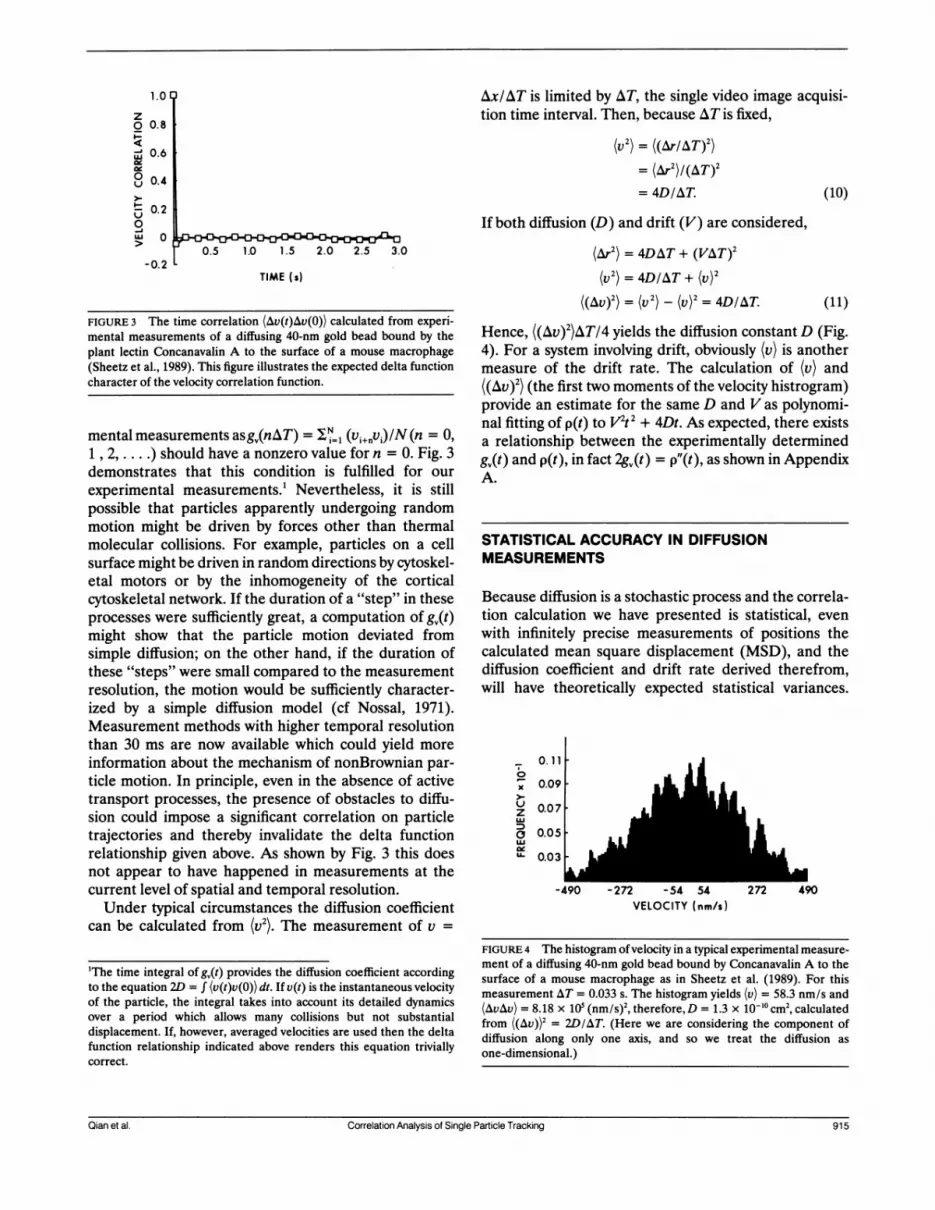
FIGURE 3 The time correlation (Av(t)Av(0)) calculated from experi-mental measurements of a diffusing 40-nm gold bead bound by theplant lectin Concanavalin A to the surface of a mouse macrophage(Sheetz et al., 1989). This figure illustrates the expected delta functioncharacter of the velocity correlation function.
mental measurements asg0(nAT) = = (v1+iv)/N (n = 0,1 , 2,.) should have a nonzero value for n = 0. Fig. 3demonstrates that this condition is fulfilled for ourexperimental measurements.! Nevertheless, it is stillpossible that particles apparently undergoing randommotion might be driven by forces other than thermalmolecular collisions. For example, particles on a cellsurface might be driven in random directions by cytoskel-etal motors or by the inhomogeneity of the corticalcytoskeletal network. If the duration of a "step" in theseprocesses were sufficiently great, a computation of gQ(t)might show that the particle motion deviated fromsimple diffusion; on the other hand, if the duration ofthese "steps" were small compared to the measurementresolution, the motion would be sufficiently character-ized by a simple diffusion model (cf Nossal, 1971).Measurement methods with higher temporal resolutionthan 30 ms are now available which could yield moreinformation about the mechanism of nonBrownian par-ticle motion. In principle, even in the absence of activetransport processes, the presence of obstacles to diffu-sion could impose a significant correlation on particletrajectories and thereby invalidate the delta functionrelationship given above. As shown by Fig. 3 this doesnot appear to have happened in measurements at thecurrent level of spatial and temporal resolution.Under typical circumstances the diffusion coefficient
can be calculated from (v2). The measurement of v =
'The time integral of gQ(t) provides the diffusion coefficient accordingto the equation 2D = f (v(t)v(0)) dt. If v(t) is the instantaneous velocityof the particle, the integral takes into account its detailed dynamicsover a period which allows many collisions but not substantialdisplacement. If, however, averaged velocities are used then the deltafunction relationship indicated above renders this equation triviallycorrect.
Ax/AT is limited by AT, the single video image acquisi-tion time interval. Then, because AT is fixed,
(v) =((Ar/AT)2)= (Ar2)/(AT)2
= 4D/AT. (10)
If both diffusion (D) and drift (V) are considered,
(Ar2) = 4DAT + (VAT)2
(V2) = 4D/AT + (V)2
((AV)2) = (V2) - (V)2 = 4DIAT (11)
Hence, ((Av)2)AT/4 yields the diffusion constant D (Fig.4). For a system involving drift, obviously (v) is anothermeasure of the drift rate. The calculation of (v) and((AV)2) (the first two moments of the velocity histrogram)provide an estimate for the same D and V as polynomi-nal fitting of p(t) to V1t2 + 4Dt. As expected, there existsa relationship between the experimentally determinedg,(t) and p(t), in fact 2gQ(t) = p"(t), as shown in AppendixA.
STATISTICAL ACCURACY IN DIFFUSIONMEASUREMENTS
Because diffusion is a stochastic process and the correla-tion calculation we have presented is statistical, evenwith infinitely precise measurements of positions thecalculated mean square displacement (MSD), and thediffusion coefficient and drift rate derived therefrom,will have theoretically expected statistical variances.
0
x
u
z
aUl.
0.11
0.09
0.07
0.05
0.03
LU
Z -54 54VELOCITY (nm/s)
FIGURE 4 The histogram ofvelocity in a typical experimental measure-ment of a diffusing 40-nm gold bead bound by Concanavalin A to thesurface of a mouse macrophage as in Sheetz et al. (1989). For thismeasurement AT = 0.033 s. The histogram yields (v) = 58.3 nm/s and(AvAv) = 8.18 x 10⁵ (nm/s)2, therefore, D = 1.3 x 10`0 cm2, calculatedfrom ((Av))2 = 2D/AT. (Here we are considering the component ofdiffusion along only one axis, and so we treat the diffusion asone-dimensional.)
Qian et al. Correlation Analysis of Single Particle Tracking 915Qian et al. Correlation Analysis of Single Particle Tracking 915

Therefore, an estimate of statistical accuracy is essentialfor our analysis.The variance arises from the stochastic nature of
diffusion. Within the limits of the accuracy of theexperimental measurements of particle positions, thisvariance will diminish as the number of position measure-ments increases.We can begin a statistical analysis by supposing that
we have K independent measurements of the squareddisplacement, e = Ir(t) - ro 2. We want to calculate thevariance of the mean e = {e(l) + t(2) + * * * + t(K)}IK.First, let us consider a pure diffusion problem. Theprobability distribution of e considered as a randomvariable is
Prob {z < i z + dz} = (1I4Dt) exp (-zI4Dt) dz
z > 0. (12)
From this, as pointed out in Appendix B, we can obtainthe probability distribution of e for which the variance is(4Dt)2/K. The measurement at time intervals AT of Nconsecutive positions r allows the calculation of Pn[= p(nAT)], the experimentally determined mean squaredisplacement for a time interval nAT by averaging overN-n + 1 measurements, i.e., 0 to n, 1 to n + 1, 2 to n +2, . . . , N - n to N. Successive determinations are notstatistically independent, however, due to the overlapbetween the measurements. Taking account of thisdependence, the expected variance for the calculated Pnwould be larger (see Eq. B5, Appendix B, setting K =
N - n + 1, and noting (2n2 + 1)/3n 2 1):
(4DnAT)2 (2n2 + 1)/3n(N - n + 1). (13)
The variance increases with increasing n. This is becausethe larger n, the smaller will be the number of statisti-cally independent samples of displacement within theinterval NAT. Hence the maximum value of n (the lastpoint calculated in the MSD), say n = m, will incur thelargest statistical uncertainty. For n = m the standarddeviation is less than 4DmAT[2mr/3(N - m + 1)112 (Fig.5). Therefore, even in the worst case, the standarddeviation in p(t), 0 < t < mAT, is less than 4DmAT[2m/3(N - m + 1)1"2. Taking the maximum uncertainty ofthis last, mth, data point, Pm, as an upper bound yields anestimated relative error in the slope of p(t) and there-fore alsoinDof ±[2m/3(N m)]"2. If3(N m)/2m
is chosen to be > 100, then the expected error in thediffusion constant due to stochastic uncertainty is smallerthan 10%.Whereas the diffusion coefficient is estimated from
the slope, the rate of systematic transport or drift isdetermined from the curvature of p(t). The statisticaluncertainty in p(t) due to the stochastic character ofdiffusion also leads to uncertainty in the estimation of
2.0,
1
._
I.
2
b 1.2
-01
30
S , j
N' 100 /
#0#0 N a I1000 P O
/ -.00
le..
-_ _ _ NN1OO
SC | | ~ ~ ~ ~~~a a1- _
~0 20 40 60TIME WHT)Os)
80 100
V.4
FIGURE 5 The relative statistical error in msd, p(t) (-). Upper andlower curve are (P.) + ((AP,APn))/2. N is the total number of positionmeasurements.
the drift rate. An upper bound of the uncertainty in thecurvature of p(t) can be obtained by considering onlythree points in p,, n = 0, 1, m, where the relativeuncertainties are 0, +1/F and +[2mn/3(N - M)]"'2,respectively. To obtain an upper bound of the curvaturelikely to be observed for a randomly diffusing particle,we use an underestimation of p(l) = 4DAT(1 - lICN),an overestimation of p(m) = 4DmAT[1 + (2m/3(N -
M))112], together with p(O) = 0 to maximize the curvaturecalculation. It should be possible to detect true system-atic transport with a velocity, V, comparable to thiscurvature. Therefore:
(VmrAT)2 = 4DmAT[[2m/3(N - M1)]"2 + 1IN]
4DmAT[m/(N )] 1/2 (14)
Hence, an error in estimating the drift rate should be< 2{D2/[M(N - m)(AT)2]J"/4. For example, with NAT =
100 s, D = 10-12cm2/s, and mAT = 50s, a drift rate assmall as 3nm/s should be detectable, and for D =
1l0olcm2/s, drift on the order of 30 nm/s would bedetectable.
Similar statistical considerations hold for determina-tion of rates of diffusion or systematic transport fromvelocity measurements. Experimental estimates of (v)and (AvAv) are obtained from the measured histogramof velocity as v =(v, + v2 + ..* vN)/N and v2 = (V2 +v2 + v 2 v)/N, respectively, whereN is the total num-ber of data points acquired. One may verify that (v)((rN - ro))/(NAT) and (v2) = p,/AT2. Therefore, v2yieldsa diffusion coefficient valid only on the time scale of AT,i.e., the experimental initial slope of p(t). By definitionv2 = (Ar/AT)2, hence the distribution of v2 correspondsto that of Ar2. Moreover, knowing the probability distri-bution of Ar2 and observing that ((Ar)2)/4DAT = (v2)AT/
91 B_pya Jora Voum 60 Ocoe
un id
r-
L
916 Biophysical Journal Volume 60 October 1991

4D, we can use the method of Appendix B to obtain theprobability distribution of v2 from a series ofN indepen-dent measurements:
Prob{z < v2ATI4D .z+dz}
= Prob {z < p1/4DAT < z + dz}
= NNzNlI(N - 1)! exp (-Nz)dz. (15)
The probability to obtain the correct diffusion coeffi-cient from P, within 10% error, can be calculated byintegrating the above distribution as follows:
Prob Iv2AT - 4D 1/4D < 0.11= Prob [vI2AT/4D - 1 < 0.11= Prob {0.9 < v2ATI4D < 1.11
NNzN-l/(N - 1)! exp (-Nz)dz. (16)
This probability is 68.4% for N = 100 and 99.8% withN = 1,000. The intrinsic statistical accuracy is high forthese measurements, but the result might not be thediffusion coefficient we seek, because there may becontributions to the MSD from other kinds of motion.Therefore, it is important to ascertain whether otherprocesses contribute by calculating the full time course
of p(t), and verifying its linearity.WhenN -x oo, for a purely diffusional process IrN roN"12, therefore IUI = IrN= rolIN N-12 0. In
experimental measurements with finite N, however,there is always a finite probability to have Iv > vo (anarbitrary velocity) even in the absence of systematictransport. That is, integrating the probability distribu-tion for v,
Prob I v > voI = Prob JrN- ro > NvoI= exp (-vWNAT/4D).
the statistical accuracy of a measurement of p(t) cannotbe judged from the smoothness of a plot of p(t) versus t.Rather, the accuracy of a measurement should beassessed from the reproducibility of a series of corre-
sponding measurements. This distinction has been previ-ously pointed out by Gross and Webb (1988) and hasalso been verified by computer simulation (Fig. 6).
EFFECT OF ERROR IN POSITIONMEASUREMENTS
As demonstrated by Gelles et al. (1988), there is an
uncertainty in the measurements of the position of a
diffusing particle. The uncertainty is due to measure-ment error and blurring over the acquisition time ofeach video frame ( - 30 ms). This uncertainty usually is a
random error which is not correlated with time. There-fore, its contribution to the MSD calculation is similar tothat of shot noise with amplitude on the order of 25-50nm2, and should have no effect on the p(t) function otherthan at zero time. Hence, the errors in position measure-ment should not influence the time course of the meansquared displacement. Nevertheless, uncertainties dueto the stochastic nature of diffusion place limits on theextent to which either positive or negative curvatures
1.2
0.8
-, 0.4r.
b.- 2.4
0
:* 1.6
(17)
The statistical error in an estimation of drift rate, i.e.,the variance in U, is f vWd[exp (-v'NAT/4D)] = 4D/NAT. Hence, with NAT = 100 s, D = 1012cm2/s, thedetectability of the drift rate (the square root of thevariance) is 2 nm/s. Therefore, at its worst the
trajectory method is comparable to the velocity analysisfor obtaining the drift rate. This is expected because inthe trajectory method, the whole range of time correla-tion is used (which is the reason that n is involved), whilein (v) only a single moment is used.
It is also worth noticing that the correlation coefficientfor pn (=p(nAT)] and Pm (=p(mAT)) is n/m, (m > n)(Appendix C). Therefore, pn and Pm are highly correlatedwhen (m - n) << n, m. This means that we should notexpect the random deviation in the experimental pn to besymmetrically distributed around (pn). In other words,
0.8
0
0 20 40 60TIME (AT)(s)
80 100
FIGURE 6 Computer simulations for a diffusing particle with the totalnumber of position measurements, N = 100 andN = 1,000. There arefive independent simulations for each N. The dashed line is thetheoretically expected mean square displacement, and the circles arethe averages of the five simulations. Although several of the curves forindividual simulations are relatively smooth, the overall standarddeviation can be judged not from the smoothness of single curve butmust be determined rather by a set of curves.
Qian et al. Correlation Analysis of Single Particle Tracking 917Qian et al. Correlation Analysis of Single Particle Tracking 917

due, respectively, to systematic transport or to con-
straints on the range of diffusion can be detected.
DISCUSSION
We have described a simple and convenient method foranalyzing the trajectories of individual particles, toobtain the macroscopic diffusion coefficients and driftvelocities which characterize their motion. This comple-ments methods such as FPR and FCS which monitorlarge populations of particles. A major advantage of theSPT method is its ability to characterize the distinctdynamic properties of minority fractions of a populationwhich might be undetectable by FCS or FPR (cf Sheetzet al., 1989). Even for a homogenous population ofparticles, however, valid SPT measurements require theobservation either of a single particle over a long timeperiod or of many particles for shorter times to accountadequately for the stochastic character of diffusion. Incontrast to FPR both SPT and FCS share the require-ment for long observation times which result from thestatistical character of the two methods.The statistical validity and mechanistic interpretation
of the measurements are governed by characteristictimes determined by the structure of the experimentalsystem and measurement times set by the experimenter.In general, there are two important operational factorsin these measurements. The time interval between eachposition measurement should be smaller than the char-acteristic time of interest. On the other hand, only thosemeasurements with time interval larger than the charac-teristic time can be considered statistically independent.Whereas the former point must be considered to providesufficient temporal resolution of the measurement, thelatter point is important in judging experimental accu-racy, which increases as the number of statisticallyindependent measurements increases.When there is only simple unrestricted diffusion, as
indicated by the linear dependence of p(t) on t, the timeresolution of the position measurements does not influ-ence the determination of the diffusion coefficient. Thisis because ([r(t) - r(0)]2) - t (i.e., there is no intrinsiccharacteristic time in the system, and the correlationbetween each measurement is negligible). Thus, theduration of a single measurement (AT) can in principlebe prolonged to any extent necessary to observe suffi-cient motion, even of very slowly diffusing particles.
In contrast, when diffusion is constrained by barriersor boundaries, an individual microscopic diffusive fluctu-ation takes place on average over a characteristic corre-lation time, ¶, defined in terms of the distance, A,separating the barriers or boundaries: r - A2/4D. Thedetermination of the diffusion coefficient from the
observation of a brief single diffusive fluctuation, even ifperformed with high precision, is relatively inaccuratedue to the stochastic nature of the diffusion process. Toobtain an accurate estimate of the diffusion coefficientmany of these microscopic fluctuations must be ob-served. The accuracy of the measurement increases withthe number of the fluctuations observed and thereforewith the total time of observations, T. Thus, the relativeerror varies as the reciprocal of the square root of thenumber of observed fluctuations and so as 1I(TIT) '.Considering again the model of membrane proteindiffusion constrained by cytoskeletal corrals (Sheetz,1983), we can suppose that, D = 10- cm2/s and A 100
nm, and T 25 ms. Therefore, if the duration of the
measurement is 25 s and AT = 2.5 ms, 10,000 data pointswill be obtained and so D should be obtained with an
estimated relative error of 1%. Of course, setting AT =
2.5 ms assumes a > 10-fold faster rate of data acquisitionthan is available using conventional video methods. Tointerpret the measurements in terms of free diffusion, itis necessary to confine attention to p(nAT) for very smalln values. When nAT becomes comparable to T, the wallsof the corrals strongly influence the diffusion behavior ofthe particles (cf Fig. 1). Even for n = 2 or 3, however, itshould be possible to determine D from the initial slopeof the plot of p(t) vs. t. For particles diffusing in a cagethe deviation of p(t) from the straight line behaviorexpected for free diffusion provides an estimate of thedimensions of the cage.
This example also demonstrates how in principle SPTcould provide a powerful approach for the analysis ofinteractions between diffusing particles and mobile or
immobile obstacles. In fact the measurement of p(t)should allow a direct comparison between theory (e.g.,Abney et al., 1989; Saxton, 1987) and experiment. In realmembranes, however, the distances between structuresthat could retard diffusion are likely to be quite small.Hence, improvements in spatial and temporal resolutionof the measurement methods will be required to achievethe full potential of this approach.To carry out a complete analysis of the statistical
accuracy of SPT measurements in systems in whichinteractions with mobile or immobile obstacles retarddiffusion would require determination of the probabilitydistribution of p(t) in the presence of the obstacles. Thisis beyond the scope of this work. Nevertheless, a more
approximate discussion is possible in much simplerterms. In the presence of mobile obstacles or of immo-bile obstacles below the percolation threshold p(t) isproportional to t (Saxton, 1987; Abney et al., 1989).Hence, even in the presence of obstacles it is possible todefine a limiting effective diffusion coefficient DCff =
(1/4)dp(t)Idt for t sufficiently great. It is reasonable tosuppose that diffusion is still essentially random under
918 Biophysical Journal Volume 60 October 1991918 Biophysical Journal Volume 60 October 1991

these circumstances and therefore that p(t) still has aGaussian probability distribution. Then the analysispresented in Appendix B remains approximately valid.Hence, as above, we can estimate the relative error inthe value of D measured from the slope dp/dt to beabout +[2mr/3(N - m)] "2 where mAT is the largestinterval used in the calculation of p(t). Because the valueofD is decreased due to interactions with the obstacles,a longer time will be required to diffuse a given distanceand so a correspondingly longer interval mAT is likely tobe selected. This in turn will require a longer totalmeasuring period NAT to achieve the same accuracy asfor unimpeded diffusion in the absence of obstacles. Asummary of the dependence of p(t) on time and ofexpected relative errors for different mechanisms oftransport is presented in Table I.A practical consideration in studies of the behavior of
membrane glycoproteins is the possible perturbationwhich results from attaching the observable particle.Likely to be most significant is the binding of a numberof glycoproteins to a single particle due both to themultivalency of the binding molecules, such as antibod-ies or lectins, and the presence of several bindingmolecules on the particle. Likely to be less significant isthe drag on the particle as it moves through the
TABLE Summary of results
ExpectedTransport mechanism p(nAT) variance
Free diffusion 4DnAT p(4DnAT)2Free diffusion + flow 4DnAT + (VnAT)2 p(4DnAT)2Diffusion confined to
a small region* A2/3 (2A4/15)zInteracting mobile
proteins'Short time 4DmnAT P(4DmflAT)2Long time 4DmnAT + A2/3 -p(4DMnAT)2(DM < Di) + (2A4/ 15)z
Percolation o(mAT)x(x < 1) NDwherep = [(2n2 + 1)/3n(N - n + 1)];N is the total number of positions
measured; and z = [A2/(4ND.AT)]
*For this example diffusion is limited to a square region of dimensionsA x A. Because the particle cannot exit from the defined region, DM =0, where DM characterizes diffusion over distances > A.tFor particles interacting with mobile obstacles the diffusion coeffi-cient can be represented as varying with time or distance diffused asindicated in the text (Abney et al., 1989; Saxton, 1989). Over distancesshort compared to the spacing of the obstacles, their effect on diffusionis small and so the apparent diffusion coefficient, D., is greater thanfor distances large compared to the spacing over which the obstaclesexert their full retarding effect to yield DM.'The variance for this example results from a generalization for freediffusion and diffusion in a confined region. A is the characteristicdistance between obstacles; A2/4Dm is the correlation time for diffu-sion across the small domains between obstacles.
TABLE ii Comparison between FPR and SPT
Diffusion coefficient
(cm2/s)FPR SPT
Acetylcholine receptoron myofibrils 6.4 x 10-'°* 4.16 x 10-'°*
ConcanavalinA receptoron macrophage 4.9 x 10-i°' 3.60 x 10"I
Sources *Dubinsky, et al., 1989; tDubinsky, unpublished measure-ments; 'Henis and Elson, 1981; ISheetz et al., 1989.
extracellular solution of relatively low viscosity. A com-parison of diffusion coefficients measured by SPT andFPR is given in Table II.An inherent advantage of the SPT method for cell
studies is that the qualitative behavior of the particle isknown before the position measurements. This is impor-tant because particles can move onto rough regions ofthe cell surface (e.g., microvilli or ruffles), reversibly stopdiffusion (Sheetz et al., 1989), or undergo rapid forwarddisplacements (Kucik et al., 1989; Sheetz et al., 1990).None of these phenomena would be distinguished in anormal FPR or FCS analysis but would obviously contrib-ute to the apparent diffusion coefficient measurement.Thus, we feel that a more reliable measurement of thetrue membrane diffusion coefficient can be made usingthe SPT method.
This analysis is not limited to the study of membraneproteins. For example, it can equally be applied to thetrajectories of cells undergoing chemotaxis and to thefacilitated diffusion ofDNA binding proteins such as lacrepressor on DNA.
APPENDIX A
Relationship between p(t) and g,(t)We consider (xi Ii = 0, 1, 2,. N| and the corresponding velocitiesIv = (xi - xj ,)/ATIi = 1, 2, .., NJ. Therefore,
(VnVn+m) = 5;n VnVn+m/N= 'INn (xn - Xn-1)(Xn+m - Xn+m-1)I(A&T)2= 1 /2N n [-(Xn- Xn+m)2 - (Xn- 1e Xn+m- 1)2+ (xn.. - Xn+m)2 + (Xn - Xn+mi)2]/(AT)2
= 1/2[-pm - pm + Pm+l + pm-,]/(AT)2Pta/ 2
that is:
2g.(t) = p"(t).
Qian et al. Correlation Analysis of Single Particle Tracking 919Qian et al. Correlation Analysis of Single Particle Tracking 919

APPENDIX B
Variance Of PnConsider the mean squared displacement of a particle diffusing in atwo-dimensional plane e = r(t) - r 12 as a random variable. Itsprobability distribution is
Prob (z < << z + dz} = (1/4Dt) exp (-zI4Dt)dz z > 0,
which yields (e) = 4Dt and, for At = - (a), (AkAk) = (t)2.If we have K independent measurements of t: t(1), k(2),. .., t(K),
and define t = ti(1) + t(2) + * *- + t(K)}IK, then the probabilitydistribution for e can be obtained as successive convolutions ofProb {z < e < z + dz) with itself (Feller, 1957). By a recurrent induc-tive calculation, we have:
Prob (z < t < z + dz}
KKzK-i exp (-KzI4Dt)
(K-1)! (4Dt)K dz z>O (Bi)
where t = nAT Therefore, (R) = (e), (LAZ) = (AkAt)/K = (t)2IK, andthe relative error ((AjAj)/(f))112 = 1/jK.
Furthermore, the probability of finding a value of the ratio {(e)greater than some number A is obtained simply by integrating Eq. Bi:
Prob {MO(i) 2 A] = f Prob (z < (/(g) < z + dzj dz
=fKJ KZK-lI(K - 1)! exp (-Kz) dz
When pn is calculated using a time average, t(1) = r - ro 12,t(2) = Ir+ r 12, t(K) = Irf+K-1 rK-, I,
Pn= {t(1) + ((2) + * + {(K)JIK/N-1
= ( ;N|Irn+ - r12)
= (Irn+i - rJ2) + (2/N2)
N-1 i-I
(Irn+l-rI2Irn+j -j12).i=i j=O
Depending on the time interval of the measurements, these i's are notnecessarily statistically independent. Hence, there will be a correctionfactor due to this consideration. A complex calculation yields:
(APnl&Pn/(Pn))"[(4n2K + 2K + n - n3)/6nK2]1"2 K (n
1[1 + (K3- 4nK2 + 4n - K)/6n2K]"12 K < n
when K > n, the relative error is:
[AP.APn/(Pn))" [(2n2 + 1)I3nK] 12 _ (2n/3K)1/2 (B5)
which is proportional to (Kln)-"2 rather than K-"12. This correction issignificant, especially when n is large. When n = 1, (APnAPI/(Pn))/2 =
1 i/k as expected for uncorrelated a's.
= [1 + KX + (KX)2/2!
+ ... (KX)K-i/(K - 1)!]e-K-= Q(2KX 2K)
Here Q(x2lv) is the x2 probability function which can be foumathematical tables (Abramowitz and Stegun, 1964).Now we define i, = Ir02) - r(O) 12 and k2 =r(Q3) - r(t,) 12, 0 s
t2 < t3. The correlation between El and t2 can be characterizedcorrelation coefficient (A*1Ak2)/((A )(A2))"2. Because the diflprocess is Markovian:
Prob (ro, 0; r,, t1; r2, t2; r3, t3}
= Prob {r3Ir2, t3 - t2} x Prob {r2Irl, t2 - tl}x Prob {r, Iro, t,| x Prob {roJ
therefore,
(*A1AU2) = ((r2- ri)4) - ((r2 - r,)2)2= [4D(t2 -tl)]2
APPENDIX C
Covariance Of Pn and Pm
(B2) We consider the correlation coefficient of any two pn and Pm.
nd in N-nPn= 2 Iri+n - ri12/(N - n + 1)
i=O>,<
by the N-mfusion Pm = kri+m - ,12/(N-m + 1),
i=O
(B3)
i.e., it is proportional to the square of the time overlap between (l andt2, and
(A\t1 )/((At21)(Ak62))112 = 2tj )2/(t- )1 A~~21 2 (2 l2t2(t3 ti).
A positive value of the correlation coefficient indicates that (l and t2are more likely to deviate from their mean values in the same
direction. Moreover, when t2 - t = 0, t, and t2 are uncorrelated. (Thisis obviously also correct for t2 < ti). When t, = 0, the coefficient isequal to t2/t3-
where N is the total number of position measurements. Without loss ofgenerality, let us assume that m > n. Therefore (pn) = 4DnAT, (Pm) =4DmAT, and:
(APnAPm)I[((Apn)2)((AtPm)2)I]
n(N - m)(3 - n/m)214m(N - n).
If m, n < Nandm - n, then:
(APnAPm)/[((APn)2)((APm))I] n/m.
We are grateful to Drs. Jeff Gelles and Scott Kuo for critical reading ofthe manuscript, and to Professor Jeffrey Skolnick and Dr. DennisKucik for useful discussions. We would also like to thank an anony-mous reviewer for a number of useful suggestions and constructivecriticisms.
This work is supported by National Institutes of Health grantsGM38838 to Elliot L. Elson, GM36277 and NS23345 and MuscularDystrophy to Michael P. Sheetz.
920 _ n m 0 O920 Biophysical Journal Volume 60 October 1991

Received for publication 23 October 1990 and in final form 29April 1991.
REFERENCES
Abney, J. R., B. A. Scalettar, and J. C. Owicki. 1989. Self diffusion ofinteracting membrane proteins. Biophys. J. 55:817-833.
Abramowitz, M., and I. A. Stegun. 1964. Handbook of MathematicalFunctions. Dover Publications, Inc., New York. 940 pp.
Angelides, K. J., W. E. Lawrence, D. J. Loftus, and E. L. Elson. 1988.Distribution and lateral mobility of voltage-dependent sodiumchannels in neurons. J. Cell Biol. 106:1911-1925.
Bretscher, M. S. 1976. Directed lipid flow in cell membranes. Nature(Lond.). 260:21-22.
Chandrasekhar, S. 1943. Stochastic problem in physics and astronomy.Rev. Mod. Phys. 15:1-89.
Chazotte, B. and C. R. Hackenbrock. 1989. Lateral diffusion as arate-limiting step in ubiquinone mediated mitochondrial electrontransport. J. Biol. Chem. 264:4978-4985.
Cummins, H. Z., F. D. Carlson, T. J. Herbert, and G. Woods. 1969.Translational and rotational diffusion constants of tobacco mosaicvirus from Rayleigh linewidths. Biophys. J. 9:520-546.
Cherry, R. J. 1979. Rotational and lateral diffusion of membraneproteins. Biochim. Biophys. Acta. 559:289-327.
de Brabander, M., R. Nuydens, H. Geerts, and C. R. Hopkins. 1988.Dynamic behavior of the transferrin receptor followed in livingepidermoid carcinoma (A431) cells with nanovid microscopy. CellMotil. Cytoskeleton. 9:30-47.
Dembo, M., and A. K. Harris. 1981. Motion of particles adhering tothe leading lamella of crawling cells. J. Cell BioL 91:528-536.
Duband, J. L., G. H. Nuckolls, A. Ishihara, T. Hasegawa, K. M.Yamada, J. P. Thiery, and K. Jacobson. 1988. Fibronectin receptorexhibits high lateral mobility in embryonic locomoting cells but isimmobile in focal contacts and fibrillar streaks in stationary cells. J.Cell Bio. 107:1385-1396.
Dubinsky, J. M., D. J. Loftus, G. D. Fischbach, and E. L. Elson. 1989.Formation of acetylcholine receptor clusters in chick myotubes:migration or new insertion? J. Cell Biol. 109:1733-1743.
Eisinger, J., J. Flores, and W. P. Petersen. 1986. A milling crowd modelfor local and long-range obstructed lateral diffusion. Biophys. J.49:987-1001.
Elson, E. L. 1985. Fluorescence correlation spectroscopy and photo-bleaching recovery. Annu. Rev. Phys. Chem. 36:379-406.
Feller, W. 1957. An Introduction to Probability Theory and ItsApplications. John Wiley and Sons, New York. 461 pp.
Geerts, H., M. De Brabander, R. Nuydens, S. Geuens, M. Moeremans,J. De Mey, and P. Hollenbeck. 1987. Nanovid tracking: a newautomatic method for the study of mobility in living cells based oncolloidal gold and video microscopy. Biophys. J. 52:775-785.
Gelles, J., B. J. Schnapp, and M. P. Sheetz. 1988. Tracking kinesin-driven movements with nanometer-scale precision. Nature (Lond.).331:450-453.
Ghosh, R. N., and Webb, W. W. 1990. Evidence for intra-membraneconstraints to cell surface LDL receptor motion. Biophys. J. 57:286a.(Abstr.)
Gross, D. J., and W. W. Webb. 1988. Cell surface clustering andmobility of the liganded LDL receptor measured by digital fluores-
cence microscopy. In Spectroscopic Membrane Probe. Vol II. L. M.Loew, editor. CRC Press Inc., Boca Raton, FL. 19-48.
Henis, Y. I., and E. L. Elson. 1981. Differences in the response ofseveral cell types to inhibition of surface receptor mobility by localconcanavalin A binding. Exp. Cell Res. 136:189-201.
Howard, J., and A. J. Hudspeth. 1987. Mechanical relaxation of thehair bundle mediates adaptation in mechanoelectrical transductionby the bullfrog's saccular hair cell. Proc. Natl. Acad. Sci. USA.84:3064-3068.
Kamimura, A., and R. Kamiya. 1989. High-frequency nanometre-scalevibration in 'quiescent' flagellar axonemes. Nature (Lond.). 340:476-478.
Kolinski, A., J. Skolnick, and R. Yaris. 1986. On the short timedynamics of dense polymeric systems and the origin of the glasstransition: a model system. J. Chem. Phys. 84:1922-1931.
Kucik, D. F., E. L. Elson, and M. P. Sheetz. 1989. Forward transport ofglycoproteins on leading lamellipodia in locomoting cells. Nature(Lond.). 340:315-317.
Landau, L. D., and E. M. Lifshitz. 1969. Statistical Physics. PergamonPress, Oxford. 484 pp.
Magde, D., W. W. Webb, and E. L. Elson. 1978. Fluorescencecorrelation spectroscopy. III. Uniform translation and laminar flow.Biopolymers. 17:361-376.
McCloskey, M. A., and M. M. Poo. 1986. Rates of membrane-associated reactions: reduction of dimensionality revised. J. CellBiol. 102:88-96.
Nossal, R. 1971. Spectral analysis of laser light scattered from motilemicroorganisms. Biophys. J. 11:341-354.
Poo, M. M. 1981. In situ electrophoresis of membrane components.Annu. Rev. Biophys. Bioeng. 10:245-276.
Ryan, T. A., J. Myers, D. Holowka, B. Baird, and W. W. Webb. 1988.Molecular crowding on the cell surface. Science (Wash. DC).239:61-64.
Saxton, M. J. 1987. Lateral diffusion in an archipelago. The effect ofmobile obstacles. Biophys. J. 52:989-997.
Saxton, M. J. 1989. Lateral diffusion in an archipelago. Distancedependence of the diffusion coefficient. Biophys. J. 56:615-622.
Saxton, M. J. 1990. Lateral diffusion in a mixture of mobile andimmobile particles. A Monte Carlo study. Biophys. J. 58:1303-1306.
Schlessinger, J. 1986. Allosteric regulation of the epidermal growthfactor receptor kinase. J. Cell Biol. 103:2067-2072.
Sheetz, M. P. 1983. Membrane skeletal dynamics: role in modulationof red cell deformability, mobility of transmembrane proteins, andshape. Semin. Hematol. 20:175-188.
Sheetz, M. P., S. Turney, H. Qian, and E. L. Elson. 1989. Nanometre-level analysis demonstrates that lipid flow does not drive membraneglycoprotein movements. Nature (Lond.). 340:284-288.
Sheetz, M. P., N. L. Baumrind, D. B. Wayne, and A. L. Pearlman.1990. Concentration of membrane antigens by forward transportand trapping in neuronal growth cones. Cell. 61:231-241.
van Zoelen, E. J. J., L. G. J. Tertoolen, and S. W. deLaat. 1983. Simplecomputer method for evaluation of lateral diffusion coefficientsfrom fluorescence photobleaching recovery kinetics. Biophys. J.42:103-108.
Yechiel, E., and M. Edidin. 1987. Micrometer-scale domains infibroblast plasma membranes. J. Cell Bio. 105:755-760.
Zwanzig, R. 1965. Time-correlation function and transport coefficientsin statistical mechanics. Annu. Rev. Phys. Chem. 16:67-102.
Qian et al. Correlation Analysis of Single Particle Tracking 921
Copyright © 2022 FDOKUMEN